January 2012 M100-S22 Vol. 32 No. 3 Replaces M100-S21 Vol. 31 No. 1 Performance Standards for Antimicrobial Susceptibility Testing; Twenty-Second Informational Supplement This document provides updated tables for the Clinical and Laboratory Standards Institute antimicrobial susceptibility testing standards M02-A11 and M07-A9. An informational supplement for global application developed through the Clinical and Laboratory Standards Institute consensus process. Licensed to: CDC Information Center Centers for Disease Control and Prevention This document is protected by copyright. CLSI order # Subscription, id # 465125, Downloaded on 1/6/2012.

Welcome message from author
This document is posted to help you gain knowledge. Please leave a comment to let me know what you think about it! Share it to your friends and learn new things together.
Transcript
January 2012
M100-S22 Vol. 32 No. 3
Replaces M100-S21 Vol. 31 No. 1
Performance Standards for Antimicrobial Susceptibility Testing; Twenty-Second Informational Supplement
This document provides updated tables for the Clinical and Laboratory Standards Institute antimicrobial susceptibility testing standards M02-A11 and M07-A9. An informational supplement for global application developed through the Clinical and Laboratory Standards Institute consensus process.
Licensed to: CDC Information Center Centers for Disease Control and PreventionThis document is protected by copyright. CLSI order # Subscription, id # 465125, Downloaded on 1/6/2012.
Clinical and Laboratory Standards Institute Advancing Quality in Health Care Testing
Clinical and Laboratory Standards Institute (CLSI) is an international, interdisciplinary, nonprofit, standards developing, and educational organization that promotes the development and use of voluntary consensus standards and guidelines within the health care community. We are recognized worldwide for the application of our unique consensus process in the development of standards and guidelines for patient testing and related health care issues. Our process is based on the principle that consensus is an effective way to improve patient testing and health care services.
In addition to developing and promoting the use of voluntary consensus standards and guidelines, we provide an open and unbiased forum to address critical issues affecting the quality of patient testing and health care.
PUBLICATIONS A document is published as a standard, guideline, or report.
Standard A document developed through the consensus process that clearly identifies specific, essential requirements for materials, methods, or practices for use in an unmodified form. A standard may, in addition, contain discretionary elements, which are clearly identified.
Guideline A document developed through the consensus process describing criteria for a general operating practice, procedure, or material for voluntary use. A guideline may be used as written or modified by the user to fit specific needs.
Report A document that has not been subjected to consensus review and is released by the appropriate consensus committee.
CONSENSUS PROCESS
CLSI’s voluntary consensus process establishes formal criteria for the following:
• Authorization of a project • Development and open review of documents • Revision of documents in response to users’ comments • Acceptance of a document as a consensus standard or
guideline
Invitation for Participation in the Consensus Process
Core to the development of all CLSI documents is the consensus process. Within the context and operation of CLSI, voluntary consensus is substantial agreement by materially affected, competent, and interested parties that may be obtained by following the consensus procedures defined in
CLSI’s Administrative Procedures. It does not always connote unanimous agreement, but does mean that the participants in the development of a consensus document have considered and resolved all relevant objections and are willing to accept the resulting agreement. CLSI documents are expected to undergo evaluation and modification in order to keep pace with advancements in technologies, procedures, methods, and protocols affecting the laboratory or health care.
Comments on Draft Documents
CLSI’s voluntary consensus process depends on experts who serve as contributing authors and/or as participants in the reviewing and commenting process. At the end of each comment period, the committee that developed the document is obligated to review all comments, respond in writing to all substantive comments, and revise the draft document as appropriate. All comments along with the committee’s responses are retained on file at CLSI and are available upon request.
Comments on Published Documents
The comments of users of published CLSI documents are essential to the consensus process. Anyone may submit a comment. All comments are addressed according to the consensus process by a committee of experts. A summary of comments and committee responses is retained on file at CLSI and is available upon request. Readers are strongly encouraged to comment at any time on any document.
APPEALS PROCESS
CLSI consensus procedures include an appeals process that is described in detail in Section 8 of the Administrative Procedures.
VOLUNTEER PARTICIPATION Health care professionals in all specialties are urged to volunteer for participation in CLSI projects.
For further information on committee participation or to submit comments, contact CLSI.
Clinical and Laboratory Standards Institute 950 West Valley Road, Suite 2500 Wayne, PA 19087 USA 610.688.0100 F: 610.688.0700 www.clsi.org [email protected]
Licensed to: CDC Information Center Centers for Disease Control and Prevention
This document is protected by copyright. CLSI order # Subscription, id # 465125, Downloaded on 1/6/2012.
Vol. 32 No. 3 M100-S22
1
Performance Standards for Antimicrobial Susceptibility Testing; Twenty-Second Informational Supplement Abstract The supplemental information presented in this document is intended for use with the antimicrobial susceptibility testing procedures published in the following Clinical and Laboratory Standards Institute (CLSI)–approved standards: M02-A11—Performance Standards for Antimicrobial Disk Susceptibility Tests; Approved Standard—Eleventh Edition; and M07-A9—Methods for Dilution Antimicrobial Susceptibility Tests for Bacteria That Grow Aerobically; Approved Standard—Ninth Edition. The standards contain information about both disk (M02) and dilution (M07) test procedures for aerobic bacteria. Clinicians depend heavily on information from the clinical microbiology laboratory for treatment of their seriously ill patients. The clinical importance of antimicrobial susceptibility test results requires that these tests be performed under optimal conditions and that laboratories have the capability to provide results for the newest antimicrobial agents. The tabular information presented here represents the most current information for drug selection, interpretation, and quality control using the procedures standardized in the most current editions of M02, M07, and M11. Users should replace the tables published earlier with these new tables. (Changes in the tables since the most current edition appear in boldface type.) Clinical and Laboratory Standards Institute. Performance Standards for Antimicrobial Susceptibility Testing; Twenty-Second Informational Supplement. CLSI document M100-S22 (ISBN 1-56238-785-5 [Print]; ISBN 1-56238-786-3 [Electronic]). Clinical and Laboratory Standards Institute, 950 West Valley Road, Suite 2500, Wayne, Pennsylvania 19087 USA, 2012.
The data in the interpretive tables in this supplement are valid only if the methodologies in M02-A11—Performance Standards for Antimicrobial Disk Susceptibility Tests; Approved Standard—Eleventh Edition; and M07-A9—Methods for Dilution Antimicrobial Susceptibility Tests for Bacteria That Grow Aerobically; Approved Standard—Ninth Edition are followed.
Licensed to: CDC Information Center Centers for Disease Control and PreventionThis document is protected by copyright. CLSI order # Subscription, id # 465125, Downloaded on 1/6/2012.

January 2012 M100-S22
2 Licensed to: CDC Information Center Centers for Disease Control and PreventionThis document is protected by copyright. CLSI order # Subscription, id # 465125, Downloaded on 1/6/2012.
M100-S22
ISBN 1-56238-785-5 (Print) ISBN 1-56238-786-3 (Electronic)
ISSN 1558-6502 (Print) ISSN 2162-2914 (Electronic)
Performance Standards for Antimicrobial Susceptibility Testing; Twenty-Second Informational Supplement Volume 32 Number 3 Franklin R. Cockerill, III, MD Matthew A. Wikler, MD, MBA, FIDSA Jeff Alder, PhD Michael N. Dudley, PharmD, FIDSA George M. Eliopoulos, MD Mary Jane Ferraro, PhD, MPH Dwight J. Hardy, PhD David W. Hecht, MD Janet A. Hindler, MCLS, MT(ASCP) Jean B. Patel, PhD, D(ABMM) Mair Powell, MD, FRCP, FRCPath Jana M. Swenson, MMSc Richard B. Thomson Jr., PhD Maria M. Traczewski, BS, MT(ASCP) John D. Turnidge, MD Melvin P. Weinstein, MD Barbara L. Zimmer, PhD
Licensed to: CDC Information Center Centers for Disease Control and PreventionThis document is protected by copyright. CLSI order # Subscription, id # 465125, Downloaded on 1/6/2012.

January 2012 M100-S22
4
Copyright ©2012 Clinical and Laboratory Standards Institute. Except as stated below, neither this publication nor any portion thereof may be adapted, copied, or otherwise reproduced, by any means (electronic, mechanical, photocopying, recording, or otherwise) without prior written permission from Clinical and Laboratory Standards Institute (“CLSI”). CLSI hereby grants permission to each individual member or purchaser to make a single reproduction of this publication for use in its laboratory procedure manual at a single site. To request permission to use this publication in any other manner, contact the Executive Vice President, Clinical and Laboratory Standards Institute, 950 West Valley Road, Suite 2500, Wayne, Pennsylvania 19087, USA. Suggested Citation CLSI. Performance Standards for Antimicrobial Susceptibility Testing; Twenty-Second Informational Supplement. CLSI document M100-S22. Wayne, PA: Clinical and Laboratory Standards Institute; 2012. Twenty-Second Informational Supplement January 2012
Fifteenth Informational Supplement January 2005
Twenty-First Informational Supplement January 2011
Fourteenth Informational Supplement January 2004
Twentieth Informational Supplement (Update) June 2010
Thirteenth Informational Supplement January 2003
Twentieth Informational Supplement January 2010
Twelfth Informational Supplement January 2002
Nineteenth Informational Supplement January 2009
Eleventh Informational Supplement January 2001
Eighteenth Informational Supplement January 2008
Tenth Informational Supplement January 2000
Seventeenth Informational Supplement January 2007
Ninth Informational Supplement January 1999
Sixteenth Informational Supplement January 2006
ISBN 1-56238-785-5 (Print) ISBN 1-56238-786-3 (Electronic) ISSN 1558-6502 (Print) ISSN 2162-2914 (Electronic)
Licensed to: CDC Information Center Centers for Disease Control and PreventionThis document is protected by copyright. CLSI order # Subscription, id # 465125, Downloaded on 1/6/2012.
Vol. 32 No. 3 M100-S22
5
Committee Membership Consensus Committee on Microbiology
Subcommittee on Antimicrobial Susceptibility Testing Franklin R. Cockerill, III, MD Chairholder Mayo College of Medicine Rochester, Minnesota, USA Matthew A. Wikler, MD, MBA, FIDSA Vice-Chairholder IASO Pharma, Inc. San Diego, California, USA Jeff Alder, PhD Bayer Healthcare Pinebrook, New Jersey, USA Michael N. Dudley, PharmD, FIDSA Rempex Pharmaceuticals, Inc. San Diego, California, USA
George M. Eliopoulos, MD Beth Israel Deaconess Medical Center Boston, Massachusetts, USA Dwight J. Hardy, PhD University of Rochester Medical Center Rochester, New York, USA David W. Hecht, MD Loyola University Medical Center Maywood, Illinois, USA Janet A. Hindler, MCLS, MT(ASCP) UCLA Medical Center Los Angeles, California, USA Jean B. Patel, PhD, D(ABMM) Centers for Disease Control and Prevention Atlanta, Georgia, USA
Mair Powell, MD, FRCP, FRCPath MHRA London, United Kingdom Richard B. Thomson, Jr., PhD Evanston Hospital, NorthShore University HealthSystem Evanston, Illinois, USA John D. Turnidge, MD SA Pathology at Women’s and Children’s Hospital North Adelaide, Australia Melvin P. Weinstein, MD Robert Wood Johnson Medical School New Brunswick, New Jersey, USA Barbara L. Zimmer, PhD Siemens Healthcare Diagnostics Inc. West Sacramento, California, USA
Acknowledgment CLSI and the Consensus Committee on Microbiology gratefully acknowledge the following individuals for their help in preparing this document: Mary Jane Ferraro, PhD, MPH Massachusetts General Hospital Boston, Massachusetts, USA
Jana M. Swenson, MMSc Consultant Atlanta, Georgia, USA
Maria M. Traczewski, BS, MT(ASCP) The Clinical Microbiology Institute Wilsonville, Oregon, USA
John H. Rex, MD, FACP Chairholder AstraZeneca Pharmaceuticals Waltham, Massachusetts, USA Mary Jane Ferraro, PhD, MPH Vice-Chairholder Massachusetts General Hospital Boston, Massachusetts, USA Nancy L. Anderson, MMSc, MT(ASCP) Centers for Disease Control and Prevention Atlanta, Georgia, USA
Barbara Ann Body, PhD, D(ABMM) Laboratory Corporation of America Burlington, North Carolina, USA Betty (Betz) A. Forbes, PhD, D(ABMM) Medical College of Virginia Campus Richmond, Virginia, USA Thomas R. Fritsche, MD, PhD Marshfield Clinic Marshfield, Wisconsin, USA
Freddie Mae Poole, MS, MT FDA Center for Devices and Radiological Health Silver Spring, Maryland, USA Fred C. Tenover, PhD, D(ABMM) Cepheid Sunnyvale, California, USA John D. Turnidge, MD SA Pathology at Women’s and Children’s Hospital North Adelaide, Australia
C
omm
ittee
Mem
bers
hip
Licensed to: CDC Information Center Centers for Disease Control and PreventionThis document is protected by copyright. CLSI order # Subscription, id # 465125, Downloaded on 1/6/2012.
January 2012 M100-S22
6
Text and Table Working Group Jana M. Swenson, MMSc Chairholder Consultant Atlanta, Georgia, USA Maria M. Traczewski, BS, MT(ASCP) Recording Secretary The Clinical Microbiology Institute Wilsonville, Oregon, USA Janet A. Hindler, MCLS, MT(ASCP) UCLA Medical Center Los Angeles, California, USA Judy Johnston, MS Siemens Healthcare Diagnostics Inc. West Sacramento, California, USA David J. Farrell, PhD, D(ABMM) JMI Laboratories North Liberty, Iowa, USA
Dyan Luper, BS, MT(ASCP)SM BD Diagnostic Systems Sparks, Maryland, USA Linda M. Mann, PhD, D(ABMM) Siemens Healthcare Diagnostics Inc. West Sacramento, California, USA Frederic J. Marsik, PhD, ABMM FDA Center for Drug Evaluation and Research Silver Spring, Maryland, USA Susan D. Munro, MT(ASCP) Campbell, California, USA Flavia Rossi, MD University of Sao Paulo Sao Paulo, Brazil Jeff Schapiro Kaiser Permanente Almo, California, USA
Dale A. Schwab, PhD, D(ABMM) Quest Diagnostics, Nichols Institute San Juan Capistrano, California, USA Albert T. Sheldon, Jr., PhD Antibiotic & Antiseptic Consultants Cypress, Texas, USA Richard B. Thomson, Jr., PhD Evanston Hospital, NorthShore University HealthSystem Evanston, Illinois, USA Mary K. York, PhD, ABMM MKY Microbiology Consulting Walnut Creek, California, USA Melvin P. Weinstein, MD Robert Wood Johnson Medical School New Brunswick, New Jersey, USA
Quality Control Working Group Steve Brown, PhD, ABMM Co-Chairholder The Clinical Microbiology Institute Wilsonville, Oregon, USA Sharon K. Cullen, BS, RAC Co-Chairholder Siemens Healthcare Diagnostics West Sacramento, California, USA William Brasso BD Diagnostic Systems Sparks, Maryland, USA Stephen Hawser, PhD IHMA Schaumburg, Illinois, USA Janet A. Hindler, MCLS, MT(ASCP) UCLA Medical Center Los Angeles, California, USA
Michael D. Huband Pfizer Global R&D Groton, Connecticut, USA Ronald N. Jones, MD JMI Laboratories North Liberty, Iowa, USA Ann Macone Paratek Pharmaceuticals, Inc. Boston, Massachusetts, USA Ross Mulder, MT(ASCP) bioMérieux, Inc. Hazelwood, Missouri, USA Susan D. Munro, MT(ASCP) Campbell, California, USA
Frank O. Wegerhoff, PhD Covance Central Laboratory Services Inc. Indianapolis, Indiana, USA Jean Patel, PhD, D(ABMM) Centers for Disease Control and Prevention Atlanta, Georgia, USA Robert P. Rennie, PhD University of Alberta Hospital Edmonton, Alberta, Canada
Com
mitt
ee M
embe
rshi
p
Licensed to: CDC Information Center Centers for Disease Control and PreventionThis document is protected by copyright. CLSI order # Subscription, id # 465125, Downloaded on 1/6/2012.
Vol. 32 No. 3 M100-S22
7
Staphylococcal and Streptococcal Working Group Jean B. Patel, PhD, D(ABMM) Chairholder Centers for Disease Control and Prevention Atlanta, Georgia, USA Sandra S. Richter, MD, D(ABMM) Recording Secretary Cleveland Clinic Cleveland, Ohio, USA Patricia A. Bradford, PhD AstraZeneca Pharmaceuticals Waltham, Massachusetts, USA William A. Craig, MD University of Wisconsin Madison, Wisconsin, USA
George M. Eliopoulos, MD Beth Israel Deaconess Medical Center Boston, Massachusetts, USA Daniel F. Sahm, PhD Eurofins Medinet Herndon, Virginia, USA Susan E. Sharp, PhD, D(ABMM) Kaiser Permanente - NW Portland, Oregon, USA Jana Swenson, MMSc Consultant Atlanta, Georgia, USA Maria M. Traczewski, BS, MT(ASCP) The Clinical Microbiology Institute Wilsonville, Oregon, USA
Melvin P. Weinstein, MD Robert Wood Johnson University Hospital New Brunswick, New Jersey, USA
Enterobacteriaceae Working Group Michael N. Dudley, PharmD, FIDSA Chairholder Rempex Pharmaceuticals Inc. San Diego, California, USA Patricia A. Bradford, PhD Recording Secretary AstraZeneca Pharmaceuticals Waltham, Massachusetts, USA Dwight J. Hardy, PhD Recording Secretary University of Rochester Medical Center Rochester, New York, USA Paul G. Ambrose, PharmD, FIDSA ICPD/Ordway Research Latham, New York, USA
William A. Craig, MD University of Wisconsin Madison, Wisconsin, USA Stephen G. Jenkins, PhD, D(ABMM), F(AAM) New York Presbyterian Hospital New York, New York, USA Ronald N. Jones, MD JMI Laboratories North Liberty, Iowa, USA James S. Lewis, II, PharmD University of Texas Health Science Center San Antonio, Texas, USA
Paul C. Schreckenberger, PhD, D(ABMM), F(AAM) Loyola University Medical Center Maywood, Illinois, USA Lauri D. Thrupp, MD University of California Irvine MedicalCenter Orange, California, USA Melvin P. Weinstein, MD Robert Wood Johnson University Hospital New Brunswick, New Jersey, USA Barbara L. Zimmer, PhD Siemens Healthcare Diagnostics Inc. West Sacramento, California, USA
Com
mitt
ee M
embe
rshi
p
Licensed to: CDC Information Center Centers for Disease Control and PreventionThis document is protected by copyright. CLSI order # Subscription, id # 465125, Downloaded on 1/6/2012.
January 2012 M100-S22
8
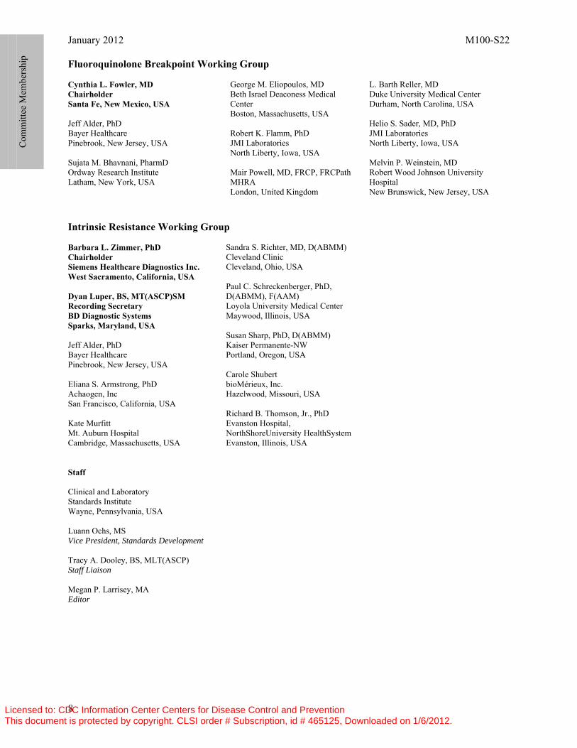
Fluoroquinolone Breakpoint Working Group Cynthia L. Fowler, MD Chairholder Santa Fe, New Mexico, USA Jeff Alder, PhD Bayer Healthcare Pinebrook, New Jersey, USA Sujata M. Bhavnani, PharmD Ordway Research Institute Latham, New York, USA
George M. Eliopoulos, MD Beth Israel Deaconess Medical Center Boston, Massachusetts, USA Robert K. Flamm, PhD JMI Laboratories North Liberty, Iowa, USA Mair Powell, MD, FRCP, FRCPath MHRA London, United Kingdom
L. Barth Reller, MD Duke University Medical Center Durham, North Carolina, USA Helio S. Sader, MD, PhD JMI Laboratories North Liberty, Iowa, USA Melvin P. Weinstein, MD Robert Wood Johnson University Hospital New Brunswick, New Jersey, USA
Intrinsic Resistance Working Group Barbara L. Zimmer, PhD Chairholder Siemens Healthcare Diagnostics Inc. West Sacramento, California, USA Dyan Luper, BS, MT(ASCP)SM Recording Secretary BD Diagnostic Systems Sparks, Maryland, USA Jeff Alder, PhD Bayer Healthcare Pinebrook, New Jersey, USA Eliana S. Armstrong, PhD Achaogen, Inc San Francisco, California, USA Kate Murfitt Mt. Auburn Hospital Cambridge, Massachusetts, USA
Sandra S. Richter, MD, D(ABMM) Cleveland Clinic Cleveland, Ohio, USA Paul C. Schreckenberger, PhD, D(ABMM), F(AAM) Loyola University Medical Center Maywood, Illinois, USA Susan Sharp, PhD, D(ABMM) Kaiser Permanente-NW Portland, Oregon, USA Carole Shubert bioMérieux, Inc. Hazelwood, Missouri, USA Richard B. Thomson, Jr., PhD Evanston Hospital, NorthShoreUniversity HealthSystem Evanston, Illinois, USA
Staff Clinical and Laboratory Standards Institute Wayne, Pennsylvania, USA Luann Ochs, MS Vice President, Standards Development Tracy A. Dooley, BS, MLT(ASCP) Staff Liaison Megan P. Larrisey, MA Editor
C
omm
ittee
Mem
bers
hip
Licensed to: CDC Information Center Centers for Disease Control and PreventionThis document is protected by copyright. CLSI order # Subscription, id # 465125, Downloaded on 1/6/2012.
Vol. 32 No. 3 M100-S22
9
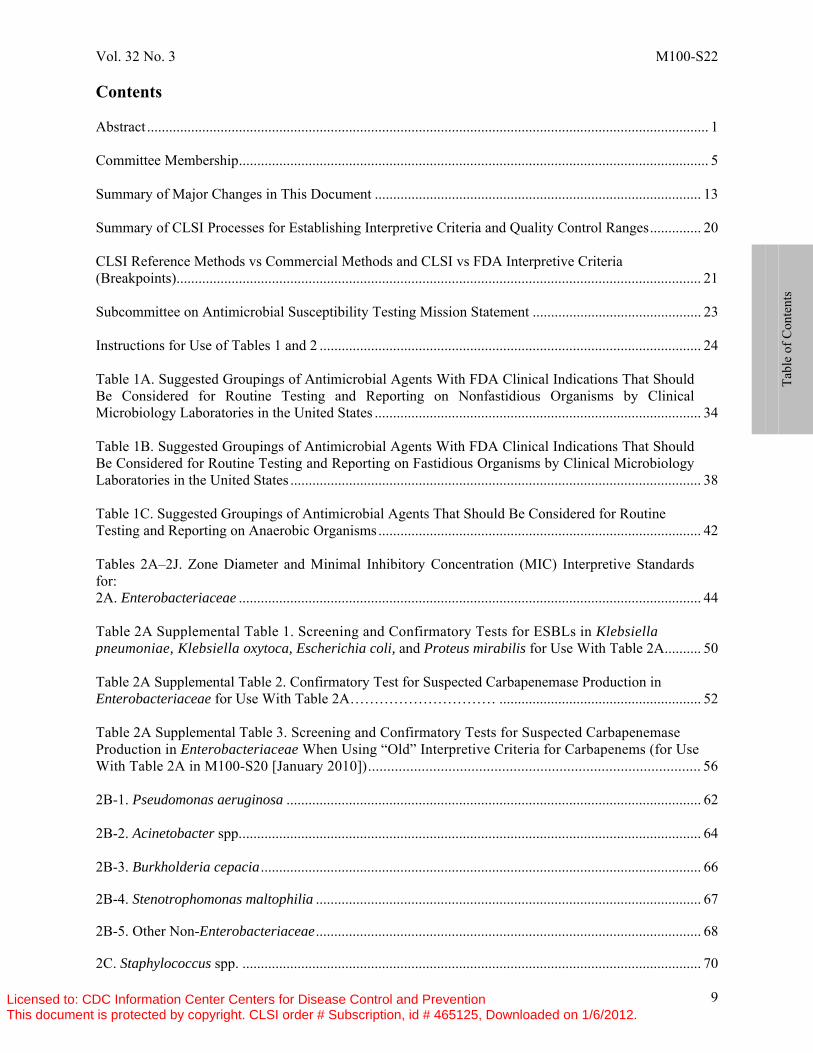
Contents Abstract ......................................................................................................................................................... 1 Committee Membership ................................................................................................................................ 5 Summary of Major Changes in This Document ......................................................................................... 13 Summary of CLSI Processes for Establishing Interpretive Criteria and Quality Control Ranges .............. 20 CLSI Reference Methods vs Commercial Methods and CLSI vs FDA Interpretive Criteria (Breakpoints)............................................................................................................................................... 21 Subcommittee on Antimicrobial Susceptibility Testing Mission Statement .............................................. 23 Instructions for Use of Tables 1 and 2 ........................................................................................................ 24
Table 1A. Suggested Groupings of Antimicrobial Agents With FDA Clinical Indications That Should Be Considered for Routine Testing and Reporting on Nonfastidious Organisms by Clinical Microbiology Laboratories in the United States ......................................................................................... 34 Table 1B. Suggested Groupings of Antimicrobial Agents With FDA Clinical Indications That Should Be Considered for Routine Testing and Reporting on Fastidious Organisms by Clinical Microbiology Laboratories in the United States ................................................................................................................ 38 Table 1C. Suggested Groupings of Antimicrobial Agents That Should Be Considered for Routine Testing and Reporting on Anaerobic Organisms ........................................................................................ 42 Tables 2A–2J. Zone Diameter and Minimal Inhibitory Concentration (MIC) Interpretive Standards for: 2A. Enterobacteriaceae .............................................................................................................................. 44 Table 2A Supplemental Table 1. Screening and Confirmatory Tests for ESBLs in Klebsiella pneumoniae, Klebsiella oxytoca, Escherichia coli, and Proteus mirabilis for Use With Table 2A .......... 50 Table 2A Supplemental Table 2. Confirmatory Test for Suspected Carbapenemase Production in Enterobacteriaceae for Use With Table 2A………………………… ....................................................... 52 Table 2A Supplemental Table 3. Screening and Confirmatory Tests for Suspected Carbapenemase Production in Enterobacteriaceae When Using “Old” Interpretive Criteria for Carbapenems (for Use With Table 2A in M100-S20 [January 2010]) ....................................................................................... 56 2B-1. Pseudomonas aeruginosa ................................................................................................................. 62 2B-2. Acinetobacter spp. ............................................................................................................................. 64 2B-3. Burkholderia cepacia ........................................................................................................................ 66 2B-4. Stenotrophomonas maltophilia ......................................................................................................... 67 2B-5. Other Non-Enterobacteriaceae ......................................................................................................... 68 2C. Staphylococcus spp. ............................................................................................................................. 70
Ta
ble
of C
onte
nts
Licensed to: CDC Information Center Centers for Disease Control and PreventionThis document is protected by copyright. CLSI order # Subscription, id # 465125, Downloaded on 1/6/2012.
January 2012 M100-S22
10
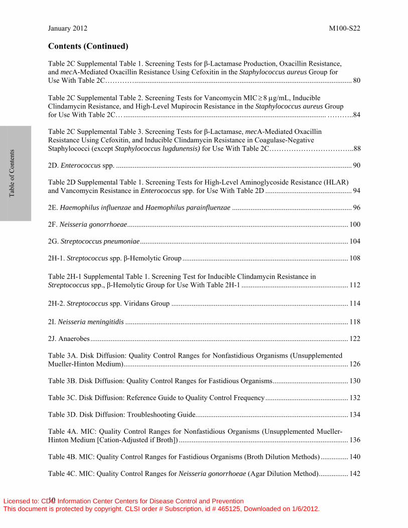
Contents (Continued) Table 2C Supplemental Table 1. Screening Tests for β-Lactamase Production, Oxacillin Resistance, and mecA-Mediated Oxacillin Resistance Using Cefoxitin in the Staphylococcus aureus Group for Use With Table 2C………….. .................................................................................................................... 80 Table 2C Supplemental Table 2. Screening Tests for Vancomycin MIC ≥ 8 μg/mL, Inducible Clindamycin Resistance, and High-Level Mupirocin Resistance in the Staphylococcus aureus Group for Use With Table 2C… .............................................................................................................. ………..84 Table 2C Supplemental Table 3. Screening Tests for β-Lactamase, mecA-Mediated Oxacillin Resistance Using Cefoxitin, and Inducible Clindamycin Resistance in Coagulase-Negative Staphylococci (except Staphylococcus lugdunensis) for Use With Table 2C……………………………..88 2D. Enterococcus spp. ................................................................................................................................ 90 Table 2D Supplemental Table 1. Screening Tests for High-Level Aminoglycoside Resistance (HLAR) and Vancomycin Resistance in Enterococcus spp. for Use With Table 2D ............................................... 94 2E. Haemophilus influenzae and Haemophilus parainfluenzae ................................................................. 96 2F. Neisseria gonorrhoeae ........................................................................................................................ 100 2G. Streptococcus pneumoniae ................................................................................................................. 104 2H-1. Streptococcus spp. β-Hemolytic Group .......................................................................................... 108 Table 2H-1 Supplemental Table 1. Screening Test for Inducible Clindamycin Resistance in Streptococcus spp., β-Hemolytic Group for Use With Table 2H-1 .......................................................... 112 2H-2. Streptococcus spp. Viridans Group ................................................................................................ 114 2I. Neisseria meningitidis ......................................................................................................................... 118 2J. Anaerobes ............................................................................................................................................ 122 Table 3A. Disk Diffusion: Quality Control Ranges for Nonfastidious Organisms (Unsupplemented Mueller-Hinton Medium) .......................................................................................................................... 126 Table 3B. Disk Diffusion: Quality Control Ranges for Fastidious Organisms ......................................... 130 Table 3C. Disk Diffusion: Reference Guide to Quality Control Frequency ............................................. 132 Table 3D. Disk Diffusion: Troubleshooting Guide................................................................................... 134 Table 4A. MIC: Quality Control Ranges for Nonfastidious Organisms (Unsupplemented Mueller-Hinton Medium [Cation-Adjusted if Broth]) ............................................................................................ 136 Table 4B. MIC: Quality Control Ranges for Fastidious Organisms (Broth Dilution Methods) ............... 140 Table 4C. MIC: Quality Control Ranges for Neisseria gonorrhoeae (Agar Dilution Method) ................ 142
Ta
ble
of C
onte
nts
Licensed to: CDC Information Center Centers for Disease Control and PreventionThis document is protected by copyright. CLSI order # Subscription, id # 465125, Downloaded on 1/6/2012.
Vol. 32 No. 3 M100-S22
11
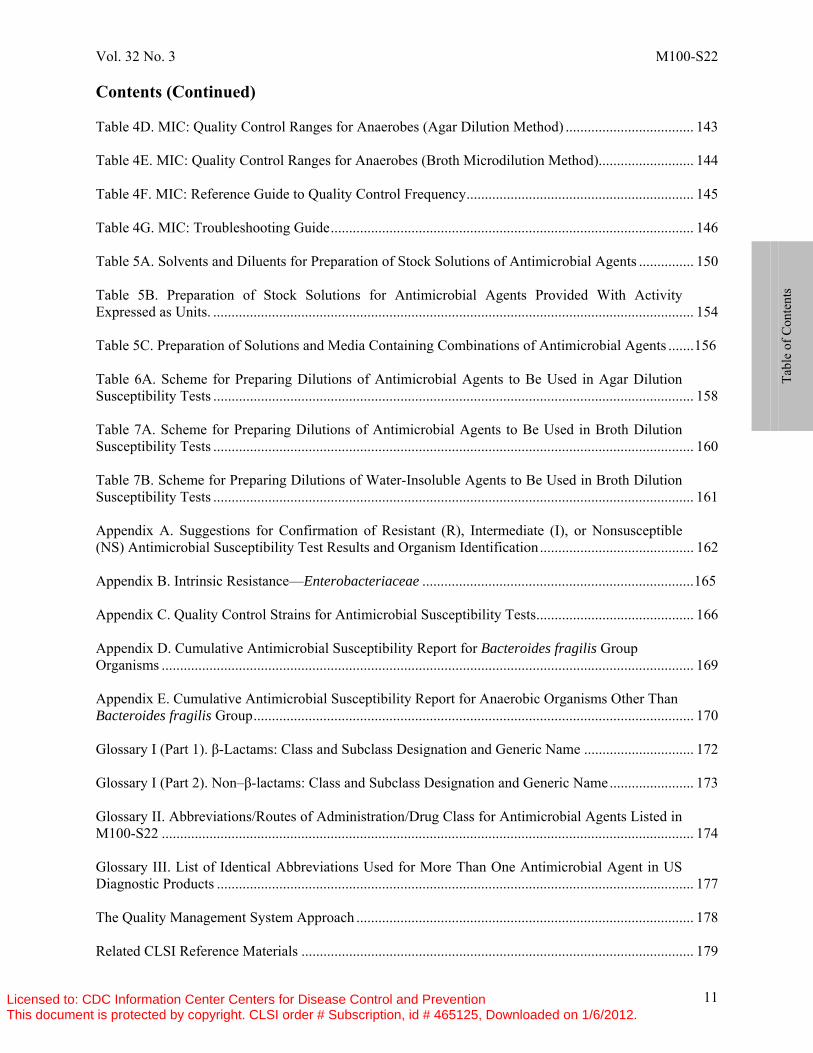
Contents (Continued) Table 4D. MIC: Quality Control Ranges for Anaerobes (Agar Dilution Method) ................................... 143 Table 4E. MIC: Quality Control Ranges for Anaerobes (Broth Microdilution Method).......................... 144 Table 4F. MIC: Reference Guide to Quality Control Frequency .............................................................. 145 Table 4G. MIC: Troubleshooting Guide ................................................................................................... 146 Table 5A. Solvents and Diluents for Preparation of Stock Solutions of Antimicrobial Agents ............... 150 Table 5B. Preparation of Stock Solutions for Antimicrobial Agents Provided With Activity Expressed as Units. ................................................................................................................................... 154 Table 5C. Preparation of Solutions and Media Containing Combinations of Antimicrobial Agents ....... 156 Table 6A. Scheme for Preparing Dilutions of Antimicrobial Agents to Be Used in Agar Dilution Susceptibility Tests ................................................................................................................................... 158 Table 7A. Scheme for Preparing Dilutions of Antimicrobial Agents to Be Used in Broth Dilution Susceptibility Tests ................................................................................................................................... 160 Table 7B. Scheme for Preparing Dilutions of Water-Insoluble Agents to Be Used in Broth Dilution Susceptibility Tests ................................................................................................................................... 161 Appendix A. Suggestions for Confirmation of Resistant (R), Intermediate (I), or Nonsusceptible (NS) Antimicrobial Susceptibility Test Results and Organism Identification .......................................... 162 Appendix B. Intrinsic Resistance—Enterobacteriaceae .......................................................................... 165 Appendix C. Quality Control Strains for Antimicrobial Susceptibility Tests ........................................... 166 Appendix D. Cumulative Antimicrobial Susceptibility Report for Bacteroides fragilis Group Organisms ................................................................................................................................................. 169 Appendix E. Cumulative Antimicrobial Susceptibility Report for Anaerobic Organisms Other Than Bacteroides fragilis Group ........................................................................................................................ 170 Glossary I (Part 1). β-Lactams: Class and Subclass Designation and Generic Name .............................. 172 Glossary I (Part 2). Non–β-lactams: Class and Subclass Designation and Generic Name ....................... 173 Glossary II. Abbreviations/Routes of Administration/Drug Class for Antimicrobial Agents Listed in M100-S22 ................................................................................................................................................. 174 Glossary III. List of Identical Abbreviations Used for More Than One Antimicrobial Agent in US Diagnostic Products .................................................................................................................................. 177 The Quality Management System Approach ............................................................................................ 178 Related CLSI Reference Materials ........................................................................................................... 179
Tabl
e of
Con
tent
s
Licensed to: CDC Information Center Centers for Disease Control and PreventionThis document is protected by copyright. CLSI order # Subscription, id # 465125, Downloaded on 1/6/2012.
January 2012 M100-S22
12
The Clinical and Laboratory Standards Institute consensus process, which is the mechanism for moving a document through two or more levels of review by the health care community, is an ongoing process. Users should expect revised editions of any given document. Because rapid changes in technology may affect the procedures, methods, and protocols in a standard or guideline, users should replace outdated editions with the current editions of CLSI documents. Current editions are listed in the CLSI catalog and posted on our website at www.clsi.org. If your organization is not a member and would like to become one, and to request a copy of the catalog, contact us at: Telephone: +610.688.0100; Fax: +610.688.0700; E-mail: [email protected]; Website: www.clsi.org.
Licensed to: CDC Information Center Centers for Disease Control and PreventionThis document is protected by copyright. CLSI order # Subscription, id # 465125, Downloaded on 1/6/2012.
Vol. 32 No. 3 M100-S22
13
Summary of Major Changes in This Document This list includes the “major” changes in this document. Other minor or editorial changes were made to the general formatting and to some of the table footnotes and comments. Changes to the tables since the previous edition appear in boldface type. Additions, Changes, and Deletions The following table indicates renaming, renumbering, and/or relocating of various tables or appendixes.
Previous Designation New M100-S22 Designation and/or
Location • Introduction to Tables 1 and 2 for Use With M02-
A10 (Disk Diffusion), M07-A8 (MIC Testing) • Instructions for Use of Tables 1 and 2
• Supplemental Table 2A-S1. Screening and Confirmatory Tests for ESBLs in Klebsiella pneumoniae, Klebsiella oxytoca, Escherichia coli, and Proteus mirabilis for Use With Table 2A
• Table 2A Supplemental Table 1. Screening and Confirmatory Tests for ESBLs in Klebsiella pneumoniae, Klebsiella oxytoca, Escherichia coli, and Proteus mirabilis for Use With Table 2A
• Supplemental Table 2A-S2. Confirmatory Test for Suspected Carbapenemase Production in Enterobacteriaceae When Using “New” Interpretive Criteria for Carbapenems
• Table 2A Supplemental Table 2. Confirmatory Test for Suspected Carbapenemase Production in Enterobacteriaceae for Use With Table 2A
• Supplemental Table 2A-S3. Screening and Confirmatory Tests for Suspected Carbapenemase Production in Enterobacteriaceae When Using “Old” Interpretive Criteria for Carbapenems (for Use With Table 2A in M100-S20 [January 2010])
• Table 2A Supplemental Table 3. Screening and Confirmatory Tests for Suspected Carbapenemase Production in Enterobacteriaceae When Using “Old” Interpretive Criteria for Carbapenems (for Use With Table 2A in M100-S20 [January 2010])
• Supplemental Table 2C-S4. Screening Tests for β-Lactamase Production, Oxacillin Resistance, mecA-Mediated Oxacillin Resistance Using Cefoxitin, Vancomycin MIC ≥ 8 μg/mL, Inducible Clindamycin Resistance, and High-Level Mupirocin Resistance in the Staphylococcus aureus Group for Use With Table 2C
The original Supplemental Table 2C-S4 was separated into two separate tables as follows:
• Table 2C Supplemental Table 1. Screening Tests for β-Lactamase Production, Oxacillin Resistance, and mecA-Mediated Oxacillin Resistance Using Cefoxitin in the Staphylococcus aureus Group for Use With Table 2C
• Table 2C Supplemental Table 2. Screening Tests for Vancomycin MIC ≥ 8 μg/mL, Inducible Clindamycin Resistance, and High-Level Mupirocin Resistance in the Staphylococcus aureus Group for Use With Table 2C
Sum
mar
y of
Cha
nges
Licensed to: CDC Information Center Centers for Disease Control and PreventionThis document is protected by copyright. CLSI order # Subscription, id # 465125, Downloaded on 1/6/2012.
January 2012 M100-S22
14
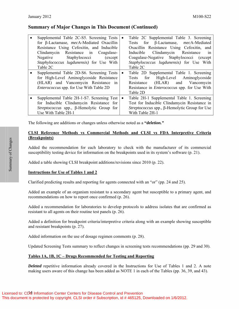
Summary of Major Changes in This Document (Continued) • Supplemental Table 2C-S5. Screening Tests
for β-Lactamase, mecA-Mediated Oxacillin Resistance Using Cefoxitin, and Inducible Clindamycin Resistance in Coagulase-Negative Staphylococci (except Staphylococcus lugdunensis) for Use With Table 2C
• Table 2C Supplemental Table 3. Screening Tests for β-Lactamase, mecA-Mediated Oxacillin Resistance Using Cefoxitin, and Inducible Clindamycin Resistance in Coagulase-Negative Staphylococci (except Staphylococcus lugdunensis) for Use With Table 2C
• Supplemental Table 2D-S6. Screening Tests for High-Level Aminoglycoside Resistance (HLAR) and Vancomycin Resistance in Enterococcus spp. for Use With Table 2D
• Table 2D Supplemental Table 1. Screening Tests for High-Level Aminoglycoside Resistance (HLAR) and Vancomycin Resistance in Enterococcus spp. for Use With Table 2D
• Supplemental Table 2H-1-S7. Screening Test for Inducible Clindamycin Resistance for Streptococcus spp., β-Hemolytic Group for Use With Table 2H-1
• Table 2H-1 Supplemental Table 1. Screening Test for Inducible Clindamycin Resistance in Streptococcus spp., β-Hemolytic Group for Use With Table 2H-1
The following are additions or changes unless otherwise noted as a “deletion.” CLSI Reference Methods vs Commercial Methods and CLSI vs FDA Interpretive Criteria (Breakpoints) Added the recommendation for each laboratory to check with the manufacturer of its commercial susceptibility testing device for information on the breakpoints used in its system’s software (p. 21). Added a table showing CLSI breakpoint additions/revisions since 2010 (p. 22). Instructions for Use of Tables 1 and 2 Clarified predicting results and reporting for agents connected with an “or” (pp. 24 and 25). Added an example of an organism resistant to a secondary agent but susceptible to a primary agent, and recommendations on how to report once confirmed (p. 26). Added a recommendation for laboratories to develop protocols to address isolates that are confirmed as resistant to all agents on their routine test panels (p. 26). Added a definition for breakpoint criteria/interpretive criteria along with an example showing susceptible and resistant breakpoints (p. 27). Added information on the use of dosage regimen comments (p. 28). Updated Screening Tests summary to reflect changes in screening tests recommendations (pp. 29 and 30). Tables 1A, 1B, 1C – Drugs Recommended for Testing and Reporting Deleted repetitive information already covered in the Instructions for Use of Tables 1 and 2. A note making users aware of this change has been added as NOTE 1 in each of the Tables (pp. 36, 39, and 43).
Sum
mar
y of
Cha
nges
Licensed to: CDC Information Center Centers for Disease Control and PreventionThis document is protected by copyright. CLSI order # Subscription, id # 465125, Downloaded on 1/6/2012.
Vol. 32 No. 3 M100-S22
15
Summary of Major Changes in This Document (Continued) Pseudomonas aeruginosa: Doripenem added to Test Report Group B (p. 34). Haemophilus spp.: Changed to Haemophilus influenzae and Haemophilus parainfluenzae to be consistent with Table 2E (p. 38). Anaerobes: Doripenem added to Test Report Group B (p. 42). Tables 2A Through 2J – Interpretive Criteria (Breakpoints) Enterobacteriaceae (Table 2A): Clarified using ampicillin interpretive criteria to predict results for amoxicillin (p. 45). Added information for laboratories that have not implemented the current interpretive criteria for cephalosporins and aztrenonam (p. 45). Added mechanism that explains why Enterobacter, Citrobacter, and Serratia may develop resistance during prolonged therapy with third-generation cephalosporins (p. 45). Added a dosage regimen comment for cefoxitin (p. 46). Added information on why cefmetazole interpretive criteria were not revised (p. 46). Added information about false-susceptible results when testing cefdinir, loracarbef, and cefprozil by disk diffusion (p. 46). New (revised) ertapenem disk diffusion and MIC interpretive criteria (p. 47). New ciprofloxacin disk diffusion and MIC interpretive criteria for reporting against Salmonella typhi and extraintestinal Salmonella spp. only (p. 48). Added recommendation for ciprofloxacin to use maximal dosage regimens for treatment of infections caused by S. typhi and extraintestinal Salmonella spp. that have MICs in the intermediate range (p. 48). Added information on strains of Salmonella that test resistant to nalidixic acid associated with clinical failure, noting that nalidixic acid may not detect all mechanisms of fluoroquinolone resistance (p. 48). Added information on what laboratories should do until they are able to implement the current carbapenem interpretive criteria (p. 52). Added a note regarding carbapenemase-producing isolates of Enterobacteriaceae and results from the modified Hodge test (MHT) (pp. 53 and 57). Pseudomonas aeruginosa (Table 2B-1): New (revised) piperacillin, piperacillin-tazobactam, ticarcillin, and ticarcillin-clavulanic acid disk diffusion and MIC interpretive criteria along with dosage regimens on which the breakpoints are based (pp. 62 and 63).
Sum
mar
y of
Cha
nges
Licensed to: CDC Information Center Centers for Disease Control and PreventionThis document is protected by copyright. CLSI order # Subscription, id # 465125, Downloaded on 1/6/2012.
January 2012 M100-S22
16
Summary of Major Changes in This Document (Continued) New doripenem disk diffusion and MIC interpretive criteria with dosage regimen on which the breakpoints are based (p. 63). New (revised) imipenem and meropenem disk diffusion and MIC interpretive criteria along with dosage regimen on which the breakpoints are based (p. 63). Deleted Rx comment regarding the susceptible category for penicillins and the need for high dose therapy for serious infections. Staphylococcus spp. (Table 2C): Added recommendations for additional testing and reporting of S. aureus isolates if oxacillin-intermediate results are obtained (p. 72). New doripenem disk diffusion and MIC interpretive criteria for methicillin-susceptible staphylococci isolates with dosage regimen on which the breakpoints are based (p. 75). Added new penicillin zone-edge test as an additional screening test for β-lactamase production in the S. aureus Group (pp. 80 and 81). Enterococcus spp. (Table 2D): Alternative inoculum method provided for the vancomycin resistance screen test (p. 91). Haemophilus influenzae and Haemophilus parainfluenzae (Table 2E): Added approximate CFU count for the McFarland suspension, recommending users to exercise care in preparing this suspension, because higher inoculum concentrations may lead to false-resistant results with some β-lactam antimicrobial agents (p. 96). Added recommended maximum number of disks per plate for disk diffusion testing (p. 96). Added cefamandole to the list of antimicrobial agents to which BLNAR strains of H. influenzae should be considered resistant (p. 97). New doripenem disk diffusion and MIC interpretive criteria (p. 98). Clarified that rifampin may only be appropriate for prophylaxis of case contacts (p. 99). Neisseria gonorrhoeae (Table 2F): Added recommended maximum number of disks per plate for disk diffusion testing (p. 100). Streptococcus pneumoniae (Table 2G): Clarified that recent studies using the agar dilution method have not been performed and reviewed by the subcommittee (p. 104). Clarified the use of colonies from an overnight sheep blood agar plate for the inoculum (p. 104). Information added for incubation when using the agar dilution method (p. 104). Added recommended maximum number of disks per plate for disk diffusion testing (p. 104). New doripenem MIC interpretive criteria (p. 106).
Sum
mar
y of
Cha
nges
Licensed to: CDC Information Center Centers for Disease Control and PreventionThis document is protected by copyright. CLSI order # Subscription, id # 465125, Downloaded on 1/6/2012.
Vol. 32 No. 3 M100-S22
17
Summary of Major Changes in This Document (Continued) Added information for predicting susceptibility for certain fluoroquinolones (p. 107). Streptococcus spp. β-Hemolytic Group (Table 2H-1): Clarified that recent studies using the agar dilution method have not been performed and reviewed by the subcommittee (p. 108). Added recommended maximum number of disks per plate for disk diffusion testing (p. 108). New doripenem MIC interpretive criteria (p. 109). Added comment noting unreliability of disk diffusion testing for testing daptomycin (p. 109). Added information on the 2010 CDC guidelines on prevention of group B streptococcal disease in neonates (p. 112). Streptococcus spp. Viridans Group (Table 2H-2): Clarified that recent studies using the agar dilution method have not been performed and reviewed by the subcommittee (p. 114). New doripenem MIC interpretive criteria (p. 115). Neisseria meningitidis (Table 2I): Added recommended maximum number of disks per plate for disk diffusion testing (p. 118). Anaerobes (Table 2J): Clarified medium, inoculum, and incubation conditions (p. 122). Added that MIC values using either Brucella blood agar or Wilkins Chalgren agar are considered equivalent (p. 122). Added information explaining that broth microdilution is only recommended for testing the Bacteroides fragilis group and has not been validated for testing other organisms (p. 122). Clarified test report group for ampicillin and penicillin for gram-positive and gram-negative organisms (p. 123). New doripenem MIC interpretive criteria (p. 124). Added information on resistance of gram-positive anaerobic rods to metronidazole (p. 124). Tables 3 and 4 – Quality Control Table 3A (pp. 126 and 127): QC ranges added for: Ceftaroline-avibactam – E. coli ATCC® 25922, S. aureus ATCC® 25923, and P. aeruginosa ATCC® 27853, and E. coli ATCC® 35218 Ceftazidime-avibactam – E. coli ATCC® 25922, S. aureus ATCC® 25923, and P. aeruginosa ATCC® 27853, and E. coli ATCC® 35218 Fusidic acid – S. aureus ATCC® 25923
Sum
mar
y of
Cha
nges
Licensed to: CDC Information Center Centers for Disease Control and PreventionThis document is protected by copyright. CLSI order # Subscription, id # 465125, Downloaded on 1/6/2012.
January 2012 M100-S22
18
Summary of Major Changes in This Document (Continued) Omadacycline – E. coli ATCC® 25922 and S. aureus ATCC® 25923 Plazomicin – E. coli ATCC® 25922, S. aureus ATCC® 25923, and P. aeruginosa ATCC® 27853 Solithromycin – S. aureus ATCC® 25923 Tedizolid – S. aureus ATCC® 25923 Table 3B (pp. 130 and 131): QC ranges added for: Ceftaroline-avibactam – H. influenzae ATCC® 49247 Ceftazidime-avibactam – H. influenzae ATCC® 49247 Doxycycline – S. pneumoniae ATCC® 49619 Fusidic acid – S. pneumoniae ATCC® 49619 Omadacycline – H. influenzae ATCC® 49247 and S. pneumoniae ATCC® 49619 Solithromycin – H. influenzae ATCC® 49247 and S. pneumoniae ATCC® 49619 Tedizolid – S. pneumoniae ATCC® 49619 Table 4A (pp. 136 and 137): QC ranges revised for: Cefepime – P. aeruginosa ATCC® 27853 Colistin – E. coli ATCC® 25922 QC ranges added for: Ceftaroline-avibactam – S. aureus ATCC® 29213, E. coli ATCC® 25922, and E. coli ATCC® 35218 Ceftazidime-avibactam – S. aureus ATCC® 29213, E. coli ATCC® 25922, and E. coli ATCC® 35218 Finafloxacin – S. aureus ATCC® 29213, E. faecalis ATCC® 29212, and P. aeruginosa ATCC® 27853 Fusidic acid – S. aureus ATCC® 29213 Omadacycline – S. aureus ATCC® 29213, E. faecalis ATCC® 29212, and E. coli ATCC® 25922 Plazomicin – S. aureus ATCC® 29213, E. faecalis ATCC® 29212, E. coli ATCC® 25922, and P. aeruginosa ATCC® 27853 Solithromycin – S. aureus ATCC® 29213, E. faecalis ATCC® 29212 Tedizolid – S. aureus ATCC® 29213, E. faecalis ATCC® 29212 Table 4B (pp. 140 and 141): QC ranges added for: Ceftaroline-avibactam – H. influenzae ATCC® 49247 Ceftazidime-avibactam – H. influenzae ATCC® 49247 Finafloxacin – S. pneumoniae ATCC® 49619 Fusidic acid – S. pneumoniae ATCC® 49619 Omadacycline – H. influenzae ATCC® 49247 and S. pneumoniae ATCC® 49619 Solithromycin – S. pneumoniae ATCC® 49619 Tedizolid – S. pneumoniae ATCC® 49619 Table 4D (p. 143): Ceftaroline, ceftaroline-avibactam, finafloxacin, and omadacycline QC ranges added for B. fragilis ATCC®
25285, B. thetaiotaomicron ATCC® 29741, C. difficile ATCC® 700057, and E. lentum ATCC® 43055.
Sum
mar
y of
Cha
nges
Licensed to: CDC Information Center Centers for Disease Control and PreventionThis document is protected by copyright. CLSI order # Subscription, id # 465125, Downloaded on 1/6/2012.
Vol. 32 No. 3 M100-S22
19
Summary of Major Changes in This Document (Continued) Table 4E (p. 144): QC ranges added for: Ceftaroline – B. fragilis ATCC® 25285, B. thetaiotaomicron ATCC® 29741, C. difficile ATCC® 700057 Ceftaroline-avibactam – B. fragilis ATCC® 25285, B. thetaiotaomicron ATCC® 29741, C. difficile ATCC® 700057, E. lentum ATCC® 43055 Omadacycline – B. fragilis ATCC® 25285, B. thetaiotaomicron ATCC® 29741, C. difficile ATCC® 700057, E. lentum ATCC® 43055 Table 5A – Solvents and Diluents (pp. 150–152) Added information for preparation of the stock solutions. Added antimicrobial agents: Avibactam Finafloxacin Fusidic acid Omadacycline Plazomicin Solithromycin Tedizolid Table 5C – Preparation of Solutions and Media Containing Combinations of Antimicrobial Agents (p. 156) Added antimicrobial agents: Ceftaroline-avibactam Ceftazidime-avibactam Appendixes and Glossaries Added Shigella to organism group listing for Salmonella spp. in Appendix A (p. 162). Glossary I – Added ceftaroline-avibactam, ceftazidime-avibactam, finafloxacin, fusidic acid, omadacycline, nitazoxanide, solithromycin, ramoplanin, tedizolid, tinidazole, and tizoxanide (pp. 172 and 173). Moved telithromycin under macrolides class/subclass ketolides. Moved tigecycline under tetracycline class/subclass glycylcyclines (p. 173) Glossary II – Added ceftaroline-avibactam, ceftazidime-avibactam, doxycycline, finafloxacin, fusidic acid, nitazoxanide, omadacycline, plazomicin, ramoplanin, solithromycin, tedizolid, tinidazole, and tinoxanide (pp. 174–176). Deleted trospectinomycin.
Sum
mar
y of
Cha
nges
Licensed to: CDC Information Center Centers for Disease Control and PreventionThis document is protected by copyright. CLSI order # Subscription, id # 465125, Downloaded on 1/6/2012.
January 2012 M100-S22
20
Summary of CLSI Processes for Establishing Interpretive Criteria and Quality Control Ranges The Clinical and Laboratory Standards Institute (CLSI) is an international, voluntary, nonprofit, interdisciplinary, standards-developing, and educational organization accredited by the American National Standards Institute (ANSI) that develops and promotes use of consensus-developed standards and guidelines within the health care community. These consensus standards and guidelines are developed to address critical areas of diagnostic testing and patient health care, and are developed in an open and consensus-seeking forum. CLSI is open to anyone or any organization that has an interest in diagnostic testing and patient care. Information about CLSI can be found at www.clsi.org. The CLSI Subcommittee on Antimicrobial Susceptibility Testing (AST) reviews data from a variety of sources and studies (eg, in vitro, pharmacokinetics/pharmacodynamics, and clinical studies) to establish antimicrobial susceptibility test methods, interpretive criteria, and quality control (QC) parameters. The details of the data required to establish interpretive criteria, QC parameters, and how the data are presented for evaluation are described in CLSI document M23—Development of In Vitro Susceptibility Testing Criteria and Quality Control Parameters. Over time, a microorganism’s susceptibility to an antimicrobial agent may decrease, resulting in a lack of clinical efficacy and/or safety. In addition, microbiological methods and QC parameters may be refined to ensure more accurate and better performance of susceptibility test methods. Because of this, CLSI continually monitors and updates information in its documents. Although CLSI standards and guidelines are developed using the most current information and thinking available at the time, the field of science and medicine is ever changing; therefore, standards and guidelines should be used in conjunction with clinical judgment, current knowledge, and clinically relevant laboratory test results to guide patient treatment. Additional information, updates, and changes in this document are found in the meeting summary minutes of the Subcommittee on Antimicrobial Susceptibility Testing at www.clsi.org.
Licensed to: CDC Information Center Centers for Disease Control and PreventionThis document is protected by copyright. CLSI order # Subscription, id # 465125, Downloaded on 1/6/2012.
Vol. 32 No. 3 M100-S22
21
CLSI Reference Methods vs Commercial Methods and CLSI vs FDA Interpretive Criteria (Breakpoints)
It is important for users of M02-A11, M07-A9, and the M100 Informational Supplement to recognize that the standard methods described in CLSI documents are reference methods. These methods may be used for routine AST of clinical isolates, for evaluation of commercial devices that will be used in clinical laboratories, or by drug or device manufacturers for testing of new agents or systems. Results generated by reference methods, such as those contained in CLSI documents, may be used by regulatory authorities to evaluate the performance of commercial susceptibility testing devices as part of the approval process. Clearance by a regulatory authority indicates that the commercial susceptibility testing device provides susceptibility results that are substantially equivalent to results generated using reference methods for the organisms and antimicrobial agents described in the device manufacturer’s approved package insert. CLSI breakpoints may differ from those approved by various regulatory authorities for many reasons, including the following: different databases, differences in interpretation of data, differences in doses used in different parts of the world, and public health policies. Differences also exist because CLSI proactively evaluates the need for changing breakpoints. The reasons why breakpoints may change and the manner in which CLSI evaluates data and determines breakpoints are outlined in CLSI document M23—Development of In Vitro Susceptibility Testing Criteria and Quality Control Parameters. Following a decision by CLSI to change an existing breakpoint, regulatory authorities may also review data in order to determine how changing breakpoints may affect the safety and effectiveness of the antimicrobial agent for the approved indications. If the regulatory authority changes breakpoints, commercial device manufacturers may have to conduct a clinical laboratory trial, submit the data to the regulatory authority, and await review and approval. For these reasons, a delay of one or more years may be required if an interpretive breakpoint change is to be implemented by a device manufacturer. In the United States, it is acceptable for laboratories that use US Food and Drug Administration (FDA)–cleared susceptibility testing devices to use existing FDA interpretive breakpoints. Either FDA or CLSI susceptibility interpretive breakpoints are acceptable to clinical laboratory accrediting bodies. Policies in other countries may vary. Each laboratory should check with the manufacturer of its antimicrobial susceptibility test system for additional information on the interpretive criteria used in its system’s software. Following discussions with appropriate stakeholders, such as infectious disease practitioners and the pharmacy department, as well as the Pharmacy and Therapeutics and Infection Control committees of the medical staff, newly approved or revised breakpoints may be implemented by clinical laboratories. CLSI disk diffusion test breakpoints may be implemented as soon as they are published in M100. If a device includes antimicrobial test concentrations sufficient to allow interpretation of susceptibility and resistance to an agent using the CLSI breakpoints, a laboratory could, after appropriate validation, choose to interpret and report results using CLSI breakpoints.
Licensed to: CDC Information Center Centers for Disease Control and PreventionThis document is protected by copyright. CLSI order # Subscription, id # 465125, Downloaded on 1/6/2012.
January 2012 M100-S22
22
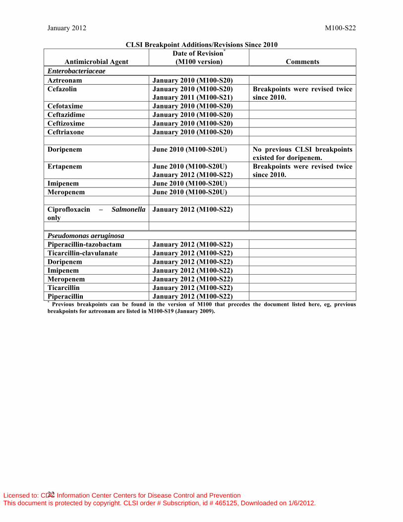
CLSI Breakpoint Additions/Revisions Since 2010
Antimicrobial Agent Date of Revision* (M100 version) Comments
Enterobacteriaceae Aztreonam January 2010 (M100-S20) Cefazolin January 2010 (M100-S20)
January 2011 (M100-S21) Breakpoints were revised twice since 2010.
Cefotaxime January 2010 (M100-S20) Ceftazidime January 2010 (M100-S20) Ceftizoxime January 2010 (M100-S20) Ceftriaxone January 2010 (M100-S20) Doripenem June 2010 (M100-S20U) No previous CLSI breakpoints
existed for doripenem. Ertapenem June 2010 (M100-S20U)
January 2012 (M100-S22) Breakpoints were revised twice since 2010.
Imipenem June 2010 (M100-S20U) Meropenem June 2010 (M100-S20U) Ciprofloxacin – Salmonella only
January 2012 (M100-S22)
Pseudomonas aeruginosa Piperacillin-tazobactam January 2012 (M100-S22) Ticarcillin-clavulanate January 2012 (M100-S22) Doripenem January 2012 (M100-S22) Imipenem January 2012 (M100-S22) Meropenem January 2012 (M100-S22) Ticarcillin January 2012 (M100-S22) Piperacillin January 2012 (M100-S22) * Previous breakpoints can be found in the version of M100 that precedes the document listed here, eg, previous breakpoints for aztreonam are listed in M100-S19 (January 2009).
Licensed to: CDC Information Center Centers for Disease Control and PreventionThis document is protected by copyright. CLSI order # Subscription, id # 465125, Downloaded on 1/6/2012.
Vol. 32 No. 3 M100-S22
23
Subcommittee on Antimicrobial Susceptibility Testing Mission Statement The Subcommittee on Antimicrobial Susceptibility Testing is composed of representatives from the professions, government, and industry, including microbiology laboratories, government agencies, health care providers and educators, and pharmaceutical and diagnostic microbiology industries. Using the CLSI voluntary consensus process, the subcommittee develops standards that promote accurate antimicrobial susceptibility testing and appropriate reporting. The mission of the Subcommittee on Antimicrobial Susceptibility Testing is to: • Develop standard reference methods for antimicrobial susceptibility tests. • Provide QC parameters for standard test methods. • Establish interpretive criteria for the results of standard antimicrobial susceptibility tests. • Provide suggestions for testing and reporting strategies that are clinically relevant and cost-
effective. • Continually refine standards and optimize detection of emerging resistance mechanisms through
development of new or revised methods, interpretive criteria, and QC parameters. • Educate users through multimedia communication of standards and guidelines. • Foster a dialog with users of these methods and those who apply them. The ultimate purpose of the subcommittee’s mission is to provide useful information to enable laboratories to assist the clinician in the selection of appropriate antimicrobial therapy for patient care. The standards and guidelines are meant to be comprehensive and to include all antimicrobial agents for which the data meet established CLSI guidelines. The values that guide this mission are quality, accuracy, fairness, timeliness, teamwork, consensus, and trust.
Licensed to: CDC Information Center Centers for Disease Control and PreventionThis document is protected by copyright. CLSI order # Subscription, id # 465125, Downloaded on 1/6/2012.
January 2012 Vol. 32 No. 3
©Clinical and Laboratory Standards Institute. All rights reserved. 24
Instructions for Use of Tables 1 and 2 I. Selecting Antimicrobial Agents for Testing and Reporting A. Selection of the most appropriate antimicrobial agents to test and to report is a decision best made
by each clinical laboratory in consultation with the infectious disease practitioners and the pharmacy, as well as the pharmacy and therapeutics and infection control committees of the medical staff. The recommendations for each organism group include agents of proven efficacy that show acceptable in vitro test performance. Considerations in the assignment of agents to specific test/report groups include clinical efficacy, prevalence of resistance, minimizing emergence of resistance, cost, FDA clinical indications for use, and current consensus recommendations for first-choice and alternative drugs. Unexpected resistance should be reported (eg, resistance of Enterobacteriaceae to carbapenems). Tests of selected agents may be useful for infection control purposes.
B. Drugs listed together in a single box are agents for which interpretive results (susceptible,
intermediate, or resistant) and clinical efficacy are similar. Within each box, an “or” between agents indicates those agents for which cross resistance and cross susceptibility are nearly complete. Results from one agent connected by an “or” can be used to predict results for the other agent. For example, Enterobacteriaceae susceptible to cefotaxime can be considered susceptible to ceftriaxone. The results obtained from testing cefotaxime could be reported
On the following pages, you will find: 1. Tables 1A and 1B—Suggested groupings of antimicrobial agents that should be
considered for routine testing and reporting by clinical microbiology laboratories. These guidelines are based on drugs with clinical indications approved by the US Food and Drug Administration (FDA) in the United States. In other countries, placement of antimicrobial agents in Tables 1A and 1B should be based on available drugs approved for clinical use by relevant regulatory agencies.
2. For each organism group, an additional table (Tables 2A through 2I) contains:
a. Recommended testing conditions. b. Minimal QC recommendations. (See also the text documents M02-A11, Section 15
and M07-A9, Section 16.) c. General comments for testing the organism group and specific comments for testing
particular drug/organism combinations. d. Suggested agents that should be considered for routine testing and reporting by
clinical microbiology laboratories, as specified in Tables 1A and 1B (test/report groups A, B, C, U).
e. Additional drugs that have an approved indication for the respective organism group, but would generally not warrant routine testing by a clinical microbiology laboratory in the United States (test/report group O for “other”; test/report group Inv. for “investigational” [not yet FDA approved]).
f. Zone diameter breakpoints and minimal inhibitory concentration (MIC) interpretive standard criteria.
3. For some organism groups, a supplemental table summarizing screening tests that may be
appropriate for use with isolates within the organism group. 4. Tables 1C and 2J address specific recommendations for testing and reporting results on
anaerobes and contain some of the information listed in 1 and 2 above.
Licensed to: CDC Information Center Centers for Disease Control and PreventionThis document is protected by copyright. CLSI order # Subscription, id # 465125, Downloaded on 1/6/2012.
For Use With M02-A11 and M07-A9 M100-S22
©Clinical and Laboratory Standards Institute. All rights reserved. 25
along with a comment that the isolate is also susceptible to ceftriaxone. For drugs connected with an “or,” combined major and very major errors are fewer than 3%, and minor errors are fewer than 10%, based on a large population of bacteria tested. In addition, to qualify for an “or,” at least 100 strains with resistance to the agents in question must be tested, and a result of “resistant” must be obtained with all agents for at least 95% of the strains. “Or” is also used for comparable agents when tested against organisms for which “susceptible-only” interpretive criteria are provided (eg, cefotaxime or ceftriaxone with Haemophilus influenzae). When no “or” connects agents within a box, testing of one agent cannot be used to predict results for another, owing either to discrepancies or insufficient data.
C. Test/Report Groups
1. As listed in Tables 1A, 1B, and 1C, agents in Group A are considered appropriate for inclusion in a routine, primary testing panel, as well as for routine reporting of results for the specific organism groups.
2. Group B includes antimicrobial agents that may warrant primary testing but they may
be reported only selectively, such as when the organism is resistant to agents of the same class, as in Group A. Other indications for reporting the result might include a selected specimen source (eg, a third-generation cephalosporin for enteric bacilli from cerebrospinal fluid [CSF] or trimethoprim-sulfamethoxazole for urinary tract isolates); a polymicrobial infection; infections involving multiple sites; cases of patient allergy, intolerance, or failure to respond to an agent in Group A; or for purposes of infection control.
3. Group C includes alternative or supplemental antimicrobial agents that may require
testing in those institutions that harbor endemic or epidemic strains resistant to several of the primary drugs (especially in the same class, eg, β-lactams); for treatment of patients allergic to primary drugs; for treatment of unusual organisms (eg, chloramphenicol for extraintestinal isolates of Salmonella spp.); or for reporting to infection control as an epidemiological aid.
4. Group U (“urine”) includes antimicrobial agents (eg, nitrofurantoin and certain
quinolones) that are used only or primarily for treating urinary tract infections. These agents should not be routinely reported against pathogens recovered from other sites of infection. Other agents with broader indications may be included in Group U for specific urinary pathogens (eg, P. aeruginosa and ofloxacin).
5. Group O (“other”) includes antimicrobial agents that have a clinical indication for the
organism group, but are generally not candidates for routine testing and reporting in the United States.
6. Group Inv. (“investigational”) includes antimicrobial agents that are investigational for
the organism group and have not yet been approved by the FDA for use in the United States.
D. Selective Reporting
Each laboratory should decide which agents in the tables to report routinely (Group A) and which might be reported only selectively (from Group B), in consultation with the infectious disease practitioners, the pharmacy, as well as the pharmacy and therapeutics and infection control committees of the health care institution. Selective reporting should improve the clinical relevance of test reports and help minimize the selection of multiresistant strains by overuse of
Licensed to: CDC Information Center Centers for Disease Control and PreventionThis document is protected by copyright. CLSI order # Subscription, id # 465125, Downloaded on 1/6/2012.
January 2012 Vol. 32 No. 3
©Clinical and Laboratory Standards Institute. All rights reserved. 26
broad-spectrum agents. Results for Group B agents tested but not reported routinely should be available on request. Unexpected resistance, when confirmed, should be reported (eg, resistance to a secondary agent but susceptibility to a primary agent, such as a P. aeruginosa isolate resistant to amikacin but susceptible to tobramycin; as such, both drugs should be reported). In addition, each laboratory should develop a protocol to address isolates that are confirmed as resistant to all agents on their routine test panels. This protocol should include options for testing additional agents in-house or sending the isolate to a reference laboratory.
II. Reporting Results The minimal inhibitory concentration (MIC) values determined as described in M07-A9 may be
reported directly to clinicians for patient care purposes. However, it is essential that an interpretive category result (S, I, or R) also be provided routinely to facilitate understanding of the MIC report by clinicians. Zone diameter measurements without an interpretive category should not be reported. Recommended interpretive categories for various MIC and zone diameter values are included in tables for each organism group and are based on evaluation of data as described in CLSI document M23.
Recommended MIC and disk diffusion interpretive criteria are based on usual dosage regimens and routes of administration in the United States.
A. Susceptible, intermediate, or resistant interpretations are reported and defined as follows: 1. Susceptible (S)
The “susceptible” category implies that isolates are inhibited by the usually achievable concentrations of antimicrobial agent when the dosage recommended to treat the site of infection is used.
2. Intermediate (I)
The “intermediate” category includes isolates with antimicrobial agent MICs that approach usually attainable blood and tissue levels, and for which response rates may be lower than for susceptible isolates. The intermediate category implies clinical efficacy in body sites where the drugs are physiologically concentrated (eg, quinolones and β-lactams in urine) or when a higher than normal dosage of a drug can be used (eg, β-lactams). This category also includes a buffer zone, which should prevent small, uncontrolled, technical factors from causing major discrepancies in interpretations, especially for drugs with narrow pharmacotoxicity margins.
3. Resistant (R) The “resistant” category implies that isolates are not inhibited by the usually achievable
concentrations of the agent with normal dosage schedules, and/or that demonstrate MICs or zone diameters that fall in the range where specific microbial resistance mechanisms (eg, β-lactamases) are likely, and clinical efficacy of the agent against the isolate has not been reliably shown in treatment studies.
4. Nonsusceptible (NS)
A category used for isolates for which only a susceptible interpretive criterion has been designated because of the absence or rare occurrence of resistant strains. Isolates that have MICs
Licensed to: CDC Information Center Centers for Disease Control and PreventionThis document is protected by copyright. CLSI order # Subscription, id # 465125, Downloaded on 1/6/2012.
For Use With M02-A11 and M07-A9 M100-S22
©Clinical and Laboratory Standards Institute. All rights reserved. 27
above or zone diameters below the value indicated for the susceptible breakpoint should be reported as nonsusceptible. NOTE 1: An isolate that is interpreted as nonsusceptible does not necessarily mean that the isolate has a resistance mechanism. It is possible that isolates with MICs above the susceptible breakpoint that lack resistance mechanisms may be encountered within the wild-type distribution subsequent to the time the susceptible-only breakpoint is set. NOTE 2: For strains yielding results in the “nonsusceptible” category, organism identification and antimicrobial susceptibility test results should be confirmed. (See Appendix A.)
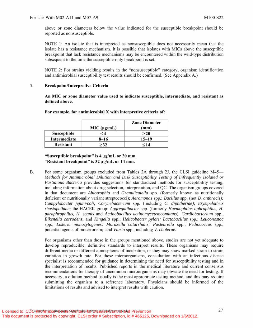
5. Breakpoint/Interpretive Criteria
An MIC or zone diameter value used to indicate susceptible, intermediate, and resistant as defined above.
For example, for antimicrobial X with interpretive criteria of:
MIC (μg/mL)
Zone Diameter (mm)
Susceptible ≤ 4 ≥ 20 Intermediate 8–16 15–19
Resistant ≥ 32 ≤ 14 “Susceptible breakpoint” is 4 μg/mL or 20 mm. “Resistant breakpoint” is 32 μg/mL or 14 mm.
B. For some organism groups excluded from Tables 2A through 2J, the CLSI guideline M45—Methods for Antimicrobial Dilution and Disk Susceptibility Testing of Infrequently Isolated or Fastidious Bacteria provides suggestions for standardized methods for susceptibility testing, including information about drug selection, interpretation, and QC. The organism groups covered in that document are Abiotrophia and Granulicatella spp. (formerly known as nutritionally deficient or nutritionally variant streptococci); Aeromonas spp.; Bacillus spp. (not B. anthracis); Campylobacter jejuni/coli; Corynebacterium spp. (including C. diphtheriae); Erysipelothrix rhusiopathiae; the HACEK group: Aggregatibacter spp. (formerly Haemophilus aphrophilus, H. paraphrophilus, H. segnis and Actinobacillus actinomycetemcomitans), Cardiobacterium spp., Eikenella corrodens, and Kingella spp.; Helicobacter pylori; Lactobacillus spp.; Leuconostoc spp.; Listeria monocytogenes; Moraxella catarrhalis; Pasteurella spp.; Pediococcus spp.; potential agents of bioterrorism; and Vibrio spp., including V. cholerae.
For organisms other than those in the groups mentioned above, studies are not yet adequate to develop reproducible, definitive standards to interpret results. These organisms may require different media or different atmospheres of incubation, or they may show marked strain-to-strain variation in growth rate. For these microorganisms, consultation with an infectious disease specialist is recommended for guidance in determining the need for susceptibility testing and in the interpretation of results. Published reports in the medical literature and current consensus recommendations for therapy of uncommon microorganisms may obviate the need for testing. If necessary, a dilution method usually is the most appropriate testing method, and this may require submitting the organism to a reference laboratory. Physicians should be informed of the limitations of results and advised to interpret results with caution.
Licensed to: CDC Information Center Centers for Disease Control and PreventionThis document is protected by copyright. CLSI order # Subscription, id # 465125, Downloaded on 1/6/2012.
January 2012 Vol. 32 No. 3
©Clinical and Laboratory Standards Institute. All rights reserved. 28
C. Policies regarding the generation of cumulative antibiograms should be developed in concert with the infectious disease service, infection control personnel, and the pharmacy and therapeutics committee. In most circumstances, the percentage of susceptible and intermediate results should not be combined into the same statistics. See CLSI document M39—Analysis and Presentation of Cumulative Antimicrobial Susceptibility Test Data.
III. Therapy-Related Comments
Some of the comments in the tables relate to therapy concerns. These are denoted with an Rx symbol. It may be appropriate to include some of these comments (or modifications thereof) on the patient report. An example would be inclusion of a comment on Enterococcus susceptibility reports from blood cultures that “combination therapy with ampicillin, penicillin or vancomycin (for susceptible strains) plus an aminoglycoside is usually indicated for serious enterococcal infections, such as endocarditis, unless high-level resistance to both gentamicin and streptomycin is documented; such combinations are predicted to result in synergistic killing of the Enterococcus.”
Antimicrobial dosage regimens often vary widely among practitioners and institutions. In some
cases, the MIC interpretive criteria rely on pharmacokinetic-pharmacodynamic (PK-PD) data, using specific human dosage regimens. In cases where specific dosage regimens are important for proper application of breakpoints, the dosage regimen is listed. These dosage regimen comments are not intended for use on individual patient reports.
IV. Confirmation of Patient Results Multiple test parameters are monitored by following the QC recommendations described in this
standard. However, acceptable results derived from testing QC strains do not guarantee accurate results when testing patient isolates. It is important to review all of the results obtained from all drugs tested on a patient’s isolate before reporting the results. This should include, but not be limited to, ensuring that 1) the antimicrobial susceptibility results are consistent with the identification of the isolate; 2) the results from individual agents within a specific drug class follow the established hierarchy of activity rules (eg, in general, third-generation cephems are more active than first- or second-generation cephems against Enterobacteriaceae); and 3) the isolate is susceptible to those agents for which resistance has not been documented (eg, vancomycin and Streptococcus spp.) and for which only “susceptible” interpretive criteria exist in M100.
Unusual or inconsistent results should be confirmed by rechecking various parameters of
testing detailed in Appendix A. Each laboratory must develop its own policies for confirmation of unusual or inconsistent antimicrobial susceptibility test results. The list provided in Appendix A emphasizes those results that are most likely to affect patient care.
V. Development of Resistance and Testing of Repeat Isolates
Isolates that are initially susceptible may become intermediate or resistant after initiation of therapy. Therefore, subsequent isolates of the same species from a similar body site should be tested in order to detect resistance that may have developed. This can occur within as little as three to four days and has been noted most frequently in Enterobacter, Citrobacter, and Serratia spp. with third-generation cephalosporins; in P. aeruginosa with all antimicrobial agents; and in staphylococci with quinolones. For S. aureus, vancomycin-susceptible isolates may become vancomycin intermediate during the course of prolonged therapy.
Licensed to: CDC Information Center Centers for Disease Control and PreventionThis document is protected by copyright. CLSI order # Subscription, id # 465125, Downloaded on 1/6/2012.
For Use With M02-A11 and M07-A9 M100-S22
©Clinical and Laboratory Standards Institute. All rights reserved. 29
In certain circumstances, testing of subsequent isolates to detect resistance that may have developed might be warranted earlier than within three to four days. The decision to do so requires knowledge of the specific situation and the severity of the patient’s condition (eg, an isolate of Enterobacter cloacae from a blood culture on a premature infant). Laboratory guidelines on when to perform susceptibility testing on repeat isolates should be determined after consultation with the medical staff.
VI. Warning Some of the comments in the tables relate to dangerously misleading results that can occur when
certain antimicrobial agents are tested and reported as susceptible against specific organisms. These are denoted with the word “Warning.”
“Warning”: The following antimicrobial agent/organism combinations may appear active in vitro, but are not effective clinically and should not be reported as susceptible.
Location Organism Antimicrobial Agents That Must Not Be Reported
as Susceptible Table 2A Salmonella spp., Shigella spp. 1st- and 2nd-generation cephalosporins,
cephamycins, and aminoglycosides Table 2C Oxacillin-resistant Staphylococcus spp. Penicillins, β-lactam/β-lactamase inhibitor
combinations, antistaphylococcal cephems, and carbapenems
Table 2D Enterococcus spp. Aminoglycosides (except high concentrations), cephalosporins, clindamycin, and trimethoprim-sulfamethoxazole
VII. Screening Tests Screening tests, as described in this document, characterize an isolate based on a specific resistance mechanism or phenotype. Some screening tests have sufficient sensitivity and specificity such that results of the screen can be reported without additional testing. Others provide presumptive results and require further testing for confirmation. A summary of the screening tests is provided here; the details for each screening test, including test specifications, limitations, and additional tests needed for confirmation, are provided in the Supplemental Tables listed below.
Licensed to: CDC Information Center Centers for Disease Control and PreventionThis document is protected by copyright. CLSI order # Subscription, id # 465125, Downloaded on 1/6/2012.
January 2012 Vol. 32 No. 3
©Clinical and Laboratory Standards Institute. All rights reserved. 30
Organism Group Table
Location
Resistance Phenotype or Mechanism Screening Tests
Further Testing or Confirmation Required?
Enterobacteriaceae 2A Supplemental
Table 1
ESBL production Broth microdilution and disk diffusion with various cephalosporins and aztreonam
Yes, if screen test positivea
2A Supplemental
Table 2
Carbapenemase production
Broth microdilution and disk diffusion with various carbapenems
Yes, if screen test positive
2A Supplemental
Table 3
Carbapenemase production
Broth microdilution and disk diffusion with various carbapenems
Yes, if screen test positive
Staphylococcus aureus
2C Supplemental
Table 1
β-Lactamase production
Penicillin disk diffusion zone-edge test or other method
Yes, if screen test negative, repeat penicillin MIC and β-lactamase test(s) (eg, penicillin disk diffusion zone-edge test or induced β-lactamase test) on subsequent isolates from same patient (if penicillin MIC ≤ 0.12 µg/mL or zone ≥
29 mm); PCR for blaZ may be considered.
Oxacillin resistance Agar dilution; MHA with 4% NaCl and 6 µg/mL oxacillin
No
mecA-mediated oxacillin resistance
Broth microdilution and disk diffusion with cefoxitin
No
2C Supplemental
Table 2
Vancomycin MIC ≥ 8 µg/mL
Agar dilution; BHI with 6 µg/mL vancomycin
Yes, if screen test positive
Inducible clindamycin resistance
Broth microdilution and disk diffusion with clindamycin and erythromycin
No
High-level mupirocin resistance
Broth microdilution and disk diffusion with mupirocin
No
Coagulase-negative staphylococci
2C Supplemental
Table 3
β-lactamase production
Chromogenic cephalosporin or other method
Yes, if screen test negative, repeat penicillin MIC and induced β-lactamase test on subsequent isolates from same patient (if penicillin MIC ≤ 0.12 µg/mL or zone ≥
29 mm); PCR for blaZ may be considered.
mecA-Mediated oxacillin resistance
Disk diffusion with cefoxitin No
Inducible clindamycin resistance
Broth microdilution and disk diffusion with clindamycin and erythromycin
No
Enterococci 2D Supplemental
Table 1
Vancomycin resistance
Agar dilution with vancomycin
Yes, if screen test positive
HLAR Broth microdilution, agar dilution, and disk diffusion with gentamicin and streptomycin
No for MIC; yes for disk, if inconclusive
Streptococcus pneumoniae
2G Penicillin resistance Disk diffusion with oxacillin Yes, if nonsusceptible
Streptococcus spp. β-hemolytic Group
2H-1 Supplemental
Table 1
Inducible clindamycin resistance
Broth microdilution and disk diffusion with clindamycin and erythromycin
No
Licensed to: CDC Information Center Centers for Disease Control and PreventionThis document is protected by copyright. CLSI order # Subscription, id # 465125, Downloaded on 1/6/2012.
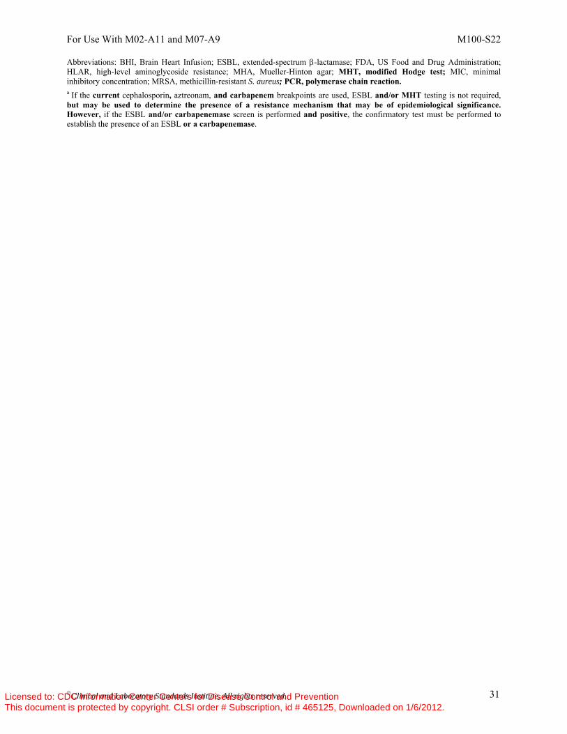
For Use With M02-A11 and M07-A9 M100-S22
©Clinical and Laboratory Standards Institute. All rights reserved. 31
Abbreviations: BHI, Brain Heart Infusion; ESBL, extended-spectrum β-lactamase; FDA, US Food and Drug Administration; HLAR, high-level aminoglycoside resistance; MHA, Mueller-Hinton agar; MHT, modified Hodge test; MIC, minimal inhibitory concentration; MRSA, methicillin-resistant S. aureus; PCR, polymerase chain reaction. a If the current cephalosporin, aztreonam, and carbapenem breakpoints are used, ESBL and/or MHT testing is not required, but may be used to determine the presence of a resistance mechanism that may be of epidemiological significance. However, if the ESBL and/or carbapenemase screen is performed and positive, the confirmatory test must be performed to establish the presence of an ESBL or a carbapenemase.
Licensed to: CDC Information Center Centers for Disease Control and PreventionThis document is protected by copyright. CLSI order # Subscription, id # 465125, Downloaded on 1/6/2012.
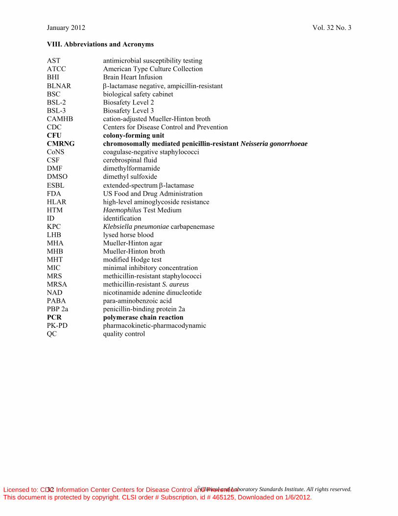
January 2012 Vol. 32 No. 3
©Clinical and Laboratory Standards Institute. All rights reserved. 32
VIII. Abbreviations and Acronyms AST antimicrobial susceptibility testing ATCC American Type Culture Collection BHI Brain Heart Infusion BLNAR β-lactamase negative, ampicillin-resistant BSC biological safety cabinet BSL-2 Biosafety Level 2 BSL-3 Biosafety Level 3 CAMHB cation-adjusted Mueller-Hinton broth CDC Centers for Disease Control and Prevention CFU colony-forming unit CMRNG chromosomally mediated penicillin-resistant Neisseria gonorrhoeae CoNS coagulase-negative staphylococci CSF cerebrospinal fluid DMF dimethylformamide DMSO dimethyl sulfoxide ESBL extended-spectrum β-lactamase FDA US Food and Drug Administration HLAR high-level aminoglycoside resistance HTM Haemophilus Test Medium ID identification KPC Klebsiella pneumoniae carbapenemase LHB lysed horse blood MHA Mueller-Hinton agar MHB Mueller-Hinton broth MHT modified Hodge test MIC minimal inhibitory concentration MRS methicillin-resistant staphylococci MRSA methicillin-resistant S. aureus NAD nicotinamide adenine dinucleotide PABA para-aminobenzoic acid PBP 2a penicillin-binding protein 2a PCR polymerase chain reaction PK-PD pharmacokinetic-pharmacodynamic QC quality control
Licensed to: CDC Information Center Centers for Disease Control and PreventionThis document is protected by copyright. CLSI order # Subscription, id # 465125, Downloaded on 1/6/2012.

For Use With M02-A11 and M07-A9 M100-S22
©Clinical and Laboratory Standards Institute. All rights reserved. 33
This page is intentionally left blank.
Licensed to: CDC Information Center Centers for Disease Control and PreventionThis document is protected by copyright. CLSI order # Subscription, id # 465125, Downloaded on 1/6/2012.
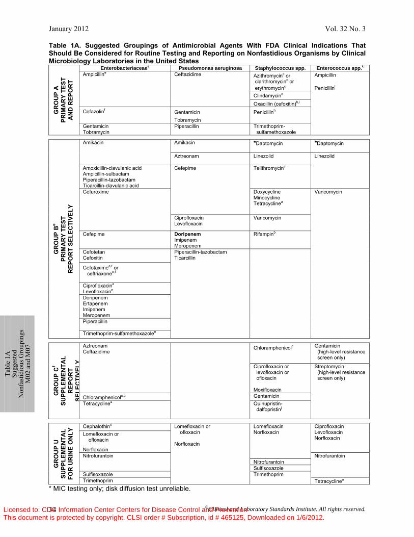
January 2012 Vol. 32 No. 3
©Clinical and Laboratory Standards Institute. All rights reserved. 34
Table 1A. Suggested Groupings of Antimicrobial Agents With FDA Clinical Indications That Should Be Considered for Routine Testing and Reporting on Nonfastidious Organisms by Clinical Microbiology Laboratories in the United States
GR
OU
P A
PR
IMA
RY
TEST
A
ND
REP
OR
T Enterobacteriaceaee Pseudomonas aeruginosa Staphylococcus spp. Enterococcus spp.k
Ampicilline Ceftazidime Azithromycinc or clarithromycinc or erythromycinc
Ampicillin Penicillinl Clindamycinc
Oxacillin (cefoxitin)h,i
Cefazolinf
Gentamicin Tobramycin
Penicillinh
Gentamicin Tobramycin
Piperacillin Trimethoprim- sulfamethoxazole
GR
OU
P B
e
PRIM
AR
Y TE
ST
REP
OR
T SE
LEC
TIVE
LY
Amikacin
Amikacin *Daptomycin *Daptomycin
Aztreonam
Linezolid Linezolid
Amoxicillin-clavulanic acid Ampicillin-sulbactam Piperacillin-tazobactam Ticarcillin-clavulanic acid
Cefepime Telithromycinc
Cefuroxime Doxycycline Minocycline Tetracyclinea
Vancomycin
Ciprofloxacin Levofloxacin
Vancomycin
Cefepime DoripenemImipenem Meropenem
Rifampinb
Cefotetan Cefoxitin
Piperacillin-tazobactam Ticarcillin
Cefotaximee,f or ceftriaxonee,f
Ciprofloxacine Levofloxacine
Doripenem Ertapenem Imipenem Meropenem
Piperacillin
Trimethoprim-sulfamethoxazolee
GR
OU
P C
f SU
PPLE
MEN
TAL
REP
OR
T SE
LEC
TIVE
LY
Aztreonam Ceftazidime
Chloramphenicolc Gentamicin (high-level resistance screen only)
Ciprofloxacin or levofloxacin or ofloxacin Moxifloxacin
Streptomycin (high-level resistance screen only)
Chloramphenicolc,e Gentamicin Tetracyclinea Quinupristin-
dalfopristinj
GR
OU
P U
SU
PPLE
MEN
TAL
FOR
UR
INE
ON
LY Cephalothind Lomefloxacin or
ofloxacin Norfloxacin
Lomefloxacin Norfloxacin
Ciprofloxacin Levofloxacin Norfloxacin
Lomefloxacin or ofloxacin Norfloxacin Nitrofurantoin Nitrofurantoin Nitrofurantoin Sulfisoxazole Sulfisoxazole Trimethoprim Trimethoprim Tetracyclinea
* MIC testing only; disk diffusion test unreliable.
Ta
ble
1A
Sugg
este
d N
onfa
stid
ious
Gro
upin
gs
M02
and
M07
Licensed to: CDC Information Center Centers for Disease Control and PreventionThis document is protected by copyright. CLSI order # Subscription, id # 465125, Downloaded on 1/6/2012.
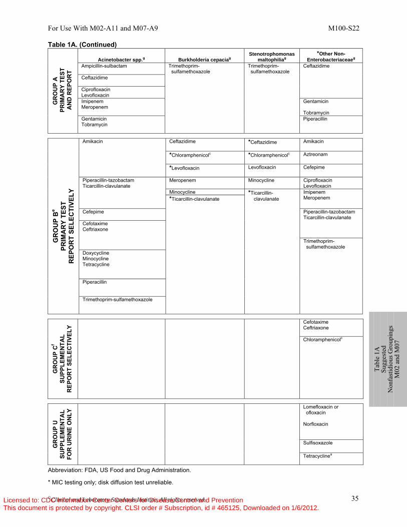
For Use With M02-A11 and M07-A9 M100-S22
©Clinical and Laboratory Standards Institute. All rights reserved. 35
Table 1A. (Continued) G
RO
UP
A
PR
IMA
RY
TEST
A
ND
REP
OR
T Acinetobacter spp.g Burkholderia cepaciag
Stenotrophomonas maltophiliag
*Other Non-Enterobacteriaceaeg
Ampicillin-sulbactam
Trimethoprim- sulfamethoxazole
Trimethoprim- sulfamethoxazole
Ceftazidime
Ceftazidime Ciprofloxacin Levofloxacin Imipenem Meropenem
Gentamicin Tobramycin
Gentamicin Tobramycin
Piperacillin
GR
OU
P B
e PR
IMA
RY
TEST
R
EPO
RT
SELE
CTI
VELY
Amikacin
Ceftazidime
*Ceftazidime
Amikacin
*Chloramphenicolc
*Chloramphenicolc
Aztreonam
*Levofloxacin
Levofloxacin Cefepime
Piperacillin-tazobactam Ticarcillin-clavulanate
Meropenem Minocycline Ciprofloxacin Levofloxacin
Minocycline *Ticarcillin- clavulanate
Imipenem Meropenem *Ticarcillin-clavulanate
Cefepime
Piperacillin-tazobactam Ticarcillin-clavulanate
Cefotaxime Ceftriaxone
Trimethoprim- sulfamethoxazole
Doxycycline Minocycline Tetracycline Piperacillin Trimethoprim-sulfamethoxazole
GR
OU
P C
f SU
PPLE
MEN
TAL
REP
OR
T SE
LEC
TIVE
LY
Cefotaxime Ceftriaxone Chloramphenicolc
GR
OU
P U
SU
PPLE
MEN
TAL
FOR
UR
INE
ON
LY Lomefloxacin or
ofloxacin Norfloxacin
Sulfisoxazole
Tetracyclinea
Abbreviation: FDA, US Food and Drug Administration. * MIC testing only; disk diffusion test unreliable.
Ta
ble
1A
Sugg
este
d N
onfa
stid
ious
Gro
upin
gs
M02
and
M07
Licensed to: CDC Information Center Centers for Disease Control and PreventionThis document is protected by copyright. CLSI order # Subscription, id # 465125, Downloaded on 1/6/2012.
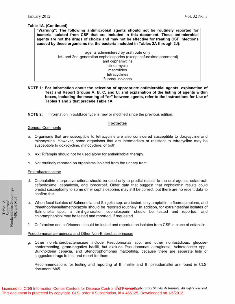
January 2012 Vol. 32 No. 3
©Clinical and Laboratory Standards Institute. All rights reserved. 36
Table 1A. (Continued) “Warning”: The following antimicrobial agents should not be routinely reported for bacteria isolated from CSF that are included in this document. These antimicrobial agents are not the drugs of choice and may not be effective for treating CSF infections caused by these organisms (ie, the bacteria included in Tables 2A through 2J):
agents administered by oral route only 1st- and 2nd-generation cephalosporins (except cefuroxime parenteral)
and cephamycins clindamycin macrolides
tetracyclines fluoroquinolones
NOTE 1: For information about the selection of appropriate antimicrobial agents; explanation of
Test and Report Groups A, B, C, and U; and explanation of the listing of agents within boxes, including the meaning of “or” between agents, refer to the Instructions for Use of Tables 1 and 2 that precede Table 1A.
NOTE 2: Information in boldface type is new or modified since the previous edition.
Footnotes General Comments a. Organisms that are susceptible to tetracycline are also considered susceptible to doxycycline and
minocycline. However, some organisms that are intermediate or resistant to tetracycline may be susceptible to doxycycline, minocycline, or both.
b. Rx: Rifampin should not be used alone for antimicrobial therapy. c. Not routinely reported on organisms isolated from the urinary tract. Enterobacteriaceae d. Cephalothin interpretive criteria should be used only to predict results to the oral agents, cefadroxil,
cefpodoxime, cephalexin, and loracarbef. Older data that suggest that cephalothin results could predict susceptibility to some other cephalosporins may still be correct, but there are no recent data to confirm this.
e. When fecal isolates of Salmonella and Shigella spp. are tested, only ampicillin, a fluoroquinolone, and
trimethoprim/sulfamethoxazole should be reported routinely. In addition, for extraintestinal isolates of Salmonella spp., a third-generation cephalosporin should be tested and reported, and chloramphenicol may be tested and reported, if requested.
f. Cefotaxime and ceftriaxone should be tested and reported on isolates from CSF in place of cefazolin. Pseudomonas aeruginosa and Other Non-Enterobacteriaceae g. Other non-Enterobacteriaceae include Pseudomonas spp. and other nonfastidious, glucose-
nonfermenting, gram-negative bacilli, but exclude Pseudomonas aeruginosa, Acinetobacter spp., Burkholderia cepacia, and Stenotrophomonas maltophilia, because there are separate lists of suggested drugs to test and report for them.
Recommendations for testing and reporting of B. mallei and B. pseudomallei are found in CLSI
document M45.
Ta
ble
1A
Sugg
este
d N
onfa
stid
ious
Gro
upin
gs
M02
and
M07
Licensed to: CDC Information Center Centers for Disease Control and PreventionThis document is protected by copyright. CLSI order # Subscription, id # 465125, Downloaded on 1/6/2012.
For Use With M02-A11 and M07-A9 M100-S22
©Clinical and Laboratory Standards Institute. All rights reserved. 37
Table 1A. (Continued) Staphylococcus spp. h. Penicillin-susceptible staphylococci are also susceptible to other penicillins, β-lactam/β-lactamase
inhibitor combinations, antistaphylococcal cephems, and carbapenems approved for use by the FDA for staphylococcal infections. Penicillin-resistant, oxacillin-susceptible strains are resistant to penicillinase-labile penicillins, but susceptible to other penicillinase-stable penicillins, β-lactam/β-lactamase inhibitor combinations, antistaphylococcal cephems, and carbapenems. Oxacillin-resistant staphylococci are resistant to all currently available β-lactam antimicrobial agents, with the exception of the newer cephalosporins with anti-MRSA activity. Thus, susceptibility or resistance to a wide array of β-lactam antimicrobial agents may be deduced from testing only penicillin and either cefoxitin or oxacillin. Routine testing of other penicillins, β-lactam/β-lactamase inhibitor combinations, cephems, or carbapenems is not advised.
i. The results of either cefoxitin disk diffusion or cefoxitin MIC tests can be used to predict the presence
of mecA-mediated oxacillin resistance in S. aureus and S. lugdunensis. For coagulase-negative staphylococci (except S. lugdunensis), the cefoxitin disk diffusion test is the preferred method for detection of mecA-mediated oxacillin resistance. Cefoxitin is used as a surrogate for detection of oxacillin resistance; report oxacillin as susceptible or resistant based on cefoxitin results. If a penicillinase-stable penicillin is tested, oxacillin is the preferred agent, and results can be applied to the other penicillinase-stable penicillins, cloxacillin, dicloxacillin, and flucloxacillin.
j. For reporting against methicillin-susceptible S. aureus. Enterococcus spp. k. Warning: For Enterococcus spp., cephalosporins, aminoglycosides (except for high-level resistance
screening), clindamycin, and trimethoprim-sulfamethoxazole may appear active in vitro, but are not effective clinically and should not be reported as susceptible.
l. Enterococci susceptible to penicillin are predictably susceptible to ampicillin, amoxicillin, ampicillin-
sulbactam, amoxicillin-clavulanate, piperacillin, and piperacillin-tazobactam for non–β-lactamase-producing enterococci. However, enterococci susceptible to ampicillin cannot be assumed to be susceptible to penicillin. If penicillin results are needed, testing of penicillin is required. Rx: Combination therapy with ampicillin, penicillin, or vancomycin (for susceptible strains) plus an aminoglycoside is usually indicated for serious enterococcal infections, such as endocarditis, unless high-level resistance to both gentamicin and streptomycin is documented; such combinations are predicted to result in synergistic killing of the Enterococcus.
Ta
ble
1A
Sugg
este
d N
onfa
stid
ious
Gro
upin
gs
M02
and
M07
Licensed to: CDC Information Center Centers for Disease Control and PreventionThis document is protected by copyright. CLSI order # Subscription, id # 465125, Downloaded on 1/6/2012.
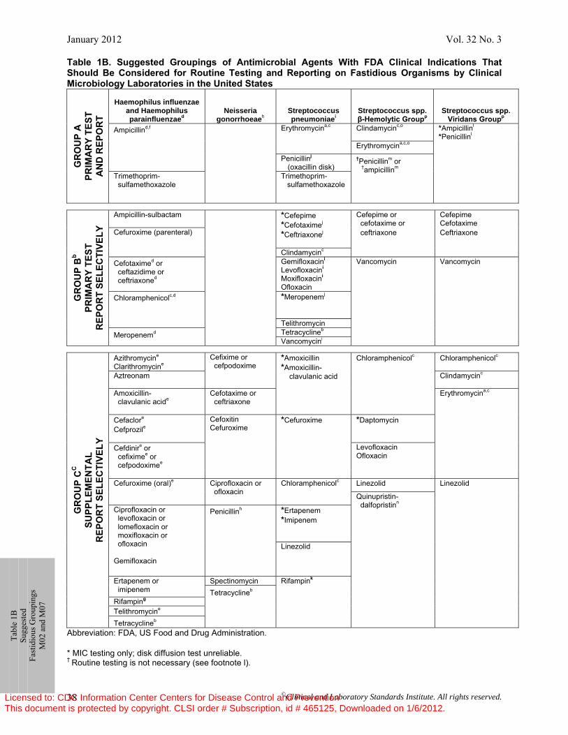
January 2012 Vol. 32 No. 3
©Clinical and Laboratory Standards Institute. All rights reserved. 38
Ta
ble
1B
Sugg
este
d Fa
stid
ious
Gro
upin
gs
M02
and
M07
Table 1B. Suggested Groupings of Antimicrobial Agents With FDA Clinical Indications That Should Be Considered for Routine Testing and Reporting on Fastidious Organisms by Clinical Microbiology Laboratories in the United States
GR
OU
P A
PR
IMA
RY
TEST
A
ND
REP
OR
T
Haemophilus influenzae and Haemophilus parainfluenzaed
Neisseria gonorrhoeaeh
Streptococcus pneumoniaei
Streptococcus spp. β-Hemolytic Groupp
Streptococcus spp. Viridans Groupp
Ampicillind,f Erythromycina,c Clindamycinc,o
*Ampicillinl
*Penicillinl
Erythromycina,c,o
Penicillinj (oxacillin disk)
†Penicillinm or †ampicillinm
Trimethoprim- sulfamethoxazole
Trimethoprim- sulfamethoxazole
GR
OU
P B
b PR
IMA
RY
TEST
R
EPO
RT
SELE
CTI
VELY
Ampicillin-sulbactam
*Cefepime *Cefotaximej *Ceftriaxonej
Cefepime or cefotaxime or ceftriaxone
Cefepime Cefotaxime Ceftriaxone Cefuroxime (parenteral)
Clindamycinc Cefotaximed or ceftazidime or ceftriaxoned
Gemifloxacini Levofloxacini Moxifloxacini Ofloxacin
Vancomycin Vancomycin
Chloramphenicolc,d
*Meropenemj
Telithromycin
Meropenemd Tetracyclineb Vancomycinj
GR
OU
P C
C
SUPP
LEM
ENTA
L
REP
OR
T SE
LEC
TIVE
LY
Azithromycine Clarithromycine
Cefixime or cefpodoxime
*Amoxicillin *Amoxicillin- clavulanic acid
Chloramphenicolc
Chloramphenicolc
Aztreonam Clindamycinc
Amoxicillin- clavulanic acide
Cefotaxime or ceftriaxone
Erythromycina,c
Cefaclore
Cefprozile
Cefoxitin Cefuroxime
*Cefuroxime
*Daptomycin
Cefdinire or cefiximee or cefpodoximee
Levofloxacin Ofloxacin
Cefuroxime (oral)e Ciprofloxacin or
ofloxacin Chloramphenicolc Linezolid Linezolid
Quinupristin- dalfopristinn Ciprofloxacin or
levofloxacin or lomefloxacin or moxifloxacin or ofloxacin Gemifloxacin
Penicillinh *Ertapenem *Imipenem
Linezolid
Ertapenem or imipenem
Spectinomycin Rifampink Tetracyclineb
Rifamping Telithromycine Tetracyclineb
Abbreviation: FDA, US Food and Drug Administration. * MIC testing only; disk diffusion test unreliable. † Routine testing is not necessary (see footnote l).
Licensed to: CDC Information Center Centers for Disease Control and PreventionThis document is protected by copyright. CLSI order # Subscription, id # 465125, Downloaded on 1/6/2012.
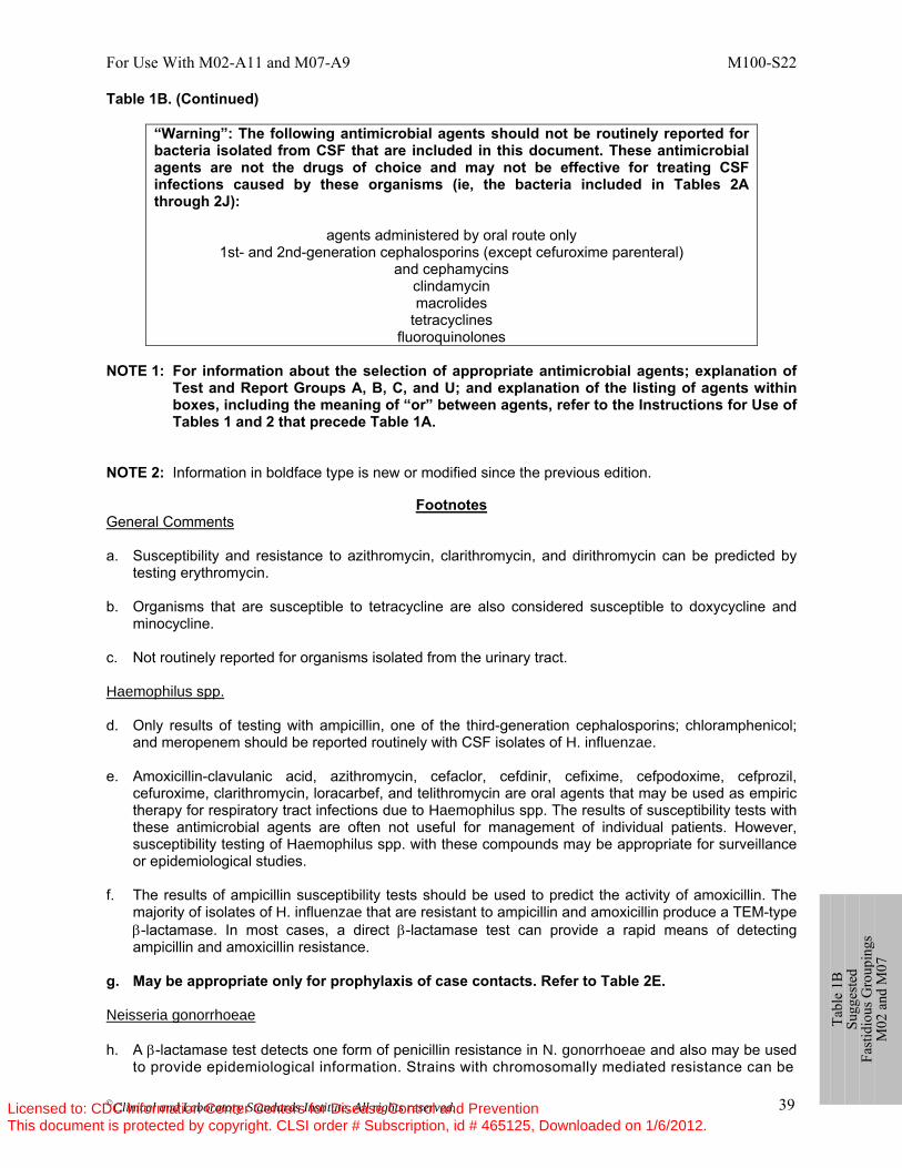
For Use With M02-A11 and M07-A9 M100-S22
©Clinical and Laboratory Standards Institute. All rights reserved. 39
Table 1B. (Continued)
“Warning”: The following antimicrobial agents should not be routinely reported for bacteria isolated from CSF that are included in this document. These antimicrobial agents are not the drugs of choice and may not be effective for treating CSF infections caused by these organisms (ie, the bacteria included in Tables 2A through 2J):
agents administered by oral route only 1st- and 2nd-generation cephalosporins (except cefuroxime parenteral)
and cephamycins clindamycin macrolides
tetracyclines fluoroquinolones
NOTE 1: For information about the selection of appropriate antimicrobial agents; explanation of
Test and Report Groups A, B, C, and U; and explanation of the listing of agents within boxes, including the meaning of “or” between agents, refer to the Instructions for Use of Tables 1 and 2 that precede Table 1A.
NOTE 2: Information in boldface type is new or modified since the previous edition.
Footnotes General Comments a. Susceptibility and resistance to azithromycin, clarithromycin, and dirithromycin can be predicted by
testing erythromycin. b. Organisms that are susceptible to tetracycline are also considered susceptible to doxycycline and
minocycline. c. Not routinely reported for organisms isolated from the urinary tract. Haemophilus spp. d. Only results of testing with ampicillin, one of the third-generation cephalosporins; chloramphenicol;
and meropenem should be reported routinely with CSF isolates of H. influenzae. e. Amoxicillin-clavulanic acid, azithromycin, cefaclor, cefdinir, cefixime, cefpodoxime, cefprozil,
cefuroxime, clarithromycin, loracarbef, and telithromycin are oral agents that may be used as empiric therapy for respiratory tract infections due to Haemophilus spp. The results of susceptibility tests with these antimicrobial agents are often not useful for management of individual patients. However, susceptibility testing of Haemophilus spp. with these compounds may be appropriate for surveillance or epidemiological studies.
f. The results of ampicillin susceptibility tests should be used to predict the activity of amoxicillin. The
majority of isolates of H. influenzae that are resistant to ampicillin and amoxicillin produce a TEM-type β-lactamase. In most cases, a direct β-lactamase test can provide a rapid means of detecting ampicillin and amoxicillin resistance.
g. May be appropriate only for prophylaxis of case contacts. Refer to Table 2E. Neisseria gonorrhoeae h. A β-lactamase test detects one form of penicillin resistance in N. gonorrhoeae and also may be used
to provide epidemiological information. Strains with chromosomally mediated resistance can be
Ta
ble
1B
Sugg
este
d Fa
stid
ious
Gro
upin
gs
M02
and
M07
Licensed to: CDC Information Center Centers for Disease Control and PreventionThis document is protected by copyright. CLSI order # Subscription, id # 465125, Downloaded on 1/6/2012.
January 2012 Vol. 32 No. 3
©Clinical and Laboratory Standards Institute. All rights reserved. 40
Table 1B. (Continued) detected only by additional susceptibility testing, such as the disk diffusion method or the agar dilution
MIC method. Streptococcus pneumoniae i. S. pneumoniae isolates susceptible to levofloxacin are predictably susceptible to gemifloxacin and
moxifloxacin. However, S. pneumoniae susceptible to gemifloxacin or moxifloxacin cannot be assumed to be susceptible to levofloxacin.
j. Penicillin and cefotaxime, ceftriaxone, or meropenem should be tested by a reliable MIC method
(such as that described in CLSI document M07-A9), and reported routinely with CSF isolates of S. pneumoniae. Such isolates should also be tested against vancomycin using the MIC or disk method. With isolates from other sites, the oxacillin disk screening test may be used. If the oxacillin zone size is ≤ 19 mm, penicillin, cefotaxime, ceftriaxone, or meropenem MICs should be determined.
k. Rx: Rifampin should not be used alone for antimicrobial therapy. Streptococcus spp. l. Rx: Penicillin- or ampicillin-intermediate isolates may require combined therapy with an
aminoglycoside for bactericidal action. m. Penicillin and ampicillin are drugs of choice for treatment of β-hemolytic streptococcal infections.
Susceptibility testing of penicillins and other β-lactams approved by the FDA for treatment of β-hemolytic streptococcal infections need not be performed routinely, because nonsusceptible isolates (ie, penicillin MICs > 0.12 and ampicillin MICs > 0.25 μg/mL) are extremely rare in any β-hemolytic streptococcus and have not been reported for Streptococcus pyogenes. If testing is performed, any β-hemolytic streptococcal isolate found to be nonsusceptible should be re-identified, retested, and, if confirmed, submitted to a public health laboratory. (See Appendix A for further instructions.)
n. Report against S. pyogenes. o. Rx: Recommendations for intrapartum prophylaxis for Group B streptococci are penicillin or
ampicillin. Although cefazolin is recommended for penicillin-allergic women at low risk for anaphylaxis, those at high risk for anaphylaxis may receive clindamycin or erythromycin. Group B streptococci are susceptible to ampicillin, penicillin, and cefazolin, but may be resistant to clindamycin and/or erythromycin. When Group B Streptococcus is isolated from a pregnant woman with severe penicillin allergy (high risk for anaphylaxis), clindamycin and erythromycin should be tested and reported.
p. For this table, the β-hemolytic group includes the large colony–forming pyogenic strains of
streptococci with Group A (S. pyogenes), C, or G antigens and strains with Group B (S. agalactiae) antigen. Small colony–forming β-hemolytic strains with Group A, C, F, or G antigens (S. anginosus group, previously termed “S. milleri”) are considered part of the viridans group, and interpretive criteria for the viridans group should be used.
Ta
ble
1B
Sugg
este
d Fa
stid
ious
Gro
upin
gs
M02
and
M07
Licensed to: CDC Information Center Centers for Disease Control and PreventionThis document is protected by copyright. CLSI order # Subscription, id # 465125, Downloaded on 1/6/2012.
For Use With M02-A11 and M07-A9 M100-S22
©Clinical and Laboratory Standards Institute. All rights reserved. 41
This page is intentionally left blank.
Licensed to: CDC Information Center Centers for Disease Control and PreventionThis document is protected by copyright. CLSI order # Subscription, id # 465125, Downloaded on 1/6/2012.
January 2012 Vol. 32 No. 3
©Clinical and Laboratory Standards Institute. All rights reserved. 42
Table 1C. Suggested Groupings of Antimicrobial Agents That Should Be Considered for Routine Testing and Reporting on Anaerobic Organisms
Bacteroides fragilis Group and Other Gram-Negative Anaerobes
Gram-Positive Anaerobesb
Gro
up A
Pr
imar
y Te
st a
nd R
epor
t
Amoxicillin-clavulanic acid Ampicillin-sulbactam Piperacillin-tazobactam Ticarcillin-clavulanic acid
Ampicillina
Penicillina Amoxicillin-clavulanic acid Ampicillin-sulbactam Piperacillin-tazobactam Ticarcillin-clavulanic acid
Clindamycin
Doripenem Ertapenem Imipenem Meropenem
Clindamycin
Doripenem Ertapenem Imipenem Meropenem
Metronidazole
Metronidazole
Gro
up C
Su
pple
men
tal
Rep
ort S
elec
tivel
y
Penicillina
Ampicillina Ceftizoxime Ceftriaxone
Ceftizoxime Ceftriaxone
Cefotetan Cefoxitin
Chloramphenicol Cefotetan Cefoxitin
Piperacillin Ticarcillin
Tetracycline
Moxifloxacin
Piperacillin Ticarcillin
Moxifloxacin
Ta
ble
1C
Sugg
este
d A
naer
obe
Gro
upin
gs
M11
Licensed to: CDC Information Center Centers for Disease Control and PreventionThis document is protected by copyright. CLSI order # Subscription, id # 465125, Downloaded on 1/6/2012.
For Use With Current Edition of M11 M100-S22
©Clinical and Laboratory Standards Institute. All rights reserved. 43
Table 1C. (Continued) NOTE 1: For information about the selection of appropriate antimicrobial agents; explanation of Test and Report Groups A, B, C, and U; and explanation of the listing of agents within boxes, including the meaning of “or” between agents, refer to the Instructions for Use of Tables 1 and 2 that precede Table 1A. NOTE 2: Information in boldface type is new or modified since the previous edition. NOTE 3: Most anaerobic infections are polymicrobial, including both β-lactamase-positive and β-lactamase-negative strains. Susceptibility of the most resistant strain must be considered first and reported. In the case of an infection caused by a single β-lactamase-negative strain, penicillin or ampicillin may be appropriate for testing and reporting. NOTE 4: Many gram-positive anaerobes are isolated from polymicrobial infections with potentially resistant organisms; however, some Clostridium species (eg, C. perfringens, C. septicum, C. sordellii) may be the singular cause of an infection, are typically susceptible to penicillin and ampicillin, and should be tested and reported. NOTE 5: The listing of drugs in a single box designates clusters of agents for which interpretive results (susceptible, intermediate, or resistant) and clinical efficacy are similar. Therefore, only one of the agents within each box ordinarily needs to be selected for testing. a. If β-lactamase positive, report as resistant to penicillin and ampicillin. Be aware that β-lactamase
negative isolates may be resistant to β-lactams by other mechanisms. b. Many non–spore-forming, gram-positive anaerobic rods are resistant to metronidazole.
Ta
ble
1C
Sugg
este
d A
naer
obe
Gro
upin
gs
M11
Licensed to: CDC Information Center Centers for Disease Control and PreventionThis document is protected by copyright. CLSI order # Subscription, id # 465125, Downloaded on 1/6/2012.
Table 2A. Zone Diameter and Minimal Inhibitory Concentration (MIC) Interpretive Standards for Enterobacteriaceae
* ATCC is a registered trademark of the American Type Culture Collection. Refer to Table 2A Supplemental Tables 1, 2, and 3 at the end of Table 2A for additional recommendations for testing conditions, reporting suggestions, and QC.
General Comments
(1) For disk diffusion, test a maximum of 12 disks on a 150-mm plate and 5 disks on a 100-mm plate (see M02 Section 9.2). Measure the diameter
of the zones of complete inhibition (as judged by the unaided eye), including the diameter of the disk. Hold the Petri plate a few inches above a black, nonreflecting background illuminated with reflected light. The zone margin should be considered the area showing no obvious, visible growth that can be detected with the unaided eye. Ignore faint growth of tiny colonies that can be detected only with a magnifying lens at the edge of the zone of inhibited growth. Strains of Proteus spp. may swarm into areas of inhibited growth around certain antimicrobial agents. With Proteus spp., ignore the thin veil of swarming growth in an otherwise obvious zone of growth inhibition. With trimethoprim and the sulfonamides, antagonists in the medium may allow some slight growth; therefore, disregard slight growth (20% or less of the lawn of growth) and measure the more obvious margin to determine the zone diameter.
(2) When fecal isolates of Salmonella and Shigella spp. are tested, only ampicillin, a fluoroquinolone, and trimethoprim-sulfamethoxazole should be
reported routinely. In addition, for extraintestinal isolates of Salmonella spp., a third-generation cephalosporin should be tested and reported, and chloramphenicol may be tested and reported if requested.
(3) The dosage regimens shown in the comment column below are those required to achieve plasma drug exposures (in adults with normal renal and
hepatic functions) on which breakpoints were based. When implementing new breakpoints, it is strongly recommended that laboratories share this information with infectious disease practitioners, pharmacists, pharmacy and therapeutics committees, and infection control committees.
NOTE: Information in boldface type is new or modified since the previous edition.
Table 2A Enterobacteriaceae
M02 and M07
44 ©C
linical and Laboratory Standards Institute. A
ll rights reserved.
Testing Conditions Medium: Disk diffusion: Mueller-Hinton agar (MHA) Broth dilution: cation-adjusted Mueller-Hinton broth (CAMHB) Agar dilution: MHA Inoculum: Growth method or direct colony suspension, equivalent to a
0.5 McFarland standard Incubation: 35 ± 2°C; ambient air; Disk diffusion: 16 to 18 hours Dilution methods: 16 to 20 hours
Minimal Quality Control (QC) Recommendations (See Tables 3A and 4A for acceptable QC ranges.) Escherichia coli ATCC®* 25922 Escherichia coli ATCC® 35218 (for β-lactam/β-lactamase inhibitor combinations)
January 2012 V
ol. 32 No. 3
Licensed to: CD
C Inform
ation Center C
enters for Disease C
ontrol and Prevention
This docum
ent is protected by copyright. CLS
I order # Subscription, id # 465125, D
ownloaded on 1/6/2012.
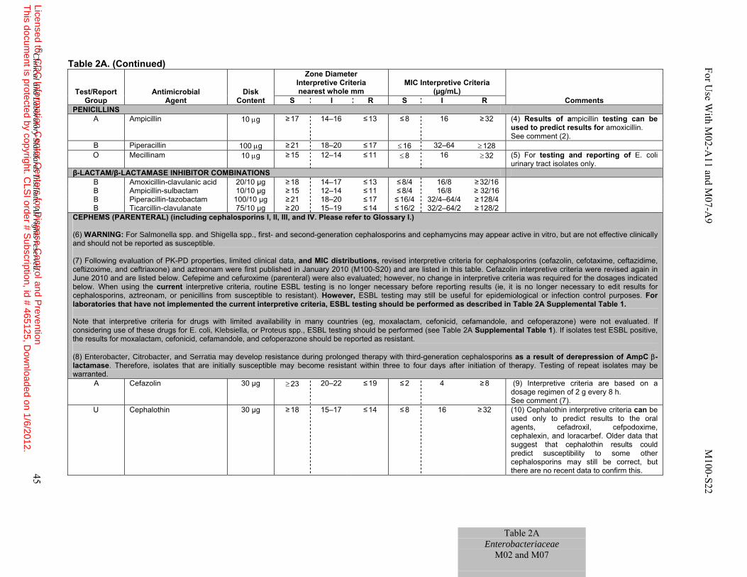
Table 2A. (Continued)
Test/Report Group
Antimicrobial Agent
Disk Content
Zone Diameter Interpretive Criteria nearest whole mm
MIC Interpretive Criteria (µg/mL)
Comments S I R S I R PENICILLINS
A Ampicillin 10 μg ≥ 17 14–16 ≤ 13 ≤ 8 16 ≥ 32 (4) Results of ampicillin testing can be used to predict results for amoxicillin. See comment (2).
B Piperacillin 100 μg ≥ 21 18–20 ≤ 17 ≤ 16 32–64 ≥ 128 O Mecillinam 10 μg ≥ 15 12–14 ≤ 11 ≤ 8 16 ≥ 32 (5) For testing and reporting of E. coli
urinary tract isolates only. β-LACTAM/β-LACTAMASE INHIBITOR COMBINATIONS
B Amoxicillin-clavulanic acid 20/10 µg ≥ 18 14–17 ≤ 13 ≤ 8/4 16/8 ≥ 32/16 B Ampicillin-sulbactam 10/10 µg ≥ 15 12–14 ≤ 11 ≤ 8/4 16/8 ≥ 32/16 B Piperacillin-tazobactam 100/10 µg ≥ 21 18–20 ≤ 17 ≤ 16/4 32/4–64/4 ≥ 128/4 B Ticarcillin-clavulanate 75/10 µg ≥ 20 15–19 ≤ 14 ≤ 16/2 32/2–64/2 ≥ 128/2
CEPHEMS (PARENTERAL) (including cephalosporins I, II, III, and IV. Please refer to Glossary I.) (6) WARNING: For Salmonella spp. and Shigella spp., first- and second-generation cephalosporins and cephamycins may appear active in vitro, but are not effective clinically and should not be reported as susceptible.
(7) Following evaluation of PK-PD properties, limited clinical data, and MIC distributions, revised interpretive criteria for cephalosporins (cefazolin, cefotaxime, ceftazidime, ceftizoxime, and ceftriaxone) and aztreonam were first published in January 2010 (M100-S20) and are listed in this table. Cefazolin interpretive criteria were revised again in June 2010 and are listed below. Cefepime and cefuroxime (parenteral) were also evaluated; however, no change in interpretive criteria was required for the dosages indicated below. When using the current interpretive criteria, routine ESBL testing is no longer necessary before reporting results (ie, it is no longer necessary to edit results for cephalosporins, aztreonam, or penicillins from susceptible to resistant). However, ESBL testing may still be useful for epidemiological or infection control purposes. For laboratories that have not implemented the current interpretive criteria, ESBL testing should be performed as described in Table 2A Supplemental Table 1.
Note that interpretive criteria for drugs with limited availability in many countries (eg, moxalactam, cefonicid, cefamandole, and cefoperazone) were not evaluated. If considering use of these drugs for E. coli, Klebsiella, or Proteus spp., ESBL testing should be performed (see Table 2A Supplemental Table 1). If isolates test ESBL positive, the results for moxalactam, cefonicid, cefamandole, and cefoperazone should be reported as resistant.
(8) Enterobacter, Citrobacter, and Serratia may develop resistance during prolonged therapy with third-generation cephalosporins as a result of derepression of AmpC β-lactamase. Therefore, isolates that are initially susceptible may become resistant within three to four days after initiation of therapy. Testing of repeat isolates may be warranted.
A Cefazolin 30 µg ≥ 23 20–22 ≤ 19 ≤ 2
4 ≥ 8 (9) Interpretive criteria are based on a dosage regimen of 2 g every 8 h. See comment (7).
U Cephalothin 30 µg ≥ 18 15–17 ≤ 14 ≤ 8 16 ≥ 32 (10) Cephalothin interpretive criteria can be used only to predict results to the oral agents, cefadroxil, cefpodoxime, cephalexin, and loracarbef. Older data that suggest that cephalothin results could predict susceptibility to some other cephalosporins may still be correct, but there are no recent data to confirm this.
Table 2A Enterobacteriaceae
M02 and M07
©C
linical and Laboratory Standards Institute. A
ll rights reserved. 45
For U
se With M
02-A11 and M
07-A9 M
100-S22
Licensed to: CD
C Inform
ation Center C
enters for Disease C
ontrol and Prevention
This docum
ent is protected by copyright. CLS
I order # Subscription, id # 465125, D
ownloaded on 1/6/2012.
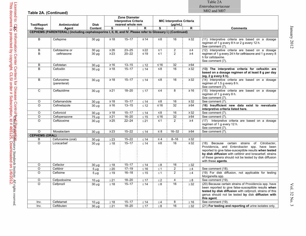
Table 2A. (Continued)
Test/Report Group
Antimicrobial Agent
Disk Content
Zone Diameter Interpretive Criteria nearest whole mm
MIC Interpretive Criteria (µg/mL)
Comments S I R S I R CEPHEMS (PARENTERAL) (including cephalosporins I, II, III, and IV. Please refer to Glossary I.) (Continued)
B Cefepime 30 µg ≥ 18 15–17 ≤ 14 ≤ 8 16 ≥ 32 (11) Interpretive criteria are based on a dosage
regimen of 1 g every 8 h or 2 g every 12 h. See comment (7).
B B
Cefotaxime or ceftriaxone
30 µg 30 µg
≥ 26 ≥ 23
23–25 20–22
≤ 22 ≤ 19
≤ 1 ≤ 1
2 2
≥ 4 ≥ 4
(12) Interpretive criteria are based on a dosage regimen of 1 g every 24 h for ceftriaxone and 1 g every 8 h for cefotaxime. See comment (7).
B Cefotetan 30 μg ≥ 16 13–15 ≤ 12 ≤ 16 32 ≥ 64 B Cefoxitin 30 μg ≥ 18 15–17 ≤ 14 ≤ 8 16 ≥ 32 (13) The interpretive criteria for cefoxitin are
based on a dosage regimen of at least 8 g per day (eg, 2 g every 6 h).
B Cefuroxime (parenteral)
30 μg ≥ 18 15–17 ≤ 14 ≤ 8 16 ≥ 32 (14) Interpretive criteria are based on a dosage regimen of 1.5 g every 8 h. See comment (7).
C Ceftazidime 30 μg ≥ 21 18–20 ≤ 17 ≤ 4 8 ≥ 16 (15) Interpretive criteria are based on a dosage regimen of 1 g every 8 h. See comment (7).
O Cefamandole 30 μg ≥ 18 15–17 ≤ 14 ≤ 8 16 ≥ 32 See comment (7). O Cefmetazole 30 μg ≥ 16 13–15 ≤ 12 ≤ 16 32 ≥ 64 (16) Insufficient new data exist to reevaluate
interpretive criteria listed here. O Cefonicid 30 μg ≥ 18 15–17 ≤ 14 ≤ 8 16 ≥ 32 See comment (7). O Cefoperazone 75 μg ≥ 21 16–20 ≤ 15 ≤ 16 32 ≥ 64 See comment (7). O Ceftizoxime 30 μg ≥ 25 22–24 ≤ 21 ≤ 1 2 ≥ 4 (17) Interpretive criteria are based on a dosage
regimen of 1 g every 12 h. See comment (7).
O Moxalactam 30 μg ≥ 23 15–22 ≤ 14 ≤ 8 16–32 ≥ 64 See comment (7). CEPHEMS (ORAL)
B Cefuroxime (oral) 30 μg ≥ 23 15–22 ≤ 14 ≤ 4 8–16 ≥ 32 O Loracarbef 30 μg ≥ 18 15–17 ≤ 14 ≤ 8 16 ≥ 32 (18) Because certain strains of Citrobacter,
Providencia, and Enterobacter spp. have been reported to give false-susceptible results when tested by disk diffusion with cefdinir and loracarbef, strains of these genera should not be tested by disk diffusion with these agents.
O Cefaclor 30 μg ≥ 18 15–17 ≤ 14 ≤ 8 16 ≥ 32 O Cefdinir 5 μg ≥ 20 17–19 ≤ 16 ≤ 1 2 ≥ 4 See comment (18). O Cefixime 5 μg ≥ 19 16–18 ≤ 15 ≤ 1 2 ≥ 4 (19) For disk diffusion, not applicable for testing
Morganella spp. O Cefpodoxime 10 μg ≥ 21 18–20 ≤ 17 ≤ 2 4 ≥ 8 See comment (19). O Cefprozil 30 μg ≥ 18 15–17 ≤ 14 ≤ 8 16 ≥ 32 (20) Because certain strains of Providencia spp. have
been reported to give false-susceptible results when tested by disk diffusion with cefprozil, strains of this genus should not be tested by disk diffusion with this agent.
Inv. Cefetamet 10 μg ≥ 18 15–17 ≤ 14 ≤ 4 8 ≥ 16 See comment (19). Inv. Ceftibuten 30 μg ≥ 21 18–20 ≤ 17 ≤ 8 16 ≥ 32 (21) For testing and reporting of urine isolates only.
Table 2AEnterobacteriaceae
M02 and M07
46 ©C
linical and Laboratory Standards Institute. A
ll rights reserved.
January 2012 V
ol. 32 No. 3
Licensed to: CD
C Inform
ation Center C
enters for Disease C
ontrol and Prevention
This docum
ent is protected by copyright. CLS
I order # Subscription, id # 465125, D
ownloaded on 1/6/2012.
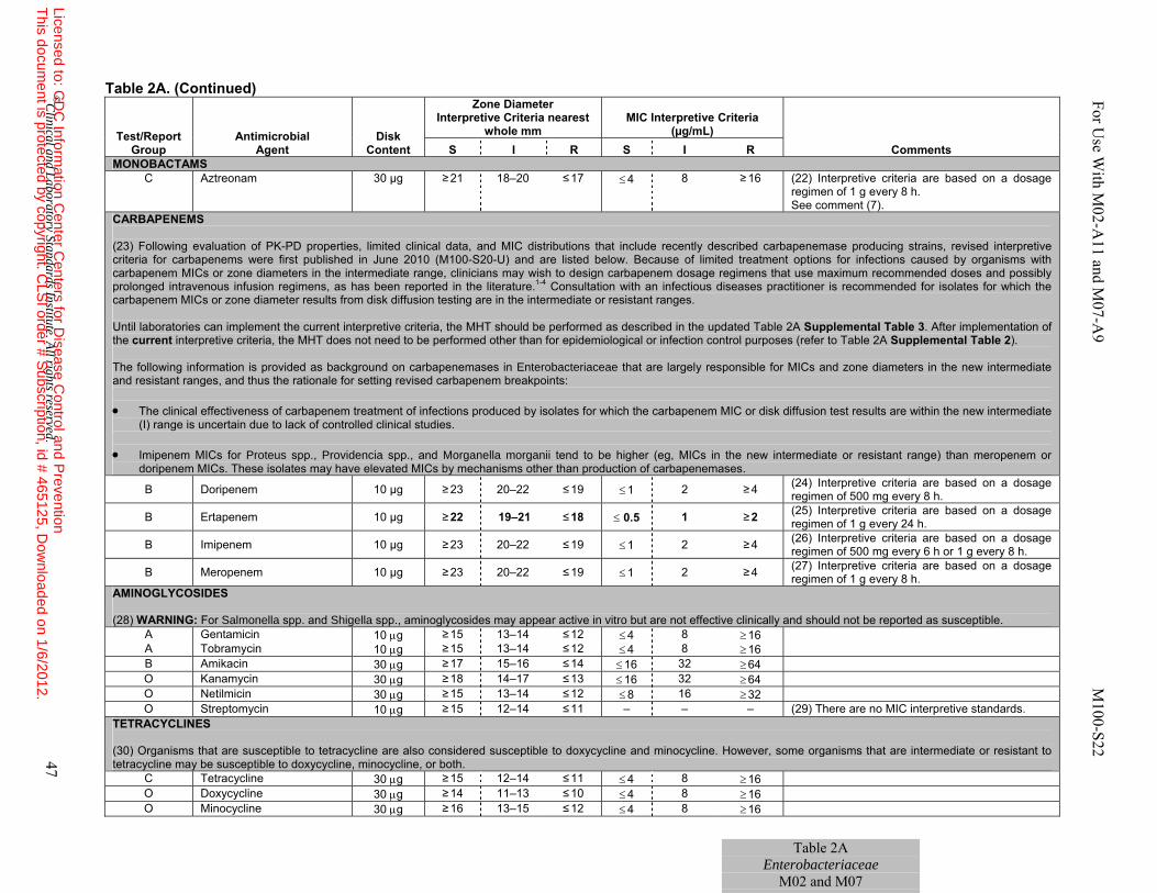
Table 2A. (Continued)
Test/Report Group
Antimicrobial Agent
Disk Content
Zone Diameter Interpretive Criteria nearest
whole mm MIC Interpretive Criteria
(µg/mL) Comments S I R S I R
MONOBACTAMS C Aztreonam 30 µg ≥ 21
18–20 ≤ 17 ≤ 4 8 ≥ 16 (22) Interpretive criteria are based on a dosage
regimen of 1 g every 8 h. See comment (7).
CARBAPENEMS (23) Following evaluation of PK-PD properties, limited clinical data, and MIC distributions that include recently described carbapenemase producing strains, revised interpretive criteria for carbapenems were first published in June 2010 (M100-S20-U) and are listed below. Because of limited treatment options for infections caused by organisms with carbapenem MICs or zone diameters in the intermediate range, clinicians may wish to design carbapenem dosage regimens that use maximum recommended doses and possibly prolonged intravenous infusion regimens, as has been reported in the literature.1-4 Consultation with an infectious diseases practitioner is recommended for isolates for which the carbapenem MICs or zone diameter results from disk diffusion testing are in the intermediate or resistant ranges. Until laboratories can implement the current interpretive criteria, the MHT should be performed as described in the updated Table 2A Supplemental Table 3. After implementation of the current interpretive criteria, the MHT does not need to be performed other than for epidemiological or infection control purposes (refer to Table 2A Supplemental Table 2). The following information is provided as background on carbapenemases in Enterobacteriaceae that are largely responsible for MICs and zone diameters in the new intermediate and resistant ranges, and thus the rationale for setting revised carbapenem breakpoints: • The clinical effectiveness of carbapenem treatment of infections produced by isolates for which the carbapenem MIC or disk diffusion test results are within the new intermediate
(I) range is uncertain due to lack of controlled clinical studies. • Imipenem MICs for Proteus spp., Providencia spp., and Morganella morganii tend to be higher (eg, MICs in the new intermediate or resistant range) than meropenem or
doripenem MICs. These isolates may have elevated MICs by mechanisms other than production of carbapenemases.
B Doripenem 10 µg ≥ 23 20–22 ≤ 19 ≤ 1 2 ≥ 4 (24) Interpretive criteria are based on a dosage regimen of 500 mg every 8 h.
B Ertapenem 10 µg ≥ 22 19–21 ≤ 18 ≤ 0.5 1 ≥ 2 (25) Interpretive criteria are based on a dosage regimen of 1 g every 24 h.
B Imipenem 10 µg ≥ 23 20–22 ≤ 19 ≤ 1 2 ≥ 4 (26) Interpretive criteria are based on a dosage regimen of 500 mg every 6 h or 1 g every 8 h.
B Meropenem 10 µg ≥ 23 20–22 ≤ 19 ≤ 1 2 ≥ 4 (27) Interpretive criteria are based on a dosage regimen of 1 g every 8 h.
AMINOGLYCOSIDES (28) WARNING: For Salmonella spp. and Shigella spp., aminoglycosides may appear active in vitro but are not effective clinically and should not be reported as susceptible.
A Gentamicin 10 μg ≥ 15 13–14 ≤ 12 ≤ 4 8 ≥ 16 A Tobramycin 10 μg ≥ 15 13–14 ≤ 12 ≤ 4 8 ≥ 16 B Amikacin 30 μg ≥ 17 15–16 ≤ 14 ≤ 16 32 ≥ 64 O Kanamycin 30 μg ≥ 18 14–17 ≤ 13 ≤ 16 32 ≥ 64 O Netilmicin 30 μg ≥ 15 13–14 ≤ 12 ≤ 8 16 ≥ 32 O Streptomycin 10 μg ≥ 15 12–14 ≤ 11 – – – (29) There are no MIC interpretive standards.
TETRACYCLINES
(30) Organisms that are susceptible to tetracycline are also considered susceptible to doxycycline and minocycline. However, some organisms that are intermediate or resistant to tetracycline may be susceptible to doxycycline, minocycline, or both.
C Tetracycline 30 μg ≥ 15 12–14 ≤ 11 ≤ 4 8 ≥ 16 O Doxycycline 30 μg ≥ 14 11–13 ≤ 10 ≤ 4 8 ≥ 16 O Minocycline 30 μg ≥ 16 13–15 ≤ 12 ≤ 4 8 ≥ 16
Table 2A Enterobacteriaceae
M02 and M07
©C
linical and Laboratory Standards Institute. A
ll rights reserved. 47
For U
se With M
02-A11 and M
07-A9 M
100-S22
Licensed to: CD
C Inform
ation Center C
enters for Disease C
ontrol and Prevention
This docum
ent is protected by copyright. CLS
I order # Subscription, id # 465125, D
ownloaded on 1/6/2012.
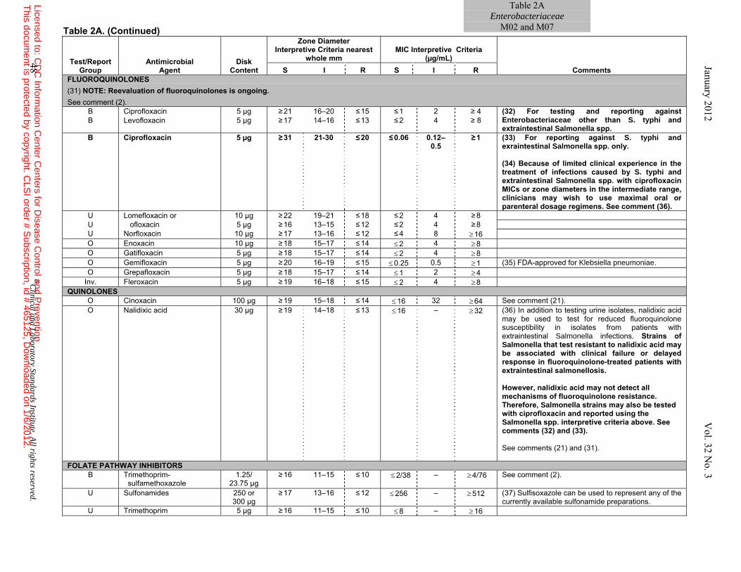
Table 2A. (Continued)
Test/Report Group
Antimicrobial Agent
Disk Content
Zone Diameter Interpretive Criteria nearest
whole mm MIC Interpretive Criteria
(µg/mL) Comments S I R S I R
FLUOROQUINOLONES (31) NOTE: Reevaluation of fluoroquinolones is ongoing. See comment (2).
B Ciprofloxacin 5 µg ≥ 21 16–20 ≤ 15 ≤ 1 2 ≥ 4 (32) For testing and reporting against Enterobacteriaceae other than S. typhi and extraintestinal Salmonella spp.
B Levofloxacin 5 µg ≥ 17 14–16 ≤ 13 ≤ 2 4 ≥ 8
B Ciprofloxacin 5 µg ≥ 31 21-30 ≤ 20 ≤ 0.06 0.12–0.5
≥ 1 (33) For reporting against S. typhi and exraintestinal Salmonella spp. only. (34) Because of limited clinical experience in the treatment of infections caused by S. typhi and extraintestinal Salmonella spp. with ciprofloxacin MICs or zone diameters in the intermediate range, clinicians may wish to use maximal oral or parenteral dosage regimens. See comment (36).
U Lomefloxacin or 10 µg ≥ 22 19–21 ≤ 18 ≤ 2 4 ≥ 8 U ofloxacin 5 µg ≥ 16 13–15 ≤ 12 ≤ 2 4 ≥ 8 U Norfloxacin 10 µg ≥ 17 13–16 ≤ 12 ≤ 4 8 ≥ 16 O Enoxacin 10 µg ≥ 18 15–17 ≤ 14 ≤ 2 4 ≥ 8 O Gatifloxacin 5 µg ≥ 18 15–17 ≤ 14 ≤ 2 4 ≥ 8 O Gemifloxacin 5 µg ≥ 20 16–19 ≤ 15 ≤ 0.25 0.5 ≥ 1 (35) FDA-approved for Klebsiella pneumoniae. O Grepafloxacin 5 µg ≥ 18 15–17 ≤ 14 ≤ 1 2 ≥ 4
Inv. Fleroxacin 5 µg ≥ 19 16–18 ≤ 15 ≤ 2 4 ≥ 8 QUINOLONES
O Cinoxacin 100 µg ≥ 19 15–18 ≤ 14 ≤ 16 32 ≥ 64 See comment (21). O Nalidixic acid 30 µg ≥ 19 14–18 ≤ 13 ≤ 16 – ≥ 32 (36) In addition to testing urine isolates, nalidixic acid
may be used to test for reduced fluoroquinolone susceptibility in isolates from patients with extraintestinal Salmonella infections. Strains of Salmonella that test resistant to nalidixic acid may be associated with clinical failure or delayed response in fluoroquinolone-treated patients with extraintestinal salmonellosis.
However, nalidixic acid may not detect all mechanisms of fluoroquinolone resistance. Therefore, Salmonella strains may also be tested with ciprofloxacin and reported using the Salmonella spp. interpretive criteria above. See comments (32) and (33). See comments (21) and (31).
FOLATE PATHWAY INHIBITORS B Trimethoprim-
sulfamethoxazole 1.25/
23.75 µg ≥ 16 11–15 ≤ 10 ≤ 2/38
–
≥ 4/76
See comment (2).
U Sulfonamides 250 or 300 µg
≥ 17 13–16 ≤ 12 ≤ 256 – ≥ 512 (37) Sulfisoxazole can be used to represent any of the currently available sulfonamide preparations.
U Trimethoprim 5 µg ≥ 16 11–15 ≤ 10 ≤ 8 – ≥ 16
January 2012 V
ol. 32 No. 3
48 ©C
linical and Laboratory Standards Institute. A
ll rights reserved.
Table 2AEnterobacteriaceae
M02 and M07
Licensed to: CD
C Inform
ation Center C
enters for Disease C
ontrol and Prevention
This docum
ent is protected by copyright. CLS
I order # Subscription, id # 465125, D
ownloaded on 1/6/2012.
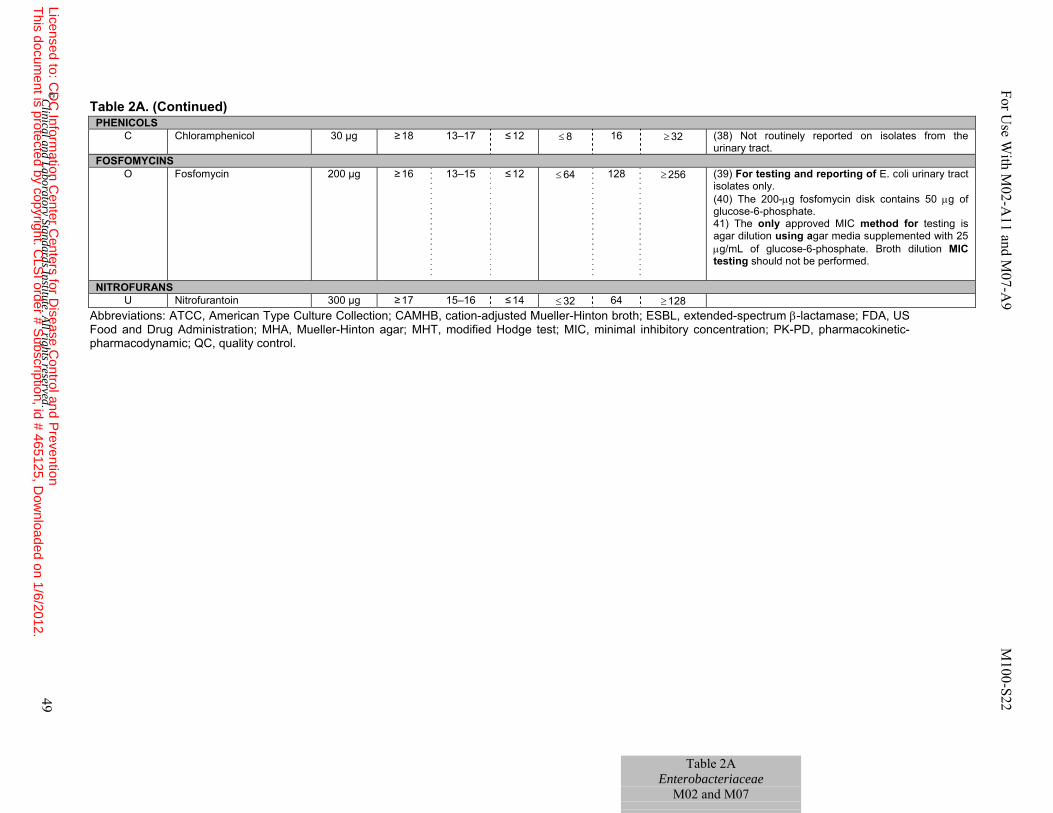
For U
se With M
02-A11 and M
07-A9 M
100-S22
Table 2A. (Continued)
Abbreviations: ATCC, American Type Culture Collection; CAMHB, cation-adjusted Mueller-Hinton broth; ESBL, extended-spectrum β-lactamase; FDA, US Food and Drug Administration; MHA, Mueller-Hinton agar; MHT, modified Hodge test; MIC, minimal inhibitory concentration; PK-PD, pharmacokinetic-pharmacodynamic; QC, quality control.
PHENICOLS C Chloramphenicol 30 µg ≥ 18 13–17 ≤ 12 ≤ 8 16 ≥ 32 (38) Not routinely reported on isolates from the
urinary tract. FOSFOMYCINS
O Fosfomycin 200 µg ≥ 16 13–15 ≤ 12 ≤ 64 128 ≥ 256 (39) For testing and reporting of E. coli urinary tract isolates only. (40) The 200-μg fosfomycin disk contains 50 μg of glucose-6-phosphate. 41) The only approved MIC method for testing is agar dilution using agar media supplemented with 25 μg/mL of glucose-6-phosphate. Broth dilution MIC testing should not be performed.
NITROFURANS U Nitrofurantoin 300 µg ≥ 17 15–16 ≤ 14 ≤ 32 64 ≥ 128
Table 2A Enterobacteriaceae
M02 and M07
©C
linical and Laboratory Standards Institute. A
ll rights reserved. 49
Licensed to: CD
C Inform
ation Center C
enters for Disease C
ontrol and Prevention
This docum
ent is protected by copyright. CLS
I order # Subscription, id # 465125, D
ownloaded on 1/6/2012.
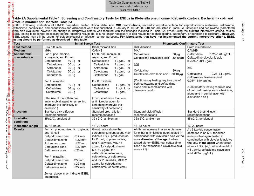
Table 2A Supplemental Table 1. Screening and Confirmatory Tests for ESBLs in Klebsiella pneumoniae, Klebsiella oxytoca, Escherichia coli, and Proteus mirabilis for Use With Table 2A NOTE: Following evaluation of PK-PD properties, limited clinical data, and MIC distributions, revised interpretive criteria for cephalosporins (cefazolin, cefotaxime, ceftazidime, ceftizoxime, and ceftriaxone) and aztreonam were first published in January 2010 (M100-S20) and are listed in Table 2A. Cefepime and cefuroxime (parenteral) were also evaluated; however, no change in interpretive criteria was required with the dosages included in Table 2A. When using the current interpretive criteria, routine ESBL testing is no longer necessary before reporting results (ie, it is no longer necessary to edit results for cephalosporins, aztreonam, or penicillins to resistant). However, ESBL testing may still be useful for epidemiological or infection control purposes. For laboratories that have not implemented the current interpretive criteria, ESBL testing should be performed as described in this table.
Test Initial Screen Test Phenotypic Confirmatory TestTest method Disk diffusion Broth microdilution Disk diffusion Broth microdilution Medium MHA CAMHB MHA CAMHBAntimicrobial concentration
For K. pneumoniae, K. oxytoca, and E. coli: Cefpodoxime 10 μg or Ceftazidime 30 μg or Aztreonam 30 μg or Cefotaxime 30 μg or Ceftriaxone 30 μg For P. mirabilis: Cefpodoxime 10 μg or Ceftazidime 30 μg or Cefotaxime 30 μg (The use of more than one antimicrobial agent for screening improves the sensitivity of detection.)
For K. pneumoniae, K. oxytoca, and E. coli: Cefpodoxime 4 μg/mL or Ceftazidime 1 μg/mL or Aztreonam 1 μg/mL or Cefotaxime 1 μg/mL or Ceftriaxone 1 μg/mL For P. mirabilis: Cefpodoxime 1 μg/mL or Ceftazidime 1 μg/mL or Cefotaxime 1 μg/mL (The use of more than one antimicrobial agent for screening improves the sensitivity of detection.)
Ceftazidime 30 μg Ceftazidime-clavulanic acida 30/10 μg and Cefotaxime 30 μg Cefotaxime-clavulanic acid 30/10 μg (Confirmatory testing requires use of both cefotaxime and ceftazidime, alone and in combination with clavulanic acid.)
Ceftazidime 0.25–128 μg/mL Ceftazidime-clavulanic acid 0.25/4–128/4 μg/mL and Cefotaxime 0.25–64 μg/mL Cefotaxime-clavulanic acid 0.25/4–64/4 μg/mL (Confirmatory testing requires use of both cefotaxime and ceftazidime, alone and in combination with clavulanic acid.)
Inoculum Standard disk diffusion recommendations
Standard broth dilution recommendations
Standard disk diffusion recommendations
Standard broth dilution recommendations
Incubation conditions
35 ± 2°C; ambient air 35 ± 2°C; ambient air 35 ± 2°C; ambient air 35 ± 2°C; ambient air
Incubation length 16–18 hours 16–20 hours 16–18 hours 16–20 hours Results For K. pneumoniae, K. oxytoca,
and E. coli: Cefpodoxime zone ≤ 17 mm Ceftazidime zone ≤ 22 mm Aztreonam zone ≤ 27 mm Cefotaxime zone ≤ 27 mm Ceftriaxone zone ≤ 25 mm For P. mirabilis:
Cefpodoxime zone ≤ 22 mm Ceftazidime zone ≤ 22 mm Cefotaxime zone ≤ 27 mm Zones above may indicate ESBL production.
Growth at or above the screening concentrations may indicate ESBL production (ie, for E. coli, K. pneumoniae, and K. oxytoca, MIC ≥ 8 μg/mL for cefpodoxime or MIC ≥ 2 μg/mL for ceftazidime, aztreonam, cefotaxime, or ceftriaxone; and for P. mirabilis, MIC ≥ 2 μg/mL for cefpodoxime, ceftazidime, or cefotaxime).
A ≥ 5-mm increase in a zone diameter for either antimicrobial agent tested in combination with clavulanic acid vs the zone diameter of the agent when tested alone = ESBL (eg, ceftazidime zone = 16; ceftazidime-clavulanic acid zone = 21).
A ≥ 3 twofold concentration decrease in an MIC for either antimicrobial agent tested in combination with clavulanic acid vs the MIC of the agent when tested alone = ESBL (eg, ceftazidime MIC
= 8 μg/mL; ceftazidime-clavulanic acid MIC = 1 μg/mL).
50 ©C
linical and Laboratory Standards Institute. A
ll rights reserved.
Table 2A Supplemental Table 1 Screening and Confirmatory
Tests for ESBLs January 2012 V
ol. 32 No. 3
Licensed to: CD
C Inform
ation Center C
enters for Disease C
ontrol and Prevention
This docum
ent is protected by copyright. CLS
I order # Subscription, id # 465125, D
ownloaded on 1/6/2012.
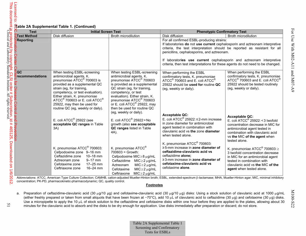
Table 2A Supplemental Table 1. (Continued)
Abbreviations: ATCC, American Type Culture Collection; CAMHB, cation-adjusted Mueller-Hinton broth; ESBL, extended-spectrum β-lactamase; MHA, Mueller-Hinton agar; MIC, minimal inhibitory concentration; PK-PD, pharmacokinetic-pharmacodynamic; QC, quality control.
Footnotes
a. Preparation of ceftazidime-clavulanic acid (30 μg/10 µg) and cefotaxime-clavulanic acid (30 μg/10 μg) disks: Using a stock solution of clavulanic acid at 1000 μg/mL (either freshly prepared or taken from small aliquots that have been frozen at −70°C), add 10 μL of clavulanic acid to ceftazidime (30 μg) and cefotaxime (30 μg) disks. Use a micropipette to apply the 10 μL of stock solution to the ceftazidime and cefotaxime disks within one hour before they are applied to the plates, allowing about 30 minutes for the clavulanic acid to absorb and the disks to be dry enough for application. Use disks immediately after preparation or discard; do not store.
Test Initial Screen Test Phenotypic Confirmatory TestTest Method Disk diffusion Broth microdilution Disk diffusion Broth microdilution Reporting For all confirmed ESBL-producing strains:
If laboratories do not use current cephalosporin and aztreonam interpretive criteria, the test interpretation should be reported as resistant for all penicillins, cephalosporins, and aztreonam. If laboratories use current cephalosporin and aztreonam interpretive criteria, then test interpretations for these agents do not need to be changed.
QC recommendations
When testing ESBL-screening antimicrobial agents, K. pneumoniae ATCC® 700603 is provided as a supplemental QC strain (eg, for training, competency, or test evaluation). Either strain, K. pneumoniae ATCC® 700603 or E. coli ATCC® 25922, may then be used for routine QC (eg, weekly or daily). E. coli ATCC® 25922 (see acceptable QC ranges in Table 3A) K. pneumoniae ATCC® 700603: Cefpodoxime zone 9–16 mm Ceftazidime zone 10–18 mm Aztreonam zone 9–17 mm Cefotaxime zone 17–25 mm Ceftriaxone zone 16–24 mm
When testing ESBL-screening antimicrobial agents, K. pneumoniae ATCC® 700603 is provided as a supplemental QC strain (eg, for training, competency, or test evaluation). Either strain, K. pneumoniae ATCC® 700603 or E. coli ATCC® 25922, may then be used for routine QC (eg, weekly or daily). E. coli ATCC® 25922 = No growth (also see acceptable QC ranges listed in Table 4A). K. pneumoniae ATCC® 700603 = Growth: Cefpodoxime MIC ≥ 8 μg/mL Ceftazidime MIC ≥ 2 μg/mL Aztreonam MIC ≥ 2 μg/mL Cefotaxime MIC ≥ 2 μg/mL Ceftriaxone MIC ≥ 2 μg/mL
When performing the ESBL confirmatory tests, K. pneumoniae ATCC® 700603 and E. coli ATCC® 25922 should be used for routine QC (eg, weekly or daily). Acceptable QC: E. coli ATCC® 25922: ≤ 2-mm increase in zone diameter for antimicrobial agent tested in combination with clavulanic acid vs the zone diameter when tested alone. K. pneumoniae ATCC® 700603:
≥ 5-mm increase in zone diameter of ceftazidime-clavulanic acid vs ceftazidime alone; ≥ 3-mm increase in zone diameter of cefotaxime-clavulanic acid vs cefotaxime alone.
When performing the ESBL confirmatory tests, K. pneumoniae ATCC® 700603 and E. coli ATCC® 25922 should be tested routinely (eg, weekly or daily). Acceptable QC: E. coli ATCC® 25922: < 3 twofold concentration decrease in MIC for antimicrobial agent tested in combination with clavulanic acid vs the MIC of the agent when tested alone. K. pneumoniae ATCC® 700603: ≥
3 twofold concentration decrease in MIC for an antimicrobial agent tested in combination with clavulanic acid vs the MIC of the agent when tested alone.
Table 2A Supplemental Table 1 Screening and Confirmatory
Tests for ESBLs
©C
linical and Laboratory Standards Institute. A
ll rights reserved. 51
For U
se With M
02-A11 and M
07-A9 M
100-S22
Licensed to: CD
C Inform
ation Center C
enters for Disease C
ontrol and Prevention
This docum
ent is protected by copyright. CLS
I order # Subscription, id # 465125, D
ownloaded on 1/6/2012.
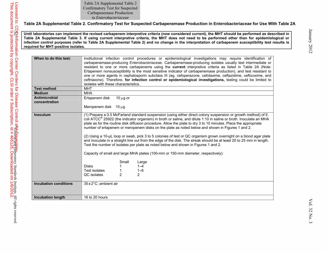
Table 2A Supplemental Table 2. Confirmatory Test for Suspected Carbapenemase Production in Enterobacteriaceae for Use With Table 2A
Until laboratories can implement the revised carbapenem interpretive criteria (now considered current), the MHT should be performed as described in Table 2A Supplemental Table 3. If using current interpretive criteria, the MHT does not need to be performed other than for epidemiological or infection control purposes (refer to Table 2A Supplemental Table 2) and no change in the interpretation of carbapenem susceptibility test results is required for MHT-positive isolates.
When to do this test: Institutional infection control procedures or epidemiological investigations may require identification of carbapenemase-producing Enterobacteriaceae. Carbapenemase-producing isolates usually test intermediate or resistant to one or more carbapenems using the current interpretive criteria as listed in Table 2A (Note: Ertapenem nonsusceptibility is the most sensitive indicator of carbapenemase production), and test resistant to one or more agents in cephalosporin subclass III (eg, cefoperazone, cefotaxime, ceftazidime, ceftizoxime, and ceftriaxone). Therefore, for infection control or epidemiological investigations, testing could be limited to isolates with these characteristics.
Test method MHT Medium MHA Antimicrobial concentration
Ertapenem disk 10 μg or Meropenem disk 10 μg
Inoculum (1) Prepare a 0.5 McFarland standard suspension (using either direct colony suspension or growth method) of E. coli ATCC® 25922 (the indicator organism) in broth or saline, and dilute 1:10 in saline or broth. Inoculate an MHA plate as for the routine disk diffusion procedure. Allow the plate to dry 3 to 10 minutes. Place the appropriate number of ertapenem or meropenem disks on the plate as noted below and shown in Figures 1 and 2. (2) Using a 10-µL loop or swab, pick 3 to 5 colonies of test or QC organism grown overnight on a blood agar plate and inoculate in a straight line out from the edge of the disk. The streak should be at least 20 to 25 mm in length. Test the number of isolates per plate as noted below and shown in Figures 1 and 2. Capacity of small and large MHA plates (100-mm or 150-mm diameter, respectively): Small Large Disks 1 1–4 Test isolates 1 1–6 QC isolates 2 2
Incubation conditions 35 ± 2°C; ambient air
Incubation length 16 to 20 hours
Table 2A Supplemental Table 2 Confirmatory Test for Suspected
Carbapenemase Production in Enterobacteriaceae
52 ©C
linical and Laboratory Standards Institute. A
ll rights reserved.
January 2012 V
ol. 32 No. 3
Licensed to: CD
C Inform
ation Center C
enters for Disease C
ontrol and Prevention
This docum
ent is protected by copyright. CLS
I order # Subscription, id # 465125, D
ownloaded on 1/6/2012.
Table 2A Supplemental Table 2. (Continued)
Abbreviations: ATCC, American Type Culture Collection; KPC, Klebsiella pneumoniae carbapenemase; MHA, Mueller-Hinton agar; MHT, modified Hodge test; MIC, minimal inhibitory concentration; QC, quality control.
Results Following incubation, examine the MHA plate for enhanced growth around the test or QC organism streak at the intersection of the streak and the zone of inhibition (see Figures 1 and 2). Enhanced growth = positive for carbapenemase production. No enhanced growth = negative for carbapenemase production. Some test isolates may produce substances that will inhibit growth of E. coli ATCC® 25922. When this occurs, a clear area will be seen around the streak (see Figure 3), and the MHT is uninterpretable for these isolates. For isolates with positive MHTs, perform MIC tests before reporting any carbapenem results, since carbapenem MIC interpretations are based solely on the MIC and should not be changed regardless of the MHT result. NOTE: Not all carbapenemase-producing isolates of Enterobacteriaceae are MHT positive and MHT-positive results may be encountered in isolates with carbapenem resistance mechanisms other than carbapenemase production.
Reporting Report results of the MHT to infection control or those requesting epidemiological information. No change in the interpretation of carbapenems susceptibility test results is required for MHT-positive isolates.
QC recommendations Test positive and negative QC organisms each day of testing. K. pneumoniae ATCC® BAA-1705—MHT positive K. pneumoniae ATCC® BAA-1706—MHT negative
Table 2A Supplemental Table 2 Confirmatory Test for Suspected
Carbapenemase Production in Enterobacteriaceae
©C
linical and Laboratory Standards Institute. A
ll rights reserved. 53
For U
se With M
02-A11 and M
07-A9 M
100-S22
Licensed to: CD
C Inform
ation Center C
enters for Disease C
ontrol and Prevention
This docum
ent is protected by copyright. CLS
I order # Subscription, id # 465125, D
ownloaded on 1/6/2012.
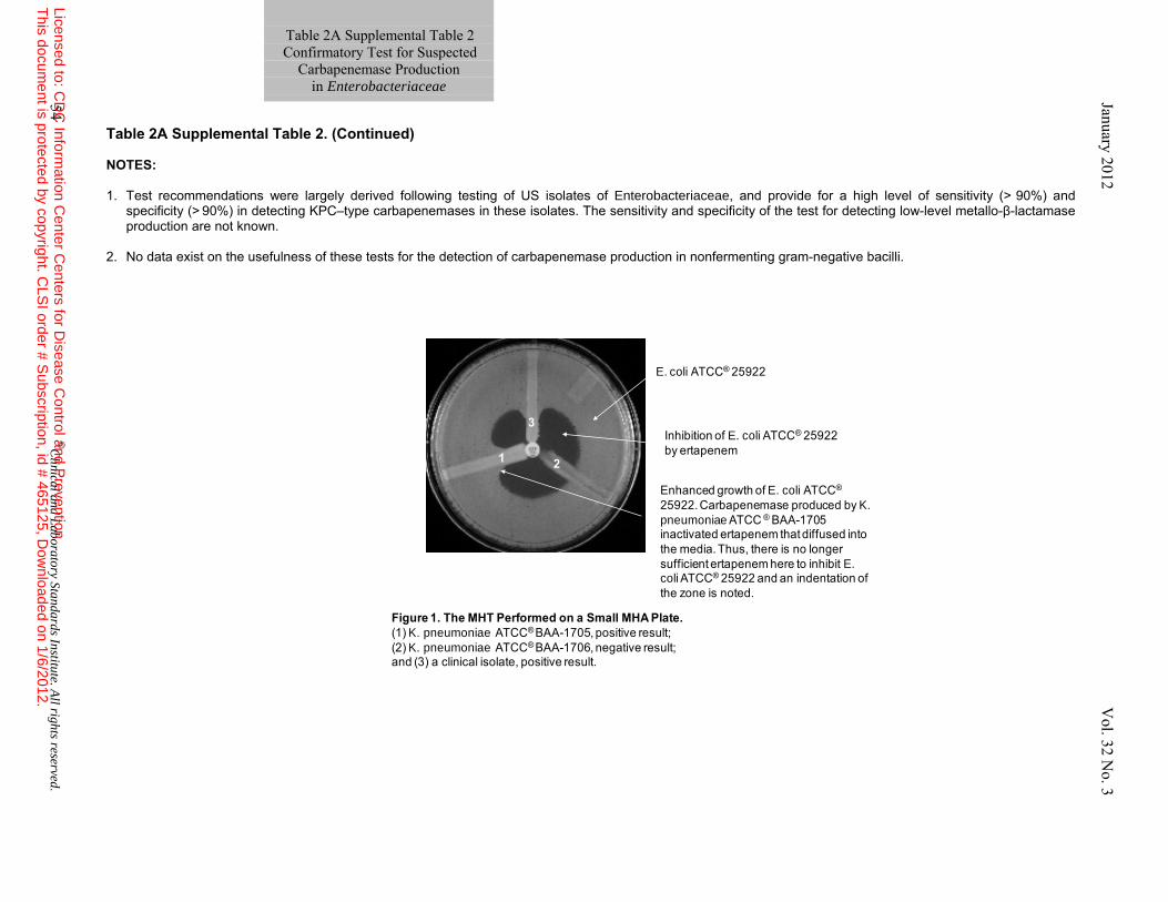
Table 2A Supplemental Table 2. (Continued) NOTES:
1. Test recommendations were largely derived following testing of US isolates of Enterobacteriaceae, and provide for a high level of sensitivity (> 90%) and
specificity (> 90%) in detecting KPC–type carbapenemases in these isolates. The sensitivity and specificity of the test for detecting low-level metallo-β-lactamase production are not known.
2. No data exist on the usefulness of these tests for the detection of carbapenemase production in nonfermenting gram-negative bacilli.
1 2
3
Figure 1. The MHT Performed on a Small MHA Plate. (1) K. pneumoniae ATCC® BAA-1705, positive result; (2) K. pneumoniae ATCC® BAA-1706, negative result; and (3) a clinical isolate, positive result.
E. coli ATCC® 25922
Inhibition of E. coli ATCC® 25922 by ertapenem
Enhanced growth of E. coli ATCC®
25922. Carbapenemase produced by K. pneumoniae ATCC ® BAA-1705 inactivated ertapenem that diffused into the media. Thus, there is no longer sufficient ertapenem here to inhibit E. coli ATCC® 25922 and an indentation of the zone is noted.
Table 2A Supplemental Table 2 Confirmatory Test for Suspected
Carbapenemase Production in Enterobacteriaceae
54 ©C
linical and Laboratory Standards Institute. A
ll rights reserved.
January 2012 V
ol. 32 No. 3
Licensed to: CD
C Inform
ation Center C
enters for Disease C
ontrol and Prevention
This docum
ent is protected by copyright. CLS
I order # Subscription, id # 465125, D
ownloaded on 1/6/2012.
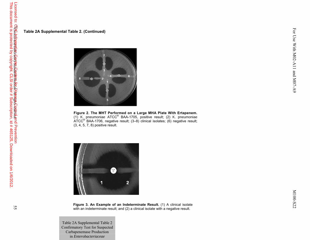
Table 2A Supplemental Table 2. (Continued)
Figure 2. The MHT Performed on a Large MHA Plate With Ertapenem. (1) K. pneumoniae ATCC® BAA-1705, positive result; (2) K. pneumoniae ATCC® BAA-1706, negative result; (3–8) clinical isolates; (6) negative result; (3, 4, 5, 7, 8) positive result.
©C
linical and Laboratory Standards Institute. A
ll rights reserved. 55
Figure 3. An Example of an Indeterminate Result. (1) A clinical isolate with an indeterminate result; and (2) a clinical isolate with a negative result.
For U
se With M
02-A11 and M
07-A9 M
100-S22
Table 2A Supplemental Table 2 Confirmatory Test for Suspected
Carbapenemase Production in Enterobacteriaceae
Licensed to: CD
C Inform
ation Center C
enters for Disease C
ontrol and Prevention
This docum
ent is protected by copyright. CLS
I order # Subscription, id # 465125, D
ownloaded on 1/6/2012.
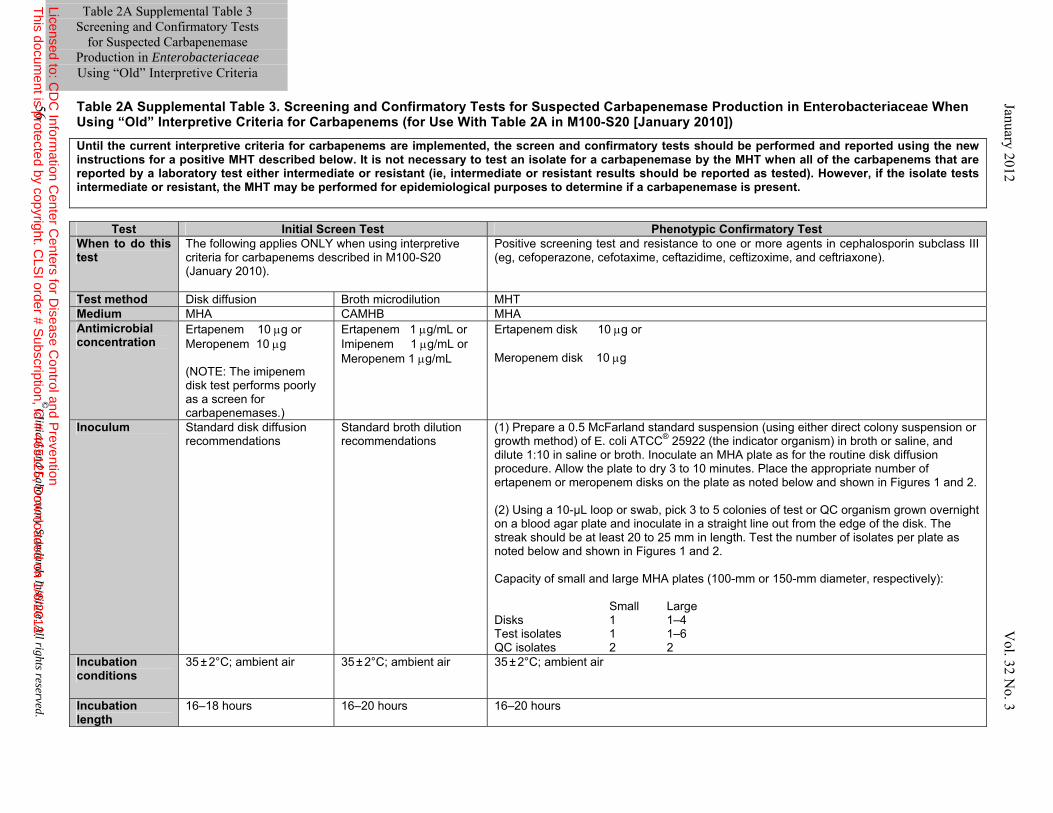
Table 2A Supplemental Table 3. Screening and Confirmatory Tests for Suspected Carbapenemase Production in Enterobacteriaceae When Using “Old” Interpretive Criteria for Carbapenems (for Use With Table 2A in M100-S20 [January 2010])
Until the current interpretive criteria for carbapenems are implemented, the screen and confirmatory tests should be performed and reported using the new instructions for a positive MHT described below. It is not necessary to test an isolate for a carbapenemase by the MHT when all of the carbapenems that are reported by a laboratory test either intermediate or resistant (ie, intermediate or resistant results should be reported as tested). However, if the isolate tests intermediate or resistant, the MHT may be performed for epidemiological purposes to determine if a carbapenemase is present.
Test Initial Screen Test Phenotypic Confirmatory Test When to do this test
The following applies ONLY when using interpretive criteria for carbapenems described in M100-S20 (January 2010).
Positive screening test and resistance to one or more agents in cephalosporin subclass III (eg, cefoperazone, cefotaxime, ceftazidime, ceftizoxime, and ceftriaxone).
Test method Disk diffusion Broth microdilution MHT Medium MHA CAMHB MHA Antimicrobial concentration
Ertapenem 10 μg or Meropenem 10 μg (NOTE: The imipenem disk test performs poorly as a screen for carbapenemases.)
Ertapenem 1 μg/mL or Imipenem 1 μg/mL or Meropenem 1 μg/mL
Ertapenem disk 10 μg or Meropenem disk 10 μg
Inoculum Standard disk diffusion recommendations
Standard broth dilution recommendations
(1) Prepare a 0.5 McFarland standard suspension (using either direct colony suspension or growth method) of E. coli ATCC® 25922 (the indicator organism) in broth or saline, and dilute 1:10 in saline or broth. Inoculate an MHA plate as for the routine disk diffusion procedure. Allow the plate to dry 3 to 10 minutes. Place the appropriate number of ertapenem or meropenem disks on the plate as noted below and shown in Figures 1 and 2. (2) Using a 10-µL loop or swab, pick 3 to 5 colonies of test or QC organism grown overnight on a blood agar plate and inoculate in a straight line out from the edge of the disk. The streak should be at least 20 to 25 mm in length. Test the number of isolates per plate as noted below and shown in Figures 1 and 2. Capacity of small and large MHA plates (100-mm or 150-mm diameter, respectively): Small Large Disks 1 1–4 Test isolates 1 1–6 QC isolates 2 2
Incubation conditions
35 ± 2°C; ambient air 35 ± 2°C; ambient air 35 ± 2°C; ambient air
Incubation length
16–18 hours 16–20 hours 16–20 hours
56 ©C
linical and Laboratory Standards Institute. A
ll rights reserved.
January 2012 V
ol. 32 No. 3
Table 2A Supplemental Table 3
Screening and Confirmatory Tests for Suspected Carbapenemase
Production in Enterobacteriaceae Using “Old” Interpretive Criteria
Licensed to: CD
C Inform
ation Center C
enters for Disease C
ontrol and Prevention
This docum
ent is protected by copyright. CLS
I order # Subscription, id # 465125, D
ownloaded on 1/6/2012.
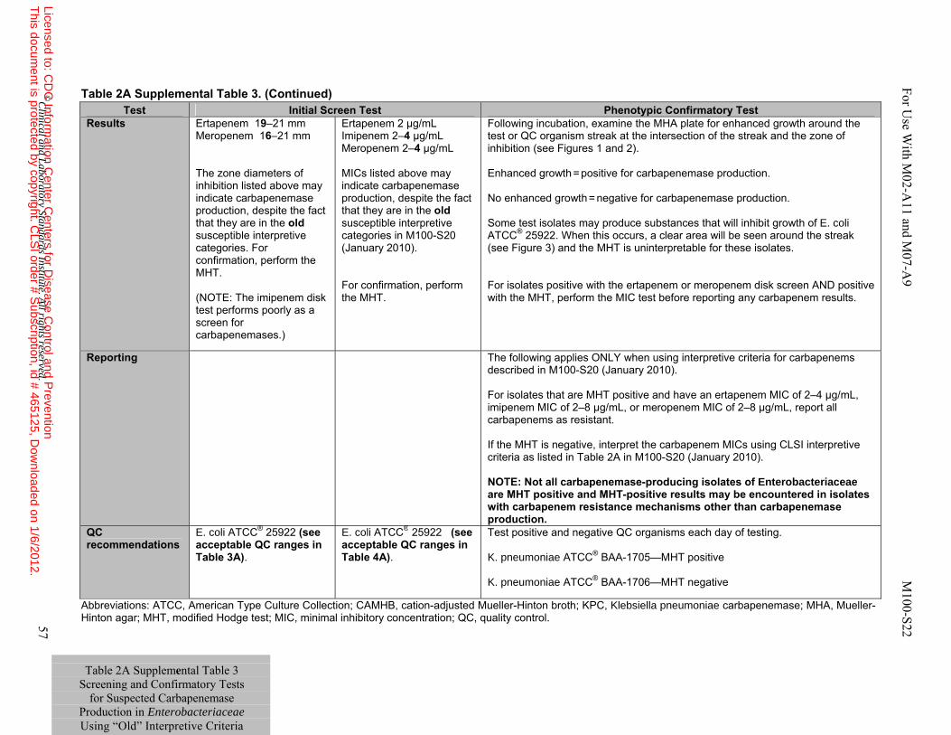
Table 2A Supplemental Table 3 Screening and Confirmatory Tests
for Suspected Carbapenemase Production in Enterobacteriaceae Using “Old” Interpretive Criteria
Table 2A Supplemental Table 3. (Continued)
Abbreviations: ATCC, American Type Culture Collection; CAMHB, cation-adjusted Mueller-Hinton broth; KPC, Klebsiella pneumoniae carbapenemase; MHA, Mueller-Hinton agar; MHT, modified Hodge test; MIC, minimal inhibitory concentration; QC, quality control.
Test Initial Screen Test Phenotypic Confirmatory TestResults Ertapenem 19–21 mm
Meropenem 16–21 mm The zone diameters of inhibition listed above may indicate carbapenemase production, despite the fact that they are in the old susceptible interpretive categories. For confirmation, perform the MHT. (NOTE: The imipenem disk test performs poorly as a screen for carbapenemases.)
Ertapenem 2 µg/mL Imipenem 2–4 µg/mL Meropenem 2–4 µg/mL MICs listed above may indicate carbapenemase production, despite the fact that they are in the old susceptible interpretive categories in M100-S20 (January 2010). For confirmation, perform the MHT.
Following incubation, examine the MHA plate for enhanced growth around the test or QC organism streak at the intersection of the streak and the zone of inhibition (see Figures 1 and 2). Enhanced growth = positive for carbapenemase production. No enhanced growth = negative for carbapenemase production. Some test isolates may produce substances that will inhibit growth of E. coli ATCC® 25922. When this occurs, a clear area will be seen around the streak (see Figure 3) and the MHT is uninterpretable for these isolates. For isolates positive with the ertapenem or meropenem disk screen AND positive with the MHT, perform the MIC test before reporting any carbapenem results.
Reporting The following applies ONLY when using interpretive criteria for carbapenems described in M100-S20 (January 2010). For isolates that are MHT positive and have an ertapenem MIC of 2–4 µg/mL, imipenem MIC of 2–8 µg/mL, or meropenem MIC of 2–8 µg/mL, report all carbapenems as resistant. If the MHT is negative, interpret the carbapenem MICs using CLSI interpretive criteria as listed in Table 2A in M100-S20 (January 2010). NOTE: Not all carbapenemase-producing isolates of Enterobacteriaceae are MHT positive and MHT-positive results may be encountered in isolates with carbapenem resistance mechanisms other than carbapenemase production.
QC recommendations
E. coli ATCC® 25922 (see acceptable QC ranges in Table 3A).
E. coli ATCC® 25922 (see acceptable QC ranges in Table 4A).
Test positive and negative QC organisms each day of testing. K. pneumoniae ATCC® BAA-1705—MHT positive K. pneumoniae ATCC® BAA-1706—MHT negative
©C
linical and Laboratory Standards Institute. A
ll rights reserved. 57
For U
se With M
02-A11 and M
07-A9 M
100-S22
Licensed to: CD
C Inform
ation Center C
enters for Disease C
ontrol and Prevention
This docum
ent is protected by copyright. CLS
I order # Subscription, id # 465125, D
ownloaded on 1/6/2012.
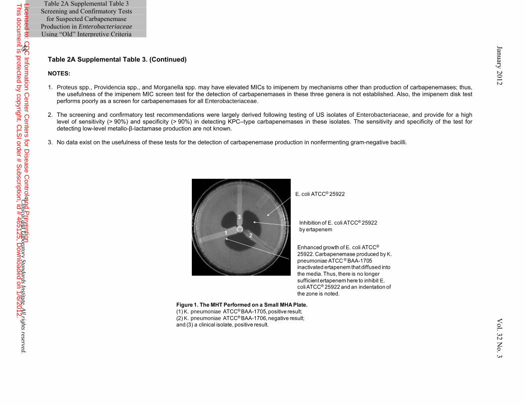
Table 2A Supplemental Table 3. (Continued) NOTES: 1. Proteus spp., Providencia spp., and Morganella spp. may have elevated MICs to imipenem by mechanisms other than production of carbapenemases; thus,
the usefulness of the imipenem MIC screen test for the detection of carbapenemases in these three genera is not established. Also, the imipenem disk test performs poorly as a screen for carbapenemases for all Enterobacteriaceae.
2. The screening and confirmatory test recommendations were largely derived following testing of US isolates of Enterobacteriaceae, and provide for a high level of sensitivity (> 90%) and specificity (> 90%) in detecting KPC–type carbapenemases in these isolates. The sensitivity and specificity of the test for detecting low-level metallo-β-lactamase production are not known.
3. No data exist on the usefulness of these tests for the detection of carbapenemase production in nonfermenting gram-negative bacilli.
1 2
3
Figure 1. The MHT Performed on a Small MHA Plate. (1) K. pneumoniae ATCC® BAA-1705, positive result; (2) K. pneumoniae ATCC® BAA-1706, negative result; and (3) a clinical isolate, positive result.
E. coli ATCC® 25922
Inhibition of E. coli ATCC® 25922 by ertapenem
Enhanced growth of E. coli ATCC®
25922. Carbapenemase produced by K. pneumoniae ATCC ® BAA-1705 inactivated ertapenem that diffused into the media. Thus, there is no longer sufficient ertapenem here to inhibit E. coli ATCC® 25922 and an indentation of the zone is noted.
58 ©C
linical and Laboratory Standards Institute. A
ll rights reserved.
January 2012 V
ol. 32 No. 3
Table 2A Supplemental Table 3 Screening and Confirmatory Tests
for Suspected Carbapenemase Production in Enterobacteriaceae Using “Old” Interpretive Criteria
Licensed to: CD
C Inform
ation Center C
enters for Disease C
ontrol and Prevention
This docum
ent is protected by copyright. CLS
I order # Subscription, id # 465125, D
ownloaded on 1/6/2012.
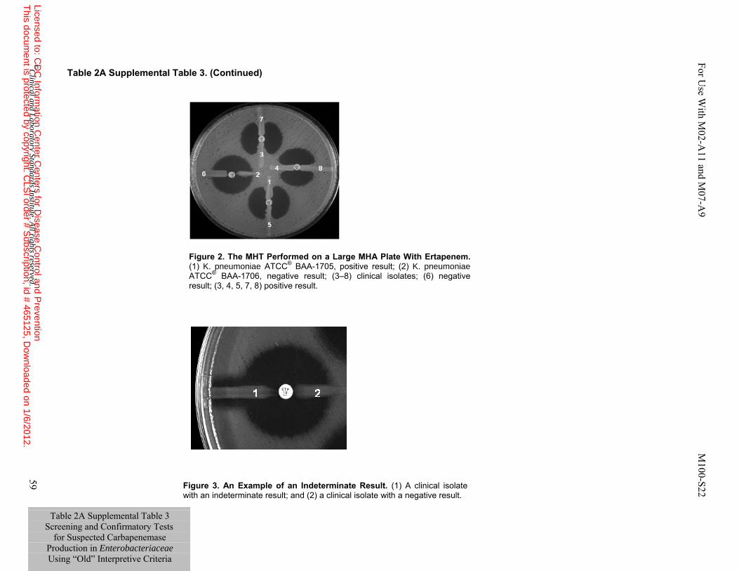
Table 2A Supplemental Table 3. (Continued)
Figure 2. The MHT Performed on a Large MHA Plate With Ertapenem. (1) K. pneumoniae ATCC® BAA-1705, positive result; (2) K. pneumoniae ATCC® BAA-1706, negative result; (3–8) clinical isolates; (6) negative result; (3, 4, 5, 7, 8) positive result.
Figure 3. An Example of an Indeterminate Result. (1) A clinical isolate with an indeterminate result; and (2) a clinical isolate with a negative result.
Table 2A Supplemental Table 3 Screening and Confirmatory Tests
for Suspected Carbapenemase Production in Enterobacteriaceae Using “Old” Interpretive Criteria
©C
linical and Laboratory Standards Institute. A
ll rights reserved. 59
For U
se With M
02-A11 and M
07-A9 M
100-S22
Licensed to: CD
C Inform
ation Center C
enters for Disease C
ontrol and Prevention
This docum
ent is protected by copyright. CLS
I order # Subscription, id # 465125, D
ownloaded on 1/6/2012.
Table 2A Supplemental Table 3. (Continued) References 1. Perrott J, Mabasa VH, Ensom MH. Comparing outcomes of meropenem administration strategies based on pharmacokinetic and pharmacodynamic principles: A qualitative systematic review. Ann Pharmacother. 2010;44:557-564.
2. Cirillo I, Vaccaro N, Turner K, Solanki B, Natarajan J, Redman R. Pharmacokinetics, safety, and tolerability of doripenem after 0.5-, 1-, and 4-hour infusions in healthy volunteers. J Clin Pharmacol. 2009;49:798-806.
3. Sakka SG, Glauner AK, Bulitta JB, et al. Population pharmacokinetics and pharmacodynamics of continuous versus short-term infusion of imipenem-cilastatin in critically ill patients in a randomized, controlled trial. Antimicrob Agents Chemother. 2007;51:3304-3310.
4. Peleg AY, Hooper DC. Hospital-acquired infections due to Gram-negative bacteria. N Engl J Med. 2010;362:1804-1813.
60 ©C
linical and Laboratory Standards Institute. A
ll rights reserved.
January 2012 V
ol. 32 No. 3
Table 2A Supplemental Table 3
Screening and Confirmatory Tests for Suspected Carbapenemase
Production in Enterobacteriaceae Using “Old” Interpretive Criteria
Licensed to: CD
C Inform
ation Center C
enters for Disease C
ontrol and Prevention
This docum
ent is protected by copyright. CLS
I order # Subscription, id # 465125, D
ownloaded on 1/6/2012.

For Use With M02-A11 and M07-A9 M100-S22
©Clinical and Laboratory Standards Institute. All rights reserved. 61
This page is intentionally left blank.
Licensed to: CDC Information Center Centers for Disease Control and PreventionThis document is protected by copyright. CLSI order # Subscription, id # 465125, Downloaded on 1/6/2012.
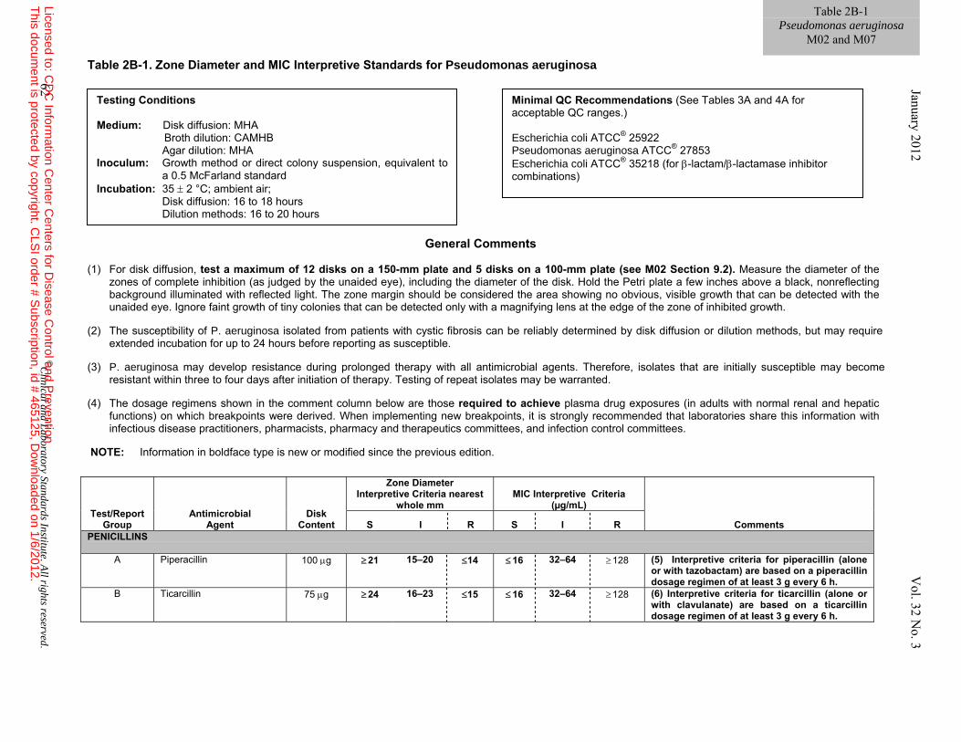
Table 2B-1. Zone Diameter and MIC Interpretive Standards for Pseudomonas aeruginosa
General Comments (1) For disk diffusion, test a maximum of 12 disks on a 150-mm plate and 5 disks on a 100-mm plate (see M02 Section 9.2). Measure the diameter of the
zones of complete inhibition (as judged by the unaided eye), including the diameter of the disk. Hold the Petri plate a few inches above a black, nonreflecting background illuminated with reflected light. The zone margin should be considered the area showing no obvious, visible growth that can be detected with the unaided eye. Ignore faint growth of tiny colonies that can be detected only with a magnifying lens at the edge of the zone of inhibited growth.
(2) The susceptibility of P. aeruginosa isolated from patients with cystic fibrosis can be reliably determined by disk diffusion or dilution methods, but may require
extended incubation for up to 24 hours before reporting as susceptible. (3) P. aeruginosa may develop resistance during prolonged therapy with all antimicrobial agents. Therefore, isolates that are initially susceptible may become
resistant within three to four days after initiation of therapy. Testing of repeat isolates may be warranted. (4) The dosage regimens shown in the comment column below are those required to achieve plasma drug exposures (in adults with normal renal and hepatic
functions) on which breakpoints were derived. When implementing new breakpoints, it is strongly recommended that laboratories share this information with infectious disease practitioners, pharmacists, pharmacy and therapeutics committees, and infection control committees.
NOTE: Information in boldface type is new or modified since the previous edition.
Test/Report Group
Antimicrobial Agent
Disk Content
Zone Diameter Interpretive Criteria nearest
whole mm MIC Interpretive Criteria
(µg/mL)
Comments S I R S I R PENICILLINS
A Piperacillin 100 μg ≥ 21 15–20 ≤14 ≤ 16 32–64 ≥ 128 (5) Interpretive criteria for piperacillin (alone or with tazobactam) are based on a piperacillin dosage regimen of at least 3 g every 6 h.
B Ticarcillin 75 μg ≥ 24 16–23 ≤15 ≤ 16 32–64 ≥ 128 (6) Interpretive criteria for ticarcillin (alone or with clavulanate) are based on a ticarcillin dosage regimen of at least 3 g every 6 h.
Table 2B-1 Pseudomonas aeruginosa
M02 and M07
Testing Conditions Medium: Disk diffusion: MHA
Broth dilution: CAMHB Agar dilution: MHA Inoculum: Growth method or direct colony suspension, equivalent to
a 0.5 McFarland standard Incubation: 35 ± 2 °C; ambient air; Disk diffusion: 16 to 18 hours Dilution methods: 16 to 20 hours
Minimal QC Recommendations (See Tables 3A and 4A for acceptable QC ranges.) Escherichia coli ATCC® 25922 Pseudomonas aeruginosa ATCC® 27853 Escherichia coli ATCC® 35218 (for β-lactam/β-lactamase inhibitor combinations)
62 ©C
linical and Laboratory Standards Institute. A
ll rights reserved.
January 2012 V
ol. 32 No. 3
Licensed to: CD
C Inform
ation Center C
enters for Disease C
ontrol and Prevention
This docum
ent is protected by copyright. CLS
I order # Subscription, id # 465125, D
ownloaded on 1/6/2012.
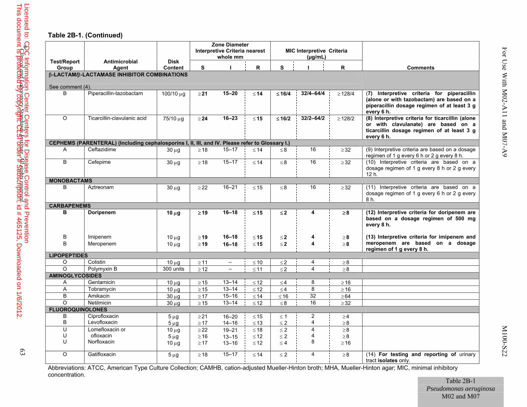
Table 2B-1. (Continued)
Abbreviations: ATCC, American Type Culture Collection; CAMHB, cation-adjusted Mueller-Hinton broth; MHA, Mueller-Hinton agar; MIC, minimal inhibitory concentration.
Test/Report Group
Antimicrobial Agent
Disk Content
Zone Diameter Interpretive Criteria nearest
whole mm MIC Interpretive Criteria
(µg/mL)
Comments S I R S I R β-LACTAM/β-LACTAMASE INHIBITOR COMBINATIONS See comment (4).
B Piperacillin-tazobactam 100/10 μg ≥ 21 15–20 ≤ 14 ≤ 16/4 32/4–64/4 ≥ 128/4 (7) Interpretive criteria for piperacillin (alone or with tazobactam) are based on a piperacillin dosage regimen of at least 3 g every 6 h.
O Ticarcillin-clavulanic acid 75/10 μg ≥ 24 16–23 ≤ 15 ≤ 16/2 32/2–64/2 ≥ 128/2 (8) Interpretive criteria for ticarcillin (alone or with clavulanate) are based on a ticarcillin dosage regimen of at least 3 g every 6 h.
CEPHEMS (PARENTERAL) (Including cephalosporins I, II, III, and IV. Please refer to Glossary I.) A Ceftazidime 30 μg ≥ 18 15–17 ≤ 14 ≤ 8 16 ≥ 32 (9) Interpretive criteria are based on a dosage
regimen of 1 g every 6 h or 2 g every 8 h. B Cefepime 30 μg ≥ 18 15–17 ≤ 14 ≤ 8 16 ≥ 32 (10) Interpretive criteria are based on a
dosage regimen of 1 g every 8 h or 2 g every 12 h.
MONOBACTAMS B Aztreonam 30 μg ≥ 22 16–21 ≤ 15 ≤ 8 16 ≥ 32 (11) Interpretive criteria are based on a
dosage regimen of 1 g every 6 h or 2 g every 8 h.
CARBAPENEMS B Doripenem 10 μg ≥ 19 16–18 ≤ 15 ≤ 2 4 ≥ 8 (12) Interpretive criteria for doripenem are
based on a dosage regimen of 500 mg every 8 h.
B Imipenem 10 μg ≥ 19 16–18 ≤ 15 ≤ 2 4 ≥ 8 (13) Interpretive criteria for imipenem and meropenem are based on a dosage regimen of 1 g every 8 h.
B Meropenem 10 μg ≥ 19 16–18 ≤ 15 ≤ 2 4 ≥ 8
LIPOPEPTIDES O Colistin 10 μg ≥ 11 – ≤ 10 ≤ 2 4 ≥ 8 O Polymyxin B 300 units ≥ 12 – ≤ 11 ≤ 2 4 ≥ 8
AMINOGLYCOSIDES A Gentamicin 10 μg ≥ 15 13–14 ≤ 12 ≤ 4 8 ≥ 16 A Tobramycin 10 μg ≥ 15 13–14 ≤ 12 ≤ 4 8 ≥ 16 B Amikacin 30 μg ≥ 17 15–16 ≤ 14 ≤ 16 32 ≥ 64 O Netilmicin 30 μg ≥ 15 13–14 ≤ 12 ≤ 8 16 ≥ 32
FLUOROQUINOLONES B B
Ciprofloxacin Levofloxacin
5 μg 5 μg
≥ 21 ≥ 17
16–20 14–16
≤ 15 ≤ 13
≤ 1 ≤ 2
2 4
≥ 4 ≥ 8
U U U
Lomefloxacin or ofloxacin Norfloxacin
10 μg 5 μg
10 μg
≥ 22 ≥ 16 ≥ 17
19–21 13–15 13–16
≤ 18 ≤ 12 ≤ 12
≤ 2 ≤ 2 ≤ 4
4 4 8
≥ 8 ≥ 8 ≥ 16
O Gatifloxacin 5 μg ≥ 18 15–17 ≤ 14 ≤ 2 4 ≥ 8 (14) For testing and reporting of urinary tract isolates only.
©C
linical and Laboratory Standards Institute. A
ll rights reserved. 63
For Use W
ith M02-A
11 and M07-A
9 M100-S22
Table 2B-1 Pseudomonas aeruginosa
M02 and M07
Licensed to: CD
C Inform
ation Center C
enters for Disease C
ontrol and Prevention
This docum
ent is protected by copyright. CLS
I order # Subscription, id # 465125, D
ownloaded on 1/6/2012.
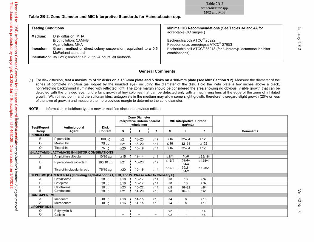
Table 2B-2 Acinetobacter spp.
M02 and M07 Table 2B-2. Zone Diameter and MIC Interpretive Standards for Acinetobacter spp.
General Comments
(1) For disk diffusion, test a maximum of 12 disks on a 150-mm plate and 5 disks on a 100-mm plate (see M02 Section 9.2). Measure the diameter of the
zones of complete inhibition (as judged by the unaided eye), including the diameter of the disk. Hold the Petri plate a few inches above a black, nonreflecting background illuminated with reflected light. The zone margin should be considered the area showing no obvious, visible growth that can be detected with the unaided eye. Ignore faint growth of tiny colonies that can be detected only with a magnifying lens at the edge of the zone of inhibited growth. With trimethoprim and the sulfonamides, antagonists in the medium may allow some slight growth; therefore, disregard slight growth (20% or less of the lawn of growth) and measure the more obvious margin to determine the zone diameter.
NOTE: Information in boldface type is new or modified since the previous edition.
Test/Report Group
Antimicrobial Agent
Disk Content
Zone Diameter Interpretive Criteria nearest
whole mm MIC Interpretive Criteria
(µg/mL)
Comments S I R S I R PENICILLINS
B Piperacillin 100 μg ≥ 21 18–20 ≤ 17 ≤ 16 32–64 ≥ 128 O Mezlocillin 75 μg ≥ 21 18–20 ≤ 17 ≤ 16 32–64 ≥ 128 O Ticarcillin 75 μg ≥ 20 15–19 ≤ 14 ≤ 16 32–64 ≥ 128
β-LACTAM/β-LACTAMASE INHIBITOR COMBINATIONS A Ampicillin-sulbactam 10/10 μg ≥ 15 12–14 ≤ 11 ≤ 8/4 16/8 ≥ 32/16
B Piperacillin-tazobactam 100/10 μg ≥ 21 18–20 ≤ 17 ≤ 16/4 32/4–64/4
≥ 128/4
B Ticarcillin-clavulanic acid 75/10 μg ≥ 20 15–19 ≤ 14 ≤ 16/2 32/2–64/2
≥ 128/2
CEPHEMS (PARENTERAL) (Including cephalosporins I, II, III, and IV. Please refer to Glossary I.) A Ceftazidime 30 μg ≥ 18 15–17 ≤ 14 ≤ 8 16 ≥ 32 B Cefepime 30 μg ≥ 18 15–17 ≤ 14 ≤ 8 16 ≥ 32 B B
Cefotaxime Ceftriaxone
30 μg 30 μg
≥ 23 ≥ 21
15–22 14–20
≤ 14 ≤ 13
≤ 8 ≤ 8
16–32 16–32
≥ 64 ≥ 64
CARBAPENEMS A Imipenem 10 μg ≥ 16 14–15 ≤ 13 ≤ 4 8 ≥ 16 A Meropenem 10 μg ≥ 16 14–15 ≤ 13 ≤ 4 8 ≥ 16
LIPOPEPTIDES O O
Polymyxin B Colistin
– – –
– –
– –
≤ 2 ≤ 2
– –
≥ 4 ≥ 4
Testing Conditions Medium: Disk diffusion: MHA Broth dilution: CAMHB Agar dilution: MHA Inoculum: Growth method or direct colony suspension, equivalent to a 0.5
McFarland standard Incubation: 35 ± 2°C; ambient air; 20 to 24 hours, all methods
Minimal QC Recommendations (See Tables 3A and 4A for acceptable QC ranges.) Escherichia coli ATCC® 25922 Pseudomonas aeruginosa ATCC® 27853 Escherichia coli ATCC® 35218 (for β-lactam/β-lactamase inhibitor combinations)
64 ©C
linical and Laboratory Standards Institute. A
ll rights reserved.
January 2012 V
ol. 32 No. 3
Licensed to: CD
C Inform
ation Center C
enters for Disease C
ontrol and Prevention
This docum
ent is protected by copyright. CLS
I order # Subscription, id # 465125, D
ownloaded on 1/6/2012.
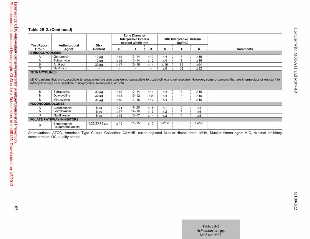
Table 2B-2 Acinetobacter spp.
M02 and M07
Table 2B-2. (Continued)
Abbreviations: ATCC, American Type Culture Collection; CAMHB, cation-adjusted Mueller-Hinton broth; MHA, Mueller-Hinton agar; MIC, minimal inhibitory concentration; QC, quality control.
Test/Report Group
Antimicrobial Agent
Disk Content
Zone Diameter Interpretive Criteria nearest whole mm
MIC Interpretive Criteria (µg/mL)
Comments S I R S I R AMINOGLYCOSIDES
A Gentamicin 10 μg ≥ 15 13–14 ≤ 12 ≤ 4 8 ≥ 16 A Tobramycin 10 μg ≥ 15 13–14 ≤ 12 ≤ 4 8 ≥ 16 B Amikacin 30 μg ≥ 17 15–16 ≤ 14 ≤ 16 32 ≥ 64 O Netilmicin – – – – ≤ 8 16 ≥ 32
TETRACYCLINES (2) Organisms that are susceptible to tetracycline are also considered susceptible to doxycycline and minocycline. However, some organisms that are intermediate or resistant to tetracycline may be susceptible to doxycycline, minocycline, or both.
B Tetracycline 30 μg ≥ 15 12–14 ≤ 11 ≤ 4 8 ≥ 16 B Doxycycline 30 μg ≥ 13 10–12 ≤ 9 ≤ 4 8 ≥ 16 B Minocycline 30 μg ≥ 16 13–15 ≤ 12 ≤ 4 8 ≥ 16
FLUOROQUINOLONES A A
Ciprofloxacin Levofloxacin
5 μg 5 μg
≥ 21 ≥ 17
16–20 14–16
≤ 15 ≤ 13
≤ 1 ≤ 2
2 4
≥ 4 ≥ 8
O Gatifloxacin 5 μg ≥ 18 15–17 ≤ 14 ≤ 2 4 ≥ 8 FOLATE PATHWAY INHIBITORS
B Trimethoprim- sulfamethoxazole
1.25/23.75 μg ≥ 16 11–15 ≤ 10 ≤ 2/38 – ≥ 4/76
©C
linical and Laboratory Standards Institute. A
ll rights reserved. 65
For U
se With M
02-A11 and M
07-A9 M
100-S22
Licensed to: CD
C Inform
ation Center C
enters for Disease C
ontrol and Prevention
This docum
ent is protected by copyright. CLS
I order # Subscription, id # 465125, D
ownloaded on 1/6/2012.
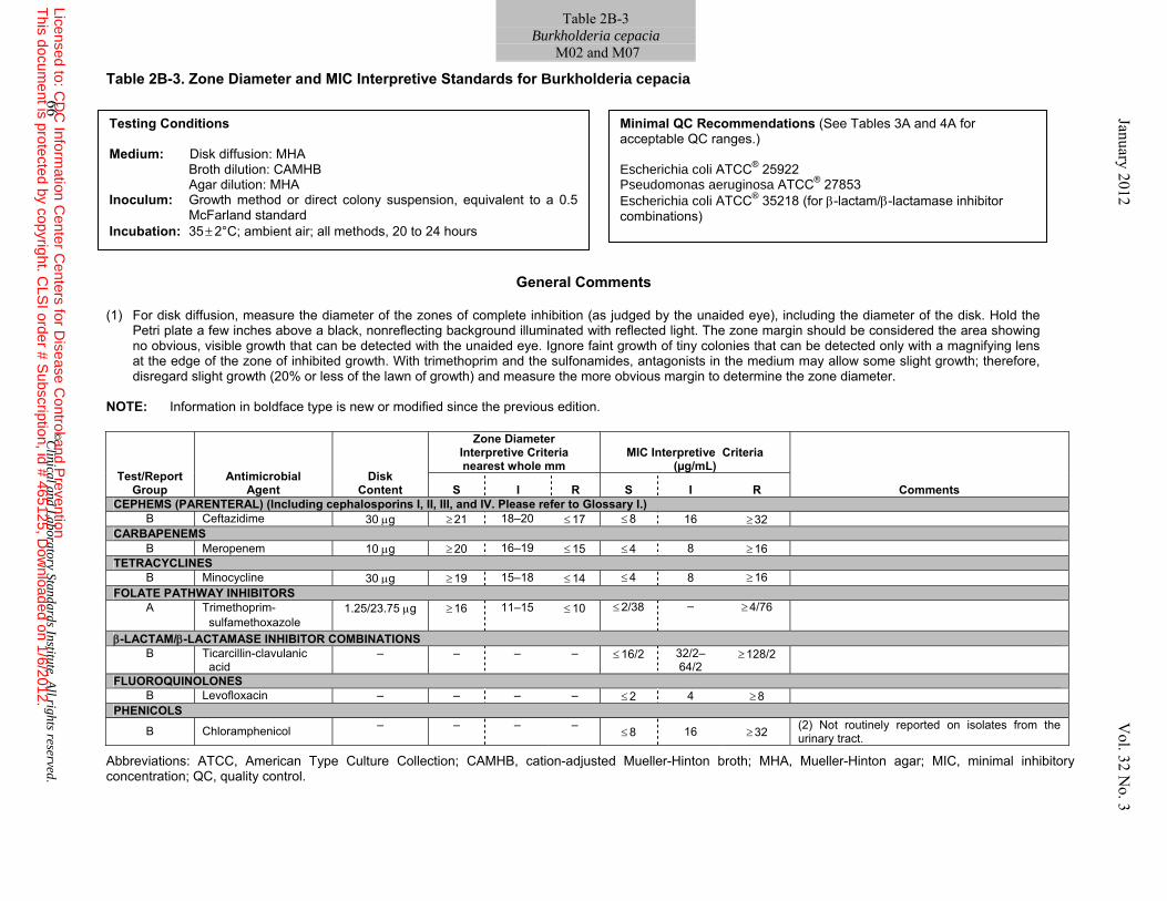
Table 2B-3 Burkholderia cepacia
M02 and M07 Table 2B-3. Zone Diameter and MIC Interpretive Standards for Burkholderia cepacia
General Comments
(1) For disk diffusion, measure the diameter of the zones of complete inhibition (as judged by the unaided eye), including the diameter of the disk. Hold the
Petri plate a few inches above a black, nonreflecting background illuminated with reflected light. The zone margin should be considered the area showing no obvious, visible growth that can be detected with the unaided eye. Ignore faint growth of tiny colonies that can be detected only with a magnifying lens at the edge of the zone of inhibited growth. With trimethoprim and the sulfonamides, antagonists in the medium may allow some slight growth; therefore, disregard slight growth (20% or less of the lawn of growth) and measure the more obvious margin to determine the zone diameter.
NOTE: Information in boldface type is new or modified since the previous edition.
Abbreviations: ATCC, American Type Culture Collection; CAMHB, cation-adjusted Mueller-Hinton broth; MHA, Mueller-Hinton agar; MIC, minimal inhibitory concentration; QC, quality control.
Test/Report Group
Antimicrobial Agent
Disk Content
Zone Diameter Interpretive Criteria nearest whole mm
MIC Interpretive Criteria (µg/mL)
Comments S I R S I R CEPHEMS (PARENTERAL) (Including cephalosporins I, II, III, and IV. Please refer to Glossary I.)
B Ceftazidime 30 μg ≥ 21 18–20 ≤ 17 ≤ 8 16 ≥ 32 CARBAPENEMS
B Meropenem 10 μg ≥ 20 16–19 ≤ 15 ≤ 4 8 ≥ 16 TETRACYCLINES
B Minocycline 30 μg ≥ 19 15–18 ≤ 14 ≤ 4 8 ≥ 16 FOLATE PATHWAY INHIBITORS
A Trimethoprim- sulfamethoxazole
1.25/23.75 μg ≥ 16 11–15 ≤ 10 ≤ 2/38
– ≥ 4/76
β-LACTAM/β-LACTAMASE INHIBITOR COMBINATIONS B Ticarcillin-clavulanic
acid – – – – ≤ 16/2 32/2–
64/2 ≥ 128/2
FLUOROQUINOLONES B Levofloxacin – – – – ≤ 2 4 ≥ 8
PHENICOLS
B Chloramphenicol – – – – ≤ 8 16 ≥ 32 (2) Not routinely reported on isolates from the
urinary tract.
Testing Conditions Medium: Disk diffusion: MHA Broth dilution: CAMHB Agar dilution: MHA Inoculum: Growth method or direct colony suspension, equivalent to a 0.5
McFarland standard Incubation: 35 ± 2°C; ambient air; all methods, 20 to 24 hours
Minimal QC Recommendations (See Tables 3A and 4A for acceptable QC ranges.) Escherichia coli ATCC® 25922 Pseudomonas aeruginosa ATCC® 27853 Escherichia coli ATCC® 35218 (for β-lactam/β-lactamase inhibitor combinations)
66 ©C
linical and Laboratory Standards Institute. A
ll rights reserved.
January 2012 V
ol. 32 No. 3
Licensed to: CD
C Inform
ation Center C
enters for Disease C
ontrol and Prevention
This docum
ent is protected by copyright. CLS
I order # Subscription, id # 465125, D
ownloaded on 1/6/2012.
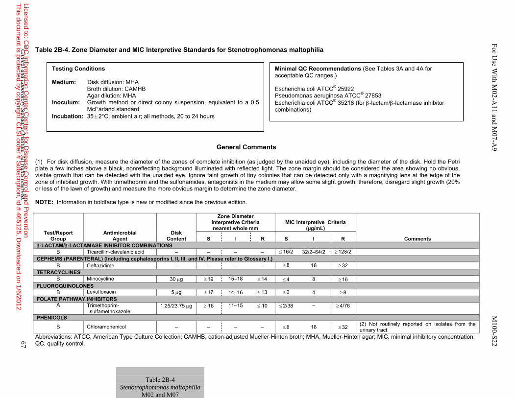
Table 2B-4. Zone Diameter and MIC Interpretive Standards for Stenotrophomonas maltophilia
General Comments
(1) For disk diffusion, measure the diameter of the zones of complete inhibition (as judged by the unaided eye), including the diameter of the disk. Hold the Petri plate a few inches above a black, nonreflecting background illuminated with reflected light. The zone margin should be considered the area showing no obvious, visible growth that can be detected with the unaided eye. Ignore faint growth of tiny colonies that can be detected only with a magnifying lens at the edge of the zone of inhibited growth. With trimethoprim and the sulfonamides, antagonists in the medium may allow some slight growth; therefore, disregard slight growth (20% or less of the lawn of growth) and measure the more obvious margin to determine the zone diameter.
NOTE: Information in boldface type is new or modified since the previous edition.
Abbreviations: ATCC, American Type Culture Collection; CAMHB, cation-adjusted Mueller-Hinton broth; MHA, Mueller-Hinton agar; MIC, minimal inhibitory concentration; QC, quality control.
Test/Report Group
Antimicrobial Agent
Disk Content
Zone Diameter Interpretive Criteria nearest whole mm
MIC Interpretive Criteria (µg/mL)
Comments S I R S I R β-LACTAM/β-LACTAMASE INHIBITOR COMBINATIONS
B Ticarcillin-clavulanic acid – – – – ≤ 16/2 32/2–64/2 ≥ 128/2 CEPHEMS (PARENTERAL) (Including cephalosporins I, II, III, and IV. Please refer to Glossary I.)
B Ceftazidime – – – – ≤ 8 16 ≥ 32 TETRACYCLINES
B Minocycline 30 μg ≥ 19 15–18 ≤ 14 ≤ 4 8 ≥ 16 FLUOROQUINOLONES
B Levofloxacin 5 μg ≥ 17 14–16 ≤ 13 ≤ 2 4 ≥ 8 FOLATE PATHWAY INHIBITORS
A Trimethoprim- sulfamethoxazole
1.25/23.75 μg ≥ 16 11–15 ≤ 10 ≤ 2/38 – ≥ 4/76
PHENICOLS
B Chloramphenicol – – – – ≤ 8 16 ≥ 32 (2) Not routinely reported on isolates from the urinary tract.
Table 2B-4
Stenotrophomonas maltophilia M02 and M07
Testing Conditions Medium: Disk diffusion: MHA Broth dilution: CAMHB Agar dilution: MHA Inoculum: Growth method or direct colony suspension, equivalent to a 0.5
McFarland standard Incubation: 35 ± 2°C; ambient air; all methods, 20 to 24 hours
Minimal QC Recommendations (See Tables 3A and 4A for acceptable QC ranges.) Escherichia coli ATCC® 25922 Pseudomonas aeruginosa ATCC® 27853 Escherichia coli ATCC® 35218 (for β-lactam/β-lactamase inhibitor combinations)
©C
linical and Laboratory Standards Institute. A
ll rights reserved. 67
For U
se With M
02-A11 and M
07-A9 M
100-S22
Licensed to: CD
C Inform
ation Center C
enters for Disease C
ontrol and Prevention
This docum
ent is protected by copyright. CLS
I order # Subscription, id # 465125, D
ownloaded on 1/6/2012.
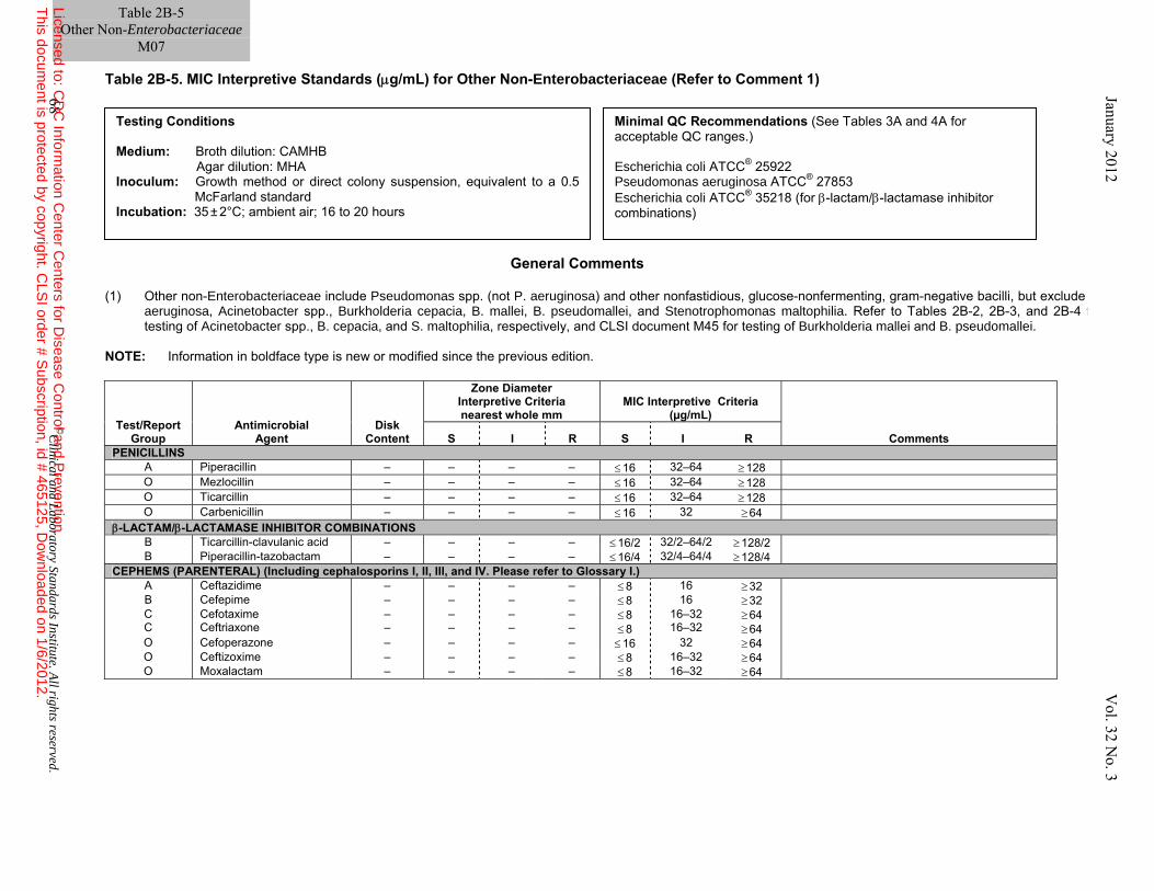
Table 2B-5. MIC Interpretive Standards (μg/mL) for Other Non-Enterobacteriaceae (Refer to Comment 1)
General Comments (1) Other non-Enterobacteriaceae include Pseudomonas spp. (not P. aeruginosa) and other nonfastidious, glucose-nonfermenting, gram-negative bacilli, but exclude P.
aeruginosa, Acinetobacter spp., Burkholderia cepacia, B. mallei, B. pseudomallei, and Stenotrophomonas maltophilia. Refer to Tables 2B-2, 2B-3, and 2B-4 for testing of Acinetobacter spp., B. cepacia, and S. maltophilia, respectively, and CLSI document M45 for testing of Burkholderia mallei and B. pseudomallei.
NOTE: Information in boldface type is new or modified since the previous edition.
Test/Report Group
Antimicrobial Agent
Disk Content
Zone Diameter Interpretive Criteria nearest whole mm
MIC Interpretive Criteria (µg/mL)
Comments S I R S I R PENICILLINS
A Piperacillin – – – – ≤ 16 32–64 ≥ 128 O Mezlocillin – – – – ≤ 16 32–64 ≥ 128 O Ticarcillin – – – – ≤ 16 32–64 ≥ 128 O Carbenicillin – – – – ≤ 16 32 ≥ 64
β-LACTAM/β-LACTAMASE INHIBITOR COMBINATIONS B Ticarcillin-clavulanic acid – – – – ≤ 16/2 32/2–64/2 ≥ 128/2 B Piperacillin-tazobactam – – – – ≤ 16/4 32/4–64/4 ≥ 128/4
CEPHEMS (PARENTERAL) (Including cephalosporins I, II, III, and IV. Please refer to Glossary I.) A Ceftazidime – – – – ≤ 8 16 ≥ 32 B Cefepime – – – – ≤ 8 16 ≥ 32 C C
Cefotaxime Ceftriaxone
– –
– –
– –
– –
≤ 8 ≤ 8
16–32 16–32
≥ 64 ≥ 64
O Cefoperazone – – – – ≤ 16 32 ≥ 64 O Ceftizoxime – – – – ≤ 8 16–32 ≥ 64 O Moxalactam – – – – ≤ 8 16–32 ≥ 64
Testing Conditions Medium: Broth dilution: CAMHB Agar dilution: MHA Inoculum: Growth method or direct colony suspension, equivalent to a 0.5
McFarland standard Incubation: 35 ± 2°C; ambient air; 16 to 20 hours
Minimal QC Recommendations (See Tables 3A and 4A for acceptable QC ranges.) Escherichia coli ATCC® 25922 Pseudomonas aeruginosa ATCC® 27853 Escherichia coli ATCC® 35218 (for β-lactam/β-lactamase inhibitor combinations)
Table 2B-5 Other Non-Enterobacteriaceae
M07
68 ©C
linical and Laboratory Standards Institute. A
ll rights reserved.
January 2012 V
ol. 32 No. 3
Licensed to: CD
C Inform
ation Center C
enters for Disease C
ontrol and Prevention
This docum
ent is protected by copyright. CLS
I order # Subscription, id # 465125, D
ownloaded on 1/6/2012.
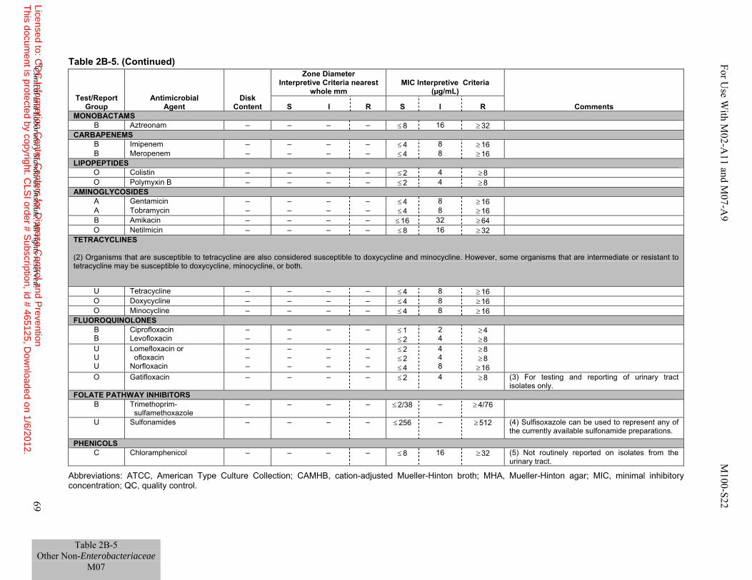
Table 2B-5. (Continued)
Abbreviations: ATCC, American Type Culture Collection; CAMHB, cation-adjusted Mueller-Hinton broth; MHA, Mueller-Hinton agar; MIC, minimal inhibitory concentration; QC, quality control.
Test/Report Group
Antimicrobial Agent
Disk Content
Zone Diameter Interpretive Criteria nearest
whole mm MIC Interpretive Criteria
(µg/mL)
Comments S I R S I R MONOBACTAMS
B Aztreonam – – – – ≤ 8 16 ≥ 32 CARBAPENEMS
B Imipenem – – – – ≤ 4 8 ≥ 16 B Meropenem – – – – ≤ 4 8 ≥ 16
LIPOPEPTIDES O Colistin – – – – ≤ 2 4 ≥ 8 O Polymyxin B – – – – ≤ 2 4 ≥ 8
AMINOGLYCOSIDES A Gentamicin – – – – ≤ 4 8 ≥ 16 A Tobramycin – – – – ≤ 4 8 ≥ 16 B Amikacin – – – – ≤ 16 32 ≥ 64 O Netilmicin – – – – ≤ 8 16 ≥ 32
TETRACYCLINES (2) Organisms that are susceptible to tetracycline are also considered susceptible to doxycycline and minocycline. However, some organisms that are intermediate or resistant to tetracycline may be susceptible to doxycycline, minocycline, or both.
U Tetracycline – – – – ≤ 4 8 ≥ 16 O Doxycycline – – – – ≤ 4 8 ≥ 16 O Minocycline – – – – ≤ 4 8 ≥ 16
FLUOROQUINOLONES B B
Ciprofloxacin Levofloxacin
– –
– –
– – ≤ 1 ≤ 2
2 4
≥ 4 ≥ 8
U U U
Lomefloxacin or ofloxacin Norfloxacin
– – –
– – –
– – –
– – –
≤ 2 ≤ 2 ≤ 4
4 4 8
≥ 8 ≥ 8 ≥ 16
O Gatifloxacin – – – – ≤ 2 4 ≥ 8 (3) For testing and reporting of urinary tract isolates only.
FOLATE PATHWAY INHIBITORS B Trimethoprim-
sulfamethoxazole – – – – ≤ 2/38 – ≥ 4/76
U Sulfonamides – – – – ≤ 256 – ≥ 512
(4) Sulfisoxazole can be used to represent any of the currently available sulfonamide preparations.
PHENICOLS C Chloramphenicol – – – – ≤ 8 16 ≥ 32 (5) Not routinely reported on isolates from the
urinary tract.
©C
linical and Laboratory Standards Institute. A
ll rights reserved. 69
For U
se With M
02-A11 and M
07-A9 M
100-S22
Table 2B-5 Other Non-Enterobacteriaceae
M07
Licensed to: CD
C Inform
ation Center C
enters for Disease C
ontrol and Prevention
This docum
ent is protected by copyright. CLS
I order # Subscription, id # 465125, D
ownloaded on 1/6/2012.
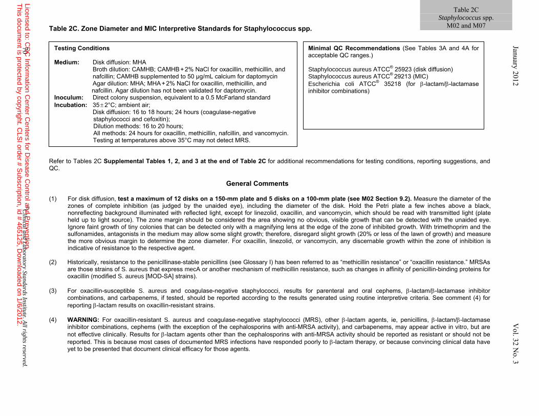
Table 2C. Zone Diameter and MIC Interpretive Standards for Staphylococcus spp.
Refer to Tables 2C Supplemental Tables 1, 2, and 3 at the end of Table 2C for additional recommendations for testing conditions, reporting suggestions, and QC.
General Comments
(1) For disk diffusion, test a maximum of 12 disks on a 150-mm plate and 5 disks on a 100-mm plate (see M02 Section 9.2). Measure the diameter of the
zones of complete inhibition (as judged by the unaided eye), including the diameter of the disk. Hold the Petri plate a few inches above a black, nonreflecting background illuminated with reflected light, except for linezolid, oxacillin, and vancomycin, which should be read with transmitted light (plate held up to light source). The zone margin should be considered the area showing no obvious, visible growth that can be detected with the unaided eye. Ignore faint growth of tiny colonies that can be detected only with a magnifying lens at the edge of the zone of inhibited growth. With trimethoprim and the sulfonamides, antagonists in the medium may allow some slight growth; therefore, disregard slight growth (20% or less of the lawn of growth) and measure the more obvious margin to determine the zone diameter. For oxacillin, linezolid, or vancomycin, any discernable growth within the zone of inhibition is indicative of resistance to the respective agent.
(2) Historically, resistance to the penicillinase-stable penicillins (see Glossary I) has been referred to as “methicillin resistance” or “oxacillin resistance.” MRSAs
are those strains of S. aureus that express mecA or another mechanism of methicillin resistance, such as changes in affinity of penicillin-binding proteins for oxacillin (modified S. aureus [MOD-SA] strains).
(3) For oxacillin-susceptible S. aureus and coagulase-negative staphylococci, results for parenteral and oral cephems, β-lactam/β-lactamase inhibitor
combinations, and carbapenems, if tested, should be reported according to the results generated using routine interpretive criteria. See comment (4) for reporting β-lactam results on oxacillin-resistant strains.
(4) WARNING: For oxacillin-resistant S. aureus and coagulase-negative staphylococci (MRS), other β-lactam agents, ie, penicillins, β-lactam/β-lactamase
inhibitor combinations, cephems (with the exception of the cephalosporins with anti-MRSA activity), and carbapenems, may appear active in vitro, but are not effective clinically. Results for β-lactam agents other than the cephalosporins with anti-MRSA activity should be reported as resistant or should not be reported. This is because most cases of documented MRS infections have responded poorly to β-lactam therapy, or because convincing clinical data have yet to be presented that document clinical efficacy for those agents.
Testing Conditions Medium: Disk diffusion: MHA Broth dilution: CAMHB; CAMHB + 2% NaCl for oxacillin, methicillin, and nafcillin; CAMHB supplemented to 50 µg/mL calcium for daptomycin Agar dilution: MHA; MHA + 2% NaCl for oxacillin, methicillin, and nafcillin. Agar dilution has not been validated for daptomycin. Inoculum: Direct colony suspension, equivalent to a 0.5 McFarland standard Incubation: 35 ± 2°C; ambient air; Disk diffusion: 16 to 18 hours; 24 hours (coagulase-negative staphylococci and cefoxitin); Dilution methods: 16 to 20 hours; All methods: 24 hours for oxacillin, methicillin, nafcillin, and vancomycin. Testing at temperatures above 35°C may not detect MRS.
Minimal QC Recommendations (See Tables 3A and 4A for acceptable QC ranges.) Staphylococcus aureus ATCC® 25923 (disk diffusion) Staphylococcus aureus ATCC® 29213 (MIC) Escherichia coli ATCC® 35218 (for β-lactam/β-lactamase inhibitor combinations)
70 ©C
linical and Laboratory Standards Institute. A
ll rights reserved.
Table 2C Staphylococcus spp.
M02 and M07 January 2012 V
ol. 32 No. 3
Licensed to: CD
C Inform
ation Center C
enters for Disease C
ontrol and Prevention
This docum
ent is protected by copyright. CLS
I order # Subscription, id # 465125, D
ownloaded on 1/6/2012.
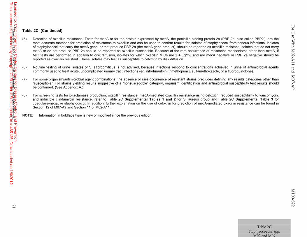
Table 2C. (Continued) (5) Detection of oxacillin resistance: Tests for mecA or for the protein expressed by mecA, the penicillin-binding protein 2a (PBP 2a, also called PBP2'), are the
most accurate methods for prediction of resistance to oxacillin and can be used to confirm results for isolates of staphylococci from serious infections. Isolates of staphylococci that carry the mecA gene, or that produce PBP 2a (the mecA gene product), should be reported as oxacillin resistant. Isolates that do not carry mecA or do not produce PBP 2a should be reported as oxacillin susceptible. Because of the rare occurrence of resistance mechanisms other than mecA, if MIC tests are performed in addition to disk diffusion, isolates for which oxacillin MICs are ≥ 4 μg/mL and are mecA negative or PBP 2a negative should be reported as oxacillin resistant. These isolates may test as susceptible to cefoxitin by disk diffusion.
(6) Routine testing of urine isolates of S. saprophyticus is not advised, because infections respond to concentrations achieved in urine of antimicrobial agents
commonly used to treat acute, uncomplicated urinary tract infections (eg, nitrofurantoin, trimethoprim ± sulfamethoxazole, or a fluoroquinolone). (7) For some organism/antimicrobial agent combinations, the absence or rare occurrence of resistant strains precludes defining any results categories other than
“susceptible.” For strains yielding results suggestive of a “nonsusceptible” category, organism identification and antimicrobial susceptibility test results should be confirmed. (See Appendix A.)
(8) For screening tests for β-lactamase production, oxacillin resistance, mecA-mediated oxacillin resistance using cefoxitin, reduced susceptibility to vancomycin,
and inducible clindamycin resistance, refer to Table 2C Supplemental Tables 1 and 2 for S. aureus group and Table 2C Supplemental Table 3 for coagulase-negative staphylococci. In addition, further explanation on the use of cefoxitin for prediction of mecA-mediated oxacillin resistance can be found in Section 12 of M07-A9 and Section 11 of M02-A11.
NOTE: Information in boldface type is new or modified since the previous edition.
©C
linical and Laboratory Standards Institute. A
ll rights reserved. 71
For U
se With M
02-A11 and M
07-A9 M
100-S22
Table 2C
Staphylococcus spp. M02 and M07
Licensed to: CD
C Inform
ation Center C
enters for Disease C
ontrol and Prevention
This docum
ent is protected by copyright. CLS
I order # Subscription, id # 465125, D
ownloaded on 1/6/2012.
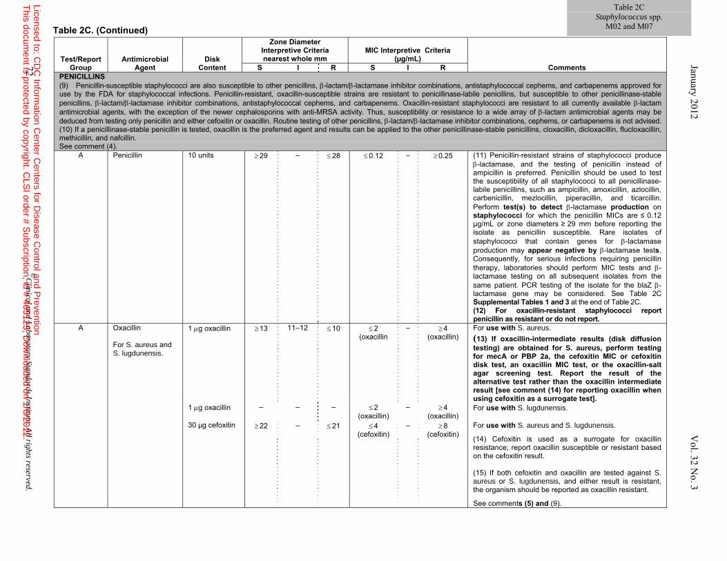
Table 2C Staphylococcus spp.
M02 and M07 Table 2C. (Continued)
Test/Report
Group Antimicrobial
Agent Disk
Content
Zone Diameter Interpretive Criteria nearest whole mm
MIC Interpretive Criteria (µg/mL)
Comments S I R S I R PENICILLINS (9) Penicillin-susceptible staphylococci are also susceptible to other penicillins, β-lactam/β-lactamase inhibitor combinations, antistaphylococcal cephems, and carbapenems approved for use by the FDA for staphylococcal infections. Penicillin-resistant, oxacillin-susceptible strains are resistant to penicillinase-labile penicillins, but susceptible to other penicillinase-stable penicillins, β-lactam/β-lactamase inhibitor combinations, antistaphylococcal cephems, and carbapenems. Oxacillin-resistant staphylococci are resistant to all currently available β-lactam antimicrobial agents, with the exception of the newer cephalosporins with anti-MRSA activity. Thus, susceptibility or resistance to a wide array of β-lactam antimicrobial agents may be deduced from testing only penicillin and either cefoxitin or oxacillin. Routine testing of other penicillins, β-lactam/β-lactamase inhibitor combinations, cephems, or carbapenems is not advised. (10) If a penicillinase-stable penicillin is tested, oxacillin is the preferred agent and results can be applied to the other penicillinase-stable penicillins, cloxacillin, dicloxacillin, flucloxacillin, methicillin, and nafcillin. See comment (4).
A Penicillin 10 units ≥ 29 – ≤ 28 ≤ 0.12 – ≥ 0.25 (11) Penicillin-resistant strains of staphylococci produce β-lactamase, and the testing of penicillin instead of ampicillin is preferred. Penicillin should be used to test the susceptibility of all staphylococci to all penicillinase-labile penicillins, such as ampicillin, amoxicillin, azlocillin, carbenicillin, mezlocillin, piperacillin, and ticarcillin. Perform test(s) to detect β-lactamase production on staphylococci for which the penicillin MICs are ≤ 0.12 µg/mL or zone diameters ≥ 29 mm before reporting the isolate as penicillin susceptible. Rare isolates of staphylococci that contain genes for β-lactamase production may appear negative by β-lactamase tests. Consequently, for serious infections requiring penicillin therapy, laboratories should perform MIC tests and β-lactamase testing on all subsequent isolates from the same patient. PCR testing of the isolate for the blaZ β-lactamase gene may be considered. See Table 2C Supplemental Tables 1 and 3 at the end of Table 2C. (12) For oxacillin-resistant staphylococci report penicillin as resistant or do not report.
A Oxacillin For S. aureus and S. lugdunensis.
1 μg oxacillin
≥ 13
11–12
≤ 10
≤ 2 (oxacillin
– ≥ 4 (oxacillin)
For use with S. aureus.
(13) If oxacillin-intermediate results (disk diffusion testing) are obtained for S. aureus, perform testing for mecA or PBP 2a, the cefoxitin MIC or cefoxitin disk test, an oxacillin MIC test, or the oxacillin-salt agar screening test. Report the result of the alternative test rather than the oxacillin intermediate result [see comment (14) for reporting oxacillin when using cefoxitin as a surrogate test].
1 μg oxacillin – – – ≤ 2 (oxacillin)
– ≥ 4 (oxacillin)
For use with S. lugdunensis.
30 µg cefoxitin ≥ 22 –
≤ 21
≤ 4 (cefoxitin)
– ≥ 8 (cefoxitin)
For use with S. aureus and S. lugdunensis.
(14) Cefoxitin is used as a surrogate for oxacillin resistance; report oxacillin susceptible or resistant based on the cefoxitin result. (15) If both cefoxitin and oxacillin are tested against S. aureus or S. lugdunensis, and either result is resistant, the organism should be reported as oxacillin resistant.
See comments (5) and (9).
72 ©C
linical and Laboratory Standards Institute. A
ll rights reserved.
January 2012 V
ol. 32 No. 3
Licensed to: CD
C Inform
ation Center C
enters for Disease C
ontrol and Prevention
This docum
ent is protected by copyright. CLS
I order # Subscription, id # 465125, D
ownloaded on 1/6/2012.

Table 2C Staphylococcus spp.
M02 and M07
Table 2C. (Continued)
Test/Report Group
Antimicrobial Agent
Disk Content
Zone Diameter Interpretive Criteria nearest
whole mm MIC Interpretive Criteria
(µg/mL)
Comments S I R S I R PENICILLINS (Continued)
A Oxacillin For coagulase-negative staphylococci except S. lugdunensis.
1 μg oxacillin – – – ≤ 0.25 (oxacillin)
–
≥ 0.5 (oxacillin)
For use with coagulase-negative staphylococci except S. lugdunensis. (16) Oxacillin interpretive criteria may overcall resistance for some coagulase-negative staphylococci, because some non–S. epidermidis strains for which the oxacillin MICs are 0.5 to 2 µg/mL lack mecA. For serious infections with coagulase-negative staphylococci other than S. epidermidis, testing for mecA or for PBP 2a or with cefoxitin disk diffusion may be appropriate for strains for which the oxacillin MICs are 0.5 to 2 µg/mL.
30 μg cefoxitin
≥ 25
– ≤ 24
– –
–
See comments (5), (9), and (14).
©C
linical and Laboratory Standards Institute. A
ll rights reserved. 73
For U
se With M
02-A11 and M
07-A9 M
100-S22
Licensed to: CD
C Inform
ation Center C
enters for Disease C
ontrol and Prevention
This docum
ent is protected by copyright. CLS
I order # Subscription, id # 465125, D
ownloaded on 1/6/2012.
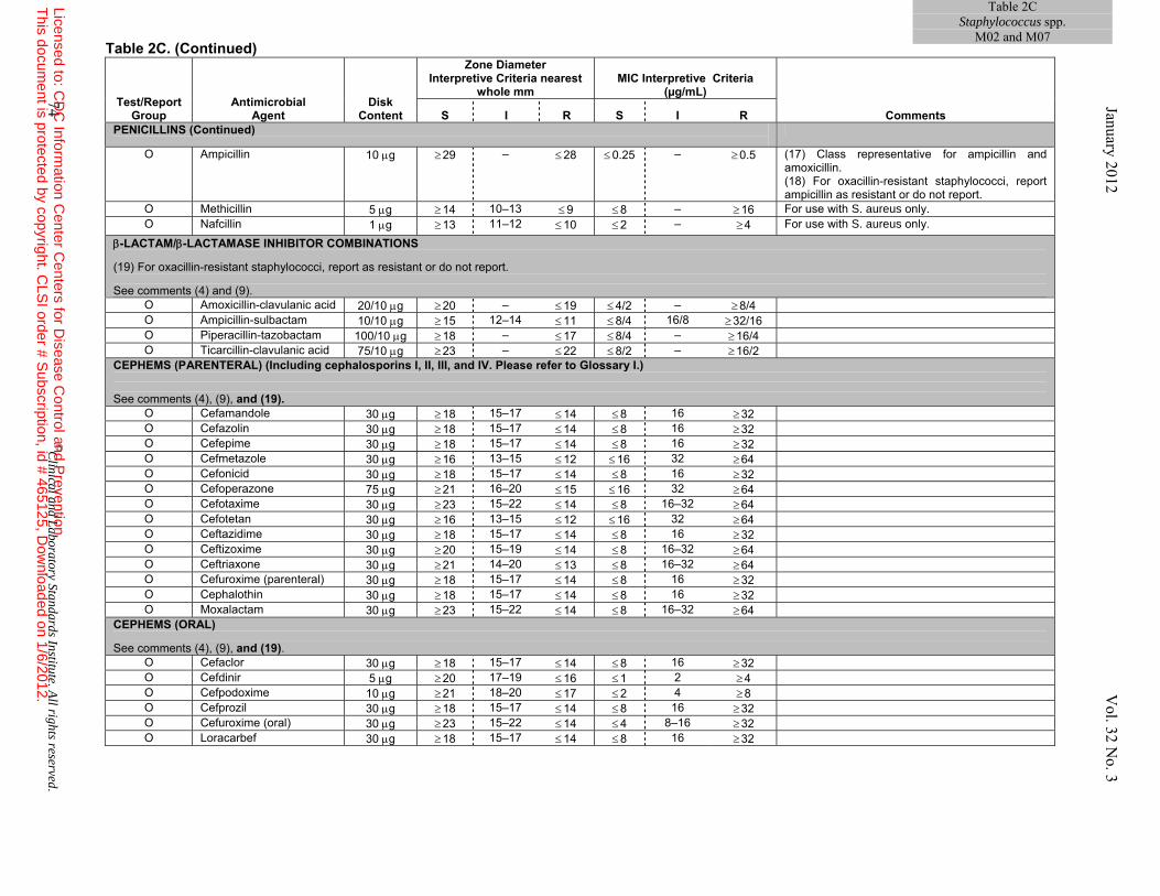
Table 2CStaphylococcus spp.
M02 and M07Table 2C. (Continued)
Test/Report Group
Antimicrobial Agent
Disk Content
Zone Diameter Interpretive Criteria nearest
whole mm MIC Interpretive Criteria
(µg/mL)
Comments S I R S I R PENICILLINS (Continued)
O Ampicillin 10 μg ≥ 29 – ≤ 28 ≤ 0.25 – ≥ 0.5 (17) Class representative for ampicillin and amoxicillin. (18) For oxacillin-resistant staphylococci, report ampicillin as resistant or do not report.
O Methicillin 5 μg ≥ 14 10–13 ≤ 9 ≤ 8 – ≥ 16 For use with S. aureus only. O Nafcillin 1 μg ≥ 13 11–12 ≤ 10 ≤ 2 – ≥ 4 For use with S. aureus only.
β-LACTAM/β-LACTAMASE INHIBITOR COMBINATIONS (19) For oxacillin-resistant staphylococci, report as resistant or do not report. See comments (4) and (9).
O Amoxicillin-clavulanic acid 20/10 μg ≥ 20 – ≤ 19 ≤ 4/2 – ≥ 8/4 O Ampicillin-sulbactam 10/10 μg ≥ 15 12–14 ≤ 11 ≤ 8/4 16/8 ≥ 32/16 O Piperacillin-tazobactam 100/10 μg ≥ 18 – ≤ 17 ≤ 8/4 – ≥ 16/4 O Ticarcillin-clavulanic acid 75/10 μg ≥ 23 – ≤ 22 ≤ 8/2 – ≥ 16/2
CEPHEMS (PARENTERAL) (Including cephalosporins I, II, III, and IV. Please refer to Glossary I.) See comments (4), (9), and (19).
O Cefamandole 30 μg ≥ 18 15–17 ≤ 14 ≤ 8 16 ≥ 32 O Cefazolin 30 μg ≥ 18 15–17 ≤ 14 ≤ 8 16 ≥ 32 O Cefepime 30 μg ≥ 18 15–17 ≤ 14 ≤ 8 16 ≥ 32 O Cefmetazole 30 μg ≥ 16 13–15 ≤ 12 ≤ 16 32 ≥ 64 O Cefonicid 30 μg ≥ 18 15–17 ≤ 14 ≤ 8 16 ≥ 32 O Cefoperazone 75 μg ≥ 21 16–20 ≤ 15 ≤ 16 32 ≥ 64 O Cefotaxime 30 μg ≥ 23 15–22 ≤ 14 ≤ 8 16–32 ≥ 64 O Cefotetan 30 μg ≥ 16 13–15 ≤ 12 ≤ 16 32 ≥ 64 O Ceftazidime 30 μg ≥ 18 15–17 ≤ 14 ≤ 8 16 ≥ 32 O Ceftizoxime 30 μg ≥ 20 15–19 ≤ 14 ≤ 8 16–32 ≥ 64 O Ceftriaxone 30 μg ≥ 21 14–20 ≤ 13 ≤ 8 16–32 ≥ 64 O Cefuroxime (parenteral) 30 μg ≥ 18 15–17 ≤ 14 ≤ 8 16 ≥ 32 O Cephalothin 30 μg ≥ 18 15–17 ≤ 14 ≤ 8 16 ≥ 32 O Moxalactam 30 μg ≥ 23 15–22 ≤ 14 ≤ 8 16–32 ≥ 64
CEPHEMS (ORAL) See comments (4), (9), and (19).
O Cefaclor 30 μg ≥ 18 15–17 ≤ 14 ≤ 8 16 ≥ 32 O Cefdinir 5 μg ≥ 20 17–19 ≤ 16 ≤ 1 2 ≥ 4 O Cefpodoxime 10 μg ≥ 21 18–20 ≤ 17 ≤ 2 4 ≥ 8 O Cefprozil 30 μg ≥ 18 15–17 ≤ 14 ≤ 8 16 ≥ 32 O Cefuroxime (oral) 30 μg ≥ 23 15–22 ≤ 14 ≤ 4 8–16 ≥ 32 O Loracarbef 30 μg ≥ 18 15–17 ≤ 14 ≤ 8 16 ≥ 32
74 ©C
linical and Laboratory Standards Institute. A
ll rights reserved.
January 2012 V
ol. 32 No. 3
Licensed to: CD
C Inform
ation Center C
enters for Disease C
ontrol and Prevention
This docum
ent is protected by copyright. CLS
I order # Subscription, id # 465125, D
ownloaded on 1/6/2012.
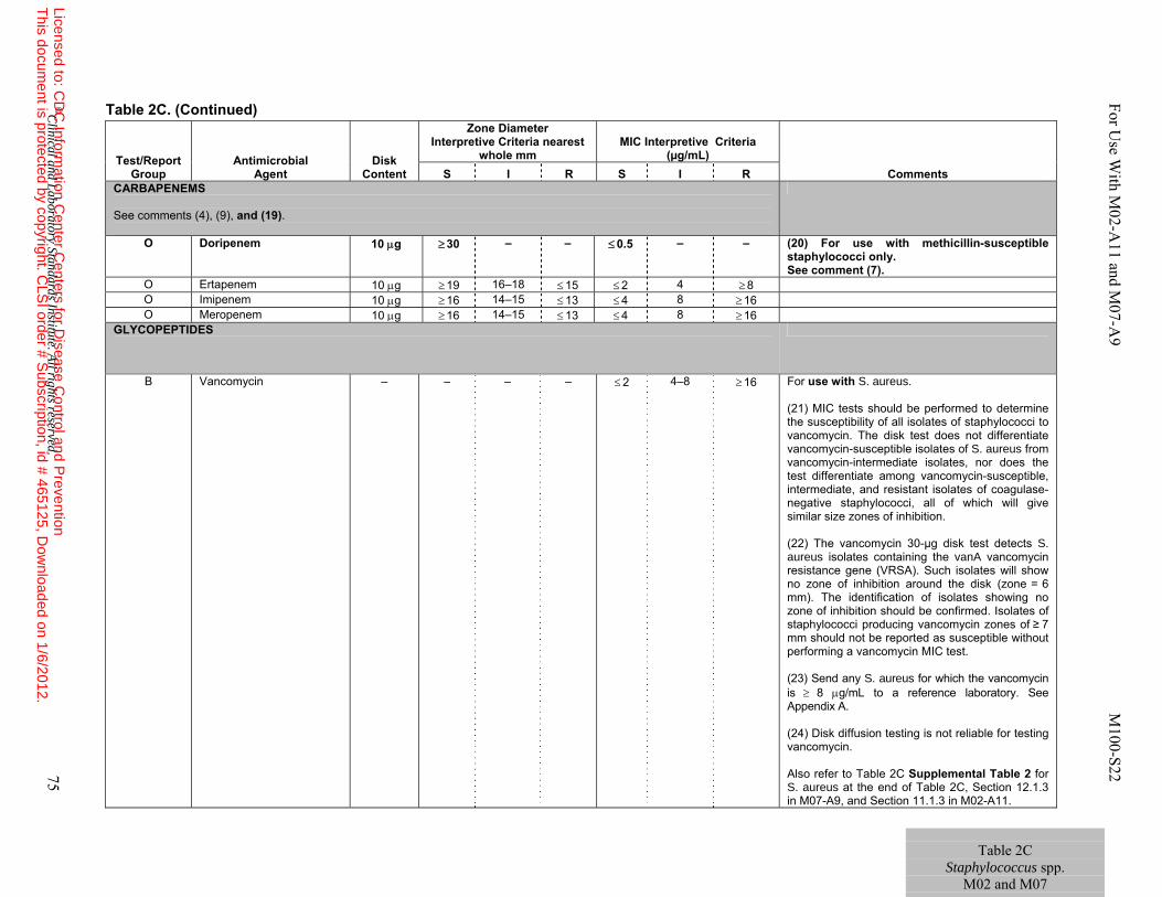
Table 2C. (Continued)
Test/Report Group
Antimicrobial Agent
Disk Content
Zone Diameter Interpretive Criteria nearest
whole mm MIC Interpretive Criteria
(µg/mL) Comments S I R S I R
CARBAPENEMS See comments (4), (9), and (19).
O Doripenem 10 μg ≥ 30 – – ≤ 0.5 – – (20) For use with methicillin-susceptible staphylococci only. See comment (7).
O Ertapenem 10 μg ≥ 19 16–18 ≤ 15 ≤ 2 4 ≥ 8 O Imipenem 10 μg ≥ 16 14–15 ≤ 13 ≤ 4 8 ≥ 16 O Meropenem 10 μg ≥ 16 14–15 ≤ 13 ≤ 4 8 ≥ 16
GLYCOPEPTIDES
B
Vancomycin
– – – – ≤ 2 4–8 ≥ 16 For use with S. aureus. (21) MIC tests should be performed to determine the susceptibility of all isolates of staphylococci to vancomycin. The disk test does not differentiate vancomycin-susceptible isolates of S. aureus from vancomycin-intermediate isolates, nor does the test differentiate among vancomycin-susceptible, intermediate, and resistant isolates of coagulase-negative staphylococci, all of which will give similar size zones of inhibition. (22) The vancomycin 30-µg disk test detects S. aureus isolates containing the vanA vancomycin resistance gene (VRSA). Such isolates will show no zone of inhibition around the disk (zone = 6 mm). The identification of isolates showing no zone of inhibition should be confirmed. Isolates of staphylococci producing vancomycin zones of ≥ 7 mm should not be reported as susceptible without performing a vancomycin MIC test. (23) Send any S. aureus for which the vancomycin is ≥ 8 μg/mL to a reference laboratory. See Appendix A. (24) Disk diffusion testing is not reliable for testing vancomycin. Also refer to Table 2C Supplemental Table 2 for S. aureus at the end of Table 2C, Section 12.1.3 in M07-A9, and Section 11.1.3 in M02-A11.
Table 2C
Staphylococcus spp. M02 and M07
©C
linical and Laboratory Standards Institute. A
ll rights reserved. 75
For U
se With M
02-A11 and M
07-A9 M
100-S22
Licensed to: CD
C Inform
ation Center C
enters for Disease C
ontrol and Prevention
This docum
ent is protected by copyright. CLS
I order # Subscription, id # 465125, D
ownloaded on 1/6/2012.
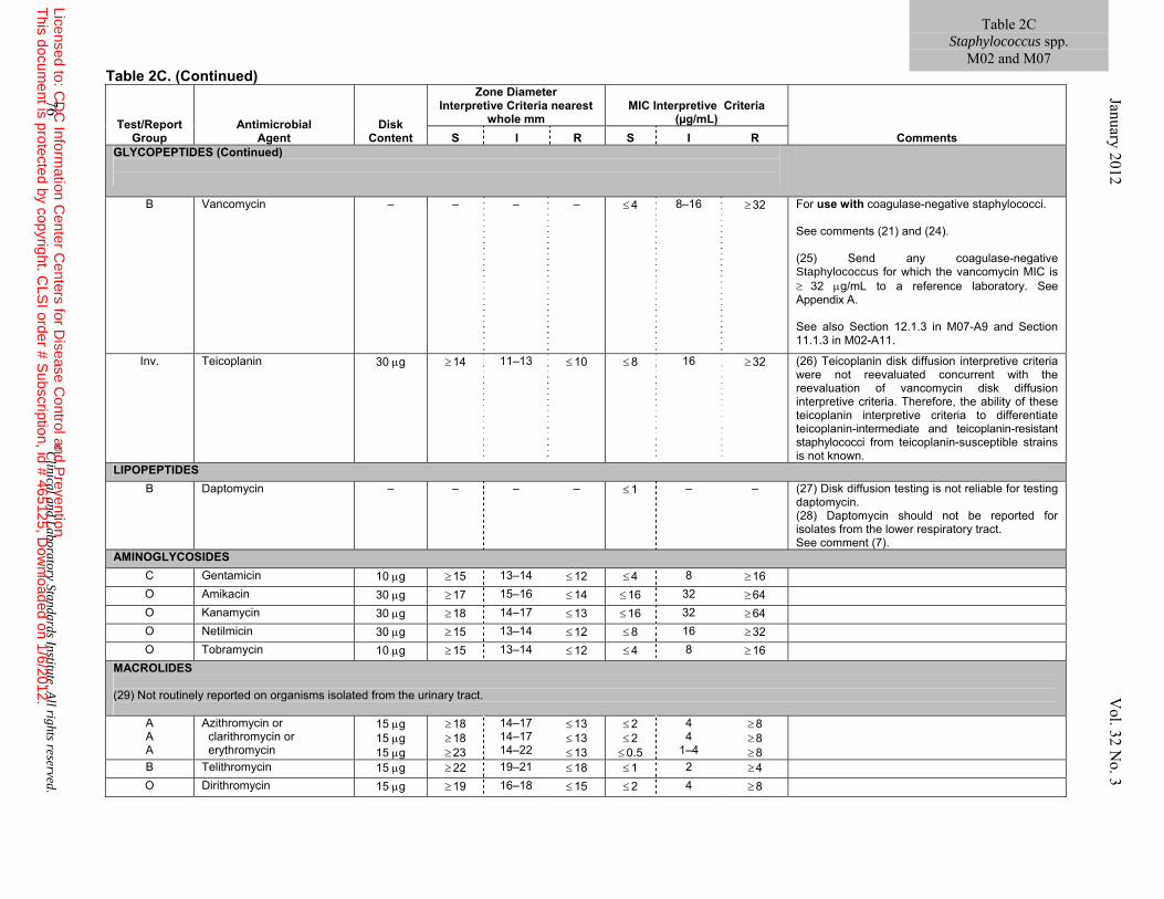
Table 2C. (Continued)
Test/Report Group
Antimicrobial Agent
Disk Content
Zone Diameter Interpretive Criteria nearest
whole mm MIC Interpretive Criteria
(µg/mL) Comments S I R S I R
GLYCOPEPTIDES (Continued)
B
Vancomycin
– – – – ≤ 4
8–16 ≥ 32 For use with coagulase-negative staphylococci. See comments (21) and (24). (25) Send any coagulase-negative Staphylococcus for which the vancomycin MIC is ≥ 32 μg/mL to a reference laboratory. See Appendix A. See also Section 12.1.3 in M07-A9 and Section 11.1.3 in M02-A11.
Inv. Teicoplanin 30 μg ≥ 14 11–13 ≤ 10 ≤ 8 16 ≥ 32 (26) Teicoplanin disk diffusion interpretive criteria were not reevaluated concurrent with the reevaluation of vancomycin disk diffusion interpretive criteria. Therefore, the ability of these teicoplanin interpretive criteria to differentiate teicoplanin-intermediate and teicoplanin-resistant staphylococci from teicoplanin-susceptible strains is not known.
LIPOPEPTIDES B Daptomycin – – – – ≤ 1 – – (27) Disk diffusion testing is not reliable for testing
daptomycin. (28) Daptomycin should not be reported for isolates from the lower respiratory tract. See comment (7).
AMINOGLYCOSIDES C Gentamicin 10 μg ≥ 15 13–14 ≤ 12 ≤ 4 8 ≥ 16 O Amikacin 30 μg ≥ 17 15–16 ≤ 14 ≤ 16 32 ≥ 64 O Kanamycin 30 μg ≥ 18 14–17 ≤ 13 ≤ 16 32 ≥ 64 O Netilmicin 30 μg ≥ 15 13–14 ≤ 12 ≤ 8 16 ≥ 32 O Tobramycin 10 μg ≥ 15 13–14 ≤ 12 ≤ 4 8 ≥ 16
MACROLIDES (29) Not routinely reported on organisms isolated from the urinary tract.
A A A
Azithromycin or clarithromycin or erythromycin
15 μg 15 μg 15 μg
≥ 18 ≥ 18 ≥ 23
14–17 14–17 14–22
≤ 13 ≤ 13 ≤ 13
≤ 2 ≤ 2 ≤ 0.5
4 4
1–4
≥ 8 ≥ 8 ≥ 8
B Telithromycin 15 μg ≥ 22 19–21 ≤ 18 ≤ 1 2 ≥ 4 O Dirithromycin 15 μg ≥ 19 16–18 ≤ 15 ≤ 2 4 ≥ 8
76 ©C
linical and Laboratory Standards Institute. A
ll rights reserved.
Table 2C Staphylococcus spp.
M02 and M07 January 2012 V
ol. 32 No. 3
Licensed to: CD
C Inform
ation Center C
enters for Disease C
ontrol and Prevention
This docum
ent is protected by copyright. CLS
I order # Subscription, id # 465125, D
ownloaded on 1/6/2012.
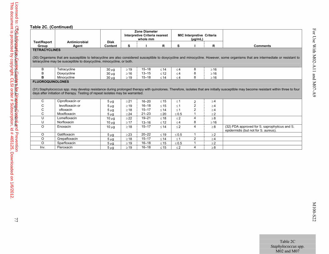
Table 2C. (Continued)
Test/Report Group
Antimicrobial Agent
Disk Content
Zone Diameter Interpretive Criteria nearest
whole mm MIC Interpretive Criteria
(µg/mL)
Comments S I R S I R TETRACYCLINES (30) Organisms that are susceptible to tetracycline are also considered susceptible to doxycycline and minocycline. However, some organisms that are intermediate or resistant to tetracycline may be susceptible to doxycycline, minocycline, or both.
B Tetracycline 30 μg ≥ 19 15–18 ≤ 14 ≤ 4 8 ≥ 16 B Doxycycline 30 μg ≥ 16 13–15 ≤ 12 ≤ 4 8 ≥ 16 B Minocycline 30 μg ≥ 19 15–18 ≤ 14 ≤ 4 8 ≥ 16
FLUOROQUINOLONES (31) Staphylococcus spp. may develop resistance during prolonged therapy with quinolones. Therefore, isolates that are initially susceptible may become resistant within three to four days after initiation of therapy. Testing of repeat isolates may be warranted.
C Ciprofloxacin or 5 μg ≥ 21 16–20 ≤ 15 ≤ 1 2 ≥ 4 C levofloxacin or 5 μg ≥ 19 16–18 ≤ 15 ≤ 1 2 ≥ 4 C ofloxacin 5 μg ≥ 18 15–17 ≤ 14 ≤ 1 2 ≥ 4 C Moxifloxacin 5 μg ≥ 24 21–23 ≤ 20 ≤ 0.5 1 ≥ 2 U Lomefloxacin 10 μg ≥ 22 19–21 ≤ 18 ≤ 2 4 ≥ 8 U Norfloxacin 10 μg ≥ 17 13–16 ≤ 12 ≤ 4 8 ≥ 16 O Enoxacin 10 μg ≥ 18 15–17 ≤ 14 ≤ 2 4 ≥ 8 (32) FDA approved for S. saprophyticus and S.
epidermidis (but not for S. aureus). O Gatifloxacin 5 μg ≥ 23 20–22 ≤ 19 ≤ 0.5 1 ≥ 2 O Grepafloxacin 5 μg ≥ 18 15–17 ≤ 14 ≤ 1 2 ≥ 4 O Sparfloxacin 5 μg ≥ 19 16–18 ≤ 15 ≤ 0.5 1 ≥ 2
Inv. Fleroxacin 5 μg ≥ 19 16–18 ≤ 15 ≤ 2 4 ≥ 8
Table 2C
Staphylococcus spp. M02 and M07
©C
linical and Laboratory Standards Institute. A
ll rights reserved. 77
For U
se With M
02-A11 and M
07-A9 M
100-S22
Licensed to: CD
C Inform
ation Center C
enters for Disease C
ontrol and Prevention
This docum
ent is protected by copyright. CLS
I order # Subscription, id # 465125, D
ownloaded on 1/6/2012.
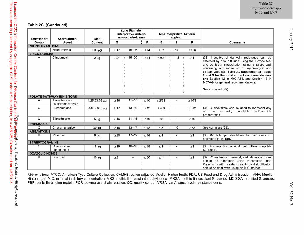
Table 2C. (Continued)
Abbreviations: ATCC, American Type Culture Collection; CAMHB, cation-adjusted Mueller-Hinton broth; FDA, US Food and Drug Administration; MHA, Mueller-Hinton agar; MIC, minimal inhibitory concentration; MRS, methicillin-resistant staphylococci; MRSA, methicillin-resistant S. aureus; MOD-SA, modified S. aureus; PBP, penicillin-binding protein; PCR, polymerase chain reaction; QC, quality control; VRSA, vanA vancomycin resistance gene.
Test/Report Group
Antimicrobial Agent
Disk Content
Zone Diameter Interpretive Criteria nearest whole mm
MIC Interpretive Criteria (µg/mL)
Comments S I R S I R NITROFURANTOINS
U Nitrofurantoin 300 μg ≥ 17 15–16 ≤ 14 ≤ 32 64 ≥ 128 LINCOSAMIDES
A Clindamycin 2 μg ≥ 21 15–20 ≤ 14 ≤ 0.5 1–2 ≥ 4 (33) Inducible clindamycin resistance can be detected by disk diffusion using the D-zone test and by broth microdilution using a single well containing a combination of erythromycin and clindamycin. See Table 2C Supplemental Tables 2 and 3 for the most current recommendations, and Section 12 in M02-A11, and Section 13 in M07-A9 for general recommendations. See comment (29).
FOLATE PATHWAY INHIBITORS A Trimethoprim-
sulfamethoxazole 1.25/23.75 μg ≥ 16 11–15 ≤ 10 ≤ 2/38
–
≥ 4/76
U Sulfonamides 250 or 300 μg ≥ 17 13–16 ≤ 12 ≤ 256
–
≥ 512
(34) Sulfisoxazole can be used to represent any of the currently available sulfonamide preparations.
U Trimethoprim 5 μg ≥ 16 11–15 ≤ 10 ≤ 8 – ≥ 16
PHENICOLS C Chloramphenicol 30 μg ≥ 18 13–17 ≤ 12 ≤ 8 16 ≥ 32 See comment (29).
ANSAMYCINS B Rifampin 5 μg ≥ 20 17–19 ≤ 16 ≤ 1 2 ≥ 4 (35) Rx: Rifampin should not be used alone for
antimicrobial therapy. STREPTOGRAMINS
C Quinupristin- dalfopristin
15 μg ≥ 19 16–18 ≤ 15 ≤ 1 2 ≥ 4 (36) For reporting against methicillin-susceptible S. aureus.
OXAZOLIDINONES B Linezolid 30 μg ≥ 21 – ≤ 20 ≤ 4 – ≥ 8 (37) When testing linezolid, disk diffusion zones
should be examined using transmitted light. Organisms with resistant results by disk diffusion should be confirmed using an MIC method.
Table 2C Staphylococcus spp.
M02 and M07
78 ©C
linical and Laboratory Standards Institute. A
ll rights reserved.
January 2012 V
ol. 32 No. 3
Licensed to: CD
C Inform
ation Center C
enters for Disease C
ontrol and Prevention
This docum
ent is protected by copyright. CLS
I order # Subscription, id # 465125, D
ownloaded on 1/6/2012.

For Use With M02-A11 and M07-A9 M100-S22
©Clinical and Laboratory Standards Institute. All rights reserved. 79
This page is intentionally left blank.
Licensed to: CDC Information Center Centers for Disease Control and PreventionThis document is protected by copyright. CLSI order # Subscription, id # 465125, Downloaded on 1/6/2012.
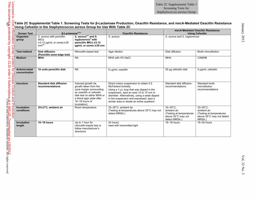
Table 2C Supplemental Table 1. Screening Tests for β-Lactamase Production, Oxacillin Resistance, and mecA-Mediated Oxacillin Resistance Using Cefoxitin in the Staphylococcus aureus Group for Use With Table 2C
Screen Test β-Lactamasea,b,c Oxacillin Resistance
mecA-Mediated Oxacillin Resistance Using Cefoxitin
Organism group
S. aureus with penicillin MICs ≤ 0.12 µg/mL or zones ≥ 29 mma,c
S. aureusa,c and S. lugdunensisb with penicillin MICs ≤ 0.12 µg/mL or zones ≥ 29 mm
S. aureus S. aureus and S. lugdunensis
Test method Disk diffusion (Penicillin zone-edge test)
Nitrocefin-based test Agar dilution Disk diffusion Broth microdilution
Medium MHA NA MHA with 4% NaCl MHA CAMHB
Antimicrobial concentration
10 units penicillin disk NA 6 μg/mL oxacillin 30 µg cefoxitin disk 4 µg/mL cefoxitin
Inoculum Standard disk diffusion recommendations
Induced growth (ie, growth taken from the zone margin surrounding an oxacillin or cefoxitin disk test on either MHA or a blood agar plate after 16–18 hours of incubation)
Direct colony suspension to obtain 0.5 McFarland turbidity. Using a 1-μL loop that was dipped in the suspension, spot an area 10 to 15 mm in diameter. Alternatively, using a swab dipped in the suspension and expressed, spot a similar area or streak an entire quadrant.
Standard disk diffusion recommendations
Standard broth microdilution recommendations
Incubation conditions
35 ± 2°C; ambient air
Room temperature
33–35°C; ambient air. (Testing at temperatures above 35°C may not detect MRSA.)
33–35°C; ambient air. (Testing at temperatures above 35°C may not detect MRSA.)
33–35°C; ambient air. (Testing at temperatures above 35°C may not detect MRSA.)
Incubation length
16–18 hours Up to 1 hour for nitrocefin-based test or follow manufacturer’s directions
24 hours;read with transmitted light
16–18 hours 16–20 hours
80 ©C
linical and Laboratory
Standards Institute. All rights reserved.
Table 2C Supplemental Table 1 Screening Tests for
Staphylococcus aureus Group January 2012 V
ol. 32 No. 3
Licensed to: CD
C Inform
ation Center C
enters for Disease C
ontrol and Prevention
This docum
ent is protected by copyright. CLS
I order # Subscription, id # 465125, D
ownloaded on 1/6/2012.
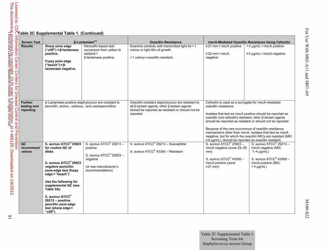
Table 2C Supplemental Table 1. (Continued)
Screen Test β-Lactamasea,b Oxacillin Resistance mecA-Mediated Oxacillin Resistance Using Cefoxitin Results Sharp zone edge
(“cliff”) = β-lactamase positive. Fuzzy zone edge (“beach”) = β-lactamase negative.
Nitrocefin-based test: conversion from yellow to red/pink =
β-lactamase positive.
Examine carefully with transmitted light for > 1 colony or light film of growth. > 1 colony = oxacillin resistant.
≤ 21 mm = mecA positive ≥ 22 mm = mecA negative
> 4 µg/mL = mecA positive ≤ 4 µg/mL = mecA negative
Further testing and reporting
β-Lactamase-positive staphylococci are resistant to penicillin, amino-, carboxy-, and ureidopenicillins.
Oxacillin-resistant staphylococci are resistant to all β-lactam agents; other β-lactam agents should be reported as resistant or should not be reported.
Cefoxitin is used as a surrogate for mecA-mediated oxacillin resistance. Isolates that test as mecA positive should be reported as oxacillin (not cefoxitin) resistant; other β-lactam agents should be reported as resistant or should not be reported. Because of the rare occurrence of oxacillin resistance mechanisms other than mecA, isolates that test as mecA negative, but for which the oxacillin MICs are resistant (MIC ≥ 4 µg/mL), should be reported as oxacillin resistant.
QC recommend-ations
S. aureus ATCC® 25923 for routine QC of disks S. aureus ATCC® 25923 negative penicillin zone-edge test (fuzzy edge = “beach”) Use the following for supplemental QC (see Table 3A): S. aureus ATCC® 29213 – positive pencillin zone edge test (sharp edge =
“cliff”)
S. aureus ATCC® 29213 – positive S. aureus ATCC® 25923 – negative (or see manufacturer’s recommendations)
S. aureus ATCC® 29213 – Susceptible S. aureus ATCC® 43300 – Resistant
S. aureus ATCC® 25923 – mecA negative (zone 23–29 mm) S. aureus ATCC® 43300 – mecA positive (zone ≤ 21 mm)
S. aureus ATCC® 29213 – mecA negative (MIC 1–4 µg/mL) S. aureus ATCC® 43300 – mecA positive (MIC > 4 µg/mL)
©C
linical and Laboratory Standards Institute. A
ll rights reserved. 81
For U
se With M
02-A11 and M
07-A9 M
100-S22
Table 2C Supplemental Table 1 Screening Tests for
Staphylococcus aureus Group
Licensed to: CD
C Inform
ation Center C
enters for Disease C
ontrol and Prevention
This docum
ent is protected by copyright. CLS
I order # Subscription, id # 465125, D
ownloaded on 1/6/2012.
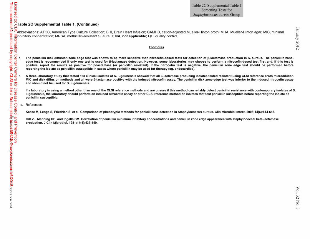
January 2012 V
ol. 32 No. 3
Table 2C Supplemental Table 1. (Continued) Abbreviations: ATCC, American Type Culture Collection; BHI, Brain Heart Infusion; CAMHB, cation-adjusted Mueller-Hinton broth; MHA, Mueller-Hinton agar; MIC, minimal inhibitory concentration; MRSA, methicillin-resistant S. aureus; NA, not applicable; QC, quality control.
Footnotes
a. The penicillin disk diffusion zone edge test was shown to be more sensitive than nitrocefin-based tests for detection of β-lactamase production in S. aureus. The penicillin zone-edge test is recommended if only one test is used for β-lactamase detection. However, some laboratories may choose to perform a nitrocefin-based test first and, if this test is positive, report the results as positive for β-lactamase (or penicillin resistant). If the nitrocefin test is negative, the penicillin zone edge test should be performed before reporting the isolate as penicillin susceptible in cases where penicillin may be used for therapy (eg, endocarditis).
b. A three-laboratory study that tested 168 clinical isolates of S. lugdunensis showed that all β-lactamase producing isolates tested resistant using CLSI reference broth microdilution MIC and disk diffusion methods and all were β-lactamase positive with the induced nitrocefin assay. The penicillin disk zone-edge test was inferior to the induced nitrocefin assay and should not be used for S. lugdunensis. If a laboratory is using a method other than one of the CLSI reference methods and are unsure if this method can reliably detect penicillin resistance with contemporary isolates of S. lugdunensis, the laboratory should perform an induced nitrocefin assay or other CLSI reference method on isolates that test penicillin susceptible before reporting the isolate as penicillin susceptible.
c. References:
Kaase M, Lenga S, Friedrich S, et al. Comparison of phenotypic methods for penicillinase detection in Staphylococcus aureus. Clin Microbiol Infect. 2008;14(6):614-616. Gill VJ, Manning CB, and Ingalls CM. Correlation of penicillin minimum inhibitory concentrations and penicillin zone edge appearance with staphylococcal beta-lactamase production. J Clin Microbiol. 1981;14(4):437-440.
Table 2C Supplemental Table 1 Screening Tests for
Staphylococcus aureus Group
82 ©
Clinical and L
aboratoryStandards
Institute. All rights reserved.
Table 2C Supplemental Table 1 Screening Tests for
Staphylococcus aureus Group
Licensed to: CD
C Inform
ation Center C
enters for Disease C
ontrol and Prevention
This docum
ent is protected by copyright. CLS
I order # Subscription, id # 465125, D
ownloaded on 1/6/2012.
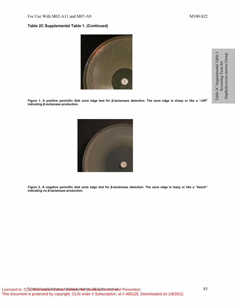
For Use With M02-A11 and M07-A9 M100-S22
©Clinical and Laboratory Standards Institute. All rights reserved. 83
Table 2C Supplemental Table 1. (Continued) Figure 1. A positive penicillin disk zone edge test for β-lactamase detection. The zone edge is sharp or like a “cliff” indicating β-lactamase production.
Figure 2. A negative penicillin disk zone edge test for β-lactamase detection. The zone edge is fuzzy or like a “beach” indicating no β-lactamase production.
Tabl
e 2C
Sup
plem
enta
l Tab
le 1
Sc
reen
ing
Test
s for
St
aphy
loco
ccus
aur
eus
Gro
up
Licensed to: CDC Information Center Centers for Disease Control and PreventionThis document is protected by copyright. CLSI order # Subscription, id # 465125, Downloaded on 1/6/2012.
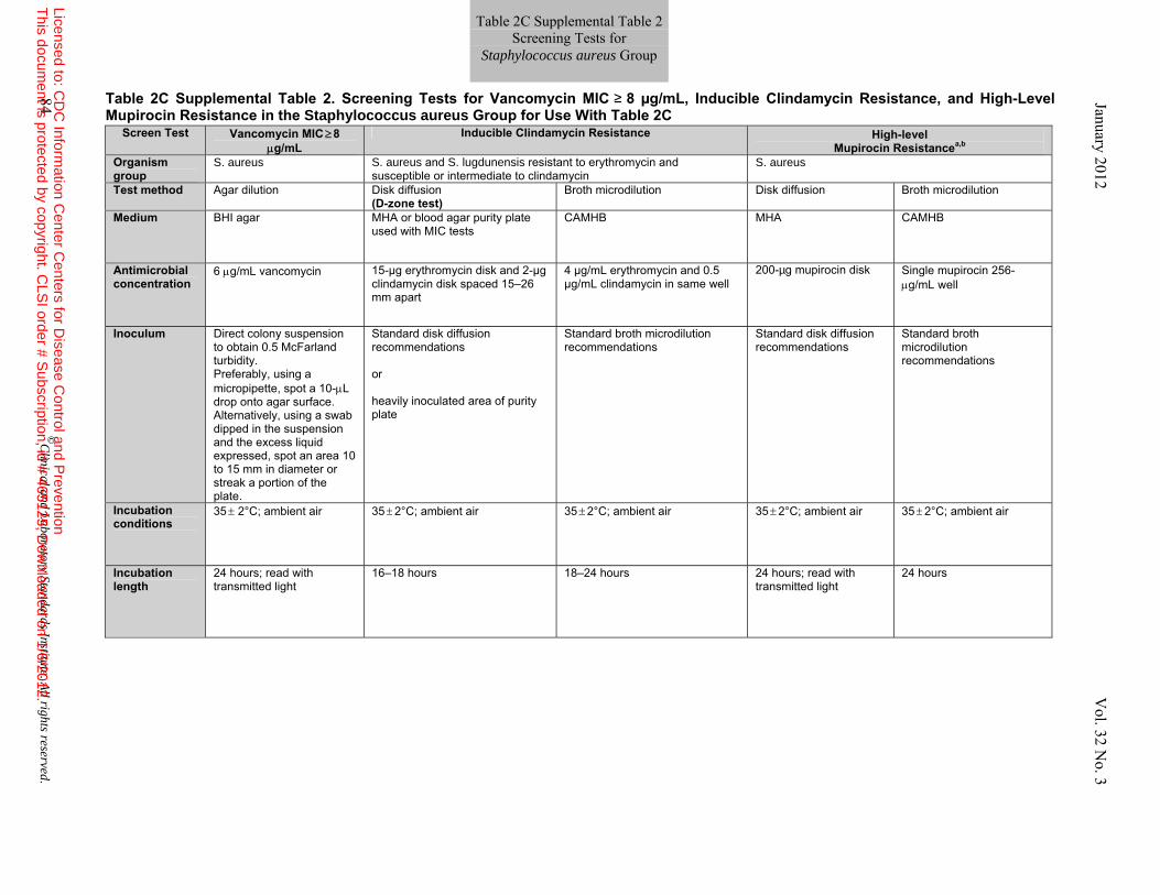
Table 2C Supplemental Table 2. Screening Tests for Vancomycin MIC ≥ 8 µg/mL, Inducible Clindamycin Resistance, and High-Level Mupirocin Resistance in the Staphylococcus aureus Group for Use With Table 2C
Screen Test Vancomycin MIC ≥ 8 μg/mL
Inducible Clindamycin Resistance High-level Mupirocin Resistancea,b
Organism group
S. aureus S. aureus and S. lugdunensis resistant to erythromycin and susceptible or intermediate to clindamycin
S. aureus
Test method Agar dilution Disk diffusion (D-zone test)
Broth microdilution Disk diffusion Broth microdilution
Medium BHI agar MHA or blood agar purity plate used with MIC tests
CAMHB MHA CAMHB
Antimicrobial concentration
6 μg/mL vancomycin 15-µg erythromycin disk and 2-µg clindamycin disk spaced 15–26 mm apart
4 µg/mL erythromycin and 0.5 µg/mL clindamycin in same well
200-µg mupirocin disk Single mupirocin 256-μg/mL well
Inoculum Direct colony suspension to obtain 0.5 McFarland turbidity. Preferably, using a micropipette, spot a 10-μL drop onto agar surface. Alternatively, using a swab dipped in the suspension and the excess liquid expressed, spot an area 10 to 15 mm in diameter or streak a portion of the plate.
Standard disk diffusion recommendations or heavily inoculated area of purity plate
Standard broth microdilution recommendations
Standard disk diffusion recommendations
Standard broth microdilution recommendations
Incubation conditions
35 ± 2°C; ambient air
35 ± 2°C; ambient air
35 ± 2°C; ambient air
35 ± 2°C; ambient air 35 ± 2°C; ambient air
Incubation length
24 hours; read with transmitted light
16–18 hours 18–24 hours 24 hours; read with transmitted light
24 hours
84 ©C
linical and Laboratory Standards Institute. A
ll rights reserved.
January 2012
V
ol. 32 No. 3
Table 2C Supplemental Table 2 Screening Tests for
Staphylococcus aureus Group
Licensed to: CD
C Inform
ation Center C
enters for Disease C
ontrol and Prevention
This docum
ent is protected by copyright. CLS
I order # Subscription, id # 465125, D
ownloaded on 1/6/2012.
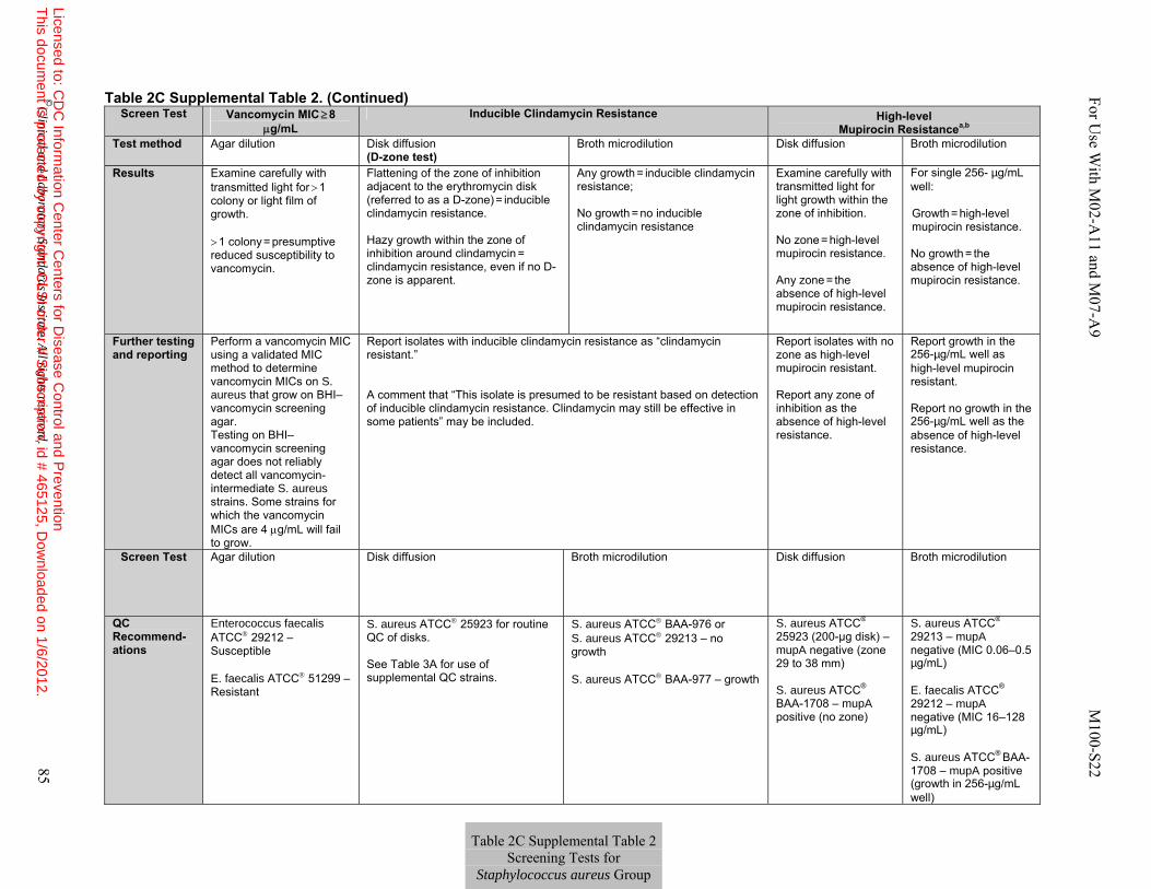
Table 2C Supplemental Table 2. (Continued) Screen Test Vancomycin MIC ≥ 8
μg/mL Inducible Clindamycin Resistance High-level
Mupirocin Resistancea,b
Test method Agar dilution Disk diffusion (D-zone test)
Broth microdilution Disk diffusion Broth microdilution
Results Examine carefully with transmitted light for > 1 colony or light film of growth. > 1 colony = presumptive reduced susceptibility to vancomycin.
Flattening of the zone of inhibition adjacent to the erythromycin disk (referred to as a D-zone) = inducible clindamycin resistance. Hazy growth within the zone of inhibition around clindamycin =
clindamycin resistance, even if no D-zone is apparent.
Any growth = inducible clindamycin resistance; No growth = no inducible clindamycin resistance
Examine carefully with transmitted light for light growth within the zone of inhibition. No zone = high-level mupirocin resistance. Any zone = the absence of high-level mupirocin resistance.
For single 256- µg/mL well:
Growth = high-level mupirocin resistance.
No growth = the absence of high-level mupirocin resistance.
Further testing and reporting
Perform a vancomycin MIC using a validated MIC method to determine vancomycin MICs on S. aureus that grow on BHI– vancomycin screening agar. Testing on BHI– vancomycin screening agar does not reliably detect all vancomycin-intermediate S. aureus strains. Some strains for which the vancomycin MICs are 4 μg/mL will fail to grow.
Report isolates with inducible clindamycin resistance as “clindamycin resistant.” A comment that “This isolate is presumed to be resistant based on detection of inducible clindamycin resistance. Clindamycin may still be effective in some patients” may be included.
Report isolates with no zone as high-level mupirocin resistant. Report any zone of inhibition as the absence of high-level resistance.
Report growth in the 256-µg/mL well as high-level mupirocin resistant. Report no growth in the 256-µg/mL well as the absence of high-level resistance.
Screen Test Agar dilution Disk diffusion Broth microdilution Disk diffusion Broth microdilution
QC Recommend-ations
Enterococcus faecalis ATCC® 29212 – Susceptible E. faecalis ATCC® 51299 – Resistant
S. aureus ATCC® 25923 for routine QC of disks. See Table 3A for use of supplemental QC strains.
S. aureus ATCC® BAA-976 or S. aureus ATCC® 29213 – no growth S. aureus ATCC® BAA-977 – growth
S. aureus ATCC® 25923 (200-µg disk) – mupA negative (zone 29 to 38 mm) S. aureus ATCC®
BAA-1708 – mupA positive (no zone)
S. aureus ATCC® 29213 – mupA negative (MIC 0.06–0.5 µg/mL) E. faecalis ATCC® 29212 – mupA negative (MIC 16–128 µg/mL) S. aureus ATCC® BAA-1708 – mupA positive (growth in 256-µg/mL well)
©C
linical and Laboratory Standards Institute. A
ll rights reserved. 85
For U
se With M
02-A11 and M
07-A9 M
100-S22
Table 2C Supplemental Table 2 Screening Tests for
Staphylococcus aureus Group
Licensed to: CD
C Inform
ation Center C
enters for Disease C
ontrol and Prevention
This docum
ent is protected by copyright. CLS
I order # Subscription, id # 465125, D
ownloaded on 1/6/2012.
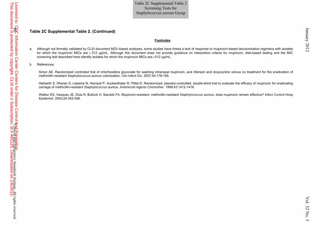
Table 2C Supplemental Table 2. (Continued)
Footnotes a. Although not formally validated by CLSI document M23–based analyses, some studies have linked a lack of response to mupirocin-based decolonization regimens with isolates
for which the mupirocin MICs are ≥ 512 µg/mL. Although this document does not provide guidance on interpretive criteria for mupirocin, disk-based testing and the MIC screening test described here identify isolates for which the mupirocin MICs are ≥ 512 µg/mL.
b. References:
Simor AE. Randomized controlled trial of chlorhexidine gluconate for washing intranasal mupirocin, and rifampin and doxycycline versus no treatment for the eradication of methicillin-resistant Staphylococcus aureus colonization. Clin Infect Dis. 2007;44:178-185. Harbarth S, Dharan S, Liassine N, Herrault P, Auckenthaler R, Pittet D. Randomized, placebo-controlled, double-blind trial to evaluate the efficacy of mupirocin for eradicating carriage of methicillin-resistant Staphylococcus aureus. Antimicrob Agents Chemother. 1999;43:1412-1416. Walker ES, Vasquez JE, Dula R, Bullock H, Sarubbi FA. Mupirocin-resistant, methicillin-resistant Staphylococcus aureus; does mupirocin remain effective? Infect Control Hosp Epidemiol. 2003;24:342-346.
86 ©
Clinical and L
aboratory Standards Institute. All rights reserved.
January 2012 V
ol. 32 No. 3
Table 2C Supplemental Table 2 Screening Tests for
Staphylococcus aureus Group
Licensed to: CD
C Inform
ation Center C
enters for Disease C
ontrol and Prevention
This docum
ent is protected by copyright. CLS
I order # Subscription, id # 465125, D
ownloaded on 1/6/2012.

For Use With M02-A11 and M07-A9 M100-S22
©Clinical and Laboratory Standards Institute. All rights reserved 87
This page is intentionally left blank.
Licensed to: CDC Information Center Centers for Disease Control and PreventionThis document is protected by copyright. CLSI order # Subscription, id # 465125, Downloaded on 1/6/2012.
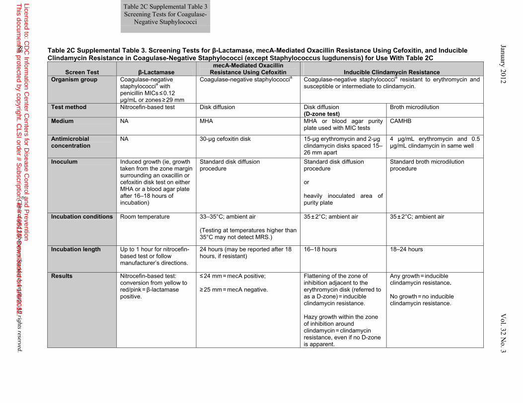
Table 2C Supplemental Table 3. Screening Tests for β-Lactamase, mecA-Mediated Oxacillin Resistance Using Cefoxitin, and Inducible Clindamycin Resistance in Coagulase-Negative Staphylococci (except Staphylococcus lugdunensis) for Use With Table 2C
Screen Test β-Lactamase mecA-Mediated Oxacillin
Resistance Using Cefoxitin Inducible Clindamycin Resistance Organism group Coagulase-negative
staphylococcia with penicillin MICs ≤ 0.12 µg/mL or zones ≥ 29 mm
Coagulase-negative staphylococcia Coagulase-negative staphylococcia resistant to erythromycin and susceptible or intermediate to clindamycin.
Test method Nitrocefin-based test Disk diffusion Disk diffusion (D-zone test)
Broth microdilution
Medium NA MHA MHA or blood agar purity plate used with MIC tests
CAMHB
Antimicrobial concentration
NA 30-µg cefoxitin disk 15-µg erythromycin and 2-µg clindamycin disks spaced 15–26 mm apart
4 µg/mL erythromycin and 0.5 µg/mL clindamycin in same well
Inoculum Induced growth (ie, growth taken from the zone margin surrounding an oxacillin or cefoxitin disk test on either MHA or a blood agar plate after 16–18 hours of incubation)
Standard disk diffusion procedure
Standard disk diffusion procedure or heavily inoculated area of purity plate
Standard broth microdilution procedure
Incubation conditions
Room temperature
33–35°C; ambient air (Testing at temperatures higher than 35°C may not detect MRS.)
35 ± 2°C; ambient air
35 ± 2°C; ambient air
Incubation length Up to 1 hour for nitrocefin-based test or follow manufacturer’s directions.
24 hours (may be reported after 18 hours, if resistant)
16–18 hours 18–24 hours
Results Nitrocefin-based test: conversion from yellow to red/pink = β-lactamase positive.
≤ 24 mm = mecA positive; ≥ 25 mm = mecA negative.
Flattening of the zone of inhibition adjacent to the erythromycin disk (referred to as a D-zone) = inducible clindamycin resistance. Hazy growth within the zone of inhibition around clindamycin = clindamycin resistance, even if no D-zone is apparent.
Any growth = inducible clindamycin resistance. No growth = no inducible clindamycin resistance.
Table 2C Supplemental Table 3 Screening Tests for Coagulase-
Negative Staphylococci January 2012 V
ol. 32 No. 3
88 ©C
linical and Laboratory Standards Institute. A
ll rights reserved. Licensed to: C
DC
Information C
enter Centers for D
isease Control and P
reventionT
his document is protected by copyright. C
LSI order # S
ubscription, id # 465125, Dow
nloaded on 1/6/2012.
Table 2C Supplemental Table 3. (Continued)
Screen Test β-Lactamase mecA-Mediated Oxacillin Resistance
Using Cefoxitin Inducible Clindamycin Resistance Test method Nitrocefin-based test Disk diffusion Disk diffusion
(D-zone test) Broth microdilution
Further testing and reporting
β-Lactamase–positive staphylococci are resistant to penicillin, amino-, carboxy-, and ureidopenicillins.
Cefoxitin is used as a surrogate for mecA-mediated oxacillin resistance. Isolates that test as mecA positive should be reported as oxacillin (not cefoxitin) resistant; other β-lactam agents should be reported as resistant or should not be reported.
Report isolates with inducible clindamycin resistance as “clindamycin resistant.” A comment that “This isolate is presumed to be resistant based on detection of inducible clindamycin resistance. Clindamycin may still be effective in some patients” may be included.
QC recommendations
S. aureus ATCC® 29213 – positive S. aureus ATCC® 25923 – negative (or see manufacturer’s recommendations)
S. aureus ATCC® 25923 – mecA negative (zone 23–29 mm) S. aureus ATCC® 43300 – mecA positive (zone ≤ 21 mm)
S. aureus ATCC® 25923 for routine QC of disks; see Table 3A for use of supplemental QC strains.
S. aureus ATCC® BAA-976 or S. aureus ATCC® 29213 – no growth S. aureus ATCC® BAA-977 – growth
Abbreviations: ATCC, American Type Culture Collection; CAMHB, cation-adjusted Mueller-Hinton broth; MHA, Mueller-Hinton agar; MIC, minimal inhibitory concentration; MRS, methicillin-resistant staphylococci; QC, quality control.
Footnote
a. Except S. lugdunensis, which is included in the S. aureus group. See Table 2C Supplemental Table 1.
©C
linical and Laboratory Standards Institute. A
ll rights reserved. 89
Table 2C Supplemental Table 3 Screening Tests for Coagulase-
Negative Staphylococci
For U
se With M
02-A11 and M
07-A9 M
100-S22
Licensed to: CD
C Inform
ation Center C
enters for Disease C
ontrol and Prevention
This docum
ent is protected by copyright. CLS
I order # Subscription, id # 465125, D
ownloaded on 1/6/2012.
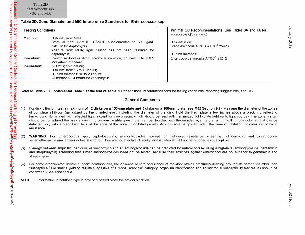
Table 2D. Zone Diameter and MIC Interpretive Standards for Enterococcus spp.
Refer to Table 2D Supplemental Table 1 at the end of Table 2D for additional recommendations for testing conditions, reporting suggestions, and QC.
General Comments (1) For disk diffusion, test a maximum of 12 disks on a 150-mm plate and 5 disks on a 100-mm plate (see M02 Section 9.2). Measure the diameter of the zones
of complete inhibition (as judged by the unaided eye), including the diameter of the disk. Hold the Petri plate a few inches above a black, nonreflecting background illuminated with reflected light, except for vancomycin, which should be read with transmitted light (plate held up to light source). The zone margin should be considered the area showing no obvious, visible growth that can be detected with the unaided eye. Ignore faint growth of tiny colonies that can be detected only with a magnifying lens at the edge of the zone of inhibited growth. Any discernable growth within the zone of inhibition indicates vancomycin resistance.
(2) WARNING: For Enterococcus spp., cephalosporins, aminoglycosides (except for high-level resistance screening), clindamycin, and trimethoprim-
sulfamethoxazole may appear active in vitro, but they are not effective clinically, and isolates should not be reported as susceptible. (3) Synergy between ampicillin, penicillin, or vancomycin and an aminoglycoside can be predicted for enterococci by using a high-level aminoglycoside (gentamicin
and streptomycin) screening test. Other aminoglycosides need not be tested, because their activities against enterococci are not superior to gentamicin and streptomycin.
(4) For some organism/antimicrobial agent combinations, the absence or rare occurrence of resistant strains precludes defining any results categories other than
“susceptible.” For strains yielding results suggestive of a “nonsusceptible” category, organism identification and antimicrobial susceptibility test results should be confirmed. (See Appendix A.)
NOTE: Information in boldface type is new or modified since the previous edition.
Testing Conditions Medium: Disk diffusion: MHA Broth dilution: CAMHB; CAMHB supplemented to 50 µg/mL
calcium for daptomycin Agar dilution: MHA; agar dilution has not been validated for
daptomycin Inoculum: Growth method or direct colony suspension, equivalent to a 0.5
McFarland standard Incubation: 35 ± 2°C; ambient air;
Disk diffusion: 16 to 18 hours; Dilution methods: 16 to 20 hours; All methods: 24 hours for vancomycin
Minimal QC Recommendations (See Tables 3A and 4A for acceptable QC ranges.) Disk diffusion: Staphylococcus aureus ATCC® 25923 Dilution methods: Enterococcus faecalis ATCC® 29212
Table 2D
Enterococcus spp. M02 and M07
90 ©C
linical and Laboratory Standards Institute. A
ll rights reserved.
January 2012 V
ol. 32 No. 3
Licensed to: CD
C Inform
ation Center C
enters for Disease C
ontrol and Prevention
This docum
ent is protected by copyright. CLS
I order # Subscription, id # 465125, D
ownloaded on 1/6/2012.
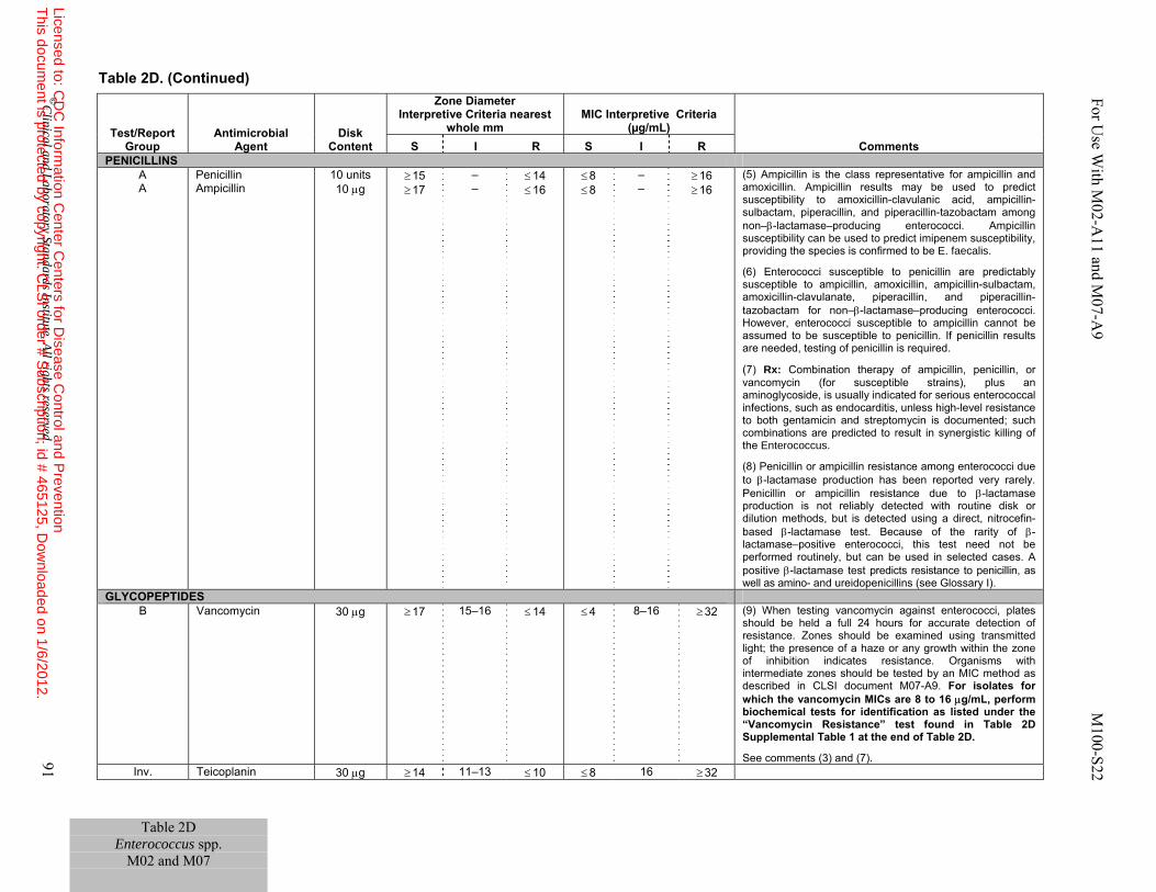
Table 2D Enterococcus spp.
M02 and M07
©C
linical and Laboratory Standards Institute. A
ll rights reserved. 91
Table 2D. (Continued)
Test/Report Group
Antimicrobial Agent
Disk Content
Zone Diameter Interpretive Criteria nearest
whole mm MIC Interpretive Criteria
(µg/mL) Comments S I R S I R
PENICILLINS A A
Penicillin Ampicillin
10 units 10 μg
≥ 15 ≥ 17
– –
≤ 14 ≤ 16
≤ 8 ≤ 8
– –
≥ 16 ≥ 16
(5) Ampicillin is the class representative for ampicillin and amoxicillin. Ampicillin results may be used to predict susceptibility to amoxicillin-clavulanic acid, ampicillin-sulbactam, piperacillin, and piperacillin-tazobactam among non–β-lactamase–producing enterococci. Ampicillin susceptibility can be used to predict imipenem susceptibility, providing the species is confirmed to be E. faecalis. (6) Enterococci susceptible to penicillin are predictably susceptible to ampicillin, amoxicillin, ampicillin-sulbactam, amoxicillin-clavulanate, piperacillin, and piperacillin-tazobactam for non–β-lactamase–producing enterococci. However, enterococci susceptible to ampicillin cannot be assumed to be susceptible to penicillin. If penicillin results are needed, testing of penicillin is required.
(7) Rx: Combination therapy of ampicillin, penicillin, or vancomycin (for susceptible strains), plus an aminoglycoside, is usually indicated for serious enterococcal infections, such as endocarditis, unless high-level resistance to both gentamicin and streptomycin is documented; such combinations are predicted to result in synergistic killing of the Enterococcus. (8) Penicillin or ampicillin resistance among enterococci due to β-lactamase production has been reported very rarely. Penicillin or ampicillin resistance due to β-lactamase production is not reliably detected with routine disk or dilution methods, but is detected using a direct, nitrocefin-based β-lactamase test. Because of the rarity of β-lactamase–positive enterococci, this test need not be performed routinely, but can be used in selected cases. A positive β-lactamase test predicts resistance to penicillin, as well as amino- and ureidopenicillins (see Glossary I).
GLYCOPEPTIDES B Vancomycin 30 μg ≥ 17 15–16 ≤ 14 ≤ 4 8–16 ≥ 32 (9) When testing vancomycin against enterococci, plates
should be held a full 24 hours for accurate detection of resistance. Zones should be examined using transmitted light; the presence of a haze or any growth within the zone of inhibition indicates resistance. Organisms with intermediate zones should be tested by an MIC method as described in CLSI document M07-A9. For isolates for which the vancomycin MICs are 8 to 16 μg/mL, perform biochemical tests for identification as listed under the “Vancomycin Resistance” test found in Table 2D Supplemental Table 1 at the end of Table 2D. See comments (3) and (7).
Inv. Teicoplanin 30 μg ≥ 14 11–13 ≤ 10 ≤ 8 16 ≥ 32
For U
se With M
02-A11 and M
07-A9 M
100-S22
Licensed to: CD
C Inform
ation Center C
enters for Disease C
ontrol and Prevention
This docum
ent is protected by copyright. CLS
I order # Subscription, id # 465125, D
ownloaded on 1/6/2012.
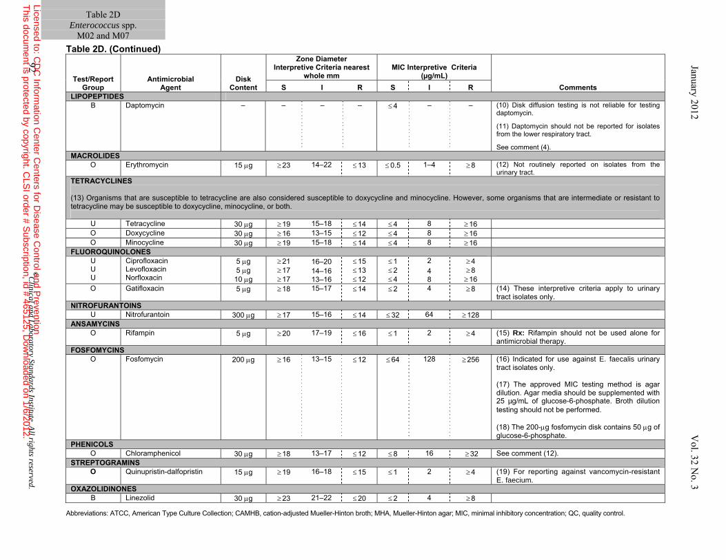
Table 2D
Enterococcus spp. M02 and M07
Table 2D. (Continued)
Abbreviations: ATCC, American Type Culture Collection; CAMHB, cation-adjusted Mueller-Hinton broth; MHA, Mueller-Hinton agar; MIC, minimal inhibitory concentration; QC, quality control.
Test/Report Group
Antimicrobial Agent
Disk Content
Zone Diameter Interpretive Criteria nearest
whole mm MIC Interpretive Criteria
(µg/mL) Comments S I R S I R
LIPOPEPTIDES B
Daptomycin
– – – – ≤ 4 – – (10) Disk diffusion testing is not reliable for testing daptomycin. (11) Daptomycin should not be reported for isolates from the lower respiratory tract. See comment (4).
MACROLIDES O Erythromycin 15 μg ≥ 23 14–22 ≤ 13 ≤ 0.5 1–4 ≥ 8 (12) Not routinely reported on isolates from the
urinary tract. TETRACYCLINES (13) Organisms that are susceptible to tetracycline are also considered susceptible to doxycycline and minocycline. However, some organisms that are intermediate or resistant to tetracycline may be susceptible to doxycycline, minocycline, or both.
U Tetracycline 30 μg ≥ 19 15–18 ≤ 14 ≤ 4 8 ≥ 16 O Doxycycline 30 μg ≥ 16 13–15 ≤ 12 ≤ 4 8 ≥ 16 O Minocycline 30 μg ≥ 19 15–18 ≤ 14 ≤ 4 8 ≥ 16
FLUOROQUINOLONES U U U
Ciprofloxacin Levofloxacin Norfloxacin
5 μg 5 μg
10 μg
≥ 21 ≥ 17 ≥ 17
16–20 14–16 13–16
≤ 15 ≤ 13 ≤ 12
≤ 1 ≤ 2 ≤ 4
2 4 8
≥ 4 ≥ 8 ≥ 16
O Gatifloxacin 5 μg ≥ 18 15–17 ≤ 14 ≤ 2 4 ≥ 8 (14) These interpretive criteria apply to urinary tract isolates only.
NITROFURANTOINS U Nitrofurantoin 300 μg ≥ 17 15–16 ≤ 14 ≤ 32 64 ≥ 128
ANSAMYCINS O Rifampin 5 μg ≥ 20 17–19 ≤ 16 ≤ 1 2 ≥ 4 (15) Rx: Rifampin should not be used alone for
antimicrobial therapy. FOSFOMYCINS
O Fosfomycin 200 μg ≥ 16 13–15 ≤ 12 ≤ 64 128 ≥ 256 (16) Indicated for use against E. faecalis urinary tract isolates only. (17) The approved MIC testing method is agar dilution. Agar media should be supplemented with 25 µg/mL of glucose-6-phosphate. Broth dilution testing should not be performed. (18) The 200-μg fosfomycin disk contains 50 μg of glucose-6-phosphate.
PHENICOLS O Chloramphenicol 30 μg ≥ 18 13–17 ≤ 12 ≤ 8 16 ≥ 32 See comment (12).
STREPTOGRAMINS O Quinupristin-dalfopristin 15 μg ≥ 19 16–18 ≤ 15 ≤ 1 2 ≥ 4 (19) For reporting against vancomycin-resistant
E. faecium. OXAZOLIDINONES
B Linezolid 30 μg ≥ 23 21–22 ≤ 20 ≤ 2 4 ≥ 8
92 ©C
linical and Laboratory Standards Institute. A
ll rights reserved.
January 2012 V
ol. 32 No. 3
Licensed to: CD
C Inform
ation Center C
enters for Disease C
ontrol and Prevention
This docum
ent is protected by copyright. CLS
I order # Subscription, id # 465125, D
ownloaded on 1/6/2012.

For Use With M02-A11 and M07-A9 M100-S22
©Clinical and Laboratory Standards Institute. All rights reserved. 93
This page is intentionally left blank.
Licensed to: CDC Information Center Centers for Disease Control and PreventionThis document is protected by copyright. CLSI order # Subscription, id # 465125, Downloaded on 1/6/2012.
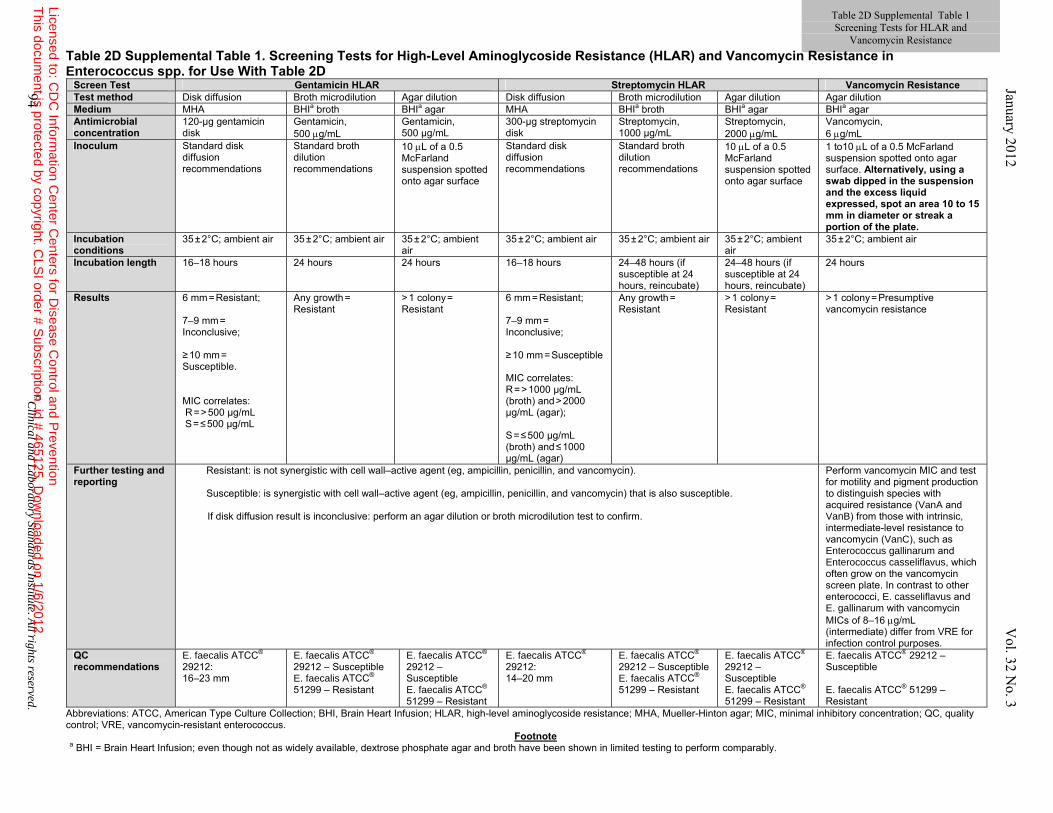
Table 2D Supplemental Table 1 Screening Tests for HLAR and
Vancomycin Resistance January 2012 V
ol. 32 No. 3
Table 2D Supplemental Table 1. Screening Tests for High-Level Aminoglycoside Resistance (HLAR) and Vancomycin Resistance in Enterococcus spp. for Use With Table 2D
Screen Test Gentamicin HLAR Streptomycin HLAR Vancomycin ResistanceTest method Disk diffusion Broth microdilution Agar dilution Disk diffusion Broth microdilution Agar dilution Agar dilution Medium MHA BHIa broth BHIa agar MHA BHIa broth BHIa agar BHIa agar Antimicrobial concentration
120-µg gentamicin disk
Gentamicin, 500 μg/mL
Gentamicin, 500 µg/mL
300-µg streptomycin disk
Streptomycin, 1000 µg/mL
Streptomycin, 2000 μg/mL
Vancomycin, 6 μg/mL
Inoculum Standard disk diffusion recommendations
Standard broth dilution recommendations
10 μL of a 0.5 McFarland suspension spotted onto agar surface
Standard disk diffusion recommendations
Standard broth dilution recommendations
10 μL of a 0.5 McFarland suspension spotted onto agar surface
1 to10 μL of a 0.5 McFarland suspension spotted onto agar surface. Alternatively, using a swab dipped in the suspension and the excess liquid expressed, spot an area 10 to 15 mm in diameter or streak a portion of the plate.
Incubation conditions
35 ± 2°C; ambient air 35 ± 2°C; ambient air 35 ± 2°C; ambient air
35 ± 2°C; ambient air 35 ± 2°C; ambient air 35 ± 2°C; ambient air
35 ± 2°C; ambient air
Incubation length 16–18 hours 24 hours 24 hours 16–18 hours 24–48 hours (if susceptible at 24 hours, reincubate)
24–48 hours (if susceptible at 24 hours, reincubate)
24 hours
Results 6 mm = Resistant; 7–9 mm =
Inconclusive; ≥ 10 mm =
Susceptible. MIC correlates: R = > 500 µg/mL S = ≤ 500 µg/mL
Any growth =
Resistant
> 1 colony = Resistant
6 mm = Resistant; 7–9 mm =
Inconclusive; ≥ 10 mm = Susceptible MIC correlates: R = > 1000 µg/mL (broth) and > 2000 µg/mL (agar); S = ≤ 500 µg/mL (broth) and ≤ 1000 µg/mL (agar)
Any growth =
Resistant
> 1 colony =
Resistant
> 1 colony = Presumptive vancomycin resistance
Further testing and reporting
Resistant: is not synergistic with cell wall–active agent (eg, ampicillin, penicillin, and vancomycin). Susceptible: is synergistic with cell wall–active agent (eg, ampicillin, penicillin, and vancomycin) that is also susceptible.
If disk diffusion result is inconclusive: perform an agar dilution or broth microdilution test to confirm.
Perform vancomycin MIC and test for motility and pigment production to distinguish species with acquired resistance (VanA and VanB) from those with intrinsic, intermediate-level resistance to vancomycin (VanC), such as Enterococcus gallinarum and Enterococcus casseliflavus, which often grow on the vancomycin screen plate. In contrast to other enterococci, E. casseliflavus and E. gallinarum with vancomycin MICs of 8–16 μg/mL (intermediate) differ from VRE for infection control purposes.
QC recommendations
E. faecalis ATCC® 29212: 16–23 mm
E. faecalis ATCC® 29212 – Susceptible E. faecalis ATCC® 51299 – Resistant
E. faecalis ATCC® 29212 – Susceptible E. faecalis ATCC® 51299 – Resistant
E. faecalis ATCC® 29212: 14–20 mm
E. faecalis ATCC® 29212 – Susceptible E. faecalis ATCC® 51299 – Resistant
E. faecalis ATCC® 29212 – Susceptible E. faecalis ATCC® 51299 – Resistant
E. faecalis ATCC® 29212 – Susceptible E. faecalis ATCC® 51299 – Resistant
Abbreviations: ATCC, American Type Culture Collection; BHI, Brain Heart Infusion; HLAR, high-level aminoglycoside resistance; MHA, Mueller-Hinton agar; MIC, minimal inhibitory concentration; QC, quality control; VRE, vancomycin-resistant enterococcus.
Footnote a BHI = Brain Heart Infusion; even though not as widely available, dextrose phosphate agar and broth have been shown in limited testing to perform comparably.
94 ©
Clinical and L
aboratory Standards Institute. All rights reserved.
Licensed to: CD
C Inform
ation Center C
enters for Disease C
ontrol and Prevention
This docum
ent is protected by copyright. CLS
I order # Subscription, id # 465125, D
ownloaded on 1/6/2012.

For Use With M02-A11 and M07-A9 M100-S22
©Clinical and Laboratory Standards Institute. All rights reserved. 95
This page is intentionally left blank.
Licensed to: CDC Information Center Centers for Disease Control and PreventionThis document is protected by copyright. CLSI order # Subscription, id # 465125, Downloaded on 1/6/2012.
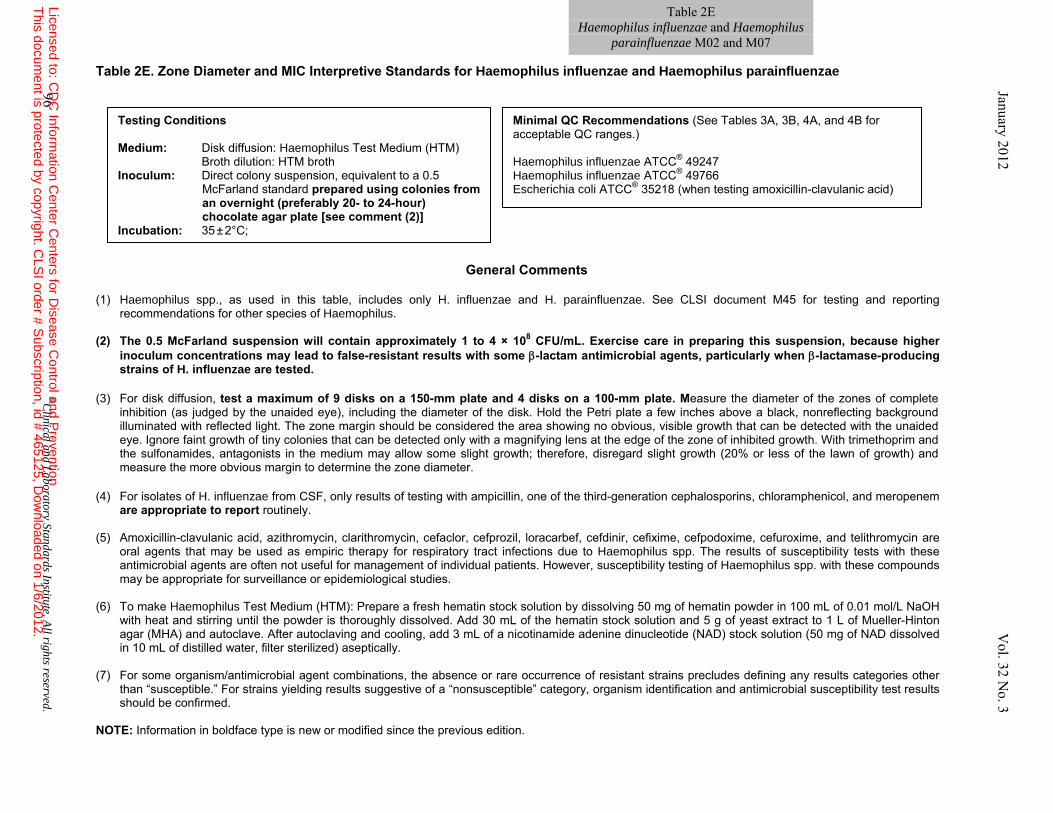
Table 2E Haemophilus influenzae and Haemophilus
parainfluenzae M02 and M07 Table 2E. Zone Diameter and MIC Interpretive Standards for Haemophilus influenzae and Haemophilus parainfluenzae
General Comments
(1) Haemophilus spp., as used in this table, includes only H. influenzae and H. parainfluenzae. See CLSI document M45 for testing and reporting recommendations for other species of Haemophilus.
(2) The 0.5 McFarland suspension will contain approximately 1 to 4 × 10⁸ CFU/mL. Exercise care in preparing this suspension, because higher
inoculum concentrations may lead to false-resistant results with some β-lactam antimicrobial agents, particularly when β-lactamase-producing strains of H. influenzae are tested.
(3) For disk diffusion, test a maximum of 9 disks on a 150-mm plate and 4 disks on a 100-mm plate. Measure the diameter of the zones of complete
inhibition (as judged by the unaided eye), including the diameter of the disk. Hold the Petri plate a few inches above a black, nonreflecting background illuminated with reflected light. The zone margin should be considered the area showing no obvious, visible growth that can be detected with the unaided eye. Ignore faint growth of tiny colonies that can be detected only with a magnifying lens at the edge of the zone of inhibited growth. With trimethoprim and the sulfonamides, antagonists in the medium may allow some slight growth; therefore, disregard slight growth (20% or less of the lawn of growth) and measure the more obvious margin to determine the zone diameter.
(4) For isolates of H. influenzae from CSF, only results of testing with ampicillin, one of the third-generation cephalosporins, chloramphenicol, and meropenem
are appropriate to report routinely. (5) Amoxicillin-clavulanic acid, azithromycin, clarithromycin, cefaclor, cefprozil, loracarbef, cefdinir, cefixime, cefpodoxime, cefuroxime, and telithromycin are
oral agents that may be used as empiric therapy for respiratory tract infections due to Haemophilus spp. The results of susceptibility tests with these antimicrobial agents are often not useful for management of individual patients. However, susceptibility testing of Haemophilus spp. with these compounds may be appropriate for surveillance or epidemiological studies.
(6) To make Haemophilus Test Medium (HTM): Prepare a fresh hematin stock solution by dissolving 50 mg of hematin powder in 100 mL of 0.01 mol/L NaOH
with heat and stirring until the powder is thoroughly dissolved. Add 30 mL of the hematin stock solution and 5 g of yeast extract to 1 L of Mueller-Hinton agar (MHA) and autoclave. After autoclaving and cooling, add 3 mL of a nicotinamide adenine dinucleotide (NAD) stock solution (50 mg of NAD dissolved in 10 mL of distilled water, filter sterilized) aseptically.
(7) For some organism/antimicrobial agent combinations, the absence or rare occurrence of resistant strains precludes defining any results categories other
than “susceptible.” For strains yielding results suggestive of a “nonsusceptible” category, organism identification and antimicrobial susceptibility test results should be confirmed.
NOTE: Information in boldface type is new or modified since the previous edition.
Testing Conditions Medium: Disk diffusion: Haemophilus Test Medium (HTM) Broth dilution: HTM broth Inoculum: Direct colony suspension, equivalent to a 0.5
McFarland standard prepared using colonies from an overnight (preferably 20- to 24-hour) chocolate agar plate [see comment (2)]
Incubation: 35 ± 2°C;
Minimal QC Recommendations (See Tables 3A, 3B, 4A, and 4B for acceptable QC ranges.) Haemophilus influenzae ATCC® 49247 Haemophilus influenzae ATCC® 49766 Escherichia coli ATCC® 35218 (when testing amoxicillin-clavulanic acid)
96 ©C
linical and Laboratory Standards Institute. A
ll rights reserved.
January 2012 V
ol. 32 No. 3
Licensed to: CD
C Inform
ation Center C
enters for Disease C
ontrol and Prevention
This docum
ent is protected by copyright. CLS
I order # Subscription, id # 465125, D
ownloaded on 1/6/2012.
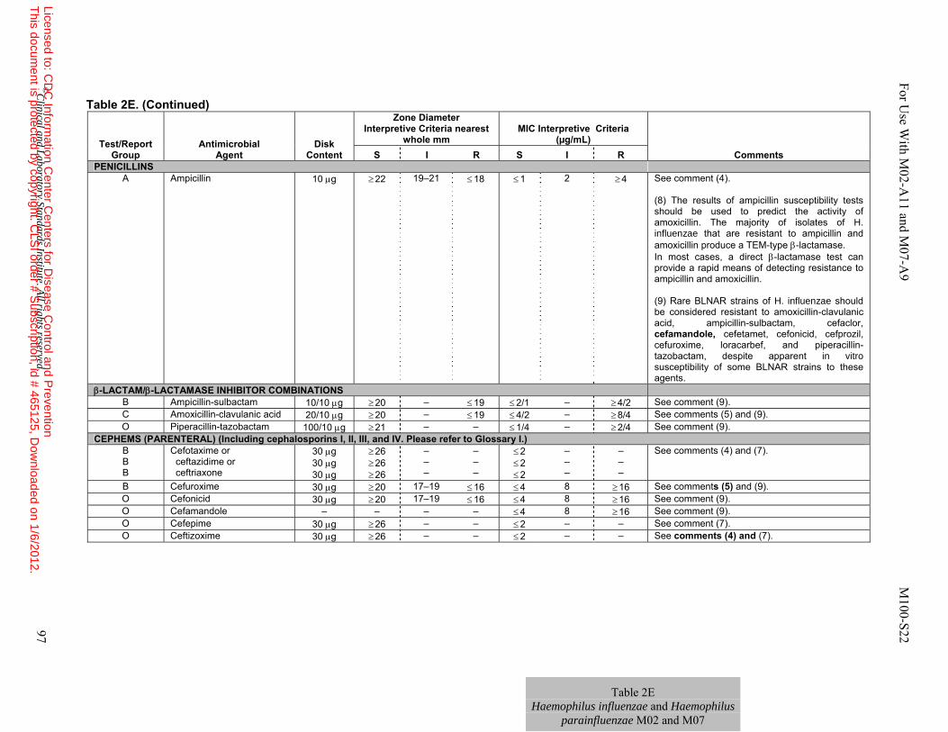
Table 2E Haemophilus influenzae and Haemophilus
parainfluenzae M02 and M07
Table 2E. (Continued)
Test/Report
Group Antimicrobial
Agent Disk
Content
Zone Diameter Interpretive Criteria nearest
whole mm MIC Interpretive Criteria
(µg/mL) Comments S I R S I R
PENICILLINS A Ampicillin 10 μg ≥ 22 19–21 ≤ 18 ≤ 1 2 ≥ 4 See comment (4).
(8) The results of ampicillin susceptibility tests should be used to predict the activity of amoxicillin. The majority of isolates of H. influenzae that are resistant to ampicillin and amoxicillin produce a TEM-type β-lactamase. In most cases, a direct β-lactamase test can provide a rapid means of detecting resistance to ampicillin and amoxicillin. (9) Rare BLNAR strains of H. influenzae should be considered resistant to amoxicillin-clavulanic acid, ampicillin-sulbactam, cefaclor, cefamandole, cefetamet, cefonicid, cefprozil, cefuroxime, loracarbef, and piperacillin-tazobactam, despite apparent in vitro susceptibility of some BLNAR strains to these agents.
β-LACTAM/β-LACTAMASE INHIBITOR COMBINATIONS B Ampicillin-sulbactam 10/10 μg ≥ 20 – ≤ 19 ≤ 2/1 – ≥ 4/2 See comment (9). C Amoxicillin-clavulanic acid 20/10 μg ≥ 20 – ≤ 19 ≤ 4/2 – ≥ 8/4 See comments (5) and (9). O Piperacillin-tazobactam 100/10 μg ≥ 21 – – ≤ 1/4 – ≥ 2/4 See comment (9).
CEPHEMS (PARENTERAL) (Including cephalosporins I, II, III, and IV. Please refer to Glossary I.) B B B
Cefotaxime or ceftazidime or ceftriaxone
30 μg 30 μg 30 μg
≥ 26 ≥ 26 ≥ 26
– – –
– – –
≤ 2 ≤ 2 ≤ 2
– – –
– – –
See comments (4) and (7).
B Cefuroxime 30 μg ≥ 20 17–19 ≤ 16 ≤ 4 8 ≥ 16 See comments (5) and (9). O Cefonicid 30 μg ≥ 20 17–19 ≤ 16 ≤ 4 8 ≥ 16 See comment (9). O Cefamandole – – – – ≤ 4 8 ≥ 16 See comment (9). O Cefepime 30 μg ≥ 26 – – ≤ 2 – – See comment (7). O Ceftizoxime 30 μg ≥ 26 – – ≤ 2 – – See comments (4) and (7).
©C
linical and Laboratory Standards Institute. A
ll rights reserved. 97
For U
se With M
02-A11 and M
07-A9 M
100-S22
Licensed to: CD
C Inform
ation Center C
enters for Disease C
ontrol and Prevention
This docum
ent is protected by copyright. CLS
I order # Subscription, id # 465125, D
ownloaded on 1/6/2012.
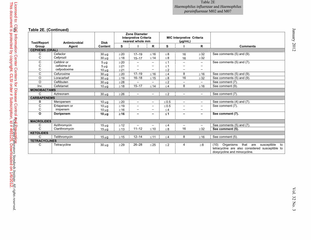
Table 2EHaemophilus influenzae and Haemophilus
parainfluenzae M02 and M07
Table 2E. (Continued)
Test/Report Group
Antimicrobial Agent
Disk Content
Zone Diameter Interpretive Criteria nearest whole mm
MIC Interpretive Criteria (µg/mL)
Comments S I R S I R CEPHEMS (ORAL)
C C
Cefaclor Cefprozil
30 μg 30 μg
≥ 20 ≥ 18
17–19 15–17
≤ 16 ≤ 14
≤ 8 ≤ 8
16 16
≥ 32 ≥ 32
See comments (5) and (9).
C C C
Cefdinir or cefixime or cefpodoxime
5 μg 5 μg
10 μg
≥ 20 ≥ 21 ≥ 21
– – –
– – –
≤ 1 ≤ 1 ≤ 2
– – –
– – –
See comments (5) and (7).
C Cefuroxime 30 μg ≥ 20 17–19 ≤ 16 ≤ 4 8 ≥ 16 See comments (5) and (9). O Loracarbef 30 μg ≥ 19 16–18 ≤ 15 ≤ 8 16 ≥ 32 See comments (5) and (9). O Ceftibuten 30 μg ≥ 28 – – ≤ 2 – – See comment (7).
Inv. Cefetamet 10 μg ≥ 18 15–17 ≤ 14 ≤ 4 8 ≥ 16 See comment (9). MONOBACTAMS
C Aztreonam 30 μg ≥ 26 – – ≤ 2 – – See comment (7). CARBAPENEMS
B Meropenem 10 μg ≥ 20 – – ≤ 0.5 – – See comments (4) and (7). C C
Ertapenem or imipenem
10 μg 10 μg
≥ 19 ≥ 16
– –
– –
≤ 0.5 ≤ 4
– –
– –
See comment (7).
O Doripenem 10 μg ≥ 16 – – ≤ 1
– – See comment (7).
MACROLIDES C C
Azithromycin Clarithromycin
15 μg 15 μg
≥ 12 ≥ 13
– 11–12
– ≤ 10
≤ 4 ≤ 8
– 16
– ≥ 32
See comments (5) and (7). See comment (5).
KETOLIDES C Telithromycin 15 μg ≥ 15 12–14 ≤ 11 ≤ 4 8 ≥ 16 See comment (5).
TETRACYCLINES C Tetracycline 30 μg ≥ 29 26–28 ≤ 25 ≤ 2 4 ≥ 8 (10) Organisms that are susceptible to
tetracycline are also considered susceptible to doxycycline and minocycline.
98 ©C
linical and Laboratory Standards Institute. A
ll rights reserved.
January 2012 V
ol. 32 No. 3
Licensed to: CD
C Inform
ation Center C
enters for Disease C
ontrol and Prevention
This docum
ent is protected by copyright. CLS
I order # Subscription, id # 465125, D
ownloaded on 1/6/2012.
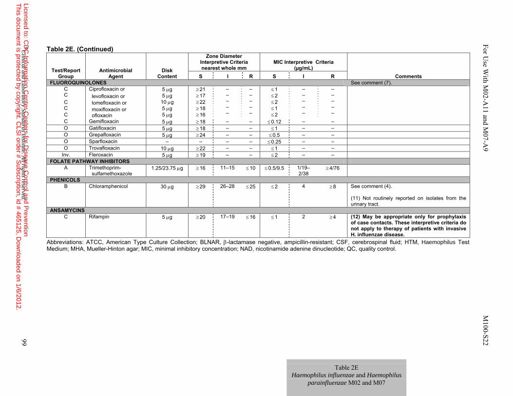
Table 2E Haemophilus influenzae and Haemophilus
parainfluenzae M02 and M07
Table 2E. (Continued)
Abbreviations: ATCC, American Type Culture Collection; BLNAR, β-lactamase negative, ampicillin-resistant; CSF, cerebrospinal fluid; HTM, Haemophilus Test Medium; MHA, Mueller-Hinton agar; MIC, minimal inhibitory concentration; NAD, nicotinamide adenine dinucleotide; QC, quality control.
Test/Report Group
Antimicrobial Agent
Disk Content
Zone Diameter Interpretive Criteria nearest whole mm
MIC Interpretive Criteria (µg/mL)
Comments S I R S I R FLUOROQUINOLONES See comment (7).
C C C C C
Ciprofloxacin or levofloxacin or lomefloxacin or moxifloxacin or ofloxacin
5 μg 5 μg
10 μg 5 μg 5 μg
≥ 21 ≥ 17
≥ 22 ≥ 18 ≥ 16
– – – – –
– – – – –
≤ 1 ≤ 2 ≤ 2 ≤ 1 ≤ 2
– – – – –
– – – – –
C Gemifloxacin 5 μg ≥ 18 – – ≤ 0.12 – – O Gatifloxacin 5 μg ≥ 18 – – ≤ 1 – – O Grepafloxacin 5 μg ≥ 24 – – ≤ 0.5 – – O Sparfloxacin – – – – ≤ 0.25 – – O Trovafloxacin 10 μg ≥ 22 – – ≤ 1 – –
Inv. Fleroxacin 5 μg ≥ 19 – – ≤ 2 – – FOLATE PATHWAY INHIBITORS
A Trimethoprim- sulfamethoxazole
1.25/23.75 μg ≥ 16 11–15 ≤ 10 ≤ 0.5/9.5 1/19–2/38
≥ 4/76
PHENICOLS B Chloramphenicol 30 μg ≥ 29 26–28 ≤ 25 ≤ 2 4 ≥ 8 See comment (4).
(11) Not routinely reported on isolates from the urinary tract.
ANSAMYCINS C Rifampin 5 μg ≥ 20 17–19 ≤ 16 ≤ 1 2 ≥ 4 (12) May be appropriate only for prophylaxis
of case contacts. These interpretive criteria do not apply to therapy of patients with invasive H. influenzae disease.
©C
linical and Laboratory Standards Institute. A
ll rights reserved. 99
For U
se With M
02-A11 and M
07-A9 M
100-S22
Licensed to: CD
C Inform
ation Center C
enters for Disease C
ontrol and Prevention
This docum
ent is protected by copyright. CLS
I order # Subscription, id # 465125, D
ownloaded on 1/6/2012.
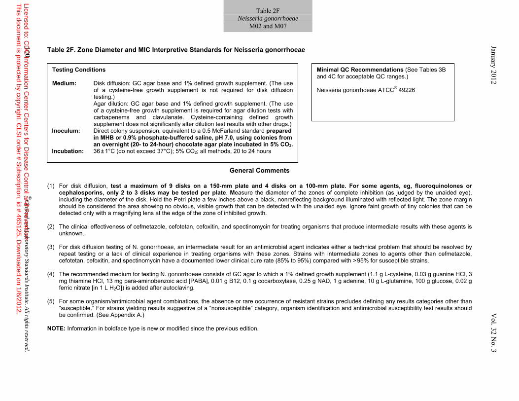
Table 2F. Zone Diameter and MIC Interpretive Standards for Neisseria gonorrhoeae
General Comments (1) For disk diffusion, test a maximum of 9 disks on a 150-mm plate and 4 disks on a 100-mm plate. For some agents, eg, fluoroquinolones or
cephalosporins, only 2 to 3 disks may be tested per plate. Measure the diameter of the zones of complete inhibition (as judged by the unaided eye), including the diameter of the disk. Hold the Petri plate a few inches above a black, nonreflecting background illuminated with reflected light. The zone margin should be considered the area showing no obvious, visible growth that can be detected with the unaided eye. Ignore faint growth of tiny colonies that can be detected only with a magnifying lens at the edge of the zone of inhibited growth.
(2) The clinical effectiveness of cefmetazole, cefotetan, cefoxitin, and spectinomycin for treating organisms that produce intermediate results with these agents is
unknown. (3) For disk diffusion testing of N. gonorrhoeae, an intermediate result for an antimicrobial agent indicates either a technical problem that should be resolved by
repeat testing or a lack of clinical experience in treating organisms with these zones. Strains with intermediate zones to agents other than cefmetazole, cefotetan, cefoxitin, and spectinomycin have a documented lower clinical cure rate (85% to 95%) compared with > 95% for susceptible strains.
(4) The recommended medium for testing N. gonorrhoeae consists of GC agar to which a 1% defined growth supplement (1.1 g L-cysteine, 0.03 g guanine HCl, 3
mg thiamine HCl, 13 mg para-aminobenzoic acid [PABA], 0.01 g B12, 0.1 g cocarboxylase, 0.25 g NAD, 1 g adenine, 10 g L-glutamine, 100 g glucose, 0.02 g ferric nitrate [in 1 L H2O]) is added after autoclaving.
(5) For some organism/antimicrobial agent combinations, the absence or rare occurrence of resistant strains precludes defining any results categories other than
“susceptible.” For strains yielding results suggestive of a “nonsusceptible” category, organism identification and antimicrobial susceptibility test results should be confirmed. (See Appendix A.)
NOTE: Information in boldface type is new or modified since the previous edition.
Testing Conditions Medium: Disk diffusion: GC agar base and 1% defined growth supplement. (The use
of a cysteine-free growth supplement is not required for disk diffusion testing.)
Agar dilution: GC agar base and 1% defined growth supplement. (The use of a cysteine-free growth supplement is required for agar dilution tests with carbapenems and clavulanate. Cysteine-containing defined growth supplement does not significantly alter dilution test results with other drugs.)
Inoculum: Direct colony suspension, equivalent to a 0.5 McFarland standard prepared in MHB or 0.9% phosphate-buffered saline, pH 7.0, using colonies from an overnight (20- to 24-hour) chocolate agar plate incubated in 5% CO2.
Incubation: 36 ± 1°C (do not exceed 37°C); 5% CO2; all methods, 20 to 24 hours
Minimal QC Recommendations (See Tables 3B and 4C for acceptable QC ranges.) Neisseria gonorrhoeae ATCC® 49226
100 ©C
linical and Laboratory Standards Institute. A
ll rights reserved.
Table 2F
Neisseria gonorrhoeae M02 and M07
January 2012 V
ol. 32 No. 3
Licensed to: CD
C Inform
ation Center C
enters for Disease C
ontrol and Prevention
This docum
ent is protected by copyright. CLS
I order # Subscription, id # 465125, D
ownloaded on 1/6/2012.
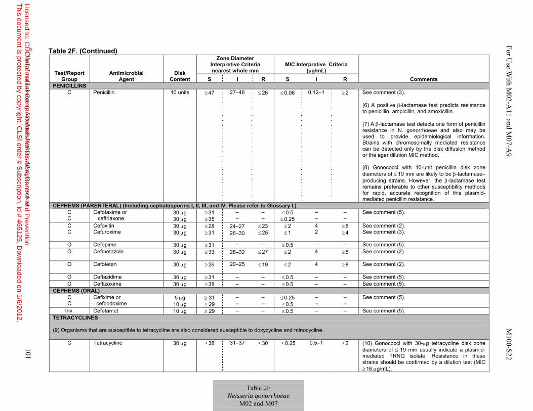
Table 2F. (Continued)
Test/Report Group
Antimicrobial Agent
Disk Content
Zone Diameter Interpretive Criteria nearest whole mm
MIC Interpretive Criteria (µg/mL)
Comments S I R S I R PENICILLINS
C Penicillin 10 units ≥ 47 27–46 ≤ 26 ≤ 0.06 0.12–1 ≥ 2 See comment (3). (6) A positive β-lactamase test predicts resistance to penicillin, ampicillin, and amoxicillin. (7) A β-lactamase test detects one form of penicillin resistance in N. gonorrhoeae and also may be used to provide epidemiological information. Strains with chromosomally mediated resistance can be detected only by the disk diffusion method or the agar dilution MIC method. (8) Gonococci with 10-unit penicillin disk zone diameters of ≤ 19 mm are likely to be β-lactamase–producing strains. However, the β-lactamase test remains preferable to other susceptibility methods for rapid, accurate recognition of this plasmid-mediated penicillin resistance.
CEPHEMS (PARENTERAL) (Including cephalosporins I, II, III, and IV. Please refer to Glossary I.) C C
Cefotaxime or ceftriaxone
30 μg 30 μg
≥ 31 ≥ 35
– –
– –
≤ 0.5 ≤ 0.25
– –
– –
See comment (5).
C C
Cefoxitin Cefuroxime
30 μg 30 μg
≥ 28 ≥ 31
24–27 26–30
≤ 23 ≤ 25
≤ 2 ≤ 1
4 2
≥ 8 ≥ 4
See comment (2). See comment (3).
O Cefepime 30 μg ≥ 31 – – ≤ 0.5 – – See comment (5). O Cefmetazole 30 μg ≥ 33 28–32 ≤ 27 ≤ 2 4 ≥ 8 See comment (2).
O Cefotetan 30 μg ≥ 26 20–25 ≤ 19 ≤ 2 4 ≥ 8 See comment (2).
O Ceftazidime 30 μg ≥ 31 – – ≤ 0.5 – – See comment (5). O Ceftizoxime 30 μg ≥ 38 – – ≤ 0.5 – – See comment (5).
CEPHEMS (ORAL) C C
Cefixime or cefpodoxime
5 μg 10 μg
≥ 31 ≥ 29
– –
– –
≤ 0.25 ≤ 0.5
– –
– –
See comment (5).
Inv. Cefetamet 10 μg ≥ 29 – – ≤ 0.5 – – See comment (5). TETRACYCLINES (9) Organisms that are susceptible to tetracycline are also considered susceptible to doxycycline and minocycline.
C Tetracycline 30 μg ≥ 38 31–37 ≤ 30 ≤ 0.25
0.5–1 ≥ 2 (10) Gonococci with 30-μg tetracycline disk zone diameters of ≤ 19 mm usually indicate a plasmid-mediated TRNG isolate. Resistance in these strains should be confirmed by a dilution test (MIC ≥ 16 μg/mL).
©C
linical and Laboratory Standards Institute. A
ll rights reserved. 101
Table 2F Neisseria gonorrhoeae
M02 and M07
For U
se With M
02-A11 and M
07-A9 M
100-S22
Licensed to: CD
C Inform
ation Center C
enters for Disease C
ontrol and Prevention
This docum
ent is protected by copyright. CLS
I order # Subscription, id # 465125, D
ownloaded on 1/6/2012.
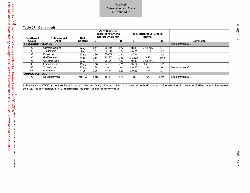
Table 2F. (Continued)
Abbreviations: ATCC, American Type Culture Collection; MIC, minimal inhibitory concentration; NAD, nicotinamide adenine dinucleotide; PABA, para-aminobenzoic acid; QC, quality control; TRNG, tetracycline-resistant Neisseria gonorrhoeae.
Test/Report Group
Antimicrobial Agent
Disk Content
Zone Diameter Interpretive Criteria nearest whole mm
MIC Interpretive Criteria (µg/mL)
Comments S I R S I R FLUOROQUINOLONES See comment (3).
C C
Ciprofloxacin or ofloxacin
5 μg 5 μg
≥ 41 ≥ 31
28–40 25–30
≤ 27 ≤ 24
≤ 0.06 ≤ 0.25
0.12–0.5 0.5–1
≥ 1 ≥ 2
O Enoxacin 10 μg ≥ 36 32–35 ≤ 31 ≤ 0.5 1 ≥ 2 O Gatifloxacin 5 μg ≥ 38 34–37 ≤ 33 ≤ 0.125 0.25 ≥ 0.5 O Grepafloxacin 5 μg ≥ 37 28–36 ≤ 27 ≤ 0.06 0.12–0.5 ≥ 1 O Lomefloxacin 10 μg ≥ 38 27–37 ≤ 26 ≤ 0.12 0.25–1 ≥ 2 O Trovafloxacin 10 μg ≥ 34 – – ≤ 0.25 – – See comment (5).
Inv. Fleroxacin 5 μg ≥ 35 29–34 ≤ 28 ≤ 0.25 0.5 ≥ 1 AMINOCYCLITOLS
C Spectinomycin 100 μg ≥ 18 15–17 ≤ 14 ≤ 32 64 ≥ 128 See comment (2).
102 ©C
linical and Laboratory Standards Institute. A
ll rights reserved.
Table 2F Neisseria gonorrhoeae
M02 and M07 January 2012 V
ol. 32 No. 3
Licensed to: CD
C Inform
ation Center C
enters for Disease C
ontrol and Prevention
This docum
ent is protected by copyright. CLS
I order # Subscription, id # 465125, D
ownloaded on 1/6/2012.

For Use With M02-A11 and M07-A9 M100-S22
©Clinical and Laboratory Standards Institute. All rights reserved. 103
This page is intentionally left blank.
Licensed to: CDC Information Center Centers for Disease Control and PreventionThis document is protected by copyright. CLSI order # Subscription, id # 465125, Downloaded on 1/6/2012.
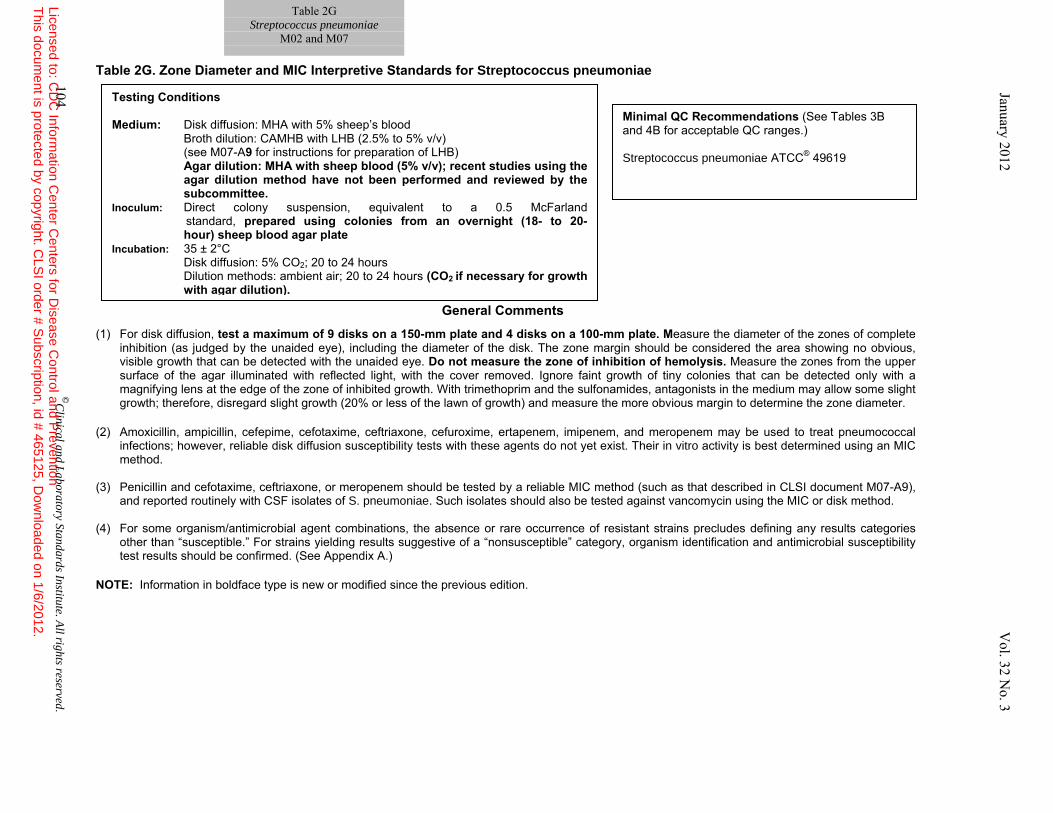
Table 2G. Zone Diameter and MIC Interpretive Standards for Streptococcus pneumoniae
General Comments (1) For disk diffusion, test a maximum of 9 disks on a 150-mm plate and 4 disks on a 100-mm plate. Measure the diameter of the zones of complete
inhibition (as judged by the unaided eye), including the diameter of the disk. The zone margin should be considered the area showing no obvious, visible growth that can be detected with the unaided eye. Do not measure the zone of inhibition of hemolysis. Measure the zones from the upper surface of the agar illuminated with reflected light, with the cover removed. Ignore faint growth of tiny colonies that can be detected only with a magnifying lens at the edge of the zone of inhibited growth. With trimethoprim and the sulfonamides, antagonists in the medium may allow some slight growth; therefore, disregard slight growth (20% or less of the lawn of growth) and measure the more obvious margin to determine the zone diameter.
(2) Amoxicillin, ampicillin, cefepime, cefotaxime, ceftriaxone, cefuroxime, ertapenem, imipenem, and meropenem may be used to treat pneumococcal
infections; however, reliable disk diffusion susceptibility tests with these agents do not yet exist. Their in vitro activity is best determined using an MIC method.
(3) Penicillin and cefotaxime, ceftriaxone, or meropenem should be tested by a reliable MIC method (such as that described in CLSI document M07-A9),
and reported routinely with CSF isolates of S. pneumoniae. Such isolates should also be tested against vancomycin using the MIC or disk method. (4) For some organism/antimicrobial agent combinations, the absence or rare occurrence of resistant strains precludes defining any results categories
other than “susceptible.” For strains yielding results suggestive of a “nonsusceptible” category, organism identification and antimicrobial susceptibility test results should be confirmed. (See Appendix A.)
NOTE: Information in boldface type is new or modified since the previous edition.
Testing Conditions
Medium: Disk diffusion: MHA with 5% sheep’s blood Broth dilution: CAMHB with LHB (2.5% to 5% v/v)
(see M07-A9 for instructions for preparation of LHB) Agar dilution: MHA with sheep blood (5% v/v); recent studies using the agar dilution method have not been performed and reviewed by the subcommittee.
Inoculum: Direct colony suspension, equivalent to a 0.5 McFarland standard, prepared using colonies from an overnight (18- to 20- hour) sheep blood agar plate
Incubation: 35 ± 2°C Disk diffusion: 5% CO2; 20 to 24 hours Dilution methods: ambient air; 20 to 24 hours (CO2 if necessary for growth
with agar dilution).
Minimal QC Recommendations (See Tables 3B and 4B for acceptable QC ranges.) Streptococcus pneumoniae ATCC® 49619
104 ©C
linical and Laboratory Standards Institute. A
ll rights reserved.
Table 2G
Streptococcus pneumoniae M02 and M07
January 2012 V
ol. 32 No. 3
Licensed to: CD
C Inform
ation Center C
enters for Disease C
ontrol and Prevention
This docum
ent is protected by copyright. CLS
I order # Subscription, id # 465125, D
ownloaded on 1/6/2012.
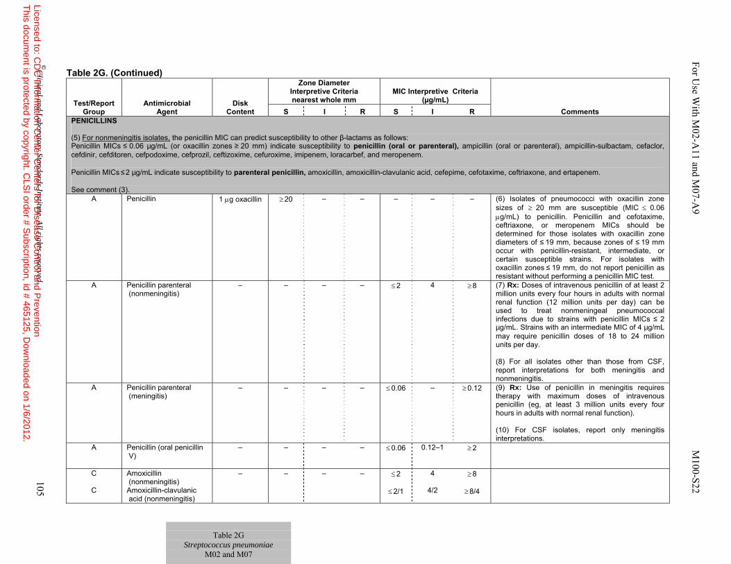
Table 2G. (Continued)
Test/Report Group
Antimicrobial Agent
Disk Content
Zone Diameter Interpretive Criteria nearest whole mm
MIC Interpretive Criteria (µg/mL)
Comments S I R S I R PENICILLINS (5) For nonmeningitis isolates, the penicillin MIC can predict susceptibility to other β-lactams as follows: Penicillin MICs ≤ 0.06 µg/mL (or oxacillin zones ≥ 20 mm) indicate susceptibility to penicillin (oral or parenteral), ampicillin (oral or parenteral), ampicillin-sulbactam, cefaclor, cefdinir, cefditoren, cefpodoxime, cefprozil, ceftizoxime, cefuroxime, imipenem, loracarbef, and meropenem. Penicillin MICs ≤ 2 µg/mL indicate susceptibility to parenteral penicillin, amoxicillin, amoxicillin-clavulanic acid, cefepime, cefotaxime, ceftriaxone, and ertapenem. See comment (3).
A Penicillin 1 μg oxacillin ≥ 20 – – – – – (6) Isolates of pneumococci with oxacillin zone sizes of ≥ 20 mm are susceptible (MIC ≤ 0.06 μg/mL) to penicillin. Penicillin and cefotaxime, ceftriaxone, or meropenem MICs should be determined for those isolates with oxacillin zone diameters of ≤ 19 mm, because zones of ≤ 19 mm occur with penicillin-resistant, intermediate, or certain susceptible strains. For isolates with oxacillin zones ≤ 19 mm, do not report penicillin as resistant without performing a penicillin MIC test.
A Penicillin parenteral (nonmeningitis)
– – – – ≤ 2 4 ≥ 8 (7) Rx: Doses of intravenous penicillin of at least 2 million units every four hours in adults with normal renal function (12 million units per day) can be used to treat nonmeningeal pneumococcal infections due to strains with penicillin MICs ≤ 2 µg/mL. Strains with an intermediate MIC of 4 µg/mL may require penicillin doses of 18 to 24 million units per day. (8) For all isolates other than those from CSF, report interpretations for both meningitis and nonmeningitis.
A Penicillin parenteral (meningitis)
– – – – ≤ 0.06 – ≥ 0.12 (9) Rx: Use of penicillin in meningitis requires therapy with maximum doses of intravenous penicillin (eg, at least 3 million units every four hours in adults with normal renal function). (10) For CSF isolates, report only meningitis interpretations.
A Penicillin (oral penicillin V)
– – – – ≤ 0.06 0.12–1 ≥ 2
C
C
Amoxicillin (nonmeningitis) Amoxicillin-clavulanic acid (nonmeningitis)
– – – – ≤ 2
≤ 2/1
4
4/2
≥ 8
≥ 8/4
©C
linical and Laboratory Standards Institute. A
ll rights reserved. 105
For U
se With M
02-A11 and M
07-A9 M
100-S22
Table 2G
Streptococcus pneumoniae M02 and M07
Licensed to: CD
C Inform
ation Center C
enters for Disease C
ontrol and Prevention
This docum
ent is protected by copyright. CLS
I order # Subscription, id # 465125, D
ownloaded on 1/6/2012.
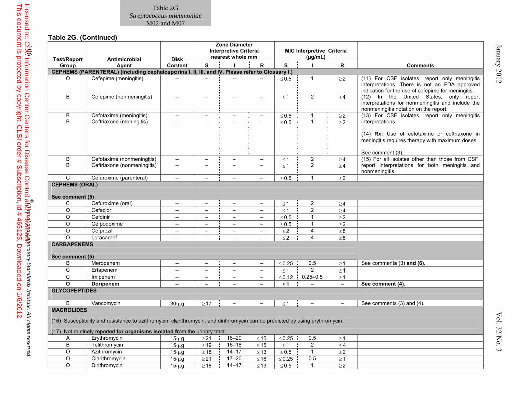
Table 2G. (Continued)
Test/Report
Group Antimicrobial
Agent Disk
Content
Zone Diameter Interpretive Criteria nearest whole mm
MIC Interpretive Criteria (µg/mL)
Comments S I R S I R CEPHEMS (PARENTERAL) (Including cephalosporins I, II, III, and IV. Please refer to Glossary I.)
O Cefepime (meningitis) – – – – ≤ 0.5 1 ≥ 2 (11) For CSF isolates, report only meningitis interpretations. There is not an FDA–approved indication for the use of cefepime for meningitis.
B Cefepime (nonmeningitis) – – – – ≤ 1 2 ≥ 4 (12) In the United States, only report interpretations for nonmeningitis and include the nonmeningitis notation on the report.
B B
Cefotaxime (meningitis) Ceftriaxone (meningitis)
– –
– –
– –
– –
≤ 0.5 ≤ 0.5
1 1
≥ 2 ≥ 2
(13) For CSF isolates, report only meningitis interpretations. (14) Rx: Use of cefotaxime or ceftriaxone in meningitis requires therapy with maximum doses. See comment (3).
B B
Cefotaxime (nonmeningitis) Ceftriaxone (nonmeningitis)
– –
– –
– –
– –
≤ 1 ≤ 1
2 2
≥ 4 ≥ 4
(15) For all isolates other than those from CSF, report interpretations for both meningitis and nonmeningitis.
C Cefuroxime (parenteral) – – – – ≤ 0.5 1 ≥ 2 CEPHEMS (ORAL) See comment (5)
C Cefuroxime (oral) – – – – ≤ 1 2 ≥ 4 O Cefaclor – – – – ≤ 1 2 ≥ 4 O Cefdinir – – – – ≤ 0.5 1 ≥ 2 O Cefpodoxime – – – – ≤ 0.5 1 ≥ 2 O Cefprozil – – – – ≤ 2 4 ≥ 8 O Loracarbef – – – – ≤ 2 4 ≥ 8
CARBAPENEMS See comment (5)
B Meropenem – – – – ≤ 0.25 0.5 ≥ 1 See comments (3) and (6). C Ertapenem – – – – ≤ 1 2 ≥ 4 C Imipenem – – – – ≤ 0.12 0.25–0.5 ≥ 1 O Doripenem – – – – ≤ 1 – – See comment (4).
GLYCOPEPTIDES
B Vancomycin 30 μg ≥ 17 – – ≤ 1 – – See comments (3) and (4). MACROLIDES (16) Susceptibility and resistance to azithromycin, clarithromycin, and dirithromycin can be predicted by using erythromycin. (17) Not routinely reported for organisms isolated from the urinary tract.
A Erythromycin 15 μg ≥ 21 16–20 ≤ 15 ≤ 0.25 0.5 ≥ 1 B Telithromycin 15 μg ≥ 19 16–18 ≤ 15 ≤ 1 2 ≥ 4 O Azithromycin 15 μg ≥ 18 14–17 ≤ 13 ≤ 0.5 1 ≥ 2 O Clarithromycin 15 μg ≥ 21 17–20 ≤ 16 ≤ 0.25 0.5 ≥ 1 O Dirithromycin 15 μg ≥ 18 14–17 ≤ 13 ≤ 0.5 1 ≥ 2
January 2012 V
ol. 32 No. 3
Table 2G Streptococcus pneumoniae
M02 and M07
106 ©C
linical and Laboratory Standards Institute. A
ll rights reserved. Licensed to: C
DC
Information C
enter Centers for D
isease Control and P
reventionT
his document is protected by copyright. C
LSI order # S
ubscription, id # 465125, Dow
nloaded on 1/6/2012.
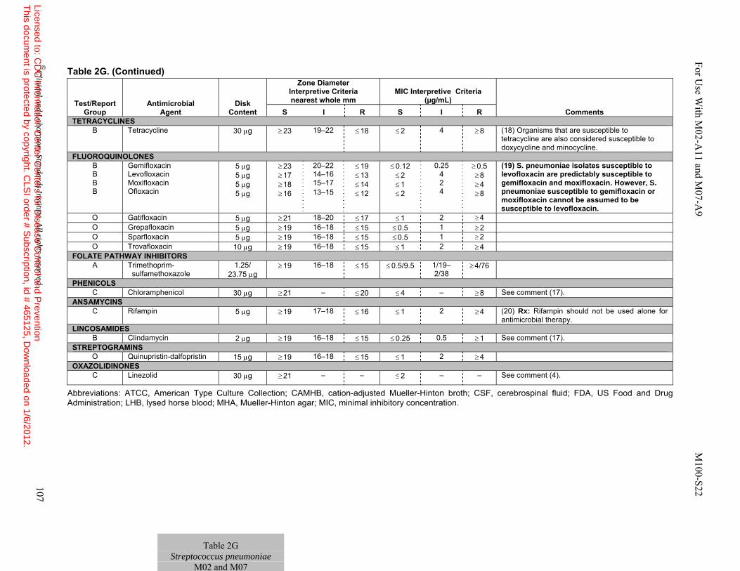
Table 2G. (Continued)
Abbreviations: ATCC, American Type Culture Collection; CAMHB, cation-adjusted Mueller-Hinton broth; CSF, cerebrospinal fluid; FDA, US Food and Drug Administration; LHB, lysed horse blood; MHA, Mueller-Hinton agar; MIC, minimal inhibitory concentration.
Test/Report Group
Antimicrobial Agent
Disk Content
Zone Diameter Interpretive Criteria nearest whole mm
MIC Interpretive Criteria (µg/mL)
Comments S I R S I R TETRACYCLINES
B Tetracycline 30 μg ≥ 23 19–22 ≤ 18 ≤ 2 4 ≥ 8 (18) Organisms that are susceptible to tetracycline are also considered susceptible to doxycycline and minocycline.
FLUOROQUINOLONES B B B B
Gemifloxacin Levofloxacin Moxifloxacin Ofloxacin
5 μg 5 μg 5 μg 5 μg
≥ 23 ≥ 17 ≥ 18 ≥ 16
20–22 14–16 15–17 13–15
≤ 19 ≤ 13 ≤ 14 ≤ 12
≤ 0.12 ≤ 2 ≤ 1 ≤ 2
0.25 4 2 4
≥ 0.5 ≥ 8 ≥ 4 ≥ 8
(19) S. pneumoniae isolates susceptible to levofloxacin are predictably susceptible to gemifloxacin and moxifloxacin. However, S. pneumoniae susceptible to gemifloxacin or moxifloxacin cannot be assumed to be susceptible to levofloxacin.
O Gatifloxacin 5 μg ≥ 21 18–20 ≤ 17 ≤ 1 2 ≥ 4 O Grepafloxacin 5 μg ≥ 19 16–18 ≤ 15 ≤ 0.5 1 ≥ 2 O Sparfloxacin 5 μg ≥ 19 16–18 ≤ 15 ≤ 0.5 1 ≥ 2 O Trovafloxacin 10 μg ≥ 19 16–18 ≤ 15 ≤ 1 2 ≥ 4
FOLATE PATHWAY INHIBITORS A Trimethoprim-
sulfamethoxazole 1.25/
23.75 μg ≥ 19 16–18 ≤ 15 ≤ 0.5/9.5 1/19–
2/38 ≥ 4/76
PHENICOLS C Chloramphenicol 30 μg ≥ 21 – ≤ 20 ≤ 4 – ≥ 8 See comment (17).
ANSAMYCINS C Rifampin 5 μg ≥ 19 17–18 ≤ 16 ≤ 1 2 ≥ 4 (20) Rx: Rifampin should not be used alone for
antimicrobial therapy. LINCOSAMIDES
B Clindamycin 2 μg ≥ 19 16–18 ≤ 15 ≤ 0.25 0.5 ≥ 1 See comment (17). STREPTOGRAMINS
O Quinupristin-dalfopristin 15 μg ≥ 19 16–18 ≤ 15 ≤ 1 2 ≥ 4 OXAZOLIDINONES
C Linezolid 30 μg ≥ 21 – – ≤ 2 – – See comment (4).
©C
linical and Laboratory Standards Institute. A
ll rights reserved. 107
For U
se With M
02-A11 and M
07-A9 M
100-S22
Table 2G Streptococcus pneumoniae
M02 and M07
Licensed to: CD
C Inform
ation Center C
enters for Disease C
ontrol and Prevention
This docum
ent is protected by copyright. CLS
I order # Subscription, id # 465125, D
ownloaded on 1/6/2012.
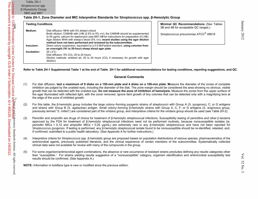
Table 2H-1 Streptococcus spp. β-Hemolytic Group
M02 and M07 Table 2H-1. Zone Diameter and MIC Interpretive Standards for Streptococcus spp. β-Hemolytic Group
Refer to Table 2H-1 Supplemental Table 1 at the end of Table 2H-1 for additional recommendations for testing conditions, reporting suggestions, and QC.
General Comments (1) For disk diffusion, test a maximum of 9 disks on a 150-mm plate and 4 disks on a 100-mm plate. Measure the diameter of the zones of complete
inhibition (as judged by the unaided eye), including the diameter of the disk. The zone margin should be considered the area showing no obvious, visible growth that can be detected with the unaided eye. Do not measure the zone of inhibition of hemolysis. Measure the zones from the upper surface of the agar illuminated with reflected light, with the cover removed. Ignore faint growth of tiny colonies that can be detected only with a magnifying lens at the edge of the zone of inhibited growth.
(2) For this table, the β-hemolytic group includes the large colony–forming pyogenic strains of streptococci with Group A (S. pyogenes), C, or G antigens
and strains with Group B (S. agalactiae) antigen. Small colony–forming β-hemolytic strains with Group A, C, F, or G antigens (S. anginosus group, previously termed “S. milleri”) are considered part of the viridans group, and interpretive criteria for the viridans group should be used (see Table 2H-2).
(3) Penicillin and ampicillin are drugs of choice for treatment of β-hemolytic streptococcal infections. Susceptibility testing of penicillins and other β-lactams
approved by the FDA for treatment of β-hemolytic streptococcal infections need not be performed routinely, because nonsusceptible isolates (ie, penicillin MICs > 0.12 and ampicillin MICs > 0.25 µg/mL) are extremely rare in any β-hemolytic streptococcus and have not been reported for Streptococcus pyogenes. If testing is performed, any β-hemolytic streptococcal isolate found to be nonsusceptible should be re-identified, retested, and, if confirmed, submitted to a public health laboratory. (See Appendix A for further instructions.)
(4) Interpretive criteria for Streptococcus spp. β-hemolytic group are proposed based on population distributions of various species, pharmacokinetics of the
antimicrobial agents, previously published literature, and the clinical experience of certain members of the subcommittee. Systematically collected clinical data were not available for review with many of the compounds in the group.
(5) For some organism/antimicrobial agent combinations, the absence or rare occurrence of resistant strains precludes defining any results categories other
than “susceptible.” For strains yielding results suggestive of a “nonsusceptible” category, organism identification and antimicrobial susceptibility test results should be confirmed. (See Appendix A.)
NOTE: Information in boldface type is new or modified since the previous edition.
Testing Conditions Medium: Disk diffusion: MHA with 5% sheep’s blood Broth dilution: CAMHB with LHB (2.5% to 5% v/v); the CAMHB should be supplemented
to 50 µg/mL calcium for daptomycin (see M07-A9 for instructions for preparation of LHB) Agar dilution MHA with sheep’s blood (5% v/v); recent studies using the agar dilution
method have not been performed and reviewed by the subcommittee. Inoculum: Direct colony suspension, equivalent to a 0.5 McFarland standard, using colonies from
an overnight (18- to 20-hour) sheep blood agar plate Incubation: 35 ± 2°C; Disk diffusion: 5% CO2; 20 to 24 hours Dilution methods: ambient air; 20 to 24 hours (CO2 if necessary for growth with agar
dilution)
Minimal QC Recommendations (See Tables 3B and 4B for acceptable QC ranges.) Streptococcus pneumoniae ATCC® 49619
108 ©C
linical and Laboratory Standards Institute. A
ll rights reserved.
January 2012 V
ol. 32 No. 3
Licensed to: CD
C Inform
ation Center C
enters for Disease C
ontrol and Prevention
This docum
ent is protected by copyright. CLS
I order # Subscription, id # 465125, D
ownloaded on 1/6/2012.
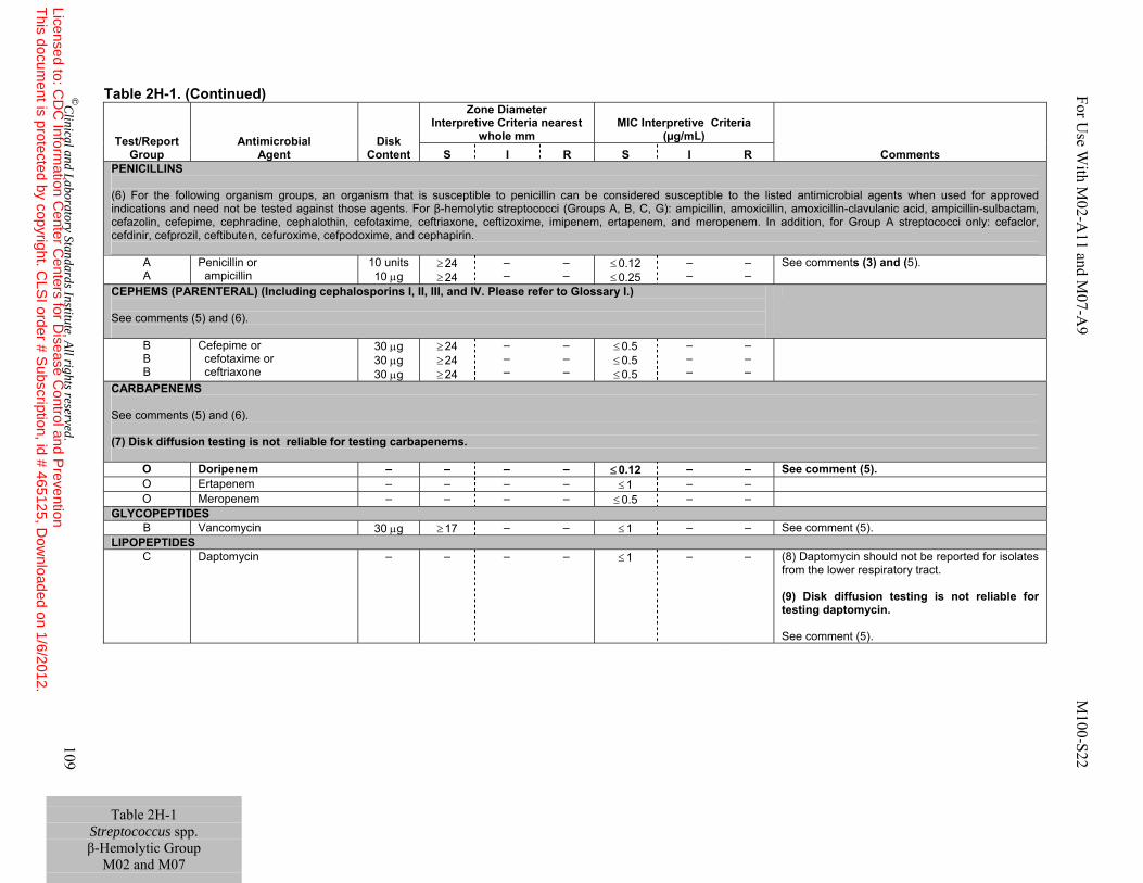
Table 2H-1 Streptococcus spp. β-Hemolytic Group
M02 and M07
Table 2H-1. (Continued)
Test/Report Group
Antimicrobial Agent
Disk Content
Zone Diameter Interpretive Criteria nearest
whole mm MIC Interpretive Criteria
(µg/mL) Comments S I R S I R
PENICILLINS (6) For the following organism groups, an organism that is susceptible to penicillin can be considered susceptible to the listed antimicrobial agents when used for approved indications and need not be tested against those agents. For β-hemolytic streptococci (Groups A, B, C, G): ampicillin, amoxicillin, amoxicillin-clavulanic acid, ampicillin-sulbactam, cefazolin, cefepime, cephradine, cephalothin, cefotaxime, ceftriaxone, ceftizoxime, imipenem, ertapenem, and meropenem. In addition, for Group A streptococci only: cefaclor, cefdinir, cefprozil, ceftibuten, cefuroxime, cefpodoxime, and cephapirin.
A A
Penicillin or ampicillin
10 units 10 μg
≥ 24 ≥ 24
– –
– –
≤ 0.12 ≤ 0.25
– –
– –
See comments (3) and (5).
CEPHEMS (PARENTERAL) (Including cephalosporins I, II, III, and IV. Please refer to Glossary I.) See comments (5) and (6).
B B B
Cefepime or cefotaxime or ceftriaxone
30 μg 30 μg 30 μg
≥ 24 ≥ 24 ≥ 24
– – –
– – –
≤ 0.5 ≤ 0.5 ≤ 0.5
– – –
– – –
CARBAPENEMS See comments (5) and (6). (7) Disk diffusion testing is not reliable for testing carbapenems.
O Doripenem – – – – ≤ 0.12 – – See comment (5). O Ertapenem – – – – ≤ 1 – – O Meropenem – – – – ≤ 0.5 – –
GLYCOPEPTIDES B Vancomycin 30 μg ≥ 17 – – ≤ 1 – – See comment (5).
LIPOPEPTIDES C Daptomycin – – – – ≤ 1 – – (8) Daptomycin should not be reported for isolates
from the lower respiratory tract. (9) Disk diffusion testing is not reliable for testing daptomycin. See comment (5).
For U
se With M
02-A11 and M
07-A9 M
100-S22
©C
linical and Laboratory Standards Institute. A
ll rights reserved. 109
Licensed to: CD
C Inform
ation Center C
enters for Disease C
ontrol and Prevention
This docum
ent is protected by copyright. CLS
I order # Subscription, id # 465125, D
ownloaded on 1/6/2012.
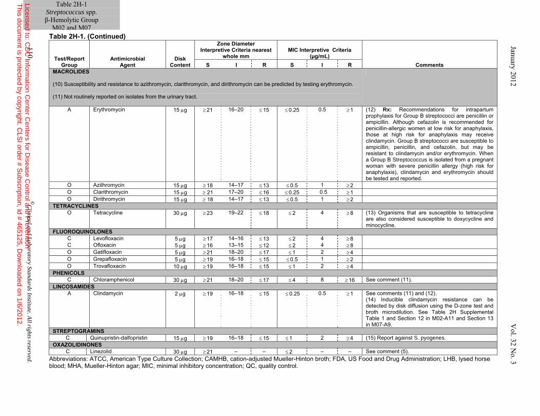
Table 2H-1. (Continued)
Abbreviations: ATCC, American Type Culture Collection; CAMHB, cation-adjusted Mueller-Hinton broth; FDA, US Food and Drug Administration; LHB, lysed horse blood; MHA, Mueller-Hinton agar; MIC, minimal inhibitory concentration; QC, quality control.
Test/Report Group
Antimicrobial Agent
Disk Content
Zone Diameter Interpretive Criteria nearest
whole mm MIC Interpretive Criteria
(µg/mL) Comments S I R S I R
MACROLIDES (10) Susceptibility and resistance to azithromycin, clarithromycin, and dirithromycin can be predicted by testing erythromycin. (11) Not routinely reported on isolates from the urinary tract.
A Erythromycin 15 μg ≥ 21 16–20 ≤ 15 ≤ 0.25 0.5 ≥ 1 (12) Rx: Recommendations for intrapartum prophylaxis for Group B streptococci are penicillin or ampicillin. Although cefazolin is recommended for penicillin-allergic women at low risk for anaphylaxis, those at high risk for anaphylaxis may receive clindamycin. Group B streptococci are susceptible to ampicillin, penicillin, and cefazolin, but may be resistant to clindamycin and/or erythromycin. When a Group B Streptococcus is isolated from a pregnant woman with severe penicillin allergy (high risk for anaphylaxis), clindamycin and erythromycin should be tested and reported.
O Azithromycin 15 μg ≥ 18 14–17 ≤ 13 ≤ 0.5 1 ≥ 2 O Clarithromycin 15 μg ≥ 21 17–20 ≤ 16 ≤ 0.25 0.5 ≥ 1 O Dirithromycin 15 μg ≥ 18 14–17 ≤ 13 ≤ 0.5 1 ≥ 2
TETRACYCLINES O Tetracycline 30 μg ≥ 23 19–22 ≤ 18 ≤ 2 4 ≥ 8 (13) Organisms that are susceptible to tetracycline
are also considered susceptible to doxycycline and minocycline.
FLUOROQUINOLONES C Levofloxacin 5 μg ≥ 17 14–16 ≤ 13 ≤ 2 4 ≥ 8 C Ofloxacin 5 μg ≥ 16 13–15 ≤ 12 ≤ 2 4 ≥ 8 O Gatifloxacin 5 μg ≥ 21 18–20 ≤ 17 ≤ 1 2 ≥ 4 O Grepafloxacin 5 μg ≥ 19 16–18 ≤ 15 ≤ 0.5 1 ≥ 2 O Trovafloxacin 10 μg ≥ 19 16–18 ≤ 15 ≤ 1 2 ≥ 4
PHENICOLS C Chloramphenicol 30 μg ≥ 21 18–20 ≤ 17 ≤ 4 8 ≥ 16 See comment (11).
LINCOSAMIDES A Clindamycin 2 μg ≥ 19 16–18 ≤ 15 ≤ 0.25 0.5 ≥ 1 See comments (11) and (12).
(14) Inducible clindamycin resistance can be detected by disk diffusion using the D-zone test and broth microdilution. See Table 2H Supplemental Table 1 and Section 12 in M02-A11 and Section 13 in M07-A9.
STREPTOGRAMINS C Quinupristin-dalfopristin 15 μg ≥ 19 16–18 ≤ 15 ≤ 1 2 ≥ 4 (15) Report against S. pyogenes.
OXAZOLIDINONES C Linezolid 30 μg ≥ 21 – – ≤ 2 – – See comment (5).
Table 2H-1 Streptococcus spp. β-Hemolytic Group
M02 and M07 January 2012 V
ol. 32 No. 3
110 ©
Clinical and L
aboratory Standards Institute. All rights reserved.
Licensed to: CD
C Inform
ation Center C
enters for Disease C
ontrol and Prevention
This docum
ent is protected by copyright. CLS
I order # Subscription, id # 465125, D
ownloaded on 1/6/2012.

For Use With M02-A11 and M07-A9 M100-S22
©Clinical and Laboratory Standards Institute. All rights reserved. 111
This page is intentionally left blank.
Licensed to: CDC Information Center Centers for Disease Control and PreventionThis document is protected by copyright. CLSI order # Subscription, id # 465125, Downloaded on 1/6/2012.
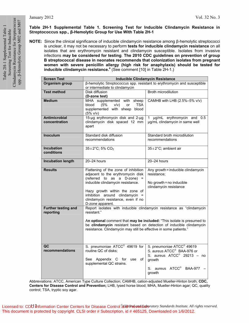
January 2012 Vol. 32 No. 3
112 ©Clinical and Laboratory Standards Institute. All rights reserved.
Table 2H-1 Supplemental Table 1. Screening Test for Inducible Clindamycin Resistance in Streptococcus spp., β-Hemolytic Group for Use With Table 2H-1 NOTE: Since the clinical significance of inducible clindamycin resistance among β-hemolytic streptococci
is unclear, it may not be necessary to perform tests for inducible clindamycin resistance on all isolates that are erythromycin resistant and clindamycin susceptible. Isolates from invasive infections may be considered for testing. The 2010 CDC guidelines on prevention of group B streptococcal disease in neonates recommends that colonization isolates from pregnant women with severe penicillin allergy (high risk for anaphylaxis) should be tested for inducible clindamycin resistance.a (See comment [10] in Table 2H-1.)
Screen Test Inducible Clindamycin Resistance Organism group β-hemolytic Streptococcus spp. resistant to erythromycin and susceptible
or intermediate to clindamycin Test method Disk diffusion
(D-zone test) Broth microdilution
Medium
MHA supplemented with sheep blood (5% v/v) or TSA supplemented with sheep blood (5% v/v)
CAMHB with LHB (2.5%–5% v/v)
Antimicrobial concentration
15-µg erythromycin disk and 2-µg clindamycin disk spaced 12 mm apart
1 µg/mL erythromycin and 0.5 µg/mL clindamycin in same well
Inoculum Standard disk diffusion recommendations
Standard broth microdilution recommendations
Incubation conditions
35 ± 2°C; 5% CO2
35 ± 2°C; ambient air
Incubation length 20–24 hours 20–24 hours
Results Flattening of the zone of inhibition adjacent to the erythromycin disk (referred to as a D-zone) =
inducible clindamycin resistance. Hazy growth within the zone of inhibition around clindamycin =
clindamycin resistance, even if no D-zone apparent.
Any growth = inducible clindamycin resistance; No growth = no inducible clindamycin resistance
Further testing and reporting
Report isolates with inducible clindamycin resistance as “clindamycin resistant.” An optional comment that may be included: “This isolate is presumed to be clindamycin resistant based on detection of inducible clindamycin resistance. Clindamycin may still be effective in some patients.”
QC recommendations
S. pneumoniae ATCC® 49619 for routine QC of disks; See Appendix C for use of supplemental QC strains.
S. pneumoniae ATCC® 49619 S. aureus ATCC® BAA-976 or S. aureus ATCC® 29213 – no growth S. aureus ATCC® BAA-977 – growth
Abbreviations: ATCC, American Type Culture Collection; CAMHB, cation-adjusted Mueller-Hinton broth; CDC, Centers for Disease Control and Prevention; LHB, lysed horse blood; MHA, Mueller-Hinton agar; QC, quality control; TSA, tryptic soy agar.
Tabl
e 2H
-1 S
uppl
emen
tal T
able
1
Scre
enin
g Te
st fo
r Ind
ucib
le
Clin
dam
ycin
Res
ista
nce
for S
trep
toco
ccus
sp
p., β
-Hem
olyt
ic G
roup
M02
and
M07
Licensed to: CDC Information Center Centers for Disease Control and PreventionThis document is protected by copyright. CLSI order # Subscription, id # 465125, Downloaded on 1/6/2012.
For Use With M02-A11 and M07-A9 M100-S22
©Clinical and Laboratory Standards Institute. All rights reserved. 113
Table 2H-1 Supplemental Table 1. (Continued)
Footnote
a Reference
Verani JR, McGee L, Schrag SJ; Division of Bacterial Diseases, National Center for Immunization and Respiratory Diseases, Centers for Disease Control and Prevention. Prevention of perinatal group B streptococcal disease – revised guidelines from CDC, 2010. MMWR Recomm Rep. 2010;59(RR-10):1-36.
Tabl
e 2H
-1 S
uppl
emen
tal T
able
1
Scre
enin
g Te
st fo
r Ind
ucib
le
Clin
dam
ycin
Res
ista
nce
for S
trep
toco
ccus
sp
p., β
-Hem
olyt
ic G
roup
M02
and
M07
Licensed to: CDC Information Center Centers for Disease Control and PreventionThis document is protected by copyright. CLSI order # Subscription, id # 465125, Downloaded on 1/6/2012.
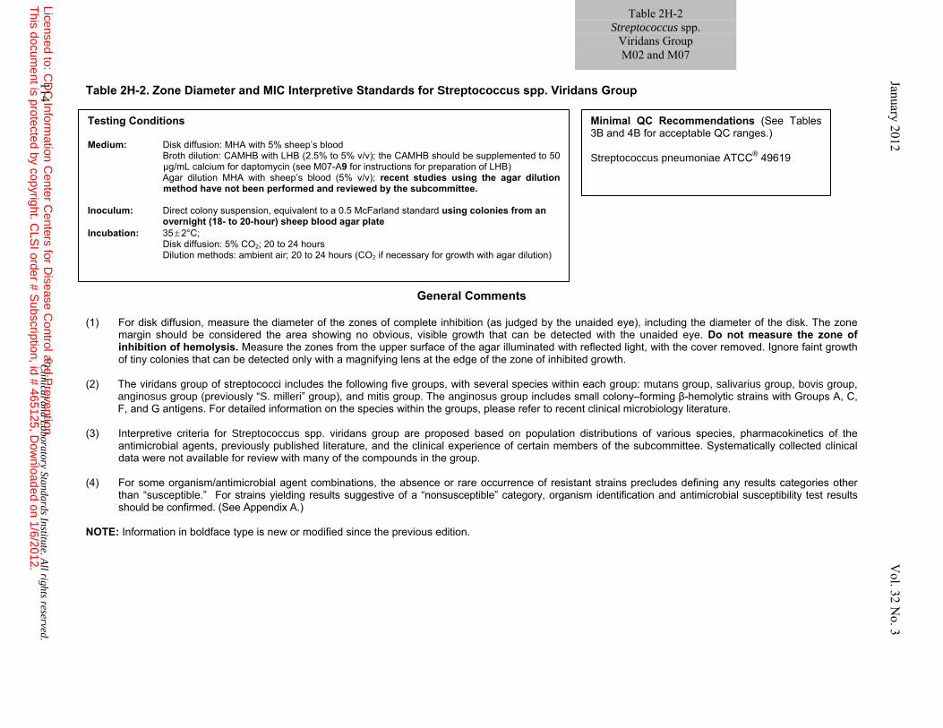
Table 2H-2. Zone Diameter and MIC Interpretive Standards for Streptococcus spp. Viridans Group
General Comments (1) For disk diffusion, measure the diameter of the zones of complete inhibition (as judged by the unaided eye), including the diameter of the disk. The zone
margin should be considered the area showing no obvious, visible growth that can be detected with the unaided eye. Do not measure the zone of inhibition of hemolysis. Measure the zones from the upper surface of the agar illuminated with reflected light, with the cover removed. Ignore faint growth of tiny colonies that can be detected only with a magnifying lens at the edge of the zone of inhibited growth.
(2) The viridans group of streptococci includes the following five groups, with several species within each group: mutans group, salivarius group, bovis group,
anginosus group (previously “S. milleri” group), and mitis group. The anginosus group includes small colony–forming β-hemolytic strains with Groups A, C, F, and G antigens. For detailed information on the species within the groups, please refer to recent clinical microbiology literature.
(3) Interpretive criteria for Streptococcus spp. viridans group are proposed based on population distributions of various species, pharmacokinetics of the
antimicrobial agents, previously published literature, and the clinical experience of certain members of the subcommittee. Systematically collected clinical data were not available for review with many of the compounds in the group.
(4) For some organism/antimicrobial agent combinations, the absence or rare occurrence of resistant strains precludes defining any results categories other
than “susceptible.” For strains yielding results suggestive of a “nonsusceptible” category, organism identification and antimicrobial susceptibility test results should be confirmed. (See Appendix A.)
NOTE: Information in boldface type is new or modified since the previous edition.
Testing Conditions Medium: Disk diffusion: MHA with 5% sheep’s blood Broth dilution: CAMHB with LHB (2.5% to 5% v/v); the CAMHB should be supplemented to 50
µg/mL calcium for daptomycin (see M07-A9 for instructions for preparation of LHB) Agar dilution MHA with sheep’s blood (5% v/v); recent studies using the agar dilution
method have not been performed and reviewed by the subcommittee. Inoculum: Direct colony suspension, equivalent to a 0.5 McFarland standard using colonies from an
overnight (18- to 20-hour) sheep blood agar plate Incubation: 35 ± 2°C; Disk diffusion: 5% CO2; 20 to 24 hours Dilution methods: ambient air; 20 to 24 hours (CO2 if necessary for growth with agar dilution)
Minimal QC Recommendations (See Tables 3B and 4B for acceptable QC ranges.) Streptococcus pneumoniae ATCC® 49619
114 ©C
linical and Laboratory Standards Institute. A
ll rights reserved.
Table 2H-2 Streptococcus spp.
Viridans Group M02 and M07
January 2012 V
ol. 32 No. 3
Licensed to: CD
C Inform
ation Center C
enters for Disease C
ontrol and Prevention
This docum
ent is protected by copyright. CLS
I order # Subscription, id # 465125, D
ownloaded on 1/6/2012.
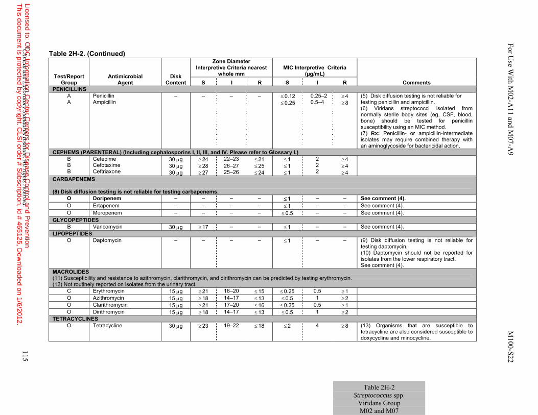
Table 2H-2. (Continued)
Test/Report Group
Antimicrobial Agent
Disk Content
Zone Diameter Interpretive Criteria nearest
whole mm MIC Interpretive Criteria
(µg/mL) Comments S I R S I R
PENICILLINS A A
Penicillin Ampicillin
– – – – ≤ 0.12 ≤ 0.25
0.25–2 0.5–4
≥ 4 ≥ 8
(5) Disk diffusion testing is not reliable for testing penicillin and ampicillin. (6) Viridans streptococci isolated from normally sterile body sites (eg, CSF, blood, bone) should be tested for penicillin susceptibility using an MIC method. (7) Rx: Penicillin- or ampicillin-intermediate isolates may require combined therapy with an aminoglycoside for bactericidal action.
CEPHEMS (PARENTERAL) (Including cephalosporins I, II, III, and IV. Please refer to Glossary I.) B B B
Cefepime Cefotaxime Ceftriaxone
30 μg 30 μg 30 μg
≥ 24 ≥ 28 ≥ 27
22–23 26–27 25–26
≤ 21 ≤ 25 ≤ 24
≤ 1 ≤ 1 ≤ 1
2 2 2
≥ 4 ≥ 4 ≥ 4
CARBAPENEMS (8) Disk diffusion testing is not reliable for testing carbapenems.
O Doripenem – – – – ≤ 1 – – See comment (4). O Ertapenem – – – – ≤ 1 – – See comment (4). O Meropenem – – – – ≤ 0.5 – – See comment (4).
GLYCOPEPTIDES B Vancomycin 30 μg ≥ 17 – – ≤ 1 – – See comment (4).
LIPOPEPTIDES O Daptomycin – – – – ≤ 1 – – (9) Disk diffusion testing is not reliable for
testing daptomycin. (10) Daptomycin should not be reported for isolates from the lower respiratory tract. See comment (4).
MACROLIDES (11) Susceptibility and resistance to azithromycin, clarithromycin, and dirithromycin can be predicted by testing erythromycin. (12) Not routinely reported on isolates from the urinary tract.
C Erythromycin 15 μg ≥ 21 16–20 ≤ 15 ≤ 0.25 0.5 ≥ 1 O Azithromycin 15 μg ≥ 18 14–17 ≤ 13 ≤ 0.5 1 ≥ 2 O Clarithromycin 15 μg ≥ 21 17–20 ≤ 16 ≤ 0.25 0.5 ≥ 1 O Dirithromycin 15 μg ≥ 18 14–17 ≤ 13 ≤ 0.5 1 ≥ 2
TETRACYCLINES O Tetracycline 30 μg ≥ 23 19–22 ≤ 18 ≤ 2 4 ≥ 8 (13) Organisms that are susceptible to
tetracycline are also considered susceptible to doxycycline and minocycline.
©C
linical and Laboratory Standards Institute. A
ll rights reserved. 115
Table 2H-2 Streptococcus spp.
Viridans Group M02 and M07
For U
se With M
02-A11 and M
07-A9 M
100-S22
Licensed to: CD
C Inform
ation Center C
enters for Disease C
ontrol and Prevention
This docum
ent is protected by copyright. CLS
I order # Subscription, id # 465125, D
ownloaded on 1/6/2012.
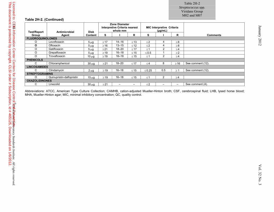
Table 2H-2. (Continued)
Abbreviations: ATCC, American Type Culture Collection; CAMHB, cation-adjusted Mueller-Hinton broth; CSF, cerebrospinal fluid; LHB, lysed horse blood; MHA, Mueller-Hinton agar; MIC, minimal inhibitory concentration; QC, quality control.
Test/Report Group
Antimicrobial Agent
Disk Content
Zone Diameter Interpretive Criteria nearest
whole mm MIC Interpretive Criteria
(µg/mL)
Comments S I R S I R FLUOROQUINOLONES
O Levofloxacin 5 μg ≥ 17 14–16 ≤ 13 ≤ 2 4 ≥ 8 O Ofloxacin 5 μg ≥ 16 13–15 ≤ 12 ≤ 2 4 ≥ 8 O Gatifloxacin 5 μg ≥ 21 18–20 ≤ 17 ≤ 1 2 ≥ 4 O Grepafloxacin 5 μg ≥ 19 16–18 ≤ 15 ≤ 0.5 1 ≥ 2 O Trovafloxacin 10 μg ≥ 19 16–18 ≤ 15 ≤ 1 2 ≥ 4
PHENICOLS C Chloramphenicol 30 μg ≥ 21 18–20 ≤ 17 ≤ 4 8 ≥ 16 See comment (12).
LINCOSAMIDES C Clindamycin 2 μg ≥ 19 16–18 ≤ 15 ≤ 0.25 0.5 ≥ 1 See comment (12).
STREPTOGRAMINS O Quinupristin-dalfopristin 15 μg ≥ 19 16–18 ≤ 15 ≤ 1 2 ≥ 4
OXAZOLIDINONES C Linezolid 30 μg ≥ 21 – – ≤ 2 – – See comment (4).
116 ©C
linical and Laboratory Standards Institute. A
ll rights reserved.
Table 2H-2 Streptococcus spp.
Viridans Group M02 and M07
January 2012 V
ol. 32 No. 3
Licensed to: CD
C Inform
ation Center C
enters for Disease C
ontrol and Prevention
This docum
ent is protected by copyright. CLS
I order # Subscription, id # 465125, D
ownloaded on 1/6/2012.

For Use With M02-A11 and M07-A9 M100-S22
©Clinical and Laboratory Standards Institute. All rights reserved. 117
This page is intentionally left blank.
Licensed to: CDC Information Center Centers for Disease Control and PreventionThis document is protected by copyright. CLSI order # Subscription, id # 465125, Downloaded on 1/6/2012.
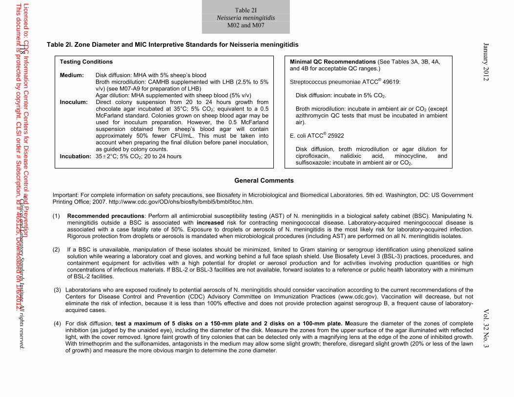
Table 2I. Zone Diameter and MIC Interpretive Standards for Neisseria meningitidis
General Comments
Important: For complete information on safety precautions, see Biosafety in Microbiological and Biomedical Laboratories. 5th ed. Washington, DC: US Government Printing Office; 2007. http://www.cdc.gov/OD/ohs/biosfty/bmbl5/bmbl5toc.htm.
(1) Recommended precautions: Perform all antimicrobial susceptibility testing (AST) of N. meningitidis in a biological safety cabinet (BSC). Manipulating N.
meningitidis outside a BSC is associated with increased risk for contracting meningococcal disease. Laboratory-acquired meningococcal disease is associated with a case fatality rate of 50%. Exposure to droplets or aerosols of N. meningitidis is the most likely risk for laboratory-acquired infection. Rigorous protection from droplets or aerosols is mandated when microbiological procedures (including AST) are performed on all N. meningitidis isolates.
(2) If a BSC is unavailable, manipulation of these isolates should be minimized, limited to Gram staining or serogroup identification using phenolized saline
solution while wearing a laboratory coat and gloves, and working behind a full face splash shield. Use Biosafety Level 3 (BSL-3) practices, procedures, and containment equipment for activities with a high potential for droplet or aerosol production and for activities involving production quantities or high concentrations of infectious materials. If BSL-2 or BSL-3 facilities are not available, forward isolates to a reference or public health laboratory with a minimum of BSL-2 facilities.
(3) Laboratorians who are exposed routinely to potential aerosols of N. meningitidis should consider vaccination according to the current recommendations of the
Centers for Disease Control and Prevention (CDC) Advisory Committee on Immunization Practices (www.cdc.gov). Vaccination will decrease, but not eliminate the risk of infection, because it is less than 100% effective and does not provide protection against serogroup B, a frequent cause of laboratory-acquired cases.
(4) For disk diffusion, test a maximum of 5 disks on a 150-mm plate and 2 disks on a 100-mm plate. Measure the diameter of the zones of complete inhibition (as judged by the unaided eye), including the diameter of the disk. Measure the zones from the upper surface of the agar illuminated with reflected light, with the cover removed. Ignore faint growth of tiny colonies that can be detected only with a magnifying lens at the edge of the zone of inhibited growth. With trimethoprim and the sulfonamides, antagonists in the medium may allow some slight growth; therefore, disregard slight growth (20% or less of the lawn of growth) and measure the more obvious margin to determine the zone diameter.
Testing Conditions Medium: Disk diffusion: MHA with 5% sheep’s blood Broth microdilution: CAMHB supplemented with LHB (2.5% to 5%
v/v) (see M07-A9 for preparation of LHB) Agar dilution: MHA supplemented with sheep blood (5% v/v) Inoculum: Direct colony suspension from 20 to 24 hours growth from
chocolate agar incubated at 35°C; 5% CO2; equivalent to a 0.5 McFarland standard. Colonies grown on sheep blood agar may be used for inoculum preparation. However, the 0.5 McFarland suspension obtained from sheep’s blood agar will contain approximately 50% fewer CFU/mL. This must be taken into account when preparing the final dilution before panel inoculation, as guided by colony counts.
Incubation: 35 ± 2°C; 5% CO2; 20 to 24 hours
Minimal QC Recommendations (See Tables 3A, 3B, 4A, and 4B for acceptable QC ranges.) Streptococcus pneumoniae ATCC® 49619:
Disk diffusion: incubate in 5% CO2. Broth microdilution: incubate in ambient air or CO2 (except azithromycin QC tests that must be incubated in ambient air).
E. coli ATCC® 25922
Disk diffusion, broth microdilution or agar dilution for ciprofloxacin, nalidixic acid, minocycline, and sulfisoxazole: incubate in ambient air or CO2.
118 ©C
linical and Laboratory Standards Institute. A
ll rights reserved.
Table 2I Neisseria meningitidis
M02 and M07 January 2012 V
ol. 32 No. 3
Licensed to: CD
C Inform
ation Center C
enters for Disease C
ontrol and Prevention
This docum
ent is protected by copyright. CLS
I order # Subscription, id # 465125, D
ownloaded on 1/6/2012.
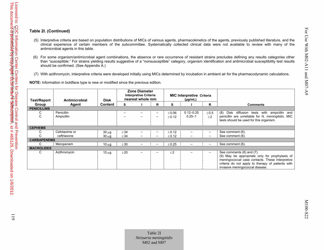
Table 2I. (Continued)
(5) Interpretive criteria are based on population distributions of MICs of various agents, pharmacokinetics of the agents, previously published literature, and the clinical experience of certain members of the subcommittee. Systematically collected clinical data were not available to review with many of the antimicrobial agents in this table.
(6) For some organism/antimicrobial agent combinations, the absence or rare occurrence of resistant strains precludes defining any results categories other
than “susceptible.” For strains yielding results suggestive of a “nonsusceptible” category, organism identification and antimicrobial susceptibility test results should be confirmed. (See Appendix A.)
(7) With azithromycin, interpretive criteria were developed initially using MICs determined by incubation in ambient air for the pharmacodynamic calculations.
NOTE: Information in boldface type is new or modified since the previous edition.
Test/Report Group
Antimicrobial Agent
Disk Content
Zone DiameterInterpretive Criteria nearest whole mm
MIC Interpretive Criteria (µg/mL)
Comments S I R S I R PENICILLINS
C C
Penicillin Ampicillin
– –
– –
– –
≤ 0.06 ≤ 0.12
0.12–0.25 0.25–1
≥ 0.5 ≥ 2
(8) Disk diffusion tests with ampicillin and penicillin are unreliable for N. meningitidis. MIC tests should be used for this organism.
CEPHEMS
C Cefotaxime or 30 μg ≥ 34 – – ≤ 0.12 – – See comment (6). C ceftriaxone 30 μg ≥ 34 – – ≤ 0.12 – – See comment (6).
CARBAPENEMS C Meropenem 10 μg ≥ 30 – – ≤ 0.25 – – See comment (6).
MACROLIDES C Azithromycin 15 μg ≥ 20 – – ≤ 2 – – See comments (6) and (7).
(9) May be appropriate only for prophylaxis of meningococcal case contacts. These interpretive criteria do not apply to therapy of patients with invasive meningococcal disease.
©C
linical and Laboratory Standards Institute. A
ll rights reserved. 119
Table 2I Neisseria meningitidis
M02 and M07
For U
se With M
02-A11 and M
07-A9 M
100-S22
Licensed to: CD
C Inform
ation Center C
enters for Disease C
ontrol and Prevention
This docum
ent is protected by copyright. CLS
I order # Subscription, id # 465125, D
ownloaded on 1/6/2012.
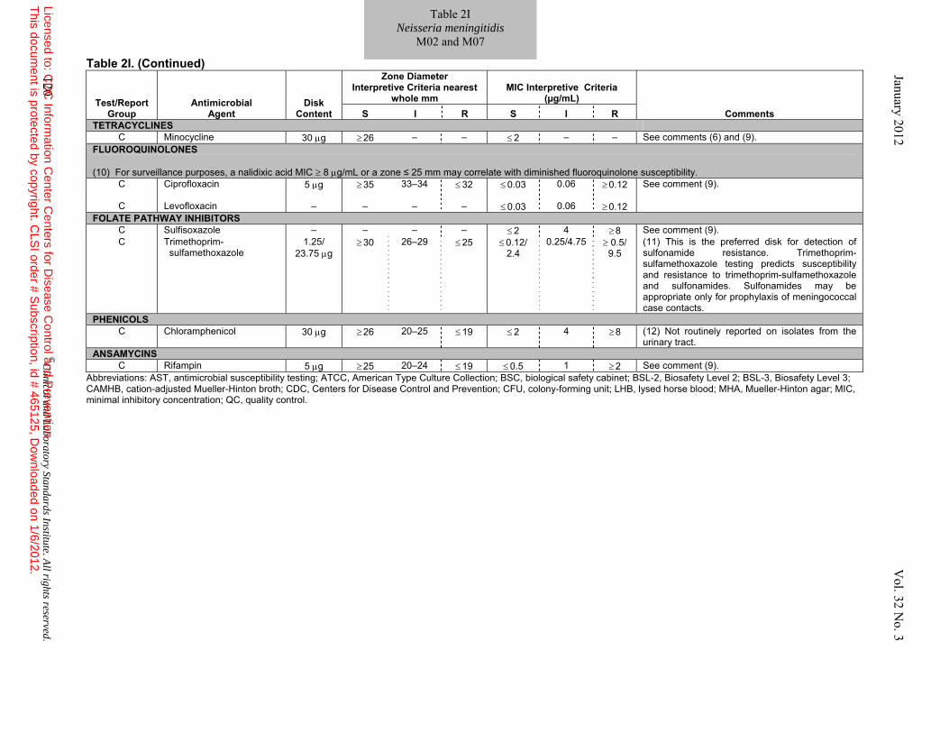
Table 2I. (Continued)
Abbreviations: AST, antimicrobial susceptibility testing; ATCC, American Type Culture Collection; BSC, biological safety cabinet; BSL-2, Biosafety Level 2; BSL-3, Biosafety Level 3; CAMHB, cation-adjusted Mueller-Hinton broth; CDC, Centers for Disease Control and Prevention; CFU, colony-forming unit; LHB, lysed horse blood; MHA, Mueller-Hinton agar; MIC, minimal inhibitory concentration; QC, quality control.
Test/Report Group
Antimicrobial Agent
Disk Content
Zone Diameter Interpretive Criteria nearest
whole mm MIC Interpretive Criteria
(µg/mL) Comments S I R S I R
TETRACYCLINES C Minocycline 30 μg ≥ 26 – – ≤ 2 – – See comments (6) and (9).
FLUOROQUINOLONES (10) For surveillance purposes, a nalidixic acid MIC ≥ 8 μg/mL or a zone ≤ 25 mm may correlate with diminished fluoroquinolone susceptibility.
C Ciprofloxacin 5 μg ≥ 35 33–34 ≤ 32 ≤ 0.03 0.06 ≥ 0.12 See comment (9).
C Levofloxacin – – – – ≤ 0.03 0.06 ≥ 0.12 FOLATE PATHWAY INHIBITORS
C Sulfisoxazole – – – – ≤ 2 4 ≥ 8 See comment (9). C Trimethoprim-
sulfamethoxazole 1.25/
23.75 μg ≥ 30 26–29 ≤ 25 ≤ 0.12/
2.4 0.25/4.75 ≥ 0.5/
9.5 (11) This is the preferred disk for detection of sulfonamide resistance. Trimethoprim-sulfamethoxazole testing predicts susceptibility and resistance to trimethoprim-sulfamethoxazole and sulfonamides. Sulfonamides may be appropriate only for prophylaxis of meningococcal case contacts.
PHENICOLS C Chloramphenicol 30 μg ≥ 26 20–25 ≤ 19 ≤ 2 4 ≥ 8 (12) Not routinely reported on isolates from the
urinary tract. ANSAMYCINS
C Rifampin 5 μg ≥ 25 20–24 ≤ 19 ≤ 0.5 1 ≥ 2 See comment (9).
120 ©C
linical and Laboratory Standards Institute. A
ll rights reserved.
Table 2I Neisseria meningitidis
M02 and M07 January 2012 V
ol. 32 No. 3
Licensed to: CD
C Inform
ation Center C
enters for Disease C
ontrol and Prevention
This docum
ent is protected by copyright. CLS
I order # Subscription, id # 465125, D
ownloaded on 1/6/2012.
For Use With M02-A11 and M07-A9 M100-S22
©Clinical and Laboratory Standards Institute. All rights reserved. 121
This page is intentionally left blank.
Licensed to: CDC Information Center Centers for Disease Control and PreventionThis document is protected by copyright. CLSI order # Subscription, id # 465125, Downloaded on 1/6/2012.
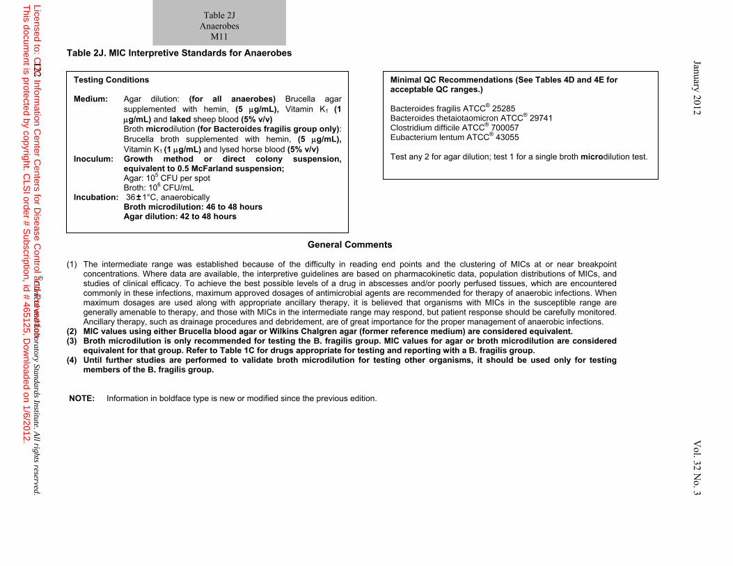
Table 2J. MIC Interpretive Standards for Anaerobes
General Comments (1) The intermediate range was established because of the difficulty in reading end points and the clustering of MICs at or near breakpoint
concentrations. Where data are available, the interpretive guidelines are based on pharmacokinetic data, population distributions of MICs, and studies of clinical efficacy. To achieve the best possible levels of a drug in abscesses and/or poorly perfused tissues, which are encountered commonly in these infections, maximum approved dosages of antimicrobial agents are recommended for therapy of anaerobic infections. When maximum dosages are used along with appropriate ancillary therapy, it is believed that organisms with MICs in the susceptible range are generally amenable to therapy, and those with MICs in the intermediate range may respond, but patient response should be carefully monitored. Ancillary therapy, such as drainage procedures and debridement, are of great importance for the proper management of anaerobic infections.
(2) MIC values using either Brucella blood agar or Wilkins Chalgren agar (former reference medium) are considered equivalent. (3) Broth microdilution is only recommended for testing the B. fragilis group. MIC values for agar or broth microdilution are considered
equivalent for that group. Refer to Table 1C for drugs appropriate for testing and reporting with a B. fragilis group. (4) Until further studies are performed to validate broth microdilution for testing other organisms, it should be used only for testing
members of the B. fragilis group. NOTE: Information in boldface type is new or modified since the previous edition.
Testing Conditions Medium: Agar dilution: (for all anaerobes) Brucella agar
supplemented with hemin, (5 μg/mL), Vitamin K1 (1 μg/mL) and laked sheep blood (5% v/v)
Broth microdilution (for Bacteroides fragilis group only): Brucella broth supplemented with hemin, (5 μg/mL), Vitamin K1 (1 μg/mL) and lysed horse blood (5% v/v)
Inoculum: Growth method or direct colony suspension, equivalent to 0.5 McFarland suspension; Agar: 105 CFU per spot
Broth: 106 CFU/mL Incubation: 36 ± 1°C, anaerobically Broth microdilution: 46 to 48 hours
Agar dilution: 42 to 48 hours
Minimal QC Recommendations (See Tables 4D and 4E for acceptable QC ranges.) Bacteroides fragilis ATCC® 25285 Bacteroides thetaiotaomicron ATCC® 29741 Clostridium difficile ATCC® 700057 Eubacterium lentum ATCC® 43055 Test any 2 for agar dilution; test 1 for a single broth microdilution test.
122 ©C
linical and Laboratory Standards Institute. A
ll rights reserved.
January 2012 V
ol. 32 No. 3
Table 2J
Anaerobes M11
Licensed to: CD
C Inform
ation Center C
enters for Disease C
ontrol and Prevention
This docum
ent is protected by copyright. CLS
I order # Subscription, id # 465125, D
ownloaded on 1/6/2012.
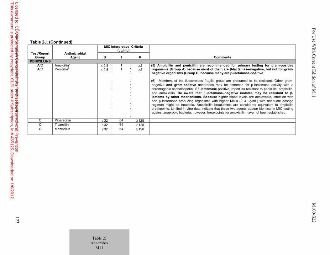
Table 2J. (Continued)
Test/Report Group
Antimicrobial Agent
MIC Interpretive Criteria (µg/mL)
Comments S I R PENICILLINS
A/C A/C
Ampicillina
Penicillina
≤ 0.5 ≤ 0.5
1 1
≥ 2 ≥ 2
(5) Ampicillin and penicillin are recommended for primary testing for gram-positive organisms (Group A) because most of them are β-lactamase-negative, but not for gram-negative organisms (Group C) because many are β-lactamase-positive. (6) Members of the Bacteroides fragilis group are presumed to be resistant. Other gram-negative and gram-positive anaerobes may be screened for β-lactamase activity with a chromogenic cephalosporin; if β-lactamase positive, report as resistant to penicillin, ampicillin, and amoxicillin. Be aware that β-lactamase–negative isolates may be resistant to β-lactams by other mechanisms. Because higher blood levels are achievable, infection with non–β-lactamase producing organisms with higher MICs (2–4 µg/mL) with adequate dosage regimen might be treatable. Amoxicillin breakpoints are considered equivalent to ampicillin breakpoints. Limited in vitro data indicate that these two agents appear identical in MIC testing against anaerobic bacteria; however, breakpoints for amoxicillin have not been established.
C Piperacillin ≤ 32 64 ≥ 128 C Ticarcillin ≤ 32 64 ≥ 128 C Mezlocillin
≤ 32 64 ≥ 128
For U
se With C
urrent Edition of M11 M
100-S22
©C
linical and Laboratory Standards Institute. A
ll rights reserved. 123
Table 2J
Anaerobes M11
Licensed to: CD
C Inform
ation Center C
enters for Disease C
ontrol and Prevention
This docum
ent is protected by copyright. CLS
I order # Subscription, id # 465125, D
ownloaded on 1/6/2012.
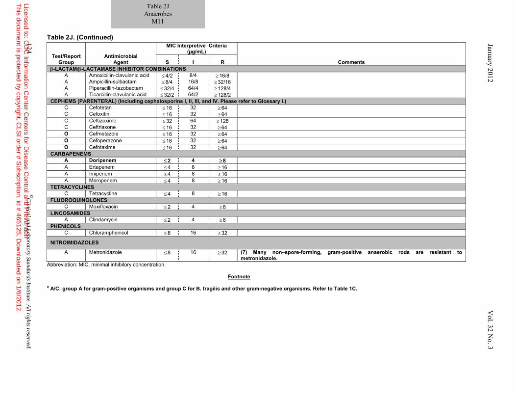
Table 2J. (Continued)
Abbreviation: MIC, minimal inhibitory concentration.
Footnote
a A/C: group A for gram-positive organisms and group C for B. fragilis and other gram-negative organisms. Refer to Table 1C.
Test/Report Group
Antimicrobial Agent
MIC Interpretive Criteria (µg/mL)
Comments S I R β-LACTAM/β-LACTAMASE INHIBITOR COMBINATIONS
A Amoxicillin-clavulanic acid ≤ 4/2 8/4 ≥ 16/8 A Ampicillin-sulbactam ≤ 8/4 16/8 ≥ 32/16 A Piperacillin-tazobactam ≤ 32/4 64/4 ≥ 128/4 A Ticarcillin-clavulanic acid ≤ 32/2 64/2 ≥ 128/2
CEPHEMS (PARENTERAL) (Including cephalosporins I, II, III, and IV. Please refer to Glossary I.) C Cefotetan ≤ 16 32 ≥ 64 C Cefoxitin ≤ 16 32 ≥ 64 C Ceftizoxime ≤ 32 64 ≥ 128 C Ceftriaxone ≤ 16 32 ≥ 64 O Cefmetazole ≤ 16 32 ≥ 64 O Cefoperazone ≤ 16 32 ≥ 64 O Cefotaxime ≤ 16 32 ≥ 64
CARBAPENEMS A Doripenem ≤ 2 4 ≥ 8 A Ertapenem ≤ 4 8 ≥ 16 A Imipenem ≤ 4 8 ≥ 16 A Meropenem ≤ 4 8 ≥ 16
TETRACYCLINES C Tetracycline ≤ 4 8 ≥ 16
FLUOROQUINOLONES C Moxifloxacin ≤ 2 4 ≥ 8
LINCOSAMIDES A Clindamycin ≤ 2 4 ≥ 8
PHENICOLS C Chloramphenicol ≤ 8 16 ≥ 32
NITROIMIDAZOLES
A Metronidazole ≤ 8 16 ≥ 32 (7) Many non–spore-forming, gram-positive anaerobic rods are resistant to metronidazole.
124 ©C
linical and Laboratory Standards Institute. A
ll rights reserved.
January 2012 V
ol. 32 No. 3
Table 2J Anaerobes
M11
Licensed to: CD
C Inform
ation Center C
enters for Disease C
ontrol and Prevention
This docum
ent is protected by copyright. CLS
I order # Subscription, id # 465125, D
ownloaded on 1/6/2012.

For Use With Current Edition of M11 M100-S22
©Clinical and Laboratory Standards Institute. All rights reserved. 125
This page is intentionally left blank.
Licensed to: CDC Information Center Centers for Disease Control and PreventionThis document is protected by copyright. CLSI order # Subscription, id # 465125, Downloaded on 1/6/2012.
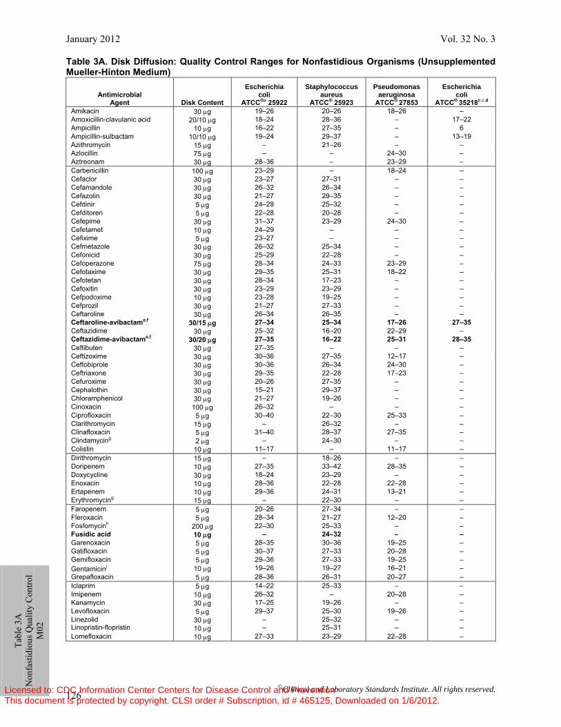
January 2012 Vol. 32 No. 3
©Clinical and Laboratory Standards Institute. All rights reserved. 126
Table 3A. Disk Diffusion: Quality Control Ranges for Nonfastidious Organisms (Unsupplemented Mueller-Hinton Medium)
Antimicrobial
Agent
Disk Content
Escherichia coli
ATCC®a 25922
Staphylococcus aureus
ATCC® 25923
Pseudomonas aeruginosa
ATCC® 27853
Escherichia coli
ATCC® 35218b,c,d Amikacin 30 μg 19–26 20–26 18–26 – Amoxicillin-clavulanic acid 20/10 μg 18–24 28–36 – 17–22 Ampicillin 10 μg 16–22 27–35 – 6 Ampicillin-sulbactam 10/10 μg 19–24 29–37 – 13–19 Azithromycin 15 μg – 21–26 – – Azlocillin 75 μg – – 24–30 – Aztreonam 30 μg 28–36 – 23–29 – Carbenicillin 100 μg 23–29 – 18–24 – Cefaclor 30 μg 23–27 27–31 – – Cefamandole 30 μg 26–32 26–34 – – Cefazolin 30 μg 21–27 29–35 – – Cefdinir 5 μg 24–28 25–32 – – Cefditoren 5 μg 22–28 20–28 – – Cefepime 30 μg 31–37 23–29 24–30 – Cefetamet 10 μg 24–29 – – – Cefixime 5 μg 23–27 – – – Cefmetazole 30 μg 26–32 25–34 – – Cefonicid 30 μg 25–29 22–28 – – Cefoperazone 75 μg 28–34 24–33 23–29 – Cefotaxime 30 μg 29–35 25–31 18–22 – Cefotetan 30 μg 28–34 17–23 – – Cefoxitin 30 μg 23–29 23–29 – – Cefpodoxime 10 μg 23–28 19–25 – – Cefprozil 30 μg 21–27 27–33 – – Ceftaroline 30 μg 26–34 26–35 – – Ceftaroline-avibactame,f 30/15 μg 27–34 25–34 17–26 27–35Ceftazidime 30 μg 25–32 16–20 22–29 – Ceftazidime-avibactame,f 30/20 μg 27–35 16–22 25–31 28–35Ceftibuten 30 μg 27–35 – – – Ceftizoxime 30 μg 30–36 27–35 12–17 – Ceftobiprole 30 μg 30–36 26–34 24–30 – Ceftriaxone 30 μg 29–35 22–28 17–23 – Cefuroxime 30 μg 20–26 27–35 – – Cephalothin 30 μg 15–21 29–37 – – Chloramphenicol 30 μg 21–27 19–26 – – Cinoxacin 100 μg 26–32 – – – Ciprofloxacin 5 μg 30–40 22–30 25–33 – Clarithromycin 15 μg – 26–32 – – Clinafloxacin 5 μg 31–40 28–37 27–35 – Clindamycing 2 μg – 24–30 – – Colistin 10 μg 11–17 – 11–17 – Dirithromycin 15 μg – 18–26 – – Doripenem 10 μg 27–35 33–42 28–35 – Doxycycline 30 μg 18–24 23–29 – – Enoxacin 10 μg 28–36 22–28 22–28 – Ertapenem 10 μg 29–36 24–31 13–21 – Erythromycing 15 μg – 22–30 – – Faropenem 5 μg 20–26 27–34 – – Fleroxacin 5 μg 28–34 21–27 12–20 – Fosfomycinh 200 μg 22–30 25–33 – – Fusidic acid 10 μg – 24–32 – –Garenoxacin 5 μg 28–35 30–36 19–25 – Gatifloxacin 5 μg 30–37 27–33 20–28 – Gemifloxacin 5 μg 29–36 27–33 19–25 – Gentamicinj 10 μg 19–26 19–27 16–21 – Grepafloxacin 5 μg 28–36 26–31 20–27 – Iclaprim 5 μg 14–22 25–33 – – Imipenem 10 μg 26–32 – 20–28 – Kanamycin 30 μg 17–25 19–26 – – Levofloxacin 5 μg 29–37 25–30 19–26 – Linezolid Linopristin-flopristin
30 μg 10 μg
– –
25–32 25–31
– –
– –
Lomefloxacin 10 μg 27–33 23–29 22–28 –
Ta
ble
3A
Non
fast
idio
us Q
ualit
y C
ontro
l M
02
Licensed to: CDC Information Center Centers for Disease Control and PreventionThis document is protected by copyright. CLSI order # Subscription, id # 465125, Downloaded on 1/6/2012.
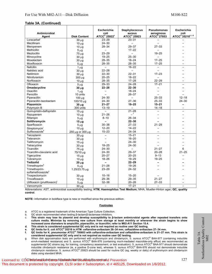
For Use With M02-A11—Disk Diffusion M100-S22
©Clinical and Laboratory Standards Institute. All rights reserved. 127
Tabl
e 3A
N
onfa
stid
ious
Qua
lity
Con
trol
M02
Table 3A. (Continued)
Antimicrobial Agent
Disk Content
Escherichia
coli ATCC® 25922a
Staphylococcus
aureus ATCC® 25923
Pseudomonas
aeruginosa ATCC® 27853
Escherichia
coli ATCC® 35218b,c d
Loracarbef 30 μg 23–29 23–31 – – Mecillinam 10 μg 24–30 – – – Meropenem 10 μg 28–34 29–37 27–33 – Methicillin 5 μg – 17–22 – – Mezlocillin 75 μg 23–29 – 19–25 – Minocycline 30 μg 19–25 25–30 – – Moxalactam 30 μg 28–35 18–24 17–25 – Moxifloxacin 5 μg 28–35 28–35 17–25 – Nafcillin 1 μg – 16–22 – – Nalidixic acid 30 μg 22–28 – – – Netilmicin 30 μg 22–30 22–31 17–23 – Nitrofurantoin 300 μg 20–25 18–22 – – Norfloxacin 10 μg 28–35 17–28 22–29 – Ofloxacin 5 μg 29–33 24–28 17–21 – Omadacycline 30 μg 22–28 22–30 – – Oxacillin 1 μg – 18–24 – – Penicillin 10 units – 26–37 – – Piperacillin 100 μg 24–30 – 25–33 12–18 Piperacillin-tazobactam 100/10 μg 24–30 27–36 25–33 24–30 Plazomicin 30 μg 21–27 19–25 15–21 – Polymyxin B 300 units 13–19 – 14–18 – Quinupristin-dalfopristin 15 μg – 21–28 – – Razupenem 10 μg 21–26 – l – – Rifampin 5 μg 8–10 26–34 – – Solithromycin 15 μg – 22–30 – – Sparfloxacin 5 μg 30–38 27–33 21–29 – Streptomycini 10 μg 12–20 14–22 – – Sulfisoxazolek 250 μg or 300 μg 15–23 24–34 – – Teicoplanin 30 μg – 15–21 – – Telavancin 30 μg – 16–20 – – Telithromycin 15 μg – 24–30 – – Tetracycline 30 μg 18–25 24–30 – – Ticarcillin 75 μg 24–30 – 21–27 6 Ticarcillin-clavulanic acid 75/10 μg 24–30 29–37 20–28 21–25 Tigecycline 15 μg 20–27 20–25 9–13 – Tobramycin 10 μg 18–26 19–29 19–25 – Tedizolid 20 μg – 22–29 – – Trimethoprimk 5 μg 21–28 19–26 – – Trimethoprim-sulfamethoxazolek
1.25/23.75 μg 23–29 24–32 – –
Trospectomycin 30 μg 10–16 15–20 – – Trovafloxacin 10 μg 29–36 29–35 21–27 – Ulifloxacin (prulifloxacin)j 5 μg 32–38 20–26 27–33 – Vancomycin 30 μg – 17–21 – –
Abbreviations: AST, antimicrobial susceptibility testing; HTM, Haemophilus Test Medium; MHA, Mueller-Hinton agar; QC, quality control. NOTE: Information in boldface type is new or modified since the previous edition.
Footnotes a. ATCC is a registered trademark of the American Type Culture Collection. b. QC strain recommended when testing β-lactam/β-lactamase inhibitors. c. This strain may lose its plasmid and develop susceptibility to β-lactam antimicrobial agents after repeated transfers onto
culture media. Minimize by removing new culture from storage at least monthly or whenever the strain begins to show increased zone diameters to ampicillin, piperacillin, or ticarcillin; refer to M02-A11 Section 15.4.
d. This strain is considered supplemental QC only and is not required as routine user QC testing. e. QC limits for E. coli ATCC® 35218 in HTM: ceftaroline-avibactam 26–34 mm; ceftazidime-avibactam 27–34 mm. f. QC limits for K. pneumoniae ATCC® 700603 with ceftaroline-avibactam and ceftazidime-avibactam is 21–27 mm. This strain is
considered supplemental QC only and is not required as routine user QC testing. g. When disk approximation tests are performed with erythromycin and clindamycin, S. aureus ATCC® BAA-977 (containing inducible
ermA-mediated resistance) and S. aureus ATCC® BAA-976 (containing msrA-mediated macrolide-only efflux) are recommended as supplemental QC strains (eg, for training, competency assessment, or test evaluation). S. aureus ATCC® BAA-977 should demonstrate inducible clindamycin resistance (ie, a positive D-zone test), whereas S. aureus ATCC® BAA-976 should not demonstrate inducible clindamycin resistance. S. aureus ATCC® 25923 should be used for routine QC (eg, weekly or daily) of erythromycin and clindamycin disks using standard MHA.
Licensed to: CDC Information Center Centers for Disease Control and PreventionThis document is protected by copyright. CLSI order # Subscription, id # 465125, Downloaded on 1/6/2012.
January 2012 Vol. 32 No. 3
©Clinical and Laboratory Standards Institute. All rights reserved. 128
Tabl
e 3A
N
onfa
stid
ious
Qua
lity
Con
trol
M02
Table 3A. (Continued) h. The 200-μg fosfomycin disk contains 50 μg of glucose-6-phosphate. i. For control limits of gentamicin 120-μg and streptomycin 300-μg disks, use E. faecalis ATCC® 29212 (gentamicin: 16–23 mm;
streptomycin: 14–20 mm). j. Ulifloxacin is the active metabolite of the prodrug prulifloxacin. Only ulifloxacin should be used for AST. k. These agents can be affected by excess levels of thymidine and thymine. See M02-A11, Section 7.1.3 for guidance, should a problem
with QC occur. l. Razupenem tested with S. aureus ATCC® 25923 can often produce the double or target zone phenomenon. For accurate QC results,
use S. aureus ATCC® 29213 (no double zones) with acceptable limit 33–39 mm.
Licensed to: CDC Information Center Centers for Disease Control and PreventionThis document is protected by copyright. CLSI order # Subscription, id # 465125, Downloaded on 1/6/2012.
For Use With M02-A11—Disk Diffusion M100-S22
©Clinical and Laboratory Standards Institute. All rights reserved. 129
This page is intentionally left blank.
Licensed to: CDC Information Center Centers for Disease Control and PreventionThis document is protected by copyright. CLSI order # Subscription, id # 465125, Downloaded on 1/6/2012.
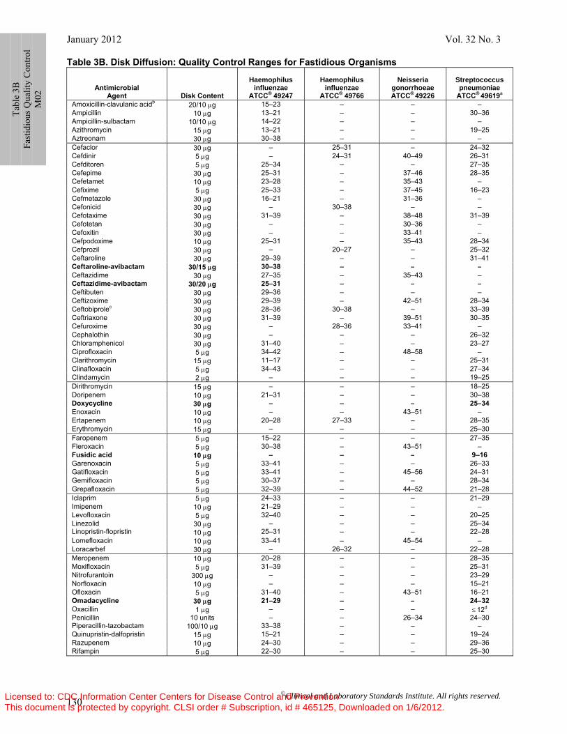
January 2012 Vol. 32 No. 3
©Clinical and Laboratory Standards Institute. All rights reserved. 130
Ta
ble
3B
Fast
idio
us Q
ualit
y C
ontro
l M
02
Table 3B. Disk Diffusion: Quality Control Ranges for Fastidious Organisms
Antimicrobial Agent
Disk Content
Haemophilus
influenzae ATCC® 49247
Haemophilus
influenzae ATCC® 49766
Neisseria
gonorrhoeae ATCC® 49226
Streptococcus pneumoniae
ATCC® 49619a Amoxicillin-clavulanic acidb 20/10 μg 15–23 – – – Ampicillin 10 μg 13–21 – – 30–36 Ampicillin-sulbactam 10/10 μg 14–22 – – – Azithromycin 15 μg 13–21 – – 19–25 Aztreonam 30 μg 30–38 – – – Cefaclor 30 μg – 25–31 – 24–32 Cefdinir 5 μg – 24–31 40–49 26–31 Cefditoren 5 μg 25–34 – – 27–35 Cefepime 30 μg 25–31 – 37–46 28–35 Cefetamet 10 μg 23–28 – 35–43 – Cefixime 5 μg 25–33 – 37–45 16–23 Cefmetazole 30 μg 16–21 – 31–36 – Cefonicid 30 μg – 30–38 – – Cefotaxime 30 μg 31–39 – 38–48 31–39 Cefotetan 30 μg – – 30–36 – Cefoxitin 30 μg – – 33–41 – Cefpodoxime 10 μg 25–31 – 35–43 28–34 Cefprozil 30 μg – 20–27 – 25–32 Ceftaroline 30 μg 29–39 – – 31–41 Ceftaroline-avibactam 30/15 μg 30–38 – – –Ceftazidime 30 μg 27–35 – 35–43 – Ceftazidime-avibactam 30/20 μg 25–31 – – –Ceftibuten 30 μg 29–36 – – – Ceftizoxime 30 μg 29–39 – 42–51 28–34 Ceftobiprolec 30 μg 28–36 30–38 – 33–39 Ceftriaxone 30 μg 31–39 – 39–51 30–35 Cefuroxime 30 μg – 28–36 33–41 – Cephalothin 30 μg – – – 26–32 Chloramphenicol 30 μg 31–40 – – 23–27 Ciprofloxacin 5 μg 34–42 – 48–58 – Clarithromycin 15 μg 11–17 – – 25–31 Clinafloxacin 5 μg 34–43 – – 27–34 Clindamycin 2 μg – – – 19–25 Dirithromycin 15 μg – – – 18–25 Doripenem 10 μg 21–31 – – 30–38 Doxycycline 30 μg – – – 25–34Enoxacin 10 μg – – 43–51 – Ertapenem 10 μg 20–28 27–33 – 28–35 Erythromycin 15 μg – – – 25–30 Faropenem 5 μg 15–22 – – 27–35 Fleroxacin 5 μg 30–38 – 43–51 – Fusidic acid 10 μg – – – 9–16Garenoxacin 5 μg 33–41 – – 26–33 Gatifloxacin 5 μg 33–41 – 45–56 24–31 Gemifloxacin 5 μg 30–37 – – 28–34 Grepafloxacin 5 μg 32–39 – 44–52 21–28 Iclaprim 5 μg 24–33 – – 21–29 Imipenem 10 μg 21–29 – – – Levofloxacin 5 μg 32–40 – – 20–25 Linezolid Linopristin-flopristin
30 μg 10 μg
– 25–31
– –
– –
25–34 22–28
Lomefloxacin 10 μg 33–41 – 45–54 – Loracarbef 30 μg – 26–32 – 22–28 Meropenem 10 μg 20–28 – – 28–35 Moxifloxacin 5 μg 31–39 – – 25–31 Nitrofurantoin 300 μg – – – 23–29 Norfloxacin 10 μg – – – 15–21 Ofloxacin 5 μg 31–40 – 43–51 16–21 Omadacycline 30 μg 21–29 – – 24–32Oxacillin 1 μg – – – ≤ 12d Penicillin 10 units – – 26–34 24–30 Piperacillin-tazobactam 100/10 μg 33–38 – – – Quinupristin-dalfopristin 15 μg 15–21 – – 19–24 Razupenem 10 μg 24–30 – – 29–36 Rifampin 5 μg 22–30 – – 25–30
Licensed to: CDC Information Center Centers for Disease Control and PreventionThis document is protected by copyright. CLSI order # Subscription, id # 465125, Downloaded on 1/6/2012.
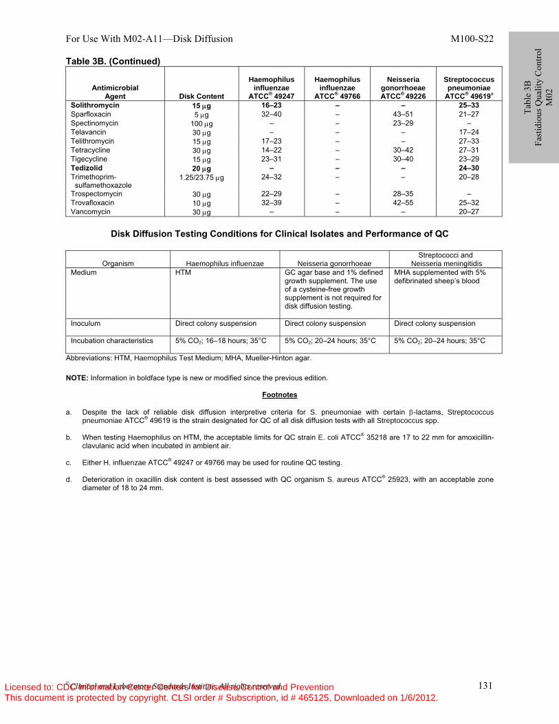
For Use With M02-A11—Disk Diffusion M100-S22
©Clinical and Laboratory Standards Institute. All rights reserved. 131
Table 3B. (Continued)
Antimicrobial Agent
Disk Content
Haemophilus
influenzae ATCC® 49247
Haemophilus
influenzae ATCC® 49766
Neisseria
gonorrhoeae ATCC® 49226
Streptococcus pneumoniae
ATCC® 49619a Solithromycin 15 μg 16–23 – – 25–33 Sparfloxacin 5 μg 32–40 – 43–51 21–27 Spectinomycin 100 μg – – 23–29 – Telavancin 30 μg – – – 17–24 Telithromycin 15 μg 17–23 – – 27–33 Tetracycline 30 μg 14–22 – 30–42 27–31 Tigecycline 15 μg 23–31 – 30–40 23–29 Tedizolid 20 μg – – – 24–30 Trimethoprim- sulfamethoxazole
1.25/23.75 μg 24–32 – – 20–28
Trospectomycin 30 μg 22–29 – 28–35 – Trovafloxacin 10 μg 32–39 – 42–55 25–32 Vancomycin 30 μg – – – 20–27
Disk Diffusion Testing Conditions for Clinical Isolates and Performance of QC
Organism Haemophilus influenzae Neisseria gonorrhoeae Streptococci and
Neisseria meningitidis Medium HTM GC agar base and 1% defined
growth supplement. The use of a cysteine-free growth supplement is not required for disk diffusion testing.
MHA supplemented with 5% defibrinated sheep’s blood
Inoculum
Direct colony suspension Direct colony suspension Direct colony suspension
Incubation characteristics
5% CO2; 16–18 hours; 35°C 5% CO2; 20–24 hours; 35°C 5% CO2; 20–24 hours; 35°C
Abbreviations: HTM, Haemophilus Test Medium; MHA, Mueller-Hinton agar. NOTE: Information in boldface type is new or modified since the previous edition.
Footnotes a. Despite the lack of reliable disk diffusion interpretive criteria for S. pneumoniae with certain β-lactams, Streptococcus
pneumoniae ATCC® 49619 is the strain designated for QC of all disk diffusion tests with all Streptococcus spp. b. When testing Haemophilus on HTM, the acceptable limits for QC strain E. coli ATCC® 35218 are 17 to 22 mm for amoxicillin-
clavulanic acid when incubated in ambient air. c. Either H. influenzae ATCC® 49247 or 49766 may be used for routine QC testing. d. Deterioration in oxacillin disk content is best assessed with QC organism S. aureus ATCC® 25923, with an acceptable zone
diameter of 18 to 24 mm.
Tabl
e 3B
Fa
stid
ious
Qua
lity
Con
trol
M02
Licensed to: CDC Information Center Centers for Disease Control and PreventionThis document is protected by copyright. CLSI order # Subscription, id # 465125, Downloaded on 1/6/2012.
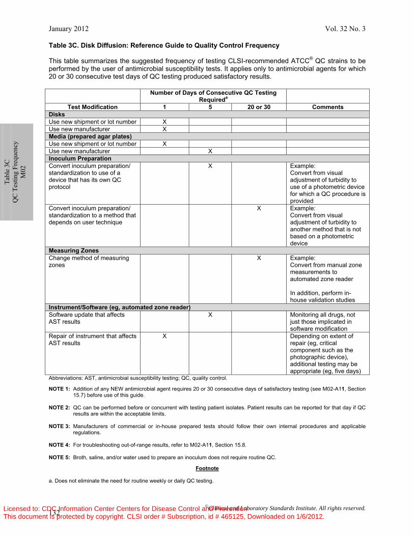
January 2012 Vol. 32 No. 3
©Clinical and Laboratory Standards Institute. All rights reserved. 132
Table 3C. Disk Diffusion: Reference Guide to Quality Control Frequency
This table summarizes the suggested frequency of testing CLSI-recommended ATCC® QC strains to be performed by the user of antimicrobial susceptibility tests. It applies only to antimicrobial agents for which 20 or 30 consecutive test days of QC testing produced satisfactory results.
Number of Days of Consecutive QC Testing
Requireda Test Modification 1 5 20 or 30 Comments
Disks Use new shipment or lot number X Use new manufacturer X Media (prepared agar plates) Use new shipment or lot number X Use new manufacturer X Inoculum Preparation Convert inoculum preparation/ standardization to use of a device that has its own QC protocol
X Example: Convert from visual adjustment of turbidity to use of a photometric device for which a QC procedure is provided
Convert inoculum preparation/ standardization to a method that depends on user technique
X Example: Convert from visual adjustment of turbidity to another method that is not based on a photometric device
Measuring Zones Change method of measuring zones
X Example: Convert from manual zone measurements to automated zone reader In addition, perform in-house validation studies
Instrument/Software (eg, automated zone reader)Software update that affects AST results
X Monitoring all drugs, not just those implicated in software modification
Repair of instrument that affects AST results
X Depending on extent of repair (eg, critical component such as the photographic device), additional testing may be appropriate (eg, five days)
Abbreviations: AST, antimicrobial susceptibility testing; QC, quality control. NOTE 1: Addition of any NEW antimicrobial agent requires 20 or 30 consecutive days of satisfactory testing (see M02-A11, Section
15.7) before use of this guide. NOTE 2: QC can be performed before or concurrent with testing patient isolates. Patient results can be reported for that day if QC
results are within the acceptable limits. NOTE 3: Manufacturers of commercial or in-house prepared tests should follow their own internal procedures and applicable
regulations. NOTE 4: For troubleshooting out-of-range results, refer to M02-A11, Section 15.8. NOTE 5: Broth, saline, and/or water used to prepare an inoculum does not require routine QC.
Footnote a. Does not eliminate the need for routine weekly or daily QC testing.
Ta
ble
3C
QC
Tes
ting
Freq
uenc
y M
02
Licensed to: CDC Information Center Centers for Disease Control and PreventionThis document is protected by copyright. CLSI order # Subscription, id # 465125, Downloaded on 1/6/2012.

For Use With M02-A11—Disk Diffusion M100-S22
©Clinical and Laboratory Standards Institute. All rights reserved. 133
This page is intentionally left blank.
Licensed to: CDC Information Center Centers for Disease Control and PreventionThis document is protected by copyright. CLSI order # Subscription, id # 465125, Downloaded on 1/6/2012.
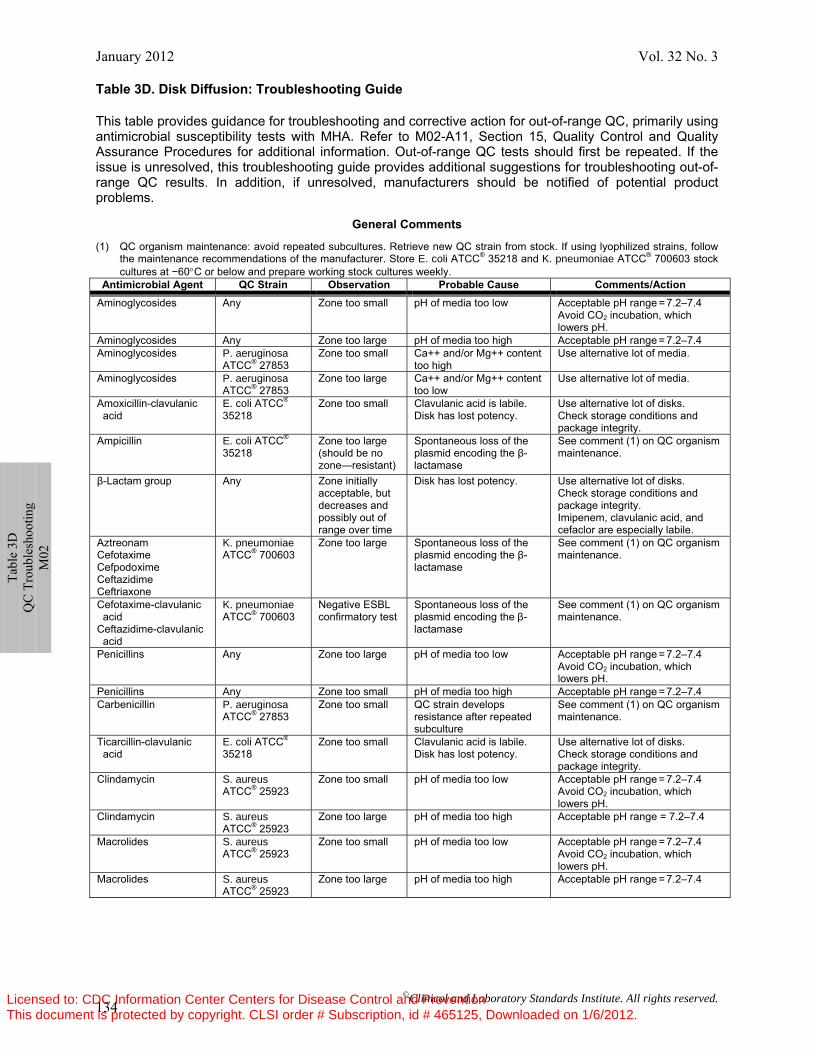
January 2012 Vol. 32 No. 3
©Clinical and Laboratory Standards Institute. All rights reserved. 134
Table 3D. Disk Diffusion: Troubleshooting Guide
This table provides guidance for troubleshooting and corrective action for out-of-range QC, primarily using antimicrobial susceptibility tests with MHA. Refer to M02-A11, Section 15, Quality Control and Quality Assurance Procedures for additional information. Out-of-range QC tests should first be repeated. If the issue is unresolved, this troubleshooting guide provides additional suggestions for troubleshooting out-of-range QC results. In addition, if unresolved, manufacturers should be notified of potential product problems.
General Comments
(1) QC organism maintenance: avoid repeated subcultures. Retrieve new QC strain from stock. If using lyophilized strains, follow the maintenance recommendations of the manufacturer. Store E. coli ATCC® 35218 and K. pneumoniae ATCC® 700603 stock cultures at −60°C or below and prepare working stock cultures weekly.
Antimicrobial Agent QC Strain Observation Probable Cause Comments/Action Aminoglycosides Any Zone too small pH of media too low Acceptable pH range = 7.2–7.4
Avoid CO2 incubation, which lowers pH.
Aminoglycosides Any Zone too large pH of media too high Acceptable pH range = 7.2–7.4 Aminoglycosides P. aeruginosa
ATCC® 27853 Zone too small Ca++ and/or Mg++ content
too high Use alternative lot of media.
Aminoglycosides P. aeruginosa ATCC® 27853
Zone too large Ca++ and/or Mg++ content too low
Use alternative lot of media.
Amoxicillin-clavulanic acid
E. coli ATCC® 35218
Zone too small Clavulanic acid is labile. Disk has lost potency.
Use alternative lot of disks. Check storage conditions and package integrity.
Ampicillin E. coli ATCC®
35218 Zone too large (should be no zone—resistant)
Spontaneous loss of the plasmid encoding the β-lactamase
See comment (1) on QC organism maintenance.
β-Lactam group Any Zone initially acceptable, but decreases and possibly out of range over time
Disk has lost potency. Use alternative lot of disks. Check storage conditions and package integrity. Imipenem, clavulanic acid, and cefaclor are especially labile.
Aztreonam Cefotaxime Cefpodoxime Ceftazidime Ceftriaxone
K. pneumoniae ATCC® 700603
Zone too large Spontaneous loss of the plasmid encoding the β-lactamase
See comment (1) on QC organism maintenance.
Cefotaxime-clavulanic acid Ceftazidime-clavulanic acid
K. pneumoniae ATCC® 700603
Negative ESBL confirmatory test
Spontaneous loss of the plasmid encoding the β-lactamase
See comment (1) on QC organism maintenance.
Penicillins Any Zone too large pH of media too low Acceptable pH range = 7.2–7.4 Avoid CO2 incubation, which lowers pH.
Penicillins Any Zone too small pH of media too high Acceptable pH range = 7.2–7.4 Carbenicillin P. aeruginosa
ATCC® 27853 Zone too small QC strain develops
resistance after repeated subculture
See comment (1) on QC organism maintenance.
Ticarcillin-clavulanic acid
E. coli ATCC® 35218
Zone too small Clavulanic acid is labile. Disk has lost potency.
Use alternative lot of disks. Check storage conditions and package integrity.
Clindamycin S. aureus ATCC® 25923
Zone too small pH of media too low Acceptable pH range = 7.2–7.4 Avoid CO2 incubation, which lowers pH.
Clindamycin S. aureus ATCC® 25923
Zone too large pH of media too high Acceptable pH range = 7.2–7.4
Macrolides S. aureus ATCC® 25923
Zone too small pH of media too low Acceptable pH range = 7.2–7.4 Avoid CO2 incubation, which lowers pH.
Macrolides S. aureus ATCC® 25923
Zone too large pH of media too high Acceptable pH range = 7.2–7.4
Ta
ble
3D
QC
Tro
uble
shoo
ting
M02
Licensed to: CDC Information Center Centers for Disease Control and PreventionThis document is protected by copyright. CLSI order # Subscription, id # 465125, Downloaded on 1/6/2012.
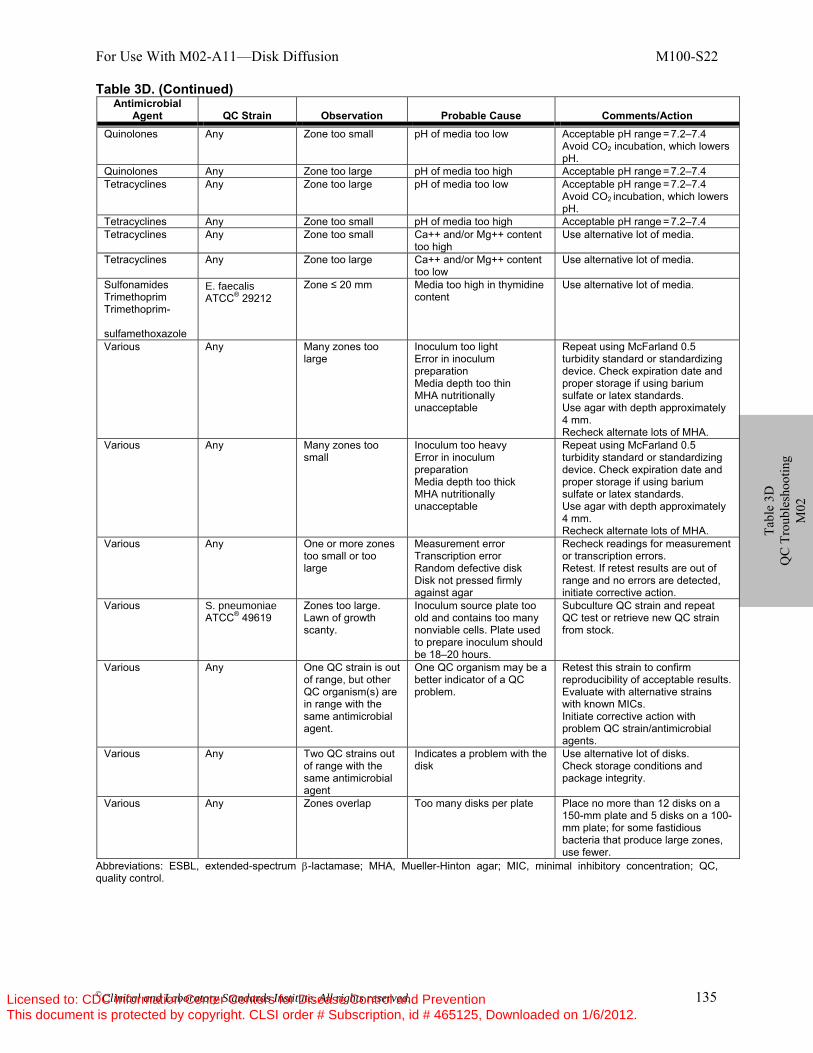
For Use With M02-A11—Disk Diffusion M100-S22
©Clinical and Laboratory Standards Institute. All rights reserved. 135
Ta
ble
3D
QC
Tro
uble
shoo
ting
M02
Table 3D. (Continued)
Abbreviations: ESBL, extended-spectrum β-lactamase; MHA, Mueller-Hinton agar; MIC, minimal inhibitory concentration; QC, quality control.
Antimicrobial Agent QC Strain Observation Probable Cause Comments/Action
Quinolones Any Zone too small pH of media too low Acceptable pH range = 7.2–7.4 Avoid CO2 incubation, which lowers pH.
Quinolones Any Zone too large pH of media too high Acceptable pH range = 7.2–7.4 Tetracyclines Any Zone too large pH of media too low Acceptable pH range = 7.2–7.4
Avoid CO2 incubation, which lowers pH.
Tetracyclines Any Zone too small pH of media too high Acceptable pH range = 7.2–7.4 Tetracyclines Any Zone too small Ca++ and/or Mg++ content
too high Use alternative lot of media.
Tetracyclines Any Zone too large Ca++ and/or Mg++ content too low
Use alternative lot of media.
Sulfonamides Trimethoprim Trimethoprim- sulfamethoxazole
E. faecalis ATCC® 29212
Zone ≤ 20 mm Media too high in thymidine content
Use alternative lot of media.
Various Any Many zones too large
Inoculum too light Error in inoculum preparation Media depth too thin MHA nutritionally unacceptable
Repeat using McFarland 0.5 turbidity standard or standardizing device. Check expiration date and proper storage if using barium sulfate or latex standards. Use agar with depth approximately 4 mm. Recheck alternate lots of MHA.
Various Any Many zones too small
Inoculum too heavy Error in inoculum preparation Media depth too thick MHA nutritionally unacceptable
Repeat using McFarland 0.5 turbidity standard or standardizing device. Check expiration date and proper storage if using barium sulfate or latex standards. Use agar with depth approximately 4 mm. Recheck alternate lots of MHA.
Various Any One or more zones too small or too large
Measurement error Transcription error Random defective disk Disk not pressed firmly against agar
Recheck readings for measurement or transcription errors. Retest. If retest results are out of range and no errors are detected, initiate corrective action.
Various S. pneumoniae ATCC® 49619
Zones too large. Lawn of growth scanty.
Inoculum source plate too old and contains too many nonviable cells. Plate used to prepare inoculum should be 18–20 hours.
Subculture QC strain and repeat QC test or retrieve new QC strain from stock.
Various Any One QC strain is out of range, but other QC organism(s) are in range with the same antimicrobial agent.
One QC organism may be a better indicator of a QC problem.
Retest this strain to confirm reproducibility of acceptable results. Evaluate with alternative strains with known MICs. Initiate corrective action with problem QC strain/antimicrobial agents.
Various Any Two QC strains out of range with the same antimicrobial agent
Indicates a problem with the disk
Use alternative lot of disks. Check storage conditions and package integrity.
Various Any Zones overlap Too many disks per plate Place no more than 12 disks on a 150-mm plate and 5 disks on a 100-mm plate; for some fastidious bacteria that produce large zones, use fewer.
Licensed to: CDC Information Center Centers for Disease Control and PreventionThis document is protected by copyright. CLSI order # Subscription, id # 465125, Downloaded on 1/6/2012.
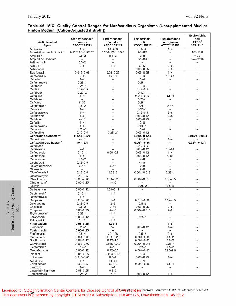
January 2012 Vol. 32 No. 3
©Clinical and Laboratory Standards Institute. All rights reserved. 136
Table 4A. MIC: Quality Control Ranges for Nonfastidious Organisms (Unsupplemented Mueller-Hinton Medium [Cation-Adjusted if Broth])
Antimicrobial Agent
Staphylococcus aureus
ATCC®a 29213
Enterococcus faecalis
ATCC® 29212
Escherichia coli
ATCC® 25922
Pseudomonas aeruginosa
ATCC® 27853
Escherichia coli
ATCC® 35218b,c,d
Amikacin 1–4 64–256 0.5–4 1–4 – Amoxicillin-clavulanic acid 0.12/0.06–0.5/0.25 0.25/0.12–1.0/0.5 2/1–8/4 – 4/2–16/8 Ampicillin 0.5–2 0.5–2 2–8 – > 32 Ampicillin-sulbactam – – 2/1–8/4 – 8/4–32/16 Azithromycin 0.5–2 – – – – Azlocillin 2–8 1–4 8–32 2–8 – Aztreonam – – 0.06–0.25 2–8 – Besifloxacin 0.015–0.06 0.06–0.25 0.06–0.25 1–4 – Carbenicillin 2–8 16–64 4–16 16–64 – Cefaclor 1–4 – 1–4 – – Cefamandole 0.25–1 – 0.25–1 – – Cefazolin 0.25–1 – 1–4 – – Cefdinir 0.12–0.5 – 0.12–0.5 – – Cefditoren 0.25–2 – 0.12–1 – – Cefepime 1–4 – 0.015–0.12 0.5–4 – Cefetamet – – 0.25–1 – – Cefixime 8–32 – 0.25–1 – – Cefmetazole 0.5–2 – 0.25–1 > 32 – Cefonicid 1–4 – 0.25–1 – – Cefoperazone 1–4 – 0.12–0.5 2–8 – Cefotaxime 1–4 – 0.03–0.12 8–32 – Cefotetan 4–16 – 0.06–0.25 – – Cefoxitin 1–4 – 2–8 – – Cefpodoxime 1–8 – 0.25–1 – – Cefprozil 0.25–1 – 1–4 – – Ceftaroline 0.12–0.5 0.25–2d 0.03–0.12 – – Ceftaroline-avibactame 0.12/4–0.5/4 – 0.03/4–0.12/4 – 0.015/4–0.06/4Ceftazidime 4–16 – 0.06–0.5 1–4 – Ceftazidime-avibactamf 4/4–16/4 – 0.06/4–0.5/4 – 0.03/4–0.12/4Ceftibuten – – 0.12–0.5 – – Ceftizoxime 2–8 – 0.03–0.12 16–64 – Ceftobiprole 0.12–1 0.06–0.5 0.03–0.12 1–4 – Ceftriaxone 1–8 – 0.03–0.12 8–64 – Cefuroxime 0.5–2 – 2–8 – – Cephalothin 0.12–0.5 – 4–16 – – Chloramphenicol 2–16 4–16 2–8 – – Cinoxacin – – 2–8 – – Ciprofloxacing 0.12–0.5 0.25–2 0.004–0.015 0.25–1 – Clarithromycin 0.12–0.5 – – – – Clinafloxacin 0.008–0.06 0.03–0.25 0.002–0.015 0.06–0.5 – Clindamycinh 0.06–0.25 4–16 – – – Colistin – – 0.25–2 0.5–4 – Dalbavancinj 0.03–0.12 0.03–0.12 – – – Daptomycink 0.12–1 1–4 – – – Dirithromycin 1–4 – – – – Doripenem 0.015–0.06 1–4 0.015–0.06 0.12–0.5 – Doxycycline 0.12–0.5 2–8 0.5–2 – – Enoxacin 0.5–2 2–16 0.06–0.25 2–8 – Ertapenem 0.06–0.25 4–16 0.004–0.015 2–8 – Erythromycinh 0.25–1 1–4 – – – Faropenem 0.03–0.12 – 0.25–1 – – Fidaxomicin 2–16 1–4 – – – Finafloxacin 0.03–0.25 0.25–1 – 1–8 –Fleroxacin 0.25–1 2–8 0.03–0.12 1–4 – Fusidic acid 0.06–0.25 – – – –Fosfomycinl 0.5–4 32–128 0.5–2 2–8 – Garenoxacin 0.004–0.03 0.03–0.25 0.004–0.03 0.5–2 – Gatifloxacin 0.03–0.12 0.12–1.0 0.008–0.03 0.5–2 – Gemifloxacin 0.008–0.03 0.015–0.12 0.004–0.015 0.25–1 – Gentamicinm 0.12–1 4–16 0.25–1 0.5–2 – Grepafloxacin 0.03–0.12 0.12–0.5 0.004–0.03 0.25–2.0 – Iclaprim 0.06–0.25 0.004–0.03 1–4 – – Imipenem 0.015–0.06 0.5–2 0.06–0.25 1–4 – Kanamycin 1–4 16–64 1–4 – – Levofloxacin 0.06–0.5 0.25–2 0.008–0.06 0.5–4 – Linezolid Linopristin-flopristin
1–4 0.06–0.25
1–4 0.5–2
– –
– –
– –
Lomefloxacin 0.25–2 2–8 0.03–0.12 1–4 –
Ta
ble
4A
Non
fast
idio
us Q
ualit
y C
ontro
l M
07
Licensed to: CDC Information Center Centers for Disease Control and PreventionThis document is protected by copyright. CLSI order # Subscription, id # 465125, Downloaded on 1/6/2012.
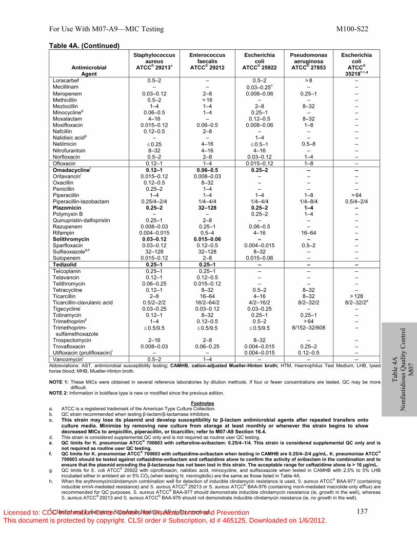
For Use With M07-A9—MIC Testing M100-S22
©Clinical and Laboratory Standards Institute. All rights reserved. 137
Tabl
e 4A
N
onfa
stid
ious
Qua
lity
Con
trol
M07
Table 4A. (Continued)
Antimicrobial Agent
Staphylococcus aureus
ATCC® 29213a
Enterococcus faecalis
ATCC® 29212
Escherichia coli
ATCC® 25922
Pseudomonas aeruginosa
ATCC® 27853
Escherichia coli
ATCC® 35218b,c,d
Loracarbef 0.5–2 – 0.5–2 > 8 – Mecillinam – – 0.03–0.25n – – Meropenem 0.03–0.12 2–8 0.008–0.06 0.25–1 – Methicillin 0.5–2 > 16 – – – Mezlocillin 1–4 1–4 2–8 8–32 – Minocyclineg 0.06–0.5 1–4 0.25–1 – – Moxalactam 4–16 – 0.12–0.5 8–32 – Moxifloxacin 0.015–0.12 0.06–0.5 0.008–0.06 1–8 – Nafcillin 0.12–0.5 2–8 – – – Nalidixic acidg – – 1–4 – – Netilmicin ≤ 0.25 4–16 ≤ 0.5–1 0.5–8 – Nitrofurantoin 8–32 4–16 4–16 – – Norfloxacin 0.5–2 2–8 0.03–0.12 1–4 – Ofloxacin 0.12–1 1–4 0.015–0.12 1–8 – Omadacyclinei 0.12–1 0.06–0.5 0.25–2 – – Oritavancinj 0.015–0.12 0.008–0.03 – – – Oxacillin 0.12–0.5 8–32 – – – Penicillin 0.25–2 1–4 – – – Piperacillin 1–4 1–4 1–4 1–8 > 64 Piperacillin-tazobactam 0.25/4–2/4 1/4–4/4 1/4–4/4 1/4–8/4 0.5/4–2/4 Plazomicin 0.25–2 32–128 0.25–2 1–4 – Polymyxin B – – 0.25–2 1–4 – Quinupristin-dalfopristin 0.25–1 2–8 – – – Razupenem 0.008–0.03 0.25–1 0.06–0.5 – – Rifampin 0.004–0.015 0.5–4 4–16 16–64 – Solithromycin 0.03–0.12 0.015–0.06 – – – Sparfloxacin 0.03–0.12 0.12–0.5 0.004–0.015 0.5–2 – Sulfisoxazoleg,p 32–128 32–128 8–32 – – Sulopenem 0.015–0.12 2–8 0.015–0.06 – – Tedizolid 0.25–1 0.25–1 – – – Teicoplanin 0.25–1 0.25–1 – – – Telavancin 0.12–1 0.12–0.5 – – – Telithromycin 0.06–0.25 0.015–0.12 – – – Tetracycline 0.12–1 8–32 0.5–2 8–32 – Ticarcillin 2–8 16–64 4–16 8–32 > 128 Ticarcillin-clavulanic acid 0.5/2–2/2 16/2–64/2 4/2–16/2 8/2–32/2 8/2–32/2q
Tigecyclinei 0.03–0.25 0.03–0.12 0.03–0.25 – – Tobramycin 0.12–1 8–32 0.25–1 0.25–1 – Trimethoprimp 1–4 0.12–0.5 0.5–2 > 64 – Trimethoprim- sulfamethoxazole
≤ 0.5/9.5 ≤ 0.5/9.5 ≤ 0.5/9.5 8/152–32/608 –
Trospectomycin 2–16 2–8 8–32 – – Trovafloxacin 0.008–0.03 0.06–0.25 0.004–0.015 0.25–2 – Ulifloxacin (prulifloxacin)l – – 0.004–0.015 0.12–0.5 – Vancomycinr 0.5–2 1–4 – – –
Abbreviations: AST, antimicrobial susceptibility testing; CAMHB, cation-adjusted Mueller-Hinton broth; HTM, Haemophilus Test Medium; LHB, lysed horse blood; MHB, Mueller-Hinton broth. NOTE 1: These MICs were obtained in several reference laboratories by dilution methods. If four or fewer concentrations are tested, QC may be more
difficult. NOTE 2: Information in boldface type is new or modified since the previous edition.
Footnotes
a. ATCC is a registered trademark of the American Type Culture Collection. b. QC strain recommended when testing β-lactam/β-lactamase inhibitors. c. This strain may lose its plasmid and develop susceptibility to β-lactam antimicrobial agents after repeated transfers onto
culture media. Minimize by removing new culture from storage at least monthly or whenever the strain begins to show decreased MICs to ampicillin, piperacillin, or ticarcillin; refer to M07-A9 Section 16.4.
d. This strain is considered supplemental QC only and is not required as routine user QC testing. e. QC limits for K. pneumoniae ATCC® 700603 with ceftaroline-avibactam: 0.25/4–1/4. This strain is considered supplemental QC only and is
not required as routine user QC testing. f. QC limits for K. pneumoniae ATCC® 700603 with ceftazidime-avibactam when testing in CAMHB are 0.25/4–2/4 µg/mL. K. pneumoniae ATCC®
700603 should be tested against ceftazidime-avibactam and ceftazidime alone to confirm the activity of avibactam in the combination and to ensure that the plasmid encoding the β-lactamase has not been lost in this strain. The acceptable range for ceftazidime alone is > 16 µg/mL.
g. QC limits for E. coli ATCC® 25922 with ciprofloxacin, nalidixic acid, minocycline, and sulfisoxazole when tested in CAMHB with 2.5% to 5% LHB incubated either in ambient air or 5% CO2 (when testing N. meningitidis) are the same as those listed in Table 4A.
h. When the erythromycin/clindamycin combination well for detection of inducible clindamycin resistance is used, S. aureus ATCC® BAA-977 (containing inducible ermA-mediated resistance) and S. aureus ATCC® 29213 or S. aureus ATCC® BAA-976 (containing msrA-mediated macrolide-only efflux) are recommended for QC purposes. S. aureus ATCC® BAA-977 should demonstrate inducible clindamycin resistance (ie, growth in the well), whereas S. aureus ATCC® 29213 and S. aureus ATCC® BAA-976 should not demonstrate inducible clindamycin resistance (ie, no growth in the well).
Licensed to: CDC Information Center Centers for Disease Control and PreventionThis document is protected by copyright. CLSI order # Subscription, id # 465125, Downloaded on 1/6/2012.
January 2012 Vol. 32 No. 3
©Clinical and Laboratory Standards Institute. All rights reserved. 138
Ta
ble
4A
Non
fast
idio
us Q
ualit
y C
ontro
l M
07
Table 4A. (Continued) i. For broth microdilution testing of omadacycline and tigecycline, when MIC panels are prepared, the medium must be prepared fresh on the day of
use. The medium must be no more than 12 hours old at the time the panels are made; however, the panels may then be frozen for later use. j. QC ranges reflect MICs obtained when CAMHB is supplemented with 0.002% polysorbate-80. k. QC ranges reflect MICs obtained when MHB is supplemented with calcium to a final concentration of 50 μg/mL. Agar dilution has not been validated for
daptomycin. l. The approved MIC susceptibility testing method is agar dilution. Agar media should be supplemented with 25 μg/mL of glucose-6-phosphate. Broth
dilution should not be performed. m. For control organisms for gentamicin and streptomycin high-level aminoglycoside screen tests for enterococci, see Table 2D Supplemental Table 1 at
the end of Table 2D. n. This test should be performed by agar dilution only. o. Ulifloxacin is the active metabolite of the prodrug prulifloxacin. Only ulifloxacin should be used for AST. p. Very medium-dependent, especially with enterococci. q. The QC limits for E. coli ATCC® 35218 when using HTM are 16/2 to 64/2 μg/mL. r. For QC organisms for vancomycin screen test for enterococci, see Table 2D Supplemental Table 1.
Licensed to: CDC Information Center Centers for Disease Control and PreventionThis document is protected by copyright. CLSI order # Subscription, id # 465125, Downloaded on 1/6/2012.

For Use With M07-A9—MIC Testing M100-S22
©Clinical and Laboratory Standards Institute. All rights reserved. 139
This page is intentionally left blank.
Licensed to: CDC Information Center Centers for Disease Control and PreventionThis document is protected by copyright. CLSI order # Subscription, id # 465125, Downloaded on 1/6/2012.
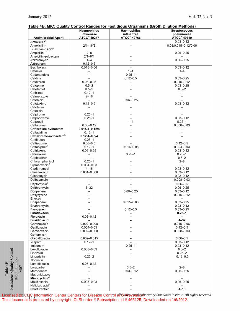
January 2012 Vol. 32 No. 3
©Clinical and Laboratory Standards Institute. All rights reserved. 140
Table 4B. MIC: Quality Control Ranges for Fastidious Organisms (Broth Dilution Methods)
Antimicrobial Agent
Haemophilus influenzae
ATCC® 49247
Haemophilus influenzae
ATCC® 49766
Streptococcus pneumoniae ATCC® 49619
Amoxicillina – – 0.03–0.12 Amoxicillin- clavulanic acida
2/1–16/8 – 0.03/0.015–0.12/0.06
Ampicillin 2–8 – 0.06–0.25 Ampicillin-sulbactam 2/1–8/4 – – Azithromycin 1–4 – 0.06–0.25 Aztreonam 0.12–0.5 – – Besifloxacin 0.015–0.06 – 0.03–0.12 Cefaclor – 1–4 1–4 Cefamandole – 0.25–1 – Cefdinir – 0.12–0.5 0.03–0.25 Cefditoren 0.06–0.25 – 0.015–0.12 Cefepime 0.5–2 – 0.03–0.25 Cefetamet 0.5–2 – 0.5–2 Cefixime 0.12–1 – – Cefmetazole 2–16 – – Cefonicid – 0.06–0.25 – Cefotaxime 0.12–0.5 – 0.03–0.12 Cefotetan – – – Cefoxitin – – – Cefpirome 0.25–1 – – Cefpodoxime 0.25–1 – 0.03–0.12 Cefprozil – 1–4 0.25–1 Ceftaroline 0.03–0.12 – 0.008–0.03 Ceftaroline-avibactam 0.015/4–0.12/4 – – Ceftazidime 0.12–1 – – Ceftazidime-avibactamb 0.12/4–0.5/4 – – Ceftibuten 0.25–1 – – Ceftizoxime 0.06–0.5 – 0.12–0.5 Ceftobiprolec 0.12–1 0.016–0.06 0.004–0.03 Ceftriaxone 0.06–0.25 – 0.03–0.12 Cefuroxime – 0.25–1 0.25–1 Cephalothin – – 0.5–2 Chloramphenicol 0.25–1 – 2–8 Ciprofloxacind 0.004–0.03 – – Clarithromycin 4–16 – 0.03–0.12 Clinafloxacin 0.001–0.008 – 0.03–0.12 Clindamycin – – 0.03–0.12 Dalbavancinf – – 0.008–0.03 Daptomycing – – 0.06–0.5 Dirithromycin 8–32 – 0.06–0.25 Doripenem – 0.06–0.25 0.03–0.12 Doxycycline – – 0.015–0.12 Enoxacin – – – Ertapenem – 0.015–0.06 0.03–0.25 Erythromycin – – 0.03–0.12 Faropenem – 0.12–0.5 0.03–0.25 Finafloxacin – – 0.25–1 Fleroxacin 0.03–0.12 – – Fusidic acid – – 4–32 Garenoxacin 0.002–0.008 – 0.015–0.06 Gatifloxacin 0.004–0.03 – 0.12–0.5 Gemifloxacin 0.002–0.008 – 0.008–0.03 Gentamicin – – – Grepafloxacin 0.002–0.015 – 0.06–0.5 Iclaprim 0.12–1 – 0.03–0.12 Imipenem – 0.25–1 0.03–0.12 Levofloxacin 0.008–0.03 – 0.5–2 Linezolid Linopristin- flopristin
– 0.25–2
– –
0.25–2 0.12–0.5
Lomefloxacin 0.03–0.12 – – Loracarbef – 0.5–2 2–8 Meropenem – 0.03–0.12 0.06–0.25 Metronidazole – – – Minocyclined – – – Moxifloxacin 0.008–0.03 – 0.06–0.25 Nalidixic acidd – – – Nitrofurantoin – – 4–16
Ta
ble
4B
Fast
idio
us Q
ualit
y C
ontro
l B
roth
Dilu
tion
M07
Licensed to: CDC Information Center Centers for Disease Control and PreventionThis document is protected by copyright. CLSI order # Subscription, id # 465125, Downloaded on 1/6/2012.
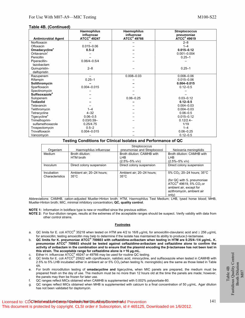
For Use With M07-A9—MIC Testing M100-S22
©Clinical and Laboratory Standards Institute. All rights reserved. 141
Table 4B. (Continued)
Antimicrobial Agent
Haemophilus influenzae
ATCC® 49247
Haemophilus influenzae
ATCC® 49766
Streptococcus pneumoniae ATCC® 49619
Norfloxacin – – 2–8 Ofloxacin 0.015–0.06 – 1–4 Omadacyclinee 0.5–2 – 0.015–0.12 Oritavancinf – – 0.001–0.004 Penicillin – – 0.25–1 Piperacillin- tazobactam
0.06/4–0.5/4 – –
Quinupristin- dalfopristin
2–8 – 0.25–1
Razupenem – 0.008–0.03 0.008–0.06 Rifampin 0.25–1 – 0.015–0.06 Solithromycin – – 0.004–0.015 Sparfloxacin 0.004–0.015 – 0.12–0.5 Spectinomycin – – – Sulfisoxazoled – – – Sulopenem – 0.06–0.25 0.03–0.12 Tedizolid – – 0.12–0.5 Telavancin – – 0.004–0.03 Telithromycin 1–4 – 0.004–0.03 Tetracycline 4–32 – 0.06–0.5 Tigecyclinee 0.06–0.5 – 0.015–0.12 Trimethoprim- sulfamethoxazole
0.03/0.59– 0.25/4.75
– 0.12/2.4– 1/19
Trospectomycin 0.5–2 – 1–4 Trovafloxacin 0.004–0.015 – 0.06–0.25 Vancomycin – – 0.12–0.5
Testing Conditions for Clinical Isolates and Performance of QC
Organism Haemophilus influenzae Streptococcus
pneumoniae and Streptococci Neisseria meningitidis Medium Broth dilution:
HTM broth Broth dilution: CAMHB with LHB (2.5%–5% v/v)
Broth dilution: CAMHB with LHB (2.5%–5% v/v)
Inoculum Direct colony suspension Direct colony suspension Direct colony suspension
Incubation Characteristics
Ambient air; 20–24 hours; 35°C
Ambient air; 20–24 hours; 35°C
5% CO2; 20–24 hours; 35°C (for QC with S. pneumoniae ATCC® 49619, 5% CO2 or ambient air, except for azithromycin, ambient air only)
Abbreviations: CAMHB, cation-adjusted Mueller-Hinton broth; HTM, Haemophilus Test Medium; LHB, lysed horse blood; MHB, Mueller-Hinton broth; MIC, minimal inhibitory concentration; QC, quality control. NOTE 1: Information in boldface type is new or modified since the previous edition. NOTE 2: For four-dilution ranges, results at the extremes of the acceptable ranges should be suspect. Verify validity with data from
other control strains.
Footnotes a. QC limits for E. coli ATCC® 35218 when tested on HTM are 4/2 to 16/8 μg/mL for amoxicillin-clavulanic acid and ≥ 256 μg/mL
for amoxicillin; testing amoxicillin may help to determine if the isolate has maintained its ability to produce β-lactamase. b. QC limits for K. pneumoniae ATCC® 700603 with ceftazidime-avibactam when testing in HTM are 0.25/4–1/4 µg/mL. K.
pneumoniae ATCC® 700603 should be tested against ceftazidime-avibactam and ceftazidime alone to confirm the activity of avibactam in the combination and to ensure that the plasmid encoding the β-lactamase has not been lost in this strain. The acceptable range for ceftazidime alone is > 16 µg.mL.
c. Either H. influenzae ATCC® 49247 or 49766 may be used for routine QC testing. d. QC limits for E. coli ATCC® 25922 with ciprofloxacin, nalidixic acid, minocycline, and sulfisoxazole when tested in CAMHB with
2.5% to 5% LHB incubated either in ambient air or 5% CO2 (when testing N. meningitidis) are the same as those listed in Table 4A.
e. For broth microdilution testing of omadacycline and tigecycline, when MIC panels are prepared, the medium must be prepared fresh on the day of use. The medium must be no more than 12 hours old at the time the panels are made; however, the panels may then be frozen for later use.
f. QC ranges reflect MICs obtained when CAMHB is supplemented with 0.002% polysorbate-80. g. QC ranges reflect MICs obtained when MHB is supplemented with calcium to a final concentration of 50 μg/mL. Agar dilution
has not been validated for daptomycin.
Tabl
e 4B
Fa
stid
ious
Qua
lity
Con
trol
Bro
th D
ilutio
n M
07
Licensed to: CDC Information Center Centers for Disease Control and PreventionThis document is protected by copyright. CLSI order # Subscription, id # 465125, Downloaded on 1/6/2012.
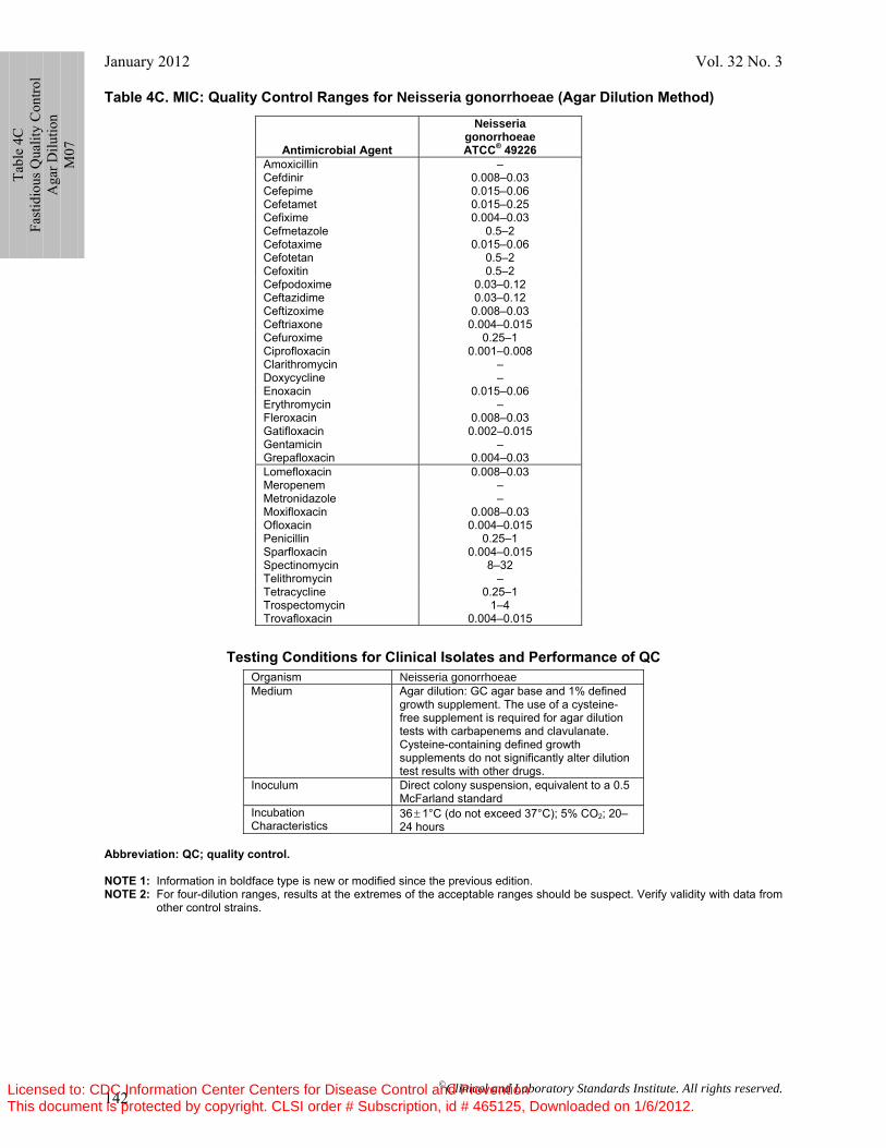
January 2012 Vol. 32 No. 3
©Clinical and Laboratory Standards Institute. All rights reserved. 142
Table 4C. MIC: Quality Control Ranges for Neisseria gonorrhoeae (Agar Dilution Method)
Testing Conditions for Clinical Isolates and Performance of QC Organism Neisseria gonorrhoeae Medium Agar dilution: GC agar base and 1% defined
growth supplement. The use of a cysteine-free supplement is required for agar dilution tests with carbapenems and clavulanate. Cysteine-containing defined growth supplements do not significantly alter dilution test results with other drugs.
Inoculum Direct colony suspension, equivalent to a 0.5 McFarland standard
Incubation Characteristics
36 ± 1°C (do not exceed 37°C); 5% CO2; 20–24 hours
Abbreviation: QC; quality control. NOTE 1: Information in boldface type is new or modified since the previous edition. NOTE 2: For four-dilution ranges, results at the extremes of the acceptable ranges should be suspect. Verify validity with data from
other control strains.
Antimicrobial Agent
Neisseria gonorrhoeae ATCC® 49226
Amoxicillin – Cefdinir 0.008–0.03 Cefepime 0.015–0.06 Cefetamet 0.015–0.25 Cefixime 0.004–0.03 Cefmetazole 0.5–2 Cefotaxime 0.015–0.06 Cefotetan 0.5–2 Cefoxitin 0.5–2 Cefpodoxime 0.03–0.12 Ceftazidime 0.03–0.12 Ceftizoxime 0.008–0.03 Ceftriaxone 0.004–0.015 Cefuroxime 0.25–1 Ciprofloxacin 0.001–0.008 Clarithromycin – Doxycycline – Enoxacin 0.015–0.06 Erythromycin – Fleroxacin 0.008–0.03 Gatifloxacin 0.002–0.015 Gentamicin – Grepafloxacin 0.004–0.03 Lomefloxacin 0.008–0.03 Meropenem – Metronidazole – Moxifloxacin 0.008–0.03 Ofloxacin 0.004–0.015 Penicillin 0.25–1 Sparfloxacin 0.004–0.015 Spectinomycin 8–32 Telithromycin – Tetracycline 0.25–1 Trospectomycin 1–4 Trovafloxacin 0.004–0.015
Tabl
e 4C
Fa
stid
ious
Qua
lity
Con
trol
Aga
r Dilu
tion
M07
Licensed to: CDC Information Center Centers for Disease Control and PreventionThis document is protected by copyright. CLSI order # Subscription, id # 465125, Downloaded on 1/6/2012.
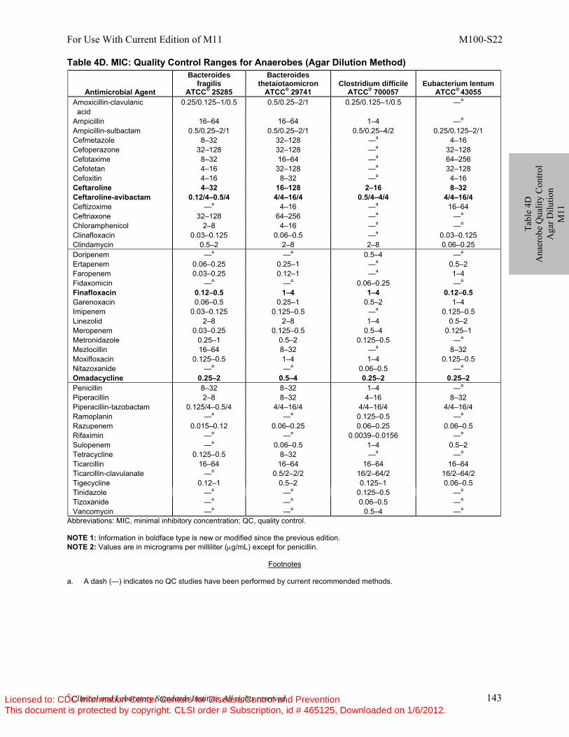
For Use With Current Edition of M11 M100-S22
©Clinical and Laboratory Standards Institute. All rights reserved. 143
Table 4D. MIC: Quality Control Ranges for Anaerobes (Agar Dilution Method)
Abbreviations: MIC, minimal inhibitory concentration; QC, quality control. NOTE 1: Information in boldface type is new or modified since the previous edition. NOTE 2: Values are in micrograms per milliliter (μg/mL) except for penicillin.
Footnotes
a. A dash (—) indicates no QC studies have been performed by current recommended methods.
Antimicrobial Agent
Bacteroides fragilis
ATCC® 25285
Bacteroides thetaiotaomicron
ATCC® 29741 Clostridium difficile
ATCC® 700057 Eubacterium lentum
ATCC® 43055 Amoxicillin-clavulanic acid
0.25/0.125–1/0.5 0.5/0.25–2/1 0.25/0.125–1/0.5 —a
Ampicillin 16–64 16–64 1–4 —a Ampicillin-sulbactam 0.5/0.25–2/1 0.5/0.25–2/1 0.5/0.25–4/2 0.25/0.125–2/1 Cefmetazole 8–32 32–128 —a 4–16 Cefoperazone 32–128 32–128 —a 32–128 Cefotaxime 8–32 16–64 —a 64–256 Cefotetan 4–16 32–128 —a 32–128 Cefoxitin 4–16 8–32 —a 4–16 Ceftaroline 4–32 16–128 2–16 8–32 Ceftaroline-avibactam 0.12/4–0.5/4 4/4–16/4 0.5/4–4/4 4/4–16/4 Ceftizoxime —a 4–16 —a 16–64 Ceftriaxone 32–128 64–256 —a —a Chloramphenicol 2–8 4–16 —a —a
Clinafloxacin 0.03–0.125 0.06–0.5 —a 0.03–0.125 Clindamycin 0.5–2 2–8 2–8 0.06–0.25 Doripenem —a —a 0.5–4 —a Ertapenem 0.06–0.25 0.25–1 —a 0.5–2 Faropenem Fidaxomicin
0.03–0.25 —a
0.12–1 —a
—a 0.06–0.25
1–4 —a
Finafloxacin 0.12–0.5 1–4 1–4 0.12–0.5 Garenoxacin 0.06–0.5 0.25–1 0.5–2 1–4 Imipenem 0.03–0.125 0.125–0.5 —a 0.125–0.5 Linezolid 2–8 2–8 1–4 0.5–2 Meropenem 0.03–0.25 0.125–0.5 0.5–4 0.125–1 Metronidazole 0.25–1 0.5–2 0.125–0.5 —a Mezlocillin 16–64 8–32 —a 8–32 Moxifloxacin 0.125–0.5 1–4 1–4 0.125–0.5 Nitazoxanide —a —a 0.06–0.5 —a Omadacycline 0.25–2 0.5–4 0.25–2 0.25–2 Penicillin 8–32 8–32 1–4 —a Piperacillin 2–8 8–32 4–16 8–32 Piperacillin-tazobactam 0.125/4–0.5/4 4/4–16/4 4/4–16/4 4/4–16/4 Ramoplanin —a —a 0.125–0.5 —a Razupenem 0.015–0.12 0.06–0.25 0.06–0.25 0.06–0.5 Rifaximin —a —a 0.0039–0.0156 —a Sulopenem —a 0.06–0.5 1–4 0.5–2 Tetracycline 0.125–0.5 8–32 —a —a Ticarcillin 16–64 16–64 16–64 16–64 Ticarcillin-clavulanate —a 0.5/2–2/2 16/2–64/2 16/2–64/2 Tigecycline 0.12–1 0.5–2 0.125–1 0.06–0.5 Tinidazole —a —a 0.125–0.5 —a Tizoxanide —a —a 0.06–0.5 —a Vancomycin —a —a 0.5–4 —a
Tabl
e 4D
A
naer
obe
Qua
lity
Con
trol
Aga
r Dilu
tion
M11
Licensed to: CDC Information Center Centers for Disease Control and PreventionThis document is protected by copyright. CLSI order # Subscription, id # 465125, Downloaded on 1/6/2012.
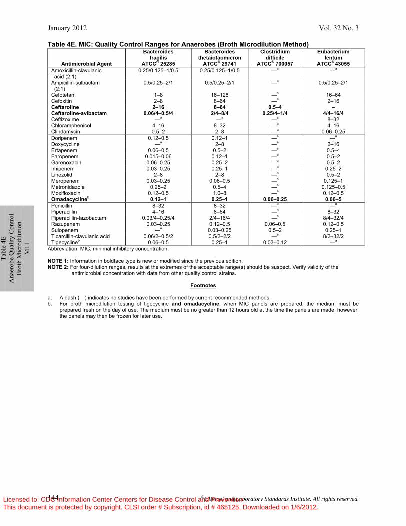
January 2012 Vol. 32 No. 3
©Clinical and Laboratory Standards Institute. All rights reserved. 144
Table 4E. MIC: Quality Control Ranges for Anaerobes (Broth Microdilution Method)
Antimicrobial Agent
Bacteroides fragilis
ATCC® 25285
Bacteroides thetaiotaomicron
ATCC® 29741
Clostridium difficile
ATCC® 700057
Eubacterium lentum
ATCC® 43055 Amoxicillin-clavulanic acid (2:1)
0.25/0.125–1/0.5 0.25/0.125–1/0.5 —a —a
Ampicillin-sulbactam (2:1)
0.5/0.25–2/1 0.5/0.25–2/1 —a 0.5/0.25–2/1
Cefotetan 1–8 16–128 —a 16–64 Cefoxitin 2–8 8–64 —a 2–16 Ceftaroline 2–16 8–64 0.5–4 – Ceftaroline-avibactam 0.06/4–0.5/4 2/4–8/4 0.25/4–1/4 4/4–16/4 Ceftizoxime —a —a —a 8–32 Chloramphenicol 4–16 8–32 —a 4–16 Clindamycin 0.5–2 2–8 —a 0.06–0.25 Doripenem 0.12–0.5 0.12–1 —a —a
Doxycycline —a 2–8 —a 2–16 Ertapenem 0.06–0.5 0.5–2 —a 0.5–4 Faropenem 0.015–0.06 0.12–1 —a 0.5–2 Garenoxacin 0.06–0.25 0.25–2 —a 0.5–2 Imipenem 0.03–0.25 0.25–1 —a 0.25–2 Linezolid 2–8 2–8 —a 0.5–2 Meropenem 0.03–0.25 0.06–0.5 —a 0.125–1 Metronidazole 0.25–2 0.5–4 —a 0.125–0.5 Moxifloxacin 0.12–0.5 1.0–8 —a 0.12–0.5 Omadacyclineb 0.12–1 0.25–1 0.06–0.25 0.06–5 Penicillin 8–32 8–32 —a —a Piperacillin 4–16 8–64 —a 8–32 Piperacillin-tazobactam Razupenem Sulopenem
0.03/4–0.25/4 0.03–0.25
—a
2/4–16/4 0.12–0.5
0.03–0.25
—a 0.06–0.5
0.5–2
8/4–32/4 0.12–0.5 0.25–1
Ticarcillin-clavulanic acid Tigecyclineb
0.06/2–0.5/2 0.06–0.5
0.5/2–2/2 0.25–1
—a 0.03–0.12
8/2–32/2 —a
Abbreviation: MIC, minimal inhibitory concentration. NOTE 1: Information in boldface type is new or modified since the previous edition. NOTE 2: For four-dilution ranges, results at the extremes of the acceptable range(s) should be suspect. Verify validity of the
antimicrobial concentration with data from other quality control strains.
Footnotes
a. A dash (—) indicates no studies have been performed by current recommended methods b. For broth microdilution testing of tigecycline and omadacycline, when MIC panels are prepared, the medium must be
prepared fresh on the day of use. The medium must be no greater than 12 hours old at the time the panels are made; however, the panels may then be frozen for later use.
Tabl
e 4E
A
naer
obe
Qua
lity
Con
trol
Bro
th M
icro
dilu
tion
M11
Licensed to: CDC Information Center Centers for Disease Control and PreventionThis document is protected by copyright. CLSI order # Subscription, id # 465125, Downloaded on 1/6/2012.
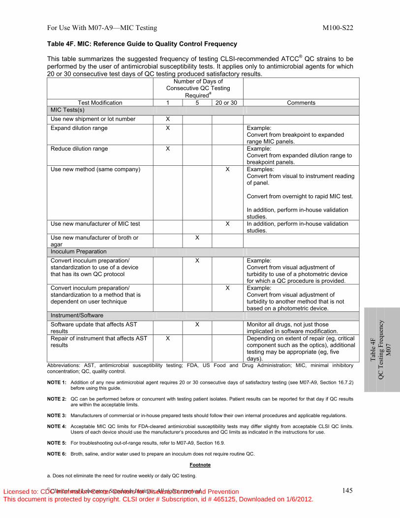
For Use With M07-A9—MIC Testing M100-S22
©Clinical and Laboratory Standards Institute. All rights reserved. 145
Table 4F. MIC: Reference Guide to Quality Control Frequency This table summarizes the suggested frequency of testing CLSI-recommended ATCC® QC strains to be performed by the user of antimicrobial susceptibility tests. It applies only to antimicrobial agents for which 20 or 30 consecutive test days of QC testing produced satisfactory results.
Number of Days of Consecutive QC Testing
Requireda Test Modification 1 5 20 or 30 Comments
MIC Tests(s) Use new shipment or lot number X Expand dilution range X Example:
Convert from breakpoint to expanded range MIC panels.
Reduce dilution range X Example: Convert from expanded dilution range to breakpoint panels.
Use new method (same company) X Examples: Convert from visual to instrument reading of panel. Convert from overnight to rapid MIC test. In addition, perform in-house validation studies.
Use new manufacturer of MIC test X In addition, perform in-house validation studies.
Use new manufacturer of broth or agar
X
Inoculum Preparation Convert inoculum preparation/ standardization to use of a device that has its own QC protocol
X Example: Convert from visual adjustment of turbidity to use of a photometric device for which a QC procedure is provided.
Convert inoculum preparation/ standardization to a method that is dependent on user technique
X Example: Convert from visual adjustment of turbidity to another method that is not based on a photometric device.
Instrument/Software Software update that affects AST results
X Monitor all drugs, not just those implicated in software modification.
Repair of instrument that affects AST results
X Depending on extent of repair (eg, critical component such as the optics), additional testing may be appropriate (eg, five days).
Abbreviations: AST, antimicrobial susceptibility testing; FDA, US Food and Drug Administration; MIC, minimal inhibitory concentration; QC, quality control. NOTE 1: Addition of any new antimicrobial agent requires 20 or 30 consecutive days of satisfactory testing (see M07-A9, Section 16.7.2)
before using this guide. NOTE 2: QC can be performed before or concurrent with testing patient isolates. Patient results can be reported for that day if QC results
are within the acceptable limits. NOTE 3: Manufacturers of commercial or in-house prepared tests should follow their own internal procedures and applicable regulations. NOTE 4: Acceptable MIC QC limits for FDA-cleared antimicrobial susceptibility tests may differ slightly from acceptable CLSI QC limits.
Users of each device should use the manufacturer’s procedures and QC limits as indicated in the instructions for use. NOTE 5: For troubleshooting out-of-range results, refer to M07-A9, Section 16.9. NOTE 6: Broth, saline, and/or water used to prepare an inoculum does not require routine QC.
Footnote a. Does not eliminate the need for routine weekly or daily QC testing.
Tabl
e 4F
Q
C T
estin
g Fr
eque
ncy
M07
Licensed to: CDC Information Center Centers for Disease Control and PreventionThis document is protected by copyright. CLSI order # Subscription, id # 465125, Downloaded on 1/6/2012.
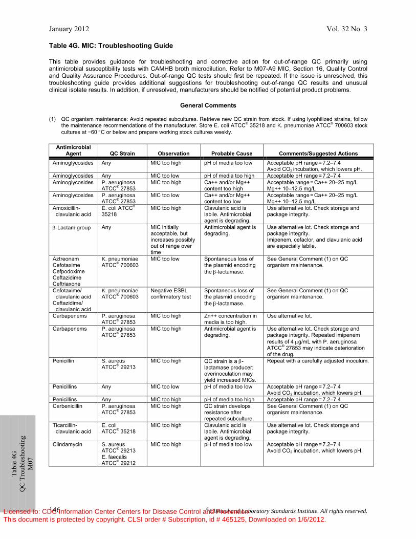
January 2012 Vol. 32 No. 3
©Clinical and Laboratory Standards Institute. All rights reserved. 146
Ta
ble
4G
QC
Tro
uble
shoo
ting
M07
Table 4G. MIC: Troubleshooting Guide This table provides guidance for troubleshooting and corrective action for out-of-range QC primarily using antimicrobial susceptibility tests with CAMHB broth microdilution. Refer to M07-A9 MIC, Section 16, Quality Control and Quality Assurance Procedures. Out-of-range QC tests should first be repeated. If the issue is unresolved, this troubleshooting guide provides additional suggestions for troubleshooting out-of-range QC results and unusual clinical isolate results. In addition, if unresolved, manufacturers should be notified of potential product problems.
General Comments
(1) QC organism maintenance: Avoid repeated subcultures. Retrieve new QC strain from stock. If using lyophilized strains, follow the maintenance recommendations of the manufacturer. Store E. coli ATCC® 35218 and K. pneumoniae ATCC® 700603 stock cultures at −60 °C or below and prepare working stock cultures weekly.
Antimicrobial
Agent QC Strain Observation Probable Cause Comments/Suggested Actions Aminoglycosides Any MIC too high pH of media too low Acceptable pH range = 7.2–7.4
Avoid CO2 incubation, which lowers pH. Aminoglycosides Any MIC too low pH of media too high Acceptable pH range = 7.2–7.4 Aminoglycosides P. aeruginosa
ATCC® 27853 MIC too high Ca++ and/or Mg++
content too high Acceptable range = Ca++ 20–25 mg/L Mg++ 10–12.5 mg/L
Aminoglycosides P. aeruginosa ATCC® 27853
MIC too low Ca++ and/or Mg++ content too low
Acceptable range = Ca++ 20–25 mg/L Mg++ 10–12.5 mg/L
Amoxicillin- clavulanic acid
E. coli ATCC® 35218
MIC too high Clavulanic acid is labile. Antimicrobial agent is degrading.
Use alternative lot. Check storage and package integrity.
β-Lactam group Any MIC initially acceptable, but increases possibly out of range over time
Antimicrobial agent is degrading.
Use alternative lot. Check storage and package integrity. Imipenem, cefaclor, and clavulanic acid are especially labile.
Aztreonam Cefotaxime Cefpodoxime Ceftazidime Ceftriaxone
K. pneumoniae ATCC® 700603
MIC too low Spontaneous loss of the plasmid encoding the β-lactamase.
See General Comment (1) on QC organism maintenance.
Cefotaxime/ clavulanic acid Ceftazidime/ clavulanic acid
K. pneumoniae ATCC® 700603
Negative ESBL confirmatory test
Spontaneous loss of the plasmid encoding the β-lactamase.
See General Comment (1) on QC organism maintenance.
Carbapenems P. aeruginosa ATCC® 27853
MIC too high Zn++ concentration in media is too high.
Use alternative lot.
Carbapenems P. aeruginosa ATCC® 27853
MIC too high Antimicrobial agent is degrading.
Use alternative lot. Check storage and package integrity. Repeated imipenem results of 4 μg/mL with P. aeruginosa ATCC® 27853 may indicate deterioration of the drug.
Penicillin S. aureus ATCC® 29213
MIC too high QC strain is a β-lactamase producer; overinoculation may yield increased MICs.
Repeat with a carefully adjusted inoculum.
Penicillins Any MIC too low pH of media too low Acceptable pH range = 7.2–7.4 Avoid CO2 incubation, which lowers pH.
Penicillins Any MIC too high pH of media too high Acceptable pH range = 7.2–7.4 Carbenicillin P. aeruginosa
ATCC® 27853 MIC too high QC strain develops
resistance after repeated subculture.
See General Comment (1) on QC organism maintenance.
Ticarcillin- clavulanic acid
E. coli ATCC® 35218
MIC too high Clavulanic acid is labile. Antimicrobial agent is degrading.
Use alternative lot. Check storage and package integrity.
Clindamycin S. aureus ATCC® 29213 E. faecalis ATCC® 29212
MIC too high pH of media too low Acceptable pH range = 7.2–7.4 Avoid CO2 incubation, which lowers pH.
Licensed to: CDC Information Center Centers for Disease Control and PreventionThis document is protected by copyright. CLSI order # Subscription, id # 465125, Downloaded on 1/6/2012.
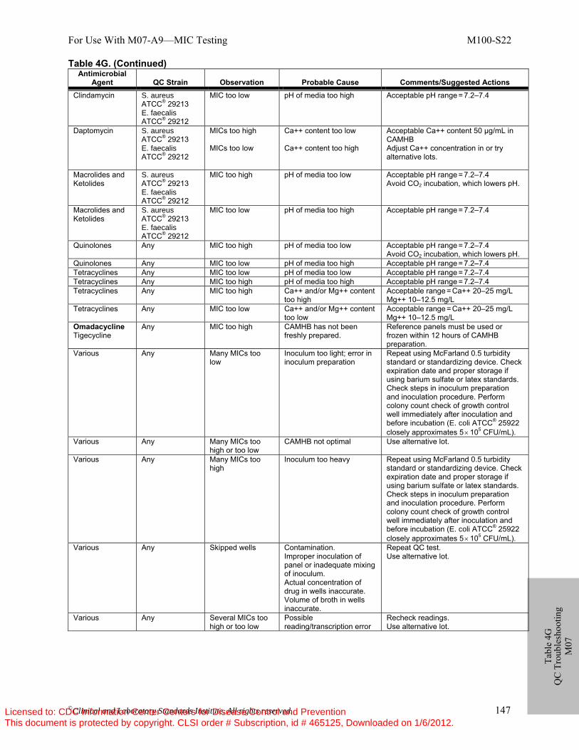
For Use With M07-A9—MIC Testing M100-S22
©Clinical and Laboratory Standards Institute. All rights reserved. 147
Tabl
e 4G
Q
C T
roub
lesh
ootin
g M
07
Table 4G. (Continued) Antimicrobial
Agent QC Strain Observation Probable Cause Comments/Suggested Actions Clindamycin S. aureus
ATCC® 29213 E. faecalis ATCC® 29212
MIC too low pH of media too high Acceptable pH range = 7.2–7.4
Daptomycin S. aureus ATCC® 29213 E. faecalis ATCC® 29212
MICs too high MICs too low
Ca++ content too low Ca++ content too high
Acceptable Ca++ content 50 µg/mL in CAMHB Adjust Ca++ concentration in or try alternative lots.
Macrolides and Ketolides
S. aureus ATCC® 29213 E. faecalis ATCC® 29212
MIC too high pH of media too low Acceptable pH range = 7.2–7.4 Avoid CO2 incubation, which lowers pH.
Macrolides and Ketolides
S. aureus ATCC® 29213 E. faecalis ATCC® 29212
MIC too low pH of media too high Acceptable pH range = 7.2–7.4
Quinolones Any MIC too high pH of media too low Acceptable pH range = 7.2–7.4 Avoid CO2 incubation, which lowers pH.
Quinolones Any MIC too low pH of media too high Acceptable pH range = 7.2–7.4 Tetracyclines Any MIC too low pH of media too low Acceptable pH range = 7.2–7.4 Tetracyclines Any MIC too high pH of media too high Acceptable pH range = 7.2–7.4 Tetracyclines Any MIC too high Ca++ and/or Mg++ content
too high Acceptable range = Ca++ 20–25 mg/L Mg++ 10–12.5 mg/L
Tetracyclines Any MIC too low Ca++ and/or Mg++ content too low
Acceptable range = Ca++ 20–25 mg/L Mg++ 10–12.5 mg/L
Omadacycline Tigecycline
Any MIC too high CAMHB has not been freshly prepared.
Reference panels must be used or frozen within 12 hours of CAMHB preparation.
Various Any Many MICs too low
Inoculum too light; error in inoculum preparation
Repeat using McFarland 0.5 turbidity standard or standardizing device. Check expiration date and proper storage if using barium sulfate or latex standards. Check steps in inoculum preparation and inoculation procedure. Perform colony count check of growth control well immediately after inoculation and before incubation (E. coli ATCC® 25922 closely approximates 5 × 10⁵ CFU/mL).
Various Any Many MICs too high or too low
CAMHB not optimal Use alternative lot.
Various Any Many MICs too high
Inoculum too heavy Repeat using McFarland 0.5 turbidity standard or standardizing device. Check expiration date and proper storage if using barium sulfate or latex standards. Check steps in inoculum preparation and inoculation procedure. Perform colony count check of growth control well immediately after inoculation and before incubation (E. coli ATCC® 25922 closely approximates 5 × 10⁵ CFU/mL).
Various Any Skipped wells Contamination. Improper inoculation of panel or inadequate mixing of inoculum. Actual concentration of drug in wells inaccurate. Volume of broth in wells inaccurate.
Repeat QC test. Use alternative lot.
Various Any Several MICs too high or too low
Possible reading/transcription error
Recheck readings. Use alternative lot.
Licensed to: CDC Information Center Centers for Disease Control and PreventionThis document is protected by copyright. CLSI order # Subscription, id # 465125, Downloaded on 1/6/2012.
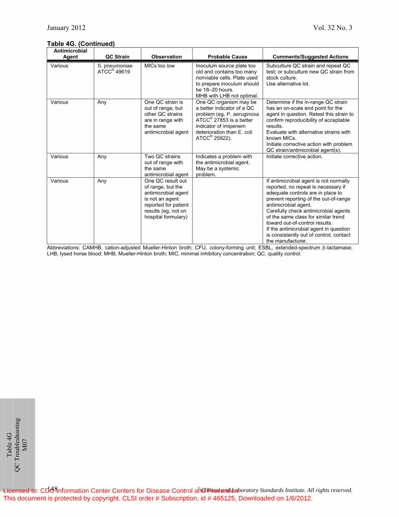
January 2012 Vol. 32 No. 3
©Clinical and Laboratory Standards Institute. All rights reserved. 148
Tabl
e 4G
Q
C T
roub
lesh
ootin
g M
07
Table 4G. (Continued) Antimicrobial
Agent QC Strain Observation Probable Cause Comments/Suggested Actions Various S. pneumoniae
ATCC® 49619 MICs too low Inoculum source plate too
old and contains too many nonviable cells. Plate used to prepare inoculum should be 18–20 hours. MHB with LHB not optimal.
Subculture QC strain and repeat QC test; or subculture new QC strain from stock culture. Use alternative lot.
Various Any One QC strain is out of range, but other QC strains are in range with the same antimicrobial agent
One QC organism may be a better indicator of a QC problem (eg, P. aeruginosa ATCC® 27853 is a better indicator of imipenem deterioration than E. coli ATCC® 25922).
Determine if the in-range QC strain has an on-scale end point for the agent in question. Retest this strain to confirm reproducibility of acceptable results. Evaluate with alternative strains with known MICs. Initiate corrective action with problem QC strain/antimicrobial agent(s).
Various Any Two QC strains out of range with the same antimicrobial agent
Indicates a problem with the antimicrobial agent. May be a systemic problem.
Initiate corrective action.
Various Any One QC result out of range, but the antimicrobial agent is not an agent reported for patient results (eg, not on hospital formulary)
If antimicrobial agent is not normally reported, no repeat is necessary if adequate controls are in place to prevent reporting of the out-of-range antimicrobial agent. Carefully check antimicrobial agents of the same class for similar trend toward out-of-control results. If the antimicrobial agent in question is consistently out of control, contact the manufacturer.
Abbreviations: CAMHB, cation-adjusted Mueller-Hinton broth; CFU, colony-forming unit; ESBL, extended-spectrum β-lactamase; LHB, lysed horse blood; MHB, Mueller-Hinton broth; MIC, minimal inhibitory concentration; QC, quality control.
Licensed to: CDC Information Center Centers for Disease Control and PreventionThis document is protected by copyright. CLSI order # Subscription, id # 465125, Downloaded on 1/6/2012.

For Use With M07-A9—MIC Testing M100-S22
©Clinical and Laboratory Standards Institute. All rights reserved. 149
This page is intentionally left blank.
Licensed to: CDC Information Center Centers for Disease Control and PreventionThis document is protected by copyright. CLSI order # Subscription, id # 465125, Downloaded on 1/6/2012.
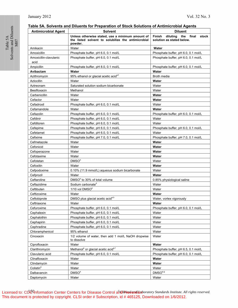
January 2012 Vol. 32 No. 3
©Clinical and Laboratory Standards Institute. All rights reserved. 150
Table 5A. Solvents and Diluents for Preparation of Stock Solutions of Antimicrobial Agents Antimicrobial Agent Solvent Diluent
Unless otherwise stated, use a minimum amount of the listed solvent to solubilize the antimicrobial powder.
Finish diluting the final stock solution as stated below.
Amikacin Water Water Amoxicillin Phosphate buffer, pH 6.0, 0.1 mol/L Phosphate buffer, pH 6.0, 0.1 mol/L Amoxicillin-clavulanic acid
Phosphate buffer, pH 6.0, 0.1 mol/L Phosphate buffer, pH 6.0, 0.1 mol/L
Ampicillin Phosphate buffer, pH 8.0, 0.1 mol/L Phosphate buffer, pH 6.0, 0.1 mol/L Avibactam Water Water Azithromycin 95% ethanol or glacial acetic acide,f Broth media Azlocillin Water Water Aztreonam Saturated solution sodium bicarbonate Water Besifloxacin Methanol Water Carbenicillin Water Water Cefaclor Water Water Cefadroxil Phosphate buffer, pH 6.0, 0.1 mol/L Water Cefamandole Water Water Cefazolin Phosphate buffer, pH 6.0, 0.1 mol/L Phosphate buffer, pH 6.0, 0.1 mol/L Cefdinir Phosphate buffer, pH 6.0, 0.1 mol/L Water Cefditoren Phosphate buffer, pH 6.0, 0.1 mol/L Water Cefepime Phosphate buffer, pH 6.0, 0.1 mol/L Phosphate buffer, pH 6.0, 0.1 mol/L Cefetamet Phosphate buffer, pH 6.0, 0.1 mol/L Water Cefixime Phosphate buffer, pH 7.0, 0.1 mol/L Phosphate buffer, pH 7.0, 0.1 mol/L Cefmetazole Water Water Cefonicid Water Water Cefoperazone Water Water Cefotaxime Water Water Cefotetan DMSOe Water Cefoxitin Water Water Cefpodoxime 0.10% (11.9 mmol/L) aqueous sodium bicarbonate Water Cefprozil Water Water Ceftaroline DMSOe to 30% of total volume 0.85% physiological saline Ceftazidime Sodium carbonated Water Ceftibuten 1/10 vol DMSOe Water Ceftizoxime Water Water Ceftobiprole DMSO plus glacial acetic acide,h Water, vortex vigorously Ceftriaxone Water Water Cefuroxime Phosphate buffer, pH 6.0, 0.1 mol/L Phosphate buffer, pH 6.0, 0.1 mol/L Cephalexin Phosphate buffer, pH 6.0, 0.1 mol/L Water Cephalothin Phosphate buffer, pH 6.0, 0.1 mol/L Water Cephapirin Phosphate buffer, pH 6.0, 0.1 mol/L Water Cephradine Phosphate buffer, pH 6.0, 0.1 mol/L Water Chloramphenicol 95% ethanol Water Cinoxacin 1/2 volume of water, then add 1 mol/L NaOH dropwise
to dissolve Water
Ciprofloxacin Water Water Clarithromycin Methanole or glacial acetic acide,f Phosphate buffer, pH 6.5, 0.1 mol/L Clavulanic acid Phosphate buffer, pH 6.0, 0.1 mol/L Phosphate buffer, pH 6.0, 0.1 mol/L Clinafloxacin Water Water Clindamycin Water Water Colistina Water Water Dalbavancin DMSOe DMSOe,g
Daptomycin Water Water
Ta
ble
5A
Solv
ents
and
Dilu
ents
M
07
Licensed to: CDC Information Center Centers for Disease Control and PreventionThis document is protected by copyright. CLSI order # Subscription, id # 465125, Downloaded on 1/6/2012.
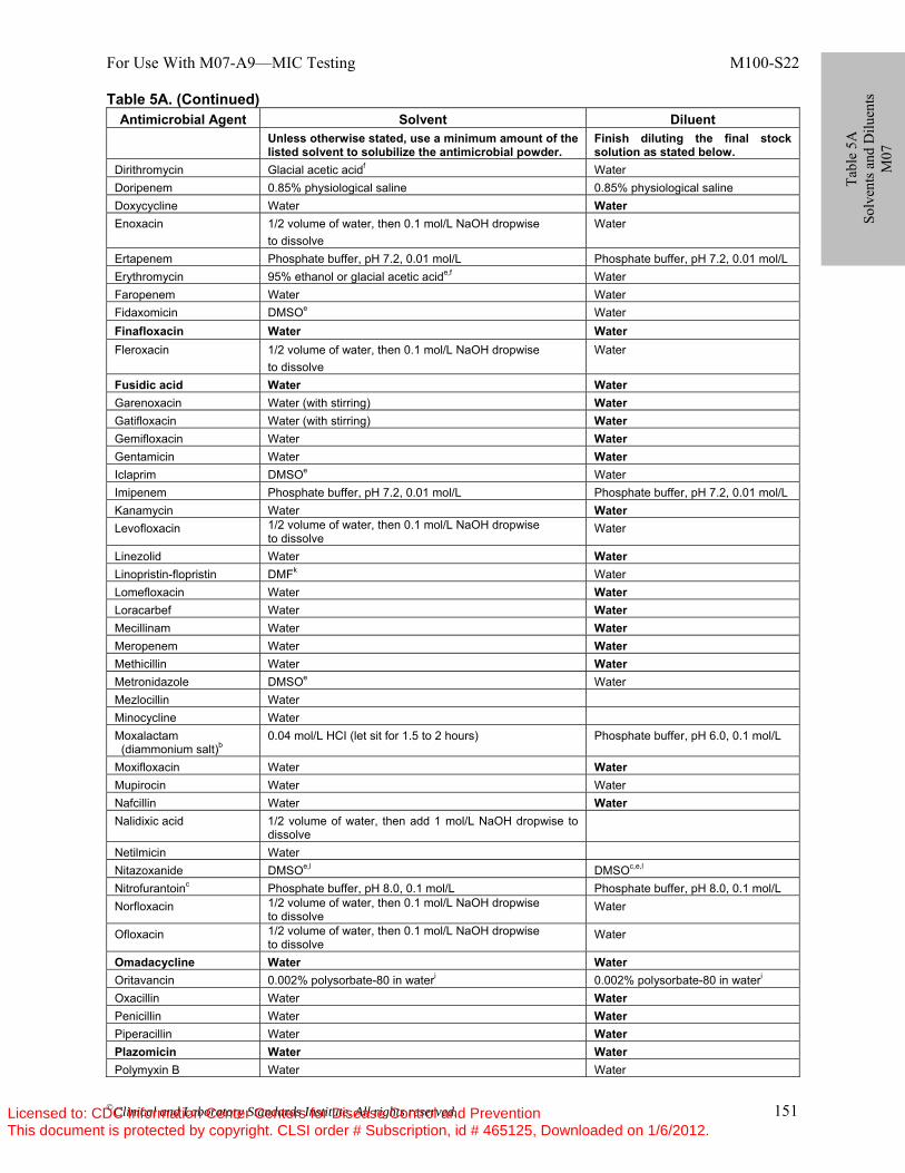
For Use With M07-A9—MIC Testing M100-S22
©Clinical and Laboratory Standards Institute. All rights reserved. 151
Table 5A. (Continued) Antimicrobial Agent Solvent Diluent
Unless otherwise stated, use a minimum amount of the listed solvent to solubilize the antimicrobial powder.
Finish diluting the final stock solution as stated below.
Dirithromycin Glacial acetic acidf Water Doripenem 0.85% physiological saline 0.85% physiological saline Doxycycline Water Water Enoxacin 1/2 volume of water, then 0.1 mol/L NaOH dropwise
to dissolve Water
Ertapenem Phosphate buffer, pH 7.2, 0.01 mol/L Phosphate buffer, pH 7.2, 0.01 mol/L Erythromycin 95% ethanol or glacial acetic acide,f Water Faropenem Water Water Fidaxomicin DMSOe Water Finafloxacin Water Water Fleroxacin 1/2 volume of water, then 0.1 mol/L NaOH dropwise
to dissolve Water
Fusidic acid Water Water Garenoxacin Water (with stirring) Water Gatifloxacin Water (with stirring) Water Gemifloxacin Water Water Gentamicin Water Water Iclaprim DMSOe Water Imipenem Phosphate buffer, pH 7.2, 0.01 mol/L Phosphate buffer, pH 7.2, 0.01 mol/L Kanamycin Water Water Levofloxacin 1/2 volume of water, then 0.1 mol/L NaOH dropwise
to dissolve Water
Linezolid Water Water Linopristin-flopristin DMFk Water Lomefloxacin Water Water Loracarbef Water Water Mecillinam Water Water Meropenem Water Water Methicillin Water Water Metronidazole DMSOe Water Mezlocillin Water Minocycline Water Moxalactam (diammonium salt)b
0.04 mol/L HCI (let sit for 1.5 to 2 hours) Phosphate buffer, pH 6.0, 0.1 mol/L
Moxifloxacin Water Water Mupirocin Water Water Nafcillin Water Water Nalidixic acid 1/2 volume of water, then add 1 mol/L NaOH dropwise to
dissolve
Netilmicin Water Nitazoxanide DMSOe,l DMSOc,e,l Nitrofurantoinc Phosphate buffer, pH 8.0, 0.1 mol/L Phosphate buffer, pH 8.0, 0.1 mol/L Norfloxacin 1/2 volume of water, then 0.1 mol/L NaOH dropwise
to dissolve Water
Ofloxacin 1/2 volume of water, then 0.1 mol/L NaOH dropwise to dissolve
Water
Omadacycline Water Water Oritavancin 0.002% polysorbate-80 in wateri 0.002% polysorbate-80 in wateri
Oxacillin Water Water Penicillin Water Water Piperacillin Water Water Plazomicin Water Water Polymyxin B Water Water
Tabl
e 5A
So
lven
ts a
nd D
iluen
ts
M07
Licensed to: CDC Information Center Centers for Disease Control and PreventionThis document is protected by copyright. CLSI order # Subscription, id # 465125, Downloaded on 1/6/2012.
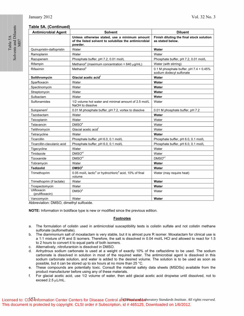
January 2012 Vol. 32 No. 3
©Clinical and Laboratory Standards Institute. All rights reserved. 152
Table 5A. (Continued) Antimicrobial Agent Solvent Diluent
Unless otherwise stated, use a minimum amount of the listed solvent to solubilize the antimicrobial powder.
Finish diluting the final stock solution as stated below.
Quinupristin-dalfopristin Water Water Ramoplanin Water Water Razupenem Phosphate buffer, pH 7.2, 0.01 mol/L Phosphate buffer, pH 7.2, 0.01 mol/L Rifampin Methanole (maximum concentration = 640 μg/mL) Water (with stirring) Rifaximin Methanole 0.1 M phosphate buffer, pH 7.4 + 0.45%
sodium dodecyl sulfonate Solithromycin Glacial acetic acidf Water Sparfloxacin Water Water Spectinomycin Water Water Streptomycin Water Water Sulbactam Water Water Sulfonamides 1/2 volume hot water and minimal amount of 2.5 mol/L
NaOH to dissolve Water
Sulopenemj 0.01 M phosphate buffer, pH 7.2, vortex to dissolve 0.01 M phosphate buffer, pH 7.2 Tazobactam Water Water Teicoplanin Water Water Telavancin DMSOe Water Telithromycin Glacial acetic acidf Water Tetracycline Water Water Ticarcillin Phosphate buffer, pH 6.0, 0.1 mol/L Phosphate buffer, pH 6.0, 0.1 mol/L Ticarcillin-clavulanic acid Phosphate buffer, pH 6.0, 0.1 mol/L Phosphate buffer, pH 6.0, 0.1 mol/L Tigecycline Water Water Tinidazole DMSOe,l Water Tizoxanide DMSOe,l DMSOe,l Tobramycin Water Water Tedizolid DMSOe Water Trimethoprim 0.05 mol/L lactice or hydrochlorice acid, 10% of final
volume Water (may require heat)
Trimethoprim (if lactate) Water Water Trospectomycin Water Water Ulifloxacin (prulifloxacin)
DMSOe Water
Vancomycin Water Water Abbreviation: DMSO, dimethyl sulfoxide. NOTE: Information in boldface type is new or modified since the previous edition.
Footnotes a. The formulation of colistin used in antimicrobial susceptibility tests is colistin sulfate and not colistin methane
sulfonate (sulfomethate). b. The diammonium salt of moxalactam is very stable, but it is almost pure R isomer. Moxalactam for clinical use is
a 1:1 mixture of R and S isomers. Therefore, the salt is dissolved in 0.04 mol/L HCl and allowed to react for 1.5 to 2 hours to convert it to equal parts of both isomers.
c. Alternatively, nitrofurantoin is dissolved in DMSO. d. Anhydrous sodium carbonate is used at a weight of exactly 10% of the ceftazidime to be used. The sodium
carbonate is dissolved in solution in most of the required water. The antimicrobial agent is dissolved in this sodium carbonate solution, and water is added to the desired volume. The solution is to be used as soon as possible, but it can be stored up to six hours at no more than 25 °C.
e. These compounds are potentially toxic. Consult the material safety data sheets (MSDSs) available from the product manufacturer before using any of these materials.
f. For glacial acetic acid, use 1/2 volume of water, then add glacial acetic acid dropwise until dissolved, not to exceed 2.5 μL/mL.
Ta
ble
5A
Solv
ents
and
Dilu
ents
M
07
Licensed to: CDC Information Center Centers for Disease Control and PreventionThis document is protected by copyright. CLSI order # Subscription, id # 465125, Downloaded on 1/6/2012.
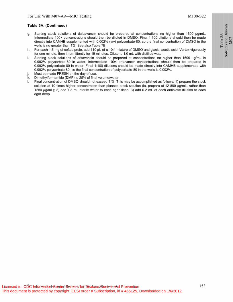
For Use With M07-A9—MIC Testing M100-S22
©Clinical and Laboratory Standards Institute. All rights reserved. 153
Table 5A. (Continued) g. Starting stock solutions of dalbavancin should be prepared at concentrations no higher than 1600 µg/mL.
Intermediate 100× concentrations should then be diluted in DMSO. Final 1:100 dilutions should then be made directly into CAMHB supplemented with 0.002% (v/v) polysorbate-80, so the final concentration of DMSO in the wells is no greater than 1%. See also Table 7B.
h. For each 1.5 mg of ceftobiprole, add 110 μL of a 10:1 mixture of DMSO and glacial acetic acid. Vortex vigorously for one minute, then intermittently for 15 minutes. Dilute to 1.0 mL with distilled water.
i. Starting stock solutions of oritavancin should be prepared at concentrations no higher than 1600 μg/mL in 0.002% polysorbate-80 in water. Intermediate 100× oritavancin concentrations should then be prepared in 0.002% polysorbate-80 in water. Final 1:100 dilutions should be made directly into CAMHB supplemented with 0.002% polysorbate-80, so the final concentration of polysorbate-80 in the wells is 0.002%.
j. Must be made FRESH on the day of use. k. Dimethylformamide (DMF) to 25% of final volume/water. l. Final concentration of DMSO should not exceed 1 %. This may be accomplished as follows: 1) prepare the stock
solution at 10 times higher concentration than planned stock solution (ie, prepare at 12 800 μg/mL, rather than 1280 μg/mL); 2) add 1.8 mL sterile water to each agar deep; 3) add 0.2 mL of each antibiotic dilution to each agar deep.
Ta
ble
5A
Solv
ents
and
Dilu
ents
M
07
Licensed to: CDC Information Center Centers for Disease Control and PreventionThis document is protected by copyright. CLSI order # Subscription, id # 465125, Downloaded on 1/6/2012.
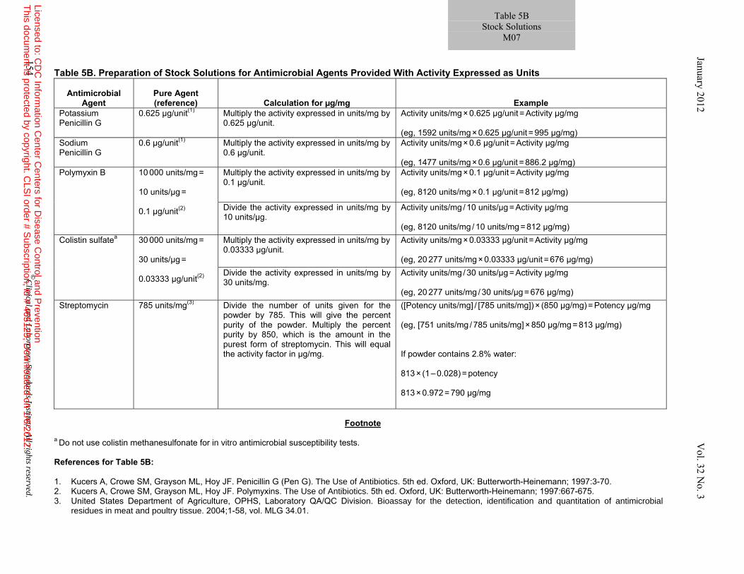
Table 5B. Preparation of Stock Solutions for Antimicrobial Agents Provided With Activity Expressed as Units
Footnote
a Do not use colistin methanesulfonate for in vitro antimicrobial susceptibility tests. References for Table 5B: 1. Kucers A, Crowe SM, Grayson ML, Hoy JF. Penicillin G (Pen G). The Use of Antibiotics. 5th ed. Oxford, UK: Butterworth-Heinemann; 1997:3-70. 2. Kucers A, Crowe SM, Grayson ML, Hoy JF. Polymyxins. The Use of Antibiotics. 5th ed. Oxford, UK: Butterworth-Heinemann; 1997:667-675. 3. United States Department of Agriculture, OPHS, Laboratory QA/QC Division. Bioassay for the detection, identification and quantitation of antimicrobial
residues in meat and poultry tissue. 2004;1-58, vol. MLG 34.01.
Antimicrobial Agent
Pure Agent (reference) Calculation for µg/mg Example
Potassium Penicillin G
0.625 µg/unit(1) Multiply the activity expressed in units/mg by 0.625 µg/unit.
Activity units/mg × 0.625 µg/unit = Activity µg/mg (eg, 1592 units/mg × 0.625 µg/unit = 995 µg/mg)
Sodium Penicillin G
0.6 µg/unit(1) Multiply the activity expressed in units/mg by 0.6 µg/unit.
Activity units/mg × 0.6 µg/unit = Activity µg/mg (eg, 1477 units/mg × 0.6 µg/unit = 886.2 µg/mg)
Polymyxin B 10 000 units/mg =
10 units/µg =
0.1 µg/unit(2)
Multiply the activity expressed in units/mg by 0.1 µg/unit.
Activity units/mg × 0.1 µg/unit = Activity µg/mg (eg, 8120 units/mg × 0.1 µg/unit = 812 µg/mg)
Divide the activity expressed in units/mg by 10 units/µg.
Activity units/mg / 10 units/µg = Activity µg/mg (eg, 8120 units/mg / 10 units/mg = 812 µg/mg)
Colistin sulfatea 30 000 units/mg =
30 units/µg =
0.03333 µg/unit(2)
Multiply the activity expressed in units/mg by 0.03333 µg/unit.
Activity units/mg × 0.03333 µg/unit = Activity µg/mg (eg, 20 277 units/mg × 0.03333 µg/unit = 676 µg/mg)
Divide the activity expressed in units/mg by 30 units/mg.
Activity units/mg / 30 units/µg = Activity µg/mg (eg, 20 277 units/mg / 30 units/µg = 676 µg/mg)
Streptomycin 785 units/mg(3) Divide the number of units given for the powder by 785. This will give the percent purity of the powder. Multiply the percent purity by 850, which is the amount in the purest form of streptomycin. This will equal the activity factor in µg/mg.
([Potency units/mg] / [785 units/mg]) × (850 µg/mg) = Potency µg/mg (eg, [751 units/mg / 785 units/mg] × 850 µg/mg = 813 µg/mg) If powder contains 2.8% water: 813 × (1 – 0.028) = potency 813 × 0.972 = 790 µg/mg
Table 5B Stock Solutions
M07
154 ©C
linical and Laboratory Standards Institute. A
ll rights reserved.
January 2012 V
ol. 32 No. 3
Licensed to: CD
C Inform
ation Center C
enters for Disease C
ontrol and Prevention
This docum
ent is protected by copyright. CLS
I order # Subscription, id # 465125, D
ownloaded on 1/6/2012.

For Use With M07-A9—MIC Testing M100-S22
©Clinical and Laboratory Standards Institute. All rights reserved. 155
This page is intentionally left blank.
Licensed to: CDC Information Center Centers for Disease Control and PreventionThis document is protected by copyright. CLSI order # Subscription, id # 465125, Downloaded on 1/6/2012.
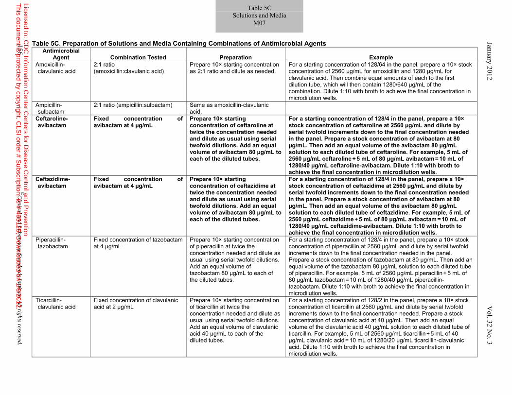
Table 5C. Preparation of Solutions and Media Containing Combinations of Antimicrobial Agents Antimicrobial
Agent
Combination Tested Preparation Example Amoxicillin-clavulanic acid
2:1 ratio (amoxicillin:clavulanic acid)
Prepare 10× starting concentration as 2:1 ratio and dilute as needed.
For a starting concentration of 128/64 in the panel, prepare a 10× stock concentration of 2560 µg/mL for amoxicillin and 1280 µg/mL for clavulanic acid. Then combine equal amounts of each to the first dilution tube, which will then contain 1280/640 µg/mL of the combination. Dilute 1:10 with broth to achieve the final concentration in microdilution wells.
Ampicillin- sulbactam
2:1 ratio (ampicillin:sulbactam) Same as amoxicillin-clavulanic acid.
Ceftaroline-avibactam
Fixed concentration of avibactam at 4 µg/mL
Prepare 10× starting concentration of ceftaroline at twice the concentration needed and dilute as usual using serial twofold dilutions. Add an equal volume of avibactam 80 µg/mL to each of the diluted tubes.
For a starting concentration of 128/4 in the panel, prepare a 10× stock concentration of ceftaroline at 2560 µg/mL and dilute by serial twofold increments down to the final concentration needed in the panel. Prepare a stock concentration of avibactam at 80 µg/mL. Then add an equal volume of the avibactam 80 µg/mL solution to each diluted tube of ceftaroline. For example, 5 mL of 2560 µg/mL ceftaroline + 5 mL of 80 µg/mL avibactam = 10 mL of 1280/40 µg/mL ceftaroline-avibactam. Dilute 1:10 with broth to achieve the final concentration in microdilution wells.
Ceftazidime-avibactam
Fixed concentration of avibactam at 4 µg/mL
Prepare 10× starting concentration of ceftazidime at twice the concentration needed and dilute as usual using serial twofold dilutions. Add an equal volume of avibactam 80 µg/mL to each of the diluted tubes.
For a starting concentration of 128/4 in the panel, prepare a 10× stock concentration of ceftazidime at 2560 µg/mL and dilute by serial twofold increments down to the final concentration needed in the panel. Prepare a stock concentration of avibactam at 80 µg/mL. Then add an equal volume of the avibactam 80 µg/mL solution to each diluted tube of ceftazidime. For example, 5 mL of 2560 µg/mL ceftazidime + 5 mL of 80 µg/mL avibactam = 10 mL of 1280/40 µg/mL ceftazidime-avibactam. Dilute 1:10 with broth to achieve the final concentration in microdilution wells.
Piperacillin-tazobactam
Fixed concentration of tazobactam at 4 µg/mL
Prepare 10× starting concentration of piperacillin at twice the concentration needed and dilute as usual using serial twofold dilutions. Add an equal volume of tazobactam 80 µg/mL to each of the diluted tubes.
For a starting concentration of 128/4 in the panel, prepare a 10× stock concentration of piperacillin at 2560 µg/mL and dilute by serial twofold increments down to the final concentration needed in the panel. Prepare a stock concentration of tazobactam at 80 µg/mL. Then add an equal volume of the tazobactam 80 µg/mL solution to each diluted tube of piperacillin. For example, 5 mL of 2560 µg/mL piperacillin + 5 mL of 80 µg/mL tazobactam = 10 mL of 1280/40 µg/mL piperacillin-tazobactam. Dilute 1:10 with broth to achieve the final concentration in microdilution wells.
Ticarcillin-clavulanic acid
Fixed concentration of clavulanic acid at 2 µg/mL
Prepare 10× starting concentration of ticarcillin at twice the concentration needed and dilute as usual using serial twofold dilutions. Add an equal volume of clavulanic acid 40 µg/mL to each of the diluted tubes.
For a starting concentration of 128/2 in the panel, prepare a 10× stock concentration of ticarcillin at 2560 µg/mL and dilute by serial twofold increments down to the final concentration needed. Prepare a stock concentration of clavulanic acid at 40 µg/mL. Then add an equal volume of the clavulanic acid 40 µg/mL solution to each diluted tube of ticarcillin. For example, 5 mL of 2560 µg/mL ticarcillin + 5 mL of 40 µg/mL clavulanic acid = 10 mL of 1280/20 µg/mL ticarcillin-clavulanic acid. Dilute 1:10 with broth to achieve the final concentration in microdilution wells.
156 ©C
linical and Laboratory Standards Institute. A
ll rights reserved.
Table 5C Solutions and Media
M07 January 2012 V
ol. 32 No. 3
Licensed to: CD
C Inform
ation Center C
enters for Disease C
ontrol and Prevention
This docum
ent is protected by copyright. CLS
I order # Subscription, id # 465125, D
ownloaded on 1/6/2012.
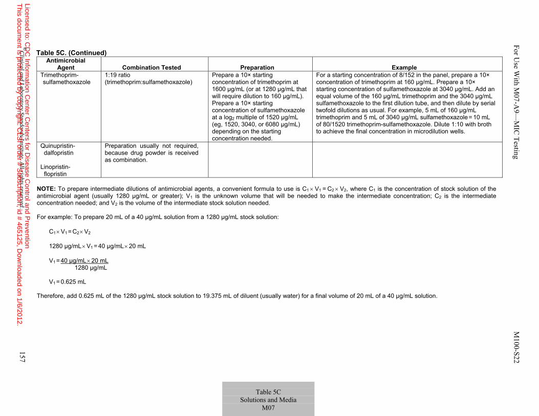
Table 5C. (Continued)
Antimicrobial Agent Combination Tested Preparation Example
Trimethoprim-sulfamethoxazole
1:19 ratio (trimethoprim:sulfamethoxazole)
Prepare a 10× starting concentration of trimethoprim at 1600 µg/mL (or at 1280 µg/mL that will require dilution to 160 µg/mL). Prepare a 10× starting concentration of sulfamethoxazole at a log2 multiple of 1520 µg/mL (eg, 1520, 3040, or 6080 µg/mL) depending on the starting concentration needed.
For a starting concentration of 8/152 in the panel, prepare a 10× concentration of trimethoprim at 160 µg/mL. Prepare a 10× starting concentration of sulfamethoxazole at 3040 µg/mL. Add an equal volume of the 160 µg/mL trimethoprim and the 3040 µg/mL sulfamethoxazole to the first dilution tube, and then dilute by serial twofold dilutions as usual. For example, 5 mL of 160 µg/mL trimethoprim and 5 mL of 3040 µg/mL sulfamethoxazole = 10 mL of 80/1520 trimethoprim-sulfamethoxazole. Dilute 1:10 with broth to achieve the final concentration in microdilution wells.
Quinupristin- dalfopristin Linopristin- flopristin
Preparation usually not required, because drug powder is received as combination.
NOTE: To prepare intermediate dilutions of antimicrobial agents, a convenient formula to use is C1 × V1 = C2 × V2, where C1 is the concentration of stock solution of the antimicrobial agent (usually 1280 µg/mL or greater); V1 is the unknown volume that will be needed to make the intermediate concentration; C2 is the intermediate concentration needed; and V2 is the volume of the intermediate stock solution needed.
For example: To prepare 20 mL of a 40 µg/mL solution from a 1280 µg/mL stock solution:
C1 × V1 = C2 × V2
1280 µg/mL × V1 = 40 µg/mL × 20 mL V1 = 40 µg/mL × 20 mL 1280 µg/mL V1 = 0.625 mL
Therefore, add 0.625 mL of the 1280 µg/mL stock solution to 19.375 mL of diluent (usually water) for a final volume of 20 mL of a 40 µg/mL solution.
©C
linical and Laboratory Standards Institute. A
ll rights reserved. 157
Table 5C
Solutions and Media M07
For U
se With M
07-A9—
MIC
Testing M100-S22
Licensed to: CD
C Inform
ation Center C
enters for Disease C
ontrol and Prevention
This docum
ent is protected by copyright. CLS
I order # Subscription, id # 465125, D
ownloaded on 1/6/2012.
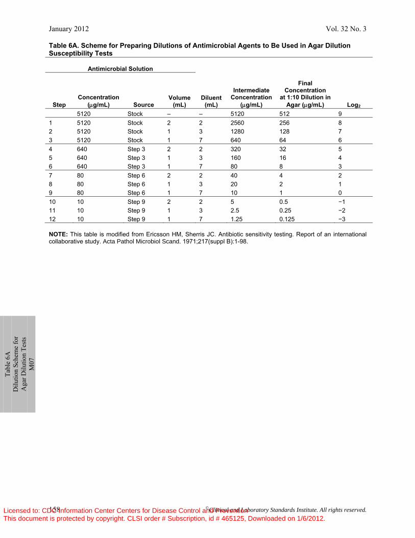
January 2012 Vol. 32 No. 3
158 ©Clinical and Laboratory Standards Institute. All rights reserved.
Ta
ble
6A
Dilu
tion
Sche
me
for
Aga
r Dilu
tion
Test
s M
07
Table 6A. Scheme for Preparing Dilutions of Antimicrobial Agents to Be Used in Agar Dilution Susceptibility Tests
Antimicrobial Solution
Step Concentration
(μg/mL) Source Volume
(mL) Diluent
(mL)
Intermediate Concentration
(μg/mL)
Final Concentration
at 1:10 Dilution in Agar (μg/mL) Log2
5120 Stock – – 5120 512 9 1 5120 Stock 2 2 2560 256 8 2 5120 Stock 1 3 1280 128 7 3 5120 Stock 1 7 640 64 6 4 640 Step 3 2 2 320 32 5 5 640 Step 3 1 3 160 16 4 6 640 Step 3 1 7 80 8 3 7 80 Step 6 2 2 40 4 2 8 80 Step 6 1 3 20 2 1 9 80 Step 6 1 7 10 1 0 10 10 Step 9 2 2 5 0.5 −1 11 10 Step 9 1 3 2.5 0.25 −2 12 10 Step 9 1 7 1.25 0.125 −3 NOTE: This table is modified from Ericsson HM, Sherris JC. Antibiotic sensitivity testing. Report of an international collaborative study. Acta Pathol Microbiol Scand. 1971;217(suppl B):1-98.
Licensed to: CDC Information Center Centers for Disease Control and PreventionThis document is protected by copyright. CLSI order # Subscription, id # 465125, Downloaded on 1/6/2012.

For Use With M07-A9—MIC Testing M100-S22
©Clinical and Laboratory Standards Institute. All rights reserved. 159
This page is intentionally left blank.
Licensed to: CDC Information Center Centers for Disease Control and PreventionThis document is protected by copyright. CLSI order # Subscription, id # 465125, Downloaded on 1/6/2012.
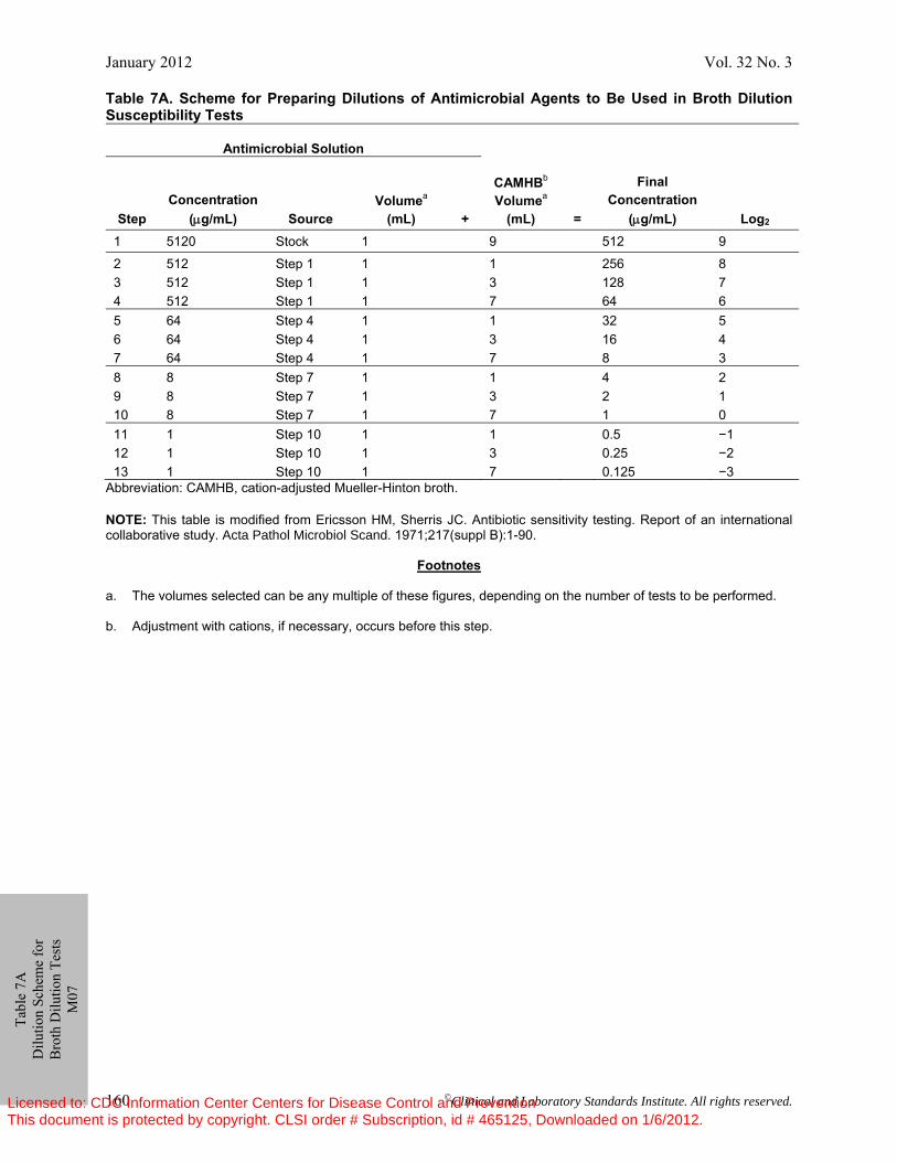
January 2012 Vol. 32 No. 3
160 ©Clinical and Laboratory Standards Institute. All rights reserved.
Table 7A. Scheme for Preparing Dilutions of Antimicrobial Agents to Be Used in Broth Dilution Susceptibility Tests
Antimicrobial Solution
Step
Concentration
(μg/mL)
Source
Volumea
(mL)
+
CAMHBb
Volumea
(mL)
=
Final Concentration
(μg/mL)
Log2
1 5120 Stock 1 9 512 9
2 512 Step 1 1 1 256 8 3 512 Step 1 1 3 128 7 4 512 Step 1 1 7 64 6 5 64 Step 4 1 1 32 5 6 64 Step 4 1 3 16 4 7 64 Step 4 1 7 8 3 8 8 Step 7 1 1 4 2 9 8 Step 7 1 3 2 1 10 8 Step 7 1 7 1 0 11 1 Step 10 1 1 0.5 −1 12 1 Step 10 1 3 0.25 −2 13 1 Step 10 1 7 0.125 −3
Abbreviation: CAMHB, cation-adjusted Mueller-Hinton broth. NOTE: This table is modified from Ericsson HM, Sherris JC. Antibiotic sensitivity testing. Report of an international collaborative study. Acta Pathol Microbiol Scand. 1971;217(suppl B):1-90.
Footnotes a. The volumes selected can be any multiple of these figures, depending on the number of tests to be performed. b. Adjustment with cations, if necessary, occurs before this step.
Ta
ble
7A
Dilu
tion
Sche
me
for
Bro
th D
ilutio
n Te
sts
M07
Licensed to: CDC Information Center Centers for Disease Control and PreventionThis document is protected by copyright. CLSI order # Subscription, id # 465125, Downloaded on 1/6/2012.
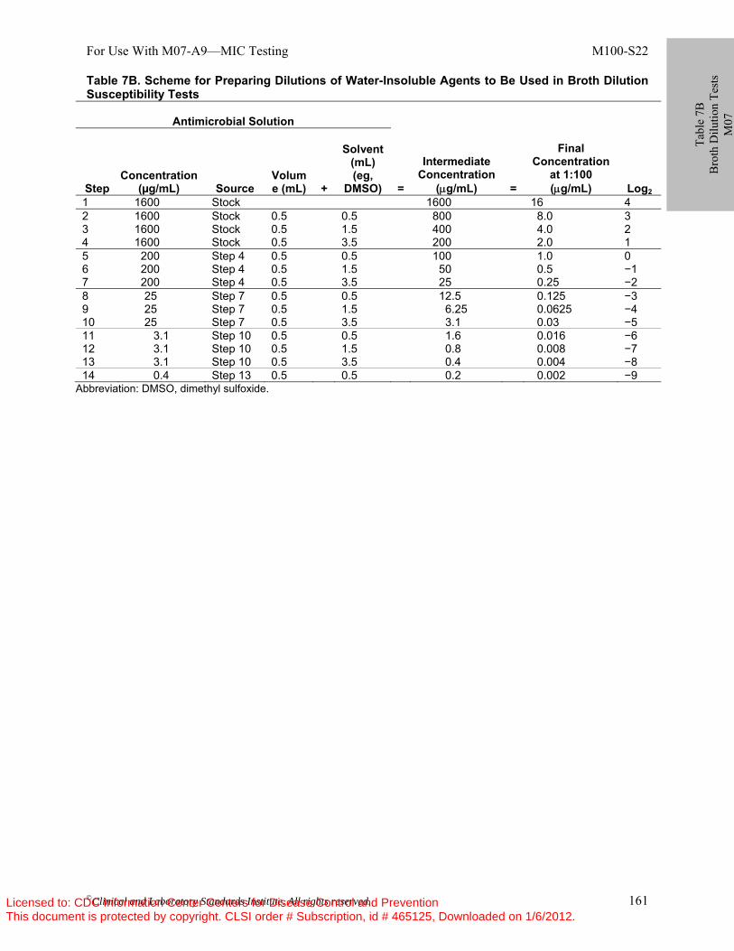
For Use With M07-A9—MIC Testing M100-S22
©Clinical and Laboratory Standards Institute. All rights reserved. 161
Table 7B. Scheme for Preparing Dilutions of Water-Insoluble Agents to Be Used in Broth Dilution Susceptibility Tests
Antimicrobial Solution
Step
Concentration
(µg/mL)
Source
Volume (mL)
+
Solvent
(mL) (eg,
DMSO)
=
Intermediate
Concentration (μg/mL)
=
Final
Concentration at 1:100 (μg/mL)
Log2
1 1600 Stock 1600 16 4 2 1600 Stock 0.5 0.5 800 8.0 3 3 1600 Stock 0.5 1.5 400 4.0 2 4 1600 Stock 0.5 3.5 200 2.0 1 5 200 Step 4 0.5 0.5 100 1.0 0 6 200 Step 4 0.5 1.5 50 0.5 −1 7 200 Step 4 0.5 3.5 25 0.25 −2 8 25 Step 7 0.5 0.5 12.5 0.125 −3 9 25 Step 7 0.5 1.5 6.25 0.0625 −4 10 25 Step 7 0.5 3.5 3.1 0.03 −5 11 3.1 Step 10 0.5 0.5 1.6 0.016 −6 12 3.1 Step 10 0.5 1.5 0.8 0.008 −7 13 3.1 Step 10 0.5 3.5 0.4 0.004 −8 14 0.4 Step 13 0.5 0.5 0.2 0.002 −9
Abbreviation: DMSO, dimethyl sulfoxide.
Ta
ble
7B
Bro
th D
ilutio
n Te
sts
M07
Licensed to: CDC Information Center Centers for Disease Control and PreventionThis document is protected by copyright. CLSI order # Subscription, id # 465125, Downloaded on 1/6/2012.
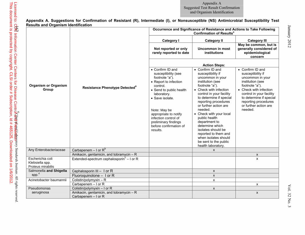
Appendix A. Suggestions for Confirmation of Resistant (R), Intermediate (I), or Nonsusceptible (NS) Antimicrobial Susceptibility Test Results and Organism Identification
Organism or Organism Group Resistance Phenotype Detecteda
Occurrence and Significance of Resistance and Actions to Take Following Confirmation of Resultsa
Category I Category II Category III
Not reported or only rarely reported to date
Uncommon in most institutions
May be common, but is generally considered of
epidemiological concern
Action Steps:
• Confirm ID and susceptibility (see footnote “a”).
• Report to infection control.
• Send to public health laboratory.
• Save isolate. Note: May be appropriate to notify infection control of preliminary findings before confirmation of results.
• Confirm ID and susceptibility if uncommon in your institution (see footnote “a”).
• Check with infection control in your facility to determine if special reporting procedures or further action are needed.
• Check with your local public health department to determine which isolates should be reported to them and when isolates should be sent to the public health laboratory.
• Confirm ID and susceptibility if uncommon in your institution (see footnote “a”).
• Check with infection control in your facility to determine if special reporting procedures or further action are needed.
Any Enterobacteriaceae Carbapenem – I or Rb x Amikacin, gentamicin, and tobramycin – R x
Escherichia coli Klebsiella spp. Proteus mirabilis
Extended-spectrum cephalosporinc – I or R x
Salmonella and Shigella spp.d
Cephalosporin III – I or R x Fluoroquinolone – I or R x
Acinetobacter baumannii Colistin/polymyxin – R x Carbapenem – I or R x
Pseudomonas aeruginosa
Colistin/polymyxin – I or R x Amikacin, gentamicin, and tobramycin – R Carbapenem – I or R
x
Appendix A Suggested Test Result Confirmation
and Organism Identification
162 ©C
linical and Laboratory Standards Institute. A
ll rights reserved.
January 2012 Vol. 32 N
o. 3
Licensed to: CD
C Inform
ation Center C
enters for Disease C
ontrol and Prevention
This docum
ent is protected by copyright. CLS
I order # Subscription, id # 465125, D
ownloaded on 1/6/2012.
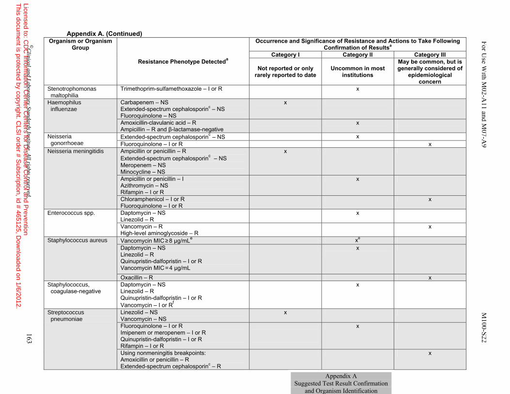
For Use W
ith M02-A
11 and M07-A
9 M100-S22
Appendix A. (Continued)
Organism or Organism Group
Resistance Phenotype Detecteda
Occurrence and Significance of Resistance and Actions to Take Following Confirmation of Resultsa
Category I Category II Category III
Not reported or only rarely reported to date
Uncommon in most institutions
May be common, but is generally considered of
epidemiological concern
Stenotrophomonas maltophilia
Trimethoprim-sulfamethoxazole – I or R x
Haemophilus influenzae
Carbapenem – NS Extended-spectrum cephalosporinc – NS Fluoroquinolone – NS
x
Amoxicillin-clavulanic acid – R Ampicillin – R and β-lactamase-negative
x
Neisseria gonorrhoeae
Extended-spectrum cephalosporinc – NS x Fluoroquinolone – I or R x
Neisseria meningitidis Ampicillin or penicillin – R Extended-spectrum cephalosporinc – NS Meropenem – NS Minocycline – NS
x
Ampicillin or penicillin – I Azithromycin – NS Rifampin – I or R
x
Chloramphenicol – I or R Fluoroquinolone – I or R
x
Enterococcus spp. Daptomycin – NS Linezolid – R
x
Vancomycin – R High-level aminoglycoside – R
x
Staphylococcus aureus Vancomycin MIC ≥ 8 µg/mLe xe Daptomycin – NS Linezolid – R Quinupristin-dalfopristin – I or R Vancomycin MIC = 4 µg/mL
x
Oxacillin – R x Staphylococcus, coagulase-negative
Daptomycin – NS Linezolid – R Quinupristin-dalfopristin – I or R Vancomycin – I or Rf
x
Streptococcus pneumoniae
Linezolid – NS Vancomycin – NS
x
Fluoroquinolone – I or R Imipenem or meropenem – I or R Quinupristin-dalfopristin – I or R Rifampin – I or R
x
Using nonmeningitis breakpoints: Amoxicillin or penicillin – R Extended-spectrum cephalosporinc – R
x
©C
linical and Laboratory Standards Institute. A
ll rights reserved. 163
Appendix A Suggested Test Result Confirmation
and Organism Identification
Licensed to: CD
C Inform
ation Center C
enters for Disease C
ontrol and Prevention
This docum
ent is protected by copyright. CLS
I order # Subscription, id # 465125, D
ownloaded on 1/6/2012.
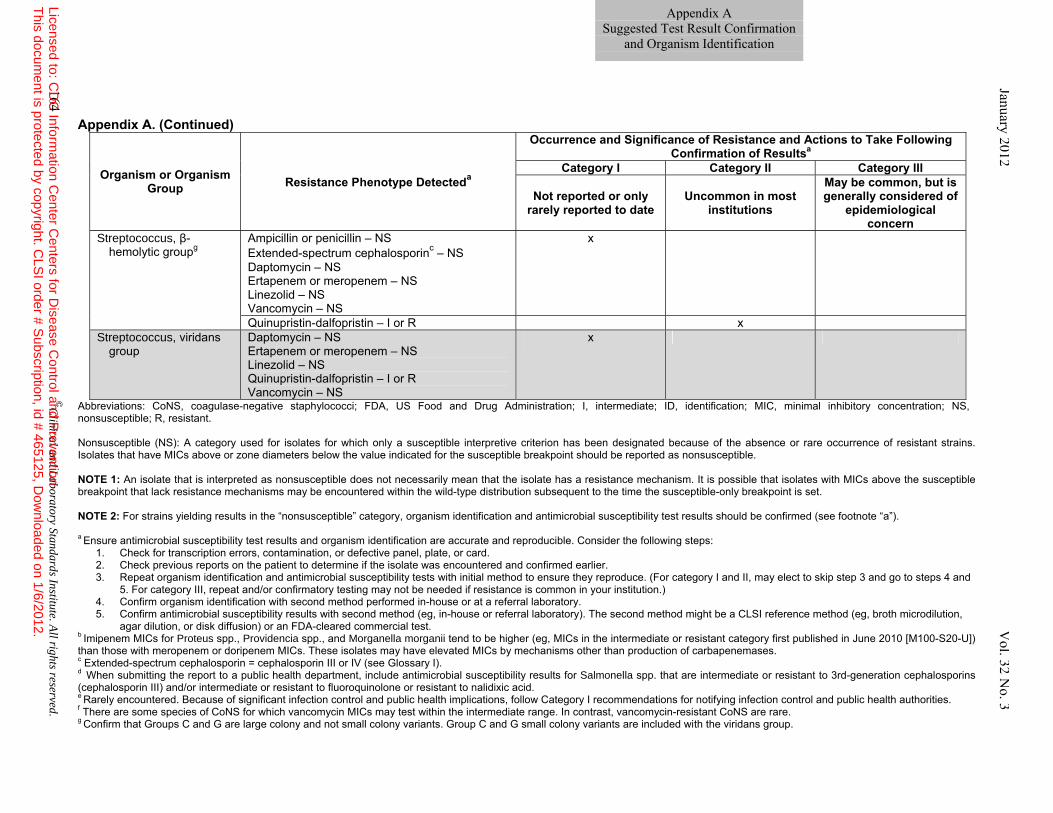
Appendix A. (Continued)
Organism or Organism Group Resistance Phenotype Detecteda
Occurrence and Significance of Resistance and Actions to Take Following Confirmation of Resultsa
Category I Category II Category III
Not reported or only rarely reported to date
Uncommon in most institutions
May be common, but is generally considered of
epidemiological concern
Streptococcus, β-hemolytic groupg
Ampicillin or penicillin – NSExtended-spectrum cephalosporinc – NS Daptomycin – NS Ertapenem or meropenem – NS Linezolid – NS Vancomycin – NS
x
Quinupristin-dalfopristin – I or R x Streptococcus, viridans
group Daptomycin – NS Ertapenem or meropenem – NS Linezolid – NS Quinupristin-dalfopristin – I or R Vancomycin – NS
x
Abbreviations: CoNS, coagulase-negative staphylococci; FDA, US Food and Drug Administration; I, intermediate; ID, identification; MIC, minimal inhibitory concentration; NS, nonsusceptible; R, resistant.
Nonsusceptible (NS): A category used for isolates for which only a susceptible interpretive criterion has been designated because of the absence or rare occurrence of resistant strains. Isolates that have MICs above or zone diameters below the value indicated for the susceptible breakpoint should be reported as nonsusceptible. NOTE 1: An isolate that is interpreted as nonsusceptible does not necessarily mean that the isolate has a resistance mechanism. It is possible that isolates with MICs above the susceptible breakpoint that lack resistance mechanisms may be encountered within the wild-type distribution subsequent to the time the susceptible-only breakpoint is set. NOTE 2: For strains yielding results in the “nonsusceptible” category, organism identification and antimicrobial susceptibility test results should be confirmed (see footnote “a”). a Ensure antimicrobial susceptibility test results and organism identification are accurate and reproducible. Consider the following steps:
1. Check for transcription errors, contamination, or defective panel, plate, or card. 2. Check previous reports on the patient to determine if the isolate was encountered and confirmed earlier. 3. Repeat organism identification and antimicrobial susceptibility tests with initial method to ensure they reproduce. (For category I and II, may elect to skip step 3 and go to steps 4 and
5. For category III, repeat and/or confirmatory testing may not be needed if resistance is common in your institution.) 4. Confirm organism identification with second method performed in-house or at a referral laboratory. 5. Confirm antimicrobial susceptibility results with second method (eg, in-house or referral laboratory). The second method might be a CLSI reference method (eg, broth microdilution,
agar dilution, or disk diffusion) or an FDA-cleared commercial test. b Imipenem MICs for Proteus spp., Providencia spp., and Morganella morganii tend to be higher (eg, MICs in the intermediate or resistant category first published in June 2010 [M100-S20-U]) than those with meropenem or doripenem MICs. These isolates may have elevated MICs by mechanisms other than production of carbapenemases. c Extended-spectrum cephalosporin = cephalosporin III or IV (see Glossary I). d When submitting the report to a public health department, include antimicrobial susceptibility results for Salmonella spp. that are intermediate or resistant to 3rd-generation cephalosporins (cephalosporin III) and/or intermediate or resistant to fluoroquinolone or resistant to nalidixic acid. e Rarely encountered. Because of significant infection control and public health implications, follow Category I recommendations for notifying infection control and public health authorities. f There are some species of CoNS for which vancomycin MICs may test within the intermediate range. In contrast, vancomycin-resistant CoNS are rare. g Confirm that Groups C and G are large colony and not small colony variants. Group C and G small colony variants are included with the viridans group.
164 ©C
linical and Laboratory Standards Institute. A
ll rights reserved.
January 2012 Vol. 32 N
o. 3
Appendix A Suggested Test Result Confirmation
and Organism Identification
Licensed to: CD
C Inform
ation Center C
enters for Disease C
ontrol and Prevention
This docum
ent is protected by copyright. CLS
I order # Subscription, id # 465125, D
ownloaded on 1/6/2012.
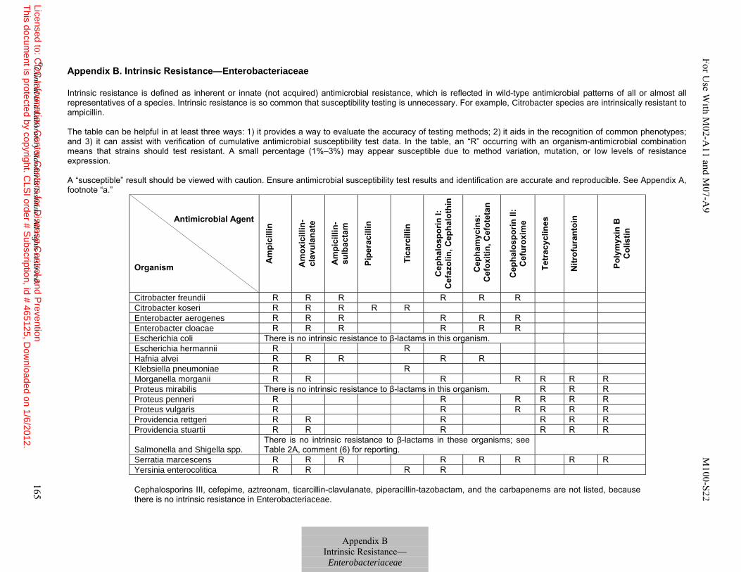
Appendix B. Intrinsic Resistance—Enterobacteriaceae Intrinsic resistance is defined as inherent or innate (not acquired) antimicrobial resistance, which is reflected in wild-type antimicrobial patterns of all or almost all representatives of a species. Intrinsic resistance is so common that susceptibility testing is unnecessary. For example, Citrobacter species are intrinsically resistant to ampicillin. The table can be helpful in at least three ways: 1) it provides a way to evaluate the accuracy of testing methods; 2) it aids in the recognition of common phenotypes; and 3) it can assist with verification of cumulative antimicrobial susceptibility test data. In the table, an “R” occurring with an organism-antimicrobial combination means that strains should test resistant. A small percentage (1%–3%) may appear susceptible due to method variation, mutation, or low levels of resistance expression. A “susceptible” result should be viewed with caution. Ensure antimicrobial susceptibility test results and identification are accurate and reproducible. See Appendix A, footnote “a.”
Antimicrobial Agent Organism
Am
pici
llin
Am
oxic
illin
-cl
avul
anat
e
Am
pici
llin-
su
lbac
tam
Pipe
raci
llin
Tica
rcill
in
Cep
halo
spor
in I:
C
efaz
olin
, Cep
halo
thin
Cep
ham
ycin
s:
Cef
oxiti
n, C
efot
etan
Cep
halo
spor
in II
: C
efur
oxim
e
Tetr
acyc
lines
Nitr
ofur
anto
in
Poly
myx
in B
C
olis
tin
Citrobacter freundii R R R R R R Citrobacter koseri R R R R R Enterobacter aerogenes R R R R R R Enterobacter cloacae R R R R R R Escherichia coli There is no intrinsic resistance to β-lactams in this organism. Escherichia hermannii R R Hafnia alvei R R R R R Klebsiella pneumoniae R R Morganella morganii R R R R R R R Proteus mirabilis There is no intrinsic resistance to β-lactams in this organism. R R R Proteus penneri R R R R R R Proteus vulgaris R R R R R R Providencia rettgeri R R R R R R Providencia stuartii R R R R R R
Salmonella and Shigella spp. There is no intrinsic resistance to β-lactams in these organisms; see Table 2A, comment (6) for reporting.
Serratia marcescens R R R R R R R R Yersinia enterocolitica R R R R Cephalosporins III, cefepime, aztreonam, ticarcillin-clavulanate, piperacillin-tazobactam, and the carbapenems are not listed, because there is no intrinsic resistance in Enterobacteriaceae.
©C
linical and Laboratory Standards Institute. A
ll rights reserved. 165
For Use W
ith M02-A
11 and M07-A
9 M100-S22
Appendix B
Intrinsic Resistance— Enterobacteriaceae
Licensed to: CD
C Inform
ation Center C
enters for Disease C
ontrol and Prevention
This docum
ent is protected by copyright. CLS
I order # Subscription, id # 465125, D
ownloaded on 1/6/2012.
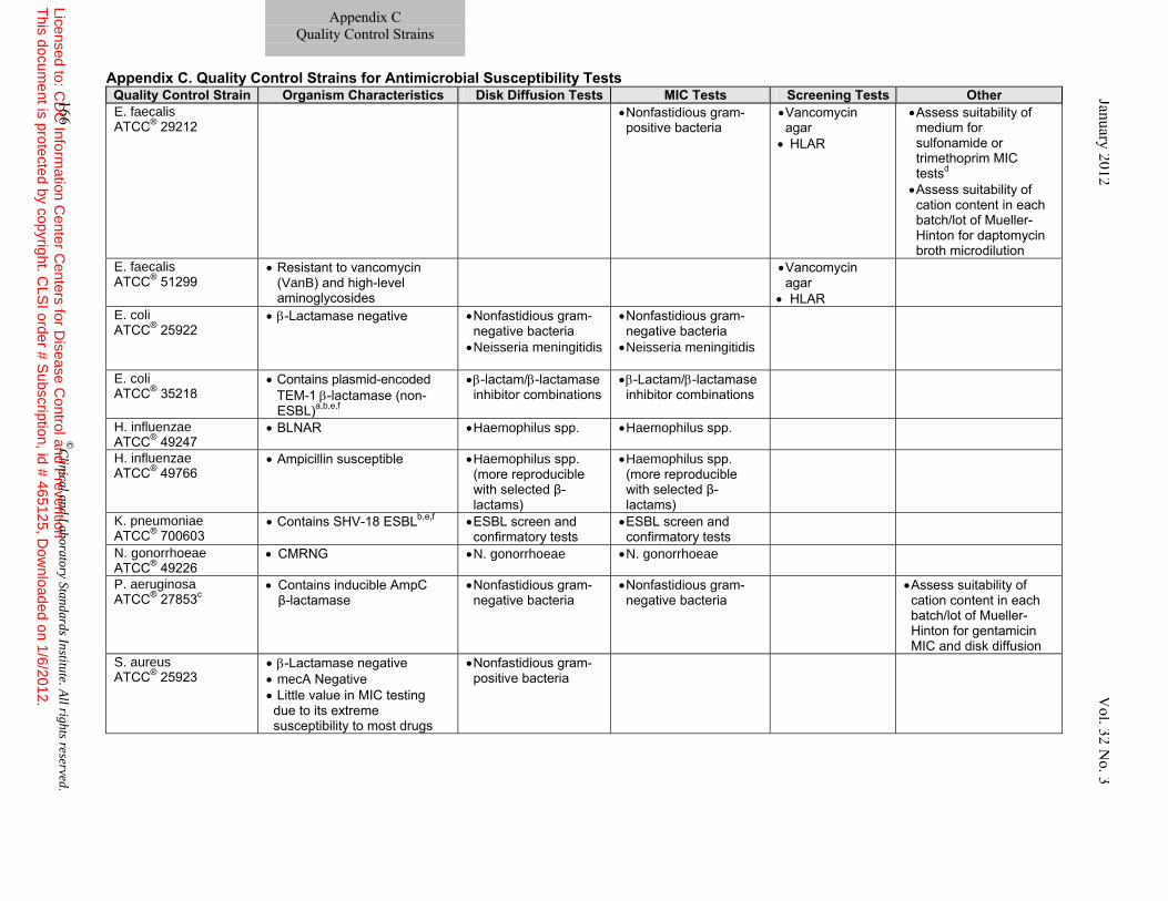
Appendix C. Quality Control Strains for Antimicrobial Susceptibility Tests Quality Control Strain Organism Characteristics Disk Diffusion Tests MIC Tests Screening Tests Other E. faecalis ATCC® 29212
• Nonfastidious gram- positive bacteria
• Vancomycin agar
• HLAR
• Assess suitability of medium for sulfonamide or trimethoprim MIC testsd • Assess suitability of cation content in each batch/lot of Mueller- Hinton for daptomycin broth microdilution
E. faecalis ATCC® 51299
• Resistant to vancomycin (VanB) and high-level aminoglycosides
• Vancomycin agar
• HLAR
E. coli ATCC® 25922
• β-Lactamase negative • Nonfastidious gram-negative bacteria • Neisseria meningitidis
• Nonfastidious gram-negative bacteria • Neisseria meningitidis
E. coli ATCC® 35218
• Contains plasmid-encoded TEM-1 β-lactamase (non- ESBL)a,b,e,f
• β-lactam/β-lactamase inhibitor combinations
• β-Lactam/β-lactamase inhibitor combinations
H. influenzae ATCC® 49247
• BLNAR • Haemophilus spp. • Haemophilus spp.
H. influenzae ATCC® 49766
• Ampicillin susceptible • Haemophilus spp. (more reproducible with selected β-lactams)
• Haemophilus spp. (more reproducible with selected β-lactams)
K. pneumoniae ATCC® 700603
• Contains SHV-18 ESBLb,e,f • ESBL screen and confirmatory tests
• ESBL screen and confirmatory tests
N. gonorrhoeae ATCC® 49226
• CMRNG • N. gonorrhoeae • N. gonorrhoeae
P. aeruginosa ATCC® 27853c
• Contains inducible AmpC β-lactamase
• Nonfastidious gram-negative bacteria
• Nonfastidious gram-negative bacteria
• Assess suitability of cation content in each batch/lot of Mueller-Hinton for gentamicin MIC and disk diffusion
S. aureus ATCC® 25923
• β-Lactamase negative • mecA Negative • Little value in MIC testing due to its extreme susceptibility to most drugs
• Nonfastidious gram-positive bacteria
Appendix C Quality Control Strains
166 ©C
linical and Laboratory Standards Institute. A
ll rights reserved.
January 2012 Vol. 32
No. 3
Licensed to: CD
C Inform
ation Center C
enters for Disease C
ontrol and Prevention
This docum
ent is protected by copyright. CLS
I order # Subscription, id # 465125, D
ownloaded on 1/6/2012.
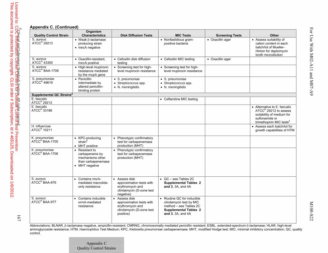
Appendix C. (Continued)
Quality Control Strain
Organism Characteristics
Disk Diffusion Tests
MIC Tests
Screening Tests
Other
S. aureus ATCC® 29213
• Weak β-lactamase producing strain
• mecA negative
• Nonfastidious gram-positive bacteria
• Oxacillin agar • Assess suitability of cation content in each batch/lot of Mueller- Hinton for daptomycin broth microdilution
S. aureus ATCC® 43300
• Oxacillin-resistant, mecA positive
• Cefoxitin disk diffusion testing
• Cefoxitin MIC testing • Oxacillin agar
S. aureus ATCC® BAA-1708
• High-level mupirocin resistance mediated by the mupA gene
• Screening test for high-level mupirocin resistance
• Screening test for high-level mupirocin resistance
S. pneumoniae ATCC® 49619
• Penicillin intermediate by altered penicillin-binding protein
• S. pneumoniae • Streptococcus spp. • N. meningitidis
• S. pneumoniae • Streptococcus spp. • N. meningitidis
Supplemental QC Strainsg E. faecalis ATCC® 29212
• Ceftaroline MIC testing
E. faecalis ATCC® 33186
• Alternative to E. faecalis ATCC® 29212 to assess suitability of medium for sulfonamide or trimethoprim MIC testsd
H. influenzae ATCC® 10211
• Assess each batch/lot for growth capabilities of HTM
K. pneumoniae ATCC® BAA-1705
• KPC-producing strainb
• MHT positive
• Phenotypic confirmatory test for carbapenemase production (MHT)
K. pneumoniae ATCC® BAA-1706
• Resistant to carbapenems by mechanisms other than carbapenemase
• MHT negative
• Phenotypic confirmatory test for carbapenemase production (MHT)
S. aureus ATCC® BAA-976
• Contains msrA-mediated macrolide-only resistance
• Assess disk approximation tests with erythromycin and clindamycin (D-zone test negative)
• QC – see Tables 2C Supplemental Tables 2 and 3, 3A, and 4A
S. aureus ATCC® BAA-977
• Contains inducible ermA-mediated resistance
• Assess disk approximation tests with erythromycin and clindamycin (D-zone test positive)
• Routine QC for inducible clindamycin test by MIC method – see Tables 2C Supplemental Tables 2 and 3, 3A, and 4A
Abbreviations: BLNAR, β-lactamase negative, ampicillin-resistant; CMRNG, chromosomally mediated penicillin resistant; ESBL, extended-spectrum β-lactamase; HLAR, high-level aminoglycoside resistance; HTM, Haemophilus Test Medium; KPC, Klebsiella pneumoniae carbapenemase; MHT, modified Hodge test; MIC, minimal inhibitory concentration; QC, quality control.
©C
linical and Laboratory Standards Institute. A
ll rights reserved. 167
Appendix C
Quality Control Strains
For Use W
ith M02-A
11 and M07-A
9 M100-S22
Licensed to: CD
C Inform
ation Center C
enters for Disease C
ontrol and Prevention
This docum
ent is protected by copyright. CLS
I order # Subscription, id # 465125, D
ownloaded on 1/6/2012.
Appendix C. (Continued)
Footnotes a E. coli ATCC® 35218 is recommended only as a control organism for β-lactamase inhibitor combinations, such as those containing clavulanic acid,
sulbactam, or tazobactam. This strain contains a plasmid-encoded β-lactamase (non-ESBL); subsequently, the organism is resistant to many penicillinase-labile drugs, but susceptible to β-lactam/β-lactamase inhibitor combinations. The plasmid must be present in the QC strain for the QC test to be valid; however, the plasmid may be lost during storage at refrigerator or freezer temperatures. To ensure the plasmid is present, test the strain with a β-lactam agent alone (ampicillin, amoxicillin, piperacillin, or ticarcillin) in addition to a β-lactam/β-lactamase inhibitor agent (eg, amoxicillin-clavulanate). If the strain loses the plasmid, it will be susceptible to the β-lactam agent when tested alone, indicating that the QC test is invalid and a new culture of E. coli ATCC® 35218 must be used.
b Careful attention to organism maintenance (eg, minimal subcultures) and storage (eg, −60°C or below) is especially important for QC strains E. coli ATCC® 35218, K. pneumoniae ATCC® 700603, and K. pneumoniae ATCC® BAA-1705, because spontaneous loss of the plasmid encoding the β-lactamase or carbapenemase has been documented. Plasmid loss leads to QC results outside the acceptable limit, such as decreased MICs for E. coli ATCC® 35218 with enzyme-labile penicillins (eg, ampicillin, piperacillin, and ticarcillin), decreased MICs for K. pneumoniae ATCC® 700603 with cephalosporins and aztreonam, and false-negative MHT with K. pneumoniae ATCC® BAA-1705.
c Develops resistance to β-lactam antimicrobial agents after repeated transfers onto laboratory media. Minimize by removing new culture from storage at least monthly or whenever the strain begins to show resistance.
d End points should be easy to read (as 80% or greater reduction in growth as compared with the control) if media have acceptable levels of thymidine. e Rasheed JK, Anderson GJ, Yigit H, et al. Characterization of the extended-spectrum beta-lactamase reference strain, Klebsiella pneumoniae K6
(ATCC® 700603), which produces the novel enzyme SHV-18. Antimicrob Agents Chemother. 2000;44(9):2382-2388. f Queenan AM, Foleno B, Gownley C, Wira E, Bush K. Effects of inoculum and beta-lactamase activity in AmpC- and extended-spectrum beta-
lactamase (ESBL)-producing Escherichia coli and Klebsiella pneumoniae clinical isolates tested by using NCCLS ESBL methodology. J Clin Microbiol. 2004;42(1):269-275.
g QC strains are tested regularly (eg, daily or weekly) to ensure the test system is working and produces results that fall within specified limits listed in M100. The QC strains recommended in this document should be included if a laboratory performs CLSI reference disk diffusion or MIC testing as described herein. For commercial test systems, manufacturer’s recommendations should be followed for all QC procedures. Supplemental QC strains are used to assess particular characteristics of a test or test system in select situations, or may represent alternative QC strains. For example, Haemophilus influenzae ATCC® 10211 is more fastidious than H. influenzae ATCC® 49247 or H. influenzae ATCC® 49766, and is used to ensure HTM can adequately support the growth of clinical isolates of H. influenzae and H. parainfluenzae. Supplemental QC strains may possess susceptibility or resistance characteristics specific for one or more special tests listed in M02-A11 and M07-A9. They can be used to assess a new test, for training new personnel, and for competency assessment. It is not necessary to include supplemental QC strains in routine daily or weekly AST QC programs.
168 ©C
linical and Laboratory Standards Institute. A
ll rights reserved.
Appendix C
Quality Control Strains
January 2012 Vol. 32 N
o. 3
Licensed to: CD
C Inform
ation Center C
enters for Disease C
ontrol and Prevention
This docum
ent is protected by copyright. CLS
I order # Subscription, id # 465125, D
ownloaded on 1/6/2012.
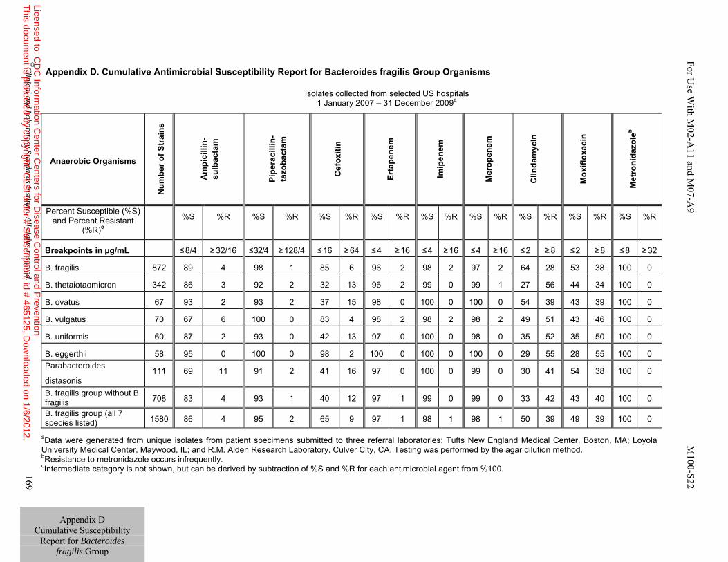
Appendix D. Cumulative Antimicrobial Susceptibility Report for Bacteroides fragilis Group Organisms
Isolates collected from selected US hospitals
1 January 2007 – 31 December 2009a
aData were generated from unique isolates from patient specimens submitted to three referral laboratories: Tufts New England Medical Center, Boston, MA; Loyola University Medical Center, Maywood, IL; and R.M. Alden Research Laboratory, Culver City, CA. Testing was performed by the agar dilution method. bResistance to metronidazole occurs infrequently. cIntermediate category is not shown, but can be derived by subtraction of %S and %R for each antimicrobial agent from %100.
Anaerobic Organisms N
umbe
r of S
trai
ns
Am
pici
llin-
sulb
acta
m
Pipe
raci
llin-
tazo
bact
am
Cef
oxiti
n
Erta
pene
m
Imip
enem
Mer
open
em
C
linda
myc
in
M
oxifl
oxac
in
M
etro
nida
zole
b
Percent Susceptible (%S) and Percent Resistant
(%R)c %S
%R %S %R %S %R %S %R %S %R %S %R %S %R %S %R %S %R
Breakpoints in µg/mL ≤ 8/4 ≥ 32/16 ≤ 32/4 ≥ 128/4 ≤ 16 ≥ 64 ≤ 4 ≥ 16 ≤ 4 ≥ 16 ≤ 4 ≥ 16 ≤ 2 ≥ 8 ≤ 2 ≥ 8 ≤ 8 ≥ 32
B. fragilis 872 89 4 98 1 85 6 96 2 98 2 97 2 64 28 53 38 100 0
B. thetaiotaomicron 342 86 3 92 2 32 13 96 2 99 0 99 1 27 56 44 34 100 0
B. ovatus 67 93 2 93 2 37 15 98 0 100 0 100 0 54 39 43 39 100 0
B. vulgatus 70 67 6 100 0 83 4 98 2 98 2 98 2 49 51 43 46 100 0
B. uniformis 60 87 2 93 0 42 13 97 0 100 0 98 0 35 52 35 50 100 0
B. eggerthii 58 95 0 100 0 98 2 100 0 100 0 100 0 29 55 28 55 100 0 Parabacteroides
distasonis 111 69 11 91 2 41 16 97 0 100 0 99 0 30 41 54 38 100 0
B. fragilis group without B. fragilis 708 83 4 93 1 40 12 97 1 99 0 99 0 33 42 43 40 100 0
B. fragilis group (all 7 species listed) 1580 86 4 95 2 65 9 97 1 98 1 98 1 50 39 49 39 100 0
Appendix D
Cumulative Susceptibility Report for Bacteroides
fragilis Group
©C
linical and Laboratory Standards Institute. A
ll rights reserved. 169
For Use W
ith M02-A
11 and M07-A
9 M100-S22
Licensed to: CD
C Inform
ation Center C
enters for Disease C
ontrol and Prevention
This docum
ent is protected by copyright. CLS
I order # Subscription, id # 465125, D
ownloaded on 1/6/2012.
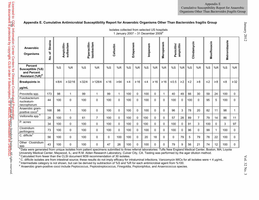
Appendix E. Cumulative Antimicrobial Susceptibility Report for Anaerobic Organisms Other Than Bacteroides fragilis Group
Isolates collected from selected US hospitals
1 January 2007 – 31 December 2009a
a Data were generated from unique isolates from patient specimens submitted to three referral laboratories: Tufts New England Medical Center, Boston, MA; Loyola University Medical Center, Maywood, IL; and R.M. Alden Research Laboratory, Culver City, CA. Testing was performed by the agar dilution method. b Calculated from fewer than the CLSI document M39 recommendation of 30 isolates. c C. difficile isolates are from intestinal source; these results do not imply efficacy for intraluminal infections. Vancomycin MICs for all isolates were < 4 µg/mL. d Intermediate category is not shown, but can be derived by subtraction of %S and %R for each antimicrobial agent from %100. e Anaerobic gram-positive cocci include Peptococcus, Peptostreptococcus, Finegoldia, Peptoniphilus, and Anaerococcus species.
Anaerobic
Organisms N
o. o
f St
rain
s
Am
pici
llin-
sulb
acta
m
Pipe
raci
llin-
tazo
bact
am
Cef
oxiti
n
Erta
pene
m
Imip
enem
Mer
open
em
Peni
cilli
n/
ampi
cilli
n
C
linda
myc
in
M
oxifl
oxac
in
Met
roni
dazo
le
Percent Susceptible (%S)
and Percent Resistant (%R)d
%S
%R %S %R %S %R %S %R %S %R %R %S %R %S %R %S %R %S %R
Breakpoints in
µg/mL
≤ 8/4 ≥ 32/16 ≤ 32/4 ≥ 128/4 ≤ 16 ≥ 64 ≤ 4 ≥ 16 ≤ 4 ≥ 16 ≥ 16 ≤ 0.5 ≥ 2 ≤ 2 ≥ 8 ≤ 2 ≥ 8 ≤ 8 ≥ 32
Prevotella spp. 173 98 1 99 1 99 1 100 0 100 0 1 40 49 66 30 59 24 100 0 Fusobacterium nucleatum-necrophorum
44 100 0 100 0 100 0 100 0 100 0 0 100 0 100 0 95 5 100 0
Anaerobic gram-positive coccie 168 98 1 100 0 100 0 100 0 100 0 0 96 3 78 20 82 11 98 1
Veillonella spp.b 28 100 0 61 7 100 0 100 0 100 0 0 57 28 89 7 79 14 86 11 P. acnes
34 100 0 100 0 100 0 100 0 100 0 0 100 0 91 3 100 0 3 97 Clostridium perfringens 73 100 0 100 0 100 0 100 0 100 0 0 100 0 96 0 99 1 100 0
C. difficilec
56 100 0 100 0 0 100 100 0 20 18 0 0 79 5 79 78 22 100 0
Other Clostridium spp. 43 100 0 100 0 47 26 100 0 100 0 0 79 9 56 21 74 12 100 0
170 ©C
linical and Laboratory Standards Institute. A
ll rights reserved.
January 2012 Vol. 32
No. 3
Appendix E Cumulative Susceptibility Report for Anaerobic
Organisms Other Than Bacteroides fragilis Group
Licensed to: CD
C Inform
ation Center C
enters for Disease C
ontrol and Prevention
This docum
ent is protected by copyright. CLS
I order # Subscription, id # 465125, D
ownloaded on 1/6/2012.

For Use With M02-A11 and M07-A9 M100-S22
©Clinical and Laboratory Standards Institute. All rights reserved. 171
This page is intentionally left blank.
Licensed to: CDC Information Center Centers for Disease Control and PreventionThis document is protected by copyright. CLSI order # Subscription, id # 465125, Downloaded on 1/6/2012.
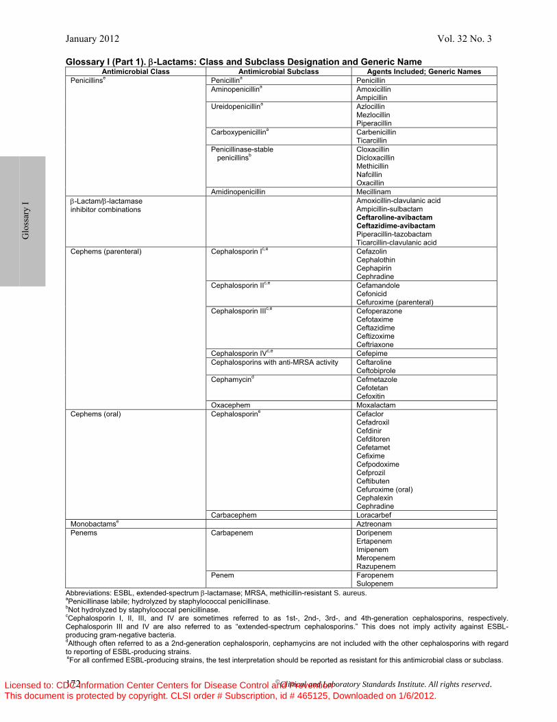
January 2012 Vol. 32 No. 3
172 ©Clinical and Laboratory Standards Institute. All rights reserved.
Glossary I (Part 1). β-Lactams: Class and Subclass Designation and Generic Name Antimicrobial Class Antimicrobial Subclass Agents Included; Generic Names
Penicillinse Penicillina Penicillin Aminopenicillina Amoxicillin
Ampicillin Ureidopenicillina Azlocillin
Mezlocillin Piperacillin
Carboxypenicillina Carbenicillin Ticarcillin
Penicillinase-stable penicillinsb
Cloxacillin Dicloxacillin Methicillin Nafcillin Oxacillin
Amidinopenicillin Mecillinam β-Lactam/β-lactamase inhibitor combinations
Amoxicillin-clavulanic acid Ampicillin-sulbactam Ceftaroline-avibactam Ceftazidime-avibactam Piperacillin-tazobactam Ticarcillin-clavulanic acid
Cephems (parenteral) Cephalosporin Ic,e Cefazolin Cephalothin Cephapirin Cephradine
Cephalosporin IIc,e Cefamandole Cefonicid Cefuroxime (parenteral)
Cephalosporin IIIc,e Cefoperazone Cefotaxime Ceftazidime Ceftizoxime Ceftriaxone
Cephalosporin IVc,e Cefepime Cephalosporins with anti-MRSA activity Ceftaroline
Ceftobiprole Cephamycind Cefmetazole
Cefotetan Cefoxitin
Oxacephem Moxalactam Cephems (oral) Cephalosporine Cefaclor
Cefadroxil Cefdinir Cefditoren Cefetamet Cefixime Cefpodoxime Cefprozil Ceftibuten Cefuroxime (oral) Cephalexin Cephradine
Carbacephem Loracarbef Monobactamse Aztreonam Penems Carbapenem Doripenem
Ertapenem Imipenem Meropenem Razupenem
Penem Faropenem Sulopenem
Abbreviations: ESBL, extended-spectrum β-lactamase; MRSA, methicillin-resistant S. aureus. aPenicillinase labile; hydrolyzed by staphylococcal penicillinase. bNot hydrolyzed by staphylococcal penicillinase. cCephalosporin I, II, III, and IV are sometimes referred to as 1st-, 2nd-, 3rd-, and 4th-generation cephalosporins, respectively. Cephalosporin III and IV are also referred to as “extended-spectrum cephalosporins.” This does not imply activity against ESBL-producing gram-negative bacteria. dAlthough often referred to as a 2nd-generation cephalosporin, cephamycins are not included with the other cephalosporins with regard to reporting of ESBL-producing strains. eFor all confirmed ESBL-producing strains, the test interpretation should be reported as resistant for this antimicrobial class or subclass.
G
loss
ary
I
Licensed to: CDC Information Center Centers for Disease Control and PreventionThis document is protected by copyright. CLSI order # Subscription, id # 465125, Downloaded on 1/6/2012.
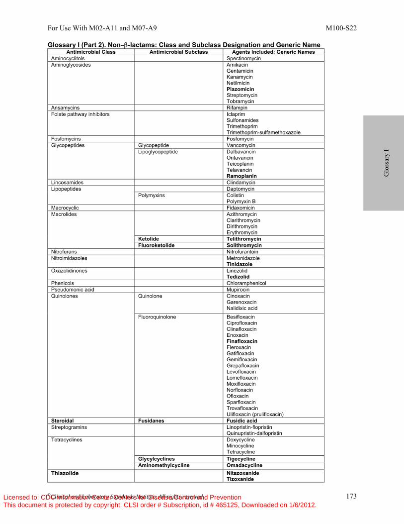
For Use With M02-A11 and M07-A9 M100-S22
©Clinical and Laboratory Standards Institute. All rights reserved. 173
Glossary I (Part 2). Non–β-lactams: Class and Subclass Designation and Generic Name Antimicrobial Class Antimicrobial Subclass Agents Included; Generic Names
Aminocyclitols Spectinomycin Aminoglycosides Amikacin
Gentamicin Kanamycin Netilmicin Plazomicin Streptomycin Tobramycin
Ansamycins Rifampin Folate pathway inhibitors Iclaprim
Sulfonamides Trimethoprim Trimethoprim-sulfamethoxazole
Fosfomycins Fosfomycin Glycopeptides Glycopeptide Vancomycin
Lipoglycopeptide Dalbavancin Oritavancin Teicoplanin Telavancin Ramoplanin
Lincosamides Clindamycin Lipopeptides Daptomycin
Polymyxins Colistin Polymyxin B
Macrocyclic Fidaxomicin Macrolides Azithromycin
Clarithromycin Dirithromycin Erythromycin
Ketolide Telithromycin Fluoroketolide Solithromycin
Nitrofurans Nitrofurantoin Nitroimidazoles Metronidazole
Tinidazole Oxazolidinones Linezolid
Tedizolid Phenicols Chloramphenicol Pseudomonic acid Mupirocin Quinolones Quinolone Cinoxacin
Garenoxacin Nalidixic acid
Fluoroquinolone Besifloxacin Ciprofloxacin Clinafloxacin Enoxacin Finafloxacin Fleroxacin Gatifloxacin Gemifloxacin Grepafloxacin Levofloxacin Lomefloxacin Moxifloxacin Norfloxacin Ofloxacin Sparfloxacin Trovafloxacin Ulifloxacin (prulifloxacin)
Steroidal Fusidanes Fusidic acid Streptogramins
Linopristin-flopristin Quinupristin-dalfopristin
Tetracyclines Doxycycline Minocycline Tetracycline
Glycylcyclines Tigecycline Aminomethylcycline Omadacycline
Thiazolide Nitazoxanide Tizoxanide
G
loss
ary
I
Licensed to: CDC Information Center Centers for Disease Control and PreventionThis document is protected by copyright. CLSI order # Subscription, id # 465125, Downloaded on 1/6/2012.
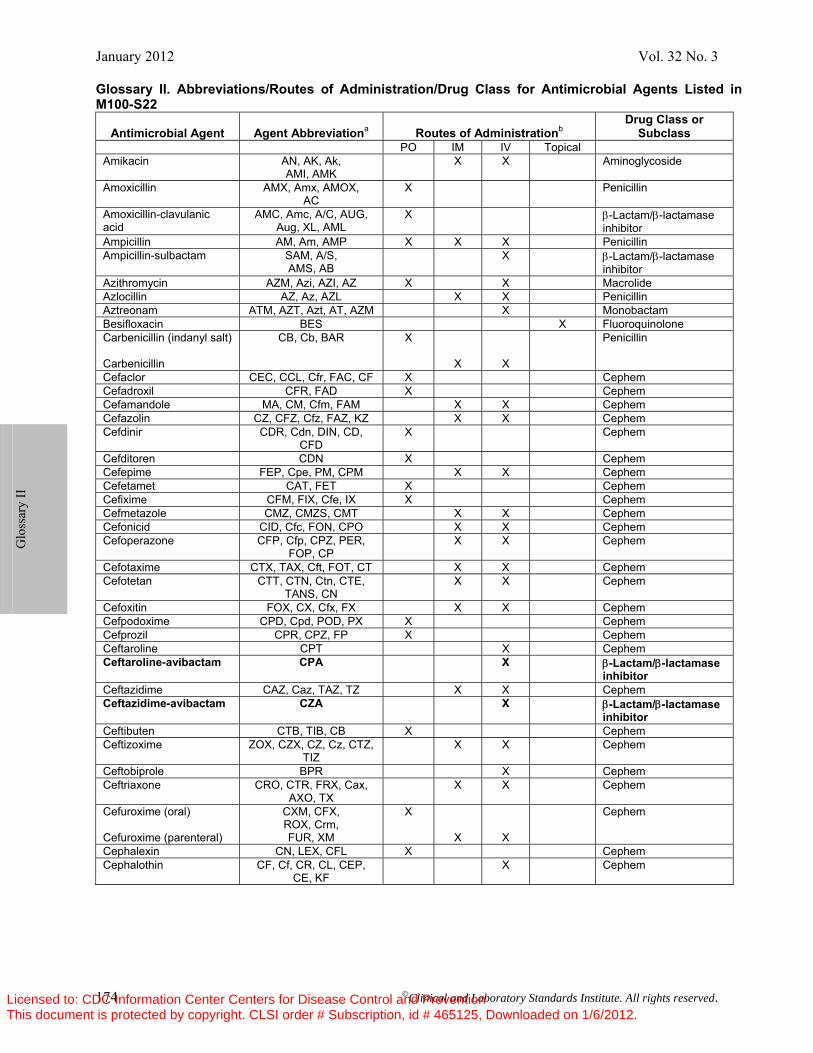
January 2012 Vol. 32 No. 3
174 ©Clinical and Laboratory Standards Institute. All rights reserved.
Glossary II. Abbreviations/Routes of Administration/Drug Class for Antimicrobial Agents Listed in M100-S22
Antimicrobial Agent Agent Abbreviationa Routes of Administrationb Drug Class or
Subclass PO IM IV Topical Amikacin AN, AK, Ak,
AMI, AMK X X Aminoglycoside
Amoxicillin AMX, Amx, AMOX, AC
X Penicillin
Amoxicillin-clavulanic acid
AMC, Amc, A/C, AUG, Aug, XL, AML
X β-Lactam/β-lactamase inhibitor
Ampicillin AM, Am, AMP X X X Penicillin Ampicillin-sulbactam SAM, A/S,
AMS, AB X β-Lactam/β-lactamase
inhibitor Azithromycin AZM, Azi, AZI, AZ X X Macrolide Azlocillin AZ, Az, AZL X X Penicillin Aztreonam ATM, AZT, Azt, AT, AZM X Monobactam Besifloxacin BES X Fluoroquinolone Carbenicillin (indanyl salt) Carbenicillin
CB, Cb, BAR X
X
X
Penicillin
Cefaclor CEC, CCL, Cfr, FAC, CF X Cephem Cefadroxil CFR, FAD X Cephem Cefamandole MA, CM, Cfm, FAM X X Cephem Cefazolin CZ, CFZ, Cfz, FAZ, KZ X X Cephem Cefdinir CDR, Cdn, DIN, CD,
CFD X Cephem
Cefditoren CDN X Cephem Cefepime FEP, Cpe, PM, CPM X X Cephem Cefetamet CAT, FET X Cephem Cefixime CFM, FIX, Cfe, IX X Cephem Cefmetazole CMZ, CMZS, CMT X X Cephem Cefonicid CID, Cfc, FON, CPO X X Cephem Cefoperazone CFP, Cfp, CPZ, PER,
FOP, CP X X Cephem
Cefotaxime CTX, TAX, Cft, FOT, CT X X Cephem Cefotetan CTT, CTN, Ctn, CTE,
TANS, CN X X Cephem
Cefoxitin FOX, CX, Cfx, FX X X Cephem Cefpodoxime CPD, Cpd, POD, PX X Cephem Cefprozil CPR, CPZ, FP X Cephem Ceftaroline CPT X Cephem Ceftaroline-avibactam CPA X β-Lactam/β-lactamase
inhibitor Ceftazidime CAZ, Caz, TAZ, TZ X X Cephem Ceftazidime-avibactam CZA X β-Lactam/β-lactamase
inhibitor Ceftibuten CTB, TIB, CB X Cephem Ceftizoxime ZOX, CZX, CZ, Cz, CTZ,
TIZ X X Cephem
Ceftobiprole BPR X Cephem Ceftriaxone CRO, CTR, FRX, Cax,
AXO, TX X X Cephem
Cefuroxime (oral) Cefuroxime (parenteral)
CXM, CFX, ROX, Crm, FUR, XM
X
X
X
Cephem
Cephalexin CN, LEX, CFL X Cephem Cephalothin CF, Cf, CR, CL, CEP,
CE, KF X Cephem
Glo
ssar
y II
Licensed to: CDC Information Center Centers for Disease Control and PreventionThis document is protected by copyright. CLSI order # Subscription, id # 465125, Downloaded on 1/6/2012.
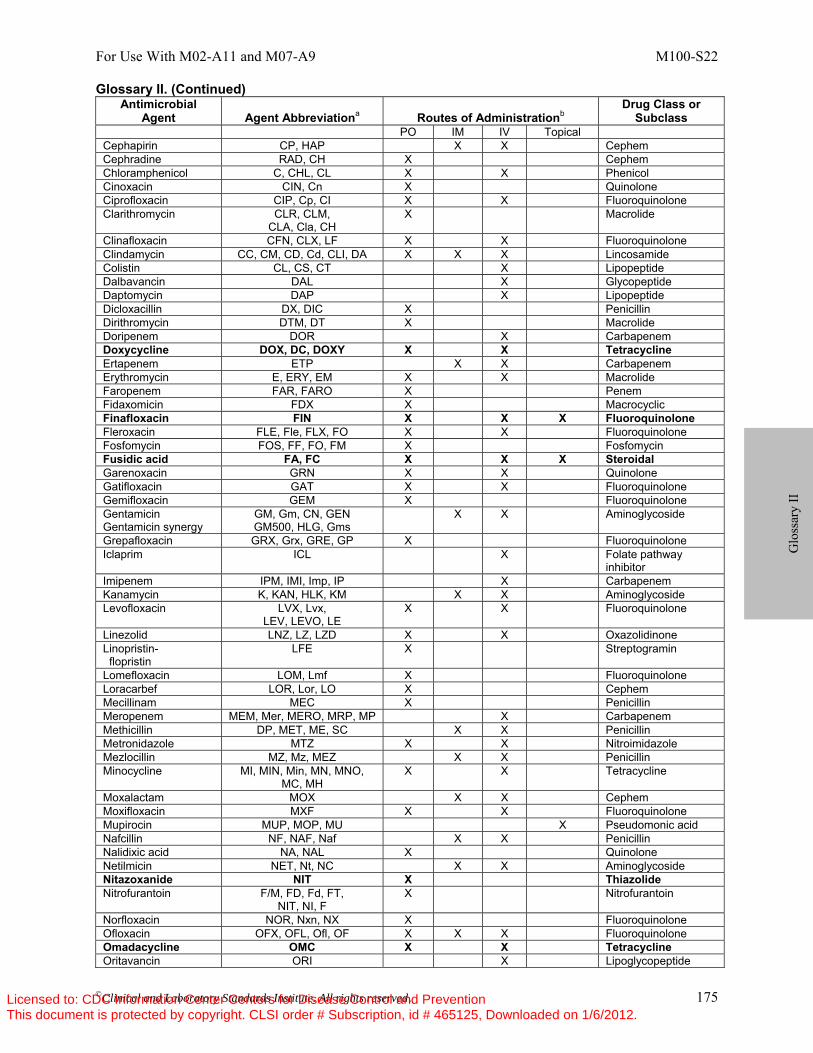
For Use With M02-A11 and M07-A9 M100-S22
©Clinical and Laboratory Standards Institute. All rights reserved. 175
Glossary II. (Continued) Antimicrobial
Agent Agent Abbreviationa Routes of Administrationb Drug Class or
Subclass PO IM IV Topical Cephapirin CP, HAP X X Cephem Cephradine RAD, CH X Cephem Chloramphenicol C, CHL, CL X X Phenicol Cinoxacin CIN, Cn X Quinolone Ciprofloxacin CIP, Cp, CI X X Fluoroquinolone Clarithromycin CLR, CLM,
CLA, Cla, CH X Macrolide
Clinafloxacin CFN, CLX, LF X X Fluoroquinolone Clindamycin CC, CM, CD, Cd, CLI, DA X X X Lincosamide Colistin CL, CS, CT X Lipopeptide Dalbavancin DAL X Glycopeptide Daptomycin DAP X Lipopeptide Dicloxacillin DX, DIC X Penicillin Dirithromycin DTM, DT X Macrolide Doripenem DOR X Carbapenem Doxycycline DOX, DC, DOXY X X TetracyclineErtapenem ETP X X Carbapenem Erythromycin E, ERY, EM X X Macrolide Faropenem FAR, FARO X Penem Fidaxomicin FDX X Macrocyclic Finafloxacin FIN X X X FluoroquinoloneFleroxacin FLE, Fle, FLX, FO X X Fluoroquinolone Fosfomycin FOS, FF, FO, FM X Fosfomycin Fusidic acid FA, FC X X X SteroidalGarenoxacin GRN X X Quinolone Gatifloxacin GAT X X Fluoroquinolone Gemifloxacin GEM X Fluoroquinolone Gentamicin Gentamicin synergy
GM, Gm, CN, GEN GM500, HLG, Gms
X X Aminoglycoside
Grepafloxacin GRX, Grx, GRE, GP X Fluoroquinolone Iclaprim ICL X Folate pathway
inhibitor Imipenem IPM, IMI, Imp, IP X Carbapenem Kanamycin K, KAN, HLK, KM X X Aminoglycoside Levofloxacin LVX, Lvx,
LEV, LEVO, LE X X Fluoroquinolone
Linezolid LNZ, LZ, LZD X X Oxazolidinone Linopristin- flopristin
LFE X Streptogramin
Lomefloxacin LOM, Lmf X Fluoroquinolone Loracarbef LOR, Lor, LO X Cephem Mecillinam MEC X Penicillin Meropenem MEM, Mer, MERO, MRP, MP X Carbapenem Methicillin DP, MET, ME, SC X X Penicillin Metronidazole MTZ X X Nitroimidazole Mezlocillin MZ, Mz, MEZ X X Penicillin Minocycline MI, MIN, Min, MN, MNO,
MC, MH X X Tetracycline
Moxalactam MOX X X Cephem Moxifloxacin MXF X X Fluoroquinolone Mupirocin MUP, MOP, MU X Pseudomonic acid Nafcillin NF, NAF, Naf X X Penicillin Nalidixic acid NA, NAL X Quinolone Netilmicin NET, Nt, NC X X Aminoglycoside Nitazoxanide NIT X ThiazolideNitrofurantoin F/M, FD, Fd, FT,
NIT, NI, F X Nitrofurantoin
Norfloxacin NOR, Nxn, NX X Fluoroquinolone Ofloxacin OFX, OFL, Ofl, OF X X X Fluoroquinolone Omadacycline OMC X X TetracyclineOritavancin ORI X Lipoglycopeptide
G
loss
ary
II
Licensed to: CDC Information Center Centers for Disease Control and PreventionThis document is protected by copyright. CLSI order # Subscription, id # 465125, Downloaded on 1/6/2012.
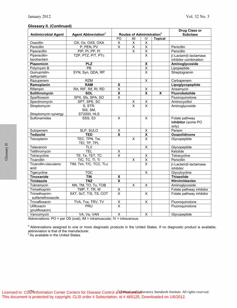
January 2012 Vol. 32 No. 3
176 ©Clinical and Laboratory Standards Institute. All rights reserved.
Glossary II. (Continued)
Antimicrobial Agent Agent Abbreviationa Routes of Administrationb Drug Class or
Subclass PO IM IV Topical Oxacillin OX, Ox, OXS, OXA X X X Penicillin P, PEN, PV X X X Penicillin Piperacillin PIP, PI, PP, Pi X X Penicillin Piperacillin-tazobactam
TZP, PTZ, P/T, PTc X β-Lactam/β-lactamase inhibitor combination
Plazomicin PLZ X AminoglycosidePolymyxin B PB X Lipopeptide Quinupristin-dalfopristin
SYN, Syn, QDA, RP X Streptogramin
Razupenem RZM X Carbapenem Ramoplanin RAM X LipoglycopeptideRifampin RA, RIF, Rif, RI, RD X X Ansamycin Solithromycin SOL X X X FluoroketolideSparfloxacin SPX, Sfx, SPA, SO X Fluoroquinolone Spectinomycin SPT, SPE, SC X X Aminocyclitol Streptomycin Streptomycin synergy
S, STR, StS, SM,
ST2000, HLS
X X Aminoglycoside
Sulfonamides SSS, S3 X X Folate pathway inhibitor (some PO only)
Sulopenem SLP, SULO X X Penem Tedizolid TED X X Oxazolidinone Teicoplanin TEC, TPN, Tei,
TEI, TP, TPL X X Glycopeptide
Telavancin TLV X Glycopeptide Telithromycin TEL X Ketolide Tetracycline TE, Te, TET, TC X X Tetracycline Ticarcillin TIC, TC, TI, Ti X X Penicillin Ticarcillin-clavulanic acid
TIM, Tim, T/C, TCC, TLc X β-Lactam/β-lactamase inhibitor
Tigecycline TGC X Glycylcycline Tinoxanide TIN X Thiazolide Tinidazole TNZ X NitroimidazolesTobramycin NN, TM, TO, To, TOB X X Aminoglycoside Trimethoprim TMP, T, TR, W X Folate pathway inhibitor Trimethoprim- sulfamethoxazole
SXT, SxT, T/S, TS, COT X X Folate pathway inhibitor
Trovafloxacin TVA, Tva, TRV, TV X X Fluoroquinolone Ulifloxacin (prulifloxacin)
PRU X Fluoroquinolone
Vancomycin VA, Va, VAN X X Glycopeptide Abbreviations: PO = per OS (oral); IM = intramuscular; IV = intravenous. a Abbreviations assigned to one or more diagnostic products in the United States. If no diagnostic product is available, abbreviation is that of the manufacturer. b As available in the United States.
G
loss
ary
II
Licensed to: CDC Information Center Centers for Disease Control and PreventionThis document is protected by copyright. CLSI order # Subscription, id # 465125, Downloaded on 1/6/2012.
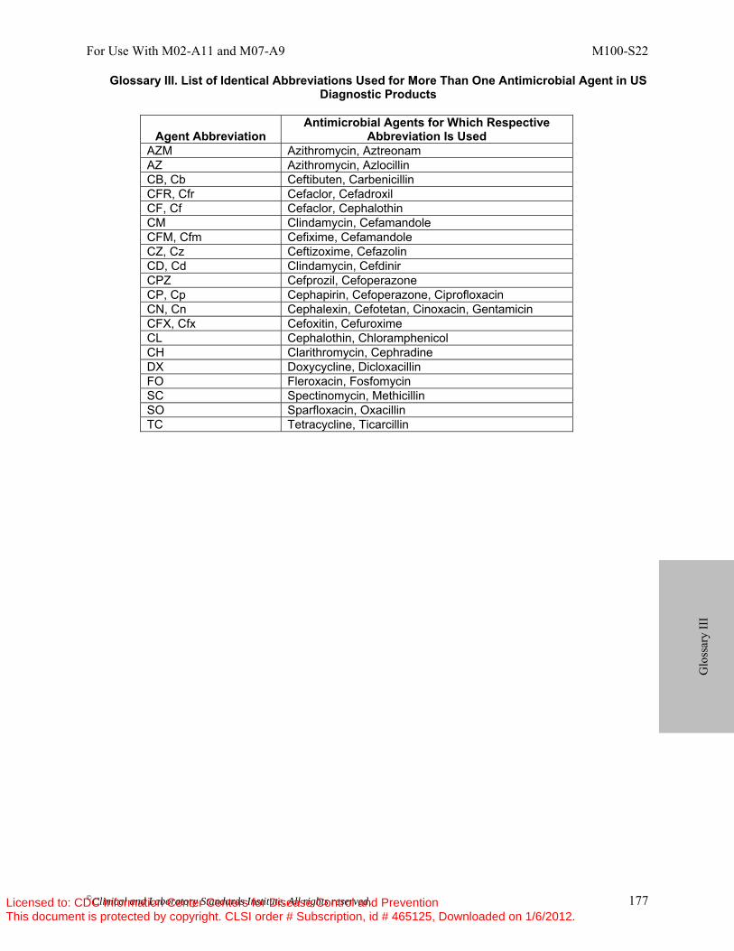
For Use With M02-A11 and M07-A9 M100-S22
©Clinical and Laboratory Standards Institute. All rights reserved. 177
Glossary III. List of Identical Abbreviations Used for More Than One Antimicrobial Agent in US Diagnostic Products
Agent Abbreviation Antimicrobial Agents for Which Respective
Abbreviation Is Used AZM Azithromycin, Aztreonam AZ Azithromycin, Azlocillin CB, Cb Ceftibuten, Carbenicillin CFR, Cfr Cefaclor, Cefadroxil CF, Cf Cefaclor, Cephalothin CM Clindamycin, Cefamandole CFM, Cfm Cefixime, Cefamandole CZ, Cz Ceftizoxime, Cefazolin CD, Cd Clindamycin, Cefdinir CPZ Cefprozil, Cefoperazone CP, Cp Cephapirin, Cefoperazone, Ciprofloxacin CN, Cn Cephalexin, Cefotetan, Cinoxacin, Gentamicin CFX, Cfx Cefoxitin, Cefuroxime CL Cephalothin, Chloramphenicol CH Clarithromycin, Cephradine DX Doxycycline, Dicloxacillin FO Fleroxacin, Fosfomycin SC Spectinomycin, Methicillin SO Sparfloxacin, Oxacillin TC Tetracycline, Ticarcillin
G
loss
ary
III
Licensed to: CDC Information Center Centers for Disease Control and PreventionThis document is protected by copyright. CLSI order # Subscription, id # 465125, Downloaded on 1/6/2012.
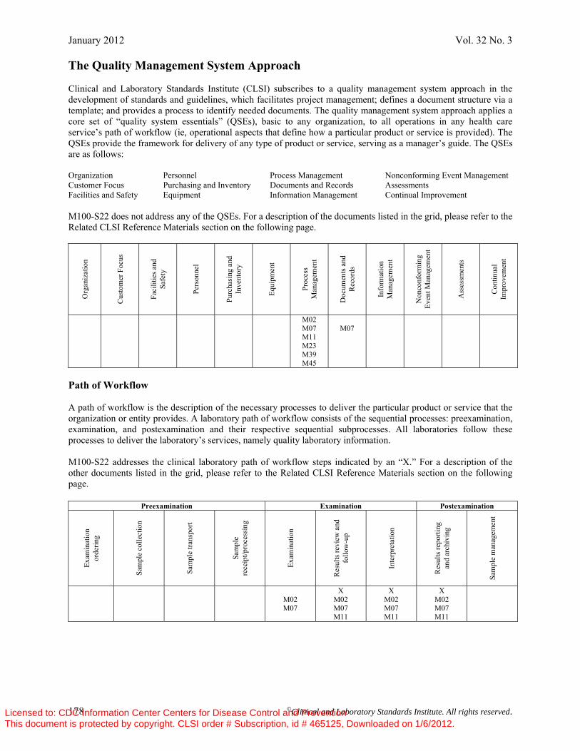
January 2012 Vol. 32 No. 3
178 ©Clinical and Laboratory Standards Institute. All rights reserved.
The Quality Management System Approach Clinical and Laboratory Standards Institute (CLSI) subscribes to a quality management system approach in the development of standards and guidelines, which facilitates project management; defines a document structure via a template; and provides a process to identify needed documents. The quality management system approach applies a core set of “quality system essentials” (QSEs), basic to any organization, to all operations in any health care service’s path of workflow (ie, operational aspects that define how a particular product or service is provided). The QSEs provide the framework for delivery of any type of product or service, serving as a manager’s guide. The QSEs are as follows: Organization Personnel Process Management Nonconforming Event Management Customer Focus Purchasing and Inventory Documents and Records Assessments Facilities and Safety Equipment Information Management Continual Improvement M100-S22 does not address any of the QSEs. For a description of the documents listed in the grid, please refer to the Related CLSI Reference Materials section on the following page.
Org
aniz
atio
n
Cus
tom
er F
ocus
Faci
litie
s and
Sa
fety
Pers
onne
l
Purc
hasi
ng a
nd
Inve
ntor
y
Equi
pmen
t
Proc
ess
Man
agem
ent
Doc
umen
ts a
nd
Rec
ords
Info
rmat
ion
Man
agem
ent
Non
conf
orm
ing
Even
t Man
agem
ent
Ass
essm
ents
Con
tinua
l Im
prov
emen
t
M02 M07 M11 M23 M39 M45
M07
Path of Workflow A path of workflow is the description of the necessary processes to deliver the particular product or service that the organization or entity provides. A laboratory path of workflow consists of the sequential processes: preexamination, examination, and postexamination and their respective sequential subprocesses. All laboratories follow these processes to deliver the laboratory’s services, namely quality laboratory information. M100-S22 addresses the clinical laboratory path of workflow steps indicated by an “X.” For a description of the other documents listed in the grid, please refer to the Related CLSI Reference Materials section on the following page.
Preexamination Examination Postexamination
Exam
inat
ion
orde
ring
Sam
ple
colle
ctio
n
Sam
ple
trans
port
Sam
ple
rece
ipt/p
roce
ssin
g
Exam
inat
ion
Res
ults
revi
ew a
nd
follo
w-u
p
Inte
rpre
tatio
n
Res
ults
repo
rting
an
d ar
chiv
ing
Sam
ple
man
agem
ent
M02 M07
X M02 M07 M11
X M02 M07 M11
X M02 M07 M11
Licensed to: CDC Information Center Centers for Disease Control and PreventionThis document is protected by copyright. CLSI order # Subscription, id # 465125, Downloaded on 1/6/2012.
For Use With M02-A11 and M07-A9 M100-S22
©Clinical and Laboratory Standards Institute. All rights reserved. 179
Related CLSI Reference Materials∗ M02-A11 Performance Standards for Antimicrobial Disk Susceptibility Tests; Approved Standard—Eleventh Edition
(2012). This standard contains the current Clinical and Laboratory Standards Institute-recommended methods for disk susceptibility testing, criteria for quality control testing, and updated tables for interpretive zone diameters.
M07-A9 Methods for Dilution Antimicrobial Susceptibility Tests for Bacteria That Grow Aerobically; Approved
Standard—Ninth Edition (2012). This document addresses reference methods for the determination of minimal inhibitory concentrations (MICs) of aerobic bacteria by broth macrodilution, broth microdilution, and agar dilution.
M11-A7 Methods for Antimicrobial Susceptibility Testing of Anaerobic Bacteria; Approved Standard—Seventh
Edition (2007). This standard provides reference methods for the determination of minimal inhibitory concentrations (MICs) of anaerobic bacteria by broth microdilution and agar dilution.
M23-A3
Development of In Vitro Susceptibility Testing Criteria and Quality Control Parameters; Approved Guideline—Third Edition (2008). This document addresses the required and recommended data needed for the selection of appropriate interpretive criteria and quality control ranges for antimicrobial agents.
M39-A3 Analysis and Presentation of Cumulative Antimicrobial Susceptibility Test Data; Approved Guideline—
Third Edition (2009). This document describes methods for recording and analysis of antimicrobial susceptibility test data, consisting of cumulative and ongoing summaries of susceptibility patterns of clinically significant microorganisms.
M45-A2 Methods for Antimicrobial Dilution and Disk Susceptibility Testing of Infrequently Isolated or Fastidious
Bacteria; Approved Guideline—Second Edition (2010). This document provides guidance to clinical microbiology laboratories for standardized susceptibility testing of infrequently isolated or fastidious bacteria that are not presently included in CLSI documents M02 or M07. The tabular information in this document presents the most current information for drug selection, interpretation, and quality control for the infrequently isolated or fastidious bacterial pathogens included in this guideline.
∗ CLSI documents are continually reviewed and revised through the CLSI consensus process; therefore, readers should refer to the most current editions.
Licensed to: CDC Information Center Centers for Disease Control and PreventionThis document is protected by copyright. CLSI order # Subscription, id # 465125, Downloaded on 1/6/2012.
Active Membership (As of 1 December 2011)
Sustaining Members Abbott American Association for Clinical
Chemistry AstraZeneca Pharmaceuticals Bayer Healthcare, LLC Diagnostic
Division BD Beckman Coulter, Inc. bioMérieux, Inc. College of American Pathologists Diagnostica Stago GlaxoSmithKline National Institute of Standards and
Technology Ortho-Clinical Diagnostics, Inc. Pfizer Inc Roche Diagnostics, Inc. Professional Members AAMI American Association for Clinical
Chemistry American Association for Laboratory
Accreditation American Medical Technologists American Society for Clinical Laboratory
Science American Society for Clinical Pathology American Society for Microbiology American Type Culture Collection Association of Public Health
Laboratories Associazione Microbiologi Clinici
Italiani (AMCLI) British Society for Antimicrobial
Chemotherapy Canadian Society for Medical Laboratory
Science COLA College of American Pathologists College of Medical Laboratory
Technologists of Ontario College of Physicians and Surgeons of
Saskatchewan Critical Path Institute ESCMID Family Health International Hong Kong Accreditation Service
Innovation and Technology Commission
International Federation of Biomedical Laboratory Science
International Federation of Clinical Chemistry
Italian Society of Clin. Biochem. and Clin. Molec. Biology
JCCLS National Society for Histotechnology,
Inc. Nova Scotia Association of Clinical
Laboratory Managers Ontario Medical Association Quality
Management Program-Laboratory Service
RCPA Quality Assurance Programs Pty Limited
SIMeL Sociedad Española de Bioquímica
Clínica y Patología Molec. Sociedade Brasileira de Análises Clínicas Sociedade Brasileira de Patologia Clínica The Joint Commission The Korean Society for Laboratory
Medicine World Health Organization Government Members Armed Forces Institute of Pathology BC Centre for Disease Control Canadian Science Center for Human and
Animal Health Centers for Disease Control and
Prevention Centers for Disease Control and
Prevention - Ethiopia Centers for Disease Control and
Prevention - Namibia Centers for Disease Control and
Prevention - Nigeria Centers for Disease Control and
Prevention - Tanzania Centers for Disease Control and
Prevention - Zambia Centers for Medicare & Medicaid
Services Centers for Medicare & Medicaid
Services/CLIA Program Chinese Committee for Clinical
Laboratory Standards
Chinese Medical Association (CMA) Clalit Health Services Department of Veterans Affairs DHSR/CLIA Certification Diagnostic Accreditation Program Ethiopian Health and Nutrition Research
Institute FDA Center for Veterinary Medicine FDA Ctr. for Devices/Rad. Health Health Canada Institute of Tropical Medicine Dept. of
Clinical Sciences MA Dept. of Public Health Laboratories Malaria Research Training Center Marion County Public Health
Department Meuhedet Central Lab Ministry of Health and Social Welfare -
Tanzania Mongolian Agency for Standardization
and Metrology Namibia Institute of Pathology National Cancer Institute, OBBR, NIH National Food Institute Technical
University of Denmark National Health Laboratory Service C/O
F&M Import & Export Services National HIV & Retrovirology Lab
Public Health Agency of Canada National Institute of Health-Maputo,
Mozambique National Institute of Standards and
Technology National Pathology Accreditation
Advisory Council New York State Dept. of Health Ontario Agency for Health Protection
and Promotion Pennsylvania Dept. of Health SA Pathology Saskatchewan Health-Provincial
Laboratory Scientific Institute of Public Health State of Alabama State of Wyoming Public Health
Laboratory The Nathan S. Kline Institute University of Iowa, Hygienic Lab US Naval Medical Research Unit #3 USAMC – AFRIMS Industry Members 3M Medical Division AB Diagnostic Systems GmBH Abbott Abbott Diabetes Care Abbott Point of Care Inc. Access Genetics Aderans Research AdvaMed Akonni Biosystems Ammirati Regulatory Consulting Anapharm, Inc. AspenBio Pharma, Inc. Astellas Pharma AstraZeneca Pharmaceuticals Astute Medical, Inc. Ativa Medical Axis-Shield PoC AS Bayer Healthcare, LLC Diagnostic
Division BD BD Biosciences - San Jose, CA BD Diagnostic Systems BD Vacutainer Systems Beaufort Advisors, LLC Beckman Coulter Cellular Analysis
Business Center Beckman Coulter, Inc. Beth Goldstein Consultant Bio-Rad Laboratories, Inc. Bio-Rad Laboratories, Inc. - France Bioanalyse, Ltd. Biocartis BioDevelopment S.r.l. Biohit Oyj. Biomedia Laboratories SDN BHD bioMeríeux, Inc. Blaine Healthcare Associates, Inc. BRI Consultants Limited Calloway Laboratories Canon U.S. Life Sciences, Inc. CBI Inc. Cempra Pharmaceuticals, Inc. Cepheid Cerilliant Corp. Compliance Insight, Inc. Constitution Medical Inc Controllab Copan Diagnostics Inc. Courtagen Life Sciences Inc. Crescendo Bioscience
Cubist Pharmaceuticals, Inc. Dahl-Chase Pathology Associates PA Diagnostica Stago DX Assays Pte Ltd. Eiken Chemical Company, Ltd. Elanco Animal Health Elkin Simson Consulting Services Emika Consulting EndPoint Associates, LLC Enigma Diagnostics Eurofins Medinet Evidia Biosciences Inc. EXACT Sciences Corporation Gen-Probe Genefluidics GlaxoSmithKline Greiner Bio-One Inc. Himedia Labs Ltd HistoGenex N.V. Hospital Sungai Buloh Icon Laboratories, Inc. Innovotech, Inc. Instrumentation Laboratory Integrated BioBank IntelligentMDx, Inc. Intuity Medical ITC Corp Japan Assn. of Clinical Reagents
Industries Johnson & Johnson Pharmaceutical
Research & Develop., L.L.C. Kaiser Permanente KoreaBIO Krouwer Consulting Lab PMM Laboratory Specialists, Inc. LifeLabs LifeScan, Inc. Liofilchem SRL LipoScience, Inc. Maine Standards Company, LLC Marketing MicroScan & Molecular
Korea-Siemens Healthcare Masimo Corp. Masimo Labs Mbio Diagnostics, Inc. MDxHealth SA Medical Device Consultants, Inc. Merck & Company, Inc. Merial Limited Meso Scale Diagnostics, LLC. Micromyx, LLC Molecular Response Moscow Antidoping Agency Nanosphere, Inc. Nihon Kohden Corporation Nissui Pharmaceutical Co., Ltd. NJK & Associates, Inc. NorDx - Scarborough Campus Nova Biomedical Corporation NovaBiotics Novartis Institutes for Biomedical
Research Optimer Pharmaceuticals, Inc. Ortho-Clinical Diagnostics, Inc. Ortho-McNeil, Inc. Oxyrase, Inc. Paratek Pharmaceuticals, Inc. PathCare Pathology Laboratory PerkinElmer Genetics, Inc. Pfizer Animal Health Pfizer Inc Pfizer Italia Srl Phadia AB Philips Healthcare Incubator PPD ProteoGenix, Inc. QML Pathology Quotient Bioresearch Ltd. R-Biopharm AG Radiometer America, Inc. Roche Diagnostics GmbH Roche Diagnostics, Inc. Roche Molecular Systems RPL Laboratory Solutions, Inc. DBA RPL Compliance Solutions Sanofi Pasteur Sarstedt, Inc. Sekisui Diagnostics Seventh Sense Biosystems Siemens Healthcare Diagnostics Inc. Siemens Healthcare Diagnostics Products
GmbH Soloy Laboratory Consulting Services,
Llc SomaLogic Sphere Medical Holding Limited Streck Laboratories, Inc. Super Religare Laboratories Ltd Sysmex America, Inc. Sysmex Corporation - Japan Tandem Diagnostics Tetraphase Pharmaceuticals
The Clinical Microbiology Institute The Medicines Company TheraDoc Theranos Theravance Inc. Thermo Fisher Scientific Thermo Fisher Scientific, Oxoid Products Thermo Fisher Scientific, Remel Transasia Bio-Medicals Limited Tulip Group Ventana Medical Systems Inc. Veracyte, Inc. Vivacta Watson Pharmaceuticals Wellstat Diagnostics, LLC XDx, Inc. Associate Active Members 31st Medical Group SGSL (AE) 3rd Medical Group (AK) 48th Medical Group/MDSS RAF
Lakenheath (AE) 55th Medical Group/SGSAL (NE) 59th MDW/859th MDTS/MTL Wilford
Hall Medical Center (TX) 82 MDG/SGSCL Sheppard AFB (TX) Academisch Ziekenhuis-VUB UZ
Brussel (Belgium) ACL Laboratories (IL) ACL Laboratories (WI) Adams County Hospital (OH) Adena Regional Medical Center Hospital
(OH) Affiliated Laboratory, Inc. (ME) Akron Children’s Hospital (OH) Al Ain Hospital (Abu Dhabi, United
Arab Emirates) Al Hada Armed Forces
Hospital/TAIF/KSA (Saudi Arabia) Al Noor Hospital (United Arab Emirates) Al Rahba Hospital (United Arab
Emirates) Alameda County Medical Center (CA) Albany Medical Center Hospital (NY) Albemarle Hospital (NC) Alberta Health Services (AB, Canada) Alexandra Hospital (Singapore) All Children’s Hospital (FL) Allegiance Health (MI) Alpena Regional Medical Center (MI) Alta Bates Summit Medical Center (CA) Alverno Clinical Laboratories, Inc. (IN) American Esoteric Laboratories (AEL)
(TN) American University of Beirut Medical
Center (NJ) Anand Diagnostic Laboratory (India) Anne Arundel Medical Center (MD) Antech Diagnostics (CA) Antelope Valley Hospital District (CA) Appalachian Regional Healthcare System
(NC) Arkansas Children’s Hospital (AR) Arkansas Dept of Health Public Health
Laboratory (AR) Arkansas Methodist Medical Center (AR) ARUP Laboratories (UT) Asan Medical Center (Korea, Republic
Of) Asante Health System (OR) Ashley County Medical Center (AR) Asiri Group of Hospitals Ltd. (Sri Lanka) Aspen Valley Hospital (CO) ASPETAR (Qatar Orthopedic and Sports
Medicine Hospital) (Qatar) Aspirus Wausau Hospital (WI) Auburn Regional Medical Center (WA) Augusta Health (VA) Aultman Hospital (OH) Avera McKennan Hospital & University
Health Center (SD) AZ Sint-Jan (Belgium) Azienda Ospedale Di Lecco (Italy) Baptist Hospital of Miami (FL) Baptist Memorial Health Care
Corporation - Hospital Laboratories Works (TN)
Barnes-Jewish Hospital (MO) Bassett Healthcare (NY) Baton Rouge General (LA) Baxter Regional Medical Center (AR) BayCare Health System (FL) Baylor Health Care System (TX) Bayou Pathology, APMC (LA) Baystate Medical Center (MA) BC Biomedical Laboratories (BC,
Canada) Beloit Memorial Hospital (WI) Beth Israel Medical Center (NY) Bio-Reference Laboratories (NJ) Blanchard Valley Hospital (OH) Bon Secours Health Partners (VA)
Licensed to: CDC Information Center Centers for Disease Control and PreventionThis document is protected by copyright. CLSI order # Subscription, id # 465125, Downloaded on 1/6/2012.
Bonnyville Health Center (AB, Canada) Boston Medical Center (MA) Boulder Community Hospital (CO) Boyce & Bynum Pathology Labs (MO) Brant Community Healthcare
System/Brant General Hospital (Ontario, Canada)
Bremerton Naval Hospital (WA) Brian All Good Community Hospital/121
Combat (AP) Bridgeport Hospital (CT) Brooke Army Medical Center (TX) Broward General Medical Center (FL) Cadham Provincial Laboratory-MB
Health (MB, Canada) Calgary Laboratory Services (AB,
Canada) California Pacific Medical Center (CA) Cambridge Health Alliance (MA) Cape Fear Valley Medical Center
Laboratory (NC) Capital Coast Health (New Zealand) Capital Health System Mercer Campus
(NJ) Caritas Norwood Hospital (MA) Carl R. Darnall Army Medical Center
Department of Pathology (TX) Carolina Medical Laboratory (NC) Carolinas Healthcare System (NC) Carpermor S.A. de C.V. (D.F., Mexico) Catholic Health Initiatives (KY) Cedars-Sinai Medical Center (CA) Cenetron Diagnostics (TX) Central Baptist Hospital (KY) Central Kansas Medical Center (KS) Centre Hospitalier Anna-Laberge
(Quebec, Canada) Centre Hospitalier Regional De Trois
Riveras (PQ, Canada) Centro Médico Imbanaco (Colombia) Chaleur Regional Hospital (NB, Canada) Chang Gung Memorial Hospital
(Taiwan) Changhua Christian Hospital (Taiwan) Changi General Hospital (Singapore) Chatham - Kent Health Alliance (ON,
Canada) Chesapeake General Hospital (VA) Chester County Hospital (PA) Children’s Healthcare of Atlanta (GA) Childrens Hosp.- Kings Daughters (VA) Children’s Hospital & Research Center
At Oakland (CA) Childrens Hospital Los Angeles (CA) Children’s Hospital Medical Center (OH) Children’s Hospital of Central California
(CA) Children’s Hospital of Orange County
(CA) Children’s Hospital of Philadelphia (PA) Childrens Hospital of Wisconsin (WI) Children’s Hospitals and Clinics (MN) Children’s Medical Center (OH) Children’s Medical Center (TX) Christiana Care Health Services (DE) CHU - Saint Pierre (Belgium) CHU Sainte-Justine (Quebec, Canada) CHUM Hospital Saint-Luc (Quebec,
Canada) CHW-St. Mary’s Medical Center (CA) City of Hope National Medical Center
(CA) Clearstone Central Laboratories (ON,
Canada) Cleveland Clinic (OH) Cleveland Heartlab, LLC (OH) Clinica Alemana De Santiago (Chile) Clinical and Laboratory Standards
Institute (PA) Clinical Labs of Hawaii (HI) Clinical Trials Service (Belgium) College of Physicians and Surgeons of
Alberta (AB, Canada) Collingwood General & Marine Hospital
(ON, Canada) Commonwealth of Kentucky (KY) Commonwealth of Virginia (DCLS)
(VA) Community Hospital (IN) Community Hospital of the Monterey
Peninsula (CA) Community Medical Center (NJ) Community Memorial Hospital (WI) Complexe Hospitalier de la Sagamie
(Quebec, Canada) CompuNet Clinical Laboratories Quest
Diagnostics JV (OH) Concord Hospital (NH) Consultants Laboratory of WI LLC (WI) Contra Costa Regional Medical Center
(CA) Cook Children’s Medical Center (TX) Cookeville Regional Medical Center
(TN) Cornwall Community Hospital (ON,
Canada) Covance CLS (IN) Covenant Medical Center (TX)
Creighton Medical Lab (NE) Crozer-Chester Medical Center (PA) Cumberland Medical Center (TN) Darwin Library NT Territory Health
Services (NT, Australia) David Grant Medical Center (CA) Daviess Community Hospital (IN) Deaconess Hospital Laboratory (IN) Dean Medical Center (WI) DHHS NC State Lab of Public Health
(NC) DiagnoSearch Life Sciences Inc.
(Maharashtra, India) Diagnostic Laboratory Services, Inc. (HI) Diagnostic Services of Manitoba (MB,
Canada) DMC University Laboratories (MI) Drake Center (OH) Driscoll Children’s Hospital (TX) DUHS Clinical Laboratories Franklin
Site (NC) Dynacare Laboratory (WI) Dynacare NW, Inc - Seattle (WA) DynaLIFE (AB, Canada) E. A. Conway Medical Center (LA) East Georgia Regional Medical Center
(GA) East Kootenay Regional Hospital
Laboratory-Interior Health (BC, Canada)
East Texas Medical Center-Pittsburg (TX)
Eastern Health - Health Sciences Centre (NL, Canada)
Eastern Health Pathology (Victoria, Australia)
Easton Hospital (PA) Edward Hospital (IL) Effingham Hospital (GA) Elmhurst Hospital Center (NY) Emory University Hospital (GA) Evangelical Community Hospital (PA) Evans Army Community Hospital (CO) Exeter Hospital (NH) Exosome Diagnostics, Inc. (MN) Federal Medical Center (MN) First Health of the Carolinas Moore
Regional Hospital (NC) Fletcher Allen Health Care (VT) Fleury S.A. (Brazil) Florida Hospital (FL) Fox Chase Cancer Center (PA) Fraser Health Authority Royal
Columbian Hospital Site (BC, Canada) Fundação Faculdade de Medicina (SP,
Brazil) Gamma-Dynacare Laboratories (ON,
Canada) Garden City Hospital (MI) Garfield Medical Center (CA) Gaston Memorial Hospital (NC) Geisinger Medical Center (PA) Genesis Healthcare System (OH) George Washington University Hospital
(DC) Gestión de Calidad (Argentina) Gettysburg Hospital (PA) Ghent University Hospital (Belgium) Good Shepherd Medical Center (TX) Grana S.A. (TX) Grand Marquis Co., LTD (Taiwan) Grand River Hospital (ON, Canada) Grand Strand Regional Medical Center
(SC) Grey Bruce Regional Health Center (ON,
Canada) Group Health Cooperative (WA) Gundersen Lutheran Medical Center
(WI) Guthrie Clinic Laboratories (PA) Hôtel-Dieu de Lévis (PQ, Canada) Halton Healthcare Services (ON, Canada) Hamad Medical Corporation (Qatar) Hamilton Regional Laboratory Medicine
Program - St. Joseph’s (ON, Canada) Hanover General Hospital (PA) Harford Memorial Hospital (MD) Harris Methodist Fort Worth (TX) Harris Methodist Hospital Southwest
(TX) Hartford Hospital (CT) Health Network Lab (PA) Health Sciences Research Institute
(Japan) Health Waikato (New Zealand) Heartland Health (MO) Heidelberg Army Hospital (AE) Helen Hayes Hospital (NY) Helix (Russian Federation) Henry Ford Hospital (MI) Henry M. Jackson Foundation for the
Advancement of Military Medicine-MD (MD)
Hi-Desert Medical Center (CA) Highlands Medical Center (AL) HJF Naval Infectious Diseases
Diagnostic Laboratory (MD)
Hoag Memorial Hospital Presbyterian (CA)
Hoboken University Medical Center (NJ) Holy Cross Hospital (MD) Holy Name Hospital (NJ) Holy Spirit Hospital (PA) Hôpital de la Cité-de-La-Santé De Laval
(Quebec, Canada) Hôpital du Haut-Richelieu (PQ, Canada) Hôpital Maisonneuve-Rosemont (PQ,
Canada) Hôpital Santa Cabrini Ospedale (PQ,
Canada) Horizon Health Network (N.B., Canada) Hospital Albert Einstein (SP, Brazil) Hospital Sacre-Coeur de Montreal
(Quebec, Canada) Hôtel-Dieu Grace Hospital Library (ON,
Canada) Hunter Area Pathology Service
(Australia) Hunter Labs (CA) Huntington Memorial Hospital (CA) Imelda Hospital (Belgium) Indian River Memorial Hospital (FL) Indiana University Health Bloomington
Hospital (IN) Indiana University Health Care-
Pathology Laboratory (IN) Inova Central Laboratory (VA) Institut fur Stand. und Dok. im Med. Lab.
(Germany) Institut National de Santé Publique Du
Quebec Centre de Doc. - INSPQ (PQ, Canada)
Institute Health Laboratories (PR) Institute of Clinical Pathology and
Medical Research (Australia) Institute of Laboratory Medicine
Landspitali Univ. Hospital (Iceland) Institute of Medical & Veterinary
Science (SA, Australia) Intermountain Health Care Lab Services
(UT) International Health Management
Associates, Inc. (IL) Irwin Army Community Hospital (KS) Jackson County Memorial Hospital (OK) Jackson Memorial Hospital (FL) Jackson Purchase Medical Center (KY) Jessa Ziekenhuis VZW (Belgium) John C. Lincoln Hospital - N.MT. (AZ) John F. Kennedy Medical Center (NJ) John H. Stroger, Jr. Hospital of Cook
County (IL) John Muir Health (CA) John T. Mather Memorial Hospital (NY) Johns Hopkins Medical Institutions (MD) Johns Hopkins University (MD) Johnson City Medical Center Hospital
(TN) JPS Health Network (TX) Kailos Genetics (AL) Kaiser Permanente (OH) Kaiser Permanente (MD) Kaiser Permanente Medical Care (CA) Kaohsiun Chang Gung Memorial
Hospital (Taiwan) Kenora-Rainy River Reg. Lab. Program
(ON, Canada) King Abdulaziz Hospital, Al Ahsa Dept.
of Pathology & Laboratory Medicine (Al-hasa, Saudi Arabia)
King Fahad National Guard Hospital KAMC - NGHA (Saudi Arabia)
King Fahad Specialist Hospital-Dammam, K.S.A. (Eastern Region, Saudi Arabia)
King Faisal Specialist Hospital & Research Center (Saudi Arabia)
King Hussein Cancer Center (Jordan) Kingston General Hospital (ON, Canada) Laboratória Médico Santa Luzia LTDA
(Brazil) Laboratory Alliance of Central New
York (NY) Laboratory Corporation of America (NJ) Laboratory Medicin Dalarna (Dalarna,
Sweden) LabPlus Auckland District Health Board
(New Zealand) LAC/USC Medical Center (CA) Lafayette General Medical Center (LA) Lakeland Regional Medical Center (FL) Lancaster General Hospital (PA) Landstuhl Regional Medical Center
(Germany) Langley Air Force Base (VA) LeBonheur Children’s Hospital (TN) Legacy Laboratory Services (OR) Letherbridge Regional Hospital (AB,
Canada) Lewis-Gale Medical Center (VA) Lexington Medical Center (SC) L’Hotel-Dieu de Québec (PQ, Canada) Licking Memorial Hospital (OH) LifeBridge Health Sinai Hospital (MD)
LifeLabs Medical Laboratory Services (BC, Canada)
Lifeline Hospital (United Arab Emirates) Loma Linda University Medical Center
(LLUMC) (CA) Long Beach Memorial Medical Center-
LBMMC (CA) Long Island Jewish Medical Center (NY) Louisiana Office of Public Health
Laboratory (LA) Louisiana State University Medical Ctr.
(LA) Lower Columbia Pathologists, P.S. (WA) Lower Mainland Laboratories (BC,
Canada) Lyndon B. Johnson General Hospital
(TX) Maccabi Medical Care and Health Fund
(Israel) Madigan Army Medical Center (WA) Mafraq Hospital (United Arab Emirates) Magnolia Regional Health Center (MS) Main Line Clinical Laboratories, Inc.
Lankenau Hospital (PA) Makerere University Walter Reed Project
Makerere University Medical School (Uganda)
Marquette General Hospital (MI) Marshfield Clinic (WI) Martha Jefferson Hospital (VA) Martin Luther King, Jr./Drew Medical
Center (CA) Martin Memorial Health Systems (FL) Mary Hitchcock Memorial Hospital (NH) Mary Washington Hospital (VA) Mater Health Services - Pathology
(Australia) Maxwell Air Force Base (AL) Mayo Clinic (MN) MCG Health (GA) Meadows Regional Medical Center (GA) Medical Center Hospital (TX) Medical Center of Louisiana At NO-
Charity (LA) Medical Centre Ljubljana (Slovenia) Medical College of Virginia Hospital
(VA) Medical University Hospital Authority
(SC) Memorial Hermann Healthcare System
(TX) Memorial Medical Center (PA) Memorial Medical Center (IL) Memorial Regional Hospital (FL) Mercy Franciscan Mt. Airy (OH) Mercy Hospital & Medical Center (IL) Methodist Dallas Medical Center (TX) Methodist Hospital (TX) Methodist Hospital (TX) Methodist Hospital Park Nicollet Health
Services (MN) Methodist Hospital Pathology (NE) MetroHealth Medical Center (OH) Metropolitan Hospital Center (NY) Metropolitan Medical Laboratory, PLC
(IA) Miami Children’s Hospital (FL) Mid Michigan Medical Center - Midland
(MI) Middelheim General Hospital (Belgium) Middlesex Hospital (CT) Mike O’Callaghan Federal Hospital (NV) Minneapolis Medical Research
Foundation (MN) Mississippi Baptist Medical Center (MS) Mississippi Public Health Lab (MS) Monongalia General Hospital (WV) Montreal General Hospital (Quebec,
Canada) Morehead Memorial Hospital (NC) Mouwasat Hospital (GA, Saudi Arabia) Mt. Carmel Health System (OH) Mt. Sinai Hospital (ON, Canada) Mt. Sinai Hospital - New York (NY) Naples Community Hospital (FL) Nassau County Medical Center (NY) National B Virus Resource Laboratory
(GA) National Cancer Center (Korea, Republic
Of) National Institutes of Health, Clinical
Center (MD) National Naval Medical Center (MD) National University Hospital Department
of Laboratory Medicine (Singapore) National University of Ireland, Galway
(NUIG) (Ireland) Nationwide Children’s Hospital (OH) Naval Hospital Oak Harbor (WA) Naval Medical Center Portsmouth (VA) Naval Medical Clinic Hawaii (HI) NB Department of Health (NB, Canada) New England Baptist Hospital (MA) New England Fertility Institute (CT) New England Sinai Hospital (MA) New Lexington Clinic (KY) New York City Department of Health
and Mental Hygiene (NY)
Licensed to: CDC Information Center Centers for Disease Control and PreventionThis document is protected by copyright. CLSI order # Subscription, id # 465125, Downloaded on 1/6/2012.
New York Presbyterian Hospital (NY) New York University Medical Center
(NY) Newark Beth Israel Medical Center (NJ) Newfoundland Public Health Laboratory
(NL, Canada) North Carolina Baptist Hospital (NC) North District Hospital (China) North Mississippi Medical Center (MS) North Shore Hospital Laboratory (New
Zealand) North Shore-Long Island Jewish Health
System Laboratories (NY) Northridge Hospital Medical Center (CA) Northside Hospital (GA) Northside Medical Center (OH) Northwest Texas Hospital (TX) Northwestern Memorial Hospital (IL) Norton Healthcare (KY) Ochsner Clinic Foundation (LA) Ohio State University Hospitals (OH) Ohio Valley Medical Center (WV) Onze Lieve Vrouwziekenhuis (Belgium) Ordre Professionnel Des Technologistes
Médicaux Du Quebec (Quebec, Canada)
Orebro University Hospital (Sweden) Orlando Health (FL) Ospedale Casa Sollievo Della Sofferenza
- IRCCS (Italy) Our Lady’s Hospital For Sick Children
(Ireland) Palmetto Baptist Medical Center (SC) Pamela Youde Nethersole Eastern
Hospital (Hong Kong East Cluster) (Hong Kong)
Pathgroup (TN) Pathlab (IA) Pathology and Cytology Laboratories,
Inc. (KY) Pathology Associates Medical Lab. (WA) Pathology Inc. (CA) Penn State Hershey Medical Center (PA) Pennsylvania Hospital (PA) Peterborough Regional Health Centre
(ON, Canada) PHS Indian Hospital - Pine Ridge (SD) Piedmont Hospital (GA) Pitt County Memorial Hospital (NC) Potomac Hospital (VA) Prairie Lakes Hospital (SD) Presbyterian Hospital - Laboratory (NC) Presbyterian/St. Luke’s Medical Center
(CO) Prince of Wales Hospital (Hong Kong) Princess Margaret Hospital (Hong Kong,
China) Providence Alaska Medical Center (AK) Providence Health Services, Regional
Laboratory (OR) Provincial Laboratory for Public Health
(AB, Canada) Queen Elizabeth Hospital (P.E.I, Canada) Queen Elizabeth Hospital (China) Queensland Health Pathology Services
(Australia) Queensway Carleton Hospital (ON,
Canada) Quest Diagnostics, Incorporated (CA) Quintiles Laboratories, Ltd. (GA) Rady Children’s Hospital San Diego
(CA) Ramathibodi Hospital (Thailand) Redington-Fairview General Hospital
(ME) Regions Hospital (MN) Reid Hospital & Health Care Services
(IN) Reinier De Graaf Groep (Netherlands) Renown Regional Medical Center (NV) Research Medical Center (MO) Response Genetics, Inc. (CA) RIPAS Hospital (Brunei-Maura, Brunei
Darussalam) Riverside County Regional Medical
Center (CA) Riverside Health System (VA) Riverside Methodist Hospital (OH)
Riyadh Armed Forces Hospital, Sulaymainia (Saudi Arabia)
Rockford Memorial Hospital (IL) Royal Victoria Hospital (ON, Canada) SAAD Specialist Hospital (Saudi Arabia) Sacred Heart Hospital (WI) Sacred Heart Hospital (FL) Sahlgrenska Universitetssjukhuset
(Sweden) Saint Francis Hospital & Medical Center
(CT) Saint Mary’s Regional Medical Center
(NV) Saints Memorial Medical Center (MA) Salem Memorial District Hospital (MO) Sampson Regional Medical Center (NC) Samsung Medical Center (Korea,
Republic Of) San Francisco General Hospital-
University of California San Francisco (CA)
Sanford USD Medical Center (SD) Santa Clara Valley Medical Center (CA) SARL Laboratoire Caron (France) Scott & White Memorial Hospital (TX) Seattle Children’s Hospital/Children’s
Hospital and Regional Medical Center (WA)
Seoul National University Hospital (Korea, Republic Of)
Seoul St. Mary’s Hospital (Korea, Republic Of)
Seton Healthcare Network (TX) Seton Medical Center (CA) Sharp Health Care Laboratory Services
(CA) Sheik Kalifa Medical City (United Arab
Emirates) Shore Memorial Hospital (NJ) Singapore General Hospital (Singapore) Slotervaart Ziekenhuis (Netherlands) South Bend Medical Foundation (IN) South Eastern Area Laboratory Services
(NSW, Australia) South Miami Hospital (FL) Southern Community Laboratories
(Canterbury, New Zealand) Southern Health Care Network
(Australia) Southwest Healthcare System (CA) Southwestern Medical Center (OK) Spectra East (NJ) Spectra Laboratories (CA) St. Agnes Healthcare (MD) St. Anthony Hospital (OK) St. Barnabas Medical Center (NJ) St. Elizabeth Community Hospital (CA) St. Eustache Hospital (Quebec, Canada) St. Francis Hospital (SC) St. Francis Memorial Hospital (CA) St. John Hospital and Medical Center
(MI) St. John’s Episcopal Hospital (NY) St. John’s Hospital & Health Center (CA) St. John’s Mercy Medical Center (MO) St. John’s Regional Health Center (MO) St. Jude Children’s Research Hospital
(TN) St. Luke’s Hospital (IA) St. Luke’s Hospital (PA) St. Mary Medical Center (CA) St. Mary’s Hospital (WI) St. Michael’s Medical Center, Inc. (NJ) St. Tammany Parish Hospital (LA) Stanford Hospital and Clinics (CA) Stanton Territorial Health Authority (NT,
Australia) State of Connecticut Department of
Public Health (CT) State of Ohio/Corrections Medical Center
Laboratory (OH) State of Washington Public Health Labs
(WA) Stillwater Medical Center (OK) Stony Brook University Hospital (NY) Stormont-Vail Regional Medical Ctr.
(KS) Strong Memorial Hospital (NY) Sudbury Regional Hospital (ON, Canada)
Sunnybrook Health Sciences Centre (ON, Canada)
Sunrise Hospital and Medical Center (NV)
Swedish Edmonds Hospital (WA) Swedish Medical Center (CO) Sydney South West Pathology Service
Liverpool Hospital (NSW, Australia) T.J. Samson Community Hospital (KY) Taichung Veterans General Hospital
(Taiwan) Taiwan Society of Laboratory Medicine
(Taiwan) Tallaght Hospital (Ireland) Tartu University Clinics (Estonia) Temple Univ. Hospital - Parkinson Pav.
(PA) Tenet Healthcare (PA) Texas Children’s Hospital (TX) Texas Department of State Health
Services (TX) Texas Health Presbyterian Hospital
Dallas (TX) The Brooklyn Hospital Center (NY) The Charlotte Hungerford Hospital (CT) The Children’s Mercy Hospital (MO) The Cooley Dickinson Hospital, Inc.
(MA) The Credit Valley Hospital (ON, Canada) The Hospital for Sick Children (ON,
Canada) The Medical Center of Aurora (CO) The Michener Inst. for Applied Health
Sciences (ON, Canada) The Naval Hospital of Jacksonville (FL) The Nebraska Medical Center (NE) The Ottawa Hospital (ON, Canada) The Permanente Medical Group (CA) The Toledo Hospital (OH) The University of Texas Medical Branch
(TX) Thomas Jefferson University Hospital,
Inc. (PA) Timmins and District Hospital (ON,
Canada) Tokyo Metro. Res. Lab of Public Health
(Japan) Touro Infirmary (LA) TriCore Reference Laboratories (NM) Trident Medical Center (SC) Trinity Medical Center (AL) Tripler Army Medical Center (HI) Tuen Mun Hospital, Hospital Authority
(China) Tufts Medical Center Hospital (MA) Tulane Medical Center Hospital & Clinic
(LA) Turku University Central Hospital
(Finland) Twin Lakes Regional Medical Center
(KY) UCI Medical Center (CA) UCLA Medical Center Clinical
Laboratories (CA) UCSD Medical Center (CA) UCSF Medical Center China Basin (CA) UMC of El Paso- Laboratory (TX) UMC of Southern Nevada (NV) UNC Hospitals (NC) Unidad De Patología Clínica (Mexico) Union Clinical Laboratory (Taiwan) United Christian Hospital (Kowloon,
Hong Kong) United States Air Force School of
Aerospace Medicine / PHE (TX) Unity HealthCare (IA) Universitair Ziekenhuis Antwerpen
(Belgium) University College Hospital (Ireland) University Hospital (GA) University Hospital Center Sherbrooke
(CHUS) (Quebec, Canada) University Medical Center at Princeton
(NJ) University of Alabama Hospital Lab
(AL) University of Chicago Hospitals
Laboratories (IL)
University of Colorado Health Sciences Center (CO)
University of Colorado Hospital (CO) University of Illinois Medical Center (IL) University of Iowa Hospitals and Clinics
(IA) University of Kentucky Medical Center
(KY) University of Maryland Medical System
(MD) University of Minnesota Medical Center-
Fairview (MN) University of Missouri Hospital (MO) University of Pennsylvania Health
System (PA) University of Pittsburgh Medical Center
(PA) University of Texas Health Center (TX) University of the Ryukyus (Japan) University of Virginia Medical Center
(VA) UPMC Bedford Memorial (PA) US Naval Hospital Naples UZ-KUL Medical Center (Belgium) VA (Asheville) Medical Center (NC) VA (Bay Pines) Medical Center (FL) VA (Central Texas) Veterans Health Care
System (TX) VA (Chillicothe) Medical Center (OH) VA (Cincinnati) Medical Center (OH) VA (Dallas) Medical Center (TX) VA (Dayton) Medical Center (OH) VA (Hines) Medical Center (IL) VA (Indianapolis) Medical Center (IN) VA (Iowa City) Medical Center (IA) VA (Miami) Medical Center (FL) VA (San Diego) Medical Center (CA) VA (Tampa) Hospital (FL) VA (Wilmington) Medical Center (DE) Vancouver Island Health Authority (SI)
(BC, Canada) Vanderbilt University Medical Center
(TN) Verinata Health, Inc. (CA) Via Christi Regional Medical Center
(KS) Viracor-IBT Reference Laboratory (MO) Virginia Regional Medical Center (MN) Virtua - West Jersey Hospital (NJ) WakeMed (NC) Walter Reed Army Medical Center (DC) Warren Hospital (NJ) Washington Hospital Center (DC) Washington Hospital Healthcare System
(CA) Waterbury Hospital (CT) Waterford Regional Hospital (Ireland) Wayne Memorial Hospital (NC) Weirton Medical Center (WV) West Jefferson Medical Center (LA) West Penn Allegheny Health System-
Allegheny General Hospital (PA) West Shore Medical Center (MI) West Valley Medical Center Laboratory
(ID) Westchester Medical Center (NY) Western Baptist Hospital (KY) Western Healthcare Corporation (NL,
Canada) Wheaton Franciscan Laboratories (WI) Wheeling Hospital (WV) White Memorial Medical Center (CA) Whitehorse General Hospital (YT,
Canada) William Beaumont Army Medical Center
(TX) William Beaumont Hospital (MI) William Osler Health Centre (ON,
Canada) Winchester Hospital (MA) Winn Army Community Hospital (GA) Wishard Health Sciences (IN) Womack Army Medical Center
Department of Pathology (NC) York Hospital (PA) Yukon-Kuskokwim Delta Regional
Hospital (AK)
Licensed to: CDC Information Center Centers for Disease Control and PreventionThis document is protected by copyright. CLSI order # Subscription, id # 465125, Downloaded on 1/6/2012.

Vol. 32 No. 3 M100-S22
©Clinical and Laboratory Standards Institute. All rights reserved. 183
NOTES
Licensed to: CDC Information Center Centers for Disease Control and PreventionThis document is protected by copyright. CLSI order # Subscription, id # 465125, Downloaded on 1/6/2012.

January 2012 M100-S22
184 ©Clinical and Laboratory Standards Institute. All rights reserved.
NOTES
Licensed to: CDC Information Center Centers for Disease Control and PreventionThis document is protected by copyright. CLSI order # Subscription, id # 465125, Downloaded on 1/6/2012.

Licensed to: CDC Information Center Centers for Disease Control and PreventionThis document is protected by copyright. CLSI order # Subscription, id # 465125, Downloaded on 1/6/2012.

950 West Valley Road Suite 2500 Wayne, PA 19087 USA PHONE 610.688.0100 FAX [email protected] www.clsi.org ISBN 1-56238-785-5 (Print) ISBN 1-56238-786-3 (Electronic)
Licensed to: CDC Information Center Centers for Disease Control and PreventionThis document is protected by copyright. CLSI order # Subscription, id # 465125, Downloaded on 1/6/2012.
Related Documents