15 Active Clinical Treatment • Case 48 Initial Presentation: January 2004 Age at Initial Presentation: 59 Active Treatment Completed: October 2006 Review of Treatment Goals Treatment goals of this patient were: (1) Provide healthy and adequate periodontal foundation for restorative treat- ment; (2) Improve oral hygiene; (3) Replace missing teeth with implant-supported restorations; (4) Restore mutually protective articulation; (5) Improve esthetics; (6) Provide canine Angle class I relationship on the right side; (7) Pro- vide proper vertical and horizontal relationships between the maxillary and mandibular anterior teeth. Phase I: Initial Therapy Comprehensive oral and facial examinations were com- pleted prior to formulation of the treatment plan. Car- ies control was performed with amalgam restorations for teeth #’s 2 and 15 on the buccal sides. Periodontal prophylaxis, localized scaling and root planing and oral hygiene instructions were provided during initial therapy. Phase II: Diagnostic Therapy The treatment planning was preceded with diagnostic data collection including, dento-facial analysis, diagnostic casts, centric relation records, facebow transfer, diagnos- tic wax-ups, and photographic documentation. The es- thetic evaluation was performed utilizing acrylic mock-ups Final facial view Treating Clinicians: Drs. Jung Nam, Scott G. Cohen and Soojin Kim Final smile Initial smile

Welcome message from author
This document is posted to help you gain knowledge. Please leave a comment to let me know what you think about it! Share it to your friends and learn new things together.
Transcript

15
Active Clinical Treatment • Case 48
Initial Presentation: January 2004Age at Initial Presentation: 59Active Treatment Completed: October 2006
Review of Treatment Goals
Treatment goals of this patient were: (1) Provide healthy and adequate periodontal foundation for restorative treat-ment; (2) Improve oral hygiene; (3) Replace missing teeth with implant-supported restorations; (4) Restore mutually protective articulation; (5) Improve esthetics; (6) Provide canine Angle class I relationship on the right side; (7) Pro-vide proper vertical and horizontal relationships between the maxillary and mandibular anterior teeth.
Phase I: Initial Therapy
Comprehensive oral and facial examinations were com-pleted prior to formulation of the treatment plan. Car-ies control was performed with amalgam restorations for teeth #’s 2 and 15 on the buccal sides. Periodontal prophylaxis, localized scaling and root planing and oral hygiene instructions were provided during initial therapy.
Phase II: Diagnostic Therapy
The treatment planning was preceded with diagnostic data collection including, dento-facial analysis, diagnostic casts, centric relation records, facebow transfer, diagnos-tic wax-ups, and photographic documentation. The es-thetic evaluation was performed utilizing acrylic mock-ups Final facial view
Treating Clinicians: Drs. Jung Nam, Scott G. Cohen and Soojin Kim
Final smileInitial smile
16
and determined the position of maxillary incisors. Also, consultations with the orthodontist and the periodontist were completed prior to the definitive treatment planning. Since the patient presented with inadequate horizontal and vertical anterior relationships, interocclusal spaces between the maxillary and the mandibular incisors had to be created to restore the incisors with a proper anterior guidance and satisfying the patient’s desire for longer maxillary anterior teeth. Treatment plan included a combination of surgical crown lengthening procedures for the maxillary anteriors, increasing vertical dimension of occlusion, and orthodon-tically intruding and retruding the mandibular incisors. Surgical aesthetic crown lengthening was planned to expose 1.5 mm of the maxillary anterior teeth structure. The goal was to provide ideal gingival levels and maxillary
anterior teeth with an aesthetically pleasing appearance. The vertical dimension of occlusion was planned to be increased by 1.5 mm at central incisor area to gain more space for restorative materials and to provide a proper anterior relationship with longer teeth. The patient had a desire to display more teeth, thus, the maxillary incisors were planned to be lengthened about 4 mm incisally. The original length of the central incisors was 6.5 mm, thus the maxillary central incisors were planned to be 12 mm in length by adding 1.5 mm apically and 4 mm incisally.
Phase III: Implant Placements and Extraction
Endosseous dental implants (ITI, Straumann) were placed to replace missing teeth #’s 19 (Regular Neck x 10 mm) and 20 (Wide Neck x 8 mm), and the hopeless tooth # 22 was extracted. Placement of endosseous implant for
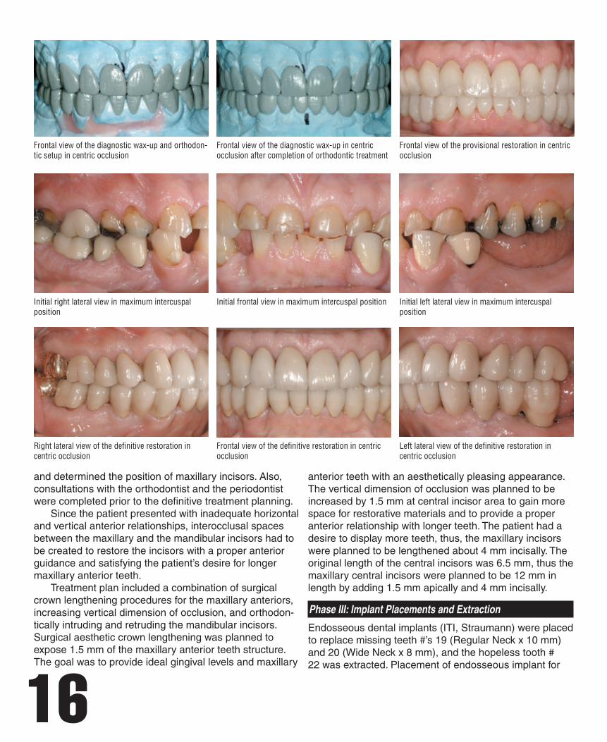
Frontal view of the diagnostic wax-up and orthodon-tic setup in centric occlusion
Frontal view of the diagnostic wax-up in centric occlusion after completion of orthodontic treatment
Frontal view of the provisional restoration in centric occlusion
Initial right lateral view in maximum intercuspal position
Initial frontal view in maximum intercuspal position Initial left lateral view in maximum intercuspal position
Right lateral view of the definitive restoration in centric occlusion
Frontal view of the definitive restoration in centric occlusion
Left lateral view of the definitive restoration in centric occlusion
17
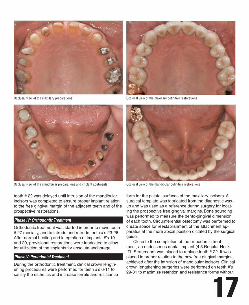
Occlusal view of the maxillary preparations Occlusal view of the maxillary definitive restorations
Occlusal view of the mandibular preparations and implant abutments Occlusal view of the mandibular definitive restorations
tooth # 22 was delayed until intrusion of the mandibular incisors was completed to ensure proper implant relation to the free gingival margin of the adjacent teeth and of the prospective restorations.
Phase IV: Orthodontic Treatment
Orthodontic treatment was started in order to move tooth # 27 mesially, and to intrude and retrude teeth #’s 23-26. After normal healing and integration of implants #’s 19 and 20, provisional restorations were fabricated to allow for utilization of the implants for absolute anchorage.
Phase V: Periodontal Treatment
During the orthodontic treatment, clinical crown length-ening procedures were performed for teeth #’s 6-11 to satisfy the esthetics and increase ferrule and resistance
form for the palatal surfaces of the maxillary incisors. A surgical template was fabricated from the diagnostic wax-up and was used as a reference during surgery for locat-ing the prospective free gingival margins. Bone sounding was performed to measure the dento-gingival dimension of each tooth. Circumferential ostectomy was performed to create space for reestablishment of the attachment ap-paratus at the more apical position dictated by the surgical guide. Close to the completion of the orthodontic treat-ment, an endosseous dental implant (4.3 Regular Neck ITI, Straumann) was placed to replace tooth # 22. It was placed in proper relation to the new free gingival margins achieved after the intrusion of mandibular incisors. Clinical crown lengthening surgeries were performed on teeth #’s 29-31 to maximize retention and resistance forms without
18
exposing the furcations. The rational behind the clinical crown lengthening surgeries on teeth #’s 29-31 and an im-plant placement on tooth # 22 prior to placing a provision-al restoration was to minimize the duration of provisional stage. Such an approach would reduce the probability of interim cementation wash out and recurrent caries.
Phase VI: Provisional Restorative Treatment
The teeth were prepared for metal-ceramic crowns and FPDPs, and for full gold cast crowns. Self-cured acrylic-resin provisional shells were fabricated utilizing a duplicate cast of the diagnostic wax-up, and shells were relined in the patient’s mouth and cemented with a non-eugenol in-terim cement. The provisional restorations were placed on the prepared teeth and the patient functioned on the full-mouth provisional restorations for 4 months to assess his adaptation to the proposed vertical dimension of occlusion and the new occlusal scheme. The esthetics and phonetics of the provisional restorations were evaluated as well. Maxillary and mandibular full-arch definitive impres-sions were made using a vinylpolysiloxane (VPS) impres-sion material (Aquasil, Dentsply Trubyte, York, PA). Double gingival non-impregnated retraction cords (Ultrapack, Ultradent, South Jordan, Utah) were utilized to expose the preparation margins prior to the making of the defini-tive impressions. Centric relation records were obtained utilizing the anterior provisional restorations as an anterior jig. VPS interocclusal records were made in the posterior
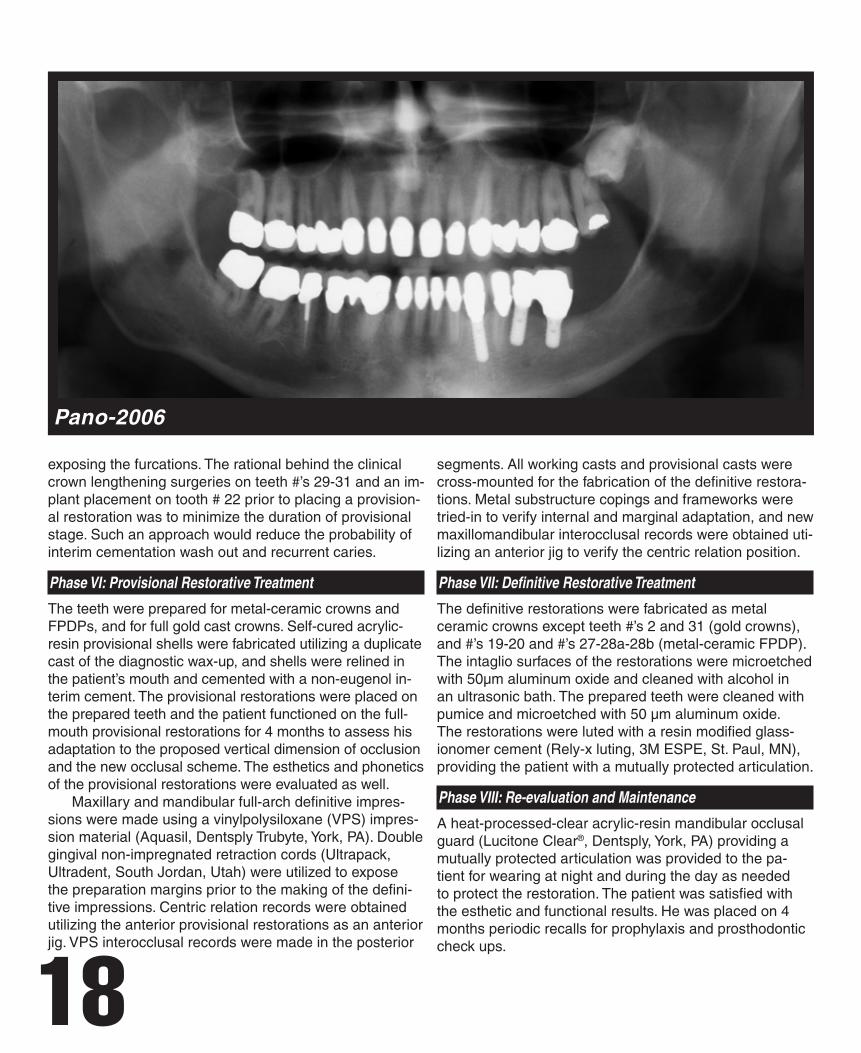
Pano-2006
segments. All working casts and provisional casts were cross-mounted for the fabrication of the definitive restora-tions. Metal substructure copings and frameworks were tried-in to verify internal and marginal adaptation, and new maxillomandibular interocclusal records were obtained uti-lizing an anterior jig to verify the centric relation position.
Phase VII: Definitive Restorative Treatment
The definitive restorations were fabricated as metal ceramic crowns except teeth #’s 2 and 31 (gold crowns), and #’s 19-20 and #’s 27-28a-28b (metal-ceramic FPDP). The intaglio surfaces of the restorations were microetched with 50µm aluminum oxide and cleaned with alcohol in an ultrasonic bath. The prepared teeth were cleaned with pumice and microetched with 50 µm aluminum oxide. The restorations were luted with a resin modified glass-ionomer cement (Rely-x luting, 3M ESPE, St. Paul, MN), providing the patient with a mutually protected articulation.
Phase VIII: Re-evaluation and Maintenance
A heat-processed-clear acrylic-resin mandibular occlusal guard (Lucitone Clear®, Dentsply, York, PA) providing a mutually protected articulation was provided to the pa-tient for wearing at night and during the day as needed to protect the restoration. The patient was satisfied with the esthetic and functional results. He was placed on 4 months periodic recalls for prophylaxis and prosthodontic check ups.
19
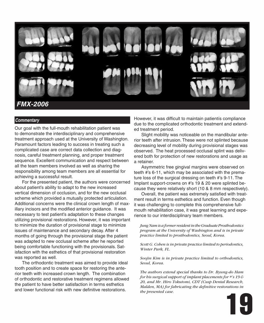
FMX-2006
However, it was difficult to maintain patientís compliance due to the complicated orthodontic treatment and extend-ed treatment period. Slight mobility was noticeable on the mandibular ante-rior teeth after intrusion. These were not splinted because decreasing level of mobility during provisional stages was observed. The heat processed occlusal splint was deliv-ered both for protection of new restorations and usage as a retainer. Asymmetric free gingival margins were observed on teeth #’s 6-11, which may be associated with the prema-ture loss of the surgical dressing on teeth #’s 9-11. The Implant support-crowns on #’s 19 & 20 were splinted be-cause they were relatively short (10 & 8 mm respectively). Overall, the patient was extremely satisfied with treat-ment result in terms esthetics and function. Even though it was challenging to complete this comprehensive full-mouth rehabilitation case, it was great learning and expe-rience to our interdisciplinary team members.
Jung Nam is a former resident in the Graduate Prosthodontics program at the University of Washington and is in private practice limited to prosthodontics, Seoul, Korea.
Scott G. Cohen is in private practice limited to periodontics, Winter Park, FL.
Soojin Kim is in private practice limited to orthodontics, Seoul, Korea.
The authors extend special thanks to Dr. Byung-do Ham for his surgical support of implant placements for #’s 19 & 20, and Mr. Hiro Tokutomi, CDT (Cusp Dental Research, Malden, MA) for fabricating the definitive restorations in the presented case.
Commentary
Our goal with the full-mouth rehabilitation patient was to demonstrate the interdisciplinary and comprehensive treatment approach used at the University of Washington. Paramount factors leading to success in treating such a complicated case are correct data collection and diag-nosis, careful treatment planning, and proper treatment sequence. Excellent communication and respect between all the team members involved as well as sharing the responsibility among team members are all essential for achieving a successful result. For the presented patient, the authors were concerned about patient’s ability to adapt to the new increased vertical dimension of occlusion, and for the new occlusal scheme which provided a mutually protected articulation. Additional concerns were the clinical crown length of max-illary incisors and the modified anterior guidance. It was necessary to test patient’s adaptation to these changes utilizing provisional restorations. However, it was important to minimize the duration of provisional stage to minimize issues of maintenance and secondary decay. After 4 months of going through the provisional stage the patient was adapted to new occlusal scheme after he reported being comfortable functioning with the provisionals. Sat-isfaction with the esthetics of that provisional restoration was reported as well. The orthodontic treatment was aimed to provide ideal tooth position and to create space for restoring the ante-rior teeth with increased crown length. The combination of orthodontic and restorative treatment regimens allowed the patient to have better satisfaction in terms esthetics and lower functional risk with new definitive restorations.
Related Documents


![Index [link.springer.com]978-3-319-02496...Index A Acanthocladia, 146 Active fault, 28, 48, 89, 97, 105, 111, 372, 375, 436, 453, 464, 465 Active normal fault, 385, 475 Aerial reconnaissance,](https://static.cupdf.com/doc/110x72/5add2cc67f8b9a4a268d334c/index-link-978-3-319-02496index-a-acanthocladia-146-active-fault-28-48.jpg)