ACTIVATION OF RAT T LYMPHOCYTES BY ANTI-CD2 MONOCLONAL ANTIBODIES BY SUSAN J . CLARK, DEBBIE A . LAW, DAVID J . PATERSON, MICHAEL PUKLAVEC, AND ALAN F. WILLIAMS From the Medical Research Council Cellular Immunology Research Unit, Sir William Dunn School of Pathology, Oxford 0X1 3RE, England The CD2 antigen (Tll, E-rosette receptor) was first identified on human T lym- phocytes with mAbs (1-3) . The antigen is a glycoprotein of 50,000 apparent M, and recently the protein sequences for CD2 of human (4, 5), rat (6), and mouse (7) have been derived from cDNA sequences . In each case the sequence indicates two ex- ternal domains that are Ig related, one transmembrane sequence, and a cytoplasmic domain with 115-116 amino acids . In humans, anti-CD2 mAbs were initially found to inhibit T lymphocyte prolifer- ation stimulated by mitogens, soluble antigens, or allogeneic cells (3, 8-10) . Then particular combinations of antibodies were found to be mitogenic and some single antibodies also had this property when present with phorbol esters (11-13) . CD2 is expressed in human thymocytes from an early stage and triggering of cell division by a natural ligand of CD2 has been suggested as an alternative route for cell activa- tion to the T lymphocyte antigen receptor (11, 14) . There is stong evidence that LFA-3 (alternatively identified as sheep erythrocyte TUTS) is the natural ligand for CD2 (15-17) and that the interaction between CD2 and LFA-3 can mediate adhesion of lymphoid cells . Also, purified LFA-3 can synergize with anti-CD2 to stimulate mito- genesis (18) . In the rat, the MRC OX-34 antibody is specific for CD2 and labels most T lym- phocytes and thymocytes (6, 19) . However, OX-34 also labels rat splenic macrophages, while similar cells in humans have not been reported to be CD2` . The MRC 0X- 34 mAb had no clear-cut effect on the function of the T lymphocytes, but has been recently shown to inhibit the cytolytic function of an NK-like cell line (20) . In the present work we have raised new mAbs against rat CD2 and show that one pairing of these activates DNA synthesis in T lymphocytes . The response was dependent on accessory cells and was potentiated by an anti-CD5 mAb and inhibited by anti-IL-2-R antibody. Materials and Methods Animals. Inbred PVG-RT1` and PVG-RTI° rats (referred to as HO and HO .B2, respec- tively) were from the Specific Pathogen Free Unit of the MRC Cellular Immunology Unit . BALB/c mice were from the Sir William Dunn School of Pathology. Monoclonal and Other Antibodies. Cloned hybrid cell lines that produce the mAbs MRC OX-53, OX-54, and OX-55 were from two fusions between mouse spleen cells and the mouse myeloma line NSO/l using standard procedures (21). The spleen cells were from BALB/c mice immunized with purified rat CD2 (6) : two 7 wg i .m . injections of CD2 emulsified in CFA J . Exp. MED. 0 The Rockefeller University Press - 0022-1007/88/06/1861/12 $2 .00 1861 Volume 167 June 1988 1861-1872 on December 20, 2015 jem.rupress.org Downloaded from Published June 1, 1988

Welcome message from author
This document is posted to help you gain knowledge. Please leave a comment to let me know what you think about it! Share it to your friends and learn new things together.
Transcript
ACTIVATION OF RAT T LYMPHOCYTES BY
ANTI-CD2 MONOCLONAL ANTIBODIES
BY SUSAN J . CLARK, DEBBIE A. LAW, DAVID J . PATERSON,MICHAEL PUKLAVEC, AND ALAN F. WILLIAMS
From the Medical Research Council Cellular Immunology Research Unit, Sir William DunnSchool of Pathology, Oxford 0X1 3RE, England
The CD2 antigen (Tll, E-rosette receptor) was first identified on human T lym-phocytes with mAbs (1-3) . The antigen is a glycoprotein of 50,000 apparent M, andrecently the protein sequences for CD2 of human (4, 5), rat (6), and mouse (7) havebeen derived from cDNA sequences . In each case the sequence indicates two ex-ternal domains that are Ig related, one transmembrane sequence, and a cytoplasmicdomain with 115-116 amino acids .
In humans, anti-CD2 mAbs were initially found to inhibit T lymphocyte prolifer-ation stimulated by mitogens, soluble antigens, or allogeneic cells (3, 8-10) . Thenparticular combinations of antibodies were found to be mitogenic and some singleantibodies also had this property when present with phorbol esters (11-13) . CD2 isexpressed in human thymocytes from an early stage and triggering of cell divisionby a natural ligand ofCD2 has been suggested as an alternative route for cell activa-tion to the T lymphocyte antigen receptor (11, 14) . There is stong evidence that LFA-3(alternatively identified as sheep erythrocyte TUTS) is the natural ligand for CD2(15-17) and that the interaction between CD2 and LFA-3 can mediate adhesion oflymphoid cells . Also, purified LFA-3 can synergize with anti-CD2 to stimulate mito-genesis (18) .
In the rat, the MRC OX-34 antibody is specific for CD2 and labels most T lym-phocytes and thymocytes (6, 19) . However, OX-34 also labels rat splenic macrophages,while similar cells in humans have not been reported to be CD2` . The MRC 0X-34 mAb had no clear-cut effect on the function ofthe T lymphocytes, but has beenrecently shown to inhibit the cytolytic function of an NK-like cell line (20) .
In the present work we have raised new mAbs against rat CD2 and show thatone pairing of these activates DNA synthesis in T lymphocytes . The response wasdependent on accessory cells and was potentiated by an anti-CD5 mAb and inhibitedby anti-IL-2-R antibody.
Materials and MethodsAnimals.
Inbred PVG-RT1` and PVG-RTI° rats (referred to as HO and HO.B2, respec-tively) were from the Specific Pathogen Free Unit of the MRC Cellular Immunology Unit .BALB/c mice were from the Sir William Dunn School of Pathology.
Monoclonal and Other Antibodies.
Cloned hybrid cell lines that produce the mAbs MRCOX-53, OX-54, and OX-55 were from two fusions between mouse spleen cells and the mousemyeloma line NSO/l using standard procedures (21). The spleen cells were from BALB/c miceimmunized with purified rat CD2 (6) : two 7 wg i .m . injections of CD2 emulsified in CFA
J . Exp. MED. 0 The Rockefeller University Press - 0022-1007/88/06/1861/12 $2 .00
1861Volume 167 June 1988 1861-1872
on Decem
ber 20, 2015jem
.rupress.orgD
ownloaded from
Published June 1, 1988
1862 MITOGENESIS VIA CD2
given on days 0 and 14, followed by one 7 pg i .p . injection of potassium alum precipitateof the pure CD2 . Mice were boosted with either 5 x 10' washed HO thymocytes given in-travenously on day 56 (MRC OX-53) or with 15 pg of the pure protein given intraperitoneallyon day 154 (OX-54 and OX-55) . MRC OX-53, OX-54, and OX-55 are all of the IgGI isotype.
Other mAbs used (referenced below or in reference 19) were : W3/13, IgGI, anti-rat leu-kosialin (LSGP) ; W3/25, IgGI, anti-rat CD4; MRC OX-1, IgGI and MRC OX-30, IgG2a,against distinct epitopes of the leucocyte common antigen (L-CA)' (22); MRC OX-22,IgG2a, anti-L-CA, restricted determinant (22) ; MRC OX-6, IgGI, anti-MHC class II (non-polymorphic) ; MRC OX-12, IgG2a, anti-rat is chain ; MRC OX-19, IgGI, anti-rat CD5 (Ly-1)(23) ; MRC OX-34, IgG2a, anti-rat CD2 ; MRC OX-39, IgGI and NDS 62 (Tellides, G.,MJ . Dallman, P.J . Morris, manuscript in preparation), IgGI, both anti-IL-2-R mAbs (24) ;MRC OX-21, IgGI, anti-human C3b inactivator, used as a control mAb. Antibodies wereused as tissue culture supernatants or purified IgG and F(ab')2 (25) .
Rabbit anti-mouse IgG antibodies (RAM) and their F(ab') 2 fragments were prepared andlabeled with fluorescein (RAM-FITC) or 1 '25 1 (' 25I-RAM) as described (25) . Iodination ofOX-54 and OX-55 mAbs was also as described (25) .
Cells.
Cervical lymph nodes, thymuses, and spleens were removed asceptically and teasedwith watchmakers' forceps in ice-cold Dulbecco's A+ B medium (DAB) containing 0.2% BSA.The cells were filtered through lens tissue, washed twice in DAB/BSA, and resuspended inRPMI-1640 medium containing 2.5 x 10 -5 M 2-ME, antibiotics, and 5% (vol/vol) FCS(Gibco Ltd ., Paisley, Scotland) (RPMI/5% FCS) .
Blasts.
Either HO thymocytes, lymph node cells (LNC), or spleen cells were incubatedat 2-5 x 10' cells/ml in RPMI/5% FCS for 72 h at 37°C in a humidified 5% C02 incu-bator with Con A, used at a final concentration of 5 pg/ml. Cells were harvested, washedin RPMI/5% FCS, and the blasts were separated on an Isopaque-Ficoll gradient (Pharmacia(GB) Ltd., Middlesex, U.K.) and centrifuged at 750 g for 20 min at room temperature . Cellsrecovered from the interface were washed twice in RPMI/5% FCS.
Tissue Culture.
All cultures were in 0.2 ml in U-bottomed wells of96-well microtiter platesat 37°C with 5% C02 . Medium was RPMI-1640 plus 5% FCS. Stimulator cells were spleencells y-irradiated with a "'Cs source (Gamma Cell 40 ; Atomic Energy of Canada Ltd.Kanata, Ontario, Canada) at 0 .85 Gy/min to yield a dose of 20 Gy.
Proliferation Assays .
For alloantigen-induced proliferation, a primary MLR was set up byculturing together 2 .5 x 105 HO LNC plus 5 x 10⁵ HO.B2 stimulator cells for a total of90 h. For proliferation induced by mAbs 2 .5 x 105 HO LNC were cultured with or without5 x 10 5 HO stimulator cells for a total of either 72 or 90 h. mAbs were added at the begin-ning of the culture period and were used as tissue culture supernatant unless otherwise stated .PMA at 2 ng/ml and IgG RAM or F(ab')2 RAM at 20 gg/ml were also added at the begin-ning of culture when used . Cultures were pulsed with 0.5 gCi tritiated thymidine (Amer-sham International, Amersham, U.K.) 18 h before harvesting onto glass fiber filters (WhatmanLtd., Maidstone, U.K.) . Results are expressed as arithmetic means of quadruplicate cultures+_ SD.
Generation of CTL and "Cr-release Assay.
Cytotoxic cells were generated in an MLR asabove cultured for 5 d. TheY3 rat myeloma cell line carrying the RTl° MHC antigens (21)was used as a target to assay the CTL activity. The NSO mouse myeloma cell line (21) wasused as a control target for non-specific killing. Percent "Cr release was calculated usingthe formula: percent specific release = 100 x [(experimental release - spontaneous re-lease)/(maximum release - spontaneous release)).
Nonspecific Killing.
''Cr release was used to measure the effect of mAbs on nonspecifickilling (26) . Effector cells were incubated in RPMI/2.5% FCS containing 2 mg/ml carbonyliron for 1 h at 37°C in 5% C02 . Phagocytic cells were then removed magnetically. Re-maining cells were washed twice in DAB/BSA, resuspended in RPMI/2.5% FCS, and two-fold serial dilutions were trade. The NSO mouse myeloma cell line was prelabeled with _"Crand used as the target at 10^ cells per well with serial dilutions of effector cells. "Cr release
' Abbreviations used in this paper: Cas, supernatant from Con A-activated rat spleen cells ; L-CA, leu-kocyte common antigen ; LNC, lymph node cells ; RAM, rabbit anti-mouse IgG antibodies .
on Decem
ber 20, 2015jem
.rupress.orgD
ownloaded from
Published June 1, 1988
CLARK ET AL .
1863
was measured after a 4-h incubation of effectors and targets . The percentage specific "Crrelease was calculated as above .
Immunoprecipitation. HO thymocytes were 1251 surface labeled using the lactoperoxi-dase/glucose oxidase method . Cell membranes were solubilized in 1% NP-40 and antigensimmunoprecipitated and analyzed as described (27) .
Indirect Binding Assays and Antibody Blocking Assays.
These were at 4°C unless otherwisestated and 5 x 10⁶ cells were incubated with mAbs and 1251-F(ab')2 RAM in successive 60-min incubations with two washes using DAB/0.25% BSA after each step . The system wascalibrated for molecules of first antibody bound by including labeling of thymocytes withW3/13 mAb and assuming that 38,000 molecules were bound (25, 28) . Competition in in-direct binding assays was assessed by seeing whether binding was additive when antibodieswere mixed (28) .
To directly assess competition between antibodies an 125 1-labeled mAb was mixed with anunlabeled mAb and then the mixture was incubated with 5 x 10⁶ thymocytes for 2 h . Cellswere washed and counted as before (25) .
Other Methods .
Labeling for FACS II (Becton Dickinson and Co., Mountain View, CA)analysis (28) ; immunoperoxidase labeling on cryostat sections (29) . Tissue culture superna-tant containing IL-2 was obtained as a lectin-free supernatant (Cas) from Con A-activatedrat spleen cells (30) . This was nonmitogenic when used alone.
ResultsThe Nature of the Antigen Recognized by MRC OX-53, OX-54, and OX-55 mAbs.
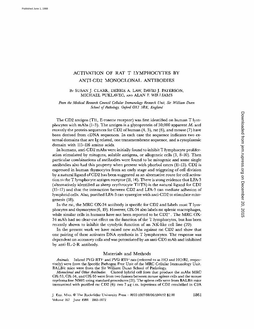
Inpreliminary studies, the new mAbs were shown to have a tissue distribution indis-tinguishable from that of MRC OX-34. In particular splenic tissue macrophageswere labeled (not shown) as well as most thymocytes and T lymphocytes . Labelingprofiles for the mAbs on lymph node cells and activated T lymphocytes are shownin Fig. 1, a and b.To establish that the mAbs were against CD2 sequential immunoprecipitations
were carried out in which solubilized 1251-labeled cell surface molecules wereprecleared with OX-34 mAb before immunoprecipitation with the other mAbs. Allthe mAbs immunoprecipitated similar bands of 50 x 103 apparent Mr and OX-34removed the material that could react with the OX-53, 54, and 55 mAbs but notthe CD4 antigen that binds to the W3/25 mAb (Fig 1 c) . Furthermore, when L cellswere transfected with rat CD2 cDNA, the transfectants but not the normal L cellswere labeled with MRC OX-34, OX-53, OX-54, and OX-55 mAbs (He Qi ; Barclay,A . N., and A. F. Williams, unpublished data) . Thus, there is no doubt that the fourantibodies all bind to CD2 .
Site Numbersfor mAb Binding.
Table I shows the molecules per cell for each anti-CD2 mAb binding at saturation to resting and blast cells derived from the thymusor lymph node . The antibodies bound in roughly similar amounts with differencesthat might be accounted for by differences in the proportion ofbivalent/monovalentbinding . Although saturating binding was similar the level of antibody needed forsaturation was 10-fold greater for OX-54 and OX-55 antibodies than for OX-34 orOX-53. Such a difference is unusual and whether or not this might be due to differ-ences in kinetics of binding remains to be established .
Between T blasts and resting cells, the increment in binding was about threefold .The volume of T blasts is about three to five times greater than resting T cells, andthus there was no increase in antigen density detected between resting and activatedcells .
Competition Between mAbs.
Initially, competition was assessed using saturating in-
on Decem
ber 20, 2015jem
.rupress.orgD
ownloaded from
Published June 1, 1988
1864
MITOGENESIS VIA CD2
FIGURE 1 .
FRCS analysis of labeling of (a) HO LNC and (b) 3-d Con A HO LN blasts withMRC OX-34, OX-53, OX-54, and OX-55 . Cells were labeled with mAb IgG at 20 pg/ml (0X-34, OX-53) and 100 pg/ml (OX-54, OX-55) at 4°C, followed by RAM-FITC . Fluorescence histo-grams were obtained on a Becton Dickinson & Co. FAGS II . 10 4 cells were analyzed for eachprofile with cell number shown on a linear scale . Labeling with MRC OX-21 is shown as a nega-tive control for each cell type (dotted line) . (c) Analysis of the antigen recognized by anti-CD2mAbs by immunoprecipitation . Lysed HO thymocytes surface labeled with 1251 were incubatedwith anti-CD2 mAbs after preclearing ofthe lysate with either an irrelevant mAb or with MRCOX-34 . Immunoprecipitated material was analyzed using 10% SDS-PAGE, and visualized withautoradiography. Separate experiments were carried out, with CD2 depletion being assessed byimmunoblotting . The results were as with 1251 labeling, except that OX-55 gave an extra dis-tinct band at a slightly lower mol wt than that ofauthentic CD2 . Preclearing with neither OX-34nor OX-55 mAbs removed this band, which is thus thought to be due to artifactual binding .
direct binding assays with mixtures of mAbs added in the first step . In other studies,mAbs against separate molecules or against nonoverlapping epitopes on the samemolecule have given additive binding when mixed in the indirect binding assay (28) .An exampleofthis is shown for noncompeting mAbs against L-CA (OX-1 and 0X-30) in Table II . In the case ofthe anti-CD2 mAbs, OX-34 and OX-53 are concludedto be fully competitive since they showed no additive binding at all. With combina-
on Decem
ber 20, 2015jem
.rupress.orgD
ownloaded from
Published June 1, 1988
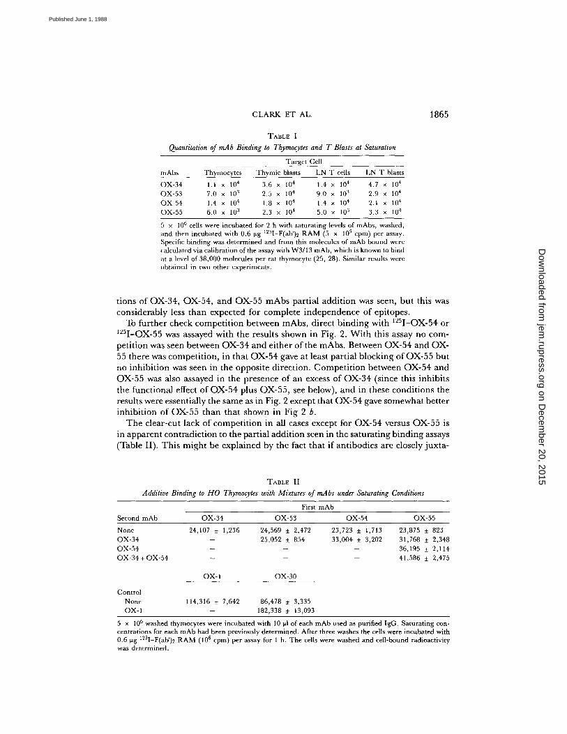
TABLE I
Quantitation of mAb Binding to Thymocytes and T Blasts al Saturation
5 x 10⁶ cells were incubated for 2 h with saturating levels of mAbs, washed,and then incubated with 0.6 Vg 125 1-F(ab')2 RAM (5 x 10⁵ cpm) per assay .Specific binding was determined and from this molecules of mAb bound werecalculated via calibration of the assay with W3/13 mAb, which is known to bindat a level of 38,000 molecules per rat thymocyte (25, 28) . Similar results wereobtained in two other experiments .
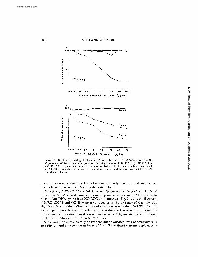
tions of OX-34, OX-54, and OX-55 mAbs partial addition was seen, but this wasconsiderably less than expected for complete independence of epitopes .To further check competition between mAbs, direct binding with 1251-OX-54 or
1251-OX-55 was assayed with the results shown in Fig. 2 . With this assay no com-petition was seen between OX-34 and either of the mAbs . Between OX-54 and 0X-55 there was competition, in that OX-54 gave at least partial blocking of OX-55 butno inhibition was seen in the opposite direction . Competition between OX-54 andOX-55 was also assayed in the presence of an excess of OX-34 (since this inhibitsthe functional effect of OX-54 plus OX-55, see below), and in these conditions theresults were essentially the same as in Fig. 2 except that OX-54 gave somewhat betterinhibition of OX-55 than that shown in Fig 2 b .The clear-cut lack of competition in all cases except for OX-54 versus OX-55 is
in apparent contradiction to the partial addition seen in the saturating binding assays(Table II) . This might be explained by the fact that if antibodies are closely juxta-
TABLE II
Additive Binding to HO Thymocytes with Mixtures of mAbs under Saturating Conditions
OX-1 OX-30
ControlNone
114,316 ± 7,642
86,478 ± 3,335OX-1
-
182,338 t 13,093
CLARK ET AL .
1865
5 x 10 6 washed thymocytes were incubated with 10 ul of each mAb used as purified IgG . Saturating con-centrations for each mAb had been previously determined . After three washes the cells were incubated with0 .6 ug 125 1-F(ab')2 RAM (106 cpm) per assay for 1 h . The cells were washed and cell-bound radioactivitywas determined .
mAbs Thymocytes ThymicTargetblasts
CellLN T cells LN T blasts
OX-34 1 .1 x 104 3 .6 x 10 4 1 .4 x 104 4 .7 x 10 4OX-53 7.0 x 10' 2 .5 x 10 4 9 .0 x 10" 2 .9 x 104OX-54 1 .4 x 104 1 .8 x 10 4 1 .4 x 104 2 .1 x 10 4OX-55 6.0 x 10' 2 .3 x 10 4 5 .0 x 10 3 3 .3 x 10 4
Second mAb OX-34First
OX-53mAb
OX-54 OX-55None 24,107 ± 1,236 24,569 t 2,472 23,723 t 1,713 23,875 ± 823OX-34 - 25,052 ± 854 33,004 ± 3,202 31,768 t 2,348OX-54 - - - 36,195 t 2,114OX-34 +OX-54 - - - 41,586 ± 2,475
on Decem
ber 20, 2015jem
.rupress.orgD
ownloaded from
Published June 1, 1988
1866
MITOGENESIS VIA CD2
v0aa
EvmmamJ
vc0aa<Ev
mamJ
0.625 1.25 2.5 5
10
25 50 100
Conc . of unlabelled mAb added
(hg/ml)
0.625 1.25 2.5 5
10
25 50 100Conc . of unlabelled mAb added
(pg/ml)
FIGURE 2 .
Blocking of binding of 12'1 anti-CD2 mAbs. Binding of WI-OX-54 (a) or 12'I-OX-55 (b) to 5 x IOh thymocytes in the presence of varying amounts of OX-34 (---C~-), OX-54and OX-55 (--o-) was determined . Cells were incubated with the mAb combinations for 2 hat 4°C . After two washes the radioactivity bound was counted and the percentage oflabeled mAbbound was calculated .
posed on a target antigen the level of second antibody that can bind may be lessper molecule than with each antibody added alone.
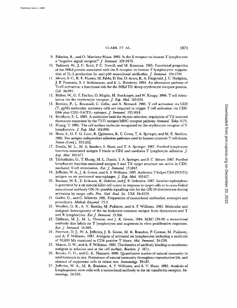
The Effect of MRC OX-54 and OX-55 on Rat Lymphoid Cell Proliferation .
None ofthe anti-CD2 mAbs used alone, either in the presence or absence ofCas, were ableto stimulate DNA synthesis in HO LNC or thymocytes (Fig . 3, a and b) . However,if MRC OX-54 and OX-55 were used together in the presence of Cas, low butsignificant levels of thymidine incorporation were seen with the LNC (Fig. 3 a) . Insome experiments the two antibodies with no additional Cas were sufficient to pro-duce some incorporation, but this result was variable . Thymocytes did not respondto the two mAbs even in the presence of Cas .Some variation in results might have been due to variable levels of accessory cells
and Fig . 3 c and d, show that addition of 5 x 10⁵ irradiated syngeneic spleen cells
on Decem
ber 20, 2015jem
.rupress.orgD
ownloaded from
Published June 1, 1988

1867
FIGURE 3 .
Effect of accessory cells on proliferative response of LNC and thymocytes to anti-CD2 mAbs . HO LNC or thymocytes (2 .5 x 105) were incubated either without (a, b) or with(c, d) 5 x 10⁵ irradiated syngeneic spleen cells for 72 h in RPMI/5°Jo FCS. mAbs were addedat the beginning of culture as tissue culture supernatant. Cells were cultured in the absence (solidcolumns) or presence (cross hatched columns) of 10% Cas.
considerably stimulated the response of both LNC and thymocytes . The anti-CD2mAbs used singly still gave no proliferation but the combination of MRC OX-54and OX-55 caused proliferation in both cell types that wasnot significantly enhancedby the addition of Cas. No other combination of anti-CD2 mAbs gave stimulationeven in the presence of accessory cells .The effect of other mAbs on stimulation of cells by OX-54 and OX-55 was exam-
ined and the results were contrasted with the effects of the same antibodies on theMLR. OX-19 (anti-CD5) is known to stimulate the MLR (23) and this also stimu-lated the C132-mediated proliferation (Fig . 4 a) . The quantitative effect was reprodu-cibly greater for the CD2 proliferation system than for the MLR.mAbs against class II MHC, CD4, and the IL-2-R are all known to inhibit the
MLR as shown in Fig. 4 d. In the CD2 system, the anti-class II mAbwas ineffectivewhile the anti-CD4 antibody was inhibitory, but not in the form of F(ab')2, whichdoes however inhibit the MLR. The anti-IL-2-R antibody also inhibited the C132-mediated proliferation .
In studies on human cells anti-CD2 mAbs can inhibit the MLR, but this is notreproducibly seen in the rat . The result shown in Fig. 4 d is one where inhibitionwith the OX-34 anti-CD2 mAb was seen . In contrast to variable inhibition in theMLR the OX-34 and OX-53 mAbs reproducibly gave strong inhibition of prolifera-tion induced by OX-54 and OX-55 . This occurred despite the fact that OX-34 didnot inhibit the binding of OX-54 or OX-55 (Fig . 2) and that competition betweenOX-54 and OX-55 was not effected by binding of OX-34 at the levels used in thefunctional assays (data not shown) .
on Decem
ber 20, 2015jem
.rupress.orgD
ownloaded from
Published June 1, 1988
1868
MITOGENESIS VIA CD2
áEE
OX-54+OX-55
'OX-19
Gcelle only
OX-54+OX-55
.OX-6
+oX-31 I9C
+OX-34 F(ob')2
+«3/25 I9G
+W3/25 F(eb')2
NDS 62
20 40 6O 60 700 120
H
bCells only
d
MLR'OW-19
-~'
cens ony
s~
MLR
H OX-6+OX-34 IQG
+OX-34 F(W)2+.3/25 I9G
+W3/25 F(cb')2
NDS 62
20 40 60 60 100 120
20 40 60 60 100 120
31-1-Thymidine incorporotion (,10 -3 )
N
H
H
r20 40 60 90 700 720
FIGURE 4 .
Factors affecting LNC proliferation . (a) 2 .5 x 10' HO LNC induced to proliferateby mAbs MRC OX-54+OX-55 in the form of tissue culture supernatant, in the presence of 5x 10 ,5 irradiated HO spleen cells . (b) 2 .5 x 10' HO LNC induced to proliferate by 5 x 10' HO.B2irradiated spleen cells in an allogeneic MLR. Additional mAbs were added to the cells at thebeginning of incubation and the effects ofthese on proliferation studied by measuring ["H)thymi-dine incorporation . Additional mAbs were used in the form ofeither tissue culture supernatantor purified IgG or F(ab')2 at 20 ug/ml . Incubation was for 72 h in RPMI/5% FCS (a, c)and for 90 h (b, d) .
In studies on human T cells there are some cases where DNA synthesis is stimu-lated by one anti-CD2 antibody plus phorbol ester (11, 12) . Using PMA at a concen-tration of2 ng/ml it was found that none ofthe anti-rat CD2 mAbs used singly wouldgive proliferation with PMA (Fig . 5, a and b) . Higher levels of PMA were toxic forrat T cells. Furthermore, PMA did not augment the proliferation induced by MRCOX-54 and OX-55 used together. The effects of crosslinking the single mAbs witheither RAM IgG or RAM F(ab')2 were also investigated . No single mAbs werefound to be mitogenic with either crosslinker, even in the presence of PMA. How-ever, although proliferation induced by MRC OX-54 and OX-55 was not affectedby the addition of RAM IgG, the addition of RAM F(ab')2 prevented almost all[ 3H]thymidine incorporation . These results were unaffected by the presence or ab-sence of PMA. The inhibition with F(ab')2 RAM may occur because interactionwith Fc receptors on accessory cells is essential for the response to occur. F(ab')2RAM may obscure the Fc of the mAbs without providing its own Fc for interaction,as would be the case with the use of IgG RAM.
Effect ofAnti-CD2 mAbs on Natural Killing and CTL-mediated Killing.
None of theanti-CD2 mAbs either singly or in combination had any effect on nonspecific killingwhen the NSO mouse myeloma cell line was used as a target for HO spleen cells .This was also true for the specific CTL-mediated killing of Y3 myeloma cells byCTL generated in a primary MLR (data not shown) .
DiscussionStimulation ofmitogenesis in T lymphocytes can be initiated by a variety of mAbs
reacting with molecules not known to be associated with the TCR complex . Thetarget antigens include Thy-1 and LY-6 in the mouse (31, 32) and CD2 (12, 13), leu-kosialin (sialophorin) (33), and CD28 (T44) (34) in humans . The results herein show
on Decem
ber 20, 2015jem
.rupress.orgD
ownloaded from
Published June 1, 1988

a
Medium
OX-34
TRAM IgG
4RAM F(nb')2
OX-54
.RAM IgG
+RAY F( .b')Z
OX-SS
TRAM IqG
+RAM F(W)2
OX-54+0X-55
.RAM WG
4RAM rt.b')2
o No PMA
D
CLARK ET AL .
1869
Medium
OX-34
.RAM IQF,
+RAM F(eb') 2
OX-54
+RAM I9G
+RNI F(ub'),
OX-5S
.RAM I9G
TRAM F(Rb')2
OX-54+GX-55
4RAM IQC.
.RAM F(eb')=
b With PUA
H
10 20 30 40
10 20 30 40
3 H-Thymidine incorpom(ion (w10 - 3)
FIGURE 5 .
Effect of crosslinking andPMAon proliferation of LNC induced by anti-CD2 mAbs .2.5 x 105 HO LNC plus 5 x 10 HO-irradiated spleen cells were incubated with anti-CD2 mAbsin the (a) absence or (b) presence of PMA at 2 ng/ml. Crosslinking was performed with eitherRAM-IgG or RAM-F(ab')2 at 20 Wig/ml, added at the beginning of incubation, which was for72 h in RPMI/5% FCS.
that CD2 can also be a mitogenic target for rat T lymphocytes. The requirementswere that the OX-54 and OX-55 mAbs were present along with an undefined popu-lation of accessory cells . It seemed likely that the accessory cells were required formore than the crosslinking of the mAbs, since crosslinking with F(ab')2 RAM in-hibited the response possibly by masking Fc determinants (see Results) . Phorbolester did not potentiate the response or allow stimulation via one mAb.The MRC OX-54 and OX-55 mAbs showed a curious pattern of competitive
binding, in that OX54 could substantially block OX-55 binding but the reverse sit-uation did not occur. As both antibodies were effective in immunoprecipitation itseemed unlikely that this was due to differences in affinity, andOX-55 did not blockOX-54binding even if cells were preincubated with the OX-55 mAb (data not shown) .This phenomenon is similar to that seen in crossblocking between the anti-humanCD2 mAbs GT2 and anti-D66 (35), which also synergise to stimulate DNA syn-thesis . The OX-34 and OX-53 mAbs did not show any blocking of OX-54 and 0X-55 binding, yet these mAbs inhibited the stimulatory effects of OX-54 and OX-55.This is a curious result given that crosslinking of OX-34 on an NK-like cell line cantrigger release of Ca2+ and turnover of inositol phosphates (20) .The rat CD2 triggering data seems quite similar to the human work with the ex-
ception that : (a) all the ratCD2 determinants so far described are expressed on restingand activated cells whereas in humans the CD2 T113 epitope is thought to be specificto activated cells (11) ; (b) synergism of one CD2 mAb and phorbol ester to triggerthe cells is not seen in the rat; (c) accessory cells are thus far essential for triggeringthe rat cells ; (d) the anti-CD2 mAbs are not inhibitors ofspecific or nonspecific cytoxiccells, although the OX-34 mAb sometimes shows inhibition of the MLR and hasbeen shown to block killing by an NK-like cell line (20) .
In humans, CD2 has been suggested as a target for stimulation of T cells by a
on Decem
ber 20, 2015jem
.rupress.orgD
ownloaded from
Published June 1, 1988

1870
MITOGENESIS VIA CD2
pathway that is independent of TCR triggering with a particular role in thymopoi-esis (14) . DNA synthesis can be triggered in rat thymocytes by the anti-CD2 mAbs,but it remains to be seen whether the activity is due only to thymocytes of im-munocompetent phenotype .
SummaryRat T cells and thymocytes were induced to proliferate by a pair of mAbs, MRC
OX-54 and MRC OX-55, directed against rat CD2 . Accessory cells were requiredbut their role was not simply for crosslinking of the two mAbs, as neither MRCOX-54 nor MRC OX-55 alone, in the presence of a crosslinking second antibody,caused T cell mitogenesis . Nor could the phorbol ester PMA replace either anti-body. The two mAbs recognized distinct epitopes on rat CD2; however, MRC 0X-54 could partially block MRC OX-55 binding whereas the reverse situation was notseen . A further CD2 epitope was recognized by two mutually competitive mAbs,MRC OX-34 and MRC OX-53, which were not mitogenic . Neither MRC OX-34nor MRC OX-53 affected the binding of MRC OX-54 or MRC OX-55, yet theyprevented the mitogenic effect induced by these mAbs. The presence ofmAbs againstCD4 and the IL-2-R also abrogated this mitogenesis, whereas an anti-CD5 mAbaugmented the CD2-induced proliferation .
Our thanks to Maggie Dallman for the kind donation ofthe anti-IL-2-R mAb, Stan Bucking-ham and Catherine Lee for photography, and Denise Roby for typing the manuscript .
Received for publication 16 February 1988 .
References1 . Kamoun, M., P. J . Martin, J . A . Hansen, M. A . Brown, A. W. Siadak, and R . C .
Nowinski . 1981 . Identification ofa human T lymphocyte surface protein associated withthe E-rosette receptor. J Exp . Med. 153 :207 .
2 . Howard, F. D ., J . A . Ledbetter, J . Wong, C . P. Bieber, E . B . Stinson, and L . A . Herzen-berg . 1981 . A human T lymphocyte differentiation marker defined by monoclonal anti-bodies that block E-rosette formation . J. Immunol. 126 :117 .
3 . Van Wauwe, J., J . Goossens, W. Decock, P. Kung, and G . Goldstein . 1981 . Suppressionofhuman Tcell mitogenesis and E-rosette formation by the monoclonal antibody OKT11A .Immunology . 44:865 .
4 . Sewell, W. A., M. H. Brown, J . Dunne, M. J . Owen, and M. J . Crumpton . 1986 .Molecular cloning of the human Tlymphocyte surface CD2 (T11) antigen . Proc. Nod.Acad Sci. USA . 83:8718 .
5 . Seed, B ., and A . Aruffo . 1987 . Molecular cloning of the CD2 antigen, the T-cell erythro-cyte receptor, by a rapid immunoselection procedure . Proc . Natl . Acad. Sci. USA . 84:3365 .
6 . Williams, A.F., A . N . Barclay, S. J . Clark, D. J . Paterson, and A. C . Willis. 1987 . Similar-itie s in sequences and cellular expression between rat CD2 and CD4 antigens . J. Exp.Med. 165:368 .
7 . Sewell, W.A., M. H . Brown, M. J . Owen, P. J_ Fink, C . A. Kozak, and M. J._Crumpton .1987 . The murine homologue of the Tlymphocyte CD2 antigen molecular cloning: chro-mosome assignment and cell surface expression . Eur. J. Immunol. 17:1015 .
8 . Martin, P. J ., G . Longton, J . A . Ledbetter, W. Newman, M. P. Braun, P. G . Beatty,and J . A . Hansen . 1983 . Identificatio n and functional characterization of two distinctepitopes on the human T cell surface protein Tp50 . J. Immunol. 131 :180 .
on Decem
ber 20, 2015jem
.rupress.orgD
ownloaded from
Published June 1, 1988
CLARK ET AL.
1871
9 . Palacios, R., and O. Martinez-Maza. 1982 . Is the E receptor on human T lymphocytesa "negative signal receptor?"f. Immunol. 129:2479 .
10 . Tadmori, W., J . C . Reed, P. C . Nowell, and M. Kamoun . 1985 . Functional propertiesof the 50Kd protein associated with the E-receptor on human T lymphocytes : suppres-sion of IL-2 production by anti-p50 monoclonal antibodies . J. Immunol. 134:1709.
11 . Meuer, S . C ., R. E . Hussey, M. Fabbi, D. Fox, O. Acuto, K . A . Fitzgerald, J . C . Hodgdon,J . P. Protentis, S . F. Schlossman, and E . L . Reinherz . 1984 . An alternative pathway ofTcell activation : a functional role for the 50Kd Tll sheep erythrocyte receptor protein .Cell. 36:897 .
12 . Holter, W., G . F. Fischer, O. Majdic, H. Stockinger, and W. Knapp. 1986 . T cell stimu-lation via the erythrocyte receptor. J. Exp. Med. 163:654 .
13 . Brottier, P, L . Boumsell, C . Gelin, and A . Bernard . 1985 . T cell activation via CD2(T, gp50) molecules : accessory cells are required to trigger T cell activation via C132-D66 plus CD2-9.6/Tll, epitopes . J. Immunol. 135:1624 .
14 . Reinherz, E . L . 1985 . A molecular basis for thymic selection: regulation of Tll inducedthymocyte expansion by the T3Ti antigen/MHC receptor pathway. Immunol. Today. 6:75 .
15 . Hunig, T. 1985 . The cell surface molecule recognized by the erythrocyte receptor of T-lymphocytes . f Exp. Med. 162:890 .
16 . Shaw, S ., G . E . G. Luce, R . Quinones, R. E . Gress, T. A . Springer, and M. E . Sanders .1986 . Two antigen-independent adhesion pathways used by human cytotoxic T cell clones.Nature (Loud.). 323:262 .
17 . Dustin, M. L ., M. E . Sanders, S . Shaw, and T. A . Springer. 1987 . Purified lymphocytefunction-associated antigen 3 binds to CD2 and mediates T lymphocyte adhesion . J.Exp. Med. 165:677 .
18 . Tiefenthaler, G., T. Hunig, M. L . Dustin, T. A . Springer, and S. C . Meuer. 1987 . Purifie dlymphocyte function-associated antigen-3 and T11 target structure are active in CD2-mediated T-cell stimulation . Eur. J Immunol. 17:1847 .
19 . Jefferies, W. A ., J . R . Green, and A . F. Williams . 1985 . Authentic T helper CD4 (W3/25)antigen on rat peritoneal macrophages . J. Exp. Med. 165:117 .
20 . Seaman, W. E., E . Eriksson, R . Dobrow, andJ . B . Imboden . 1987 . Inosito l triphosphateis generated by a rat natural killer cell tumor in response to target cells or to cross-linkedmonoclonal antibody OX-34 : possible signalling role for the OX-34 determinant duringactivation by target cells. Proc. Natl. Acad. Sci . USA . 84:4239 .
21 . Galfre, G., and C . Milstein . 1981 . Preparation of monoclonal antibodies : strategies andprocedures . Methods Enzymol. 73:3 .
22 . Woollett, G . R ., A . N . Barclay, M. Puklavec, and A. F. Williams . 1985 . Molecular andantigenic heterogeneity of the rat leukocyte-common antigen from thymocytes and Tand B lymphocytes . Eurf. Immunol. 15:168 .
23 . Dallman, M. J ., M. L . Thomas, and J . R . Green . 1984 . MRC OX-19 : a monoclonalantibody that labels rat T lymphocytes and augments in vitro proliferative responses .Eur. J. Immunol. 14:260 .
24 . Paterson, D. J ., W. A . Jefferies, J . R. Green, M. R . Brandon, P. Cortese, M . Puklavec,and A . F. Williams . 1987 . Antigens of activated rat lymphocytes including a moleculeof 50,000 Mr restricted to CD4 positive T blasts . Mol. Immunol. 24:1281 .
25 . Mason, D. W., and A. F. Williams . 1980 . The kinetics ofantibody binding to membraneantigens in solution and at the cell surface . Biochem . J. 187 :1 .
26 . Brooks, C . G ., and G. R . Flannery. 1980 . Quantitative studies of natural immunity tosolid tumours in rats. Persistence ofnatural immunity throughout reproductive life, andabsence of suppressor cells in infant rats . Immunology. 39 :187 .
27 . Jefferies, W. A., M. R. Brandon, A. F. Williams, and S. V. Hunt . 1985 . Analysi s oflymphopoietic stem cells with a monoclonal antibody to the rat transferrin receptor. Im-munology. 54:333 .
on Decem
ber 20, 2015jem
.rupress.orgD
ownloaded from
Published June 1, 1988
18'72
MITOGENESIS VIA CD2
28 . Brideau, R. J ., P. B. Carter, W. R. McMaster, D. W. Mason, and A. F. Williams . 1980 .Two subsets of rat T lymphocytes defined with monoclonal antibodies . Eur. J. Immunol.
10:609 .29 . Barclay, A. N. 1981 . The localization of populations of lymphocytes defined by mono-
clonal antibodies in rat lymphoid tissues . Immunology. 42 :593 .30 . Wilson, A., W. -F. Chen, R. Scollay, and K. Shortman . 1982 . Semi-automated limit-
dilution assay and clonal expansion of all T-cell precursors of cytotoxic lymphocytes. JImmunol. Methods. 52 :283 .
31 . Kroczek, R. A., K. C. Gunter, R. N. Germain, and E. M . Shevach. 1986. Thy-1 func-tions as a signal transduction molecule in Tlymphocytes and transfected B-lymphocytes.Nature (Land) . 332 :181 .
32 . Rock, K. L., E. T. H . Yeh, C. F. Gramm, S. I. Haber, H. Reiser, and B. Benacerraf.1986 . TAP, A novel T cell-activating protein involved in the stimulation ofMHC-restrictedT lymphocytes . .J. Exp. Med. 163:315 .
33 . Mentzer, S. J., E. Remold-O'Donnell, M. A. V. Crimmins, B. E. Bierer, F. S. Rosen,and S. J. Burakoff. 1987 . Sialophorin, a surface sialoglycoprotein defective in the Wiskott-Aldrich syndrome, is involved in humanTlymphocyte proliferation . j Exp. Med. 165:1383 .
34 . Hara, T., S. M. Fu, and J . A. Hansen . 1985 . Human Tcell activation 11 . A new activa-tion pathway used by a major T cell population via a disulfide-bonded dimer of a 44kilodalton polypeptide (9 .3 antigen) . J. Exp. Med. 161:1513 .
35 . Huet, S., H . Wakasugi, G. Sterkers, J. Gilmour, T. Tursz, L. Boumsell, and A. Bernard.1986 . T-cell activation via CD2 [T, gp50]: the role ofaccessory cells in activating restingTcells via CD2 . J. Immunol. 137:1420.
on Decem
ber 20, 2015jem
.rupress.orgD
ownloaded from
Published June 1, 1988
Related Documents


![[1993]City Hunter CD2](https://static.cupdf.com/doc/110x72/56d6be561a28ab301691ae4d/1993city-hunter-cd2.jpg)
