Activation of p38/JNK Pathway Is Responsible for Embelin Induced Apoptosis in Lung Cancer Cells: Transitional Role of Reactive Oxygen Species Deepa R. Avisetti 1,2 , K. Suresh Babu 3 , Shasi V. Kalivendi 1,2 * 1 Centre for Academy of Scientific & Innovative Research, CSIR-Indian Institute of Chemical Technology (CSIR-IICT), Hyderabad, Andhra Pradesh, India, 2 Centre for Chemical Biology, CSIR-Indian Institute of Chemical Technology (CSIR-IICT), Hyderabad, Andhra Pradesh, India, 3 Natural Products Chemistry, CSIR-Indian Institute of Chemical Technology (CSIR-IICT), Hyderabad, Andhra Pradesh, India Abstract The natural product embelin has been demonstrated to possess a wide range of therapeutic properties, however, the mechanisms by which it exerts anticancer effects are not yet clear. By monitoring the molecular changes associated during early apoptotic phase, we have identified the crucial role of oxidative stress induced MAP kinase signalling as a predominant mechanism for its anticancer effects. Treatment of A549 lung cancer cells with embelin resulted in the enhancement of phospho-p38 and phospho-JNK levels as early as 4h. Pretreatment of cells with specific inhibitors of p38 (PD169316) and JNK (SP600125) abrogated embelin-induced caspase-3 activation. Studies employing embelin in the presence or absence of specific MAP kinase inhibitors indicated that the observed changes in phosphorylation levels of p38, JNK and ERK 1/2 are solely due to embelin and not because of cross-talk between MAP kinases. Reactive oxygen species (ROS) play a crucial role in embelin induced alterations in MAP kinase phosphorylation and apoptosis as pretreatment of cells with FeTMPyP mitigated this effect. The observed changes are not due to the inhibitory effect of embelin on XIAP as cells treated with SMAC-N7-Ant peptide, a specific inhibitor of XIAP’s BIR3 domain did not mimic embelin induced apoptotic effects. The findings of the present study clearly indicate the crucial role of p38 and JNK pathways in embelin induced apoptosis and provide us with new clues for improving its therapeutic efficacy. Citation: Avisetti DR, Babu KS, Kalivendi SV (2014) Activation of p38/JNK Pathway Is Responsible for Embelin Induced Apoptosis in Lung Cancer Cells: Transitional Role of Reactive Oxygen Species. PLoS ONE 9(1): e87050. doi:10.1371/journal.pone.0087050 Editor: Shrikant Anant, University of Kansas School of Medicine, United States of America Received September 24, 2013; Accepted December 17, 2013; Published January 22, 2014 Copyright: ß 2014 Avisetti et al. This is an open-access article distributed under the terms of the Creative Commons Attribution License, which permits unrestricted use, distribution, and reproduction in any medium, provided the original author and source are credited. Funding: This work was supported by SMiLE project from CSIR, India. Senior Research Fellowship to DRA from UGC, India, is gratefully acknowledged. The funders had no role in study design, data collection and analysis, decision to publish, or preparation of the manuscript Competing Interests: The authors have declared that no competing interests exist. * E-mail: [email protected] Introduction Embelin, an active component of fruits of Embelia ribes, has been demonstrated to possess a broad-spectrum of therapeutic proper- ties such as anticancer, anti-inflammation, anti-diabetes, anti- obesity, analgesic, anti-fertility and anti-helminthic [1–5]. The initial discovery of embelin as an inhibitor of XIAP by virtue of its interaction with the BIR3 domain and its observed selectivity towards cancer cells as compared to the normal cells inspired us to consider it as a lead compound for further studies against cancer [6]. As many of the cancers express elevated levels of XIAP and become refractory to apoptosis, treatment with embelin or in combination with other known anticancer drugs was found to sensitize them towards apoptosis [6,7]. Mechanism based studies indicate that embelin inactivates NF-kB by inhibiting nuclear transportation of p65 and also shown to inhibit STAT3 phosphorylation by inducing the expression of PTEN [8,9]. Specific efforts to identify the precise molecular target of embelin resulted in the identification of embelin as an inhibitor against XIAP’s BIR3 domain [6]. In addition, embelin was also demonstrated to be an inhibitor of 5-lipoxigenase and microsomal prostaglandin E2 synthase-1 (mPGES)-1; plasminogen activator inhibitor-1 (PAI-1) and P300/CBP associated factor (PCAF) [10– 12]. Moreover, embelin has been shown to interfere with the oxidative phosphorylation of mitochondria and can undergo both redox and non-redox mediated mechanisms [13,14]. Though the affinity of embelin against some of the molecular targets and cell signalling mechanisms have been identified, the primary intracellular target responsible for its anti-cancer property is not yet clear as many of the earlier studies have been carried out at later time points where the signal transduction cascade becomes complex due to the cross-talk between multiple cell signalling mechanisms [8,15,16,17]. Hence, in the present study, we sought to identify the alterations in signalling pathways responsible for the anticancer property of embelin during the early apoptotic phase. The present study identified for the first time the pivotal role of MAP kinase pathway, especially p38 and JNK, in embelin induced apoptosis. Materials and Methods Materials Embelin was purified from the fruits of Embelia ribes as described previously [18,19]. Minimal essential medium (MEM), Dulbecco’s modified Eagle’s medium (DMEM), Dulbecco’s phosphate buff- ered saline (DPBS), penicillin, streptomycin, sulphorhodamine B (SRB), Ac-DEVD-7-AFC, Ac-LEHD-7-AFC, PD169316, SP600125, N-acetyl-L-cysteine (NAC), radioimmune precipitation PLOS ONE | www.plosone.org 1 January 2014 | Volume 9 | Issue 1 | e87050

Welcome message from author
This document is posted to help you gain knowledge. Please leave a comment to let me know what you think about it! Share it to your friends and learn new things together.
Transcript
Activation of p38/JNK Pathway Is Responsible forEmbelin Induced Apoptosis in Lung Cancer Cells:Transitional Role of Reactive Oxygen SpeciesDeepa R. Avisetti1,2, K. Suresh Babu3, Shasi V. Kalivendi1,2*
1Centre for Academy of Scientific & Innovative Research, CSIR-Indian Institute of Chemical Technology (CSIR-IICT), Hyderabad, Andhra Pradesh, India, 2Centre for
Chemical Biology, CSIR-Indian Institute of Chemical Technology (CSIR-IICT), Hyderabad, Andhra Pradesh, India, 3Natural Products Chemistry, CSIR-Indian Institute of
Chemical Technology (CSIR-IICT), Hyderabad, Andhra Pradesh, India
Abstract
The natural product embelin has been demonstrated to possess a wide range of therapeutic properties, however, themechanisms by which it exerts anticancer effects are not yet clear. By monitoring the molecular changes associated duringearly apoptotic phase, we have identified the crucial role of oxidative stress induced MAP kinase signalling as a predominantmechanism for its anticancer effects. Treatment of A549 lung cancer cells with embelin resulted in the enhancement ofphospho-p38 and phospho-JNK levels as early as 4h. Pretreatment of cells with specific inhibitors of p38 (PD169316) andJNK (SP600125) abrogated embelin-induced caspase-3 activation. Studies employing embelin in the presence or absence ofspecific MAP kinase inhibitors indicated that the observed changes in phosphorylation levels of p38, JNK and ERK 1/2 aresolely due to embelin and not because of cross-talk between MAP kinases. Reactive oxygen species (ROS) play a crucial rolein embelin induced alterations in MAP kinase phosphorylation and apoptosis as pretreatment of cells with FeTMPyPmitigated this effect. The observed changes are not due to the inhibitory effect of embelin on XIAP as cells treated withSMAC-N7-Ant peptide, a specific inhibitor of XIAP’s BIR3 domain did not mimic embelin induced apoptotic effects. Thefindings of the present study clearly indicate the crucial role of p38 and JNK pathways in embelin induced apoptosis andprovide us with new clues for improving its therapeutic efficacy.
Citation: Avisetti DR, Babu KS, Kalivendi SV (2014) Activation of p38/JNK Pathway Is Responsible for Embelin Induced Apoptosis in Lung Cancer Cells: TransitionalRole of Reactive Oxygen Species. PLoS ONE 9(1): e87050. doi:10.1371/journal.pone.0087050
Editor: Shrikant Anant, University of Kansas School of Medicine, United States of America
Received September 24, 2013; Accepted December 17, 2013; Published January 22, 2014
Copyright: � 2014 Avisetti et al. This is an open-access article distributed under the terms of the Creative Commons Attribution License, which permitsunrestricted use, distribution, and reproduction in any medium, provided the original author and source are credited.
Funding: This work was supported by SMiLE project from CSIR, India. Senior Research Fellowship to DRA from UGC, India, is gratefully acknowledged. Thefunders had no role in study design, data collection and analysis, decision to publish, or preparation of the manuscript
Competing Interests: The authors have declared that no competing interests exist.
* E-mail: [email protected]
Introduction
Embelin, an active component of fruits of Embelia ribes, has been
demonstrated to possess a broad-spectrum of therapeutic proper-
ties such as anticancer, anti-inflammation, anti-diabetes, anti-
obesity, analgesic, anti-fertility and anti-helminthic [1–5]. The
initial discovery of embelin as an inhibitor of XIAP by virtue of its
interaction with the BIR3 domain and its observed selectivity
towards cancer cells as compared to the normal cells inspired us to
consider it as a lead compound for further studies against cancer
[6]. As many of the cancers express elevated levels of XIAP and
become refractory to apoptosis, treatment with embelin or in
combination with other known anticancer drugs was found to
sensitize them towards apoptosis [6,7]. Mechanism based studies
indicate that embelin inactivates NF-kB by inhibiting nuclear
transportation of p65 and also shown to inhibit STAT3
phosphorylation by inducing the expression of PTEN [8,9].
Specific efforts to identify the precise molecular target of
embelin resulted in the identification of embelin as an inhibitor
against XIAP’s BIR3 domain [6]. In addition, embelin was also
demonstrated to be an inhibitor of 5-lipoxigenase and microsomal
prostaglandin E2 synthase-1 (mPGES)-1; plasminogen activator
inhibitor-1 (PAI-1) and P300/CBP associated factor (PCAF) [10–
12]. Moreover, embelin has been shown to interfere with the
oxidative phosphorylation of mitochondria and can undergo both
redox and non-redox mediated mechanisms [13,14].
Though the affinity of embelin against some of the molecular
targets and cell signalling mechanisms have been identified, the
primary intracellular target responsible for its anti-cancer property
is not yet clear as many of the earlier studies have been carried out
at later time points where the signal transduction cascade becomes
complex due to the cross-talk between multiple cell signalling
mechanisms [8,15,16,17]. Hence, in the present study, we sought
to identify the alterations in signalling pathways responsible for the
anticancer property of embelin during the early apoptotic phase.
The present study identified for the first time the pivotal role of
MAP kinase pathway, especially p38 and JNK, in embelin induced
apoptosis.
Materials and Methods
MaterialsEmbelin was purified from the fruits of Embelia ribes as described
previously [18,19]. Minimal essential medium (MEM), Dulbecco’s
modified Eagle’s medium (DMEM), Dulbecco’s phosphate buff-
ered saline (DPBS), penicillin, streptomycin, sulphorhodamine B
(SRB), Ac-DEVD-7-AFC, Ac-LEHD-7-AFC, PD169316,
SP600125, N-acetyl-L-cysteine (NAC), radioimmune precipitation
PLOS ONE | www.plosone.org 1 January 2014 | Volume 9 | Issue 1 | e87050
assay buffer (RIPA) and protease inhibitor cocktail were purchased
from Sigma-Aldrich, Germany. U0126 and FeTMPyP were
purchased from Calbiochem. SMAC-N7-Ant peptide (AV-
PIAQK-P-RQIKIWFQNRRMKWKK) was synthesized by Gen-
Pro Biotech, Noida, India. Annexin-V assay kit was purchased
from Clontech Inc, USA. All the chemicals for buffer preparations
and fine chemicals were purchased from Sigma-Aldrich, Ger-
many.
Cell Culture and Experimental ConditionsAll the cell lines were obtained from ATCC, USA. A549,
DU145, MCF-7 and WPMY-1 cells were grown in MEM
(supplemented with 10% FBS, 100 units/ml penicillin and 100
units/ml streptomycin) while H9c2 and MRC-5 cells were grown
in DMEM (supplemented with 10% FBS, 100 units/ml penicillin
and 100 units/ml streptomycin). Cells were maintained in
humidified atmosphere with 5% CO2 at 37uC. Twelve hours
before treatments, the cell culture media was replaced with
respective media containing 2% FBS, unless otherwise indicated.
In intervention studies, cells were pretreated with the respective
MAP kinase inhibitors or antioxidants for 1h before the addition of
embelin (15 mM). For experiments involving SMAC-N7-Ant
peptide, cells were treated with 100 mM peptide for a period of 8h.
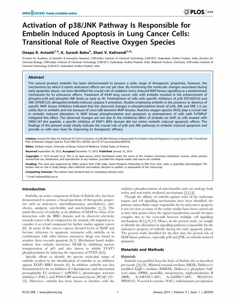
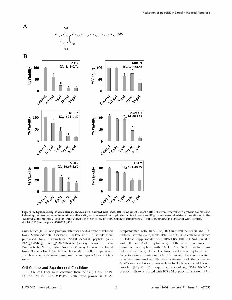
Figure 1. Cytotoxicity of embelin in cancer and normal cell lines. (A) Structure of Embelin (B) Cells were treated with embelin for 48h andfollowing the termination of incubation, cell viability was measured by sulphorhodamine B assay and IC50 values were calculated as mentioned in the‘‘Materials and Methods’’ section. Data shown are mean 6 SD of three separate experiments. * indicates p,0.01as compared with controls.doi:10.1371/journal.pone.0087050.g001
Activation of p38/JNK in Embelin Induced Apoptosis
PLOS ONE | www.plosone.org 2 January 2014 | Volume 9 | Issue 1 | e87050
Cytotoxicity AssayThe effect of embelin on cell viability was determined by
sulphorhodamine B (SRB) assay as described previously [20]. SRB
is an aminoxanthene dye that binds to basic amino acid residues of
cells (fixed to tissue culture plates by trichloroacetic acid) under
mild acidic conditions [20]. Briefly, cells (in 24 well plates, , 80%
confluence) were treated with different concentrations of embelin
for 48h in media supplemented with 10% fetal bovine serum.
Following the termination of incubation, cells were fixed by the
addition of 30% trichloroacetic acid to the medium at 4uC for 1h.
Later, cells were washed with deionised water and air dried. SRB
(0.04%, w/v) was added to the cells and incubated further for
30 min at room temperature. Finally, cells were washed with 1%
acetic acid (three times) and air dried. SRB bound to the cells was
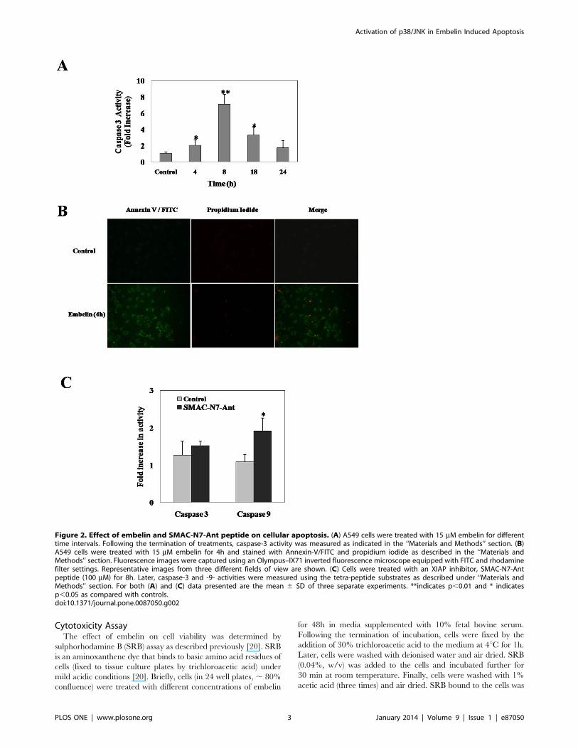
Figure 2. Effect of embelin and SMAC-N7-Ant peptide on cellular apoptosis. (A) A549 cells were treated with 15 mM embelin for differenttime intervals. Following the termination of treatments, caspase-3 activity was measured as indicated in the ‘‘Materials and Methods’’ section. (B)A549 cells were treated with 15 mM embelin for 4h and stained with Annexin-V/FITC and propidium iodide as described in the ‘‘Materials andMethods’’ section. Fluorescence images were captured using an Olympus–IX71 inverted fluorescence microscope equipped with FITC and rhodaminefilter settings. Representative images from three different fields of view are shown. (C) Cells were treated with an XIAP inhibitor, SMAC-N7-Antpeptide (100 mM) for 8h. Later, caspase-3 and -9- activities were measured using the tetra-peptide substrates as described under ‘‘Materials andMethods’’ section. For both (A) and (C) data presented are the mean 6 SD of three separate experiments. **indicates p,0.01 and * indicatesp,0.05 as compared with controls.doi:10.1371/journal.pone.0087050.g002
Activation of p38/JNK in Embelin Induced Apoptosis
PLOS ONE | www.plosone.org 3 January 2014 | Volume 9 | Issue 1 | e87050
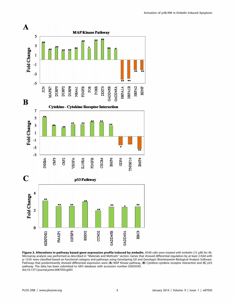
Figure 3. Alterations in pathway based gene expression profile induced by embelin. A549 cells were treated with embelin (15 mM) for 4h.Microarray analysis was performed as described in ‘‘Materials and Methods’’ section. Genes that showed differential regulation by at least 2-fold withp,0.05 were classified based on functional category and pathways using GeneSpring GX and Genotypic Biointerpreter-Biological Analysis Software.Pathways that predominantly showed differential expression were (A) MAP Kinase pathway, (B) Cytokine-cytokine receptor interaction and (C) p53pathway. The data has been submitted to GEO database with accession number GSE50545.doi:10.1371/journal.pone.0087050.g003
Activation of p38/JNK in Embelin Induced Apoptosis
PLOS ONE | www.plosone.org 4 January 2014 | Volume 9 | Issue 1 | e87050
solubilised in 10 mM Tris-base and absorbance was measured at
565 nm using EnSpire multimode plate reader (Perkin Elmer).
Caspase 3 and 9 AssayFollowing the termination of incubation, cellular caspase-3 and
-9- activities were measured using AFC conjugated tetrapeptide
substrates as described previously [21]. Briefly, cells were washed
with ice-cold DPBS and lysed in ice-cold lysis buffer (50 mM
HEPES pH 7.4, 5 mM CHAPS and 5 mM DTT) [22]. Lysates
were pelleted down at 12,000 g for 10 min at 4uC and
supernatants were collected. To the lysates equal volumes of assay
buffer (40 mM HEPES pH7.4, 0.2% CHAPS, 10 mM DTT,
4 mM EDTA) containing either caspase-3 substrate (Ac-DEVD-7-
AFC, 40 mM) or caspase-9 substrate (Ac-LEHD-7-AFC, 40 mM)
was added and incubated at 37uC. Increase in the fluorescence
readings due to the release of AFC was monitored for every 5 min
interval at lex 400 nm and lem 505 nm for 1h using EnSpire
multimode plate reader (Perkin Elmer). Protein estimation was
performed by Bradford’s method and the fluorescence units were
normalized to the total protein in the incubation mixture.
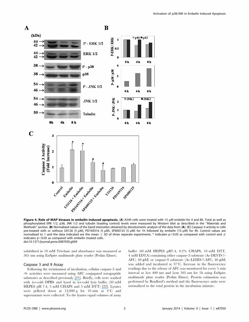
Figure 4. Role of MAP kinases in embelin induced apoptosis. (A) A549 cells were treated with 15 mM embelin for 4 and 8h. Total as well asphosphorylated ERK 1/2, p38, JNK 1/2 and tubulin (loading control) levels were measured by Western blot as described in the ‘‘Materials andMethods’’ section. (B) Normalized values of the band intensities obtained by densitometric analysis of the data from (A). (C) Caspase-3 activity in cellspre-treated with or without U0126 (5 mM), PD169316 (5 mM), SP600125 (5 mM) for 1h followed by embelin (15 mM) for 4h. Control values arenormalized to 1 and the data indicated are the mean 6 SD of three separate experiments. * indicates p,0.05 as compared with control and #indicates p,0.05 as compared with embelin treated cells.doi:10.1371/journal.pone.0087050.g004
Activation of p38/JNK in Embelin Induced Apoptosis
PLOS ONE | www.plosone.org 5 January 2014 | Volume 9 | Issue 1 | e87050
Annexin-V/FITC Analysis for ApoptosisA549 cells grown on coverslips in 6-well plates were treated with
embelin for 4h. After treatment, coverslips were washed twice with
PBS followed by 1X binding buffer. Cells were then stained with
annexin-V/FITC antibody by incubating in the dark for 30 min
and washed with 1X binding buffer to remove any unbound
antibody as per the Manufacturer’s protocol (Clontech Inc, USA).
Formaldehyde (2%) was added to fix the cells at the end of
incubation. Fluorescence was monitored using an Olympus-
IX71inverted microscope equipped with FITC and rhodamine
filter settings.
Gene Expression Profiling using MicroarrayA549 cells were treated with embelin for 4h. Following
treatments, RNA was isolated using Qiagen’s kit as per the
manufacturer’s instructions. The concentration and purity of the
RNA extracted were evaluated using the Nanodrop Spectropho-
tometer (Thermo Scientific). The integrity of the extracted RNA
was analyzed on the Bioanalyzer (Agilent). RNA was considered to
be of good quality based on the 260/280 values, rRNA 28S/18S
ratios and RNA integrity number (RIN). The samples were labeled
using Agilent Quick Amp Kit. 500 ng of total RNA was reverse
transcribed using oligo-dT primer tagged to T7 promoter
sequence. cDNA thus obtained was converted to double stranded
cDNA in the same reaction. Further the cDNA was converted to
cRNA in the in vitro transcription step using T7 RNA polymerase
enzyme and Cy3 dye was added into the reaction mix. cRNA
obtained was cleaned up using RNeasy columns (Qiagen Inc) and
the concentration and amount of dye incorporated was deter-
mined using Nanodrop. The specific activity for all the samples
greater than 8 pmol dye/mg cRNA were considered ideal for
hybridization. Labeled cRNA (600 ng) was hybridized on the
array (Custom Whole Genome Human 8660k designed by
Genotypic Technology Private Limited AMADID: 027114) using
the Gene Expression Hybridization kit in Sure hybridization
Chambers (Agilent) at 65uC for 16h. Hybridized slides were
washed using Gene Expression wash buffers. The hybridized,
washed microarray slides were then scanned on a microarray
scanner (G2505C, Agilent Technologies). Data extraction from
images was done using Feature Extraction software and images
were quantified (Version 10.7 of Agilent). Feature extracted raw
data was analyzed using GeneSpring GX Version 11.5 software
from Agilent. Normalization of the data was done in GeneSpring
GX using the 75th percentile shift. Significant genes up and down
regulated showing two-fold and above within the samples with
respect to control sample were identified. Statistical t-test p-value
was calculated based on Student’s t-test Algorithm. Genes were
classified based on functional category and pathways using
GeneSpring GX and Genotypic Biointerpreter-Biological Analysis
Software. The microarray data has been submitted to GEO
database with accession number GSE50545.
Intracellular ROS MeasurementReactive oxygen species generation in cells was determined by
carboxy-H2-DCFDA (Molecular Probes) as described previously
[23]. Following the termination of treatments, media was aspirated
and cells in 12-well plates were washed twice with DPBS. Serum
free media containing 10 mM carboxy-H2-DCFDA was added to
cells and incubated further at 37uC for 20 min. Finally, cells were
washed twice with DPBS before adding culture medium.
Intracellular fluorescence was monitored using an Olympus-
IX71inverted microscope equipped with FITC filter setting.
Western Blot AnalysisFollowing treatments, cells were washed with DPBS, gently
scraped and collected by brief centrifugation (300 g for 3 min) and
resuspended in 100 ml RIPA containing protease inhibitor cocktail
and sodium ortho-vanadate, 10 mM (Sigma). The resulting cell
suspension was passed through a 26 gauge needle 10 times to
ensure complete lysis. The lysate was centrifuged at 12000 g for
15 min at 4uC and the clear supernatants were collected in
separate tubes. Since all the antibodies employed are monoclonal,
instead of stripping and reprobing the immunoblots for total and
phospho-specific proteins, we have performed immunoblotting
separately in order to avoid any background signals. Following
protein estimation by Bradford’s method, proteins (25 mg) wereresolved on 10% SDS-PAGE and blotted on to a nitrocellulose
membrane. Blots were probed with monoclonal antibodies raised
against total and phospho specific antibodies for ERK 1/2, p38,
JNK (Cell Signaling Technology); and tubulin (Sigma-Aldrich).
Anti-rabbit Ig-G conjugated to HRP (GE Life Sciences) and anti-
mouse IgG conjugated to alkaline phosphatase (Sigma-Aldrich)
were employed as the secondary antibodies for the MAP kinase
and tubulin antibodies respectively. Bands were developed using
ECL Prime Western blotting reagent (GE, Life Sciences) or BCIP/
NBT reagent for tubulin (Sigma-Aldrich) and the band intensities
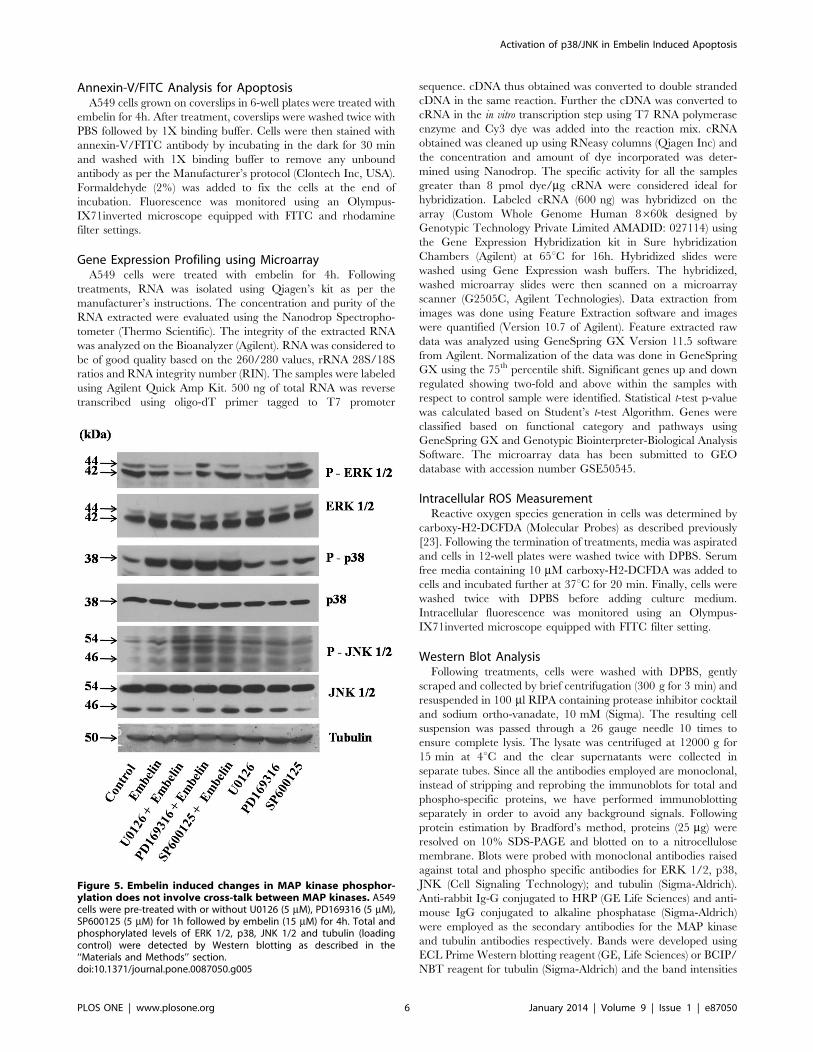
Figure 5. Embelin induced changes in MAP kinase phosphor-ylation does not involve cross-talk between MAP kinases. A549cells were pre-treated with or without U0126 (5 mM), PD169316 (5 mM),SP600125 (5 mM) for 1h followed by embelin (15 mM) for 4h. Total andphosphorylated levels of ERK 1/2, p38, JNK 1/2 and tubulin (loadingcontrol) were detected by Western blotting as described in the‘‘Materials and Methods’’ section.doi:10.1371/journal.pone.0087050.g005
Activation of p38/JNK in Embelin Induced Apoptosis
PLOS ONE | www.plosone.org 6 January 2014 | Volume 9 | Issue 1 | e87050
were calculated using GeneTools software (Syngene gel documen-
tation system).
Statistical AnalysisAll experiments were performed in triplicates and the results
were expressed as mean6S.D. Statistical significance was deter-
mined by Student’s t test using SIGMAPLOT software.
Results
Embelin Exhibits Enhanced Cytotoxicity in Cancer Cells asCompared to Normal CellsThe anti-proliferative activities of embelin were compared by
SRB assay in selected cancer and normal cell lines (Fig. 1). Cells
were subjected to increasing concentrations of embelin (2.5, 5, 10
and 25 mM) for 48h. Among the cancerous cells, embelin was
found to be more toxic to A549 cells with an IC50 value of 4.4 mMfollowed by DU145 and MCF7 with 6.31 and 10.66 mMrespectively. However, the observed IC50 values were compara-
tively less than the normal cell lines viz., MRC5, WPMY-1 and
H9c2 which demonstrated an IC50 value of 24.4, 10.9 and
23.4 mM respectively. The difference between the observed IC50
values of lung cancer and normal cells (4.4460.76 and
24.4465.32 mM) appeared to be more significant. As A549 cells
exhibited enhanced sensitivity towards embelin, all further studies
have been carried out using this cell line for understanding the
mode of action of embelin to gain novel insights to selectively
target lung cancer cells as compared to their normal cell
counterpart. Hence, in order to identify the early apoptotic phase,
we have analyzed the time dependent effect of embelin on cellular
caspase-3 activity in A549 cells (Fig. 2A). Embelin (15 mM)
induced nearly 2-fold increase in the caspase-3 activity as early as
4h time period which further increased upto 6-fold by 8h (Fig. 2A).
Annexin-V/FITC staining of cells treated with embelin (4h)
clearly indicates the early apoptotic stage of cells as they were
stained only with annexin-V but not with propidium iodide
(Fig. 2B). However, at later time points i.e., 18 and 24h, the
caspase-3 activities decreased nearly to 4 and 2-fold respectively
indicating that apoptotic cells may have completely died by the
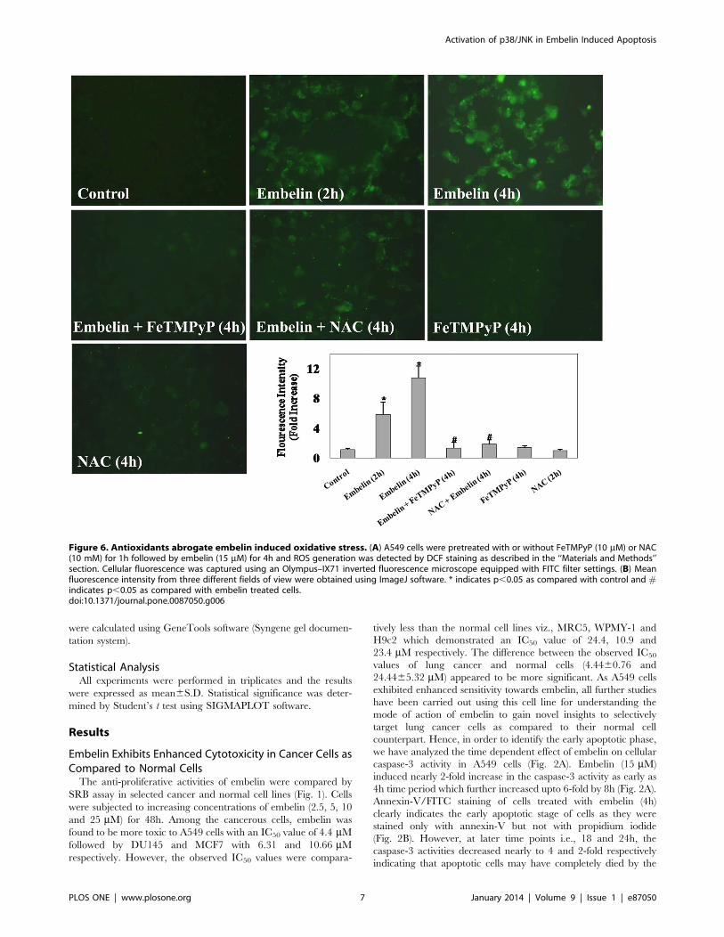
Figure 6. Antioxidants abrogate embelin induced oxidative stress. (A) A549 cells were pretreated with or without FeTMPyP (10 mM) or NAC(10 mM) for 1h followed by embelin (15 mM) for 4h and ROS generation was detected by DCF staining as described in the ‘‘Materials and Methods’’section. Cellular fluorescence was captured using an Olympus–IX71 inverted fluorescence microscope equipped with FITC filter settings. (B) Meanfluorescence intensity from three different fields of view were obtained using ImageJ software. * indicates p,0.05 as compared with control and #indicates p,0.05 as compared with embelin treated cells.doi:10.1371/journal.pone.0087050.g006
Activation of p38/JNK in Embelin Induced Apoptosis
PLOS ONE | www.plosone.org 7 January 2014 | Volume 9 | Issue 1 | e87050
end of 18 or 24h or there could also be a possibility of multiple cell
death mechanisms due to the cross-talk between various signalling
mechanisms.
As embelin is known to inhibit XIAP by binding to the BIR3
domain similar to that of SMAC, we next examined whether the
observed affects of embelin on cellular apoptosis could be
demonstrated by a cell permeable SMAC-N7-Ant peptide
(comprised of mature SMAC’s amino terminal 7 amino acid
peptide - AVPIAQK bound to Ant peptide -RQI-
KIWFQNRRMKWKK, for cell permeability by a proline linker)
which is known to specifically interact with BIR3 domain of XIAP
[24,25]. Results demonstrate that treatment of A549 cells with
SMAC-N7-Ant peptide (100 mM for 8h) increased cellular
caspase-9 activity to nearly two-fold with respect to untreated
control, however, no significant increase in caspase-3 activity was
observed (Fig. 2C). Though the observed effects of SMAC-N7-Ant
peptide are in accordance with earlier report [24], lack of caspase-
3 activation with SMAC-N7-Ant peptide, but not with embelin,
prompted us to investigate the responsible pathways mediating
embelin induced apoptosis for gaining novel insights into its
mechanism of action.
Alteration in Gene Expression Profile by EmbelinIn order to identify the pathways responsible in embelin induced
apoptosis, we have analyzed the altered gene expression profile in
A549 cells treated with embelin (15 mM for 4h). A total of 215
upregulated and 80 downregulated genes were identified which
showed at least two-fold difference over controls with a
significance of p,0.05.
Classification of these genes based on functional category and
pathways using GeneSpring GX and Genotypic Biointerpreter-
Biological analysis software indicated that the upregulated genes
majorly belong to five different pathways (with at least 4 altered
genes in each pathway) and the number of genes altered are in the
order of Wnt (4 genes),Focal adhesion (5 genes) ,p53 (8
genes),cytokine-cytokine receptor interaction (11 genes),MAP
kinase pathway (16 genes) (Fig. 3 A–C). Amongst the downreg-
ulated genes with at least three genes in each pathway possessing a
2-fold change with a significance of p,0.05 belonged to cytokine-
cytokine receptor and MAP kinase pathway (Fig. 3A and B).
At a glance, the obtained results indicate that amongst the
altered pathways, MAP kinase signalling pathway appears to be
more predominant and significantly affected with nearly 16 altered
genes and many of the genes are either upstream or downstream
to p38/JNK/ERK pathway such as p53 or regulators of these
pathway (Fig. 3A). From the functional point of view, the
phosphorylation status of the identified proteins (such as, DUSPs,
GADD45A/B, p53 etc) but not merely their transcript levels
dictate the cellular fate. Nevertheless, the results obtained
indicated the substantial role of MAP kinase signalling and
inspired us to focus on the involvement of MAP kinase pathway in
embelin induced apoptosis.
Embelin Induced Activation of p38 and JNK PathwayBased on the initial clues obtained from microarray studies on
the potential role of MAP kinase pathway in embelin induced
apoptosis, we sought to investigate further on the involvement of
MAPK signalling and monitored embelin induced alterations in
the phosphorylation status of ERK, p38 and JNK proteins (Fig. 4).
Results as shown in figure-3 indicate that embelin (15 mM)
induced the phosphorylation of p38 to nearly 2.5 and 3-fold by 4
and 8h respectively. Phospho-JNK 1/2 levels were also increased
to 1.2 and 1.9 fold respectively by 8h. However, under similar
treatment conditions there was a significant decrease in the
phosphorylation status of ERK 1/2 (p42 and p44) and the values
were found to be 0.3 and 0.2 fold less than that of controls (Fig. 4A
and B). In order to understand whether the changes in the
phosphorylation status of these MAP kinase proteins has any
relevance to the observed apoptosis, we have pretreated cells for
1h individually with specific inhibitors for p38 (PD169316), JNK
(SP600125) and MEK (U0126) at 5 mM concentration followed by
embelin (15 mM) for 4h. Embelin-induced caspase-3 activity was
significantly inhibited by both p38 and JNK inhibitors to nearly
control values (Fig. 4C). However, under similar experimental
conditions MEK inhibitor (U0126) did not exhibit any protective
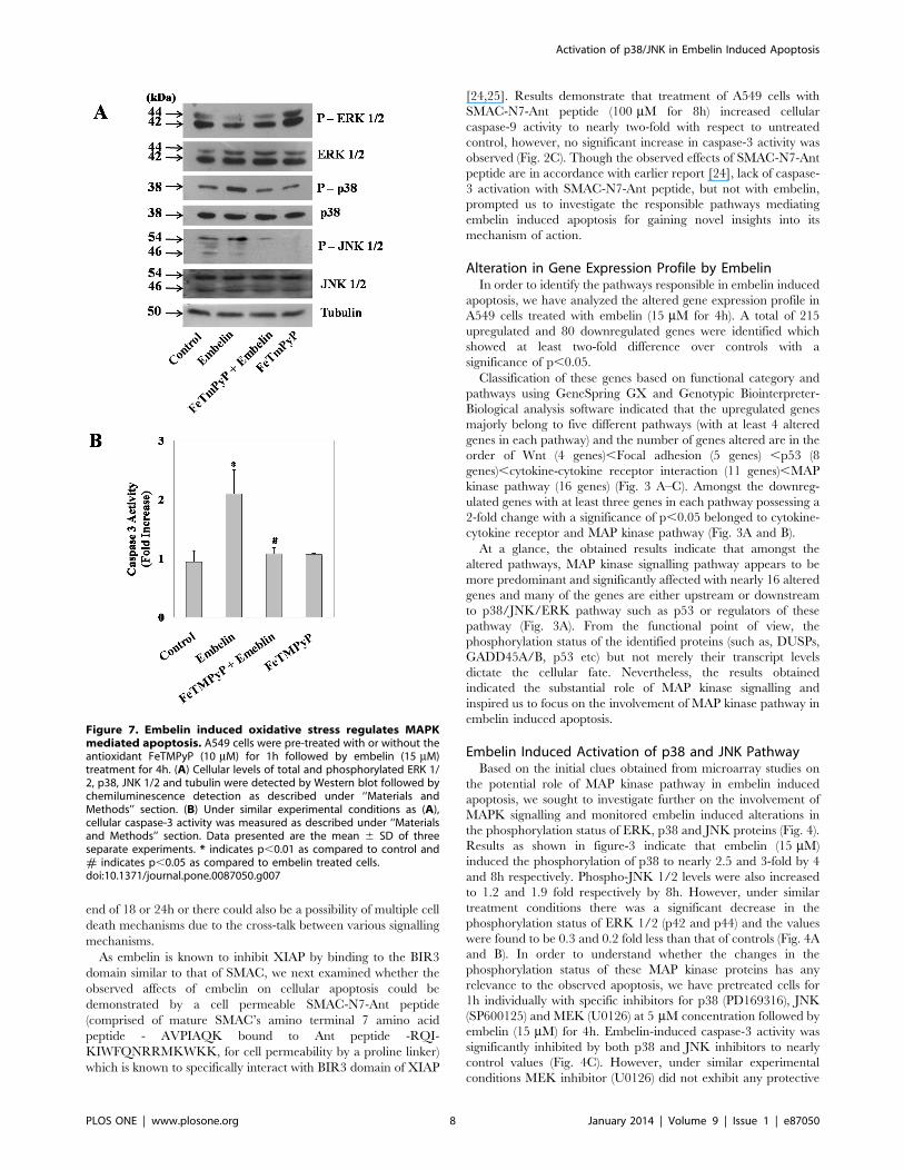
Figure 7. Embelin induced oxidative stress regulates MAPKmediated apoptosis. A549 cells were pre-treated with or without theantioxidant FeTMPyP (10 mM) for 1h followed by embelin (15 mM)treatment for 4h. (A) Cellular levels of total and phosphorylated ERK 1/2, p38, JNK 1/2 and tubulin were detected by Western blot followed bychemiluminescence detection as described under ‘‘Materials andMethods’’ section. (B) Under similar experimental conditions as (A),cellular caspase-3 activity was measured as described under ‘‘Materialsand Methods’’ section. Data presented are the mean 6 SD of threeseparate experiments. * indicates p,0.01 as compared to control and# indicates p,0.05 as compared to embelin treated cells.doi:10.1371/journal.pone.0087050.g007
Activation of p38/JNK in Embelin Induced Apoptosis
PLOS ONE | www.plosone.org 8 January 2014 | Volume 9 | Issue 1 | e87050
effect against embelin induced apoptosis and also no significant
increase in caspase-3 activity was observed in cells treated with
inhibitors alone (Fig. 4C). The observed changes clearly indicate
that alterations in the phosphorylation status of both p38 and JNK
appears to be crucial in embelin induced apoptosis.
In order to determine whether the observed alterations in the
MAPK phosphorylation are because of embelin treatment alone
or due to the regulatory effect of one MAP kinase over the other
MAPK’s, we have treated the cells individually with embelin
(15 mM) in the presence and absence of MEK inhibitor (U0126,
5 mM) or p38 inhibitor (PD169316, 5 mM) or JNK inhibitor
(SP600125, 5 mM) for 4h and analyzed the phosphorylation levels
of all the three MAP kinases (Fig. 5). The MEK inhibitor U0126
inhibits its downstream target ERK. p38 inhibitor, PD169316 and
JNK inhibitor, SP600125 specifically inhibit p38 and JNK activity
respectively by competitively binding to the ATP binding pockets
preventing the phosphorylation of proteins downstream, but, as
such does not result in the decreased phosphorylation levels of
either p38 or JNK [26,27]. Results indicate that treatment of cells
with the MEK inhibitor (U0126) inhibited phospho-ERK 1/2, but
did not alter the levels of embelin induced phospho-p38 and
phospho-JNK levels. Similarly, treatment of cells with p38
inhibitor (PD169316) did not affect the levels of phospho-JNK
and phospho-ERK caused by embelin. Also, treatment of cells
with JNK inhibitor (SP600125) did not affect the levels of
phospho-p38 and phospho-ERK in the presence or absence of
embelin (Fig. 5). The above results indicate that the observed
changes in the phosphorylation levels of p38, JNK and ERK
appears to be directly mediated by embelin treatment, but not due
to the cross-talk between the MAP kinases.
ROS Mediates MAP Kinase Regulation by EmbelinMAPK proteins are known to be regulated by oxidative stress
[28]. Moreover, the benzoquinone structure of embelin has been
demonstrated to form semiquinone radical by redox mechanism
which eventually leads to reactive oxygen species generation
[13,29]. These observations suggest an important role for ROS in
embelin induced apoptosis. To evaluate the pro-oxidant properties
of embelin, we studied its effects on the generation of oxidative
stress in A549 cells. The intracellular ROS generated by embelin
was detected by an enhancement in the intracellular fluorescence
of DCF (Fig. 6). Embelin (15 mM) induced ROS generation in a
time dependent manner with nearly 5 and 10-fold increase over
untreated controls by the end of 2 and 4h respectively (Fig. 6).
Pretreatment of cells with the antioxidant, FeTMPyP (10 mM) or
N-acetyl-L-cysteine (NAC) (10 mM) significantly inhibited embe-
lin-induced DCF staining to that of control values (Fig. 6).
We further assessed the effect of embelin-induced ROS on
MAPK signalling in the presence and absence of the antioxidant,
FeTMPyP (Fig. 7). Results indicate that embelin induced ROS is
responsible for the observed alterations in the phospho-protein
levels of p38, JNK and ERK 1/2 as pretreatment of cells with
FeTMPyP nullified this effect (Fig. 7A). In accordance with the
above results, pretreatment of cells with FeTMPyP (10 mM) also
inhibited the apoptotic effects of embelin indicating that altered
MAP kinase signalling due to enhanced ROS plays a pivotal role
in embelin-induced apoptosis (Fig. 7B).
Discussion
In the present study, we report that oxidative stress induced
MAPK signalling plays an important role in embelin induced
apoptosis. Analysis of gene expression profiling by microarray
studies indicated the possible involvement of MAP kinase pathway
in A549 cells treated with embelin for 4h. Pretreatment of cells
with specific inhibitors of either p38 or JNK significantly inhibited
embelin induced caspase-3 activation as well as nullified embelin-
induced alterations in phosphorylation levels of p38, JNK and
ERK 1/2 MAP kinases. Reactive oxygen species (ROS) appears
to play a pivotal role between embelin and MAP kinase pathway.
All the observed effects of embelin are not due to the inhibition of
XIAP as treatment of cells with cell permeable SMAC-N7-Ant
peptide, which binds to the BIR3 domain of XIAP did not affect
cellular caspase-3 activation.
The natural product, embelin, has been paid more attention in
recent times for its anticancer properties. More importantly, it has
been demonstrated to have more selectivity towards cancer cells as
compared to the normal cells (6). Even in the present work, we
observed a similar trend and a significant difference in the IC50
values of embelin was evident between lung cancer and normal
cell lines (Fig. 1). Though embelin was initially identified to be an
inhibitor of XIAP by means of its interaction at BIR3 domain,
subsequent studies demonstrated the direct in vitro effects of
embelin on the oxidative phosphorylation of mitochondria,
inhibition of 5-lipoxygenase (5-LO) and microsomal prostaglandin
E2 synthase-1 (mPGES)-1 and inactivation of plasminogen
activator inhibitor-1 (PAI-1) [10,11]. However, identification of
the primary intracellular target which is responsible for the
anticancer property of embelin might eventually help in the
structural refinement of embelin for improving its efficacy and
selectivity.
Recently, various studies have been carried out to understand
the mode of action of embelin and it has been demonstrated to
have a role in the inactivation of NF-kB, inhibition of STAT3
signalling via protein tyrosine phosphatase PTEN, lysosomal
destabilization and AKT and mTOR pathways [8,9,15,30,31].
However, whether all the observed effects are interdependent or
independent of each other is not yet clear as many of the reported
experiments were carried out at a fixed duration of either 24 or
48h [8,10,16,17].
Data from microarray studies during the early stages of embelin
induced apoptosis pointed us to the changes in the regulation of
transcription factors downstream to MAPK proteins (Fig. 3). In
the present study, we have identified a prominent role of MAP
kinase pathway, (increased levels of phospho-p38 and phospho-
JNK) in embelin-induced apoptosis. All the three MAP kinases are
regulated independently by embelin/embelin-induced ROS as
none of the specific inhibitors for individual MAP kinases affected
the phosphorylation status of other MAP kinases (Fig. 4).
MAPK proteins play a key role in cellular events affecting
diverse end points including cell proliferation, differentiation, cell
survival and cell death [32]. Phosphorylation of ERK 1/2
decreased in time dependent fashion with embelin treatment
(Fig. 4A). ERK 1/2 is activated in response to growth stimuli in
cancer and targeting it directly or indirectly is known to cause
tumour cell death [32,33]. In addition, embelin also induced
significant elevation in the phosphorylation of p38 and JNK 1/2.
JNK, also referred as stress activated protein kinase, is activated by
various stress stimuli like changes in osmolarity or metabolism,
DNA damage, heat shock, inflammatory cytokines, shear stress,
UV irradiation or oxidative stress [32]. p38 in most cases is
activated simultaneously with JNK [32]. The anti-apoptotic effects
of ERK 1/2 and pro-apoptotic effects of p38/JNK are already
described [34]. In accordance with these earlier reports, p38 and
JNK inhibitors (PD169316 and SP600125) abrogated embelin-
induced apoptosis, while MEK inhibitor (U0126) did not show any
significant effect (Fig. 4C). However, these events involving
simultaneous down-regulation in the phospho-ERK levels and
Activation of p38/JNK in Embelin Induced Apoptosis
PLOS ONE | www.plosone.org 9 January 2014 | Volume 9 | Issue 1 | e87050
concomitant activation of p38/JNK pathways during embelin
mediated apoptosis are regulated independent of each other
(Fig. 5).
Embelin is a benzoquinone with an aliphatic chain which has
quinone and hydroquinone groups on the aromatic ring. Because
of which, it can either be oxidised or reduced to form a
semiquinone radical [13]. Recently, embelin has also been shown
to generate intracellular ROS [29]. Even the present study
demonstrates an enhancement in cellular ROS generated by
embelin as early as 4h and pretreatment of cells with the
antioxidants abrogated this effect as well as embelin-induced
alterations in phosphorylation status of MAP kinases and apoptosis
(Fig. 6 & 7). Very similar to our findings, Allensworth et al.,
reported that SOD mimic MnTnHex-2-PyP5+ reversed the toxic
effects of embelin when treated in combination with TRAIL in
XIAP overexpressed SUM149 cells [29]. Moreover, the lack of
cellular caspase-3 activation by SMAC-N7-Ant peptide as shown
in figure 2B, clearly indicates that the observed effects of embelin
on MAP kinase mediated apoptosis is independent of its
interaction at the BIR3 domain of XIAP.
Overall, the present study identified the role of ROS induced
alterations in the MAPK signalling pathway, especially activation
of p38 and JNK, as responsible mediators in embelin induced
apoptosis. The observed effects are not mediated by XIAP
inhibition alone as treatment of cells with a known XIAP’s BIR3
domain inhibitor, i.e., SMAC-N7-Ant peptide did not replicate
embelin-induced apoptosis. Further studies aimed at enhancing
the apoptotic potential or selectivity of embelin towards cancer cell
lines in combination with MAP kinase modulators or their
downstream targets might lead to novel therapeutic strategies as
well as improve the therapeutic efficacy of embelin.
Author Contributions
Conceived and designed the experiments: SVK DRA. Performed the
experiments: DRA. Analyzed the data: DRA. Contributed reagents/
materials/analysis tools: SVK KSB. Wrote the paper: SVK DRA.
References
1. Chitra M, Sukumar E, Suja V, Devi CS (1994) Antitumor, anti-inflammatoryand analgesic property of embelin, a plant product. Chemotherapy 40 (2): 109–
113.
2. Gandhi GR, Stalin A, Balakrishna K, Ignacimuthu S, Paulraj MG, et al. (2013)Insulin sensitization via partial agonism of PPARc and glucose uptake through
translocation and activation of GLUT4 in PI3K/p-AKT signalling pathway byembelin in type 2 diabetic rats. Biochim Biophys Acta. 1830 (1): 2243–2255.
3. Chaudhari HS, Bhandari U, Khanna G (2012) Preventive effects of embelin
from Embelia ribes on lipid metabolism and oxidative stress in high-fat diet-induced obesity in rats. Planta Med. 78(7): 651–657..
4. Johri RK, Pahwa GS, Sharma SC, Zutshi U (1991) Determination ofestrogenic/antiestrogenic potential of antifertility substances using rat uterine
peroxidise assay. Contraception 44 (5): 549–557.5. Githiori JB, Hoglund J, Waller PJ, Leyden Baker R (2003) Evaluation of
anthelmintic properties of extracts from some plants used as livestock dewormers
by pastoralist and smallholder farmers in Kenya against Heligmosomoides polygyrusinfections in mice. Vet Parasitol. 118(3–4): 215–226.
6. Nikolovska-Coleska Z, Xu L, Hu Z, Tomita Y, Li P, et al. (2004) Discovery ofembelin as a cell-permeable, small-molecular weight inhibitor of XIAP through
structure-based computational screening of a traditional herbal medicine three-
dimensional structure database. J Med Chem. 2004 47(10): 2430–2440.7. Lu J, Huang Y, Zhao W, Marquez RT, Meng X, et al. (2013) PEG-derivatized
embelin as a nanomicellar carrier for delivery of paclitaxel to breast and prostatecancers. Biomaterials 34(5): 1591–1600.
8. Park SY, Lim SL, Jang HJ, Lee JH, Um JY, et al. (2013) Embelin induces
apoptosis in human glioma cells through inactivating NF-kB. J Pharmacol Sci.121(3): 192–199.
9. Heo JY, Kim HJ, Kim SM, Park KR, Park SY, et al. (2011) Embelin suppressesSTAT3 signalling, proliferation, and survival of multiple myeloma via the
protein tyrosine phosphatase PTEN. Cancer Lett. 308(1): 71–80.10. Schaible AM, Traber H, Temml V, Noha SM, Filosa R, et al. (2013) Potent
inhibition of human 5-lipoxygenase and microsomal prostaglandin E2 synthase-
1 by the anti-carcinogenic and anti-inflammatory agent embelin. BiochemPharmacol. 86 (4): 476–486.
11. Modak R, Basha J, Bharathy N, Maity K, Mizar P, et al. (2013) Probing p300/CBP Associated Factor (PCAF)-Dependent Pathways with a Small Molecule
Inhibitor. ACS Chem Biol. May 9. doi: 10.1021/cb4000597 [Epub ahead of
print].12. Lin Z, Jensen JK, Hong Z, Shi X, Hu L, et al. (2013) Structural insight into
inactivation of plasminogen activator inhibitor-1 by a small-molecule antagonist.Chem Biol. 20 (2): 253–261.
13. Joshi R, Ghanty TR, Mukherjee T (2009) Formation of semiquinone radical inthe reaction of embelin (2,5-dihydroxy-3-undecyl-1,4-benzoquinone) with
reductants as well as oxidants. Characterization by pulse radiolysis and structure
investigation by quantum chemical study. J Mol Struct. 928: 46–53.14. Makawiti DW, Konji VN, Olowookere JO (1990) Interaction of benzoquinones
with mitochondria interferes with oxidative phosphorylation characteristics.FEBS Lett. 266(1–2): 26–28.
15. Joy B, Sivadasan R, Abraham TE, John M, Sobhan PK, et al. (2010) Lysosomal
destabilization and cathepsin B contributes for cytochrome c release and caspaseactivation in embelin-induced apoptosis. Mol Carcinog. 49(4): 324–336.
16. Hussain AR, Uddin S, Ahmed M, Bu R, Ahmed SO, et al. (2010) Prognosticsignificance of XIAP expression in DLBCL and effect of its inhibition on AKT
signalling. J Pathol. 222(2): 180–190.
17. Dai Y, Qiao L, Chan KW, Yang M, Ye J, et al. (2009) Peroxisome proliferator-
activated receptor-gamma contributes to the inhibitory effects of Embelin on
colon carcinogenesis. Cancer Res. 69(11): 4776–4783.
18. Chitra M, Devi CS, Viswanathan S, Sukumar E (2003) Antibacterial activity of
embelin. Fitoterapia 74: 401–403.
19. Chitra M, Devi CS, Viswanathan S, Sukumar E (2003) Effect of embelin on lipid
profile in transplanted fibrosarcoma in rats. Indian J. Pharmcol. 35: 241–244.
20. Vichai V, Kirtikira K (2006) Sulphorhodamine B colorimetric assay for
cytotoxicity screening. Nat. Protoc. 1(3): 1112–1116.
21. Gurtu V, Kain SR, Zhang G (1997) Fluorometric and colorimetric detection of
caspase activity associated with apoptosis. Anal Biochem. 251(1): 98–102.
22. Dhanasekaran A, Kotamraju S, Kalivendi SV, Matsunaga T, Shang T, et al.
(2004) Supplementation of endothelial cells with mitochondria-targeted
antioxidants inhibit peroxide-induced mitochondrial iron uptake, oxidative
damage, and apoptosis. J Biol Chem. 279(36): 37575–37587.
23. Kalivendi SV, Konorev EA, Cunningham S, Vanamala SK, Kaji EH, et al.
(2005) Doxorubicin activates nuclear factor of activated T lymphocytes and Fas
ligand transcription: role of mitochondrial reactive oxygen species and calcium.
Biochem J. 389 (Pt 2): 527–539.
24. Fandy TE, Shankar S, Srivastava RK (2008) Smac/DIABLO enhances the
therapeutic potential of chemotherapeutic drugs and irradiation, and sensitizes
TRAIL-resistant breast cancer cells. Mol. Cancer 7: 60.
25. Konorev EA, Vanamala S, Kalyanaraman B (2008) Differences in doxorubicin-
induced apoptotic signalling in adult and immature cardiomyocytes. Free Radic
Biol Med. 45 (12): 1723–1728.
26. Bennett BL, Sasaki DT, Murray BW, O’Leary EC, Sakata ST, et al. (2001)
SP600125, an anthrapyrazolone inhibitor of Jun N- terminal kinase. Proc Natl
Acad Sci U S A. 98(24): 13681–13686.
27. Kayali AG, Austin DA, Webster NJ (2000) Stimulation of MAPK cascades by
insulin and osmotic shock: lack of an involvement of p38 mitogen-activated
protein kinase in glucose transport in 3T3-L1 adipocytes. Diabetes 49(11): 1783–
1793.
28. Trachootham D, Lu W, Ogasawara MA, Nilsa RD, Huang P (2008) Redox
regulation of cell survival. Antioxid Redox Signal. 10(8): 1343–1374.
29. Allensworth JL, Aird KM, Aldrich AJ, Batinic-Haberle I, Devi GR (2012) XIAP
inhibition and generation of reactive oxygen species enhances TRAIL sensitivity
in inflammatory breast cancer cells. Mol Cancer Ther. 11(7): 1518–1527.
30. Ahn KS, Sethi G, Aggarwal BB (2007) Embelin, an inhibitor of X chromosome-
linked inhibitor-of-apoptosis protein, blocks nuclear factor-kB (NF-kB) signalingpathway leading to suppression of NF-kB-regulated antiapoptotic and metastatic
gene products. Mol Pharmacol. 71(1): 209–219.
31. Kim SW, Kim SM, Bae H, Nam D, Lee JH, et al. (2013) Embelin inhibits
growth and induces apoptosis through the suppression of Akt/mTOR/S6K1
signaling cascades. Prostate 73(3): 296–305.
32. Wada T, Penninger JM (2004) Mitogen-activated protein kinases in apoptosis
regulation. Oncogene 23: 2838–2849.
33. Balmanno K, Cook SJ (2009) Tumour cell survival signalling by the ERK 1/2
pathway. Cell Death Differ. 2009; 16(3): 368–77.
34. Xia Z, Dickens M, Raingeaud J, Davis RJ, Greenberg ME (1995) Opposing
effects of ERK and JNK-p38 MAP kinases on apoptosis. Science 270(5240):
1326–1331.
Activation of p38/JNK in Embelin Induced Apoptosis
PLOS ONE | www.plosone.org 10 January 2014 | Volume 9 | Issue 1 | e87050
Related Documents


