JOURNAL OF VIROLOGY, Oct. 2009, p. 10761–10769 Vol. 83, No. 20 0022-538X/09/$08.000 doi:10.1128/JVI.00770-09 Copyright © 2009, American Society for Microbiology. All Rights Reserved. Activation of MDA5 Requires Higher-Order RNA Structures Generated during Virus Infection Andreas Pichlmair, 1,2 Oliver Schulz, 1 Choon-Ping Tan, 1 † Jan Rehwinkel, 1 Hiroki Kato, 3 Osamu Takeuchi, 3 Shizuo Akira, 3 Michael Way, 4 Giampietro Schiavo, 5 and Caetano Reis e Sousa 1 * Immunobiology Laboratory, Cancer Research UK, London Research Institute, London, United Kingdom 1 ; Center for Molecular Medicine of the Austrian Academy of Sciences, Vienna, Austria 2 ; Department of Host Defense, Research Institute for Microbial Diseases, Osaka University, 3-1 Yamada-oka, Osaka 565-0871, Japan 3 ; Cell Motility Laboratory, Cancer Research UK, London Research Institute, London, United Kingdom 4 ; and Molecular Neuropathobiology Laboratory, Cancer Research UK, London Research Institute, London, United Kingdom 5 Received 16 April 2009/Accepted 30 July 2009 Recognition of virus presence via RIG-I (retinoic acid inducible gene I) and/or MDA5 (melanoma differen- tiation-associated protein 5) initiates a signaling cascade that culminates in transcription of innate response genes such as those encoding the alpha/beta interferon (IFN-/) cytokines. It is generally assumed that MDA5 is activated by long molecules of double-stranded RNA (dsRNA) produced by annealing of comple- mentary RNAs generated during viral infection. Here, we used an antibody to dsRNA to show that the presence of immunoreactivity in virus-infected cells does indeed correlate with the ability of RNA extracted from these cells to activate MDA5. Furthermore, RNA from cells infected with encephalomyocarditis virus or with vaccinia virus and precipitated with the anti-dsRNA antibody can bind to MDA5 and induce MDA5-dependent IFN-/ production upon transfection into indicator cells. However, a prominent band of dsRNA apparent in cells infected with either virus does not stimulate IFN-/ production. Instead, stimulatory activity resides in higher-order structured RNA that contains single-stranded RNA and dsRNA. These results suggest that MDA5 activation requires an RNA web rather than simply long molecules of dsRNA. The innate immune response to virus infection is largely dependent on type I (alpha/beta) interferons (IFN-/). IFN- / induces expression of IFN-stimulated genes that have di- verse antiviral properties, including sequestration of virus pro- teins, blocking of cellular translation, and degradation of viral and cellular RNA (12, 13, 21). It is believed that viral genomes and replication products are the main triggers of the key pat- tern recognition receptors (PRRs) that sense virus infection and that signal for IFN-/ induction. PRRs known to induce IFN-/ in response to viruses include Toll-like receptor 3 (TLR-3), TLR-7/TLR-8, and TLR-9. These TLRs are re- stricted in distribution to immune cells and a few nonimmune cell types and are activated by double-stranded RNA (dsRNA), single-stranded RNA (ssRNA), and DNA delivered into endosomes during the infection process (8). Most cells rely on another set of PRRs, the retinoic acid-inducible gene I (RIG-I)-like receptors (RLRs), to sense RNA that accumu- lates in the cytoplasm during infection with many viruses (21). Two RLR members are known to signal for IFN-/ induction: RIG-I and MDA5 (melanoma differentiation-associated pro- tein 5) (4, 11, 32). Both proteins contain an RNA binding DEAD-box helicase domain and tandem caspase recruitment domains. The caspase recruitment domains are necessary for downstream signaling via shared adaptor MAVS (the mito- chondrial antiviral signaling protein; also called CARDIF, IPS-1, or VISA) (20). Notably, some viruses such as Dengue virus and West Nile virus are sensed by both RIG-I and MDA5 such that loss of either RLR is redundant for IFN-/ re- sponses (24). However, RIG-I is nonredundant for responses to many negative-strand RNA viruses such as influenza virus and Sendai virus and some positive-strand RNA viruses such as Japanese encephalitis virus (11). In contrast, MDA5 is essen- tial for responses to picornaviruses (4, 11). These data suggest that although RIG-I and MDA5 are similar in sequence and signal via a conserved pathway, they are activated by distinct RNA species. Indeed, we along with others could show that RIG-I but not MDA5 is activated by 5 triphosphorylated RNA such as that present in the genomes of influenza virus and other negative-strand RNA viruses (7, 22). Interestingly, picornaviruses do not have triphosphorylated RNA genomes (23), which may explain why they do not activate RIG-I. How- ever, the picornavirus-derived agonist for MDA5 has not been defined, and it is unclear why MDA5 agonists are generated during infection with picornaviruses but not influenza A virus and some other RNA viruses. One possible explanation is that MDA5 is activated by long dsRNA, which is made during infection with positive-strand RNA viruses (including picorna- viruses) and DNA viruses but not with negative-strand RNA viruses such as influenza virus (22, 28). Consistent with this notion, MDA5 is activated by poly(I:C), a synthetic RNA that is often described as an equivalent of long dsRNA. Notably, Kato et al. recently showed that MDA5 can be activated by * Corresponding author. Mailing address: Immunobiology Labora- tory, Cancer Research UK, London Research Institute, Lincoln’s Inn Fields Laboratories, 44 Lincoln’s Inn Fields, London WC2A 3PX, United Kingdom. Phone: 44 20 7269 2832. Fax: 44 20 7269 2833. E-mail: [email protected]. † Present address: Sir William Dunn School of Pathology, University of Oxford, South Parks Road, Oxford, United Kingdom. Published ahead of print on 5 August 2009. 10761 on May 26, 2016 by guest http://jvi.asm.org/ Downloaded from

Welcome message from author
This document is posted to help you gain knowledge. Please leave a comment to let me know what you think about it! Share it to your friends and learn new things together.
Transcript

JOURNAL OF VIROLOGY, Oct. 2009, p. 10761–10769 Vol. 83, No. 200022-538X/09/$08.00�0 doi:10.1128/JVI.00770-09Copyright © 2009, American Society for Microbiology. All Rights Reserved.
Activation of MDA5 Requires Higher-Order RNA StructuresGenerated during Virus Infection�
Andreas Pichlmair,1,2 Oliver Schulz,1 Choon-Ping Tan,1† Jan Rehwinkel,1Hiroki Kato,3 Osamu Takeuchi,3 Shizuo Akira,3 Michael Way,4
Giampietro Schiavo,5 and Caetano Reis e Sousa1*Immunobiology Laboratory, Cancer Research UK, London Research Institute, London, United Kingdom1; Center for Molecular Medicine of the
Austrian Academy of Sciences, Vienna, Austria2; Department of Host Defense, Research Institute for Microbial Diseases,Osaka University, 3-1 Yamada-oka, Osaka 565-0871, Japan3; Cell Motility Laboratory, Cancer Research UK,
London Research Institute, London, United Kingdom4; and Molecular Neuropathobiology Laboratory,Cancer Research UK, London Research Institute, London, United Kingdom5
Received 16 April 2009/Accepted 30 July 2009
Recognition of virus presence via RIG-I (retinoic acid inducible gene I) and/or MDA5 (melanoma differen-tiation-associated protein 5) initiates a signaling cascade that culminates in transcription of innate responsegenes such as those encoding the alpha/beta interferon (IFN-�/�) cytokines. It is generally assumed thatMDA5 is activated by long molecules of double-stranded RNA (dsRNA) produced by annealing of comple-mentary RNAs generated during viral infection. Here, we used an antibody to dsRNA to show that the presenceof immunoreactivity in virus-infected cells does indeed correlate with the ability of RNA extracted from thesecells to activate MDA5. Furthermore, RNA from cells infected with encephalomyocarditis virus or with vacciniavirus and precipitated with the anti-dsRNA antibody can bind to MDA5 and induce MDA5-dependent IFN-�/�production upon transfection into indicator cells. However, a prominent band of dsRNA apparent in cellsinfected with either virus does not stimulate IFN-�/� production. Instead, stimulatory activity resides inhigher-order structured RNA that contains single-stranded RNA and dsRNA. These results suggest that MDA5activation requires an RNA web rather than simply long molecules of dsRNA.
The innate immune response to virus infection is largelydependent on type I (alpha/beta) interferons (IFN-�/�). IFN-�/� induces expression of IFN-stimulated genes that have di-verse antiviral properties, including sequestration of virus pro-teins, blocking of cellular translation, and degradation of viraland cellular RNA (12, 13, 21). It is believed that viral genomesand replication products are the main triggers of the key pat-tern recognition receptors (PRRs) that sense virus infectionand that signal for IFN-�/� induction. PRRs known to induceIFN-�/� in response to viruses include Toll-like receptor 3(TLR-3), TLR-7/TLR-8, and TLR-9. These TLRs are re-stricted in distribution to immune cells and a few nonimmunecell types and are activated by double-stranded RNA(dsRNA), single-stranded RNA (ssRNA), and DNA deliveredinto endosomes during the infection process (8). Most cellsrely on another set of PRRs, the retinoic acid-inducible gene I(RIG-I)-like receptors (RLRs), to sense RNA that accumu-lates in the cytoplasm during infection with many viruses (21).Two RLR members are known to signal for IFN-�/� induction:RIG-I and MDA5 (melanoma differentiation-associated pro-tein 5) (4, 11, 32). Both proteins contain an RNA bindingDEAD-box helicase domain and tandem caspase recruitment
domains. The caspase recruitment domains are necessary fordownstream signaling via shared adaptor MAVS (the mito-chondrial antiviral signaling protein; also called CARDIF,IPS-1, or VISA) (20). Notably, some viruses such as Denguevirus and West Nile virus are sensed by both RIG-I and MDA5such that loss of either RLR is redundant for IFN-�/� re-sponses (24). However, RIG-I is nonredundant for responsesto many negative-strand RNA viruses such as influenza virusand Sendai virus and some positive-strand RNA viruses such asJapanese encephalitis virus (11). In contrast, MDA5 is essen-tial for responses to picornaviruses (4, 11). These data suggestthat although RIG-I and MDA5 are similar in sequence andsignal via a conserved pathway, they are activated by distinctRNA species. Indeed, we along with others could show thatRIG-I but not MDA5 is activated by 5� triphosphorylatedRNA such as that present in the genomes of influenza virusand other negative-strand RNA viruses (7, 22). Interestingly,picornaviruses do not have triphosphorylated RNA genomes(23), which may explain why they do not activate RIG-I. How-ever, the picornavirus-derived agonist for MDA5 has not beendefined, and it is unclear why MDA5 agonists are generatedduring infection with picornaviruses but not influenza A virusand some other RNA viruses. One possible explanation is thatMDA5 is activated by long dsRNA, which is made duringinfection with positive-strand RNA viruses (including picorna-viruses) and DNA viruses but not with negative-strand RNAviruses such as influenza virus (22, 28). Consistent with thisnotion, MDA5 is activated by poly(I:C), a synthetic RNA thatis often described as an equivalent of long dsRNA. Notably,Kato et al. recently showed that MDA5 can be activated by
* Corresponding author. Mailing address: Immunobiology Labora-tory, Cancer Research UK, London Research Institute, Lincoln’s InnFields Laboratories, 44 Lincoln’s Inn Fields, London WC2A 3PX,United Kingdom. Phone: 44 20 7269 2832. Fax: 44 20 7269 2833.E-mail: [email protected].
† Present address: Sir William Dunn School of Pathology, Universityof Oxford, South Parks Road, Oxford, United Kingdom.
� Published ahead of print on 5 August 2009.
10761
on May 26, 2016 by guest
http://jvi.asm.org/
Dow
nloaded from

long dsRNA from the genome of reoviruses (ReoVs) or madeby annealing sense and antisense strands of in vitro transcribedRNA (10). Therefore, it has come to be believed that thephysiological agonist for MDA5 is simply long molecules ofdsRNA.
Here, we investigated the nature of MDA5 agonists that aregenerated during viral infection. We show that the presence ofimmunodetectable dsRNA in cells infected with picornavi-ruses, alphaviruses, ReoV, and, notably, vaccinia virus (VV),correlates with generation of MDA5 agonists and that adsRNA-specific antibody can immunoprecipitate RNA/MDA5complexes containing stimulatory RNA from infected cells.However, we find that infected cells contain not only dsRNAbut also RNA of high molecular weight (HMW) bearing bothdsRNA and ssRNA regions and show that only the HMWfraction contains stimulatory activity. Our data suggest thatMDA5 may be activated by branches of RNA rather thansimply by long stretches of dsRNA.
MATERIALS AND METHODS
Reagents. IFN-A/D, a human/mouse hybrid IFN, was a gift from Ian Kerr(Cancer Research UK). Anti-dsRNA antibody clone K1 (26) was from Englishand Scientific Consulting Bt. The goat anti-mouse antibody and isotype controlantibody immunoglobulin G1 (IgG1) was purchased from ZyMed. Goat poly-clonal anti-influenza A virus (H1N1) was from Europa Bioproducts Ltd. Anti-hemagglutinin (HA) antibody (clone HA7) conjugated to horseradish peroxidaseand anti-FLAG (clone M2) was from Sigma. Calf intestinal phosphatase (CIP)was from New England Biolabs. Acridine orange, ribavirin (final concentration,400 �M), and cycloheximide (CHX; final concentration, 10 �g/ml) were pur-chased from Sigma. RNase A, RNase T1, and RNase V1 were purchased fromAmbion. The pGEM-T vector system was bought from Promega. pHA-MDA5was described previously (22). The FLAG-MDA5 plasmid was generated byPCR amplification using following primers: 5�-GACAATGGACTACAAAGACCATGACGGTGATTATAAAGATCATGACATCGATTACAAGGATGACGATGACAAGTCGAATGGGTATTCCACAGACGAGAATTT-3� and 5�-GACACTAATCCTCATCACTAAATAAACAGCATTCTGAAT-3�, followed bycloning into pCDNA3.1/V5-His-TOPO (Invitrogen). T3-tagged random hexam-ers had the following sequence: 5�-ATTAACCCTCACTAAAGGGANNNNN-3�.pBABE-puro-LargeT was from a kind gift of Gordon Peters (Cancer Re-search UK).
Cells and virus titrations. 3T3, 293T, HeLa S3, and Vero cells were fromCancer Research UK. HEK293 cells were a gift from Friedemann Weber(Freiburg, Germany). LL171 cells (L929 containing a stable IFN-stimulatedresponse element-luciferase reporter plasmid [ISRE-Luc]) were a kind gift fromMireia Pelegrin (Montpellier, France). Mouse embryonic fibroblasts (MEFs)from MDA5�/�, RIG-I�/�, and wild-type littermate controls were generated asdescribed previously (11) and were immortalized with simian virus 40 large Tantigen by infection with retrovirus prepared from 48-h supernatants of Phoenixcells transfected with pBABE-puro-LargeT and a vesicular stomatitis virus Gprotein expression plasmid (pVSV-G; Clontech). Immortalized MEFs were se-lected on puromycin (final concentration, 2 �g/ml) for 2 weeks. All cells weregrown in Dulbecco’s modified Eagle’s medium containing 10% fetal calf serum,2 mM glutamine, 100 units/ml penicillin, and 100 �g/ml streptomycin. Encepha-lomyocarditis virus (EMCV) titration was done by determining the 50% tissueculture infective dose using Vero cells.
Viruses, stimuli, and cytokine induction assays. Influenza A/PR/8/34 virus and�NS1 virus were a gift from Thomas Muster (Vienna, Austria). Semliki Forestvirus (SFV), EMCV, Sindbis virus (SiV), and ReoV were a gift from Ian Kerr.Theiler’s murine encephalomyelitis virus (TMEV) was kindly provided byThomas Michiels (Leuven, Belgium). VV was propagated on HeLa cells. 5�Triphosphate-containing RNA (PPP-RNA; 7SK-antisense RNA) was describedearlier (22). To generate stimulatory RNA preparations, Vero or HeLa cellswere infected overnight at a multiplicity of infection (MOI) of 0.1, unless indi-cated otherwise. RNA from cells and immunoprecipitates was isolated by Trizolreagent (Invitrogen) according to the protocol provided by the manufacturer.
For stimulation, cells were seeded in 24 plates at 2 �105 cells per ml andtransfected with RNA for 12 to 15 h. IFN-� was measured by sandwich enzyme-linked immunosorbent assay (ELISA) as described previously (22). Total IFN-
�/� was measured by titration on LL171 cells and compared to recombinantIFN-A/D used as a cytokine standard.
Confocal microscopy. HeLa or Vero cells were grown overnight on coverslipsand infected with viruses for the times specified in the figure legends. Cells werefixed in 4% paraformaldehyde, blocked with phosphate-buffered saline (PBS)containing 1% bovine serum albumin, permeabilized in 0.1% Triton X-100, andstained with K1 antibody or goat polyclonal anti-influenza A virus, followed byappropriate secondary antibodies including a goat anti-mouse antibody, AlexaFluor 488-conjugated anti-mouse (Molecular Probes), and DRAQ5. Coverslipswere mounted on a slide, and images were acquired with a laser scanningconfocal microscope (LSM 510; Zeiss).
Western blotting and immunoprecipitation. 293T cells were transfected withpHA-MDA5 and 48 h later lysed in PBS containing 0.2% Triton X-100, proteaseinhibitors (Complete, Roche), and RNasin (Promega). Equal amounts of celllysate or RNA of EMCV-infected Vero cells (Vero-EMCV RNA) or of VV-infected HeLa cells (HeLa-VV RNA) were incubated with Sepharose beads(Gammabind plus; Pierce) that had been loaded with 2 �g of anti-dsRNA K1antibody or isotype control IgG1. Lysates or Sepharose beads were mixed withsample buffer and separated by sodium dodecyl sulfate-polyacrylamide gel elec-trophoresis, blotted onto an Immobilon-P membrane (Millipore), probed withanti-HA conjugated to horseradish peroxidase, washed four times in PBS-Tween(0.05%), and detected using the Supersignal West Pico developing reagent(Pierce).
RNA analysis. RNA from uninfected or infected cells was isolated usingTrizol, and the amount of RNA indicated in the figure legends was separated ona 1% agarose gel. Gels were stained for 15 min with 1 mM acridine orange inTris-acetate EDTA buffer. Thereafter, the gel was destained in deionized waterfor 1 to 3 h until clear bands were visible. Pictures were taken using a UVtransilluminator and a Dimage Xt digital camera (Minolta) mounted on a stand.The blue channel (showing the UV lamp) was removed using Photoshop CS(Adobe). To isolate RNA from agarose gels, low-melting-point agarose (0.7%)was used followed by phenol-chloroform extraction.
For RNase digestion, 1 �g of RNA was diluted in RNA structure buffer(Ambion) and incubated for 1 h with 10-fold dilutions of a mix of RNase A (0.1�g/�l) and RNase T1 (0.1 U/�l) or RNase V1 (0.01 U/�l). RNA was loaded onan agarose gel containing 0.05 �g/ml ethidium bromide or directly used fortransfection into 3T3 cells.
To quantify IFN-� mRNA and EMCV viral RNA, total RNA was isolatedfrom infected or uninfected cells using an RNeasy kit (Qiagen) combined with aDNA digestion step (DNase set; Qiagen). Single-stranded cDNA was synthe-sized using SuperScript II (Invitrogen) and random hexamer primers. Real-timePCR amplification for IFN-� was carried out using TaqMan universal master mix(Applera) and predeveloped TaqMan assay reagents (containing primers andfluorescent probe) for murine IFN-� and 18S rRNA (Applera). EMCV RNAwas quantified by SYBR Green (Invitrogen) reverse transcription-PCR using thefollowing primers: 5�-TTGAAAGCCGGGGGTGGGAGATCC-3� and 5�-TCTGTTGTTATTTTGGGGTGGC-3�. PCRs were analyzed on an ABI 7900HTthermal cycler (Applera).
RESULTS
Activation of MDA5 and RIG-I by RNA isolated from cellsinfected with RNA viruses. To characterize RNA species thatmight act as RLR agonists in virus-infected cells, we isolatedRNA from uninfected Vero cells (Vero-RNA) or Vero cellsthat had been infected with influenza virus (Vero-Flu RNA) orVero-EMCV RNA and tested its IFN-�/�-inducing ability.Vero-RNA did not elicit IFN-� production (Fig. 1A) or induceactivation of an IFN-� reporter construct (data not shown)when transfected into MEFs or human fibroblast cell lines. Incontrast, total RNA extracted from virus-infected cells washighly stimulatory in the same assays (Fig. 1A and data notshown). Use of the stimulatory RNA faithfully reproduced thepattern of RLR dependence seen with intact virus: RIG-I-deficient MEFs were selectively unresponsive to Vero-FluRNA whereas MDA5-deficient MEFs did not respond toVero-EMCV RNA (Fig. 1A), as reported for intact influenzavirus and EMCV (4, 11).
10762 PICHLMAIR ET AL. J. VIROL.
on May 26, 2016 by guest
http://jvi.asm.org/
Dow
nloaded from
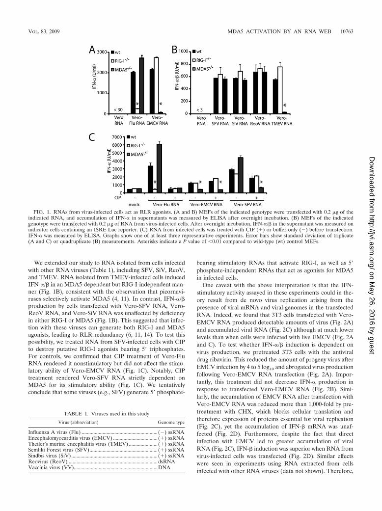
We extended our study to RNA isolated from cells infectedwith other RNA viruses (Table 1), including SFV, SiV, ReoV,and TMEV. RNA isolated from TMEV-infected cells inducedIFN-�/� in an MDA5-dependent but RIG-I-independent man-ner (Fig. 1B), consistent with the observation that picornavi-ruses selectively activate MDA5 (4, 11). In contrast, IFN-�/�production by cells transfected with Vero-SFV RNA, Vero-ReoV RNA, and Vero-SiV RNA was unaffected by deficiencyin either RIG-I or MDA5 (Fig. 1B). This suggested that infec-tion with these viruses can generate both RIG-I and MDA5agonists, leading to RLR redundancy (6, 11, 14). To test thispossibility, we treated RNA from SFV-infected cells with CIPto destroy putative RIG-I agonists bearing 5� triphosphates.For controls, we confirmed that CIP treatment of Vero-FluRNA rendered it nonstimulatory but did not affect the stimu-latory ability of Vero-EMCV RNA (Fig. 1C). Notably, CIPtreatment rendered Vero-SFV RNA strictly dependent onMDA5 for its stimulatory ability (Fig. 1C). We tentativelyconclude that some viruses (e.g., SFV) generate 5� phosphate-
bearing stimulatory RNAs that activate RIG-I, as well as 5�phosphate-independent RNAs that act as agonists for MDA5in infected cells.
One caveat with the above interpretation is that the IFN-stimulatory activity assayed in these experiments could in the-ory result from de novo virus replication arising from thepresence of viral mRNA and viral genomes in the transfectedRNA. Indeed, we found that 3T3 cells transfected with Vero-EMCV RNA produced detectable amounts of virus (Fig. 2A)and accumulated viral RNA (Fig. 2C) although at much lowerlevels than when cells were infected with live EMCV (Fig. 2Aand C). To test whether IFN-�/� induction is dependent onvirus production, we pretreated 3T3 cells with the antiviraldrug ribavirin. This reduced the amount of progeny virus afterEMCV infection by 4 to 5 log10 and abrogated virus productionfollowing Vero-EMCV RNA transfection (Fig. 2A). Impor-tantly, this treatment did not decrease IFN-� production inresponse to transfected Vero-EMCV RNA (Fig. 2B). Simi-larly, the accumulation of EMCV RNA after transfection withVero-EMCV RNA was reduced more than 1,000-fold by pre-treatment with CHX, which blocks cellular translation andtherefore expression of proteins essential for viral replication(Fig. 2C), yet the accumulation of IFN-� mRNA was unaf-fected (Fig. 2D). Furthermore, despite the fact that directinfection with EMCV led to greater accumulation of viralRNA (Fig. 2C), IFN-� induction was superior when RNA fromvirus-infected cells was transfected (Fig. 2D). Similar effectswere seen in experiments using RNA extracted from cellsinfected with other RNA viruses (data not shown). Therefore,
FIG. 1. RNAs from virus-infected cells act as RLR agonists. (A and B) MEFs of the indicated genotype were transfected with 0.2 �g of theindicated RNA, and accumulation of IFN-� in supernatants was measured by ELISA after overnight incubation. (B) MEFs of the indicatedgenotype were transfected with 0.2 �g of RNA from virus-infected cells. After overnight incubation, IFN-�/� in the supernatant was measured onindicator cells containing an ISRE-Luc reporter. (C) RNA from infected cells was treated with CIP (�) or buffer only (�) before transfection.IFN-� was measured by ELISA. Graphs show one of at least three representative experiments. Error bars show standard deviation of triplicate(A and C) or quadruplicate (B) measurements. Asterisks indicate a P value of 0.01 compared to wild-type (wt) control MEFs.
TABLE 1. Viruses used in this study
Virus (abbreviation) Genome type
Influenza A virus (Flu) .......................................................... (�) ssRNAEncephalomyocarditis virus (EMCV) .................................. (�) ssRNATheiler’s murine encephalitis virus (TMEV) ...................... (�) ssRNASemliki Forest virus (SFV).................................................... (�) ssRNASindbis virus (SiV).................................................................. (�) ssRNAReovirus (ReoV) .................................................................... dsRNAVaccinia virus (VV)................................................................DNA
VOL. 83, 2009 MDA5 ACTIVATION BY AN RNA WEB 10763
on May 26, 2016 by guest
http://jvi.asm.org/
Dow
nloaded from

we conclude that RNA extracted from cells infected with RNAviruses contains preformed stimulatory species that act as ago-nists for RLRs independently of further virus replication.
VV generates dsRNA that activates MDA5. The presence ofinfectious RNAs in preparations extracted from cells infectedwith RNA viruses complicated our attempts to characterizeagonists for RLRs. We therefore sought to establish an addi-tional virus model in which RNA extracted from infected cellswould be noninfectious. DNA viruses have been reported togenerate dsRNA in infected cells (1, 9, 28), but, by definition,such RNA cannot be infectious. Notably, HeLa-VV RNA ac-tivated the IFN-� promoter upon transfection into HEK293(Fig. 3A) or 3T3 cells (Fig. 3B) and induced secretion ofIFN-�/� protein after transfection in MEFs (Fig. 3C). Thestimulatory activity of HeLa-VV RNA was appreciably lowerthan that of Vero-EMCV RNA or of a control 5� PPP-RNA(Fig. 3A to C). Nevertheless, it was completely MDA5 depen-
dent as MEFs that lacked MDA5 were totally unresponsive toHeLa-VV RNA (Fig. 3C). Therefore, RNA from VV-infectedcells can be used as a tool to dissect MDA5-dependent recog-nition processes without the added complication of infectiousRNA and de novo virus replication.
The dsRNA-specific K1 antibody recognizes an MDA5 ago-nist. VV, EMCV, and SFV have all previously been shown togenerate dsRNA that can be detected by staining cells with thedsRNA-specific K1 monoclonal antibody (MAb) (27, 28). Be-cause all three viruses activate MDA5 (see above), we testedfor K1 immunoreactivity upon infection with all MDA5-acti-vating viruses. K1 stained cells infected with EMCV, TMEV,SFV, ReoV, SiV, and VV but did not stain cells infected withinfluenza virus or influenza virus lacking the NS1 protein(Flu�NS1) (Fig. 4A), despite the fact that the latter wereclearly infected (Fig. 4B). Given this correlation betweenMDA5-stimulatory ability and generation of K1 ligands, wenext asked whether RNA bound to the dsRNA-specific K1antibody can also bind to MDA5. We transfected 293T cellswith HA-tagged MDA5 and infected these cells with EMCV
FIG. 2. Transfection of RNA from virus-infected cells generatesprogeny virus. (A and B) 3T3 cells were left untreated or were pre-treated with ribavirin for 1 h and transfected with Vero-EMCV RNA(0.2 �g in A; 1 and 0.2 �g in B) or infected with EMCV (MOI of 1).(A) Accumulation of infectious virus particles quantified by 50% tissueculture infective dose. (B) IFN-� was measured after overnight incu-bation. ELISA shows average of triplicate measurements � standarddeviation. (C and D) Quantitative reverse transcription-PCR analysisfor EMCV RNA (C) and IFN-� mRNA (D) in cells treated with CHXfor 30 min and subsequently infected with EMCV or transfected with0.2 �g of Vero-EMCV RNA for 4 h. Bars show averages of duplicatemeasurements � standard deviations. Graphs show one of two (A andB) or three representative experiments (C and D). Error bars showstandard deviations of quadruplicate (A), triplicate (B), or duplicate(C and D) measurements. Asterisks indicate a P value of 0.001compared to dimethyl sulfoxide (DMSO) treatment.
FIG. 3. RNA from VV-infected cells stimulates MDA5. HEK293(A) or 3T3 (B) cells were transfected with reporter constructs forIFN-�-luciferase and control plasmid pRL-TK and subsequently stim-ulated with HeLa-VV RNA (1, 0.2, and 0.04 �g) or control 5� PPP-RNA (0.2 and 0.04 �g), respectively. Graphs show relative activationof the IFN-� promoter. Data are the average of duplicate measure-ments standard deviation. (C) MEFs of the indicated genotype weretransfected with HeLa-RNA (1 and 0.2 �g), HeLa-VV RNA (1, 0.2,and 0.04 �g), Vero-EMCV RNA (0.2 �g), or with control 5� PPP-RNA(0.2 �g). The graph shows the accumulation of IFN-�/� in superna-tants after overnight incubation. *, P 0.001 compared to wild-type(wt) MEFs. Data are average standard deviations of duplicate (Aand B) or quadruplicate (C) measurements from one experiment re-peated three times.
10764 PICHLMAIR ET AL. J. VIROL.
on May 26, 2016 by guest
http://jvi.asm.org/
Dow
nloaded from

(MOI of 10 for 8 h) or left them uninfected. The dsRNAantibody did not coprecipitate MDA5 when the cells were notinfected, as expected (Fig. 5A). However, infection withEMCV promoted formation of a complex between MDA5 andK1. Notably, this complex could also be formed after cell lysisby the addition to the lysates of poly(I:C), which binds stronglyto both K1 (26) and to MDA5 (11), or of Vero-EMCV RNA(Fig. 5A).
To test whether RNA bound to the K1 antibody has stimu-latory activity, we precipitated RNA extracted from EMCV-infected cells and tested the bound fractions for IFN-�/� in-duction. As expected, control Sepharose beads coated withIgG did not precipitate stimulatory activity from Vero-EMCVRNA (Fig. 5B). However, beads coupled to the K1 antibodyprecipitated Vero-EMCV RNA that induced large amounts ofIFN-�/� upon transfection into indicator cells (Fig. 5B). Sim-ilar to Vero-EMCV RNA, the fraction of HeLa-VV RNAprecipitated by K1 contained stimulatory activity, whereas con-trol IgG-precipitated RNA was inactive (Fig. 5C). Notably, theamount of RNA isolated from K1-precipitated fractions wasdramatically reduced (�100-fold) compared to the amountisolated from unbound fractions (data not shown), indicatingthat the dsRNA recognizing MAb bound selected RNA spe-cies. Collectively, these experiments suggest that the K1 anti-body can bind to MDA5 agonist(s) present in virus-infectedcells and indicate that MDA5 and the K1 antibody bind tosimilar RNAs.
Lack of specific sequence motifs in stimulatory RNA frac-tions from VV-infected cells. We attempted to define specificsequences in the stimulatory RNA from VV-infected cells. Aswe did not know whether terminal OH groups were present inMDA5-agonistic RNA and whether sequences necessary for
activating MDA5 were at the end of the putative RNA se-quence, we resorted to an approach that involved tagged ran-dom hexamer priming (29) (Fig. 6A gives a schematic repre-sentation). RNA from virus-infected or uninfected cells wasused for immunoprecipitation with K1 or control antibodies.RNA was reverse transcribed and amplified by PCR for 5 to 10cycles, and the resulting DNA fragments were ligated intopGEM-T (Promega). Colonies were screened for �-galactosi-dase activity, and white colonies were selected for sequencing.K1 immunoprecipitates of HeLa-VV RNA yielded more col-onies than control precipitations (IgG precipitates fromHeLa-VV RNA or K1 precipitations of RNA from uninfectedcells) (Fig. 6B). This was encouraging as it suggested enrich-ment of specific stimulatory RNAs with the K1 antibody. How-ever, sequencing revealed mostly rRNA, a few viral sequences,and a few sequences from human genes (Fig. 6C shows atypical example of one experiment). In total, we identified 31
FIG. 4. MDA5-activating viruses generate immunodetectabledsRNA. (A) Vero cells were infected with the indicated virus (MOI of0.5 to 1) for 8 h (EMCV, TMEV, and SFV) or 16 h (influenza virus(Flu), Flu�NS1, and ReoV). HeLa cells were infected with VV (MOIof 0.5) for 16 h. Cells were fixed and stained with the K1 (dsRNA)MAb (green) and DAPI (blue). (B) Influenza virus-infected cellsstained with anti-influenza (Flu) antibodies (green) and DAPI (blue).
FIG. 5. K1 and MDA5 bind to the same type of RNA. (A) 293Tcells were transfected with HA-tagged MDA5 and 40 h later infectedwith EMCV (if EMCV) or left uninfected. Eight hours later, celllysates were prepared and used for coimmunoprecipitation experi-ments. Uninfected cells (lanes 5, 6, and 8) or infected cells (lane 7)were used for immunoprecipitation with the K1 (dsRNA) antibody.Poly(I:C) and Vero-EMCV RNA were added to the lysates in lanes 6and 8, respectively. HA-MDA5 in total cell lysates (lanes 1 to 3) or K1immunoprecipitates (lanes 4 to 8) was visualized by Western blotting(WB) with antibody against the HA epitope. (B and C) The K1 anti-body or a control irrelevant antibody was used to precipitate RNAextracted from EMCV-infected Vero cells (B) or VV-infected HeLacells (C). The RNA in the precipitated fraction was extracted withTrizol and used to transfect 3T3 cells. Graphs show average (� stan-dard deviation) accumulation of IFN-�/� in supernatants (measured inquadruplicate) after 16 h. One representative experiment of three isshown.
VOL. 83, 2009 MDA5 ACTIVATION BY AN RNA WEB 10765
on May 26, 2016 by guest
http://jvi.asm.org/
Dow
nloaded from

clones containing viral sequences, and in one of three experi-ments, there was some enrichment in the A11R gene locus (bp121859 to 122815) (Fig. 6D). However, overall we failed toidentify any particular sequence or sequence motif or evenclustering when sequences were aligned to the virus genome.Thus, the data obtained by this method do not reveal anyparticular sequence preference for MDA5-stimulatory RNA.
HMW RNA but not dsRNA stimulates MDA5. The associa-tion between MDA5-agonistic activity and recognition by K1, adsRNA-specific MAb, prompted us to analyze RNA prepara-tions from virus-infected cells for the presence of dsRNA.Double-strandedness of nucleotides can be visualized by stain-ing with acridine orange, a metachromatic stain that stainssingle-stranded polynucleotides red and double-strandedpolynucleotides green (18). In the past, this dye has been usedextensively to categorize viruses according to the nature oftheir genome (15–17). As expected, a double-stranded DNAmarker appears green on an acridine orange-stained gel,whereas in vitro transcribed ssRNA appears red (Fig. 7A).RNA isolated from uninfected HeLa cells (HeLa-RNA) orVero-RNA showed two prominent red bands of rRNA (Fig.7A to C). Consistent with the fact that EMCV degrades rRNA(30), the rRNA bands were faint in Vero-EMCV RNA, whichcontained a prominent dsRNA band of 11,000 or more bp (Fig.7A and C). Similarly, HeLa-VV RNA contained a dsRNAband that was absent from uninfected cells (Fig. 7B and C). Inaddition, RNA from both EMCV- and VV-infected cells con-tained a very-high-molecular-weight (HMW) fraction that didnot enter the agarose gel and remained trapped in the well andvariably stained green and red with acridine orange (Fig. 7A toC). Similarly, well-trapped HMW RNA ranging in stainingfrom green to red was detected in all RNA preparations fromvirus-infected cells able to stimulate MDA5 but not in Vero-Flu RNA, which exclusively activates RIG-I (Fig. 7C). Weisolated the dsRNA band and the HMW well-trapped fractionfrom Vero-EMCV cells and tested them for stimulatory activ-ity (Fig. 7D, inset). To our surprise, only the HMW RNA andnot the dsRNA fraction elicited IFN-�/� when transfected into3T3 cells (Fig. 7D). Similarly, HMW RNA from VV-infectedcells contained stimulatory potential, whereas the dsRNA frac-tion did not generate measurable IFN-�/� (Fig. 7E). For bothviruses, HMW RNA allowed association of MDA5 to agarosebeads coupled to the dsRNA antibody (Fig. 7F). Thus, weconclude that HMW RNA is generated by some viruses andthat this RNA has the ability to bind to and activate MDA5.Interestingly, although dsRNA arising from virus infection canbind to MDA5, it is not necessarily an agonist for the helicase.
HMW RNA contained both ssRNA and dsRNA regions asit stained red and green on acridine orange gels, with somevariability between experiments (Fig. 7A to C). The presenceand functional importance of dsRNA and ssRNA regions inHMW RNA were further suggested by experiments using nu-clease digestion with the ssRNA-specific nucleases RNase Aand RNase T1 and the dsRNA-specific RNase V1 (Fig. 8A andB). Therefore, we envisaged that HMW RNA may resemble anRNA web rather than perfectly base-paired long molecules ofdsRNA. We hypothesized that destroying the secondary andtertiary structure of this HMW RNA would diminish its po-tential to stimulate MDA5. Indeed, when we heat denaturedand flash froze HeLa-VV RNA, both the dsRNA fraction and
FIG. 6. Lack of specific sequence motifs in MDA5-agonistic RNA.(A) Experimental outline. RNA from cells infected with VV (13 or24 h) was immunoprecipitated with K1 or IgG control antibodies.RNA was isolated and reverse transcribed using random hexamerprimers with an attached T3 promoter site. cDNA was amplified in aPCR step (5 to 10 cycles) with T3 primers, and the resulting DNA wascloned into pGEM-T (Promega). Bacterial colonies were screened for�-galactosidase activity, and white colonies that contained an insertwere sequenced using T7 primers. (B) Average number of white CFUfrom a typical experiment. Error bars represent standard deviationsfrom two independent transformations. (C) Distribution of identifiedsequences from IgG immunoprecipitation (n � 21) and K1 (dsRNA)immunoprecipitation (n � 136). The average recovered sequencelength was 369 bp. One representative experiment of three is shown.(D) Identified sequences from 31 identified VV gene products. Thenumber shows start of the sequence in the VV genome (VV NCBIaccession number AY243312). IP, immunoprecipitation.
10766 PICHLMAIR ET AL. J. VIROL.
on May 26, 2016 by guest
http://jvi.asm.org/
Dow
nloaded from

the HMW RNA fraction disappeared (Fig. 8C, insert), to-gether with the ability to stimulate IFN-�/� (Fig. 8C). In con-trast, heating and flash freezing of an in vitro transcribed con-trol 5� PPP-RNA did not result in a change of IFN induction(Fig. 8C). The stimulatory activity of RNA preparations fromcells infected with picornaviruses was similarly sensitive to heatdenaturation followed by rapid freezing (Fig. 8D). Finally, theassociation between MDA5 and beads coated with the dsRNAantibody was lost when RNA denatured by heating and freezingwas used (Fig. 8E). We conclude that the ability to stimulateMDA5 requires highly structured RNA.
DISCUSSION
The identification of RIG-I and MDA5 as pattern recogni-tion receptors for viruses marked a milestone in our under-standing of antiviral innate responses (11, 13, 31, 32). Interest-ingly, genetic deletion experiments demonstrated that RIG-Iand MDA5 possess distinct virus specificities and that MDA5but not RIG-I is essential for innate responses to picornavi-ruses, whereas RIG-I but not MDA5 is indispensable for in-nate immunity to influenza A virus, paramyxoviruses, and Jap-anese encephalitis virus (11). The basis for differential virusrecognition by the two RLRs is not entirely understood. RIG-Iagonists have been variously defined as 5� PPP-RNAs, shortdsRNA duplexes bearing or not a 5� monophosphate, or 3�monophosphate-containing cleavage products of RNase L (7,10, 22). In contrast, agonists for MDA5 remain less well char-acterized. It is generally thought that MDA5 recognizes longdsRNA stretches such as those found in poly(I:C) and in thegenomes of ReoVs (10). In this study, we set out to investigatethe characteristics of RNA species that can stimulate MDA5 invirus-infected cells. As others have noted (7), we found thatRNA extracted from uninfected cells does not stimulate IFNproduction, indicating that it does not contain enough RNAspecies bearing, for example, 5� triphosphate moieties and/orthe necessary secondary structure to act as agonists for RLRs.In contrast, RNA extracted from cells infected with RNA vi-ruses contains abundant stimulatory activity, and the depen-dence of that activity on RIG-I and MDA5 largely matches thedependence reported for infection with the intact virus (Fig. 1and 3). In particular, we show that influenza A virus generatesstimulatory RNA for RIG-I, whereas TMEV, EMCV, SiV,
FIG. 7. HMW RNA activates MDA5. (A) Amounts of 1 �g and 0.2�g of the indicated nucleic acid were electrophoretically separated ona 1% agarose gel and subsequently stained with acridine orange. Dou-ble-stranded nucleic acid stains green; single-stranded nucleic acidstains red. (B) A 1-�g and 0.2-�g DNA ladder and 1 �g of HeLa-VV
RNA were electrophoretically separated at the indicated time pointson an agarose gel and stained with acridine orange. (C) A 1-�g DNAladder and 1 �g of the indicated RNA preparation were electro-phoretically separated on an agarose gel and stained with acridineorange. Numbers to the left of the DNA ladders (A to C) show sizesof DNA markers. (D and E) The indicated RNA fraction (see inset ofacridine orange gel) was isolated from the agarose gel, and 0.5 or 0.1�g was used to transfect 3T3 cells. In panel D 3T3 cells were treatedwith ribavirin to prevent EMCV replication. Graphs show averageaccumulation � standard deviation of IFN-�/� after overnight culturemeasured in quadruplicate. (F) Lysate of 293T cells transfected withFLAG-MDA5 for 48 h was used for immunoprecipitation with the K1(dsRNA) antibody in the absence or presence of 1 �g of the indicatedRNA. K1 immunoprecipitates were visualized by Western blotting(WB) with antibody against the FLAG epitope. The arrow points toFLAG-tagged MDA5.
VOL. 83, 2009 MDA5 ACTIVATION BY AN RNA WEB 10767
on May 26, 2016 by guest
http://jvi.asm.org/
Dow
nloaded from
ReoV, and SFV all generate RNAs capable of stimulatingMDA5 activity (Fig. 1). RNA isolated from cells infected withRNA viruses is infectious, which can complicate approaches toidentifying the stimulatory fraction. For example, we isolated asingle fraction of RNA from EMCV-infected cells which was
able to induce large amounts of IFN-�/� upon transfectioninto reporter cells (data not shown). However, this fractionturned out to contain the intact EMCV genome, and its trans-fection resulted in a full infectious cycle, rendering it difficult todetermine whether the IFN-inducing activity was attributableto properties of the genome itself or to RNAs made duringsubsequent virus replication (data not shown). Although thisproblem could be circumvented in part by using ribavirin, moredefinitive evidence for a preformed stimulatory species camefrom analysis of RNA extracted from cells infected with DNAviruses. A recent paper suggests that modified VV Ankarainduces IFN-�/� in an MDA5- and MAVS-dependent manner(3). Here, we show that RNAs generated during VV infectionindeed activate MDA5-dependent IFN-�/� responses. There-fore, DNA viruses have the potential to generate RNA thatactivates RLRs, and VV infection may be a useful system inwhich to characterize MDA5-agonistic RNAs.
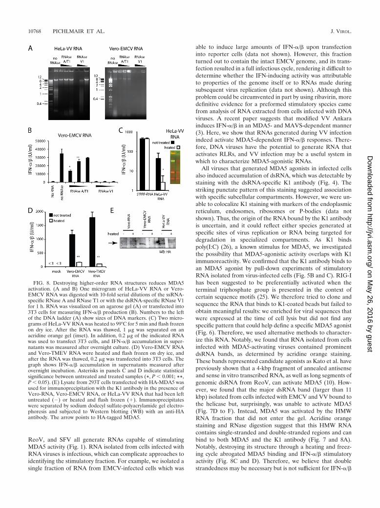
All viruses that generated MDA5 agonists in infected cellsalso induced accumulation of dsRNA, which was detectable bystaining with the dsRNA-specific K1 antibody (Fig. 4). Thestriking punctate pattern of this staining suggested associationwith specific subcellular compartments. However, we were un-able to colocalize K1 staining with markers of the endoplasmicreticulum, endosomes, ribosomes or P-bodies (data notshown). Thus, the origin of the RNA bound by the K1 antibodyis uncertain, and it could reflect either species generated atspecific sites of virus replication or RNA being targeted fordegradation in specialized compartments. As K1 bindspoly(I:C) (26), a known stimulus for MDA5, we investigatedthe possibility that MDA5-agonistic activity overlaps with K1immunoreactivity. We confirmed that the K1 antibody binds toan MDA5 agonist by pull-down experiments of stimulatoryRNA isolated from virus-infected cells (Fig. 5B and C). RIG-Ihas been suggested to be preferentially activated when theterminal triphosphate group is presented in the context ofcertain sequence motifs (25). We therefore tried to clone andsequence the RNA that binds to K1-coated beads but failed toobtain meaningful results: we enriched for viral sequences thatwere expressed at the time of cell lysis but did not find anyspecific pattern that could help define a specific MDA5 agonist(Fig. 6). Therefore, we used alternative methods to character-ize this RNA. Notably, we found that RNA isolated from cellsinfected with MDA5-activating viruses contained prominentdsRNA bands, as determined by acridine orange staining.These bands represented candidate agonists as Kato et al. havepreviously shown that a 4-kbp fragment of annealed antisenseand sense in vitro transcribed RNA, as well as long segments ofgenomic dsRNA from ReoV, can activate MDA5 (10). How-ever, we found that the major dsRNA band (larger than 11kbp) isolated from cells infected with EMCV and VV bound tothe helicase but, surprisingly, was unable to activate MDA5(Fig. 7D to F). Instead, MDA5 was activated by the HMWRNA fraction that did not enter the gel. Acridine orangestaining and RNase digestion suggest that this HMW RNAcontains single-stranded and double-stranded regions and canbind to both MDA5 and the K1 antibody (Fig. 7 and 8A).Notably, destroying its structure through a heating and freez-ing cycle abrogated MDA5 binding and IFN-�/� stimulatoryactivity (Fig. 8C and D). Therefore, we believe that doublestrandedness may be necessary but is not sufficient for IFN-�/�
FIG. 8. Destroying higher-order RNA structures reduces MDA5activation. (A and B) One microgram of HeLa-VV RNA or Vero-EMCV RNA was digested with 10-fold serial dilutions of the ssRNA-specific RNase A and RNase T1 or with the dsRNA-specific RNase V1for 1 h. RNA was visualized on an agarose gel (A) or transfected into3T3 cells for measuring IFN-�/� production (B). Numbers to the leftof the DNA ladder (A) show sizes of DNA markers. (C) Two micro-grams of HeLa-VV RNA was heated to 99°C for 5 min and flash frozenon dry ice. After the RNA was thawed, 1 �g was separated on anacridine orange gel (inset). In addition, 0.2 �g of the indicated RNAwas used to transfect 3T3 cells, and IFN-�/� accumulation in super-natants was measured after overnight culture. (D) Vero-EMCV RNAand Vero-TMEV RNA were heated and flash frozen on dry ice, andafter the RNA was thawed, 0.2 �g was transfected into 3T3 cells. Thegraph shows IFN-�/� accumulation in supernatants measured afterovernight incubation. Asterisks in panels C and D indicate statisticalsignificance between untreated and treated samples (*, P 0.001; **,P 0.05). (E) Lysate from 293T cells transfected with HA-MDA5 wasused for immunoprecipitation with the K1 antibody in the presence ofVero-RNA, Vero-EMCV RNA, or HeLa-VV RNA that had been leftuntreated (�) or heated and flash frozen (�). Immunoprecipitateswere separated by sodium dodecyl sulfate-polyacrylamide gel electro-phoresis and subjected to Western blotting (WB) with an anti-HAantibody. The arrow points to HA-tagged MDA5.
10768 PICHLMAIR ET AL. J. VIROL.
on May 26, 2016 by guest
http://jvi.asm.org/
Dow
nloaded from
induction and that the MDA5-stimulating RNA present invirus-infected cells is in the form of RNA aggregates consistingof single-stranded and double-stranded moieties that are gen-erated during the replication/transcription process. Poly(I:C)binds to K1 and acts as an agonist for MDA5 signaling. Al-though poly(I:C) is regarded as a dsRNA homologue, it doesnot consist of perfectly matched dsRNA strands becausepoly(I:C) is generated through annealing of enzymatically pro-duced inosine and cytidine homopolymers of undefined length(5), which results in a web-like structure. We speculate thatbranched RNA may be an important part in MDA5-dependentrecognition. Interestingly, recognition of branched nucleic acidis a long-studied phenomenon: junction-resolving enzymes rec-ognize Holliday junctions (branched DNA) during DNA rep-lication (2). Binding of junction-resolving enzymes must meetspecific structural requirements (DNA branches) but is inde-pendent of the exact nucleic acid sequence. Similarly, MDA5recognition appears to require structural features (Fig. 7 and8), but we could not identify specific sequences (Fig. 6). Re-ducing the size of poly(I:C) by sonication decreases its abilityto activate MDA5 (10), which could be explained by the de-struction of the RNA web necessary for MDA5 activation.Notably, annealing polyadenylic and polyuridylic homopoly-mers of undefined length to form poly(A � U) is not sufficientto generate RLR-stimulatory activity (data not shown). Onepossible explanation could lie in the high affinity of poly(A) forpoly(U), which would make poly(A � U) quite inflexible andprevent it from adopting a loose web structure, unlike poly(I:C). Recently, Myong et al. reported that recognition of triphos-phorylated RNA cannot activate RIG-I without local second-ary RNA structure (19). Similarly, dsRNA or RNA aggregatesmay be necessary for binding of MDA5 (Fig. 7F), but this maynot be sufficient for potent MDA5 activation, which may re-quire hitherto undefined additional signals to distinguish cel-lular from viral RNA. We speculate that, similar to junction-resolving enzymes that recognize branched DNA duringcellular replication, MDA5 may have evolved to recognizebranched RNA during virus replication.
ACKNOWLEDGMENTS
We thank the members of the immunobiology laboratory for stim-ulating discussions, Mike Gait and Svend Petersen-Mahrt for invalu-able intellectual input, Ina Weisswange for infecting cells with VV, andCatalina Vasquez for secretarial support.
This work was supported by Cancer Research UK and a long-termEMBO fellowship to A.P. J.R. is a recipient of FEBS and ABSPlong-term fellowships.
REFERENCES
1. Colby, C., and P. H. Duesberg. 1969. Double-stranded RNA in vaccinia virusinfected cells. Nature 222:940–944.
2. Declais, A. C., and D. M. Lilley. 2008. New insight into the recognition ofbranched DNA structure by junction-resolving enzymes. Curr. Opin. Struct.Biol. 18:86–95.
3. Delaloye, J., T. Roger, Q. G. Steiner-Tardivel, D. Le Roy, M. Knaup Rey-mond, S. Akira, V. Petrilli, C. E. Gomez, B. Perdiguero, J. Tschopp, G.Pantaleo, M. Esteban, and T. Calandra. 2009. Innate immune sensing ofmodified vaccinia virus Ankara (MVA) is mediated by TLR2-TLR6, MDA-5and the NALP3 inflammasome. PLoS Pathog. 5:e1000480.
4. Gitlin, L., W. Barchet, S. Gilfillan, M. Cella, B. Beutler, R. A. Flavell, M. S.Diamond, and M. Colonna. 2006. Essential role of mda-5 in type I IFNresponses to polyriboinosinic:polyribocytidylic acid and encephalomyocardi-tis picornavirus. Proc. Natl. Acad. Sci. USA 103:8459–8464.
5. Grunberg-Manago, M., P. J. Ortiz, and S. Ochoa. 1956. Enzymic synthesis ofpolynucleotides. I. Polynucleotide phosphorylase of azotobacter vinelandii.Biochim. Biophys. Acta 20:269–285.
6. Holm, G. H., J. Zurney, V. Tumilasci, S. Leveille, P. Danthi, J. Hiscott, B.Sherry, and T. S. Dermody. 2007. Retinoic acid-inducible gene-I and inter-feron-beta promoter stimulator-1 augment proapoptotic responses followingmammalian reovirus infection via interferon regulatory factor-3. J. Biol.Chem. 282:21953–21961.
7. Hornung, V., J. Ellegast, S. Kim, K. Brzozka, A. Jung, H. Kato, H. Poeck, S.Akira, K. K. Conzelmann, M. Schlee, S. Endres, and G. Hartmann. 2006.5�-Triphosphate RNA is the ligand for RIG-I. Science 314:994–997.
8. Iwasaki, A., and R. Medzhitov. 2004. Toll-like receptor control of the adap-tive immune responses. Nat. Immunol. 5:987–995.
9. Jurale, C., J. R. Kates, and C. Colby. 1970. Isolation of double-strandedRNA from T4 phage infected cells. Nature 226:1027–1029.
10. Kato, H., O. Takeuchi, E. Mikamo-Satoh, R. Hirai, T. Kawai, K. Matsushita, A.Hiiragi, T. S. Dermody, T. Fujita, and S. Akira. 2008. Length-dependent recognitionof double-stranded ribonucleic acids by retinoic acid-inducible gene-I and mela-noma differentiation-associated gene 5. J. Exp. Med. 205:1601–1610.
11. Kato, H., O. Takeuchi, S. Sato, M. Yoneyama, M. Yamamoto, K. Matsui, S.Uematsu, A. Jung, T. Kawai, K. J. Ishii, O. Yamaguchi, K. Otsu, T. Tsu-jimura, C. S. Koh, C. Reis e Sousa, Y. Matsuura, T. Fujita, and S. Akira.2006. Differential roles of MDA5 and RIG-I helicases in the recognition ofRNA viruses. Nature 441:101–105.
12. Kawai, T., and S. Akira. 2006. Innate immune recognition of viral infection.Nat. Immunol. 7:131–137.
13. Kawai, T., and S. Akira. 2008. Toll-like receptor and RIG-I-like receptorsignaling. Ann. N. Y. Acad. Sci. 1143:1–20.
14. Loo, Y. M., J. Fornek, N. Crochet, G. Bajwa, O. Perwitasari, L. Martinez-Sobrido, S. Akira, M. A. Gill, A. Garcia-Sastre, M. G. Katze, and M. Gale,Jr. 2008. Distinct RIG-I and MDA5 signaling by RNA viruses in innateimmunity. J. Virol. 82:335–345.
15. Matsunaga, Y., S. Matsuno, and J. Mukoyama. 1977. Isolation and charac-terization of a parvovirus of rabbits. Infect. Immun. 18:495–500.
16. Mayor, H. D. 1963. The nucleic acids of viruses as revealed by their reactionswith fluorochrome acridine orange. Int. Rev. Exp. Pathol. 2:1–45.
17. Mayor, H. D., and A. R. Diwan. 1961. Studies on the acridine orange staining of twopurified RNA viruses: poliovirus and tobacco mosaic virus. Virology 14:74–82.
18. McMaster, G. K., and G. G. Carmichael. 1977. Analysis of single- anddouble-stranded nucleic acids on polyacrylamide and agarose gels by usingglyoxal and acridine orange. Proc. Natl. Acad. Sci. USA 74:4835–4838.
19. Myong, S., S. Cui, P. V. Cornish, A. Kirchhofer, M. U. Gack, J. U. Jung, K. P.Hopfner, and T. Ha. 2009. Cytosolic viral sensor RIG-I is a 5�-triphosphate-dependent translocase on double-stranded RNA. Science 323:1070–1074.
20. Onomoto, K., M. Yoneyama, and T. Fujita. 2007. Regulation of antiviralinnate immune responses by RIG-I family of RNA helicases. Curr. Top.Microbiol. Immunol. 316:193–205.
21. Pichlmair, A., and C. Reis e Sousa. 2007. Innate recognition of viruses.Immunity 27:370–383.
22. Pichlmair, A., O. Schulz, C. P. Tan, T. I. Naslund, P. Liljestrom, F. Weber,and C. Reis e Sousa. 2006. RIG-I-mediated antiviral responses to single-stranded RNA bearing 5�-phosphates. Science 314:997–1001.
23. Racaniello, V. R. 2001. Picornaviridae: the viruses and their replication, p.685–722. In D. Knipe, P. M. Howley, D. E. Griffin, R. A. Lamb, M. A.Martin, B. Roizman, and S. E. Straus (ed.), Fields virology. LippincottWilliams & Wilkins, Philadelphia, PA.
24. Saito, T., and M. Gale, Jr. 2007. Principles of intracellular viral recognition.Curr. Opin. Immunol. 19:17–23.
25. Saito, T., D. M. Owen, F. Jiang, J. Marcotrigiano, and M. Gale, Jr. 2008.Innate immunity induced by composition-dependent RIG-I recognition ofhepatitis C virus RNA. Nature 454:523–527.
26. Schonborn, J., J. Oberstrass, E. Breyel, J. Tittgen, J. Schumacher, and N.Lukacs. 1991. Monoclonal antibodies to double-stranded RNA as probes ofRNA structure in crude nucleic acid extracts. Nucleic Acids Res. 19:2993–3000.
27. Schulz, O., S. S. Diebold, M. Chen, T. I. Naslund, M. A. Nolte, L. Alexopoulou, Y. T.Azuma, R. A. Flavell, P. Liljestrom, and C. Reis e Sousa. 2005. Toll-like receptor 3promotes cross-priming to virus-infected cells. Nature 433:887–892.
28. Weber, F., V. Wagner, S. B. Rasmussen, R. Hartmann, and S. R. Paludan.2006. Double-stranded RNA is produced by positive-strand RNA virusesand DNA viruses but not in detectable amounts by negative-strand RNAviruses. J. Virol. 80:5059–5064.
29. Wong, K. K., L. C. Stillwell, C. A. Dockery, and J. D. Saffer. 1996. Use oftagged random hexamer amplification (TRHA) to clone and sequenceminute quantities of DNA—application to a 180 kb plasmid isolated fromSphingomonas F199. Nucleic Acids Res. 24:3778–3783.
30. Wreschner, D. H., T. C. James, R. H. Silverman, and I. M. Kerr. 1981.Ribosomal RNA cleavage, nuclease activation and 2-5A(ppp(A2�p)nA) ininterferon-treated cells. Nucleic Acids Res. 9:1571–1581.
31. Yoneyama, M., and T. Fujita. 2007. Function of RIG-I-like receptors inantiviral innate immunity. J. Biol. Chem. 282:15315–15318.
32. Yoneyama, M., M. Kikuchi, T. Natsukawa, N. Shinobu, T. Imaizumi, M.Miyagishi, K. Taira, S. Akira, and T. Fujita. 2004. The RNA helicase RIG-Ihas an essential function in double-stranded RNA-induced innate antiviralresponses. Nat. Immunol. 5:730–737.
VOL. 83, 2009 MDA5 ACTIVATION BY AN RNA WEB 10769
on May 26, 2016 by guest
http://jvi.asm.org/
Dow
nloaded from
Related Documents