EUKARYOTIC CELL, Nov. 2005, p. 1840–1850 Vol. 4, No. 11 1535-9778/05/$08.000 doi:10.1128/EC.4.11.1840–1850.2005 Copyright © 2005, American Society for Microbiology. All Rights Reserved. Activation of AP-1-Dependent Transcription by a Truncated Translation Initiation Factor† Caroline C. L. Jenkins, 1 Juan Mata, 2 Richard F. Crane, 3 Benjamin Thomas, 1 Alexandre Akoulitchev, 1 Ju ¨rg Ba ¨hler, 2 and Chris J. Norbury 1 * Sir William Dunn School of Pathology, University of Oxford, Oxford OX1 3RE, United Kingdom 1 ; The Wellcome Trust Sanger Institute, Hinxton, Cambridge CB10 1SA, United Kingdom 2 ; and Department of Cell Biology, Harvard Medical School, Boston, MA 02115 3 Received 5 July 2005/Accepted 8 September 2005 Int6/eIF3e is a highly conserved subunit of eukaryotic translation initiation factor 3 (eIF3) that has also been reported to interact with subunits of the proteasome and the COP9 signalosome. Overexpression of full-length Int6 or a 13-kDa C-terminal fragment, Int6CT, in the fission yeast Schizosaccharomyces pombe causes multidrug resistance that requires the otherwise inessential AP-1 transcription factor Pap1. Here we show for the first time that Int6CT acts to increase the transcriptional activity of Pap1. Microarray hybridization data indicate that Int6CT overexpression resulted in the up-regulation of 67 genes; this expression profile closely matched that of cells overexpressing Pap1. Analysis of the upstream regulatory sequences of these genes showed that the majority contained AP-1 consensus binding sites. Partial defects in ubiquitin-dependent proteolysis have been suggested to confer Pap1-dependent multidrug resistance, but no such defect was seen on Int6CT overexpres- sion. Indeed, none of the previously identified interactions of endogenous Int6 was required for the activation of Pap1 transcription described here. Moreover, Int6CT-induced activation of Pap1-responsive gene expression was independent of the ability of Pap1 to undergo a redox-regulated conformational change which mediates its relocalization to the nucleus and expression of oxidative stress response genes. Int6CT therefore activates Pap1-dependent transcription by a novel mechanism. AP-1-dependent transcription is important in a wide variety of biological processes and has been implicated in tumor mul- tidrug resistance, a phenomenon that frequently hinders effec- tive chemotherapy (7, 14). The Schizosaccharomyces pombe int6 gene was identified through a screen for cDNAs that caused multidrug resistance when overexpressed (10). This screen also identified cDNAs encoding the previously de- scribed multidrug resistance determinant Pap1 (33), an AP-1- like transcription factor, and a partial int6 cDNA encoding the last 121 amino acids of the protein (Int6CT). Int6-induced multidrug resistance is dependent on Pap1 and was previously shown to be associated with the up-regulation of several known Pap1-dependent transcripts (10). Closely related to the yeast AP-1-like transcription factor (Yap1) in Saccharomyces cerevisiae, Pap1 is central to the ox- idative stress response of S. pombe. Following oxidative stress, Pap1 accumulates in the nucleus (34). Under nonstressed con- ditions, the Crm1 nuclear export factor binds a C-terminal nuclear export sequence within Pap1 and mediates its nuclear export. Upon oxidative stress, Pap1 undergoes a redox-depen- dent conformational change, preventing interaction between the nuclear export sequence and Crm1 (8, 36), resulting in an accumulation of Pap1 in the nucleus and activation of Pap1- dependent gene expression. Cells defective in Crm1 function exhibit Pap1-dependent multidrug resistance (20, 35), owing to the increased abundance of nuclear Pap1. In addition, S. pombe conditional mutants defective in various subunits of the 19S regulatory particle of the proteasome have been shown to exhibit drug resistance (15, 16, 26) that was suggested to be Pap1-dependent (26). The highly conserved Int6 protein was identified indepen- dently in human cells as the fifth largest subunit (eIF3e) of eukaryotic translation initiation factor 3 (eIF3) (3) and has been shown to interact with the core components of this mul- tisubunit initiation factor (1). However, int6 strains do not exhibit a dramatic defect in bulk translation initiation (5, 10), suggesting that the role of Int6/eIF3e within eIF3 may be to regulate the translation of specific transcripts under certain conditions. This view was reinforced by a recent study indicat- ing that distinct eIF3 complexes either containing or lacking Int6/eIF3e associate with distinct mRNA subpopulations (40). Int6/eIF3e also interacts with subunits of the COP9 signalo- some and the 26S proteasome (18, 38, 39) and has been pro- posed to regulate proteasome assembly via an interaction with Ras (39). In this study, we have compared the transcriptional profile induced by Int6CT with that seen on overexpression of Pap1 and have investigated the mechanism of activation of AP-1- dependent transcription by this truncated translation initiation factor. MATERIALS AND METHODS General fission yeast methods. S. pombe manipulations were carried out as described elsewhere (25) using EMM2 (Edinburgh minimal medium 2) contain- ing, where necessary, leucine and uracil at 225 g/ml. Strains used in this study are listed in Table 1. Strains were transformed by electroporation (gene pulser; Bio-Rad, Richmond, CA) with derivatives of the vector pREP3X or pREP4X * Corresponding author. Mailing address: Sir William Dunn School of Pathology, University of Oxford, Oxford OX1 3RE, United King- dom. Phone: 44 1865 275540. Fax: 44 1865 275501. E-mail: chris [email protected]. † Supplemental material for this article may be found at http: //ec.asm.org/. 1840 on September 16, 2015 by guest http://ec.asm.org/ Downloaded from

Welcome message from author
This document is posted to help you gain knowledge. Please leave a comment to let me know what you think about it! Share it to your friends and learn new things together.
Transcript

EUKARYOTIC CELL, Nov. 2005, p. 1840–1850 Vol. 4, No. 111535-9778/05/$08.00�0 doi:10.1128/EC.4.11.1840–1850.2005Copyright © 2005, American Society for Microbiology. All Rights Reserved.
Activation of AP-1-Dependent Transcription by a TruncatedTranslation Initiation Factor†
Caroline C. L. Jenkins,1 Juan Mata,2 Richard F. Crane,3 Benjamin Thomas,1Alexandre Akoulitchev,1 Jurg Bahler,2 and Chris J. Norbury1*
Sir William Dunn School of Pathology, University of Oxford, Oxford OX1 3RE, United Kingdom1;The Wellcome Trust Sanger Institute, Hinxton, Cambridge CB10 1SA, United Kingdom2; and
Department of Cell Biology, Harvard Medical School, Boston, MA 021153
Received 5 July 2005/Accepted 8 September 2005
Int6/eIF3e is a highly conserved subunit of eukaryotic translation initiation factor 3 (eIF3) that has also beenreported to interact with subunits of the proteasome and the COP9 signalosome. Overexpression of full-lengthInt6 or a 13-kDa C-terminal fragment, Int6CT, in the fission yeast Schizosaccharomyces pombe causes multidrugresistance that requires the otherwise inessential AP-1 transcription factor Pap1. Here we show for the firsttime that Int6CT acts to increase the transcriptional activity of Pap1. Microarray hybridization data indicatethat Int6CT overexpression resulted in the up-regulation of 67 genes; this expression profile closely matchedthat of cells overexpressing Pap1. Analysis of the upstream regulatory sequences of these genes showed that themajority contained AP-1 consensus binding sites. Partial defects in ubiquitin-dependent proteolysis have beensuggested to confer Pap1-dependent multidrug resistance, but no such defect was seen on Int6CT overexpres-sion. Indeed, none of the previously identified interactions of endogenous Int6 was required for the activationof Pap1 transcription described here. Moreover, Int6CT-induced activation of Pap1-responsive gene expressionwas independent of the ability of Pap1 to undergo a redox-regulated conformational change which mediates itsrelocalization to the nucleus and expression of oxidative stress response genes. Int6CT therefore activatesPap1-dependent transcription by a novel mechanism.
AP-1-dependent transcription is important in a wide varietyof biological processes and has been implicated in tumor mul-tidrug resistance, a phenomenon that frequently hinders effec-tive chemotherapy (7, 14). The Schizosaccharomyces pombeint6 gene was identified through a screen for cDNAs thatcaused multidrug resistance when overexpressed (10). Thisscreen also identified cDNAs encoding the previously de-scribed multidrug resistance determinant Pap1 (33), an AP-1-like transcription factor, and a partial int6 cDNA encoding thelast 121 amino acids of the protein (Int6CT). Int6-inducedmultidrug resistance is dependent on Pap1 and was previouslyshown to be associated with the up-regulation of several knownPap1-dependent transcripts (10).
Closely related to the yeast AP-1-like transcription factor(Yap1) in Saccharomyces cerevisiae, Pap1 is central to the ox-idative stress response of S. pombe. Following oxidative stress,Pap1 accumulates in the nucleus (34). Under nonstressed con-ditions, the Crm1 nuclear export factor binds a C-terminalnuclear export sequence within Pap1 and mediates its nuclearexport. Upon oxidative stress, Pap1 undergoes a redox-depen-dent conformational change, preventing interaction betweenthe nuclear export sequence and Crm1 (8, 36), resulting in anaccumulation of Pap1 in the nucleus and activation of Pap1-dependent gene expression. Cells defective in Crm1 functionexhibit Pap1-dependent multidrug resistance (20, 35), owing to
the increased abundance of nuclear Pap1. In addition, S.pombe conditional mutants defective in various subunits of the19S regulatory particle of the proteasome have been shown toexhibit drug resistance (15, 16, 26) that was suggested to bePap1-dependent (26).
The highly conserved Int6 protein was identified indepen-dently in human cells as the fifth largest subunit (eIF3e) ofeukaryotic translation initiation factor 3 (eIF3) (3) and hasbeen shown to interact with the core components of this mul-tisubunit initiation factor (1). However, int6� strains do notexhibit a dramatic defect in bulk translation initiation (5, 10),suggesting that the role of Int6/eIF3e within eIF3 may be toregulate the translation of specific transcripts under certainconditions. This view was reinforced by a recent study indicat-ing that distinct eIF3 complexes either containing or lackingInt6/eIF3e associate with distinct mRNA subpopulations (40).Int6/eIF3e also interacts with subunits of the COP9 signalo-some and the 26S proteasome (18, 38, 39) and has been pro-posed to regulate proteasome assembly via an interaction withRas (39).
In this study, we have compared the transcriptional profileinduced by Int6CT with that seen on overexpression of Pap1and have investigated the mechanism of activation of AP-1-dependent transcription by this truncated translation initiationfactor.
MATERIALS AND METHODS
General fission yeast methods. S. pombe manipulations were carried out asdescribed elsewhere (25) using EMM2 (Edinburgh minimal medium 2) contain-ing, where necessary, leucine and uracil at 225 �g/ml. Strains used in this studyare listed in Table 1. Strains were transformed by electroporation (gene pulser;Bio-Rad, Richmond, CA) with derivatives of the vector pREP3X or pREP4X
* Corresponding author. Mailing address: Sir William Dunn Schoolof Pathology, University of Oxford, Oxford OX1 3RE, United King-dom. Phone: 44 1865 275540. Fax: 44 1865 275501. E-mail: [email protected].
† Supplemental material for this article may be found at http://ec.asm.org/.
1840
on Septem
ber 16, 2015 by guesthttp://ec.asm
.org/D
ownloaded from
containing the last 121 codons of int6 (encoding Int6CT), a full-length pap1cDNA, or no insert (herein referred to as “vector”); inserts in these vectors areunder the control of the thiamine-repressible nmt1 promoter (23). Drug resis-tance was assayed after derepression of pREP3X gene expression by growth inthe absence of thiamine for 17 h and plating appropriate dilutions from mid-logphase cultures onto EMM2 agar containing 10 �g/ml methyl benzimidazole-2-ylcarbamate (MBC) for pap1� strains and 20 �g/ml MBC for nmt41 pap1� strains;the latter exhibit slightly higher background drug resistance than the wild type,due to increased levels of Pap1 protein (our unpublished data). Plates wereincubated at 30°C for 3 to 4 days.
obr1-GFP reporter construction and flow cytometry. The sup3-5 gene (encod-ing a mutant tRNA that suppresses the S. pombe ade6-704 nonsense mutation)was cloned into pCRScript SK(�) using XhoI and HindIII. The minimal obr1promoter was obtained by PCR from S. pombe genomic DNA. The sequenceencoding green fluorescent protein (GFP) was obtained by PCR from pFA6a-GFP (4). A total of 20 bp of sequence from the cdc2 5� untranscribed region(UTR) was inserted upstream of the GFP open reading frame (ORF). Thecomplete construct was obtained by PCR from a mixture of the above two PCRproducts. This was then cloned into pCRScript SK(�) sup3-5 using NotI andBamHI. Details of all oligonucleotide sequences are available from the authorson request.
The reporter was integrated at the sup3 locus; the resulting strain was trans-formed by electroporation with pREP3X, pREP3X-int6CT, or pREP3X-pap1.Transformants were grown at 30°C for 17 h and kept either repressed by thepresence of 10 �g/ml thiamine in the medium or derepressed by washing out thethiamine prior to culturing. Cells in log phase from each culture were analyzedby flow cytometry (FACScan; Becton Dickinson). The machine was calibratedsuch that the autofluorescence was contained within the first log decade of thedetection scale. Green fluorescence data from 10,000 cells of each sample wereacquired in triplicate. Data were analyzed using CellQuest software (BectonDickinson).
Immunoblotting. Whole cell extracts were prepared as described previously(25), fractionated by sodium dodecyl sulfate-polyacrylamide gel electrophoresis,and transferred electrophoretically to nitrocellulose membranes (Optitran BA-S85; Schleicher & Schuell). Membranes were subsequently incubated with theappropriate primary antibodies as follows: rabbit polyclonal antibodies againstInt6 (CN28) (10) used at a 1:1,500 dilution, rabbit polyclonal antibodies againstPap1 (kindly provided by Mark Toone) used at a 1:1,000 dilution, mouse mono-clonal Y100 antibodies against Cdc2 (kindly provided by J. Gannon) used at a
1:1,500 dilution, and rabbit polyclonal antiubiquitin (FL-76), catalog no. sc-9133(Santa Cruz), used at a 1:1,000 dilution. Horseradish peroxidase-conjugatedsecondary antibodies (Sigma) were used at a 1:1,000 dilution. Protein bands weredetected using enhanced chemiluminescence (ECL; Amersham Biosciences) andquantified using Labworks software (UVP BioImaging Systems).
Microarray analysis. Global transcriptional profiles for the transformants andculture conditions shown in Table 2 were determined (all strains were h� leu1-32). MBC was added, where appropriate, for the last 2 h of growth. Eachcondition was tested in triplicate.
Whole-cell RNA was extracted from mid-log phase cultures grown in theabsence of thiamine at 30°C for 17 h (21) and purified using the RNeasy mini kit(QIAGEN, Valencia, CA). Total RNA (20 �g) was labeled by directly incorpo-rating Cy-3-dCTP and Cy-5-dCTP using Superscript (Invitrogen, Carlsbad, CA)reverse transcriptase, and the resulting cDNA was hybridized onto glass DNAmicroarrays containing PCR probes for 99.3% of all known and predicted S.pombe genes (21) (http://www.sanger.ac.uk/PostGenomics/S_pombe/). Microar-rays were scanned using a GenePix 4000B laser scanner (Axon Instruments,Foster City, CA) and analyzed with GenePix Pro software. Unreliable signalswere filtered out, and data were normalized using a customized Perl script (21).Data were analyzed using GeneSpring software (Silicon Genetics, Redwood City,CA). All processed sets will be available at http://www.sanger.ac.uk/PostGenomics/S_pombe.
To assay global gene expression upon ectopic Pap1 expression, total RNA wasextracted and purified as above from h� leu1-32 transformed with pREP3X orpREP3X-pap1 and grown to mid-log phase in the absence of thiamine at 30°C for17 h. These samples were sent to Eurogentec (Brussels) for microarray analysis.Two replicates were used in this experiment. Data were qualitatively comparedwith those obtained from Sanger Institute S. pombe arrays using GeneSpringsoftware. An analogous approach was taken using whole-cell RNA from S.pombe TP108-3C (pap1�) transformed with pREP3X or pREP3X-int6CT andgrown in the absence of thiamine.
Northern hybridization. Total RNA was extracted as described above, sepa-rated by formaldehyde-agarose gel electrophoresis (20 �g per lane), and trans-ferred to Hybond-N� (Amersham Biosciences) as described previously (10).Probes were amplified by PCR from genomic S. pombe DNA (details of oligo-nucleotide sequences are available from the authors on request). Probes wereradiolabeled as described previously (10).
Sequence analysis. Multiple expectation maximization for motif elicitation(MEME; http://meme.sdsc.edu/meme) analysis was carried out on sequencesupstream (up to 2,000 bp) of predicted ORFs. High-scoring consensus motifswere then analyzed by a transcription element search system (TESS; http://www.cbil.upenn.edu/tess) to identify potential transcription factor binding sites.Thresholds were set as follows: maximum allowable string mismatch, 20%; min-imum element length, 5 bp; minimum log-likelihood ratio score, 8.0.
Tandem affinity purification. Whole-cell extracts were prepared from 10 litersof log-phase culture by grinding cell pellets under liquid nitrogen (RetschRM100) as a 1:1 suspension in lysis buffer (6 mM Na2HPO4, 4 mM NaH2PO4
H2O, 0.2% IGEPAL, 150 mM NaCl, 2 mM EDTA, 50 mM NaF, and 20%glycerol plus protease inhibitors). Centrifuged lysate was further clarified using aPD10 G-25 desalting column (Amersham Biosciences) according to the manu-facturer’s protocol. The clarified lysate was incubated with 1 ml of pre-equili-brated immunoglobulin G Sepharose beads on a rotating wheel (16 h, 4°C). Afterbinding, beads were washed in 3� 25 ml IPP50 buffer (10 mM Tris [pH 8.0], 150mM NaCl, 0.02% [vol/vol] IGEPAL, 10% glycerol) and packed into a Bio-RadPolyPrep chromatography column (0.8 by 4 cm). Following 5 column volumewashes with IPP50, beads were pre-equilibrated with 5 column volumes of TEV
TABLE 1. S. pombe strains used in this study
Strain no. Genotype Reference and/orsource
CJN18 h� leu1-32 Laboratory stockTP108-3C h� leu1-32 ura4-D18 pap1::ura4 33CJN255 h� leu1-32 mts2-1 15CJN270 h� leu1-32 ade6-704 Laboratory stockCJN278 h� leu1-32 mts3-1 16HE622 h� leu1-32 ura4-294 prp4-73 29EHH14 h� leu1-32 ura4-D18
pap1::nmt41-pap1-GFP-leu18
EHH14.C278A h� leu1-32 ura4-D18pap1::nmt41-pap1-C278A-GFP-leu1
8
CJN391 h� leu1-32 ade6-704 obr1-GFP-reporter
This study
TABLE 2. Strains and culture conditions tested in microarray analysisa
Transformantb
Microarray analysis result for:
pREP3X insert Presence of 10 �g/mlthiamine? nmt1 promoter status Medium contained
15 �g/ml MBC?
Vector � thiamine None No Derepressed NoVector � thiamine � MBC None No Derepressed YesInt6CT � thiamine int6CT No Derepressed NoInt6CT � thiamine � MBC int6CT No Derepressed YesInt6CT � thiamine int6CT Yes Repressed No
a The reference sample for each condition tested was vector � thiamine.b �, without; �, with.
VOL. 4, 2005 TRUNCATED eIF3e ACTIVATES AP-1 1841
on Septem
ber 16, 2015 by guesthttp://ec.asm
.org/D
ownloaded from

cleavage buffer (TCB) (10 mM Tris [pH 8.0], 150 mM NaCl, 0.02% [vol/vol]IGEPAL, 0.5 mM EDTA, 10% glycerol). Beads were incubated in 5 ml TCB and150 units of TEV protease for three hours at 16°C. One-ml fractions were thenmixed with 10 �l 100 mM magnesium acetate.
Immunoblotting was used to determine which fractions contained the cleavedfusion protein. These fractions were pooled and used for the second affinitypurification. Calmodulin affinity resin (Stratagene) pre-equilibrated in calmod-ulin binding buffer (CBB) (10 mM Tris [pH 8.0], 150 mM NaCl, 1 mM magne-sium acetate, 2 mM CaCl2, 10% glycerol) was resuspended 1:1 with CBB. Pooledfractions were incubated with 1 ml calmodulin affinity resin and an additional 3�l 1 M CaCl2 per 1-ml fraction (16 h, 4°C). Beads were washed in 3� 10 ml CBB(containing 0.02% IGEPAL) and packed into a Bio-Rad PolyPrep chromatog-raphy column (0.8 by 4 cm) by gravity. Following 5 column volume washes withCBB, the bound proteins were eluted into 0.5-ml fractions with 10 columnvolumes of calmodulin elution buffer (10 mM Tris [pH 8.0], 150 mM NaCl,0.02% Igepal, 1 mM magnesium acetate, 20 mM EGTA) and stored at �80°C.
Pooled, positive fractions (again identified by immunoblotting) were concen-trated by trichloracetic acid precipitation, washed in acetone, and resuspended in15 �l 10 mM Tris (pH 8.0), 150 mM NaCl, and 10% glycerol (16 h, 4°C). A totalof 5 �l 4� NuPAGE LDS sample buffer (Invitrogen) was added, and the sampleswere run on 4 to 12% gradient polyacrylamide gels (Invitrogen). Gels werestained with Sypro Ruby protein stain (Bio-Rad). Bands of interest were excised,washed twice in 100 �l 50 mM ammonium bicarbonate in 50% H2O, 50%acetonitrile followed by a wash in 100% acetonitrile, dried in a SpeedVac(Thermo Finnigan), and rehydrated with 250 ng trypsin (Promega) in 20 �l 50mM ammonium bicarbonate followed by 16 h of digestion at 37°C. Samples wereacidified with 1 �l formic acid prior to analysis on a Micromass Q-ToF (qua-druple time of flight) micromass spectrometer. Data were compared using Mas-cot (www.matrixscience.com) against the SwissProt database with parametersettings as follows: peptide tolerance, �0.2 Da; tandem mass spectrometry tol-erance, �0.2 Da; peptide charge state, �1, �2, �3; variable modification, me-thionine oxidation; no taxonomy restriction.
Analysis of ubiquitin-proteasome pathway. mts2-1 and mts3-1 transformantswere grown to mid-log phase at 26°C for 18 h in the absence of thiamine.Cultures were split in two with one half remaining at 26°C for a further 4 h andthe other being shifted to 36°C for 4 h, after which cells were harvested andprocessed for immunoblotting with antiubiquitin antibodies. Wild-type cells werederepressed at 26°C for 22 h and were not subjected to a temperature shift.
RESULTS
Induction of Pap1-dependent drug resistance by the C-ter-minal region of Int6/eIF3e. The minimal region of Int6/eIF3eknown to be able to induce multidrug resistance is Int6CT (10).To investigate whether Int6CT-induced resistance like thatcaused by overexpression of full-length Int6/eIF3e was depen-dent on Pap1, S. pombe strains CJN18 (wild type) andTP108-3C (pap1�) were transformed with the thiamine-re-pressible vector pREP3X or pREP3X-int6CT, grown in theabsence of thiamine and tested for their resistance to MBC.Int6CT-induced drug resistance was indeed dependent on thepresence of Pap1 (Fig. 1A).
In line with its role as a subunit of a translation initiationfactor, Int6 is a cytoplasmic protein in S. pombe (10). As a firststep towards characterization of the mechanism by whichInt6CT induces Pap1-dependent drug resistance, we deter-mined the subcellular localization in live cells of an Int6CT-GFP fusion expressed from pREP3X (Fig. 1B). The fusionprotein, which conferred a level of MBC resistance compara-ble to that induced by untagged Int6CT (our unpublished re-sults), was uniformly distributed throughout the cell, suggest-ing that Int6CT-induced drug resistance could involveinteraction with nuclear and/or cytoplasmic components.
Tandem affinity purification (TAP) tagging (28) and massspectrometry were used to compare proteins associated withfull-length Int6/eIF3e (expressed as a C-terminal TAP tag fu-sion from the int6 locus) with those associated with Int6CT-
TAP (expressed from pREP3X at a level sufficient to inducedrug resistance). Full-length Int6-TAP interacted with multiplecomponents of the eIF3 complex (Fig. 1C), though no inter-actions with either COP9 signalosome or proteasome subunits
FIG. 1. Overexpressed Int6CT causes Pap1-dependent drug resis-tance but does not associate with eIF3 subunits or influence endoge-nous Int6/eIF3e levels. (A) Transformants of S. pombe CJN18 (WT;pap1�) or TP108-3C (pap1�) containing either pREP3X (vector) orpREP3X-int6CT (Int6CT) were derepressed and streaked onto mini-mal agar plates with or without 10 �g/ml MBC. Plates were photo-graphed after 4 days of incubation at 30°C. (B) S. pombe CJN18 wastransformed with pREP3X-int6CT-GFP and grown in EMM2 lackingthiamine at 30°C for 17 h. Live cells harvested by centrifugation werestained with Hoechst 33342 and examined by fluorescence microscopyto reveal DNA and septa (Hoechst, left panel) and GFP fluorescence(right panel). Bar, 10 �m. (C) Lysates of approximately 1011 cells ofstrains expressing either Int6-TAP from the genomic int6 locus orInt6CT-TAP from the multicopy plasmid pREP3X, as indicated, weresubjected to tandem affinity purification. The purified proteins wereseparated by sodium dodecyl sulfate-polyacrylamide gel electrophore-sis, stained with Sypro Ruby, and identified by mass spectrometryfollowing trypsin digestion. Protein bands identified unambiguouslyare indicated. The band marked with an asterisk was not identified.(D) Transformants of S. pombe CJN18 containing either pREP3X(vector) or pREP3X-int6CT (Int6CT) were grown in EMM2 lackingthiamine at 30°C for 17 h. Whole-cell lysates were subjected to immu-noblotting using antibodies against Int6 (upper panel) or Cdc2 (load-ing control; lower panel).
1842 JENKINS ET AL. EUKARYOT. CELL
on Septem
ber 16, 2015 by guesthttp://ec.asm
.org/D
ownloaded from

were detected. No interactions with eIF3 subunits were de-tected with Int6CT-TAP; the only Int6CT-TAP-interactingprotein identified by this approach was the Hsp70 homologSsa2. These data suggest that the drug resistance induced byInt6CT is unlikely to result from sequestration of proteins thatnormally bind to full-length Int6/eIF3e. Given that the pheno-types induced by overexpression of Int6/eIF3e and Int6CTwere indistinguishable, a further possibility was that Int6CTaffected the expression level of endogenous Int6/eIF3e, butimmunoblotting showed that this was not the case (Fig. 1D).
Overexpression of Int6/eIF3e causes the up-regulation ofseveral known Pap1-responsive mRNAs (10). As Int6/eIF3e isprimarily a component of a translation initiation factor (Fig.1C), and mutations in genes encoding other such factors havebeen shown to affect mRNA stability (30), it was important toaddress the possibility that Int6CT overexpression might in-crease the stability of Pap1-responsive mRNAs. We thereforemeasured the half-life of mature mRNA of the SPBC409.13gene, which is up-regulated in response to both Int6CT andPap1 overexpression (see below). To make these measure-ments possible, the production of mature mRNA was shut offusing the temperature-sensitive prp4-73 mutation, which rap-idly abolishes pre-RNA splicing following a shift to the restric-tive temperature (17). This approach allowed a more completeand specific inhibition of mature mRNA production in S.pombe than was possible using other temperature-sensitivemutants or chemical inhibitors of transcription (our unpub-lished data). Following the induction of either Pap1 or Int6CTin a prp4-73 strain and the shift to the restrictive temperature,we monitored SPBC409.13 mRNA decay by Northern blotting(Fig. 2A). The rate of decay was similar in cells that had beeninduced to express SPBC409.13 by Int6CT or Pap1 overexpres-sion (Fig. 2B). Overexpression of Pap1 would be expected toincrease target expression at the level of transcription initiationrather than mRNA stabilization; these data therefore indicatethat the Int6CT-induced up-regulation of SPBC409.13 mRNAwas not due to stabilization of this target transcript.
To investigate further whether Int6CT-induced up-regula-tion of mRNAs was due to an increase in Pap1 transcriptionalactivity, a GFP reporter was constructed (Fig. 2C) using theminimal promoter of the Pap1 responsive gene obr1 (32). Theconstruct was integrated into the genome of S. pombe h�
leu1-32 ade6-704 (CJN270) to create the obr1-GFP-reporterstrain. This was transformed with pREP3X, pREP3X-int6CT,or pREP3X-pap1, the plasmid-borne genes were derepressed(or kept repressed), and their overexpression was monitoredby immunoblotting (Fig. 2D). Overexpression of Pap1 causedincreased expression of GFP (as measured by flow cytometry),presumably due to the increased abundance of active transcrip-tion factor. Overexpression of Int6CT also elevated GFP ex-pression, although not as potently as Pap1 overexpression (Fig.2E). Strains with int6CT or pap1 expression repressed by theaddition of thiamine to the growth medium showed no detect-able green fluorescence above background. Although Int6CToverexpression activated Pap1-dependent transcription, thiswas not associated with an increase in the abundance of Pap1protein (Fig. 2F).
Int6CT overexpression activates a wide variety of genes.Genome-wide expression analyses were performed on S.pombe CJN18 transformed with pREP3X or pREP3X-int6CT,
FIG. 2. Int6CT overexpression causes Pap1-dependent drug resis-tance by activating Pap1-dependent transcription. (A) S. pombe h�
leu1-32 ura4-294 prp4-73 (HE622) was transformed with pREP3X-int6CT (Int6CT) or pREP3X-pap1 (Pap1), and transformants weregrown to mid-log phase for 21 h at the permissive temperature, 25°C,in the absence of thiamine. The cultures were shifted to the restrictivetemperature, 36°C, and grown for a further hour. A total of 5 � 107
cells were harvested every 6 min from the time of temperature shiftand were processed for Northern blotting. The blots were probed todetect SPBC409.13 mRNA. Intensities of bands corresponding to ma-ture SPBC409.13 mRNA (arrow) were normalized to rRNA levels andrelated to the band intensity at the time of temperature shift.SPBC409.13 pre-mRNA species (asterisks) accumulated after the tem-perature shift-induced inactivation of Prp4. (B) Quantification of datafrom panel A from three independent experiments. Error bars showstandard deviations. (C) Schematic of obr1-GFP reporter construct(not to scale). GFP was placed under the control of the minimal obr1promoter, with 20 bp of cdc2 5� UTR upstream of the ATG codon.(D) Expression of Int6CT and Pap1 in the obr1-GFP-reporter strain(CJN391) was confirmed by immunoblotting with anti-Int6 and anti-Pap1 antibodies. CJN391 was transformed with pREP3X (lane 1), withpREP3X-int6CT (lane 2), or with pREP3X-pap1 (lane 3). Cdc2 wasused as a loading control. (E) Detection of green fluorescence by flowcytometry from the reporter strains used in panel D grown in thepresence or absence of 10 �g/ml thiamine (repressed or derepressed,respectively). The error bars represent the standard errors from threeindependent experiments. (F) Int6CT overexpression does not in-crease Pap1 protein levels. Levels of Pap1 protein from S. pombeCJN18 transformed with pREP3X or pREP3X-int6CT and dere-pressed were assayed by immunoblotting with the antibodies used inpanel D (left panel; three independent experiments) and quantified(right panel; error bars represent standard errors from six independentexperiments).
VOL. 4, 2005 TRUNCATED eIF3e ACTIVATES AP-1 1843
on Septem
ber 16, 2015 by guesthttp://ec.asm
.org/D
ownloaded from

grown in the presence or absence of thiamine and/or MBC(Table 2). Each growth condition was compared with a com-mon reference, CJN18 transformed with pREP3X and grownin the presence of thiamine.
Upon combining the data from the growth conditions stud-ied, a large number of genes were found to be significantlyup-regulated (for details of statistical tests, see reference 21.Some of these were up-regulated due to the absence of thia-mine from the growth medium; these genes (along with allothers that were unaffected by Int6CT overexpression) exhib-ited similar expression profiles in “vector without thiamine”and “Int6CT without thiamine” (Fig. 3A) and could be dis-counted by subtracting the gene list obtained from vector with-out thiamine from that obtained from Int6CT without thia-mine. Twenty genes were found to be significantly up-regulated specifically due to the absence of thiamine (Table 3).
The remaining 67 genes were up-regulated specifically as aresult of overexpression of Int6CT (Table 4). The up-regula-tion of these genes only occurred if int6CT was present in thevector and derepressed by thiamine removal, as indicated inthe graphical representation of the behavior of the 20 mosthighly up-regulated genes across all conditions tested (Fig.3B). Of the 67 genes, 23 have functions potentially related tothe multidrug resistance phenotype induced by Int6CT over-expression, such as roles in drug efflux, antioxidant, and detox-ification pathways. Of the remaining 44 genes, 6 may functionin signal transduction and transcriptional regulation, 2 areinvolved in the regulation of the cell cycle and spore formation,24 are involved in cellular metabolism, and 10 are yet to becharacterized. Twenty-four of the 67 genes are also induced inthe core environmental stress response (CESR) (see Table S1
FIG. 3. Overexpression of Int6CT causes up-regulation of a widevariety of stress response genes. (A) Identification of genes up-regu-lated due to the absence of thiamine from the culture medium. Thescatter plot indicates the overlap between the global transcriptionalprofiles of derepressed (-thiamine) S. pombe CJN18 transformed withpREP3X (vector) or pREP3X-int6CT (Int6CT). The axes denote thesignal ratios for the indicated strains relative to the common reference(vector with thiamine). Each data point represents a single gene and isthe mean of three independent replicates. Solid diagonal lines indicatetwofold cutoffs. (B) Signal ratios of the 20 genes most highly up-regulated by Int6CT across all conditions tested (presence [�] orabsence [�] of Int6CT and MBC; nmt1 promoter on or off). Each linejoins the data for a single gene, with data points being derived as inpanel A. (C) Northern hybridization analysis of representative genesidentified from microarray experiments, using the culture conditionstested in panel B. Actin mRNA (act1) was used as a loading control.
TABLE 3. Genes up-regulated due to thiamine removal fromgrowth medium
Gene name Annotationa
thi2 (nmt2) ................................Thiazole biosynthetic enzymebsu1............................................MFS efflux transporterSPBC530.07c.............................Hypothetical proteinthi3 (nmt1) ................................Pyrimidine precursor biosynthesis
enzymeSPBP8B7.18c ............................Putative thiamine biosynthesis proteinpho4 ...........................................Thiamine-repressible acid phosphatase
precursorSPAC9.10 ..................................Putative amino acid permeasethi4 .............................................Probable thiamine biosynthetic
bifunctional enzymeSPBC1604.04 ............................Thiamine pyrophosphate transporterSPCC162.03 ..............................Short-chain dehydrogenasemfm2..........................................M-factor precursor 2SPCC31H12.06.........................Hypothetical proteinSPCC794.03 ..............................Putative amino acid permeaseSPAC29A4.12c .........................Hypothetical proteinSPBP8B7.30c ............................Putative transcriptional regulatorSPBC21H7.03c .........................Thiamine-repressible acid phosphatase
precursorSPCC18B5.05c..........................Pyrimidine kinase involved in thiamine
biosynthesiszym1 ...........................................MetallothioneinSPBC36.02c...............................Membrane transporter of unknown
specificitysnz1 ............................................Involved in pyridoxine metabolism
a Annotations are from Gene DB (http://www.genedb.org/genedb/pombe/index.jsp).
1844 JENKINS ET AL. EUKARYOT. CELL
on Septem
ber 16, 2015 by guesthttp://ec.asm
.org/D
ownloaded from

TABLE 4. Genes up-regulated due to Int6CT overexpression
Gene name Annotationa Pap1-responsiveb
Transporterscaf5 MFS multidrug efflux transporter �nic1 High-affinity nickel transport protein �bfr1 Brefeldin A resistance protein; ABC transporter �SPCC417.10 Putative allantoate permease; MFS transporterpmd1 ABC transporter family �SPBC1683.03c MFS transporter of unknown specificity �SPAC11D3.05 MFS membrane transporter of unknown specificity
Antioxidant and detoxificationgst2 Glutathione S-transferase II �gst1 Glutathione S-transferase I �SPCC1281.04 Pyridoxal reductase homologSPCC1281.07c Putative glutathione S-transferase �tpx1 Thioredoxin peroxidase �pgr1 Glutathione reductase �SPCC737.06c Putative glutamate-cysteine ligase regulatory subunit �gsa1 Glutathione synthetase large chain �trr1 Thioredoxin reductase �
Flavin metabolismSPBC23G7.10c Putative NADH-dependent flavin oxidoreductase �SPAC869.02c Probable flavohemoprotein �SPBC2A9.02 Putative dyhydroflavanol-4-reductase �obr1 Flavodoxin �SPBC409.13 6,7-Dimethyl-8-ribityllumazine synthase synthase family �SPAP27G11.09 Putative GTP cyclohydrolase, possible riboflavin
biosynthesis�
Metal detoxificationccs1 Copper chaperone for Sod1 (superoxide dismutase) �
Signal transduction and transcriptional regulationSPAC22G7.08 Probable serine-threonine protein kinase; ion homeostasis �hhp2 Serine/threonine protein kinase �SPBC16G5.02c Putative ribokinase �SPBC2D10.04 Conserved hypothetical; arrestin family �SPBC1271.05c Zinc finger ZF-AN1 protein �SPBC651.09c RNA polymerase II associated Paf1 complex �
Cell cycle and differentiation regulationcut1 Separaseisp7 Sexual differentiation protein
Other metabolismSPCC663.06c Putative short-chain dehydrogenase protein �SPCC663.08c Putative short-chain dehydrogenase protein �SPCC24B10.20 Hypothetical short-chain dehydrogenase �SPBC215.11c Putative oxidoreductase; aldo-keto family �SPAC977.14c Putative oxidoreductase �SPAC513.07 Putative cinnamoyl-CoA reductase �SPAC2E1P3.01 Putative zinc binding dehydrogenase �plr1 Pyridoxal reductase �SPBC16A3.02c Putative quinone oxidoreductase �SPAC26F1.04c Zinc binding dehydrogenase (predicted) �SPAC5H10.05c NADHDH_2 domain protein �SPAC513.06c Probable dimeric dihydrodiol dehydrogenase �SPAC186.03 L-asparaginase precursorSPAC9E9.09c Aldehyde dehydrogenase (predicted) �SPBC4F6.16c ER disulfide oxidoreductase �SPBC115.03 OxidoreductaseSPAC1952.08c Pyridoxamine 5�-phosphate oxidase (predicted) �SPAC1782.01 Similar to yeast Ecm29 cell wall stucture/biosynthesis
protein�
itt1 Conserved hypothetical C3HC4-type zinc finger protein;Ub E3 ligase (predicted)
pdf1 Palmitoyl-protein thioesterase �SPBC725.01 Aspartate aminotransferase, mitochondrial �
Continued on following page
VOL. 4, 2005 TRUNCATED eIF3e ACTIVATES AP-1 1845
on Septem
ber 16, 2015 by guesthttp://ec.asm
.org/D
ownloaded from
in the supplemental material), with 50% of these being in-volved in cellular metabolism. It should be noted that thedefinition of the CESR stipulated the induction in at least fourof the stress conditions tested (9); it is possible that additionalInt6CT-inducible genes are involved in multiple stress re-sponses but were excluded by this strict definition of theCESR.
Genes representative of the various families up-regulated byInt6CT overexpression were selected as follows: obr1, encod-ing a flavodoxin; caf5, a multifacilitator superfamily (MFS)drug transporter; gst2, glutathione S-transferase II; SPAC977.14c, a putative oxidoreductase, and SPCC663.06c, a putativeshort chain dehydrogenase. The transcripts of these genes wereexamined by northern hybridization across the various condi-tions tested in the microarray analysis. Consistent with themicroarray data, induction of these genes was dependent onInt6CT overexpression (Fig. 3C). The basal level of expressionfor several of these genes was very low, in line with the hugefold increases in expression indicated by the microarray anal-ysis.
In contrast to the results of a previous study (9) in whichmore than 400 genes were found to be repressed in response toat least three types of cellular stress (the repressed CESRgenes), only 3 genes were found to be reproducibly down-regulated upon Int6CT overexpression, none of which waspreviously identified as a repressed CESR gene. Int6CT over-expression therefore caused induction of stress response genes,without the concomitant reduction in the expression of genesusually repressed following cellular stress.
To investigate whether S. pombe mounted a transcriptionalresponse to the presence of MBC, the drug was added to thegrowth medium for the last 2 h of growth before cells wereharvested, and the total RNA from these cells was also sub-jected to microarray analysis. In both the presence and theabsence of overexpressed Int6CT, exposure to MBC did not
cause a significant difference in global transcription profiles(Fig. 3B).
Most Int6CT-induced genes have upstream AP-1-like sites.The sequences upstream and downstream of the ORFs of the67 genes induced by Int6CT were analyzed in order to ascer-tain whether they contained any common regulatory elements.Because the UTRs of most S. pombe genes have not yet beenmapped, sequences of up to 2 kb flanking each ORF wereextracted from the S. pombe genome database (http://www.sanger.ac.uk/PostGenomics/S_pombe). These were groupedaccording to position relative to the ORF (5� or 3�) and to theeffect of Int6CT overexpression on the gene and then subjectedto MEME analysis. Several common motifs were revealed (seeTable S2 in the supplemental material), and in order to iden-tify potential transcription factor binding sites within the mo-tifs, they were subjected to further sequence analysis using thetranscription element search system TESS. The only knownbinding site identified by this process was located upstream(but not downstream) from the initiator ATG codons of up-regulated genes and was the consensus AP-1 site TTAGTCAto which Pap1 has been shown to bind (31). The sequencesupstream of the ORFs of the 20 most highly up-regulatedgenes were examined for the presence of this site. In addition,because Pap1 can bind to AP-1 sites other than TTAGTCA(19), the frequency of the binding site for the S. cerevisiae Pap1ortholog Yap-1 (TTAGTAA) in these upstream sequences wasalso investigated.
Of the 67 genes induced by Int6CT (Table 4), 25 possessedthe consensus AP-1 binding site upstream of their ORFs. Ofthe 20 most highly up-regulated genes, 11 possessed at leastone AP-1 consensus site and 13 possessed at least one Yap1-binding site (Fig. 4A). These sites are both 7 bp long and sowould be expected to occur by chance once in approximatelyevery 16 kb. The sequences upstream of 20 of the genes shownto be up-regulated due to thiamine removal (Table 3) were
TABLE 4—Continued
Gene name Annotationa Pap1-responsiveb
SPBC21D10.11 Aminotransferase class V; probable cysteine desulfuraseSPBC1773.03c Aminotransferasezwf1 Glucose-6-phosphate 1-dehydrogenase �
Unknown functionSPAC27D7.10c Hypothetical protein; pombe specific �SPAC6G10.03c Hypothetical protein �SPAC977.01 Hypothetical proteinSPAC17D11.03 Hypothetical protein �SPAC688.03c Hypothetical protein �SPBC337.10c Conserved hypothetical protein �SPAC14C4.05c Hypothetical protein �SPCC550.01c Hypothetical protein �SPCC188.09c Predicted cell surface-localized, serine/threonine-rich
Sequence orphanSPBC1289.06c Hypothetical protein �
PseudogenesSPAC750.01 Oxidoreductase pseudogene �SPCC663.07c Short-chain dehydrogenase pseudogene �
a Annotations are from Gene DB (http://www.genedb.org/genedb/pombe/index.jsp). ER, endoplasmic reticulum; NADHDH_2, NADH dehydrogenase 2; Ub,ubiquitin.
b �, also up-regulated on Pap1 overexpression.
1846 JENKINS ET AL. EUKARYOT. CELL
on Septem
ber 16, 2015 by guesthttp://ec.asm
.org/D
ownloaded from

then searched to see if these sites were equally abundant. Onlyfour genes possessed one AP-1 consensus site, and seven genespossessed one or more Yap-1 binding sites within sequences upto 2 kb upstream of their ORFs (Fig. 4B). This indicated thatthe high density of AP-1-like sites shown in Fig. 3A is nonran-dom and suggested that these genes are likely to be activatedby an AP-1-like transcription factor, presumably Pap1.
Pap1 overexpression and Int6CT overexpression inducesimilar transcript profiles. Due to the high frequency of po-tential AP-1 binding sites in the regulatory regions of genesactivated by Int6CT and the Pap1-dependence of Int6CT-in-
duced multidrug resistance, further microarray experimentswere performed to investigate whether the drug resistancecaused by Pap1 overexpression (33) could be attributed to asimilar pattern of gene expression. S. pombe h� leu1-32 wastransformed with pREP3X or pREP3X-pap1, and transfor-mants were grown in the absence of thiamine to allow com-parison of the presence or absence of overexpressed Pap1protein. The expression profile upon Pap1 overexpression wasdetermined using two replicates, allowing semiquantitativecomparison with the effects of Int6CT overexpression.
Of the 67 genes induced by Int6CT (Table 4), 55 were alsoup-regulated in response to Pap1 overexpression, with 19 ofthe top 20 genes responding similarly in terms of relative signalratios. Pap1 overexpression appeared to be slightly more po-tent in up-regulating these common targets, consistent with theresults of the obr1-GFP reporter assay (Fig. 2E).
The Int6CT-induced transcriptional profile is entirely Pap1-dependent. Although it was known that the multidrug resis-tance phenotype associated with Int6CT overexpression wasPap1-dependent (Fig. 1A), the transcription of several geneslisted in Table 4 appeared to be affected by Int6CT overex-pression but not by Pap1 overexpression. We therefore inves-tigated whether or not Int6CT overexpression could activateany genes in the absence of Pap1. S. pombe TP108-3C (pap1�)was transformed with pREP3X or pREP3X-int6CT, and trans-formants were grown in the absence of thiamine. Whole-cellRNA was harvested and used for microarray hybridization.None of the 67 genes, including those unaffected by Pap1overexpression, was affected by Int6CT overexpression in thisstrain (our unpublished results). These data indicate that allthe transcriptional effects seen on Int6CT overexpression inwild-type cells were mediated by activation of Pap1; Int6CThad no apparent influence on other pathways of transcriptionalactivation.
Int6CT-induced drug resistance is independent of the COP9signalosome and Ras. Int6 has been reported to interact withsubunits of the COP9 signalosome (18, 38) and has also beenimplicated in the regulation of proteasome assembly via theRas pathway (39). However, overexpression of Int6CT stillcaused substantial drug resistance in strains deleted for Csn1,Csn2, Csn4, or Csn5 and in the Ras mutant, ste5� (our unpub-lished results), indicating that neither the COP9 signalosomenor Ras is essential for Int6CT-induced drug resistance.
Int6CT overexpression does not influence bulk polyubiquiti-nylation. As several conditional proteasome mutants havebeen shown to exhibit multidrug resistance, we examined theactivity of the proteasome in cells overexpressing Int6CT. Cellextracts were analyzed by immunoblotting for the accumula-tion of polyubiquitinylated proteins. The temperature-sensitivemutants mts2-1 and mts3-1 were used as positive controls asthey had already been shown to accumulate polyubiquitiny-lated proteins at the restrictive temperature (15). It was par-ticularly of interest to see if the level of proteins marked fordegradation at the permissive temperature (where the cells areviable and drug resistant) was similar to that in Int6CT over-expressing cells. S. pombe mts2-1 and mts3-1 were transformedwith pREP3X, and an isogenic mts� control strain was trans-formed with pREP3X or pREP3X-int6CT. Immunoblottingclearly showed the accumulation of polyubiquitinylated pro-teins in the mts mutants after growth at the restrictive temper-
FIG. 4. Genes up-regulated on Int6CT overexpression have anoverrepresentation of AP-1-like sites in their putative upstream regu-latory regions. (A) Position and frequency of Pap1 and Yap1 consen-sus-binding sites in sequences up to 2 kb upstream of the initiatorcodon of the top 20 genes up-regulated due to Int6CT overexpression.(B) Position and frequency of the same sites in sequences up to 2 kbupstream of the initiator codon of the genes up-regulated due to theabsence of thiamine.
VOL. 4, 2005 TRUNCATED eIF3e ACTIVATES AP-1 1847
on Septem
ber 16, 2015 by guesthttp://ec.asm
.org/D
ownloaded from
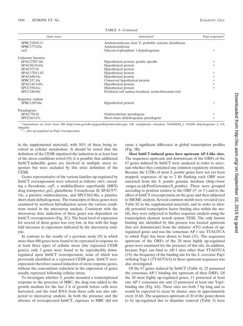
ature (Fig. 5). Overexpression of Int6CT did not cause anaccumulation of polyubiquitinylated proteins above wild-typelevels, and these levels were also lower than those of the mtsmutants at the permissive temperature. The overexpression ofInt6CT had a mild additive effect on the multidrug resistantphenotype of the proteasome mutants (our unpublished re-sults). These data suggest that the mechanism by which Int6CToverexpression causes multidrug resistance is not analogous tothat proposed for the mts mutants (26).
Int6CT-induced multidrug resistance is independent of theability of Pap1 to respond to oxidative stress. S. pombe cellsthat are subjected to oxidative stress relocalize Pap1 to thenucleus to activate Pap1-dependent transcription (34). Previ-ous microscopy data from our laboratory suggested that Pap1(tagged with GFP) did not accumulate in the nucleus uponInt6 overexpression (10). However, these conclusions were re-evaluated in the light of a recent study of Pap1 regulationfollowing oxidative stress (36), which demonstrated that thisnuclear relocalization is extremely transient, lasting approxi-mately 5 to 10 min.
Derepression of the nmt1 promoter takes approximately 14to 16 h and is therefore not as abrupt a stimulus as the additionof H2O2 to culture medium; therefore any potentially analo-gous effect of Int6CT overexpression on Pap1 localizationwould be markedly more gradual and difficult to detect byfluorescence microscopy. Fortunately it was possible to addressthis question using a mutant form of Pap1. Cysteine residue278 is critically required for the reversible oxidation and nu-clear accumulation of Pap1; mutation of this residue to alanine(Pap1-C278A) prevents Pap1 nuclear accumulation in re-sponse to oxidative stress (8).
S. pombe EHH14 (pap1-GFP) and EHH14.C278A (pap1-
C278A-GFP) were transformed with pREP4X or pREP4X-int6CT and derepressed by growth in the absence of thiamine.Int6CT overexpression was confirmed by immunoblotting (ourunpublished results). Resistance to 20 �g/ml MBC was in-duced by Int6CT overexpression in both pap1-GFP and pap1-C278A-GFP (Fig. 6), indicating that Int6CT-induced drug re-sistance is independent of redox regulation of Pap1 nuclearaccumulation.
DISCUSSION
This study demonstrates the activation of AP-1-dependenttranscription by the ectopic expression of a truncated transla-tion initiation factor, Int6CT. Earlier studies (1, 10) pointedtowards full-length Int6 being a component of the multisubunittranslation initiation factor eIF3, a view supported here by ourTAP-tagging data (Fig. 1C). While our earlier experimentsshowed that Int6-induced drug resistance in S. pombe wasgenetically dependent on pap1�, at that time it seemed mostlikely that the resistance was mediated through altered trans-lation (10). For example, Int6 overexpression might have fa-vored the translation of mRNAs transcribed in a pap1-depen-dent manner. We have shown in this study that overexpressionof Int6CT caused pap1-dependent multidrug resistance in S.pombe (Fig. 1A), which resulted from an increase in pap1-dependent transcription, rather than the stabilization of pap1-responsive mRNAs (Fig. 2). Pap1 overexpression caused thetranscriptional up-regulation of a variety of stress response-related genes. Remarkably, Int6CT overexpression induced atranscriptional profile almost identical to that seen upon Pap1overexpression (Table 4), and a large number of the targetgenes possess potential AP-1 binding sites in their 5� flankingregions (Fig. 4). Indeed, in the absence of Pap1, overexpres-sion of Int6CT did not result in the up-regulation of any of the67 genes shown in Table 4. Thus, genes lacking consensus Pap1sites in their promoter regions are also likely to be Pap1-dependent. Preliminary studies indicate that Pap1 is also ableto bind the Yap1 site in vitro (our unpublished results); byextension, it seems likely that a variety of related sites arebound in vivo.
Since Pap1 is normally activated in response to oxidativestress, it is pertinent to ask whether Int6CT expression simplyinduces a stress condition to which Pap1 responds. If this werethe case, the stress would appear to be quite specific, as thevast majority of S. pombe cDNAs (including, for example, a
FIG. 5. Int6CT overexpression does not perturb the ubiquitin-pro-teasome pathway. Abundance of polyubiquitylated proteins [indicatedby (Ub)n] in derepressed S. pombe CJN18 (WT/mts�) transformedwith pREP3X (vector) or pREP3X-int6CT (Int6CT) and S. pombe h�
leu1-32 CJN255 (mts2-1) and CJN278 (mts3-1) grown at the permissivetemperature (26°C) and the restrictive temperature (36°C) was exam-ined by immunoblotting with antiubiquitin antibodies. Int6CT overex-pression was confirmed by immunoblotting with anti-Int6 antibodies;Cdc2 served as a loading control.
FIG. 6. Int6CT-induced Pap1 activation is independent of redox-regulated Pap1 nuclear accumulation. Tenfold serial dilutions of mid-log phase cultures of derepressed S. pombe strains EHH14 (WT Pap1-GFP) and EHH14.C278A (Pap1-C278A-GFP) transformed withpREP3X (vector) or pREP3X-int6CT (Int6CT) were spotted ontominimal agar plates with or without 20 �g/ml MBC. Plates were pho-tographed after 4 days of incubation at 30°C.
1848 JENKINS ET AL. EUKARYOT. CELL
on Septem
ber 16, 2015 by guesthttp://ec.asm
.org/D
ownloaded from
cDNA encoding only the N-terminal region of Int6) fail toinduce Pap1 activation, as judged by the induction of drugresistance (10). Furthermore, a single missense mutation in thesequence encoding Int6CT can abolish Pap1 activation withoutaltering the steady-state level of Int6CT protein (E. Rawsonand C. J. Norbury, unpublished data). The failure of Int6CTexpression to trigger the pattern of transcriptional repressionthat characterizes S. pombe cells exposed to a variety ofstresses (9) also argues against the establishment of a consti-tutively stressed state. Instead, Int6CT appears able to triggerspecifically the Pap1 activation arm of the oxidative stressresponse in the absence of other aspects of this response.
How then does Int6CT activate Pap1? Previously, Pap1-responsive gene expression has been shown to be activated bya variety of pathways, resulting in a pattern of drug resistancealso seen when Pap1 is overexpressed (33). Conditional muta-tions in subunits of the 19S proteasome regulatory particlecause MBC resistance, and reduced proteolysis of Pap1 hasbeen proposed as the mechanism for this resistance (26). Im-pairment of Pap1 nuclear export, as a consequence of muta-tions in Crm1 or Pap1, results in Pap1-dependent drug resis-tance due to the accumulation of Pap1 in the nucleus (6, 20,35). Intriguingly, Pap1 activation by Int6CT overexpressiondoes not appear to involve any of these mechanisms; Pap1protein levels remain unchanged following Int6CT induction(Fig. 2F), and the ubiquitin-proteasome pathway is apparentlyunaffected (Fig. 5). Int6CT-induced Pap1 activation is alsoindependent of the COP9 signalosome and the Ras pathway(our unpublished data), with which endogenous Int6 has beenlinked previously (18, 38, 39), and independent of the recentlydescribed oxidative regulation of Pap1 localization (36); Fig.6).
The possibility that Int6/eIF3e, an accessory component of amultisubunit translation factor, might also act to modulateAP-1-dependent transcription is an intriguing one, but at thisstage it is unclear whether the activation of Pap1 by Int6CTrepresents a normal physiological mechanism. Fluorescencemicroscopy indicates that the majority of Int6/eIF3e is cyto-plasmic in S. pombe (10) and that the protein is relocalized togranular structures following cellular stress (13). The functionof these stress granules, which also contain other translationfactors and RNA, is not yet established in fission yeast; how-ever, analogous structures in human cells have been proposedto participate in recovery from stress by determining the fate ofstalled translation initiation complexes (2). In the light of thesefindings, it is conceivable that Int6 directs the selective trans-lation of a Pap1 coactivator under stressed conditions. Directinteraction between Int6 and transcription factors in the nu-cleus may also be possible; a significant proportion of Int6 isnuclear in human cells (37) and has been found to associatewith the human T-cell leukemia virus transactivator Tax (11).It is therefore conceivable that Int6CT interacts directly withPap1 to influence its activity, but our tandem affinity purifica-tion studies of Int6CT and Pap1 have not revealed any inter-actors other than heat shock proteins, which are common ar-tifacts of this technique (Fig. 1C). We cannot rule out thepossibility that a putative Int6CT-Pap1 interaction is eitherinsufficiently stable to withstand the purification process oroccurs in an insoluble subcellular fraction. A further possibilityis that the selective activation of Pap1 is a consequence of
biochemical properties of the Int6CT fragment that do notrelate to the physiological role of the full-length Int6 protein.An analogous activity has been ascribed to pathogenic variantsof the human huntingtin protein that include long polyglu-tamine tracts (12); such proteins are capable of inhibiting Sp1and TAFII130-dependent transcription, even though the wild-type huntingtin protein is not considered to function as a tran-scriptional regulator. Discrimination between the various pos-sible modes of Int6CT-mediated activation of Pap1 will requirefurther study.
Murine Int-6 was first identified as a gene truncated byintegration of mouse mammary tumor virus (MMTV) proviralDNA in mammary cancers and a preneoplastic outgrowth (22).Intriguingly the MMTV-truncated Int-6 alleles, which can in-duce malignant transformation in transfection assays (24, 27),encode Int6/eIF3e proteins lacking the C-terminal region cor-responding to the Int6CT fragment used in this study. It willnow be worthwhile to investigate the possible involvement oftranscriptional changes resulting from Int-6 truncation in theseMMTV-induced tumors.
ACKNOWLEDGMENTS
This work was supported by Cancer Research UK and the Associ-ation for International Cancer Research.
We thank Michael Mitchell of Cancer Research UK, London, forcarrying out the MEME analysis and William O’Gorman for help withthe preparation of samples for mass spectrometry. We also thankGavin Burns of the Wellcome Trust Sanger Institute, Cambridge,United Kingdom for his assistance with the microarray experiments,Norbert Kaufer and Elena Hidalgo for providing yeast strains, andElizabeth Patton for comments on the manuscript.
REFERENCES
1. Akiyoshi, Y., J. Clayton, L. Phan, M. Yamamoto, A. G. Hinnebusch, Y.Watanabe, and K. Asano. 2001. Fission yeast homolog of murine Int-6protein, encoded by mouse mammary tumor virus integration site, is asso-ciated with the conserved core subunits of eukaryotic translation initiationfactor 3. J. Biol. Chem. 276:10056–10062.
2. Anderson, P., and N. Kedersha. 2002. Stressful initiations. J. Cell Sci. 115:3227–3234.
3. Asano, K., W. C. Merrick, and J. W. Hershey. 1997. The translation initiationfactor eIF3-p48 subunit is encoded by int-6, a site of frequent integration bythe mouse mammary tumor virus genome. J. Biol. Chem. 272:23477–23480.
4. Bahler, J., J. Q. Wu, M. S. Longtine, N. G. Shah, A. McKenzie, III, A. B.Steever, A. Wach, P. Philippsen, and J. R. Pringle. 1998. Heterologousmodules for efficient and versatile PCR-based gene targeting in Schizosac-charomyces pombe. Yeast 14:943–951.
5. Bandyopadhyay, A., T. Matsumoto, and U. Maitra. 2000. Fission yeast Int6is not essential for global translation initiation, but deletion of int6� causeshypersensitivity to caffeine and affects spore formation. Mol. Biol. Cell.11:4005–4018.
6. Benko, Z., C. Fenyvesvolgyi, M. Pesti, and M. Sipiczki. 2004. The transcrip-tion factor Pap1/Caf3 plays a central role in the determination of caffeineresistance in Schizosaccharomyces pombe. Mol. Genet. Genomics 271:161–170.
7. Bonovich, M., M. Olive, E. Reed, B. O’Connell, and C. Vinson. 2002. Ad-enoviral delivery of A-FOS, an AP-1 dominant negative, selectively inhibitsdrug resistance in two human cancer cell lines. Cancer Gene Ther. 9:62–70.
8. Castillo, E. A., J. Ayte, C. Chiva, A. Moldon, M. Carrascal, J. Abian, N.Jones, and E. Hidalgo. 2002. Diethylmaleate activates the transcription fac-tor Pap1 by covalent modification of critical cysteine residues. Mol. Micro-biol. 45:243–254.
9. Chen, D., W. M. Toone, J. Mata, R. Lyne, G. Burns, K. Kivinen, A. Brazma,N. Jones, and J. Bahler. 2003. Global transcriptional responses of fissionyeast to environmental stress. Mol. Biol. Cell. 14:214–229.
10. Crane, R., R. Craig, R. Murray, I. Dunand-Sauthier, T. Humphrey, and C.Norbury. 2000. A fission yeast homolog of Int-6, the mammalian oncoproteinand eIF3 subunit, induces drug resistance when overexpressed. Mol. Biol.Cell. 11:3993–4003.
11. Desbois, C., R. Rousset, F. Bantignies, and P. Jalinot. 1996. Exclusion ofInt-6 from PML nuclear bodies by binding to the HTLV-I Tax oncoprotein.Science 273:951–953.
VOL. 4, 2005 TRUNCATED eIF3e ACTIVATES AP-1 1849
on Septem
ber 16, 2015 by guesthttp://ec.asm
.org/D
ownloaded from
12. Dunah, A. W., H. Jeong, A. Griffin, Y. M. Kim, D. G. Standaert, S. M.Hersch, M. M. Mouradian, A. B. Young, N. Tanese, and D. Krainc. 2002. Sp1and TAFII130 transcriptional activity disrupted in early Huntington’s dis-ease. Science 296:2238–2243.
13. Dunand-Sauthier, I., C. Walker, C. Wilkinson, C. Gordon, R. Crane, C.Norbury, and T. Humphrey. 2002. Sum1, a component of the fission yeasteIF3 translation initiation complex, is rapidly relocalized during environmen-tal stress and interacts with components of the 26S proteasome. Mol. Biol.Cell. 13:1626–1640.
14. Eferl, R., and E. F. Wagner. 2003. AP-1: a double-edged sword in tumori-genesis. Nat. Rev. Cancer 3:859–868.
15. Gordon, C., G. McGurk, P. Dillon, C. Rosen, and N. D. Hastie. 1993.Defective mitosis due to a mutation in the gene for a fission yeast 26Sprotease subunit. Nature 366:355–357.
16. Gordon, C., G. McGurk, M. Wallace, and N. D. Hastie. 1996. A conditionallethal mutant in the fission yeast 26 S protease subunit mts3� is defective inmetaphase to anaphase transition. J. Biol. Chem. 271:5704–5711.
17. Gross, T., K. Richert, C. Mierke, M. Lutzelberger, and N. F. Kaufer. 1998.Identification and characterization of srp1, a gene of fission yeast encodinga RNA binding domain and a RS domain typical of SR splicing factors.Nucleic Acids Res. 26:505–511.
18. Hoareau Alves, K., V. Bochard, S. Rety, and P. Jalinot. 2002. Association ofthe mammalian proto-oncoprotein Int-6 with the three protein complexeseIF3, COP9 signalosome and 26S proteasome. FEBS Lett. 527:15–21.
19. Jones, R. H., S. Moreno, P. Nurse, and N. C. Jones. 1988. Expression of theSV40 promoter in fission yeast: identification and characterization of anAP-1-like factor. Cell 53:659–667.
20. Kumada, K., M. Yanagida, and T. Toda. 1996. Caffeine-resistance in fissionyeast is caused by mutations in a single essential gene, crm1�. Mol. Gen.Genet. 250:59–68.
21. Lyne, R., G. Burns, J. Mata, C. J. Penkett, G. Rustici, D. Chen, C. Langford,D. Vetrie, and J. Bahler. 2003. Whole-genome microarrays of fission yeast:characteristics, accuracy, reproducibility, and processing of array data. BMCGenomics 4:27.
22. Marchetti, A., F. Buttitta, S. Miyazaki, D. Gallahan, G. H. Smith, and R.Callahan. 1995. Int-6, a highly conserved, widely expressed gene, is mutatedby mouse mammary tumor virus in mammary preneoplasia. J. Virol. 69:1932–1938.
23. Maundrell, K. 1993. Thiamine-repressible expression vectors pREP andpRIP for fission yeast. Gene 123:127–130.
24. Mayeur, G. L., and J. W. Hershey. 2002. Malignant transformation by theeukaryotic translation initiation factor 3 subunit p48 (eIF3e). FEBS Lett.514:49–54.
25. Moreno, S., A. Klar, and P. Nurse. 1991. Molecular genetic analysis of fissionyeast Schizosaccharomyces pombe. Methods Enzymol. 194:795–823.
26. Penney, M., C. Wilkinson, M. Wallace, J. P. Javerzat, K. Ferrell, M. Seeger,W. Dubiel, S. McKay, R. Allshire, and C. Gordon. 1998. The Pad1� geneencodes a subunit of the 26 S proteasome in fission yeast. J. Biol. Chem.273:23938–23945.
27. Rasmussen, S. B., E. Kordon, R. Callahan, and G. H. Smith. 2001. Evidencefor the transforming activity of a truncated Int6 gene, in vitro. Oncogene20:5291–5301.
28. Rigaut, G., A. Shevchenko, B. Rutz, M. Wilm, M. Mann, and B. Seraphin.1999. A generic protein purification method for protein complex character-ization and proteome exploration. Nat. Biotechnol. 17:1030–1032.
29. Schmidt, H., K. Richert, R. A. Drakas, and N. F. Kaufer. 1999. spp42,identified as a classical suppressor of prp4-73, which encodes a kinase in-volved in pre-mRNA splicing in fission yeast, is a homologue of the splicingfactor Prp8p. Genetics 153:1183–1191.
30. Schwartz, D. C., and R. Parker. 1999. Mutations in translation initiationfactors lead to increased rates of deadenylation and decapping of mRNAs inSaccharomyces cerevisiae. Mol. Cell Biol. 19:5247–5256.
31. Shimanuki, M., Y. Saka, M. Yanagida, and T. Toda. 1995. A novel essentialfission yeast gene pad1� positively regulates pap1�-dependent transcriptionand is implicated in the maintenance of chromosome structure. J. Cell Sci.108:569–579.
32. Toda, T., M. Shimanuki, Y. Saka, H. Yamano, Y. Adachi, M. Shirakawa, Y.Kyogoku, and M. Yanagida. 1992. Fission yeast pap1-dependent transcrip-tion is negatively regulated by an essential nuclear protein, crm1. Mol. CellBiol. 12:5474–5484.
33. Toda, T., M. Shimanuki, and M. Yanagida. 1991. Fission yeast genes thatconfer resistance to staurosporine encode an AP-1-like transcription factorand a protein kinase related to the mammalian ERK1/MAP2 and buddingyeast FUS3 and KSS1 kinases. Genes Dev. 5:60–73.
34. Toone, W. M., S. Kuge, M. Samuels, B. A. Morgan, T. Toda, and N. Jones.1998. Regulation of the fission yeast transcription factor Pap1 by oxidativestress: requirement for the nuclear export factor Crm1 (Exportin) and thestress-activated MAP kinase Sty1/Spc1. Genes Dev. 12:1453–1463.
35. Turi, T. G., P. Webster, and J. K. Rose. 1994. Brefeldin A sensitivity andresistance in Schizosaccharomyces pombe. Isolation of multiple genes con-ferring resistance. J. Biol. Chem. 269:24229–24236.
36. Vivancos, A. P., E. A. Castillo, N. Jones, J. Ayte, and E. Hidalgo. 2004.Activation of the redox sensor Pap1 by hydrogen peroxide requires modu-lation of the intracellular oxidant concentration. Mol. Microbiol. 52:1427–1435.
37. Watkins, S. J., and C. J. Norbury. 2004. Cell cycle-related variation insubcellular localization of eIF3e/INT6 in human fibroblasts. Cell Prolif.37:149–160.
38. Yahalom, A., T. H. Kim, E. Winter, B. Karniol, A. G. von Arnim, and D. A.Chamovitz. 2001. Arabidopsis eIF3e (INT-6) associates with both eIF3c andthe COP9 signalosome subunit CSN7. J. Biol. Chem. 276:334–340.
39. Yen, H. C., C. Gordon, and E. C. Chang. 2003. Schizosaccharomyces pombeInt6 and Ras homologs regulate cell division and mitotic fidelity via theproteasome. Cell 112:207–217.
40. Zhou, C., F. Arslan, S. Wee, S. Krishnan, A. R. Ivanov, A. Oliva, J. Leath-erwood, and D. A. Wolf. 2005. PCI proteins eIF3e and eIF3m define distincttranslation initiation factor 3 complexes. BMC Biol. 3:14.
1850 JENKINS ET AL. EUKARYOT. CELL
on Septem
ber 16, 2015 by guesthttp://ec.asm
.org/D
ownloaded from
Related Documents