of June 17, 2011 This information is current as .1100502 http://www.jimmunol.org/content/early/2011/06/13/jimmunol doi:10.4049/jimmunol.1100502 ; Prepublished online 13 June 2011; J Immunol Bear and Masoud H. Manjili D. Toor, Adly Yacoub, Xiang-Yang Wang, Lisa Smith, Harry Maciej Kmieciak, Debasmita Basu, Kyle K. Payne, Amir Transgenic Mouse Therapy against Breast Cancer in the FVBN202 Cells and Result in an Effective Adoptive Cellular Cells Resistant to Myeloid-Derived Suppressor Activated NKT Cells and NK Cells Render T Data Supplementary 02.DC1.html http://www.jimmunol.org/content/suppl/2011/06/13/jimmunol.11005 Subscriptions http://www.jimmunol.org/subscriptions is online at The Journal of Immunology Information about subscribing to Permissions http://www.aai.org/ji/copyright.html Submit copyright permission requests at Email Alerts http://www.jimmunol.org/etoc/subscriptions.shtml/ Receive free email-alerts when new articles cite this article. Sign up at initial publication. Advance online articles must include the digital object identifier (DOIs) and date of publication priority; they are indexed by PubMed from initial publication. Citations to available prior to final publication). Advance online articles are citable and establish not yet appeared in the paper journal (edited, typeset versions may be posted when Advance online articles have been peer reviewed and accepted for publication but have Print ISSN: 0022-1767 Online ISSN: 1550-6606. Immunologists, Inc. All rights reserved. by The American Association of Copyright ©2011 9650 Rockville Pike, Bethesda, MD 20814-3994. The American Association of Immunologists, Inc., is published twice each month by The Journal of Immunology on June 17, 2011 www.jimmunol.org Downloaded from

Welcome message from author
This document is posted to help you gain knowledge. Please leave a comment to let me know what you think about it! Share it to your friends and learn new things together.
Transcript

of June 17, 2011This information is current as
.1100502http://www.jimmunol.org/content/early/2011/06/13/jimmunoldoi:10.4049/jimmunol.1100502
; Prepublished online 13 June 2011;J Immunol Bear and Masoud H. Manjili
D.Toor, Adly Yacoub, Xiang-Yang Wang, Lisa Smith, Harry Maciej Kmieciak, Debasmita Basu, Kyle K. Payne, Amir Transgenic MouseTherapy against Breast Cancer in the FVBN202 Cells and Result in an Effective Adoptive CellularCells Resistant to Myeloid-Derived Suppressor Activated NKT Cells and NK Cells Render T
DataSupplementary
02.DC1.htmlhttp://www.jimmunol.org/content/suppl/2011/06/13/jimmunol.11005
Subscriptions http://www.jimmunol.org/subscriptions
is online atThe Journal of ImmunologyInformation about subscribing to
Permissions http://www.aai.org/ji/copyright.html
Submit copyright permission requests at
Email Alerts http://www.jimmunol.org/etoc/subscriptions.shtml/
Receive free email-alerts when new articles cite this article. Sign up at
initial publication. Advance online articles must include the digital object identifier (DOIs) and date ofpublication priority; they are indexed by PubMed from initial publication. Citations to available prior to final publication). Advance online articles are citable and establishnot yet appeared in the paper journal (edited, typeset versions may be posted when Advance online articles have been peer reviewed and accepted for publication but have
Print ISSN: 0022-1767 Online ISSN: 1550-6606.Immunologists, Inc. All rights reserved.
by The American Association ofCopyright ©2011 9650 Rockville Pike, Bethesda, MD 20814-3994.The American Association of Immunologists, Inc.,
is published twice each month byThe Journal of Immunology
on June 17, 2011w
ww
.jimm
unol.orgD
ownloaded from

The Journal of Immunology
Activated NKT Cells and NK Cells Render T Cells Resistantto Myeloid-Derived Suppressor Cells and Result in anEffective Adoptive Cellular Therapy against Breast Cancer inthe FVBN202 Transgenic Mouse
Maciej Kmieciak,*,1 Debasmita Basu,*,1 Kyle K. Payne,*,1 Amir Toor,† Adly Yacoub,‡
Xiang-Yang Wang,x Lisa Smith,{ Harry D. Bear,{ and Masoud H. Manjili*
Attempts to cure breast cancer by adoptive cellular therapy (ACT) have not been successful. This is primarily due to the presence of
tumor-induced immune-suppressive mechanisms as well as the failure of tumor-reactive T cells to provide long-term memory
responses in vivo. To address these clinically important challenges, we developed an ex vivo protocol for the expansion of
tumor-reactive immune cells obtained from tumor-bearing animals prior to or after local radiation therapy. We used an Ag-
free protocol that included bryostatin 1/ionomycin and sequential common g-chain cytokines (IL-7/IL-15 + IL-2). The proposed
protocol expanded tumor-reactive T cells as well as activated non-T cells, including NKT cells, NK cells, and IFN-g–producing
killer dendritic cells. Antitumor efficacy of T cells depended on the presence of non-T cells. The effector non-T cells also rendered
T cells resistant to myeloid-derived suppressor cells. Radiation therapy altered phenotypic distribution and differentiation of
T cells as well as their ability to generate central memory T cells. ACT by means of the expanded cells protected animals from
tumor challenge and generated long-term memory responses against the tumor, provided that leukocytes were derived from
tumor-bearing animals prior to radiation therapy. The ex vivo protocol was also able to expand HER-2/neu–specific T cells
derived from the PBMC of a single patient with breast carcinoma. These data suggest that the proposed ACT protocol should be
studied further in breast cancer patients. The Journal of Immunology, 2011, 187: 000–000.
The rationale for adoptive cellular therapy (ACT) for canceris based on overcoming the low frequency of endogenoustumor-reactive T cells by ex vivo activation and expan-
sion and to direct differentiation of T cells of interest toward themost effective phenotype(s). Several groups have shown that ACTdirected against melanoma-associated Ags results in objectiveresponses in animal models as well as in some melanoma patients
(1, 2). To improve objective responses of ACT, a number ofstrategies have been developed which include using geneticallymodified lymphocytes (3), highly effective T cell phenotypes (4)and use of common g-chain cytokines (5). However, unlike animalmodels, cancer patients usually receive ACT after conventionaltherapies, which could interfere with the efficacy of the donorT cells. No comparative analysis has been performed to determinewhether previous radiation therapy reduces or enhances the anti-tumor efficacy of ACT. ACT has also been tested against breastcancer both in mouse models and breast cancer patients (6, 7).However, unlike melanoma, ACT has not produced completeprotection against breast tumors. Barriers to success include dif-ficulty in the ex vivo expansion of tumor-reactive T cells (8),uncertainty as to the most relevant Ags, a lack of consensus as tothe appropriate origin of the T cells to be used for expansion aswell as phenotypic distribution of the most effective T cells,presence of myeloid-derived suppressor cells (MDSC) in cancerpatients and during premalignant carcinogenesis, which couldabrogate antitumor efficacy of ACT (7, 9), and finally tumorstroma as a major barrier, which prevents penetration of T cellsinto the solid tumor (10).In the current study, we addressed a number of key barriers listed
above to produce objective responses against primary breast tumorsand to generate long-term memory against recall tumor challenge.We took advantage of an Ag-free protocol for selective activation oftumor-primed immune cells by using bryostatin 1/ionomycin (B/I)as previously described by our group (7, 11). Bryostatin 1 activatesprotein kinase C and ionomycin increases intracellular calcium(12, 13). Taken together, B/I mimics signaling through the CD3/TcR complex and leads to activation and proliferation of tumor-primed T cells. Most recently, it was reported that bryostatin 1 can
*Department of Microbiology and Immunology, Virginia Commonwealth University,Massey Cancer Center, Richmond, VA 23298; †Department of Internal Medicine,Virginia Commonwealth University, Massey Cancer Center, Richmond, VA 23298;‡Department of Neurosurgery, Virginia Commonwealth University, Massey CancerCenter, Richmond, VA 23298; xDepartment of Human and Molecular Genetics,Virginia Commonwealth University, Massey Cancer Center, Richmond, VA 23298;and {Department of Surgery, Virginia Commonwealth University, Massey CancerCenter, Richmond, VA 23298
1M.K., D.B., and K.K.P. contributed equally to this work.
Received for publication February 16, 2011. Accepted for publication May 12, 2011.
This work was supported by National Institutes of Health Grant R01 CA104757 (toM.H.M.) and the Virginia Commonwealth University Presidential Research IncentiveProgram (to M.H.M.).
The study sponsors provided funding for the study and had no role in study design;the collection, analysis, and interpretation of data; the writing of the report; or thedecision to submit the paper for publication.
Address correspondence and reprint requests to Dr. Masoud H. Manjili, VirginiaCommonwealth University, Massey Cancer Center, Box 980035, 401 College Street,Richmond, VA 23298. E-mail address: [email protected]
The online version of this article contains supplemental material.
Abbreviations used in this article: ACT, adoptive cellular therapy; B/I, bryostatin 1/ionomycin; CYP, cyclophosphamide; DC, dendritic cell; GrB, granzyme B; IKDC,IFN-g–producing killer dendritic cells; MDSC, myeloid-derived suppressor cells;MMC, mouse mammary carcinoma; PI, propidium iodide; Prf, perforin; TCM, centralmemory T cells; TE, effector T cells; TEM, effector memory T cells.
Copyright� 2011 by The American Association of Immunologists, Inc. 0022-1767/11/$16.00
www.jimmunol.org/cgi/doi/10.4049/jimmunol.1100502
Published June 13, 2011, doi:10.4049/jimmunol.1100502 on June 17, 2011
ww
w.jim
munol.org
Dow
nloaded from
act as a TLR-4 ligand and activate innate immunity (14). We thendeveloped a sequential common g-chain cytokine protocol forexpansion of the B/I-activated tumor-primed immune cells. Thisex vivo protocol induced expansion of tumor-reactive immunecells composed of central memory T cells (TCM) and effectorT cells (TE) as well as cells of the innate immune system or non-T cells. The expanded cells were found to be resistant to MDSCand were capable of generating long-term memory responsesagainst the tumor in the FVBN202 transgenic mouse model ofHER-2/neu overexpressing breast carcinoma. We also showed thatHER-2/neu–specific T cells can be expanded from PBMC ofa breast cancer patient by using B/I activation and addition of thecommon g-chain cytokines ex vivo.
Materials and MethodsMouse model
FVBN202 transgenic female mice (Charles River Laboratories) were usedbetween 8 and 12 wk of age throughout these experiments. These miceoverexpress an unactivated rat neu transgene under the regulation of themouse mammary tumor virus promoter (15). These mice develop pre-malignant mammary hyperplasia similar to ductal carcinoma in situ priorto the development of spontaneous carcinoma (16). Premalignant events inFVBN202 mice include increased endogenous MDSC (16). These studieshave been reviewed and approved by the Institutional Animal Care andUse Committee at Virginia Commonwealth University.
Tumor cell lines
The neu overexpressing mouse mammary carcinoma (MMC) cell line wasestablished from a spontaneous mammary tumor harvested from FVBN202mice as previously described by our group (17). Tumor cells were main-tained in RPMI 1640 supplemented with 10% FBS. Mice were challengedwith 3–4 3 10⁶ MMC cells s.c. in the lower part of the mammary regionclose to the groin area.
Flow cytometry
Flow cytometry analyses were performed as previously described by ourgroup (16, 18). Briefly, spleens were disrupted into a single cell suspensionand 106 cells were aliquoted into each sample tube. Non-specific bindingof Abs to FcRs was blocked by incubating the cells with anti-CD16/32 Ab(BioLegend). Cells were stained with surface Abs toward various markersand incubated on ice in the dark for 20 min, then washed twice with cellstaining buffer (PBS, 1% FBS, 0.1% sodium azide) and fixed with 1%paraformaldehyde. For intracellular staining of perforin (Prf) and Foxp3,we followed the Foxp3 staining protocol provided by the manufacturer(BioLegend). Cells stained for granzyme B (GrB) and IFN-g were fixedwith 2.5% paraformaldehyde for 10 min on ice, washed twice with 0.1%saponin cell staining buffer, and then stained with the indicated Abs. Cellswere then washed twice with normal cell staining buffer and fixed with1% paraformaldehyde. For Annexin V staining, cells were stained forrespective surface markers, washed with cell staining buffer, and thenwashed with 13 Annexin V buffer (BD Pharmingen). The Annexin Vstaining protocol given in the product data sheet was then followed. Absused for flow cytometry were purchased from BioLegend (FITC-, PE-, PE/Cy5-CD3, FITC-, PE-, PE/Cy5-CD4, FITC-, PE-, PE/Cy5-, allophycocyanin-CD8, FITC-, PE/Cy5-CD11b, PE-, PE/Cy5-Gr1, FITC-, PE-CD25, FITC-CD44, PE-CD62L, PE-, allophycocyanin-CD49b, PE-CD69, FITC-, PE-CD122, FITC-, PE-CD127, FITC-, PE-B220, PE-IFN-g, and PE-Foxp3) oreBioscience (FITC-, allophycocyanin-, PE-Prf, PE-, and PE/Cy5-GrB). AllAbs were used at the manufacture’s recommended concentrations. Mul-ticolor data acquisition was performed on a BD FACSCanto II and ana-lyzed using BD FACSDiva software.
Cell sorting
To sort distinct cellular populations of splenocytes, 107 cells were addedto sample tubes in which FcRs were blocked and surface markers werestained with FITC-conjugated CD4 and PE-conjugated CD8 Abs. Cellswere then washed with sterile PBS supplemented with 2% FBS. T cellsand non-T cells were sorted into 100% FBS using a BD FACSAria III cellsorter. Purity of sorted cells was .98%.
Cytotoxicity assay
Freshly isolated tumor-primed splenocytes or the ex vivo-expanded cellswere cultured with MMC at a 10:1 E:T ratio in 3 ml complete medium
(RPMI 1640 supplemented with 100 U/ml penicillin, 100 mg/ml strepto-mycin, 10% FBS, 10 mM L-glutamine, and 5 3 1025M 2-ME) with 20 U/ml IL-2 (PeproTech) in 6-well culture dishes. After 48 h, cells were har-vested and stained for neu (anti–c-Erb2/c-Neu; Calbiochem), Annexin V,and propidium iodide (PI) according to the manufacturer’s protocol (BDPharmingen). Flow cytometry was used to analyze the viability of neu+ cells.
IFN-g ELISA
Freshly isolated tumor-primed splenocytes or the ex vivo-expanded cellswere cultured in complete medium at a 10:1 ratio with irradiatedMMC cells(14,000 rad) for 24 h. Supernatants were then collected and stored at280˚Cuntil assayed. IFN-g was detected using a Mouse IFN-g ELISA set (BDPharmingen) according to the manufacturer’s protocol.
Expansion of effectors T cells from FVBN202 mice
FVBN202 transgenic mice were inoculated with 4 3 106 MMC cells, andsplenocytes were harvested after 21–25 d. Splenocytes (106 cells/ml) werethen stimulated in complete medium containing 15% FBS as well asbryostatin 1 (5 nM)/ionomycin (1 mM), and 80 U/ml IL-2 for 16 h, aspreviously described by our group (12). Cells were then washed threetimes and cultured at 106cells/ml in complete medium with 10–20 ng/mleach of IL-7 and IL-15 (PeproTech). After 24 h, 20 U/ml IL-2 was addedto the culture. On the next day, cells were washed three times and culturedat 106cells/ml in complete medium with 40 U/ml IL-2. Cells were split andcultured at 106cells/ml in complete medium with 40 U/ml IL-2 every otherday for a total of 6 d. After day 6, cells were then used for ACT or in vitrostudies.
Adoptive cellular therapy
Twenty-four hours prior to ACT, FVBN202 mice were injected i.p. withCYP (100 mg/kg) to induce lymphopenia. Mice were challenged with 3 3106 MMC cells and then received 70 3 106 of the expanded cells by tailvein injection later the same day. Tumor growth was monitored by digitalcaliper, and tumor volumes were calculated by volume (v) = (L [length] 3W [width]2)/2.
Isolation of MDSC in vitro
The Gr1+ MDSC population was isolated using an EasySep-FITC selectionkit from StemCell Technologies, as previously described by our group (7,19). We have previously shown that endogenous MDSC isolated from bonemarrow or secondary lymphoid tissues can inhibit T cell responsivenessto anti-CD3/anti-CD28 Abs (7, 19–22). Also, the Ly6C+Ly6G2 subset butnot the Ly6G+Ly6C+ subset isolated from FVBN202 mice was found to besuppressive (19). Because of the higher proportion of Ly6C+Ly6G2 subsetin tumor-bearing mice compared with tumor-free mice, the suppressiveeffects of MDSC isolated from tumor-bearing mice was greater using anoptimal 2:1 ratio of T cells to MDSC (19). Therefore, in this study, we usedan optimal 2:1 ratio of splenic MDSC isolated from tumor-bearingFVBN202 mice.
Expansion of HER-2/neu–specific T cells from PBMC
PBMC were harvested from a breast cancer patient under InstitutionalReview Board protocol HM10920. PBMC were cultured at 37˚C for 2 h;adherent cells were used for the generation of monocyte-derived dendriticcells (DCs) in the presence of GM-CSF and IL-4, as previously describedby our group (17). Floater cells were split into two groups. One group wasmaintained with IL-2 (40 U/ml/106 cells) for 6–7 d until autologous DCsbecame available. Another group was activated with B/I and expandedwith common g-chain cytokines. The expanded cells or IL-2–maintainedcells were cultured with autologous DCs (4:1) in the presence or absenceof recombinant HER-2/neu (100 mg/ml) or LPS (10 mg/ml). After 24 h,supernatants and cells were collected and subjected to IFN-g ELISA andflow cytometry analysis, respectively.
Recombinant HER-2/neu protein
Extracellular domain and intracellular domain of human HER-2/neu proteinwere expressed in Escherichia coli and purified by Ni-NTA–Agarose (Qia-gen, Valencia, CA), as previously described by our group (23). Concentrationof the recombinant proteins was determined using the Bradford assay.
Statistical analysis
Graphical data are presented as means with SEs. Statistical comparisonsbetween groups were made using Student t test with p , 0.05 being sta-tistically significant.
2 A NOVEL CELLULAR THERAPY FOR BREAST CANCER
on June 17, 2011w
ww
.jimm
unol.orgD
ownloaded from
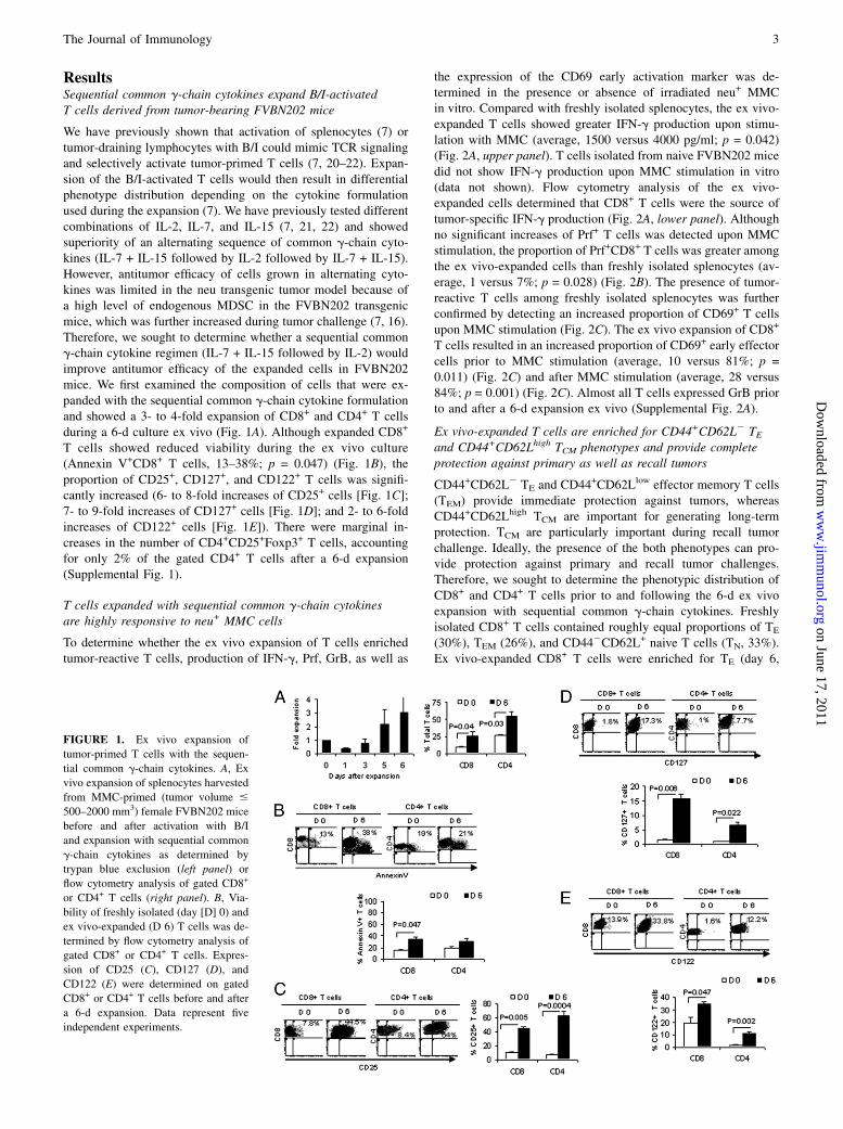
ResultsSequential common g-chain cytokines expand B/I-activatedT cells derived from tumor-bearing FVBN202 mice
We have previously shown that activation of splenocytes (7) ortumor-draining lymphocytes with B/I could mimic TCR signalingand selectively activate tumor-primed T cells (7, 20–22). Expan-sion of the B/I-activated T cells would then result in differentialphenotype distribution depending on the cytokine formulationused during the expansion (7). We have previously tested differentcombinations of IL-2, IL-7, and IL-15 (7, 21, 22) and showedsuperiority of an alternating sequence of common g-chain cyto-kines (IL-7 + IL-15 followed by IL-2 followed by IL-7 + IL-15).However, antitumor efficacy of cells grown in alternating cyto-kines was limited in the neu transgenic tumor model because ofa high level of endogenous MDSC in the FVBN202 transgenicmice, which was further increased during tumor challenge (7, 16).Therefore, we sought to determine whether a sequential commong-chain cytokine regimen (IL-7 + IL-15 followed by IL-2) wouldimprove antitumor efficacy of the expanded cells in FVBN202mice. We first examined the composition of cells that were ex-panded with the sequential common g-chain cytokine formulationand showed a 3- to 4-fold expansion of CD8+ and CD4+ T cellsduring a 6-d culture ex vivo (Fig. 1A). Although expanded CD8+
T cells showed reduced viability during the ex vivo culture(Annexin V+CD8+ T cells, 13–38%; p = 0.047) (Fig. 1B), theproportion of CD25+, CD127+, and CD122+ T cells was signifi-cantly increased (6- to 8-fold increases of CD25+ cells [Fig. 1C];7- to 9-fold increases of CD127+ cells [Fig. 1D]; and 2- to 6-foldincreases of CD122+ cells [Fig. 1E]). There were marginal in-creases in the number of CD4+CD25+Foxp3+ T cells, accountingfor only 2% of the gated CD4+ T cells after a 6-d expansion(Supplemental Fig. 1).
T cells expanded with sequential common g-chain cytokinesare highly responsive to neu+ MMC cells
To determine whether the ex vivo expansion of T cells enrichedtumor-reactive T cells, production of IFN-g, Prf, GrB, as well as
the expression of the CD69 early activation marker was de-termined in the presence or absence of irradiated neu+ MMCin vitro. Compared with freshly isolated splenocytes, the ex vivo-expanded T cells showed greater IFN-g production upon stimu-lation with MMC (average, 1500 versus 4000 pg/ml; p = 0.042)(Fig. 2A, upper panel). T cells isolated from naive FVBN202 micedid not show IFN-g production upon MMC stimulation in vitro(data not shown). Flow cytometry analysis of the ex vivo-expanded cells determined that CD8+ T cells were the source oftumor-specific IFN-g production (Fig. 2A, lower panel). Althoughno significant increases of Prf+ T cells was detected upon MMCstimulation, the proportion of Prf+CD8+ T cells was greater amongthe ex vivo-expanded cells than freshly isolated splenocytes (av-erage, 1 versus 7%; p = 0.028) (Fig. 2B). The presence of tumor-reactive T cells among freshly isolated splenocytes was furtherconfirmed by detecting an increased proportion of CD69+ T cellsupon MMC stimulation (Fig. 2C). The ex vivo expansion of CD8+
T cells resulted in an increased proportion of CD69+ early effectorcells prior to MMC stimulation (average, 10 versus 81%; p =0.011) (Fig. 2C) and after MMC stimulation (average, 28 versus84%; p = 0.001) (Fig. 2C). Almost all T cells expressed GrB priorto and after a 6-d expansion ex vivo (Supplemental Fig. 2A).
Ex vivo-expanded T cells are enriched for CD44+CD62L2 TEand CD44+CD62Lhigh TCM phenotypes and provide completeprotection against primary as well as recall tumors
CD44+CD62L2 TE and CD44+CD62Llow effector memory T cells(TEM) provide immediate protection against tumors, whereasCD44+CD62Lhigh TCM are important for generating long-termprotection. TCM are particularly important during recall tumorchallenge. Ideally, the presence of the both phenotypes can pro-vide protection against primary and recall tumor challenges.Therefore, we sought to determine the phenotypic distribution ofCD8+ and CD4+ T cells prior to and following the 6-d ex vivoexpansion with sequential common g-chain cytokines. Freshlyisolated CD8+ T cells contained roughly equal proportions of TE
(30%), TEM (26%), and CD442CD62L+ naive T cells (TN, 33%).Ex vivo-expanded CD8+ T cells were enriched for TE (day 6,
FIGURE 1. Ex vivo expansion of
tumor-primed T cells with the sequen-
tial common g-chain cytokines. A, Ex
vivo expansion of splenocytes harvested
from MMC-primed (tumor volume #
500–2000 mm3) female FVBN202 mice
before and after activation with B/I
and expansion with sequential common
g-chain cytokines as determined by
trypan blue exclusion (left panel) or
flow cytometry analysis of gated CD8+
or CD4+ T cells (right panel). B, Via-
bility of freshly isolated (day [D] 0) and
ex vivo-expanded (D 6) T cells was de-
termined by flow cytometry analysis of
gated CD8+ or CD4+ T cells. Expres-
sion of CD25 (C), CD127 (D), and
CD122 (E) were determined on gated
CD8+ or CD4+ T cells before and after
a 6-d expansion. Data represent five
independent experiments.
The Journal of Immunology 3
on June 17, 2011w
ww
.jimm
unol.orgD
ownloaded from

55.6% versus day 0, 30%; p = 0.02) and TCM (day 6, 26% versusday 0, 7.2%; p = 0.008) (Fig. 3A). Ex vivo-expanded CD4+ T cellsshowed an unchanged proportion of TE (day 6, 31.9% versus day0, 26.3%) but were enriched for TCM (day 6, 61.3% versus day 0,6.6%; p = 0.002). TN phenotypes almost disappeared in the ex-panded CD8+ T cells (day 6, 1.8% versus day 0, 33.7%; p = 0.009)
and CD4+ T cells (day 6, 2% versus day 0, 14.1%; p = 0.003).Such a phenotypic distribution toward CD8+ TE and TCM sug-gests the potential for immediate as well as long-term memoryresponses against the tumor. We then performed in vitro cyto-toxicity assays and in vivo tests of tumor growth inhibition todetermine the antitumor efficacy of the expanded cells. Freshly
FIGURE 2. Ex vivo-expanded, MMC-primed T cells respond to MMC cells in vitro. A, Tumor reactivity of freshly isolated (day [D] 0) and ex vivo-
expanded (D 6) T cells was determined by a 24-hs culture of T cells in the presence or absence of irradiated MMC cells, followed by the detection of IFN-g
in the supernatant (upper panel). Medium alone and MMC alone were used as negative controls for IFN-g production. The MMC-specific IFN-g production
in gated CD8+ and CD4+ T cells was determined by flow cytometry analysis (lower panel). The MMC-specific expression of Prf (B) and CD69 (C) were
determined in gated T cells. Data represent five independent experiments.
FIGURE 3. Phenotypic distribution of tumor-reactive T cells and their antitumor efficacy in vitro and in vivo. A, Phenotypic distribution of freshly
isolated (day [D] 0) and ex vivo-expanded (D 6) splenic T cells was determined by flow cytometry analysis of gated CD8+ or CD4+ T cells. Distribution of
T cell phenotypes including CD44+CD62L2 effector (E: TE), CD44+CD62Llow effector memory (EM: TEM), CD44
+CD62Lhigh central memory (CM: TCM),
and CD442CD62L+ naive (N: TN) was determined. B, Gated neu+ MMC cells were analyzed for apoptosis (Annexin V+/PI+) in the absence or presence of
freshly isolated or ex vivo-expanded T cells. C, CYP-treated FVBN202 mice (n = 3) were inoculated with MMC cells and received no further treatment (left
panel) or received ACT (middle panel). Animals that had rejected MMC following ACTwere given rest for 2 mo and then were challenged with MMC cells
on the contralateral side (right panel). Data represent five independent experiments.
4 A NOVEL CELLULAR THERAPY FOR BREAST CANCER
on June 17, 2011w
ww
.jimm
unol.orgD
ownloaded from

isolated splenocytes or expanded T cells were cultured with viableneu+ MMC tumor cells in an E:T ratio of 10:1 for 2 d. Gated neu+
MMC cells were then analyzed for the detection of apoptosis asdetermined by Annexin V+/PI+ cells. As shown in Fig. 3B, freshlyisolated T cells reduced viability of neu+ MMC from 87.5 to50.79% (1.7-fold), whereas the ex vivo-expanded T cells dis-played greater cytotoxic function, reducing the viability of MMCfrom 68.8 to 17.6% (3.9-fold).To test in vivo efficacy of expanded T cells, we used FVBN202
mice, which harbor increased MDSC because of premalignantmammary hyperplasia preceding spontaneous mammary tumors.Endogenous MDSCs were further increased upon MMC tumorchallenge (7, 16). In this study, we injected FVBN202 mice withcyclophosphamide (CYP) 1 d prior to ACT to generate a semi-lymphopenic condition. Animals were then challenged with MMC,followed by i.v. injection of the ex vivo-expanded cells 6–8 h afterthe MMC challenge. Recipients of ACT rejected the neu+ MMC(Fig. 3C), despite the presence of MDSC before and 7 d afterMMC challenge (Supplemental Fig. 2B). All control mice that hadreceived CYP alone developed tumors. To determine memoryresponses, ACT-treated mice were challenged on the contralateralside with MMC 2 mo after the rejection of primary MMC cells.During recall tumor challenge, animals received neither CYPnor ACT, yet all the mice rejected the recall tumors (Fig. 3C).To determine which T cell phenotypes were effective in vivo, wesorted T cells into CD62L2/low (TE/TEM) and CD62Lhigh (TCM)and performed ACT with the sorted cells. No protection was ob-
served against the tumors (data not shown). These data suggestcritical interactions among tumor-reactive T cell phenotypes,which require further investigation.
The ex vivo-expanded T cells acquire resistance to inhibitoryfunction of MDSC
Because the ex vivo-expanded cells protected FVBN202 miceagainst primary and recall tumor challenges even in the absence ofMDSC depletion (Fig. 3C), we sought to determine whether the exvivo-expanded cells were resistant to MDSC in vitro. We havepreviously reported that MDSC isolated from bone marrow orspleens of tumor-bearing FVBN202 mice can inhibit T cell re-sponsiveness to CD3/CD28 stimulation (7, 19). MDSC can alsoinhibit MMC tumor-specific IFN-g production by T cells ex-panded with alternating common g-chain cytokines (SupplementalFig. 2C). Therefore, splenocytes expanded with sequential com-mon g-chain cytokines were cultured for 24 h in the presence orabsence of irradiated MMC (10:1 ratio of expanded cells to MMC)to show their reactivity with MMC in the presence or absence ofsplenic MDSC (2:1 ratio of expanded cells to MDSC). Superna-tants were collected and subjected to IFN-g ELISA. T cells werealso analyzed for the expression of IFN-g, Prf, GrB, and CD69. Asshown in Fig. 4A, upper panel, the ex vivo-expanded cells pro-duced IFN-g in the presence of MMC (p = 0.004) as expected.Importantly, addition of MDSC not only failed to suppress MMC-specific IFN-g secretion by the expanded cells but also increasedthe IFN-g response (p = 0.012). Flow cytometry analysis of the
FIGURE 4. Ex vivo-expanded T cells are resistant to MDSC. The ex vivo-expanded T cells were cultured with irradiated MMC in the presence or
absence of MDSC. MMC-specific IFN-g was detected in the supernatant of a 24-h culture (A, upper panel) and in gated CD8+ or CD4+ T cells (A, middle
and bottom panels). B, MMC-specific Prf production in gated CD8+ or CD4+ T cells was also determined. C, The ex vivo-expanded T cells were cultured
with viable MMC cells in the presence or absence of MDSC for 48 h. Viability (Annexin V2/PI2) of gated neu+ MMC cells was determined by flow
cytometry analysis. Data represent three independent experiments.
The Journal of Immunology 5
on June 17, 2011w
ww
.jimm
unol.orgD
ownloaded from

expanded cells determined that CD8+ T cells were the main sourceof MMC-specific IFN-g production (Fig. 4A, lower panel). Ad-dition of MDSC increased MMC-induced production of IFN-g byboth CD8+ and CD4+ T cells. Increasing the dose of MDSC didnot increase MMC-specific production of IFN-g by the expandedT cells (data not shown). The presence of MDSC also resulted inan increased production Prf in CD8+ T cells upon MMC stimu-lation (p = 0.035) (Fig. 4B). There was no IFN-g or Prf productionby MMC as determined by flow cytometry analysis of gated neu+
MMC in the coculture (Supplemental Fig. 3A). In addition, MDSCdid not suppress production of GrB or expression of the CD69early activation marker in the expanded T cells (Supplemental Fig.3B, 3C). PBMC from naive FVBN202 mice that was depleted ofGr1+ cells had no suppressive effect or supportive effect on tumor-reactive T cells (data not shown). These data suggest that MDSCdid not inhibit antitumor responses of the T cells expanded withthe sequential g-chain cytokine regimen. We next determinedwhether MDSC could inhibit cytotoxicity of these T cells againstMMC tumor cells in vitro. Expanded cells were cultured withviable neu+ MMC (10:1 ratio) in the presence or absence ofMDSC (2:1 ratio). Control MMC cells were cultured with mediumalone. As shown in Fig. 4C, expanded cells induced apoptosis inMMC cells in a 2-d culture, and the presence of MDSC did notalter the cytotoxic function of the tumor-reactive cells.
Presence of non-T cells in the ex vivo-expanded cellsovercomes MDSC and enhances T cell responses to MMC cells
The ex vivo-expanded cells showed a significantly reduced pro-portion of CD42CD82 cells compared with that of freshly isolatedsplenocytes (day 6, 20% versus day 0, 57%; p = 0.0001) (Fig. 5A).The expanded cells contained 17–20% CD42CD82 cells. As
shown in Fig. 5B, gated CD42CD82 cells contained a signifi-cantly higher proportion of CD3+ cells in the expanded cellscompared with freshly isolated cells (day 6, 60.8% versus day 0,5.5%; p = 0.002). Then, we sought to determine the cellularcomposition of these CD42CD82 cells. CD49b is a commonmarker for NK cells, NKT cells, and IFN-g–producing killer DC(IKDC) (24). As shown in Fig. 5C, NKT cells (CD49b+CD3+) andNK cells (CD49b+CD32) showed significant increases in the ex-panded CD42CD82 cells. The proportion of IKDC (CD49b+
CD32B220+) in the gated CD32CD11b+ cells was also signifi-cantly increased after the expansion (p = 0.001) (Fig. 5D). TheCD3+CD49+ NKT cells and CD32CD49b+ NK cells showedhigher expression of the activation marker CD25 after a 6-d ex-pansion (day 6) compared with freshly isolated cells (day 0)(Supplemental Fig. 3D). The expanded CD3+ non-T cells(NKT cells) showed higher viability compared with the expandedCD32 non-T cells (NK cells and IKDC) (Fig. 5E: 71.9 versus37.5% on day 6; p = 0.004).
To determine whether the presence of non-T cells renders thetumor-reactive T cells resistant to MDSC, in vitro and in vivostudies were performed on the sorted cells. The ex vivo-expandedcells were sorted into CD4+ plus CD8+ T cells and CD42CD82
non-T cells. Sorted cells were then cultured for 24 h in the pres-ence or absence of irradiated MMC (10:1 ratio of expanded cellsto MMC) to show their reactivity with MMC in the presenceor absence of MDSC (2:1 ratio of expanded cells to MDSC).Supernatants were collected and subjected to IFN-g ELISA. Asshown in Fig. 5F, MDSC induced secretion of IFN-g by CD42
CD82 non-T cells in the presence of MMC but not in CD4+ plusCD8+ T cells (p = 0.031). Lower amounts of IFN-g were secretedby sorted CD4+ plus CD8+ T cells compared with unsorted cells
FIGURE 5. Ex vivo-expanded T cells in-
clude non-T cells (NKT cells, NK cells, and
IKDC) that are responsible for rendering
T cells resistant to MDSC. Freshly isolated
(day [D] 0) and ex vivo-expanded (D 6)
splenic cells were analyzed for the presence of
CD42CD82 non-T cells and CD4+ or CD8+
T cells (A). Expression of CD3 on non-T cells
was determined (B). Non-T cells were also
analyzed to determine the proportion of NKT
cells and NK cells (C). Cells were gated on
CD32CD11b+ DC population and analyzed
for the expression of CD49b and B220 as well
as IFN-g to identify IKDC population (D).
Viability (Annexin V2/PI2) of CD3+ and
CD32 non-T cells was determined (E). Ex
vivo-expanded splenocytes from MMC-primed
FVBN202 mice were sorted into non-T cells
(CD42CD82) and T cells (CD4+ plus CD8+).
The sorted cells were cultured with irradiated
MMC in the presence or absence of MDSC for
24 h, and IFN-g production was determined in
the supernatants (F). The sorted cells were used
for ACT (n = 3) at doses proportional to the
unsorted cells, and tumor growth was deter-
mined (G). Data represent three independent
experiments.
6 A NOVEL CELLULAR THERAPY FOR BREAST CANCER
on June 17, 2011w
ww
.jimm
unol.orgD
ownloaded from

(107 pg/ml in Fig. 5F compared with 2642 pg/ml in Fig. 4A). Thissuggests that the presence of non-T cells boosts the tumor re-activity of T cells. In addition, the presence of MDSC signifi-cantly increased MMC-induced IFN-g production by non-T cells(p = 0.006). However, CD4+ plus CD8+ T cells in the absence ofnon-T cells lost their ability to secrete MMC-specific IFN-g whileMDSC were present. This suggests that MDSC-stimulated, MMC-activated non-T cells render T cells resistant to MDSC. ACT withsorted T cells or non-T cells failed to protect FVBN202 mice fromchallenge with MMC cells (Fig. 5G). Because IL-12 induces theexpression of IFN-g by NK cells and T cells, we sought to de-termine whether CD42CD82CD49b+ cells produce IL-12 in thepresence of MMC and MDSC, resulting in the induction of en-hanced IFN-g production by T cells. No IL-12 production wasdetected in non-T cells or T cells (data not shown).
Radiation therapy of tumor-bearing mice prior to the isolationof donor T cells results in failure of the expanded T cells togenerate objective responses upon ACT despite sustainedantitumor responses of the T cells in vitro
Cancer patients who participate in clinical trials of ACT haveusually received conventional therapies, often including radiationtherapy. Therefore, it is important to determine whether tumor-primed T cells that were isolated following radiation therapy can
also be expanded and can generate objective responses against thetumors following ACT. To test this, FVBN202 mice were in-oculated with MMC cells, and as soon as tumors reached 75–150mm3, animals received three doses of local radiation therapy to thetumor site (5 Gy) in a 3-d interval. Animals were then sacrificed1 wk after the last radiation treatment, and their splenocytes weresubjected to B/I activation and a 6-d expansion with sequentialcommon g-chain cytokines. The frequency of freshly isolatedT cells was significantly lower after radiation therapy (Fig. 6A)compared with that without radiation therapy (Fig. 1A) (CD8+
T cells, p = 0.002; CD4+ T cells, p = 0.0002). However, radiationtherapy did not alter the ability of T cells to grow after B/I acti-vation, such that after 6 d in culture, the cells showed similar ratesof expansion compared with those from mice that did not receiveany radiation (Figs. 1A, 6A). The frequency of apoptotic T cellsdid not increase during the ex vivo expansion (Supplemental Fig.4A). However, expanded T cells from mice subjected to radiationfailed to increase CD127 (Supplemental Fig. 4B), and increasedexpression of CD122 was evident only in CD8+ T cells (Supple-mental Fig. 4C).To determine the tumor reactivity of the expanded T cells from
mice whose tumors had been irradiated, in vitro studies wereperformed. As shown in Fig. 6B, freshly isolated T cells (day 0)and ex vivo-expanded cells (day 6) failed to produce significant
FIGURE 6. In vitro and in vivo efficacy of the ex vivo-expanded and MMC-primed T cells harvested from FVBN202 mice after radiation therapy. A, Ex
vivo expansion of splenocytes harvested from MMC-primed female FVBN202 mice that received three cycles of local radiation therapy (5 Gy) in 3-
d intervals before and after activation with B/I and expansion with sequential common g-chain cytokines as determined by trypan blue exclusion (left panel)
or flow cytometry analysis of gated CD8+ or CD4+ T cells (right panel). B, Tumor reactivity of freshly isolated (day [D] 0) and ex vivo-expanded (D 6)
T cells was determined by a 24-h culture of T cells in the presence or absence of irradiated MMC, followed by the detection of IFN-g in the supernatant.
Medium alone and MMC alone were used as negative controls for IFN-g production. C, The MMC-specific IFN-g production in gated CD8+ and CD4+
T cells derived from donor mice with no radiation treatment (NT) and radiation treatment (RAD) was determined by flow cytometry analysis. D, Gated neu+
MMC cells were analyzed for apoptosis (Annexin V+/PI+) in the absence or presence of freshly isolated or ex vivo-expanded T cells. E, Phenotypic
distribution of freshly isolated (D 0) and ex vivo-expanded (D 6) splenic T cells was determined by flow cytometry analysis of gated CD4+ or CD8+ T cells.
Data represent three independent experiments. F, CYP-treated FVBN202 mice (n = 3) were inoculated with MMC and received no further treatment
(control) or received the expanded cells derived from donors after the local radiation therapy (ACT). Data represent three independent experiments.
The Journal of Immunology 7
on June 17, 2011w
ww
.jimm
unol.orgD
ownloaded from

amounts of IFN-g upon MMC stimulation. However, the additionof MDSC resulted in the induction of IFN-g by the expandedT cells (Fig. 6B). The expanded cells were also composed of19.2% non-T cells (Supplemental Fig. 4D). Interestingly, theproportion of Prf+ T cells in the expanded T cells (day 6) wasmarkedly higher in this group that had received prior radiationtherapy compared with T cells that were isolated from donors withno prior radiation therapy (Fig. 6C). Expanded CD8+ T cells werehighly positive for the expression of CD69 and GrB (Supple-mental Fig. 4E, 4F). Importantly, lack of IFN-g production by theexpanded T cells did not alter the ability of these cells to kill neu+
MMC cells in vitro such that viability (Annexin V2/PI2) of MMCwas reduced from 64.64 to 12.35% in the presence of the ex-panded T cells (Fig. 6D). In addition, expanded T cells were ableto kill neu+ MMC cells even in the presence of MDSC in vitro, asshown by a reduced viability from 64.64 to 12.35 and 8.46% (Fig.6D).
Results of in vivo studies presented in Fig. 3 suggest a correla-tion between phenotypic distribution of T cells and objective re-sponses following ACT such that high proportions of TCM wereassociated with the rejection of primary and recall tumor chal-lenge. Therefore, we performed phenotype analysis of post-radiation T cells before proceeding with ACT. As shown in Fig.6E, phenotypic distribution of CD8+ and CD4+ T cells was dif-ferent from those isolated from animals with no prior radiationtherapy (Fig. 3A). Freshly isolated CD8+ T cells (D 0) weremainly of TCM and TN phenotypes, whereas CD4+ T cells con-tained TE and TEM (Fig. 6E). After 6 d of ex vivo expansion, CD8+
T cells were enriched for TE, whereas CD4+ T cells contained TE
and TEM phenotypes. After a 6-d expansion, CD8+ TCM and TN
phenotypes had almost disappeared. An increased proportion ofCD8+ TE cells may account for the in vitro efficacy of the ex-panded T cells against MMC cells, although antitumor efficacyin vivo may require tumor-specific TCM. To test this possibility,ACT studies were performed as described above. As shown inFig. 6F, ACT using the ex vivo-expanded cells from mice whosetumors had been irradiated showed minimal tumor inhibitoryeffects compared with the control group. The rate of tumor growthwas the same in the two groups (p , 0.21), although the changefrom day to day was significant (p , 0.0001).
HER-2/neu–specific T cells can be expanded from PBMC ofa patient with breast cancer
Splenocytes isolated from tumor-bearing mice with no radiationtherapy were effective against the neu+ MMC cells despite thelack of splenic tumor metastasis. This suggests that tumor-reactiveT cells may be present in the circulation. To test this, PBMCwere collected from a breast cancer patient after Ficoll–Paquegradient centrifugation of blood and split into two fractions. Ad-herent cells were selected by 2-h culture and used for generatingautologous DCs by a 6-d culture in the presence of GM-CSF andIL-4, as described elsewhere (25). Nonadherent cells were splitinto two fractions. One fraction was maintained in complete RPMI1640 supplemented with 10% FBS and IL-2 (40 U/ml/106 cells) at37˚C/5% CO2 for 6 d (IL-2–maintained cells). The remaining cellswere subjected to B/I activation and expansion with the commong-chain cytokines (expanded cells with IL-7, IL-15, and IL-2).Viability of the T cells (Annexin V2) after a 6-d culture or exvivo expansion was .85% (data not shown). Cells were thencocultured with autologous DCs in the presence or absence ofrecombinant HER-2/neu protein for 24 h. To rule out nonspecificIFN-g production by CD4+ T cells as a result of low endotoxinlevels in the HER-2/neu recombinant protein, we pulsed controlwells with LPS. As shown in Fig. 7A and 7B, compared with IL-2–
maintained T cells, the B/I-activated and expanded cells producedsignificantly higher amounts of IFN-g when stimulated withHER-2/neu (average, 8,600 versus 32,500 pg/ml; p = 0.001). Flowcytometry analysis of IL-2–maintained and ex vivo-expandedT cells determined that both CD8+ T cells and CD4+ T cellswere sources of HER-2/neu–stimulated IFN-g production (Fig.7C). It is yet to be determined whether consistently similar resultscan be obtained in a large group of breast cancer patients.
DiscussionDevelopment of an ex vivo protocol that can expand highly efficientpopulations of tumor-reactive immune cells, which include cellsof the adaptive and innate immune systems, may be the key tosuccessful ACT in breast cancer model. Others have reported thatrejection of mammary carcinoma in HER-2/neu transgenic micedepends on the stimulation of both innate and adaptive immunity(25). In addition, NKT cells have been shown to be involved insecondary antitumor T cell responses (26).We demonstrated that activation of tumor-primed lymphoid cells
with B/I, followed by ex vivo expansion with sequential commong-chain cytokines, can activate and expand tumor-reactive T cellsand non-T cells including NKT cells, NK cells, and IKDC. We
FIGURE 7. Patient with breast cancer harbors peripheral HER-2/neu–
specific T cell precursors which can be activated by B/I activation and
expanded with common g-chain cytokines. A, T cells maintained with low-
dose IL-2 (40 U/ml) for 6–7 d were cultured in the presence or absence of
autologous DCs and in the presence or absence of recombinant human
HER-2/neu for 24 h. IFN-g production was detected in the supernatant of
triplicate wells. B, B/I-activated and common g-chain cytokine-expanded
T cells were cultured in the presence or absence of autologous DCs and in
the presence or absence of recombinant human HER-2/neu for 24 h. IFN-g
production was detected in the supernatant of triplicate wells. C, IL-2–
maintained T cells (N) and ex vivo-expanded T cells (n) were stained with
anti-CD4, anti-CD8, and anti–IFN-g Abs to determine cellular source of
HER-2/neu–specific IFN-g production. Data represent two independent
experiments.
8 A NOVEL CELLULAR THERAPY FOR BREAST CANCER
on June 17, 2011w
ww
.jimm
unol.orgD
ownloaded from

have tested a number of the cytokine combinations (7, 21, 22) andfound that IL-7 + IL-15, followed by IL-2, was the best sequencefor the expansion of the most effective cells. The presence ofactivated non-T cells in the expanded cells was critical not only forthe in vivo antitumor efficacy of T cells but also for their resistanceto MDSC. The absence of such activated non-T cells in freshlyisolated splenocytes or depletion of these non-T cells in the ex-panded cells resulted in susceptibility to MDSC-induced sup-pression of tumor-reactive T cells. Neither tumor-reactive T cellsnor these non-T cells alone were able to protect FVBN202 miceagainst tumor challenge when they were used separately in ACT.Because the viability of NK cells was very low (37.5%) as op-posed to the high viability (72%) of NKT cells, it is likely thatNKT cells are the key component of the supportive cells. Ourfindings are consistent with recent reports showing that cells of theinnate and adaptive immune system work together to produceobjective responses against tumors (25, 26). Our results alsoshowed that MDSC can further activate the ex vivo-expanded non-T cells, as shown by an increased CD25 expression, thereby en-hancing the supportive function of non-T cells for tumor-reactiveT cells. To our knowledge, this is the first report showing a cellularmechanism by which T cells may become resistant to MDSC.Because of the T cell inhibitory role of MDSC in a variety ofcancers, the proposed protocol could be applicable to a variety ofcarcinomas.Although the presence of T cells and cells of the innate immune
system were critical for antitumor efficacy of the immune response,long-term protection against the tumor depended on the presence ofTCM. Our data suggest that B/I activation and ex vivo expansionwith sequential common g-chain cytokines may have improvedthe quality of neu-specific cells for tumor rejection, and it was notjust because of an increase in frequency of neu-specific T cells.For instance, freshly isolated T cells from tumor-bearing but notfrom tumor-free FVBN202 mice produced IFN-g upon stimula-tion with MMC in vitro; yet, such increased frequency of en-dogenous neu-specific T cells did not induce tumor rejection indonor mice. In addition, ACT with an increased numbers offreshly isolated T cells derived from tumor-bearing donors (2 3109 cells/mouse) did not protect mice against tumor challenge(data not shown). These data suggest that an increase in neu-specific T cells without ex vivo expansion/differentiation usingthe proposed protocol cannot provide protection against the tumor.Whereas T cells from nonirradiated and postradiation donorsshowed comparable levels of antitumor efficacy in vitro (Fig. 6D),T cells obtained from nonirradiated donors provided long-termmemory responses against recall tumor challenge in vivo, likelybecause of the phenotypic distribution of T cells toward TCM.Local radiation therapy of the tumors in donor mice altered thephenotypic distribution of freshly isolated T cells as well as thecapacity of T cells to differentiate into TCM during the ex vivoexpansion. These data suggest that CD8+ TE and TEM may bemore susceptible to radiation therapy than previously establishedTCM, as has been reported by others (27). Our data suggest thatlocal radiation therapy could alter the differentiation of tumor-reactive CD8+ TE and TEM toward TCM. However, we performedlocal radiation therapy of primary tumors, whereas breast cancerpatients usually receive radiation therapy after surgery to destroyresidual microscopic disease. Also, patients with advanced breastcancer have undergone multiple radiation treatments, followed bya period of recovery prior to ACT. These scenarios are somewhatdifferent from the treatment protocol that we used in our study.For T cells to be effective for ACT, we had to isolate T cells fromtumor-bearing animals. Therefore, we had to perform radiationtherapy on primary tumors. Application of the proposed approach
may be limited to patients with early-stage breast cancer (stagesI–III), provided that PBMC are harvested and cryopreserved priorto radiation therapy for ACT in future. Feasibility of this strategyremains to be determined. The importance of TCM against cancerhas also been reported by others (4). Our data showing a greaterantitumor efficacy of ACT in association with the presence of TCM
are consistent with other reports showing that effector cells de-rived from TCM rather than TEM possess greater ability to surviveand establish immunologic memory following infusion (28).However, naive T cells have been reported to convey more anti-tumor activity than memory cells (28). Such contradictory resultsmay be due to the use of a mouse model harboring a transgenicTCR for gp100 tumor Ag, which is different from the FVB202mouse model of spontaneous breast carcinoma with no transgenicTcR against the tumor Ag.IL-7 has been shown to support viability and homeostatic
proliferation of T cells and enhance NK cell function (29). IL-15supports differentiation of memory T cells and activation of qui-escent NK cells more efficiently than IL-2 (30). IL-2 is T cellgrowth factor and is also involved in NK cell activation andproliferation. Therefore, culture of tumor-primed T cells initiallywith IL-7 + IL-15, followed by IL-2, can support differentiation ofT cells as well as non-T cells. The presence of non-T cells in thismodel appears to be critical for rendering T cells resistant toMDSC, regardless of whether T cells were obtained before or afterradiation therapy. A similar observation has been made in miceand humans with hematologic malignancies undergoing alloge-neic stem cell transplantation. In the animal model, depletion ofdonor NK cells abrogated antileukemia effects of donor T cells(31). In humans, early donor-derived NK cell recovery has beenshown to be associated with a lower relapse risk in the non-myeloablative setting in the recipients of T cell-replete allografts(32). Earlier observations in the same clinical model had dem-onstrated a significant impact of NK cell dose in the graft on day28 T cell chimerism (33). Similarly, our group has shown a trendtoward a higher level donor T cell chimerism at 12 wk post-transplant, in patients with superior NK cell recovery at 4 wk (datanot shown). Taken together, these data provide intriguing evidenceof T cell–NK cell interactions in the clinical transplant setting andsuggest interdependence between innate and adaptive immunity.Altogether, these data suggest that lymph nodes (11) or PBMC
of breast cancer patients may be explored as a source of tumor-reactive immune cells for ex vivo expansion and use in ACT,provided that immune cells are obtained prior to radiation therapyand expanded with the sequential common g-chain cytokines.Expanded T cells and non-T cells obtained prior to radiationtherapy may be able to be cryopreserved and used for experi-mental ACT protocols after the completion of conventional ther-apies in an attempt to eliminate residual disease and prevent tumorrelapse. In fact, the majority of patients with breast cancer die oftumor metastases rather than from the primary cancer. Althoughtumor stroma in breast cancer patients makes regression of es-tablished solid tumors difficult, regression of residual disease ispossible (6, 10). Therefore, the proposed ACT protocol should befurther explored in breast cancer patients. We have previouslytested antitumor efficacy of ACT using different common g-chaincytokine regimens (22). The efficacy of cells expanded with se-quential common g-chain cytokines remains to be determined inmelanoma and other tumor models.
AcknowledgmentsWe thank the Virginia Commonwealth University Massey Cancer Center
and the Commonwealth Foundation for Cancer Research for support.
The Journal of Immunology 9
on June 17, 2011w
ww
.jimm
unol.orgD
ownloaded from

DisclosuresThe authors have no financial conflicts of interest.
References1. Yee, C., J. A. Thompson, D. Byrd, S. R. Riddell, P. Roche, E. Celis, and
P. D. Greenberg. 2002. Adoptive T cell therapy using antigen-specific CD8+
T cell clones for the treatment of patients with metastatic melanoma: in vivopersistence, migration, and antitumor effect of transferred T cells. Proc. Natl.Acad. Sci. USA 99: 16168–16173.
2. Dudley, M. E., J. R. Wunderlich, P. F. Robbins, J. C. Yang, P. Hwu,D. J. Schwartzentruber, S. L. Topalian, R. Sherry, N. P. Restifo, A. M. Hubicki,et al. 2002. Cancer regression and autoimmunity in patients after clonal repo-pulation with antitumor lymphocytes. Science 298: 850–854.
3. Johnson, L. A., R. A. Morgan, M. E. Dudley, L. Cassard, J. C. Yang,M. S. Hughes, U. S. Kammula, R. E. Royal, R. M. Sherry, J. R. Wunderlich,et al. 2009. Gene therapy with human and mouse T-cell receptors mediatescancer regression and targets normal tissues expressing cognate antigen. Blood114: 535–546.
4. Berger, C., M. C. Jensen, P. M. Lansdorp, M. Gough, C. Elliott, andS. R. Riddell. 2008. Adoptive transfer of effector CD8+ T cells derived fromcentral memory cells establishes persistent T cell memory in primates. J. Clin.Invest. 118: 294–305.
5. Liu, S., J. Riley, S. Rosenberg, and M. Parkhurst. 2006. Comparison of commong-chain cytokines, interleukin-2, interleukin-7, and interleukin-15 for the in vitrogeneration of human tumor-reactive T lymphocytes for adoptive cell transfertherapy. J. Immunother. 29: 284–293.
6. Bernhard, H., J. Neudorfer, K. Gebhard, H. Conrad, C. Hermann, J. Nahrig,F. Fend, W. Weber, D. H. Busch, and C. Peschel. 2008. Adoptive transfer ofautologous, HER2-specific, cytotoxic T lymphocytes for the treatment of HER2-overexpressing breast cancer. Cancer Immunol. Immunother. 57: 271–280.
7. Morales, J. K., M. Kmieciak, L. Graham, M. Feldmesser, H. D. Bear, andM. H. Manjili. 2009. Adoptive transfer of HER2/neu-specific T cells expandedwith alternating g chain cytokines mediate tumor regression when combinedwith the depletion of myeloid-derived suppressor cells. Cancer Immunol.Immunother. 58: 941–953.
8. Ho, W. Y., C. Yee, and P. D. Greenberg. 2002. Adoptive therapy with CD8+
T cells: it may get by with a little help from its friends. J. Clin. Invest. 110:1415–1417.
9. Diaz-Montero, C. M., M. L. Salem, M. I. Nishimura, E. Garrett-Mayer,D. J. Cole, and A. J. Montero. 2009. Increased circulating myeloid-derivedsuppressor cells correlate with clinical cancer stage, metastatic tumor burden,and doxorubicin-cyclophosphamide chemotherapy. Cancer Immunol. Immun-other. 58: 49–59.
10. Blankenstein, T. 2005. The role of tumor stroma in the interaction between tu-mor and immune system. Curr. Opin. Immunol. 17: 180–186.
11. Bear, H. D., J. Roberts, D. Cornell, M. B. Tombes, and B. Kyle. 2001. Adoptiveimmunotherapy of cancer with pharmacologically activated lymph node lympho-cytes: a pilot clinical trial. Cancer Immunol. Immunother. 50: 269–274.
12. Kazanietz, M. G., N. E. Lewin, F. Gao, G. R. Pettit, and P. M. Blumberg. 1994.Binding of [26-3H]bryostatin 1 and analogs to calcium-dependent and calcium-independent protein kinase C isozymes. Mol. Pharmacol. 46: 374–379.
13. Chatila, T., L. Silverman, R. Miller, and R. Geha. 1989. Mechanisms of T cellactivation by the calcium ionophore ionomycin. J. Immunol. 143: 1283–1289.
14. Ariza, M. E., R. Ramakrishnan, N. P. Singh, A. Chauhan, P. S. Nagarkatti, andM. Nagarkatti. 2011. Bryostatin-1, a naturally occurring antineoplastic agent,acts as a Toll-like receptor 4 (TLR-4) ligand and induces unique cytokines andchemokines in dendritic cells. J. Biol. Chem. 286: 24–34.
15. Guy, C. T., M. A. Webster, M. Schaller, T. J. Parsons, R. D. Cardiff, andW. J. Muller. 1992. Expression of the neu protooncogene in the mammary ep-ithelium of transgenic mice induces metastatic disease. Proc. Natl. Acad. Sci.USA 89: 10578–10582.
16. Kmieciak, M., J. K. Morales, J. Morales, E. Bolesta, M. Grimes, andM. H. Manjili. 2008. Danger signals and nonself entity of tumor antigen are bothrequired for eliciting effective immune responses against HER-2/neu positivemammary carcinoma: implications for vaccine design. Cancer Immunol.Immunother. 57: 1391–1398.
17. Kmieciak, M., M. Gowda, L. Graham, K. Godder, H. D. Bear, F. M. Marincola,and M. H. Manjili. 2009. Human T cells express CD25 and Foxp3 upon acti-
vation and exhibit effector/memory phenotypes without any regulatory/suppressor function. J. Transl. Med. 7: 89.
18. Worschech, A., M. Kmieciak, K. L. Knutson, H. D. Bear, A. A. Szalay, E. Wang,F. M. Marincola, and M. H. Manjili. 2008. Signatures associated with rejectionor recurrence in HER-2/neu-positive mammary tumors. Cancer Res. 68: 2436–2446.
19. Morales, J. K., M. Kmieciak, K. L. Knutson, H. D. Bear, and M. H. Manjili.2010. GM-CSF is one of the main breast tumor-derived soluble factors involvedin the differentiation of CD11b–Gr1– bone marrow progenitor cells into myeloid-derived suppressor cells. Breast Cancer Res. Treat. 123: 39–49.
20. Le, H. K., L. Graham, E. Cha, J. K. Morales, M. H. Manjili, and H. D. Bear.2009. Gemcitabine directly inhibits myeloid derived suppressor cells in BALB/cmice bearing 4T1 mammary carcinoma and augments expansion of T cells fromtumor-bearing mice. Int. Immunopharmacol. 9: 900–909.
21. Cha, E., L. Graham, M. H. Manjili, and H. D. Bear. 2010. IL-7 + IL-15 aresuperior to IL-2 for the ex vivo expansion of 4T1 mammary carcinoma-specificT cells with greater efficacy against tumors in vivo. Breast Cancer Res. Treat.122: 359–369.
22. Le, H. K., L. Graham, C. H. Miller, M. Kmieciak, M. H. Manjili, and H. D. Bear.2009. Incubation of antigen-sensitized T lymphocytes activated with bryostatin1 + ionomycin in IL-7 + IL-15 increases yield of cells capable of inducing re-gression of melanoma metastases compared to culture in IL-2. Cancer Immunol.Immunother. 58: 1565–1576.
23. Manjili, M. H., R. Henderson, X.-Y. Wang, X. Chen, Y. Li, E. Repasky,L. Kazim, and J. R. Subjeck. 2002. Development of a recombinant HSP110-HER-2/neu vaccine using the chaperoning properties of HSP110. Cancer Res.62: 1737–1742.
24. Terme, M., G. Mignot, E. Ullrich, M. Bonmort, V. Minard-Colin, A. Jacquet,J. L. Schultze, G. Kroemer, C. Leclerc, N. Chaput, and L. Zitvogel. 2009. Thedendritic cell-like functions of IFN-producing killer dendritic cells reside inthe CD11b+ subset and are licensed by tumor cells. Cancer Res. 69: 6590–6597.
25. Spadaro, M., E. Ambrosino, M. Iezzi, E. Di Carlo, P. Sacchetti, C. Curcio,A. Amici, W.-Z. Wei, P. Musiani, P. L. Lollini, et al. 2005. Cure of mammarycarcinomas in Her-2 transgenic mice through sequential stimulation of innate(neoadjuvant interleukin-12) and adaptive (DNA vaccine electroporation) im-munity. Clin. Cancer Res. 11: 1941–1952.
26. Hong, C., H. Lee, Y. K. Park, J. Shin, S. Jung, H. Kim, S. Hong, and S. H. Park.2009. Regulation of secondary antigen-specific CD8+ T-cell responses by naturalkiller T cells. Cancer Res. 69: 4301–4308.
27. De Ruysscher, D., M. Waer, M. Vandeputte, R. Aerts, K. Vantongelen, andE. van der Schueren. 1992. Changes of lymphocyte subsets after local irradiationfor early stage breast cancer and seminoma testis: long-term increase of activated(HLA-DR+) T cells and decrease of “naive” (CD4-CD45R) T lymphocytes. Eur.J. Cancer 28A: 1729–1734.
28. Hinrichs, C. S., Z. A. Borman, L. Cassard, L. Gattinoni, R. Spolski, Z. Yu,L. Sanchez-Perez, P. Muranski, S. J. Kern, C. Logun, et al. 2009. Adoptivelytransferred effector cells derived from naive rather than central memory CD8+
T cells mediate superior antitumor immunity. Proc. Natl. Acad. Sci. USA 106:17469–17474.
29. Tan, J. T., E. Dudl, E. LeRoy, R. Murray, J. Sprent, K. I. Weinberg, andC. D. Surh. 2001. IL-7 is critical for homeostatic proliferation and survival ofnaive T cells. Proc. Natl. Acad. Sci. USA 98: 8732–8737.
30. Pillet, A. H., F. Bugault, J. Theze, L. A. Chakrabarti, and T. Rose. 2009. Aprogrammed switch from IL-15– to IL-2–dependent activation in human NKcells. J. Immunol. 182: 6267–6277.
31. De Somer, L., B. Sprangers, S. Fevery, O. Rutgeerts, C. Lenaerts, L. Boon,M. Waer, and A. D. Billiau. 2011. Recipient lymphocyte infusion in MHC-matched bone marrow chimeras induces a limited lymphohematopoietic host-versus-graft reactivity but a significant antileukemic effect mediated by CD8+
T cells and natural killer cells. Haematologica 96: 424–431.32. Baron, F., E. W. Petersdorf, T. Gooley, B. M. Sandmaier, M. Malkki,
T. R. Chauncey, D. G. Maloney, and R. Storb. 2009. What is the role for donornatural killer cells after nonmyeloablative conditioning? Biol. Blood MarrowTransplant. 15: 580–588.
33. Panse, J. P., S. Heimfeld, K. A. Guthrie, M. B. Maris, D. G. Maloney, B. B. Baril,M. T. Little, T. R. Chauncey, B. E. Storer, R. Storb, and B. M. Sandmaier. 2005.Allogeneic peripheral blood stem cell graft composition affects early T-cellchimaerism and later clinical outcomes after non-myeloablative conditioning.Br. J. Haematol. 128: 659–667.
10 A NOVEL CELLULAR THERAPY FOR BREAST CANCER
on June 17, 2011w
ww
.jimm
unol.orgD
ownloaded from
Related Documents