1 19 ‘ VOLUME: 45 ISSUE: 3 ACTA VETERINARIA EURASIA Formerly Journal of the Faculty of Veterinary Medicine Istanbul University Official Journal of Istanbul University-Cerrahpasa Faculty of Veterinary Medicine ISSN 2618-639X • EISSN 2619-905X actaveteurasia.istanbulc.edu.tr

Welcome message from author
This document is posted to help you gain knowledge. Please leave a comment to let me know what you think about it! Share it to your friends and learn new things together.
Transcript

1
19‘
VOLUME: 45 ISSUE: 3
ACTA VE TER INAR IA EURAS IAFormerly Journal of the Faculty of Veter inary Medicine Istanbul University
Official Journal of Istanbul University-Cerrahpasa Faculty of Veter inary Medicine
ISSN 2618-639X • EISSN 2619-905X actaveteurasia.istanbulc.edu.tr

Publisherİbrahim KARA
Publication DirectorAli ŞAHİN
Editorial DevelopmentGizem KAYAN
Finance and AdministrationZeynep YAKIŞIRER ÜREN
Deputy Publication DirectorGökhan ÇİMEN
Publication CoordinatorsBetül ÇİMENÖzlem ÇAKMAKOkan AYDOĞANİrem DELİÇAYArzu YILDIRIM
Project CoordinatorsSinem KOZDoğan ORUÇ
Graphics DepartmentÜnal ÖZERDeniz DURANBeyzanur KARABULUT
Contact Address: Büyükdere Cad. 105/9 34394 Mecidiyeköy, Şişli, İstanbul, TURKEYPhone: +90 212 217 17 00Fax: +90 212 217 22 92E-mail: [email protected]
A- I
Editor in ChiefSerhat ALKANDepartment of Reproduction and Artificial Insemination, İstanbul University-Cerrahpaşa, Faculty of Veterinary Medicine, İstanbul, [email protected] ID: 0000-0003-4741-1416
EditorsAli AYDINDepartment of Food Hygiene and Technology, İstanbul University-Cerrahpaşa, Faculty of Veterinary Medicine, İstanbul, [email protected] ID: 0000-0002-4931-9843
Atila ATEŞ Department of Biochemistry, İstanbul University-Cerrahpaşa, Faculty of Veterinary Medicine, İstanbul, [email protected] ID: 0000-0002-9013-930X
Bülent EKİZDepartment of Animal Breeding and Husbandry, İstanbul University-Cerrahpaşa, Faculty of Veterinary Medicine, İstanbul, [email protected] ID: 0000-0001-6458-5747
Gülcan DEMİRELDepartment of Animal Nutrition and Nutritional Diseases, İstanbul University-Cerrahpaşa, Faculty of Veterinary Medicine, İstanbul, [email protected] ID: 0000-0002-6864-5134
İsmail KIRŞANDepartment of Obstetrics and Gynecology, İstanbul University-Cerrahpaşa, Faculty of Veterinary Medicine, İstanbul, [email protected] ID: 0000-0003-0780-0118
Hüseyin YILMAZDepartment of Virology, İstanbul University-Cerrahpaşa, Faculty of Veterinary Medicine, İstanbul, [email protected] ID: 0000-0002-7897-2358
Karlo MURATOĞLUDepartment of Food Hygiene and Technology, İstanbul University-Cerrahpaşa, Faculty of Veterinary Medicine, İstanbul, [email protected] ID: 0000-0001-8705-6813
O. B. Burak ESENERDepartment of Histology and Embryology, İstanbul University-Cerrahpaşa, Faculty of Veterinary Medicine, İstanbul, [email protected] ID: 0000-0001-9444-3598
Özge TURNADepartment of Obstetrics and Gynecology, İstanbul University-Cerrahpaşa, Faculty of Veterinary Medicine, İstanbul, [email protected] ID: 0000-0002-7638-0519
İstanbul Üniversitesi-Cerrahpaşa Veteriner Fakültesi adına sahibi / Owner on behalf of the İstanbul University-Cerrahpaşa Faculty of Veterinary Medicine: Güven Kaşıkçı • Sorumlu Yazı İşleri Müdürü / Responsible Manager: Serhat Alkan • Yayın türü / Publication Type: Yerel süreli / Local Periodical • Basım yeri / Printed at: Hamdioğulları İç ve Dış Ticaret A.Ş. Zübeyde Hanım Mah. Elif Sokak No.7/197 Altındağ, Ankara, Türkiye. Tel: +90 (542) 695 77 60 • Basım tarihi / Printing Date: Eylül 2019 / September 2019
Editorial Management
ACTA VE TER INAR IA EURAS IAFormerly Journal of the Faculty of Veter inary Medicine Istanbul University

A-I I
Altay USSENBAYEVFaculty of Veterinary Medicine, S. Seifullin Kazakh Agro Technical University, Astana, Kazakhstan
Anton GERILOVICHNational Scientific Center ‘Institute of Experimental and Clinical Veterinary Medicine’, Kharkov, Ukraine
Eduardo BERRIATUADepartment of Animal Health Murcia, University of Murcia, Spain
Georgios FTHENAKISUniversity of Thessaly, School of Health Sciences, Faculty of Veterinary Science, Department of Obstetrics and Reproduction, Karditsa, Greece
Gianluca NEGLIADepartment of Animal Science and Food Hygiene, Faculty of Veterinary Medicine, University of Naples Federico II, Naples, Italy
Giuseppe CAMPANILEDepartment of Animal Science and Food Hygiene, Faculty of Veterinary Medicine, University of Naples Federico II, Naples, Italy
Jurgen A. RICHTDepartment of Diagnostic Medicine/Pathobiology, Kansas State University, USA
Kamil BOSTANDepartment of Gastronomy and Culinary Arts, İstanbul Aydın University, İstanbul, Turkey
Melih AKSOYDepartment of Reproduction and Artificial Insemination, Adnan Menderes University, Aydın, Turkey
Munir IQBALThe Pirbright Institute, Avian Viral Diseases Programme, UK
Mustafa HASÖKSÜZDepartment of Virology, İstanbul University-Cerrahpaşa, Faculty of Veterinary Medicine, İstanbul, Turkey
Nasko VASILEVDepartment of Obstetrics, Reproduction and Reproductive Disorders, Veterinary Faculty University of Trakia, Bulgaria
Nikos PAPAIOANNOUDepartment of Pathology, Faculty of Veterinary Medicine, Aristotle University of Thessaloniki, Greece
Rao Zahid ABBASDepartment of Parasitology, University of Agriculture Faisalabad, Pakistan
Rizah AVDICUniversity of Sarajevo, Faculty of Veterinary Medicine, Bosnia and Herzegovina
Sema BİRLERDepartment of Reproduction and Artificial Insemination, İstanbul University-Cerrahpaşa, Faculty of Veterinary Medicine, İstanbul, Turkey
Stanimir YOTOVDepartment of Obstetrics, Reproduction and Reproductive Disorders- Faculty of Veterinary Medicine, Trakia University, Bulgaria
Vedat ONARDepartment of Anatomy, İstanbul University-Cerrahpaşa, Faculty of Veterinary Medicine, İstanbul, Turkey
Editorial and Advisory Board
ACTA VE TER INAR IA EURAS IAFormerly Journal of The Faculty of Veter inary Medicine Istanbul University

Aims and Scope
Acta Veterinaria Eurasia (Acta Vet Eurasia) is an internation-al, scientific, open access periodical published in accor-dance with independent, unbiased, and double-blinded peer-review principles. The journal is the official publica-tion of İstanbul University-Cerrahpaşa Faculty of Veteri-nary Medicine and published three times a year (January, May and September). The publication language of the journal is English.
Acta Veterinaria Eurasia (Acta Vet Eurasia) aims to contrib-ute to the literature by publishing manuscripts at the high-est scientific level on all fields of veterinary medicine. The journal publishes original articles, reviews, case reports, short communications, and letters to the editor that are prepared in accordance with the ethical guidelines.
The scope of the journal covers all animal species includ-ing the topics related to basic and clinical veterinary sci-ences, livestock breeding and husbandry, veterinary ge-netics, animal nutrition and nutritional diseases, zooneses, veterinary medicinal products and public health, and food hygiene and technology.
The target audience of the journal includes specialists and professionals working and interested in all disciplines of veterinary medicine. The editorial and publication processes of the journal are shaped in accordance with the guidelines of the Interna-tional Committee of Medical Journal Editors (ICMJE), World Association of Medical Editors (WAME), Council of Science Editors (CSE), Committee on Publication Ethics (COPE), Eu-ropean Association of Science Editors (EASE), and National Information Standards Organization (NISO). The journal is in conformity with the Principles of Transparency and Best Practice in Scholarly Publishing (doaj.org/bestpractice).
Acta Veterinaria Eurasia is currently indexed in Web of Science-Emerging Sources Citation Index, Web of Sci-ence-Zoological Records, Scopus, DOAJ, Embase, Gale, Proquest, AgBiotechNet, Animal Breeding Abtracts, An-imal Science Database, CAB Abstracts, Dairy Science Ab-stract, Helmintological Abstracts, Index Veterinarius, Nu-trition Abstracts and Reviews Series B: Livestock Feeds, Nutrition and Food Database, Parasitology Database, Poul-
try Abstracts, Review of Medical and Veterinary Mycology, Tropical Diseases Bulletin, Veterinary Bulletin, VetMed Re-source, TUBITAK ULAKBIM TR Index.
All expenses of the journal are covered by the of İstanbul University-Cerrahpaşa Faculty of Veterinary Medicine. Pro-cessing and publication are free of charge with the jour-nal. No fees are requested from the authors at any point throughout the evaluation and publication process. All manuscripts must be submitted via the online submission system, which is available at actaveteurasia.istanbulc.edu.tr. The journal guidelines, technical information, and the required forms are available on the journal’s web page.
Statements or opinions expressed in the manuscripts pub-lished in the journal reflect the views of the author(s) and not the opinions of the İstanbul University-Cerrahpaşa Faculty of Veterinary Medicine, editors, editorial board, and/or publisher; the editors, editorial board, and publish-er disclaim any responsibility or liability for such materials. Acta Veterinaria Eurasia is an open access publication and the journal’s publication model is based on Budapest Open Access Initiative (BOAI) declaration. Journal’s archive is available online, free of charge at http://actaveteurasia.istanbulc.edu.tr. Acta Veterinaria Eurasia’s content is li-censed under a Creative Commons Attribution-NonCom-mercial 4.0 International License.
Editor in Chief: Serhat AlkanAddress: İstanbul University-Cerrahpaşa Faculty of Veterinary Medicine, 34320 Avcılar, İstanbul, TurkeyPhone: +90 (212) 473 70 70/17260Fax: +90 (212) 473 72 40E-mail: [email protected]
Publisher: AVESAddress: Büyükdere Avenue, 105/9 34394 Mecidiyeköy, Şişli, İstanbul, TurkeyPhone: +90 212 217 17 00Fax: +90 212 217 22 92E-mail: [email protected] page: avesyayincilik.com
A- I I I
ACTA VE TER INAR IA EURAS IAFormerly Journal of The Faculty of Veter inary Medicine Istanbul University
Instructions to Authors
Aims and ScopeActa Veterinaria Eurasia (Acta Vet Eurasia) is an international, sci-entific, open access periodical published in accordance with inde-pendent, unbiased, and double-blinded peer-review principles. The journal is the official publication of Faculty of Veterinary Medicine, İstanbul University-Cerrahpaşa and published three times in a year (January, May, and September). The publication language of the journal is English. Acta Veterinaria Eurasia (Acta Vet Eurasia) aims to contribute to the literature by publishing manuscripts at the highest scientific level on all fields of veterinary medicine. The journal pub-lishes research articles, reviews, case reports, short communications, and letters to the editor that are prepared in accordance with the ethical guidelines.The scope of the journal covers all animal species including the topics related to basic and clinical veterinary sciences, livestock breeding and husbandry, veterinary genetics, animal nutri-tion and nutritional diseases, zooneses, veterinary medical products, public health, and food hygiene and technology.The target audi-ence of the journal includes specialists and professionals working and interested in all disciplines of veterinary medicine.
Editorial PolicyThe submitted articles/materials must not be under consider-ation for publication anywhere else except in a limited form (e.g. abstract books of congresses or symposiums, part of MSc or PhD theses). The editorial and publication processes of the journal are shaped in accordance with the guidelines of the International Council of Medical Journal Editors (ICMJE), the World Association of Medical Editors (WAME), the Council of Science Editors (CSE), the Committee on Publication Ethics (COPE), the European Asso-ciation of Science Editors (EASE), and National Information Stan-dards Organization (NISO). The journal conforms to the Principles of Transparency and Best Practice in Scholarly Publishing (doaj.org/bestpractice). Originality, high scientific quality, and citation potential are the most important criteria for a manuscript to be accepted for publication. Manuscripts that have been presented in a meeting should be submitted with detailed information on the organization, including the name, date, and location of the organization. Manuscripts submitted to Acta Veterinaria Eurasia will go through a double-blind peer-review process. Each submis-sion will be reviewed by at least two independent peer review-ers who are experts in their fields in order to ensure an unbiased evaluation process. The Editor in Chief is the final authority in the decision-making process for all submissions.
Ethics of ExperimentationAn approval of research protocols by an Animal Ethics Committee in accordance with international principles is required for experi-mental, clinical and drug studies and for some case reports that are carried out on animals. If required, ethics committee reports or an equivalent official document will be requested from the authors. For studies carried out on animals, the measures taken to prevent pain and suffering of the animals should be stated clearly. The name of the ethics committee, and the ethics committee approval num-ber should be stated in the “Methods” section of the manuscript.
For studies involving client-owned animals, author’s must provide the information on informed consent from the client or the owner and adherence to a high standard (best practice) of veterinary care. The editor has the right to reject manuscripts on suspicion of ani-mal welfare or research protocols that are not consistent with the international principles of animal research. The scientific and ethi-cal responsibility of the research belongs to the authors.
Plagiarism DetectionActa Veterinaria Eurasia (Acta Vet Eurasia) takes publication eth-ics very seriously and verifies the originality of content submitted before publication. All submissions are screened by a similarity detection software (iThenticate by CrossCheck). In the event of alleged or suspected research misconduct, e.g., plagiarism, cita-tion manipulation, and data falsification/fabrication, the Editorial Board will follow and act in accordance with COPE guidelines.
Authorship Criteria and Statement for Conflict of InterestEach individual listed as an author should fulfill the authorship crite-ria recommended by the International Committee of Medical Jour-nal Editors (ICMJE - www.icmje.org). Acta Veterinaria Eurasia requires corresponding authors to submit a signed and scanned version of the Copyright Agreement and Acknowledgement of Authorship Form (available for download through actaveteurasia.istanbulc.edu.tr) during the initial submission process in order to act appropriately on authorship rights and to prevent ghost or honorary authorship and conflict of interest. If the editorial board suspects a case of “gift authorship,” the submission will be rejected without further review. As part of the submission of the manuscript, the corresponding au-thor should also send a short statement declaring that he/she ac-cepts to undertake all the responsibility for authorship during the submission and review stages of the manuscript. Acta Veterinaria Eurasia requires and encourages the authors and the individuals involved in the evaluation process of submitted manuscripts to disclose any existing or potential conflicts of interests, including financial, consultant, and institutional, that might lead to potential bias or a conflict of interest. Any financial grants or other support re-ceived for a submitted study from individuals or institutions should be disclosed to the Editorial Board. To disclose a potential conflict of interest, the ICMJE Potential Conflict of Interest Disclosure Form should be filled in and submitted by all contributing authors. Cases of a potential conflict of interest of the editors, authors, or review-ers are resolved by the journal’s Editorial Board within the scope of COPE and ICMJE guidelines.
Any conflict of interest must be included at the end of the man-uscript.
Changes of AuthorshipChanges to authorship (addition, deletion and rearrangement) can only be made before the manuscript has been accepted by approv-al of the Editorial board. In case of a request for the author change to the Editor, corresponding author must provide the reason for a change in author list and an aggreement letter signed by all au-thors.
A- IV
ACTA VE TER INAR IA EURAS IAFormerly Journal of The Faculty of Veter inary Medicine Istanbul University
In the case of Editorial Board’s approval on the change of au-thorship, authors should send a new Copyright Agreement Form signed by all authors.
RetractionsRetraction of an article is very difficult. Therefore, authors should declare at the beginning if they have any conflict of interest or major errors. Retraction is only possible in the presence of very important errors. To do that, significant justification from all au-thors is required to satisfy the editorial board. If the paper is re-tracted, a “retraction statement” will be put on the article contain-ing the reasons of the article retraction.
Change in authorship and retraction requests will be reviewed by the Editorial Board, and the final decision about this kind of re-quests rest on Editorial Decision.
Copyright and Access PolicyThe Editorial Board of the journal handles all appeal and com-plaint cases within the scope of COPE guidelines. In such cases, authors should get in direct contact with the editorial office re-garding their appeals and complaints. When needed, an om-budsperson may be assigned to resolve cases that cannot be resolved internally. The Editor in Chief is the final authority in the decision-making process for all appeals and complaints. Acta Vet-erinaria Eurasia requires each submission to be accompanied by a Copyright Agreement and Acknowledgement of Authorship Form (available for download at http://actaveteurasia.istanbulc.edu.tr). When using previously published content, including fig-ures, tables, or any other material in both print and electronic for-mats, authors must obtain permission from the copyright holder. Legal, financial and criminal liabilities in this regard belong to the author(s). By signing this form, authors agree that the article, if accepted for publication by the Acta Veterinaria Eurasia, will be licensed under a Creative Commons Attribution-NonCommercial 4.0 International License (CC-BY-NC). Statements or opinions ex-pressed in the manuscripts published in Acta Veterinaria Eurasia reflect the views of the author(s) and not the opinions of the edi-tors, the editorial board, or the publisher; the editors, the editorial board, and the publisher disclaim any responsibility or liability for such materials. The final responsibility in regard to the published content rests with the authors.
Article Submission and Processing Charges Acta Veterinaria Eurasia (Acta Vet Eurasia) does not require pay-ment from authors or their institutions or funding agencies as an Article Submission and/or Processing Charges for publication of their work. All articles become available right after publication to everyone from everywhere without any cost or subscription.
MANUSCRIPT PREPARATIONThe manuscripts should be prepared in accordance with ICM-JE-Recommendations for the Conduct, Reporting, Editing, and Publication of Scholarly Work in Medical Journals (updated in December 2018 - http://www.icmje.org/icmje-recommendations.
pdf ). Authors are required to prepare manuscripts in accordance with the CONSORT guidelines for randomized research studies, STROBE guidelines for observational original research studies, STARD guidelines for studies on diagnostic accuracy, PRISMA guidelines for systematic reviews and meta-analysis, ARRIVE guidelines for experimental animal studies, and TREND guidelines for non-randomized public behavior. Manuscripts can only be submitted through the journal’s on-line manuscript submission and evaluation system, available at actaveteurasia.istanbulc.edu.tr. Manuscripts submitted via any other medium will not be eval-uated. Manuscripts submitted to the journal will first go through a technical evaluation process where the editorial office staff will ensure that the manuscript has been prepared and submitted in accordance with the journal’s guidelines. Submissions that do not fulfill the criteria of the journal’s guidelines will be returned to the submitting author with technical correction requests.
Authors are required to submit the following:
• Copyright Agreement and Acknowledgement of Authorship Form
• ICMJE Conflict of Interest Form
Text FormatThe manuscripts should be formatted in Times New Roman, 12 in size, double space lining and lines should be numbered in every page.
Title PageA separate title page should be submitted with all submissions and this page should include:
• The full title of the manuscript as well as a short title (running head) of no more than 50 characters,
• Name(s), affiliations, highest academic degree(s), and ORCID ID(s) of the author(s)
• Name, address, telephone (including the mobile phone num-ber) numbers, and email address of the corresponding author.
AbstractAn English abstract should be submitted with all submissions ex-cept for Letters to the Editor. The Abstract section of all types of ar-ticles should be unstructured. This section should not exceed 300 words in research articles, 250 words in reviews and 200 words in case reports and short communications. Abstract section should not include references, citations to the figures and tables, and there should not be any undefined abbreviations.
KeywordsEach submission must be accompanied by a minimum of three to a maximum of six keywords for subject indexing at the end of the ab-stract. The keywords should be listed in full without abbreviations.
Symbols and AbbreviationsWith respect to symbols in the manuscript, International System of Units (SI) should be used. Abbreviations should be defined at first mention and used consistently thereafter.
A-V
ACTA VE TER INAR IA EURAS IAFormerly Journal of The Faculty of Veter inary Medicine Istanbul University

Manuscript TypesOriginal Research Articles: This is the most important type of ar-ticle since it provides new information based on original research. The main text of original research articles should be structured with Introduction, Materials and Methods, Results, and Discussion subheadings. The results and discussion may be combined into one section, if desired.
Statistical analyses must be conducted in accordance with in-ternational statistical reporting standards (Altman DG, Gore SM, Gardner MJ, Pocock SJ. Statistical guidelines for contributors to medical journals. Br Med J 1983: 7; 1489-93). Information on sta-tistical analyses should be provided with a separate subheading under the Materials and Methods section.
Review Articles: Reviews prepared by authors who have exten-sive knowledge on a particular field and whose scientific back-ground has been translated into a high volume of publications with a high citation potential are welcomed. “Invited reviews” are considered to be published in the journal. However, review articles submitted by experts and experienced researchers are also taken into evaluation. In such cases, the first author or cor-responding author should have at least ten research articles pub-lished in the journals covered by SCI-expended. All authors of the review article should have PhD degree. Reviews should describe, discuss, and evaluate the current level of knowledge of a topic in the field and should guide future studies. The main text of review articles should begin with an Introduction section and finalized with a Conclusion section. The remaining parts can be named rel-evantly to the essence of the research. Short reviews will be con-sidered as Mini Review. Mini reviews can only be considered after the evaluation by the editorial board according to emergency and importance of the subject in relation to animal and public health.
Case Reports: There is limited space for case reports in the journal and reports on rare cases or conditions that constitute challenges in diagnosis and treatment, those offering new therapies or revealing knowledge not included in the literature, and interesting and educa-tive case reports are accepted for publication. The text should include subheadings of Introduction, Case Presentation, and Discussion.
Short Communications: Short communications are the nar-row-scoped research articles that provides new scientific infor-mation. These types of articles should be prepared in the original article format and contain Introduction, Materials and Methods, Results, and Discussion subheadings.
Letters to the Editor: This type of manuscript discusses import-ant parts, overlooked aspects, or lacking parts of a previously published article. Articles on subjects within the scope of the jour-nal that might attract the readers’ attention, particularly educative cases, may also be submitted in the form of a “Letter to the Editor.” Readers can also present their comments on the published man-uscripts in the form of a “Letter to the Editor.” Abstract, Keywords, Tables, Figures, Images, and other media should not be included.
The text should be unstructured. The manuscript that is being commented on must be properly cited within this manuscript.
TablesTables should be included in the main document, presented after the reference list, and they should be numbered consecutively in the order they are referred to within the main text. A descriptive title must be placed above the tables. Abbreviations used in the ta-bles should be defined below the tables by footnotes (even if they are defined within the main text). Tables should be created using the “insert table” command of the word processing software and they should be arranged clearly to provide easy reading. Data pre-sented in the tables should not be a repetition of the data present-ed within the main text but should be supporting the main text.
Figures and Figure LegendsFigures, graphics, and photographs should be submitted as separate files (in TIFF or JPEG format) through the submission system. The files should not be embedded in a Word document or the main document. When there are figure subunits, the sub-units should not be merged to form a single image. Each subunit should be submitted separately through the submission system. Images should not be labeled (a, b, c, etc.) to indicate figure sub-units. Thick and thin arrows, arrowheads, stars, asterisks, and sim-ilar marks can be used on the images to support figure legends. Like the rest of the submission, the figures too should be blind. Any information within the images that may indicate an individ-ual or institution should be blinded. The minimum resolution of each submitted figure should be 300 DPI. To prevent delays in the evaluation process, all submitted figures should be clear in reso-lution and large in size (minimum dimensions: 100 × 100 mm). Figure legends should be listed at the end of the main document.
All acronyms and abbreviations used in the manuscript should be de-fined at first use, both in the abstract and in the main text. The abbre-viation should be provided in parentheses following the definition.
The Latin scientific names of a species should be written in italics. Apart from the names of species, italicization should be avoided as much as possible.
When a drug, product, hardware, or software program is mentioned within the main text, product information, including the name of the product, the producer of the product, and city and the country of the company (including the state if in USA), should be provided in paren-theses in the following format: “Chelex-100 (BioRad, California, USA)”
All references, tables, and figures should be referred to within the main text, and they should be numbered consecutively in the or-der they are referred to within the main text.
Limitations, drawbacks, and the shortcomings of original articles should be mentioned in the Discussion section before the conclu-sion paragraph.
Ethical Approval: The name and approval number of the ethics committee should be given at the end of the manuscript, if ethics committee approval is required for the research protocol.
A-VI
ACTA VE TER INAR IA EURAS IAFormerly Journal of The Faculty of Veter inary Medicine Istanbul University
Acknowledgement: The individuals contributed to the study but do not fulfill the authorship criteria can be acknowledged at the end of the manuscript. Grant information, grant number and detailed information on the other sources of support should also be acknowledged.
ReferencesWhile citing publications, preference should be given to the lat-est, most up-to-date publications. Both in-text citations and the references must be prepared according to American Psycholog-ical Association (APA) 6th edition style. In the main text of the manuscript, references should be cited by author’s name and the publication year in parenthesis. In the case of direct citations in the main text, only publication year should be stated in parenthe-sis after the name of the author. Please see below the examples:
For single authored reference: (Bell, 2005)
For double authored reference: (Nielsen and Engberg, 2006)
For direct citation in a sentence: “According to Bell (2005)…….” or “According to Nielsen and Engberg (2006)…….”
Reference with multiple authors: (Doyle et al., 2007)
For multiple references, in order of year: (Bell, 2005; Bell, 2008; Doyle et al., 2007; Nielsen and Engberg, 2006; Willis and Murray, 1997)
For references with the same author and year: (Bell, 2005a; Bell, 2005b; Bell, 2005c)
Reference ListThe list of references should only include works that are cited in the text and that have been published or accepted for publica-tion. The references must be listed alphabetically according to the last name of the author. The author names and the publication year should be written in bold. Journal titles should not be abbre-viated. If an ahead-of-print publication is cited, the DOI number should be provided. Authors are responsible for the accuracy of references.
The reference styles for different types of publications are pre-sented in the following examples:
Journal Article: Cohen, N.D., Vontur, C.A., Rakestraw, P.C., 2000. Risk factors for enterolithiasis among horses in Texas. Jour-nal of the American Veterinary Medical Association 216, 1787-1794.
Book Section: Kramer, J.M., Gilbert, R.J., 1989. Bacillus cereus. In: Doyle, M.P. (Ed.), Foodborne Bacterial Pathogens. Marcel Dek-ker, New York, pp. 22-70.
Books with a Single Author: Combs, G.F., 1992. The Vitamins: Fundamental Aspects in Nutrition and Health. Academic Press, San Diego.
Conference Proceedings: Cardinali, R., Rebollar P.G., Mugnai, C., Dal Bosco, A., Cuadrado, M., Castellini, C., 2008. Pasture avail-ability and genotype effects in rabbits: 2. development of gastro-in-testinal tract and immune function of the vermiphorm appendix. In: Proc. 9th World Rabbit Congress, Verona, Italy, 1159-1164.
Thesis: Bacınoğlu, S., 2002. Boğa spermasında farklı eritme süreleri ve eritme sonrasında oluşturulan soğuk şoklarının sper-matolojik özelliklere etkisi. Doktora Tezi, İstanbul Üniversitesi Sağlık Bilimleri Enstitüsü, İstanbul.
Manuscripts Published in Electronic Format: Thierry, F., 2006. Contagious equine metritis: a review. Equine Reproductive Infections: http://www.equinereproinfections.com (Accessed on 07.07.2006].
REVISIONSWhen submitting a revised version of a paper, the author must submit a detailed “Response to the reviewers” that states point by point how each issue raised by the reviewers has been covered and where it can be found (each reviewer’s comment, followed by the author’s reply and line numbers where the changes have been made) as well as an annotated copy of the main document. Revised manuscripts must be submitted within 30 days from the date of the decision letter. If the revised version of the manuscript is not submitted within the allocated time, the revision option may be canceled. If the submitting author(s) believe that addi-tional time is required, they should request this extension before the initial 30-day period is over.
Accepted manuscripts are copy-edited for grammar, punctua-tion, and format. Once the publication process of a manuscript is completed, it is published online on the journal’s webpage as an ahead-of-print publication before it is included in its scheduled issue. A PDF proof of the accepted manuscript is sent to the cor-responding author and their publication approval is requested within 3 days of their receipt of the proof.
Editor in Chief: Serhat AlkanAddress: İstanbul University-Cerrahpaşa Faculty of Veterinary Medicine, 34320 Avcılar, İstanbul, TurkeyPhone: +90 (212) 473 70 70/17260Fax: +90 (212) 473 72 40E-mail: [email protected]
Publisher: AVESAddress: Büyükdere Cad. 105/9 34394 Mecidiyeköy, Şişli, İstanbul, TurkeyPhone: +90 212 217 17 00Fax: +90 212 217 22 92E-mail: [email protected]
A-VI I
ACTA VE TER INAR IA EURAS IAFormerly Journal of The Faculty of Veter inary Medicine Istanbul University
Contents
Original ArticlesPathogens Transmission and Cytological Composition of Cow’s MilkOksana SHKROMADA, Oleksandr SKLIAR, Alina PIKHTIROVA, Gerun INESSA
Determination of Critical Control Points and Potential Hazard Analysis in the Production of Frozen Silverfish (Atherina boyeri Risso, 1810)Uğur GÜNŞEN, Hüseyin ESECELİ, Ramazan Mert ATAN
Morphological Characteristics of Pacing Horses and Examination of Breeding ConditionsHüseyin AKYOL, Serdar KOÇAK
Electrocardiographic Studies in Shall SheepMuhammadmehdi MIRABAD, Ali REZAKHANI
Antibacterial Efficacy of Some Antiseptics and Disinfectants against Common Bacterial Agents Isolated from Horses in TurkeyAlper METE
REVIEWER LIST
73
80
91
96
101
108
A-I I IA- I A-VI I I
ACTA VE TER INAR IA EURAS IAFormerly Journal of The Faculty of Veter inary Medicine Istanbul University

73
Address for Correspondence: Alina PIKHTIROVA • E-mail: [email protected]
Received Date: 19 March 2019 • Accepted Date: 16 August 2019 • DOI: 10.26650/actavet.2019.19004
Available online at actaveteurasia.istanbulc.edu.tr
Abstract
The article deals with the data on the quantitative and spe-cies composition of somatic cells in milk of cows of Black spotted breed. In the main period of lactation, the number of somatic cells in milk is up to 100 ths/cm3. In cases of sub-clinical mastitis, the somatic cell count in the udder secretion increases to 30-35 mL/cm3. However, it should be noted that in the case of subclinical mastitis their number increases in thousands times. Thus, studying the species composition of somatic cells and morphological structure of basophils in milk of cows with subclinical mastitis, we did not find any relationship between their number, morphological structure
and period of disease. Results of our study show that patho-genic staphylococci (Staphylococcus aureus) were the cause of subclinical mastitis in 67-73% of cases. Streptococcus agalactiae caused the disease in about 20% of all cases. The results of the study of bacterial contamination of the udder skin showed that regardless of the animal age, pathogens of subclinical mastitis are always present on the udder skin. The main carrier of the subclinical mastitis pathogens from the sick animal to the healthy one is the rubber of milking cups.
Keywords: Cow, milk, somatic cells
Pathogens Transmission and Cytological Composition of Cow’s Milk
Oksana SHKROMADA1 , Oleksandr SKLIAR1 , Alina PIKHTIROVA2,3 , Gerun INESSA1 1Department of Therapy, Pharmacology, Clinical Diagnostics and Chemistry, Sumy National Agrarian University, Sumy, Ukraine2Department of Anatomy, Normal and Pathological Physiology, Sumy National Agrarian University, Sumy, Ukraine3Department of Public Health, Sumy State University, Sumy, Ukraine
Cite this article as: Shkromada, O., Skliar, O., Pikhtirova, A., Inessa, G., 2019. Pathogens transmission and cytological composition of cow’s milk. Acta Vet Eurasia 45, 73-79.
ORCID IDs of the authors: O.Sh. 0000-0003-1751-7009; O.Sk. 0000-0002-0111-1277; A.P. 0000-0003-3106-8828; G.I. 0000-0002-7761-0371.
Original Article Acta Vet Eurasia 2019; 45: 73-79
This work is licensed under a Creative Commons Attribution-NonCommercial 4.0 International License.
Introduction
Milk and dairy products make up a huge part of the food chain of people of any age. In addition to the main components (fat, protein, carbohydrates), cow milk contains about 150 nutrients (vitamins, micro-, macroelements, etc.), which are important for the vital functions of the human body. In addition to the fact that milk and dairy products are essential for life of people, they are also a good nutritional medium for the development of microorganisms. And in case of violation of the sanitary con-ditions of milk collecting and storing, milk can become a dan-gerous source of infections (Jensen and Newburg, 1995; Ma et al., 2000).
According to the World Health Organization (WHO), as well as the statistical results of the Sanitary and Epidemiological Ser-vice of Ukraine, milk and dairy products are classified in the first
category of risks that cause food intoxication of microbial etiol-ogy. At present, one of the most important conditions for the export of domestic dairy products to European markets is the achieving of European level of quality and safety according to the European Union standards. This is an extremely important and responsible task, since the problem of the dairy products safety in Ukraine has not been resolved. According to the inter-national food standard, it is not enough to control the quality and safety of products at the final stage, since it cannot guar-antee its real safety. High quality in physico-chemical compo-sition, milk collected in unsanitary conditions can quickly be-come unsuitable for human consumption or harmful to health. However, high quality and safe milk can only be collected from healthy animals. To solve such problems, modern world food industry introduces new quality management systems. One of them is HACCP (Lelieveld et al., 2016; Romain et al., 2000).
74
SHKROMADA et al. Pathogens in Cow’s MilkActa Vet Eurasia 2019; 45: 73-79
The quality of milk and milk products and its epidemiological safety, to a large extent, depends on the sanitary state of the technological equipment, inventory and containers. The rea-son for the release of inappropriate quality products, as a rule, is their poor quality washing and disinfection. Sanitary treat-ment of milking equipment and dairy equipment is a manda-tory operation in the technological process of obtaining, pri-mary processing, storage and transportation of milk. During its operation, on the surfaces in contact with milk, its residues, pro-tein-fat deposits, milk stones gradually accumulate, which in the future is a favorable environment for the development of microorganisms. Therefore, after each milking, it is necessary to carry out sanitary treatment of the entire set of dairy equipment using highly effective detergents and disinfectants without vio-lating their application regimes (Murphy and Boor, 2000).
Among the diseases of dairy cows, mastitis, especially its sub-clinical (latent) form, deserves special attention. The main cause of this disease is the violation of housing conditions and milking technologies. Non-compliance with the milking tech-nology (violation of the vacuum condition, old rubber of milk-ing cups, “dry milking”, etc.) cause microtrauma of the skin, milk epithelium, and parenchyma of the udder. As a result, there are some negative environmental factors, which are subsequently complemented by the pathogenic microflora (Hussain et al., 2012; Olde et al., 2010).
An important indicator of milk safety is the presence of somatic cells (blood cells and epithelial cells that are rejected from the secretory part of the udder and streak canals). According to the cell theory of inflammation, under the inflammatory process in the mammary gland (mastitis) the number of leukocytes in-creases and the process of phagocytosis begins. As a result, the total number of somatic cells increases, which is an indicator of the cow’s udder condition. At the same time, not only the num-ber of somatic cells changes, but also the ratio of their species composition. Thus, to diagnose subclinical mastitis, cytological examination can be used (Dufour et al., 2011; Schalm et al., 1971; Wilson et al., 1997).
Literature data suggest the following changes in the milk com-position from quarters definitely positive to mastitis screening tests based on somatic cell counts compared to normal quar-ters. Although most of the changes in milk composition in high cell count milk can be related to decreased synthesis or increased “leakage” due to damage to udder tissue, these ex-planations are obviously over simplified and much more com-plex phenomena are involved in the total changes occurring (Schukken et al., 2003; Schultz, 1977).
The second indicator of milk safety is the bacterial contamina-tion which reflects sanitary conditions of milk production the most accurately. The number of somatic cells depends on the cow’s udder condition. But the bacterial contamination depends on many factors: milking conditions, sanitary condition of the
milking equipment, cleanliness of the cow udder and skin cover-ing adjacent to the udder, etc. (Knight-Jones et al., 2016).
So, the determination of the quantitative and species compo-sition of somatic cells in the milk of clinically healthy animals and animals with subclinical mastitis, as well as to find out the main sources and ways of milk contamination by the microflora is relevant and requires more detailed research.
Material and Methods
The research protocol of the current study was approved by the Ethic Committee of the Sumy National Agrarian University (Ap-proval number: 2017/01).
The work was carried out in the Laboratory of Clinical Diag-nostics of the Sumy National Agrarian University and in con-ditions of production at the FH “Vladana” of the Sumy region (North-eastern Ukraine) during May-June of 2017.
AnimalsThe study was conducted on cows of Black-spotted breed (I-IV lactation).
The experiment involved 780 heads of cows, from which 4 groups of animals with evidence of subclinical mastitis were formed. First (I) group (the first lactation) included 10 heads of cows, the II group (second lactation) - 16 heads of cows, the III group (third lactation) - 16 heads of cows, the IV group (fourth lactation) - 12 heads of cows.
Milking cows runs 2 times a day by means of milking equip-ment “Delaval”.
The animals are kept unconstrained in a typical building. Pa-rameters of the microclimate in the room in the study period were the following: air temperature – 16.0±0.7°C, relative hu-midity – 56.8±2.0%, carbon dioxide – 0.19±0.09%, hydrogen sulfide – 7.0±0.7 mg/m3, ammonia – 15.0±0.7 mg/m3, air speed – 1.6±0.04 m/s, bacterial contamination – 60.2±2.1 thousands of CFU/m3 (colonies forming units).
All experimental procedures were carried out in accordance with the “Regulations for the Use of Animals in Biomedical Re-search” and in accordance with the recommendations of the European Convention for the Protection of Animals used for experimental purposes (Porter, 1992).
Somatic cell countTo determine healthy and infected udder quarters, was used the Rapid Mastitis Test (Kerbl Shoof, Germany), and test for the SCC (somatic cell count) in milk. After the state of udder quarters was determined, secretion from positively reacting quarters was collected into sterile cups, observing the rules of asepsis. Milk was smeared in the laboratory on Standard Meth-ods for the (Marshall, 1992) and other references. The total so-matic cell count was calculated by Prescott and Breed method
75
(Prescott and Breed, 2010) and their species composition was determined.
Milk samples were taken during the morning milking from ev-ery quarter of the udder in quantity 50 mL.
To determine the number of somatic cells in cm3, we made a smear of 1 cm2 in a volume of 0.02 mL. After drying, the smear in the air was fixed with alcohol-denaturate for 30 min. Then again dried and stained for Levowitz-Weber (L-W). Number of somatic cells was determined using a microscope “XS 2610 (MICROmed, Poltava, Ukraine)”. To convert the number of somatic cells into 1 cm3 of milk, we used a constant of 120.405, which was determined by us earlier (Andrievskyi et al., 2013; Shkromada et al., 2019).
Microbiological studiesBefore the start of milking, disinfection of milking equipment was carried out. The study of bacterial contamination of milk cups was carried out every time when cows were milked.
To determine the microflora composition of milk, skin, udder, teats and milking equipment microbiological methods were used (Arulraj et al., 2015). For microbiological research, R-BIO-PHARM TEST SYSTEMS (Germany) were used, namely RIDA® COUNT, RIDA CHECK. LumitesterPD-20; LuciPacPen, RIDAS-CREEN Verotoxin, RIDASCREEN SET A, B, C, D, RIDASCREEN Salmonella (AFNOR EN/ISO 16140), RIDACREEN Listeria, Sure-FoodBAC, which enable rapid and qualitative determine not only the presence of microorganisms, but also their number. To determine the conditional pathogenic microflora on the milk cups, the rapid control of the surface and liquid purity using the RIDA®ATP set was used, for the rapid control of pathogenic microorganisms RIDA®COUNT cards were used.
Used the next time and the incubation mode: to determine the total microbial number – 35°C – 24 h, to identify coliforms – 35°C – 24 h, Escherichia coli – 35°C – 24 h, Salmonella – 35°C – 24
h, Staphylococci – 35°С – 24 h. For microbiological monitoring the computer program “WHONET” was used.
Statistical analysisThe obtained data are statistically processed using the Fish-er-Student method, taking into account the arithmetic mean-ings and their statistical errors, as well as the determination of the probable difference of the indicators that were compared. Significance was declared at p<0.05, p<0.01 and differences between means with 0.05<p<0.10 were accepted as represent-ing tendencies (Mankiewicz, 2004).
Results and Discussion
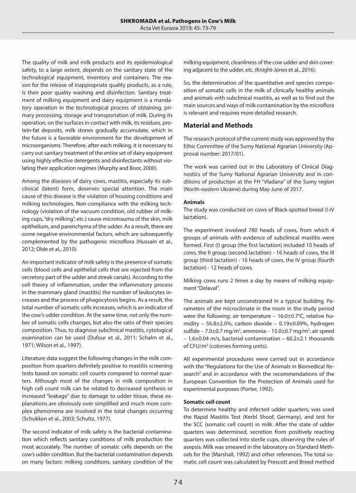
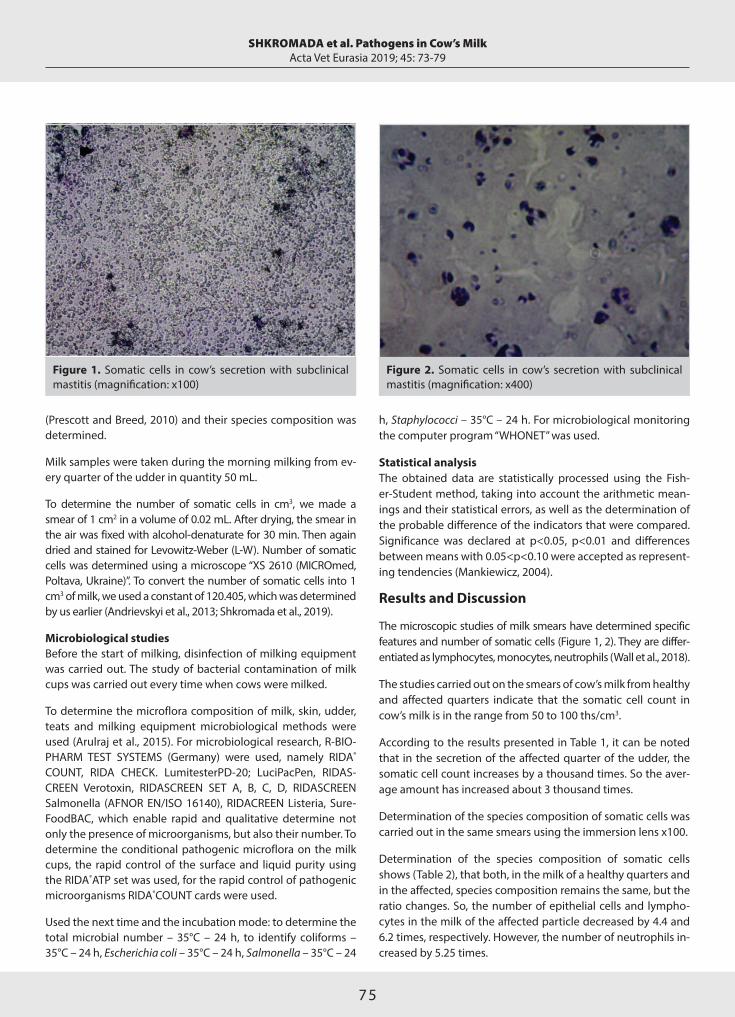
The microscopic studies of milk smears have determined specific features and number of somatic cells (Figure 1, 2). They are differ-entiated as lymphocytes, monocytes, neutrophils (Wall et al., 2018).
The studies carried out on the smears of cow’s milk from healthy and affected quarters indicate that the somatic cell count in cow’s milk is in the range from 50 to 100 ths/cm3.
According to the results presented in Table 1, it can be noted that in the secretion of the affected quarter of the udder, the somatic cell count increases by a thousand times. So the aver-age amount has increased about 3 thousand times.
Determination of the species composition of somatic cells was carried out in the same smears using the immersion lens x100.
Determination of the species composition of somatic cells shows (Table 2), that both, in the milk of a healthy quarters and in the affected, species composition remains the same, but the ratio changes. So, the number of epithelial cells and lympho-cytes in the milk of the affected particle decreased by 4.4 and 6.2 times, respectively. However, the number of neutrophils in-creased by 5.25 times.
SHKROMADA et al. Pathogens in Cow’s MilkActa Vet Eurasia 2019; 45: 73-79
Figure 1. Somatic cells in cow’s secretion with subclinical mastitis (magnification: х100)
Figure 2. Somatic cells in cow’s secretion with subclinical mastitis (magnification: х400)

76
Studies on determining the number of somatic cells showed that in the main period of lactation of clinically healthy animal, the SCC is up to 100 ths/cm3. In the case of subclinical mastitis, SCC increases in tens and even thousands times.
Thus, in Figure 3, the milk lymphocyte is shown in cow’s secre-tion with subclinical mastitis. Typically, it is rounded when col-ored by Levowitz-Weber (LW), its nucleus of a dense consisten-cy is intensively stained in a dark purple color; a small circle of bluish cytoplasm is clearly visible around the nucleus.
In Figure 4 segmented neutrophil is shown. Our studies have shown that neutrophils can be found in both milk of clinical-ly healthy cows and in milk of cows with subclinical mastitis. However, it should be noted that in the case of subclinical mas-titis their number increases in thousands times. In the case of disease, the number of neutrophils can amount up to 90% of all cells. Along with segmental neutrophils, stab and immature neutrophils appear in milk.
In the udder secretion of cows with subclinical mastitis, mono-cytes appear (Figure 5). Macrophages accumulate in large quantities in the areas of inflammation. They have a strong ca-pacity for phagocytosis.
Basophils are granulocytes that are clearly visible on the Figure 6. They have an incorrectly rounded shape, with the nucleus of a dense consistency pushed to the periphery.
SHKROMADA et al. Pathogens in Cow’s MilkActa Vet Eurasia 2019; 45: 73-79
Table 2. Species composition of somatic cells in the milk of healthy and affected quarters (M±S.E., n=16, %)
Quarter
Healthy Affected
Lactation Somatic cells Somatic cell count
Epithelial 44.8±1.7 10.1±1.0*
II Lymphocytes 38.6±1.4 6.2±0.7*
Neutrophils 16.6±1.4 84.9±5.8*
*p<0.001; SCC: somatic cell count (compared to healthy samples)
Table 1. The content of somatic cells in cow's milk from healthy quarters and affected by subclinical mastitis (M±S.E., SCC - ths/cm3)
Group of animals (lactation) Healthy particle Affected particle
I (n=10) 76.1±1.9 29012.4±275.0*
II (n=16) 77.8±1.4 33396.2±265.3*
III (n=16) 78.1±1.2 27567.1±248.1*
IV (n=12) 80.4±1.3 24832.8±279.0*
Average 78.1±1.3 228702.1±286.5*
*p<0.001; SCC: somatic cell count (compared to healthy samples)
Figure 3. Lymphocyte in cow’s secretion with subclinical mastitis (magnification: х1000)
Figure 4. Segmented neutrophil in cow’s secretion with subclinical mastitis (magnification: х1000)
Figure 5. Basophil in cow’s secretion with subclinical mastitis (magnification: х1000)

77
It is known that subclinical mastitis is an infectious disease; therefore, the disease of animals can be transmitted from one animal to another. Since the transmission path is pin and the greatest contact occurs through the milk cups.
In accordance with the research objectives, we have studied the dynamics of bacterial contamination of milk cups. Before the start of milking, milk cups was thoroughly mechanically cleaned, washed with water, and disinfected. After the disinfection, milk cups was thoroughly washed with distilled water, and dried. The study of bacterial contamination of milk cups was carried out at the beginning of milking (before connecting to cows) and then every five cows. The results of the study are presented in Table 3.
According to the results of the study (Table 3), it can be not-ed that the total bacterial contamination of the milk cups was within the limits 2.1±0.1-2.3±0.3 CFU/cm2 in the beginning of milking. The total bacterial contamination of the milk cups after milking five cows of I group increased by 254.6 times and after ten cows – by 636.5 times.
The same tendency was observed in relation to the gener-al bacterial contamination of the milk cups, which were ex-posed to the skin of the cow’s teats from other experimental groups.
Studies have shown, that on the udder skin of cows I group (Ta-ble 4) S. aureus forms 29%, S. agalactiae – 60% and associated microflora – 11% of the total number of colony-forming units.
However, the percentage of pathogenic microorganisms var-ied depending on the age of the animals. So, on the udder skin of cows IV group rate of S. aureus increased to 48% and rate of S. agalactiae decreased to 43% of the total number of colo-ny-forming units.
Thus, it can be stated that even after careful cleaning and disin-fection of milk cups, microorganisms still remain on it and the general microbial contamination is dynamic in the direction of increase. So, it can be assumed that the pathogens of the subclinical mastitis from the skin of the affected cow through the milk cups affect the tissues of healthy animals, thus causing transfer infection from animal to animal.
The results of the study of microbial contamination of teat and udder skin show that it always contains microorganisms that can cause subclinical mastitis (Busato et al., 2000). There is only difference in the ratio of pathogens.
Cows of 1st lactation have the smallest number of S. aureus and at the same time, the largest number of S. agalactiae. How-ever, this ratio changes somewhat with animal aging. So, the amount of S. aureus slightly increases and the amount of S. aga-lactiae conversely decreases. Moreover, we have not detected any changes in the amount of associated microflora. One might assume that microorganisms on the udder skin are antagonists among themselves, especially S. аureus and S. agalactiae (Joshi and Gokhale, 2006; Schwarz et al., 2011).
Thus, pathogens of subclinical mastitis are always present on the udder skin of animals. Therefore, it is mostly impossible to treat the herd of cows completely of subclinical mastitis. But it possible to control it and keep the rate of animals infected by mastitis pathogens within 5-6%.
The main stages of disease prevention are strict compliance of the milking technology, systematic examination of cows by “cow side” tests, such as the California Mastitis Test, and mea-suring the electrical conductivity of milk. The separation of infected animals from healthy ones can also be used in order
SHKROMADA et al. Pathogens in Cow’s MilkActa Vet Eurasia 2019; 45: 73-79
Figure 6. Monocyte in cow’s secretion with subclinical mastitis (magnification: х1000)
Table 3. The dynamics of bacterial contamination of milk cups (M±S.E.)
Group of Before The fifth cow The tenth cow animals milking thousand thousand (samples) CFU/cm2 CFU/cm2 CFU/cm2
I (n=60) 2.1±0.1 534.7±29.9* 1336.7±45.1*
II (n=60) 2.3±0.2 601.4±23.0* 1486.3±34.6*
III (n=60) 2.3±0.3 843.6±45.0* 1568.4±67.6*
IV (n=60) 2.1±0.2 811.3±48.1* 1489.9±59.0*
*p<0.05 compared to the start of milking; CFU: colony-forming units
Table 4. Microbial contamination of the udder skin of cows (M±S.E.)
Group of animals Skin microflora of teats (%)(lactation) S. aureus S. agalactiae Associated
I (n=10) 29±1.4 60±3.7 11±2.0
II (n=16) 38±2.0 56±2.3 6±1.4
III (n=16) 49±3.8 44±2.9 7±1.3
IV (n=12) 48±3.7 43±4.2 9±1.2

78
to break the epizootic chain. One of the reasons for the rap-id spread of subclinical mastitis in the herd is the transfer of pathogens during milking, especially from the diseased animal with increased pathogenicity to the healthy one. The main me-chanical carrier of pathogens is the milk cup, since it directly contacts with the udder skin both of affected and healthy.
Therefore, if a cow affected by subclinical mastitis is present on the first stage of milking, there is a high probability that the mastitis pathogens will be transmitted to the udder skin of other animals.
Conclusion
1. In the case of subclinical mastitis the number of somatic cells in the secretion of the affected quarter of udder increases and its species composition changes.
2. Subclinical mastitis pathogens are always present on the skin of a cow’s udder, but only their ratio changes with aging of the animal.
3. The main carrier of pathogens of subclinical mastitis from the infected cow to healthy one is the milk cup.
Ethics Committee Approval: Ethics committee approval was received for this study from the ethics committee of Sumy National Agrarian University with approval number 2017/01.
Peer-review: Externally peer-reviewed.
Author Contributions: Concept – O.Sk., O.Sh.; Design – O.Sk., A.P.; Su-pervision – O.Sh., G.I.; Resources – O.Sk., O.Sh., A.P., G.I.; Materials – O.Sk., G.I.; Data Collection and/or Processing – O.Sk., O.Sh.; Analysis and/or Interpretation – O.Sh., A.P.; Literature Search – O.Sh., G.I.; Writing Manu-script – O.Sk., A.P.; Critical Review – O.Sh.
Conflict of Interest: The authors have no conflict of interest to declare.
Financial Disclosure: The authors declared that this study has received no financial support.
References
Andrievskyi, V.Y., Kostenko, O.I., Ostapiuk, M.P., Kasianchuk, V.V., Sk-liar, O.I., 2013. Quality management systems. Milk. Calculation of somat-ic cells. Part 1. Microscope method (control method) (ISO 13366-1: 2008 IDF 148-1:2008): DSTU ISO 13366-1: 2008. Kyiv: State Consumer Standard of Ukraine, 12.
Arulraj, P., Asarudheen, I., Jasmin, T., Mohammed, Sarfas, Sahnas, N.K., Vedha, Satheesh, Venkatanarayanan, R., 2015. Dairy bacteriology and microbial analysis of milk using syringe filter. European Journal of Biomedical and Pharmaceutical Sciences 2, 380-388.
Dufour, S., Fréchette, A., Barkema, H.W., Mussell, A., Scholl, D.T., 2011. Invited review: effect of udder health management practices on herd somatic cell count. Journal of Dairy Sciences 94, 563-579. [CrossRef]
Busato, A., Trachsel, P., Schällibaum, M., Blum, J.W., 2000. Udder health and risk factors for subclinical mastitis in organic dairy farms in Switzerland. Preventive Veterinary Medicine 44, 205-220. [CrossRef]
Hussain, R., Javed, M.T., Khan, A., 2012. Changes in some biochemi-cal parameters and somatic cell counts in the milk of buffalo and cat-tle suffering from mastitis. Pakistan Veterinary Journal 32, 418-421.
Jensen, R.G., Newburg, D.S., 1995. Bovine milk lipids. In: Jensen RG, edi-tor. Handbook of Milk Composition. Academic Press, USA, pp. 543-575. [CrossRef]
Joshi, S., Gokhale, S., 2006. Status of mastitis as an emerging disease in improved and periurban dairy farms in India. Annals of the New York Academy of Sciences 1081, 74-83. [CrossRef]
Lelieveld, H.L.M., Holah, J., Gabric, D., 2016. Handbook of Hygiene Control in the Food Industry, (Woodhead Publishing Series in Food Sci-ence, Technology and Nutrition), 2nd Edition, Woodhead Publishing.
Ma, Y., Ryan, C., Barbano, D.M., Galton, D.M., Rudan, M.A., Boor, K.J., 2000. Effects of somatic cell count on quality and shelf-life of pasteur-ized fluid milk. Journal of Dairy Sciences 83, 264-274. [CrossRef]
Mankiewicz, R., 2004. The Story of Mathematics. Princeton, NJ: Princ-eton University Press.
Marshall, R.T., 1992. Standard Methods for the Examination of Dairy Products, 16th ed. American Public Health Association, Washington, DC.
Murphy, S.C., Boor, K.J., 2000. Trouble-shooting sources and causes of high bacteria counts in raw milk. Dairy, Food and Environmental Sanitation 8, 606-611.
Olde Riekerink, R.G., Barkema, H.W., Scholl, D.T., Poole, D.E., Kelt-on, D.F., 2010. Management practices associated with the bulk-milk prevalence of Staphylococcus aureus in Canadian dairy farms. Veter-inary Medicine 97, 20-80. [CrossRef]
Porter, D.S., 1992. Ethical scores for animal experiments. Nature 356, 101-102. [CrossRef]
Prescott, S.C., Breed, R.S., 2010. The Determination of the Number of Body Cells in Milk by a Direct Method. American Journal of Public Hygiene 20, 662-640.
Romain, H.T., Adesiyun, A.A., Webb, L.A., Lauckner, F.B., 2000. Study on risk factors and their association with subclinical mastitis in lactating dairy cows in Trinidad. Journal of Veterinary Medicine Infec-tious Diseases and Veterinary Public Health 47, 257-271. [CrossRef]
Schalm, O.W., Carroll, E.J., Jain, N.C., 1971. Bovine Mastitis. Lea and Febiger, Philadelphia, pp. 94-127.
Schukken, Y.H., Wilson, D.J., Welcome, F., Garrison-Tikofsky, L., Gonzalez, R.N., 2003. Monitoring udder health and milk quality us-ing somatic cell counts. Veterinary Research 34, 579-596. [CrossRef]
Schultz, L.H., 1977. Somatic cells in milk – physiological aspects and relationship to amount and composition of milk. Journal of Food Protection 40, 125-131. [CrossRef]
Schwarz, D., Diesterbeck, U.S., König, S., Brügemann, K., Schlez, K., Zschöck, M., Wolter, W., Czerny, C.P., 2011. Flow cytometric differ-ential cell counts in milk for the evaluation of inflammatory reactions in clinically healthy and subclinically infected bovine mammary glands. Journal of Dairy Sciences 94, 5033-5044. [CrossRef]
Shkromada, O., Skliar, O., Paliy, A., Ulko, L., Gerun, I., Naumenko, О., Ish-chenko, K., Kysterna, O., Musiienko, O., Paliy, A., 2019. Development of measures to improve milk quality and safety during production. East-ern-European Journal of Enterprise Technologies 3, 30-39. [CrossRef]
Knight-Jones, T.J., Hang’ombe, M.B., Songe, M.M., Sinkala, Y., Grace, D., 2016. Microbial Contamination and Hygiene of Fresh Cow’s Milk Produced by Smallholders in Western Zambia. International Journal of Environmental Research and Public Health, 13, E737. [CrossRef]
SHKROMADA et al. Pathogens in Cow’s MilkActa Vet Eurasia 2019; 45: 73-79

79
Wall, S.K., Wellnitz, O., Bruckmaier, R.M., Schwarz, D., 2018. Differ-ential somatic cell count in milk before, during, and after lipopoly-saccharide- and lipoteichoic-acid-induced mastitis in dairy cows. Journal of Dairy Sciences 101, 5362-5373. [CrossRef]
Wilson, D.J., Das, H.H., Gonzalez, R.N., Sears, P.M., 1997. Association between management practices, dairy herd characteristics, and so-matic cell count of bulk tank milk. Journal of the American Veterinary Medical Association 210, 1499-1502.
SHKROMADA et al. Pathogens in Cow’s MilkActa Vet Eurasia 2019; 45: 73-79
80
Address for Correspondence: Uğur GÜNŞEN • E-mail: [email protected]
Received Date: 28 June 2019 • Accepted Date: 30 September 2019 • DOI: 10.5152/actavet.2019.19015
Available online at actaveteurasia.istanbulc.edu.tr
It is presented as an oral presentation with the same title in International Symposium Bandırma and Its Surroundings-UBS’18, September 17-19, 2018 Bandırma, Turkey.
Abstract
The Bandırma district of Balıkesir province has an important place in terms of production and export of aquatic and fro-zen fish products. Silverfish (Atherina boyeri Risso, 1810) has high protein quality (protein content >70%) and low price. It constitutes an important alternative source of raw material for economic fish meal production. Fresh silverfish exported to the European countries in recent years is demanded in two different product forms: Frozen and breaded frozen. In this study, the production of frozen silverfish was carried out in a business that produced aquaculture products for a conside-rable level of export in Bandırma. The production flow diag-
ram, in accordance with TS EN ISO 22000 Food Safety Mana-gement System and British Retail Consortium Standard, was defined to obtain a safe product in accordance with customer expectations and needs. Hazard analysis was carried out by analyzing each step using decision tree. In this way, potenti-al hazards and the precautions to be taken to prevent them, critical control points, and critical limits belonging to these points have been set forth.
Keywords: Critical control point, frozen silverfish Atherina bo-yeri Risso 1810, hazard analysis
Determination of Critical Control Points and Potential Hazard Analysis in the Production of Frozen Silverfish (Atherina boyeri Risso, 1810)
Uğur GÜNŞEN , Hüseyin ESECELİ , Ramazan Mert ATAN Department of Nutrition and Dietetic, Bandırma Onyedi Eylül University, Faculty of Health Science, Balıkesir, Turkey
Cite this article as: Günşen, U., Eseceli, H., Atan, R.M., 2019. Determination of Critical Control Points and Potential Hazard Analysis in the Production of Frozen Silverfish (Atherina boyeri Risso, 1810). Acta Vet Eurasia 45, 80-90.
ORCID IDs of the authors: U.G. 0000-0001-9858-6019; H.E. 0000-0002-5912-5479; R.M.A. 0000-0003-4608-605X.
Original Article Acta Vet Eurasia 2019; 45: 80-90
Introduction
Today’s society wants the foodstuff to be hygienic and economi-cal, as well as to contain protein, fat, carbohydrates, vitamins, and minerals in a balanced ratio. Fish meat is an excellent food with high protein quality, mineral and vitamin richness, low amount of energy, and abundant polyunsaturated fatty acids. Because the energy value is low, it also has a dietetic characteristic. The widely used form of fishery products for human consumption is the frozen product form, which is used in many countries as well as in various products such as salting, smoking, marinating, es-pecially fresh consumption (Turan et al., 2006; Varlık et al., 2004).
Silverfish (Atherina Boyeri Risso, 1810, A. boyeri) is a member of Atherinidae family that has good adaptation talent and shows regional differentiation for morphological and biological char-
acteristics (Çetinkaya et al., 2010). Silverfish is found in rivers, lakes, ponds, and reservoirs (Küçük et al., 2006). In Turkey this fish species lives in İznik, Sapanca and Köyceğiz lakes in a dense population. This fish, which did not have economic importance in our country until recently, has gained value in recent years; and it has been in demand for consumption in domestic and foreign markets (Çolakoğlu et al., 2006).
The high quality of silverfish (protein content >70%) as well as its low price show that it is an important source of alterna-tive raw materials in terms of economic fish feed production (Gümüş et al., 2009). Fresh silverfish, which was exported to the foreign market, has been demanded by the European Union countries in two different product forms: frozen and frozen breaded (Çolakoğlu et al., 2006).
This work is licensed under a Creative Commons Attribution-NonCommercial 4.0 International License.
81
GÜNŞEN et al. Critical Control Points in the Production of Frozen SilverfishActa Vet Eurasia 2019; 45: 80-90
The Bandırma district of Balıkesir province, located at the level of TR 22, has an important place in terms of the production and export of aquaculture. Frozen seafood such as frozen fish, lobster, shrimp, mussels, crabs, and frog and land snail prod-ucts are exported to Europe and the Far East (Anonymous, 2000).
The increase in the level of welfare of the countries and the awareness of the consumers has forced the firms in the food sector to seek for new pursuits in terms of food safety (Başaran, 2016). It is defined as taking necessary measures to ensure reliable food production during food safety, raw ma-terial supply, production, processing, storage, transportation, distribution, and presentation of food. The starting point of food safety is farm, and the end point is consumer. Therefore, food safety includes the procurement of healthy raw materials from “farm to consumer”, the production, processing, storage, transportation, distribution, and presentation of food (Giray and Soysal, 2007).
Hazard Analysis and Critical Control Points (HACCP) is fre-quently used as the best system, which helps food producers to produce safe foods for consumption (Ayhan, 2013). This system aims to determine the potential hazards that may oc-cur at any stage in the business, not only the end-product, but also the whole business where the product is produced, to control the necessary preventive and corrective actions for all possible hazards in a systematic way, and to minimize the potential physical, chemical, and microbiological diseas-es (Moterjemi and Mortimore, 2005; Turantaş and Ünlütürk, 1998). ISO 22000 Food Safety Management System, which was developed in recent years as based on HACCP, is a sys-tem that was developed to obtain safe food worldwide (DPT, 2007).
In this study, frozen silverfish was produced in a business that produced aquaculture products for export in Bandırma (TR221), which is located at TR 22 level in the scope of TS EN ISO 22000 Food Safety Management System, obtain a safe product according to customer expectations and needs. The produc-tion flow chart has been defined, and hazard analysis has been performed by examining with decision tree in each step. In this way, critical control points and critical limits of these points are put forward with the measures to be taken for the prevention of potential hazards.
Materials and Methods
MaterialsSilverfishes that were caught by trammel net from İznik Lake and reached the laboratory within 48 h were used as research material. A total of 100 fishes were used in the analysis.
MethodsAll the analysis in the obtained samples were performed by ref-erence methods reported by the Turkish Republic Ministry of
Agriculture and Forestry (T.C. Tarım ve Orman Bakanlığı, 2012). The physical analyses were piece size of a fish, the amount of pieces in 1 kg, the min-max weight, the amount of foreign mat-ter. The chemical analyses were histamine, mercury (Hg), cad-mium (Cd), lead (Pb), benzo(a)pyrene, total dioxins, and total dioxins and dioxin-like PCBs. The microbiological analyses were numbers of total aerobic bacteria (NTAB), coliform bacteria (CB), Escherichia coli (E. coli), Staphylococcus aureus (S. aureus), Salmonella spp., Listeria monocytogenes (L. monocytogenes), Vibrio parahaemolyticus (V. parahaemolyticus), and Vibrio chol-erae (V. cholerae).
Results and DiscussionAccording to the ISO 22000 Food Safety Management System, a complete description of the product, including the relevant safety information, must be made. The product and its compo-sition; physical, chemical, and biological properties (pH, water activity, etc.), and processes applied to the product such as heat treatment, freezing, smoking, salting, must be fully defined. The packaging properties, distribution, and storage conditions as well as the storage life and instructions for use should be specified.
The information, such as whether each product obtained in the business is to be directly used for consumption or used as an intermediate or food additive in another business, the form of packaging (large packaging such as bulk, sack or barrel, final consumer packaging), whether it needs to be exposed to heat treatment for the last time before consumption, should be in-dicated in detail (Batu and Gök, 2006).
The product description of frozen silverfish is shown in Table 1. In creating product properties, the reported criteria in Turkish Republic Ministry of Agriculture and Forestry General Director-ate of Food and Control Regulations on Fisheries (Anonymous, 1995), were taken into consideration.
The flow chart is defined as the schematic representation of the relationship between order and steps or processes ap-plied in the production of a particular nutrient (Anonymous, 2003). A production flow chart should be created before the hazard analysis is performed. In the flow chart, all the steps taken by the food until it reaches the consumer’s table should be shown in detail. Starting from the procurement of raw materials, entry points in the processing line of all additives and auxiliaries, all applications in the process line, waiting times and temperatures if any, packaging, heat treatment, storage, distribution operations and again, if any, quality con-trol stages should be shown in detail (Batu and Gök, 2006). In our study, the production flow chart of frozen silverfish was formed as follows (Figure 1).
Pre-requisite programs (PRPs) are the basic conditions and activities to ensure a proper production by providing the nec-essary hygienic environment along the food chain, to ensure the safe preparation of the end-product, and to provide safe

82
food for human consumption. PRPs depend on the food chain parts of the organization and the type of organization. For example, good agricultural practices (GAP), good veterinary practices (GVP), good manufacturing practices (GMP), good hygiene practices (GHP), good laboratory practices (GLP), good distribution practices (GDP), and good trading practices (GTP).
The operational pre-requisite program (OpPRP) is defined as a pre-requisite program defined by hazard analyses where it is compulsory to control possible food safety hazards and/or con-tamination or proliferation of food safety hazards in the prod-uct or process environment (Anonymous, 2006).
The silverfish used as research material were hunted from İznik Lake after the chemical and microbiological analysis results of the samples obtained from the controls carried out by the Pro-vincial/District Directorate of Agriculture at the beginning of the hunting season were reported to be in compliance with the legal limits and in accordance with the prohibition periods de-termined by Republic of Turkey Ministry of Agriculture and For-estry the General Directorate of Food and Control (Table 2). So, the step of “Raw material supply and acceptance”, which is the first step of frozen silverfish production flow chart, has been determined as OpPRP because of the risk of possible biological and chemical pollution risk analysis score was more than four points (Table 3).
The HACCP application is performed within an HACCP plan. The HACCP plan has been developed to ensure con-trol of potential hazards in the food chain and is import-
GÜNŞEN et al. Critical Control Points in the Production of Frozen SilverfishActa Vet Eurasia 2019; 45: 80-90
Table 1. Product specification of frozen silverfish
Name of product: Frozen silverfish (Atherina boyeri Risso, 1810)
Product specification:
Physical properties
Piece size of a fish 5-7 cm
Amount of pieces in a kilogram 500-550
Min-max weight 1.8-2 g
Foreign matter ratio Max 2%
Chemical properties
Histamine n=9, c=2, m=100 mg/kg, M=200 mg/kg
Hg 0.5 ppm
Cd 0.05 ppm
Pb 0.3 ppm
Benzo(a)pyrene 2.0 ppb
Total dioxins (max) 4.0 pg/g
Total dioxins and dioxin-like PCBs (max) 8.0 pg/g
Microbiological properties
Parameters Maximum tolerance
NTAB 30°C (/g) n=5, c=2, m=106, M=107
CB (/g) n=5, c=2, m=160, M=210
E. coli (/g) n=5, c=2, m=9, M=12
S. aureus (/g) n=5, c=2, m=103, M=5 × 10³
Salmonella spp. none / 25 g
L. monocytogenes n=5, c=0
V. parahaemolyticus none
V. cholerae none
Usage and purpose: Consume by fried.
User / consumer group: Except allergic to babies and fish,
Suitable for people of all ages.
Allergen presence: Allergen
GMO presence: The product and the auxiliary materials used do not contain GMO (genetically modified organism).
Packaging: In 1 kg nylon (PE) bag or bag in parcel,
10 kg / parcel is packed in bulk.
Shelf life and storage conditions: 24 months at -18°C
Place of sale: Domestic market, hotel, restaurant, foreign market customer groups.
Warnings in the label: Allergen. After thawing, do not freeze again.
Special distribution control: Transport / storage temperature -18°C min.
Hg: mercury; Cd: cadmium; Pb: lead; PCBs: polychlorinated biphenyls; NTAB: number of total aerobic bacteria; CB: coliform bacteria
Figure 1. The production flow chart of frozen silverfish

83
ant in terms of food safety and prepared in accordance with HACCP principles (Burson, 2002). The first principle required to implement the HACCP system is to carry out hazard analysis. According to this system, physical, chem-ical, and biological agents constitute potential hazards to health in foods. Hazard analysis involves the assessment of potential hazards that may occur at any stage of pro-duction, and their assessment of the likelihood of occur-rence and the seriousness of the hazard they create. Haz-ard analysis critical control points must ensure physical, chemical, and microbiological safety. Failure to perform this step, which requires technical expertise and experi-ence, may cause the food produced in the future not be-ing at the desired level of safety. Hazard analyses include the identification and evaluation of hazards arising from raw materials, additives, processing, distribution, retail sale, and consumption (Barrie, 1996; Göktan and Tunçel, 1992; Kayaardı, 2004).
The brainstorming technique is used to identify potential hazards as the first stage when performing hazard analyses. At this stage, the HACCP team creates a list by identifying the potential hazards in all stages from the raw material to the use of the product. The “decision tree” is used to identify the hazards (Anonymous, 1997). The decision tree is a set of systematic questions that are taken into account in terms of deciding whether the point is a critical control point for a de-fined hazard at a point in the production process of the food. For the application of the decision tree, each process step specified in the flow chart must be included in the process, respectively. The decision tree in each step should be applied to every expected hazard and every control measure (Anon-ymous, 2005a). Table 3 shows the frozen silverfish hazard/risk analysis.
The Critical Control Point (CCP) is the stage where the food safety hazard is prevented or eliminated or reduced to an acceptable level (Anonymous, 2006). A critical point deter-mination for the control of a hazard can be facilitated by the use of decision trees. In the application of the decision tree, each process step specified in the flow chart must be included in the process, respectively. At every step, the de-cision tree should be applied to every hazard expected to occur and every control measure determined (Anonymous, 2005a).
The HACCP plan contains all necessary information, referenc-es, and records related to the system. All substances, except PRPs for the implementation of the HACCP system, are in fact included in the HACCP plan (Anonymous, 2005b; Burson, 2002). Table 4 shows the critical control points identified by the hazard analysis and risk analysis performed on the flow chart and information on the implementation of HACCP prin-ciples at these points.
GÜNŞEN et al. Critical Control Points in the Production of Frozen SilverfishActa Vet Eurasia 2019; 45: 80-90
Tabl
e 2.
OpP
RP p
lan
of fr
ozen
silv
erfis
h pr
oduc
tion
OpP
RP
No.
H
azar
d Co
ntro
l mea
sure
s W
hat
Trac
ing
how
Fr
eque
ncy
Corr
ecti
ve a
ctio
n Re
spon
sibl
e Tr
acin
g re
cord
s
Pa
thog
en b
acte
ria
In c
ompl
ianc
e M
icro
biol
ogic
al
Rela
ted
One
tim
e at
the
Not
hun
ting
from
Bu
sine
ss
Ana
lysi
s by
The
beca
use
of p
ollu
tion
w
ith
anal
ysis
m
etho
ds
seas
on o
peni
ng
unsu
itabl
e la
kes
Lab.
M
inis
try
in
the
lake
, cau
ght s
ilver
fish
hunt
ing
perio
ds
Refe
renc
e O
rigin
doc
umen
ts
E.
coli
desi
gnat
ed b
y
La
b.
Cont
rols
mad
e in
Salm
onel
la s
pp.
Gen
eral
Dire
ctor
ate
Gen
eral
en
d-pr
oduc
t
V. c
hole
rae
of P
rote
ctio
n an
d
D
irect
orat
e of
Fo
rmal
ana
lysi
s
V. p
arah
aem
olyt
icus
Co
ntro
l
Pr
even
ting
and
resu
lts
L.
mon
ocyt
ogen
es
Co
ntro
l
Co
ntai
ning
che
mic
al p
ollu
tion
Hun
ting
from
lake
s,
H
g w
here
in o
btai
ned
Cd
ap
prop
riate
resu
lts
Pb
of
ana
lysi
s of
the
Be
nzo(
a)py
rene
sa
mpl
es b
y th
e
In
clud
ing
diox
ins
and
Pr
ovin
cial
and
diox
in li
ke P
CBs
Dis
tric
t Dire
ctor
ate
Hg:
mer
cury
; Cd:
cad
miu
m; P
b: le
ad; P
CBs:
pol
ychl
orin
ated
bip
heny
ls
1. Supply and admission of raw material

84
GÜNŞEN et al. Critical Control Points in the Production of Frozen SilverfishActa Vet Eurasia 2019; 45: 80-90
Tabl
e 3.
Haz
ard/
risk
anal
ysis
of fr
ozen
silv
erfis
h pr
oduc
tion
Prel
imin
ary
ques
tion
(PQ
): Ca
n th
is h
azar
d be
con
trol
led
wit
h PR
P?
Q1:
Is th
ere
any
cont
rol m
easu
re th
at c
an b
e ap
plie
d by
the
oper
ator
at a
ny s
tage
of t
he p
rodu
ctio
n fo
r thi
s ha
zard
? Ca
n th
e ha
zard
be
avoi
ded
in b
usin
ess?
Whi
ch p
roce
ss s
tep?
Q2:
Is th
e co
ntam
inat
ion
caus
ed b
y th
is h
azar
d up
per t
han
the
acce
ptab
le le
vel o
r can
it re
ach
unac
cept
able
leve
ls?
Q3:
Is th
is o
pera
tion
ste
p de
sign
ed s
peci
fical
ly to
rem
ove
this
haz
ard
or to
redu
ce th
e po
ssib
ility
of r
ealiz
atio
n to
the
acce
ptab
le le
vels
(ope
rati
ons
desi
gned
spe
cific
ally
suc
h as
au
tocl
avin
g, p
aste
uriz
atio
n, m
etal
det
ecto
r)?
Q4:
Can
this
defi
ned
haza
rd b
e re
mov
ed in
any
follo
win
g st
ep, o
r can
it b
e re
duce
d to
the
acce
ptab
le le
vels
?
Y: Y
es
N
: No
N C
: N
ot C
CP
PQ
Q
1 Q
2 Q
3 Q
4
Co
ntro
l mea
sure
ass
essm
ent
an
d se
lect
ion
of c
ombi
nati
ons
deci
sion
tree
Proc
essi
ng
Haz
ard
Co
ntro
l mea
sure
1-Ra
w m
ater
ial s
uppl
y
B - P
rese
nce
of E
. col
i, Sa
lmon
ella
, 2
4 8
Cont
rol o
f lak
e w
ater
and
ope
ning
to h
untin
g Y
X
and
acce
ptan
ce
V. c
hole
rae,
V. p
arah
aem
olyt
icus
and
acco
rdin
g to
the
Min
istr
y m
onito
ring
prog
ram
. Fis
h (fr
esh
athe
rina)
L.
mon
ocyt
ogen
es b
ecau
se o
f
inta
ke is
mad
e fr
om th
e cl
ean
area
s al
low
ed b
y Th
e
pollu
tion
in th
e la
kes
from
whe
re
M
inis
try.
In a
dditi
on, m
icro
biol
ogic
al a
naly
sis
of th
e
silv
erfis
h ar
e ca
ught
.
raw
mat
eria
l in
the
perio
ds s
peci
fied
in th
e
mic
robi
olog
ical
ana
lysi
s pl
an is
car
ried
out.
P
- Pre
senc
e of
par
asite
in s
ilver
fish.
2
2 4
Dur
ing
each
org
anol
eptic
exa
min
atio
n, p
aras
ite
Y
cont
rol i
s pe
rfor
med
. Uns
uita
ble
part
ies
are
reje
cted
.
C
- Exc
eedi
ng li
mits
spe
cifie
d by
the
1
5 5
Hea
vy m
etal
ana
lysi
s is
mad
e at
the
begi
nnin
g of
the
Y
X
Min
istr
y of
Hg,
Cd,
Pb,
Ben
zo(a
)
seas
on in
the
raw
mat
eria
l tak
en fr
om e
ach
regi
on.
py
rene
and
dio
xins
and
dio
xin-
like
PCBs
in s
ilver
fish.
2-W
ashi
ng a
nd c
lean
ing
B - C
onta
min
atio
n of
NTA
B, C
B, E
. col
i 1
4 4
Wat
er w
as c
hlor
inat
ed a
nd p
asse
d th
roug
h U
V fil
ter,
Y
and
C. p
erfri
ngen
s fro
m w
ater
.
and
drin
king
wat
er q
ualit
y. N
TAB,
CB,
and
E. c
oli
an
alys
is a
re p
erfo
rmed
, wee
kly.
NTA
B, C
B, E
. col
i, an
d
C.
per
fring
ens a
naly
ses
are
perf
orm
ed b
y offi
cial
auth
oriti
es e
very
thre
e m
onth
s.
C
- Upp
er li
mits
of c
onta
min
atio
n of
1
4 4
Wat
er is
che
mic
ally
sui
tabl
e. C
ompl
ianc
e is
che
cked
Y
fr
ee c
hlor
ine,
Fe,
nitr
ite, a
mm
oniu
m,
by
ana
lysi
s ev
ery
thre
e m
onth
s. Ch
lorin
e co
ntro
l is
al
umin
um fr
om w
ater
perf
orm
ed tw
ice
a da
y.
P
- Exc
essi
ve e
xpos
ure
to w
ater
will
1
1 1
The
fishe
s ar
e w
ashe
d w
ith c
old
wat
er w
ithou
t giv
ing
Y
caus
e its
sto
mac
h to
exp
lode
.
too
muc
h pr
essu
re to
the
wat
er.
3-Co
ld s
tora
ge (2
/4°C
) B
- Num
ber o
f NTA
B m
ay in
crea
se
1 2
2 In
the
war
ehou
se, t
he P
LC s
yste
m a
utom
atic
ally
Y
if
the
tank
tem
pera
ture
rise
s.
mea
sure
s an
d re
cord
s te
mpe
ratu
re e
very
two
hour
s.
It
is u
nder
con
stan
t obs
erva
tion.
PRP
Y
N (Q1)
Y
N - C P
Y - CCP
N
Y (Q3)
N - CCP
OpPRP
CCP
Possibility
Severity
Risk score

85
GÜNŞEN et al. Critical Control Points in the Production of Frozen SilverfishActa Vet Eurasia 2019; 45: 80-90
Tabl
e 3.
Haz
ard/
risk
anal
ysis
of fr
ozen
silv
erfis
h pr
oduc
tion
(Con
tinue
d)
Prel
imin
ary
ques
tion
(PQ
): Ca
n th
is h
azar
d be
con
trol
led
wit
h PR
P?
Q1:
Is th
ere
any
cont
rol m
easu
re th
at c
an b
e ap
plie
d by
the
oper
ator
at a
ny s
tage
of t
he p
rodu
ctio
n fo
r thi
s ha
zard
? Ca
n th
e ha
zard
be
avoi
ded
in b
usin
ess?
Whi
ch p
roce
ss s
tep?
Q2:
Is th
e co
ntam
inat
ion
caus
ed b
y th
is h
azar
d up
per t
han
the
acce
ptab
le le
vel o
r can
it re
ach
unac
cept
able
leve
ls?
Q3:
Is th
is o
pera
tion
ste
p de
sign
ed s
peci
fical
ly to
rem
ove
this
haz
ard
or to
redu
ce th
e po
ssib
ility
of r
ealiz
atio
n to
the
acce
ptab
le le
vels
(ope
rati
ons
desi
gned
spe
cific
ally
suc
h as
au
tocl
avin
g, p
aste
uriz
atio
n, m
etal
det
ecto
r)?
Q4:
Can
this
defi
ned
haza
rd b
e re
mov
ed in
any
follo
win
g st
ep, o
r can
it b
e re
duce
d to
the
acce
ptab
le le
vels
?
Y: Y
es
N
: No
N C
: N
ot C
CP
PQ
Q
1 Q
2 Q
3 Q
4
Co
ntro
l mea
sure
ass
essm
ent
an
d se
lect
ion
of c
ombi
nati
ons
deci
sion
tree
Proc
essi
ng
Haz
ard
Co
ntro
l mea
sure
B
- Pos
sibi
lity
of c
ross
-con
tam
inat
ion
1
3 3
No
othe
r raw
mat
eria
l / p
rodu
ct is
sto
red
in th
e co
ld
Y
in s
tore
.
stor
age
whe
re th
e si
lver
fish
is k
ept.
4-Fo
reig
n fis
h an
d
C - C
B an
d E.
coli
cont
amin
atio
n fr
om
2 3
6 Th
e st
aff d
isin
fect
han
ds a
nd w
ear g
love
s. Th
e Y
item
sel
ectio
n th
e se
lect
ion
band
and
per
sonn
el
se
lect
ion
tape
is c
lean
ed a
nd d
isin
fect
ed a
ccor
ding
hand
s.
to th
e cl
eani
ng p
lan.
Wee
kly
swap
con
trol
s ar
e
pe
rfor
med
.
P
- The
pre
senc
e of
alg
ae, p
lant
par
ts,
3 3
9 Fo
reig
n su
bsta
nces
are
take
n by
fem
ale
wor
kers
N
X
X
X
X
X
ston
e fr
agm
ents
, cra
yfish
, and
oth
er
st
andi
ng in
the
band
. Ban
ding
spe
ed is
adj
uste
d
alie
n fis
h an
d in
suffi
cien
t sel
ectio
n
ac
cord
ing
to p
erso
n an
d th
e ra
te o
f for
eign
fish
.
and
rem
aini
ng o
f the
se im
purit
ies
in th
e fin
al p
rodu
ct.
C
- Con
tam
inat
ion
of d
eter
gent
and
1
3 3
The
dete
rgen
ts a
nd d
isin
fect
ants
use
d w
ill n
ot le
ave
Y
disi
nfec
tant
resi
dues
from
the
any
resi
due
by g
ood
rinse
. Pro
duct
saf
ety
data
she
ets
se
lect
ion
band
.
are
avai
labl
e. T
he c
lean
ing
staff
is c
aref
ul a
bout
rinsi
ng. A
fter
rins
ing,
the
resi
due
is v
erifi
ed b
y ch
ecki
ng.
5-IQ
F Fr
eezi
ng (-
35°C
) B
- NTA
B, C
B, E
.col
i con
tam
inat
ion
2
3 6
The
IQF
is c
lean
ed a
nd d
isin
fect
ed a
ccor
ding
to th
e Y
du
e to
the
inad
equa
te c
lean
ing
of IQ
F.
clea
ning
sch
edul
e af
ter e
ach
use.
It is
con
trol
led
by s
wab
.
C
- Con
tam
inat
ion
of d
eter
gent
and
1
3 3
The
dete
rgen
ts a
nd d
isin
fect
ants
use
d w
ill n
ot le
ave
Y
disi
nfec
tant
resi
due
from
the
IQF
band
.
any
resi
due
by g
ood
rinse
. Pro
duct
saf
ety
data
shee
ts a
re a
vaila
ble.
The
cle
anin
g st
aff is
car
eful
abou
t rin
sing
. Aft
er ri
nsin
g, th
e re
sidu
e is
ver
ified
by c
heck
ing.
6-Pa
ckag
ing
B - N
TAB
and
CB c
onta
min
atio
n fr
om
2 3
6 N
ylon
bag
s ar
e su
pplie
d hy
gien
ical
ly fr
om th
e Y
us
ed n
ylon
bag
s an
d pa
ckag
ing
appr
oved
sup
plie
rs a
nd s
tore
d in
the
plan
t as
m
achi
ne.
ap
prop
riate
. The
sw
ap te
st is
don
e fr
om th
e
PRP
Y
N (Q1)
Y
N - C P
Y - CCP
N
Y (Q3)
N - CCP
OpPRP
CCP
Possibility
Severity
Risk score

86
GÜNŞEN et al. Critical Control Points in the Production of Frozen SilverfishActa Vet Eurasia 2019; 45: 80-90
Tabl
e 3.
Haz
ard/
risk
anal
ysis
of fr
ozen
silv
erfis
h pr
oduc
tion
(Con
tinue
d)
Prel
imin
ary
ques
tion
(PQ
): Ca
n th
is h
azar
d be
con
trol
led
wit
h PR
P?
Q1:
Is th
ere
any
cont
rol m
easu
re th
at c
an b
e ap
plie
d by
the
oper
ator
at a
ny s
tage
of t
he p
rodu
ctio
n fo
r thi
s ha
zard
? Ca
n th
e ha
zard
be
avoi
ded
in b
usin
ess?
Whi
ch p
roce
ss s
tep?
Q2:
Is th
e co
ntam
inat
ion
caus
ed b
y th
is h
azar
d up
per t
han
the
acce
ptab
le le
vel o
r can
it re
ach
unac
cept
able
leve
ls?
Q3:
Is th
is o
pera
tion
ste
p de
sign
ed s
peci
fical
ly to
rem
ove
this
haz
ard
or to
redu
ce th
e po
ssib
ility
of r
ealiz
atio
n to
the
acce
ptab
le le
vels
(ope
rati
ons
desi
gned
spe
cific
ally
suc
h as
au
tocl
avin
g, p
aste
uriz
atio
n, m
etal
det
ecto
r)?
Q4:
Can
this
defi
ned
haza
rd b
e re
mov
ed in
any
follo
win
g st
ep, o
r can
it b
e re
duce
d to
the
acce
ptab
le le
vels
?
Y: Y
es
N
: No
N C
: N
ot C
CP
PQ
Q
1 Q
2 Q
3 Q
4
Co
ntro
l mea
sure
ass
essm
ent
an
d se
lect
ion
of c
ombi
nati
ons
deci
sion
tree
Proc
essi
ng
Haz
ard
Co
ntro
l mea
sure
in
com
ing
part
y. T
he p
acka
ging
mac
hine
is c
lean
ed
an
d di
sinf
ecte
d ac
cord
ing
to th
e cl
eani
ng p
lan.
Wee
kly
swap
con
trol
s ar
e pe
rfor
med
.
C
- Nyl
on b
ags
used
are
not
3
4 12
Th
e su
itabi
lity
of th
e ch
emic
al p
aram
eter
s ha
s be
en
Y
appr
opria
te in
che
mic
al p
aram
eter
s
confi
rmed
in th
e sp
ecifi
catio
n gi
ven
to th
e su
pplie
r.
and
mig
ratio
n.
C
- Con
tam
inat
ion
of d
eter
gent
and
1
3 3
Det
erge
nts
and
disi
nfec
tant
s us
ed a
re g
iven
goo
d Y
di
sinf
ecta
nt re
sidu
e fr
om th
e
rin
se to
leav
e no
resi
due.
Pro
duct
saf
ety
data
she
ets
pa
ckag
ing
mac
hine
.
are
avai
labl
e. T
he c
lean
ing
staff
is c
aref
ul a
bout
rinsi
ng. A
fter
the
rinsi
ng o
pera
tion,
the
resi
due
is
ve
rified
by
chec
king
.
7-M
etal
det
ekto
r P
- Met
al p
arts
rem
ain
betw
een
5
5 25
M
etal
det
ecto
r cal
ibra
tion
is c
onfir
med
bef
ore
and
N
X
X
X
X
pr
oduc
ts.
du
ring
each
use
, the
pro
duct
box
es a
re p
asse
d on
e
by
one
thro
ugh
the
met
al d
etec
tor.
The
met
al
de
tect
or is
use
d on
ly b
y tr
aine
d pe
rson
nel.
As
it is
the
equi
pmen
t tha
t affe
cts
food
saf
ety,
it is
ser
vice
d
in
thre
e m
onth
s.
8-La
belin
g -
-
9-Pr
eser
vatio
n (-1
8°C)
B
- Inc
reas
ed b
acte
rial l
oad
as a
resu
lt 1
2 2
Not
ifica
tion:
200
4/46
Y
of
tem
pera
ture
rise
abo
ve -1
8ºC.
Tem
pera
ture
mea
sure
men
t and
reco
rdin
g is
don
e
au
tom
atic
ally
with
PLC
sys
tem
in w
areh
ouse
s
fo
r tw
o ho
urs.
W
hen
ther
e is
a m
alfu
nctio
n in
the
war
ehou
se,
th
e pr
oduc
ts a
re ta
ken
to a
noth
er w
areh
ouse
.
PRP
Y
N (Q1)
Y
N - C P
Y - CCP
N
Y (Q3)
N - CCP
OpPRP
CCP
Possibility
Severity
Risk score

87
GÜNŞEN et al. Critical Control Points in the Production of Frozen SilverfishActa Vet Eurasia 2019; 45: 80-90
Tabl
e 3.
Haz
ard/
risk
anal
ysis
of fr
ozen
silv
erfis
h pr
oduc
tion
(Con
tinue
d)
Prel
imin
ary
ques
tion
(PQ
): Ca
n th
is h
azar
d be
con
trol
led
wit
h PR
P?
Q1:
Is th
ere
any
cont
rol m
easu
re th
at c
an b
e ap
plie
d by
the
oper
ator
at a
ny s
tage
of t
he p
rodu
ctio
n fo
r thi
s ha
zard
? Ca
n th
e ha
zard
be
avoi
ded
in b
usin
ess?
Whi
ch p
roce
ss s
tep?
Q2:
Is th
e co
ntam
inat
ion
caus
ed b
y th
is h
azar
d up
per t
han
the
acce
ptab
le le
vel o
r can
it re
ach
unac
cept
able
leve
ls?
Q3:
Is th
is o
pera
tion
ste
p de
sign
ed s
peci
fical
ly to
rem
ove
this
haz
ard
or to
redu
ce th
e po
ssib
ility
of r
ealiz
atio
n to
the
acce
ptab
le le
vels
(ope
rati
ons
desi
gned
spe
cific
ally
suc
h as
au
tocl
avin
g, p
aste
uriz
atio
n, m
etal
det
ecto
r)?
Q4:
Can
this
defi
ned
haza
rd b
e re
mov
ed in
any
follo
win
g st
ep, o
r can
it b
e re
duce
d to
the
acce
ptab
le le
vels
?
Y: Y
es
N
: No
N C
: N
ot C
CP
PQ
Q
1 Q
2 Q
3 Q
4
Co
ntro
l mea
sure
ass
essm
ent
an
d se
lect
ion
of c
ombi
nati
ons
deci
sion
tree
Proc
essi
ng
Haz
ard
Co
ntro
l mea
sure
10-L
oadi
ng a
nd
B - C
old
chai
n br
eaka
ge d
urin
g 1
2 2
Befo
re lo
adin
g, th
e ve
hicl
e in
terio
r tem
pera
ture
is
Y
D
istr
ibut
ing
(-18°
C)
load
ing.
set t
o -1
8°C
and
the
palle
ts a
re lo
aded
with
out
br
eaki
ng th
e co
ld c
hain
.
B
- Cro
ss c
onta
min
atio
n du
e to
1
2 2
The
vehi
cle
is d
isin
fect
ed a
nd c
ontr
olle
d ac
cord
ing
Y
non-
disi
nfec
tion
of th
e ve
hicl
e.
to
the
load
ing
inst
ruct
ions
.
P
- Mis
iden
tifica
tion
of th
e pa
lett
e.
1 1
1 Th
e pr
oduc
tion
resp
onsi
ble
iden
tifies
the
palle
ts
Y
at th
e be
ginn
ing
of th
e in
stal
latio
n.
P
- Lo
st c
rush
ing
of c
arto
ns, b
reak
age
1
1 1
The
prod
uctio
n m
anag
er s
tand
s at
the
load
ing,
the
Y
of p
rodu
cts,
dam
age
due
to im
prop
er
pa
llets
, and
par
cels
are
vis
ually
che
cked
and
the
pl
acem
ent o
f pal
lets
.
expe
rienc
ed s
tore
keep
er s
uppo
rts
the
gaps
by
plac
ing
the
palle
ts.
NTA
B: n
umbe
r of t
otal
aer
obic
bac
teria
; IQ
F: in
divi
dual
qui
ck fr
eezi
ng; P
CB: p
olyc
hlor
inat
ed b
iphe
nyl;
PLC:
pro
gram
mab
le lo
gic
cont
rolle
r; CC
P: c
ritic
al c
ontr
ol p
oint
PRP
Y
N (Q1)
Y
N - C P
Y - CCP
N
Y (Q3)
N - CCP
OpPRP
CCP
Possibility
Severity
Risk score

88
GÜNŞEN et al. Critical Control Points in the Production of Frozen SilverfishActa Vet Eurasia 2019; 45: 80-90
Tabl
e 4.
HAC
CP p
lan
of fr
ozen
silv
erfis
h pr
oduc
tion
Trac
ing
Im
port
ant
Cont
rol
CCP
haza
rd
mea
sure
Cr
itic
al li
mit
W
hat
How
Fr
eque
ncy
Who
Re
cord
s Co
rrec
tion
Ve
rific
atio
n
Th
e pr
esen
ce o
f Fo
reig
n St
one
shou
ld
Fish
es
Han
d A
ll fis
h pa
ssin
g Fe
atur
ed
Fina
l pro
duct
Pe
rson
nel
No
cust
omer
alga
e, p
lant
par
ts,
subs
tanc
es
neve
r be
se
lect
ing
thro
ugh
staff
co
ntro
l in
crem
ent
com
plai
nt
st
one
frag
men
ts,
stan
ding
on
the
N
o m
etal
th
e ba
nd
Cont
rol
reco
rds
cr
ayfis
h, a
nd
tape
are
Ba
rbed
fish
office
r Pe
rson
nel
ot
her a
lien
fish,
ta
ken
by fe
mal
e
shou
ld n
ever
be
iden
tifica
tion
in
suffi
cien
t w
orke
rs.
The
bran
ch
form
sele
ctio
n an
d
Band
ing
spee
d
shou
ld n
ever
be
ex
trac
tion
of
is s
et a
ccor
ding
Th
e lim
it of
thes
e im
purit
ies
to th
e ra
te o
f ot
her h
arm
less
in th
e fin
al
pers
on a
nd
fore
ign
fish
pr
oduc
t. fo
reig
n fis
h.
shou
ld b
e m
ax
1%
/
M
etal
par
ts
Met
al d
etec
tor
Whe
n th
e te
st
re
mai
n be
twee
n
calib
ratio
n
kits
of
Chec
king
the
The
dete
ctor
Be
fore
eac
h
Resp
onsi
ble
Met
al d
etec
tor
If th
e m
achi
ne d
oes
Met
al d
etec
tor
pr
oduc
ts.
is c
onfir
med
th
e m
achi
ne a
re
calib
ratio
n lig
ht u
pon
use
for u
se o
f us
age
not s
igna
l with
us
age
reco
rds
are
befo
re a
nd
switc
hed
on, t
he
of th
e w
ith te
st k
its
m
etal
re
gist
ratio
n th
e te
st k
its, t
he
chec
ked
daily
by
durin
g ea
ch u
se,
lam
p ill
umin
ates
m
achi
ne.
and
give
s an
dete
ctor
s fo
rm
mai
nten
ance
pr
oduc
tion
/ qua
lity
the
prod
uct
and
the
acou
stic
audi
ble
sign
al.
offi
cer,
the
qual
ity
assu
ranc
e.
boxe
s ar
e pa
ssed
si
gnal
indi
cate
s
assu
ranc
e un
it, a
nd
Each
cus
tom
er
thro
ugh
the
th
at it
is w
orki
ng
th
e pr
oduc
tion
com
plai
nt is
m
etal
det
ecto
r,
and
at th
e
engi
neer
are
re
view
ed b
y th
e
se
para
tely
. pr
oper
info
rmed
. Fo
od S
afet
y
Th
e m
etal
ca
libra
tion.
From
the
last
M
anag
emen
t
de
tect
or is
use
d
Test
kits
are
chec
k, th
e la
bele
d Re
pres
enta
tive
only
by
trai
ned
Fe
<1
mm
,
prod
ucts
are
In
appr
opria
te
pers
onne
l. N
on-F
e <2
.5 m
m,
se
para
ted
and
pr
oduc
t rep
orts
are
AIS
I <3
mm
and
mar
ked.
The
mac
hine
ch
ecke
d by
the
Food
met
al b
ande
d
is re
paire
d an
d
Safe
ty M
anag
emen
t
blue
ban
dage
.
adju
sted
by
the
Repr
esen
tativ
e.
te
chni
cal s
ervi
ce.
Te
st k
its a
re v
alid
ated
.
Aft
er th
e m
achi
ne is
set,
the
sepa
rate
d
pr
oduc
ts a
re p
asse
d
th
roug
h th
e m
etal
dete
ctor
aga
in.
CCP1 / P – Physical selection of foreign items
CCP2 / P – Physical metal detector

89
In our study, as a result of the hazard analysis and risk analysis carried out on the frozen silverfish production flowchart, step 4 “foreign fish and substance selection” was determined as the Critical Control Point 1, Physical (CCP 1P), (risk analysis score was 9 points, Table 3) by physical hazard due to the foreign substanc-es, the presence of algae, plant parts, piece of stone, crayfish and other alien fish, and remaining these foreign substances in the end-product by inadequate selection. “Metal detector”, which is the seventh step of the frozen silverfish production flowchart, has also been determined as Critical Control Point 2 P (CCP 2 P) (risk analysis score was 25 points, Table 3) due to the physical hazard of metal part remaining between products according to the risk analysis and decision tree application. The HACCP plan of frozen silverfish production is shown in Table 4.
Conclusion
Frozen food industry is a food industry branch operating in various stages of freezing, preservation of freezing, storage, transportation, distribution and consumption of high-quality vegetables, fruits, fish products, meat products after pre-treat-ment such as selection, sorting, washing, cutting, chopping, and scalding (DPT, 2001).
A quality raw material is required to produce a high-quality product (Varlık et al., 2004). Fish and other seafood products contain many microorganisms from marine environment. These products can be contaminated during transport and processing (Turantaş ve Ünlütürk, 1998). Many elements found in aquaculture can be essential for human life in trace amounts. However, the accumulation of elements such as lead, cadmium, and mercury in the organism is known to be harmful to human health (Çaklı, 2007).
In our country, with the harmonization laws of the European Union, the existing laws, regulations, and related instructions and notifications have entered into a rapid change and the ad-aptations on various subjects continue. This situation is import-ant to make our products in the global market more qualified and reliable in Europe and worldwide, especially in terms of competition abroad (Yeşilsu and Özyurt, 2013).
The HACCP system is often used as the best system for plan design to assist food manufacturers in producing safe foods for consumption (Ayhan, 2013). As based on HACCP, ISO 22000 Food Safety Management System developed in recent years is a system developed to obtain safe food worldwide. In the light of this information, it is considered that this study is a basic study that can be applied to businesses that produce frozen silverfish and export abroad.
Peer-review: Externally peer-reviewed.
Author Contributions: Concept - U.G.; Design - U.G.; Supervision - U.G., H.E.; Resources - U.G., H.E.; Materials - U.G., H.E.; Data Collection and/or
GÜNŞEN et al. Critical Control Points in the Production of Frozen SilverfishActa Vet Eurasia 2019; 45: 80-90
Tabl
e 4.
HAC
CP p
lan
of fr
ozen
silv
erfis
h pr
oduc
tion
(Con
tinue
d)
Trac
ing
Im
port
ant
Cont
rol
CCP
haza
rd
mea
sure
Cr
itic
al li
mit
W
hat
How
Fr
eque
ncy
Who
Re
cord
s Co
rrec
tion
Ve
rific
atio
n
Pa
ckag
es, f
ound
met
al p
iece
s ar
e
open
ed, m
etal
par
ts
ar
e fo
und,
the
sour
ce
is
inve
stig
ated
by
the
prod
uctio
n. E
ngin
eer
an
d qu
ality
ass
uran
ce
un
it. N
eces
sary
corr
ectiv
e or
prev
entiv
e ac
tion
is in
itiat
ed.
HAC
CP: h
azar
d an
alys
is a
nd c
ritic
al c
ontr
ol p
oint
; CCP
: crit
ical
con
trol
poi
nt
CCP2 / P – Physical metal detector

90
Processing - U.G., H.E., R.M.A.; Analysis and/or Interpretation - U.G., H.E., R.M.A.; Literature Search - U.G., H.E., R.M.A.; Writing Manuscript - U.G., H.E., R.M.A.; Critical Review - U.G., H.E., R.M.A.
Conflict of Interest: The authors have no conflicts of interest to de-clare.
Financial Disclosure: The authors declared that this study has re-ceived no financial support.
References
Anonymous, 1995. Su Ürünleri Yönetmeliği. Resmi Gazete. Tar-ih: 10.03.1995, Sayı: 22223: https://www.resmigazete.gov.tr/ar-siv/22223.pdf (Accessed on 02.06.2019).
Anonymous, 1997. Guidebook for the Preparation of HACCP Plans. USDA Food Safety and Inspection Service, Washington, D.C., USA: http://www.haccpalliance.org/sub/haccpmodels/guidebook.pdf (Accessed on 05.06.2019).
Anonymous, 2000. Bandırma Sanayi. Bandırma Ticaret Odası: http://www.bantb.org.tr/upload/dosyalar/E95_sanay.pdf (Accessed on 25.05.2019).
Anonymous, 2003. Tehlike Analizi ve Kritik Kontrol Noktalarına (HAC-CP) Göre Gıda Güvenliği Yönetimi – Gıda Üreten Kuruluşlar ve Teda-rikçileri için Yönetim Sistemine İlişkin Kurallar: https://intweb.tse.org.tr/standard/standard/Kapak.aspx?081118051115108051104119110104055047105102120088111043113104073100089116099053107048057053090049 (Accessed on 03.06.2019).
Anonymous, 2005a. Guidance document implementation of proce-dures based on the HACCP principles, and facilitation of the HACCP principles in certain food businesses. European Commission Health & Consumer Protection Directorate-General. Brussels.
Anonymous, 2005b. Commission Regulation (EC) 2073/2005 of the European Parliament and of the Council of 15 November 2005: on Microbiological Criteria for Foodstuffs. https://eur-lex.europa.eu/LexUriServ/LexUriServ.do?uri=OJ:L:2005:338:0001:0026:EN:PDF (Ac-cessed on 27.05.2019).
Anonymous, 2006. Gıda Güvenliği Yönetim Sistemleri-Gıda Zin-cirindeki Tüm Kuruluşlar için Şartlar: http://cisam.cu.edu.tr/tr/Belgel-er/13-ISO%2022000.pdf (Accessed on 06.06.2019).
Ayhan, K., 2013. Her Yönüyle Gıda. In: Coşansu S., Özkaya D. F., Ayhan K. (Ed.), Tarladan Sofraya Gıda Güvenliği. Sidaş Press, İzmir, pp. 251-288.
Barrie, D., 1996. The provision of food and catering services for hospi-tal. Journal of Hospital Infection 33, 13-33. [CrossRef]
Başaran, B., 2016. ISO 22000 Gıda güvenliği yönetim sistemi. Journal of Food and Health Science 2, 9-26. [CrossRef]
Batu, A., Gök, V., 2006. Pekmez üretiminde HACCP uygulaması. Gıda Teknolojileri Elektronik Dergisi 1-18.
Burson, D., 2002. Hazard analysis and critical control point (HAC-CP) program heat treated, not fully cooked not shelf stable bacon: http://foodsafety.unl.edu/files/bacon.pdf (Accessed on 20.06.2019).
Çaklı, Ş., 2007. Su Ürünlerinde İşleme Teknolojisi-1. Ege Üniversitesi Press, İzmir.
Çetinkaya, S., Bolat, Y., Bilgin, Ş., 2010. Gümüş balığının (Atherina boyeri Risso, 1810) biyolojisi. E-Journal of New World Sciences Acad-emy 5, 50-61.
Çolakoğlu, F.A., Ova, G., Köseoğlu, B., 2006. Taze ve işlenmiş gümüş balığının (Atherina boyeri Risso, 1810) mikrobiyolojik kalitesi. Ege Üniversitesi Su Ürünleri Dergisi 23, 393-395.
DPT, 2001. Gıda Sanayi Özel İhtisas Komisyon Raporu, Dondurulmuş Gıda Sanayi Alt Komisyon Raporu, VIII. Beş Yıllık Kalkınma Raporu. DPT Yayınları No: 2637, ÖİK: 645.
DPT, 2007. Gıda Güvenliği, Bitki ve Hayvan Sağlığı Özel İhtisas Komisyonu Raporu. T.C. Başbakanlık Devlet Planlama Teşkilatı, Ankara, pp. 75.
Giray, H., Soysal, A., 2007. Türkiye’de gıda güvenliği ve mevzuatı. TSK Koruyucu Hekimlik Bülteni 6, 485-490.
Göktan, D., Tunçel, G., 1992. Gıda Sanayinde HACCP Uygulamaları ve Bazı Örnekler. Ege Üniversitesi Mühendislik Fakültesi Çoğaltma Yayın, İzmir.
Gümüş, E., Özen, M.R., Balcı, A., İkiz, R., Aydın, B., 2009. Aynalı sazan (Cyprinus carpio L., 1758) yavru yeminde gümüş balığı (Atherina boy-eri Risso, 1810) unu kullanımının karaciğer histolojisi üzerine etkisi. Ege Üniversitesi Su Ürünleri Dergisi 26, 29-33.
Kayaardı, S. 2004. Gıda Hijyeni ve Sanitasyon. Sidaş Press, Manisa.
Küçük, F., Gülle, İ., Güçlü, S.S., Gümüş, E., Demir, O., 2006. Effect on fishing and lake ecosystem of silverfish (Atherine boyeri Risso, 1810) as an invasive species in Lake Eğirdir. In: I. National Fishing and Reser-voir Management Symposium, Antalya, Turkey, pp. 119-128.
Moterjemi, Y., Mortimore, S., 2005. Industry’s need and expections to meet food safety. 5th International Meeting: Noordwijk Food Safety and HACCP Forum. Food Control 16, 523-529. [CrossRef]
T.C. Tarım ve Orman Bakanlığı, 2012. Teşhiste Metod Birliği: https://www.tarimorman.gov.tr/Konu/1008/Teshiste-Metod-Birligi (Accessed on 20.06.2019).
Turan, H., Kaya, Y., Erdem, M.E., Sönmez, G., Kodalak, N., Erkoyu-ncu, İ., 2006. Donmuş alabalıkların kalitesi üzerine farklı çözdürme koşullarının etkisi. İstanbul Üniversitesi Su Ürünleri Dergisi 20, 21-32.
Turantaş, F., Ünlütürk, A., 1998. Tehlike Analizi ve Kritik Kontrol Nok-taları. Mengi Tan Press, İzmir, Ünlütürk, A., Turantaş, F., 1998. Gıda Mikrobiyolojisi. Mengi Tan Press, İzmir.
Varlık, C., Erkan, N., Özden, Ö., Mol, S., Baygar, T., 2004. Su Ürünleri İşleme Teknolojisi. İstanbul Üniversitesi Yayın No.4465, Su Ürünleri Fakültesi No.7
Yeşilsu, A.F., Özyurt, G., 2013. Turkish and global legislations for sea-food safety and quality. Journal of Fisheries Sciences 7, 58-71.
GÜNŞEN et al. Critical Control Points in the Production of Frozen SilverfishActa Vet Eurasia 2019; 45: 80-90

91
Address for Correspondence: Serdar KOÇAK • E-mail: [email protected]
Received Date: 24 May 2019 • Accepted Date: 01 October 2019 • DOI: 10.5152/actavet.2019.19012
Available online at actaveteurasia.istanbulc.edu.tr
*This study was summarized from the master thesis of first author.
Abstract
The purpose of this study was to examine the morphological characteristics and breeding conditions of pacing horses in the Afyonkarahisar province. A total of 117 head of pacing hor-ses, as well as farm operations and opinions of horse owners, were evaluated. The overall means of height at wither, body length, rump length, chest depth, chest circumference, head length, and forehead width were measured: 142.42, 145.15, 49.77, 55.43, 161.44, 51.94, and 21.52 cm respectively. It was determined that horses with Turkish native genotypes and 1-3 elder horses had the lowest body measurements. It was de-termined that the pacing horses had the bay, chestnut, gray, black, and chestnut paint coat colors. It has been determined that, in the choosing of pacing horses, horse owners pay great
attention to the parent information (71.1%), the temperament (71.1%), body condition (68.9%), and the foot-nail structure (62.2%) of horses. As a result, it was concluded that the pacing horses with native genotypes in the Afyonkarahisar province were smaller than those who were crossbred and of foreign origin. Also, it was determined that the horses examined were of the bay, chestnut, gray, and black coat colors. In addition, it was concluded that the breeding conditions of pacing horses should be improved, and the horse owners should be infor-med about horse training and exercising.
Keywords: Body measurements, breeding, coat colors and marking, pacing horse
Morphological Characteristics of Pacing Horses and Examination of Breeding Conditions*Hüseyin AKYOL1 , Serdar KOÇAK2 1Republic of Turkey Ministry of Agriculture and Forestry, Sandıklı Directorate of District Agriculture and Forestry, Afyonkarahisar, Turkey 2Department of Animal Breeding and Husbandry, Afyon Kocatepe University Faculty of Veterinary Medicine, Afyonkarahisar, Turkey
Cite this article as: Akyol, H., Koçak, S., 2019. Morphological Characteristics of Pacing Horses and Examination of Breeding Conditions. Acta Vet Eurasia 45, 91-95.
ORCID IDs of the authors: H.A. 0000-0002-2722-769X; S.K. 0000-0002-7832-887X.
Original Article Acta Vet Eurasia 2019; 45: 91-95
Introduction
Horses have been used in the past as labor force and are current-ly being breeding for various competitions, mostly sports, and they are still used in agriculture in operations in the highlands in some countries (Özbeyaz and Akçapınar, 2005). The number of horses in the world decreased after the Second World War, and according to the FAO (Food and Agriculture Organization of the United Nations) data from 2017, there are a total of 60 566 601 heads in the world. The number of horses in Turkey has de-creased similarly to the rest of the world, and there were 120 040 heads in 2017 (FAO, 2019). In Turkey, a significant portion of the horse presence constitutes Thoroughbred, Arabian horses, and
native horses. Thoroughbred and Arabian horses are used in racing, while native horses are used in traditional horse sports such as pacing and javelin.
The pacing is characterized by the limbs moving together on the same side, and when two feet on one side rise at the same time, the two feet on the other side are on the ground (Ar-pacık, 1999). For 2, 3, 4, and 5-year-old foals, pacing runs in Turkey are performed on a 10-meter-wide track (Anonymous, 2017d). In a study conducted on pacing horses, Andersson et al. (2012) named the DMRT3-Ser 301 STOP mutation as hav-ing an essential relationship with pacing. Özbeyaz et al. (2016) found the DMRT3 mutant allele frequency in the pacing hors-
This work is licensed under a Creative Commons Attribution-NonCommercial 4.0 International License.

92
AKYOL and KOÇAK. Characteristics of Pacing Horses and Breeding ConditionsActa Vet Eurasia 2019; 45: 91-95
es in Turkey at 90.7%, 98.40%, 95.80%, and 96.40%, in native, Iranian, Afghanistan, and Bulgaria origins, respectively. Yüceer et al. (2016a) reported that the pacing horses in Turkey did not differ significantly from the region regarding genotypical-ly and that they were considerably different from the Arabian horses and Thoroughbred and that the allele variety of the pacing horses was much higher than the Arabian horses and Thoroughbred. In a study carried out by Çağlayan et al. (2010) on pacing horses in Turkey, the overall means for the height at wither, height at rump, body length, chest depth, chest cir-cumference, cannon bone circumference, and head length were found 139.21, 138.28, 141.60, 58.38, 155.30, 17.69, and 56.49 cm, respectively. In another study carried out by Yüceer et al. (2016b), on the Turkish native pacing horses, the means for the height at with er, height at rump, body length, chest depth, chest circumfer ence, cannon bone circumference, and head length were found to be 138.92, 139.67, 145.51, 61.91, 156.45, 17.06, and 52.53 cm, respectively . In the stud-ies carried out on Turkish native horses in the Van and Kars provinces in Turkey; the gray, bay, chestnut, black, isabelline, and buckskin coat colors were identified (Bayram et al., 2005; Kırmızıbayrak et al., 2004).
This study was conducted to examine some body measure-ments of pacing horses, determination of coat colors and white markings, breeding conditions in operations, training, and choosing of pacing horses.
Materials and Methods
MaterialsThis study included 117 heads of Turkish native, crossbred, and foreign origin pacing male and female horses at different ages in the Afyonkarahisar province in 2016 and 2017, Turkey. More-over, in this study, the breeding, feeding, and barn conditions of 41 operations and the practices of 45 horse owners regarding pacing horse training and choosing were evaluated. The mea-surements, detection and notifications were recorded in the form. The genotype of the horses used in the study was based on the declaration of the horse owners. Also, the age of horses was established by determining the age, as well as the declara-tion of the horse owners. This study was conducted according to the ethical principles with the letter dated 06/14/2016 and numbered 49533702/105 of the Local Ethics Committee of An-imal Experiments at Afyon Kocatepe University.
MethodsThe height at wither, height at rump, body length, back length, rump length, chest circumference, chest depth, can-non bone circumference, head length, and forehead width were determined with the horse standing on a flat surface us-ing the measuring stick (Hauptner) and tape (Arpacık, 1999). The training of pacing horses, coat colors and white markings, housing type, feeding, grooming and frequency, farrier sup-ply, horseshoe, saddle and bit type, training obtained from
face-to-face interviews with horse owners, training frequency and duration, the importance of choosing of pacing horses, and frequently encountered injuries were recorded in the form. In the creation of this form, Yıldırım and Yıldız (2013) no-tifications were used.
Statistical analysisFor the statistical analysis of the obtained body measurements, the Yijkl= μ + Gi+ Sj+ Ak+eijkl model was used in the variance anal-ysis. In this model, Yijk was the observation value, μ is the over-all mean value, Gi is the effect of genotype (i=native, crossbred, and foreign), Sj is the effect of gender (j= male and female), Ak is the effect of age (k=1-3, 4-6, and 7≤), and eijkl represents the random error. In each subgroup, the means was compared with the Duncan’s Multiple tests. Information about the man-agement, feeding, training, and choosing preferences of horse owners in operations is given in as a proportion (%). The PASW Statistics 18.0 program was used in calculations.
Results
Morphological characteristics of pacing horsesThe values of body measurements of pacing horses in the prov-ince of Afyonkarahisar are presented in Table 1. In these pacing horses, the height at wither, height at rump, body length, chest depth, chest circumference, cannon bone circumference, head length, and the forehead width for overall means were de-tected as 142.42±0.83, 142.50±0.81, 145.15±1.06, 55.43±0.56, 161.44±1.39, 17.58±0.16, 51.94±0.33, and 21.52±0.16 cm, re-spectively. The effects of the genotype (native, crossbred, and foreign origin), gender (male and female), and age (1-3, 4-6, and 7≤ years) on some body measurements were found to be statistically significant (p<0.05, p<0.01, p<0.001). According to genotype, the lowest body size values were determined in Turkish native pacing horses. In this study, it was determined that pacing horses were of the bay (53.0%), chestnut (23.1%), gray (18.8%), black (4.2%) and chestnut paint (0.9%) coat colors. In addition, 43.6% of these horses had white facial markings, and 34.2% of these horses had white leg markings.
Breeding conditions, management, and feedingIn this study, breeding conditions, housing, management, and feeding information were examined in the operations visited. It was found that the barns were tie stall (75.6%) and box stall (24.4%) housing. A total of 3 to 5 kg/day roughage (hay, fod-der, alfalfa, vetch) and 3 to 6 kg/day concentrated feed (barley, vetch, and oats ration) were reported to be given to horses in operations. Also, the proportion of giving vitamin-mineral mix-tures (powder, injectable, and licking block), raisins, and carrots were found to be 73.17% in operations. It has been stated that 92.7% of the visited operations were grooming, and 68.3% of them were providing farrier from outside the operations. In ad-dition, it was determined that the horseshoe type on pacing horses was usually closed, and an imported saddle and a port bit were used.

93
During the meetings with the pacing horse owners, only 14 owners said they were interested in pacing horse train-ing. On the other hand, they expressed the age of horse training as 18, 24, and 25 months. It was said that the hors-es were given training for 60-120 minutes per day, and the experience of the trainers was over 5 years. It was stated that the training of pacing foals was continued by attach-ing chains to the foal’s foot after the training of the bridle and saddle. During the meetings with horse owners, 31 breeders stated that they exercise their horses regular-ly. The duration of exercising is 30-120 minutes, and the majority of the horses have 60-120 minutes of exercising. When determining the injuries, the breeders stated that they are mostly encountering, pastern, tarsus, bridle, and saddle injuries in horses. Table 2 presents the findings re-garding the issues that horse owners pay attention when choosing a pacing horse. It was determined that horse owners pay more attention to the parent information (71.1%), temperament (71.1%), body condition (68.9%), and foot-nail structure (62.2%) when choosing pacing horses.
Discussion
Morphological characteristics of pacing horsesThe least-squares means and standard errors for body measurements presented in Table 1 showed that the Turkish native pacing horses were generally lower than those that were crossbred and foreign origin. Regarding age, the lowest body measurement values were found in horses 1-3 years old. The height at wither, height at rump, rump length, chest circumference, and head length were 138.81, 138.95, 49.09, 158.32, and 51.09 cm for Turkish native pacing horses; 140.70, 139.91, 49.93, 161.24, and 51.15 cm for crossbred pacing horses; and 147.75, 148.63, 50.28, 164.77, and 53.57 cm for foreign origin pacing hors-es, respectively. Similar to the statements by Yüceer et al. (2016b), this situation shows that Turkish native pacing horses were smaller than other pacing horse genotypes. The body measurements of pacing horses in the province of Afyonkarahisar, such as the height at wither, height at rump, body length and chest circumference overall means, were higher than the values reported by Çağlayan et al. (2010) for pacing horses. In this study, the height at wither, height at rump, and body length averages for Turk-ish native pacing horses were higher than those reported in the studies on Turkish native horses in the Van and Kars provinces (Bayram et al., 2005; Kırmızıbayrak et al., 2004). On the other hand, the values reported by Yüceer et al. (2016b) for Turkish native pacing horses and the values re-ported for Arabian horses in Turkey (Gücüyener Hacan and Akçapınar, 2011) were lower. This may be due to factors such as genotype, breeding conditions, and feeding. Also, it was found that the height at wither of the pacing horses in this study was in the range 133-142 cm reported for the
AKYOL and KOÇAK. Characteristics of Pacing Horses and Breeding ConditionsActa Vet Eurasia 2019; 45: 91-95
Tabl
e 1.
Lea
st-s
quar
es m
eans
and
stan
dard
erro
rs fo
r bod
y m
easu
rem
ents
in p
acin
g ho
rses
Hei
ght a
t H
eigh
t at
Body
Ba
ck
Rum
p Ch
est
Ches
t Ca
nnon
bon
e H
ead
Fore
head
w
ithe
r ru
mp
leng
th
leng
th
leng
th
dept
h
circ
umfe
renc
e ci
rcum
fere
nce
leng
th
wid
th
n
X
± S x
X ±
S x
X
± S x
X ±
S x
X
± S x
X ±
S x
X
± S x
X ±
S x
X
± S x
X ±
S x
µ 11
7 14
2.42
±0.8
3 14
2.50
±0.8
1 14
5.15
±1.0
6 55
.40±
0.71
49
.77±
0.44
55
.43±
0.56
16
1.44
±1.3
9 17
.58±
0.16
51
.94±
0.33
21
.52±
0.16
Gen
otyp
e
***
***
**
***
- -
- -
**
*
Turk
ish
Nat
ive
97
138.
81±0
.56b
138.
95±0
.55b
142.
98±0
.72b
53.0
5±0.
48b
49.0
9±0.
30
54.7
6±0.
38
158.
32±0
.94
17.6
9±0.
11
51.0
9±0.
22b
21.2
0±0.
11b
Cros
sbre
d 11
14
0.70
±1.6
5b 13
9.91
±1.6
1b 14
1.59
±2.1
1b 53
.37±
1.41
b 49
.93±
0.88
54
.21±
1.11
16
1.24
±2.7
8 17
.36±
0.33
51
.15±
0.66
b 21
.17±
0.32
b
Fore
ign
orig
in
9 14
7.75
±1.7
8a 14
8.63
±1.7
4a 15
0.87
±2.2
9a 59
.77±
1.53
a 50
.28±
0.95
57
.34±
1.20
16
4.77
±3.0
0 17
.69±
0.35
53
.57±
0.71
a 22
.20±
0.35
a
Gen
der
-
- *
* -
***
***
**
- -
Mal
e 78
14
2.41
±0.8
8 14
2.33
±0.8
6 14
3.73
±1.1
2b 54
.48±
0.75
b 49
.41±
0.47
54
.06±
0.59
b 15
8.38
±1.4
8b 17
.85±
0.17
a 52
.03±
0.35
21
.49±
0.17
Fem
ale
39
142.
43±1
.08
142.
67±1
.05
146.
56±1
.38a
56.3
1±0.
92a
50.1
2±0.
57
56.8
1±0.
72a
164.
50±1
.81a
17.3
1±0.
21b
51.8
5±0.
43
21.5
6±0.
21
Age
**
* **
* **
* **
* **
* **
* **
* **
* **
* **
*
1-3
38
139.
41±1
.06b
139.
73±1
.04b
140.
32±1
.37b
52.7
8±0.
91c
48.0
3±0.
57b
53.2
5±0.
72b
155.
23±1
.79c
16.6
7±0.
21b
50.3
7±0.
43b
20.6
6±0.
21b
4-6
39
143.
77±1
.13a
143.
83±1
.11a
147.
17±1
.45a
56.0
1±0.
97b
50.6
9±0.
60a
56.2
6±0.
76a
162.
47±1
.90b
17.9
5±0.
22a
52.4
5±0.
45a
21.8
6±0.
22a
7≤
40
144.
08±1
.05a
143.
94±1
.03a
147.
96±1
.35a
57.3
2±0.
90a
50.5
8±0.
56a
56.8
0±0.
71a
166.
62±1
.77a
18.1
2±0.
21a
52.9
9±0.
42a
22.0
4±0.
21a
µ: T
he o
ver a
ll m
ean;X
±S
x: Lea
st-s
quar
es m
ean±
stan
dard
err
or o
f mea
n-:
Not
sig
nific
ant (
p>0.
05);
*: p
<0.0
5; *
*:p<
0.0
1; *
**:p
< 0.
001
a, b
, c: D
iffer
ent s
uper
scrip
t let
ters
diff
er s
igni
fican
tly in
eac
h su
bgro
up in
the
sam
e co
lum
n (p
<0.0
5)

94
Paso Fino horse and higher than Icelandic horse (Anonymous, 2017a; Anonymous, 2017b). The length of the back and rump values was the lowest for the native pacing horses. The fore-head width is similar to the native and crossbred genotypes and is lower than for foreign origin pacing horses.
In this study, it was found that pacing horses were of the bay (53.0%), chestnut (23.1%), gray (18.8%), black (4.2%), and chest-nut paint (0.9%) coat colors. Also, in 43.6% and 34.2%, White facial and leg markings were detected, respectively. Some re-searchers reported that American Saddlebred, Ayvacık pony, and Canik horses were of the bay, black, gray, chestnut, and chestnutpaint coat colors, and Arabian horses in Turkey were of the chestnut, gray, and bay coat colors (Anonymous, 2017c; Arıtürk, 1956; Gücüyener Hacan and Akçapınar, 2012; Güleç, 1995). The presence of the chestnut, bay, and gray colors in the native horses in the Kars region is mentioned, in addition to these colors in the horses in the Van region, the presence of the black, gray, buckskin, and isabelline coat colors has also been reported (Bayram et al., 2005; Kırmızıbayrak et al., 2004).
Breeding conditions, management, and feedingIt was determined that the pacing horse barns in the province of Afyonkarahisar are tie stall (75.6%) and box stall (24.4%) housing. For horses in the tie stall housing, the possibility of movement is restricted, which is considered to be a disadvan-tage regarding the horse performance. In addition, according to observations and determinations made during the research, housing measures and ventilation facilities concerning housing conditions are thought to be improved. While 92.7% of the op-erations stated that they were grooming regularly, 68.3% said they were bringing the farrier from the outside. Grooming in horse operations is an affirmative situation. The saddle used in pacing horses is usually imported (European origin), and the port bit is used. It is considered preferable because the import-ed saddle is robust, and the port bit gives the rider an advan-tage in horse control. On the other hand, almost half (41.5%) of the operations used the closed type horseshoe on four feet, and 36.5% stated that they used closed horseshoe on the front legs only or the hind legs only. The closed type horseshoe is considered to be preferred because of the protection it pro-
vides on the racecourse. It was stated that horses were given 3 to 5 kg/day roughage, 3 to 6 kg/day concentrated, and 73.17% of the operations were given vitamin-mineral mix (powder, in-jectable, and licking block), raisins, and carrots. It is a positive situation to use vitamin-mineral mixtures to feed horses in an essential part of operations and to pay attention to nutrition.
In the meetings held with the owners of pacing horses, it was determined that only 14 of them were interested in pacing horse training. These breeders expressed that the age of the horses to start training was 18, 24, and 25 months. Also, it was also determined that after the bridle and saddle training, the horse continued with the chain attached to the foot. This prac-tice is thought to be performed to ensure that the limbs on the same side move together. During the meetings with the horse owners, 31 (68.9%) stated that they regularly exercise their horses, 14 (31.1%) did not exercise them, but only rode a horse intermittently. The training and the exercising schedule applied to the horses needs to be developed. In the determination of injuries in horses, 36 of the breeders did not encounter injuries, and 9 of them said that pastern-, tarsus-, bridle-, and saddle-re-lated injuries were the most frequent. Such situations can occur during the use of horses, so this makes the bridle, saddle, and foot problems even more prominent. Horse owners stated that they paid more attention to the parent information (71.1%), temperament (71.1%), body condition (68.9 %), and the foot-nail structure (62.2%) when choosing the pacing horses.
As a result, it was found that the Turkish native pacing horses in the province of Afyonkarahisar were smaller than those that were crossbred and foreign origin and that the bay, chestnut, gray, and black coat colors were found frequently. The effects of genotype, age and gender on body measurements were found to be statistically significant. In addition, it was concluded that the breeding conditions of pacing horses should be improved and that the owners should be informed about horse training and exercising.
Ethics Committee Approval: Ethics committee approval was received for this study from the local ethics committee of animal experiments at Afyon Kocatepe University (Approval number: 49533702/105, date: 06/14/2016).
Peer-review: Externally peer-reviewed.
Author Contributions: Concept – H.A., S.K.; Design – H.A., S.K.; Super-vision – S.K.; Resources – H.A.; Data Collection and/or Processing – H.A., S.K.; Analysis and/or Interpretation – H.A., S.K.; Literature Search – H.A.; Writing Manuscript – H.A., S.K.; Critical Review – H.A., S.K.
Acknowledgments: The authors thank the pacing horse owners.
Conflict of Interest: The authors have no conflicts of interest to declare.
Financial Disclosure: The authors declared that this study has received no financial support.
AKYOL and KOÇAK. Characteristics of Pacing Horses and Breeding ConditionsActa Vet Eurasia 2019; 45: 91-95
Table 2. Preferences of horse owners in the pacing horse choosing
Important Not important No idea
% % %
Parent information 32 71.1 - - 13 28.9
Coat colors 9 20.0 22 48.9 14 31.1
White markings 8 17.8 23 51.1 14 31.1
Foot-nail structure 28 62.2 3 6.7 14 31.1
Temperament 32 71.1 1 2.2 12 26.7
Body condition 31 68.9 - - 14 31.1

95
References
Andersson, L.S., Larhammar, M., Memic, F., Wootz, H., Schwochow, D., Rubin, C.J., Patra, K., Arnason, T., Wellbring, L., Hjälm, G., Imsland, F., Petersen, J.L., Mccue, M.C., Mickelson, J.R., Cothran, G., Ahituv, N., Roepstorff, L., Mikko, S., Vallstedt, A., Lindgren, G., Andersson, L., Kullander, K., 2012. Mutations in DMRT3 affect locomotion in horses and circuit function in Mice. Nature 488, 642-646. [CrossRef]
Anonymous, 2017a. Icelandic Horse: https://www.icelandics.org/stan-dards.php#6 (Accessed on 10.11.2017).
Anonymous, 2017b. Paso Fino Horse: http://www.pfha.org/ (Accessed on 13.11.2017).
Anonymous, 2017c. American Saddlebred Horse: https://www.asha.net/aboutus/theamericansaddlebred (Accessed on 10.11.2017).
Anonymous, 2017d. Türkiye Geleneksel Spor Dalları Federasyonu Rah-van Binicilik Müsabaka Talimatı: https://www.gsdf.gov.tr/uploads/default/reports/7418be2f1b20ff38539f1df7ed74b678.pdf (Accessed on 13.11.2017).
Arıtürk, E., 1956. Türkiye Atçılığının Bugünkü Durumu, Meseleleri ve Yerli Atlarımızın Morfolojik Vasıfları Üstüne Araştırmalar. Yeni Desen Matbaası, Ankara.
Arpacık, R., 1999. At Yetiştiriciliği. 3. Baskı. Şahin Matbaası, Ankara.
Bayram, D., Öztürk, Y., Küçük, M., 2005. Van yöresinde yetiştirilen at-larda fenotipik özellikler. Yüzüncü Yıl Üniversitesi Veteriner Fakültesi Dergisi 16, 85-88.
Çağlayan, T., İnal, Ş., Garip, M., Coşkun, B., İnal, F., Günlü, A., Güleç, E., 2010. The determination of situation and breed characteristics of Turkish rahvan horse in Turkey. Journal of Animal and Veterinary Advances 9, 674-680. [CrossRef]
FAO, 2019. FAO Statistics: http://faostat3.fao.org/download//q1/e (Ac-cessed on 05.01.2019).
Gücüyener Hacan, Ö., Akçapınar, H., 2011. Farklı haralarda yetiştiri-len Safkan Arap atlarında bazı fenotipik ve genetik parametreler. I. Vücut ölçüleri ve kalıtım dereceleri. Lalahan Hayvancılık Araştırma Enstitüsü Dergisi 51, 55-70.
Gücüyener Hacan, Ö., Akçapınar, H., 2012. Farklı haralarda yetiştiri-len Safkan Arap atlarında bazı fenotipik ve genetik parametreler. II. Don, nişaneler, alında servi ve kalıtım dereceleri. Lalahan Hayvancılık Araştırma Enstitüsü Dergisi 52, 15-26.
Güleç, E., 1995. Türk At Irkları. Anadolu At Irklarını Yaşatma ve Geliştirme Derneği, Ankara.
Kırmızıbayrak, T., Aksoy, A., Tilki, M., Saatçi, M., 2004. Kars yöre-si Türk yerli atlarının morfolojik özelliklerinin incelenmesi. Kafkas Üniversitesi Veteriner Fakültesi Dergisi 10, 69-72.
Özbeyaz, C., Akçapınar, H., 2005. At Yetiştiriciliği Ders Notları. Ankara Üniversitesi, Veteriner Fakültesi, Zootekni Anabilim Dalı, Ankara.
Özbeyaz, C., Yüceer, B., Güngör, Ö.F., 2016. Türkiye’deki rahvan yürüyüşlü atlarda doublesex and mab-3 related transcription fac-tor 3 (DMRT3) mutant allel dağılımı. Ankara Üniversitesi Veteriner Fakültesi Dergisi 63, 47-52. [CrossRef]
SPSS Inc. PASW Statistical Program. Version 18.0.0. Chicago, IL, USA.
Yıldırım, F., Yıldız, A., 2013. Cirit atları: Anket çalışması. Atatürk Üniver-sitesi Veteriner Bilimleri Dergisi 8, 35-41.
Yüceer, B., Erdoğan, M., Yaralı, C., Özarslan, B., Özbeyaz, C., 2016a. Türkiye’de rahvan koşan atlar arasındaki genetik çeşitlilik. Ankara Üniversitesi Veteriner Fakültesi Dergisi 63, 201-210. [CrossRef]
Yüceer, B., Özarslan, B., Özbeyaz, C., 2016b. Türkiye’de rahvan koşan atlarda fenotipik çeşitlilik. Ankara Üniversitesi Veteriner Fakültesi Dergisi 63, 195-199. [CrossRef]
AKYOL and KOÇAK. Characteristics of Pacing Horses and Breeding ConditionsActa Vet Eurasia 2019; 45: 91-95

96
Address for Correspondence: Muhammadmehdi MIRABAD • E-mail: [email protected]
Received Date: 3 July 2019 • Accepted Date: 11 November 2019 • DOI: 10.5152/actavet.2019.19018
Available online at actaveteurasia.istanbulc.edu.tr
Abstract
The use of sheep in experimental animal models has increased recently. In the present study, we investigated normal electro-cardiogram (ECG) parameters of clinically healthy Shall sheep. The animals were divided into two gender and age groups. Electrocardiograms were recorded on a base-apex lead, using limb lead I for at least 2 minutes. The heart rate range was 71–166 beats/min, with an average and standard deviation of 112.47±29.36. Statistical tests did not reveal any signifi-cant differences between two genders and ECG parameters. On the other hand, there was a significant difference betwe-en different age groups in the heart rate (p<0.001), P durati-
on (p=0.030), QRS duration (p=0.005), and the P–R interval (p=0.005), Q–T interval (p<0.001), and R–R interval (p<0.001). Sheep with sinus arrhythmias had a significantly lower mean heart rate than sheep with normal rhythm (p=0.007). Further-more, an analysis indicated that there was a significant diffe-rence between the age groups and the cardiac dysrhythmias (p<0.01). The results of this study can be used as a reference in studies on the Shall sheep breed.
Keywords: Age, electrocardiogram, gender, parameter, Shall sheep
Electrocardiographic Studies in Shall Sheep
Muhammadmehdi MIRABAD1 , Ali REZAKHANI2 1Department of Large Animal Internal Medicine, Tehran University School of Veterinary Medicine, Tehran, Iran2Department of Large Animal Internal Medicine, Shiraz University School of Veterinary Medicine, Shiraz, Iran
Cite this article as: Mirabad, M., Rezakhani, A., 2019. Electrocardiographic Studies in Shall Sheep. Acta Vet Eurasia 45, 96-100.
ORCID IDs of the authors: M.M. 0000-0002-5241-1456; A.R. 0000-0001-9859-8028.
Original Article Acta Vet Eurasia 2019; 45: 96-100
Introduction
Electrocardiography (ECG) is a non-invasive, easy-to-use di-agnostic technique that evaluates the electrical system of the heart to assess cardiac dysrhythmias and conduction abnor-malities. It also provides a meaningful reflection of cardiac size, enlargement of chambers, myocardial damages, and metabolic disorders (Constable et al., 2016). Determination of the electro-cardiogram normal values allows investigators to distinguish between pathological and physiological cases. A base apex lead has been shown as an ideal lead system for detecting cardiac dysrhythmias in large animals (Cedeno et al., 2016; Ku-mar et al., 2015; Rezakhani et al., 2004b). The Shall breed, as a dual-purpose sheep breed, originates from the Ghazvin area in central Iran with the population of over 600,000 heads. The Shall sheep is fat tailed, mostly black or brown, with white spots on the head (Hossein-Zadeh, 2015; Salehi and Taherpour-Dari,
2005). Recently, a number of studies have been carried out to investigate the normal ECG values in different sheep breeds (Chalmeh et al., 2015; Sudhakara and Sivajothi, 2018; Tajik J. et al., 2016). On the other hand, reviewing the literature revealed that no information is available on the normal ECG parameters of clinically healthy Shall sheep breed considering different gender and age groups. Hence, this study was conducted to investigate the ECG values of Shall sheep and to evaluate the effect of gender and different age on the ECG of this breed.
Materials and Methods
This study was carried out on 53 Shall sheep kept at the Am-inabad Research Institute of Tehran University (latitude of 35° 34’ N and longitude 51° 29’ E, 1029 m above the sea level). A proportional stratified random sampling method was used to select the study population. A total of 530 shall sheep were
This work is licensed under a Creative Commons Attribution-NonCommercial 4.0 International License.

97
MIRABAD and REZAKHANI. Electrocardiographic Studies in Shall SheepActa Vet Eurasia 2019; 45: 96-100
selected, and the animals were divided into four groups (240 male sheep, 290 female sheep, 250 lambs, and 280 adults) re-garding their gender and age. Ten percent of each stratum was randomly chosen based on the ear tag. No signs of failure in the cardiovascular system (edema, jugular distension, or pul-sation) of the sheep were observed. Before obtaining the ECG, the health of animals was confirmed by physical examination. A single-channel electrocardiographic machine (Fukuda 501B-III, Japan) with the paper speed of 25 mm/sec and calibration of 10 mm equal to 1 mV was used. The sheep were held in the restraint box, 10 minutes prior to recording the ECG. To avoid stress reactions of sheep separated from the rest of the flock, the restraint box was placed inside the sheep flock. As much as possible, familiar shepherds were used to carry out the study. The connection between the small alligator electrode clips and the sheep’s skin were moistened by 70% isopropyl alcohol. The positive electrode (left arm) was positioned in the fifth left intercostal space at the elbow level near the cardiac apex, the negative electrode (right arm) was attached to the left jugular groove at the cardiac base top, and the ground lead was placed on site away from the heart (Constable et al., 2016). To record the electrocardiograms, the base apex lead, limb lead I was used for at least 2 minutes. To analyze and measure the ECG pa-rameters, a magnifying glass was used. By means of this meth-od, the precision of duration and amplitude was 0.02 s and 0.05 mV, respectively. In the next step, the cardiac rate and rhythm, the amplitude of P, Q, R, S, and T waves; the duration of P, QRS, and T waves; and the P–R, Q–T, and R–R intervals were calculat-ed and recorded. The heart rate was determined by measuring the R–R interval. A statistical analysis was carried out using the Statistical Package for the Social Sciences 20 (SPSS IBM Corp.; Armonk, NY, USA), and the data were expressed as the mean±-standard deviation. The independent samples t-tests were used to evaluate statistical differences in the heart rate, wave ampli-tude, and duration, and the duration of the P–R, Q–T, and R–R intervals between the two genders and the two age groups.
Pearson’s correlation test was used to evaluate the relationship between the heart rate and age. Comparison of dysrhythmias between the two genders and age groups were performed us-ing chi-square tests. The findings of this study were considered statistically significant at a p-value <0.05.
Results
The heart rate range was 71–166 beats/min, with an average and standard deviation of 112.47±29.36. There was a strong negative correlation between the heart rate and age (r=−0.875, p<0.001). The analysis of the ECG waveform and the associated parameters (features) is presented in Tables 1 and 2.
Statistical tests in the studied animals did not reveal any sig-nificant differences between the heart rate, amplitude, and duration of ECG parameters and the duration of the P–R, Q–T, and R–R intervals with gender. In contrast, there was a signif-icant difference between different age groups in the heart rate (p<0.001), P duration (p=0.030), QRS duration (p=0.005), P–R interval (p=0.005), Q–T interval (p<0.001), and R–R interval (p<0.001). Further analysis showed that the sheep with sinus arrhythmias had a significantly lower mean heart rate than sheep with normal rhythms (p=0.007). A chi-squared analysis indicated that there was a significant difference between the age groups and the dysrhythmias (p<0.01, df=2, χ2=10.967). The frequency of cardiac dysrhythmias in different age groups of Shall sheep is shown in the Figure 1.
Sinus tachycardia and sinus arrhythmia were two cardiac irreg-ularities observed in the study. It is interesting to note that the normal cardiac rhythm was not observed in any of the 25 lambs examined in the study. From the Figure 1, it can be seen that the frequency of sinus tachycardia in Group 1 (lambs) is higher than in Group 2 (adults), and the difference is statistically signif-icant (p<0.001, df=1, χ2=26.323).
Table 1. Heart rate, amplitude, and duration of the electrocardiographic waves in different genders and age groups of Shall sheep
All sheep Male sheep Female sheep Group 1 (lambs) Group 2 (adults)
Number of animals 53 24 29 25 28
Heart rate (Beats/min) 112.47±29.36 117.50±29.11 108.31±29.42 139.40±12.89** 88.42±15.56**
P amplitude 0.152±0.038 0.141±0.038 0.161±0.036 0.160±0.032 0.145±0.042
P duration 0.040±0.007 0.039±0.007 0.041±0.006 0.038±0.007* 0.042±0.006*
P–R interval 0.118±0.027 0.117±0.020 0.119±0.032 0.107±0.013† 0.128±0.033†
QRS amplitude 0.670±0.193 0.676±0.223 0.665±0.169 0.689±0.144 0.653±0.230
QRS duration 0.044±0.011 0.042±0.009 0.045±0.012 0.040±0.005† 0.048±0.013†
Q–T interval 0.262±0.049 0.256±0.045 0.266±0.052 0.222±0.015** 0.297±0.040**
T amplitude 0.431±0.234 0.427±0.198 0.435±0.263 0.456±0.153 0.409±0.289
T duration 0.063±0.025 0.063±0.013 0.062±0.031 0.058±0.013 0.067±0.031
R–R interval 0.569±0.151 0.544±0.146 0.589±0.154 0.433±0.039** 0.690±0.102**
*p≤0.05; **p≤0.001; †p≤0.01
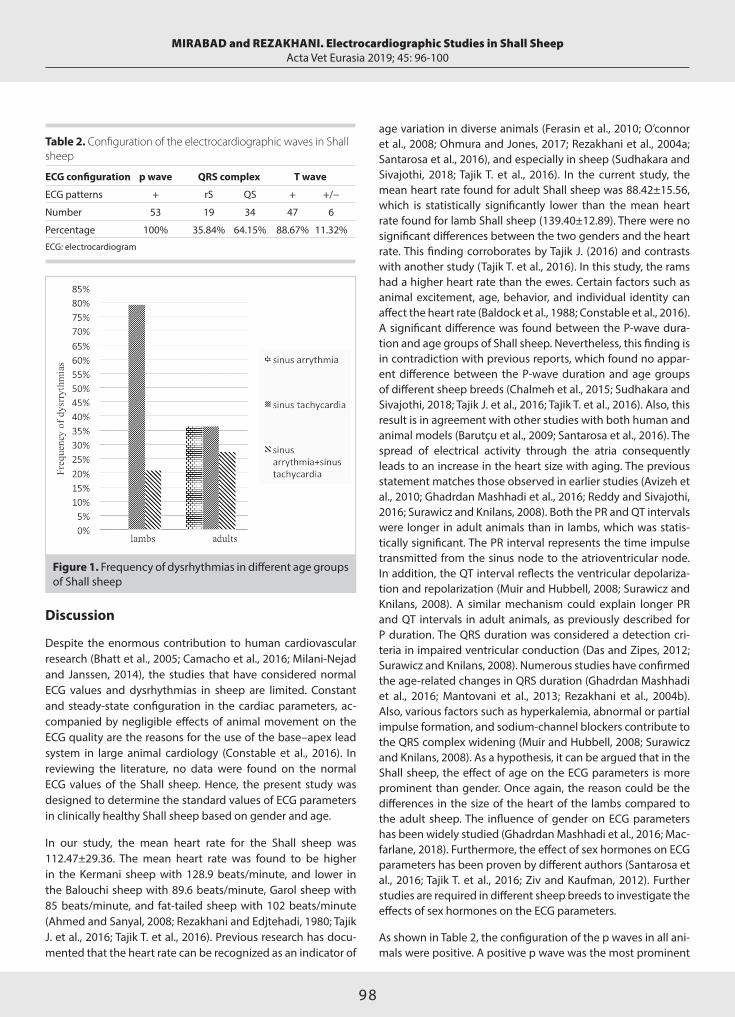
98
Discussion
Despite the enormous contribution to human cardiovascular research (Bhatt et al., 2005; Camacho et al., 2016; Milani-Nejad and Janssen, 2014), the studies that have considered normal ECG values and dysrhythmias in sheep are limited. Constant and steady-state configuration in the cardiac parameters, ac-companied by negligible effects of animal movement on the ECG quality are the reasons for the use of the base–apex lead system in large animal cardiology (Constable et al., 2016). In reviewing the literature, no data were found on the normal ECG values of the Shall sheep. Hence, the present study was designed to determine the standard values of ECG parameters in clinically healthy Shall sheep based on gender and age.
In our study, the mean heart rate for the Shall sheep was 112.47±29.36. The mean heart rate was found to be higher in the Kermani sheep with 128.9 beats/minute, and lower in the Balouchi sheep with 89.6 beats/minute, Garol sheep with 85 beats/minute, and fat-tailed sheep with 102 beats/minute (Ahmed and Sanyal, 2008; Rezakhani and Edjtehadi, 1980; Tajik J. et al., 2016; Tajik T. et al., 2016). Previous research has docu-mented that the heart rate can be recognized as an indicator of
age variation in diverse animals (Ferasin et al., 2010; O’connor et al., 2008; Ohmura and Jones, 2017; Rezakhani et al., 2004a; Santarosa et al., 2016), and especially in sheep (Sudhakara and Sivajothi, 2018; Tajik T. et al., 2016). In the current study, the mean heart rate found for adult Shall sheep was 88.42±15.56, which is statistically significantly lower than the mean heart rate found for lamb Shall sheep (139.40±12.89). There were no significant differences between the two genders and the heart rate. This finding corroborates by Tajik J. (2016) and contrasts with another study (Tajik T. et al., 2016). In this study, the rams had a higher heart rate than the ewes. Certain factors such as animal excitement, age, behavior, and individual identity can affect the heart rate (Baldock et al., 1988; Constable et al., 2016). A significant difference was found between the P-wave dura-tion and age groups of Shall sheep. Nevertheless, this finding is in contradiction with previous reports, which found no appar-ent difference between the P-wave duration and age groups of different sheep breeds (Chalmeh et al., 2015; Sudhakara and Sivajothi, 2018; Tajik J. et al., 2016; Tajik T. et al., 2016). Also, this result is in agreement with other studies with both human and animal models (Barutçu et al., 2009; Santarosa et al., 2016). The spread of electrical activity through the atria consequently leads to an increase in the heart size with aging. The previous statement matches those observed in earlier studies (Avizeh et al., 2010; Ghadrdan Mashhadi et al., 2016; Reddy and Sivajothi, 2016; Surawicz and Knilans, 2008). Both the PR and QT intervals were longer in adult animals than in lambs, which was statis-tically significant. The PR interval represents the time impulse transmitted from the sinus node to the atrioventricular node. In addition, the QT interval reflects the ventricular depolariza-tion and repolarization (Muir and Hubbell, 2008; Surawicz and Knilans, 2008). A similar mechanism could explain longer PR and QT intervals in adult animals, as previously described for P duration. The QRS duration was considered a detection cri-teria in impaired ventricular conduction (Das and Zipes, 2012; Surawicz and Knilans, 2008). Numerous studies have confirmed the age-related changes in QRS duration (Ghadrdan Mashhadi et al., 2016; Mantovani et al., 2013; Rezakhani et al., 2004b). Also, various factors such as hyperkalemia, abnormal or partial impulse formation, and sodium-channel blockers contribute to the QRS complex widening (Muir and Hubbell, 2008; Surawicz and Knilans, 2008). As a hypothesis, it can be argued that in the Shall sheep, the effect of age on the ECG parameters is more prominent than gender. Once again, the reason could be the differences in the size of the heart of the lambs compared to the adult sheep. The influence of gender on ECG parameters has been widely studied (Ghadrdan Mashhadi et al., 2016; Mac-farlane, 2018). Furthermore, the effect of sex hormones on ECG parameters has been proven by different authors (Santarosa et al., 2016; Tajik T. et al., 2016; Ziv and Kaufman, 2012). Further studies are required in different sheep breeds to investigate the effects of sex hormones on the ECG parameters.
As shown in Table 2, the configuration of the p waves in all ani-mals were positive. A positive p wave was the most prominent
MIRABAD and REZAKHANI. Electrocardiographic Studies in Shall SheepActa Vet Eurasia 2019; 45: 96-100
Figure 1. Frequency of dysrhythmias in different age groups of Shall sheep
Table 2. Configuration of the electrocardiographic waves in Shall sheep
ECG configuration p wave QRS complex T wave
ECG patterns + rS QS + +/−
Number 53 19 34 47 6
Percentage 100% 35.84% 64.15% 88.67% 11.32%
ECG: electrocardiogram

99
configuration of p wave in other studies (Rezakhani and Edjte-hadi, 1980; Tajik T. et al., 2016; Torío et al., 1997). The dominant QRS configuration in our sheep was QS. The QS morphology has been reported to be the main QRS morphology (in the base-apex lead system) by various researchers (Kamali et al., 2017; Rezakhani and Edjtehadi; 1980, Rezakhani et al., 2004b). The T wave was positive or diphasic (+/−). This finding has been reported by different authors in different sheep breeds (Tajik J. et al., 2016; Tajik T. et al., 2016). There was no normal sinus rhythm in lambs (Figure 1). The impulse initiation and con-duction above normal (90 and 120 for adult sheep and lambs, respectively) with origin of the sinus node considered to be si-nus tachycardia (Constable et al., 2016). Previous studies have shown that sinus tachycardia is the most prominent arrhythmia in lambs (Chalmeh et al., 2015; Pourjafar et al., 2011). Physiolog-ic sinus tachycardia is mainly rooted in catecholamine, howev-er synergistic inhibition of the vagus nerve plays a role in the formation of this cardiac arrhythmia (Yusuf and Camm, 2005). Sinus tachycardia is frequently associated with excitement, pain, exercise, anemia, hypotension, and the administration of adrenergic agents (Constable et al., 2016; Muir and Hubbell, 2008). As sinus tachycardia is a physiological response to stress, treatment is rarely required (Reed et al., 2018). Another dys-rhythmia observed in this study was sinus arrhythmia. The PP interval variability >10% is clearly and unambiguously indica-tive of sinus arrhythmia (Constable et al., 2016). Physiological reaction to respiration or drug-induced vagal stimulation are predisposing factors for sinus arrhythmia (Das and Zipes, 2012). In the current study, sheep with sinus arrhythmias had a signifi-cantly lower mean heart rate than sheep with normal rhythms (p=0.007). This finding has been confirmed in other studies (Rezakhani and Edjtehadi, 1980; Rezakhani et al., 2004a; Tajik T. et al., 2016). Cardiac irregularities are considered a normal physiological phenomenon since there were no clinical signs of cardiovascular disease. Finally, results obtained from this study can be used as a reference for future studies, and one of the more significant findings is the suitability of the base–apex lead system in the Shall sheep.
Peer-review: Externally peer-reviewed.
Author Contributions: Concept – M.M., A.R.; Design - M.M., A.R.; Su-pervision - M.M., A.R.; Resources - M.M., A.R.; Materials - M.M.; Data Col-lection and/or Processing - M.M., A.R.; Analysis and/or Interpretation - M.M., A.R.; Literature Search - M.M., A.R.; Writing Manuscript - M.M., A.R.; Critical Review - M.M., A.R.
Acknowledgement: We gratefully acknowledge the help provided by Dr.Seyed Mahdi Ghamsari, the head of Amin-Abad Research Institute.
Conflict of Interest: The authors have no conflicts of interest to de-clare.
Financial Disclosure: The authors declared that this study has re-ceived no financial support.
References
Ahmed, J.A., Sanyal, S., 2008. Electrocardiographic studies in Garol sheep and black Bengal goats. Research Journal of Cardiology 1, 1-8. [CrossRef]
Avizeh, R., Papahn, A., Ranjbar, R., Rasekh, A., Molaee, R., 2010. Electrocardiographic changes in the littermate mongrel dogs from birth to six months of life. Iranian Journal of Veterinary Research 11, 304-311.
Baldock, N., Sibly, R., Penning, P., 1988. Behaviour and seasonal vari-ation in heart rate in domestic sheep, Ovis aries. Animal Behaviour 36, 35-43. [CrossRef]
Barutçu, İ., Esen, Ö., Kaya, D., Onrat, E., Melek, M., Çelik, A., Kilit, C., Esen, A.M., 2009. The Relationship Between Aging and P Wave Dispersion. Kosuyolu Heart Journal 12, 5-9.
Bhatt, L., Nandakumar, K., Bodhankar, S., 2005. Experimental animal models to induce cardiac arrhythmias. Indian Journal of Pharmacolo-gy 37, 348-357. [CrossRef]
Camacho, P., Fan, H., Liu, Z., He, J.Q., 2016. Large mammalian animal models of heart disease. Journal of Cardiovascular Development and Disease 3, 1-11. [CrossRef]
Cedeno, D.A., Lourenço, M.L., Daza, C.A., Chiacchio, S.B., 2016. Elec-trocardiogram assessment using the Einthoven and base-apex lead systems in healthy Holstein cows and neonates. Pesquisa Veterinária Brasileira 36, 1-7. [CrossRef]
Chalmeh, A., Akhtar, I.S., Zarei, M.H., Badkoubeh, M., 2015. Elec-trocardiographic indices of clinically healthy Chios sheep. Veterinary Science Development 5, 99-102. [CrossRef]
Constable, P.D., Hinchcliff, K.W., Done, S.H., Grünberg, W., 2016. Veterinary Medicine-E-Book: A Textbook of the Diseases of Cattle, Horses, Sheep, Pigs and Goats. Elsevier Health Sciences, St. Louis.
Das, M.K., Zipes, D.P., 2012. Electrocardiography of Arrhythmias: A Comprehensive Review E-Book: A Companion to Cardiac Electro-physiology. Saunders Company, Philadelphia.
Ferasin, L., Ferasin, H., Little, C., 2010. Lack of correlation between canine heart rate and body size in veterinary clinical practice. Journal of Small Animal Practice 51, 412-418. [CrossRef]
Ghadrdan Mashhadi, A.R., Kamali, S., Haji Hajikolaei, M.R., Rezakhani, A., Fatemi, S.R., 2016. Determination the Normal Pa-rameters (amplitude and duration) of Electrocardiogram Waves in River Buffaloes (Bubalus Bubalis) of Khuzestan. Iranian Journal of Ruminants Health Research 1, 23-31.
Hossein-Zadeh, N.G., 2015. Modeling the growth curve of Iranian Shall sheep using non-linear growth models. Small Ruminant Re-search 130, 60-66. [CrossRef]
Kamali, S., Ghadrdan, M.A., Haji, H.M., Fatemi, T.S., Rezakhani, A., 2017. Survey on Frequency of Various Forms of QRS Complex in Khu-zestan River Buffalo. Iranian Veterinary Journal 13, 91-97.
Kumar, C.P., Sundar, N.S., Reddy, B., Praveena, G., Kumar, R., 2015. Electrocardiogram of small ruminants by base apex lead system. In-dian Journal of Small Ruminants 21, 141-142. [CrossRef]
Macfarlane, P.W., 2018. The influence of age and sex on the electro-cardiogram. In: Kerkhof, P.L.M., Miller, VM. (Ed.), Sex-Specific Analysis of Cardiovascular Function. Springer, Cham, pp. 93-106. [CrossRef]
Mantovani, M., Tsuruta, S., Muzzi, R., Machado, T., Pádua, M., Coim-bra, C., Muzzi, L., Jacomini, J., 2013. Electrocardiographic study in
MIRABAD and REZAKHANI. Electrocardiographic Studies in Shall SheepActa Vet Eurasia 2019; 45: 96-100

100
the American Quarter Horse breed. Arquivo Brasileiro de Medicina Veterinária e Zootecnia 65, 1389-1393. [CrossRef]
Milani-Nejad, N., Janssen, P.M., 2014. Small and large animal mod-els in cardiac contraction research: advantages and disadvantages. Pharmacology & Therapeutics 141, 235-249. [CrossRef]
Muir, W.W., Hubbell, J.A.E, 2008. Equine Anesthesia: Monitoring and Emergency Therapy, Saunders Company, St. Louis. [CrossRef]
O’connor, M., Mcdaniel, N., Brady, W.J., 2008. The pediatric electro-cardiogram: Part I: Age-related interpretation. The American Journal of Emergency Medicine 26, 506-512. [CrossRef]
Ohmura, H., Jones, J.H., 2017. Changes in heart rate and heart rate variability as a function of age in Thoroughbred horses. Journal of Equine Science 28, 99-103. [CrossRef]
Pourjafar, M., Badiei, K., Chalmeh, A., Sanati, A., Bagheri, M., Bad-kobeh, M., Shahbazi, A., 2011. Cardiac arrhythmias in clinically healthy newborn Iranian fat tailed lambs. Global Veterinaria 6, 185-189.
Reddy, B., Sivajothi, S., 2016. Electrocardiographic parameters of nor-mal dairy cows during different ages. Journal of Veterinary Science & Medicine 4, 5. [CrossRef]
Reed, S.M., Bayly, W.M., Sellon, D.C., 2018. Equine Internal Medicine. Elsevier, St. Louis.
Rezakhani, A., Edjtehadi, M., 1980. Some electrocardiographic pa-rameters of the Fat‐tailed sheep. Zentralblatt für Veterinärmedizin Reihe A 27, 152-156. [CrossRef]
Rezakhani, A., Paphan, A., Gheisari, H., 2004a. Cardiac dysrhythmias in clinically healthy heifers and cows. Revue de Medecine Veterinaire 155, 159-162.
Rezakhani, A., Paphan, A.A., Shekarfroush, S., 2004b. Analysis of base apex lead electrocardiograms of normal dairy cows. Veterinar-ski Arhiv 74, 351-358.
Salehi, M., Taherpour-Dari, N., 2005. Evaluation of wool characteristics of Iranian sheep breeds 4 _ Arabi breed. Animal Science Research Insti-tute, Final Report, Ministry of Agriculture, Iran. http://agris.fao.org
Santarosa, B.P., Lourenço, M.L., Dantas, G.N., Ulian, C., Heckler, M.C., Sudano, M.J., Gonçalves, R.C., Chiacchio, S.B., 2016. Electrocardio-graphic parameters of the American Miniature Horse: influence of age and sex. Pesquisa Veterinária Brasileira 36, 551-558. [CrossRef]
Sudhakara, R.B., Sivajothi, S., 2018. Electrocardiographic studies in different age groups of Nellore cross-breed sheep. International Clin-ical Pathology Journal 6, 30-32. [CrossRef]
Surawicz, B., Knilans, T., 2008. Chou’s Electrocardiography in Clinical Practice: Adult and Pediatric. Saunders Company, Philadelphia.
Tajik, J., Samimi, A.S., Shojaeepour, S., Jarakani, S., 2016. Analysis of base-apex lead electrocardiogram in clinically healthy Kermani sheep. Acta Vet Eurasia 42, 74-79. [CrossRef]
Tajik, T., Aslani, M.R., Tajik, J., 2016. Electrocardiographic parameters in clinically healthy Balouchi sheep. Iranian Journal of Veterinary Sci-ence and Technology 7, 84-91.
Torío, R., Cano, M., Montes, A., Prieto, F., Benedito, J., 1997. Com-parison of two methods for electrocardiographic analysis in Gallega sheep. Small Ruminant Research 24, 239-246. [CrossRef]
Yusuf, S., Camm, A.J., 2005. Deciphering the sinus tachycardias. Clini-cal Cardiology 28, 267-276. [CrossRef]
Ziv, O., Kaufman, E.S., 2012. Age and gender modulation of the long QT syndrome phenotype. Cardiac Electrophysiology Clinics 4, 39-51. [CrossRef]
MIRABAD and REZAKHANI. Electrocardiographic Studies in Shall SheepActa Vet Eurasia 2019; 45: 96-100

101
Address for Correspondence: Alper METE • E-mail: [email protected]
Received Date: 17 August 2019 • Accepted Date: 18 November 2019 • DOI: 10.5152/actavet.2019.19022
Available online at actaveteurasia.istanbulc.edu.tr
Abstract
Nowadays, many disinfectants and antiseptics are used for decontamination purposes in equine hospitals, on racetracks, and breeding farms, but generally, these antimicrobial agents are not tested against commonly encountered pathogens, and they are used with unknown antimicrobial efficacy. The antimicrobial efficacies of ethanol, chlorhexidine, povidone iodine, sodium hypochloride, peroxymonosulfate compound, and benzalkonium chloride were analyzed using the quan-titative suspension test method against the field isolates of Escherichia coli, Pseudomonas aeruginosa, Salmonella spp., Streprococcus zooepidemicus, Streptococcus equi, Rhodococcus equi, and Staphylococcus auerus, which are the most frequ-ently encountered pathogens of equines, in the presence of organic load (10% fetal bovine serum) after 1 min, 5 mins, and
30 mins contact times at 20°C. A log reduction of five or more (5 log ≤) in cfu counts of the tested pathogens was considered as effective for each disinfectant and antiseptic. According to the results, except for sodium hypochloride in the 1/100 dilu-tion, all other disinfectants and antiseptics achieved a mini-mum 5 log reduction and were found to be effective against all tested isolates. Decreased dilutions and/or direct use of the sodium hypochloride should be tested against the same bac-terial agents, as well as with multiple field strains. In addition, reference strains of the microorganisms should be evaluated in further studies.
Keywords: Antiseptics, bacterial pathogens, biosecurity, di-sinfectants, horse
Antibacterial Efficacy of Some Antiseptics and Disinfectants against Common Bacterial Agents Isolated from Horses in Turkey
Alper METE Jockey Club of Turkey İstanbul Veliefendi Racetrack Horse Hospital, İstanbul, Turkey
Cite this article as: Mete, A., 2019. Antibacterial Efficacy of Some Antiseptics and Disinfectants against Common Bacterial Agents Isolated from Horses in Turkey. Acta Vet Eurasia 45, 101-107.
ORCID ID of the author: A.M. 0000-0002-4810-5579.
Original Article Acta Vet Eurasia 2019; 45: 101-107
Introduction
Control of infectious diseases in horse populations involves two critical aspects: vaccination and disinfection. Many adequate vaccines against infectious diseases are commercially avail-able, but none of them can be warranted to be 100% effective (Dwyer, 2004). In times of an epidemic disease, it is common to find significant environmental microbial contamination in hos-pitals, on racetracks, farms, and in any facilities where horses reside. This microbial contamination commonly orginates from infected animals’ secretions, such as blood, urine, feces, nasal, and conjunctival secretions, etc. (Saklou et al., 2016). It is also important to minimize animal trafficking and distribution of potential pathogens by movement of personnel and fomites
(Morley et al., 2005). Therefore, disinfection and antisepsis man-agement practices are essential parts of providing a healthy en-vironment for horses. Disinfection also plays an important role in the prevention and control of nosocomial infections, espe-cially for the multi-resistant bacteria, where disinfection is the only way to slow down the disease outbreak.
Many bacterial pathogens can cause systemic and local in-fections in horses. Streptococcus equi subsp. zooepidemicus (S. zooepidemicus) and S. equi subsp. equi (S. equi) cause the lower respiratory tract, joint, genital tract, eye and guttural pouch in-fections, and abscess formation. In foals, Rhodococcus equi (R. equi) cause pleuropneumonia, gastrointestinal tract infections, and abscess formation as well. Staphylococcus aureus (S. aureus)
This work is licensed under a Creative Commons Attribution-NonCommercial 4.0 International License.
102
METE A. Disinfectant Efficacy agaInst Horse PathogensActa Vet Eurasia 2019; 45: 101-107
may cause wound infections, mastitis, and abscess formation. Salmonella spp. are mostly isolated from the gastrointestinal tract infections and neonatal sepsis. Escherichia coli (E. coli) may cause genital tract infections, mastitis in mares, and septicemia in neonatal foals. Pseudomonas aeruginosa (P. aeruginosa) cause genital tract infections and mastitis in mares (Sellon and Long, 2013). These bacterial agents can survive on environmental surfaces for long periods with a possible transmission to sus-ceptible hosts. Therefore, it is imperative to use an effective disinfectant/antiseptic to prevent the spread of these agents (Köse and Yapar, 2017).
In field conditions, a good disinfectant should be effective in the presence of organic matter, such as blood, urine, feces, and other body secretions; have a low or zero toxicity against ani-mals; and show the bactericidal activity in a relatively short pe-riod of time. Among the horse pathogens, gram-positive and gram-negative bacteria and enveloped viruses are considered to be susceptible to the disinfectants in the absence of organic load. But besides these generalizations, because they are in the same susceptibility category, Salmonella species are extremely difficult to eliminate from horse facilities (Dwyer, 2004).
In Turkey, many commercially available antiseptics and disin-fectants with different active ingredients are used in equine industry for decontamination of the bacterial agents. But to the best of author’s knowledge, no antimicrobial efficacy studies with commercially available antiseptics and disinfectants were performed against the reference and field strains of the horse bacterial pathogens up to this date in Turkey.
The aim of this study was to evaluate the antibacterial effec-tiveness of disinfectants and antiseptics often used in equine facilities and hospitals, including sodium hypocloride (house-hold bleach), potassium peroxymonosulfate (Virkon S; İstanbul, Turkey), and benzalkonium chloride (Quaternary ammonium compound-QAC, Zefirolum; İstanbul, Turkey) as disinfectants as well as ethanol, povidone iodine (Poviiodeks; İstanbul, Tur-key), and chlorhexidine (Hibitanol; İstanbul, Turkey) as antisep-
tics against the field isolate of gram-positive species such as S. zooepidemicus, S. equi, S. aureus, and R. equi, and gram-negative species such as P. aeruginosa, E. coli, and Salmonella spp. in the presence of organic load to examine the antimicrobial activities of the commercial compound(s) commonly used in horse care facilities and hospitals in Turkey.
Materials and Methods
Bacterial StrainsThe field isolates of S. zooepidemicus, S. equi, S. aureus, R. equi, E. coli, P. aeruginosa, and Salmonella spp. were used in the study. The isolation side, date of isolation, and isolation re-gion in the country were shown in the table below (Table 1). Briefly, all clinical samples were streaked to 5% sheep blood agar and MacConkey agar, and they were incubated at 37°C in both aerobic and microaerophilic (5% CO2) conditions for 48 hours. In addition, rectal swab samples were also inocu-lated in selenite broth for 18 hours and passaged to the XLD agar media for Salmonella spp. isolation. Suspected colonies were identified with routine methods, such as colony mor-phology, microscopic morphology, and gram characteristics, catalase, oxidase, and other biochemical tests using the BBL crystal E/NF and gram-positive identification systems (Bec-ton Dictinson; Sparks, U.S.). After the identification process, the isolates were passaged into tryptone soy broth (TSB) (Ox-oid; Basingstoke, UK), containing 20% glycerol, and stored at −20°C until laboratory analysis. The isolates were revived by passaging to the tryptone soy agar (TSA) (Oxoid; Basingstoke, UK) from the storage media.
Disinfectants and AntisepticsCommercially available three different classes of disinfectants and three different classes of antiseptics were chosen to repre-sent different range of active compounds in the present study. Disinfectant/antiseptic classes, active ingredients, and the dilu-tions used in the experiment are given in Table 2. Disinfectants and ethanol were diluted using tap water of 207 mg CaCO3/L hardness in each test tube. The hardness of the tap water was determined with SM 2340:B; ISO 17294-2 (ICP-MS) method by a private enviromental analysis laboratory (Çevre Industrial Anal-ysis Laboratory, İstanbul, Turkey).
Culture MediaTryptone soy broth and TSA were used for maintenance and determination of viable cell counts in the experiment.
Neutralization MediaNeutralization solution was prepared by using a mixture of tryptone (5.0 g/L), yeast extract (2.5 g/L), dextrose (10.0 g/L), sodium thioglycollate (1.0 g/L), sodium thiosulphate (6.0 g/L), sodium bisulphite (2.5 g/L), llecithin (7.0 g/L), polysorbate 80 (5.0 g/L), and bromocresol purple (0.020 g/L). The final pH val-ues of neutralization media were measured and adjusted to 7.6±0.2 before use.
Table 1. Sample type isolated, date of isolation, and geographical isolation region of the bacterial isolates used in the study
Isolate Sample type isolated Geographical origin
Gram positive
S. zooepidemicus Tracheal wash fluid İstanbul
S. equi Guttural pouch wash fluid İstanbul
S. aureus Skin wound swab İstanbul
R. equi Tracheal wash fluid İzmit
Gram negative
E. coli Intrauterine swab Thrace
P. aeruginosa Intrauterine swab Thrace
Salmonella spp. Rectal swab İstanbul
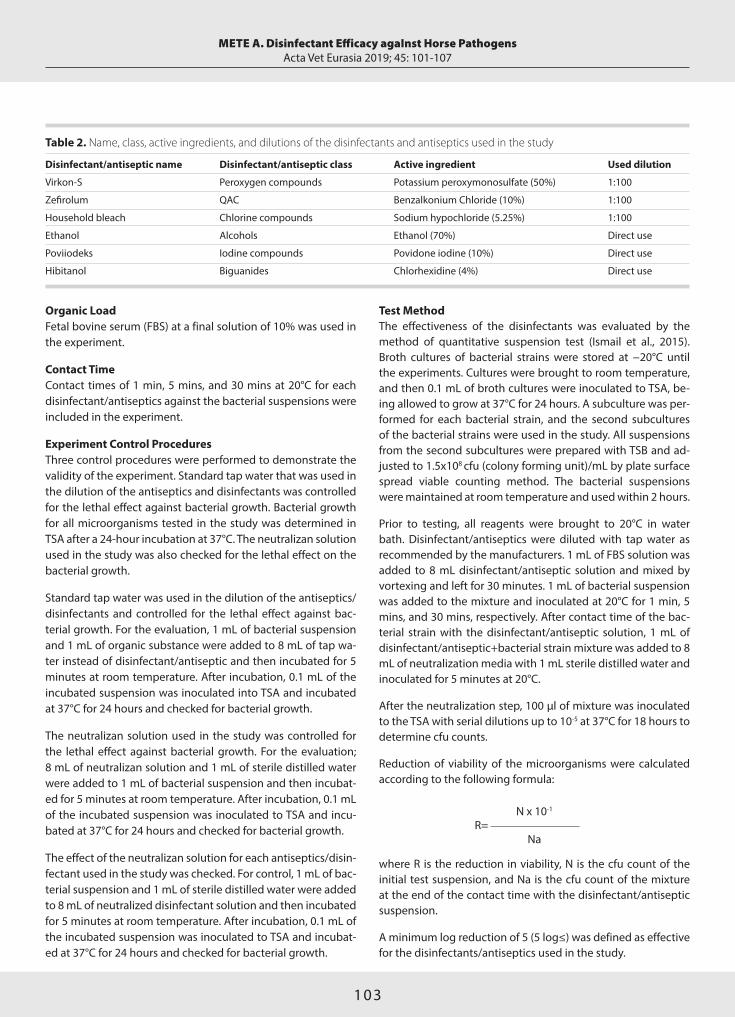
103
Organic LoadFetal bovine serum (FBS) at a final solution of 10% was used in the experiment.
Contact TimeContact times of 1 min, 5 mins, and 30 mins at 20°C for each disinfectant/antiseptics against the bacterial suspensions were included in the experiment.
Experiment Control ProceduresThree control procedures were performed to demonstrate the validity of the experiment. Standard tap water that was used in the dilution of the antiseptics and disinfectants was controlled for the lethal effect against bacterial growth. Bacterial growth for all microorganisms tested in the study was determined in TSA after a 24-hour incubation at 37°C. The neutralizan solution used in the study was also checked for the lethal effect on the bacterial growth.
Standard tap water was used in the dilution of the antiseptics/disinfectants and controlled for the lethal effect against bac-terial growth. For the evaluation, 1 mL of bacterial suspension and 1 mL of organic substance were added to 8 mL of tap wa-ter instead of disinfectant/antiseptic and then incubated for 5 minutes at room temperature. After incubation, 0.1 mL of the incubated suspension was inoculated into TSA and incubated at 37°C for 24 hours and checked for bacterial growth.
The neutralizan solution used in the study was controlled for the lethal effect against bacterial growth. For the evaluation; 8 mL of neutralizan solution and 1 mL of sterile distilled water were added to 1 mL of bacterial suspension and then incubat-ed for 5 minutes at room temperature. After incubation, 0.1 mL of the incubated suspension was inoculated to TSA and incu-bated at 37°C for 24 hours and checked for bacterial growth.
The effect of the neutralizan solution for each antiseptics/disin-fectant used in the study was checked. For control, 1 mL of bac-terial suspension and 1 mL of sterile distilled water were added to 8 mL of neutralized disinfectant solution and then incubated for 5 minutes at room temperature. After incubation, 0.1 mL of the incubated suspension was inoculated to TSA and incubat-ed at 37°C for 24 hours and checked for bacterial growth.
Test MethodThe effectiveness of the disinfectants was evaluated by the method of quantitative suspension test (Ismail et al., 2015). Broth cultures of bacterial strains were stored at −20°C until the experiments. Cultures were brought to room temperature, and then 0.1 mL of broth cultures were inoculated to TSA, be-ing allowed to grow at 37°C for 24 hours. A subculture was per-formed for each bacterial strain, and the second subcultures of the bacterial strains were used in the study. All suspensions from the second subcultures were prepared with TSB and ad-justed to 1.5x10⁸ cfu (colony forming unit)/mL by plate surface spread viable counting method. The bacterial suspensions were maintained at room temperature and used within 2 hours.
Prior to testing, all reagents were brought to 20°C in water bath. Disinfectant/antiseptics were diluted with tap water as recommended by the manufacturers. 1 mL of FBS solution was added to 8 mL disinfectant/antiseptic solution and mixed by vortexing and left for 30 minutes. 1 mL of bacterial suspension was added to the mixture and inoculated at 20°C for 1 min, 5 mins, and 30 mins, respectively. After contact time of the bac-terial strain with the disinfectant/antiseptic solution, 1 mL of disinfectant/antiseptic+bacterial strain mixture was added to 8 mL of neutralization media with 1 mL sterile distilled water and inoculated for 5 minutes at 20°C.
After the neutralization step, 100 µl of mixture was inoculated to the TSA with serial dilutions up to 10-5 at 37°C for 18 hours to determine cfu counts.
Reduction of viability of the microorganisms were calculated according to the following formula:
N x 10-1
R= Na
where R is the reduction in viability, N is the cfu count of the initial test suspension, and Na is the cfu count of the mixture at the end of the contact time with the disinfectant/antiseptic suspension.
A minimum log reduction of 5 (5 log≤) was defined as effective for the disinfectants/antiseptics used in the study.
METE A. Disinfectant Efficacy agaInst Horse PathogensActa Vet Eurasia 2019; 45: 101-107
Table 2. Name, class, active ingredients, and dilutions of the disinfectants and antiseptics used in the study
Disinfectant/antiseptic name Disinfectant/antiseptic class Active ingredient Used dilution
Virkon-S Peroxygen compounds Potassium peroxymonosulfate (50%) 1:100
Zefirolum QAC Benzalkonium Chloride (10%) 1:100
Household bleach Chlorine compounds Sodium hypochloride (5.25%) 1:100
Ethanol Alcohols Ethanol (70%) Direct use
Poviiodeks Iodine compounds Povidone iodine (10%) Direct use
Hibitanol Biguanides Chlorhexidine (4%) Direct use

104
Results
Experiment ControlTest results indicated that no antibacterial effect of the neu-tralizan solution was determined for all tested microorganisms in the study. The neutralizan effect of the neutralizan solution against antiseptics/disinfectants was evaluated for each disin-fectant and antiseptic as well. No inhibition in bacterial growth was determined for each neutralized disinfectant and antisep-tic used in the study (data not shown).
Antibacterial Activity of the Tested Antiseptics and Disin-fectantsAccording to the standards, animicrobials tested must show a minimum 5 log (105) reduction in cfu/mL to be considered as effective. After the determined contact times with 70% etha-
nol, chlorhexidine, povidone iodine, virkonS (1/100), and ben-zalkonium chloride, a 8.17 log reduction was identified against E. coli, P. aeruginosa, Salmonella spp., S. zooepidemicus, S. equi, R. equi, and S. aureus in the presence of the organic load (10% FBS). But on the other hand, sodium hypochloride (1/100) failed to pass the test standard against E. coli after 1 min and 5 mins; against P. aeruginosa after 1 min, 5 mins, and 30 mins; against Salmonella spp. after 1 min, 5 mins, and 30 mins; against S. equi after 1 min, 5 mins, and 30 mins; against R. equi after 1 min and 5 mins; and against S. aureus after 1 min and 5 mins contact times in the presence of organic load (10% FBS). The cfu/ml of bacterial agents after 1 min, 5 mins, and 30 mins contact times with the antiseptics and disinfectants are listed as in the tables (Tables 3-9). Results of reduction in viabilities (log reduction) obtained in the present study were given as graph (Figure 1).
METE A. Disinfectant Efficacy agaInst Horse PathogensActa Vet Eurasia 2019; 45: 101-107
Table 3. Cfu/mL values of E. coli after contact with tested antiseptics and disinfectants at 20°C
Antiseptics-disinfectants/contact time 1 min 5 mins 30 mins
Ethanol 0 0 0
Chlorhexidine 0 0 0
Povidone iodine 0 0 0
Sodium hypochloride 1.04x10⁵ 6x10⁴ 7x100
Virkon S 0 0 0
Benzalkonium Chloride 0 0 0
Table 4. Cfu/mL values of P. aeruginosa after contact with tested antiseptics and disinfectants at 20°C
Antiseptics-disinfectants/contact time 1 min 5 mins 30 mins
Ethanol 0 0 0
Chlorhexidine 0 0 0
Povidone iodine 0 0 0
Sodium hypochloride 5.4x10⁴ 7x10⁴ 3x10⁴
Virkon S 0 0 0
Benzalkonium Chloride 0 0 0
Table7. Cfu/mL values of S. equi after contact with tested antiseptics and disinfectants at 20°C
Antiseptics-disinfectants/contact time 1 min 5 mins 30 mins
Ethanol 0 0 0
Chlorhexidine 0 0 0
Povidone iodine 0 0 0
Sodium hypochloride 2.02x10⁴ 1.92x10⁴ 1.17x10⁴
Virkon S 0 0 0
Benzalkonium Chloride 0 0 0
Table 6. Cfu/mL values of S. zooepidemicus after contact with tested antiseptics and disinfectants at 20°C
Antiseptics-disinfectants/contact time 1 min 5 mins 30 mins
Ethanol 0 0 0
Chlorhexidine 0 0 0
Povidone iodine 0 0 0
Sodium hypochloride 4x101 0 0
Virkon S 0 0 0
Benzalkonium Chloride 0 0 0
Table 5. Cfu/mL values of Salmonella spp. after contact with tested antiseptics and disinfectants at 20°C
Antiseptics-disinfectants/contact time 1 min 5 mins 30 mins
Ethanol 0 0 0
Chlorhexidine 0 0 0
Povidone iodine 0 0 0
Sodium hypochloride 8.75x10³ 2.2x10⁴ 2.6x10⁴
Virkon S 0 0 0
Benzalkonium Chloride 0 0 0
Table 8. Cfu/mL values of R. equi after contact with tested antiseptics and disinfectants at 20°C
Antiseptics-disinfectants/contact time 1 min 5 mins 30 mins
Ethanol 0 0 0
Chlorhexidine 0 0 0
Povidone iodine 0 0 0
Sodium hypochloride 2.31x10³ 1.9x10⁴ 0
Virkon S 0 0 0
Benzalkonium Chloride 0 0 0

105
Discussion
The use of disinfectants and antiseptics is of paramount im-portance in biosecurity and infection control in individuals and populations. Proper use of disinfectants and antiseptics could be expected to be cheaper than economic cost of antimicrobial treatment in horses or loss of part or all of that horse popula-tion due to a disease outbreak (Dwyer, 1995). Microorganisms are known to vary in their susceptibility against disinfectants and antiseptics, and some studies reveal that the efficacy of disinfectants are gradually reduced (Orji, 2014). Inappropriate consumption, inaccurate concentration, and lack of training for
preparation and storage are the most common reasons of in-creasing resistance to disinfectants (Zareniya et al., 2017).
Karayıldırım and Çelenk (2016) expressed that 20% benzalko-nium chloride was found to be effective against E. coli, S. au-reus, and P. aeruginosa with a 1 min contact time. In the present study, it was determined that the 1/100 dilution of 10% ben-zalkonium chloride was also effective (log 5 ≤ reduction) in 1 min, 5 mins, and 30 mins against the same bacteria that were isolated from clinical cases of horses. Gehan et al. (2009) indi-cated that 1% of benzalkonium chloride was effective against P. aeruginosa, E. coli, S. typhimurium, and S. aureus at the 30 min contact time. In another study, 3% of benzalkonium chloride achieved a 5 log reduction in 30 mins, and 1% in 60 mins (El Aal et al., 2008). Fazlara and Ekhtelat (2012) described that Listeria monocytogenes was the most susceptible bacteria to benzal-konium chloride, followed by S. aureus and E. coli, respectively, according to the minimum inhibitory concentration (MIC) and minimum bactericidal concentration (MBC) test results. Consid-ering all these results, in addition to 20% after 1 min of contact time, 1% and 3% concentration of benzalkonium chloride after 30 mins of contact time, and 1/100 dilution of %10 benzalkoni-um chloride can be used for inactivating E. coli, S. aureus, and P. aeruginosa after 1 min, 5 min, and 30 mins contact time.
METE A. Disinfectant Efficacy agaInst Horse PathogensActa Vet Eurasia 2019; 45: 101-107
Table 9. Cfu/mL values of S. aureus after contact with tested antiseptics and disinfectants at 20°C
Antiseptics-disinfectants/contact time 1 min 5 mins 30 mins
Ethanol 0 0 0
Chlorhexidine 0 0 0
Povidone iodine 0 0 0
Sodium hypochloride 1.39x10⁵ 1.11x10⁵ 0
Virkon S 0 0 0
Benzalkonium Chloride 0 0 0
Figure 1. Results of reduction in viability (log reductions) of bacterial isolates against tested antiseptics/disinfectants
E. coli
Ethanol 1 min.
9.00
8.00
7.00
6.00
5.00
4.00
3.00
2.00
1.00
0.00
Ethanol 5 min. Ethanol 30 min.
Sodium hypochloride 1 min. Sodium hypochloride 5 min. Sodium hypochloride 30 min.
Benzalkonium chloride 1 min. Benzalkonium chloride 5 min. Benzalkonium chloride 30 min.
Virkon S 1 min. Virkon S 5 min. Virkon S 30 min.
Povidone iodine 1 min. Povidone iodine 5 min. Povidone iodine 30 min.
Chlorhexidine 1 min. Chlorhexidine 5 min. Chlorhexidine 30 min.
Salmonellaspp.
S. zooepidemicus
P. aeruginosa S. equi R. equi S. aureus

106
Saklou et al. (2016) determined that the mist application of 2% of peroxymonosulfate compound disinfectant at 30 mins contact time created a 84%, 99%, and 99% reduction against S. enterica, P. aeruginosa, and S. aureus, respectively, and it was found to be effective if used after cleaning of the surfaces. Ge-han et al. (2009) found that 1% peroxymonosulfate compound was effective against P. aeruginosa, E. coli, S. typhimurium, and S. aureus after 30 mins of contact time in the presence of organic matter. Chima et al. (2013) also declared that peroxymonosul-fate compound was 100% effective against Salmonella spp., E. coli, Klebsiella spp., and P. aeruginosa. Another study also yield-ed that 1/100 dilution of 50% peroxymonosulfate compound demonstrated 5 log ≤ reduction against S. typhimurium ATCC 13311 strain (Jang et al., 2017). These results were in concor-dance with the present study’s results: 50% peroxymonosulfate disinfectant in 1/100 dilution showed a 8.17 log reduction at 1 min, 5 mins, and 30 mins, and it was found to be effective (5 log ≤) for all pathogens that participated in the study and found to be effective against the same bacterial pathogens of the previ-ous studies. The present study’s results for the peroxymonosul-fate compound have been also confirmed in previous studies.
In the present study, 5.25% sodium hypochloride in 1/100 dilu-tion failed to create the 5 log ≤ reduction against P. aeruginosa at 1 min, 5 mins, 30 mins and S. aureus at 1 min and 5 mins of contact time. But contrary to the present study, 5% sodium chloride in 1/100 dilution showed 7.22 and 8.11 log reductions at 5 mins and 30 mins, respectively, in a previous study (Bho-sale, 2017). The discrepancy might have been due to the differ-ence of the antimicrobial resistancy of bacterial strains tested in the studies. On the other hand, Addie et al. (2015) speci-fied that household bleach produced a 5 log ≤ reduction in E. coli 0157:H7 and Salmonella typhimurium after 1 min contact time. In the present study, the household bleach was used in 1/100 dilution so that the efficacy might have been reduced due to the dilution factor. In parallel, Avcı and Otkun (2017) claimed that the 1/100 dilution of sodium chloride was ineffec-tive against S. aureus and P. aeruginosa after 1 min and 2 mins contact time but effective after 5 mins, 10 mins, and 30 mins contact time. According to the author, further studies should be designed to test more concentrated dilutions or direct use of the sodium hypochloride against the same pathogens in to test the antimicrobial efficacy. It was also claimed that sodium hypochloride was inactivated by organic debris (Addie et al., 2015). Another reason for reduction in the efficacy of sodium hypochloride against the tested pathogens might have been the interaction with organic material (10% FBS) in the present experiment.
Zareniya et al. (2017) determined that povidone iodine was more effective than 70% ethanol in 49 P. aeruginosa isolates ac-cording to MIC and MBC values. In the present study both anti-septics demonstrated a full reduction (8.17 log) in P. aeruginosa. In another study, 10% povidone iodine was found to be effec-tive after 1 min contact time against S. aureus, P. aeruginosa,
and E. coli (Avcı and Otkun, 2017). In the same study 70% etha-nol was effective against P. aeruginosa and E. coli, but just inef-fective against S. aureus after 1 min contact time. After 2 mins, 5 mins, 10 mins, and 30 mins, 70% ethanol was found to be effec-tive by Avcı and Otkun (2017). The present study revealed that 70% ethanol and 10% povidone iodine demonstrated efficacy (5 log ≤) against P. aeruginosa, S. aureus, and E. coli after 1 mins, 5 mins, and 30 mins contact time, and the results were mostly in concordance with Avcı and Otkun’s (2017) results. According to the past and present study results, 70% ethanol and 10% po-vidone iodine can be used for antisepsis and disinfection pur-poses. The present study has some limitations, such as limited number of field bacterial isolates were tested, and a single test method (quantative suspension test) was used. Therefore, fu-ture studies should include more of field strains and evaluate different efficacy methods to monitor the antimicrobial activity of the tested disinfectants.
In conclusion, 50% peroxymonosulfate compound in 1/100 dilution; 10% benzalkonium chloride in 1/100 dilution as dis-infectants; and 70% ethanol, 4% chlorhexidine, and 10% povi-don iodine as antiseptics may be used in equine hospitals and equine care facilities as decontaminating agents against E. coli, P. aeruginosa, Salmonella spp., S. zooepidemicus, S. equi, R. equi, and S. aureus after 1 min, 5 mins, and 30 mins contact time. So-dium hypochloride in 1/100 dilution did not yield satisfactory results, and it failed to achieve a 5-log reduction against most of the bacterial agents tested in the study. The dilution ratio of sodium hypochloride may be decreased or used without dilut-ing while testing against the bacterial agents in further stud-ies. The present study tested one field isolate of each bacterial species against disinfectants and antiseptics as representatives. Future comprehensive studies should also be performed with multiple field and reference strains of E. coli, P. aeruginosa, Sal-monella spp., S. zooepidemicus, S. equi, R. equi, and S. aureus, as well as other bacterial and mycotic pathogens with different analytical methods to evaluate the antimicrobial efficacies of the disinfectants and antiseptics.
Peer-review: Externally peer-reviewed.
Conflict of Interest: The author has no conflicts of interest to declare.
Financial Disclosure: The authors declared that this study has received no financial support.
References
Addie, D.D., Corine, B.B., Egberink, H., Frymus, T., Jones, T.G., Hart-mann, K., Horzinek, M.C., Hosie, M.J., Lloret, A., Lutz, H., Maris-ilio, F., Pennisi, M.G., Radford, A.D., Thiry, E., Truyen, U., Möstl, K., 2015. Disinfectant choices in veterinary practices, shelters and households ABCD guidelines on safe and effective disinfection for feline enviroments. Journal of Feline Medicine and Surgery 17, 594-605. [CrossRef]
METE A. Disinfectant Efficacy agaInst Horse PathogensActa Vet Eurasia 2019; 45: 101-107

107
Avcı, D., Otkun, M., 2017. Bazı antiseptik ve dezenfektanların antibak-teriyel etkinliklerinin araştırılması. Türk Hijyen ve Deneysel Biyoloji Dergisi 74, 211-220.
Bhosale, N.K., 2017. Evaluation and comparison of efficacy of the surfce disinfectants used in a tertiary care hospital, India. Interna-tional Journal of Current Microbiology and Applied Sciences 6, 2608-2614. [CrossRef]
Chima, I.U., Uchegbu, M.C., Okoli, I.C., Ezema, C., Wehke, S.N., 2013. Evaluation of the efficacy of disinfectants used against bac-terial isolates from intensive poultry farming environments in Imo State, Nigeria. Journal of Biological Sciences 13, 349-356. [CrossRef]
Dwyer, R.M., 1995. Disinfecting equine facilities. Revue Scientifique et Technique (International Office of Epizootics) 14, 403-418. [CrossRef]
Dwyer, R.M., 2004. Environmental disinfection to control equine in-fectious diseases. Veterinary Clinics Equine Practice 20, 531-542. [CrossRef]
El Aal, S.F.A.A., Hunsinger, B., Böhm, R., 2008. Determination of the bactericidal activity of chemical disinfectants against bacteria in dairies according to the DVG-guidelines. Hygiene Medical 33, 463-471.
Fazlara, A., Ekhtelat, M., 2012. The disinfectant effects of benzalko-nium chloride on some important foodborne pathogens. Ameri-can-Eurasian Journal of Agricultural & Environmental Sciences 12, 23-29.
Gehan, M.Z., Anwer, W., Amer, H.M., El-Sabagh, E.L., Rezk, A., Bad-awy, E.M., 2009. In vitro Efficacy comparisons of disinfectants used in the commercial poultry farms. International Journal of Poultry Sci-ence 8, 237-241. [CrossRef]
Ismail, E., Kaoud, H.A., Hamouda, M., Nermeen, A.E., 2015. Bacte-ricidal effectiveness of some commerical disinfectants against Aero-monas hydrophila and Vibrio vulnificus. Ewemen Journal of Microbial Research 1, 9-15.
Jang, Y., Lee, K., Lee, M., Song, J., Chang, B., Choe, N.H., 2017. Effi-cacy evaluation of commercial disinfectants by using Salmonella en-terica serovar Typhimurium as a test organism. Journal of Veterinary Science 18, 209-216. [CrossRef]
Karayıldırım, Ç.K., Çelenk, V.U., 2016. Antibacterial efficiency of ben-zalkonium chloride base disinfectant according to European Stan-dard 13727, chemical analysis and validation studies. Celal Bayar University Journal of Science 12, 39-47. [CrossRef]
Köse, H., Yapar, N., 2017. The comparison of various disinfectants’ effi-cacy on Staphylococcus aureus and Pseudomonas aeruginosa biofilm layers. Turkish Journal of Medical Sciences 47, 1287-1294. [CrossRef]
Morley, P.S., Morris, S.N., Hyatt, D.R., 2005. Evaluation of the efficacy of disinfectant footbaths as used in veterinary hospitals. Journal of American Veterinary Medicine Association 226, 2053-2058. [CrossRef]
Orji, N.M., 2014. Comparative efficacies of disinfectants against clini-cal and non clinical microorganisms. International Journal of Agricul-ture Innovations and Research 3, 155-156.
Saklou, N.T., Burgess, B.A., Van Metre, D.C., Hornig, K.J., Morley, P.S., Byers, S.R., 2016. Comparison of disinfectant efficacy when us-ing high-volume directed mist application of accelerated hydrogen peroxide and peroxymonosulphate disinfectants in a large animal hospital. Equine Veterinary Journal 48, 485-489. [CrossRef]
Sellon, D.C., Long, M.T., 2013. Equine Infectious Diseases (No. Ed. 2). Elsevier.
Zareniya, M., Nezhadi, S.H., Dinmohamadi, F., Haghi, F., Hassan, M., 2017. Study the efficacy of antimicrobial activities of eight clinical-ly applied disinfectants against clinical isolated of Enterococci and Pseudomonas aeruginosa. Journal of Pharmaceutical Sciences 23, 159-165. [CrossRef]
METE A. Disinfectant Efficacy agaInst Horse PathogensActa Vet Eurasia 2019; 45: 101-107

108
45th Volume IndexREVIEWER LIST
January 2019-December 2019
Rewiewer List Acta Vet Eurasia 2019; 45: 108; DOI: 10.5152/actavet.2019.221119
Ahmet Yavuz Pekel
Ali Rışvanlı
Alper Baran
Anneke Anggraeni
Artun Yıbar
Aydın Gürel
Aytekin Günlü
Burhan Çetinkaya
Çağla Parkan Yaramış
Değer Toplu Oral
Doğukan Özen
Ekaterini Theodosiadou
Eleni Tzika
Elias Papadopoulos
Elif Ergül Ekiz
Elmutaz Atta Awad
Erdal Matur
Erhan Keyvan
Funda Yıldırım
George C. Fthenakis
George Valiakos
Handan Çetinkaya
Hasan Hüseyin Dönmez
Huchang Liao
Hülya Yalçıntan
Hüseyin Erdem
Hüseyin Yılmaz
Hüsnü Erbay Bardakçıoğlu
Jie Li
Kemal Ak
Mahmoud Dawood
Mehmet Tolga Tan
Muammer Göncüoğlu
Mustafa Saatçi
Mustafa Uğurlu
Müjdat Alp
Neşe Kocabağlı
Olarinke Victoria Adeniyi
Omolade Oladele
Ömür Koçak
Özge Sızmaz
Remzi Gönül
Sami Şimşek
Sevil Atalay Vural
Shanker K. Singh
Simten Yeşilmen Alp
Şerife Şule Cengiz
Şükrü Kırkan
Taci Cangül
Trpe Ristoski
Uğur Uslu
Ulaş Acaröz
Ümüt Cirit
Yavuz Nak
Yılmaz Aral
Yusuf Oğrak
Zekeriya Nur
Zoi Polizopoulou
Related Documents