________________________________________________________________________ 1 ACR OA Guidelines Non-pharmacological - Knee and Hip September 2009 Table of Contents 1. EXERCISE .................................................................................................................4 1.1 Balance exercises .............................................................................................................................. 4 1.1.1 Home-based balance exercises versus home-based strengthening exercises for knee OA............... 4 1.1.2 Balance exercises in addition to strengthening exercises versus strengthening exercises alone for knee OA .............................................................................................................................................................. 8 1.2 Land-based exercise ......................................................................................................................... 12 1.2.1 Cardiovascular land-based exercise versus usual care for knee OA ..................................................12 1.2.2 Resistance land-based exercise versus usual care for knee OA .........................................................16 1.3 Aquatic exercises ............................................................................................................................. 19 1.3.1 Aquatic exercise versus no exercise for OA of hip or knee.................................................................19 1.3.2 Aquatic exercise versus land-based exercise of knee OA ...................................................................23 1.4 Tai chi ............................................................................................................................................... 27 1.5 General hip exercise ......................................................................................................................... 31 2. INSOLES ................................................................................................................ 34 2.1 Laterally wedged insoles versus neutrally wedged insoles for knee OA ........................................ 34 2.2 Medial wedged insoles versus neutrally wedged insoles for knee OA ............................................. 37 2.3. Subtalar strapped insoles versus inserted laterally wedged insoles for knee OA ............................. 40 3. SELF-MANAGEMENT ............................................................................................. 45 4. MANUAL THERAPY ................................................................................................ 49 4.1 Manual therapy program versus exercise therapy program for hip OA ............................................ 49

Welcome message from author
This document is posted to help you gain knowledge. Please leave a comment to let me know what you think about it! Share it to your friends and learn new things together.
Transcript

________________________________________________________________________ 1
ACR OA Guidelines
Non-pharmacological - Knee and HipSeptember 2009
Table of Contents
1. EXERCISE ................................................................................................................. 4
1.1 Balance exercises .............................................................................................................................. 41.1.1 Home-based balance exercises versus home-based strengthening exercises for knee OA ............... 41.1.2 Balance exercises in addition to strengthening exercises versus strengthening exercises alone for knee OA .............................................................................................................................................................. 8
1.2 Land-based exercise ......................................................................................................................... 121.2.1 Cardiovascular land-based exercise versus usual care for knee OA .................................................. 121.2.2 Resistance land-based exercise versus usual care for knee OA ......................................................... 16
1.3 Aquatic exercises ............................................................................................................................. 191.3.1 Aquatic exercise versus no exercise for OA of hip or knee ................................................................. 191.3.2 Aquatic exercise versus land-based exercise of knee OA ................................................................... 23
1.4 Tai chi ............................................................................................................................................... 27
1.5 General hip exercise ......................................................................................................................... 31
2. INSOLES ................................................................................................................ 34
2.1 Laterally wedged insoles versus neutrally wedged insoles for knee OA ........................................ 34
2.2 Medial wedged insoles versus neutrally wedged insoles for knee OA ............................................. 37
2.3. Subtalar strapped insoles versus inserted laterally wedged insoles for knee OA ............................. 40
3. SELF-MANAGEMENT ............................................................................................. 45
4. MANUAL THERAPY ................................................................................................ 49
4.1 Manual therapy program versus exercise therapy program for hip OA ............................................ 49

________________________________________________________________________ 2
4.2 Manual therapy in combination with supervised exercise and home exercise program versus home exercise program alone for knee OA ...................................................................................................... 56
5. PSYCHOSOCIAL INTERVENTIONS ............................................................................ 60
6. WEIGHT LOSS ........................................................................................................ 64
7. BRACES ................................................................................................................. 67
7.1 Braces and medical (conservative) treatment versus medical (conservative) treatment in knee OA 67
7.2 Braces with medical (conservative) treatment versus medical (conservative) treatment alone in knee OA ................................................................................................................................................. 71
7.3 Braces and medical treatment versus neoprene sleeve with medical treatment in knee OA ........... 74
8. TAPING ................................................................................................................. 78
8.1 Medially-directed patellar taping versus no taping in knee OA ........................................................ 78
8.2 Medially-directed patellar taping versus sham taping in knee OA .................................................... 82
8.3 Laterally-directed patellar taping versus medially-directed patellar taping in knee OA .................... 87
8.4 Laterally-directed patellar taping versus neutral sham taping in knee OA ........................................ 91
ABBREVIATIONS........................................................................................................ 95
GRADE EVIDENCE PROFILES ....................................................................................... 96
Table 1 a: Home-based balance exercises versus home-based strengthening exercises for knee OA ....................... 96Table 1 b: Balance exercises in addition to strengthening exercises versus strengthening exercises alone for knee
OA ........................................................................................................................................................................ 97Table 1 c: Cardiovascular land-based exercise versus usual care for knee OA ............................................................ 98Table 1 d: Resistance land-based exercise versus usual care for knee OA .................................................................. 99Table 1 e: Aquatic exercise versus no exercise for OA of hip or knee ....................................................................... 100Table 1 f: Aquatic exercise versus land-based exercise for knee OA .......................................................................... 101Table 1 g: Tai Chi compared to no exercise (education on OA) for knee OA .............................................................. 102Author(s): Jessie McGowan, Maria Benkhalti Date: 2009-07-23 Question: Should tai chi versus no exercise (education
on OA) be used for osteoarthritis of the knee? Settings: Bibliography: ........................................................... 102Table 1 h: Exercise compared to no exercise for osteoarthritis of the hip ................................................................ 103Table 2 a: Laterally wedged insoles versus neutrally wedged insoles for knee OA .................................................. 103Table 2 b: Medial wedged insoles versus neutrally wedged insoles for knee OA ..................................................... 104Table 2 c: Subtalar strapped insoles versus inserted laterally wedged insoles for knee OA..................................... 105
________________________________________________________________________ 3
Table 3: Self-management programs for knee OA ..................................................................................................... 106Table 4 a: Manual therapy program versus exercise therapy program for hip OA .................................................... 107Table 4 b: Manual therapy in combination with supervised exercise and home exercise program versus home
exercise program alone for knee OA ................................................................................................................. 108Table 5: Psychosocial intervention compared to no intervention for OA of the hip and knee .................................. 109Table 6: Weight loss compared to control (no weight loss program) for knee OA .................................................... 110Table 7 a: Braces and medical (conservative) treatment versus medical (conservative) treatment knee OA .......... 111Table 7 b: Braces and medical (conservative) treatment versus medical (conservative) treatment alone in knee OA
............................................................................................................................................................................ 112Table 7 c: Braces and medical treatment versus neoprene sleeve with medical treatment in knee OA.................. 113Table 8 a: Medially-directed patellar taping versus no taping in knee OA ................................................................. 114Table 8 b: Medially-directed patellar taping versus sham taping in knee OA ............................................................ 115Table 8 c: Laterally-directed patellar taping versus medially-directed patellar taping in knee OA ........................... 116Table 8 d: Laterally-directed patellar taping versus neutral sham taping in knee OA ............................................... 117

________________________________________________________________________ 4
1. EXERCISE
1.1 Balance exercises
1.1.1 Home-based balance exercises versus home-based strengthening exercises for knee OAAre balance exercises effective in reducing pain and improving function in patients with symptomatic knee OA compared to strengthening exercises?
Step 1: Search ResultsThere were no SRs which reported the efficacy of balance exercises specifically in patients with OA (Orr, 2008, assessed the efficacy of progressive resistive training which is a different treatment and Howe, 2007 did not report any study with OA patients). There was one RCT which assessed the efficacy of balance exercises versus strengthening exercises in OA patients: Chaipinyo, 2009.
Intervention description: Participants in the balance group performed 30 repetitions of stepping forward and backward then sideways for each leg, 5 days a week for 4 weeks. They also performed 30 repetitions of a bilateral mini squat within pain free range (i.e., 15-30 degrees of knee flexion) in order to strengthen the quadriceps muscle in standing. The sequence of the exercises was as follows: stepping forward and backward with left leg 30 times, bilateral mini squat 10 times, stepping forward and backward with right leg 30 times, bilateral mini squat 10 times, stepping sideward to the left 30 times, bilateral mini squat 10 times, stepping sideward to the right 30 times. Exercises were performed at home.
________________________________________________________________________ 5
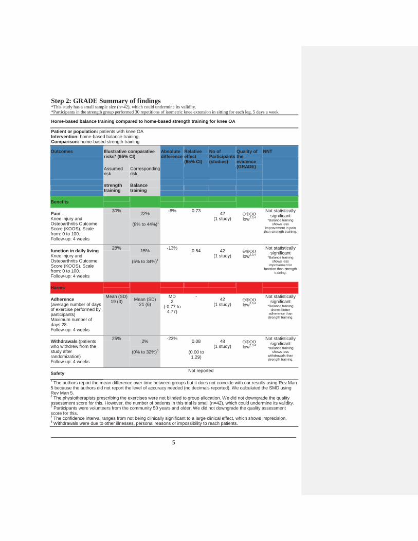
Step 2: GRADE Summary of findings*This study has a small sample size (n=42), which could undermine its validity. *Participants in the strength group performed 30 repetitions of isometric knee extension in sitting for each leg, 5 days a week.
Home-based balance training compared to home-based strength training for knee OA
Patient or population: patients with knee OAIntervention: home-based balance trainingComparison: home-based strength training
Outcomes Illustrative comparative risks* (95% CI)
Absolute difference
Relative effect(95% CI)
No of Participants(studies)
Quality of the evidence(GRADE)
NNT
Assumed risk
Corresponding risk
strength training
Balance training
Benefits
PainKnee injury and Osteoarthritis Outcome Score (KOOS). Scale from: 0 to 100.Follow-up: 4 weeks
30% 22%
(8% to 44%)1
-8% 0.73 42(1 study) low2,3,4
Not statistically significant
*Balance training shows less
improvement in pain than strength training.
function in daily livingKnee injury and Osteoarthritis Outcome Score (KOOS). Scale from: 0 to 100.Follow-up: 4 weeks
28% 15%
(5% to 34%)1
-13% 0.54 42(1 study) low2,3,4
Not statistically significant
*Balance training shows less
improvement in function than strength
training.
Harms
Adherence (average number of days of exercise performed by participants) Maximum number of days:28.Follow-up: 4 weeks
Mean (SD) 19 (3) Mean (SD)
21 (6)
MD 2
(-0.77 to 4.77)
- 42(1 study) low2,3,4
Not statistically significant
*Balance training shows better
adherence than strength training.
Withdrawals (patients who withdrew from the study after randomization)Follow-up: 4 weeks
25% 2%
(0% to 32%)5
-23% 0.08
(0.00 to 1.29)
48 (1 study) low2,3,4
Not statistically significant
*Balance training shows less
withdrawals than strength training.
Safety Not reported
1 The authors report the mean difference over time between groups but it does not coincide with our results using Rev Man 5 because the authors did not report the level of accuracy needed (no decimals reported). We calculated the SMD using Rev Man 5.2 The physiotherapists prescribing the exercises were not blinded to group allocation. We did not downgrade the quality assessment score for this. However, the number of patients in this trial is small (n=42), which could undermine its validity.3 Participants were volunteers from the community 50 years and older. We did not downgrade the quality assessment score for this. 4 The confidence interval ranges from not being clinically significant to a large clinical effect, which shows imprecision. 5 Withdrawals were due to other illnesses, personal reasons or impossibility to reach patients.

________________________________________________________________________ 6
Visual Summary of findings figure:Home-based balance training compared to home-based strength training for knee OAChance: Improving pain after 4 weeksNNT: n/a
Not statistically significant
70 people out of 100 don’t improve with either type of training.
22 people out of 100 improve with either type of training.
8 FEWER people out of 100 improve with balance training at home.
Chance: Improving function after 4 weeksNNT: n/a
Not statistically significant
72 people out of 100 don’t improve with either type of training.
15 people out of 100 improve with either type of training.
13 FEWER people out of 100 improve with balance training at home.
Chance: Adherence after 4 weeksNNH: n/a
Not statistically significantOn average, people performed the exercises for 19 days with either type of training On average, people did not perform the exercises for 7 days (out of maximum possible of 28 days) with either type of trainingOn average, people performed exercises for 2 less days with strengthening than balance training at home.
Chance: Withrawals from the trials after 4 weeksNNH: n/a
Not statistically significant75 people out of 100 did not drop out of either type of training.
2 people out of 100 dropped out of eithertype of training..
23 fewer people out of 100 dropped out ofbalance training at home.
________________________________________________________________________ 7
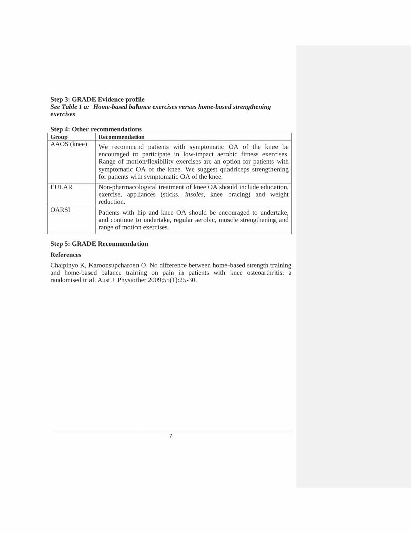
Step 3: GRADE Evidence profileSee Table 1 a: Home-based balance exercises versus home-based strengthening exercises
Step 4: Other recommendationsGroup RecommendationAAOS (knee) We recommend patients with symptomatic OA of the knee be
encouraged to participate in low-impact aerobic fitness exercises. Range of motion/flexibility exercises are an option for patients with symptomatic OA of the knee. We suggest quadriceps strengthening for patients with symptomatic OA of the knee.
EULAR Non-pharmacological treatment of knee OA should include education, exercise, appliances (sticks, insoles, knee bracing) and weight reduction.
OARSI Patients with hip and knee OA should be encouraged to undertake, and continue to undertake, regular aerobic, muscle strengthening and range of motion exercises.
Step 5: GRADE RecommendationReferencesChaipinyo K, Karoonsupcharoen O. No difference between home-based strength training and home-based balance training on pain in patients with knee osteoarthritis: a randomised trial. Aust J Physiother 2009;55(1):25-30.

________________________________________________________________________ 8
1.1.2 Balance exercises in addition to strengthening exercises versus strengthening exercises alone for knee OAAre balance exercises in addition to strengthening exercises effective in reducing pain and improving function in patients with symptomatic OA compared to strengthening exercises alone?Step 1: Search ResultsThere were no SRs which reported the efficacy of balance exercises specifically in patients with OA (Orr, 2008 assessed the efficacy of progressive resistive training which is a different treatment and Howe, 2007 did not report any study with OA patients). There was one RCT which assessed the efficacy of balance exercises in addition to strengthening exercises vs. strengthening exercises alone (Diracoglu, 2005).
Intervention description: The first group (kinesthesia group) received kinesthesia,balance, and strengthening exercises and the second group (strengthening group) received only strengthening exercises. Patients in both groups were informed about knee OA and protective recommendations for the knee were made. The exercises were done 3 days a week in groups of 5 people in a clinical setting under the supervision of a physiotherapist.The total duration of the exercises was determined as 8 weeks. Isometric exercises were applied with 6-second contractions with 8 repetitions and a rest period of 2 seconds. Isotonic exercises were started from the third week and the maximum weight that can belifted 10 times (10-repetition maximum = 10 RM) was determined. The exercises were applied as 10 repetitions with half of this weight, 10 repetitions with three fourths of thisweight, and 10 repetitions with the whole 10 RM.10 RM was determined again every week.
Step 2: GRADE Summary of findings
kinesthesia and balance exercises in addition to strengthening exercises compared to strengthening exercises for knee OA
Patient or population: patients with knee OAIntervention: kinesthesia and balance exercises in addition to strengthening exercisesComparison: strengthening exercises
Outcomes Illustrative comparative risks* (95% CI)
Absolute difference
Relative effect
(95% CI)
No of Participants(studies)
Quality of the evidence(GRADE)
NNT
Assumed risk Corresponding risk
strengthening exercises
kinesthesia and balance exercises in addition to strengthening exercises
Benefits

________________________________________________________________________ 9
Physical functionWOMAC. Scale from: 0 to 10.Follow-up: 8 weeks
31% 48%
(29% to 68%)1
17% 1.55 60(1 study) low2,3,4
Not statistically significant
Pain No evidence available5
Harms
Adverse effectsnumber of patients with eventFollow-up: 8 weeks
0% 0% 0% 1 60(1 study) moderate2,3
Not statistically significant
Adherence mean number of missed visitsMaximum number of visits:24 Follow-up: 8 weeks
Mean 6
Mean 4
MD -2
- 48
(1 study)moderate2,3
Not statistically significant
Withdrawals number of patients who withdrew after randomization Follow-up: 8 weeks
9% 9%
(2% to 42%)6
0% 1
(0.22 to 4.6)
66(1 study) moderate2,3
Not statistically significant
1 The authors reported the end of study results in both groups, which showed a statistically significant difference. However, their results did not coincide with our results from Rev Man 5 because the authors did not report the level of accuracy needed. 2 The randomization method used is the "one-to-one" method which allocates one patient to the study group and the other patient to the control group one by one according to their order of application to the outpatient clinic. This method could lead to biases. Furthermore, blinding was not reported and intention to treat analyses were not performed. 3 All patients included in the study were women 35 to 65 years old. We did not downgrade the quality of the study because of this.4 The confidence interval ranges from not being clinically significant to a large clinical effect, which shows imprecision. 5 Pain was not measured in the RCT. However, the use of paracetamol was reported, which could represent a proxy measure for pain to some extent. The authors report that 5 patients used paracetamol during the study in a dosage of less than 500 mg daily. The 2 groups were not significantly different from each other regarding paracetamol use (P > 0.05).6 Patients withdrew because of the difficulty to come to the clinic for exercises.

________________________________________________________________________ 10
Visual Summary of findings figure:Kinesthesia and balance exercises in addition to strengthening exercises compared to strengthening exercises for knee OAChance: Improving function after 8 weeksNNT: n/a
Not statistically significant
52 people out of 100 don’t improve with either type of training.
31 people out of 100 improve with either type of training.
17 more people out of 100 improve with kinesthesia and balance exercises in addition to strengthening exercises.
Chance: Improving pain after 8 weeksNNT: n/a
Pain was not measured in this study, but there may be no difference in pain. People used the same amount of paracetomol (a pain reliever) whether they did kinesthesia and balance exercises
in addition to strengthening exercises or just strengthening exercisesChance: Adverse events after 8 weeksNNH: n/a
Not statistically significant
0 People out of 100 experienced adverse events.
Chance: Adherence after 8 weeksNNH: n/a
Not statistically significantOn average, people attended 18 visits with either type of trainingOn average, people missed 4 visits with either type of training (out of maximum possible of 24 visits)On average, people missed 2 more visitswith strengthening exercises alone.
Chance: Withdrawals from the trials after 8 weeksNNH: n/a
Not statistically significant91 people out of 100 did not drop out of either type of exercise.

________________________________________________________________________ 11
9 people out of 100 dropped out of either type of exercise.
There was no difference in the number of people out of 100 who dropped out of kinesthesia and balance exercises in addition to strengthening exercises.
Step 3: GRADE Evidence profile See Table 1b: Balance exercises in addition to strengthening exercises versus strengthening exercises alone
Step 4: Other recommendations
Group RecommendationAAOS (knee) We recommend patients with symptomatic OA of the knee be
encouraged to participate in low-impact aerobic fitness exercises. Range of motion/flexibility exercises are an option for patients with symptomatic OA of the knee. We suggest quadriceps strengthening for patients with symptomatic OA of the knee.
EULAR Non-pharmacological treatment of knee OA should include education, exercise, appliances (sticks, insoles, knee bracing) and weight reduction.
OARSI Patients with hip and knee OA should be encouraged to undertake, and continue to undertake, regular aerobic, muscle strengthening and range of motion exercises.
Step 5: GRADE Recommendation
ReferencesDiracoglu D, Aydin R, Baskent A, Celik A. Effects of kinesthesia and balance exercises in knee osteoarthritis. J Clin Rheumatol 2005;11(6):303-10.

________________________________________________________________________ 12
1.2 Land-based exercise
1.2.1 Cardiovascular land-based exercise versus usual care for knee OAIs cardiovascular land exercise effective in reducing pain and improving function in patients with symptomatic knee osteoarthritis (OA) compared to usual care?
Step 1: Search ResultsThree systematic reviews (SR) were found. Pisters (2007), was excluded from this comparison because it did not provide a description of the exercises used (combination of land, water, balance) and it did not report adherence. The second, Hart (2008), was excluded because it did not focus on osteoarthritis patients. Therefore, Fransen (2008) was chosen as the best available evidence. One overview of SRs on therapeutic exercise was found (Taylor, 2007) and its overall conclusions followed those of the chosen SR. Four randomized controlled trials published after the chosen SR were also found (Chua, 2008; Lund, 2008; Dincer, 2008; Olejarova, 2008). Their results were largely similar to those of the chosen SR. Evidence for withdrawals were extracted from the best RCT from Fransen, 2008: Ettinger, 1997.
Interventions description: non-perioperative walking program
Step 2: GRADE Summary of findings
cardiovascular land exercise compared to no exercise for osteoarthritis of the knee
Patient or population: patients with osteoarthritis of the kneeSettings:Intervention: cardiovascular land exercise Comparison: no exercise
Outcomes Illustrative comparative risks* (95% CI)
Absolute difference
Relative effect(95% CI)
No of Participants(studies)
Quality of the evidence(GRADE)
NNT
Assumed risk
Corresponding risk
no exercise cardiovascular land exercise
Benefits
painpooled studies with different scales including WOMAC and VAS amongst others
24% 41%of those cardiovascular exercise group experienced a decrease in pain(31% to 55%)
17% 1.71 351(43) high1
5(3 to 12)

________________________________________________________________________ 13
function pooled studies with different scales including WOMAC and VAS amongst others
22% 34%of those cardiovascular exercise group experienced a decrease in pain(26% to 43%)
12% 1.55 317(34) high1
7(4 to 20)
Harms
withdrawalsnumber of(follow-up: mean 18 months)
15% 19%(11% to 31%) 4%
RR 1.27(0.76 to 2.12)
293(15) moderate
Not statistically significant
Safety (falls while walking) 1.4% of intervention group fell
during walking (2/144)
RR 5.17(0.25 to 106.82)
293(15) moderate
Not statistically significant
Adherence 95% 68%(60% to 76%)
27% RR 0.71(0.63 to 0.80)
293(15) high
5(4 to 7)
*The basis for the assumed risk (e.g. the median control group risk across studies) is provided in footnotes. The corresponding risk (and its 95% confidence interval) is based on the assumed risk in the comparison group and the relative effect of the intervention (and its 95% CI).
CI: Confidence interval; RR: Risk ratio; GRADE Working Group grades of evidenceHigh quality: Further research is very unlikely to change our confidence in the estimate of effect. Moderate quality: Further research is likely to have an important impact on our confidence in the estimate of effect and may change the estimate.Low quality: Further research is very likely to have an important impact on our confidence in the estimate of effect and is likely to change the estimate.Very low quality: We are very uncertain about the estimate.1 Evidence mostly included participants with early or mild symptomatic disease.3 Minor 1989, Ettinger 1997, Bautch 1997, Talbot 20034 Minor 1989, Ettinger 1997, Bautch 19975Is imprecise; includes no effect and significant benefit (0.76, 2.12)6 Ettinger 1997
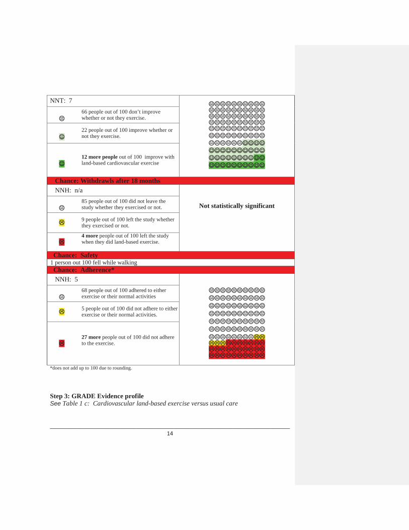
Visual Summary of Findings TableCardiovascular land exercise compared to no exercise for osteoarthritis of the kneeChance: Improving painNNT: 5
59 people out of 100 don’t improvewhether or not they exercise.
24 people out of 100 improve whether or not they exercise.
17 more people out of 100 improve with cardiovascular land-based exercise.
Chance: Improving function
________________________________________________________________________ 14
NNT: 766 people out of 100 don’t improvewhether or not they exercise.
22 people out of 100 improve whether or not they exercise.
12 more people out of 100 improve with land-based cardiovascular exercise
Chance: Withdrawls after 18 monthsNNH: n/a
Not statistically significant85 people out of 100 did not leave the study whether they exercised or not.
9 people out of 100 left the study whether they exercised or not.
4 more people out of 100 left the study when they did land-based exercise.
Chance: Safety1 person out 100 fell while walking Chance: Adherence*NNH: 5
68 people out of 100 adhered to either exercise or their normal activities
5 people out of 100 did not adhere to either exercise or their normal activities.
27 more people out of 100 did not adhere to the exercise.
*does not add up to 100 due to rounding.
Step 3: GRADE Evidence profileSee Table 1 c: Cardiovascular land-based exercise versus usual care

________________________________________________________________________ 15
Step 4: Other recommendations
Group RecommendationEULAR Non-pharmacological treatment of knee OA should include education,
exercise, appliances (sticks, insoles, knee bracing) and weight reduction.
OARSI Patients with hip and knee OA should be encouraged to undertake, and continue to undertake, regular aerobic, muscle strengthening and range of motion exercises. For patients with systematic hip OA, exercises in the water can be effective.
AAOS (knee only)
We recommend patients with symptomatic OA of the knee be encouraged to participate in low-impact aerobic fitness exercises. Range of motion/flexibility exercises are an option for patients with symptomatic OA of the knee. We suggest quadriceps strengthening for patients with symptomatic OA of the knee.
Step 5: GRADE Recommendation
References Bautch JC, Malone DG, Vailas AC. Effects of exercise on knee joints with osteoarthritis: a pilot study of biologic markers. Arthritis Care Res 1997;10(1):48-55.
Ettinger WH, Burns R, Messier SP, Applegate W, Rejeski WJ, Morgan T, et al. A randomized trial comparing aerobic exercise and resistance exercise with a health education program in older adults with knee osteoarthritis. The Fitness Arthritis and Seniors Trial (FAST). JAMA 1997;277(1):25-31.
Fransen M, McConnell S. Exercise for osteoarthritis of the knee. Cochrane Database of Syst Rev 2008;(4):CD004376.
Minor MA, Hewett JE, Webel RR, Anderson SK, Kay DR. Efficacy of physical conditioning exercise in patients with rheumatoid arthritis and osteoarthritis. Arthritis Rheum 1989;32(11):1396-405.
Talbot LA, Gaines JM, Huynh TN, Metter EJ. A home-based pedometer-driven walking program to increase physical activity in older adults with osteoarthritis of the knee: a preliminary study. J Am Geriatr Soc 2003;51(3):387-92.

________________________________________________________________________ 16
1.2.2 Resistance land-based exercise versus usual care for knee OAIs resistance land exercise effective in reducing pain and improving function in patients with symptomatic knee OA compared to usual care?
Step 1: Search ResultsThree systematic reviews (SR) were found. One, Pisters (2007), was excluded from this comparison because it did not provide a description of the exercises used (combination of land, water, balance) and it did not report adherence. The second, Hart (2008), was excluded because it did not focus on osteoarthritis patients. Therefore, Fransen (2008) was chosen as best available evidence. One overview of SR on therapeutic exercise was found (Taylor, 2007) and its overall conclusions followed those of the chosen SR. Four randomized controlled trials published after the chosen SR were also found (Chua, 2008; Lund, 2008; Dincer, 2008; Olejarova, 2008). Their results were largely similar to those of the chosen evidence. Safety, adherence, and withdrawals were not included in the best RCT included in Fransen, 2008 (Huang, 2005).
Intervention description: non-perioperative lower limb muscle strengthening
Step 2: GRADE Summary of findingsresistance land exercise compared to no exercise for knee OA
Patient or population: patients with osteoarthritis of the kneeSettings:Intervention: resistance land exerciseComparison: no exercise
Outcomes Illustrative comparative risks* (95% CI)
Absolute difference
Relative effect(95% CI)
No of Participants(studies)
Quality of the evidence(GRADE)
NNT
Assumed risk
Corresponding risk
noexercise
resistance land exercise
Benefits
Painpooled studies with different scales including WOMAC and VAS amongst others
32% 53%of those in strengthening exercise group experienced a decrease in pain(43% to 63%)
21% 1.66 1383(93)
Omoderate1,2
4(3 to 8)
Function pooled studies with different scales including
10% 25%of those in strengthening exercise group experienced a decrease in pain
15% 2.5 1383(93)
Omoderate1,2
6(4 to 22)

________________________________________________________________________ 17
WOMAC and VAS amongst others
(35% to 69%)
Harms
Safety 14% patients in exercise group stopped due to intolerable pain during exercise.
Adherence Not reported
Withdrawals 9% 14%(4 to 56%)
5% RR 1.67(0.43 to 6.45)
70(14) high
Not statistically significant
*The basis for the assumed risk (e.g. the median control group risk across studies) is provided in footnotes. The corresponding risk (and its 95% confidence interval) is based on the assumed risk in the comparison group and the relative effect of the intervention (and its 95% CI).
CI: Confidence interval; GRADE Working Group grades of evidenceHigh quality: Further research is very unlikely to change our confidence in the estimate of effect. Moderate quality: Further research is likely to have an important impact on our confidence in the estimate of effect and may change the estimate.Low quality: Further research is very likely to have an important impact on our confidence in the estimate of effect and is likely to change the estimate.Very low quality: We are very uncertain about the estimate.1 Evidence mostly included participants with early or mild symptomatic disease.2 Large confidence interval ranging from small to large effect3 Schilke 2006, Ettinger 1997, Baker 2001, Thomas 2002, Gur 2002, Huang 2003, Huang 2005, Thorstensson 2005, Mikesky 20064 Huang 2005
Visual Summary of Findings TableResistance land exercise compared to no exercise for osteoarthritis of the kneeChance: Improving painNNT: 4
47 people out of 100 don’t improvewhether or not they exercise.
32 people out of 100 improve whether or not they exercise.
21 more people out of 100 improve with exercise.
Chance: Improving function NNT: 6
75 people out of 100 don’t improvewhether or not they exercise.

________________________________________________________________________ 18
10 people out of 100 improve whether or not they exercise.
15 more people out of 100 improve with exercise
Chance: Withdrawals NNH: n/a
Not statistically significant86 people out of 100 did not leave the study whether they exercised or not.
9 people out of 100 left the study whether they exercised or not.
5 more people out of 100 left the study in the lower limb exercise group.
Chance: Safety14% patients in exercise group stopped due to intolerable pain during exercise.Chance: Adherence
The number of people who adhered to resistance exercise was not reported.
Step 3: GRADE Evidence profileSee Table 1 d: Resistance land-based exercise versus usual care
Step 4: Other recommendations
Group RecommendationAAOS (knee only)
We recommend patients with symptomatic OA of the knee be encouraged to participate in low-impact aerobic fitness exercises. Range of motion/flexibility exercises are an option for patients with symptomatic OA of the knee. We suggest quadriceps strengthening for patients with symptomatic OA of the knee.
EULAR Non-pharmacological treatment of knee OA should include education, exercise, appliances (sticks, insoles, knee bracing) and weight reduction.
OARSI Patients with hip and knee OA should be encouraged to undertake, and continue to undertake, regular aerobic, muscle strengthening and range of motion exercises. For patients with systematic hip OA, exercises in the water can be effective.
Step 5: GRADE Recommendation
________________________________________________________________________ 19
References Fransen M, McConnell S. Exercise for osteoarthritis of the knee. Cochrane Database of Syst Rev 2008;(4):CD004376.
Huang MH, Lin YH, Lee CL, Yang RC. Use of ultrasound to increase effectiveness of idokinetic exercise for knee osteoarthritis. Arch Phys Med Rehabil 2005;86(8):1545-51.
1.3 Aquatic exercises
1.3.1 Aquatic exercise versus no exercise for OA of hip or kneeIs aquatic exercise effective in reducing pain and improving function in patients with symptomatic knee and hip OA compared to usual care?Interventions description: All types of exercises developed in the therapeutic/heated indoor pool (range of motion, dynamics, aerobics, etc.).
Step 1: Search ResultsOnly one meta-analysis was found that assessed aquatic exercise for knee osteoarthritis (Bartels, 2007). Two more recent randomized controlled trials were also found (Lund, 2008; Gill, 2009). Although Lund (2008) found no improvement following aquatic exercise, Gill (2009) found similar results to those reported below whereby pain was decreased.
** NOTE: This evidence is the same as that found in the hip exercise summary of findings because data from both joints were pooled**
Step 2: GRADE Summary of findings
aquatic exercise compared to no exercise for osteoarthritis of hip or knee
Patient or population: patients with osteoarthritis of hip or kneeSettings:Intervention: aquatic exercise Comparison: no exercise
Outcomes Illustrative comparative risks* (95% CI)
Relative effect(95% CI)
Absolute difference
No of Participants(studies)
Quality of the evidence(GRADE)
NNT
Assumed risk Corresponding risk
no exercise aquatic exercise
Benefits
________________________________________________________________________ 20
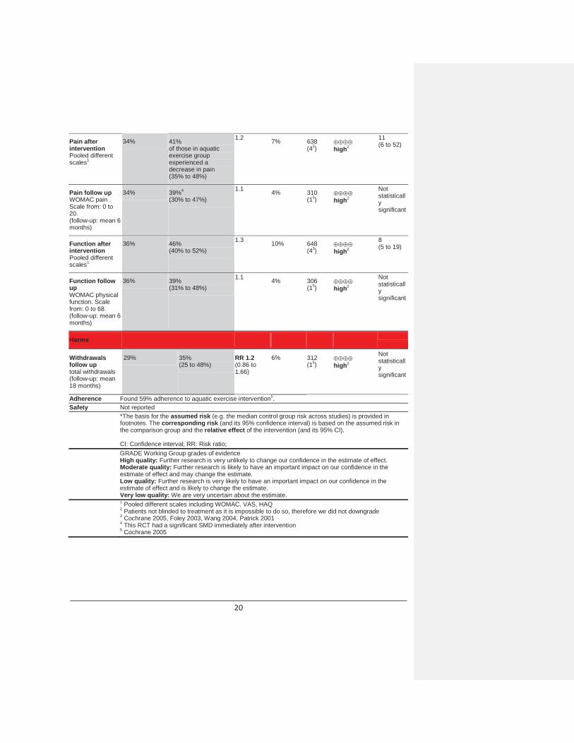
Pain after interventionPooled different scales1
34% 41%of those in aquatic exercise group experienced a decrease in pain(35% to 48%)
1.2 7% 638(43) high2
11(6 to 52)
Pain follow upWOMAC pain . Scale from: 0 to 20.(follow-up: mean 6 months)
34% 39%4
(30% to 47%)
1.1 4% 310(15) high2
Not statistically significant
Function after interventionPooled different scales1
36% 46%(40% to 52%)
1.3 10% 648(43) high2
8(5 to 19)
Function follow upWOMAC physical function. Scale from: 0 to 68.(follow-up: mean 6 months)
36% 39%(31% to 48%)
1.1 4% 306(15) high2
Not statistically significant
Harms
Withdrawals follow up total withdrawals(follow-up: mean 18 months)
29% 35%(25 to 48%)
RR 1.2(0.86 to 1.66)
6% 312(15) high2
Not statistically significant
Adherence Found 59% adherence to aquatic exercise intervention5.Safety Not reported
*The basis for the assumed risk (e.g. the median control group risk across studies) is provided in footnotes. The corresponding risk (and its 95% confidence interval) is based on the assumed risk in the comparison group and the relative effect of the intervention (and its 95% CI).
CI: Confidence interval; RR: Risk ratio; GRADE Working Group grades of evidenceHigh quality: Further research is very unlikely to change our confidence in the estimate of effect. Moderate quality: Further research is likely to have an important impact on our confidence in the estimate of effect and may change the estimate.Low quality: Further research is very likely to have an important impact on our confidence in the estimate of effect and is likely to change the estimate.Very low quality: We are very uncertain about the estimate.1 Pooled different scales including WOMAC, VAS, HAQ2 Patients not blinded to treatment as it is impossible to do so, therefore we did not downgrade3 Cochrane 2005, Foley 2003, Wang 2004, Patrick 20014 This RCT had a significant SMD immediately after intervention5 Cochrane 2005

________________________________________________________________________ 21
Visual Summary of Findings TableAquatic exercise compared to no exercise for osteoarthritis of hip or kneeChance: Improving pain immediately after aquatic exerciseNNT: 11
59 people out of 100 don’t improvewhether or not they did aquatic exercise
34 people out of 100 improve whether or not they did aquatic exercise
7 more people out of 100 improve withaquatic exercise
Chance: Improving pain after 6 monthsNNT: n/a
Not statistically significant
61 people out of 100 don’t improvewhether or not they did aquatic exercise
34 people out of 100 improve whether or not they did aquatic exercise
5 more people out of 100 improve with aquatic exercise
Chance: Improving function immediately after aquatic exerciseNNT: 8
54 people out of 100 don’t improvewhether or not they did aquatic exercise
36 people out of 100 improve whether or not they did aquatic exercise
10 more people out of 100 improve with aquatic exercise
Chance: Improving function after 6 monthsNNT: n/a
Not statistically significant
61 people out of 100 don’t improvewhether or not they did aquatic exercise
36 people out of 100 improve whether or not they did aquatic exercise
3 more people out of 100 improve with aquatic exercise
________________________________________________________________________ 22
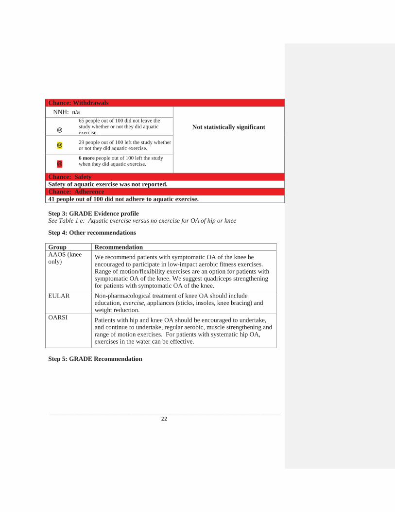
Chance: Withdrawals NNH: n/a
Not statistically significant65 people out of 100 did not leave the study whether or not they did aquatic exercise.
29 people out of 100 left the study whether or not they did aquatic exercise.
6 more people out of 100 left the study when they did aquatic exercise.
Chance: SafetySafety of aquatic exercise was not reported.Chance: Adherence41 people out of 100 did not adhere to aquatic exercise.
Step 3: GRADE Evidence profileSee Table 1 e: Aquatic exercise versus no exercise for OA of hip or knee
Step 4: Other recommendations
Group RecommendationAAOS (knee only)
We recommend patients with symptomatic OA of the knee be encouraged to participate in low-impact aerobic fitness exercises. Range of motion/flexibility exercises are an option for patients with symptomatic OA of the knee. We suggest quadriceps strengthening for patients with symptomatic OA of the knee.
EULAR Non-pharmacological treatment of knee OA should include education, exercise, appliances (sticks, insoles, knee bracing) and weight reduction.
OARSI Patients with hip and knee OA should be encouraged to undertake, and continue to undertake, regular aerobic, muscle strengthening and range of motion exercises. For patients with systematic hip OA, exercises in the water can be effective.
Step 5: GRADE Recommendation

________________________________________________________________________ 23
References
Bartels ME, Lund H, Hagen KB, Dagfinrud H, Christensen R, Danneskiold-Samsoe B. Aquatic exercise for the treatment of knee and hip osteoarthritis. Cochrane Database of Syst Rev 2007(4):CD005523.
Cochrane T, Davey RC, Matthes Edwards SM. Randomised controlled trial of the cost-effectiveness of water-based therapy for lower limb osteoarthritis. Health Technol Assess 2005;9(31):iii-xi, ix-xi, 1-114.
Wyatt FB, Milam S, Manske RC, Deere R. The effects of aquatic and traditional exercise programs on programs on persons with knee osteoarthritis. J Strength Cond Res 2001;15(3):337-40.
1.3.2 Aquatic exercise versus land-based exercise of knee OAIs aquatic exercise effective in reducing pain and improving function in patients with symptomatic knee OA compared to land-based exercise?
Step 1: Search ResultsOnly one SR was found considering aquatic exercise for knee osteoarthritis (Bartels, 2007). This SR included only one RCT analyzing aquatic exercise vs. land-based exercise for knee OA (Wyatt, 2001).
Interventions description: All types of exercises developed in the therapeutic/heated indoor pool (range of motion, dynamics, aerobics, etc.).
Step 2: GRADE Summary of findings
aquatic exercise compared to land exercise for osteoarthritis of the knee
Patient or population: patients with osteoarthritis of the kneeSettings:Intervention: aquatic exercise Comparison: land exercise
Outcomes Illustrative comparative risks* (95% CI)
Relative effect(95% CI)
Absolute difference
No of Participants(studies)
Quality of the evidence(GRADE)
NNT
Assumed risk Corresponding risk
land exercise aquatic exercise
Benefits

________________________________________________________________________ 24
painVAS. Scale from: 0 to 10.(follow-up: mean 6 weeks)
32% 65%of those in aquatic exercise group experienced a decrease in pain(41% to 84%)
2.0 33% 46(14)
OOOvery low1,2,3
3(2 to 9)
function -walking abilitytimed 1-mile walk. Scale from 0 to 25 min(follow-up: mean 6 weeks)
15% 28%(12% to 50%)
1.9 13% 46(14)
OOOvery low1,2,3
Not statistically significant
Harms
Withdrawals 4 out of 46 subjects withdrew due to illness5
Adherence Not reported
Safety Not reported
*The basis for the assumed risk (e.g. the median control group risk across studies) is provided in footnotes. The corresponding risk (and its 95% confidence interval) is based on the assumed risk in the comparison group and the relative effect of the intervention (and its 95% CI).
CI: Confidence interval; GRADE Working Group grades of evidenceHigh quality: Further research is very unlikely to change our confidence in the estimate of effect. Moderate quality: Further research is likely to have an important impact on our confidence in the estimate of effect and may change the estimate.Low quality: Further research is very likely to have an important impact on our confidence in the estimate of effect and is likely to change the estimate.Very low quality: We are very uncertain about the estimate.1 Concealment of allocation was unclear2 no comparision to placebo3 Only end-of-study data could be reported here and N is low (n=42) and large CI4 Wyatt 20015 RCT does not specify to which group they pertained
________________________________________________________________________ 25
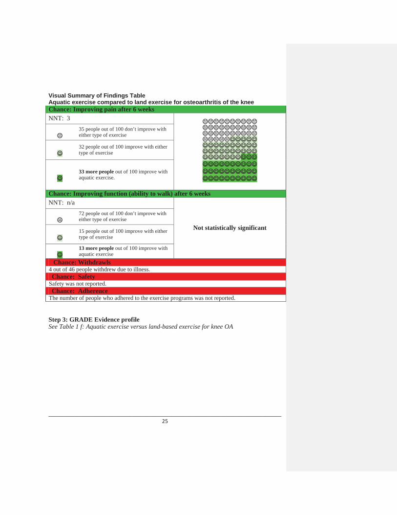
Visual Summary of Findings TableAquatic exercise compared to land exercise for osteoarthritis of the kneeChance: Improving pain after 6 weeksNNT: 3
35 people out of 100 don’t improve with either type of exercise
32 people out of 100 improve with either type of exercise
33 more people out of 100 improve withaquatic exercise.
Chance: Improving function (ability to walk) after 6 weeksNNT: n/a
Not statistically significant
72 people out of 100 don’t improve with either type of exercise
15 people out of 100 improve with either type of exercise
13 more people out of 100 improve with aquatic exercise
Chance: Withdrawls 4 out of 46 people withdrew due to illness.Chance: Safety
Safety was not reported.Chance: Adherence
The number of people who adhered to the exercise programs was not reported.
Step 3: GRADE Evidence profileSee Table 1 f: Aquatic exercise versus land-based exercise for knee OA

________________________________________________________________________ 26
Step 4: Other recommendations
Group RecommendationAAOS (knee only)
We recommend patients with symptomatic OA of the knee be encouraged to participate in low-impact aerobic fitness exercises. Range of motion/flexibility exercises are an option for patients with symptomatic OA of the knee. We suggest quadriceps strengthening for patients with symptomatic OA of the knee.
EULAR Non-pharmacological treatment of knee OA should include education, exercise, appliances (sticks, insoles, knee bracing) and weight reduction.
OARSI Patients with hip and knee OA should be encouraged to undertake, and continue to undertake, regular aerobic, muscle strengthening and range of motion exercises. For patients with systematic hip OA, exercises in the water can be effective.
Step 5: GRADE Recommendation
References Bartels ME, Lund H, Hagen KB, Dagfinrud H, Christensen R, Danneskiold-Samsoe B. Aquatic exercise for the treatment of knee and hip osteoarthritis. Cochrane Database of Syst Rev 2007(4):CD005523.
Wyatt FB, Milam S, Manske RC, Deere R. The effects of aquatic and traditional exercise programs on programs on persons with knee osteoarthritis. J Strength Cond Res 2001;15(3):337-40.

________________________________________________________________________ 27
1.4 Tai chi
Is tai chi effective in reducing pain and improving function in patients with symptomatic knee OA compared to usual care?
Step 1: Search ResultsOne systematic review (Lee 2008) assessed the effect of tai chi in patients with both hip and knee OA. However, results of the 5 included RCTs and 7 non-randomized studies were not pooled due to high heterogeneity. Therefore, we chose the RCT from this systematic review which most closely matched our PICO question by having an appropriate control group and with the largest sample size. The RCT by Brismee, 2007 was the closest match to having a control group (defined as “attention control in Brismee 2007) since the other studies had control groups of hydrotherapy, routine care and bingo.
Intervention description: Simplified Yang-style tai chi with instructor three times a week for six weeks followed by six weeks with home video.
Note: the study included has a sample size of 31 people, and 24% of the participants were lost to follow-up.
Step 2: GRADE Summary of findings
Tai chi compared to no exercise (education on OA) for knee OA
Patient or population: patients with osteoarthritis of the kneeSettings:Intervention: tai chiComparison: no exercise (education on OA)
Outcomes Illustrative comparative risks* (95% CI) Relative effect(95% CI)
Absolute difference
No of Participants(studies)
Quality of the evidence(GRADE)
NNT
Assumed risk Corresponding risk
no exercise (education on OA)
Tai chi
Benefit
Pain WOMAC . Scale from: 0 to 35.(follow-up: mean 12 weeks)
33% 35%of those in tai chi group experienced a decrease in pain(11% to 58%)
2% 1.1 31(12) low1
Not statistically significant

________________________________________________________________________ 28
Function WOMAC. Scale from: 0 to 85.(follow-up: mean 12 weeks)
33% 35%(11% to 58%)
2% 1.1 31(12) low1
Not statistically significant
Harms
WithdrawalsNumber of drop-outs(follow-up: mean 12 weeks)
32% 18%(6 to 55%)
RR 0.58(0.19 to 1.74)
13% 41(12) moderate1
Not statistically significant
(Note: more people in the control group withdrew from the study)
Adherence 90% adherence in tai chi group Safety Not reported
*The basis for the assumed risk (e.g. the median control group risk across studies) is provided in footnotes. The corresponding risk (and its 95% confidence interval) is based on the assumed risk in the comparison group and the relative effect of the intervention (and its 95% CI).
CI: Confidence interval; RR: Risk ratio; GRADE Working Group grades of evidenceHigh quality: Further research is very unlikely to change our confidence in the estimate of effect. Moderate quality: Further research is likely to have an important impact on our confidence in the estimate of effect and may change the estimate.Low quality: Further research is very likely to have an important impact on our confidence in the estimate of effect and is likely to change the estimate.Very low quality: We are very uncertain about the estimate.1 Large CI and small N=352 Brismee, 2007

________________________________________________________________________ 29
Visual Summary of Findings TableTai chi compared to no exercise (education on OA) for osteoarthritis of the kneeChance: Improving painNNT: n/a
Not statistically significant65 people out of 100 don’t improve with either treatment.
33 people out of 100 improve with either treatment.
2 more people out of 100 improve with tai chi.
Chance: Improving function NNT: n/a
Not statistically significant
65 people out of 100 don’t improve with either treatment.
33 people out of 100 improve with either treatment.
2 more people out of 100 improve with tai chi.
Chance: Withdrawals*NNH: n/a
Not statistically significant(Note: more people in the control group
withdrew from the study)
68 people out of 100 did not leave the study with either treatment.
18 people out of 100 left the study with either treatment.
13 more people out of 100 left the study in the control group than the tai chi.
Chance: SafetySafety of tai chi was not reported.Chance: Adherence
90% of people in the tai chi group adhered to the program.*does not add up to 100 due to rounding
Step 3: GRADE Evidence profileSee Table 1 g: Tai Chi compared to no exercise (education on OA) for knee OA

________________________________________________________________________ 30
Step 4: Other recommendations
Group RecommendationAAOS (knee only)
We recommend patients with symptomatic OA of the knee be encouraged to participate in low-impact aerobic fitness exercises. Range of motion/flexibility exercises are an option for patients with symptomatic OA of the knee. We suggest quadriceps strengthening for patients with symptomatic OA of the knee.
EULAR Non-pharmacological treatment of knee OA should include education, exercise, appliances (sticks, insoles, knee bracing) and weight reduction.
OARSI Patients with hip and knee OA should be encouraged to undertake, and continue to undertake, regular aerobic, muscle strengthening and range of motion exercises. For patients with systematic hip OA, exercises in the water can be effective.
Step 5: GRADE Recommendation
References Brismee JM, Paige RL, Chyu MC, Boatright JD, Hagar JM, McCaleb JA, Quintela MM, Feng D, Xu KT, Shen CL. Group and home-based tai chi in elderly subjects with knee osteoarthritis: a randomized controlled trial. Clin Rehabil 2007;21:99-111.
Lee MS, Pittler MH, Ernst E. Tai chi for osteoarthritis: a systematic review. Clin Rheumatol 2008;27(2):211-8.
________________________________________________________________________ 31
1.5 General hip exercise
Is exercise effective in reducing pain and improving function in patients with symptomatic hip osteoarthritis (OA) compared to usual care?
Step 1: Search ResultsOne meta-analysis (Hernandez-Molina, 2008) was found which pooled land-based, aquatic, and tai chi exercises. The remaining RCTs found which were not included in the meta-analysis did not follow the guideline’s inclusion criteria since they were post-operative interventions.
Intervention description: For the pain outcome, the systematic review (SR) included any exercise program of at least 4 weeks duration (Hernandez-Molina, 2008). For the function outcome, “The exercise group performed water and land-based exercise 3 times weekly over a 6-week period immediately prior to surgery. During the first 3 weeks, participants performed 1–2 sets of 8–12 repetitions of single-joint movements while standing in chest-deep, 93°F water. Pool exercises focused on single planar motion of the cervical spine, shoulders, elbows, wrists, hands, hips, knees, and ankles. During weeks 4–6, exercise sessions involved a total body fitness program of cardiovascular, strength, and flexibility training” (Rooks, 2006).
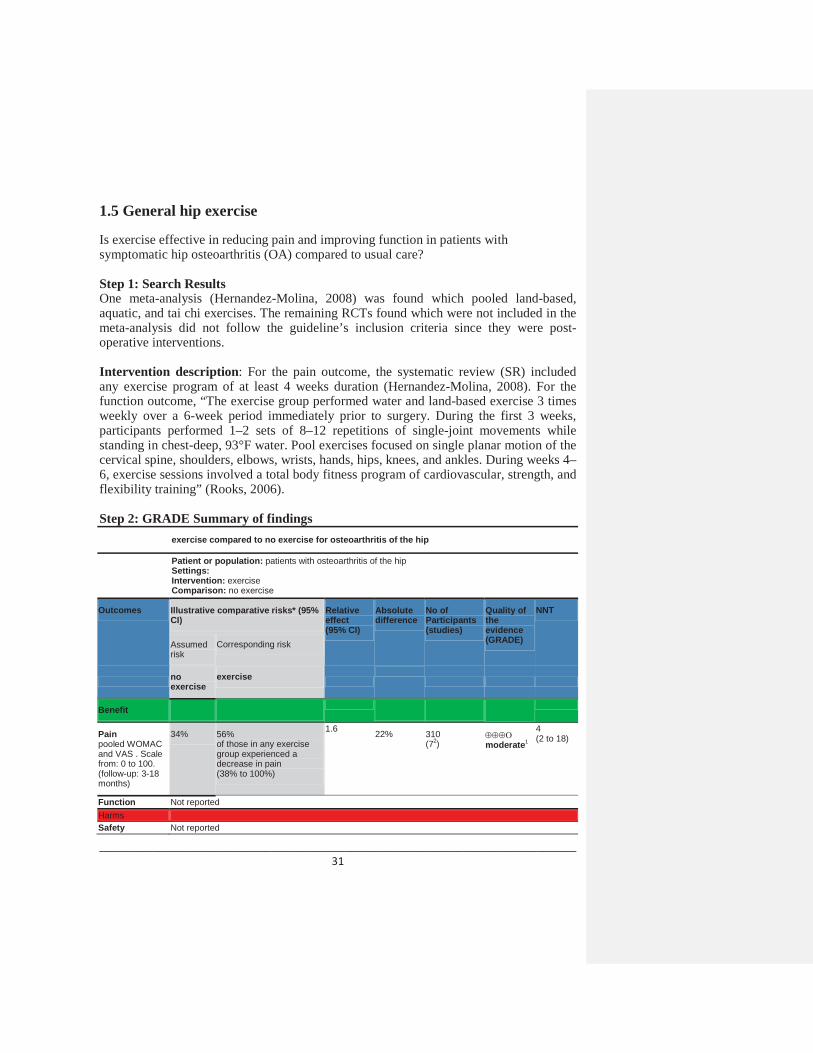
Step 2: GRADE Summary of findingsexercise compared to no exercise for osteoarthritis of the hip
Patient or population: patients with osteoarthritis of the hipSettings:Intervention: exerciseComparison: no exercise
Outcomes Illustrative comparative risks* (95% CI)
Relative effect(95% CI)
Absolute difference
No of Participants(studies)
Quality of the evidence(GRADE)
NNT
Assumed risk
Corresponding risk
noexercise
exercise
Benefit
Painpooled WOMACand VAS . Scale from: 0 to 100.(follow-up: 3-18months)
34% 56%of those in any exercise group experienced a decrease in pain(38% to 100%)
1.6 22% 310(72) moderate1
4(2 to 18)
Function Not reportedHarms Safety Not reported

________________________________________________________________________ 32
Withdrawals Not reportedAdherence Not reported
*The basis for the assumed risk (e.g. the median control group risk across studies) is provided in footnotes. The corresponding risk (and its 95% confidence interval) is based on the assumed risk in the comparison group and the relative effect of the intervention (and its 95% CI).
CI: Confidence interval; GRADE Working Group grades of evidenceHigh quality: Further research is very unlikely to change our confidence in the estimate of effect. Moderate quality: Further research is likely to have an important impact on our confidence in the estimate of effect and may change the estimate.Low quality: Further research is very likely to have an important impact on our confidence in the estimate of effect and is likely to change the estimate.Very low quality: We are very uncertain about the estimate.1 although Isquared = 0, different interventions pooled, including aquatic, tai chi, and land exercise. 2 Fransen 2007, Rooks 2006, Cochrane 2005, Tak 2005, Foley 2003, Hopman-Rock 2000, Van Baar 1998.* Hinman 2007 was not included in analysis since hip was not index joint and Ravaud 2007 was not included in analysis because it created large heterogeneity.
Visual Summary of Findings TableExercise compared to no exercise for osteoarthritis of the hip
Chance: Improving pain after 3-18 monthsNNT: 4
44 people out of 100 don’t improvewhether or not they exercise
34 people out of 100 improve whether or not they exercise
22 more people out of 100 improve with exercise
Chance: Improving function after 3-18 monthsImprovement in function with exercise was not reportedChance: Withdrawls The number of people who left the study was not reported.Chance: SafetySafety of exercise was not reported.Chance: AdherenceAdherence to exercise was not reported.
Step 3: GRADE Evidence profile See Table 1 h: Exercise compared to no exercise for osteoarthritis of the hip
Formatted: Line spacing: single, Tab stops:Not at 2.47"

________________________________________________________________________ 33
Step 4: Other recommendationsGroup RecommendationEULAR Non-pharmacological treatment of knee OA should include education, exercise, appliances (sticks,
insoles, knee bracing) and weight reduction.OARSI Patients with hip and knee OA should be encouraged to undertake, and continue to undertake, regular
aerobic, muscle strengthening and range of motion exercises. For patients with systematic hip OA, exercises in the water can be effective.
AAOS (knee only)
We recommend patients with symptomatic OA of the knee be encouraged to participate in low-impact aerobic fitness exercises. Range of motion/flexibility exercises are an option for patients with symptomatic OA of the knee. We suggest quadriceps strengthening for patients with symptomatic OA of the knee.
Step 5: GRADE Recommendation
References
Hernandez-Molina G, Reichenbach S, Zhang B, Lavalley M, Felson DT. Effect of Therapeutic Exercise for Hip Osteoarthritis Pain: Results of a Meta-analysis. Arthritis & Rheum 2008;59(9):1221-8.
Rooks DS, Huang J, Bierbaum BE, Bolus SA, Rubano J, Connolly CE, et al. Effect of preoperative exercise on measures of functional status in men and women undergoing total hip and knee arthroplasty. Arthritis Rheum 2006;55:700-8.
________________________________________________________________________ 34
2. INSOLES
2.1 Laterally wedged insoles versus neutrally wedged insoles for knee OA
Are laterally wedged insoles effective in reducing pain and improving function in patients with symptomatic medial compartment knee OA compared to neutrally wedged insoles?Are patients adherent to these treatment regimens?Step 1: Search ResultsWe chose Brouwer, 2008 for lateral wedge insoles since it is the most recent and relevant SR (SR). This SR reported only one RCT comparing laterally and neutrally wedged insoles: Maillefert, 2001.
Intervention description: Insoles were made of Ledos material (Société Française d’Orthopodie, Paris, France), mounted on a leather strip. The Ledos material is made of pure rubber with cork powder, and has a great capacity to absorb impact loading. The laterally elevated insoles were individually modeled, with elevation depending on static pedometer evaluation, but without any biomechanical evaluation during walking.

________________________________________________________________________ 35
Step 2: GRADE Summary of findingsLaterally wedged insoles compared to neutrally wedged insoles for painful medial knee osteoarthritis
Patient or population: patients with painful medial Knee OAIntervention: Laterally wedged insolesComparison: neutrally wedged insoles
Outcomes Illustrative comparative risks* (95% CI)
Absolute difference
Relativeeffect(95% CI)
No of Participants(studies)
Quality of the evidence(GRADE)
NNT
Assumed risk
Corresponding risk
neutrally wedged insoles
Laterally wedged insoles
Benefits
PainWOMAC. Scale from: 0 to 100.(follow-up: 6 months)
35%1 25%
(16% to 36%)
-10% 0.71 147(1)
low2,3
Not statistically significant*Laterally wedged insoles show less improvement in pain than
neutrally wedged insoles.
Physical functionWOMAC. Scale from: 0 to 100.(follow-up: mean 6 months)
35%4 25%
(16% to 37%)
-10% 0.71 147(1)
low2,3
Not statistically significant*Laterally wedged insoles show
less improvement in function than neutrally wedged insoles.
Harms
Adherencenumber of patients who wore insoles permanently during the study period (follow-up: 6 months)
74% 88%
(75% to 100%)
14% 1.18 (1.01
to 1.38)
156(1) moderate 2
7 (4 to 135)*Laterally wedged
insoles show better compliance than neutrally
wedged insoles.
Withdrawals due to intolerance to the treatment number of patients who withdrew from the study because of intolerance to the treatment (follow-up: 6 months)
1% 0%
(0% to 10%)
-1% 0.30 (0.01
to 7.28)
156(1)
low2,3
Not statistically significant*Laterally wedged insoles show
less withdrawals due to intolerance than neutrally wedged
insoles.
1 This SMD was calculated using RevMan 5 with the 6-month end of study data. WOMAC pain was more decreased in the neutrally wedged group than the laterally wedged group. This result along with those at 1, 3, 12 and 24 months is not statistically significant.2 The randomization procedure and allocation concealment were not described. The trial (Maillefert, 2001) did not blind the outcome assessors and the care providers. The insoles were individually modeled and therefore the intervention was not identical for all patients. The quality assessment score was not reduced because of this. 3 The confidence interval ranges from not being clinically significant to a very large clinical effect, which is a sign of imprecision.4 This SMD was calculated using RevMan 5 with the 6-month end of study data. WOMAC function was more decreased in the laterally wedged group than the neutrally wedged group. This result along with those at 1, 3, 12 and 24 months is not statistically significant.

________________________________________________________________________ 36
Visual Summary of findings figure:Laterally wedged insoles compared to neutrally wedged insoles for painful medial Knee OAChance: Improving pain and physical function (6 Months)NNT: Not statistically significant
Not statistically significant
65 people out of 100 don’t improve
25 people out of 100 improve either type ofinsole
10 fewer people out of 100 improve withlaterally wedged insoles
Chance: Adherence (6 months): number of patients who wore insoles permanently during the study periodNNH: 7
74 people out of 100 wore either type of insole permanently during the study period.
12 people out of 100 did not wear either type of insole permanently during the study period.
14 fewer people out of 100 wore neutrallywedged insoles permanently during the study period.
Chance: Withdrawing from the trials after 6 months because of intolerance to the treatment.NNH: Not statistically significant
Not statistically significant99 out of 100 people did not drop out of the trials0 out of 100 people dropped out with either type of insole
1 more person out of 100 dropped out with neutrally wedged insoles.

________________________________________________________________________ 37
Step 3: GRADE Evidence profile
See Table 2 a: Laterally wedged insoles versus neutrally wedged insoles
Step 4: Other recommendationsGroup RecommendationAAOS (knee) We suggest lateral heel wedges not be prescribed for patients with
symptomatic medial compartmental OA of the knee. EULAR Non-pharmacological treatment of knee OA should include education,
exercise, appliances (sticks, insoles, knee bracing) and weight reduction.
OARSI Every patient with hip and knee OA should receive advice concerning appropriate footwear. In patients with knee OA, insoles can reduce pain and improve ambulation. Lateral wedged insoles can be of symptomatic benefit for some patients with medial tibio-femoral compartment OA.
Step 5: GRADE Recommendation
ReferencesBrouwer RW, Jakma TS, Verhagen AP, Verhaar JA, Bierma-Zeinstra SM. Braces and orthoses for treating osteoarthritis of the knee. Cochrane Database of Syst Rev2005;(1):CD004020.
Maillefert JF, Hudry C, Baron G et al. Laterally elevated wedged insoles in the treatment of medial knee osteoarthritis: a prospective randomized controlled study. Osteoarthritis Cartilage 2001;9(8):738-45.
2.2 Medial wedged insoles versus neutrally wedged insoles for knee OA
Are medial wedged insoles effective in reducing pain and improving function in patients with symptomatic lateral compartment knee OA compared to neutrally wedged insoles?
Step 1: Search ResultsWe chose Rodrigues, 2008 for medial wedged insoles since it is the only RCT we found in the literature review and no SRs have been done on the subject.
________________________________________________________________________ 38
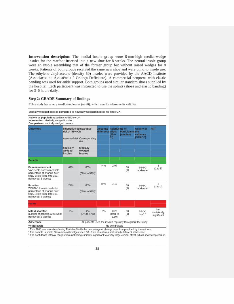
Intervention description: The medial insole group wore 8-mm-high medial-wedge insoles for the rearfoot inserted into a new shoe for 8 weeks. The neutral insole group wore an insole resembling that of the former group but without raised wedges for 8 weeks. Patients of both groups received the same new shoe and were blind to insole use. The ethylene-vinyl-acetate (density 50) insoles were provided by the AACD Institute (Associaçao de Assistência à Criança Deficiente). A commercial neoprene with elastic banding was used for ankle support. Both groups used similar standard shoes supplied by the hospital. Each participant was instructed to use the splints (shoes and elastic banding) for 3–6 hours daily.
Step 2: GRADE Summary of findings*This study has a very small sample size (n=30), which could undermine its validity.
Medially wedged insoles compared to neutrally wedged insoles for knee OA
Patient or population: patients with knee OAIntervention: Medially wedged insolesComparison: neutrally wedged insoles
Outcomes Illustrative comparative risks* (95% CI)
Absolute difference
Relative effect(95% CI)
No of Participants(studies)
Quality of the evidence(GRADE)
NNT
Assumed risk Corresponding risk
neutrally wedged insoles
Medially wedged insoles
Benefits
Pain on movementVAS scale transformed into percentage of change over time. Scale from: 0 to 100.(follow-up: 8 weeks)
41% 85%
(60% to 97%)1
44% 2.07 30(1) moderate2
3(2 to 5)
FunctionWOMAC transformed into percentage of change over time. Scale from: 0 to 100.(follow-up: 8 weeks)
27% 86%
(59% to 97%)1
59% 3.19 30(1) moderate2
2(2 to 3)
Harms
Mild discomfortnumber of patients with event(follow-up: 8 weeks)
7% 2%(0% to 47%)
-5% 0.29(0.01 to
6.69)
30(1) low2,3
Not statistically significant
Adherence All patients used the insoles regularly throughout the studyWithdrawals No withdrawals1 This SMD was calculated using RevMan 5 with the percentage of change over time provided by the authors.2 The sample is small: 30 women with valgus knee OA. Pain at rest was statistically different at baseline.3 The confidence interval ranges from not being clinically significant to a very large clinical effect, which shows imprecision.
________________________________________________________________________ 39
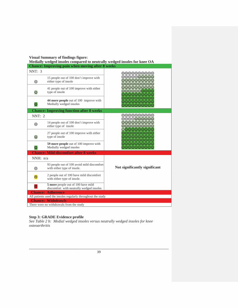
Visual Summary of findings figure:Medially wedged insoles compared to neutrally wedged insoles for knee OAChance: Improving pain when moving after 8 weeksNNT: 3
15 people out of 100 don’t improve with either type of insole
41 people out of 100 improve with either type of insole
44 more people out of 100 improve with Medially wedged insoles
Chance: Improving function after 8 weeksNNT: 2
14 people out of 100 don’t improve with either type of insole
27 people out of 100 improve with either type of insole
59 more people out of 100 improve with Medially wedged insoles
Chance: Mild discomfort after 8 weeksNNH: n/a
Not significantly significant93 people out of 100 avoid mild discomfort with either type of insole.
2 people out of 100 have mild discomfort with either type of insole.
5 more people out of 100 have mild discomfort with neutrally wedged insoles
Chance: AdherenceAll patients used the insoles regularly throughout the studyChance: Withdrawls
There were no withdrawals from the study
Step 3: GRADE Evidence profile See Table 2 b: Medial wedged insoles versus neutrally wedged insoles for knee osteoarthritis

________________________________________________________________________ 40
Step 4: Other recommendationsGroup RecommendationAAOS (knee) We suggest lateral heel wedges not be prescribed for patients with
symptomatic medial compartmental OA of the knee. EULAR Non-pharmacological treatment of knee OA should include education,
exercise, appliances (sticks, insoles, knee bracing) and weight reduction.OARSI Every patient with hip and knee OA should receive advice concerning
appropriate footwear. In patients with knee OA, insoles can reduce pain and improve ambulation. Lateral wedged insoles can be of symptomatic benefit for some patients with medial tibio-femoral compartment OA.
Step 5: GRADE Recommendation
ReferencesRodrigues PT. Effectiveness of medial-wedge insole treatment for valgus knee osteoarthritis. Arthritis and rheumatism 2008;59(5):603-8.
2.3. Subtalar strapped insoles versus inserted laterally wedged insolesfor knee OA
Are subtalar strapped insoles effective in reducing pain and improving function in patients with symptomatic knee OA compared to inserted laterally wedged insoles?
Step 1: Search ResultsWe chose the SR by Brouwer 2008 which reported one RCT which can be found in three articles by Toda (RCT published in 2001 with follow-up data published in 2004 and 2006). We are presenting the data at 6 months follow-up for efficacy and at 8 weeks for side effects as these were the only time points at which these were evaluated respectively.
Intervention description: Radiographs were evaluated for changes characteristic of OA in anteroposterior views using the Kellgren-Lawrence grade, as described in the Atlas of Standard Radiographs. Two types of lateral wedge insoles were prepared: urethane wedges made from household bath mat material with elevations of 6.35 mm strapped to an ankle sprain supporter (Sofra Wolfer®, Taketora Co. Ltd., Japan) designed to fit around the ankle and subtalar joints (strapped insole, Figure 1A); and a traditional inserted insole (Wedge Heel Type®, Sanshinkousan Co. Ltd., Japan), a lateral rubber heel wedge with an elevation of 6.35 mm (inserted insole, Figure 1B). Each participant
________________________________________________________________________ 41
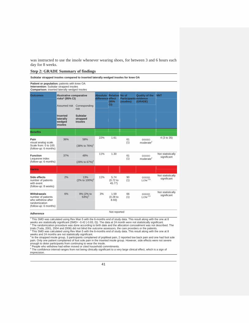
was instructed to use the insole whenever wearing shoes, for between 3 and 6 hours each day for 8 weeks.Step 2: GRADE Summary of findingsSubtalar strapped insoles compared to inserted laterally wedged insoles for knee OA
Patient or population: patients with knee OAIntervention: Subtalar strapped insolesComparison: inserted laterally wedged insoles
Outcomes Illustrative comparative risks* (95% CI)
Absolute difference
Relative effect(95% CI)
No of Participants(studies)
Quality of the evidence(GRADE)
NNT
Assumed risk Corresponding risk
Inserted laterally wedged insoles
Subtalar strapped insoles
Benefits
Painvisual analog scale. Scale from: 0 to 100.(follow-up: 6 months)
36% 58%
(38% to 76%)1
22% 1.61 61(1) moderate2
4 (3 to 35)
FunctionLequesne index(follow-up: 6 months)
37% 48%
(29% to 67%)3
11% 1.30 61(1) moderate2
Not statistically significant
Harms
Side effectsnumber of patients with event(follow-up: 8 weeks)
2% 13%(2% to 100%)4
11% 5.74(0.72 to 45.77)
90(1) LOW 2,6
Not statistically significant
Withdrawals number of patients who withdrew after randomization(follow-up: 6 months)
6% 9% (2% to 53%)5
3% 1.59 (0.28 to
8.93)
66 (1) LOW 2,6
Not statistically significant
Adherence Not reported
1 This SMD was calculated using Rev Man 5 with the 6-months end of study data. This result along with the one at 8 weeks are statistically significant (SMD= -0.42 (-0.83, 0)). The data at 24 month were not statistically significant. 2 The randomization procedure was done according to birth date and the allocation concealment was not described. The trials (Toda, 2001, 2004 and 2006) did not blind the outcome assessors, the care providers or the patients.3 This SMD was calculated using Rev Man 5 with the 6-months end of study data. This result along with the one at 8 weeks and 24 months are not statistically significant. 4 In the strapped insole group, 3 participants complained of popliteal pain, 2 reported low back pain and one had foot sole pain. Only one patient complained of foot sole pain in the inserted insole group. However, side effects were not severe enough to deter participants from continuing to wear the insole.5 People who withdrew had either moved or cited household commitments.6 The confidence interval ranges from not being clinically significant to a very large clinical effect, which is a sign of imprecision.
________________________________________________________________________ 42
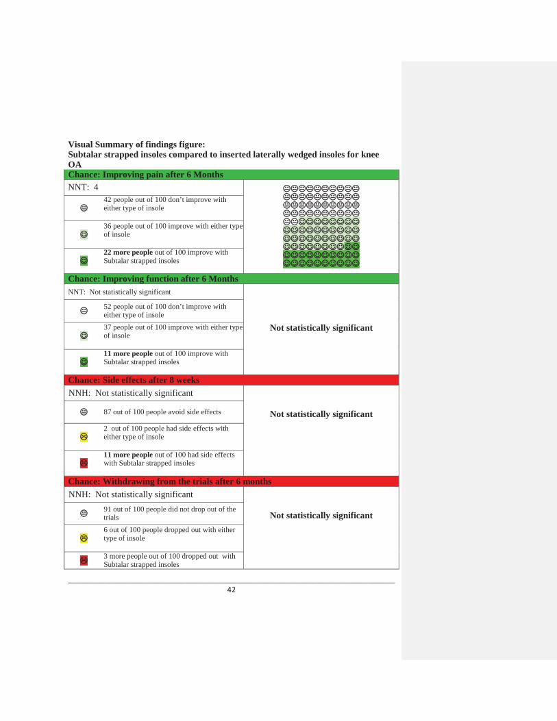
Visual Summary of findings figure:Subtalar strapped insoles compared to inserted laterally wedged insoles for knee OAChance: Improving pain after 6 Months NNT: 4
42 people out of 100 don’t improve with either type of insole
36 people out of 100 improve with either type of insole
22 more people out of 100 improve with Subtalar strapped insoles
Chance: Improving function after 6 MonthsNNT: Not statistically significant
Not statistically significant
52 people out of 100 don’t improve with either type of insole37 people out of 100 improve with either type of insole
11 more people out of 100 improve with Subtalar strapped insoles
Chance: Side effects after 8 weeksNNH: Not statistically significant
Not statistically significant87 out of 100 people avoid side effects
2 out of 100 people had side effects with either type of insole
11 more people out of 100 had side effects with Subtalar strapped insoles
Chance: Withdrawing from the trials after 6 monthsNNH: Not statistically significant
Not statistically significant91 out of 100 people did not drop out of the trials6 out of 100 people dropped out with either type of insole
3 more people out of 100 dropped out with Subtalar strapped insoles

________________________________________________________________________ 43
Chance: AdherenceAdherence was not reported
Step 3: GRADE Evidence profileSee Table 2 c: Subtalar strapped insoles versus inserted laterally wedged insoles
Step 4: Other recommendationsGroup RecommendationAAOS (knee) We suggest lateral heel wedges not be prescribed for patients with
symptomatic medial compartmental OA of the knee. EULAR Non-pharmacological treatment of knee OA should include education,
exercise, appliances (sticks, insoles, knee bracing) and weight reduction.
OARSI Every patient with hip and knee OA should receive advice concerning appropriate footwear. In patients with knee OA, insoles can reduce pain and improve ambulation. Lateral wedged insoles can be of symptomatic benefit for some patients with medial tibio-femoral compartment OA.
Step 5: GRADE Recommendation
ReferencesBrouwer RW, Jakma TS, Verhagen AP, Verhaar JA, Bierma-Zeinstra SM. Braces and orthoses for treating osteoarthritis of the knee. Cochrane Database of Syst Rev 2005;(1):CD004020.
Toda Y, Tsukimura N. A six-month followup of a randomized trial comparing the efficacy of a lateral-wedge insole with subtalar strapping and an in-shoe lateral-wedge insole in patients with varus deformity osteoarthritis of the knee. Arthritis Rheum 2004; 50(10):3129-3136.
Toda Y. A 2-year follow-up of a study to compare the efficacy of lateral wedged insoles with subtalar strapping and in-shoe lateral wedged insoles in patients with varus deformity osteoarthritis of the knee. Osteoarthritis and cartilage / OARS , Osteoarthritis Research Society 2006;14(3):231-7.
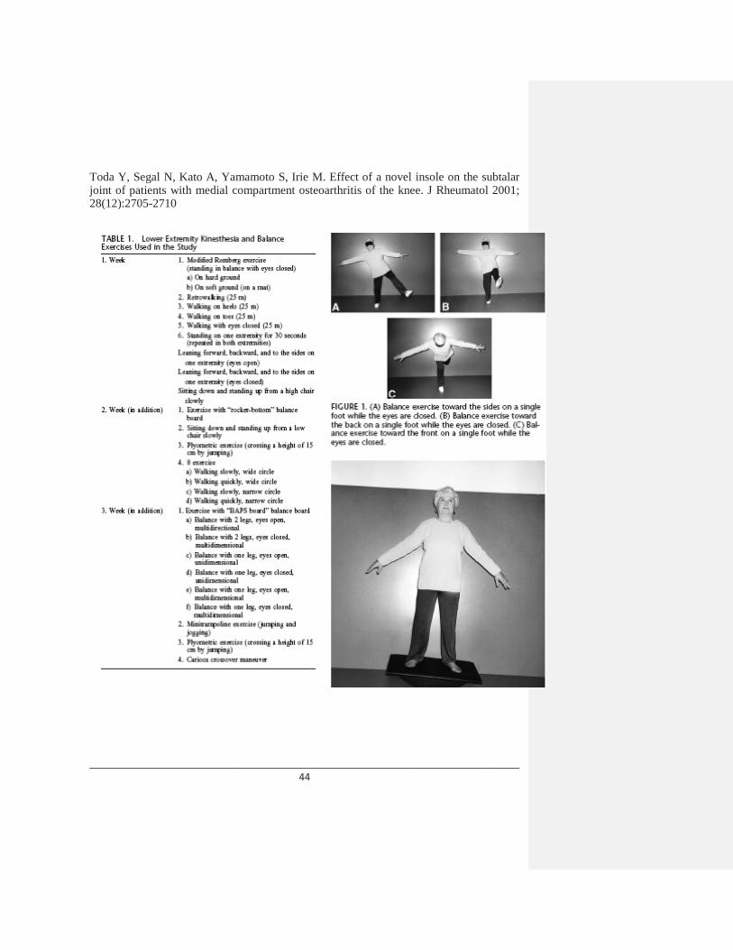
________________________________________________________________________ 44
Toda Y, Segal N, Kato A, Yamamoto S, Irie M. Effect of a novel insole on the subtalar joint of patients with medial compartment osteoarthritis of the knee. J Rheumatol 2001; 28(12):2705-2710

________________________________________________________________________ 45
3. SELF-MANAGEMENT
Are self-management programs effective in reducing pain and improving function in patients with symptomatic knee osteoarthritis (OA) compared to usual care?
Step 1: Search ResultsThree meta-analyses on self-management programs were found (Chodosh, 2005; Devos-Comby, 2006; Warsi, 2004). Although Devos-Comby (2006) was the most recent evidence, exercise and self-management were presented such that outcomes from each intervention could not be separated. Warsi (2004) did not focus on OA. Chodosh (2005) met our selection criteria and was therefore chosen as the best available evidence. Devos-Comby (2006) had similar results to Chodosh (2005), whereby, no clinically significanteffect was found on physical outcomes.
Interventions description: Chronic disease self-management program was defined by the authors of the systematic review as “a systematic intervention that is targeted toward patients with chronic disease. The intervention should help them actively participate in either or both of the following: self-monitoring (of symptoms or of physiologic processes) or decision making (managing the disease or its impact through self-monitoring)” (Chodosh, 2005).
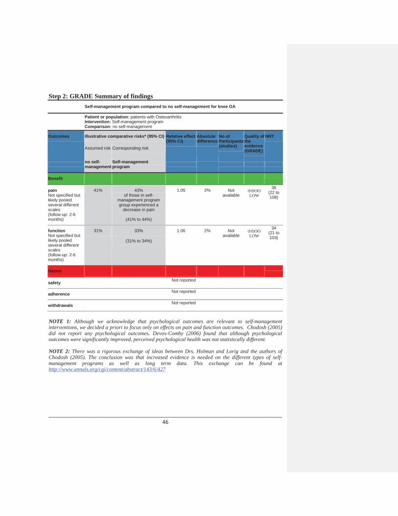
________________________________________________________________________ 46
Step 2: GRADE Summary of findingsSelf-management program compared to no self-management for knee OA
Patient or population: patients with OsteoarthritisIntervention: Self-management programComparison: no self-management
Outcomes Illustrative comparative risks* (95% CI) Relative effect(95% CI)
Absolute difference
No of Participants(studies)
Quality of the evidence(GRADE)
NNT
Assumed risk Corresponding risk
no self-management
Self-management program
Benefit
painNot specified but likely pooledseveral different scales(follow-up: 2-6months)
41% 43%of those in self-
management program group experienced a
decrease in pain
(41% to 44%)
1.05 2% Not available LOW
36(22 to 108)
functionNot specified but likely pooled several different scales(follow-up: 2-6months)
31% 33%
(31% to 34%)
1.06 2% Not available LOW
34(21 to 103)
Harms
safety Not reported
adherence Not reported
withdrawals Not reported
NOTE 1: Although we acknowledge that psychological outcomes are relevant to self-management interventions, we decided a priori to focus only on effects on pain and function outcomes. Chodosh (2005) did not report any psychological outcomes. Devos-Comby (2006) found that although psychological outcomes were significantly improved, perceived psychological health was not statistically different.
NOTE 2: There was a rigorous exchange of ideas between Drs. Holman and Lorig and the authors of Chodosh (2005). The conclusion was that increased evidence is needed on the different types of self-management programs as well as long term data. This exchange can be found at http://www.annals.org/cgi/content/abstract/143/6/427

________________________________________________________________________ 47
Visual Summary of Findings TableSelf-management program compared to no self-management for osteoarthritis
Chance: Improving pain after 8 weeksNNT: 6
57 people out of 100 don’t improve whether they take a self management program or not
41 people out of 100 improve with either intervention
.2 more people out of 100 improve witha self-management program
Chance: Improving function after 8 weeksNNT: 6
67 people out of 100 don’t improve whether they take a self management program or not
31 people out of 100 improve with either intervention
2 more people out of 100 improve witha self-management program
Chance: Safety, Adherence, WithdrawalsNNH: n/a
The safety of self-management and the number of people who adhered to a self-management program and the number of people who withdrew from self management programs was not reported.
Not reported
Step 3: GRADE Evidence profile See Table 3: Self-management
NOTE: Post-hoc tests including 5 essential elements (tailoring, group setting, feedback, psychological, and medical care) were unrevealing.

________________________________________________________________________ 48
Step 4: Other recommendationsGroup RecommendationAAOS (knee) We suggest patients with symptomatic OA of the knee be
encouraged to participate in self-management educational programs such as those conducted by the Arthritis Foundation, and incorporate activity modifications (e.g. walking instead of running; alternative activities) into their lifestyle. Regular contact to promote self-care is an option for patients with symptomatic OA of the knee. (No recommendations for hip).
EULAR Non-pharmacological treatment of knee OA should include education, exercise, appliances (sticks, insoles, knee bracing) and weight reduction.
OARSI Optimal management of OA requires a combination of non-pharmacological and pharmacological modalities.
All patients with hip and knee OA should be given information access and education about the objectives of treatment and the importance of changes in lifestyle, exercise, pacing of activities, weight reduction, and other measures to unload the damaged joint(s). The initial focus should be on self-help and patient-driven treatments rather than on passive therapies delivered by health professionals. Subsequently emphasis should be placed on encouraging adherence to the regimen of non-pharmacological therapy.
The clinical status of patients with hip or knee OA can be improved if patients are contacted regularly by phone.
Step 5: GRADE Recommendation
References Chodosh J, Morton S, Mojica W, Maglione M, Suttorp M, Hilton L, Rhodes S, Shekelle P. Meta-analysis: Chronic disease self-management programs for older adults. Ann Int Med 2005;143(6):427-38.
Devos-Comby L, Cronan T, Roesch SC. Do exercise and self-management interventions benefit patients with osteoarthritis of the knee? A metaanalytic review. J Rheumatol 2006;33(4):744-56.

________________________________________________________________________ 49
Lorig KR, Sobel DS, Stewart AL, Brown BW Jr., Bandura A, Ritter P, et al. Evidence suggesting that a chronic disease self-management program can improve health status while reducing hospitalization : a randomized trial. Med Care 1999;37(1):5-14.
Warsi A, Wang PS, L Valley MP, Avorn J, Solomon DH. Self-management education programs in chronic disease: a systematic review and methodological critique of theliterature. Arch Intern Med 2004;164(15):1641-49.
4. MANUAL THERAPY
4.1 Manual therapy program versus exercise therapy program for hip OA
Is manual therapy effective in reducing pain and improving function in patients with symptomatic hip osteoarthritis (OA) compared to exercise therapy? Are patients compliant to these treatment regimens and do they experience adverse effects?
Step 1: Search ResultsThere were no meta-analyses which reported the efficacy of manual therapy in patients with hip OA. There was one RCT which assessed the efficacy of manual therapy vs. exercise therapy in patients with hip OA: Hoeksma (2004).
Intervention description: Subjects in both the manual therapy program and the exercise therapy program attended 25-minute sessions twice a week for a total of 9 treatments. Manual therapy consists of manipulation and stretching with the aim of improving the elasticity of the joint capsule and surrounding muscles. Each manual therapy session began with 10 to 15 minutes of stretching of shortened muscles. Manipulation was then performed using a traction manipulation technique. The exercise therapy program was tailored to each individual participant’s needs. The 4 main treatment goals were 1) increase of muscle function through muscle strengthening exercises using weight or strengthening equipment; endurance by treadmill walking of cycling on a home trainer; and coordination by walking and balancing exercises; 2) improvement of range of joint motion by motions that go beyond the daily activity range of motion and stretching; 3) decrease of pain through active joint and stretching exercises as well as second and third degree traction; 4) improvement of walking ability through specific walking exercises to adjust gait pattern, use of walking aids, and stair-climbing instruction.
In both groups, participants also received education and advice on the load ability of the hip joint and increasing their physical activity. The exercise group received additional instruction for home exercise, based on the specific exercises performed during the treatment session.
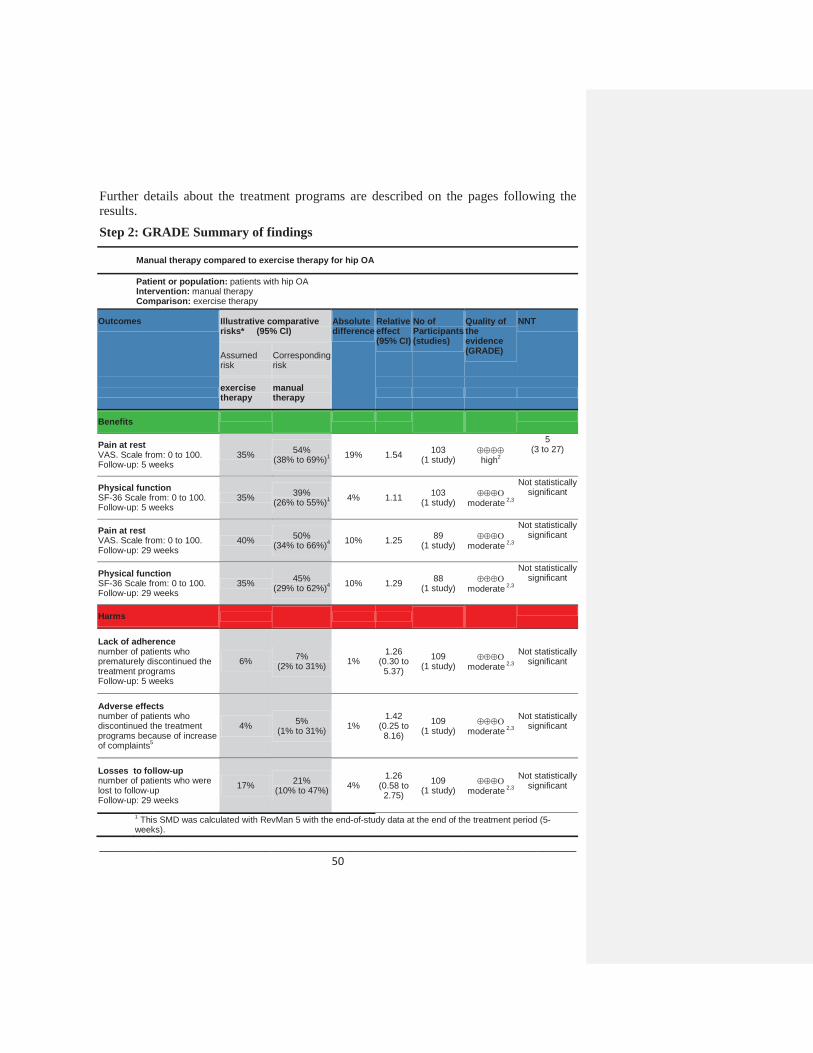
________________________________________________________________________ 50
Further details about the treatment programs are described on the pages following the results. Step 2: GRADE Summary of findings
Manual therapy compared to exercise therapy for hip OA
Patient or population: patients with hip OAIntervention: manual therapyComparison: exercise therapy
Outcomes Illustrative comparative risks* (95% CI)
Absolute difference
Relative effect(95% CI)
No of Participants(studies)
Quality of the evidence(GRADE)
NNT
Assumed risk
Corresponding risk
exercise therapy
manual therapy
Benefits
Pain at restVAS. Scale from: 0 to 100.Follow-up: 5 weeks
35% 54% (38% to 69%)1 19% 1.54 103
(1 study) high2
5(3 to 27)
Physical functionSF-36 Scale from: 0 to 100. Follow-up: 5 weeks
35% 39% (26% to 55%)1 4% 1.11 103
(1 study) moderate 2,3
Not statistically significant
Pain at rest VAS. Scale from: 0 to 100.Follow-up: 29 weeks
40% 50% (34% to 66%)4 10% 1.25 89
(1 study) moderate 2,3
Not statistically significant
Physical functionSF-36 Scale from: 0 to 100. Follow-up: 29 weeks
35% 45% (29% to 62%)4 10% 1.29 88
(1 study) moderate 2,3
Not statistically significant
Harms
Lack of adherencenumber of patients who prematurely discontinued the treatment programsFollow-up: 5 weeks
6% 7%(2% to 31%) 1%
1.26 (0.30 to 5.37)
109(1 study) moderate 2,3
Not statistically significant
Adverse effectsnumber of patients who discontinued the treatment programs because of increase of complaints5
4% 5%(1% to 31%) 1%
1.42 (0.25 to 8.16)
109(1 study) moderate 2,3
Not statistically significant
Losses to follow-up number of patients who were lost to follow-upFollow-up: 29 weeks
17% 21% (10% to 47%) 4%
1.26 (0.58 to 2.75)
109 (1 study) moderate 2,3
Not statistically significant
1 This SMD was calculated with RevMan 5 with the end-of-study data at the end of the treatment period (5-weeks).
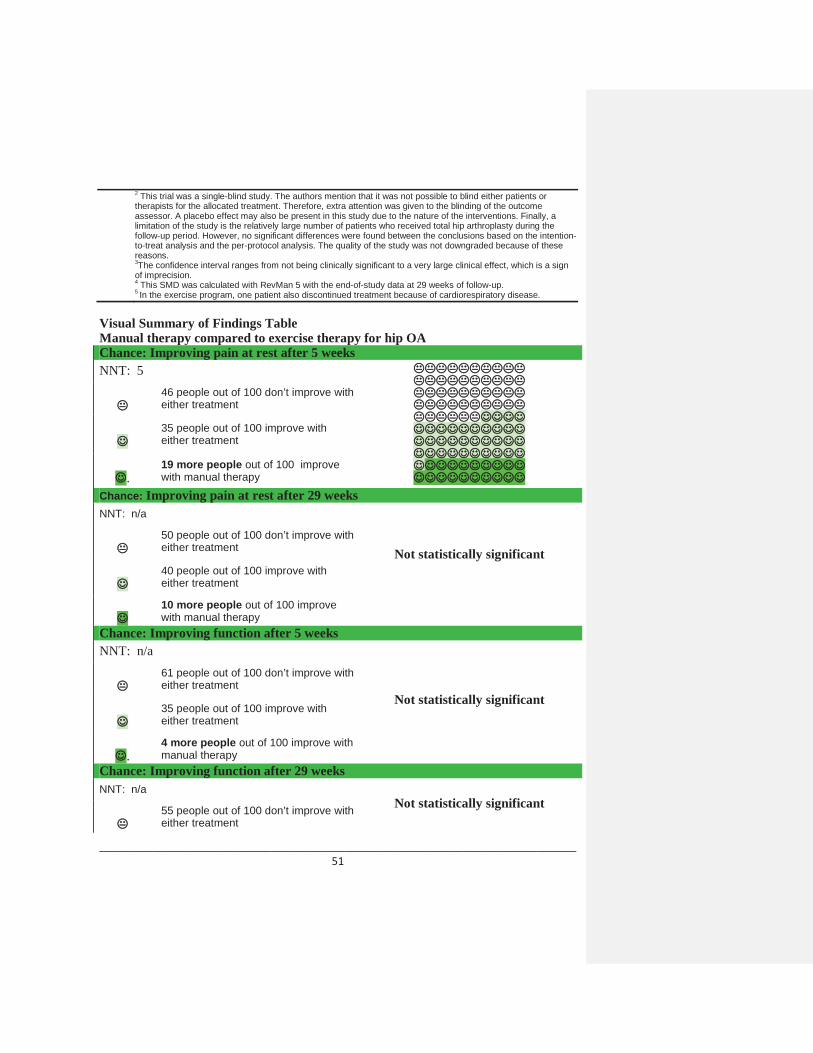
________________________________________________________________________ 51
2 This trial was a single-blind study. The authors mention that it was not possible to blind either patients or therapists for the allocated treatment. Therefore, extra attention was given to the blinding of the outcome assessor. A placebo effect may also be present in this study due to the nature of the interventions. Finally, a limitation of the study is the relatively large number of patients who received total hip arthroplasty during the follow-up period. However, no significant differences were found between the conclusions based on the intention-to-treat analysis and the per-protocol analysis. The quality of the study was not downgraded because of these reasons. 3The confidence interval ranges from not being clinically significant to a very large clinical effect, which is a sign of imprecision.4 This SMD was calculated with RevMan 5 with the end-of-study data at 29 weeks of follow-up. 5 In the exercise program, one patient also discontinued treatment because of cardiorespiratory disease.
Visual Summary of Findings TableManual therapy compared to exercise therapy for hip OAChance: Improving pain at rest after 5 weeksNNT: 5
46 people out of 100 don’t improve with either treatment
35 people out of 100 improve with either treatment
.19 more people out of 100 improve with manual therapy
Chance: Improving pain at rest after 29 weeksNNT: n/a
Not statistically significant50 people out of 100 don’t improve with either treatment
40 people out of 100 improve with either treatment
10 more people out of 100 improve with manual therapy
Chance: Improving function after 5 weeksNNT: n/a
Not statistically significant
61 people out of 100 don’t improve with either treatment
35 people out of 100 improve with either treatment
.4 more people out of 100 improve with manual therapy
Chance: Improving function after 29 weeksNNT: n/a
Not statistically significant55 people out of 100 don’t improve with either treatment

________________________________________________________________________ 52
35 people out of 100 improve with either treatment
10 more people out of 100 improve with manual therapy
Chance: Lack of adherence; discontinuation of therapy after 5 weeksNNH: n/a
Not significantly significant93 people out of 100 continued with either treatment
6 people discontinued the study with either treatment
1 more person discontinued the study while taking manual therapy
Chance: Adverse effects
NNH: n/a
Not significantly significant95 people out of 100 completed either treatment because of complaints about the therapy they received.
4 people out of 100 dropped out of either treatment because of complaints about the therapy they received.
1 more person out of 100 dropped out of manual therapy because of complaints about the therapy.
Chance: Loss to follow-up (people who did not complete the study)NNH: n/a
Not significantly significant79 people out of 100 completed the study with either therapy
17 people out of 100 did not complete the study with either therapy
4 more people out of 100 did not complete the study when taking part inmanual therapy

________________________________________________________________________ 53
Step 3: GRADE Evidence profile See Table 4 a: Manual therapy program versus exercise therapy program for hip OA
Step 4: Other recommendationsGroup RecommendationAAOS N/A No recommendations for hip.EULAR Non-pharmacological treatment of knee OA should include education,
exercise, appliances (sticks, insoles, knee bracing) and weight reduction.
OARSI Optimal management of OA requires a combination of non-pharmacological and pharmacological modalities.
Step 5: GRADE Recommendation
ReferencesHoeksma HL, Dekker J, Ronday HK et al. Comparison of manual therapy and exercise therapy in osteoarthritis of the hip: a randomized clinical trial. Arthritis Rheum 2004; 51(5):722-9.
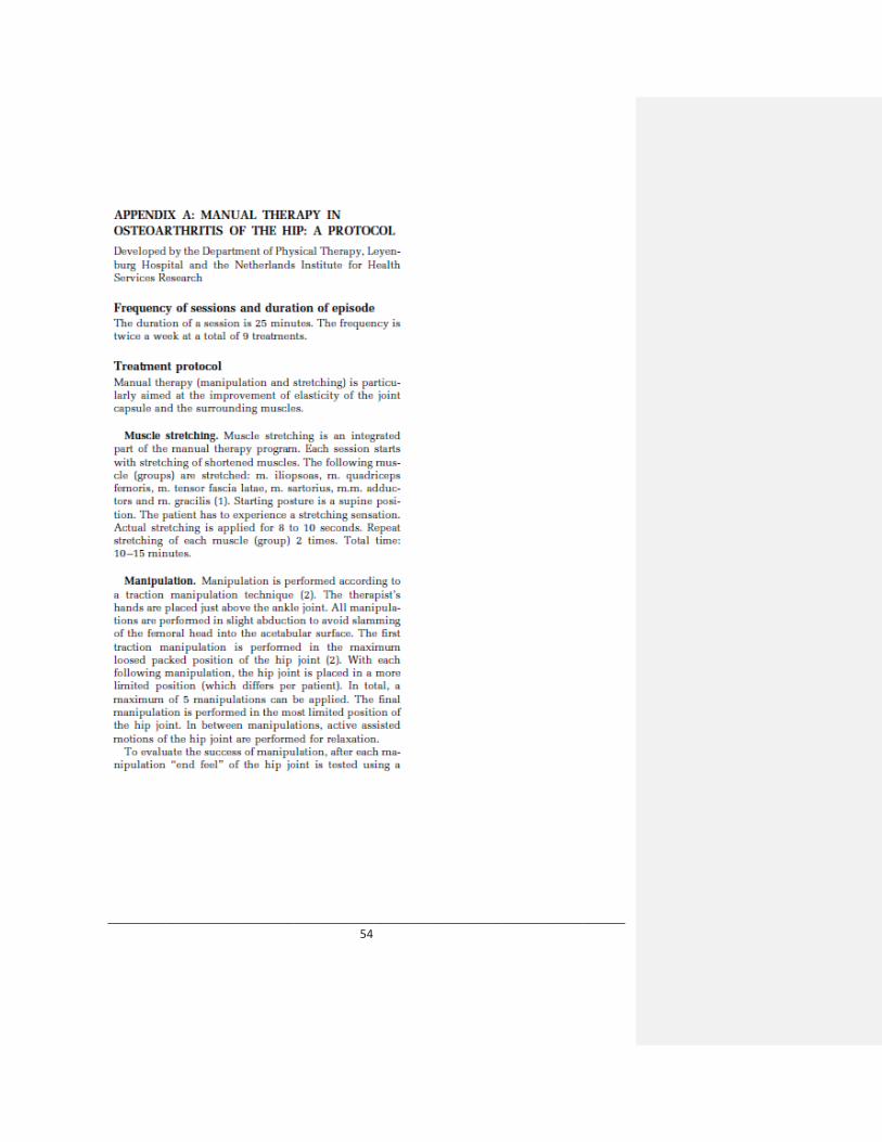
________________________________________________________________________ 54

________________________________________________________________________ 55

________________________________________________________________________ 56
4.2 Manual therapy in combination with supervised exercise and home exercise program versus home exercise program alone for knee OA
Is individualized manual therapy in combination with supervised exercise and home exercise program effective in reducing pain and improving function in patients with symptomatic knee OA compared to home exercise program? Are patients compliant to these treatment regimens?
Step 1: Search Results
There were no meta-analyses which reported the efficacy of manual therapy in patients with knee OA. A few RCTs assessed the efficacy of manual therapy specifically in patients with knee OA but most had limitations (sample size smaller than 50 participants) or used manual therapy in combination with other modalities such as taping and massage, making it difficult to evaluate its efficacy. We chose the only RCT conducted in patients with knee OA which assessed the efficacy of manual therapy in combination with supervised exercises, the treatment combination deemed the most used in clinical practice by our team of experts: Deyle (2005). We contacted the authors in order to report results for the pain and function subscales of the WOMAC since only the total WOMAC score was reported in their publication. The treatment programs used in this study are described following the results.
Intervention description: Subjects in the clinic treatment group attended 8 treatmentsessions over a 4 week period in the physical therapy clinic. Manual therapy programs were individualized based on the results of the examination. The manual therapy techniques, consisting of passive physiological and accessory movements, muscle stretching, and soft tissue mobilization, were applied by the treating physical therapistprimarily to the knee and surrounding structures. In addition to receiving manual therapy treatments, subjects in the clinic treatment group performed a standardized knee exercise program at each treatment session. This program consisted of active ROM exercises,muscle strengthening, muscle stretching, and riding a stationary bicycle. A physical therapist or physical therapy technician supervised these exercises. The number of strengthening exercise bouts and stationary bicycle riding time were increased or decreased by the treating physical therapist based on subject response. Subjects in the clinic treatment group performed the same home exercise program as the home exercise group each day that they were not treated in the physical therapy clinic.

________________________________________________________________________ 57
Step 2: GRADE Summary of findingsManual therapy in combination with supervised exercise and home exercise program compared to home exercise for knee OA
Patient or population: patients with knee OAIntervention: manual therapy in combination with supervised exercise and home exercise programComparison: home exercise
Outcomes Illustrative comparative risks* (95% CI)
Absolute difference
Relative effect(95% CI)
No of Participants(studies)
Quality of the evidence(GRADE)
NNT
Assumed risk
Corresponding risk
Home exercise
Manual therapy +supervised exercise and home exercise program
Benefits
PainWOMAC. Scale from: 0 to 500.Follow-up: 8 weeks
37% 53% (39% to 67%)
16% 1.43 120(1 study2) high 1
6 (3 to 43)
FunctionWOMAC. Scale from: 0 to 1700.Follow-up: 8 weeks
37% 52%(38% to 66%) 15% 1.41 120
(1 study) high6
(3 to 70)
Harms
Safety Not reported
Discontinuations due to lack of adherencenumber of patients who discontinued due to lack of adherence to the treatment regimen (whether subjects attended all clinical appointments and reported for testing at 0, 4 and 8 weeks).Follow-up: 8 weeks
0% 0% 0% 0 120(1 study) high
Not statistically significant
Withdrawalspeople who withdrew from the study after randomization. Follow-up: 8 weeks
12 %3 9%(3% to 25%)4 -3%
0.77 (0.28 to
2.11)134
(1 study) moderate5
Not statistically significant
1 The authors report that the intention to treat results with 134 subjects did not differ substantially from the results of the 120 subjects.2 Another outcome reported by the author was the use of medications for OA by patients at 52 weeks. Use of medications for OA was higher in the home exercise group (68%) than the clinic treatment group (48%) and this difference was statistically significant (p=0.03). 3 In the control group, withdrawals were due to: knee injections (1), changed medications (1), shoulder surgery (1), not willing to return (2) and moved from area (3).4 In the treatment group, withdrawals were due to: knee injections (2), changed medications (1), not willing to return (1), notwilling to walk (1) and unrelated medical condition (1).5 The confidence interval ranges from not being clinically significant to a very large clinical effect, which is a sign of imprecision.
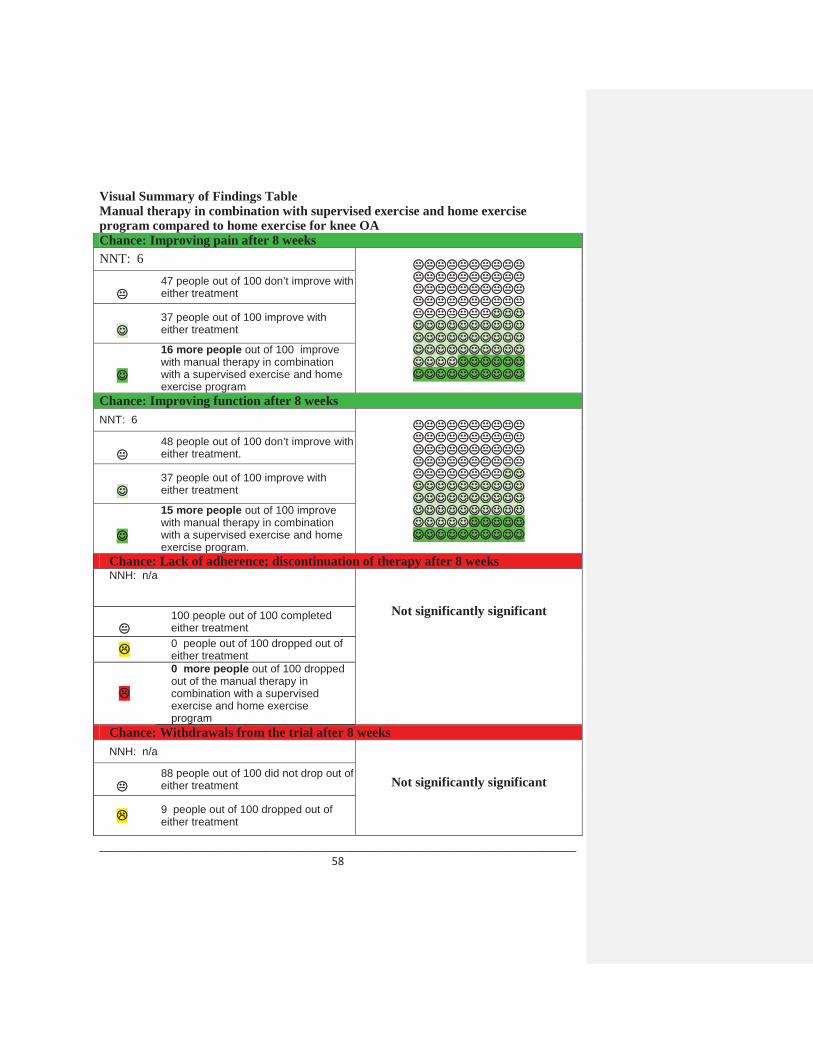
________________________________________________________________________ 58
Visual Summary of Findings TableManual therapy in combination with supervised exercise and home exercise program compared to home exercise for knee OAChance: Improving pain after 8 weeksNNT: 6
47 people out of 100 don’t improve with either treatment
37 people out of 100 improve with either treatment
16 more people out of 100 improve with manual therapy in combination with a supervised exercise and home exercise program
Chance: Improving function after 8 weeksNNT: 6
48 people out of 100 don’t improve with either treatment.
37 people out of 100 improve with either treatment
15 more people out of 100 improve with manual therapy in combination with a supervised exercise and home exercise program.
Chance: Lack of adherence; discontinuation of therapy after 8 weeksNNH: n/a
Not significantly significant100 people out of 100 completedeither treatment0 people out of 100 dropped out of either treatment 0 more people out of 100 dropped out of the manual therapy in combination with a supervised exercise and home exercise program
Chance: Withdrawals from the trial after 8 weeksNNH: n/a
Not significantly significant88 people out of 100 did not drop out of either treatment
9 people out of 100 dropped out of either treatment
________________________________________________________________________ 59
3 more people out of 100 dropped out of the home exercise program.
SafetyNNH: n/a Not reported
Step 3: GRADE Evidence profile See Table 4 b: Manual therapy in combination with supervised exercise and home exercise program versus home exercise program alone for knee OA
Step 4: Other recommendationsGroup RecommendationAAOS (knee) No recommendations for manual therapy.EULAR Non-pharmacological treatment of knee OA should include education,
exercise, appliances (sticks, insoles, knee bracing) and weight reduction.
OARSI Optimal management of OA requires a combination of non-pharmacological and pharmacological modalities.
Step 5: GRADE Recommendation
ReferencesDeyle GD, Allison SC, Matekel RL et al. Physical therapy treatment effectiveness for osteoarthritis of the knee: a randomized comparison of supervised clinical exercise and manual therapy procedures versus a home exercise program. Phys Ther 2005;85(12):1301-17.Description of the treatment programs:
Subjects in the clinic treatment group attended 8 treatment sessions over a 4 week periodin the physical therapy clinic. Manual therapy programs were individualized based on theresults of the examination. The manual therapy techniques, consisting of passive physiological and accessory movements, muscle stretching, and soft tissue mobilization,were applied by the treating physical therapist primarily to the knee and surrounding structures. In addition to receiving manual therapy treatments, subjects in the clinic treatment group performed a standardized knee exercise program at each treatmentsession. This program consisted of active ROM exercises, muscle strengthening, muscle stretching, and riding a stationary bicycle. A physical therapist or physical therapytechnician supervised these exercises. The number of strengthening exercise bouts and stationary bicycle riding time were increased or decreased by the treating physical therapist based on subject response. Subjects in the clinic treatment group performed the
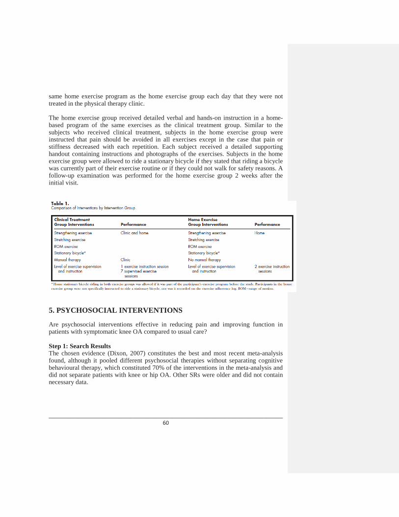
________________________________________________________________________ 60
same home exercise program as the home exercise group each day that they were not treated in the physical therapy clinic.
The home exercise group received detailed verbal and hands-on instruction in a home-based program of the same exercises as the clinical treatment group. Similar to the subjects who received clinical treatment, subjects in the home exercise group were instructed that pain should be avoided in all exercises except in the case that pain or stiffness decreased with each repetition. Each subject received a detailed supporting handout containing instructions and photographs of the exercises. Subjects in the home exercise group were allowed to ride a stationary bicycle if they stated that riding a bicycle was currently part of their exercise routine or if they could not walk for safety reasons. Afollow-up examination was performed for the home exercise group 2 weeks after the initial visit.
5. PSYCHOSOCIAL INTERVENTIONS
Are psychosocial interventions effective in reducing pain and improving function in patients with symptomatic knee OA compared to usual care?
Step 1: Search ResultsThe chosen evidence (Dixon, 2007) constitutes the best and most recent meta-analysis found, although it pooled different psychosocial therapies without separating cognitive behavioural therapy, which constituted 70% of the interventions in the meta-analysis and did not separate patients with knee or hip OA. Other SRs were older and did not containnecessary data.
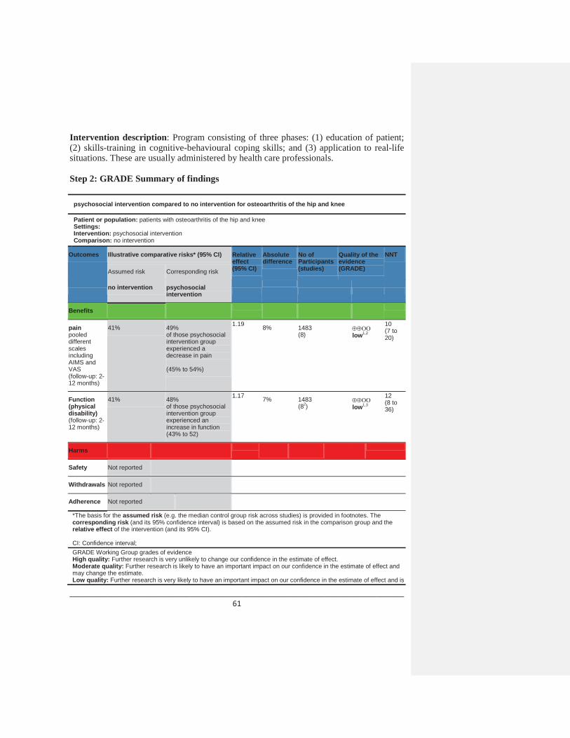
________________________________________________________________________ 61
Intervention description: Program consisting of three phases: (1) education of patient; (2) skills-training in cognitive-behavioural coping skills; and (3) application to real-life situations. These are usually administered by health care professionals.
Step 2: GRADE Summary of findings
psychosocial intervention compared to no intervention for osteoarthritis of the hip and knee
Patient or population: patients with osteoarthritis of the hip and kneeSettings:Intervention: psychosocial interventionComparison: no intervention
Outcomes Illustrative comparative risks* (95% CI) Relative effect(95% CI)
Absolute difference
No of Participants(studies)
Quality of the evidence(GRADE)
NNT
Assumed risk Corresponding risk
no intervention psychosocial intervention
Benefits
painpooled different scales including AIMS and VAS(follow-up: 2-12 months)
41% 49%of those psychosocial intervention group experienced a decrease in pain
(45% to 54%)
1.19 8% 1483(8) low1,2
10(7 to 20)
Function(physical disability)(follow-up: 2-12 months)
41% 48%of those psychosocial intervention group experienced anincrease in function(43% to 52)
1.17 7% 1483(82) low1,3
12(8 to 36)
Harms
Safety Not reported
Withdrawals Not reported
Adherence Not reported
*The basis for the assumed risk (e.g. the median control group risk across studies) is provided in footnotes. The corresponding risk (and its 95% confidence interval) is based on the assumed risk in the comparison group and the relative effect of the intervention (and its 95% CI).
CI: Confidence interval; GRADE Working Group grades of evidenceHigh quality: Further research is very unlikely to change our confidence in the estimate of effect.Moderate quality: Further research is likely to have an important impact on our confidence in the estimate of effect and may change the estimate.Low quality: Further research is very likely to have an important impact on our confidence in the estimate of effect and is

________________________________________________________________________ 62
likely to change the estimate.Very low quality: We are very uncertain about the estimate.1 Pooled wide range of psychosocial interventions2 Affected joints not described therefore could not distinguish between hip, knee, and other.3 No description of type of scales used.4 Calfas 1992, Gay 2002, Keefe 2004, Keefe 1996, Keefe 1999, Keefe 1990, Keefe 1990, Lin 2003.
Visual Summary of Findings TablePsychosocial intervention compared to no intervention for osteoarthritis of the hip and kneeChance: Improving pain after 2-12 monthsNNT: 10
51 people out of 100 don’t improvewhether or not they take part in a psychosocial intervention.41 people out of 100 improve whether or not they take part in a psychosocial intervention.
8 more people out of 100 improve with apsychosocial intervention.
Chance: Improving function after 2-12 monthsNNT: 12
52 people out of 100 don’t improvewhether or not they take part in a psychosocial intervention.41 people out of 100 improve whether or not they take part in a psychosocial intervention.
7 more people out of 100 improve with apsychosocial intervention.
Chance: Withdrawls The number of people who left the study was not reported.Chance: Safety
Safety of psychosocial interventions was not reported.Chance: Adherence
Adherence to psychosocial interventions was not reported.

________________________________________________________________________ 63
Step 3: GRADE Evidence profile See Table 5: Psychosocial intervention compared to no intervention for OA of the hip and knee
Step 4: Other recommendationsGroup RecommendationAAOS (knee) We suggest patients with symptomatic OA of the knee be encouraged
to participate in self-management educational programs such as those conducted by the Arthritis Foundation, and incorporate activity modifications (e.g., walking instead of running; alternative activities) into their lifestyle.
EULAR Non-pharmacological treatment of knee OA should include education, exercise, appliances (sticks, insoles, knee bracing) and weight reduction.
OARSI Optimal management of OA requires a combination of non-pharmacological and pharmacological modalities.
2. All patients with hip and knee OA should be given information access and education about the objectives of treatment and the importance of changes in lifestyle, exercise, pacing of activities, weight reduction, and other measures to unload the damaged joint(s). The initial focus should be on self-help and patient-driven treatments rather than on passive therapies delivered by health professionals. Subsequently emphasis should be placed on encouraging adherence to the regimen of non-pharmacological therapy.
Step 5: GRADE Recommendation
References Dixon KE, Keefe FJ, Scipio CD, Perri LCM, Abernethy AP. Psychological interventionsfor arthritis for arthritis pain management in adults: a meta-analysis. Health Psychol 2007;26(3):241-50.
Lin EH, Katon W, Von Korff M, Tang L, Williams JW, Kroenke K et al. Effect of improving depression care on pain and functional outcomes among older adults with arthritis: a randomized controlled trial. JAMA 2003;290(18):2428-9.

________________________________________________________________________ 64
6. WEIGHT LOSS
Is weight loss effective in reducing pain and improving function in patients with symptomatic knee OA compared to usual care and sham acupuncture?
Step 1: Search ResultsWe found one meta-analysis (Christensen, 2007), which pooled the results from 4 randomized controlled trials (Christensen, 2005; Messier, 2000; Messier, 2004; Toda, 1998). Toda, 1998 results were not included in this summary of findings due to the use of pharmacological intervention to achieve weight loss. [The 8 remaining publications found were single randomized controlled trials (RCTs) and were not included. These were either already included in the meta-analysis (2), did not fall under the inclusion criteria (2) or were written in a language other than English (2). It is uncertain why 2 RCTs (Fotch 2005 and Miller 2006) were not included in the meta-analysis; it is suggested thatthese RCTs were indexed after the search performed in 2006. All of the additional RCTs findings were in the same direction as those of Christensen 2007.]
Interventions description: interventions included were weight loss interventions using CBT, nutrition, and/or exercise approaches and excluded pharmacological interventions
Step 2: GRADE Summary of findingsweight loss compared to control (no weight loss program) for knee OA
Patient or population: patients with knee osteoarthritisSettings:Intervention: weight lossComparison: control (no weight loss program)
Outcomes Illustrative comparative risks* (95% CI)
Relative effect(95% CI)
Absolute difference
No of Participants(studies)
Quality of the evidence(GRADE)
NNT
Assumed risk Corresponding risk
Control (no weight loss)
Weight loss
Benefits
pain WOMAC 500mm. Scale from: 0 to 500.(follow-up: 8-24 weeks)
36% 44%of those in weight loss group experienced a decrease in pain
(37% to 52%)
1.2 7.8% 416(22) moderate1
11(not estimable)
functionWOMAC 1700mm. Scale from: 0 to 1700.
34% 43%
(36% to 50%)
1.26 9% 416(22) moderate1
9(5 to 52)
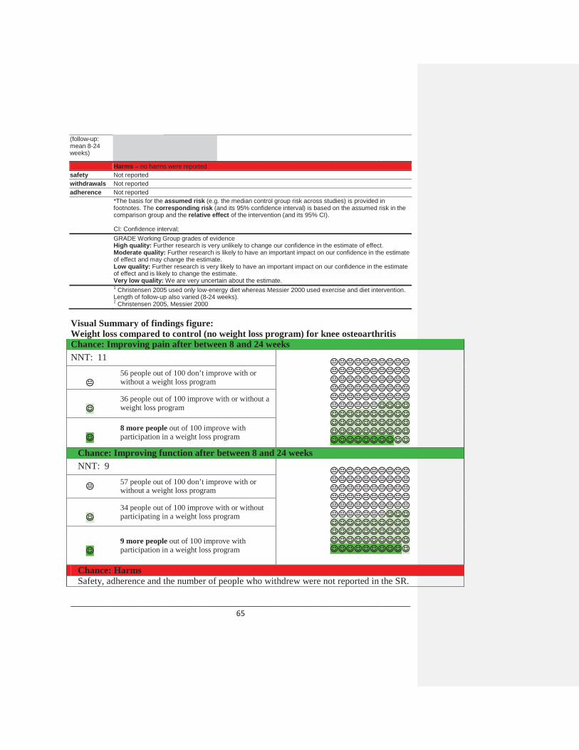
________________________________________________________________________ 65
(follow-up: mean 8-24 weeks)
Harms – no harms were reported safety Not reportedwithdrawals Not reportedadherence Not reported
*The basis for the assumed risk (e.g. the median control group risk across studies) is provided in footnotes. The corresponding risk (and its 95% confidence interval) is based on the assumed risk in the comparison group and the relative effect of the intervention (and its 95% CI).
CI: Confidence interval; GRADE Working Group grades of evidenceHigh quality: Further research is very unlikely to change our confidence in the estimate of effect. Moderate quality: Further research is likely to have an important impact on our confidence in the estimate of effect and may change the estimate.Low quality: Further research is very likely to have an important impact on our confidence in the estimate of effect and is likely to change the estimate.Very low quality: We are very uncertain about the estimate.1 Christensen 2005 used only low-energy diet whereas Messier 2000 used exercise and diet intervention. Length of follow-up also varied (8-24 weeks). 2 Christensen 2005, Messier 2000
Visual Summary of findings figure:Weight loss compared to control (no weight loss program) for knee osteoarthritis Chance: Improving pain after between 8 and 24 weeksNNT: 11
56 people out of 100 don’t improve with or without a weight loss program
36 people out of 100 improve with or without a weight loss program
8 more people out of 100 improve with participation in a weight loss program
Chance: Improving function after between 8 and 24 weeksNNT: 9
57 people out of 100 don’t improve with or without a weight loss program
34 people out of 100 improve with or without participating in a weight loss program
9 more people out of 100 improve with participation in a weight loss program
Chance: HarmsSafety, adherence and the number of people who withdrew were not reported in the SR.

________________________________________________________________________ 66
Step 3: GRADE Evidence profile See Table 6: Weight loss compared to control (no weight loss program) for knee OA
Step 4: Other recommendationsGroup RecommendationAAOS - knee We recommend patients with symptomatic OA of the knee, who are
overweight (as defined by a BMI>25), should be encouraged to lose weight (a minimum of five percent (5%) of body weight) and maintain their weight at a lower level with an appropriate program of dietary modification and exercise.
EULAR - knee Non-pharmacological treatment of knee OA should include regular education, exercise, appliances (sticks, insoles) and weight reduction if obese or overweight.
EULAR –hip
Non-pharmacological treatment of knee OA should include education, exercise, appliances (sticks, insoles, knee bracing) and weight reduction.
OARSI All patients with hip and knee OA should be given information access and education about the objectives of treatment and the importance of changes in lifestyle, exercise, pacing of activities,weight reduction, and other measures to unload the damaged joint(s). The initial focus should be on self-help and patient-driven treatments rather than on passive therapies delivered by health professionals. Subsequently emphasis should be placed on encouraging adherence to the regimen of non-pharmacological therapy.
Step 5: GRADE Recommendation
References
Christensen R, Bartels EM, Astrup A, Bliddal H. Effect of weight reduction in obese patients diagnosed with knee osteoarthritis: a systematic review and meta-analysis. Ann Rheum Dis 2007;66(4): 433-9.
Toda Y, Toda T, Takemura S, Wada T, Morimoto T, Ogawa R. Change in body fat, but not body weight or metabolic correlates of obesity, is related to symptomatic relief of obese patients with knee osteoarthritis after a weight control program. J Rheumatol 1998; 25(11):2181–6.

________________________________________________________________________ 67
Messier SP, Loeser RF, Mitchell MN, Valle G, Morgan TP, Rejeski WJ, et al. Exercise and weight loss in obese older adults with knee osteoarthritis: a preliminary study. J Am Geriatr Soc 2000;48(9):1062–72.
Messier SP, Loeser RF, Miller GD, Morgan TM, Rejeski WJ, Sevick MA, et al. Exercise and dietary weight loss in overweight and obese older adults with knee osteoarthritis: the Arthritis, Diet, and Activity Promotion Trial. Arthritis Rheum 2004;50(5):1501–10.
Christensen R, Astrup A, Bliddal H. Weight loss: the treatment of choice for knee osteoarthritis? A randomized trial. Osteoarthritis Cartilage 2005;13(1):20–7.
7. BRACES
7.1 Braces and medical (conservative) treatment versus medical (conservative) treatment in knee OA
Are braces and conservative treatment effective in reducing pain and improving function in patients with symptomatic uni-compartmental knee osteoarthritis (OA) and a mal-alignment compared to conservative treatment alone?
Step 1: Search ResultsThe most recent systematic review (SR) was the one by Brouwer, 2008 which reported one RCT conducted by the same author in 2006 and one by Kirkley in 1999. The RCT conducted by Kirkley in 1999 showed different results than the RCT by Brouwer (2006), thus we decided to display the results from both studies in the present document (section 1a and 1b).
For part 1a, we found the results reported in the SR are not the same as the ones in theBrouwer 2006 RCT so we contacted the authors. The authors mentioned that the RCT reported results stemming from an analysis which forwarded last measurements available for subjects who were lost to follow-up or for whom data were incomplete. Results in the RCT were also adjusted for baseline characteristics which were not similar. The authorsrecommended that we report the data from the RCT.
Intervention description: The conservative treatment was identical in both groups and consisted of standard care: i.e., patient education (adaptation of activities and/or weight loss), and (if needed) physical therapy and analgesics. In the intervention group patients were fitted with a knee brace (OAsys brace, Innovation Sports, Irvine, CA, USA); this brace is commercially available for right/left leg in four sizes. The brace consists of athigh shell and a calf shell (both of carbon fiber) connected by titanium hinges on the medial and lateral sides. The adjustable slide bar on the medial side of the brace provides

________________________________________________________________________ 68
valgisation (1 to 12.5 degrees) with medial unloading, or varisation (1 to 10 degrees) with lateral unloading. The degree of varisation or valgisation depends on the degree ofmalalignment and the acceptance of the patient (extensive correction will cause pressure ulcers). A specialized orthopedic technician applied the brace and gave instructions to the patients. During the follow-up this specialized orthopedic technician was present at the orthopedic outpatient department. If necessary, the brace was adjusted during the follow-up visits.
Step 2: GRADE Summary of findingsBrace and standard conservative treatment compared to standard conservative treatment only for knee OA
Patient or population: patients with knee OAIntervention: brace and standard conservative treatmentComparison: standard conservative treatment only
Outcomes Illustrative comparative risks* (95% CI)
Absolute difference
Relative effect(95% CI)
No of Participants(studies)
Quality of the evidence(GRADE)
NNT
Assumed risk Corresponding risk
standard conservative treatment only
Brace and standard conservative treatment
Benefits
PainVAS. Scale from: 0 to 10.Follow-up: 6 months
38% 43% (36% to 50%)2 5% 1.13 117
(1 study) 1 moderate3,4
Not statistically significant
Knee functionHospital for Special Surgery Score (HSS). Scale from: 0 to 100.Follow-up: 6 months
24% 28% (23% to 35%)2 4% 1.17 117
(1 study) 1 moderate3,4
Not statistically significant
Harms
Withdrawal from treatment due to adverse eventsnumber of patients who stopped the treatment dueto adverse eventsFollow-up: 12 months
7%5 0% -7%8.56
(0.47 to 155.45)
117(1 study) 1 low 3,4,6
Not statistically significant
Withdrawals from treatmentnumber of patients who stopped the treatment after randomizationFollow-up: 12 months
25% 42%(24% to 72%)7 17%
1.70 (0.98 to
2.92)117
(1 study) 1 low 3,4,6
Not statistically significant
Adherence Not reported
1 The SR by Brouwer (2008) reported one trial by the same authors (Brouwer, 2006).

________________________________________________________________________ 69
2 We calculated the SMD using the mean difference and confidence interval between groups with RevMan. The MD was adjusted by the authors for baseline values for age, gender, BMI, duration of complaints, severity of knee OA, pain severity, knee function, walking distance, medication and quality of life since these characteristics were not similar at baseline.3 The trial (Brouwer, 2006) did not blind the outcome assessors, the care providers nor the patients. Outcomes of interest were not similar at baseline.4 The authors of the meta-analysis conducted the present study, which may lead to a potential conflict of interest. The quality was not downgraded because of this.5 Adverse events include skin irritation (n=2) and bad fit (n=2).6 The confidence interval ranges from not being clinically significant to a large clinical effect, which shows imprecision. 7 Patients stopped treatment mostly because of lack of effectiveness (n=15).
[1. a]Visual Summary of Findings TableBrace and standard conservative treatment compared to standard conservative treatment only for knee OA
Chance: Improving pain after 6 monthsNNT: n/a
Not statistically significant
57 people out of 100 don’t improvewhether they use a brace or not.
38 people out of 100 improve whether they use a brace or not.
5 more people out of 100 improve with abrace.
Chance: Withdrawals due to treatment after 12 monthsNNH: n/a
Not statistically significant93 people out of 100 did not leave the study due to adverse events whether they use a brace or not.7 people out of 100 left the study due to adverse events whether they use a brace or not.No more people out of 100 left the study when they used a brace.
Chance: Withdrawals due to any reason after 12 monthsNNH: n/a
Not statistically significant58 people out of 100 did not leave the study whether they use a brace or not..
25 people out of 100 left the study whether they use a brace or not.
17 more people out of 100 left the study when they used a brace.

________________________________________________________________________ 70
Chance: AdherenceAdherence to using a brace was not reported.
Step 3: GRADE Evidence profile See Table 7 a: Braces and medical (conservative) treatment versus medical (conservative) treatment
Step 4: Other recommendationsGroup RecommendationAAOS (knee) We are unable to recommend for or against the use of a brace with a
valgus directing force for patients with medial uni-compartmental OA of the knee.
We are unable to recommend for or against the use of a brace with a varus directing force for patients with lateral uni-compartmental OA of the knee.
We suggest patients with symptomatic OA of the knee use patellar taping for short term relief of pain and improvement in function.
EULAR Non-pharmacological treatment of knee OA should include education, exercise, appliances (sticks, insoles, knee bracing) and weight reduction.
OARSI In patients with knee OA and mild/moderate varus or valgusinstability, a knee brace can reduce pain, improve stability and diminish the risk of falling.
________________________________________________________________________ 71
Step 5: GRADE Recommendation
ReferencesBrouwer RW, Jakma TS, Verhagen AP, Verhaar JA, Bierma-Zeinstra SM. Braces and orthoses for treating osteoarthritis of the knee. Cochrane Database of Syst Rev 2005;(1):CD004020.
Brouwer RW, van Raaij TM, Verhaar JA, Coene LN, Bierma-Zeinstra SM. Brace treatment for osteoarthritis of the knee: a prospective randomized multi-centre trial. Osteoarthritis Cartilage 2006;14(8):777.
7.2 Braces with medical (conservative) treatment versus medical (conservative) treatment alone in knee OA
Are braces in addition to medical treatment effective in reducing pain and improving function in patients with varus gonarthrosis compared to medical treatment alone?
Step 1: Search ResultsSince an RCT conducted by Kirkley in 1999 showed different results than the RCT by Brouwer (2006), we decided to display the results from both studies in the present document (section 1a and 1b). The results shown in the present table were computed using the data sent to our research team by Dr. Brouwer, who had recently received it from the Kirkley Research Group but did not have time to report it in his systematic review.
Intervention description: The treatment that was provided in the medical treatment group represents the standard medical management of patients who have osteoarthritis of the knee. These patients were given an educational pamphlet on osteoarthritis, whichdescribed the pathological characteristics of the disease, how the diagnosis is determined, methods of coping, and the medical treatments available; instructions to use plain acetaminophen on an as-needed basis for relief of pain; and instructions on a home program to maintain flexibility. The regimen did not include formal physiotherapy.Patients who were taking nonsteroidal anti-inflammatory drugs at the time of presentation were asked to continue taking these medications as they had previously. All patients were asked to keep a diary about any medication that they used during the course of the trial.The patients in the unloader brace group had the same medical treatment as the control group, but they also were fitted with a Generation II valgus-producing functional knee (unloader) brace (Generation II Orthotics, Richmond, British Columbia, Canada). The brace is custom-made and consists of a polyethylene thigh shell connected to a polyethylene calf shell through a polyaxial hinge on the medial side. The hinge was

________________________________________________________________________ 72
altered with use of a calibrated apparatus to allow application of a 4-degree increase in valgus in the anteroposterior plane. The patients were instructed to wear the brace while they were awake for activities that had been troublesome to them in the past and to keep a diary about their use of the brace.
Step 2: GRADE Summary of findingsBrace and medical treatment compared to medical treatment for knee OA
Patient or population: patients with knee OAIntervention: brace and medical treatmentComparison: medical treatment
Outcomes Illustrative comparative risks* (95% CI)
Absolute difference
Relative effect(95% CI)
No of Participants(studies)
Quality of the evidence(GRADE)
NNT
Assumed risk
Corresponding risk
Medical treatment
brace and medical treatment
Benefits
PainWOMAC pain. Scale from: 0 to 500.Follow-up: 6 months
29% 64% (45% to 80%)1 35% 2.21 74
(1 study) moderate23
(2 to 6)
FunctionWOMAC function. Scale from: 0 to 1700.Follow-up: 6 months
29% 58% (39% to 75%)1 29% 2 74
(1 study) moderate23
(2 to 8)
Harms
Withdrawalsnumber of patients whowithdrew from the study after randomizationFollow-up: 6 months
18% 0%(0% to 19%) -18%
0.07 (0.00 to 1.10)3
81(1 study) moderate2
Not statistically significant
Safety Not reported
Adherence Not reported1 The results shown in the present table were computed using the data sent to our research team by Dr. Brouwer who had recently received it from the Kirkley Research Group but did not have time to report it in his systematic review. 2 Blinding of patients and assessors as well as intention-to-treat analyses were not mentioned in this study.3 We calculated this relative risk using Rev Man 5. Reasons for withdrawals include: dissatisfaction with the group to which they had been randomized (n=5), inability to attend appointments (n=2), ill health (n=1) and a change in a scheduled date for an operation (n=1).

________________________________________________________________________ 73
[1. b] Visual Summary of Findings TableBrace and medical treatment compared to medical treatment for knee OA
Chance: Improving pain after 6 monthsNNT: 3
36 people out of 100 don’t improvewhether they use a brace or not.
29 people out of 100 improve whether they use a brace or not.
35 more people out of 100 improve with abrace.
Chance: Improving function after 6 monthsNNT: 3
42 people out of 100 don’t improvewhether they use a brace or not.
29 people out of 100 improve whether they use a brace or not.
29 more people out of 100 improve with a brace.
Chance: Withdrawals after 6 monthsNNH: n/a
Not statistically significant82 people out of 100 did not leave the study whether they use a brace or not.
18 people out of 100 left the study whether they use a brace or not.
No more people out of 100 left the study when they used a brace.
Chance: SafetySafety of using a brace was not reportedChance: Adherence
Adherence to using a brace was not reported.

________________________________________________________________________ 74
Step 3: GRADE Evidence profile See Table 7 b: Braces with medical (conservative) treatment versus medical (conservative) treatment alone
Step 4: Other recommendationsGroup RecommendationAAOS (knee) We are unable to recommend for or against the use of a brace with a
valgus directing force for patients with medial uni-compartmental OA of the knee.
We are unable to recommend for or against the use of a brace with a varus directing force for patients with lateral uni-compartmental OA of the knee.
We suggest patients with symptomatic OA of the knee use patellar taping for short term relief of pain and improvement in function.
EULAR Non-pharmacological treatment of knee OA should include education, exercise, appliances (sticks, insoles, knee bracing) and weight reduction.
OARSI In patients with knee OA and mild/moderate varus or valgus instability,a knee brace can reduce pain, improve stability and diminish the risk of falling.
Step 5: GRADE Recommendation
ReferencesKirkley A, Webster-Bogaert S, Litchfield R et al. The effect of bracing on varus gonarthrosis. J Bone Joint Surg Am 1999;81(4):539-48.
7.3 Braces and medical treatment versus neoprene sleeve with medical treatment in knee OA
Are braces in addition to medical treatment effective in reducing pain and improving function in patients with varus gonarthrosis compared to a neoprene sleeve combined with medical treatment?
Step 1: Search ResultsThe most recent SR on braces for knee OA was the one by Brouwer, 2008 which reported one RCT for braces versus neoprene sleeve conducted by Kirkley (1999). The results shown in the present table were computed using the data sent to our research team by Dr. Brouwer who had recently received it from the Kirkley Research Group but did not have time to report it in his systematic review.

________________________________________________________________________ 75
Intervention description: Patients in the neoprene-sleeve group were directed to use the neoprene sleeve while they were awake for activities that had been troublesome to them in the past. Patients in the unloader-brace group were fitted with a Generation II valgus-producing functional knee brace. The brace is custom-made and consists of a polyethylene calf shell through a polyaxial hinge on the medial side. The hinge was altered with use of a calibrated apparatus to allow application of a 4-degree increase in valgus in the anteroposterior plane. Patients were instructed to wear the brace in the same way as the other group. The length of the treatment program was not clearly stated in the article. However, given there was a 6-month follow-up assessment, we assumed participants received treatment for that length of time.
Step 2: GRADE Summary of findings
Brace and medical treatment compared to neoprene sleeve and medical treatment for knee OA
Patient or population: patients with knee OAIntervention: brace and medical treatmentComparison: neoprene sleeve and medical treatment
Outcomes Illustrative comparative risks* (95% CI)
Absolute effect
Relative effect(95% CI)
No of Participants(studies)
Quality of the evidence(GRADE)
NNT
Assumed risk Corresponding risk
neoprene sleeve and medical treatment
brace and medical treatment
Benefits
PainWOMAC pain. Scale from: 0 to 500.Follow-up: 6 months
30% 47% (30% to 65%)1
17% 1.57 77(1 study) low2,3
Not statistically significant
FunctionWOMAC function. Scale from: 0 to 1700.Follow-up: 6 months
31% 45% (28% to 62%)1
14% 1.45 77(1 study) low2,3
Not statistically significant
Harms
Withdrawalsnumber of patients who withdrew from the study after randomizationFollow-up: 6 months
5% 0%(0% to 20%)
-5% 0.19 (0.01 to 3.75)4
79(1 study) low2,3
Not statistically significant
Safety Not reported
Adherence Not reported
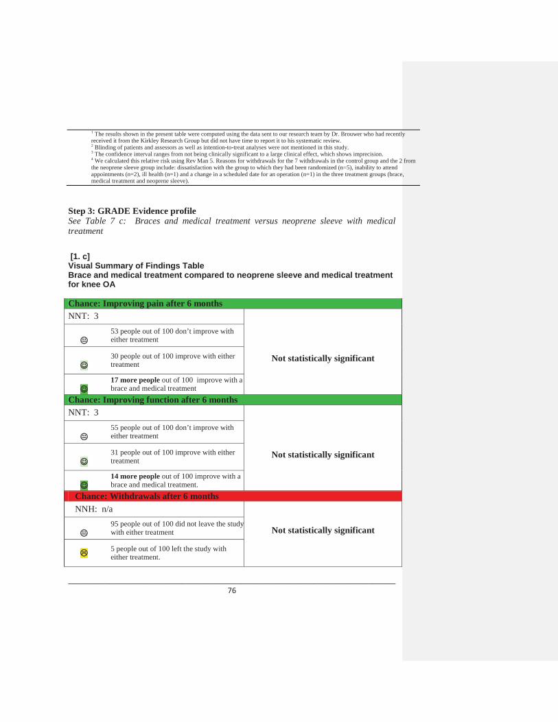
________________________________________________________________________ 76
1 The results shown in the present table were computed using the data sent to our research team by Dr. Brouwer who had recentlyreceived it from the Kirkley Research Group but did not have time to report it to his systematic review. 2 Blinding of patients and assessors as well as intention-to-treat analyses were not mentioned in this study. 3 The confidence interval ranges from not being clinically significant to a large clinical effect, which shows imprecision. 4 We calculated this relative risk using Rev Man 5. Reasons for withdrawals for the 7 withdrawals in the control group and the 2 from the neoprene sleeve group include: dissatisfaction with the group to which they had been randomized (n=5), inability to attend appointments (n=2), ill health (n=1) and a change in a scheduled date for an operation (n=1) in the three treatment groups (brace, medical treatment and neoprene sleeve).
Step 3: GRADE Evidence profile See Table 7 c: Braces and medical treatment versus neoprene sleeve with medical treatment
[1. c] Visual Summary of Findings TableBrace and medical treatment compared to neoprene sleeve and medical treatment for knee OA
Chance: Improving pain after 6 monthsNNT: 3
Not statistically significant
53 people out of 100 don’t improve with either treatment
30 people out of 100 improve with either treatment
17 more people out of 100 improve with a brace and medical treatment
Chance: Improving function after 6 monthsNNT: 3
Not statistically significant
55 people out of 100 don’t improve with either treatment
31 people out of 100 improve with either treatment
14 more people out of 100 improve with abrace and medical treatment.
Chance: Withdrawals after 6 monthsNNH: n/a
Not statistically significant95 people out of 100 did not leave the study with either treatment
5 people out of 100 left the study with either treatment.
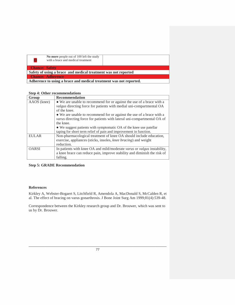
________________________________________________________________________ 77
No more people out of 100 left the study with a brace and medical treatment
Chance: SafetySafety of using a brace and medical treatment was not reportedChance: Adherence
Adherence to using a brace and medical treatment was not reported.
Step 4: Other recommendationsGroup RecommendationAAOS (knee) We are unable to recommend for or against the use of a brace with a
valgus directing force for patients with medial uni-compartmental OA of the knee.
We are unable to recommend for or against the use of a brace with a varus directing force for patients with lateral uni-compartmental OA of the knee.
We suggest patients with symptomatic OA of the knee use patellar taping for short term relief of pain and improvement in function.
EULAR Non-pharmacological treatment of knee OA should include education, exercise, appliances (sticks, insoles, knee bracing) and weight reduction.
OARSI In patients with knee OA and mild/moderate varus or valgus instability,a knee brace can reduce pain, improve stability and diminish the risk of falling.
Step 5: GRADE Recommendation
ReferencesKirkley A, Webster-Bogaert S, Litchfield R, Amendola A, MacDonald S, McCalden R, et al. The effect of bracing on varus gonarthrosis. J Bone Joint Surg Am 1999;81(4):539-48.
Correspondence between the Kirkley research group and Dr. Brouwer, which was sent to us by Dr. Brouwer.

________________________________________________________________________ 78
8. TAPING
8.1 Medially-directed patellar taping versus no taping in knee OA
Is medially-directed patellar taping effective in reducing pain and improving function in patients with symptomatic knee OA compared to no taping?
Step 1: Search ResultsWe chose the most recent SR conducted by Warden 2008 which pooled results from 2 studies on patellar taping in OA patients (Hinman, 2003 and Hinman, 2003) for pain. However, only one of these trials (published in the British Medical Journal) reported function, safety, adherence and withdrawals.
Intervention description in the RCT by Hinman 2003 published in BMJ:The trial comprised a three week intervention period and a three week follow up. Tape was applied by 12 trained physiotherapists at the university (n=4) and in private practice (n=8) around the metropolitan region. The tape was worn for three weeks and reapplied weekly. Skin was shaved before application. Therapeutic tape provided medial glide, medial tilt, and anteroposterior tilt to the patella. As inflamed soft tissue is aggravated by stretch, tape was also applied to unload either the infrapatellar fat pad or the pes anserinus (determined by clinical assessment to ascertain the most tender). Hypoallergenic undertape (Fixomull stretch; Beiersdorf, North Rhyde, NSW) was applied beneath the rigid tape (Leuko Sportstape Premium Plus; Beiersdorf) to prevent irritation of the skin. Control tape aimed to provide sensory input only. Hypoallergenic tape alone was laid over the same areas of skin as the therapeutic tape. Participants allocated to the no tape group received no intervention. All participants continued current treatments but were instructed to refrain from starting new ones.
Intervention description in the crossover study by Hinman 2003 published in Rheumatology:Therapeutic tape was applied in a standardized manner by the same investigator, regardless of clinical presentation. Skin was shaved prior to tape application. Two pieces of rigid tape (Leuko Sportstape Premium Plus, Beiersdorf Australia Ltd) applied a medial patellar glide and corrected lateral and AP tilt. Two further pieces of tape applied distal to the patella unloaded the infrapatellar fat pad. Hypoallergenic undertape (Fixomull1 stretch, Beiersdorf Australia Ltd) was applied beneath the rigid tape to prevent skin irritation. For the neutral taping condition, hypoallergenic undertape was applied over the same areas of skin as therapeutic tape, but with no force applied to realign the patella or unload soft tissues. Participants rested for 5 min between test conditions to minimize carry-over effects of tape on cutaneous sensation. The length of time the tape was worn and the timing of the outcome assessment was not reported.

________________________________________________________________________ 79
Step 2: GRADE Summary of findings
Medially-directed patellar taping compared to no taping for knee OA
Patient or population: patients with knee OAIntervention: medially-directed patellar tapingComparison: no taping
Outcomes Illustrative comparative risks* (95% CI) Absolute difference
Relative effect(95% CI)
No of Participants(studies)
Quality of the evidence(GRADE)
NNT
Assumed risk Corresponding risk
No taping Medially-directed patellar taping
Benefits
Pain1
VAS. Scale from: 0 to 100.Follow-up: 3 weeks2
40% 82% (72% to 90%)3
42% 2.05 94(2 studies4) low 5,6
3 (2 to 3)
FunctionWOMAC. Scale from: 0 to 68.Follow-up: 3 weeks
37% 52% (32% to 71%)7
15% 1.41 58(1 study8) low 9,10
Not statistically significant
Harms
Minor skin irritationsnumber of subjects presenting with minor skin irritationsFollow-up: 6 weeks
0% 28%28% 17
(1.03 to 281.5)58
(1 study11) low 9,10
6 (0 to 3333)*by estimating control risk at
1%
Withdrawalsnumber of patients who withdrew after randomizationFollow-up: 6 weeks
3% 0%(0% to 27%)
-3% 0.33 (0.01 to 7.86)
58(1 study12) low 9,10
Not statistically significant
Adherence number of participants who continued to wear the tape as prescribedFollow-up: 6 weeks
100% 100% 0% 1 58(1 study13) moderate 10
Not statistically significant
1 Two studies were pooled by the authors who reported a SMD (Hinman, 2003 and Hinman, 2003).2 One study looks at the immediate effect of taping and the other one at 3 weeks.3 This effect size was reported in the SR by Warden.4 One study was a crossover study and the other was a controlled study. 5 According to the trials, both studies did not blind subjects and therapists who administered the treatment. However, subjects were not aware of which taping technique was considered therapeutic. Furthermore, because one of the studies (published in Rheumatology) used a crossover (within subject) design, it did not ensure proper allocation concealment and comparability of group characteristics at baseline. The quality assessment reported in the SR by Warden is not consistent with the information given in the RCTs. 6 There is a publication bias indicated by significant funnel plot asymmetry in the SR. This asymmetry indicates that negative studies investigating patellar taping are less likely to be published and smaller studies are more likely to produce larger effect sizes.7 We calculated the SMD with the end of study data using RevMan.8 The SR did not report function. One study (Hinman, 2003 in BMJ) reported function at 3 weeks.9 The confidence interval ranges from not being clinically significant to a large clinical effect, which shows imprecision. 10 There is a possibility of publication bias since the funnel plot showed asymmetry in the SR. This asymmetry indicates that negative studies investigating patellar taping are less likely to be published and smaller studies are more likely to produce larger effect sizes.

________________________________________________________________________ 80
11 One study (Hinman, 2003 in BMJ) reported adverse effects. Another study by the same author (Hinman, 2003 in Rheumatology) reported an absence of adverse effects.12 One study (Hinman, 2003 in BMJ) reported withdrawals. 13 One study (Hinman, 2003 in BMJ) reported adherence.
Step 3: GRADE Evidence profile See table 8 a: Medially-directed patellar taping versus no taping in knee OA
[2 a.]Visual Summary of Findings TableMedially-directed patellar taping compared to no taping for knee OA
Chance: Improving pain after 3 weeksNNT: 3
18 people out of 100 don’t improvewhether they applied taping or not.
40 people out of 100 improve whether they applied taping or not.
42 more people out of 100 improve with taping.
Chance: Improving function after 3 weeksNNT: n/a
Not statistically significant
48 people out of 100 don’t improvewhether they applied taping or not.
37 people out of 100 improve whether they applied taping or not.
15 more people out of 100 improve with taping.
Chance: Minor skin irritation after 6 weeksNNH: 6
72 people out of 100 did not have minor skin irritation whether they applied taping or not
No one had a minor skin irritation whether they applied taping or not
28 more people out of 100 had minor skin irritation when they applied tape.
Chance: Withdrawals after 6 weeks

________________________________________________________________________ 81
NNH: n/a
Not statistically significant100 people out of 100 stayed in the study whether they applied taping or not.
3 people out of 100 left the study whether they applied taping or not.
3 fewer people out of 100 left the study when they applied tape.
Chance: Adherence after 6 weeksNNH: n/a
Not statistically significant0 people out of 100 did not adhere to the treatment whether they use applied taping or not.100 people out of 100 adhered to the treatment whether they applied taping or not.There was no difference in the number of people who adhered to the treatment.
Step 4: Other recommendations
Group RecommendationAAOS (knee) We are unable to recommend for or against the use of a brace with a
valgus directing force for patients with medial uni-compartmental OA of the knee.
We are unable to recommend for or against the use of a brace with a varus directing force for patients with lateral uni-compartmental OA of the knee.
We suggest patients with symptomatic OA of the knee use patellar taping for short term relief of pain and improvement in function.
EULAR Non-pharmacological treatment of knee OA should include education, exercise, appliances (sticks, insoles, knee bracing) and weight reduction.
OARSI In patients with knee OA and mild/moderate varus or valgus instability,a knee brace can reduce pain, improve stability and diminish the risk of falling.

________________________________________________________________________ 82
Step 5: GRADE Recommendation
ReferencesWarden SJ, Hinman RS, Watson MA, Jr., Avin KG, Bialocerkowski AE, Crossley KM. Patellar taping and bracing for the treatment of chronic knee pain: a systematic review and meta-analysis. Arthritis Rheum 2008;59(1):73-83.
Hinman RS, Crossley KM, McConnell J, Bennell KL. Efficacy of knee tape in the management of osteoarthritis of the knee: blinded randomised controlled trial. BMJ 2003;327(7407):135.
Hinman RS, Bennell KL, Crossley KM, McConnell J. Immediate effects of adhesive tape on pain and disability in individuals with knee osteoarthritis. Rheumatology (Oxford) 2003;42(7):865-9.
8.2 Medially-directed patellar taping versus sham taping in knee OA
Is medially-directed patellar taping effective in reducing pain and improving function in patients with symptomatic knee OA compared to sham taping?
Step 1: Search ResultsWe chose the most recent SR conducted by Warden 2008 which pooled results from 3 studies on patellar taping in OA patients (Cushnaghan, 1994, Hinman, 2003 and Hinman, 2003) for pain.
Intervention description in the RCT by Hinman 2003 published in BMJ:
The trial comprised a three week intervention period and a three week follow up. Tape was applied by 12 trained physiotherapists at the university (n=4) and in private practice (n=8) around the metropolitan region. The tape was worn for three weeks and reapplied weekly. Skin was shaved before application. Therapeutic tape provided medial glide, medial tilt, and anteroposterior tilt to the patella. As inflamed soft tissue is aggravated by stretch, tape was also applied to unload either the infrapatellar fat pad or the pes anserinus (determined by clinical assessment to ascertain the most tender). Hypoallergenic undertape (Fixomull stretch; Beiersdorf, North Rhyde, NSW) was applied beneath the rigid tape (Leuko Sportstape Premium Plus; Beiersdorf) to prevent irritation of the skin. Control tape aimed to provide sensory input only. Hypoallergenic tape alone was laid over the same areas of skin as the therapeutic tape. Participants allocated to the no tape

________________________________________________________________________ 83
group received no intervention. All participants continued current treatments but were instructed to refrain from starting new ones.
Intervention description in the crossover study by Hinman 2003 published in Rheumatology:Therapeutic tape was applied in a standardized manner by the same investigator, regardless of clinical presentation. Skin was shaved prior to tape application. Two pieces of rigid tape (Leuko Sportstape Premium Plus, Beiersdorf Australia Ltd) applied a medial patellar glide and corrected lateral and AP tilt. Two further pieces of tape applied distal to the patella unloaded the infrapatellar fat pad. Hypoallergenic undertape (Fixomull1stretch, Beiersdorf Australia Ltd) was applied beneath the rigid tape to prevent skin irritation. For the neutral taping condition, hypoallergenic undertape was applied over the same areas of skin as therapeutic tape, but with no force applied to realign the patella or unload soft tissues. Participants rested for 5 min between test conditions to minimize carry-over effects of tape on cutaneous sensation. The length of time the tape was worn and the timing of the outcome assessment was not reported.
Intervention description of the crossover study by Cushnaghan, 1994 : The three types of taping were: neutral, in which the tape was applied directly over the front of the patella, without any pressure; medial, in which the tape pulled the patella to the medial side of the knee joint; and lateral, in which the tape was used to pull the patella to the lateral side. The taping consisted of a strip of Leukotape P (Beiersdorf, UK) applied by the same person in each case. Each tape was applied for four days, with three days of no treatment between tape positions.Step 2: GRADE Summary of findings
Medially-directed patellar taping compared to sham taping for knee OA
Patient or population: patients with knee OAIntervention: medially-directed patellar tapingComparison: sham taping
Outcomes Illustrative comparative risks* (95% CI)
Absolute difference
Relative effect(95% CI)
No of Participants(studies)
Quality of the evidence(GRADE)
NNT
Assumed risk
Corresponding risk
sham taping
medially-directed patellar taping
Benefits
Pain1
VAS. Scale from: 0 to 100.Follow-up: 3 weeks2
41% 68% (52% to 81%)3
27% 1.66 122(3 studies4) low 5,6 4 (3 to 8)
FunctionWOMAC. Scale from: 0 to 38% 37%
(19% to 57%)-1% 0.97 58
(1 study7) low 8, 9
Not statistically significant

________________________________________________________________________ 84
68.Follow-up: 3 weeks
Harms
Minor skin irritationsnumber of subjects presenting with minor skin irritationsFollow-up: 3 weeks
3% 27% (4% to 100%)
24% 8(1.07 to 59.95)
58(1 study10) low 8,9 36 (1 to 476)
Adherencenumber of participants who continued to wear the tape as prescribedFollow-up: 6 weeks
100% 100% 0%
1 58(1 study11) moderate 9
Not statistically significant
Withdrawalsnumber of participants who withdrew after randomizationFollow-up: 6 weeks
0% 0% 0% 1 58(1 study11) moderate 9
Not statistically significant
1 Three studies were pooled by the systematic review authors who reported a SMD (Hinman, 2003, Hinman, 2003 and Cushnagan, 1994).2 Studies looked at the immediate effect of taping as well as the effect after 4 days and after 3 weeks of intervention. 3 This effect size was reported in the SR by Warden. 4 Two were crossover studies and one was an RCT. 5 According to the trials, all studies did not blind subjects and therapists who administered the treatment. However, subjects were not aware of which taping technique was considered therapeutic. Furthermore, because the two other studies used a crossover (within subject) design, it did not ensure proper allocation concealment and comparability of group characteristics at baseline. The quality assessment reported in the SR by Warden is not consistent with the information given in the RCTs. 6 There is a publication bias indicated by significant funnel plot asymmetry. This asymmetry indicates that negative studies investigating patellar taping are less likely to be published and smaller studies are more likely to produce larger effect sizes. 7 The SR did not report function. One study (Hinman, 2003 in BMJ) reported function at 3 weeks. investigating patellar taping are less likely to be published and smaller studies are more likely to produce larger effect sizes. 8 The confidence interval ranges from not being clinically significant to a large clinical effect, which shows imprecision. 9 There is a possibility of publication bias since the funnel plot showed asymmetry in the SR. This asymmetry indicates that negative studies investigating patellar taping are less likely to be published and smaller studies are more likely to produce larger effect sizes. 10 One study (Hinman, 2003 in BMJ) reported adverse effects. The other studies reported an absence of adverse effects. 11 One study (Hinman, 2003 in BMJ) reported adherence to the treatment regimen and withdrawals. Cushnagan also reported that all patients followed prescribe taping. [2 b.]

________________________________________________________________________ 85
Visual Summary of Findings TableMedially-directed patellar taping compared to sham taping for knee OA
Chance: Improving pain after 3 weeksNNT: 4
32 people out of 100 don’t improve nomatter which type of taping was used.
41 people out of 100 improve no matter which type of taping was used.
27 more people out of 100 improve with medially-directed patellar taping.
Chance: Improving function after 3 weeksNNT: n/a
Not statistically significant
63 people out of 100 don’t improve nomatter which type of taping was used.
37 people out of 100 improve no matter which type of taping was used.
1 fewer person out of 100 improve with medially-directed patellar taping.
Chance: Minor skin irritation after 3 weeksNNH: 36
73 people out of 100 did not have minor skin irritation with either type of taping.
3 people out of 100 had minor skin irritation with either type of taping.
24 more people out of 100 had minor skin irritation with medially-directed patellar taping.
Chance: Adherence after 6 weeksNNH: n/a
Not statistically significant100 people out of 100 adhered to the treatment with either type of taping.
No one did not adhere to the treatment with either type of taping.

________________________________________________________________________ 86
There was no difference in the number of people who adhered to either type of taping.
Chance: WithdrawalsNNH: n/a
Not statistically significant100 people out of 100 remained in the study with either type of taping.
No one left the study with either type of taping
There was no difference in the number of people who left the study with either type of taping.
Step 3: GRADE Evidence profile
Table 8 b: Medially-directed patellar taping versus sham taping in knee OA
Step 4: Other recommendationsGroup RecommendationAAOS (knee) We are unable to recommend for or against the use of a brace
with a valgus directing force for patients with medial uni-compartmental OA of the knee.
We are unable to recommend for or against the use of a brace with a varus directing force for patients with lateral uni-compartmental OA of the knee.
We suggest patients with symptomatic OA of the knee use patellar taping for short term relief of pain and improvement in function.
EULAR Non-pharmacological treatment of knee OA should include education, exercise, appliances (sticks, insoles, knee bracing) and weight reduction.
OARSI In patients with knee OA and mild/moderate varus or valgusinstability, a knee brace can reduce pain, improve stability and diminish the risk of falling.
Step 5: GRADE Recommendation

________________________________________________________________________ 87
References
Warden SJ, Hinman RS, Watson MA, Jr., Avin KG, Bialocerkowski AE, Crossley KM. Patellar taping and bracing for the treatment of chronic knee pain: a systematic review and meta-analysis. Arthritis Rheum 2008;59(1):73-83.
Hinman RS, Crossley KM, McConnell J, Bennell KL. Efficacy of knee tape in the management of osteoarthritis of the knee: blinded randomised controlled trial. BMJ 2003;327(7407):135.
Hinman RS, Bennell KL, Crossley KM, McConnell J. Immediate effects of adhesive tape on pain and disability in individuals with knee osteoarthritis. Rheumatology (Oxford) 2003;42(7):865-9.
Cushnaghan J, McCarthy C, Dieppe P. Taping the patella medially: a new treatment for osteoarthritis of the knee joint? BMJ 1994;308:753–5.
8.3 Laterally-directed patellar taping versus medially-directed patellar taping in knee OA
Is laterally-directed patellar taping effective in reducing pain and improving function in patients with symptomatic knee OA compared to medially-directed patellar taping?
Step 1: Search ResultsWe chose the most recent SR conducted by Warden 2008 which reported one study comparing lateral to medial patellar taping in OA patients for pain (Cushnaghan, 1994). Intervention description: The three types of taping in the Cushnaghan study were: neutral, in which the tape was applied directly over the front of the patella, without any pressure; medial, in which the tape pulled the patella to the medial side of the knee joint; and lateral, in which the tape was used to pull the patella to the lateral side. The taping consisted of a strip of Leukotape P (Beiersdorf, UK) applied by the same person in each case. Each tape was applied for four days, with three days of no treatment between tape positions.

________________________________________________________________________ 88
Step 2: GRADE Summary of findingsLaterally-directed patellar taping compared to medially-directed patellar taping for knee OA
Patient or population: patients with knee OAIntervention: laterally-directed patellar tapingComparison: medially-directed patellar taping
Outcomes Illustrative comparative risks* (95% CI)
Absolute difference
Relative effect (95% CI)
No of Participants(studies)
Quality of the evidence(GRADE)
NNT
Assumed risk Corresponding risk
medially-directed patellar taping
laterally-directed patellar taping
Benefits
PainVAS. Scale from: 0 to 100.Follow-up: 4 days
*Not estimable due to lack of data
SMD 0.95 (0.42 to 1.48) 1
* 28(1 study2)
low 3,4*
Function Not reported
Harms
Safety number of patients who reported adverse events follow-up: 4 days
0% 0% 0% 1 28(1 study2) low 3,4
Not statistically significant
Adherence number of patients who wore tapes on for the full four days follow-up: 4 days
100% 100% 0% 1 28(1 study2) low 3,4
Not statistically significant
Withdrawals number of patients who withdrew after entry to the study follow-up: 4 days
0% 0% 0% 1 28(1 study2) low 3,4
Not statistically significant
1 The SR by Warden reported an SMD for pain comparing lateral and medial taping based on the Cushnagan, 1994 study.2 This study has a crossover design with 14 participants. 3 This study did not blind therapists who administered the treatment and it is unclear if patients were blinded. However, subjects were not aware of which taping technique was considered therapeutic. Also, because this study used a crossover (within subject) design, it did not ensure proper allocation concealment and comparability of group characteristics at baseline. 4 There is a possibility of publication bias since the funnel plot showed asymmetry in the SR. This asymmetry indicates that negative studies investigating patellar taping are less likely to be published and smaller studies are more likely to produce larger effect sizes.

________________________________________________________________________ 89
[2 c.]Visual Summary of Findings TableLaterally-directed patellar taping compared to medially-directed patellar taping for knee OA
Chance: Improving pain after 4 daysThe improvement in pain was not estimable due to lack of data.Chance: Improving function after 4 days
The improvement in function was not reported.Chance: SafetyNNH: n/a
Not statistically significant
100 people out of 100 did not have adverse events with either type of taping
No one had adverse events with either type of taping
There was no difference in the safety of the two types of taping.
Chance: Adherence after 4 daysNNH: n/a
Not statistically significant100 people out of 100 adhered to either type of taping
No one did not adhere to the treatment with either type of taping
There was no difference in the number of people who adhered to either type of taping.
Chance: Withdrawals after 4 daysNNH: n/a
Not statistically significant100 people out of 100 remained in the study with either type of taping.
No one left the study with either type of taping
There was no difference in the number of people who left the study with either type of taping.
Step 3: GRADE Evidence profile See Table 8c: Laterally-directed patellar taping versus medially-directed patellar taping in knee OA

________________________________________________________________________ 90
Step 4: Other recommendationsGroup RecommendationAAOS (knee) We are unable to recommend for or against the use of a brace with
a valgus directing force for patients with medial uni-compartmental OA of the knee.
We are unable to recommend for or against the use of a brace with a varus directing force for patients with lateral uni-compartmental OA of the knee.
We suggest patients with symptomatic OA of the knee use patellar taping for short term relief of pain and improvement in function.
EULAR Non-pharmacological treatment of knee OA should include education, exercise, appliances (sticks, insoles, knee bracing) and weight reduction.
OARSI In patients with knee OA and mild/moderate varus or valgusinstability, a knee brace can reduce pain, improve stability and diminish the risk of falling.
Step 5: GRADE Recommendation
ReferencesWarden SJ, Hinman RS, Watson MA, Jr., Avin KG, Bialocerkowski AE, Crossley KM. Patellar taping and bracing for the treatment of chronic knee pain: a systematic review and meta-analysis. Arthritis Rheum 2008;59(1):73-83.
Cushnaghan J, McCarthy C, Dieppe P. Taping the patella medially: a new treatment for osteoarthritis of the knee joint? BMJ 1994;308:753–5.

________________________________________________________________________ 91
8.4 Laterally-directed patellar taping versus neutral sham taping in knee OA
Is laterally-directed patellar taping effective in reducing pain and improving function in patients with symptomatic knee OA compared to sham taping?
Step 1: Search ResultsWe chose the most recent SR conducted by Warden 2008 which reported one study comparing lateral patellar taping to neutral sham taping in OA patients for pain (Cushnaghan, 1994).
Intervention description: The three types of taping in the Cushnaghan study were: neutral, in which the tape was applied directly over the front of the patella, without any pressure; medial, in which the tape pulled the patella to the medial side of the knee joint; and lateral, in which the tape was used to pull the patella to the lateral side. The taping consisted of a strip of Leukotape P (Beiersdorf, UK) applied by the same person in each case. Each tape was applied for four days, with three days of no treatment between tape positions.
Step 2: GRADE Summary of findingsLaterally-directed patellar taping compared to neutral sham taping for knee OA
Patient or population: patients with knee OAIntervention: laterally-directed patellar tapingComparison: neutral sham taping
Outcomes Illustrative comparative risks* (95% CI)
Absolute difference
Relative effect(95% CI)
No of Participants(studies)
Quality of the evidence(GRADE)
NNT
Assumed risk
Corresponding risk
neutral sham taping
laterally-directed patellar taping
Benefits
Pain1
VAS. Scale from: 0 to 100.Follow-up: 4 days
35% 33% (17% to 54%)
-2%0.94 28
(1 study2) very low 3,4,5
Not statistically significant
Function Not reported
Harms
Safety number of patients who reported adverse events
0% 0% 0% 1 28(1 study2) low 3,5
Not statistically significant

________________________________________________________________________ 92
follow-up: 4 days
Adherence number of patients who wore tapes on for the full four days follow-up: 4 days
100% 100% 0% 1 28(1 study2) low 3,5
Not statistically significant
Withdrawals number of patients who withdrew after entry to the study follow-up: 4 days
0% 0% 0% 1 28(1 study2) low 3,4
Not statistically significant
1 The SR by Warden reported an SMD for pain comparing lateral and neutral taping based on the Cushnagan, 1994 study.2 This study has a crossover design with 14 participants. 3 This study did not blind therapists who administered the treatment and it is unclear if patients were blinded. Also, because this study used a crossover (within subject) design, it did not ensure proper allocation concealment and comparability of group characteristics at baseline. 4 The confidence interval ranges from not being clinically significant to a large clinical effect, which shows imprecision. 5 There is a possibility of publication bias since the funnel plot showed asymmetry in the SR. This asymmetry indicates that negative studies investigating patellar taping are less likely to be published and smaller studies are more likely to produce larger effect sizes.
[2 d.]Visual Summary of Findings TableLaterally-directed patellar taping compared to neutral sham taping for knee OA
Chance: Improving pain after 4 daysNNT: n/a
Not statistically significant
67 people out of 100 don’t improve with either type of taping
33 people out of 100 improve with either type of taping
2 fewer people out of 100 improve with laterally-directed patellar taping.
Chance: Improving function after 4 daysThe improvement in function was not reported.
Chance: Safety after 4 daysNNH: n/a Not statistically significant
100 people out of 100 did not report adverse effects with either type of taping.
0 people out of 100 reported adverse effects with either type of taping
There was no difference in the safety of the two types of taping.
Chance: Adherence after 4 daysNNH: n/a
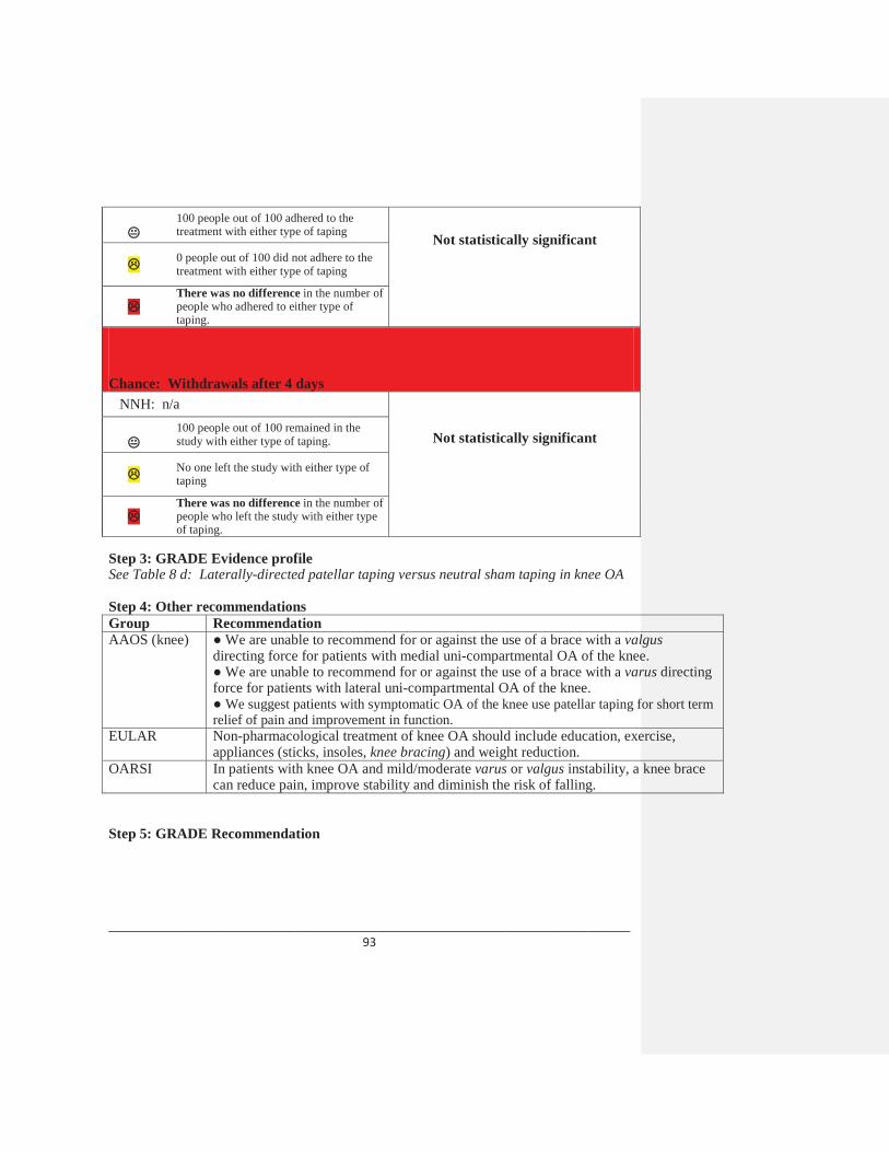
________________________________________________________________________ 93
100 people out of 100 adhered to the treatment with either type of taping Not statistically significant0 people out of 100 did not adhere to the treatment with either type of taping
There was no difference in the number of people who adhered to either type of taping.
Chance: Withdrawals after 4 daysNNH: n/a
Not statistically significant100 people out of 100 remained in the study with either type of taping.
No one left the study with either type of taping
There was no difference in the number of people who left the study with either type of taping.
Step 3: GRADE Evidence profile See Table 8 d: Laterally-directed patellar taping versus neutral sham taping in knee OA
Step 4: Other recommendationsGroup RecommendationAAOS (knee) We are unable to recommend for or against the use of a brace with a valgus
directing force for patients with medial uni-compartmental OA of the knee.We are unable to recommend for or against the use of a brace with a varus directing
force for patients with lateral uni-compartmental OA of the knee.We suggest patients with symptomatic OA of the knee use patellar taping for short term
relief of pain and improvement in function.EULAR Non-pharmacological treatment of knee OA should include education, exercise,
appliances (sticks, insoles, knee bracing) and weight reduction.OARSI In patients with knee OA and mild/moderate varus or valgus instability, a knee brace
can reduce pain, improve stability and diminish the risk of falling.
Step 5: GRADE Recommendation

________________________________________________________________________ 94
ReferencesWarden SJ, Hinman RS, Watson MA, Jr., Avin KG, Bialocerkowski AE, Crossley KM. Patellar taping and bracing for the treatment of chronic knee pain: a systematic review and meta-analysis. Arthritis Rheum 2008;59(1):73-83.
Cushnaghan J, McCarthy C, Dieppe P. Taping the patella medially: a new treatment for osteoarthritis of the knee joint? BMJ 1994;308:753–5.
________________________________________________________________________ 95
ABBREVIATIONS
OA - osteoarthritis
RCT – randomized controlled trial
SR – SR

________________________________________________________________________ 96
GRADE evidence profiles
Table 1 a: Home-based balance exercises versus home-based strengthening exercises for knee OA
Author(s): Karine Toupin AprilDate: 2009-06-12Question: Should balance training versus strength training be used for knee OA?Bibliography: Chaipinyo, 2009
Quality assessmentSummary of findings
ImportanceNo of patients Effect
QualityNo of studies Design Limitations Inconsistency Indirectness Imprecision Other
considerationsbalance training
strength training
Relative(95% CI)
Absolute
pain (follow-up 4 weeks; measured with: Knee injury and Osteoarthritis Outcome Score (KOOS); range of scores: 0-100; Better indicated by higher values)1 randomised
trialsserious1 no serious
inconsistencyno serious indirectness2
serious3 None
24 18 0.73
SMD -0.23
(-0.85 to 0.38)4
LOW CRITICAL
function in daily living (follow-up 4 weeks; measured with: Knee injury and Osteoarthritis Outcome Score (KOOS); range of scores: 0-100; Better indicated by higher values)1 randomised
trialsserious1 no serious
inconsistencyno seriousindirectness2
serious3 None24 18 0.54
SMD -0.45
(-1.07 to 0.17)4
LOW CRITICAL
Adherence (follow-up 4 weeks; Maximum number of days:28; measured with: average number of days of exercise performed by participants Better indicated by higher values)1 randomised
trialsserious1 no serious
inconsistencyno serious indirectness2
serious3 None
24 18 -
MD 2
(-0.77 to 4.77)
LOW CRITICAL
Withdrawals1 randomised
trialsserious1 no serious
inconsistencyno serious indirectness2
serious3 None
0/240%
6/24 (25%)
0.08 (0.00 to
1.29)
23 fewer per 100
(from 25 fewer to 7 more)5
LOW CRITICAL
SafetyNot reported
1 The physiotherapists prescribing the exercises were not blinded to group allocation. We did not downgrade the quality assessment score for this. However, the number of patients in this trial is small (n=42), which could undermine its validity.2 Participants were volunteers from the community 50 years and older. We did not downgrade the quality assessment score for this. 3 The confidence interval ranges from not being clinically significant to a large clinical effect, which shows imprecision. 4 The authors report the mean difference over time between groups but it does not coincide with our results using Rev Man 5 because the authors did not report the level of accuracy needed (no decimals reported). We calculated the SMD using Rev Man 5.
5 Withdrawals were due to other illnesses, personal reasons or impossibility to reach patients.
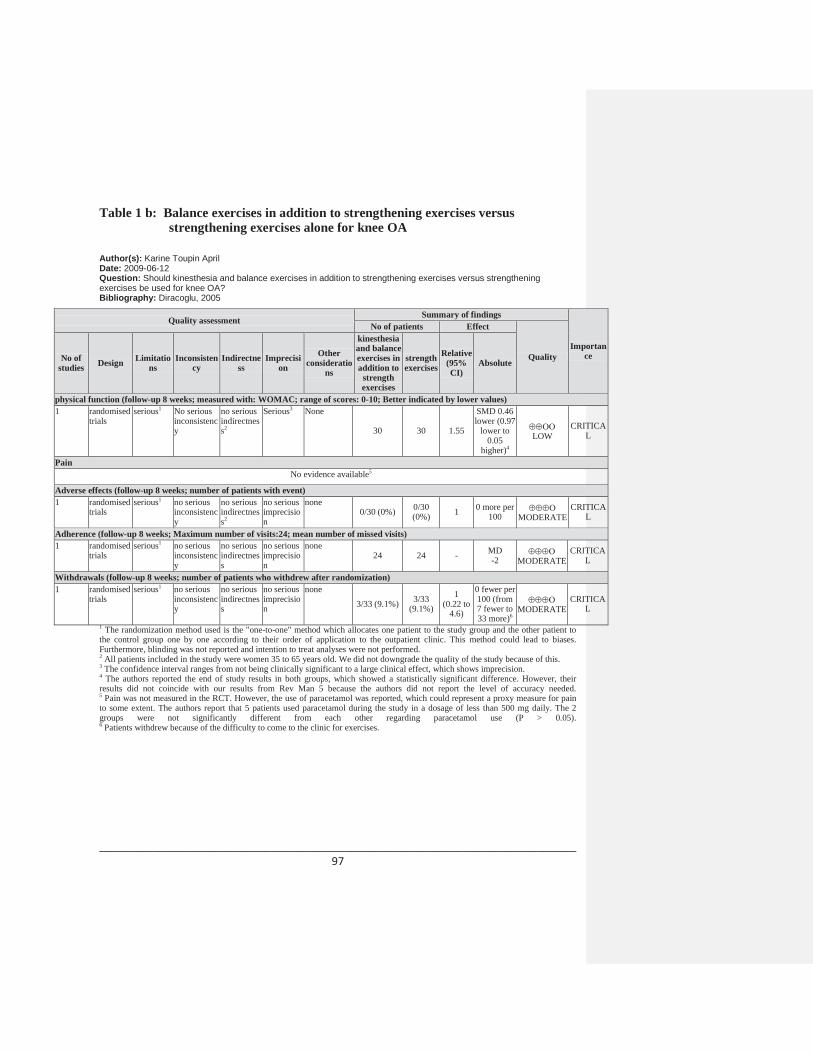
________________________________________________________________________ 97
Table 1 b: Balance exercises in addition to strengthening exercises versus strengthening exercises alone for knee OA
Author(s): Karine Toupin AprilDate: 2009-06-12Question: Should kinesthesia and balance exercises in addition to strengthening exercises versus strengthening exercises be used for knee OA?Bibliography: Diracoglu, 2005
Quality assessmentSummary of findings
Importance
No of patients Effect
QualityNo of studies Design Limitatio
nsInconsisten
cyIndirectne
ssImprecisi
on
Other consideratio
ns
kinesthesia and balance exercises in addition to
strengthexercises
strength exercises
Relative(95% CI)
Absolute
physical function (follow-up 8 weeks; measured with: WOMAC; range of scores: 0-10; Better indicated by lower values)1 randomised
trialsserious1 No serious
inconsistency
no serious indirectness2
Serious3 None
30 30 1.55
SMD 0.46 lower (0.97
lower to 0.05
higher)4
LOWCRITICA
L
PainNo evidence available5
Adverse effects (follow-up 8 weeks; number of patients with event)1 randomised
trialsserious1 no serious
inconsistency
no serious indirectness2
no serious imprecision
none0/30 (0%) 0/30
(0%) 1 0 more per100 MODERATE
CRITICAL
Adherence (follow-up 8 weeks; Maximum number of visits:24; mean number of missed visits)1 randomised
trialsserious1 no serious
inconsistency
no serious indirectness
no serious imprecision
none24 24 - MD
-2 MODERATECRITICA
L
Withdrawals (follow-up 8 weeks; number of patients who withdrew after randomization)1 randomised
trialsserious1 no serious
inconsistency
no serious indirectness
no serious imprecision
none
3/33 (9.1%) 3/33 (9.1%)
1 (0.22 to
4.6)
0 fewer per 100 (from 7 fewer to 33 more)6
MODERATECRITICA
L
1 The randomization method used is the "one-to-one" method which allocates one patient to the study group and the other patient to the control group one by one according to their order of application to the outpatient clinic. This method could lead to biases. Furthermore, blinding was not reported and intention to treat analyses were not performed. 2 All patients included in the study were women 35 to 65 years old. We did not downgrade the quality of the study because of this.3 The confidence interval ranges from not being clinically significant to a large clinical effect, which shows imprecision. 4 The authors reported the end of study results in both groups, which showed a statistically significant difference. However, their results did not coincide with our results from Rev Man 5 because the authors did not report the level of accuracy needed. 5 Pain was not measured in the RCT. However, the use of paracetamol was reported, which could represent a proxy measure for pain to some extent. The authors report that 5 patients used paracetamol during the study in a dosage of less than 500 mg daily. The 2 groups were not significantly different from each other regarding paracetamol use (P > 0.05).
6 Patients withdrew because of the difficulty to come to the clinic for exercises.
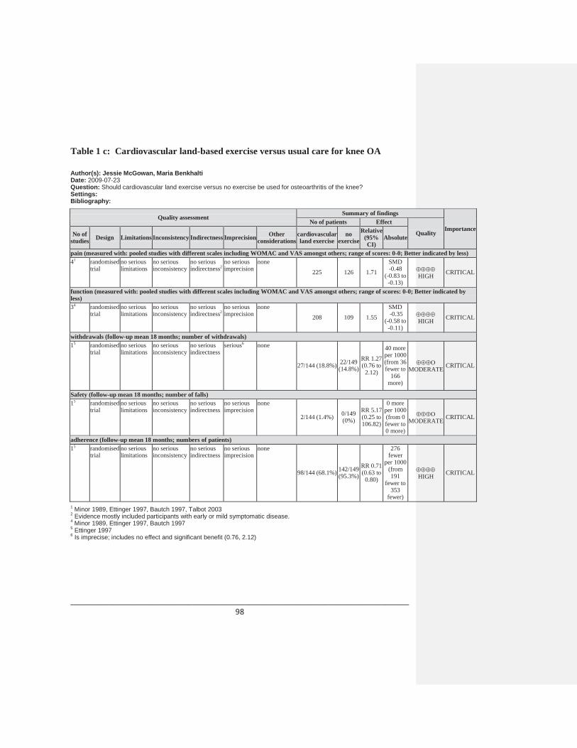
________________________________________________________________________ 98
Table 1 c: Cardiovascular land-based exercise versus usual care for knee OA
Author(s): Jessie McGowan, Maria Benkhalti Date: 2009-07-23Question: Should cardiovascular land exercise versus no exercise be used for osteoarthritis of the knee?Settings: Bibliography:
Quality assessmentSummary of findings
ImportanceNo of patients Effect
QualityNo of studies Design Limitations Inconsistency Indirectness Imprecision Other
considerationscardiovascular land exercise
no exercise
Relative(95% CI)
Absolute
pain (measured with: pooled studies with different scales including WOMAC and VAS amongst others; range of scores: 0-0; Better indicated by less)41 randomised
trialno serious limitations
no serious inconsistency
no serious indirectness2
no serious imprecision
none
225 126 1.71
SMD -0.48
(-0.83 to -0.13)
HIGH CRITICAL
function (measured with: pooled studies with different scales including WOMAC and VAS amongst others; range of scores: 0-0; Better indicated by less)34 randomised
trialno serious limitations
no serious inconsistency
no serious indirectness2
no serious imprecision
none
208 109 1.55
SMD-0.35
(-0.58 to -0.11)
HIGH CRITICAL
withdrawals (follow-up mean 18 months; number of withdrawals)15 randomised
trialno serious limitations
no serious inconsistency
no serious indirectness
serious6 none
27/144 (18.8%) 22/149 (14.8%)
RR 1.27 (0.76 to
2.12)
40 more per 1000 (from 36 fewer to
166 more)
MODERATE CRITICAL
Safety (follow-up mean 18 months; number of falls)15 randomised
trialno serious limitations
no serious inconsistency
no serious indirectness
no serious imprecision
none
2/144 (1.4%) 0/149 (0%)
RR 5.17 (0.25 to 106.82)
0 more per 1000 (from 0 fewer to 0 more)
MODERATE CRITICAL
adherence (follow-up mean 18 months; numbers of patients)15 randomised
trialno serious limitations
no serious inconsistency
no serious indirectness
no serious imprecision
none
98/144 (68.1%) 142/149 (95.3%)
RR 0.71 (0.63 to
0.80)
276 fewer
per 1000 (from 191
fewer to 353
fewer)
HIGH CRITICAL
1 Minor 1989, Ettinger 1997, Bautch 1997, Talbot 20032 Evidence mostly included participants with early or mild symptomatic disease.4 Minor 1989, Ettinger 1997, Bautch 19975 Ettinger 19976 Is imprecise; includes no effect and significant benefit (0.76, 2.12)
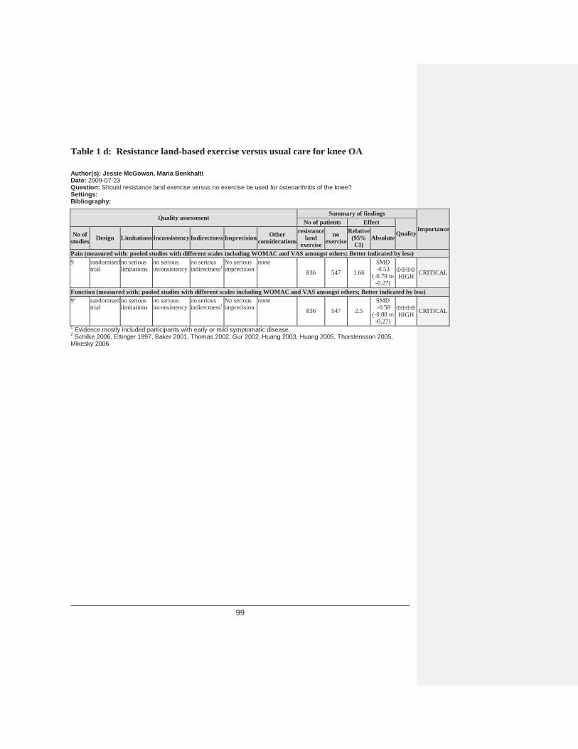
________________________________________________________________________ 99
Table 1 d: Resistance land-based exercise versus usual care for knee OA
Author(s): Jessie McGowan, Maria Benkhalti Date: 2009-07-23Question: Should resistance land exercise versus no exercise be used for osteoarthritis of the knee?Settings: Bibliography:
Quality assessmentSummary of findings
ImportanceNo of patients Effect
QualityNo of studies Design Limitations Inconsistency Indirectness Imprecision Other
considerations
resistance land
exercise
no exercise
Relative(95% CI)
Absolute
Pain (measured with: pooled studies with different scales including WOMAC and VAS amongst others; Better indicated by less)9 randomised
trialno serious limitations
no serious inconsistency
no serious indirectness1
No seriousimprecision
none
836 547 1.66
SMD -0.53
(-0.79 to -0.27)
HIGH CRITICAL
Function (measured with: pooled studies with different scales including WOMAC and VAS amongst others; Better indicated by less)92 randomised
trialno serious limitations
no serious inconsistency
no serious indirectness1
No seriousimprecision
none836 547 2.5
SMD-0.58
(-0.88 to -0.27)
HIGH CRITICAL
1 Evidence mostly included participants with early or mild symptomatic disease.2 Schilke 2006, Ettinger 1997, Baker 2001, Thomas 2002, Gur 2002, Huang 2003, Huang 2005, Thorstensson 2005, Mikesky 2006
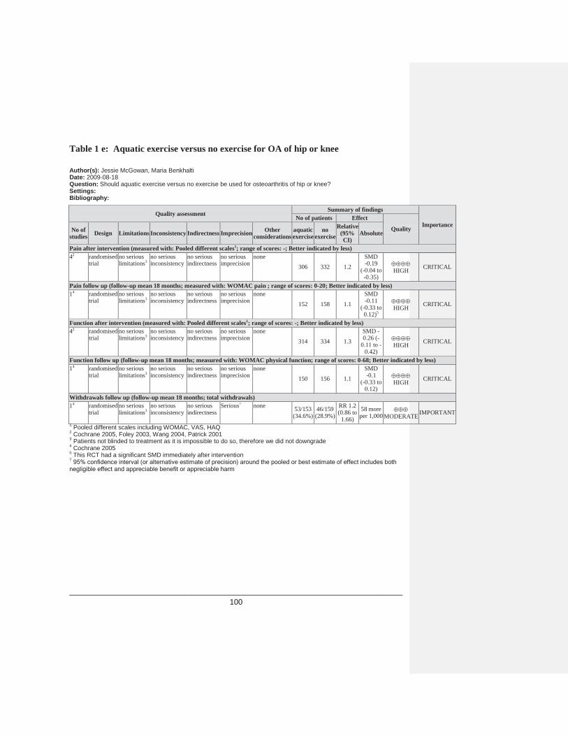
________________________________________________________________________ 100
Table 1 e: Aquatic exercise versus no exercise for OA of hip or knee
Author(s): Jessie McGowan, Maria BenkhaltiDate: 2009-08-18Question: Should aquatic exercise versus no exercise be used for osteoarthritis of hip or knee?Settings: Bibliography:
Quality assessmentSummary of findings
ImportanceNo of patients Effect
QualityNo of studies Design Limitations Inconsistency Indirectness Imprecision Other
considerationsaquatic exercise
no exercise
Relative(95% CI)
Absolute
Pain after intervention (measured with: Pooled different scales1; range of scores: -; Better indicated by less)42 randomised
trialno serious limitations3
no serious inconsistency
no serious indirectness
no serious imprecision
none306 332 1.2
SMD -0.19
(-0.04 to -0.35)
HIGH CRITICAL
Pain follow up (follow-up mean 18 months; measured with: WOMAC pain ; range of scores: 0-20; Better indicated by less)14 randomised
trialno serious limitations3
no serious inconsistency
no serious indirectness
no serious imprecision
none
152 158 1.1
SMD -0.11
(-0.33 to 0.12)5
HIGH CRITICAL
Function after intervention (measured with: Pooled different scales1; range of scores: -; Better indicated by less)42 randomised
trialno serious limitations3
no serious inconsistency
no serious indirectness
no serious imprecision
none
314 334 1.3
SMD -0.26 (-
0.11 to -0.42)
HIGH CRITICAL
Function follow up (follow-up mean 18 months; measured with: WOMAC physical function; range of scores: 0-68; Better indicated by less)14 randomised
trialno serious limitations3
no serious inconsistency
no serious indirectness
no serious imprecision
none
150 156 1.1
SMD -0.1
(-0.33 to 0.12)
HIGH CRITICAL
Withdrawals follow up (follow-up mean 18 months; total withdrawals)14 randomised
trialno serious limitations3
no serious inconsistency
no serious indirectness
Serious7 none 53/153 (34.6%)
46/159 (28.9%)
RR 1.2(0.86 to
1.66)
58 more per 1,000 MODERATE IMPORTANT
1 Pooled different scales including WOMAC, VAS, HAQ2 Cochrane 2005, Foley 2003, Wang 2004, Patrick 20013 Patients not blinded to treatment as it is impossible to do so, therefore we did not downgrade4 Cochrane 20055 This RCT had a significant SMD immediately after intervention7 95% confidence interval (or alternative estimate of precision) around the pooled or best estimate of effect includes both negligible effect and appreciable benefit or appreciable harm
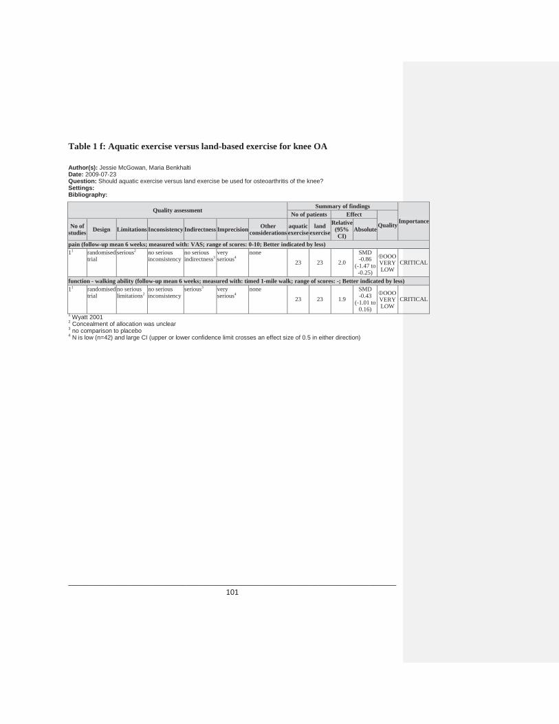
________________________________________________________________________ 101
Table 1 f: Aquatic exercise versus land-based exercise for knee OA
Author(s): Jessie McGowan, Maria BenkhaltiDate: 2009-07-23Question: Should aquatic exercise versus land exercise be used for osteoarthritis of the knee?Settings: Bibliography:
Quality assessmentSummary of findings
ImportanceNo of patients Effect
QualityNo of studies Design Limitations Inconsistency Indirectness Imprecision Other
considerationsaquatic exercise
land exercise
Relative(95% CI)
Absolute
pain (follow-up mean 6 weeks; measured with: VAS; range of scores: 0-10; Better indicated by less)11 randomised
trialserious2 no serious
inconsistencyno serious indirectness3
very serious4
none23 23 2.0
SMD -0.86
(-1.47 to -0.25)
VERY LOW
CRITICAL
function - walking ability (follow-up mean 6 weeks; measured with: timed 1-mile walk; range of scores: -; Better indicated by less)11 randomised
trialno serious limitations2
no serious inconsistency
serious3 very serious4
none
23 23 1.9
SMD -0.43
(-1.01 to 0.16)
VERY LOW
CRITICAL
1 Wyatt 20012 Concealment of allocation was unclear3 no comparison to placebo4 N is low (n=42) and large CI (upper or lower confidence limit crosses an effect size of 0.5 in either direction)
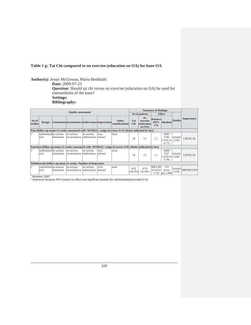
________________________________________________________________________ 102
Table 1 g: Tai Chi compared to no exercise (education on OA) for knee OA
Author(s): Jessie McGowan, Maria BenkhaltiDate: 2009-07-23Question: Should tai chi versus no exercise (education on OA) be used for osteoarthritis of the knee?Settings: Bibliography:
Quality assessmentSummary of findings
Importance
No of patients Effect
QualityNo of studies Design Limitations Inconsistency Indirectness Imprecision Other
considerationsTai Chi
no exercise
(educationon OA)
Relative(95% CI)
Absolute
Pain (follow-up mean 12 weeks; measured with: WOMAC; range of scores: 0-35; Better indicated by less)11 randomised
trialno serious limitations
no serious inconsistency
no serious indirectness
very serious2
none
18 13 1.1
SMD 0.06
(-0.65 to 0.77)
LOW CRITICAL
Function (follow-up mean 12 weeks; measured with: WOMAC; range of scores: 0-85; Better indicated by less)11 randomised
trialno serious limitations
no serious inconsistency
no serious indirectness
very serious2
none18 13 1.1
SMD 0.07
(-0.65 to 0.78)
LOW CRITICAL
Withdrawals (follow-up mean 12 weeks; Number of drop-outs)11 randomised
trialno serious limitations
no serious inconsistency
no serious indirectness
Very serious2
none 4/22 (18.2%)
6/19 (31.6%)
RR 0.58(0.19 to
1.74)
133fewer
per 1,000 LOW IMPORTANT
1 Brismee, 20072 Imprecise because RR crosses no effect and significant benefit (for withdrawals)and small N=31
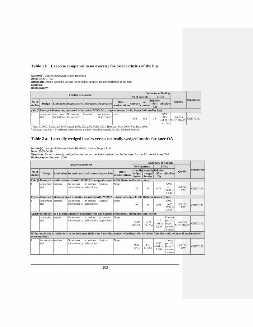
________________________________________________________________________ 103
Table 1 h: Exercise compared to no exercise for osteoarthritis of the hip
Author(s): Jessie McGowan, Maria BenkhaltiDate: 2009-07-23Question: Should exercise versus no exercise be used for osteoarthritis of the hip?Settings: Bibliography:
Quality assessmentSummary of findings
ImportanceNo of patients Effect
QualityNo of studies Design Limitations Inconsistency Indirectness Imprecision Other
considerations exercise no exercise
Relative(95% CI)
Absolute
pain (follow-up 3-18 months; measured with: pooled WOMAC ; range of scores: 0-100; Better indicated by less)71 randomised
trialno serious limitations
No seriousindirectness
Serious2 no serious imprecision
none158 152 1.6
SMD -0.58
(-0.81 to -0.35)
MODERATE CRITICAL
1 Fransen 2007, Rooks 2006, Cochrane 2005, Tak 2005, Foley 2003, Hopman-Rock 2000, Van Baar 1998. 2 although Isquared = 0, different interventions pooled, including aquatic, tai chi, and land exercise.
Table 2 a: Laterally wedged insoles versus neutrally wedged insoles for knee OA
Author(s): Jessie McGowan, Maria Benkhalti, Karine Toupin AprilDate: 2009-04-28Question: Should Laterally wedged insoles versus neutrally wedged insoles be used for painful medial Knee OA?Bibliography: Brouwer, 2008
Quality assessmentSummary of findings
ImportanceNo of patients Effect
QualityNo of studies Design Limitations Inconsistency Indirectness Imprecision Other
considerationsLaterally wedged insoles
neutrally wedged insoles
Relative(95% CI)
Absolute
Pain (follow-up 6 months; measured with: WOMAC; range of scores: 0-100; Better indicated by less)1 randomised
trialserious1 No serious
inconsistencyno serious indirectness
Serious2 None
78 69 0.71
SMD 0.31
(-0.01 to 0.64)3
LOW CRITICAL
Physical function (follow-up mean 6 months; measured with: WOMAC; range of scores: 0-100; Better indicated by less)1 randomised
trialserious1 No serious
inconsistencyno serious indirectness
Serious2 None
78 69 0.71
SMD 0.30
(-0.03 to 0.62)4
LOW CRITICAL
Adherence (follow-up 6 months; number of patients who wore insoles permanently during the study period)1 randomised
trialserious1 No serious
inconsistencyno serious indirectness
no serious imprecision
None72/82
(87.8%)55/74
(74.3%)
1.18 (1.01 to
1.38)
13 more per 100 (from 1 more to
28 more)MODERATE CRITICAL
Withdrawals due to intolerance to the treatment (follow-up 6 months; number of patients who withdrew from the study because of intolerance to the treatment )1 Randomized
trialserious1 No serious
inconsistencyno serious indirectness
Serious2 None
0/82 (0%)
1/74 (1.4%)
0.30 (0.01 to
7.28)
1 more per 100 (from 1 more to 8 more)
LOW CRITICAL

________________________________________________________________________ 104
1 The randomization procedure and allocation concealment were not described. The trial (Maillefert, 2001) did not blind the outcome assessors and the care providers. The insoles were individually modeled and therefore the intervention was not identical for all patients. The quality assessment score was not reduced because of this. 2 The confidence interval ranges from not being clinically significant to a very large clinical effect, which shows imprecision. 3 This SMD was calculated using RevMan 5 with the 6-month end of study data. WOMAC pain was more decreased in the neutrally wedged group than the laterally wedged group. This result along with those at 1, 3, 12 and 24 months is not statistically significant.4 This SMD was calculated using RevMan 5 with the 6-month end of study data. WOMAC function was more decreased in the laterally wedged group than the neutrally wedged group. This result along with those at 1, 3, 12 and 24 months is not statistically significant.
Table 2 b: Medial wedged insoles versus neutrally wedged insoles for knee OA
Author(s): Karine Toupin AprilDate: 2009-05-01Question: Should Medially wedged insoles versus neutrally wedged insoles be used for knee OA?Bibliography: Rodrigues 2008
Quality assessmentSummary of findings
ImportanceNo of patients Effect
QualityNo of studies Design Limitations Inconsistency Indirectness Imprecision Other
considerations
Medially wedged insoles
neutrally wedged insoles
Relative(95% CI)
Absolute
Pain on movement (follow-up 8 weeks; measured with: VAS scale transformed into percentage of change over time; range of scores: 0-100; Better indicated by lower values)1 randomised
trialserious1 no serious
inconsistencyno serious indirectness
no serious imprecision
None
16 14 2.07
SMD -1.25
(-2.04 to-0.46)2
MODERATE CRITICAL
Function (follow-up 8 weeks; measured with: WOMAC transformed into percentage of change over time; range of scores: 0-100; Better indicated by lower values)1 randomised
trialserious1 no serious
inconsistencyno serious indirectness
no serious imprecision
None16 14 3.19
SMD -1.70
(-2.55 to 0.84)2
MODERATE CRITICAL
Mild discomfort (follow-up 8 weeks; number of patients with event)1 randomised
trialserious1 no serious
inconsistencyno serious indirectness
serious3 None
0/16 (0%)
1/14 (7.1%)
0.29 (0.01 to
6.69)
5 fewer per 100 (from 7 fewer to
41 fewer)
LOW IMPORTANT
AdherenceAll patients used the insoles regularly throughout the study
WithdrawalsNo withdrawals
1 The sample is small: 30 women with valgus knee OA. Pain at rest was statistically different at baseline.2 This SMD was calculated using RevMan 5 with the percentage of change over time provided by the authors. 3 The confidence interval ranges from not being clinically significant to a very large clinical effect, which shows imprecision.

________________________________________________________________________ 105
Table 2 c: Subtalar strapped insoles versus inserted laterally wedged insoles for knee OA
Author(s): Karine Toupin AprilDate: 2009-05-02Question: Should Subtalar strapped insoles versus inserted laterally wedged insoles be used for knee OA?Bibliography: Brouwer 2008
Quality assessmentSummary of findings
ImportanceNo of patients Effect
QualityNo of studies Design Limitations Inconsistency Indirectness Imprecision Other
considerations
Subtalar strapped insoles
inserted laterally wedged insoles
Relative(95% CI)
Absolute
Pain (follow-up 6 months; measured with: visual analog scale; range of scores: 0-100; Better indicated by lower values)1 Randomized
trialserious1 no serious
inconsistencyno serious indirectness
no serious imprecision
None29 32 1.61
SMD -0.57 (-
1.09 to -0.06)2
MODERATE CRITICAL
Function (follow-up 6 months; measured with: Lequesne index; range of scores: 0-24; Better indicated by lower values)1 Randomized
trialserious1 no serious
inconsistencyno serious indirectness
no serious imprecision
None
29 32 1.30
SMD -0.27 (-0.78 to 0.23)3
MODERATE CRITICAL
Side effects (follow-up 8 weeks; number of patients with event)1 Randomized
trialserious1 no serious
inconsistencyno serious indirectness
serious4 None
6/46 (13%)
1/44 (2.3%)
5.74(0.72 to 45.77)
11 more per 100 (from 1 fewer to
102 more)5
LOW CRITICAL
Withdrawals1 Randomized
trialserious1 no serious
inconsistencyno serious indirectness
serious4 None
3/32 (9.4%)
2/34 (5.9%)
1.59 (0.28 to
8.93)
3 more per 100 (from 4fewer to
47more)6
LOW CRITICAL
AdherenceNot reported
1 The randomization procedure was done according to birth date and the allocation concealment was not described. The trials (Toda, 2001, 2004 and 2006) did not blind the outcome assessors, the care providers or the patients.2 This SMD was calculated using Rev Man 5 with the 6-months end of study data. This result along with the one at 8 weeks are statistically significant (SMD= -0.42 (-0.83, 0)). The data at 24 month were not statistically significant. 3 This SMD was calculated using Rev Man 5 with the 6-months end of study data. This result along with the one at 8 weeks and 24 months are not statistically significant. 4 The confidence interval ranges from not being clinically significant to a very large clinical effect, which is a sign of imprecision.
5 In the strapped insole group, 3 participants complained of popliteal pain, 2 reported low back pain and one had foot sole pain. Only one patient complained of foot sole pain in the inserted insole group. However, side effects were not severe enough to deter participants from continuing to wear the insole.
6 People who withdrew had either moved or cited household commitments.
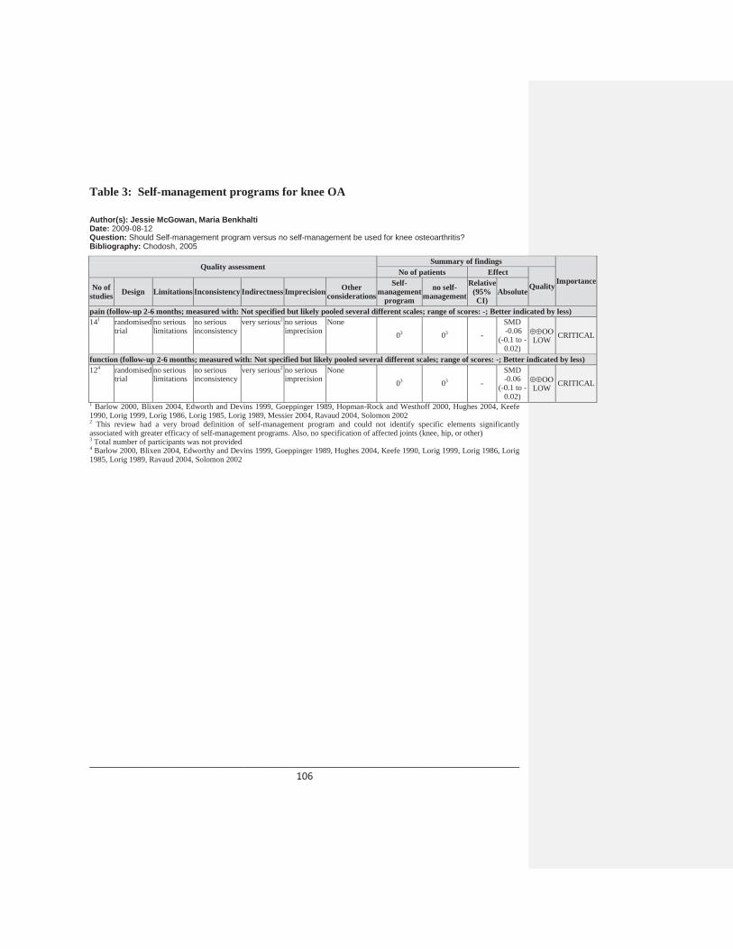
________________________________________________________________________ 106
Table 3: Self-management programs for knee OA
Author(s): Jessie McGowan, Maria Benkhalti Date: 2009-08-12Question: Should Self-management program versus no self-management be used for knee osteoarthritis?Bibliography: Chodosh, 2005
Quality assessmentSummary of findings
ImportanceNo of patients Effect
QualityNo of studies Design Limitations Inconsistency Indirectness Imprecision Other
considerations
Self-management
program
no self-management
Relative(95% CI)
Absolute
pain (follow-up 2-6 months; measured with: Not specified but likely pooled several different scales; range of scores: -; Better indicated by less)141 randomised
trialno serious limitations
no serious inconsistency
very serious2 no serious imprecision
None
03 03 -
SMD-0.06
(-0.1 to -0.02)
LOW CRITICAL
function (follow-up 2-6 months; measured with: Not specified but likely pooled several different scales; range of scores: -; Better indicated by less)124 randomised
trialno serious limitations
no serious inconsistency
very serious2 no serious imprecision
None03 03 -
SMD -0.06
(-0.1 to -0.02)
LOW CRITICAL
1 Barlow 2000, Blixen 2004, Edworth and Devins 1999, Goeppinger 1989, Hopman-Rock and Westhoff 2000, Hughes 2004, Keefe 1990, Lorig 1999, Lorig 1986, Lorig 1985, Lorig 1989, Messier 2004, Ravaud 2004, Solomon 20022 This review had a very broad definition of self-management program and could not identify specific elements significantly associated with greater efficacy of self-management programs. Also, no specification of affected joints (knee, hip, or other)3 Total number of participants was not provided4 Barlow 2000, Blixen 2004, Edworthy and Devins 1999, Goeppinger 1989, Hughes 2004, Keefe 1990, Lorig 1999, Lorig 1986, Lorig 1985, Lorig 1989, Ravaud 2004, Solomon 2002

________________________________________________________________________ 107
Table 4 a: Manual therapy program versus exercise therapy program for hip OA
Author(s): Karine Toupin AprilDate: 2009-08-07Question: Should manual therapy versus exercise therapy be used for hip OA?Bibliography: Hoeksma 2004
Quality assessmentSummary of findings
ImportanceNo of patients Effect
QualityNo of studies Design Limitations Inconsistency Indirectness Imprecision Other
considerationsmanual therapy
exercise therapy
Relative(95% CI)
Absolute
pain at rest (follow-up 5 weeks; measured with: visual analog scale; range of scores: 0-100; Better indicated by lower values)1 randomised
trialsno serious limitations1
no serious inconsistency
no serious indirectness
no serious imprecision
None
53 50 1.54
SMD-0.47
(-0.86 to -0.08)3
HIGH CRITICAL
physical function (follow-up 5 weeks; range of scores: 0-100; Better indicated by higher values)1 randomised
trialsno serious limitations1
no serious inconsistency
no serious indirectness
serious2 None53 50 1.11
SMD 0.10
(-0.28 to 0.49)3
MODERATE CRITICAL
pain at rest (follow-up 29 weeks; measured with: visual analog scale; range of scores: 0-100; Better indicated by lower values)1 randomised
trialsno serious limitations1
no serious inconsistency
no serious indirectness
serious2 None
45 44 1.25
SMD-0.26
(-0.68 to 0.15)4
MODERATE CRITICAL
physical function (follow-up 29 weeks; range of scores: 0-100; Better indicated by higher values)1 randomised
trialsno serious limitations1
no serious inconsistency
no serious indirectness
serious2 None
44 44 1.29
SMD 0.25
(-0.17 to 0.67)4
MODERATE CRITICAL
Adherence (follow-up 5 weeks; number of patients who prematurely discontinued the treatment programs)1 randomised
trialsno serious limitations1
no serious inconsistency
no serious indirectness
serious2 None
4/56 (7.1%)
3/53 (5.7%)
1.26 (0.30 to
5.37)
1 more per 100 (from 4 fewer to 25 more)
MODERATE CRITICAL
Adverse effects (number of patients who discontinued the treatment programs because of increase of complaints)1 randomised
trialsno serious limitations1
no serious inconsistency
no serious indirectness
serious2 None
3/56 (5.4%)
2/53 (3.8%)
1.42 (0.25 to
8.16)
2 more per 100 (from 3 fewer to
27 more)5
MODERATE CRITICAL
Losses to follow-up (follow-up 29 weeks; number of patients who were lost to follow-up)1 randomised
trialsno serious limitations1
no serious inconsistency
no serious indirectness
serious2 None
12/56 (21.4%)
9/53 (17%)
1.26 (0.58 to
2.75)
4 more per 100 (from 7 fewer to
30more)5
MODERATE CRITICAL
1 This trial was a single-blind study. The authors mention that it was not possible to blind either patients or therapists for the allocated treatment. Therefore, extra attention was given to the blinding of the outcome assessor. A placebo effect may also be present in this study due to the nature of the interventions. Finally, a limitation of the study is the relatively large number of patients who received total hip arthroplasty during the follow-up period. However, no significant differences were found between the conclusions based on the intention-to-treat analysis and the per-protocol analysis.The quality of the study was not downgraded because of these reasons.

________________________________________________________________________ 108
2 The confidence interval ranges from not being clinically significant to a very large clinical effect, which is a sign of imprecision.3 This SMD was calculated with RevMan 5 with the end-of-study data at the end of the treatment period (5-weeks). 4 This SMD was calculated with RevMan 5 with the end-of-study data at 29 weeks of follow-up. 5 In the exercise program, one patient also discontinued treatment because of cardio-respiratory disease.
Table 4 b: Manual therapy in combination with supervised exercise and home exercise program versus home exercise program alone for knee OA
Author(s): Karine Toupin AprilDate: 2009-08-19Question: Should manual therapy in combination with supervised exercise and home exercise program vs home exercise be used for knee OA?Bibliography: Deyle, 2005
Quality assessmentSummary of findings
Importance
No of patients Effect
QualityNo of studies Design Limitations Inconsistency Indirectness Imprecision Other
considerations
Manual therapy+
supervised exercise
and home exercise program
Home exercise
Relative(95% CI)
Absolute
pain (follow-up 8 weeks; measured with: WOMAC; range of scores: 0-500; Better indicated by lower values)11 randomised
trialsno serious limitations2
no serious inconsistency
no serious indirectness
no serious imprecision
none
60 60 1.43
SMD -0.41
(-0.77 to -0.05)
HIGH CRITICAL
function (follow-up 8 weeks; measured with: WOMAC; range of scores: 0-1700; Better indicated by lower values)1 randomised
trialsno serious limitations
no serious inconsistency
no serious indirectness
no serious imprecision
none60 60 1.41
SMD -0.40
(-0.76 to -0.03)
HIGH CRITICAL
Discontinuations due to lack of adherence (follow-up 8 weeks; number of patients who were discontinued to lack of adherence to the treatment regimen)1 randomised
trialsno serious limitations
no serious inconsistency
no serious indirectness
no serious imprecision
none
0/60(0%)
0/60 (0%)
0 (0 to 0)
0 fewer per 100 (from 0 fewer to 0 fewer)
HIGH CRITICAL
Withdrawals (follow-up 8 weeks; people who withdrew from the study after randomization)1 randomised
trialsno serious limitations
no serious inconsistency
no serious indirectness
serious3 none6/66
(9.1%)48/68
(11.8%)5
RR 0.77 (0.28 to
2.11)
3 fewer per 100 (from 8 fewer to 13 more)
MODERATE CRITICAL
1 Another outcome reported by the author was the use of medications for OA by patients at 52 weeks. Use of medications for OA was higher in the home exercise group (68%) than the clinic treatment group (48%) and this difference was statistically significant (p=0.03). 2 The authors report that the intention to treat results with 134 subjects did not differ substantially from the results of the 120 subjects.3 The confidence interval ranges from not being clinically significant to a very large clinical effect, which is a sign of imprecision. 4 In the treatment group, withdrawals were due to: knee injections (2), changed medications (1), not willing to return (1), not willing to walk (1) and unrelated medical condition (1).5 In the control group, withdrawals were due to: knee injections (1), changed medications (1), shoulder surgery (1), not willing to return (2) and moved from area (3).

________________________________________________________________________ 109
Table 5: Psychosocial intervention compared to no intervention for OA of the hip and knee
Author(s): Jessie McGowan, Maria Benkhalti Date: 2009-07-23Question: Should psychosocial intervention vs no intervention be used for osteoarthritis of the hip and knee?Settings: Bibliography:
Quality assessmentSummary of findings
ImportanceNo of patients Effect
QualityNo of studies Design Limitations Inconsistency Indirectness Imprecision Other
considerationspsychosocial intervention
no intervention
Relative(95% CI)
Absolute
pain (follow-up 2-12 months; measured with: pooled different scales including AIMS and VAS; range of scores: 0-0; Better indicated by less)8 randomised
trialserious1 no serious
inconsistencyserious2 no serious
imprecisionnone
7833 700 1.19SMD -0.22 (-
0.11 to -0.33)
LOW CRITICAL
function (physical disability) (follow-up 2-12 months; range of scores: 0-0; Better indicated by less)84 randomised
trialserious1 no serious
inconsistencyserious5 no serious
imprecisionnone
783 700 1.17
SMD 0.18
(0.06 to 0.29)
LOW CRITICAL
1 Pooled wide range of psychosocial interventions2 Affected joints not described therefore could not distinguish between hip, knee, and other. 3 Data obtained from Dixon 2007 supplement (appendix 5)4 Calfas 1992, Gay 2002, Keefe 2004, Keefe 1996, Keefe 1999, Keefe 1990, Keefe 1990, Lin 2003. 5 No description of type of scales used.

________________________________________________________________________ 110
Table 6: Weight loss compared to control (no weight loss program) for knee OA
Author(s): Jessie McGowan, Maria BenkhaltiDate: 2009-04-28Question: Should weight loss versus control (no weight loss program) be used for knee OA?Bibliography: Christensen, 2007
Quality assessmentSummary of findings
Importance
No of patients Effect
QualityNo of studies Design Limitations Inconsistency Indirectness Imprecision Other
considerationsweight
loss
control (no
weight loss)
Relative(95% CI)
Absolute
pain (follow-up 8-24 weeks; measured with: pooled WOMAC 500mm; range of scores: 0-500 and Likert; range of scores 1-5; Better indicated by less)21 randomised
trialno serious limitations
serious2 no serious indirectness
no serious imprecision
none208 208 1.2
SMD -0.2 (-0.39 to
0)MODERATE CRITICAL
function (follow-up mean 8-24 weeks; measured with: pooled WOMAC 1700mm; range of scores: 0-1700 and self-reported disability; range of scores 23-115 ; Better indicated by less)21 randomised
trialno serious limitations
serious2 no serious indirectness
no serious imprecision
none
208 208 1.3
SMD -0.23 (-
0.42 to -0.04)
MODERATE CRITICAL
1 Christensen 2005, Messier 20002 Christensen 2005 used only low-energy diet whereas Messier 2000 used exercise and diet intervention. Length of follow-up also varied (8-24 weeks).
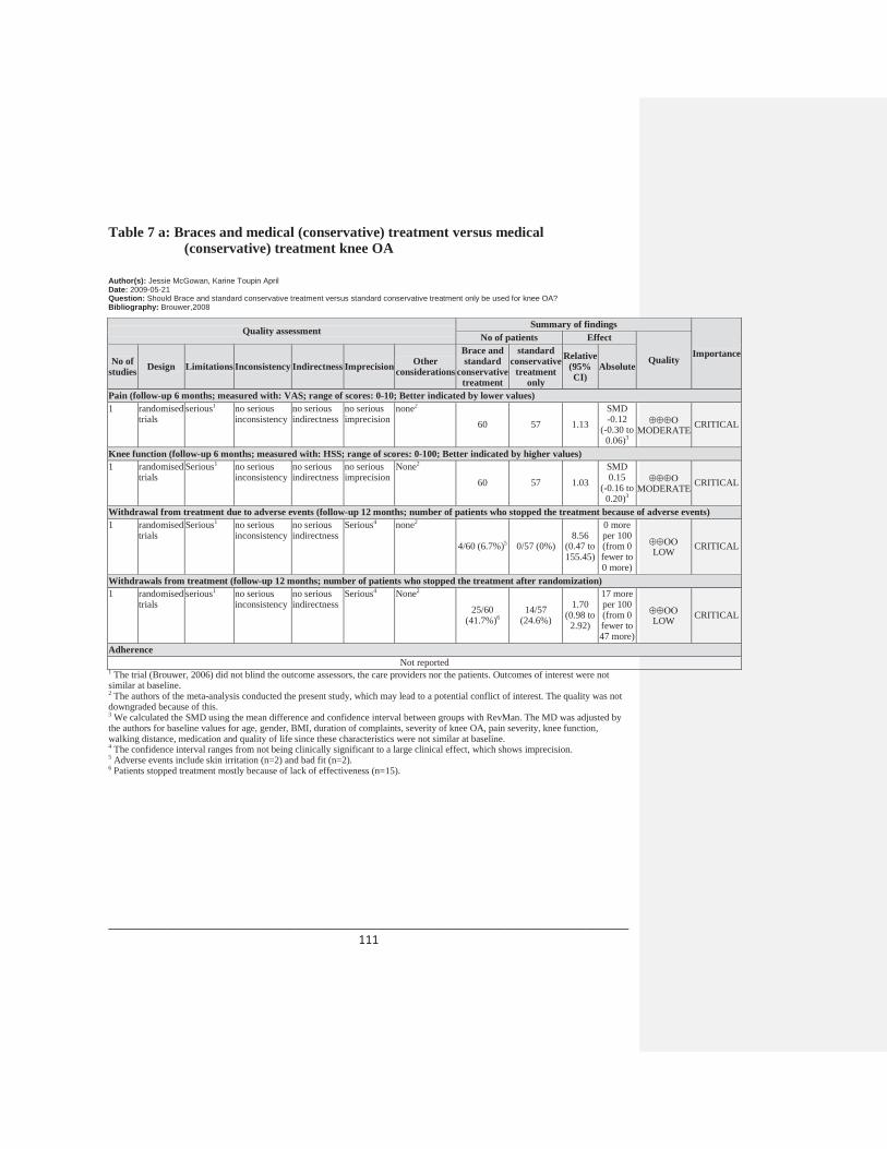
________________________________________________________________________ 111
Table 7 a: Braces and medical (conservative) treatment versus medical (conservative) treatment knee OA
Author(s): Jessie McGowan, Karine Toupin AprilDate: 2009-05-21Question: Should Brace and standard conservative treatment versus standard conservative treatment only be used for knee OA?Bibliography: Brouwer,2008
Quality assessmentSummary of findings
Importance
No of patients Effect
QualityNo of studies Design Limitations Inconsistency Indirectness Imprecision Other
considerations
Brace and standard
conservative treatment
standard conservative
treatment only
Relative(95% CI)
Absolute
Pain (follow-up 6 months; measured with: VAS; range of scores: 0-10; Better indicated by lower values)1 randomised
trialsserious1 no serious
inconsistencyno serious indirectness
no serious imprecision
none2
60 57 1.13SMD -0.12
(-0.30 to 0.06)3
MODERATE CRITICAL
Knee function (follow-up 6 months; measured with: HSS; range of scores: 0-100; Better indicated by higher values)1 randomised
trialsSerious1 no serious
inconsistencyno serious indirectness
no serious imprecision
None2
60 57 1.03
SMD 0.15
(-0.16 to 0.20)3
MODERATE CRITICAL
Withdrawal from treatment due to adverse events (follow-up 12 months; number of patients who stopped the treatment because of adverse events)1 randomised
trialsSerious1 no serious
inconsistencyno serious indirectness
Serious4 none2
4/60 (6.7%)5 0/57 (0%)8.56
(0.47 to 155.45)
0 more per 100 (from 0 fewer to 0 more)
LOW CRITICAL
Withdrawals from treatment (follow-up 12 months; number of patients who stopped the treatment after randomization)1 randomised
trialsserious1 no serious
inconsistencyno serious indirectness
Serious4 None2
25/60 (41.7%)6
14/57 (24.6%)
1.70 (0.98 to
2.92)
17 more per 100 (from 0 fewer to 47 more)
LOW CRITICAL
AdherenceNot reported
1 The trial (Brouwer, 2006) did not blind the outcome assessors, the care providers nor the patients. Outcomes of interest were not similar at baseline.2 The authors of the meta-analysis conducted the present study, which may lead to a potential conflict of interest. The quality was not downgraded because of this.3 We calculated the SMD using the mean difference and confidence interval between groups with RevMan. The MD was adjusted by the authors for baseline values for age, gender, BMI, duration of complaints, severity of knee OA, pain severity, knee function, walking distance, medication and quality of life since these characteristics were not similar at baseline.4 The confidence interval ranges from not being clinically significant to a large clinical effect, which shows imprecision. 5 Adverse events include skin irritation (n=2) and bad fit (n=2).6 Patients stopped treatment mostly because of lack of effectiveness (n=15).

________________________________________________________________________ 112
Table 7 b: Braces and medical (conservative) treatment versus medical (conservative) treatment alone in knee OA
Author(s): Karine Toupin AprilDate: 2009-09-14Question: Should brace and medical treatment versus medical treatment be used for knee OA?Bibliography: Kirkley 1999
Quality assessmentSummary of findings
Importance
No of patients Effect
QualityNo of studies Design Limitations Inconsistency Indirectness Imprecision Other
considerations
brace and
medical treatment
medical treatment
Relative(95% CI)
Absolute
pain (follow-up 6 months; measured with: WOMAC pain; range of scores: 0-500; Better indicated by lower values)1 randomised
trialsserious1 no serious
inconsistencyno serious indirectness
No serious imprecision
None41 33 2.21
SMD -0.89
(-1.38 to -0.41)2
MODERATE CRITICAL
function (follow-up 6 months; measured with: WOMAC function; range of scores: 0-1700; Better indicated by lower values)1 randomised
trialsserious1 no serious
inconsistencyno serious indirectness
No serious imprecision
None
41 33 2
SMD -0.76
(-1.23 to -0.28)2
MODERATE CRITICAL
withdrawals (follow-up 6 months; number of patients who withdrew from the study after randomization)1 randomised
trialsSerious1 no serious
inconsistencyno serious indirectness
No serious imprecision
None
0/41 (0%) 7/40 (17.5%)
0.07 (0.00 to 1.10)3
16 fewer per 100
(from 17 fewer to 2 more)
MODERATE CRITICAL
SafetyNot reported
AdherenceNot reported
1 Blinding of patients and assessors as well as intention-to-treat analyses were not mentioned in this study.2 The results shown in the present table were computed using the data sent to our research team by Dr. Brouwer who had recently received it from the Kirkley Research Group but did not have time to report it to his systematic review. 3 We calculated this relative risk using Rev Man 5. Reasons for withdrawals include: dissatisfaction with the group to which they had been randomized (n=5), inability to attend appointments (n=2), ill health (n=1) and a change in a scheduled date for an operation (n=1).

________________________________________________________________________ 113
Table 7 c: Braces and medical treatment versus neoprene sleeve with medical treatment in knee OA
Author(s): Karine Toupin AprlDate: 2009-09-14Question: Should brace and medical treatment versus neoprene sleeve and medical treatment be used for knee OA?Bibliography: Kirkley, 1999
Quality assessmentSummary of findings
Importance
No of patients Effect
QualityNo of studies Design Limitations Inconsistency Indirectness Imprecision Other
considerations
brace and
medical treatment
neoprene sleeve and
medical treatment
Relative(95% CI)
Absolute
Pain (follow-up 6 months; measured with: WOMAC pain; range of scores: 0-500; Better indicated by lower values)1 randomised
trialsSerious1 no serious
inconsistencyno serious indirectness
Serious2 None
41 36 1.57
SMD -0.44
(-0.89 to 0.01)3
LOW CRITICAL
function (follow-up 6 months; measured with: WOMAC function; range of scores: 0-1700; Better indicated by lower values)1 randomised
trialsSerious1 no serious
inconsistencyno serious indirectness
Serious2 None
41 36 1.45
SMD -0.35
(-0.80 to 0.10)3
LOW CRITICAL
withdrawals (follow-up 6 months; number of patients who withdrew from the study after randomization)1 randomised
trialsserious1 no serious
inconsistencyno serious indirectness
Serious2 None
0/41 (0%) 2/38 (5.3%)
0.19 (0.01 to 3.75)4
4 fewer per 100 (from 5 fewer to 14 more)
LOW CRITICAL
SafetyNot reported
AdherenceNot reported
1 Blinding of patients and assessors as well as intention-to-treat analyses were not mentioned in this study. 2 The confidence interval ranges from not being clinically significant to a large clinical effect, which shows imprecision. 3 The results shown in the present table were computed using the data sent to our research team by Dr. Brouwer who had recentlyreceived it from the Kirkley Research Group but did not have time to report it to his systematic review. The SMDs were computed using the change in outcomes over time.4 We calculated this relative risk using Rev Man 5. Reasons for withdrawals the 7 withdrawals in the control group and the 2 from the neoprene sleeve group include: dissatisfaction with the group to which they had been randomized (n=5), inability to attend appointments (n=2), ill health (n=1) and a change in a scheduled date for an operation (n=1) in the three treatment groups (brace, medical treatment and neoprene sleeve).

________________________________________________________________________ 114
Table 8 a: Medially-directed patellar taping versus no taping in knee OA
Author(s): Karine Toupin AprilDate: 2009-06-16Question: Should medially-directed patellar taping versus no taping be used for knee OA?Bibliography: Warden, 2008
Quality assessmentSummary of findings
Importance
No of patients Effect
QualityNo of studies Design Limitations Inconsistency Indirectness Imprecision Other
considerations
medially-directed patellar taping
no taping
Relative(95% CI)
Absolute
pain (follow-up 3 weeks1; measured with: VAS; range of scores: 0-100; Better indicated by lower values)2 randomised
trials2serious3 no serious
inconsistencyno serious indirectness
no serious imprecision
reporting bias4
47 475 2.05
SMD -1.17 (-1.51 to -0.83)6
LOW CRITICAL
function (follow-up 3 weeks; measured with: WOMAC; range of scores: 0-68; Better indicated by lower values)17 randomised
trialsno serious limitations
no serious inconsistency
no serious indirectness
serious8 reporting bias9
29 29 1.41
SMD -0.37 (-0.89 to 0.15)10
LOW CRITICAL
minor skin irritations (follow-up 6 weeks; number of subjects presenting with minor skin irritations)111 randomised
trialsno serious limitations
no serious inconsistency
no serious indirectness
serious8 reporting bias9
8/29 (27.6%)
0/29 (0%)
17 (1.03 to 281.5)
0 more per 100 (from 0 more to 0 more)
LOW CRITICAL
withdrawals (follow-up 6 weeks; number of patients who withdrew after randomization)112 randomised
trialsno serious limitations
no serious inconsistency
no serious indirectness
serious8 reporting bias9
0/29 (0%)
1/29 (3.4%)
0.33 (0.01 to
7.86)
2 fewer per 100 (from 3 fewer to 24 more)
LOW CRITICAL
Adherence (follow-up 6 weeks; number of participants who continued to wear the tape as prescribed)113 randomised
trialsno serious limitations
no serious inconsistency
no serious indirectness
no serious imprecision
reporting bias9
29/29 (100%)
29/29 (100%) 1
0 fewer per 100 (from 100
fewer to 100
fewer)
MODERATE CRITICAL
1 One study looks at the immediate effect of taping and the other one at 3 weeks.2 One study was a crossover study and the other was a controlled study. 3 According to the trials, both studies did not blind subjects and therapists who administered the treatment. However, subjects were not aware of which taping technique was considered therapeutic. Furthermore, because one of the studies (published in Rheumatology) used a crossover (within subject) design, it did not ensure proper allocation concealment and comparability of group characteristics at baseline. The quality assessment reported in the SR by Warden is not consistent with the information given in the RCTs. 4 There is a publication bias indicated by significant funnel plot asymmetry in the SR. This asymmetry indicates that negative studies investigating patellar taping are less likely to be published and smaller studies are more likely to produce larger effect sizes.5 The study in BMJ included 29 in each group and the crossover study in Rheumatology included 18 patients who had both medially-directed taping and no taping.6 This effect size was reported in the SR by Warden.7 The SR did not report function. One study (Hinman, 2003 in BMJ) reported function at 3 weeks.8 The confidence interval ranges from not being clinically significant to a large clinical effect, which shows imprecision. 9 There is a possibility of publication bias since the funnel plot showed asymmetry in the SR. This asymmetry indicates that negative studies investigating patellar taping are less likely to be published and smaller studies are more likely to produce larger effect sizes. 10 We calculated the SMD with the end of study data using RevMan.11 One study (Hinman, 2003 in BMJ) reported adverse effects. Another study by the same author (Hinman, 2003 in Rheumatology) reported an absence of adverse effects.

________________________________________________________________________ 115
12 One study (Hinman, 2003 in BMJ) reported withdrawals.13 One study (Hinman, 2003 in BMJ) reported adherence.
Table 8 b: Medially-directed patellar taping versus sham taping in knee OA
Author(s): Karine Toupin AprilDate: 2009-09-16Question: Should medially-directed patellar taping versus sham taping be used for knee OA?Bibliography: Warden, 2008
Quality assessmentSummary of findings
ImportanceNo of patients Effect
QualityNo of studies Design Limitations Inconsistency Indirectness Imprecision Other
considerations
medially-directed patellar taping
sham taping
Relative(95% CI)
Absolute
pain (follow-up 3 weeks1; measured with: VAS; range of scores: 0-100; Better indicated by lower values)3 randomised
trials2serious3 no serious
inconsistencyno serious indirectness
no serious imprecision
reporting bias4
61 615 1.66
SMD -0.69
(-1.11 to -0.28)6
LOW CRITICAL
function (follow-up 3 weeks; measured with: WOMAC; range of scores: 0-68; Better indicated by lower values)17 randomised
trialsno serious limitations
no serious inconsistency
no serious indirectness
Serious8 reporting bias9
29 29 0.97
SMD 0.04
(-0.47 to 0.56)
LOW CRITICAL
minor skin irritations (follow-up 3 weeks; number of subjects presenting with minor skin irritations)110 randomised
trialsno serious limitations
no serious inconsistency
no serious indirectness
Serious8 Reporting bias9
8/29 (27.6%)
1/29 (3.4%)
8 (1.07 to 59.95)
24 more per 100 (from 0 more to
203 more)
LOW CRITICAL
adherence (follow-up 6 weeks; number of participants who continued to wear the tape as prescribed)111 randomised
trialsno seriouslimitations
no serious inconsistency
no serious indirectness
no serious imprecision
Reporting bias9
29/29 (100%)
29/29 (100%) 1
0 fewer per 100 (from 100
fewer to 100
fewer)
MODERATE CRITICAL
Withdrawals111 randomised
trialsno serious limitations
no serious inconsistency
no serious indirectness
no serious imprecision
Reporting bias9
0/29 (0%)
0/29 (0%) 1
0 fewer per 100 (from 100
fewer to 100
fewer)
MODERATE CRITICAL
1 Studies looked at the immediate effect of taping as well as the effect after 4 days and after 3 weeks of intervention. 2 Two were crossover studies and one was an RCT. 3 According to the trials, studies did not blind subjects (though it is unclear in the Cushnagan study if patients were blinded) and therapists who administered the treatment. However, subjects were not aware of which taping technique was considered therapeutic. Furthermore, because the two other studies used a crossover (within subject) design, it did not ensure proper allocation concealment and comparability of group characteristics at baseline. The quality assessment reported in the SR by Warden is not consistent with the information given in the RCTs. 4 There is a publication bias indicated by significant funnel plot asymmetry. This asymmetry indicates that negative studies investigating patellar taping are less likely to be published and smaller studies are more likely to produce larger effect sizes. 5 The study published by Hinman in BMJ included 29 in each group, the study by the same author in Rheumatology included 18 patients and the study by Cushnagan included 14 patients.

________________________________________________________________________ 116
6 This effect size was reported in the SR by Warden. 7 The SR did not report function. One study (Hinman, 2003 in BMJ) reported function at 3 weeks. 8 The confidence interval ranges from not being clinically significant to a large clinical effect, which shows imprecision. 9 There is a possibility of publication bias since the funnel plot showed asymmetry in the SR. This asymmetry indicates that negative studies investigating patellar taping are less likely to be published and smaller studies are more likely to produce larger effect sizes. 10 One study (Hinman, 2003 in BMJ) reported adverse effects. The other studies reported an absence of adverse effects. 11 One study (Hinman, 2003 in BMJ) reported adherence to the treatment regimen. Cushnagan also reported that all patients followed prescribe taping.
Table 8 c: Laterally-directed patellar taping versus medially-directed patellar taping in knee OA
Author(s): Karine Toupin AprilDate: 2009-09-16Question: Should laterally-directed patellar taping versus medially-directed patellar taping be used for knee OA?Bibliography: Warden, 2008
Quality assessmentSummary of findings
Importance
No of patients Effect
QualityNo of studies Design Limitations Inconsistency Indirectness Imprecision Other
considerations
laterally-directed patellar taping
medially-directed patellar taping
Relative(95% CI)
Absolute
Pain (follow-up 4 days; measured with: VAS; range of scores: 0-100; Better indicated by lower values)1 randomised
trials1Serious2 no serious
inconsistencyno serious indirectness
no serious imprecision
reporting bias3
14 145
*Not estimable
due to lack of
data
SMD 0.95
(0.42 to 1.48) 4
LOW CRITICAL
FunctionNot reported
Safety (follow-up 4 days; number of patients who reported adverse events)1 randomised
trials1Serious2 no serious
inconsistencyno serious indirectness
No seriousimprecision
Reporting bias3
0/14(0%) 0/145 (0%) 1 0 fewer
per 100 LOW CRITICAL
Adherence (follow-up 4 days; number of patients who wore tapes on for the full four days)1 randomised
trials1Serious2 no serious
inconsistencyno serious indirectness
No seriousimprecision
Reporting bias3
14/14(0%) 14/145(0%) 1 0 fewer
per 100 LOW CRITICAL
Withdrawals1 randomised
trials1Serious2 no serious
inconsistencyno serious indirectness
No seriousimprecision
Reporting bias3
0/14(0%) 0/145(0%) 1 0 fewer
per 100 LOW CRITICAL1 This study by Cushnaghan has a crossover design with 14 patients.2 This study did not blind therapists who administered the treatment and it is unclear if patients were blinded. However, subjects were not aware of which taping technique was considered therapeutic. Also, because this study used a crossover (within subject) design, it did not ensure proper allocation concealment and comparability of group characteristics at baseline. 3 There is a possibility of publication bias since the funnel plot showed asymmetry in the SR. This asymmetry indicates that negative studies investigating patellar taping are less likely to be published and smaller studies are more likely to produce larger effect sizes.4 This effect size was reported in the SR by Warden. 5 14 patients received all three types of taping (medial, lateral and neutral) at different time points.
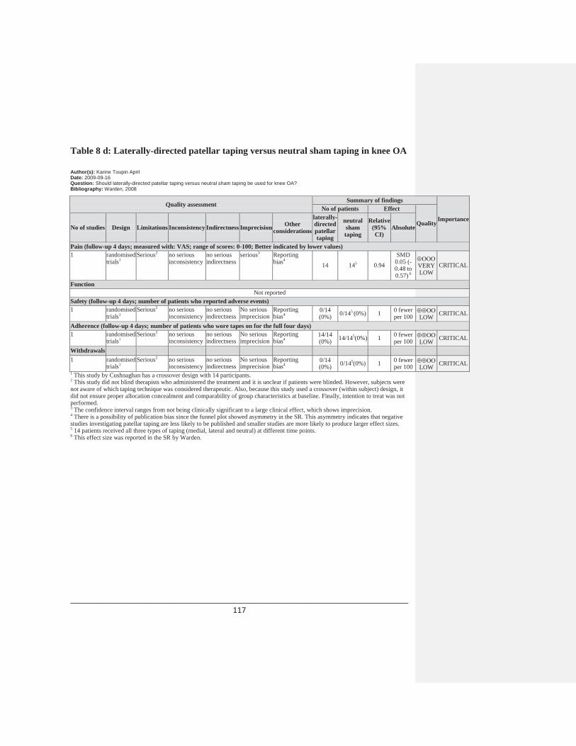
________________________________________________________________________ 117
Table 8 d: Laterally-directed patellar taping versus neutral sham taping in knee OA
Author(s): Karine Toupin AprilDate: 2009-09-16Question: Should laterally-directed patellar taping versus neutral sham taping be used for knee OA?Bibliography: Warden, 2008
Quality assessmentSummary of findings
Importance
No of patients Effect
QualityNo of studies Design Limitations Inconsistency Indirectness Imprecision Other
considerations
laterally-directed patellar taping
neutral sham taping
Relative(95% CI)
Absolute
Pain (follow-up 4 days; measured with: VAS; range of scores: 0-100; Better indicated by lower values)1 randomised
trials1Serious2 no serious
inconsistencyno serious indirectness
serious3 Reporting bias4
14 145 0.94
SMD 0.05 (-0.48 to 0.57) 6
VERY LOW
CRITICAL
FunctionNot reported
Safety (follow-up 4 days; number of patients who reported adverse events)1 randomised
trials1Serious2 no serious
inconsistencyno serious indirectness
No seriousimprecision
Reporting bias4
0/14(0%) 0/145 (0%) 1 0 fewer
per 100 LOW CRITICAL
Adherence (follow-up 4 days; number of patients who wore tapes on for the full four days)1 randomised
trials1Serious2 no serious
inconsistencyno serious indirectness
No seriousimprecision
Reporting bias4
14/14(0%) 14/145(0%) 1 0 fewer
per 100 LOW CRITICAL
Withdrawals1 randomised
trials1Serious2 no serious
inconsistencyno serious indirectness
No seriousimprecision
Reporting bias4
0/14(0%) 0/145(0%) 1 0 fewer
per 100 LOW CRITICAL1 This study by Cushnaghan has a crossover design with 14 participants.2 This study did not blind therapists who administered the treatment and it is unclear if patients were blinded. However, subjects were not aware of which taping technique was considered therapeutic. Also, because this study used a crossover (within subject) design, it did not ensure proper allocation concealment and comparability of group characteristics at baseline. Finally, intention to treat was not performed. 3 The confidence interval ranges from not being clinically significant to a large clinical effect, which shows imprecision. 4 There is a possibility of publication bias since the funnel plot showed asymmetry in the SR. This asymmetry indicates that negative studies investigating patellar taping are less likely to be published and smaller studies are more likely to produce larger effect sizes. 5 14 patients received all three types of taping (medial, lateral and neutral) at different time points.6 This effect size was reported in the SR by Warden.
Related Documents