ACR Appropriateness Criteria ® on Suspected Physical Abuse—Child James S. Meyer, MD a , Richard Gunderman, MD, PhD b , Brian D. Coley, MD c , Dorothy Bulas, MD d , Matthew Garber, MD e,f , Boaz Karmazyn, MD b , Marc S. Keller, MD a , Abhaya V. Kulkarni, MD g,h,i , Sarah S. Milla, MD j , John S. Myseros, MD d,h,i , Charles Paidas, MD k,l , Peter D. Pizzutillo, MD m,n , Daniel J. Podberesky, MD o , Jeffrey Scott Prince, MD p , John Ragheb, MD h,i,q The appropriate imaging for pediatric patients being evaluated for suspected physical abuse depends on the age of the child, the presence of neurologic signs and symptoms, evidence of thoracic or abdominopelvic injuries, and whether the injuries are discrepant with the clinical history. The clinical presentations reviewed consider these factors and provide evidence-based consensus recommendations by the ACR Appropriateness Criteria ® Expert Panel on Pediatric Imaging. Key Words: Appropriateness Criteria ® , child abuse, shaken baby syndrome, children, trauma, pediatric J Am Coll Radiol 2011;8:87-94. Copyright © 2011 American College of Radiology SUMMARY OF LITERATURE REVIEW In 2007, on the basis of reports to child and protective service agencies, an estimated 794,000 children were vic- tims of maltreatment (neglect, emotional abuse, sexual abuse, and physical abuse) in the United States [1]. Of these children, more than 79,000 were victims of physi- cal abuse, and an estimated 1,760 children died from abuse or neglect [1]. As high as these numbers are, the extent of the problem is actually much greater, as offi- cially reported cases grossly understate the true incidence of abuse [2,3]. In some children, physical examination and history may clearly indicate that physical abuse has occurred. In other children, however, the diagnosis of physical abuse is not so straightforward. It requires consider- ation of possible underlying metabolic and genetic conditions [4-8] and usually relies on the findings of a multidisciplinary team that includes physicians, social workers, and legal authorities. Imaging often plays a major role in the detection and documentation of physical injury. The type and extent of imaging per- formed in a child who is a suspected victim of abuse depend on the child’s age, signs, symptoms, and other social considerations, such as being the twin of a phys- ically abused infant [9]. Child abuse injuries can involve any site in the human body. Physically abused children may present with hol- low viscus and solid organ injuries, superficial and deep soft tissue injuries, thermal injuries, or fractures [10]. Fractures occur in up to 55% of child abuse victims [11]. Fractures most often involve the long bones with lesser involvement of the skull, ribs, clavicles, pelvis, and other bones [10,12]. a Children’s Hospital of Philadelphia, Philadelphia, Pennsylvania. b Riley Hospital for Children, Indiana University, Indianapolis, Indiana. c Nationwide Children’s Hospital, Columbus, Ohio. d Children’s National Medical Center, Washington, District of Columbia. e Division of General and Hospital Pediatrics, Columbia, South Carolina. f American Academy of Pediatrics, Elk Grove, Illinois. g Hospital for Sick Children, Toronto, Ontario, Canada. h American Association of Neurological Surgeons, Rolling Meadows, Illinois. i Congress of Neurological Surgeons, Schaumberg, Illinois. j New York University Medical Center, New York, New York. k Tampa General Hospital, Tampa, Florida. l American Pediatric Surgical Association, Deerfield, Illinois. m Saint Christopher’s Hospital for Children, Philadelphia, Pennsylvania. n American Academy of Orthopaedic Surgeons, Rosemont, Illinois. o Cincinnati Children’s Hospital Medical Center, Cincinnati, Ohio. p Primary Children’s Medical Center, Salt Lake City, Utah. q Miami Children’s Hospital, Miami, Florida. The ACR seeks and encourages collaboration with other organizations on the development of the ACR Appropriateness Criteria ® through society rep- resentation on expert panels. Participation by representatives from collaborat- ing societies on the expert panel does not necessarily imply society endorse- ment of the final document. Corresponding author and reprints: James S. Meyer, MD, American College of Radiology, 1891 Preston White Drive, Reston, VA 20191; e-mail: [email protected]. © 2011 American College of Radiology 0091-2182/11/$36.00 ● DOI 10.1016/j.jacr.2010.09.008 87

Welcome message from author
This document is posted to help you gain knowledge. Please leave a comment to let me know what you think about it! Share it to your friends and learn new things together.
Transcript

S
Ist
trimCom
©0
ACR Appropriateness Criteria® onSuspected Physical Abuse—Child
James S. Meyer, MDa, Richard Gunderman, MD, PhDb, Brian D. Coley, MDc,Dorothy Bulas, MDd, Matthew Garber, MDe,f, Boaz Karmazyn, MDb,
Marc S. Keller, MDa, Abhaya V. Kulkarni, MDg,h,i, Sarah S. Milla, MDj,John S. Myseros, MDd,h,i, Charles Paidas, MDk,l, Peter D. Pizzutillo, MDm,n,
Daniel J. Podberesky, MDo, Jeffrey Scott Prince, MDp, John Ragheb, MDh,i,q
The appropriate imaging for pediatric patients being evaluated for suspected physical abuse depends on the ageof the child, the presence of neurologic signs and symptoms, evidence of thoracic or abdominopelvic injuries,and whether the injuries are discrepant with the clinical history. The clinical presentations reviewed considerthese factors and provide evidence-based consensus recommendations by the ACR Appropriateness Criteria®
Expert Panel on Pediatric Imaging.
Key Words: Appropriateness Criteria®, child abuse, shaken baby syndrome, children, trauma, pediatric
J Am Coll Radiol 2011;8:87-94. Copyright © 2011 American College of Radiology
atcaeco
mIaacmwmpfdsi
blsFFi
UMMARY OF LITERATURE REVIEW
n 2007, on the basis of reports to child and protectiveervice agencies, an estimated 794,000 children were vic-ims of maltreatment (neglect, emotional abuse, sexual
aChildren’s Hospital of Philadelphia, Philadelphia, Pennsylvania.bRiley Hospital for Children, Indiana University, Indianapolis, Indiana.cNationwide Children’s Hospital, Columbus, Ohio.dChildren’s National Medical Center, Washington, District of Columbia.eDivision of General and Hospital Pediatrics, Columbia, South Carolina.fAmerican Academy of Pediatrics, Elk Grove, Illinois.gHospital for Sick Children, Toronto, Ontario, Canada.hAmerican Association of Neurological Surgeons, Rolling Meadows, Illinois.iCongress of Neurological Surgeons, Schaumberg, Illinois.jNew York University Medical Center, New York, New York.kTampa General Hospital, Tampa, Florida.lAmerican Pediatric Surgical Association, Deerfield, Illinois.mSaint Christopher’s Hospital for Children, Philadelphia, Pennsylvania.nAmerican Academy of Orthopaedic Surgeons, Rosemont, Illinois.oCincinnati Children’s Hospital Medical Center, Cincinnati, Ohio.pPrimary Children’s Medical Center, Salt Lake City, Utah.qMiami Children’s Hospital, Miami, Florida.
The ACR seeks and encourages collaboration with other organizations onhe development of the ACR Appropriateness Criteria® through society rep-esentation on expert panels. Participation by representatives from collaborat-ng societies on the expert panel does not necessarily imply society endorse-
ent of the final document.orresponding author and reprints: James S. Meyer, MD, American Collegef Radiology, 1891 Preston White Drive, Reston, VA 20191; e-mail:
2011 American College of Radiology091-2182/11/$36.00 ● DOI 10.1016/j.jacr.2010.09.008
buse, and physical abuse) in the United States [1]. Ofhese children, more than 79,000 were victims of physi-al abuse, and an estimated 1,760 children died frombuse or neglect [1]. As high as these numbers are, thextent of the problem is actually much greater, as offi-ially reported cases grossly understate the true incidencef abuse [2,3].
In some children, physical examination and historyay clearly indicate that physical abuse has occurred.
n other children, however, the diagnosis of physicalbuse is not so straightforward. It requires consider-tion of possible underlying metabolic and geneticonditions [4-8] and usually relies on the findings of aultidisciplinary team that includes physicians, socialorkers, and legal authorities. Imaging often plays aajor role in the detection and documentation of
hysical injury. The type and extent of imaging per-ormed in a child who is a suspected victim of abuseepend on the child’s age, signs, symptoms, and otherocial considerations, such as being the twin of a phys-cally abused infant [9].
Child abuse injuries can involve any site in the humanody. Physically abused children may present with hol-
ow viscus and solid organ injuries, superficial and deepoft tissue injuries, thermal injuries, or fractures [10].ractures occur in up to 55% of child abuse victims [11].ractures most often involve the long bones with lesser
nvolvement of the skull, ribs, clavicles, pelvis, and other
ones [10,12].87
bllsm
iw[aifaap
vAf[cga(oopccbcpap“fiileibi
88 Journal of the American College of Radiology/Vol. 8 No. 2 February 2011
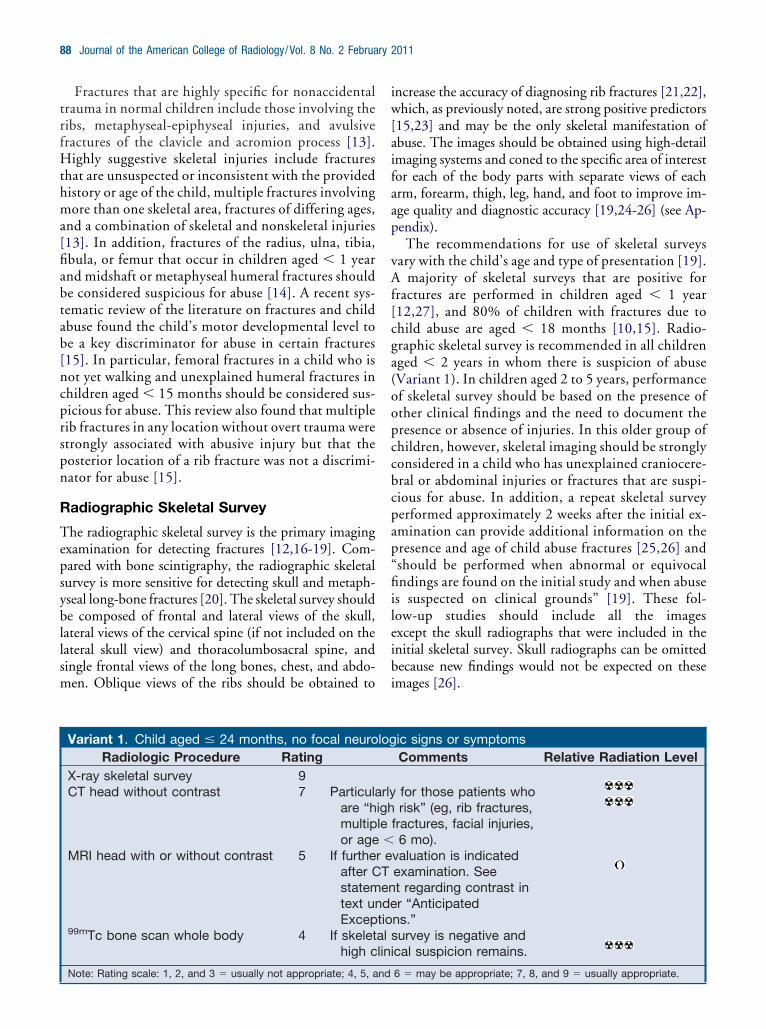
Fractures that are highly specific for nonaccidentaltrauma in normal children include those involving theribs, metaphyseal-epiphyseal injuries, and avulsivefractures of the clavicle and acromion process [13].Highly suggestive skeletal injuries include fracturesthat are unsuspected or inconsistent with the providedhistory or age of the child, multiple fractures involvingmore than one skeletal area, fractures of differing ages,and a combination of skeletal and nonskeletal injuries[13]. In addition, fractures of the radius, ulna, tibia,fibula, or femur that occur in children aged � 1 yearand midshaft or metaphyseal humeral fractures shouldbe considered suspicious for abuse [14]. A recent sys-tematic review of the literature on fractures and childabuse found the child’s motor developmental level tobe a key discriminator for abuse in certain fractures[15]. In particular, femoral fractures in a child who isnot yet walking and unexplained humeral fractures inchildren aged � 15 months should be considered sus-picious for abuse. This review also found that multiplerib fractures in any location without overt trauma werestrongly associated with abusive injury but that theposterior location of a rib fracture was not a discrimi-nator for abuse [15].
Radiographic Skeletal Survey
The radiographic skeletal survey is the primary imagingexamination for detecting fractures [12,16-19]. Com-pared with bone scintigraphy, the radiographic skeletalsurvey is more sensitive for detecting skull and metaph-yseal long-bone fractures [20]. The skeletal survey shoulde composed of frontal and lateral views of the skull,
ateral views of the cervical spine (if not included on theateral skull view) and thoracolumbosacral spine, andingle frontal views of the long bones, chest, and abdo-en. Oblique views of the ribs should be obtained to
Variant 1. Child aged � 24 months, no focal neuroRadiologic Procedure Rating
X-ray skeletal survey 9CT head without contrast 7 Particula
are “hmultipor age
MRI head with or without contrast 5 If furtheafter Cstatemtext unExcep
99mTc bone scan whole body 4 If skelethigh c
Note: Rating scale: 1, 2, and 3 � usually not appropriate; 4, 5, and
ncrease the accuracy of diagnosing rib fractures [21,22],hich, as previously noted, are strong positive predictors
15,23] and may be the only skeletal manifestation ofbuse. The images should be obtained using high-detailmaging systems and coned to the specific area of interestor each of the body parts with separate views of eachrm, forearm, thigh, leg, hand, and foot to improve im-ge quality and diagnostic accuracy [19,24-26] (see Ap-endix).The recommendations for use of skeletal surveys
ary with the child’s age and type of presentation [19].majority of skeletal surveys that are positive for
ractures are performed in children aged � 1 year12,27], and 80% of children with fractures due tohild abuse are aged � 18 months [10,15]. Radio-raphic skeletal survey is recommended in all childrenged � 2 years in whom there is suspicion of abuseVariant 1). In children aged 2 to 5 years, performancef skeletal survey should be based on the presence ofther clinical findings and the need to document theresence or absence of injuries. In this older group ofhildren, however, skeletal imaging should be stronglyonsidered in a child who has unexplained craniocere-ral or abdominal injuries or fractures that are suspi-ious for abuse. In addition, a repeat skeletal surveyerformed approximately 2 weeks after the initial ex-mination can provide additional information on theresence and age of child abuse fractures [25,26] andshould be performed when abnormal or equivocalndings are found on the initial study and when abuse
s suspected on clinical grounds” [19]. These fol-ow-up studies should include all the imagesxcept the skull radiographs that were included in thenitial skeletal survey. Skull radiographs can be omittedecause new findings would not be expected on thesemages [26].
ic signs or symptomsComments Relative Radiation Level
for those patients whorisk” (eg, rib fractures,
fractures, facial injuries,6 mo).
valuation is indicatedexamination. Seet regarding contrast inr “Anticipated
ns.”survey is negative andcal suspicion remains.
log
rlyighle�
r eTende
tioallini
6 � may be appropriate; 7, 8, and 9 � usually appropriate.

H
Ac
dnrcl
d(sdfdgrsTdqaiat
weCs
nd
Meyer et al/Suspected Physical Abuse—Child 89
Bone Scintigraphy
Bone scintigraphy is a complementary examinationfor detecting bone injuries [11,20,28]. It should beused when the radiographic skeletal survey is negativebut clinical suspicion remains high and a search forfurther evidence of skeletal trauma is still necessary.To increase sensitivity, the bone scan should includethe use of pinhole collimators and differential countsof the metaphyses. A bone scan is especially good fordetecting periosteal trauma and rib, spine, pelvic, andacromion fractures [28].
ead Trauma
lthough less frequent than skeletal injuries, mosthild abuse fatalities are the result of head trauma [7],
and head injury due to child abuse is the principalcause of death in children aged � 2 years [29]. Sub-
ural hematoma is the most commonly seen intracra-ial abnormality [30]. Additional craniocerebral inju-ies include cerebral contusion, epidural hematoma,erebral edema, subarachnoid hemorrhage, and uni-ateral hypoxic-ischemic injury [30-34].
Variant 2. Child aged � 24 months, head trauma bevidence of visceral injury
Radiologic Procedure RatingX-ray skeletal survey 9CT head without contrast 9MRI head with or without
contrast6 If further ev
CT examinregarding“Anticipat
99mTc bone scan whole body 4 If skeletal suclinical su
Note: Rating scale: 1, 2, and 3 � usually not appropriate; 4, 5, a
Variant 3. Child aged � 24 months, with seizures ophysical findings
Radiologic Procedure RatingX-ray skeletal survey 9CT head without contrast 9MRI head with or without
contrast8 Do not dela
symptomawhether CSee statemtext underException
99mTc bone scan whole body 4 If skeletal suclinical su
Note: Rating scale: 1, 2, and 3 � usually not appropriate; 4, 5, and
Imaging the head in children with suspected abuseepends on the child’s age and type of presentationVariant 2). In children with skull fractures or clinicaligns and symptoms of intracranial injury, an imme-iate noncontrast CT scan of the head should be per-ormed (Variants 3 and 4). If the CT scan does notetect significant lesions that require rapid neurosur-ical intervention and the clinical presentation war-ants further assessment, an MRI scan of the headhould be performed. The MRI study should include1-weighted and T2-weighted sequences with protonensity or inversion recovery and gradient-echo se-uences. In addition, diffusion-weighted sequencesre suggested to indicate whether acute cerebral injurys present [19]. In a child with abnormal CT findings,dditional assessment with MRI should be consideredo further assess the extent of posttraumatic injury.
There are varying opinions on how to image childrenho are suspected abuse victims and have no objective
vidence suggesting intracranial injury [29,33,35,36].hildren, especially those aged � 12 months, may have
ignificant intracranial injury without signs or symptoms
istory, no focal neurologic signs or symptoms, no
omments Relative Radiation Level
ation is indicated afterion. See statementntrast in text underExceptions.”ey is negative and highcion remains.
6 � may be appropriate; 7, 8, and 9 � usually appropriate.
eurologic signs and symptoms, with or without
omments Relative Radiation Level
ead CT for MRI inchild. May be useful
is positive or negative.nt regarding contrast innticipated
ey is negative and highcion remains.
y h
C
aluatcoedrv
spi
r n
C
y hticTe“A
s.”rv
spi
6 � may be appropriate; 7, 8, and 9 � usually appropriate.

tui[si
whswaucdhGlhopil
NP
Nc
cdcicckailtrtvijwcn
waCfijsnrwcfp
nd
90 Journal of the American College of Radiology/Vol. 8 No. 2 February 2011
of head injury [18,29,33]. Physical examination, in par-icular the absence of retinal hemorrhages, should not besed to determine the need for imaging, as intracranial
njury may occur in the absence of retinal hemorrhages29,33]. Skull radiographs are also unreliable. Althoughkull radiographs may detect fractures associated withntracranial pathology [37], they do not provide ade-
quate screening, since significant traumatic intracranialpathology may occur in the absence of skull fractures[33,38,39].
In one study [29], 37% of children aged � 2 yearswith “high-risk” criteria (defined as rib fractures, mul-tiple fractures, facial injuries, or age � 6 months) and
ithout overt signs of head injury who underwentead CT or MRI had occult head injuries. In thistudy, 18 of the 19 children with occult head injuriesere aged � 1 year. Another study [33] of 51 children
ged � 4 years with no signs of intracranial injury whonderwent skeletal survey for abuse found that 29% ofhildren who underwent neurologic imaging had evi-ence of intracranial injury that included subduralematoma, epidural hematoma, or cerebral edema.iven these studies, clinicians should have a relatively
ow threshold for performing either CT or MRI of theead in children with suspected abuse. Magnetic res-nance imaging avoids the radiation of CT and is aarticularly good choice in the nonemergent setting tomage these “high-risk” children without overt neuro-ogic signs or symptoms.
onskeletal Chest, Abdominal, andelvic Injuries
onskeletal injuries to the chest, abdomen, and pelvis
Variant 4. Child aged � 24 months, with seizures ophysical findings
Radiologic Procedure RatingCT head without contrast 9MRI head with or without
contrast8 Do not dela
symptomawhether CSee statemtext underException
X-ray skeletal survey 6 Value of surRadiograptailored toinjury.
99mTc bone scan whole body 4 If skeletal suclinical su
Note: Rating scale: 1, 2, and 3 � usually not appropriate; 4, 5, a
an occur as the result of child abuse. Injuries to the g
hest are rare but may include hemopericardium, car-iac contusions, and lacerations, pleural effusion, lungontusions, and chylothorax [7,40,41]. Nonskeletalnjuries to the abdomen and pelvis include pan-reatitis, pancreatic pseudocysts, and lacerations andontusions of the liver, adrenal gland, spleen, andidneys, as well as injury and rupture of the bladdernd bowel [40,42]. Victims of nonaccidental abdom-nal trauma tend to be younger and have a more de-ayed presentation than those who experience acciden-al trauma [43]. The delay in presentation may beelated to the caretaker’s delay in bringing the patiento medical attention, inconsistent history, or theagueness of symptoms that may accompany thenjuries. In addition, independent of concomitant in-ury, blunt trauma due to child abuse “is associatedith a 6-fold increased odds of death compared to
hildren whose injuries resulted from other mecha-isms” [44].In most cases, imaging for assessing these injuries
ill be directed by the patient’s clinical presentationnd consist of a CT scan of the involved body part. AT scan of the chest should generally be per-
ormed with intravenous contrast to detect vascularnjuries. A CT scan for suspected intra-abdominal in-ury should include both the abdomen and pelvis andhould be performed with intravenous contrast. Theeed for oral contrast is at the discretion of theadiologist, and its use should be strongly consideredhen there is concern for duodenal hematoma. Paren-
hymal or late arterial phase imaging is most helpfulor detecting solid organ injury. Delayed, excretoryhase imaging is suggested detecting disruption of the
eurologic signs and symptoms, with or without
omments Relative Radiation Level
ead CT for MRI inchild. May be useful
is positive or negative.nt regarding contrast innticipated
y is less as age rises.should usually be
e areas of suspected
ey is negative and highcion remains.
6 � may be appropriate; 7, 8, and 9 � usually appropriate.
r n
C
y hticTe“A
s.”vehsth
rvspi
enitourinary tract. In patients with spinal injury, ei-

oAacobc
nd
Meyer et al/Suspected Physical Abuse—Child 91
ther CT or MRI should be performed dependingon the severity of the patient’s signs and symp-toms.
Child abuse should be considered in any age childwith thoracoabdominal injuries that are not consistentwith the provided history (Variants 5 and 6). A skeletalsurvey can be helpful by detecting the presence of
Variant 5. Child aged � 24 months, thoracic and/oRadiologic Procedure Rating
X-ray skeletal survey 9CT abdomen and pelvis with contrast 9CT head without contrast 8CT chest with contrast 6 When
abnfindand
MRI head with or without contrast 5 If furaftestain tExc
CT abdomen and pelvis withoutcontrast
1 Shoutheconintradm
Note: Rating scale: 1, 2, and 3 � usually not appropriate; 4, 5, a
Variant 6. Child aged � 24 months, thoracic and/oRadiologic Procedure Rating
CT abdomen and pelvis with contrast 9CT head without contrast 8X-ray skeletal survey 6 Value
riseusuare
CT chest with contrast 6 Whenabnfindand
MRI head with or without contrast 5 If furaftestain tExc
CT abdomen and pelvis withoutcontrast
1 Shoutheconintradm
Note: Rating scale: 1, 2, and 3 � usually not appropriate; 4, 5, and
ther injuries and may confirm the diagnosis of abuse.s a result, skeletal survey is recommended in childrenged � 24 months when an apparently isolated thora-oabdominal injury is found and raises the possibilityf physical abuse. In addition, a skeletal survey shoulde strongly considered in older patients in the samelinical setting.
bdominopelvic injuries, discrepancy with historyComments Relative Radiation Level
dicated on the basis ofmal chest radiographics or patient’s signs
ymptoms.r evaluation is indicatedT examination. Seeent regarding contrast
t under “Anticipatedtions.”be considered only ifis an absoluteindication to
enous contrastistration.
6 � may be appropriate; 7, 8, and 9 � usually appropriate.
bdominopelvic injuries, discrepancy with historyComments Relative Radiation Level
f survey is less as ageRadiographs shouldy be tailored to theof suspected injury.dicated on the basis of
mal chest radiographics or patient’s signs
ymptoms.r evaluation is indicatedT examination. Seeent regarding contrast
t under “Anticipatedtions.”be considered only ifis an absoluteindication to
enous contrastistration.
r a
inorings
ther C
temexepldretraav
in
r a
os.allas
inorings
ther C
temexepldretraav
in
6 � may be appropriate; 7, 8, and 9 � usually appropriate.

altibttfit
RI
PeiwecTwpiiotedcdfpt
t
92 Journal of the American College of Radiology/Vol. 8 No. 2 February 2011
SUMMARY
● The appropriate imaging of pediatric patients beingevaluated for suspected physical abuse depends on theage of the child, the presence of neurologic signs andsymptoms, evidence of thoracic or abdominopelvicinjuries, and whether the child’s injuries are discrepantwith the clinical history.
● An x-ray skeletal survey is always indicated in a childaged � 24 months. In older children, a skeletal surveycan be performed, but it is often more appropriate totailor the radiographs to the areas of suspected injury.
● A CT scan of the head without contrast is always indi-cated in a patient with seizures or neurologic signs andsymptoms and when there is a history of head trauma.
● A CT scan of the head should be strongly considered in“high-risk” children (rib fractures, multiple fractures,facial injuries, or age � 6 months) and children withthoracic or abdominopelvic injuries and discrepantclinical histories.
● MRI of the head may be needed for further assessmentafter head CT and may be useful whether head CTresults are positive or negative. When the child issymptomatic, however, head CT should not be de-layed if MRI is to be obtained.
● A whole-body 99mTc bone scan may be helpful if thex-ray skeletal survey is negative and high clinical sus-picion remains.
● CT scans of the chest, abdomen, or pelvis are indicatedif there are signs and symptoms of abuse or if abnormalfindings are seen on conventional radiography, partic-ularly when there is a discrepancy with clinical history.
● A CT scan of the abdomen and pelvis should always beperformed with intravenous contrast unless the patienthas an absolute contraindication to it.
ANTICIPATED EXCEPTIONS
Nephrogenic systemic fibrosis is a disorder with a sclero-derma-like presentation and a spectrum of manifesta-tions that can range from limited clinical sequelae tofatality. It seems to be related to both underlying severerenal dysfunction and the administration of gadolinium-based contrast agents. It has occurred primarily in pa-tients on dialysis, rarely in patients with very limitedglomerular filtration rates (ie, �30 mL/min/1.73 m2),nd almost never in other patients. There is growingiterature regarding nephrogenic systemic fibrosis. Al-hough some controversy and lack of clarity remain, theres a consensus that it is advisable to avoid all gadolinium-ased contrast agents in dialysis-dependent patients unlesshe possible benefits clearly outweigh the risk and to limithe type and amount in patients with estimated glomerularltration rates � 30 mL/min/1.73 m2. For more informa-
ion, please see the ACR’s Manual on Contrast Media [45].ELATIVE RADIATION LEVELNFORMATION
otential adverse health effects associated with radiationxposure are an important factor to consider when select-ng the appropriate imaging procedure. Because there is aide range of radiation exposures associated with differ-
nt diagnostic procedures, a relative radiation level indi-ation has been included for each imaging examination.he relative radiation levels are based on effective dose,hich is a radiation dose quantity that is used to estimateopulation total radiation risk associated with an imag-ng procedure. Patients in the pediatric age group are atnherently higher risk from exposure, both because ofrgan sensitivity and longer life expectancy (relevant tohe long latency that appears to accompany radiationxposure). For these reasons, the relative radiation levelose estimate ranges for pediatric examinations are lowerompared with those specified for adults (Table 1). Ad-itional information regarding radiation dose assessmentor imaging examinations can be found in ACR Appro-riateness Criteria®: Radiation Dose Assessment Introduc-ion [46].
Disclaimer: The ACR Committee on Appropriateness Cri-eria® and its expert panels have developed criteria for deter-
mining appropriate imaging examinations for the diagnosisand treatment of specified medical conditions. These criteria areintended to guide radiologists, radiation oncologists, and refer-ring physicians in making decisions regarding radiologic imag-ing and treatment. Generally, the complexity and severity of apatient’s clinical condition should dictate the selection of appro-priate imaging procedures or treatments. Only those examina-tions generally used for the evaluation of a patient’s conditionare ranked. Other imaging studies necessary to evaluate othercoexistent diseases or other medical consequences of this condi-tion are not considered in this document. The availability ofequipment or personnel may influence the selection of appropri-
Table 1. Relative radiation level designationsRelative
RadiationLevel
Adult EffectiveDose EstimateRange (mSv)
Pediatric EffectiveDose EstimateRange (mSv)
0 0�0.1 �0.03
0.1-1 0.03-0.31-10 0.3-3
10-30 3-1030-100 10-30
Note: Relative radiation level assignments for some of theexaminations cannot be made, because the actual patientdoses in these procedures vary as a function of a number offactors (eg, region of the body exposed to ionizing radiation, theimaging guidance that is used). The relative radiation levels for
these examinations are designated as not specified.
Meyer et al/Suspected Physical Abuse—Child 93
ate imaging procedures or treatments. Imaging techniques clas-sified as investigational by the FDA have not been considered indeveloping these criteria, but the study of new equipment andapplications should be encouraged. The ultimate decision re-garding the appropriateness of any specific radiologic examina-tion or treatment must be made by the referring physician andradiologist in light of all the circumstances presented in anindividual examination.
REFERENCES
1. US Department of Health and Human Services, Administration on Chil-dren, Youth, and Families. Child maltreatment 2007. Available at: http://www.acf.hhs.gov/programs/cb/pubs/cm07/cm07.pdf.
2. Gilbert R, Kemp A, Thoburn J, et al. Recognising and responding to childmaltreatment. Lancet 2009;373:167-80.
3. Hobbs CJ, Bilo RA. Nonaccidental trauma: clinical aspects and epidemi-ology of child abuse. Pediatr Radiol 2009;39:457-60.
4. Chapman S, Hall CM. Non-accidental injury or brittle bones. PediatrRadiol 1997;27:106-10.
5. Jenny C. Evaluating infants and young children with multiple fractures.Pediatrics 2006;118:1299-303.
6. Keller KA, Barnes PD. Rickets vs. abuse: a national and internationalepidemic. Pediatr Radiol 2008;38:1210-6.
7. Kellogg ND. Evaluation of suspected child physical abuse. Pediatrics2007;119:1232-41.
8. Parmar CD, Sinha AK, Hayhurst C, May PL, O’Brien DF. Epiduralhematoma formation following trivial head trauma in a child with osteo-genesis imperfecta. Case report. J Neurosurg 2007;106:57-60.
9. Becker JC, Liersch R, Tautz C, Schlueter B, Andler W. Shaken baby syn-drome: report on four pairs of twins. Child Abuse Negl 1998;22:931-7.
10. Mok JY. Non-accidental injury in children—an update. Injury 2008;39:
Appendix. Complete Skeletal Survey TableAppendicular skeleton
Humeri (AP)Forearms (AP)Hands (PA)Femurs (AP)Lower legs (AP)Feet (PA) or (AP)
Axial skeletonThorax (AP and lateral), to include ribs,�
thoracic and upper lumbar spinePelvis (AP), to include the mid lumbar spineLumbosacral spine (lateral)Cervical spine (AP and lateral)Skull (frontal and lateral)
Source: American College of Radiology [24].Note: AP � anteroposterior; PA � posteroanterior.�The addition of both oblique projections to the AP view of therib cage may increase the yield of rib fractures.
978-85.
11. Kemp AM, Butler A, Morris S, et al. Which radiological investigationsshould be performed to identify fractures in suspected child abuse? ClinRadiol 2006;61:723-36.
12. Merten DF, Radkowski MA, Leonidas JC. The abused child: a radiolog-ical reappraisal. Radiology 1983;146:377-81.
13. Merten DF, Carpenter BL. Radiologic imaging of inflicted injury in thechild abuse syndrome. Pediatr Clin North Am 1990;37:815-37.
14. Leventhal JM, Thomas SA, Rosenfield NS, Markowitz RI. Fractures inyoung children. Distinguishing child abuse from unintentional injuries.Am J Dis Child 1993;147:87-92.
15. Kemp AM, Rajaram S, Mann M, et al. What neuroimaging should beperformed in children in whom inflicted brain injury (iBI) is suspected? Asystematic review. Clin Radiol 2009;64:473-83.
16. Hudson M, Kaplan R. Clinical response to child abuse. Pediatr ClinNorth Am 2006;53:27-39.
17. Kleinman PK. Diagnostic imaging in infant abuse. AJR Am J Roentgenol1990;155:703-12.
18. Swinson S, Tapp M, Brindley R, Chapman S, Offiah A, Johnson K. Anaudit of skeletal surveys for suspected non-accidental injury followingpublication of the British Society of Paediatric Radiology guidelines. ClinRadiol 2008;63:651-6.
19. Diagnostic imaging of child abuse. Pediatrics 2009;123:1430-5.
20. Mandelstam SA, Cook D, Fitzgerald M, Ditchfield MR. Complementaryuse of radiological skeletal survey and bone scintigraphy in detection ofbony injuries in suspected child abuse. Arch Dis Child 2003;88:387-90.
21. Hansen KK, Prince JS, Nixon GW. Oblique chest views as a routine partof skeletal surveys performed for possible physical abuse—is this practiceworthwhile? Child Abuse Negl 2008;32:155-9.
22. Ingram JD, Connell J, Hay TC, et al. Oblique radiographs of the chest innonaccidental trauma. Emerg Radiol 2000;7:42-6.
23. Barsness KA, Cha ES, Bensard DD, et al. The positive predictive value ofrib fractures as an indicator of nonaccidental trauma in children.J Trauma 2003;54:1107-10.
24. American College of Radiology. ACR practice guideline for skeletal sur-veys in children. In: Practice guidelines and technical standards. Reston,Va: American College of Radiology; 2006:253-7.
25. Kleinman PK, Nimkin K, Spevak MR, et al. Follow-up skeletal surveys insuspected child abuse. AJR Am J Roentgenol 1996;167:893-6.
26. Zimmerman S, Makoroff K, Care M, Thomas A, Shapiro R. Utility offollow-up skeletal surveys in suspected child physical abuse evaluations.Child Abuse Negl 2005;29:1075-83.
27. Day F, Clegg S, McPhillips M, Mok J. A retrospective case series ofskeletal surveys in children with suspected non-accidental injury. J ClinForensic Med 2006;13:55-9.
28. Conway JJ, Collins M, Tanz RR, et al. The role of bone scintigraphy indetecting child abuse. Semin Nucl Med 1993;23:321-33.
29. Rubin DM, Christian CW, Bilaniuk LT, Zazyczny KA, Durbin DR.Occult head injury in high-risk abused children. Pediatrics 2003;111:1382-6.
30. Sato Y, Yuh WT, Smith WL, Alexander RC, Kao SC, Ellerbroek CJ.Head injury in child abuse: evaluation with MR imaging. Radiology1989;173:653-7.
31. Chabrol B, Decarie JC, Fortin G. The role of cranial MRI in identifyingpatients suffering from child abuse and presenting with unexplained neu-rological findings. Child Abuse Negl 1999;23:217-28.
32. Duhaime AC, Christian CW, Rorke LB, Zimmerman RA. Nonacciden-tal head injury in infants—the “shaken-baby syndrome.” N Engl J Med
1998;338:1822-9.
94 Journal of the American College of Radiology/Vol. 8 No. 2 February 2011
33. Laskey AL, Holsti M, Runyan DK, Socolar RR. Occult head trauma inyoung suspected victims of physical abuse. J Pediatr 2004;144:719-22.
34. McKinney AM, Thompson LR, Truwit CL, Velders S, Karagulle A,Kiragu A. Unilateral hypoxic-ischemic injury in young children fromabusive head trauma, lacking craniocervical vascular dissection or cordinjury. Pediatr Radiol 2008;38:164-74.
35. Jaspan T, Griffiths PD, McConachie NS, Punt JA. Neuroimaging fornon-accidental head injury in childhood: a proposed protocol. Clin Ra-diol 2003;58:44-53.
36. Stoodley N. Neuroimaging in non-accidental head injury: if, when, whyand how. Clin Radiol 2005;60:22-30.
37. Saulsbury FT, Alford BA. Intracranial bleeding from child abuse: thevalue of skull radiographs. Pediatr Radiol 1982;12:175-8.
38. Lloyd DA, Carty H, Patterson M, Butcher CK, Roe D. Predictive value ofskull radiography for intracranial injury in children with blunt headinjury. Lancet 1997;349:821-4.
39. Quayle KS, Jaffe DM, Kuppermann N, et al. Diagnostic testing for acutehead injury in children: when are head computed tomography and skull
radiographs indicated? Pediatrics 1997;99:E11.40. Lonergan GJ, Baker AM, Morey MK, Boos SC. From the archives of theAFIP. Child abuse: radiologic-pathologic correlation. Radiographics2003;23:811-45.
41. Sivit CJ, Taylor GA, Eichelberger MR. Visceral injury in battered chil-dren: a changing perspective. Radiology 1989;173:659-61.
42. Sirotnak AP. Intraperitoneal bladder rupture: an uncommon manifesta-tion of child abuse. Clin Pediatr (Phila) 1994;33:695-6.
43. Ledbetter DJ, Hatch EI Jr, Feldman KW, Fligner CL, Tapper D. Diag-nostic and surgical implications of child abuse. Arch Surg 1988;123:1101-5.
44. Trokel M, DiScala C, Terrin NC, Sege RD. Blunt abdominal injury inthe young pediatric patient: child abuse and patient outcomes. ChildMaltreat 2004;9:111-7.
45. American College of Radiology. Manual on contrast media v7. Availableat: http://www.acr.org/SecondaryMainMenuCategories/quality_safety/contrast_manual.aspx.
46. American College of Radiology. ACR Appropriateness Criteria®:radiation dose assessment introduction. Available at: http://www.acr.org/SecondaryMainMenuCategories/quality_safety/app_criteria/RRLInformation.
aspx.
Related Documents