Welcome message from author
This document is posted to help you gain knowledge. Please leave a comment to let me know what you think about it! Share it to your friends and learn new things together.
Transcript
i

ii
Acknowledgments
This manual was initially developed by core members of the National Maternal and Child
Health Center (NMCHC) in Cambodia, with technical support from Japan International
Cooperation Agency (JICA) Project for Improving Continuum of Care with focus on
Intrapartum and Neonatal Care in Cambodia (IINeoC Project). The Ministry of Health
also deeply thanks precious technical advice from Société Cambodgienne de Gynécologie
et d’Obstétrique (SCGO).

iii
Table of Contents
Foreword
Acknowledgements
Abbreviations
Overview of Initial Assessment Sheet
Section 1. Immediate response to an emergency for pregnant women ........ 3
1-1. Level of consciousness .................................................................................... 4
1-2. Airway and breathing ...................................................................................... 7
1-3. Signs of shock ................................................................................................. 9
1-4. Abnormal vital signs - Elevated Diastolic Blood Pressure ................................ 13
1-5. Abnormal vital signs - Fever .......................................................................... 15
1-6. Abnormal vital signs - Bleeding ..................................................................... 17
1-7. Dystocia presentation .................................................................................... 18
Section 2. Listen to the woman’s complaint .................................................. 19
2-1. Bleeding ....................................................................................................... 20
2-2. Fluid leakage from vagina .............................................................................. 22
2-3. Uterine contraction and labor pain .................................................................. 25
2-4. Fetal movements ........................................................................................... 28
Section 3. Woman’s general information and obstetrical history ............... 29
3-1. Gestational age at admission .......................................................................... 30
3-2. Fundal height at admission ............................................................................. 34
3-3. Age .............................................................................................................. 38
3-4. Gravidity, Parity and Induced/Spontaneous abortion ....................................... 40
3-5. Number of fetuses ......................................................................................... 42
3-6. Height of woman ........................................................................................... 43
3-7. Anemia ......................................................................................................... 45
3-8-1. Infectious status - HIV ................................................................................ 47
3-8-2. Infectious status - Syphilis .......................................................................... 49
3-9-1. History of current pregnancy - Antepartum hemorrhage ............................... 51
3-9-2. History of current pregnancy – Hypertension ............................................... 51

iv
3-10. Outcome of previous delivery ...................................................................... 53
3-11. Previous medical history .............................................................................. 55
Section 4. Observe fetal condition .................................................................. 56
4-1. Fetal lie, presentation, and position ................................................................. 57
4-1-1. Fetal lie and presentation ............................................................................ 57
4-1-2. Fetal position in vertex presentation ............................................................ 65
4-2. Well-being of fetus ........................................................................................ 71
4-2-1. Fetal Heart Rate (FHR)/BCF ....................................................................... 71
4-2-2. Amniotic fluid ............................................................................................ 75
Section 5. Assess the delivery progress .......................................................... 77
5-1. 4Ps – Power, Passage, Passenger, and Psychology .......................................... 78
5-2. Decide the stage of labor ................................................................................ 80
5-3. Practice of assessment of delivery progress ..................................................... 83
5-4. Assessment of duration of labor ..................................................................... 92
5-5. Conditions to be considered ........................................................................... 93
Section 6. Observe maternal condition .......................................................... 94
6-1. Blood pressure .............................................................................................. 95
6-2. Symptoms with hypertension ......................................................................... 98
6-3. Pulse ............................................................................................................. 99
6-4. Body temperature ........................................................................................ 101
6-5. Urinalysis ................................................................................................... 104
6-6. Bleeding ..................................................................................................... 107
6-7. Psychological state ...................................................................................... 110
Annex 1 ............................................................................................................ 112
Annex 2 ............................................................................................................ 118
Annex 3 ............................................................................................................ 121
Reference .......................................................................................................... 122
Members for development and edition of this guide ......................................... 124

v
Abbreviations
ANC Antenatal Care
BP Blood Pressure
CPD Cephalopelvic Disproportion
EDD Estimated Due Date
FHR Fetal Heart Rate
Hb Hemoglobin
HIV Human Immunodeficiency Virus
IA Initial Assessment
IAS Initial Assessment Sheet
IM Intra-Muscular
IV Intra-Venous
LMP Last Menstrual Period
LOA Left Occiput Anterior
LOP Left Occiput Posterior
LOT Lett Occiput Transverse
MAS Meconium Aspiration Syndrome
MgSO4 Magnesium Sulfate
NS Normal Saline
OA Occiput Anterior
OP Occiput Posterior
OT Occiput Transverse
PPH Postpartum Hemorrhage

vi
PROM Pre-labor Rupture of Membranes
RPR test Rapid Plasma Reagin Test
ROA Right Occiput Anterior
ROP Right Occiput Posterior
ROT Right Occiput Transverse
SI Shock Index
STI Sexually Transmitted Infections
WHO World Health Organization

1
Overview of Initial Assessment Sheet (IAS)
<What is the ‘Initial Assessment’?>
The responsibility of a health center midwife is to take care of the pregnant woman, fetus,
and newborn baby continuously as far as they are in stable condition. Three conditions
(woman, fetus, and delivery progress) should be always observed comprehensively. It
is also important to detect problems and, if necessary, to refer the woman adequately.
When a pregnant woman visits a health center for delivery, the midwife should check her
condition systematically. The midwife needs to respond immediately to emergency cases,
listen to complaints, collect her general information, and assesses the delivery progress.
Finally, she determines the woman’s situation using all integrated information. We call
these steps an ‘Initial Assessment’.
<What is an ‘Initial Assessment Sheet (IAS)’?>
For the initial assessment, the midwife can use IAS. IAS is a convenient tool to collect
information comprehensively at first contact with a woman who will deliver a baby soon.
Using IAS, a midwife can categorize the condition into three stages/colors: ‘Normal
(green)’, ‘Risk of being complicated (yellow)’ and ‘Abnormal/complicated/
emergency (red)’. The midwife can refer the ‘red’ cases immediately. They also can
observe the ‘yellow’ cases carefully in order to prevent complications.
<Contents of IAS>
IAS is a series of tables that include all of the information which should be collected. IAS
covers six components:
1. Immediate response to an emergency for pregnant women
2. Listen to the woman’s complaint

2
3. Woman’s general information and obstetric history
4. Observe fetal condition
5. Assess the delivery progress
6. Observe maternal condition.
The order of IA from 1. to 6. is flexible and depends on the condition of the woman.
For each component in the table, there are several rows of topics to be checked. For each
row, the standard data (cut-off, range, etc.) for assessment are shown in the three columns:
‘Normal (green)’, ‘Risk of being complicated (yellow)’ and ‘Abnormal/complicated/
emergency (red)’. The midwife can tick (✔) in the correct space after her assessment.
3
Section 1. Immediate response to an emergency for pregnant women
SUMMARY
- Objectives of this section are: (1) To distinguish emergency cases and (2) To
provide immediate initial treatment to the case before referral.
- General impression of the woman’s condition is very important to distinguish the
emergency case.
- Once you consider that the pregnant woman is in very severe status, check
‘consciousness’, ‘breathing’, ‘signs of shock’, ‘vital signs (blood pressure, pulse,
- It is important to refer the woman as soon as possible to save her life. Therefore, no
need to check all items of Initial Assessment record sheet in case of emergency.

4
1-1. Level of consciousness
1-1-1. Check the level of consciousness
- The simplest way to check ‘consciousness’ is to talk with the woman.
- If she does not reply or recognize you, immediately move to the next steps: check
airway and breathing status.
- At the same time, check vital signs (blood pressure, pulse and body temperature)
and vaginal bleeding.
- If she is unconscious or convulsing, position her on her left side to reduce the risk of
aspiration of secretions, vomit and blood.
- Ask her family if she has had a convulsion recently.
《Summary》
- Unconsciousness is an important sign of brain damage. Therefore, it is a very urgent
condition for the pregnant woman.
- The simplest way to check ‘consciousness’ is to talk with the woman.
- If she does not reply or recognize you well, immediately move to the next steps:
check breathing, vital signs (blood pressure, pulse and body temperature), and
vaginal bleeding.
- If unconsciousness is accompanied by convulsions or a recent history of
convulsions, eclampsia is the most probable cause.
- Giving Magnesium Sulphate (MgSO4) for treatment of eclampsia is required before
referral.

5
1-1-2. Complications within emergency status
1-1-2.1. Unconsciousness
Unconsciousness is an important sign of brain damage. Therefore, it is very urgent
condition for the pregnant woman. If having unconsciousness or convulsion (including
recent history of convulsion), eclampsia is the most probable reason. It is very
important to confirm the history with the accompanied person (family members). Refer
the woman with giving MgSO4 for first dose.
1-1-2-2. Convulsions
- Convulsions are also a sign of brain damage. When convulsions happen, they are
frequently accompanied with difficulty in breathing. It means the oxygen supply
from mother to baby is cut off. Convulsions also cause vomiting, which may block
airway (throat and trachea) of the pregnant woman.
- If the convulsion is accompanied with severe hypertension (Diastolic Blood
Pressure ≥ 110mmHg), eclampsia is the most probable cause. Other causes may be
severe malaria, epilepsy, or meningitis. However, it is better to provide the initial
treatment of eclampsia regardless of the cause, because it is difficult to determine the
cause at the health center level.
1-1-3. Necessary treatment before referral
(Refer, Safe Motherhood Clinical Management National Protocol for health center 1, p. 25)
- First, try to insert a peripheral venous line with a catheter (plastic cannula) into a
peripheral vein. A bottle of Normal Saline or Ringer’s lactate solution is appropriate
for connecting the line.
- Although the intravenous line is important, it may be difficult to insert it while the
convulsion is still going on. In that case, try to give muscular injection of MgSO4
first.

6
- Keep the position of the woman on her left side.
- The process for providing MgSO4 is as follows:
• For intravenous (IV) injection, one ampoule of 50% MgSO4 (10 ml) will be
aspirated by using 30 or 50 ml of syringe with 18G needle. You can use another
size needle, but it is easier to aspirate the drug with a bigger needle. Then aspirate
20 ml of Normal Saline in the same syringe. It results in dilution of MgSO4. Then
change the needle from 18G to 25G for scalp vein. The drug can be loaded
thorough the rubber tubing of the intravenous line (described above). It should
be given over 15 to 20 minutes. DO NOT give MgSO4 rapidly; it can cause
apnea and death.
• For intra-muscular (IM) injection, draw up one ampoule of 50% MgSO4 (10 ml)
with 10 ml syringe. Prepare two syringes of this, and inject into each buttock (one
in the left, another in the right) of the patient.
- If Diastolic Blood Pressure is >100 mmHg, give Hydralazine, as antihypertensive
drug. Dilute Hydralazine 10 mg (1 ml) in an ample with 9 ml injection solvent. Give
10 mg by IV slowly, taking 3 to 4 minutes. If IV is not possible, give IM.
Note:
- While waiting for an ambulance or transport, if 30 minutes has passed and the
diastolic blood pressure still remains > 90 mmHg, repeat Hydralazine solution
10 mg IV again. Do not give more than 20 mg in total.
- Record the dose and time of injection on the Referral Slip.

7
1-2. Airway and breathing
1-2-1. Check the airway and breathing
- When the pregnant woman is unconscious, check if she is breathing.
- If she is not breathing, check if there is anything in her mouth as it may block her
airway.
- When she is breathing, listen to her breath or look her chest moving and count the
respiratory rate.
- Check the color around her lips and on the tips of fingers to see if there is any
cyanosis. Cyanosis around lips indicates severe deficiency of oxygen.
1-2-2. Complications signifying emergency status
- Difficulty breathing, shallow or rapid breathing (> 30 times per minute), and
central cyanosis (bluish skin or mucous around mouth) are signs of insufficient
oxygen in blood.
- Shock is one of causes of insufficient oxygen in the blood.
《Summary》
- When the woman is unconscious or not responding to you well, check the airway
and ensure that it is open.
- Listen to her breathing or look at her chest moving. If she is breathing, count the
respiratory rate.
- If she is not breathing, provide ventilation using a mask and Ambu-bag.
- Indicators of insufficient oxygen in blood are shallow or rapid breathing (>30 times
per minute), difficulty breathing, or central cyanosis on skin mucous around mouth.
- If those symptoms are observed, give oxygen before referral (if it is available).

8
- It may also be caused by insufficient ventilation due to respiratory or heart problems,
such as pneumonia, asthma, acute pulmonary edema, obstructed breathing, or heart
failure.
1-2-3. Necessary treatment before referral
- If there are symptoms of the above, treat based on pathologies. What we can do at
the health center level is to give oxygen at 4-6 L per minute by mask or cannulae5
(if it is available). If there is anything blocking the airway (i.e. vomit), try to remove
it.
- If the woman is not breathing, put her in upright position and start breathing support
by using a mask and Ambu-bag.

9
1-3. Signs of shock
1-3-1. Definition and signs of shock
1-3-1-1. Definition of shock
- Shock is a failure of the circulatory system to maintain adequate blood flow in the
body. It may result in maternal and fetal death if it is left untreated.
1-3-1-2. Signs of shock
- The main feature of shock is low blood pressure. It can be diagnosed if Systolic
Blood Pressure is < 90 mmHg.
- Other symptoms are:
• Weak and/or rapid pulse (> 100 bpm)
• Cold, sweaty and sticky skin
• Cyanosis (palms or around lips)
• Rapid breathing
《Summary》
- Shock is a failure of the circulatory system to maintain adequate blood flow in the
body. It may result in maternal and fetal death if it is left untreated.
- If the woman is unconscious or not responding you well, check for signs of shock:
low blood pressure (Systolic BP < 90 mmHg); weak pulse; cold, sweaty and sticky
skin; cyanosis in palms or around lips; and rapid breathing.
- If the signs of shock are accompanied by severe bleeding or abnormal labor pain, it
may be hypovolemic shock.
- If the signs of shock are accompanied by high fever, it may be septic shock.
- Insert an IV line and give fluids rapidly before referral.

10
• Decreased urine flow
1-3-2. Check the circulation and signs of shock
1-3-2-1. Blood pressure
- It may be difficult to measure the blood pressure by standard method (by using
sphygmomanometer and stethoscope) in case of shock.
- Measurement by feeling arterial pulse by touching radial artery pulse (palpatory
method) is recommended.
- As indicated in the definition, Systolic Blood Pressure less than 90 mmHg
indicates shock. Measurement of the pulse should be conducted at the same time.
1-3-2-2. Pulse
Tachycardia (pulse >100 beats per minutes (bpm)) can be one of the signs of shock.
Calculation of ‘shock index (SI)’ is a useful indicator to evaluate the grade of shock.
SI is drawn by a simple formula.
SI = Pulse
Systolic Blood Pressure
Evaluation of SI is shown in the next table.
SI Evaluation
1.0 < Mild shock
1.5 < Moderate shock
2.0 < Severe shock
For example, SI is 1.1 if the pulse is 100 bpm and systolic blood pressure is 90 mmHg.
It indicates mild shock. SI is 1.5 if pulse is 120 bpm and systolic blood pressure is
80 mmHg. It indicates moderate shock. SI is 2.0 if pulse is 140 bpm and systolic blood
pressure is 70 mmHg. It indicates severe shock.

11
= 1.1
= indicates mild shock
= 1.5
= indicates moderate shock
= 2.0
= indicates severe shock
Note: According to Safe Motherhood Clinical Management National Protocol for
health center 1, criteria of shock are indicated as systolic blood pressure < 90 mmHg
and pulse > 110 bpm. However, as it is shown above, it already indicates mild shock.
Evaluation of pulse with systolic blood pressure is always recommended.
1-3-3. Possible causes of shock
Shock can be caused by: (1) lower blood volume (hypovolemic shock), (2) excessive
widening of blood vessels (distributive shock), and (3) inadequate pumping action of
the heart (cardiogenic shock).
1-3-3-1. Hypovolemic shock
- When the blood volume is suddenly lost, the heart cannot pump out enough blood to
the body. The reduced blood volume results in shock status, which is referred to as
hypovolemic shock.
- For pregnant women, the most common cause of hypovolemic shock is severe
bleeding.
SI =
Pulse 100 bpm
90 mmHg
SI =
Pulse 120 bpm
80 mmHg
SI =
Pulse 140 bpm
70 mmHg

12
- Check if there is any external bleeding as well as abnormal labor pain due to
placental abruption or ruptured uterus which cause internal bleeding of the uterus
or abdomen.
- In hypovolemic shock, the pulse becomes rapid to maintain circulation. When the
pulse per minute is the same or more than the number of systolic blood pressure, it
indicates a substantial amount of blood loss.
1-3-3-2. Distributive shock
- Excessive widening of blood vessels decreases blood pressure resulting in a decrease
of blood flow and oxygen delivery to organs.
- The excessive widening of blood vessels is caused by a serious allergic reaction
(called anaphylactic shock), severe bacterial infection (called septic shock) or
other reasons such as drugs or neurogenic.
- In cases of septic shock, it is mostly accompanied by high fever (> 38℃), and the
hands are warm in the first stage.
1-3-3-3. Cardiogenic shock
- Inadequate pumping action of the heart can result in inadequate amount of blood
being pumped out with every heartbeat, called cardiogenic shock.
- It can be caused by heart disease or a blood clot in the lungs.
1-3-4. Necessary treatment before referral
(Refer, Safe Motherhood Clinical Management National Protocol for Health Center1,
p.14)
- When there are any signs of shock, insert an IV line with 16G or 18G catheter and
give fluids rapidly (Ringer’s lactate or Normal Saline).
- When the woman is bleeding, position her on her left side with the legs higher than
chest and keep her warm.

13
1-4. Abnormal vital signs - Elevated Diastolic Blood Pressure
1-4-1. Definition of pre-eclampsia
(Refer, Safe Motherhood Clinical Management National Protocol for health center1,
p. 24 and 6. Observe maternal condition, 6-1. Blood pressure, p.95)
- High Diastolic Blood Pressure is defined as 90 mmHg or more.
- If it is accompanied with proteinuria, it is typical pre-eclampsia.
- If Diastolic Blood Pressure is 110 mmHg or more with proteinuria, it is severe
pre-eclampsia.
1-4-2. Complications with abnormal status
- Any type of pre-eclampsia has a risk of developing eclampsia.
- Fetuses frequently grow slower than normal in cases of pre-eclampsia, which can be
a cause of fetal distress during delivery.
- Therefore, immediate referral is recommended.
《Summary》
- Elevation of Diastolic Blood Pressure (≥ 90 mmHg) is one of the important signs
of pre-eclampsia.
- High Diastolic Blood Pressure (≥ 110 mmHg) highly indicates severe pre-
eclampsia. Immediate treatment and referral are required.
Note: With a urine dipstick, if it is accompanied with proteinuria (++), it is mild pre-
eclampsia. If Diastolic Blood Pressure is 110 mmHg or more with proteinuria (more
than ++), it is severe pre-eclampsia.

14
1-4-3. Necessary treatment before referral
- In case of severe pre-eclampsia, provision of Magnesium Sulphate (MgSO4) is
highly recommended before referral. The method is as same as the case of eclampsia.
(See, 1-1-3. Necessary treatment before referral, p.5)

15
1-5. Abnormal vital signs - Fever
1-5-1. Definition of fever
- Fever can indicate abnormal signs of the body.
- If it exceeds 38.0ºC, there can be bacterial infection.
1-5-2. Complications indicating abnormal status
(See, 6. Observe maternal condition, p.94 and 6-4. Body temperature, p.101)
- Typical form of severe bacterial infection in a pregnant woman or a woman in labor
is intrauterine infection. It also affects the health status of fetus.
- Sepsis is an important cause of maternal and neonatal deaths.
1-5-3. Necessary treatment before referral
- Appropriate description is necessary.
- Start an IV infusion as well as encourage fluid intake.1
- Check for other infectious signs and give appropriate antibiotics before the referral1.
《Summary》
- High grade fever (> 38.0ºC) indicates a severe form of infection. It may indicate
sepsis.
- If fever is accompanied by ruptured membranes, intrauterine infection is suspected.
- Immediate treatment with an IV infusion and antibiotics before referral are
recommended if bacterial infection is suspected.

16
Symptoms Kind of antibiotics Route Dose
Rupture of membranes
AND
> 38℃ OR foul smelling vaginal
discharge
Ampicillin IV/IM 2 g
(AND)
Gentamicin IM 80 mg
(AND)
Metronidazole IV 500 mg
Rupture of membranes
for over 18 hours
Ampicillin IV/IM 1 g
(AND)
Gentamicin IM 80 mg
Any signs of urinary tract infection:
- Burning urination
- Painful or difficult urination
- Increased frequency and urgency
of urination
- Lower abdominal pain
Amoxicillin2 Tablet 500 mg2
(OR)
Trimethoprim/
Sulfamethoxazole2
Tablet 80/400 mg2

17
1-6. Abnormal vital signs - Bleeding
1-6-1. Definition of abnormal vaginal hemorrhage
- Appropriate description of bleeding is necessary.
- When the pad or cloth is soaked or wet in a few minutes, or there is continuous
fresh bleeding from vagina, the bleeding is obviously abnormal.
1-6-2. Complications signifying abnormal status
- Point of bleeding can be from placenta, umbilical cord, or uterus. It affects blood
flow both to fetus and mother.
- If blood flow to placenta and umbilical cord is severely affected, it can cause intra-
uterine fetal death and stillbirths.
- The probable causes are placental abruption, ruptured uterus and placenta
previa (See, 2. Listen to women’s complaint, 2-1. Bleeding, p.20 and 6. Observe
maternal condition, 6-6. Bleeding, p.107).
1-6-3. Necessary treatment before referral
- No specific treatment for vaginal bleeding before or during labor can be conducted
at the health center level.
- Therefore, it is necessary to provide treatment as written in ‘shock’. (See, 1-3-4.
Necessary treatment before referral, p.12)
- Continuous observation of vital signs is also required.
《Summary》
- Excessive bleeding before or during labor indicates severe abnormalities.
- Frequently check blood pressure and pulse if there is abnormal vaginal bleeding.
- Provide treatment as written in ‘shock’ before referral.

18
1-7. Dystocia presentation
When you detect any abnormality (brow, sinciput, face, transverse, oblique lie, neglected
transverse, breech, compound presentation, cord prolapse, etc.), refer the woman
immediately.
<Fetal lie and presentation>

19
Section 2. Listen to the woman’s complaint
SUMMARY
- Objectives of this section are: (1) To identify which of the mother’s sign/symptom
is her priority, and (2) To listen to her complaint to make her feel comforted.
- It’s necessary to listen to a woman’s complaint (subjective information) first. After
listening to her explanation, check her and fetal condition objectively.
- Confirm if there is any severe abnormality in her delivery process.
- ‘Bleeding’, ‘rupture of membranes’, ‘uterine contraction and labor pain’ and ‘fetal
movement’ should be checked by interviewing (asking) the woman.

20
2-1. Bleeding
2-1-1. Complication with abnormal bleeding
(See, 6. Observe maternal condition, 6-6. Bleeding, p.107)
2-1-1-1. Soaked pad or wet clothes in < 5 minutes
- Three major causes of bleeding are:
(1) Placenta previa is a condition in which the placenta is located in the lower part of
uterus, and the placenta covers the cervix partially or totally. Severe bleeding occurs
when the cervix starts to dilate due to the separation of the placenta. Normally the
woman does not complain of severe pain.
(2) Placental abruption is the separation of the placenta which is located in the middle
or upper part of uterus. Bleeding comes out of uterus as vaginal bleeding or blood-
stained amniotic fluid, but sometimes there is no external bleeding. The woman
often has severe abdominal pain or tenderness, and a quite hard firmness of the
uterus. (See, 2. Listen to woman’s complaint, 2.3 Uterine contraction and labor pain,
p.25)
《Summary》
- Bleeding is usually observed during normal delivery process.
- However, if the amount of bleeding exceeds ‘normal’ level, it can be a sign of some
complications.
- Check other signs of bleeding (abnormal pain, tenderness, etc.).
- Ask if she has had any bleeding before you do the vaginal examination.
- When the woman has more than usual bleeding with abnormal labor pain, a
placental abruption or ruptured uterus can be suspected.

21
(3) Ruptured uterus means rupture of uterine wall or muscle at the previous uterine
incision or scar. The woman complains of severe abdominal pain.
(See, 2. Listen to woman’s complaint, 2.3 Uterine contraction and labor pain, p.25)
Note: Do not conduct vaginal examination if there is severe bleeding (pad or cloth
soaked in relatively short period, say within five minutes), since it may be caused by
placenta previa and the examination may worsen the bleeding.

22
2-2. Fluid leakage from vagina
2-2-1. Definition of rupture of membranes
- Fetus inside of uterus is surrounded by amniotic fluid, which is entirely kept by
‘amniotic membrane’.
- Rupture of membranes happens anytime during delivery process, and it can be
known by leakage of amniotic fluid. The rupture can occur either in the front part or
inside part of the membranes.
- When the inside part of membranes ruptures, amniotic fluid may not leak so much
while the front part of the membranes remains intact.
2-2-2. The way to know if the membranes are ruptured
(1) Look to see if any fluid is continuously leaking from the vagina by using a speculum
(if it’s available).
《Summary》
- Rupture of membranes always happens somewhere during delivery process.
- If the pregnant woman feels ‘wet’ or ‘leakage of warm water’, those are possible
signs of ‘rupture of membranes’.
- Confirm the rupture of membranes by looking for continuous leakage of fluid from
the vagina or touching membranes, or fetus head (hair) by vaginal examination.
- Ask the mother when she felt the rupture of membranes. Record the time and
calculate the time elapsed since the rupture of membranes.
- Some complications (intrauterine infection, fetal distress by lower volume of
amniotic fluid, prolapse of umbilical cord) can happen, especially with pre-labor
rupture of membranes or longer time elapsed from rupture of membranes.

23
(2) Feel whether you can touch the intact membranes or fetus head (hair) by vaginal
examination.
(3) If fluid leaking or intact membranes is uncertain, observe if she continues to feel
leaking when she moves. Place a sanitary pad or any cloth and wait for a short time.
If the pad or cloth gets wet, it may be a rupture of membranes.
<If the membranes are ruptured>
(1) Check the prolapsed cord by vaginal examination and Fetal Heart Rate (FHR).
(2) Check the characteristics of amniotic fluid (See, 4. Observe fetal condition, 4-2-2.
Amniotic fluid, p.75), body temperature, and any signs of infection.
(3) Ask the mother when she felt the ruptured membranes. Record the date and time
and calculate the time elapsed from the rupture of membranes.
2-2-3. Complications with the rupture of membranes
- Some complications can happen after the rupture of membranes.
- The membranes work as a barrier to isolate the fetus from the outside environment.
When part of membranes ruptures, it means that there is a connection between the
inside and outside of the uterus. This connection may increase the risk of
intrauterine infection.
- After the membranes are ruptured, amniotic fluid continues to leak. The reduced
amniotic fluid may cause umbilical cord compression during uterine contraction,
and fetal distress is more likely to occur.
- If the fetal head is not fixed in the pelvis, cord prolapse may occur.
2-2-3-1. Pre-labor Rupture of Membranes (PROM)
- The best physiological time of the rupture of membranes is during the active phase
in the 1st or 2nd stage of labor.
- However, the membranes may sometimes rupture before the onset of labor. It is
called Pre-labor Rupture of Membranes (PROM).

24
- The fetal head is often not engaged into the pelvis before starting labor. There is a
high risk of deficiency of amniotic fluid and prolapsed cord, and it may result in
fetal distress.
- There is a high risk of intrauterine infection as time elapses from the rupture of
membranes.
2-2-3-2. > 18 hours past from the rupture of membranes
- When > 18 hours from the rupture of membranes has passed, the risk of fetal
bacterial infection increases. Refer the woman to prevent fetal infection.
- Give antibiotics (Ampicillin 1 g IV or IM and Gentamicin 80 mg IM) before referral.
(Refer, Safe Motherhood Clinical Management National Protocol for health center1,
p. 41 and 1. Immediate response to emergency for pregnant woman, 1-5-3. Necessary
treatment before referral, p.15)

25
2-3. Uterine contraction and labor pain
2-3-1. Definition of uterine contractions in labor
- Uterine contraction is an involuntary contraction of the uterine muscle. It is essential
for delivery since it works as a force to open the cervix and push out the fetus from
the uterus.
- Characteristic of uterine contraction in labor, so called ‘true labor’ is repeating
uterine contractions at regular intervals (relaxation of uterine muscle). The
labor uterine contractions are mostly accompanied with pain. It usually has short-
duration contractions with longer intervals at the beginning. The duration of
contraction becomes longer and interval becomes shorter as delivery progresses.
《Summary》
- Uterine contraction during labor is essential for delivery since it works as a force to
open the cervix and push out the fetus from the uterus.
- When the woman complains of uterine contractions, first check to see if it is uterine
contractions in labor, ‘true labor’ or not.
- Characteristic of uterine contraction during labor is regularity with interval.
- When the uterine contraction is true labor, confirm the time when regular
contractions started and record it.
- Diagnosing the onset of labor is very important to assess the duration of the latent
phase.
- If the pain is too strong or continues without an interval, these may be the signs of
abnormal labor or complications.

26
2-3-2. Definition of the onset of labor
- Diagnosing the onset of labor is very important to assess the duration of the latent
phase. The onset of labor is the time the regular uterine contractions started9.
- It’s important to know that a show and cervical dilatation never define the onset
of labor. A show is just a sign that the labor is about to happen, and it is released
prior to the onset of labor or during latent phase6, 9. Cervical dilatation also does not
define the onset of labor; it is an indicator to decide the stage and phase of labor.
2-3-3. The way to confirm the onset of labor
- Ask the mother if she has regular or rhythmic uterine contractions. Most mothers
feel pain, but some do not.
“Do you feel repeating uterine contractions?”
“How long is the interval?”
“Is it the same or a varying interval?”
“Are the uterine contractions more painful or uncomfortable than usual?”
- Find out and record what time the regular uterine contractions started. The time
is the onset of labor.
- Confirm the uterine contractions by palpation. (See, 5. Assess the delivery progress,
5-3-1. Power, p.83)
2-3-4. Complications with abnormal pain
2-3-4-1. Constant pain between contractions; Sudden and severe abdominal pain
These symptoms may indicate placental abruption or a ruptured uterus.
• Placental abruption is the separation of the placenta from uterus. The woman
feels severe abnormal pain, constant pain between contractions and tenderness (feels
pain when the provider palpates) because the uterus tries to contract stronger in order
to stop the bleeding from the attached part of placenta.

27
• Ruptured uterus is a rupture of the uterine wall or muscle. A horizontal ridge across
may be found in lower abdomen before rupture. The ruptured uterus likely occurs at
the previous uterine incision or scar such as caesarean section and curettage.
Source: Wellness kara mita boseikango katei, 3rd edition
2-3-4-2. The pain that differs from the pain associated with contraction
- The pain only in one side of abdomen, accompanied with tenderness or not
associated with uterine contractions may indicate appendicitis or other surgical
causes such as peritonitis, pelvic abscess, or ovarian cyst5.
2-3-4-3. Irregular uterine contractions or no uterine contraction
- In late pregnancy, some women feel painful uterine contractions. The contractions
are usually irregular, or the regularity does not continue for a long time9 and the
cervix does not dilate. This uterine contraction is called a ‘false labor’, and it means
the true labor may not have started yet. Observe the woman for at least 8 hours1 as
well as the latent phase, paying attention to characteristics of uterine contractions
and the change of cervix.
Horizontal ridge across

28
2-4. Fetal movements
2-4-1. Normal characteristic of fetal movements
- Recognition of fetal movement by mother starts from 16 to 20 weeks. The
movement becomes more frequent and stronger during the second trimester9.
2-4-2. Complications with reduced or no fetal movement
- If the woman complains of decreased fetal movement or no movement, it may
indicate fetal distress caused by an obstetrical reason such as placental abruption1, 5
(S-155).
- First, listen to FHR. If it is normal, ask the woman to lie down and rest to feel fetal
movements. During labor, it may be difficult to feel fetal movement. Always confirm
the fetus condition by listening FHR.
- If fetal movements are totally absent and the fetal heartbeat cannot be heard, suspect
fetal death5 (S-155).
《Summary》
- If a woman complains of decreased fetal movement or no movement, it may
indicate fetal distress. Confirm the fetal condition by listening FHR.
- During labor, confirm the fetus condition by FHR auscultation rather than fetal
movement.

29
Section 3. Woman’s general information and obstetrical history
SUMMARY
- Objectives of this section are: (1) To understand the mother’s general information
and obstetrical history and (2) To identify if she has normal or abnormal signs in her
general information and obstetrical history.
- Risks of some severe complications or abnormality can be detected by assessment
of the basic information of the woman and fetus.
- Basic information can be collected from the woman, Mother’s Health Record (‘pink
book’) and simple measurements, or calculation.

30
3-1. Gestational age at admission
3-1-1. Definition of gestational age
- Gestational age is the time elapsed since the first day of the Last Menstrual
Period (LMP)6. Gestational age is expressed in both completed weeks and days8,
such as 37 weeks and 4 days.
3-1-2. The ways to know gestational age
3-1-2-1. Calculation from the Last Menstrual Period (LMP)
- Estimated Due Date (EDD) and gestational age can be measured from the first day
of the LMP8 by pregnancy wheel calendar or method of EDD calculation.
- If the gestational age is unknown and the mother does not know LMP, check fundal
height (See, 3-2-2. The way to measure fundal height, p.35).
《Summary》
- It’s necessary to know gestational age to identify term, preterm or post-term
delivery.
- Calculate the gestational age with the Last Menstrual Period (LMP).
- If the woman does not remember the LMP, she should be treated as unknown
gestational age.
- Premature newborn babies require respiratory, thermal, and feeding support in
clean environments due to their immaturity.
- Post-term fetuses are more likely to have fetal distress during labor due to placental
dysfunction. Prolonged labor likely also happens due to the large size of a fetus.

31
<Pregnancy wheel calendar>
• A pregnancy wheel is the small calendar that helps to find gestational age at
admission and EDD from LMP.
• EDD is set at 40 weeks and 0 days of gestational age.
• Select the arrow for the LMP; then you can find the current gestational age when
you see today’s date.
• Another arrow with 40 weeks and 0 days shows EDD.
Source: https://www.acog.org/About-ACOG/News-Room/News-Releases/2016/
ACOG-Reinvents-the-Pregnancy-Wheel?IsMobileSet=false
<Simple methods to calculate EDD>
Method 1: Add 7 days to LMP and deduct 3 months (and add 1 year).
Method 2: Add 7 days to LMP and add 9 months.
Example 1: 22 December 2018
↓ +7 ↓ -3 ↓ +1
EDD 29 September 2019

32
Example 2: 2 January 2019
↓ +7 ↓ +9 ↓
EDD 9 October 2019
3-1-2-2. Ultrasound
- Ultrasound is another way to know gestational age, but the estimation must be done
in the first trimester (up to 13 weeks 6 days of gestation)7. Gestational age and
EDD estimated by ultrasound after this period are not accurate.
- Therefore, you must not use the EDD and current gestational age written in the
ultrasound at second and third trimesters.
3-1-3. Classification of gestational age
The gestational age is classified into three periods:
(1) Term: 37 weeks and 0 day to 41 weeks and 6 days
(2) Preterm: ≤ 36 weeks and 6 days
(3) Post-term: ≥ 42 weeks and 0 day
3-1-4. Complications due to preterm and post-term delivery
3-1-4-1. Preterm delivery
For a preterm fetus or newborn baby, the prematurity contributes several complications.
(1) A preterm fetus is more likely to be affected by the stress of uterine contraction
so that their FHR easily drops during labor and may result in fetal distress.
(2) A preterm newborn baby’s lungs are still premature so he/she is more likely to
have respiratory problems.
(3) A preterm newborn baby cannot generate enough heat to keep its body

33
temperature, so hypothermia (low body temperature) is more likely to happen.
(4) A preterm newborn baby is still developing an immune system. It is common
that infections can quickly spread to the blood stream (sepsis).
(5) A preterm newborn baby has difficulty latching onto the breast by him/ herself.
3-1-4-2. Post-term delivery
When gestational age is 42 weeks or more, the placenta function to supply oxygen and
nutrients starts to decrease (placental dysfunction). This contributes several
complications.
(1) Placental dysfunction causes insufficient oxygen supply from mother to fetus.
As a result, FHR may often drop and result in fetal distress.
(2) The volume of amniotic fluid continues to decrease from term period6. During
labor, the umbilical cord is easily compressed with uterine contractions due to
the decreased amniotic fluid, so it causes fetal distress during labor.
(3) The fetus continues to grow and becomes a large size. It may cause prolonged
labor or shoulder dystocia.

34
3-2. Fundal height at admission
3-2-1. Definition of fundal height
- Fundal height is the distance along the abdominal wall from the upper edge of
the symphysis pubis to the top of the fundus.
- The fundal height correlates with the size of the uterine contents. The uterine
contents are the fetus, amniotic fluid, and placenta.
- The size of the fetus and the volume of amniotic fluid changes as gestational age
proceeds.
- Therefore, the fundal height can be used as a guide to assess the size of the fetus and
the volume of amniotic fluid for each gestational age.
《Summary》
- The fundal height is one of the indicators to assess the contents of the uterus because
it correlates with the size of uterine contents: fetus/fetuses, amniotic fluid, and
placenta.
- If the fundal height is abnormally big or small, the size of fetus or the volume of
amniotic fluid may be abnormal.
- When gestational age is unknown, small fundal height indicates a small fetus due
to preterm or growth restriction.

35
Source: https://www.grepmed.com/images/4172/approximation-obstetrics-diagnosis-fundal-height-obgyn
3-2-2. The way to measure fundal height
(1) The bladder must be emptied before fundal measurement.
(2) Ask the mother to take a supine position.
(3) Find the fundus: place your hand just below the xiphisternum and press gently;
move the hand down the abdomen until you feel the curved upper border of the
fundus.
(4) Find the symphysis pubis: Hold the measure at the fundus and extend down to the
upper edge of symphysis pubis.
Note: You can ask the mother to bend her knees while you are finding the fundus, but
it should be extended when you measure the fundal height.

36
Source: https://www.grepmed.com/images/4172/approximation-obstetrics-diagnosis-fundal-height-obgyn
3-2-3. Complication with fundal height deviated from normal range
3-2-3-1. Fundal height ≥ 35 cm
Fundal height ≥ 35 cm is too big at term period for a single fetus. It may indicate the
following complications:
- There is a possibility of multiple fetuses. (See, 3-5. Number of fetuses, p.42)
- There is a possibility of a large fetus. The fetus head may not be able to descend due
to the disproportion between pelvic inlet and fetus head (Cephalopelvic
Disproportion: CPD). A large fetus may also cause prolonged labor or obstructed
labor such as shoulder dystocia.
- There is a possibility of abnormality of lie, presentation, and position.
- There is a possibility of abnormality of increased volume of amniotic fluid:
abnormally increased amniotic fluid associated with prolonged labor due to
overdistension of uterus, abnormality of lie, presentation, or position. When the
membranes are ruptured, placental abruption and prolapsed cord may occur5, 6.
Top of fundus
Top of Symphysis
pubis

37
- There is a possibility that the uterus is overdistended. It may lead to Postpartum
Hemorrhage (PPH) due to uterine atony which is the failure of the uterus to
contract sufficiently to stop bleeding from vessels at the placental implantation site6.
3-2-3-2. Fundal height 33 to 34 cm
- Fundal height of 33 to 34 cm is within normal range, but relatively big. Be aware of
symphysis above possible complications and monitor the mother, fetus, and delivery
progress routinely.
3-2-3-3. Fundal height ≤ 28 cm
Fundal height ≤ 28 cm is too small at term period. It may indicate the following
complications:
- There is a possibility of preterm pregnancy when the gestational age is unknown.
- There is a possibility of a small fetus for the term period. The fetus growth may be
restricted due to some problems with fetus, mother, or placenta.
- There is a possibility of abnormally decreased volume of amniotic fluid. During
labor, the umbilical cord is easily compressed with uterine contractions due to the
decreased amniotic fluid, so it causes FHR decrease during labor. The fetus may
have a problem in the development of lungs, and it leads respiratory problem of
newborn baby.
Note: The cut-off (threshold) of fundal height for referral and risk to be complicated
is tentatively set after discussion with core member for intrapartum care training.

38
3-3. Age
3-3-1. The way to know age
- Confirm the birthday (year, month, and date) with ID card or other document
(medical record, mother’s health record (pink card), etc.). If nothing indicates
her exact birthday, please ask her or her family.
3-3-2. Complications from younger or older maternal age
3-3-2-1. Pregnancy ≤ 17 years old
- Their body immaturity may cause some obstetrical complications, such as anemia,
pre-eclampsia, prolonged labor, and low birth weight infants.6(p161)
3-3-2-2. Pregnancy ≤ 15 years old
- In addition to above risks, pelvic bones and birth canal of girls ≤15 years old are still
immature. This may cause obstructed labor and other obstetric complications such
as fistulas10.
《Summary》
- Younger or older maternal age may cause obstetrical complications.
- Check her birthday with ID card or other relevant documents.
- The physiological characteristics based on age may influence maternal, delivery,
and fetus conditions.
- For young women, their body immaturity may influence the delivery progress. For
older women, the risk for obstetrical complications increases.

39
3-3-2-3. Pregnancy ≥ 35 years old
- As maternal age advances, the risk of complications increases such as hypertension,
diabetes, placenta previa, placental abruption and postpartum hemorrhage
(PPH)4,6.
- For both young and older mothers, routine monitoring of mother (especially for
blood pressure and condition of bleeding), fetus and delivery progress is especially
important.

40
3-4. Gravidity, Parity and Induced/Spontaneous abortion
3-4-1. Theoretical definition of gravidity, parity and abortion
(1) Gravidity: total number of pregnancies, including current pregnancy, irrespective
of the pregnancy outcome.
(2) Parity: the number of times that the woman has delivered at 27 weeks 0 days or
more1, irrespective of single or multiple fetuses, or if the baby was born alive or
dead. ‘Grand multiparity’ means the woman has delivered ≥5 fetuses.4
(3) Spontaneous abortion: the unintended loss of pregnancy before 26 weeks 6 days
or less.1
(4) Induced abortion: a process by which pregnancy is intentionally terminated in a
medical procedure before 26 weeks 6 days 1.
3-4-2. The way to know parity and abortion
Check the mother’s health record (pink book) or ask the mother how many times she
has been pregnant, delivered, and experienced abortion, as follows:
(1) Number of pregnancies
(2) Number of deliveries
a. Number of children now living
《Summary》
- Confirm the number of pregnancies, deliveries, and abortions by asking the mother.
- The number of deliveries significantly influences to the progress of labor. Notice
that process of labor in multipara is usually quicker than that of primipara.
- In addition, the number of delivery and induced abortion may lead some
obstetrical complications.

41
b. Number of stillbirths
(3) Number of induced abortions
c. Kind of abortions she experienced
(4) Number of spontaneous abortions
3-4-3. Complications associated with grand multipara and experience of
induced or spontaneous abortion
3-4-3-1. Grand multiparity: the woman who has delivered ≥ 5 fetuses.4
- The risk of anemia and undernutrition becomes high as the parity increases,
especially when the pregnancy interval is short, and it may cause PPH and low birth
weight4. Those risks become much higher among multiparas ≥ 5 parity.
- Advanced maternal age of grand multiparas may also cause other complications.
(See, 3-3. Age, p.38)
- The delivery progress of multiparas is generally faster than primiparas, and it
becomes faster as parity increases.
3-4-3-2. 4th parity
- The mother can deliver at a health center but still has risks of the above complications.
- Monitor the mother and fetus condition routinely, and carefully observe any sign of
delivery progress.
3-4-3-3. Experience of induced or spontaneous abortion
- The experience of induced or spontaneous abortion may indicate a history of
surgical abortion (Manual Vacuum Aspiration, Dilatation and Curettage or
Dilatation and Evacuation).
- In the case that the mother experienced any surgical abortion or procedure, the
mother may have a scar inside of her uterus. The scar inside of the uterus may lead
to placenta previa and retained placenta6.

42
3-5. Number of fetuses
3-5-1. The way to examine the number of fetuses
- Check the mother’s health record (pink book), or ask the mother whether she has
ever been told of having multiple pregnancies at previous ANC or ultrasound
examinations.
- Palpating two fetal heads is another way for twin diagnosis, but it is difficult when
one twin overlies the other.
3-5-2. Complications from multiple pregnancies
- Multiple pregnancies is a great burden to the maternal body, and it may cause pre-
eclampsia or anemia.
- The big content of the uterus (≥ 2 fetuses) makes the uterus overdistended. It may
cause prolonged labor or PPH. When the uterus cannot keep the large content of
the uterus, preterm delivery may happen.
- The positions and presentation of fetuses are often abnormal, which cause
interlocking collision or obstructed labor6. Cord prolapse is also frequent in the
circumstances.
《Summary》
- Confirm the number of fetuses with asking the woman, or check her mother’s health
record (pink book).
- Multiple pregnancies must be referred immediately, since they indicate a higher
chance of developing complications for the woman, fetus, or delivery progress.

43
3-6. Height of woman
3-6-1. The purpose of measuring height
- Height is the measurement of the human body from the top of the head to the foot.
- The size of her pelvis correlates to the body height, so it is essential to measure her
height.
- Height is affected by a deformity in the backbone, pelvic bone or hip joints, or legs.
The deformity may prevent the birth canal from widening. Check the deformity when
you measure the body height.
3-6-2. The way to measure height
- Measure height with the woman standing straight, barefoot with the toes open at a
30- to 40-degree angle.
- Put the occiput, back, hip, and heels on the measure and pull in chin.
3-6-3. Complication with shorter height
3-6-3-1. ≤ 145 cm
- A small woman is likely to have a small pelvis, and she may have a contracted
pelvis inlet (the entrance of pelvis), which affects the delivery progress.
《Summary》
- Size of a woman’s pelvis correlates to her body height, so it is essential to measure
the height.
- Check mother’s health record (pink book) for height. If there is no information,
measure height.
- There is a possibility of CPD when the fetal head does not descend for a small
woman ≤ 150 cm.

44
- A contracted pelvic inlet may prevent the fetus from entering the pelvis. This is one
of the causes of CPD, and labor may be prolonged or obstructed.
3-6-3-2. 145 to 150 cm
A woman with a height of 145 to 150 cm does not require immediate referral, but note
that there is a possibility of CPD. The delivery progress must be monitored routinely,
especially the fetal descent.

45
3-7. Anemia
3-7-1. Definition of anemia in pregnancy
- Anemia is a condition in which the number of red blood cells or their oxygen-
carrying capacity is insufficient to meet the physiological needs9.
- Hemoglobin in red blood cells has a function to carry oxygen. Physiologically,
hemoglobin concentration (Hb) values decline with pregnancy.
- The most common case of anemia is iron deficiency9.
3-7-2. The way to check anemia
- Check the mother’s health record (pink book) for anemia and the result of
hemoglobin (Hb).
- Check symptoms:
• Check palmer and conjunctival pallor.
• Ask if the woman has had dizziness, tiredness, or breathlessness recently.
• If there is a HemoCue, measure the Hb.
《Summary》
- Anemia in pregnancy reduces the chances of survival when the woman has
bleeding. And severe anemia during pregnancy increases the risk of low birth
weight infants.
- Check the record about anemia at antenatal care (ANC) in the mother’s health
record (pink book).
- Check the palmer and conjunctival color, and other signs of anemia.

46
3-7-3. Classification of anemia in pregnancy
3-7-3-1. Severe anemia
- Hb<7.0g/dl is defined as severe anemia.
- When a woman has severe anemia, she shows severe palmer and/or conjunctival
pallor. The woman may also complain of dizziness, tiredness, or breathlessness, even
at resting status1.
3-7-3-2. Mild anemia
- Hb7.0 -11.0g/dl2 is defined as mild anemia1,2.
- When a woman has mild anemia, she shows palmer and and/or conjunctival pallor.
3-7-4. Complications from anemia
3-7-4-1. Severe palmer and/or conjunctival pallor, or Hb < 8.0 g/dl
- Anemia in pregnancy reduces the chance of survival when the woman bleeds at and
after birth1. In case of PPH, women with severe anemia need further treatment such
as transfusions.
- Severe anemia during pregnancy increases the risk of low birth weight infants.
3-7-4-2. Palmer and/or conjunctival pallor or Hb 8.0-11.0g/dl
- Even mild anemia leads to poor recovery from blood loss at delivery.
- Observe the conditions of bleeding routinely, to detect any abnormal bleeding and
enable early referral.
47
3-8-1. Infectious status - HIV
Note: Reason behind the cut-off of Hb
- In this guide, the cut-off of Hb for referral is set as < 8.0g/dl which is higher than
the definition of severe anemia (< 7.0g/dl) (See, Safe Motherhood Protocol1, p.27),
based on the discussion with core members of intrapartum care training.
- This is because Hb7.0 g/dl are still at risk of survival when the woman develops
severe hemorrhages. At health center level, the woman should be referred in
advance.
《Summary》
- HIV transmits from mother to child by (1) transplacental infection, (2) infection in
the birth canal, or (3) lactational infection. The most common cause of pediatrics
HIV infection is the mother-to-child transmission at the time of delivery.
- Check the results of an HIV test at ANC with Mother’s health record (Pink book).
- If the HIV result is not written, test it immediately with dual HIV/syphilis rapid
test.
- ART sites for Antiretroviral Therapy (ART).
- If the HIV status is unknown and delivery is imminent, refer the women and her
baby to ART sites after delivery for ART.

48
3-8-1-1. What is HIV?
- The Human Immunodeficiency Virus (HIV) is a retrovirus that weakens an
individuals’ immune system making it difficult to respond to infection9.
3-8-1-2. The way to know HIV status
- Check the page of Test for HIV and Obstetric Information of Mother’s health record
(Pink book).
- If the status is unknown, offer counseling and get verbal consent, then provide dual
HIV/syphilis rapid test11.
3-8-1-3. Complications with an HIV-positive mother
3-8-1-3-1. HIV positive
- HIV transmits from mother to child by (1) transplacental infection, (2) infection in
the birth canal, or (3) lactational infection. The most common cause of pediatrics
HIV infection is the mother-to-child transmission at the time of delivery6.
- HIV-infected pregnant women should deliver at referral or provincial hospitals
(ART sites), where they are able to provide appropriate ARV drugs to mother and
baby11.
3-8-1-3-2. Unknown HIV status
- For all women with an unknown status, offer the dual HIV/syphilis rapid test
immediately. If delivery is imminent, offer the test as soon as possible after delivery.
- If the result is ‘reactive’, refer the mother to an ART site when delivery is not
imminent. If delivery is imminent, refer her and her newborn baby to an ART site
after delivery.

49
3-8-2. Infectious status - Syphilis
《Summary》
- Syphilis can transmit from mother to fetus via placenta. Adequate treatment with
antibiotics (penicillin) in the first trimester is effective at preventing maternal
transmission to the fetus.
- When syphilis is not treated by about 14 weeks of gestation, the risk of fetal
infection increases with gestational age. Stillbirth, preterm delivery, or low birth
weight due to growth restriction may occur.
- Check the result of syphilis test at ANC with the Mother’s health record (Pink
book).
- If the syphilis result is not written, test it immediately with a dual HIV/syphilis
rapid test.
- If the syphilis status is ‘reactive’ and delivery is not imminent, refer the woman to
a provincial hospital for treatment.
- If the syphilis status is unknown and delivery is imminent, refer the woman to a
provincial hospital after delivery for treatment of both newborn baby and mother.
Note: When you assist the delivery of a mother with HIV ‘positive’, ‘reactive’ or
unknown status, follow the universal precautions to protect providers from exposure
of HIV infected blood, such as wearing impermeable plastic apron, eye shields,
mask, cap, and boot during delivery.
Refer further information for National Guidelines for the prevention of Mother-to-
Child Transmission of HIV and Syphilis11, p. 33.

50
3-8-2-1. What is Syphilis?
Syphilis is a sexually transmitted infection (STI) caused by bacterium, “Treponema
pallidum”. Transmission occurs through contact with syphilis sores (chancre).
3-8-2-2. The way to know syphilis status
- Check the page of ‘Obstetric Information’ of the Mother’s health record (Pink book).
- If the status is unknown, offer counseling and get consent, then provide dual
HIV/syphilis rapid test.
- If the results of the test are ‘reactive’, the woman should be referred to the provincial
hospital to confirm the results (‘positive’ or ‘negative’) with a RPR test.
3-8-2-3. Complication with a syphilis positive mother.
3-8-2-3-1. Risk from a syphilis positive
- Syphilis can be transmitted from mother to fetus via placenta. Adequate treatment
with antibiotics (penicillin) in the first trimester is effective at preventing maternal
transmission to the fetus.
- When syphilis is not treated by about 14 weeks gestation, the risk of fetal infection
increases with gestational age15, and stillbirth and preterm delivery may occur.
The newborn baby may be a low birth weight due to growth restriction and may be
born with congenital syphilis.
- If the mother is syphilis positive, she should deliver at a provincial hospital because
all newborn babies need a treatment with antibiotics regardless of whether the mother
got the syphilis treatment during pregnancy1(p112).
3-8-2-3-2. Unknown syphilis status
- For all women with unknown status, offer the dual HIV/syphilis rapid test
immediately. If delivery is imminent, offer the test after delivery as soon as possible.
- When the result is ‘reactive’, refer the mother to a provincial hospital if the delivery
is not imminent. If the delivery is imminent, refer her and her newborn baby to
provincial hospital after delivery.
51
3-9-1. History of current pregnancy - Antepartum hemorrhage
3-9-1-1. Check the history of current pregnancy
- Read carefully the Mother’s health record (Pink book) for ‘antenatal visit’ for vaginal
bleeding in late pregnancy.
- If there is no record, ask the mother if she had any abnormal bleeding in late
pregnancy.
3-9-1-2. Possible reasons behind antepartum hemorrhage
- The mother with antepartum hemorrhage may have marginal or partial placenta
previa, which may lead to bleeding during or after delivery.
3-9-2. History of current pregnancy – Hypertension
3-9-2-1. Check the history of current pregnancy
- Check the blood pressure (BP) in each ANC record in the Mother’s health record
(Pink book).
《Summary》
- The woman who has bleeding during pregnancy may have marginal or partial
placenta previa.
- Check the ANC record in the mother’s health record (pink book) or ask the woman
if she had abnormal bleeding in late pregnancy.
《Summary》
- If the mother has the history of hypertension during antenatal period, there is a
possibility of pre-eclampsia or eclampsia, or other hypertensive disorders.
- Check the ANC record on the Mother’s health record (Pink book) to know BP
during pregnancy.

52
3-9-2-2. Possible reasons behind a history of hypertension
- If the mother has the history of hypertension during antenatal period, it may be pre-
eclampsia or eclampsia, or other hypertensive disorders.
- If the BP at ANC is severe hypertension, immediately refer.
- If the BP at ANC is moderate hypertension, check the BP and other symptoms of
eclampsia (See, 6. Observe maternal condition, 6-1 Blood pressure, p. 95).

53
3-10. Outcome of previous delivery
3-10-1. Check the outcome of previous delivery
- Check mother’s health record (pink book), on the “Previous health problems” page.
- When there is any information as follows, collect more detailed information from
mother or her family:
(1) Used any instruments at previous birth such as forceps or vacuum
(2) History of high blood pressure in previous pregnancies
(3) The weight at birth of her children
(4) Any children who died during delivery or on their birthday
- Check the perineum if there are any warts, keloid tissue, or scars in perineum.
- Check the abdomen if there is Caesarean section scar.
3-10-2. Complications from abnormality of previous delivery
- Previous delivery with forceps or vacuum extraction indicates there was a problem
with mother, fetus or the delivery progress.
- Warts, keloid tissue, or scars in perineum may disturb the delivery progress when
the fetal head comes out. If those warts, keloid tissues, or scars disturb the current
delivery progress, perform an episiotomy.
《Summary》
- If there were any problems during previous pregnancies or deliveries, there is a
chance of reoccurrence or that it could affect the current pregnancy and delivery
progress.
- Check mother’s health record (pink book) or ask if she had any problems with the
previous delivery.

54
- History of convulsion, eclampsia, and pre-eclampsia may reoccur.
- Prior delivery by Caesarean section indicates the uterus has an incision from the
operation. It increases the risk of placenta previa and retained placenta. When the
mother is in labor at the current pregnancy, uterine rupture may occur with the
uterine incision.
- History of a small baby indicates the baby was born for some reason premature or
small for the term period. Stillbirth or death on birthday also indicates there was
a pregnancy or intrapartum problem.

55
3-11. Previous medical history
3-11-1. Check the previous medical history
- Firstly, check Mother’s health record (Pink book), on the page titled “Previous health
problems”.
- When there isn’t any information, ask the mother or family if she has had any
medical problems before current pregnancy.
3-11-2. Complications stemming from previous medical history
- History of diabetes before pregnancy should be managed as a pregnancy
complicated with diabetes mellitus requires the management of blood sugar and/or
further treatment. The fetus may become large, and it may cause shoulder dystocia.
- History of respiratory or heart disease before pregnancy may be worsened by
pregnancy. When there are respiratory or heart problems, it may affect to fetus such
as growth restriction or preterm delivery.
《Summary》
- Some previous medical conditions may affect the fetus, or the medical condition
may get worse in pregnancy.
- Check mother’s health record (pink book) or ask the woman if she has had any
medical history before her current pregnancy.

56
Section 4. Observe fetal condition
SUMMARY
- Objectives of this section are: (1) To identify the fetal lie, presentation, and
position to prevent complication during labor and (2) To identify the condition of
the fetus and if he/she is in emergency status or not.
- Fetal lie, presentation, and position should be in normal status for the normal
progress of labor.
- The lie, presentation, and position can be examined by palpation and vaginal
examination.
- Abnormality of fetus lie and presentation may result in obstructed or arrested
labor, and a prolapsed cord more likely happens.
- Fetus condition during labor can be assessed by fetal heart rate, color of amniotic
fluid, and fetal movement.
- During uterine contraction, fetus often experiences low oxygen status. Even
during normal labor, it is not an easy event for the fetus.
- Therefore, it is important to confirm whether the fetus is fine or weak during labor.

57
4-1. Fetal lie, presentation, and position
4-1-1. Fetal lie and presentation
4-1-1-1. Definition of fetal lie
- The lie of the fetus is the relationship between the fetal axis and the uterus axis.
4-1-1-2. Definition of fetal presentation
- The presentation is the part of fetal body that presents foremost in the birth canal.
《Summary》
- Fetal lie is how the fetus lies inside of the uterus.
- Fetus should lie on the same axis as the uterus for normal progress of labor.
- Fetal presentation means which part of fetal body that is foremost in the birth canal.
- When fetal lies is on the same axis as the uterus, the fetus head (ideally occiput)
should be foremost for normal progress of labor. Examine the fetal lie and
presentation by palpation and vaginal examination.
- It can be assessed by where you touch the fetus head (hard and round shape) and
back (one side with a hard and large smooth shape) on the maternal abdomen.
- Abnormality of fetal lie and presentation cannot be managed in a health center
because it may result in obstructed and arrested labor, and a prolapsed cord more
likely happens.

58
4-1-1-3. Classification of fetal lie and fetal presentation
4-1-1-3-1. Longitudinal, Transverse, and Oblique lie
- When the fetal axis is the same as the uterus axis, it refers to longitudinal lie (Figure 4A).
Figure 4A. Longitudinal lie
Source: Myles Textbook for midwives. 16th ed.
- When the fetal axis is transverse with the uterus axis, it refers to transvers lie. When the
fetal axis is obliquely across the uterus axis, it refers to oblique axis (Figure 4B).
Figure 4B. Transverse and oblique lie
Source: Myles Textbook for midwives. 16th ed.

59
4-1-1-3-2. Vertex, Sinciput, Brow and Face presentation (Figure 4C)
- When the fetal head is presenting, it refers Cephalic presentation.
- Cephalic presentation is further classified into Vertex, Sinciput, Brow, and Face
presentation.
- When the back of fetal head (Occiput) is presenting, it refers to Vertex presentation.
- When the front of fetal head is presenting, it refers to Sinciput presentation.
- When the forehead is presenting, it refers to Brow presentation.
- When the face is presenting, it refers to Face presentation.
4-1-1-3-3. Breech and shoulder presentation (Figure 4D)
- When the fetal feet, knee, or hip are presenting, it refers breech presentation.
- When the shoulder is presenting, it refers shoulder presentation.
Figure 4D. Breech and shoulder presentation
Source: Myles Textbook for midwives. 16th ed.
Figure 4C. Vertex, Sinciput, Brow and Face presentation
Source: Vertex, Blow, Face: Myles Textbook for midwives. 16th ed.
Sinciput: https://slideplayer.com/slide/7070627/
Sinciput
Sinciput

60
4-1-1-3-4. Compound presentation and neglected transverse (Figure 4F)
- When a hand or arm is foremost alongside the presenting part9, it refers to a compound
presentation.
- When an arm is presenting outside of uterus, it refers to neglected transverse.
Figure 4F. Compound presentation and neglected transverse
Source: Compound presentation: Integrated Management of Pregnancy and Childbirth, Pregnancy, Managing
Complications in Pregnancy and Childbirth: A guide for midwives and doctors (2017) WHO, S-91
Neglected transverse: https://medicalguidelines.msf.org/viewport/ONC/english/7-6-transverse-lie-and-shoulder-
presentation-51417541.html
Figure 4G. Summary of fetal lie and presentation
Source: Myles Textbook for midwives. 16th ed.

61
4-1-1-4. The way to know the fetal lie and presentation
- First, find the fetal lie and presentation by palpation with Leopold’s maneuvers. Then,
listen to FHR on the spot identified by palpation.
4-1-1-4-1. Palpation with Leopold’s maneuvers
Before starting palpation, ask the woman to empty her bladder. Then assist her to lie in a supine
position with her knees bend. Palpation should be provided gently.
Table 4H. The procedure of Leopold’s maneuvers
Place your palms on Finding Diagnosis
First maneuver: Fundal palpation to determine the presence of the buttocks or the head in
the fundus.
- Stand at the woman’s side and facing her.
- Use two hands to palpate the top of the fundus to determine fetal condition.
Soft and irregular (buttocks) Cephalic
Hard and round shape (head) Breech
Feel nothing, empty Transverse
Second maneuver: Lateral (both sides of abdomen) palpation to find the location of the
fetal back
- Use two hands to palpate both side of the abdomen from the top of the fundus toward
the lower part of uterus to determine the fetal back.
One side with a firm and large smooth shape Back
Another side with numerous small, irregular,
mobile parts are felt Extremities
Hard and round shape (head) Transverse
62
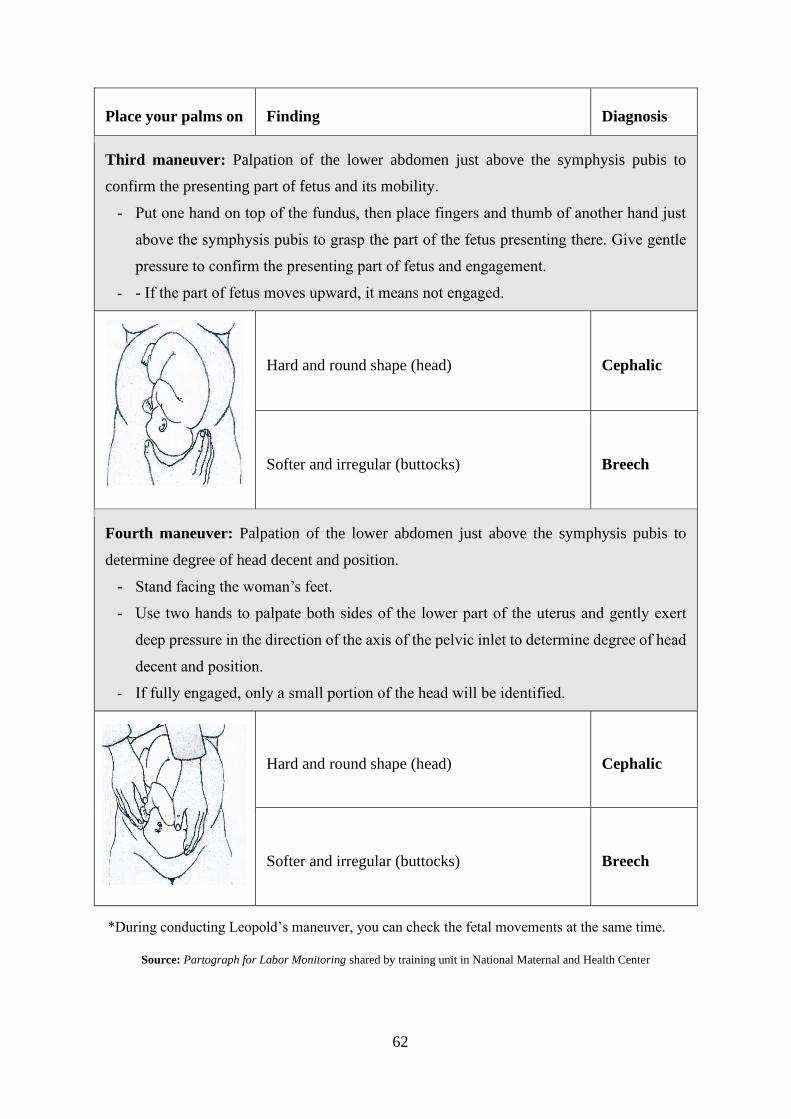
Place your palms on Finding Diagnosis
Third maneuver: Palpation of the lower abdomen just above the symphysis pubis to
confirm the presenting part of fetus and its mobility.
- Put one hand on top of the fundus, then place fingers and thumb of another hand just
above the symphysis pubis to grasp the part of the fetus presenting there. Give gentle
pressure to confirm the presenting part of fetus and engagement.
- - If the part of fetus moves upward, it means not engaged.
Hard and round shape (head) Cephalic
Softer and irregular (buttocks) Breech
Fourth maneuver: Palpation of the lower abdomen just above the symphysis pubis to
determine degree of head decent and position.
- Stand facing the woman’s feet.
- Use two hands to palpate both sides of the lower part of the uterus and gently exert
deep pressure in the direction of the axis of the pelvic inlet to determine degree of head
decent and position.
- If fully engaged, only a small portion of the head will be identified.
Hard and round shape (head) Cephalic
Softer and irregular (buttocks) Breech
*During conducting Leopold’s maneuver, you can check the fetal movements at the same time.
Source: Partograph for Labor Monitoring shared by training unit in National Maternal and Health Center

63
4-1-1-4-2. FHR auscultation
- Listen to FHR to confirm your palpation finding of lie and presentation.
- If the spot where you can hear the loudest fetal heart beat sound is in the upper part
of the abdomen, the fetus may be in breech presentation (Figure 4I).
Figure 4I. The spot to listen to FHR in breech presentation
Source: http://www.open.edu/openlearncreate/mod/oucontent/view.php?id=41&printable=1
4-1-1-4-3. Vaginal examination
- Vaginal examination is another way to confirm your palpation findings. When you
feel fetal feet, knee or hip, it is in breech presentation. When you feel a round,
smooth and hard shape with the sagittal suture, it is the head and Cephalic
presentation.
- For Cephalic presentation, further classification is found by vaginal examination.
(1) Vertex presentation: You feel the small fontanel.
(2) Sinciput presentation: You feel the big fontanel.
(3) Brow presentation: You feel the big fontanel and forehead or nose.
(4) Face presentation: You feel the parts of face such as the mouth.
<The small and big fontanels of fetal head>
- The sutures are the born joint formed where two bones meet. The fontanelle is
formed where two or more sutures meet (Figure 4J).

64
• The small (posterior) fontanel is located in the back of the fetal head (Occiput).
• The big (anterior) fontanel is located in the front of the fetal head (Sinciput).
- These fontanels are landmarks to know the fetal presentation and position.
Figure 4J. The fetus skull (View from above of the head)
Source: Marshall, J. and Raynor, M. (2014). Myles Textbook for midwives. 16th ed. Elsevier.
4-1-1-5. Complication with abnormal lies and presentation
4-1-1-5-1. Abnormal fetal lies and presentation: Transverse or oblique lie, shoulder
presentation, breech presentation, sinciput, brow and face presentation
- For these abnormal fetal lies and presentation, the presenting part of fetus is not
accommodated by the maternal pelvis. Those cases can result in obstructed or
arrested labor.
- Umbilical cord prolapse is more frequent for transverse or oblique lie and breech
or shoulder presentation when membranes are ruptured.
4-1-1-5-2. Compound presentation and neglected transverse
- It may cause obstructed labor or necrosis of the hands.

65
4-1-2. Fetal position in vertex presentation
4-1-2-1. Definition of fetal position
- Fetal position is the relationship between the fetal presenting part and maternal
pelvic (posterior, front, right, or left). Posterior is maternal spine, and front is
maternal symphysis pubis. The fetal position changes with fetal rotation.
4-1-2-2. Definition of fetal rotation
- Fetal rotation is the movement of turning the fetal head through the birth canal.
- In the normal process of labor, the fetus occiput gradually moves from the maternal
left/right side to the maternal symphysis pubis.
4-1-2-3. Classification of fetal position in vertex presentation
4-1-2-3-1. Occiput Transverse6 (OT)
- When the Occiput points to transverse sides of maternal pelvis, it refers Occiput
Transverse (OT).
- Most commonly, the fetus enters the pelvic inlet in OT position to fit the shape of
the pelvic inlet. It is classified into Left OT (LOT) or Right OT (ROT), depending
on which side the Occiput points to.
-
《Summary》
- Fetal position refers to the relationship between the fetal presenting part and
maternal pelvis (posterior or front, right or left).
- Fetal position changes with fetal rotation.
- It can be assessed by where you touch the Occiput with small fontanel in the
maternal pelvis during vaginal examination.
- In the normal process of fetal rotation, the fetal position changes from Occiput
Transverse to Occiput Anterior.
- When the fetal position is abnormal, there is a possibility of obstructed labor.

66
Figure 4K. Left Occiput Transverse (LOT) and Right Occiput Transverse (ROT)
Source: Myles Textbook for midwives. 16th ed.
4-1-2-3-2. Occiput Anterior (OA)
- When the Occiput locates at the maternal symphysis pubis (i.e. the anterior of
maternal pelvis), it refers to Occiput Anterior (OA).
- In the normal rotation process, the Occiput gradually moves toward the symphysis
pubis anteriorly from the original position (left or right of maternal pelvis). The fetus
head descends into the pelvis at the 45-degree angle of Left Occiput Anterior (LOA)
or Right Occiput Anterior (ROA) position.
Figure 4L. Left Occiput Anterior (LOA) and Right Occiput Anterior (ROA)
Source: Myles Textbook for midwives. 16th ed.
4-1-2-3-3. Occiput Posterior (OP)
- When the Occiput is located at the maternal supine (i.e. the posterior of maternal
pelvis), it refers to Occiput Posterior (OP).
- The Occiput sometimes moves toward the maternal spine from the original position
(left or right of maternal pelvis). The fetus descends into the pelvis at the 45-degree
angle of Left Occiput Posterior (LOP) or Right Occiput Posterior (ROP) position.

67
Figure 4M. Left Occiput Posterior (LOP) and Right Occiput Posterior (ROP)
Source: Myles Textbook for midwives. 16th ed.
4-1-2-4. The way to know the fetal position
4-1-2-4-1. Vaginal examination
(1) Feel the direction of the sagittal suture and the small fontanel or big fontanel.
- When you touch the small fontanel, you may feel a small dip-like triangle or
sharp angle like the letter “Y”. Confirm if it is formed by three born joints.
- On the other hand, when you touch the big fontanel, you may feel bigger dip
like a diamond shape (rhomboid). Confirm if it is formed by four born joints.
(2) Find the location of the small fontanel
- If you touch the big fontanel, the small fontanel is located on the opposite side (Table
4N)

68
Table 4N. Location of the small fontanel and fetal position
Location of the small fontanel Fetal position
Maternal left side
LOT
Maternal right side
ROT
Between maternal left side
and symphysis pubis
LOA
Between maternal right side and
symphysis pubis
ROA
Maternal symphysis pubis
OA
Between maternal left side
and maternal spine
LOP
Between maternal right side
and maternal spine
ROP
Maternal supine
OP
Source:Integrated Management of Pregnancy and Childbirth, Pregnancy, Managing Complications in
Pregnancy and Childbirth: A guide for midwives and doctors (2017) WHO, S-91

69
Table 4O. Location of the small fontanel and fetal position
Source:
http://intranet.tdmu.edu.ua/data/kafedra/internal/ginecology2/classes_stud/en/nurse/adn/ptn/2/Nursing
%20Care%20of%20Childbearing%20Family/02.%20Unit%20test%20II.htm
<Points to measure by vaginal examination>
- Direction of the sagittal suture: Small (Posterior) fontanel
or big (Anterior) fontanel.
- In normal process of fetal rotation, the fetal position changes from
Occiput Transverse (OT) to Occiput Anterior (OA).
- When the fetal position is abnormal, there is a possibility of obstructed
(prolonged or arrested labor).
- BUT we cannot evaluate the progress of labor with a one-point assessment.
Continuous monitoring is essential.
4-1-2-5. Complication with Occiput Posterior or Occiput Transverse position
4-1-2-5-1. Occiput Posterior (OP)
- OP is caused by a failure of fetal rotation9. It may interfere with the fetal descent
through the pelvis and cause prolonged labor or result in arrested labor.
O
O

70
- In OP position, Occiput puts strong pressure on a woman’s spine. The woman may
complain of continuous and severe backache with contractions9. It may lead to
maternal exhaustion and result in prolonged labor.
- Tell the woman to walk around or change position (e.g. kneeling position,
squatting), to reduce backache and encourage spontaneous rotation5(S-91),9. Observe
the delivery progress routinely, particularly be aware of uterine contractions and fetal
descent and monitor the location of the small fontanel.
4-1-2-5-2. Occiput Transverse (OT)
- OT is the position when the fetus enters the pelvis, so it may become to OA when
the fetus head rotates normally.
- If OT persists, even if there are effective uterine contractions, it may indicate
transverse arrest, in which the fetal head cannot pass through the pelvis.
- Observe uterine contractions and fetal descent with the location of the small
fontanel routinely, to confirm if the fetal rotation normally happens.

71
4-2. Well-being of fetus
4-2-1. Fetal Heart Rate (FHR)/BCF
4-2-1-1. Definition of FHR
- Fetal Heart Rate (FHR) is the speed of fetal heart beat measured by the number
of beats per unit of time, usually per minute.
4-2-1-2. The way to auscultate FHR
(1) Find the fetal presentation and position by abdominal palpation (See, 4-1. Fetal
lie, presentation, position, p.57).
(2) Find the spot to listen to FHR. The upper chest or upper back of fetus is where the
fetal heart beat is loudest (Figure 4P). Therefore, when the fetus is facing mother’s
left, the spot to listen FHR is right down (Figure 4Q).
《Summary》
- FHR is the most important indicator to know the fetus condition during labor.
- Find the spot to listen to FHR by palpation and listen to FHR for one minute.
Confirm if the FHR is within normal range.
- When FHR is slower than normal range, it may be caused by low oxygen and/or
cord compression. Change the mother’s position immediately and continue to listen
to FHR.
- When the FHR is more rapid than normal range, it may be caused by infection or
other causes. Continue to listen to FHR.
- If abnormal FHR persists, it may result in fetal death or neonatal asphyxia.
- For normal FHR, routine observation in recommended interval is important.

72
Figure 4P. The spot where the heartbeat is loudest Figure 4Q. In the case fetus is facing to mother’s left
Source: http://www.open.edu/openlearncreate/mod/oucontent/view.php?id=41&printable=1
- The spot to listen FHR is moving down with fetal descent. The spot comes down
with fetal descent and moves towards center as fetal rotation (Figure 4R).
Figure 4R. The changes of spot to listen to FHR during the 2nd stage of labor
Source: http://www.open.edu/openlearncreate/mod/oucontent/view.php?id=41&printable=1
<False heart beat sounds>
- If you hear a ‘swishy’ sound (shee-oo, shee-oo, shee-oo), you may be hearing
the pulse in the umbilical cord. Cord sounds cannot tell the actual FHR.
- You also may hear the mother’s radial pulse. Feel the maternal pulse together
with listening to FHR, to establish that what is being heard is the fetal, not
maternal, heart rate10.

73
(3) Listen to the FHR as follows:
(4) Record the FHR on the Initial Assessment Record Sheet, partograph and
delivery record1
4-2-1-3. Classification of normal and abnormal FHR
- Normal FHR at term pregnancy is 110 to 160 beats per minute1.
- FHR may be abnormal when it deviates from this range, either decreasing or
increasing.
4-2-1-4. Complication from a FHR deviated from normal range
4-2-1-4-1 . FHR <110
- The fetus receives oxygen from mother through placenta and umbilical cord. When
the fetal heart rate decreases during labor, uterine contraction may (1) Reduce
placental blood flow or (2) Cause compression of the umbilical cord.
(1) Reduce placental blood flow:
- During contraction, blood vessels in the uterus are compressed by the uterine
muscle. Then, the placental blood flow decreases and the oxygen supply
from mother to fetus reduces.
- In a result, the fetus cannot receive enough oxygen and the FHR decreases as
a response to the low oxygen status.
- When the fetus is strong enough to tolerate the insufficient oxygen supply,
FHR does not change or severely decrease. However, if the fetus is weak or
placental dysfunction is present, FHR becomes slow repeatedly or severely,
even with mild contractions6, and it may lead to fetal death or neonatal
asphyxia.
Duration: For 1 full minute
Timing: Immediately following uterine contraction1
Frequency: [1st stage] Every 30 minutes, [2nd stage] Every 5 mins

74
(2) Compression of the umbilical cord:
- During contraction, the umbilical cord is compressed by pressure between the
fetus and the uterus. Then the blood flow reduces, and it makes the FHR
decrease.
- Umbilical cord compression is likely to happen when: the cord is wrapping
around the fetus body or neck, a knotted or looped umbilical cord, or less
amniotic fluid.
- If it occurs repeatedly or persistently, the fetus may be in low oxygen status
and so it should be referred.
- Possible intervention for lower FHR <110 is to change the maternal
position to release the pressure to the umbilical cord.
- When the membranes are ruptured, check if there is a prolapsed cord and
thick-meconium stained amniotic fluid.
4-2-1-4-2. FHR >160
- A rapid FHR may be a response to maternal fever, infection, drugs causing rapid
maternal heart rate (e.g. tocolytic drug), or hypertension. In the absence of a rapid
maternal heart rate, a rapid FHR should be considered a sign of fetal distress6,
1(p87),5(S109).
Note: If FHR is abnormal (<110bpm or >160bpm), place mother on left side and check
FHR again.

75
4-2-2. Amniotic fluid
4-2-2-1. Normal characteristics of amniotic fluid
- Amniotic fluid is a clear liquid surrounding the fetus.
4-2-2-2. The way to observe amniotic fluid
- When the membranes are ruptured, the midwife should check the following
characteristics:
• Thickness (viscous or watery)
• Color (yellow, slight/light green, dark green or dark)
• Smell (odorless or foul-smelling)
• Volume (check whether the fluid draining is absent or not)
4-2-2-3. Complications from abnormal characteristics of amniotic fluid
4-2-2-3-1. Blood-stained fluid
(See, 6. Observe maternal condition, 6-6-3. Complication with abnormal bleeding, p.108)
4-2-2-3-2. Thick meconium-stained amniotic fluid
- Meconium is the stool of fetus.
- Sometimes the fetus passes meconium into the amniotic fluid during labor when the
fetus has low oxygen status.
- Meconium-stained amniotic fluid is seen frequently among term fetus, so slight
meconium-stained fluid is not an indicator of fetal distress5,6.
《Summary》
- Color of amniotic fluid is another indicator of fetus condition during labor or
internal bleeding from placenta.
- If the membranes are ruptured, check the color in a vaginal examination.
- If the thick meconium-stained fluid is found, check FHR. If the FHR is abnormal,
it indicates fetal distress.

76
- However, thick meconium-stained amniotic fluid is one of the signs of fetal
distress, especially if FHR abnormality is present1.
- Meconium Aspiration Syndrome (MAS), which is a respiratory problem with
inhalation of meconium stained-fluid may be indicated.
4-2-2-3-3. Foul smelling amniotic fluid
- It may indicate intra-amniotic infection and needs further treatment with referral.
4-2-2-3-4. Absence of amniotic fluid with rupture of membranes
- An absence of fluid draining after rupture of membranes is an indication of reduced
volume of amniotic fluid5.
- It indicates that the cord compression most likely happened. Listen to the FHR
properly and observe closely when an abnormal FHR is heard.

77
Section 5. Assess the delivery progress
SUMMARY
- The objectives of this section are: (1) To assess the delivery progress by the
condition of 4Ps (Power, Passage, Passenger, and Psychology), and (2) To
identify the stage of labor.
- Assessment of the delivery progress requires understanding the 4Ps – Power,
Passage, Passenger, and Psychology.
- The delivery process is divided into four stages. The first and second stages are
focused in this section. The criterion to distinguish the stages is ‘cervical
dilatation’.
- 4Ps – Power, Passage, Passenger, and Psychology – should be assessed at every
stage (and phase) of labor.

78
5-1. 4Ps – Power, Passage, Passenger, and Psychology
5-1-1. Power
- Power means a force to push out the baby from uterus to our world. It is uterine
contraction or labor pain.
- Uterine contraction has several aspects, which are ‘duration’ of each contraction,
‘frequency’ (usually measured as times during 10 minutes), and strength (though it
is often subjective indicator). These three aspects should be assessed separately.
- Uterine contractions should be strong enough, otherwise the delivery will not
progress. However, it should also be noted that strong uterine contractions
themselves will compromise the baby in the uterus because strong contractions
decrease blood flow in the uterus which results in a decrease in oxygen supply to the
baby.
- Therefore, careful monitoring of uterine contractions with baby’s condition is
essential in delivery care.
《Summary》
- - Nice delivery progress depends on four components, which are called the 4Ps: (1)
Power, (2) Passage, (3) Passenger, and (4) Psychology.
- (1) Power is a force to deliver the baby. Therefore, it is ‘uterine contraction’ or
‘labor pain’.
- (2) Passage means the way from uterus to outside of the maternal body. It consists
of uterine cervix, pelvis (bone), vagina, and perineum.
- (3) Passenger is the baby her/himself (shape and size).
- (4) Psychology of mother strongly affects entire process of labor, especially uterine
contractions.

79
5-1-2. Passage
- Passage means the way from the uterus to outside of maternal body. It consists of
pelvis (bone), uterine cervix, vagina, and perineum.
- Soft and fully dilated cervix, sufficient form and size of the pelvic bone, soft vagina
and perineum are required for smooth delivery.
- Cervical dilatation continuously changes, so regular monitoring is required.
- The Pelvic bone does not change, but an important thing is the balance between the
bone size and the baby (the passenger). Even if the size of the pelvic bone is
sufficiently large, if the baby is very large the passage (pelvic bone) will block the
baby. On the other hand, a small baby can pass through a small pelvis.
- Therefore, we always have to examine the balance between the passage and the
passenger.
5-1-3. Passenger
- Passenger is the baby (shape and size). The largest part of the baby is the head.
So how the head is going into the pelvis is very important for normal birth. (See, 4.
Observe fetal condition, 4-1. Fetal lie, presentation and position, p.57)
- As mentioned above, proportion between the passage and the passenger is
important.
- For normal birth, it is necessary to assess ‘descent’ of baby’s head. It can be
measured as the degree to enter fetal head into pelvis.
5-1-4. Psychology
- Psychology (mental status) of the mother is also an important factor in delivery
progress. If the mother has fear or anxiety, it definitely decreases important internal
hormones (i.e. oxytocin, endorphins) which facilitate labor. (See, 6. Observe
maternal condition, 6-7. Psychological state, p.110)

80
5-2. Decide the stage of labor
5-2-1. Definition of the stage of labor
- The delivery is classified into four stages. Only the first stage of labor is divided
into latent and active phases (see Table 5A).
(1) The latent phase of the first stage is defined as cervical dilatation ‘3cm or less’
in Cambodia.
(2) The active phase of the first stage is defined as cervical dilatation more than 3cm
(i.e. 4cm+) until full dilatation of the cervix.
- The second stage of labor is defined as from the full dilatation of the cervix until
delivery of the baby.
《Summary》
- Stages of labor consist of four: first, second, third, and fourth.
- The first stage of labor is divided into two phases: latent and active.
- Cervical dilatation (in centimeters) is an indicator to determine the stage and phase.
However, regular uterine contraction (labor pain) should accompany the dilatation.
- Cervical dilatation itself does not indicate the onset of labor.

81
Table 5A. Definition of stages and phases of labor
Stage Phase Definition
First Latent From the onset of labor,
until 3 cm of cervical dilatation
Active From more than 3cm of cervical dilatation,
until full dilatation of cervix
Second - From full dilatation of cervix,
until birth of baby (expulsion of fetus)
Third - From birth of baby,
until expulsion (delivery) of placenta
Fourth From expulsion of placenta,
until two hours

82
Note: Different definitions of latent and active phases in the first stage of labor
- World Health Organization (WHO) has updated the definition of active phase in
the year 2018. The new definition of active phase is from 5 cm until full dilatation.13
- Because accumulated scientific evidence shows that rapid progress of delivery
(cervical dilatation and effacement) is observed from 5 cm of dilatation. It also
shows that duration of latent phase is varies by each pregnant woman, therefore, it
is not necessary to put a cut-off at eight hours.
- This threshold considers reducing an early referral and unnecessary obstetrical
intervention for woman with slower but normal progress.
- However, the core members for this guide has decided to use same definition in the
previous version of WHO recommendations. Therefore, we keep the definition of
active phase is from 3 cm until full dilatation of cervix. It is along with the
Cambodian national protocol.
- Our intention to keep the previous threshold is to prevent delayed referral of
prolonged labor from health centers, especially in rural areas. Therefore, we
should not immediately apply the old recommendation of active phase and
definition of prolonged labor in the level of referral hospital. The new WHO
recommendation will be examined in another discussion for higher level of health
facilities.

83
5-3. Practice of assessment of delivery progress
5-3-1. Power
- ‘Power’ is the uterine contraction. It is a power to push the baby out from the uterus
through the birth canal. Thanks to the power, there will be changes in passage
(uterine cervix and vagina) and passenger (fetus, especially head molding and
rotation).
- Several components of the uterine contractions should be measured and assessed.
The components are duration of contraction, frequency of contractions, and
intensity (strength) of contraction. How to measure each is as follows.
• Duration: count the duration of one contraction in seconds
• Frequency: count the number of uterine contractions in 10 minutes
• Intensity (strength): observe the woman’s reaction to uterine contraction and
feel the firmness of abdomen.
- The characteristics of each component changes gradually according to the stage of
labor. It is shown in Table 5B.
《Summary》
- It is necessary to assess the delivery progress comprehensively by observation of
the 4Ps.
- Condition of the components of 4Ps (Power, Passage, Passenger, and Psychology)
change gradually according to the stage of labor.

84
Table 5B. Changes of characteristics of uterine contraction
Stage of
labor Duration Frequency Intensity (strength)
(Pre-labor) Up to
30 seconds
Once in 15-20 minutes Weak (sometimes subtle),
no constant pain and
having time to rest
1st stage /
Latent
phase
Between
20 and 40
seconds
Gradually increases:
from 1 or 2
contractions, to 4 or 5
per 10 minutes
Weak to moderate, no
constant pain and having
time to rest
1st stage /
Active
phase
Up to
60 seconds
Up to 5 contractions per
10 minutes
Moderate to intense, no
constant pain and having
time to rest
2nd stage Around
60 seconds
ditto Intense, no constant pain
and having time to rest
- As it is shown in Table 5B, uterine contraction usually starts from ‘short and less
frequent’. It gradually becomes longer, more frequent, and stronger. However, please
note that its progress varies by every pregnant woman. Even if the duration is
shorter and the frequency is less than expected, you can continue to observe the
woman as long as labor progress (cervical dilatation and fetal descent) is confirmed.
<Cautions in observation of uterine contraction>
- If the contraction is ‘too weak’ or ‘too strong’, it can indicate abnormality.
- ‘Too weak’ can be evaluated if the duration is short (up to 30 seconds); frequency
is less than twice per 10 minutes; and intensity is weak. It can be a false-labor (pre-
labor) stage. If it is a false labor, the contractions will disappear without any change

85
in the uterine cervix. So continuous observation is necessary. Please do not confuse
this false-labor with ‘prolonged latent phase’ (described afterwards).
- ‘Too strong’ can be evaluated if the duration of contraction is more than one
minute or continuous; frequent uterine contractions (6 times or more per 10
minutes); or the woman complaints that the contraction is too strong. Too strong
contractions can be a sign of placental abruption (DPPNI); uterine rupture or
pre-rupture; or inappropriate use of medicine (oxytocin, misoprostol, traditional
herbal medicine, etc.) that stimulates uterine contractions. Immediate evaluation of
the fetal and maternal status is required if you find this type of abnormal contractions.
5-3-2. Passage
- ‘Passage’ means the pathway from the uterus to outside of the maternal body.
- The pelvis is the most important part of ‘passage’. It consists of bones and has a
canal inside. Examine the shape and size of the pelvis carefully because normal birth
is not possible if there is a severe deformity in the pelvis. Deformities in hip joints
may also be a barrier to normal birth.
- The uterine cervix is dilated and effaced by uterine contractions and by internal
pressure of uterus. Dilatation can be measured by the fingers when you touch the
cervix. It is better to practice the measurement by using a model of cervical dilatation.
Cross-checking vaginal examination by supervisors (trainers) is also an effective
method of learning for trainees.
- Effacement can be also measured by the finger. It is the length of the cervix. If the
dilatation is small (e.g. 2 or 3 cm), you may feel the cervix is long. However, when
the cervix is fully dilated, the length itself usually disappears. It means that the cervix
is fully effaced. Since the dilatation is caused passively, monitoring other
components – power, passage passenger, and psychology – is essential, especially if
you think that the dilatation is not sufficient.

86
5-3-3. Passenger
5-3-3-1. Monitoring items
- The main passenger is the fetus, who is going to come through the narrow pathway.
The fetus (or baby) tries to adapt her/his head to the shape of mother’s pelvis. This
adaption consists of ‘flexion’, ‘rotation’, and ‘molding’.
- During this adaption process, the baby’s head is going into pelvis and descending
towards outside. It is necessary to know anatomy of pelvis and fetal head in three-
dimensional manner in order to understand the process.
What we have to monitor and assess is the entire process of descent of the fetal head,
rotation, and molding.
5-3-3-2. Anatomical background
- Flexion means bending a head forward. It enables the head to go into the pelvis
with its smallest circumference. However, if the fetal head is too small, or the
maternal pelvis is too large, this flexion may be only in little degree or even not
happen.
- Rotation is a process to adapt the form of the head inside of the pelvic canal.
Fetal head is an ellipse (oval) shape if we see it from the top. Shapes of the pelvic
inlet and outlet are also ellipses, but the directions of the long-axis and short-axis are
different. Anteroposterior distance is shorter than transverse distance at the inlet. On
the other hand, transverse is shorter than anteroposterior at the outlet. This is the
reason why the baby’s head should rotate in the pelvis.
- Molding is another mechanism to make the fetal head smaller. The baby’s head
consists of two frontal bones, two parietal bones and one occipital bone. The
borders of each bone have small openings, which is called as suture and fontanelle.
Molding is an anatomical alteration in shape, which is characterized by an
overriding of head bones at the sutures. It allows a considerable reduction in the
size of the presenting diameters.

87
- Caput succedaneum is a swelling (edematous) of the scalp at the presenting part
of head. It is caused by external pressure on the fetal head from the birth canal.
5-3-3-3. Measurement of fetal head descent
- It is the abdominal method to assess descent of the fetal head. The level of the head
is measured by abdominal palpation with fingers by placing the radial margin of
finger above the symphysis pubis (which is anterior brim of pelvis) and expressed in
terms of fifths above the brim like 5/5, 4/5, 3/5, 2/5, 1/5, or 0/5 (Figure 5C).
- The points to be confirmed are the entire head, ‘sinciput’ and ‘occiput’. Several
images of the measurement are shown below (Figure 5D, 5E). The picture below
shows an image of head and two points of the head (sinciput and occiput). The
picture with actual photo (Figure 5D) also indicates how baby’s head rotates
according to the fetal descent. You can understand that the rotation starts when the
baby’s head comes near to the pelvic floor.
5/5
2/5
Figure 5C. The level of head measured by abdominal palpation with finger
Source: Partograph for Labor Monitoring shared by training unit in National Maternal and Health Center

88
Figure 5D. Fetal head decent and rotation

89
S: Sinciput - O: Occiput
Figure 5E. Fetal head decent
Source: https://www.madeformums.com/pregnancy/what-does-it-mean-when-your-babys-head-is-engaged/
5-3-3-4. Measurement of molding and Caput succedaneum
5-3-3-4-1. Molding
- The level of molding is measured as changes in the fetus’s skull bones. It is
classified into four degrees as follows:
《Summary》
- Molding and Caput Succedaneum can be one of the indicators of cephalopelvic
disproportion (CPD) or prolonged labor.
- In a vaginal examination, touch the fetal head if there is the overlap of the parietal
bones (Molding) and a swelling of the scalp at the presenting part (Caput
Succedaneum).
- Those fetal head changes are formed by strong pressure from the birth canal.
- Severe molding may indicate the possibility of CPD.

90
0 The bones are separate and head joints are easy to feel.
+ The bones on the top of the head touch each other only.
++ The bones on the top of the head slightly overlap (one over the other)
+++ The bones on the top of the head are predominantly on top of the other
(overlap each other to a significant degree)
Figure 5F. The level of molding
Source: https://elearning.rcog.org.uk//easi-resource/maternal-and-fetal-assessment/examination/moulding
- Please remember that this level of molding depends on the relative relationship
between the fetal head size (passenger) and pelvic bone size (passage).
- Significant level of molding (+++) indicates that the pelvis is narrow to the fetal head.
But it does not always mean Cephalopelvic disproportion (CPD). Because the
molding itself is a strategy for the fetus to pass through the narrow canal. As long as
the progress of labor is observed, for example, nice descent of the fetal head with
appropriate uterine contractions can indicate that the process will be normal.
- In other words, DO NOT make a diagnosis of CPD with only the findings of molding.
- However, if you feel that molding is significant and fetal head is still floating (not
engaged), it is a sign to suggest CPD.
5-3-3-4-2. Caput Succedaneum
- It is difficult to ‘standardize’ the measurement of Caput Succedaneum, because only
vaginal examination is the way to know it and it is frequently difficult to touch all
the part of fetal head.

91
- However, Caput sometimes hampers evaluation of molding due to thick edema in
the head skin, which prevents to feel the overlapping of the bone.
- Therefore, you should consult to your colleagues to evaluate it together.

92
5-4. Assessment of duration of labor
5-4-1. Criteria for the assessment
The criteria are defined only for the first stage of labor, but for the latent and active
phases separately.
Table 5G. Criteria of duration of labor in the latent and active phases in the first stage.
Phase Duration of labor Prolonged labor
Latent 8 hours
(Confirm if real labor pain OR
false labor pain!)
More than 8 hours in the latent phase
(Confirm if real labor pain OR false
labor pain!)
Active At least 1 cm of cervical
dilatation per hour
Less than 1 cm of dilatation during
the active phase
Note: The core members of this guide decided to use a previous version of WHO
partograph. Therefore, the criteria to evaluate the duration of labor depend on it.

93
5-5. Conditions to be considered
As it is explained in 5-1. 4Ps – Power, Passage, Passenger, and Psychology, there are
four important factors for progress of labor.
- ‘Power’, it is ‘labor’ or ‘uterine contraction’, is an essential component of delivery.
If you think power is not sufficient during latent phase, please consider the possibility
of ‘false labor’.
- Inappropriate power in active phase may suggest ‘weak pain’.
- There are several important causes of ‘weak pain’, therefore, careful investigation of
the cause and appropriate management are required.
- ‘Passenger’ and ‘Passage’ are always in their relative relationship. In other words,
their conditions and relationship should be evaluated together. Suppose there is a
car at very slow speed. Guess the reason why. It may be due to bad road condition,
which is ‘Passage’ factor. It may be due to the bad condition of the car itself, which
is the ‘Passenger’ factor. It may be due to big size of the car in a narrow road, which
is caused by both ‘Passenger’ and ‘Passage’ factors.
- Please remember the 4Ps (Power, Passage, Passenger, and Psychology) and their
relationships when you record the partograph.
- The timing of continuous monitoring is shown in ‘Annex 2 Observation Time chart’.
- We cannot evaluate the progress of labor with one-point assessment. After initial
assessment, please continue regular monitoring according to Annex 2.

94
Section 6. Observe maternal condition
SUMMARY
- Objectives of this section are: (1) To identify any abnormal status in maternal
condition, and (2) To provide immediate initial treatment to the case before referral.
- It’s necessary to check the vital signs (BP, pulse, body temperature), urinalysis and
bleeding to confirm if there is any abnormal status in maternal condition during
labor.
- Pre-eclampsia, intra-uterine infection, bleeding are most fatal complications during
labor both for the woman and fetus.
-

95
6-1. Blood pressure
6-1-1. Definition of Blood pressure and proteinuria
6-1-1-1. Blood Pressure (BP)
When a heart beats, the blood is pushed out from the heart to the body. BP is the pressure
of the circulating blood against the blood vessels. Two kinds of BP are measured:
• Systolic BP: highest BP during the heart beat
• Diastolic BP: lowest BP between the heart beat
6-1-1-2. The way to measure
6-1-1-2-1. Blood pressure
- Measure BP in a resting state and at intervals of uterine contraction. This is because
that BP is elevated by body activity, uterine contractions, and the pain and
psychological factors, such as distress or nervous.
- When the Systolic BP ≥140-160mmHg OR Diastolic BP ≥90-110mmHg:
(1) Let the woman rest and measure BP 15 minutes later.
(2) Check the symptoms with hypertension. (See, 6-2. Symptoms with
hypertension, p.98)
《Summary》
- Maternal hypertension affects several organs and leads to dysfunctions. It can
result in fetal distress and maternal complications. It may develop to eclampsia.
- Measure BP in a resting state and interval of uterine contraction.
- When the Systolic or Diastolic BP is severe hypertension, refer the woman.
- When the Systolic or Diastolic BP is moderate hypertension, let the woman rest and
measure BP 15 minutes later. Also check the symptoms with hypertension
(headache, blurred vision, and epigastric pain).

96
6-1-1-3. Classification of BP
Normal BP
Moderate
Hypertension
Severe
hypertension
Systolic BP < 140 mmHg ≥ 140mmHg ≥ 160mmHg
AND OR OR
Diastolic BP < 90mmHg ≥ 90mmHg ≥ 110mmHg
6-1-1-4. Complication with hypertension
6-1-1-4-1. Systolic BP ≥ 160 mmHg OR Diastolic BP ≥ 110 mmHg
- It is said pre-eclampsia is caused by an abnormal placenta implantation affecting
the placenta blood flow, and the poor placenta dysfunction may lead to hypertension.
The Maternal hypertension damages the vessels of several organs and leads to
dysfunctions (See, 1. Immediate response to emergency for pregnant woman, 1-4.
Abnormal vital signs – Elevated Diastolic Blood Pressure, p. 13).
- Especially, it may affect kidney, brain, eyes, liver, and lung and contribute to the
following complications:
- Placental dysfunction causes in sufficient supply of oxygen and nutrients. As a result,
fetus growth is restricted during pregnancy and there is high possibility of low birth
weight infants. FHR easily decreases during labor due to insufficient supply of
oxygen.
- Placental dysfunction may cause placental abruption.
- Kidney dysfunction may cause proteinuria (See, 6-5. Urinalysis, p. 104).
- Liver dysfunction may cause epigastric pain, nausea, or vomiting (See, 6-2.
Symptoms with hypertension, p. 98).
- Damaged brain vessels may cause headaches. When the damage is severe or
persisted, it may develop the onset of convulsions (See, 6-2. Symptoms with
hypertension, p. 98).

97
- Damaged eyes vessels may cause visual symptom (blurred vision) (See, 6-2.
Symptoms with hypertension, p. 98).
- Lung dysfunction may cause an increase of respiratory rate or breathing difficulty.
6-1-1-4-2. Systolic BP ≥ 140 mmHg OR Diastolic BP ≥ 90 mmHg
- Even moderate hypertension has risk of above complication and risk to elevate BP
during labor. BP must be closely observed every hour1(p22) and check whether the
woman has any signs with hypertension (See, 6-2. Symptoms with hypertension,
p.98).
- FHR should be monitored closely, every 15 mins at the first stage and every 5 mins
at the second stage.

98
6-2. Symptoms with hypertension
6-2-1. Symptoms with hypertension
Some symptoms may occur as BP elevates or hypertension persists. Those symptoms can
be an indicator of the severity of hypertension.
6-2-2. Confirm the symptoms with measuring BP
- Ask if she has:
(1) Severe headache
(2) Blurred vision
(3) Epigastric pain
6-2-3. Complications with hypertension accompanied with symptoms
6-2-3-1. Severe head ache, blurred vision, and epigastric pain
- These symptoms may indicate that the brain and liver are damaged by hypertension.
The woman may be severe pre-eclampsia, and there is a risk to develop to
eclampsia.
- Even she does not have those symptoms, when the woman starts to complain, you
must check BP immediately.
《Summary》
- Some symptoms may occur as BP elevates or hypertension persists. Those
symptoms can be an indicator of the severity of hypertension.
- When the woman has hypertension, check the major three symptoms: severe
headache, blurred vision, and epigastric pain. Because those symptoms indicate the
brain and the liver are damaged by maternal hypertension.
- When she has those symptoms with hypertension, she may be pre-eclampsia or
eclampsia.

99
6-3. Pulse
6-3-1. Pulse rate in pregnancy
- The normal range of adult pulse is 60-100 bpm.
- For pregnant women, the resting pulse rate increases approximately 10-20 bpm6,9
more than non-pregnant period.
- Physiologically, pulse becomes rapid with exercise, pain, or emotional status and
becomes slow during sleeping or relaxed status.
6-3-2. The way to measure adult pulse
- The pulse is normally measured in wrist (radial artery).
- When you cannot find a pulse, check at another place such as neck (carotid artery)
or between the lower abdomen and upper thigh (inguinal artery). Measure it in
a resting state and interval of uterine contraction.
6-3-3. Complication with abnormal adult pulse
6-3-3-1. Tachycardia > 100 bpm
- When the woman is not bleeding and has no abnormal pain, which may indicate
《Summary》
- The rapid pulse is affected by bleeding, fever, dehydration, anemia, heart disease,
etc.
- The slower pulse is affected by heart disease or may be caused by heart failure.
- When rapid pulse is accompanied by low Systolic BP, it may indicate the shock due
to bleeding.
- Count the pulse in a resting state and interval of uterine contraction.

100
internal bleeding, or the rapid pulse may be caused by other reason such as fever,
dehydration, anemia, heart disease, etc.
- If the rhythm of heart beat does not normal, refer the woman.
- Check the fever and other infectious symptoms (See, 6-4. Body temperature, p.101).
- If the woman does not drink enough water, encourage her to drink more.
101
6-4. Body temperature
6-4-1. Body temperature in pregnancy
- The body temperature in pregnancy rises at first trimester due to a hormonal action,
but it falls to same level as non-pregnancy at second trimester.
6-4-2. The way to measure temperature
- The body temperature of an adult is normally measured in an armpit.
- The censer of the thermometer should be put in the center of the armpit. When the
armpit is too sweaty, wipe it first.
6-4-3. Classification of body temperature in pregnancy
- An abnormally high body temperature is >38.0℃.
6-4-4. Complications of high temperature
- When the woman has an infection, the body temperature usually rises because it tries
to fight with the cause of infection.
- In addition to the high body temperature, there must be various infection signs.
《Summary》
- When the body temperature is >38.0℃, any infection can be considered.
- Measure the body temperature in an armpit
- If the fever is due to infection, there must be other infection signs
- Intrauterine and fetus infection is most common infection during labor, especially
after rupture of membranes. Check for other symptoms such as foul-smelling
discharge or amniotic fluid, or tenderness of the lower abdomen.

102
6-4-4-1. > 38.0℃
Four major causes of infection are (1) Intrauterine and fetus infection, (2) Urinary tract
infection, (3) Respiratory infection, and (4) Malaria.
(1) Intra-uterine and fetus infection
- Vaginal bacterial infection sometimes goes up to cervix and reaches to the
membranes. The membranes’ infection further expands to amniotic fluid,
umbilical cord, and finally reaches the fetus.
- If the membranes are ruptured, the infection expands more quickly. The woman
may show maternal rapid pulse, foul-smelling discharge and tenderness of lower
abdomen. Fetal heart rate may increase, and amniotic fluid may smell foul. (See,
1. Immediate response to emergency for pregnant woman, 1-5. Abnormal vital
signs –Fever, p.15).
(2) Urinary tract infection
When the urinary tract is infected, the woman may complain of burning, pain or
difficulties urinating, increased frequency and urgency of urination, and abdominal pain
or spiking fever and chills.1 (See 1. Immediate response to emergency for pregnant
woman, 1-5. Abnormal vital signs –Fever, p.15)
(3) Respiratory infection
When lungs are infected, the woman may complain of difficulty breathing, or cough
with sputum. In this condition, pneumonia or tuberculosis can be suspected.
(4) Malaria:
When the woman comes from a malarial area, she may be infected with malaria. She
may complain of fever with chills, sweating, headache, or muscle joint pain.
6-4-4-2. > 37.5℃
- First of all, check for infection signs mentioned above. If there are any of the signs,
the woman should be referred.
- If there are no infection signs, the body temperature must be closely observed every

103
hour.1(p22) Encourage the woman to drink more, and confirm that the room
temperature is comfortable for the woman, although it should be at over 25℃ for a
newborn baby.

104
6-5. Urinalysis
6-5-1. Definition of Proteinuria in pregnancy
- Proteinuria is the presence of excess protein in the urine. A normal kidney does
not allow much protein to pass through, therefore protein in urine is of very little
amount.
- During pregnancy, the amount of protein in urine increase. This is because the burden
on the kidneys increases due to increased blood volume. However, if the amount of
protein in urine is abnormally high, it may indicate the decline in kidney function.
6-5-2. Definition of pre-eclampsia and eclampsia
- Pre-eclampsia is a condition of pregnancy characterized by hypertension and
proteinuria presenting after 20 weeks9. The presence of proteinuria changes the
diagnosis from hypertension to pre-eclampsia5.
- Eclampsia is a condition characterized by the onset of convulsions in a pregnant
woman with pre-eclampsia. The onset of convulsion changes the diagnosis from
pre-eclampsia to eclampsia.
《Summary》
- If there is proteinuria, it may indicate the decline in kidney function due to
hypertension.
- Test the proteinuria at admission with dipstick.
- Proteinuria of (+/-) or (+) is normal as physiological pregnancy changes.
- Proteinuria of (++) or (+++) is one of the indicators to diagnose pre-eclampsia.
- In case you do not have urinary dipstick, judge the referral with only BP.

105
6-5-3. The way to measure proteinuria
- Dip the coated side of a dipstick in urine sample and wait the specified time (see
the instructions on the container of dipstick).
- Compare the result with the color chart on the container, and determine the result
with symbol of (–) and (+).
6-5-4. Classification of Proteinuria
Normal: (-), (+/-) , (+)
Proteinuria: (++)
Severe proteinuria: (+++)
6-5-5. Complication with proteinuria accompanied by hypertension
(See, 1. Immediate response to emergency for pregnant woman, 1-4. Abnormal vital signs
– Elevated Diastolic Blood Pressure, p.13 and 6-1. Blood pressure, p.95)
6-5-5-1. Proteinuria (+++)
When the kidney function declines, protein leaks into urine. It indicates severe pre-
eclampsia and causes several complications. It may develop into eclampsia with the
onset of convulsion.
6-5-5-2. Proteinuria (++)
- If BP is normal, it may be normal proteinuria as pregnancy. BP should be observed
routinely.
- If hypertension is present, it indicates pre-eclampsia and causes several
complications.
- If hypertension and symptoms with hypertension are present, it indicated severe
pre-eclampsia. It may develop eclampsia.
- If the woman does not take enough fluid, encourage to drink.

106
6-5-5-3. Table of pre-eclampsia, severe pre-eclampsia and eclampsia
107
6-6. Bleeding
6-6-1. Physiological bleeding during labor
6-6-1-1. A show
- A show is a small amount of blood or blood-stained mucus, which are shown
before or after a few hours from labor starts. It is caused by the detachment of the
membranes from cervix when the cervix starts to dilate.
6-6-1-2. Bleeding from birth canal
- During labor, bleeding more than a show is often observed. This is from cracks or
scratches in cervix or vagina when the fetus passes the birth canal.
- Amount of bleeding may seem to be increased when the membranes are already
ruptured, and certain amount of amniotic fluid is leaked.
6-6-2. The way to know/measure the bleeding during labor
- When woman complaints the bleeding, you must check the bleeding condition. In
addition, the bleeding should be always observed and recorded when you conduct
vaginal examination.
- If woman use sanitary napkins during labor, observe the pad or measure it if bleeding
《Summary》
- When the bleeding is more than a show, some complications can be considered.
- Check the bleeding condition when the woman complains of any bleeding. It also
should be observed when you conduct a vaginal examination.
- Observe or measure the napkins or cloths a woman wears.
- Other signs of complications (abnormal pain and uterine contraction) should be
observed together.

108
looks increasing. You need to know the weight of the original napkin.
- In the case woman does not use napkins, observe how the woman’s cloths is soaked.
6-6-3. Complications with abnormal bleeding
6-6-3-1. Bleeding more than 100 ml since labor began/Pad or cloth soaked in < 5 min.
- Three possible causes of abnormal bleeding during labor are: (1) placental
abruption, (2) ruptured uterus, and (3) placenta previa. Bleeding should be
always checked with abnormal labor pain (See, 2. Listen to woman’s complaint,
2.1 Bleeding, p.20). Fetal heart rate severely decreases in the case of bleeding due to
these causes.
(1) Placental abruption is the separation of the placenta which is located in the
middle or upper part of uterus.
- Bleeding comes out of uterus as vaginal bleeding or blood-stained amniotic
fluid, but sometimes there is internal bleeding inside of uterus.
- The woman often has severe abdominal pain or tenderness, and the firmness
of uterus is quite hard. (See, 2. Listen to woman’s complaint, 2-3-4.
Complications with abnormal pain, p.26).
(2) Ruptured uterus is a rupture of the uterine wall or muscle at the previous
uterine incision or scar.
- Vaginal bleeding may be not so severe, despite heavy intra-abdominal
haemorrhage5.
- The woman complains severe abdominal pain (See, 2. Listen to woman’s
complaint, 2-3-4. Complications with abnormal pain, p.26).
(3) Placenta previa is a condition the placenta is located in the lower part of uterine,
and the placenta covers the cervix partially or totally.
- Severe bleeding occurs when the cervix starts to dilate due to the separation
of placenta.

109
- Normally a woman does not complain of severe pain. (See, 2. Listen to
woman’s complaint, 2-1. Bleeding, p.20).
6-6-3-2. Bleeding more than usual
- If there is any abnormal labor pain and abnormal FHR, it may be bleeding from
birth canal.
- Continue to observe the quantities of bleeding with monitoring FHR and abnormal
pain. If fetal distress or abnormal labor pain accompanies with bleeding, it may
indicate placental abruption or ruptured uterus (See, 2. Listen to the woman, 2-3.
Uterine contraction and labor pain, p.25).

110
6-7. Psychological state
6-7-1. Psychological state during labor
- When a woman is in labor, it is normal to have mixture emotions of excitement, hope,
anxiety or fear9.
- The psychological state is also influenced by external environment, such as birth
companion, the environment of the delivery place and the attitude of the care giver.
6-7-2. Observation of psychological state14
- Look at the woman’s facial expression, mood, and listen to her complaints well.
Panic or shouting is often caused by excessive stress and anxiety.
6-7-3. Influence of distress and anxiety during labor14
- Fear or anxiety disturb oxytocin production, that is necessary hormone of delivery
process. It may result in slowing labor and slower dilatation of the cervix due to
weak uterine contractions.
- When the woman has excessive stress, adrenalin is released. Adrenalin reduces the
production of oxytocin and endorphin (that is another hormone to be calm and pain
relieving). The delivery progress may be disturbed due to low oxytocin level, and
woman feels more pain due to low endorphin level.
《Summary》
- Distress or strong anxiety can affect the delivery progress.
- Always care about the woman’s mood and facial expression and listen to her
complaints during labor.
- Avoid leaving the woman alone and make a comfortable environment.

111
- Do not leave the woman alone and ask the companion to stay with her. Make a
comfortable environment such as with silent and dim light (low-lighted).
- Refer to The Guide to Individualized Midwifery Care for Normal Pregnancy and
Birth14 for more possible psychological supports for women.
<Influence of distress and anxiety during labor>

112
Annex 1

113

114

115

116

117

118
Annex 2
Observation Time Chart
In Latent Phase
* Refer immediately to comprehensive emergency obstetric facility (CEmONC/ CPA 2 or CPA3) if she has any reason for refer.
Before referring, please provide first aid properly and check Gestational Age, Onset of labor (antepartum, intrapartum,
postpartum).
4. Observe fetal condition Normal Risk to be complicated *
Listen Fetal Heart Rate Every 30 min1(p59) Every 15 min1(p88)
Amniotic fluid (if rupture just now) Check immediately
Amniotic fluid (if ruptured) Every 4 hours (At Vaginal examination) 1(p60)
5. Assess the delivery progress Normal Risk to be complicated *
Cervical dilatation by vaginal examination Every 4 hours1(p59)
Fetal descent Every 4 hours (Before vaginal examination)
Uterine contraction (frequency, duration, strength) Every 1 hour1(p59)
6. Observe maternal condition Normal Risk to be complicated *
Vital sign (Blood Pressure, Pulse, Body Temperature) Every 2 hours1(p59) Every 1 hour4(p22), 1(p.22-24)
Bleeding Every 4 hours (At Vaginal examination) 6(p341)

119
In Active Phase
* Refer immediately to comprehensive emergency obstetric facility (CEmONC/ CPA 2 or CPA3) if she has any reason for refer.
Before referring, please provide first aid properly and check Gestational Age, Onset of labor (antepartum, intrapartum,
postpartum).
4. Observe fetal condition Normal Risk to be complicated *
Listen Fetal Heart Rate Every 30 min1(p60) Every 15 min1(p88)
Amniotic fluid (if rupture just now) Check immediately
Amniotic fluid (if ruptured) Every 4 hours (At Vaginal examination) 1(p60)
5. Assess the delivery progress Normal Risk to be complicated *
Cervical dilatation by vaginal examination Every 4 hours1(p60) + more according to woman’s condition
Fetal Descent Every 4 hours (before vaginal examination) 1(p60)
Uterine contraction (frequency, duration, strength) Every 30 min1(p60)
Molding Every 4 hours (At vaginal examination) 1(p60)
6. Observe maternal condition Normal Risk to be complicated *
Vital sign (Blood Pressure, Pulse, Body Temperature) Every 2 hours1(p61, 62) Every 1 hour 4(p22), 1(p.22-24),2(D23)
Bleeding Every 4 hours (At vaginal examination) 6(p341)

120
In Second Stage of Labor
* Refer immediately to comprehensive emergency obstetric facility (CEmONC/ CPA 2 or CPA3) if she has any reason for refer.
Before referring, please provide first aid properly and check Gestational Age, Onset of labor (antepartum, intrapartum,
postpartum). Reference:
1. Safe Motherhood Clinical Management National Protocol for health center (2016) Ministry of Health, Kingdom of Cambodia
2. Integrated Management of Pregnancy and Childbirth, Pregnancy, Childbirth, Postpartum and Newborn care: A guide for essential practice (2015) WHO
3. Midwifery Curriculum for Health Center (2016) NMCHC
4. Intrapartum core for health women and babies, Clinical guidelines190 (2014) NICE
5. Integrated Management of Pregnancy and Childbirth, Pregnancy, Managing Complications in Pregnancy and Childbirth: A guide for midwives and doctors
(2017) WHO
6. Williams obstetrics 24th edition (2014)
7. Partograph Guideline for labor monitoring
4. Observe fetal condition Normal Risk to be complicated *
Listen Fetal Heart Rate Every 5 min1(p67) Every Interval of contraction 1(p88), 2(D14)
5. Assess the delivery progress Normal Risk to be complicated *
Fetal Descent Observe every pushing
Uterine contraction (frequency, duration, strength) Every 10 min1(p67)
6. Observe maternal condition Normal Risk to be complicated *
Vital sign (Blood Pressure, Pulse, Body Temperature) Every 5 min 7
Bleeding Every 5 min

121
Annex 3
These inventories are necessary items for initial assessment and providing the necessary treatment.

122
Reference
1. Safe Motherhood Clinical Management National Protocol for health center (2016)
Ministry of Health, Kingdom of Cambodia
2. Integrated Management of Pregnancy and Childbirth, Pregnancy, Childbirth,
Postpartum and Newborn care: A guide for essential practice (2015) WHO
3. Midwifery Curriculum for Health Center (2016) NMCHC
4. Intrapartum core for health women and babies, Clinical guidelines190 (2014) NICE
5. Integrated Management of Pregnancy and Childbirth, Pregnancy, Managing
Complications in Pregnancy and Childbirth: A guide for midwives and doctors
(2017) WHO
6. Williams obstetrics 24th edition (2014)
7. Methods for Estimating the Due Date, Committee Opinion No. 611. American
College of Obstetricians and Gynecologists. Obstet. Gynecol. 2017; Number 700:
8. ICD-10: International statistical classification of diseases and related health
problems: tenth revision. — 2nd ed. Ver.3, WHO
9. Marshall, J. and Raynor, M. (2014). Myles Textbook for midwives. 16th ed. Elsevier.
10. World Health Organization, 2004, Adolescent Pregnancy Issues in Adolescent
Health and Development, Department of Child and Adolescent Health and
Development World Health Organization, Geneva
11. National Guideline for the prevention of Mother-to-Child Transmission of HIV and
Syphilis, 2016, 4th edition Ministry of Cambodia
12. Royal College of Obstetricians & Gynecologists, eLearning and Simulation for
Instrumental Delivery (EaSi), resource, Accessed on 15th June, Available at:
https://elearning.rcog.org.uk/tutorials/technical-skills/elearning-and-simulation-
instrumental-delivery-easi/easi-resource
13. World Health Organization, 2018, WHO recommendations Intrapartum care for a
positive childbirth experience. Geneva: WHO

123
14. NMCHC and JICA Project for Improving Maternal and New born Care through
Midwifery Capacity Development, 2011, Guide to Individualized Midwifery Care
for Normal Pregnancy and Birth
15. De Santis, M., De Luca, C., Mappa, I., Spagnuolo, T., Licameli, A., Straface, G.,
and Scambia, G., 2012, Syphilis Infection during Pregnancy: Fetal Risks and
Clinical Management. Infectious Diseases in Obstetrics and Gynecology, 2012,
pp.1-5

124
Members for development and edition of this guide
<Core members from NMCHC>
Prof. Tung Rathavy Director
Assi Prof. Pech Sothy Deputy director, In-charge of Traning Unit
Asso Prof. Som vanrithy Chief of Technical Bureau
Dr. Saing Sona Vice chief of Training Unit
Dr. Nuon Veasna Chief of Labor and Delivery ward
Dr. Ros Saphath Vice chief of Labor and Delivery ward
Dr. Krouch Rayounette Vice chief of Outpatient department
Ms. Chhay Sveng Chea Ath Director of Nursing,
President, Cambodian Midwifery Association
Ms. Oung Lida Chief of Nursing, Labor and Delivery ward,
Vice president, Cambodian Midwifery Association
Ms. Keo Vantha Chief of Nursing, Maternity and Postnatal Care
Ms. Pan Kimleang Chief of Nursing, Outpatient department
Ms. Chhin Soknay Midwife, Training Unit
Ms. Heng Ngim Midwife, Training Unit
Vice president, Cambodian Midwifery Association
<Special technical supervisors>
Prof. Koum Kanal President,
Cambodian Society of Gynecology & Obstetrics
Prof. Keth Ly Sotha Vice-president,
Cambodian Society of Gynecology & Obstetrics
Assi Prof. Uong Sokhan Cambodian Society of Gynecology & Obstetrics

125
<JICA IINeoC experts >
Ms. Kanda (Ishioka) Miwa Short-term expert (Midwifery care)
Ms. Masuda Chisato
Ms. Masaki Yoko
Ms. Tsukada Minori
Short-term expert (Midwifery care)
Long-term expert (Community maternal and newborn
health)
Long-term expert (Midwifery care)
Dr. Matsui Mitsuaki Short-term expert (Obstetric care)
Dr. Iwamoto Azusa
Chief advisor
<JICA IINeoC project staff >
Ms. Neang Munin
Mr. Hong Sunhao
Technical staff
Technical staff
Related Documents