Acid Base Change Acid base Disorders MUHAMMAD SHOAIB M.Phil. Biochemistry 1

Welcome message from author
This document is posted to help you gain knowledge. Please leave a comment to let me know what you think about it! Share it to your friends and learn new things together.
Transcript
1
Acid Base Change Acid base Disorders
MUHAMMAD SHOAIBM.Phil. Biochemistry
2
Contents
BasicsNormal PhysiologyAbnormalitiesRespiratory Acid Base DisordersMetabolic Acid Base DisordersCase study
3
ACID AND BASE Acid
Any compound which forms H ions in solution ⁺(proton donors)eg: Carbonic acid releases H ions⁺
BaseAny compound which combines with H ions in ⁺
solution (proton acceptors)eg: Bicarbonate(HCO3 ) accepts H+ ions⁻
Acids and bases can be Strong or Weak

4
ACID–BASE BALANCE Normal pH : 7.35-7.45
Homeostasis of pH is tightly controlled
Extracellular fluid = 7.4
Blood = 7.35 – 7.45
< 6.8 or > 8.0 death occurs
If any of these changes causes the pH to change to a value
outside the normal range, the suffix emia is used to describe
the acid-base derangement:
Acidosis (acidemia) below 7.35
Alkalosis (alkalemia) above 7.45

5
3 SYSTEMS THAT MAINTENANCE PH
• Moves or release hydrogen ions
1. Buffers
• Regulate carbonic acid by eliminating or retaining CO2
2. Respiratory system
• Long term regulation of acid-base in body by regulating bicarbonate ions.
3. Renal system

6
1. BUFFER SYSTEMS Chemical buffers are available in extracellular/
intracellular compartments• Phosphate• Protiens• bicarbonate
Helps to maintain a stable pH , Removes or release H+ ions• Excess acid (acidosis) pH <7.35, buffers bind with H+
ions• Too alkaline (alkalosis) pH >7.45, buffers release H+
ions
7
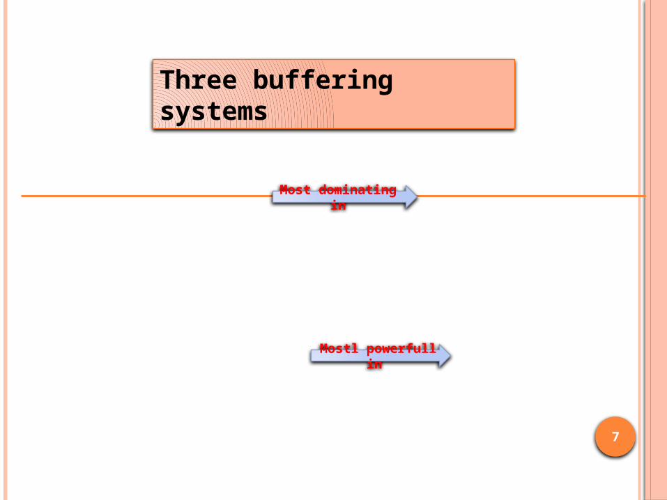
Bicarbonate system constituted of plasma sodium bicarbonate (NaHCO3) and carbonic acid (H2CO3) and cellular H2CO3 and potassium bicarbonate (KHCO3)
extracellular space
The phosphate system found in renal tubular fluid and intracellularly
Proteins
intracellular space
Mostl powerfull in
Most dominating in
Three buffering systems

8
2. RESPIRATORY SYSTEM
Normal PaCO2 = 35- 45 mmHg
Pulmonary buffering system is as effective as the chemical buffering system . The lungs respond to deviations in pH by altering the rate and depth of ventilation. Eliminates or retains carbon dioxide
↑ carbon dioxide (acid) stimulate respiration
↑rate & depth of resp ↓ pH to normal range
Alkalosis depresses respiration
↓ rate & depth of resp retains carbon dioxide

9
The kidneys are the third line of defence against wide
changes in body fluid pH
• movement of bicarbonate
• Retention/Excretion of acids
• Generating additional buffers
Long term regulator of ACID – BASE balance
May take hours to days for correction
3. Renal System
10
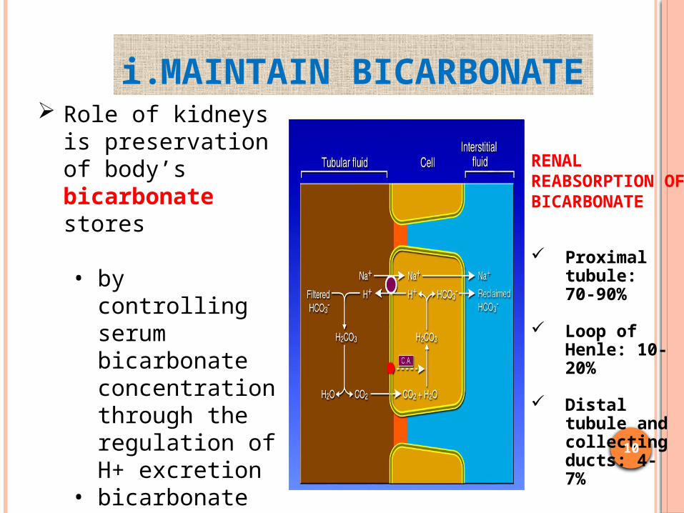
Role of kidneys is preservation of body’s bicarbonate stores
• by controlling serum bicarbonate concentration through the regulation of H+ excretion
• bicarbonate reabsorption
• production of new bicarbonate
Proximal tubule: 70-90%
Loop of Henle: 10-20%
Distal tubule and collecting ducts: 4-7%
RENAL REABSORPTION OF BICARBONATE
i. MAINTAIN BICARBONATE

11
Regeneration of titrated bicarbonate
by excretion of:
• Titratable acidity (mainly phosphate)
• Ammonium salts
ii. Regeneration Of Bicorbonate

12
• TITRATABLE ACIDITY Occurs when secreted H+
encounter & titrate phosphate in tubular fluid
Refers to amount of strong base needed to titrate urine back to pH 7.4
40% (15-30 mEq) of daily fixed acid load
13
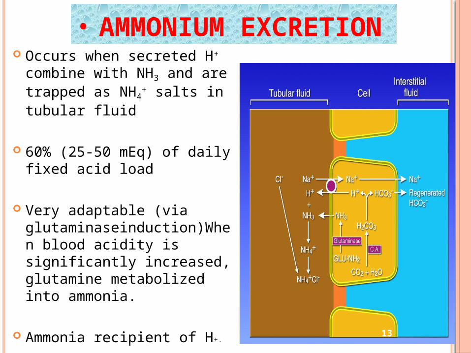
• AMMONIUM EXCRETION Occurs when secreted H+
combine with NH3 and are trapped as NH4
+ salts in tubular fluid
60% (25-50 mEq) of daily fixed acid load
Very adaptable (via glutaminaseinduction)When blood acidity is significantly increased, glutamine metabolized into ammonia.
Ammonia recipient of H+.

14
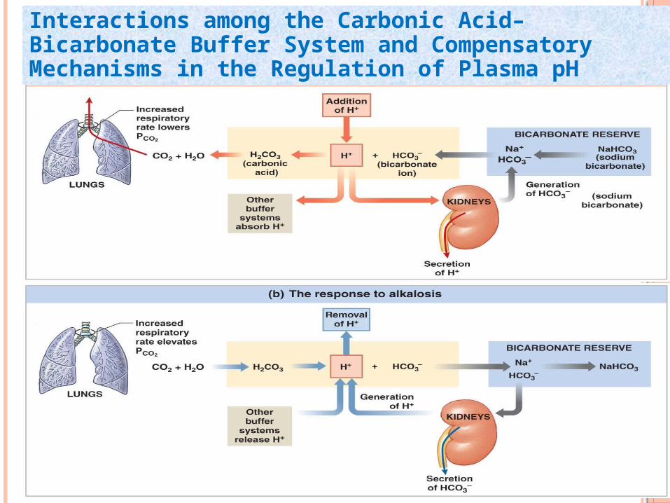
Normal bicarbonate 22-26 mEq/L Acidosis• Excess H+ ions • pH falls • kidneys excrete H+ and retain bicarbonate
Alkalosis• Less H+ ions• pH increases• Kidneys retains H+ ions & excrete bicarbonate
Interactions Among The Carbonic Acid–bicarbonate Buffer System And Compensatory Mechanisms In The Regulation Of Plasma pH
15
Interactions among the Carbonic Acid–Bicarbonate Buffer System and Compensatory Mechanisms in the Regulation of Plasma pH

16
HENDERSON-HASSELBACH EQUATION
pH = pKa + log([HCO3-]/.03xpCO2)
Shows that pH is a function of the ratio between bicarbonate and pCO2
PCO₂ - ventilatory parameter (40 +/- 4) HCO₃ - metabolic parameter (22-26 mmol/L)⁻
3
2
24HCO
PaCOH
Kassirer-Bleich equation
17
ACID–BASE MONITORING Acid–base status can be monitored
intermittently or continuously. Arterial blood gas (ABG) analysis remains
the gold standard in assessing for acid–base disorders.
In the ICU, ABGs can be obtained by arterial puncture or through an indwelling arterial catheter

18
GETTING AN ARTERIAL BLOOD GAS SAMPLE

19
SAMPLE ANALYSIS The blood gas machines in most labs actually
measure the pH ,the pCO2 and the pO2. bicarbonate level -------- from a serum
sample.
The [HCO3-] and the base difference are calculated values using the Henderson-Hasselbalch equation.

20
INTERPRETATION OF BLOOD GASMEASUREMENTS It is an easy mathematical exercises
immediately and rapidly get a insight into the underlying process causing the disturbance in acid–base status.

21
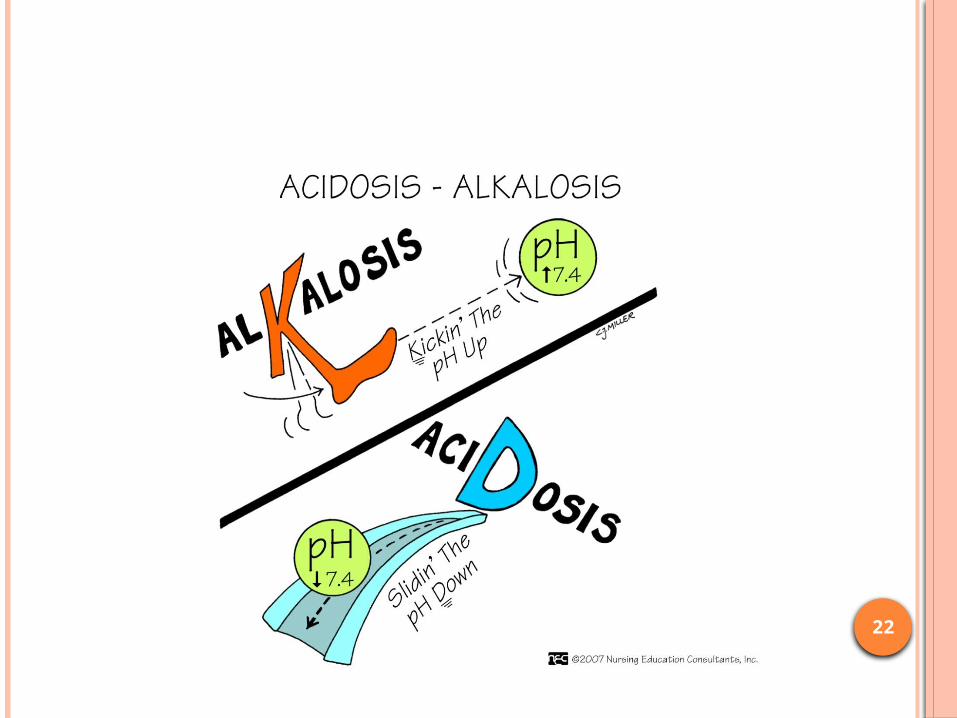
Acidosis “is a disorder that predisposes/lead to low systemic pH. Utilizing the Henderson–Hasselbalch equation, this can be caused by a fall in systemic bicarbonate concentration or by an elevation in the pCO2 .
Alkalosis is a disorder that predisposes/lead to high systemic pH. This is usually caused either by an increase in systemic bicarbonate concentration or by a fall in the pCO2.
22

23
FOUR BASIC TYPES OF IMBALANCE
Metabolic Acidosis
Metabolic Alkalosis
Respiratory Acidosis
Respiratory Alkalosis

24
A change in either the PCO2 or the HCO3 will cause a change in the pH of extracellular fluid.
When the change involves the PCO2, the condition is called a respiratory acid-base disorder: an increase in PCO2 is a respiratory acidosis, and a decrease in PCO2 is a respiratory alkalosis.
When the change involves the HCO3, the condition is called a metabolic acid-base disorder: a decrease in HCO3 is a metabolic acidosis, and an increase in HCO3 is a metabolic alkalosis.
Acid Base Disorders
25
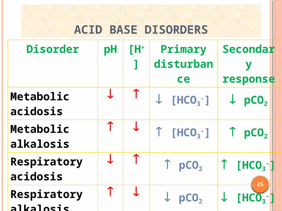
ACID BASE DISORDERS
Disorder pH [H+] Primary disturbance
Secondary response
Metabolic acidosis
[HCO3-] pCO2
Metabolic alkalosis
[HCO3-] pCO2
Respiratory acidosis
pCO2 [HCO3-]
Respiratory alkalosis
pCO2 [HCO3-]

26
ACIDOSIS Principal effect of acidosis is depression of the CNS
through ↓ in synaptic transmission. Generalized weakness Deranged CNS function the greatest threat Severe acidosis causes
Disorientationcoma death

27
28
ALKALOSIS Alkalosis causes over excitability of the
central and peripheral nervous systems. Numbness Lightheadedness It can cause :
Nervousness muscle spasms or tetany Convulsions Loss of consciousness Death

29

30
OUTLINE OF ACID-BASE INTERPRETATION RULESStep 1 : Determine if data is consistent using Henderson’s
Equation
Step 2 : Check pH & PaCO2 (If either of them is normal, or
both are normal, got to step 6 to diagnose
mixed acid-base disorder)
Step 3 : Determine Primary acid base
disorder
Step 4 : Check for compensation
Step 5 : Check Anion Gap/hypoalbuminemi
a or delta/delta
Step6 : Mix Acid Base disorders
31
RESPIRATORY ACIDOSIS
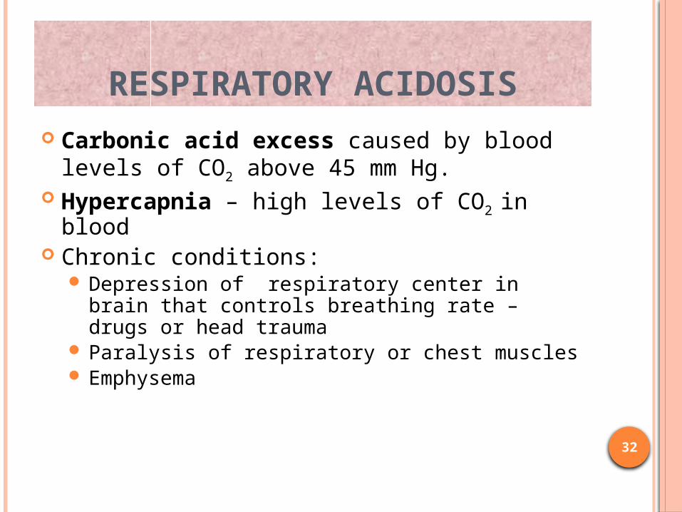
RESPIRATORY ACIDOSIS Carbonic acid excess caused by blood
levels of CO2 above 45 mm Hg. Hypercapnia – high levels of CO2 in blood Chronic conditions:
Depression of respiratory center in brain that controls breathing rate – drugs or head trauma
Paralysis of respiratory or chest muscles Emphysema
32

RESPIRATORY ACIDOSIS Acute conditions:
Adult Respiratory Distress Syndrome Pulmonary edema Pneumothorax
33

COMPENSATION FOR RESPIRATORY ACIDOSIS
Kidneys eliminate hydrogen ion and retain bicarbonate ion
34
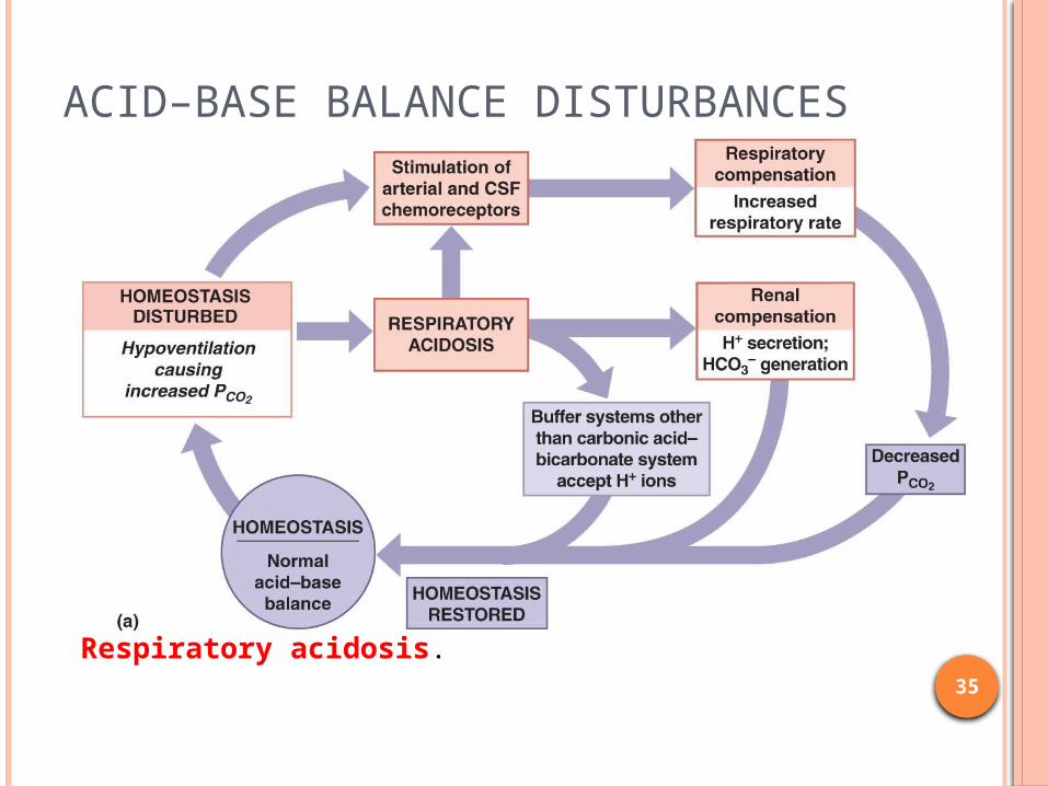
ACID–BASE BALANCE DISTURBANCES
Respiratory acidosis. 35

36
RESPIRATORY ALKALOSIS
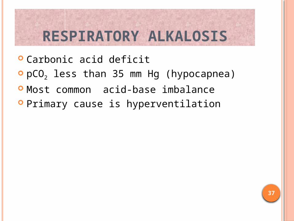
RESPIRATORY ALKALOSIS Carbonic acid deficit pCO2 less than 35 mm Hg (hypocapnea) Most common acid-base imbalance Primary cause is hyperventilation
37

RESPIRATORY ALKALOSIS Conditions that stimulate respiratory center:
Oxygen deficiency at high altitudes Pulmonary disease and Congestive heart failure –
caused by hypoxia Acute anxiety Fever, anemia Early salicylate intoxication Cirrhosis Gram-negative sepsis
38

COMPENSATION OF RESPIRATORY ALKALOSIS
Kidneys conserve hydrogen ion Excrete bicarbonate ion
39

ACID–BASE BALANCE DISTURBANCES
Respiratory Alkalosis. 40
41
METABOLIC ACIDOSIS

42
METABOLIC ACIDOSIS Bicarbonate deficit - blood concentrations of bicarb
drop below 22mEq/L Causes:
Loss of bicarbonate through diarrhea or renal dysfunction
Accumulation of acids (lactic acid or ketones) Failure of kidneys to excrete H+

43
COMPENSATION FOR METABOLIC ACIDOSIS Increased ventilation Renal excretion of hydrogen ions if possible K+ exchanges with excess H+ in ECF ( H+ into cells, K+ out of cells)

44
ACID–BASE BALANCE DISTURBANCES
.
Responses to Metabolic Acidosis

45
METABOLIC ALKALOSIS
46
METABOLIC ALKALOSIS Bicarbonate excess - concentration in
blood is greater than 26 mEq/L Causes:
Excess vomiting = loss of stomach acid Excessive use of alkaline drugs Certain diuretics Endocrine disorders Heavy ingestion of antacids Severe dehydration
47
COMPENSATION FOR METABOLIC ALKALOSIS
Alkalosis most commonly occurs with renal dysfunction, so can’t count on kidneys
Respiratory compensation difficult – hypoventilation limited by hypoxia

48
ACID–BASE BALANCE DISTURBANCES
.
Metabolic Alkalosis
49
ANION GAP This step identifies the type of metabolic acidosis
present, i.e., whether it is secondary to an anion that creates an AG on electrolyte measurement or not.
The AG is a diagnostic tool to uncover the actual anions elevated in the blood but not routinely included in our measurements under normal conditions.
It is calculated as follows:AG = serum sodium − serum chloride − serum bicarbonate. A normal anion gap is <12 mmol L−1.
50
HYPOALBUMINEMIA In this step interpreting metabolic acidosis is
adjusting for factors that would falsely lower the anion gap if one existed, e.g., hypoalbuminemia and lithiumor bromide ingestion
Adjusted AG in hypoalbuminemia = observed AG + [2.5(normal albumin − observed albumin)].

51
DELTA/DELTA This step is the comparison of the degree of
change in AG with the change in serum bicarbonate, aiming to assess the extent of contribution of the AG-producing process to the actual acidosis. This measurement is called delta/delta:
delta/delta= ΔAG/ ΔHCO- =(AG -12) / (24 - HCO3-).

52
MNEMONIC VERSION OF EXPECTED COMPENSATORY RESPONSES TO ACID–BASE DISTURBANCES
Assuming a normal ABG of pH 7.4, pCO2 40, HCO3− 24, and utilizing meq L−1 or mmol L−1 for bicarbonate and mmHg for pCO2,

53
MIXED ACID BASE DISORDER
If the Arterial pH is relatively normal and the PCO2 and/or HCO3 are abnormal, one can assume that a mixed abnormality is present.

54
MIXED ACID BASE DISORDER Diagnosed by combination of clinical assessment, application of
expected compensatory responses , assessment of the anion gap, and application of principles of physiology.
Respiratory acidosis and alkalosis never coexist
Metabolic disorders can coexist
Eg: lactic acidosis/DKA with vomiting
Metabolic and respiratory AB disorders can coexist
Eg: salicylate poisoning (met.acidosis + resp.alkalosis)

55
1-CASE STUDY 11 year old girl Mild bronchial asthma with fever and increased
breathing Asthmaticus diagnosed ABGs Ph 7.22 p CO2 38mmHg serum bicarbonate
15med L-1 Serum albumin level 1gdL-1 What is your interpretation …………….? Hypoalbuminemia ( primary metabolic acidosis
and acute respiratory acidosis )

56
2-CASE STUDY 2 year old child was foun unconscious increased
breathing ABGs pH 7.38
pCO2 28mmHg serum bicarbonate 16 meqL-1
What is your interpretation……………? Salicylate poisoning

Related Documents