UNIVERSITATIS OULUENSIS MEDICA ACTA D D 1012 ACTA Ari Pajala OULU 2009 D 1012 Ari Pajala ACHILLES TENDON RUPTURE COMPARISON OF TWO SURGICAL TECHNIQUES, EVALUATION OF OUTCOMES AFTER COMPLICATIONS AND BIOCHEMICAL AND HISTOLOGICAL ANALYSES OF COLLAGEN TYPE I AND III AND TENASCIN-C EXPRESSION IN THE ACHILLES TENDON FACULTY OF MEDICINE, INSTITUTE OF CLINICAL MEDICINE, DEPARTMENT OF SURGERY, DIVISION OF ORTHOPAEDIC AND TRAUMA SURGERY, INSTITUTE OF DIAGNOSTICS, DEPARTMENT OF CLINICAL CHEMISTRY, UNIVERSITY OF OULU

Welcome message from author
This document is posted to help you gain knowledge. Please leave a comment to let me know what you think about it! Share it to your friends and learn new things together.
Transcript

ABCDEFG
UNIVERS ITY OF OULU P.O.B . 7500 F I -90014 UNIVERS ITY OF OULU F INLAND
A C T A U N I V E R S I T A T I S O U L U E N S I S
S E R I E S E D I T O R S
SCIENTIAE RERUM NATURALIUM
HUMANIORA
TECHNICA
MEDICA
SCIENTIAE RERUM SOCIALIUM
SCRIPTA ACADEMICA
OECONOMICA
EDITOR IN CHIEF
PUBLICATIONS EDITOR
Professor Mikko Siponen
University Lecturer Elise Kärkkäinen
Professor Hannu Heusala
Professor Olli Vuolteenaho
Senior Researcher Eila Estola
Information officer Tiina Pistokoski
University Lecturer Seppo Eriksson
Professor Olli Vuolteenaho
Publications Editor Kirsti Nurkkala
ISBN 978-951-42-9091-6 (Paperback)ISBN 978-951-42-9092-3 (PDF)ISSN 0355-3221 (Print)ISSN 1796-2234 (Online)
U N I V E R S I TAT I S O U L U E N S I S
MEDICA
ACTAD
D 1012
ACTA
Ari Pajala
OULU 2009
D 1012
Ari Pajala
ACHILLES TENDON RUPTURECOMPARISON OF TWO SURGICAL TECHNIQUES, EVALUATION OF OUTCOMES AFTER COMPLICATIONS AND BIOCHEMICAL AND HISTOLOGICAL ANALYSES OF COLLAGEN TYPE I AND III AND TENASCIN-C EXPRESSIONIN THE ACHILLES TENDON
FACULTY OF MEDICINE,INSTITUTE OF CLINICAL MEDICINE,DEPARTMENT OF SURGERY, DIVISION OF ORTHOPAEDIC AND TRAUMA SURGERY,INSTITUTE OF DIAGNOSTICS,DEPARTMENT OF CLINICAL CHEMISTRY,UNIVERSITY OF OULU


A C T A U N I V E R S I T A T I S O U L U E N S I SD M e d i c a 1 0 1 2
ARI PAJALA
ACHILLES TENDON RUPTUREComparison of two surgical techniques, evaluation of outcomes after complications and biochemical and histological analyses of collagen type I and III and tenascin-C expression in the Achilles tendon
Academic Dissertation to be presented with the assent ofthe Faculty of Medicine of the University of Oulu forpublic defence in Auditorium 1 of Oulu UniversityHospital, on 8 May 2009, at 12 noon
OULUN YLIOPISTO, OULU 2009
Copyright © 2009Acta Univ. Oul. D 1012, 2009
Supervised byDocent Juhana LeppilahtiDocent Juha Risteli
Reviewed byDocent Pekka KannusDocent Sakari Orava
ISBN 978-951-42-9091-6 (Paperback)ISBN 978-951-42-9092-3 (PDF)http://herkules.oulu.fi/isbn9789514290923/ISSN 0355-3221 (Printed)ISSN 1796-2234 (Online)http://herkules.oulu.fi/issn03553221/
Cover designRaimo Ahonen
OULU UNIVERSITY PRESSOULU 2009

Pajala, Ari, Achilles tendon rupture. Comparison of two surgical techniques,evaluation of outcomes after complications and biochemical and histologicalanalyses of collagen type I and III and tenascin-C expression in the Achilles tendonFaculty of Medicine, Institute of Clinical Medicine, Department of Surgery, Division ofOrthopaedic and Trauma Surgery, Institute of Diagnostics, Department of Clinical Chemistry,University of Oulu, P.O.Box 5000, FI-90014 University of Oulu, Finland Acta Univ. Oul. D 1012, 2009
AbstractThe Achilles tendon is the largest tendon in the human body and is affected by many diseases andis vulnerable to many forms of damage due to the heavy loads it must bear. Rupture of the Achillestendon has become more common in recent times, with an almost four-fold increase in prevalencefrom 1979–1990 to 1991–2000 and a peak incidence of 19 ruptures per 100 000 of population in1999 in our epidemiological assessment. The incidences of major complications, re-rupture anddeep infection, increased along with primary ruptures, peaking in 1999. The results aftersuccessful primary repair are good in over 90% of cases, as we have shown in a randomized studyand in a review of the literature, and the result after re-rupture is still good in about 70% of cases,but achieving good performance after deep infection is a highly random matter. Our retrospectivesurvey did not identify any good results, but the deep infection cases in our randomized studyshowed good performance due to prompt action taken for their treatment.
The best method for treating a ruptured Achilles tendon has been under debate for almost 100years, with surgery and conservative methods advocated to equal extents. We have advocatedsurgical treatment as the primary choice and conservative treatment is given for selected high riskpatients, for example patients with diabetes, skin problems, systemic use of corticosteroids orsevere other illness. The type of surgery technique is not a straightforward choice, either, andvarious forms of open surgery and percutaneous techniques exist. We compared an end-to-endsimple suture with the same suture augmented with one central gastrocnemius turn-over flap in arandomized series of 60 patients and found no differences with respect to subjective complaints,calf muscle strength or tendon elongation with time. The end-to-end technique is simpler and istherefore justified as the primary method of choice for the surgical repair of fresh completeAchilles tendon ruptures.
The tissue composition has been shown to alter not only with time but also after repeatedtearing of the tendon collagen fibres. A normal tendon is mainly composed of type I collagen, butthe rupture areas express more type III collagen, which is thinner and withstands loads lesseffectively. Type III collagen accumulates slowly in the tendon, since its production does notincrease very much, a situation that is indicative of microtrauma. Crosslinking of the fibres isimportant for collagen matrix properties, and we found that there is a change in the quality ofcrosslinking with age and that this may have role in the observed changes in tendon stiffness, asalso noted in other studies.
We also studied the appearance of tenascin-C at the rupture site in the Achilles tendon and attwo other sites in the same tendon, but found no difference in its expression. It has been proposedthat tenascin-C may take part in the tendon’s reaction to loading, but its exact function remainsunknown.
Keywords: Achilles tendon rupture, collagen type I, collagen type III, surgical repair,tenascin-C, tendon elongation


Acknowledgements
The present study was carried out at the Department of Surgery, Division of Ortho-
paedics, Department of Clinical Chemistry, the Department of Pathology and
Department of Physical Medicine and Rehabilitation, Oulu University Hospital
during the years 1998–2009. Oulu University Hospital has a well known tradition
of Achilles tendon research.
Docent Juhana Leppilahti, M.D., Ph.D., has been the pioneer researcher in this
field, publishing his doctoral thesis in 1996, and Jarmo Kangas, M.D., Ph.D., fol-
lowed with a thesis of his own in 2007. I greatly admire their knowledge in this
field and would like to express my greatest thanks to both of them for helping me
to complete my work.
Professor Juha Risteli of the Department of Clinical Chemistry and Docent
Jukka Melkko of the Department of Pathology have placed the resources and well
widely acknowledged skills of their departments at my disposal for the biochemi-
cal and histological parts of this work, and I extend my grateful thanks to them for
this.
Heidi Eriksen, M.D. deserves special attention as the first author of the
biochemical paper belonging to this thesis and the person who introduced me to
some of the secrets of the world of collagen. Professor Pekka Jalovaara, M.D.,
Ph.D., Professor Tatu Juvonen, M.D., Ph.D., Professor Martti Hämäläinen, M.D.,
Ph.D., Docent Kari Haukipuro, M.D., Ph.D., and Docent Timo Niinimäki, M.D.,
Ph.D., are all acknowledged for giving me their full support and allowing me to
carry out work belonging to my thesis under their leadership.
I would also like to thank Professor Pekka Kannus, M.D., Ph.D., and Professor
Sakari Orava, M.D., Ph.D., for their expert review of my thesis before publication,
and Docent Ylermi Soini, M.D., Ph.D., Pasi Ohtonen, M.Sc., and Pertti Siira, PT,
for their collaboration as co-authors. I respectfully thank all the numerous profes-
sionals on the staff of Oulu University Hospital who participated in the treatment
and evaluation of the patients and biopsy specimens, and I would also like to thank
all the patients who participated in research without any compensation and gave up
valuable time in the cause of better health care.
Special thanks go to Malcolm Hicks, M.A., for his prompt and reliable revi-
sion of the language of the original articles and of the manuscript of the thesis
itself. My orthopaedic colleagues, Tapio Flinkkilä, M.D., Ph.D., Juha Haataja,
M.D., Kyösti Haataja, M.D., Pekka Hyvönen, M.D., Ph.D., Docent Juhani Junila,
M.D., Ph.D., Inger Karumo, M.D., Ph.D., Martti Lakovaara, M.D., Pirkka Mäkelä
5

M.D., Juha Partanen, M.D., Ph.D., Tapio Peljo, M.D., Maija Pesola, M.D., Ph.D.,
docent Jukka Ristiniemi, M.D., Ph.D., Antero Sundin, M.D. and Reetta Willig,
M.D., Ph.D., deserve warm thanks both for their teaching and for sharing daily
work with me.
I am deeply thankful to my parents, Mauno († 2003) and Sirkka Pajala for
their everlasting support and trust during my life. My brother, Markku Pajala,
M.D., has been and always will be a special person in my life. I also thank my clo-
sest friends, Mr. Jukka Laiho and Mrs. Maria Rosenlund-Laiho, for their support in
my life outside the medical profession and for all the delightful holidays we have
had together. Mr. Erkki Vornanen and Mrs. Leena Vornanen deserve warm thanks
for their help in daily routines of our family. Finally, I would thank my wife, Tuuli
Vornanen, M.D., for the 16 years we have shared together, and especially those
shared with our lovely daughters Anna and Ella. In the end all that matters is the
love that we have for each other, not fame or riches.
This research was supported financially by the Foundation for the Study of
Orthopaedics and Trauma in Finland.
6
Abbreviations
AR Augmented repair group
AT Achilles tendon
CONT1 Control 1 site
CONT2 Control 2 site
DF Dorsiflexion
ECM Extracellular matrix
MMP Matrix metalloproteinases
MRI Magnetic resonance imaging
PINP Aminoterminal polypeptide collagen type I
PICP Carboxyterminal polypeptide collagen type I
PF Plantar flexion
PT Peak torque
PTA Peak torque angle
PW Peak work
RIA Radio immunologic assay
RUPT Rupture site
SD Standard deviation
SEM Standard error of measurement
SR Simple repair group
TIMP Tissue inhibitors of matrix metalloproteinases
US Ultrasonography
VAS Visual analogical scale
7

8

List of original publications
This thesis is based on the following articles, which are referred to in the text by
their Roman numerals:
I Pajala A, Kangas J, Siira P, Ohtonen P & Leppilahti J. Augmented surgical
repair compared with non-augmented in fresh total Achilles tendon rupture: A
prospective, randomized study. J Bone Joint Surg (Am) (In press).
II Pajala A, Kangas J, Ohtonen P & Leppilahti J (2002) Rerupture and deep
infection following treatment of total Achilles tendon rupture. J Bone Joint
Surg (Am) 84:2016–2021.
III Eriksen HA, Pajala A, Leppilahti J & Risteli J. (2002) Increased content of
type III collagen at the rupture site of human Achilles tendon. J Orthop Res
20:1352–1357.
IV Pajala A, Melkko J, Leppilahti J, Ohtonen P, Soini Y & Risteli J. Tenascin,
type I and III collagen expression at the Achilles tendon rupture: An
immunohistochemical study. Histol Histopathol (In press)
9

10

Contents
Abstract Acknowledgements
Abbreviations
List of original publications
Contents1 Introduction 132 Review of the literature 15
2.1 Normal Achilles Tendon (AT) ................................................................ 15
2.1.1 Anatomy of the normal AT........................................................... 15
2.1.2 Biomechanics of the normal AT ................................................... 16
2.1.3 Histology of the normal AT.......................................................... 17
2.2 Achilles tendon rupture ........................................................................... 18
2.2.1 Epidemiology of AT rupture......................................................... 18
2.2.2 Aetiology of AT rupture ............................................................... 19
2.2.3 Diagnosis of AT rupture ............................................................... 20
2.3 Surgical treatment ................................................................................... 21
2.3.1 Surgical techniques ....................................................................... 21
2.3.2 Non-surgical treatments ................................................................ 22
2.3.3 Complications of surgical treatment ............................................. 23
2.3.4 Complications of non-surgical treatment...................................... 23
2.3.5 Treatment of complications .......................................................... 24
2.4 Tendon histology changes upon AT rupture........................................... 25
2.4.1 Collagen changes .......................................................................... 25
2.4.2 Tenascin-C .................................................................................... 26
3 Purpose of the research 274 Materials and methods 29
4.1 Materials.................................................................................................. 29
4.2 Methods................................................................................................... 30
4.2.1 Treatments .................................................................................... 30
4.2.2 Evaluation methods ...................................................................... 36
4.2.3 Statistical methods (I, II, III, IV) .................................................. 40
4.2.4 Ethics (I, II, III, IV)....................................................................... 41
5 Results 43
11

5.1 Augmented vs. non-augmented surgical repair of acute total
AT rupture. A randomized prospective study (I) .................................... 43
5.2 Re-rupture and deep infection following treatment of total
AT rupture (II)......................................................................................... 51
5.3 Increased type III collagen content at the human AT rupture site (III) .. 55
5.4 Tenascin-C and type I and III collagen expression in total AT
rupture (IV) ............................................................................................. 60
6 Discussion 636.1 Augmented vs. non-augmented surgical repair of acute total
AT rupture............................................................................................... 63
6.2 Re-rupture and deep infection following treatment of total AT rupture. 65
6.3 Type I and III collagen expression in total AT rupture........................... 68
6.4 Tenascin-C in Achilles tendon rupture ................................................... 69
7 Conclusions 718 Future prospects for Achilles tendon rupture research 73
References
Original publications
12

1 Introduction
It was the well-known French surgeon Ambroise Pare who first recorded an
attempt to treat an Achilles tendon rupture, in 1575. He introduced a bandage dip-
ped in wine and spices to be wrapped around the ankle to treat this injury (Ronel et
al. 2004).
Prior to the 20th century, Achilles tendon ruptures were most often treated
non-operatively. Various means of immobilization for varying periods are descri-
bed in the literature (Wills et al. 1986). Since 1929, however, surgery has been pro-
posed as the treatment of choice (Ronel et al. 2004). The first recorded works by
Christensen (1954) and Arner et al. (1958/59) compared patients treated operati-
vely and non-operatively and found better results in the former group. Based on
their findings, the surgical treatment of total Achilles tendon rupture became popu-
lar, although along with it came various complications of this surgery. A final con-
sensus regarding the most effective management with least complications is still
lacking.
Rupture of the Achilles tendon is often accidental in nature, but debilitating
histological alterations in the tendon tissue have been suggested as the primary
cause (Josza et al. 1989a). The mechanical and degenerative theories do not totally
exclude one another, however, and much research has been done in order to clarify
more closely histological and biomechanical backgrounds to total Achilles tendon
rupture.
Our research focused on the outcomes of two surgical techniques, reporting
the results after re-ruptures and infections and comparing the biochemical and his-
tological changes at the rupture site with those at two other sites in the same Achil-
les tendon.
13

14

1 Review of the literature
1.1 Normal Achilles Tendon (AT)
1.1.1 Anatomy of the normal AT
The Achilles tendon, formed of tendinous parts of the gastrocnemius and soleus
muscles, is the largest tendon in the human body. The gastrocnemius muscle has
its origin on the femur above the knee joint, while the soleus muscle originates
below the knee joint, at the proximal posterior tibia and fibula. The gastrocnemius
muscle fibres extend 11–26 cm above the heel bone and those of the soleus 3–11
cm (Cummins et al. 1946). The AT is formed by three broad, flat aponeuroses of
the gastrocnemius and soleus muscles. The tendon is circular in shape at its
midpoint and widens out at the insertion into the heel bone. As the AT runs down
towards the heel bone, its fibres rotate 90° so that the posterior gastrocnemius
tendon fibres are turned anterolaterally and the anterior soleus fibres
posteromedially (Cummins et al. 1946, Stein et al. 2000) (Figure 1). The shape
and cross-sectional dimensions of the AT vary from 80 to 140 mm2 along the
course of the tendon (O’Brien 1992). The AT receives its blood circulation at the
muscle and heel bone insertions and at the endotenon and peritenon. At its
midpoint, 4–7 cm from the heel bone insertion, arterial nutrition is very limited
and takes place through anterior vessels running through the fatty tissue into the
peritenon. The number of mid-tendon blood vessels is very low, as is their area
relative cross-sectional area (Carr & Norris 1989). The AT is covered by a thin
epitenon and paratenon and does not have a true sheath. The paratenon forms
several thin membranes on the dorsomediolateral side which can glide in relation
to each other, while on the ventral side it consists of fatty tissue, blood vessels and
connective tissue in thin septal structures.
15

Fig. 1. Anatomical drawing of gastrocnemius and soleus muscles running down to heel
bone. They turn 90° on the way down so that gastrocnemius tendon attaches heel bone
on the lateral side and soleus on the medial side.
1.1.2 Biomechanics of the normal AT
The AT transmits the tension generated by the gastrocnemius and soleus muscles
to the heel bone. These two tendons are capable of resisting high tensile forces
with no marked elongation (Best & Garrett 1994). There are high forces in the AT
during normal active daily living, ranging from 600N in cycling up to 9kN (11kN/
cm2) in running at a speed of 6m/sec (Komi et al. 1992). The AT also has the
ability to deform and recover its original length. Its unloaded collagen fibres
assume a wavy configuration, but this will disappear when the tendon is stretched
by about 2%. Loading the tendon tissue gives a linear stress-strain curve at less
than 4% strain as the collagen fibres deform within their capacity. After loading of
tendon at less than 4% strain all the fibres regain their wavy configuration and no
damage occurs, but strain above 4% starts to destroy the intermolecular collagen
crosslinking and the stress-strain curve starts to become more horizontal. Loading
at this level causes microscopic damage to the tendon tissue and a repair process
will start. Strain over 8% will cause macroscopic damage (rupture) to tendon tissue
(O'Brien 1992). The tensile strength of tendons is about 50 N/mm² in vitro (Jozsa
16
& Kannus 1997), and a tendon such as the AT, with a thickness of 1 cm (78.5mm²),
is capable of supporting a weight of 500 to 1000kg. The diameter of the AT is
correlated with calf muscle size and the height and age of the individual
(Koivunen-Niemelä & Parkkola 1995). It has also been demonstrated that the
thickness of the AT is load-dependent (Rosager et al. 2002, Kallinen & Suominen
1994), and that continuous loading of the AT results in exercise-induced tendon
hypertrophy (Woo et al. 1980, Birch et al. 1999).
The tensile strength of a healthy tendon in humans increases until 30 years of
age and gradually weakens thereafter, being about 40% lower by the age of 70
years (Barfred 1973, Thermann et al. 1995a). This is caused by alterations in colla-
gen crosslinking (Ippolito et al. 1980).
1.1.3 Histology of the normal AT
Tendon tissue is composed of cellular and extracellular materials. The number of
cells in a tendon is limited, and fibroblasts are encountered most often. These
fibroblasts produce collagens and other proteins (Karpakka 1991), the collagens
being secreted into the extracellular matrix as procollagens and prepared by
enzymatic cleavage for collagen matrix binding. The residual amino acids
resulting from the cleavage of type I collagen are called N-terminal (PINP) and
C-terminal (PICP) polypeptides (Kielty et al. 1993) and are measurable in vitro
and in secretory substances. The rate of collagen metabolism is relatively slow, the
turnover time for tendon collagen ranging from 50 to 100 days (Curwin & Stanish
1984). A balance between synthesis and breakdown prevails in normal tendon
(Josza & Kannus 1997), but net synthesis can speed up during growth and after
injury, while immobilization will slow down synthesis and accelerate breakdown,
at the same time causing alterations at the molecular level to reduce tendon
strength (Tipton et al. 1986, Karpakka 1991).
The extracellular matrix of the AT is mainly formed of type I collagen, as is
the case with all the tendons in the human body, but small amounts of type II colla-
gen are found at the osteotendinous junction and types III, IV and V in the endote-
non and vascular wall (Josza & Kannus 1997). The dry weight collagen content of
the Achilles tendon is about 70% (Josza et al. 1989b). There are other minor extra-
cellular proteins such as elastin, proteoglycans, glycosaminglycans and wide
variety of other small molecules between the collagen fibres (Karpakka 1991). The
function of the extracellular matrix is to support and regulate the cellular elements
and the structure of the tendon. The collagen molecules form microfibrils, fibrils
17
and finally collagen fibres by intermolecular crosslinking, which is important for
tissue stiffness and strength, i.e. the more crosslinks there are, the stiffer the tissue
(Zernicke & Loitz 1992).
Type I collagen has a micromolecular construction in which two ά¹ chains and
one ά² chain form a left-handed triple helix. Each ά chain has a repeated glycine –
X – Y aminoacid sequence in which X and Y are often (in 25% of cases) repre-
sented by proline and 4-hydroxyproline. Some Y positions are occupied by hyd-
roxylysyl amino acids, and the collagen triple helix can form intermolecular cross-
links at these sites (Kivirikko & Myllylä 1982). The mature type I collagen cross-
links in tendon are 3-hydroxylysylpyridinoline and 3-lysylpyridinoline (Eyre et al.
1984). The number of crosslinks is important for the structure of the tendon, and
even slight changes in their number, which can happen in a variety of diseases and
during ageing, will weaken and alter its mechanical properties (Last & Reiser
1984).
1.2 Achilles tendon rupture
1.2.1 Epidemiology of AT rupture
The incidence of AT rupture has increased dramatically during the last three
decades. AT injury was reported before the 1950´s, but no study of its incidence is
available for the period 1900–1950. The highest incidence in the age group 40–50
years in Malmö (Sweden) between 1950 and 1973 was 8.5 / 105 / year (Nillius et
al. 1976), while the average reported incidence in the Salo region of Finland
between 1980 and 1991 was 2 / 105/ year (Rantanen et al. 1993). In Scotland the
annual incidence increased from 4.7 /105 in 1981 to 6/105 in 1994 (Maffulli et al.
1999). In the area concerned here, that of Oulu, Finland, the average incidence
between the years 1979 and 1986 was 2 / 105 / year and that between 1987 and
1995 12 / 105 / year, with a peak incidence of 18 / 105 / year in 1994 (Leppilahti et
al. 1996a).
Most AT ruptures occur in men, the ratio ranging from 2:1 (Carden et al.
1987) to 19:1 (Zollinger et al. 1983). The peak incidence is reached at 30–40 years
of age, earlier than that for other spontaneous tendon ruptures (Jozsa et al. 1989a).
AT injury is most often unilateral, and a slight left leg predominance has been
reported (Hattrup & Johnson 1985, Jozsa et al. 1989a).
18
1.2.2 Aetiology of AT rupture
AT injuries occur most often (75%) in sports requiring jumping and rapid
acceleration (Josza et al. 1989a, Leitner et al. 1991). Although there are
considerable national differences in the frequencies of particular sports, ball games
constitute up to 60% in many series (Nillius et al. 1976, Josza et al. 1989a,
Järvinen 1992, Cetti et al. 1993, Möller et al. 2001). Approximately 10% of cases
involve professional athletes, 80% recreational athletes and 10% non-athletes
(Nistor 1981, Leppilahti et al.1998, Möller et al. 2001).
Most AT ruptures occur spontaneously, without any direct trauma to the ten-
don. These indirect mechanisms may be sudden, unexpected dorsiflexion of the
ankle, violent dorsiflexion of a plantarflexed foot, and pushing off with a
weight-bearing forefoot while extending the knee joint (Arner & Lindholm 1959).
Direct trauma mechanisms include accidental forceful contact on the activated ten-
don, and open tendon lacerations with broken glass, knives or axe cuts and rare
cases of bone fracture lacerations from within.
The two most popular pathogenesis theories are the degenerative theory and
the mechanical theory. The degenerative theory is based on studies showing histo-
logical signs of repetitive microtrauma and hypovascularity in the AT (Kannus &
Josza 1991, Carr & Norris 1989), these two being regarded as predisposing factors
for chronic degeneration, which will weaken the AT.
Special aetiological cases include the use of anabolic hormones in competitive
sports (Michna & Hartmann 1989, Laseter & Russell 1991) or courses of fluoro-
quinolone antibiotics (McGarvey et al. 1990, Jagose et al. 1996). There are also
clinical case reports of AT ruptures related to systemic corticosteroids (Smaill
1961, Melmed 1965, Baruah 1984) or local corticosteroid injections (Kleinman &
Gross 1983), but no reliable studies of the risk of rupture associated with corticos-
teroid use have been published.
Most younger patients have never had any symptoms in the AT region before
the rupture. In Scotland, for example, only 5% of the 176 AT rupture patients stu-
died had had previous symptoms (Maffulli 1999). Other studies have reported
figures from 4% to 32% of their patients being symptomatic (manifested by pain,
tenderness or stiffness in the Achilles tendon region) prior to rupture (Lea & Smith
1972, Bradley & Tibone 1990, Böhm et al. 1990). Preceding AT symptoms are
more common in older patient groups, sometimes involving up to 40% of cases
(Nestorson et al. 2000).
19
Bilateral cases are rare and related to the older patient groups with systemic
disease and a history of long-term corticosteroid medication or prolonged mixed
use of fluoroquinolone antibiotics and corticosteroids (Cowan & Alexander 1961,
Smaill 1961, Lee 1961, Melmed 1965, Haines 1983, Price et al. 1986, Weinstabl &
Herz 1990, Orava et al. 1996).
1.2.3 Diagnosis of AT rupture
The majority of AT ruptures are typical with respect to their clinical symptoms and
findings and also in terms of their causative mechanism (Maffulli 1998). The
clinical diagnosis should be easy to make. The patients usually report a sudden
pain in their calf with a simultaneous snapping sound from behind, followed by
loss of push-off strength. Occasionally the pain may be slight or even absent.
Painless ruptures have sometimes been reported in as many as one-third of the
patients (Christensen 1954) .
AT rupture typically occurs 2 to 6 cm above the heel bone (Schönbauer 1964,
Fox et al. 1975). Almost always there is a palpable gap in the tendon (Figure 2),
but this can be obscured by local oedema and swelling. About 20% of all AT rup-
ture diagnoses are missed since the long toe flexor muscles can imitate plantar fle-
xion of the ankle, or else there is an uninjured plantaris longus tendon medial to
the AT (Simmonds 1957, Inglis et al. 1976, Carden et al. 1987). The most common
clinical diagnostic test is Thompson’s test, which involves squeezing the calf in
order to achieve plantar flexion (Thompson & Doherty 1962). In the sphygmoma-
nometer test a cuff applied around the calf muscle with the knee flexed to 90° is
inflated to approximately 100 mm Hg with the ankle plantar flexed. Passive dor-
siflexion of the ankle will cause no change in the mercury in the case of a ruptured
AT, but a healthy tendon will cause a rise of about 40 mm Hg depending on the
control value as tested on the patient’s healthy calf (Copeland 1990).
The diagnosis can be confirmed with ultrasonography (US) or magnetic reso-
nance imaging (MRI). The US examination is cheap, rapid and widely available,
but investigator-dependent, whereas MRI has many diagnostic advantages, inclu-
ding superior soft-tissue contrast, non-invasiveness, direct three-dimensional ima-
ging and lack of ionizing radiation (Panageas et al. 1990, Mink et al. 1991), but it
is quite expensive and time-consuming. US and MRI are also valuable tools for the
differential diagnosis of AT rupture, with respect to partial AT rupture, calf muscle
strain and rupture, rupture of the flexor hallucis longus tendon, rupture of the plan-
taris tendon, posterior tibial tendon injury, peroneal tendon injuries, posterior tibial
20

stress syndrome, ligament injuries in the ankle and fractures of the ankle and cal-
caneus (Lehtinen et al. 1994, Karjalainen et al. 2000).
Fig. 2. On the left picture palpable gap is clear when thumb is carried down on the
Achilles tendon.
1.3 Surgical treatment
1.3.1 Surgical techniques
Two types of surgical technique, with subgroups, are used by surgeons today. The
major distinction is between open and percutaneous surgery. Open surgery is then
divided into three subgroups: conventional open surgery, open surgery with
several types of augmentation (one central gastrocnemius fascia flap, two
gastrocnemius fascia flaps, plantaris longus tendon augmentation, flexor hallucis
longus tendon, flexor digitorum longus and peroneus brevis tendon) and open
surgery with multiple stab wounds. At least 41 open surgery options have been
reported in the literature (Wong et al. 2002). We have found only one randomized
trial comparing open surgical techniques. A recent study in Turkey compared a
simple end-to-end suture (Krackow) to suture with plantaris longus tendon
augmentation in 30 patients and found no difference between the groups (Aktas et
al. 2007). Some retrospective trials have compared open surgery techniques, but
they, similarly, have not been able to show any difference between the groups
(Jessig & Hansen 1975, Rantanen et al.1993, Nyyssönen et al. 2003). Different
augmentations have been used in non-comparative studies, however (White &
Kraynick 1957, Quickley & Scheller 1980, Mann et al. 1991, Wapner et al.1993),
and a mechanical experiment has shown the augmented technique to provide extra
strength at the suture (Gebauer et al. 2007).
21

Percutaneous surgery constitutes a category of its own, with a variety of
technical devices available. Good results have been reported (Ma & Griffith 1977,
Rowley & Scotland 1982, Klein et al. 1993, Webb & Bannister 1999, Lim et al.
2001), but no randomized comparison between the techniques has been found.
Open surgery has been compared with percutaneous techniques in several studies,
and the latter has been reported to give a better cosmetic result (Bradley & Tibone
1990), cause lower rates of wound complications (Wong et al. 2002), cause more
sural nerve injuries (Wong et al. 2002) and give an overall lower complication rate
(Lim et al. 2001, Khan et al. 2005). The evidence for all these findings is weak,
however (Khan et al. 2005).
Casting for 4 to 9 weeks without weight bearing was the standard postopera-
tive treatment regardless of the surgical technique until the 1980's, but more detai-
led and faster rehabilitation protocols have been developed since then as know-
ledge of tendon healing has accumulated (Hurme et al. 1990, Maxwell & Enwem-
eka 1992, Rantanen et al. 1999). In some controlled randomized series early func-
tional postoperative rehabilitation has been found to result in faster recovery than
the conventional postoperative cast immobilization (Cetti et al. 1994, Mortensen et
al.1999, Maffulli et al. 2003, Costa et al. 2003), and a functional postoperative
protocol has also been observed to improve patient satisfaction without any inc-
rease in the complication rate (Suchak et al. 2006).
1.3.2 Non-surgical treatments
Various brace and cast options have also been used to treat AT ruptures. The
non-surgical option has been preferred for elderly patients with skin problems and
chronic diseases affecting wound healing (Maffulli 1999). The traditional
treatment regimen has most often consisted of a below-knee plaster boot with the
ankle held in the plantar flexed position for 8 weeks and thereafter a heel rise for 4
weeks, allowing walking and calf muscle exercises (Lea & Smith 1972, Stein &
Lukens 1976, Movin et al. 2005). Some authors have even advocated 12 weeks of
casting with simultaneous reduction of the plantar flexed angle to neutral (Blake &
Ferguson 1991). As knowledge on tendon healing has accumulated the use of more
active non-surgical options has been advocated (Hurme et al. 1990, Maxwell &
Enwemeka 1992, Rantanen et al. 1999). Cast immobilization for 2–3 weeks
followed by controlled early mobilization in a splint was introduced in the 1990´s,
and the authors reported more rapid recovery of ankle motion and return to normal
activities than with the traditional 8 weeks of cast treatment (Saleh et al. 1992). A
22
randomized 50-patient comparison of functional bracing (CAM walker) for 8
weeks with cast treatment pointed to less re-ruptures in the CAM walker group,
but no difference in patient satisfaction, muscle strength or ankle mobility
(Petersen et al. 2002). Non-operative treatments have been compared in five
randomized studies to operative treatments (Nistor 1981, Cetti et al. 1993,
Thermann et al. 1995b, Möller et al. 2001, Metz et al. 2008). Operative treatments
have resulted in 2%, 4% and 5% re-rupture rate and respectively non-operative
treatments in 8%, 15% and 21% re-rupture rate. If a re-rupture can be avoided the
results are comparable (Möller et al. 2001). Non-operative treatments have gained
in popularity over the last five years, especially since modern walkers are
adjustable for ankle motion limitations and are more comfortable to use.
1.3.3 Complications of surgical treatment
Surgery has been reported to cause re-rupture rates of 0–10% (Khan et al. 2005),
regardless of the treatment option employed (Jessing & Hansen 1975, Rantanen et
al. 1993, Nyyssönen & Luthje 2000, Maes et al. 2006). In a study from Norway
the re-rupture incidence after surgical treatment was equal between groups with
postoperative immobilization by means of either a cast or a brace (Borchgrevink &
Crøntvedt et al. 2005). Deep wound and soft tissue infections are to be feared as
complications, as the AT has limited soft tissue protection. The rates reported for
superficial infections are between 3 and 14% and those for deep infections 1–5%
(Khan et al. 2005). There are known risk factors which increase the rate of wound
complications, most notably smoking and steroid use (Bruggeman et al. 2004).
Percutaneous techniques have been reported to cause sural nerve injuries (Wong et
al. 2002, Maes et al. 2006). Surgically treated patients also have the same risks as
non-surgically treated ones in the postoperative period, since all modern protocols
involve some method of immobilization. Deep venous thrombosis is less common,
but skin irritation and muscle atrophy are to a great extent problems that affect
every patient.
1.3.4 Complications of non-surgical treatment
The major complication after non-surgical treatment is re-rupture, the incidence of
which has also been shown to be higher than after surgery in randomized series
(Nistor 1981, Cetti et al. 1993, Möller et al. 2001). Re-rupture rates have varied
from 8% to 21%, and a meta-analysis suggests that there is a three to four-fold risk
23

of re-rupture in non-surgical relative to surgically treated patients (Khan et al.
2004). Less numerous complications are venous thrombosis and pathological
tendon elongation. Problems that recur very regularly are skin irritation and
muscle atrophy after prolonged immobilization. The results following non-surgical
treatment are usually good if re-rupture can be avoided (Möller et al. 2001).
1.3.5 Treatment of complications
If the guidelines on how to treat a fresh AT rupture vary, even less evidence-based
data are available on how to treat typical complications. When non-surgical
primary treatment fails and re-rupture occurs it is feasible to continue with open
operative treatment. Re-ruptures after a percutaneous first attempt can be turned
into open surgery with an end-to-end or augmented technique, and a failed
non-augmented surgical repair can be re-operated on with an augmented technique
of any kind. The results of surgery after re-rupture of an AT are not well
documented, perhaps because the material is scarce. There are case or
technique-based reports, but comparisons of different techniques for handling
re-ruptures is missing.
Deep infections are dangerous complications, because the soft tissue coverage
over the AT is very limited and there may be no repairable AT tissue left after tho-
rough removal of the infected tissue, making reconstruction difficult and rendering
the results unpredictable. A loss of tendon tissue can be dealt with either by means
of a free tendon transplant or with turn-over flaps of the gastrocnemius fascia or a
frozen cadaver transplant. Skin coverage can be achieved with free vascularized
flaps, local vascularized flaps or local non-vascularized flaps (Ronel et al. 2004).
A meshed cutaneous transplant can be used in the case of a very limited loss of
skin coverage. Antibiotic therapy should be given according to the culture fin-
dings, the most commonly encountered bacteria being the normal skin bacteria
Staphylococcus aureus and Streptococcus epidermidis.
Deep venous thrombosis should be treated according to healthcare guidelines.
A first-time thrombosis is not an indication for life-long warfarin.
Damage to the nerves, which is sometimes seen with percutaneous techniques,
is difficult to treat. If obvious total damage to a certain nerve is seen immediately
after surgery, a re-operation attempt should be made to free the nerve and repair
the AT rupture by open surgery.
One reported means of dealing with a troublesome excess AT lengthening
involves a shortening Z-plasty (Mafulli & Ajis 2008). The operative method is
24
simple, although there is no hard evidence supporting the need for such a pro-
cedure.
1.4 Tendon histology changes upon AT rupture
1.4.1 Collagen changes
Tendon is normally formed mainly (98%) of type I collagen (Josza & Kannus
1997). This is synthesized by fibroblasts and its metabolism is well regulated by
the surrounding extracellular matrix components. The collagen turnover period in
a tendon is 50–100 days. Any damage to a tendon will dramatically alter its
composition, however, so that four normal phases of healing after injury have been
distinguished: inflammation, proliferation, remodelling and maturation
(Gelbermann et al. 1999). At strains below 8% the injury may be microscopic, but
at strains above 8% it will be macroscopic, implying total rupture of the tendon.
The healing process after a macroscopic rupture is quite clearly defined and
follows the given four phases, whereas microscopic tendon damage is more
complex and it has been looked on as preceding total rupture (Josza et al. 1989a).
Type I collagen, the major substance making up a normal tendon, is partially
replaced by type III collagen in injury areas (Leadbetter 1992, Liu et al. 1995), the
latter appearing in wounds after 48 hours and reaching its peak during the first
week (Haukipuro et al. 1990). Type III collagen is weaker in resisting tensile
forces and its fibre bundle formation is less firmly oriented, since its crosslinking
differs from that of type I collagen. After two weeks type III collagen synthesis
will decrease and it will slowly be replaced with type I collagen, which will gradu-
ally increase the tensile strength of the tendon and restore its mechanical properties
(Leadbetter 1992). In good biochemical surroundings healing will result in similar
tendon tissue to that which preceded the injury.
Microscopic rupture, in other words repeated microtrauma, is still a partly
unknown mechanism and is more difficult to treat. Ruptured tendons have been
shown to exhibit significant changes in collagen structure relative to the time
before trauma (Kannus & Jozsa 1991, Järvinen et al. 2004). The theory based on
such findings is known as the degenerative theory. Repeated microtrauma in the
area of maximum loading in a tendon will prolong the healing process and prevent
it from proceeding to the next phase. This will result in excess type III collagen
and in mucoid and hypoxic tissue changes similar to those seen in degenerative
tendon diseases. These changes will naturally weaken the tendon and it will finally
25
break if the tensile forces exceed its altered capacity. Biopsies of healthy Achilles
tendons show markedly less degenerative changes than those of ruptured Achilles
tendons (Maffulli et al. 2000).
1.4.2 Tenascin-C
The tenascins form a group of small glycoproteins to be found in the extracellular
matrix. There are several types that differ in molecular size and tissue specificity
(Chiquet-Ehrismann & Tucker 2004), and their function in the extracellular matrix
is not well known, but it has been suggested that they may control cell-to-cell and
cell-to-ECM binding (Sage & Bornstein 1992). Tenascins are highly elastic and
can be stretched three to four-fold relative to their resting length without damage
(Oberhauser et al. 1998). Their molecular construction is well developed for
multiple adhesion to surrounding cells and other ECM proteins. With these
features it is understandable that they are often to be found in locations where
heavy mechanical stresses are present (Settles et al. 1996). Tenascin-C is
encountered in tendon, cartilage, bone and skeletal muscle, around tumours and in
wounds (Mackie et al. 1988, Riley et al. 1996, Erickson 1997, Kaarteenaho-Wiik
et al. 2002, Chiquet-Ehrismann & Tucker 2004). It has three forms, with
molecular weights ranging from 50 to 200 kDaltons. The amount of tenascin-C in
normal musculoskeletal tissue is very limited, but it has been shown to be
expressed more abundantly in tissue pathologies where extracellular matrix
activity is increased (Erickson 1997). Thus elevated expression of tenascin-C has
been found in the musculotendinous and osteotendinous junctions of the
musculoskeletal system after loading periods (Fluck et al. 2000, Järvinen et al.
1999) and in stressed fibroblasts in vitro (Chiquet-Ehrismann et al. 1994). Its exact
function in musculoskeletal tissue is unclear, but it is evidently also present in
degenerative diseases such as osteoarthritis (Salter 1993) and in supraspinatus
tendons (Riley et al. 1996) and supraspinatus bursae (Hyvönen et al. 2003).
Whether its expression in degenerative tissues is a part of the repair process or part
of the degeneration itself is not known, but tenascin-C gene variation has recently
been linked with an increased risk of Achilles tendon injury (Mokone et al. 2005).
Interestingly, when mice tenascin genes are blocked, the other ECM proteins
replace the tenascin function in tissues (Forsberg et al. 1996). Despite all the data
available, the actual function of tenascin remains unknown.
26

3 Purpose of the research
The specific aims of this research were as follows:
A prospective, randomized study to compare the results of the open aug-
mented and end-to-end surgical repair techniques for the treatment of fresh comp-
lete AT ruptures. The null hypothesis was that augmented repair entails no advan-
tage over simple end-to-end repair.
The incidence of complete AT ruptures has increased, but we are not aware of
any reports on the incidence of re-ruptures and deep infections following its treat-
ment. The outcome after successful treatment of an acute AT rupture is good, but
that after complications is presumably much worse. We therefore determined the
incidences of primary ruptures and deep infections and re-ruptures at Oulu Univer-
sity Hospital over a 20-year period and examined the late results after treatment for
complications.
We measured by biochemical means the amounts of type I and type III colla-
gen, their synthesis and crosslinked telopeptides at the rupture site and compared
the results with those for two other sites in the same tendon for samples collected
from six healthy cadaver controls. Our hypothesis was that there collagen type III
will be elevated and the crosslinking of collagen fibrils reduced at the rupture site.
We examined the expression of tenascin-C and type I and III collagen in the
ruptured human AT by comparing expression at the rupture site with that at two
other sites within the same tendon. The hypothesis was that there would be ele-
vated expression of tenascin-C and type III collagen at the rupture site.
27

28

4 Materials and methods
The research was performed at the Department of Surgery, Oulu University Hospi-
tal, Oulu, Finland, in co-operation with the Department of Physical Medicine and
Rehabilitation (I,II), the Department of Diagnostic Radiology (I, II), the Depart-
ment of Clinical Chemistry (III, IV) and the Department of Clinical Pathology
(IV).
4.1 Materials
Paper I: Eighty-three patients with a closed total ATR were treated at Oulu Uni-
versity Hospital between October 1998 to January 2001. Twenty-three of these
were excluded from the present study on the grounds of age over 65 (six patients),
rupture over 7 days old (four patients), local corticosteroid injections around the
AT within six months of the rupture (one patient), open ATR (one patient), skin
problems over the AT (one patient), living abroad (two patients), occurrence while
the main author was out of office (six patients) and refusal to participate (two
patients). Thus there were 60 patients eligible for randomization, 49 of whom
(82%) had sustained the rupture during a sports-related activity, most frequently
ball games (70%).
Paper II: Out of a total of 409 patients with a complete closed Achilles tendon
rupture were treated at Oulu University Hospital between 1979 and 2000, 23 had a
re-rupture. This group included twenty-one men and two women, with a mean age
of forty years. These patients represented all social classes and a variety of occupa-
tions. Nineteen of the twenty-three patients (83%) had sustained the primary rup-
ture during participation in sports activities, most commonly badminton (seven
patients) and volleyball (five patients). Nine patients had sustained a deep infec-
tion after surgery. This group included seven men and two women with a mean age
of fifty-three years. Only two of these patients had sustained the primary Achilles
tendon injury during participation in sports, the other seven having suffered acci-
dents of various kinds during normal activities of daily life. In six cases the infec-
tion had occurred after the primary operation, and in three it had occurred after a
second operation had been performed to treat a re-rupture.
Altogether twelve patients with a re-rupture and seven with a deep infection
were available for a follow-up evaluation at a mean of 4.1 years after the primary
Achilles tendon rupture. Of the remaining ten patients, one had died, one (who had
a well-healed re-rupture) was unable to attend because of work obligations, three
29
had a follow-up visit scheduled within less than six months, and five (one of whom
was living abroad) did not reply to our questionnaire.
Paper III: Tissue samples from 10 consecutive patients (9 men, 1 woman,
average age 38 years, range 30–48) with a closed total Achilles tendon rupture
were taken during surgery at Oulu University Hospital. Exclusion criteria were a
previous Achilles tendon injury, age over 60 years, use of corticosteroids and a
rupture more than 48 h before the operation. The study was approved by the
Research Ethics Committee of Oulu University Hospital, and voluntary informed
consent was obtained in writing from all the patients. Presumably healthy Achilles
tendon cadaver samples from corresponding sites were obtained from 5 men and
one woman (average age 43, range 13–56) within 72 hours post mortem. Permis-
sion for the use of the cadaver samples was obtained from the National Board of
Medicolegal Affairs, Helsinki, Finland.
Paper IV: The material for this paper consisted of tissue samples from the
same 10 consecutive patients as for Paper III above.
4.2 Methods
4.2.1 Treatments
Paper I
Sixty patients with acute complete Achilles tendon rupture (ATR) were randomi-
zed preoperatively to receive end-to-end suturation by the Krackow locking loop
technique either without augmentation (simple repair (SR) group n=32) or with
one central down-turned gastrocnemius fascia flap (augmented repair (AR) group
n=28) (Silfverskiöld 1941) (Figure 3). The first available operation time within 48
hours of randomization was scheduled. All the patients were operated on by the
main author (AP) under spinal anaesthesia in a prone position using a tourniquet.
In both groups a posteromedial skin incision was made, curving to the middle line
in the more proximal part of the calf. The fascia and paratenon were divided on the
same line. Irregular tendon ends were cleaned and repaired by the Krackow techni-
que (Mandelbaum et al. 1995) with two separate 0-gauge absorbable PDS (poly-
dioxanone) sutures (Ethicon, Johnson & Johnson Inc. Somerville, New Jersey) and
smaller 2–0 gauge apposition sutures made with Vicryl (polylactin, Ethicon). No
augmentation was used in the SR group, whereas in the AR group a 10mm wide
30
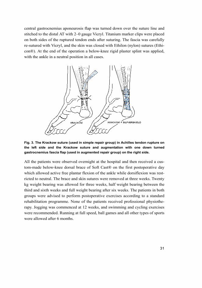
central gastrocnemius aponeurosis flap was turned down over the suture line and
stitched to the distal AT with 2–0 gauge Vicryl. Titanium marker clips were placed
on both sides of the ruptured tendon ends after suturing. The fascia was carefully
re-sutured with Vicryl, and the skin was closed with Ethilon (nylon) sutures (Ethi-
con®). At the end of the operation a below-knee rigid plaster splint was applied,
with the ankle in a neutral position in all cases.
Fig. 3. The Krackow suture (used in simple repair group) in Achilles tendon rupture on
the left side and the Krackow suture and augmentation with one down turned
gastrocnemius fascia flap (used in augmented repair group) on the right side.
All the patients were observed overnight at the hospital and then received a cus-
tom-made below-knee dorsal brace of Soft Cast® on the first postoperative day
which allowed active free plantar flexion of the ankle while dorsiflexion was rest-
ricted to neutral. The brace and skin sutures were removed at three weeks. Twenty
kg weight bearing was allowed for three weeks, half weight bearing between the
third and sixth weeks and full weight bearing after six weeks. The patients in both
groups were advised to perform postoperative exercises according to a standard
rehabilitation programme. None of the patients received professional physiothe-
rapy. Jogging was commenced at 12 weeks, and swimming and cycling exercises
were recommended. Running at full speed, ball games and all other types of sports
were allowed after 6 months.
31

Paper II
Primary treatment. Twenty-eight of the twenty-nine patients had initially been tre-
ated operatively, and one had been treated non-operatively with a cast. Our stan-
dard protocol for operative treatment was tendon suturing with absorbable size-0
non-braided sutures. The repair was augmented with one turn-down flap of the
gastrocnemius aponeurosis (the Silfverskiöld technique) in fourteen cases, with
two flaps (the Lindholm technique) in five cases, and with the plantaris tendon (the
Lynn technique) in one case. For twenty of the twenty-eight patients who had had
surgical treatment the planned postoperative rehabilitation protocol consisted of
immobilization in a below-the-knee cast for six weeks, with the ankle in plantar
flexion for three weeks and then in a neutral position for three weeks. Weight-bea-
ring was allowed gradually after the first three weeks, with full weight-bearing
achieved at six weeks. The other eight patients who had had surgical treatment
were enrolled in a clinical trial in which a customized below-the-knee brace was
used postoperatively. This device allowed active free plantar flexion of the ankle
but restricted dorsiflexion to neutral. Weight-bearing was limited to one-half of
body weight until six weeks, at which time active ankle exercises with full
weight-bearing and strengthening exercises were allowed. All of the patients who
were treated surgically were instructed to begin jogging and controlled sports acti-
vities at three months. Jumping sports and professional activities were begun at six
months. The one patient who had been treated non-operatively wore a
below-the-knee cast with the ankle in plantar flexion for three weeks and then a
second cast with the ankle in a neutral position for an additional three weeks.
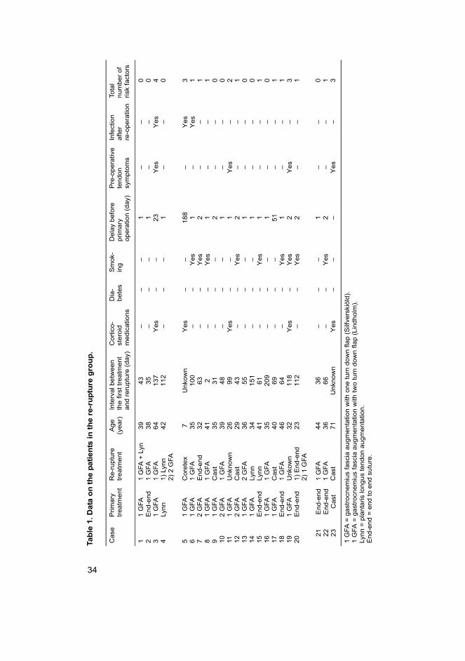
Treatment of re-ruptures. The twenty-three re-ruptures occurred at a mean of
seventy-nine days (range, two to 209 days) after the primary operation. Nineteen
were treated operatively and four non-operatively with six weeks of immobiliza-
tion in a below-the-knee cast. The repair of the re-rupture was augmented with one
turn-down flap of the gastrocnemius fascia in nine cases, with two turn-down flaps
in two cases, and with the plantaris tendon in three cases. In one case the re-rupture
was reconstructed with exogenous material (Leeds-Keio; Howmedica, Rutherford,
New Jersey). The operative technique was not accurately described in the records
for two of the patients. (Table 1)
Two patients had a second re-rupture. The first had had two previous surgical
repairs that had been performed with the end-to-end technique, and the third repair
was performed with one turn-down flap for augmentation. The other patient had
32
had two previous surgical repairs with plantaris tendon augmentation and the third
repair was augmented with two turn-down flaps. (Table 1)
Treatment of infections. There were a total of nine deep infections. Six occur-
red after the primary repair and three after the repair of a re-rupture. Wound revi-
sion was performed in all cases, and necrotic Achilles tendon tissue was totally
removed from five patients during the surgical treatment. The results of bacterial
cultures, recorded for six of the nine patients, were positive for Staphylococcus
aureus (four patients), Staphylococcus epidermidis (one patient) and Propionibac-
terium acnes and diphtheroid species (one patient). (Table 2)
Six patients were treated with repeat débridement and primary split-thickness
skin-grafting. Four of these patients lost the Achilles tendon entirely during the
course of treatment, while in the other two cases the infection resolved before all
the tendon tissue had been removed. Two patients were managed with a microvas-
cular radial forearm flap and a tensor fasciae latae graft after débridement. One of
these microvascular reconstructions was successful, but the other failed because of
thrombosis. The latter patient ultimately had total loss of the tendon and was even-
tually treated with split-thickness skin grafting. One patient needed a local two-tail
full-thickness transposition flap to cover the exposed tendon after débridement.
(Table 2)
33
Tab
le 1
. D
ata
on
th
e p
ati
en
ts in
th
e r
e-r
up
ture
gro
up
.
Cas
eP
rimar
y tre
atm
ent
Re-
rupt
ure
treat
men
tA
ge
(yea
r)In
terv
al b
etw
een
the
first
trea
tmen
t an
d re
rupt
ure
(day
)
Cor
tico-
ster
oid
med
icat
ions
Dia
-be
tes
Sm
ok-
ing
Del
ay b
efor
e pr
imar
y op
erat
ion
(day
)
Pre
-ope
rativ
e te
ndon
sy
mpt
oms
Infe
ctio
n af
ter
re-o
pera
tion
Tota
l nu
mbe
r of
risk
fact
ors
1 2 3 4
1 G
FAE
nd-e
nd1
GFA
Lynn
1 G
FA +
Lyn
1 G
FA1
GFA
1) L
ynn
2) 2
GFA
39 38 64 42
43 35 137
112
– – Yes –
– – – –
– – – –
1 1 23 1
– – Yes –
– – Yes –
0 0 4 0
5 6 7 8 9 10 11 12 13 14 15 16 17 18 19 20
1 G
FA1
GFA
2 G
FA1
GFA
1 G
FA2
GFA
1 G
FA2
GFA
1 G
FA1
GFA
End
-end
1 G
FA1
GFA
End
-end
1 G
FAE
nd-e
nd
Cor
etex
1 G
FAE
nd-e
nd1
GFA
Cas
t1
GFA
Unk
now
nC
ast
2 G
FALy
nnLy
nn1
GFA
Cas
t1
GFA
Unk
own
1) E
nd-e
nd2)
1 G
FA
7 35 32 41 35 39 26 29 36 34 41 35 40 46 32 23
Unk
own
100
63 2 31 48 99 43 55 151
61 209
69 64 118
112
Yes – – – – – Yes – – – – – – – Yes –
– – – – – – – – – – – – – – – –
– Yes
Yes
Yes – – – Yes – – Yes – – Yes
Yes
Yes
188 1 2 1 2 1 1 2 1 1 1 1 51 1 2 2
– – – – – – Yes – – – – – – – Yes –
Yes
Yes – – – – – – – – – – – – – –
3 1 1 1 0 0 2 1 0 0 1 0 1 1 3 1
21 22 23
End
-end
End
-end
Cas
t
1 G
FA1
GFA
Cas
t
44 36 71
36 66U
nkno
wn
– – Yes
– – –
– Yes –
1 2 –
– – Yes
– – –
0 1 3
1 G
FA =
gas
trocn
emiu
s fa
scia
aug
men
tatio
n w
ith o
ne tu
rn d
own
flap
(Silf
vers
kiöl
d).
1 G
FA =
gas
trocn
emiu
s fa
scia
aug
men
tatio
n w
ith tw
o tu
rn d
own
flap
(Lin
dhol
m).
Lynn
= p
lant
aris
long
us te
ndon
aug
men
tatio
n.E
nd-e
nd =
end
to e
nd s
utur
e.
34

Tab
le 2
. D
ata
on
th
e p
ati
en
ts in
th
e d
eep
in
fecti
on
gro
up
.
Cas
eP
rimar
y tre
atm
ent
Re-
rupt
ure
treat
men
tA
ge
(yea
r)C
ortic
o-st
eroi
d m
edca
tions
Dia
bete
sS
mok
ing
Del
ay b
efor
e pr
imar
y op
erat
ion
(day
)
Pre
-ope
rativ
e te
ndon
sy
mpt
oms
Infe
ctio
n af
ter p
rimar
y op
erat
ion
Infe
ctio
n af
ter
re-o
pera
tion
Tota
l nu
mbe
r of
risk
fact
ors
24 3' 5* 6* 25 26 27 28 29
1 G
FA1
GFA
1 G
FA1
GFA
End
-end
End
-end
1 G
FA2
GFA
2 G
FA
–1
GFA
Cor
etex
1 G
FA – – – – –
61 64 70 35 79 56 36 32 45
Yes
Yes
Yes – Yes
Yes – – –
– – – – – Yes – – –
– – – Yes – Yes – – Yes
7 23 188 1 9 4 2 2 6
Yes
Yes – – Yes
Yes – – –
Yes – – Yes
Yes
Yes
Yes
Yes
– Yes
Yes
Yes – – – – –
4 4 3 1 4 4 0 0 1
1 G
FA =
gas
trocn
emiu
s fa
scia
aug
men
tatio
n w
ith o
ne tu
rn d
own
flap
(Silf
vers
kiöl
d).
1 G
FA =
gas
trocn
emiu
s fa
scia
aug
men
tatio
n w
ith tw
o tu
rn d
own
flap
(Lin
dhol
m).
Lynn
= p
lant
aris
long
us te
ndon
aug
men
tatio
n.E
nd-e
nd =
end
to e
nd s
utur
e.*C
ases
3, 5
, and
6 a
re in
clud
ed in
bot
h th
e re
rptu
re g
roup
and
the
deep
infe
ctio
n gr
oup.
35

Papers III and IV
All ten patients with a complete closed Achilles tendon rupture were operated on
using the one turn-down flap augmentation technique as described by Silfver-
skiöld.
4.2.2 Evaluation methods
Achilles tendon rupture score (Papers I and, II)
Paper I: All available patients were examined clinically at 3, 6, 12 and 52 weeks
and the clinical outcome was assessed at the 12 and 52-week check-ups by the cli-
nical scoring method described by Leppilahti (Leppilahti et al.1998), which inclu-
ded subjective factors such as pain, stiffness, muscle weakness, footwear restric-
tions and subjective outcome and objective factors such as the range of active
ankle motion and isokinetic calf muscle strength. The patients were asked to fill in
a non-validated subjective symptoms questionnaire sheet. The assessors, two phy-
siotherapists and A.Pajala, of the objective clinical outcome were not blinded to
the treatment groups.
Paper II: Twenty-three re-ruptures (prevalence 5.6%) and nine deep infections
(prevalence 2.2%) occurred in twenty-nine patients. We reviewed the records of
these patients retrospectively to determine the overall incidence of ruptures,
re-ruptures and deep infections and to record the known risk factors for these
major complications. The final clinical outcome for twelve patients with a re-rup-
ture and seven with a deep infection at a mean of 4.1 years after the initial treat-
ment was assessed in terms of the Achilles tendon rupture score.
Strength measurements (Papers I and II)
Isokinetic and isometric muscle function parameters were assessed for the two
groups at 3 months and 12 months using a Lido Multi-Joint II dynamometer linked
to a computer (Loredan Biomedical Inc, Davis, CA). One physiotherapist perfor-
med all the isokinetic tests. All the patients were informed of the procedure, and a
ten-minute warm-up period of ergometer cycling was included prior to the test.
The testing position was supine, and the patient was fixed to the testing apparatus
with straps round the foot and the pelvis, with the knee supported in extension. The
extent of ankle motion was from 40 degrees of plantar flexion to 20 degrees of dor-
36
siflexion. Before testing, the patient performed some submaximal and maximal
repetitions of the ankle flexion and extension movements at the isokinetic test
velocity. The isokinetic dorsiflexion and plantar flexion strengths were measured,
first at a speed of 60°/sec, then at 120°/sec, and finally at 180°/sec after two minu-
tes of rest. Five maximal voluntary muscular torque contractions were required.
After the isokinetic tests, the maximal isometric plantar flexion strength was
measured with the ankle in the neutral position.
For evaluation of muscle strength, the peak torques (PT, unit: Newton meters)
of plantar flexion and dorsiflexion of the ankles were analysed. The isokinetic
strength scale for scoring the peak torque of the ankle during plantar flexion and
dorsiflexion at three test velocities was used to analyse the strength results (Leppi-
lahti et al. 1996b).
Peak work (unit: joule, Paper I) was defined as the sum of the total area under
all the torque versus angular displacement (time) plots in the best repetition of the
test. The peak work-displacement curves for both legs were divided into five parts,
making it possible to calculate the PW deficits at intervals of 10 degrees over the
range of ankle motion.
Radiographic measurements (Paper I)
Standardized radiographs for the measurement of previously placed radiographic
markers were taken on the first day postoperatively and at 3, 6, 12 and 52 weeks.
The ankle was fixed in the plantigrade position in the brace, the distance between
the x-ray source and the film plate was fixed at 100 centimetres and the radiograph
was focused at the midpoint of the AT. A magnification of 1.1 was taken into
account. The AT elongation curves for both groups were analysed and correlated
with the clinical data.
Biochemical and histopathological characterization (Papers III amd IV)
Tissue samples (III, IV) were taken from the rupture site (RUPT), from the lower
end of the flap (CONT1) and from the top corner of the flap (CONT2) (Figure 4).
37

Fig. 4. Locations of tissue samples taken for Papers III and IV. A is the rupture site, B is
control 1 and C is control 2. The distances from the heel bone insertion are A 4cm, B
8cm and C 16cm. As seen in the picture, tendon-like tissue is still available at site C.
Tissue sample preparation (Paper III). The Achilles tendon samples from the rup-
ture patients (weight 13 to 335 mg) and cadavers (weight 61 to 273 mg) were cut
into small pieces, suspended into PBS, pH 7.2 (20 mg/ml), homogenized by
sonication (4 times for 15 seconds in an ice bath), incubated in ice for 30 minutes
and centrifuged (8 000 g, 30 min) to separate the soluble tissue from the insoluble
pellet. The supernatants were collected for type I and III procollagen propeptide
analyses. The insoluble pellet was lyophilized for further processing (see below).
Stabilization of collagen crosslinks and degradation of insoluble pellets
(Paper III). The pellets were suspended in PBS, pH 7.2 (20 mg/ml), and reduced
with NaBH4 (1 mg of NaBH4 to 40 mg of tissue, reaction time 2 h with magnetic
stirring) to stabilize any immature, divalent collagen crosslinks (Knott et al. 1997).
The samples were washed several times with distilled water, centrifuged (8000 g,
30 min), decanted and finally lyophilized.
The reduced samples were dissolved in 0.2 M NH4HCO3, pH 7.8 (20 mg/ml).
The tissue pieces were heat-denatured at 65°C for 30 minutes, cooled to 37°C and
digested with TPCK-treated trypsin (Worthington Biochemicals, Lakewood, NJ)
38

(Sassi et al. 2000). After incubation for 6 h at 37°C, the heat denaturation and tryp-
sin digestion were repeated and the samples were incubated overnight. The samp-
les were treated similarly for a third time on the next day and then incubated for 6
h at 37°C. After final heat denaturation, they were centrifuged (8000 g, 30 min)
and the supernatants analysed for hydroxyproline, ICTP, IIINTP and PIIINP.
Measurement of type I and III collagen antigens and total collagen content
(Paper III). Procollagen synthesis in the soluble tissue extracts was assessed by
radioimmunoassays (RIA) for the aminoterminal (PINP) and carboxyterminal
(PICP) propeptides of type I collagen and for the aminoterminal propeptide (PII-
INP) of type III collagen (Orion Diagnostica, Oulunsalo, Finland) (Melkko et al.
1990, Melkko et al. 1996, Risteli et al. 1988). The results are expressed per wet
weight of the original tissue sample.
Type I collagen in the insoluble tissue digests was analysed with an assay for
the trivalent pyridinoline cross-linked carboxyterminal telopeptide structure, ICTP,
and with an in-house SP 4 assay based on a synthetic peptide sequence (SAGFD-
FSFLPQPPQEKY, Neosystem Laboratories, Strasbourg, France) (Bode et al.
2000a, Sassi et al. 2000, 2001). The trivalently pyridinoline-crosslinked aminoter-
minal telopeptide of type III collagen, IIINTP, was also analysed by means of an
in-house RIA as described earlier (Bode et al. 1999, Kauppila et al. 1999, Bode et
al. 2000a,b), and PIIINP was also analysed in the digests. To determine the total
collagen content, hydroxyproline was measured with a novel colorimetric microti-
tre plate assay (Brown et al. 2001), on the assumption that this accounts for 12.4%
(w/w) of the total collagens. These results were expressed per dry weight of the
insoluble tissue residues.
Reverse phase HPLC (Paper III). The insoluble tissue digests of the RUPT
site of one individual with a ruptured tendon and one cadaver used for C8 reverse
phase HPLC (228TP1010, Vydac, Hesperia, CA, U.S.A) with 0.4% ammonium
acetate (pH 7.4) and 75% acetonitrile. Amounts of sample equivalent to 100 µg of
ICTP were loaded and their pyridinoline crosslink fluorescence (ex 320 nm, em
405 nm) was monitored. ICTP and IIINTP were measured from the fractions col-
lected.
Tissue sample preparation for Paper IV. All the tissue samples were fixed in
10% buffered formalin, embedded in paraffin and cut to a thickness of 5 μm.
Immunohistochemical staining was carried out by the avidin-biotin-immunoperox-
idase technique, and the tissue sample slides were counterstained with haematoxy-
lin-eosin. A monoclonal mouse antibody reacting to two major isoforms of human
tenascin-C was employed to visualize tenascin. Polyclonal antibodies were raised
39

in rabbits, cross-absorbed with other connective tissue antigens and purified by
immunoabsorption on the relevant antigens coupled to Sepharose 4B. These were
used to characterize the type I and III procollagens and collagens deposited in the
tissue samples. Since anti-PINP detects the aminoterminal propeptide of type I
procollagen, positive staining indicates newly synthesized type I collagen still hav-
ing the aminoterminal propeptide attached. This form is also called type I pN col-
lagen. Anti–PICP detects the carboxyterminal propeptide and is also used as a
marker of type I collagen synthesis. Anti-PIIINP detects the aminoterminal
propeptide of type III collagen, which can be found in free form in the extracellu-
lar matrix or as type III pN collagen on the surfaces of type III collagen fibrils. It is
used as a marker of type III collagen synthesis. Anti-IIINTP detects the type III
collagen which is bound to collagen fibrils with normal intermolecular crosslink-
ing.
The amounts of tenascin-C and of type I and III procollagens and collagens
were determined by analysing the immunoreactivity of the tissue sections, the eva-
luation being performed on a semiquantitative scale from 0 to 3, corresponding to
the abundance of labelled tissue in the samples (0: reactivity absent; 1: under 33%;
2: 33–66%; 3: over 66%). The reactivity was evaluated independently by two pat-
hologists (Figure 12).
4.2.3 Statistical methods (I, II, III, IV)
Paper I: The summary statistics are expressed as means and standard deviations
(SD) unless other stated. The mixed model approach was used to analyse continu-
ous variables measured repeatedly using a combined covariance pattern and ran-
dom coefficient model. P values are reported as follows: p between groups (Pg),
indicates a level of difference between the groups, p-measure*group (Pm*g), indi-
cates group measurement interaction and (Pm) indicates a change between meas-
urement points, the word measurement was substituted for time when presenting
the AT elongation results. The Mann-Whitney U test or Student’s t test was used to
assess the distribution of continuous variables between the groups (the former if
the t-test assumptions were not met), and the paired t-test was used to compare the
3 and 12-month strength measurements. Fisher’s exact test was used for categorial
variables. Spearman’s correlation coefficient (ρ) was calculated to present simple
correlations between two continuous variables. Two-tailed significance levels are
reported. Readers should treat the p values with caution, since several comparisons
40

are made and no p value correction coefficient method is used. The analyses were
performed with SPSS (version 15.0; SPSS, Chicago, Illinois).
Paper II: The summary statistics for continuous variables were expressed as
means and standard deviations. The Mann-Whitney U test was used to calculate
differences between continuous variables, and Fisher's exact test for differences
between frequencies. Two-tailed p values were reported, and the level of signifi-
cance was set at p < 0.05. The analyses were performed with SPSS (version 10.0;
SPSS, Chicago, Illinois).
Paper III: Wilcoxon’s signed-rank test and the Mann-Whitney test were used
to assess the statistical significances of the differences. The data were expressed as
medians and ranges (min-max). Linear regression analysis was used for the corre-
lation analysis. The analyses were performed with SPSS software (SPSS, Chicago,
Illinois).
Paper IV: The non-parametric rank test was used to assess the statistical signi-
ficances of the differences. The data are expressed as frequencies of rankings on
the semiquantitative scale. Statistical significances of differences in ranking bet-
ween the three sites studied were calculated using the Friedman test. P values in
the Sign test used for comparing the rupture and control sites were calculated if
Friedman’s test gave p < 0.05). The statistical analyses were performed using
SPSS software (SPSS, Chicago, Illinois).
4.2.4 Ethics (I, II, III, IV)
All four papers were approved by the local Research Ethics Committee. Permis-
sion for the use of cadavers for Paper III was obtained from the National Board of
Medicolegal Affairs, Helsinki, Finland.
41

42

5 Results
5.1 Augmented vs. non-augmented surgical repair of acute total AT
rupture. A randomized prospective study (I)
The average surgery time was 77 (SD 12.1) minutes in the AR-group and 52 (SD
8.0) minutes in the SR-group (p<0.001), and the length of the incision was 18 (SD
1.3) cm and 11 (SD 1.4) cm, respectively (p<0.001), with circulatory arrest times
of 64 (SD 9.8) minutes and 44 (SD 8.5) minutes (p=0.01).
There were six re-ruptures, three in each group, and two deep infections, both
in the AR group. These re-ruptures and deep infections were regarded in the analy-
sis as early treatment failures. There is one patient in the simple repair group and
three in the augmented repair group for whom we could not perform strength tests
with Lido-device as they refused to any given appointment and this affects the
group sizes in Leppilahti score and isokinetic strength score. Also one patient in
the simple repair group was totally lost after he moved abroad and this reduces
group size in all outcome variables of that group.
The primary outcome measure, Leppilahti´s outcome score, was classified as
excellent at the 12-month check-up in nineteen cases (63%) in the SR-group and as
good in eight (27%), with three early failures (3 re-ruptures; 10%). The corre-
sponding scores in the AR-group were excellent in fourteen cases (56%) and good
in six (24%), with five early failures (3 re-ruptures, 2 deep infections; 20%). (0.68)
(Table 3)
Twenty-two of the patients in the SR group (71%) were very satisfied at 12
months, and six (19%) were satisfied with minor reservations, whereas twenty
patients in the AR-group (71%) were very satisfied and three (11%) were satisfied
with minor reservations. The early failures three (10%) in SR-group and five
(18%) in AR-group.(p=0.55) (Table 3)
Twenty-eight Achilles tendons in the SR group (90%) were painless at 12
months, whereas twenty-two tendons (79%) were painless and one (3%) mildly
painful in the AR-group. The early failures three (10%) in SR group and five
(18%) in AR-group. (p=0.35) (Table 3)
Twenty-three patients (74%) in the SR group and thirteen (46%) in the AR
group reported no stiffness at the 12-month check-up, while five patients (16%) in
the SR group and ten (36%) in the AR-group reported occasional mild stiffness.
The early failures three (10%) in SR-group and five(18%) in AR-group. (p=0.10)
(Table 3)
43

Twenty patients in the SR group (65%) had no subjective calf muscle weak-
ness at the 12-month check-up, while eight (26%) had mild weakness, whereas
twenty in the AR group (71%) had no subjective calf muscle weakness, two (7%)
had mild weakness and one (4%) had moderate weakness. The early failures three
(10%) in SR-group and five (18%) in AR-group. (p=0.14) (Table 3)
Twenty-eight (90%) patients in the SR group had no footwear restrictions at
12 months, while twenty-two in the AR group (79%) had no footwear restrictions
and one (4%) had mild restrictions but tolerated most shoes. The early failures
three (10%) in SR-group and five(18%) in AR-group. (p=0.35) (Table 3)
Twenty-six patients (84%) in the SR group had a normal range of ankle
motion at the 12-month check-up and two (6%) had mild limitation, whereas nine-
teen patients in the AR-group (68%) had a normal range of motion and four (14%)
had mild limitation. The early failures three (10%) in SR-group and five(18%) in
AR-group. (p=0.40) (Table 3)
44
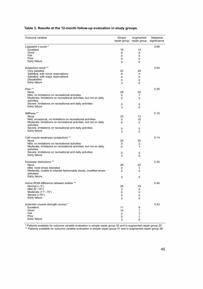
Table 3. Results at the 12-month follow-up evaluation in study groups.
Outcome variable Simple repair group
Augmented repair group
Statisticalsignificance
Leppilahti´s score *ExcellentGoodFairPoorEarly failure
198003
146005
0.68
Subjective result **Very satisfiedSatisfied, with minor reservationsSatisfied, with major reservationsDissatisfiedEarly failure
226003
203005
0.55
Pain **NoneMild, no limitations on recreational activitiesModerate, limitations on recreational activities, but not on daily activitiesSevere, limitations on recreational and daily activitiesEarly failure
2800
03
2210
05
0.35
Stiffness **NoneMild, occasional, no limitations on recreational activitiesModerate, limitations on recreational activities, but not on daily activitiesSevere, limitations on recreational and daily activitiesEarly failure
2350
03
13100
05
0.10
Calf muscle weakness (subjective) **NoneMild, no limitations on recreational activitiesModerate, limitations on recreational activities, but not on daily activitiesSevere, limitations on recreational and daily activitiesEarly failure
2080
03
2021
05
0.14
Footwear restrictions **NoneMild, most shoes toleratedModerate, unable to tolerate fashionable shoes, modified shoes tolerated Early failure
2800
3
2210
5
0.35
Active ROM difference between ankles **Normal (≤ 5°)Mild (6°–10°)Moderate (11°–15°)Severe (≥16°)Early failure
262003
194005
0.40
Isokinetic muscle strength (score) *ExcellentGoodFairPoorEarly failure
1114203
97315
0.43
* Patients available for outcome variable evaluation in simple repair group 30 and in augmented repair group 25** Patients available for outcome variable evaluation in simple repair group 31 and in augmented repair group 28
45

Isokinetic and isometric calf muscle strength
The isokinetic strength outcome score at the 12-month check-up was excellent for
eleven patients in the SR-group (36%), good for fourteen (47%) and fair for two
(7%), whereas it was excellent for nine patients in the AR-group (36%), good for
seven patients (28%), fair for three patients (12%) and poor for one patient (4%)
The early failures one patient (4%). There were early failures three (10%) in
SR-group and five (20%) in AR-group. (p=0.43) (Table 3)
The mean relative peak torque deficits for plantar flexion in the injured limb at
velocities of 60, 120 and 180º/sec did not differ significantly between the groups at
the 3-month and 12-month follow-up examinations. The figures at 12 months were
7%, 7% and 2%, respectively, for the SR group and 7%, 5% and 2% for the
AR-group (Table 4). The differences between the 3-month and 12-month results
were significant at velocities of 60º/sec and 120º/sec in both groups (Table 4).
The mean relative isometric strength deficit in the injured limb in plantar flex-
ion was 29% for the SR group and 34% for the AR-group at the 3-month check-up
and 10% for the SR group and 2% for the AR-group at the 12-month check-up (In
both groups p<0.001 between 3 and 12 months) (Table 4).
The differences in isokinetic dorsi flexion strength deficits were not signifi-
cant between the study groups. There was a slight tendency so that dorsi flexion
was weaker in the healthy leg than in the injured side (Table 4).
The mean peak work-displacement relationships upon plantar flexion of the
ankle in both groups at the 3-month and 12-month check-ups are shown in Figure
5. The deficit in plantar flexion motion was larger in the injured leg in both groups
at the 3–month check-up (p< 0.001), but the differences between the injured and
non-injured sides had diminished by the 12-month check-up. The differences in
the deficit in dorsiflexion of the ankle at the 3 and 12-month check-ups were not
significant between the groups or within the groups (Figure 6).
46

Fig. 5. Mean peak work in plantarflexion measured in 10-degree intervals at the
3-month and 12-month follow-ups (error bars denote SD). Control value is the healthy
leg in each group.
Fig. 6. Mean peak work in dorsiflexion measured in 10-degree intervals at the 3-month
and 12-month follow-ups (error bars denote SD). Control value is the healthy leg in
each group.
0
2
4
6
8
10
12
14
16
18
20
22
24
26
0
2
4
6
8
10
12
14
16
18
20
22
24
26
Pm < 0.001Pg = 0.79Pm*g = 0.25
10-2020-10
12 Months postoperatively Non-augmented - Operated Non-augmeted - Contol Augmented - Operated Augmented - Contol
Peak
Wor
k (J
)Displacement (degrees)
20-300-1010-020-10 10-0 0-10 10-20
Peak
Wor
k (J
)
Displacement (degrees)
Non-augmented - Operated Non-augmeted - Contol Augmented - Operated Augmented - Contol
3 Months postoperatively
20-30
Pm < 0.001Pg = 0.56Pm*g = 0.63
0
1
2
3
4
5
6
7
8
0
1
2
3
4
5
6
7
8
Pm < 0.001Pg = 0.90Pm*g = 0.90
12 Months postoperatively Non-augmented - Operated Non-augmeted - Contol Augmented - Operated Augmented - Contol
Peak
Wor
k (J)
Displacement (degrees)
30-20 20-10 10-0 0-10 10-2010-200-1010-020-10
Peak
Wor
k (J)
Displacement (degrees)
Non-augmented - Operated Non-augmeted - Contol Augmented - Operated Augmented - Contol
3 Months postoperatively
30-20
Pm < 0.001Pg = 0.75Pm*g = 0.63
47

Table 4. Mean (SD) strength deficits in percentages between the operated leg and
healthy leg at different angle speed velocities (60°/sec, 120°/sec and 180°/sec). P values
are calculated for comparisons between the groups and between the 3-month and
12-month results. Negative values in dorsiflexion results indicate that the operated leg
showed better strength than the healthy leg.
Tendon elongation
The elongation curves increased significantly up to twelve weeks in both groups
and decreased between 3 and 12 months (Ptime<0.001, Figure 7). The elongation
was more marked in the non-augmentation group at 3 months and 12 months,
although the difference between the groups was not significant. The median AT
At 3 months At 12 months P(3 month vs. 12 month)
Mean plantar flexion strength deficit %
At 60° / sec angle speedAugmentation group (n=21)Non-augmented group (n=28)
20.3 (SD 22.7)18.3 (SD 12.0)
6.5 (SD 11.5)7.4 (SD 9.1)
0.006<0.001
P (between groups) 0.37 >0.9
At 120° / sec angle speedAugmentation group (n=21)Non-augmented group (n=28)
17.2 (SD 21.2)18.1 (SD 12.1)
5.2 (SD 13.2)6.9 (SD 10.3)
0.0260.001
P (between groups) >0.9 0.59
At 180° / sec angle speedAugmentation group (n=21)Non-augmented group (n=28)
4.0 (SD 17.3)4.2 (SD 12.3)
1.7 (SD 12.7)1.6 (SD 10.4)
0.550.49
P (between groups) 0.74 0.86
Isometric plantar flexion strength deficit%Augmentation group (n=21)Non-augmented group (n=28)
34.4 (SD 13.5)28.9 (SD 13.5)
2.1 (SD 26.7)10.4 (SD 11.9)
<0.001<0.001
P (between groups) 0.32 0.24
Mean dorsiflexion strength deficit %
At 60° / sec angle speedAugmentation group (n=21)Non-augmented group (n=28)
–8.3 (SD 18.8)–9.2 (SD 15.6)
2.3 (SD 15.3)–0.4 (SD 12.6)
0.130.001
P (between grous) >0.9 0.66
At 120° / sec angle speedAugmentation group (n=21)Non-augmented group (n=28)
–4.8 (SD 17.4)–9.3 (SD 20.0)
3.2 (SD 12.6)–3.8 (SD 14.2)
0.0250.094
P (between groups) 0.45 0.045
At 180° / sec angle speedAugmentation group (n=21)Non-augmented group (n=28)
–2.6 (SD 17.5)–5.6 (SD 20.5)
–2.3 (SD 13.1)–11.3 (SD 16.5)
>0.90.13
P (between groups) 0.79 0.027
48

elongation was 5.0 mm (25th and 75th percentiles 2.5–9.2) in the SR-group and 6.0
mm (3.0–10.5) in the AR-group at 3 weeks, 8.0 mm (4.8–12.0) and 11.0 mm (7.0–
14.5) respectively at 6 weeks, and 14.0 mm (7.0–21.0) and 12.0 mm (8.0–20.5) at
3 months. After 3 months the AT shortened in both groups, the median elongation
being 10.0 mm (5.0–19.0) in the SR-group and 5.0 mm (2.0–20.0) in the AR group
at 12 months.
AT elongation correlated significantly with the isokinetic peak torque deficits
at velocities of 120º/sec (ρ=0.52, p=0.013) and 180º/sec (ρ=0.64, p=0.001) and
with the isometric strength deficits (ρ=0.475, p=0.026) in the SR-group.
Fig. 7. Achilles tendon elongation (millimetres) in both groups at the 3-week, 6-week,
3-month and 12-month follow-up examinations. Error bars denote SD.
Complications
Re-ruptures. There were three re-ruptures in each group. The mean time elapsing
before re-rupture was 58 (min. 2 and max. 112) days, and all the cases occurred in
males. Three patients had obviously suffered a new injury during the recovery
period. The first of these had fallen on a slippery bathroom floor two days after the
operation and had to forcefully step on the operated foot, the second was repairing
his wife’s bicycle at four weeks and he hit the edge of the pavement during a test
49

run, so that he had to put his weight on the toe of the operated foot, and the third
was walking in a swimming pool at four weeks and placed the toes of his operated
foot on the edge when climbing out of the pool and pushed with his full body
weight. The fourth re-rupture occurred at 10 weeks, when the patient was cycling
up a steep hill, and the fifth and sixth cases occurred at 12 weeks with a minimal
trauma. Both of these patients were in the non-augmented group. When a careful
history was taken, they recalled having had a slight trauma during the first 3 weeks
as well, while the dorsal brace was still on. The subjective clinical results in all the
re-rupture cases were good.
Infections. There were two deep infections in the augmentation group and
none in the simple repair group. The first such patient was a 36-year-old male who
developed a clear drainage at the incision after three weeks and was immediately
taken into hospital. The wound was revised and left open. None of the samples
yielded a positive bacterial culture. The infection had been brought under control
with intravenous antibiotics by 7 days and we were able to close the wound with a
two-tailed cutaneous turnover flap. He was able to walk normally at the one-year
check-up. The second patient was a 40-year-old male who had slow drainage from
the lower edge of the wound. He was taken into hospital at 6 weeks and the wound
was revised and left open. The bacterial cultures were positive for both Streptococ-
cus aureus and acinetobacteria. The Achilles tendon had already partially healed.
The situation was brought under control after 6 days with repeated revisions and
intravenous antibiotics, and it was possible to close his wound with a two-tailed
cutaneous turnover flap, which showed epidermal necrosis at first, but eventually
healed well. He was walking well at the one-year check-up and is satisfied with the
result. There were four superficial wound infections in the non-augmented group
and one in the augmented group. All these resolved after oral antibiotics and were
followed up according to the research protocol. These cases are included in the
results section in the normal way.
There was one case of a deep venous thrombosis in the augmented group that
was diagnosed at three weeks and treated with warfarin for six months. The patient
was followed up according to the protocol and is included in the results.
Return to previous level of activity. All patients in the study, including the
early failures, were able to return to their previous level of activity.
50
5.2 Re-rupture and deep infection following treatment of total AT
rupture (II)
Pain. Six Achilles tendons in the re-rupture group were painless, 5 were mildly
painful and one moderately painful. Only one tendon in the infection group was
painless, while 4 were mildly painful and 2 moderately painful (p=0.3).(Table 5)
Stiffness. Eight patients in the re-rupture group and 2 in the infection group
reported no stiffness in the Achilles tendon region. Four in the re-rupture group
and 5 in the infected group reported occasional mild stiffness (p=0.17).(Table 5)
Subjective calf muscle weakness. Six patients in the re-rupture group had no
subjective calf muscle weakness and 6 had mild weakness. One of those in the
infection group had mild subjective calf muscle weakness, 5 had moderate weak-
ness and one had severe weakness (p<0.001).(Table 5)
Footwear restrictions. None of the patients in the re-rupture group had foot-
wear restrictions, whereas 3 of those in the infection group had no footwear
restrictions and 4 had mild restrictions but tolerated most shoes (p=0.009). (Table
5)
Range of ankle motion. Eleven patients in the re-rupture group had a normal
range of ankle motion and one had mild limitation. Two of those in the infection
group had a normal range of motion, 4 had mild limitations and one had moderate
limitation (p=0.01). (Table 5)
Subjective outcome. Three patients in the re-rupture group were very satisfied
with the outcome, 8 were satisfied with minor reservations and one was satisfied
with major reservations, while one patient in the infection group was satisfied with
minor reservations and 6 with major reservations (p=0.004) (Table 5). Two
patients reported a definite loss of sensation in the foot, one having undergone an
unsuccessful reconstruction with a radial forearm flap and tensor fasciae latae graft
and the other sustaining damage to a branch of the sural nerve at the time of the
repair of the re-rupture.
Isokinetic and isometric muscle strength. In re-rupture group the isometric
strength was 14% lower in injured side when compared to healthy leg (p=0.012).
Respectfully in infection group the same difference was 42% in favour of healthy
side (p=0.07). In isokinetic tests the differences at speeds 60°/ sec, 120°/ sec and
180°/ sec in re-rupture group were 12%, 12% and 7% in favour of healthy leg
(p=0.007, p=0.003 and p=0.039) and in infection group at the same speeds 40%,
40% and 26% in favour of healthy leg (p=0.006, p=0.022 and p=0.022) (Table 6
and 7).
51
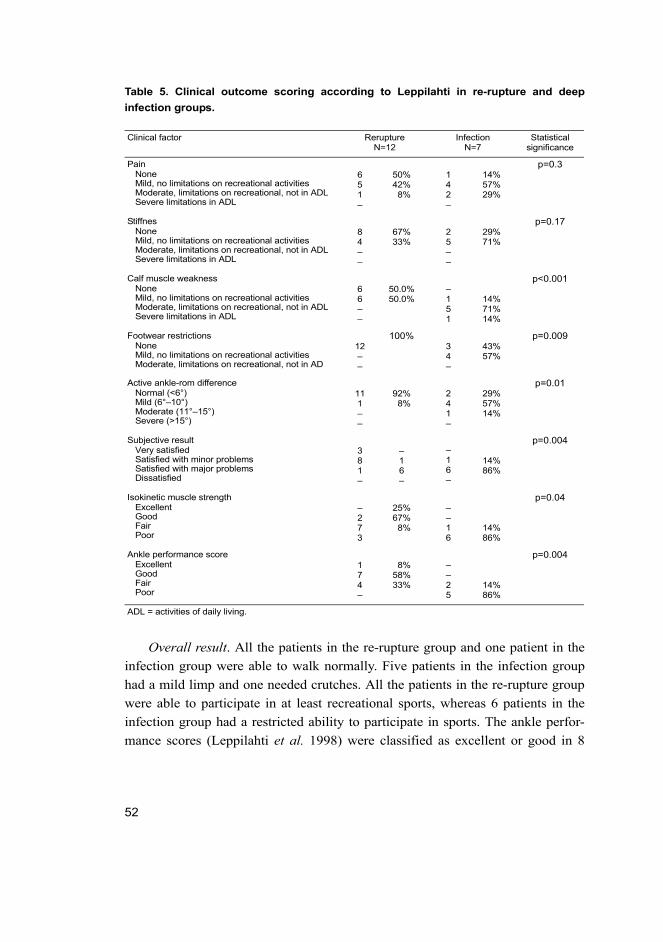
Table 5. Clinical outcome scoring according to Leppilahti in re-rupture and deep
infection groups.
Overall result. All the patients in the re-rupture group and one patient in the
infection group were able to walk normally. Five patients in the infection group
had a mild limp and one needed crutches. All the patients in the re-rupture group
were able to participate in at least recreational sports, whereas 6 patients in the
infection group had a restricted ability to participate in sports. The ankle perfor-
mance scores (Leppilahti et al. 1998) were classified as excellent or good in 8
Clinical factor ReruptureN=12
InfectionN=7
Statistical significance
PainNoneMild, no limitations on recreational activitiesModerate, limitations on recreational, not in ADLSevere limitations in ADL
651–
50%42%8%
142–
14%57%29%
p=0.3
StiffnesNoneMild, no limitations on recreational activitiesModerate, limitations on recreational, not in ADLSevere limitations in ADL
84––
67%33%
25––
29%71%
p=0.17
Calf muscle weaknessNoneMild, no limitations on recreational activitiesModerate, limitations on recreational, not in ADLSevere limitations in ADL
66––
50.0%50.0%
–151
14%71%14%
p<0.001
Footwear restrictionsNoneMild, no limitations on recreational activitiesModerate, limitations on recreational, not in AD
12––
100%34–
43%57%
p=0.009
Active ankle-rom differenceNormal (<6°)Mild (6°–10°)Moderate (11°–15°)Severe (>15°)
111––
92%8%
241–
29%57%14%
p=0.01
Subjective resultVery satisfiedSatisfied with minor problemsSatisfied with major problemsDissatisfied
381–
–16–
–16–
14%86%
p=0.004
Isokinetic muscle strengthExcellentGoodFairPoor
–273
25%67%8%
––16
14%86%
p=0.04
Ankle performance scoreExcellentGoodFairPoor
174–
8%58%33%
––25
14%86%
p=0.004
ADL = activities of daily living.
52

re-rupture cases and as fair in 4, while 2 results in the infection group were scored
as fair and 5 as poor (p=0.004). (Table 5)
Table 6. Mean peak torques (Nm) during plantar flexion and dorsiflexion of the ankle at
velocities 60º/sec, 120º/sec and 180º/sec and mean isometric strength of plantar flexion
in re-rupture group.
Table 7. Mean peak torques (Nm) during plantar flexion and dorsiflexion of the ankle at
velocities 60º/sec, 120º/sec and 180º/sec and mean isometric strength of plantar flexion
in infection group.
Incidence of re-rupture and deep infection
The population for which our university hospital served as the only unit treating
Achilles tendon ruptures increased from 94 000 to 120 000 during the period stud-
Test Speed
Rerupted group (N=12)
InjuredMean (SD)
UninjuredMean (SD)
Pairwise %differenceMean (SD) 95%CI P-value
Peak Torgue
Plantar flexion60°/sec120°/sec180°/sec
113.3 /21.7)89.3 (16.6)69.1 (12.1)
128.3 (17.9)102.3 (18.9) 75.2 (14.3)
11.6 (12.1)12.2 (10.9) 7.1 (12.5)
3.9, 19.35.3, 19.1–0.8, 15.0
0.0070.0030.039
Dorsiflextion60°/sec120°/sec180°/sec
30.3 (6.5)25.8 (5.5)24.8 (5.8)
30.8 (8.8)25.3 (4.0)24.2 (4.3)
–3.2 (18.7)–2.4 (18.1)–4.2 (23.7)
–15.1, 8.7–13.9, 9.2–19.3, 10.8
0.950.770.74
Isometric strength (N)Plantar flextion 130.3 (33.8) 154.6 (33.9) 14..4 (18.8) 2.5, 26.4 0.012
Test Speed
Infection group (N=7)
InjuredMean (SD)
UninjuredMean (SD)
Pairwise %differenceMean (SD) 95%CI P-value
Peak Torgue
Plantar flexion60°/sec120°/sec180°/sec
47.2 (29.3)40.4 (19.9)40.0 (19.0)
78.2 (42.4)68.4 (36.5)54.8 (26.8)
39.5 (11.3)39.5 (8.7)25.9 (10.7)
27.7, 51.428.6, 50.312.6, 39.2
0.0060.0220.022
Dorsiflextion60°/sec120°/sec180°/sec
26.3 (12.2)21.8 (10.5)21.4 (8.6)
Isometric strength (N)Plantar flextion 58.8 (40.7) 109.2 (55.9) 42.1 (24.8) 11.4, 72.9 0.07
53
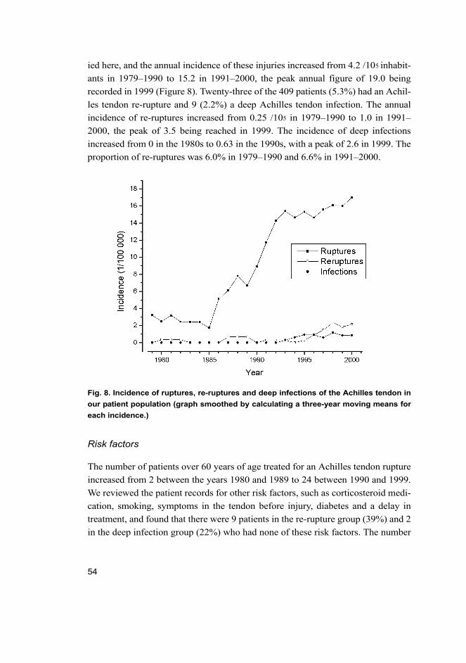
ied here, and the annual incidence of these injuries increased from 4.2 /105 inhabit-
ants in 1979–1990 to 15.2 in 1991–2000, the peak annual figure of 19.0 being
recorded in 1999 (Figure 8). Twenty-three of the 409 patients (5.3%) had an Achil-
les tendon re-rupture and 9 (2.2%) a deep Achilles tendon infection. The annual
incidence of re-ruptures increased from 0.25 /105 in 1979–1990 to 1.0 in 1991–
2000, the peak of 3.5 being reached in 1999. The incidence of deep infections
increased from 0 in the 1980s to 0.63 in the 1990s, with a peak of 2.6 in 1999. The
proportion of re-ruptures was 6.0% in 1979–1990 and 6.6% in 1991–2000.
Fig. 8. Incidence of ruptures, re-ruptures and deep infections of the Achilles tendon in
our patient population (graph smoothed by calculating a three-year moving means for
each incidence.)
Risk factors
The number of patients over 60 years of age treated for an Achilles tendon rupture
increased from 2 between the years 1980 and 1989 to 24 between 1990 and 1999.
We reviewed the patient records for other risk factors, such as corticosteroid medi-
cation, smoking, symptoms in the tendon before injury, diabetes and a delay in
treatment, and found that there were 9 patients in the re-rupture group (39%) and 2
in the deep infection group (22%) who had none of these risk factors. The number
54
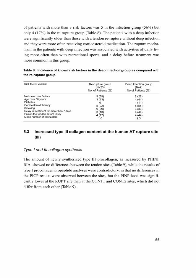
of patients with more than 3 risk factors was 5 in the infection group (56%) but
only 4 (17%) in the re-rupture group (Table 8). The patients with a deep infection
were significantly older than those with a tendon re-rupture without deep infection
and they were more often receiving corticosteroid medication. The rupture mecha-
nism in the patients with deep infection was associated with activities of daily liv-
ing more often than with recreational sports, and a delay before treatment was
more common in this group.
Table 8. Incidence of known risk factors in the deep infection group as compared with
the re-rupture group.
5.3 Increased type III collagen content at the human AT rupture site
(III)
Type I and III collagen synthesis
The amount of newly synthesized type III procollagen, as measured by PIIINP
RIA, showed no differences between the tendon sites (Table 9), while the results of
type I procollagen propeptide analyses were contradictory, in that no differences in
the PICP results were observed between the sites, but the PINP level was signifi-
cantly lower at the RUPT site than at the CONT1 and CONT2 sites, which did not
differ from each other (Table 9).
Risk factor variable Re-rupture group(N=23)
No. of Patients (%)
Deep Infection group(N=9)
No.of Patients (%)
No known risk factorsAge over 60 yearsDiabetesCorticosteroid therapySmokingDelay in treatment for more than 7 daysPain in the tendon before injuryMean number of risk factors
9 (39)3 (13)
05 (22)9 (39)3 (13)4 (17)
1.0
2 (22)4 (44)1 (11)5 (56)3 (33)4 (44)4 (44)
2.3
55

Table 9. Median (range min-max) content of type I and III collagen markers and total
collagen at the sampling sites.
Total collagen content and type I and III collagen structures in the insoluble
matrix
Collagens accounted for about 70% of the dry weight of the insoluble matrix of the
ruptured Achilles tendons, whereas the insoluble matrix in the cadavers contained
significantly more collagen, about 90% of dry weight at all sites (Table 9).
Both IIINTP and tryptic PIIINP were markedly increased at the RUPT site in
the individuals with total Achilles tendon rupture relative to either the CONT1 or
CONT2 site. The CONT1 site contained more IIINTP than did CONT2 in the rup-
ture patients but not in the cadavers, while the RUPT site in the rupture patients
contained more IIINTP than did that in the cadavers. The tryptic PIIINP levels did
not differ significantly between the rupture patients and cadavers, but there was
tendency in that direction. When the very high tryptic PIIINP value in the cadaver
group (for a 13-year-old male) was excluded, the difference between the rupture
patients and the cadavers at the RUPT site was statistically significant (p < 0.05).
The type III pN-collagen content as calculated from the molar amounts of IIINTP
and tryptic PIIINP (less than 0.1%) did not differ between the sites or between the
rupture patients and cadavers, however.
Sample site
Rupture Control 1 Control 2
Soluble tissue extract (μg/g of wet tissue weight)
PIIINPPINPPICP
558 (143–4584)227 (50–543) *516 (137–1103)
384 (157–1414)428 (233–1107)463 (183–1581)
516 (142–1298)524 (173–1019)417 (225–954)
Insoluble tissue digests (mg/g of dry tissue weights, except PIIINP μg/g)
IIINTP rupturecadaver
15.3 (6.5–30.9)** ±3.7 (2.1–13.6)*
3.0 (1.2–8.9)#2.1 (0.7–5.5)
1.2 (0.7–8.3)1.2 (0.6–1.9)
Tryptic PIIINP rupturecadaver
15.4 (3.5–82.1)**5.7 (2.2–48.4)
3.6 (1.2–19.2)3.9 (1.8–30.1)
1.2 (0.7–10.0)1.2 (0.7–9.9)
ICTP rupturecadaver
2.6 (1.5–3.6)2.4 (1.5–18.3)
2.4 (1.4–3.9)2.5(1.5–20.7)
1.2 (0.7–3.2)#1.2 (0.7–17.2)♦
Total collagen rupturecadaver
731 (540–770)±±881 (830–923)
725 (674–798)±±905 (873–981)
705 (375–788)±±927 (833–959)
* p<0.05 when compared to CONT1 and CONT2, ** p<0.005 when compared to CONT1 and CONT2.# p<0.005 when compared to CONT2, ## p<0.01 when compared to RUPT and CNT1♦p<0.05 when compared to RUPT and CONT1± p<0.005 when compared to cadaver, ±± p<0.001 when compared to cadaver
56
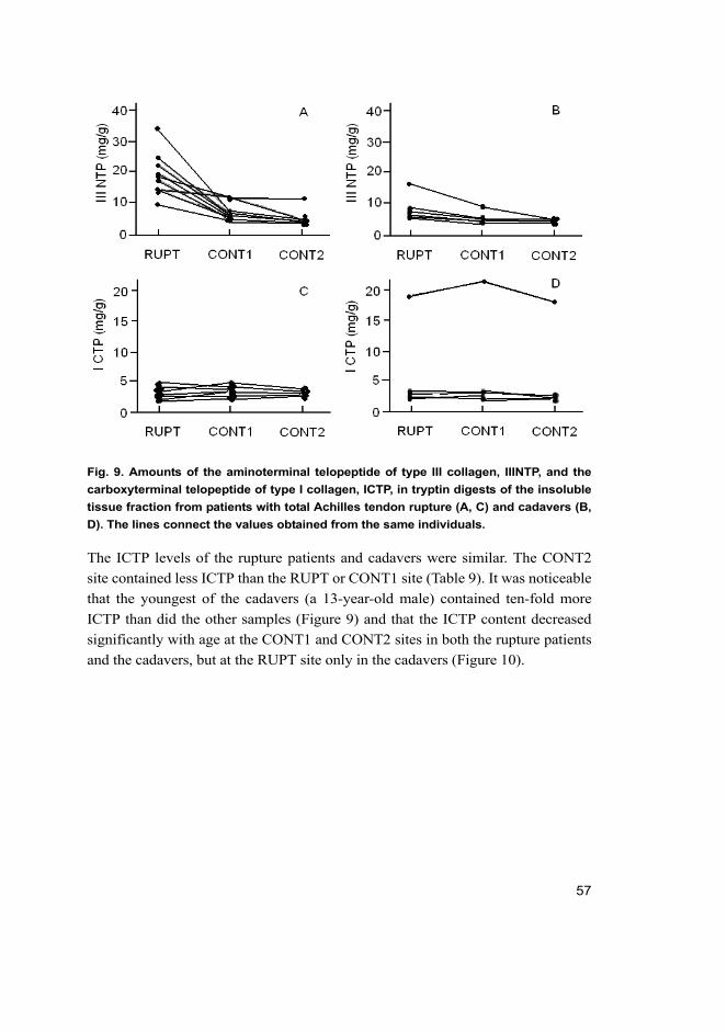
Fig. 9. Amounts of the aminoterminal telopeptide of type III collagen, IIINTP, and the
carboxyterminal telopeptide of type I collagen, ICTP, in tryptin digests of the insoluble
tissue fraction from patients with total Achilles tendon rupture (A, C) and cadavers (B,
D). The lines connect the values obtained from the same individuals.
The ICTP levels of the rupture patients and cadavers were similar. The CONT2
site contained less ICTP than the RUPT or CONT1 site (Table 9). It was noticeable
that the youngest of the cadavers (a 13-year-old male) contained ten-fold more
ICTP than did the other samples (Figure 9) and that the ICTP content decreased
significantly with age at the CONT1 and CONT2 sites in both the rupture patients
and the cadavers, but at the RUPT site only in the cadavers (Figure 10).
57
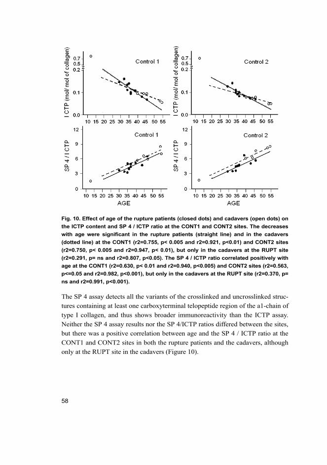
Fig. 10. Effect of age of the rupture patients (closed dots) and cadavers (open dots) on
the ICTP content and SP 4 / ICTP ratio at the CONT1 and CONT2 sites. The decreases
with age were significant in the rupture patients (straight line) and in the cadavers
(dotted line) at the CONT1 (r2=0.755, p< 0.005 and r2=0.921, p<0.01) and CONT2 sites
(r2=0.750, p< 0.005 and r2=0.947, p< 0.01), but only in the cadavers at the RUPT site
(r2=0.291, p= ns and r2=0.807, p<0.05). The SP 4 / ICTP ratio correlated positively with
age at the CONT1 (r2=0.630, p< 0.01 and r2=0.940, p<0.005) and CONT2 sites (r2=0.563,
p<=0.05 and r2=0.982, p<0.001), but only in the cadavers at the RUPT site (r2=0.370, p=
ns and r2=0.991, p<0.001).
The SP 4 assay detects all the variants of the crosslinked and uncrosslinked struc-
tures containing at least one carboxyterminal telopeptide region of the a1-chain of
type I collagen, and thus shows broader immunoreactivity than the ICTP assay.
Neither the SP 4 assay results nor the SP 4/ICTP ratios differed between the sites,
but there was a positive correlation between age and the SP 4 / ICTP ratio at the
CONT1 and CONT2 sites in both the rupture patients and the cadavers, although
only at the RUPT site in the cadavers (Figure 10).
58

ICTP and IIINTP at the RUPT site in the reverse phase run
By loading equal amounts of ICTP antigen, the two runs could be standardized to
observe the differences in the IIINTP antigen. In the case of the rupture patients
IIINTP eluted at a position where pyridinoline fluorescence was also observed,
although the majority of the pyridinoline eluted in the same position as with ICTP.
The cadaver sample had less IIINTP, and the pyridinoline peak was also lower
(Figure 11). These data show that IIINTP contains pyridinoline.
Fig. 11. HPLC reverse phase analysis of the insoluble tissue digests from the RUPT site
of one patient with a ruptured Achilles tendon (A) and from a site corresponding to the
rupture site in a cadaver (B). IIINTP (closed triangles) and ICTP (closed dots) were
analysed from the fractions. Pyridinoline fluorescence (straight line) was followed
during the run. The elution position of the trivalent pyridinoline-crosslinked IIINTP
structure is marked with arrows.
25 30 35 40 45 500
4
8
12
0.0
0.3
0.6
0.9A
IIIN
TP (
mg
/L)
ICTP
(mg
/L)
25 30 35 40 45 500
4
8
12
0.0
0.3
0.6
0.9B
Time (min)
IIIN
TP (
mg/
L) IC
TP (m
g/L
)
59

5.4 Tenascin-C and type I and III collagen expression in total AT
rupture (IV)
Samples. The final series included nine men and one woman, with an average age
of 38 years (range 30–48), who were operated on a mean of 29 (10–43) hours after
the injury. One sample taken at the rupture site and one sample used in the type I
collagen analysis were spoiled during processing.
Tenascin-C. There was no significant difference between the sites (Table 10).
Type I and III procollagens. The type I carboxyterminal (PICP) and aminoter-
minal (PINP) procollagens were almost equal in expression at the sites (Table 10),
but the expression of type III procollagen (PIIINP) was significantly higher at the
rupture site than at control site 2 (Table 10, p=0.016 Sign Test).
Mature type III collagen. The amount of mature type III collagen present was
significantly higher at the rupture site than at control sites 1 and 2 (Table 10,
P=0.008 Sign Test).
60
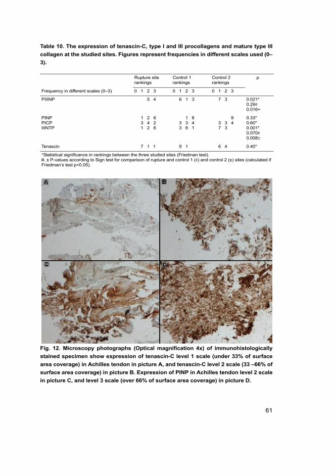
Table 10. The expression of tenascin-C, type I and III procollagens and mature type III
collagen at the studied sites. Figures represent frequencies in different scales used (0–
3).
Fig. 12. Microscopy photographs (Optical magnification 4x) of immunohistologically
stained specimen show expression of tenascin-C level 1 scale (under 33% of surface
area coverage) in Achilles tendon in picture A, and tenascin-C level 2 scale (33 –66% of
surface area coverage) in picture B. Expression of PINP in Achilles tendon level 2 scale
in picture C, and level 3 scale (over 66% of surface area coverage) in picture D.
Rupture site rankings
Control 1 rankings
Control 2 rankings
p
Frequency in different scales (0–3) 0 1 2 3 0 1 2 3 0 1 2 3
PIIINP 5 4 6 1 3 7 3 0.021*0.29#0.016+
PINPPICPIIINTP
1 2 63 4 21 2 6
1 83 3 43 6 1
93 3 47 3
0.33*0.60*0.001*0.070#0.008±
Tenascin 7 1 1 9 1 6 4 0.40*
*Statistical significance in rankings between the three studied sites (Friedman test).#, ± P-values according to Sign test for comparison of rupture and control 1 (#) and control 2 (±) sites (calculated if Friedman’s test p<0.05).
61

62

6 Discussion
6.1 Augmented vs. non-augmented surgical repair of acute total AT
rupture
Surgery has been suggested by many authors as the standard treatment for a com-
plete Achilles tendon rupture, especially where young, active patients are con-
cerned, as the risk of re-ruptures is lower than after conservative treatment. Surgi-
cal procedures naturally entail more wound complications, but the majority of
these do not affect the final outcome. The most popular surgical approaches today
are suture without augmentation, suture with augmentation and percutaneous tech-
niques. Augmented techniques have been justified in terms of the higher tensile
strength attained while non-augmented techniques in terms of the shorter incision
and lower incidence of wound problems (Khan et al. 2004).
We found only three studies in the literature which compared the results
achieved with augmented and non-augmented suture techniques, two of which had
a retrospective research design (Jessing & Hansen 1975, Nyyssönen et al. 2003),
while the one prospective study involved quite a small number of patients (Aktas
et al. 2007).
In our prospective, randomized study we compared the results of surgical
repair of a fresh complete Achilles tendon rupture with an end-to-end Krackow
locking loop suture alone and augmented with one central down-turned gastrocne-
mius fascia flap (Silfverskiöld 1941), but failed to find any benefit of using the
augmented repair technique in this population, the results in terms of subjective
and objective ankle outcomes, isokinetic strength scores, mean peak work-dis-
placement relationships and tendon elongation being equally good and the number
of deep infections (2/28 vs. 0/32), re-ruptures (3/28 vs 3/32) and superficial infec-
tions comparable. On the other hand, the statistically shorter operating time (a dif-
ference of 25 min) and the smaller incision (a difference of 7cm) argue in favour of
end-to-end repair. This result is in line with other examinations of the same clini-
cally relevant question (Jessing & Hansen 1975, Nyyssönen et al. 2003, Aktas et
al. 2007).
The primary outcome measure employed, Leppilahti´s outcome score, pointed
to excellent or good results in 90% of cases in the simple repair group and 81% in
the augmented repair group at the 12-month follow up. Early failures amounted to
10% in the simple repair group and 19% in the augmented repair group. No differ-
ences were seen between the two groups with regard to pain, stiffness, subjective
63
calf muscle weakness, footwear restrictions or range of ankle motion. These
results were slightly better than in the author’s previous report, where two postop-
erative regimens were randomly compared after Achilles tendon suture with the
augmented technique, yielding excellent or good outcome scores in 88% of cases
in the early motion group, fair in 4% and poor in 8%, whereas the scores in the cast
group were excellent or good in 92% of cases and fair in 8% (Kangas et al. 2003).
Our results were also better than in a previous series from our hospital in which the
postoperative treatment consisted of 6 weeks of below-knee cast immobilization
with the ankle in an equinus position for 3 weeks and in a neutral position for 3
weeks, allowing gradual weight bearing after three weeks, for which the ankle
scores were excellent or good in 79% of the 101 cases, fair in 17% and poor in 4%
a mean of 3 years postoperatively (Leppilahti et al. 1998).
The re-rupture rate was about 10% in both of our two groups, which is higher
than expected (Khan et al. 2005). One contributing factor may have been the short
period of brace immobilization together with considerable weight bearing. The
soft cast brace was removed at three weeks and 20 kg weight bearing was allowed
for three weeks, half weight bearing for the next three weeks and full weight bear-
ing after six weeks. Even the augmentation technique could not prevent re-rup-
tures. Similar results regarding early weight bearing have been reported with a
cross-stitch suture (Aoki et al. 1998). Three of our patients had an obvious new
injury during the recovery period. Some of the re-rupture mechanisms described in
the results section evidently involved serious exceeding of the limits laid down in
the rehabilitation programme. The other two cases of re-rupture, both in the
non-augmented group, occurred as a result of low-force activity at 12 weeks, and it
transpired that these patients had also suffered a slight trauma during the first 3
weeks, while the dorsal brace was still on. It is our opinion that this initial trauma
had interfered with the repair and caused a gap between the tendon ends. Recovery
of strength in the tendon repair process has been shown in canine model to be
slower upon gap formation (Gelbermann et al. 1999), and this may have meant
that the remaining suture strength and improperly healed tendon could no longer
resist normal forces at 12 weeks, a situation that resembles delayed union in bone
healing. In the authors’ previous study the re-rupture rate was 6% when full weight
bearing was allowed after three weeks but dorsiflexion was restricted to neutral
until six weeks (Kangas et al. 2003), and elsewhere active rehabilitation and mobi-
lization has been shown to improve the functional results and tendon healing
(Enwemenka et al. 1988, Mortensen et al. 1999), although we think that dorsiflex-
ion should be controlled by means of a brace for longer than three weeks.
64
There is no universal consensus that a given suture type and suture thickness is
the method of choice for ATR repair. We wanted to use a #0 absorbable monofila-
ment, with minimal tissue response but enough tensile strength for tendon healing,
and many authorities in Europe use monofilament or braided absorbable sutures,
whereas the tendency in North America is to use mechanically stronger stitches
extending above and below the site of rupture. No randomized clinical studies of
the suture materials used for ATR repair are available. The material that we used
(polydioxanon) has been reported to lose 50% of its strength during the first 4
weeks (data from Ethicon, Johnson & Johnson Inc., Somerville, New Jersey)
(O´Broin 1995).
Infections occurring after the surgical repair of a ruptured Achilles tendon are
often extremely difficult to manage and the final outcome is poor. We were suc-
cessful in treating our two cases of deep infection with débridement and two-tail
cutaneous transfer flaps. We strongly advocate careful postoperative observation
of Achilles tendon ruptures by experienced surgeons, since problems tend to arise
unexpectedly and often remain unnoticed at first. Prompt and precise action is
needed in the early phase of an infection.
The strengths of this study lie in its prospective, randomized design and the
homogeneous groups of patients. There were no known biases, all the patients
were operated on by the same surgeon and they were advised to perform postoper-
ative exercises according to a standard rehabilitation programme. Also, thorough
evaluations were performed using various clinical outcome tools, an isokinetic
strength score, peak work-displacement relationships and tendon elongation mea-
surements. The only possible object of criticism could be that our evaluators were
not blinded to the assignment of patients to treatment groups. Also the relative
small number of subjects at the follow-ups prevent definite conclusions concern-
ing some outcome variables.
6.2 Re-rupture and deep infection following treatment of total AT
rupture
The incidence of total Achilles tendon ruptures is increasing (Leppilahti et al.
1996c), and deep infections are reported to occur after surgery in 1% to 2% of
cases, re-ruptures in 2% to 15% and minor complications in 15% to 20% (Khan et
al. 2005). There are also more re-ruptures after conservative treatment than after
surgical treatment (Khan et al. 2005), but overall clinical outcome can be very
similar (Möller et al. 2001). The overall incidence of re-ruptures and deep infec-
65

tions after the treatment of Achilles tendon ruptures has scarcely ever been
reported in the literature, and the outcomes after these complications are men-
tioned only in case reports.
We conducted the present study in response to a treatment-based suspicion of
an increase in the incidence of re-ruptures and deep infections in our hospital. The
outcome after successful treatment of a total Achilles tendon rupture is good
regardless of the method used, but there are very few conclusive reports on the
final outcome after complications have occurred. Ankle flexion forces have been
reported to be low in patients who have a re-rupture, and the sequelae after infec-
tion are said to be particularly difficult, often requiring plastic surgery interven-
tions. Previous studies have suggested that diabetes, corticosteroid usage, age and
previous symptoms in the Achilles tendon area are related to a higher risk of deep
infection. A second operation also poses a risk of infection, especially in the Achil-
les tendon area, where the subcutaneous tissue is very thin. We are not aware of
any conclusive report on the final outcome after re-rupture or deep infection. We
believe that more profound information on the final outcome after complicated
Achilles tendon rupture repair could be very useful for doctors who see these
patients in practice and need guidelines for their treatment.
The incidence of total Achilles tendon ruptures in our geographical area
increased nearly four-fold between 1979–1990 and 1991–2000, from 4.2 to 15.2
(per 100,000 inhabitants), while that of re-ruptures increased from 0.25 to 1.0 and
that of deep infections from 0 to 0.63. The ratio of re-ruptures and deep infections
to primary Achilles tendon ruptures did not change substantially over this period.
The rate of re-ruptures was in our university hospital-based patient series was
5.6% and the rate of deep infections 2.2%. These figures are reliable and are not
affected by any known bias. Similar figures for re-ruptures and deep infections
have been reported in a recent meta-analysis (Khan et al. 2005). Increases in the
incidence of total Achilles tendon ruptures have been reported in Scotland, Copen-
hagen, Malmö, and Oulu, and it is evident that the total number of complications is
also increasing, and that surgeons must be more aware of these complications. At
the time of our highest complication rates, in 1999, we were very aggressively
recruiting patients for operative treatment, and some of them were high at risk to
receive treatment complications.
The two groups formed in the present study clearly differed in terms of risk
factors such as advanced age, diabetes, corticosteroid use, smoking, delayed treat-
ment and previous tendon symptoms, these factors being more numerous in the
deep infection group. As the number of known risk factors for surgery in the
66

Achilles tendon area was markedly higher in the deep infection group, we feel that
at least some of the complications could have been predicted and perhaps avoided
with better patient selection. Interestingly, the majority of our patients with a sim-
ple re-rupture had sustained their original injury during participation in sports
activities, whereas the majority of those with deep infections had sustained their
initial rupture during normal activities of daily life. It is possible that an Achilles
tendon that is liable to rupture during normal daily activities may already be in
such a poor condition that normal healing is impaired and the risk of postoperative
complications is increased.
Eleven of the twelve patients in the present re-rupture group were subjectively
satisfied with the final clinical outcome and mentioned only minor problems,
whereas only one out of the seven in the deep infection group was equally satis-
fied. The results in terms of the clinical outcome score were even worse, with only
eight patients in the re-rupture group and none in the deep infection group achiev-
ing a good or excellent level of recovery. Likewise, the mean isokinetic plantar
flexion strength deficit for three test velocities was 10% in the re-rupture group
and 35% in the deep infection group. The results in both groups were nevertheless
inferior to those achieved following successful primary surgical treatment. In a
previous study of 101 patients with an Achilles tendon rupture without re-rupture
or infection who were monitored in our clinic for a mean of three years postopera-
tively, the mean isokinetic calf-muscle strength deficit was only 7% (Leppilahti et
al. 1996c).
A deep infection after surgical repair of an Achilles tendon rupture is a rela-
tively rare but devastating problem, as the skin and soft-tissue defects associated
with Achilles tendon loss constitute a major challenge for surgeons. Methods for
the reconstruction of soft tissues after failed surgery have been presented in the lit-
erature (Maffulli & Ajis 2008), but the results have been variable and there are
insufficient data in general to support any of the techniques. In our clinic the infec-
tion is first brought under control with débridement and administration of antibiot-
ics. A skin cover is provided with split-thickness skin grafts, local transposition
flaps, or free-tissue transfers by means of a microvascular anastomosis. The ten-
don tissue itself is reconstructed with an autograft or allograft. The present series
included two patients who had reconstruction with a lateral radial forearm flap and
a tensor fasciae latae graft; the reconstruction being successful in one patient and a
failure in the other forever. The cases with deep infection in our randomized series
were successfully treated with two-tailed skin and subcutaneous tissue transfers.
Their infections were rapidly recognized and prompt action saved us from tendon
67

tissue reconstructions. It is not possible on the available data to decide which
method of reconstruction is best.
6.3 Type I and III collagen expression in total AT rupture
The main purpose of the work reported in Papers III and IV was to examine the
patterns of tenascin-C and type I and III collagen expression and collagen cross-
linking in the ruptured human Achilles tendon by comparing expression at the rup-
ture site with that at two other sites within the same tendon, and also with that in
presumably healthy cadavers in Paper III. The tendon samples from living patients
were harvested less than 43 hours after rupture and should at most represent the
tendon tissue composition before the trauma (Haukipuro et al. 1990, Fluck et al.
2000). The samples from the cadavers were harvested within 72 hours post mor-
tem.
The level of mature type III collagen was markedly higher at the rupture site
than at the two control sites with both methods used. Similar results have been
reported in other studies (Kannus & Josza 1991).
The type III collagen content of the insoluble tissue digests of patients with
total Achilles tendon rupture was markedly increased at the rupture site. IIINTP
represents type III collagen which has been incorporated into the collagen fibrils
and stabilized there by intermolecular pyridinoline crosslinks, forming what is
known as mature type III collagen. The type III collagen content at the site in the
cadavers corresponding to the RUPT site was significantly lower than in the rup-
ture patients. Only one cadaver (a 41-year-old male) exhibited IIINTP levels com-
parable with those of the rupture patients, but his whole tendon felt much stiffer
than the other tendons and could be represent a case of undiagnosed tendinopathy.
Type III pN-collagen, immature type III collagen, did not differ in content between
the sites, indicating similar processing of type III collagen.
The soluble propeptide antigens represent newly synthesized procollagens or
partially processed forms which have not yet been covalently crosslinked into the
insoluble matrix, i.e. immature type III collagen. Since the unchanged PIIINP lev-
els in the soluble tissue extracts indicate a slow overall rate of type III procollagen
synthesis, the accumulation of a large amount of type III collagen at the rupture
site must have taken place over a longish period. This suggests that there a contin-
uous, long-lasting microtraumatic process may have been taking place prior to the
total rupture of the Achilles tendon. The reason for this microtrauma is not clear,
and the role of mechanical overloading cannot be excluded. Such a gradual accu-
68
mulation of type III collagen will eventually cause a decrease in the biomechanical
strength of the tendon.
The concentration of PINP was lower at the rupture site than at the control
sites, but the PICP levels remained unchanged. The lower PINP levels may have
been caused by the presence of type I pN-collagen, where part of the PINP is
retained on the surface of the newly synthesized collagen molecules in the insolu-
ble matrix. This may lead to thinner type I collagen fibres, as has been found in
embryonic skin (Fleishmajer et al. 1983). It is also possible that the PINP antigen
may not be as stable as PICP and was degraded during processing.
The ICTP concentration was lowest at the control 2 site in both the rupture
patients and the cadavers, probably due to the tendon tissue gradually changing
into fascia. Ageing is known to affect collagen crosslinking profiles, and the
amount of pyridinoline in the human Achilles tendon, for example, has been
reported to increase up to the age of thirty and then to decrease gradually
(Moriguchi et al. 1978). The ICTP levels decreased and the ratio of SP4 to ICTP
increased with age at both control sites, suggesting a relative increase in the
amount of unknown variants of the crosslinked carboxyterminal telopeptide struc-
tures that only the SP4 assay is capable of detecting. The 13-year-old male in the
cadaver series had ten-fold higher ICTP concentration and a lower SP4/ICTP ratio
than did the other cases, suggesting a pyridinoline-like crosslinking pattern. The
mature crosslinked telopeptide structure appearing in the Achilles tendon with age
could be identical or analogous to the crosslinking structure predominating in the
human skin (Sassi et al. 2001). It has been shown that a reduction in pyridinoline
crosslink density causes biomechanical weakening of the healing rabbit medial
collateral ligament (Frank et al. 1995), and the same authors discussed the possi-
bility that skin-like crosslinking might be the reason for the decrease in the pyridi-
noline content. The skin-like crosslinked telopeptide can be measured with the SP
4 assay, but not with the ICTP assay (Sassi et al. 2001). If the low ICTP content
contributes to total Achilles tendon rupture, the change in the collagen fibril orga-
nization must be a generalized one.
6.4 Tenascin-C in Achilles tendon rupture
Although elevated expression of tenascin-C has been found in certain regions of
degenerated human supraspinatus tendons, our results show no differences
between the sites in this respect (Riley et al. 1996). Similarly, higher tenascin-C
expression has been reported at the musculo-tendinous junction of a rat Achilles
69
tendon after increased physical loading (Järvinen et al. 1999). We think that the
long-term mechanical loading affecting the rupture site of the human Achilles ten-
don does not differ from that at other sites in the same tendon. Since any changes
in tenascin-C expression should take place after a delay of more than 36 hours
(Fluck et al. 2000), the levels obtained in the present ruptured tendons must mainly
have originated from the time before the rupture. We suspect that there may be a
more universal mechanical loading pathology associated with the degenerative
process and rupture of the human Achilles tendon. Abnormal tension in the gas-
trocnemius apparatus could be one explanation for our findings, and we would
agree that tenascin-C has some as yet unknown role in the loading changes taking
place in connective tissue (Järvinen et al. 2000).
Since we could not find any correlation between the expression of tenascin-C
and type III collagen synthesis or accumulation., we believe that the role of tenas-
cin-C in tendon degeneration is variable. A similar observation has been made
regarding the appearance of tenascin-C in supraspinatus bursae, which did not cor-
relate with normal histopathological findings of degeneration (Hyvönen et al.
2003).
70

7 Conclusions
The augmented repair seems to have no clear advantages over simple end-to-end
repair in cases of fresh complete Achilles tendon rupture. The outcome measure
tools we used did not show any significant difference between these two groups.
Very active postoperative rehabilitation combined with short-term brace immobili-
zation and considerable weight bearing gives excellent or good results with most
patients, but there is also an increased risk of early re-ruptures that are unrelated to
the surgical technique.
The incidence of Achilles tendon re-ruptures and deep infections has
increased. The outcome is satisfactory after a simple re-rupture without infection,
but the results after a deep infection are often devastating.
Clear evidence of a slow accumulation of type III collagen at the rupture site
of the Achilles tendon was found. This may lead to biomechanically weaker tissue
due to the thinner collagen fibres. The unexpectedly low level of ICTP structures
in the tendon tissue and the decrease in these with age may exacerbate qualitative
changes, which reduce the strength of the matrix before total rupture of the Achil-
les tendon occurs.
Although tenascin-C is expressed evenly over the whole Achilles tendon, we
do not believe that it has any specific value as a predictive or diagnostic marker of
tendon degeneration. Our findings support results in which tenascin-C expression
has been shown to be elevated after mechanical loading, but its exact function in
the extracellular matrix of human tendon tissue still remains unresolved.
71

72

8 Future prospects for Achilles tendon rupture research
There is still no definite answer of which method is the best for treatment of an
acute Achilles tendon rupture. Large size randomized studies comparing surgical
techniques and even surgery and non-surgery are needed for statistically signifi-
cant results and therefore a multi-centre co-operation would be needed. The inci-
dence of Achilles tendon ruptures, re-ruptures and deep infections has increased,
but we still do not know why the tendon tissue is more prone to this injury in some
individuals than in others. Future Achilles tendon research can be expected to
expand our knowledge of the biochemical alterations, especially with respect to
changes in collagen structure. There are already numerous studies available deal-
ing with the matrix metalloproteinases (MMP) and their inhibitors (TIMP), which
are said to control the degradation of collagen, and the presence of certain types of
MMP´s is known to be correlated with painful tendon syndromes (Jones et al.
2006).
Another area of special interest is the enhancement of tendon healing. Local
administration of growth factors or tissue scaffolds, including tendon healing pro-
moting substances, has already been studied in animal models (Anitua et al. 2006).
73

74

References
Aktas S, Kocaoglu B, Nalbantoglu U, Seyhan M & Guven O (2007) End-to-end versus aug-mented repair in the treatment of acute Achilles tendon ruptures. J Foot Ankle Surg 46:336–40.
Anitua E, Sanchez M, Nurden AT, Zalduendo M, de la Fuente M, Orive G, Azofra J &Andia I (2006) Autologous fibrin matrices: a potential source of biological mediatorsthat modulate tendon cell activities. J Biomed Mat Res 77:285–93.
Aoki M, Ogiwara N, Ohta T & Nabeta Y (1998) Early active motion and weightbearingafter cross-stitch achilles tendon repair. Am J Sports Med 26:794–800.
Arner O & Lindholm Å (1959) Subcutaneous rupture of the Achilles tendon. Acta ChirScand 239:7–51.
Arner O, Lindholm Å & Orell SR (1958/1959) Histologic changes in subcutaneous ruptureof the Achilles tendon. Acta Chir Scand 116:484–490.
Barfred T (1973) Achilles tendon rupture. Aetiology and pathogenesis of subcutananeousrupture assessed on the basis of literature and rupture experiments on rats. Acta OrthopScand Suppl 152:1–124.
Baruah DR (1984) Bilateral spontaneous rupture of the Achilles tendons in a patient onlong-term systemic steroid therapy. Br J Sports Med 18:128–129.
Best T & Garrett W (1994) Basic sience of soft tissue: Muscle and tendon. OrthopaedicSports Medicine, Philadelphia, WB Saunders 1–45.
Birch HL, McLaughlin L & Smith RK (1999) Treadmill exercise-induced tendon hypertro-phy: assessment of tendon with different mechanical functions. Equine Vet J Suppl30:222–226.
Blake RL & Ferguson HJ (1991) Achilles tendon rupture. A protocol for conservative man-agement. J Am Pod Med Ass 81:486–489.
Bode MK, Mosorin M, Satta J, Risteli L, Juvonen T & Risteli J (1999) Complete processingof type III collagen in atherosclerotic plaques. Arterioscler Thromb Vasc Biol 19:1506–1511.
Bode MK, Karttunen TJ, Mäkelä J, Risteli L & Risteli J (2000a) Type I and III collagens inhuman colon cancer and diverticulosis. Scand J Gastroenterol 35:747–752.
Bode MK, Soini Y, Melkko J, Satta J, Risteli L & Risteli J (2000b) Increased amount oftype III pN-collagen in human abdominal aortic aneurysms: evidence for impaired typeIII collagen fibrillogenesis. J Vasc Surg 32:1201–1207.
Böhm VE, Thiel A & Czieske S (1990) Die Achillessehnenruptur. Anamnestische und mor-phologische Untersuchungen sowie berlegungen zur etiologie. Sportverletz Sport-schaden 4:22–28.
Borchgrevink G & Grøntvedt T (2005) Re-rupture after operation for total Achilles tendonrupture. Tidsskr Nor Laegeforen 125:2488–90.
Bradley JP & Tibone JE (1990) Percutaneous and open surgical repairs of Achilles tendonruptures. Am J Sports Med 18:188–195.
Brown SJ, Worsfold M & Sharp CA (2001) Microplate assay for the measurement ofhydroxyproline in acid-hydrolyzed tissue samples. Biotech 30:38–42.
75
Bruggeman NB, Turner NS, Dahm DL, Voll AE, Hoskin TL, Jacofsky DJ & HaidukewychGJ (2004) Wound complications after open Achilles tendon repair: an analysis of riskfactors. Clin Orthop Relat Res 427:63–6.
Carden DG, Jonathan N, Chalmers J, Lunn P & Ellis J (1987) Rupture of the calcaneal ten-don. The early and late management. J Bone Joint Surg (Br) 69:416–420.
Carr AJ & Norris SH (1989) The blood supply of the calcaneal tendon. J Bone Joint Surg(Br) 71:100–101.
Cetti R, Christensen S-E, Ejsted R, Jense NM & Jorgensen U (1993) Operative versus non-operative treatment of Achilles tendon rupture. Am J Sports Med 21:791–799.
Cetti R, Henriksen LO & Jacobsen KS (1994) A new treatment of ruptured Achilles ten-dons. Clin Orthop 308:155–165.
Chiquet-Ehrismann R, Tannheimer M, Koch M, Brunner A, Spring J, Martin D, Baumgart-ner S & Chiquet M (1994) Tenascin-C expression by fibroblasts is elevated in stressedcollagen gels. J Cell Biol 127:2093–2101.
Chiquet-Ehrismann R & Tucker RP (2004) Connective tissues: signalling by tenascins. Int JBiochem Cell Biol 36:1085–1089.
Christensen IB (1954) Rupture of the Achille tendon: analysis of 57 cases. Acta Chir Scand106:50–60.
Copeland SA (1990) Rupture of the Achilles tendon: a new clinical test. Ann R Coll SurgEngl 72:270–271.
Costa ML, Shepstone L, Darrah C, Marshall T & Donell ST (2003) Immediatefull-weight-bearing mobilisation for repaired Achilles tendon ruptures: a pilot study.Injury 34:874–876.
Cowan MA & Alexander S (1961) Simultaneous bilateral rupture of Achilles tendons due totriamcinolone. BMJ 10:1658.
Cummins EJ, Anson BJ, Carr BW & Wrigh RR (1946) The structure of calcaneal tendon (ofAchilles) in relation to orthopaedic surgery. With additional observations on the planta-ris muscle. Surg Gynecol Obstet 83:107–116.
Curwin S & Stanish WD (1984) Tendinitis: its etiology and treatment. Lexington: Collam-ore Press, DC Health and Co, 45–90.
Enwemeka CS, Spielholz NI & Nelson AJ (1988) The effect of early functional activities onexperimentally tenotomized Achilles tendons in rats. Am J Phys Med Rehab 67:264–269.
Erickson HP (1997) A tenascin knockout with a phenotype. Nat Genet 17: 5–7.Eyre DR, Paz MA & Gallop PM (1984) Cross-linking in collagen and elastin. Annu Rev
Biochem 53:717–48.Fleischmajer R, Olsen BR, Timpl R, Perlish JS & Lovelace O (1983) Collagen fibril forma-
tion in embryogenesis. Proc Natl Acad Sci 80:3354–8.Fluck M, Tunc-Civelek V & Chiquet M (2000) Rapid and reciprocal regulation of tenas-
cin-C and tenascin-Y expression by loading of skeletal muscle. J Cell Sci 113 Pt 20:3583–3591.
76

Forsberg E, Hirsch E, Frölich L, Meyer M, Ekblom P, Aszodi A, Werner S & Fässler R(1996) Skin wound and nerves heal normally in mice lacking tenascin-C. Proc NatAcad Sci USA 93:6594–6599.
Fox JM, Blazina ME, Jobe FW, Kerlan RK, Carter VS, Shields CL Jr & Carlson RN (1975)Degeneration and rupture of the Achilles tendon. Clin Orthop 107:221–224.
Frank C, McDonald D, Wilson J, Eyre D & Shrive N (1995) Rabbit medial collateral liga-ment scar weakness is associated with decreased collagen pyridinoline crosslink den-sity. J Orthop Res 13:157–165.
Gebauer M, Beil FT, Beckmann J, Sárváry AM, Ueblacker P, Ruecker AH, Holste J &Meenen NM (2007) Mechanical evaluation of different techniques for Achilles tendonrepair. Arch Orthop Trauma Surg 127:795–799.
Gelbermann RH, Boyer MI, Brodt MD, Winters SC, & Silva MJ (1999) The effect of gapformation at the repair site on the strength and excursion of intrasynovial flexor ten-dons. An experimental study on the early stages of tendon-healing in dogs. J Bone JointSurg (Am) 81:975–982.
Haines JF (1983) Bilateral rupture of the Achilles tendon in patients on steroid therapy. AnnRheum Dis 42:652–654.
Hattrup SJ & Johnson KA (1985) A review of ruptures of the Achilles tendon. Foot Ankle6:34–38.
Haukipuro K, Risteli L, Kairaluoma MI & Risteli J (1990) Aminoterminal propeptide oftype III procollagen in serum during wound healing in human beings. Surgery 107:381–388.
Hurme T, Lehto M, Kalimo H, Kannus P & Järvinen M (1990) Sequence of changes in fibresize and type in muscle immobilized at various lengths. J Sports Traumatol Rel Res2:77–85.
Hyvönen P, Melkko J, Lehto VP & Jalovaara P (2003) Involvement of the subacromialbursa in impingement syndrome of the shoulder as judged by expression of tenascin-Cand histopathology. J Bone Joint Surg Br 85:299–305
Inglis AE, Scott WN, Sculco TP & Patterson AH (1976) Ruptures of the tendo Achillis. JBone Joint Surg (Am) 58:990–993.
Ippolito E, Natali PG, Postacchini F, Accinni L & Martino CD (1980) Morphological,immunochemical and biomechanical study of rabbit Achilles tendon at various ages. JBone Joint Surg (Am) 62:583–598.
Jagose JT, McGregor DR & Nind GR (1996) Achilles tendon rupture due to ciprofloxacin.NZ Med J 109:471–472.
Järvinen M (1992) Epidemiology of tendon injuries in sports. Clin Sports Med 11:493–504.Järvinen TA, Jozsa L, Kannus P, Järvinen TL, Kvist M, Hurme T, Isola J, Kalimo H & Jarvi-
nen M (1999) Mechanical loading regulates tenascin-C expression in the osteotendi-nous junction. J. Cell Sci 112 Pt 18:3157–3166.
Järvinen TA, Kannus P, Järvinen TL, Jozsa L, Kalimo H & Järvinen M (2000) Tenascin-C inthe pathobiology and healing process of musculoskeletal tissue injury. Scand J Med SciSports 10:376–382.
77

Järvinen TA, Järvinen TL, Kannus P, Józsa L & Järvinen M (2004) Collagen fibres of thespontaneously ruptured human tendons display decreased thickness and crimp angle. JOrthop Res 22:1303–1309.
Jessing P & Hansen E (1975) Surgical treatment of 102 tendo Achillis ruptures. Suture ortenontoplasty? Acta Chir Scand 141:370–377.
Jones GC, Corps AN, Pennington CJ, Clark IM, Edwards DR, Bradley MM, Hazleman BL& Riley GP (2006) Expression profiling of metalloproteinases and tissue inhibitors ofmetalloproteinases in normal and degenerate human achilles tendon. Arth & Rheum54:832–842.
Jozsa L, Kvist M, Balint BJ, Reffy A, Järvinen M, Lehto M & Barzo M (1989a) The role ofrecreational sport activity in Achilles tendon rupture. A clinical, pathoanatomical, andsosiological study of 292 cases. Am J Sports Med 17:338–343.
Josza, Lehto M, Kvist M, Bálint JB & Reffy A (1989b) Alterations in dry mass content ofcollagen fibers in degenerative tendinopathy and tendon-rupture. Matrix 9:140–146.
Jozsa LG & Kannus P (1997) Human tendons: anatomy, physiology and pathology. Humankinetics. Champaign, Illinois.
Kaarteenaho-Wiik R, Kinnula VL, Herva R, Soini Y, Pöllänen R & Pääkkö P (2002)Tenascin-C is highly expressed in respiratory distress syndrome and broncho-pulmonary dysplasia. J Histochem Cytochem 50:423–431.
Kallinen M & Suominen H (1994) Ultrasonographic measurements of the Achilles tendonin elderly athletes and sedentary men. Acta Radiol 35:560–563.
Kangas J, Pajala A, Siira P, Hämäläinen M & Leppilahti J (2003) Early functional treatmentversus early immobilization in tension of the musculotendinous unit after Achilles rup-ture repair: a prospective, randomized, clinical study. J Trauma 54:1171–1180.
Kannus P & Jozsa L (1991) Histopathological changes preceding spontaneous rupture of atendon. J Bone Joint Surg (Am) 73:1507–1525.
Karjalainen PT, Soila K, Aronen HJ, Pihlajamäki HK, Tynninen O, Paavonen T & TirmanPF (2000) MR imaging of overuse injuries of the Achilles tendon. AJR Am J Roent-genol 175:251–260.
Karpakka J (1991) Effects of physical activity and inactivity on collagen synthesis in ratskeletal muscle and tendon. Thesis. Acta Universitas Ouluensis. Series D, Medica 231.Oulu University, Finland.
Kauppila S, Bode MK, Stenbäck F, Risteli L & Risteli J (1999) Cross-linked telopeptides oftype I and III collagens in malignant ovarian tumours in vivo. Br J Cancer 81:654–651.
Khan RJK, Fick D, Brammar TJ, Crawford J & Parker MJ (2004) Surgical interventions fortreating acute Achilles tendon ruptures. Cochrane Database Syst Rev (3): CD003674.
Khan R, Fick D, Keogh A, Crawford J, Brammar T & Parker M (2005) Treatment of AcuteAchilles Tendon Ruptures. A Meta-Analysis of Randomized, Controlled Trials J BoneJoint Surg (Am) 87:2202–2210.
Kielty CM, Hopkinson I & Grant ME (1993) Collagen: the collagen family, structure,assembly and organizations in the extracellular matrix. In: Connective tissue and itsinherited disorders. Molecular, genetic and medical aspects. New York, Wiley-Lisz,103–147.
78

Kivirikko KI & Myllylä R (1982) Biosynthesis of collagen. In: Pietz KA & Reddi AH (eds)Extracellular matrix bichemistry. Elsevier, New York, 83–118.
Klein W, Lang D & Saleh M (1991) The use of the Ma-Griffith technique for percutaneusrepair of fresh ruptured tendo Achillis. Chir Organi Mov 76:223–228.
Kleinman M & Gross AE (1983) Achilles tendon rupture following steroid injection J BoneJoint Surg (Am) 65:1345–1347.
Knott L, Tarlton JF & Bailey AJ (1997) Chemistry of collagen cross-linking: biochemicalchanges in collagen during the partial mineralization of turkey leg tendon. Biochem J322:535–542.
Koivunen-Niemelä T & Parkkola K (1995) Anatomy of the Achilles tendon (tendocalcaneus) with respect to tendon thickness measurements. Surg Radiol A Nat 17:263–268.
Komi PV, Fukashiro S & Järvinen M (1992) Biomechanical loading of Achilles tendon dur-ing normal locomotion. Clin Sports Med 11:521–531.
Laseter JT & Russell JA (1991) Anabolic steroid-induced tendon pathology: a review of theliterature. Med Ski Sports Exerc 23:1–3.
Last & Reiser (1984) Collagen biosynthesis. Environ Health Persp 55:169–177.Lea RB & Smith L (1972) Non-surgical treatment of tendo Achillis rupture. J Bone Joint
Surg (Am) 54:1398–1407.Leadbetter (1992) Cell matrix response in tendon injury. Clin Sports Med 11: 533–678 Lee MLH (1961) Bilateral rupture of Achilles tendon. BMJ 10:1829–1830.Lehtinen A, Peltokallio P & Taavitsainen M (1994) Sonography of Achilles tendon corre-
lated to operative findings. Ann Chir Gynaecol 83:143–148.Leitner A, Muller A, Voigt C & Rahmanzadeh R (1991) Eine modifierte Nachbehandlung
nach primar versorgter Achillessehnenruptur. Akt Traumatol 21:285–292.Leppilahti J, Puranen J & Orava S (1996a) Incidence of Achilles tendon rupture. Acta
Orthop Scand. 67:277–279.Leppilahti J, Siira P, Vanharanta H & Orava S (1996b) Isokinetic evaluation of calf muscle
performance after Achilles rupture repair. Int J Sports Med 17:619–623.Leppilahti J (1996c) Achilles tendon rupture with special reference to epidemiology and
results of surgery. Thesis. Acta Universitas Ouluensis, Series D, Medica 383. OuluUniversity, Finland.
Leppilahti J, Forsman K, Puranen J & Orava S (1998) Outcome and prognostic factors ofAchilles rupture repair using a new scoring method. Clin Orthop 346:152–161.
Lim J, Dalal R & Waseem M (2001) Percutaneus vs. open repair of the ruptured Achillestendon a prospective randomized controlled study. Foot Ankle Int 22:559–568.
Liu SH, Yang RS, Al-Shaikh R & Lane JM (1995) Collagen in tendon, ligament, and bonehealing. A current review. Clin Orthop 318:265–278.
Mackie E.J, Halfter D & Liverani D (1988) Induction of tenascin in healing wounds. J CellBiol 107:2757–2767.
Maes R, Copin G & Averous C (2006) Is percutaneous repair of the Achilles tendon a safetechnique? A study of 124 cases. Acta Orthop Belg 72:179–183.
79
Ma GW & Griffith TG (1977) Percutaneous repair of acute closed ruptured achilles tendon:a new technique. Clin Orthop 128:247–255.
Maffulli N (1998) The clinical diagnosis of subcutaneous tear of the Achilles tendon. A pro-spective study in 174 patients. Am J Sports Med 26:266–270.
Maffulli N (1999) Rupture of the Achilles tendon. J Bone Joint Surg (Am) 81: 1019–1036.Maffulli N, Barrass V & Ewen SW (2000) Light microscopic histology of achilles tendon
ruptures. A comparison with unruptured tendons. Am J Sports Med 28: 857–863.Maffulli N, Tallon C, Wong J, Peng Lim K & Bleakney R (2003) No adverse effect of early
weight bearing following open repair of acute tears of the Achilles tendon. J SportsMed Phys Fitness 43:367–379.
Maffulli N & Ajis A (2008) Management of chronic ruptures of the Achilles tendon. J BoneJoint Surg Am 90:1348–1360.
Mandelbaum BR, Myerson MS & Forster R (1995) Achilles tendon ruptures. A newmethod of repair, early range of motion, and functional rehabilitation. Am J SportsMed 23:392–395.
Mann RA, Holmes GP, Seale KS & Collins DN (1991) Chronic rupture of the Achilles ten-don: a new technique of repair. J Bone Joint Surg (Am) 73:214–219.
Maxwell LC & Enwemeka CS (1992) Immobilization-induced muscle atrophy is notreserved by lengthening the muscle. Anat Rec 234:55–61.
McGarvey WC, Singh D & Trevino SG (1990) Partial Achilles tendon ruptures associatedwith fluoroquinolone antibiotics; a case report and literature review. Foot Ankle Int17:496–498.
Melkko J, Niemi S, Risteli L & Risteli J (1990) Radioimmunoassay of the carboxyterminalpropeptide of human type I procollagen. Clin Chem 36:1328–1332.
Melkko J, Kauppila S, Niemi S, Risteli L, Haukipuro K & Jukkola A (1996) Immunoassayfor intact aminotermnal propeptide of human type I procollagen. Clin Chem 42:947–954.
Melmed EP (1965) Spontaneous bilateral rupture of the calcaneal tendon during steroidtherapy. J Bone Joint Surg (Br) 47:104–105.
Metz R,Verleisdonk EJ, van der Heijden GJ, Clevers GJ, Hammacher ER, Verhofstad MH& van der Werken C (2008) Acute Achilles tendon rupture: minimally invasive surgeryversus nonoperative treatment with immediate full weight bearing – a randomized con-trolled trial. Am J Sports Med 36:1688–1694.
Michna H & Hartmann G (1989) Adaptation of tendon collagen to exercise. Int Orthop 13:161–165.
Mink JH, Deutsch AL & Kerr R (1991) Tendon injuries of the lower extremity: Magneticresonance assessment. Top Magn Reson Imaging 3:23–38.
Mokone GG, Gajjar M, September AV, Schwellnus MP, Greenberg J, Noakes TD & CollinsM (2005) The guanine-thymine dinucleotide repeat polymorphism within the tenas-cin-C gene is associated with achilles tendon injuries. Am J Sports Med 33:1016–1021.
Möller M, Movin T, Granhed H, Lind K, Faxén E & Karlsson J (2001) Acute rupture oftendo Achillis A prospective, randomized Study of comparison between surcical andnon-surcical treatment. J Bone Joint Surg (Br) 83:843–848.
80
Moriguchi T & Fujimoto D (1978) Age-related changes in the content of the collagen cross-link, pyridinoline. J Biochem 84:933–935.
Mortensen HM, Skov O & Jensen PE (1999) Early motion of the ankle after operative treat-ment of a rupture of the Achilles tendon. A prospective, randomized clinical and radio-graphic study. J Bone Joint Surg (Am) 81:983–990.
Movin T, Ryberg A, McBride DJ & Maffulli N. (2005) Acute rupture of the Achilles ten-don. Foot Ankle Clin 10:331–356.
Nestorson J, Movin T, Möller M & Karlsson J (2000) Function after Achilles tendon rupturein the eldery: 25 patients older than 65 years followed for 3 years. Acta Orthop Scand7:64–68.
Nillius SA, Nilsson BE & Westlin NE (1976) The incidence of Achilles tendon rupture.Acta Orthop Scand 47:118–121.
Nistor L (1981) Surgical and non-surgical treatment of Achilles tendon rupture. J BoneJoint Surg (Am) 63:394–399.
Nyyssönen T & Luthje P (2000) Achilles tendon ruptures in South-East Finland between1986–1996, with special reference to epidemiology, complications of surgery and hos-pital costs. Ann Chir Gyn 89:53–57.
Nyyssönen T, Saarikoski H, Kaukonen JP, Luthje P, & Hakovirta H (2003) Simpleend-to-end suture versus augmented repair in acute Achilles tendon ruptures: a retro-spective comparison in 98 patients. Acta Orthop Scand 74:206–208.
Oberhauser A.F, Marszalek P.E, Erickson H.P & Fernandez J.M (1998) The molecular last-icity of the extracellular matrix protein tenascin. Nature 393:181–185.
O'Brien M (1992) Functional anatomy and physiology of tendons. Clin Sports Med 11:505–520.
O´Broin ES, Earley MJ, Smyth H & Hooper ACB (1995) Absorbable sutures in tendonrepair. A comparison of PDS with prolene in rabbit tendon repair. J Hand Surg 20:505–508.
Orava S, Hurme M & Leppilahti J (1996) Bilateral Achilles tendon rupture: a report on twocases. Scand J Med Sci Sports 6:309–312.
Panageas E, Greenberg S, Franklin PD, Carter AP & Bloom DB (1990) Magnetic resonanceimaging of pathologic conditions of the Achilles tendon. Orthop Rev 19:975–980.
Petersen OF, Nielsen MB, Jensen KH & Solgaard S (2002) Randomized comparison ofCAM walker and light-weight plaster cast in the treatment of first-time Achilles tendonrupture. Ugeskr Laeger 164:3852–3855.
Price AE, Evanski PM & Waugh (1986) TR Bilateral simultaneous Achilles tendon rup-tures. Clin Orthop 213:249–250.
Quickley TB & Scheller AD (1980) Surgical repair of the ruptured Achilles tendon. Am JSports Med 8:244–250.
Rantanen J, Hurme T & Paananen M (1993) Immobilization in neutral versus equinus posi-tion after Achilles tendon repair. Acta Orthop Scand 64:333–335.
81

Rantanen J, Hurme T & Kalimo H (1999) Calf muscle atrophy and Achilles tendon healingfollowing experimental tendon division and surgery in rats. Comparison of postopera-tive immobilization of the muscle-tendon complex in relaxed and tensioned positions.Scand J Med Sci Sports 9:57–61.
Riley GP, Harrall RL, Cawston TE, Hazleman BL & Mackie EJ (1996) Tenascin-C andhuman tendon degeneration. Am J Pathol 149:933–943.
Risteli J, Niemi S, Trivedi P, Mäentausta O, Mowat AP & Risteli L (1988) Rapid equilib-rium radioimmunoassay for the aminoterminal propeptide of human type III procolla-gen. Clin Chem 34:715–718.
Ronel DN, Newman MI, Gayle LB & Hoffman LA (2004) Recent advances in the recon-struction of complex Achilles tendon defects. Microsurg 24:18–23.
Rosager S, Aagaard P & Dyhre-Poulsen P (2002) Load-displacement properties of thehuman triceps surae aponeurosis and tendon in runners and non-runners. Scand J MedSci Sports 12:90–98.
Rowley D & Scotland T (1982) Rupture of the Achilles tendon treated by a simple operativeprocedure. Injury 14:252–254.
Sage H & Bornstein P (1992). Extracellular proteins that mediate cell-matrix interactions.Mini-review. J Biol Chem 266:14831–14834.
Saleh M, Marshall PD, Senior R & MacFarlane A (1992) The Sheffield splint for controlledearly mobilisation after rupture of the calcaneal tendon. A prospective, randomisedcomparison with plaster treatment J Bone Joint Surg (Br) 74:206–209.
Salter DM (1993) Tenascin is increased in cartilage and synovium from arthritic knees. Br JRheumatol 32:780–786.
Sassi ML, Eriksen HA, Risteli L, Niemi S, Mansell J & Gowen M (2000) Immunochemicalchracterization of assay for carboxyterminal telopeptide of human type I collagen.Bone 26:367–373.
Sassi ML, Jukkola A, Reikki R, Höyhtyä M, Risteli L & Oikarinen A (2001) Type I colla-gen turnover and cross-linking are increased in irradiated skin of breast cancer patients.Radiother Oncol 58:317–323.
Schönbauer HR (1964) Gedeckte Achillessehnenrisse. Wiederherst Chir Traumat 8:160–185.
Settles DL, Cihak RA & Erickson HP (1996) Tenascin-C expression in dystrofin-relatedmuscular dystrophy. Muscle&Nerve 19:147–154.
Silfverskiöld N (1941) Über die subkutane totale Achillessehnenruptur und deren Behand-lung. Acta Chir Scand 84:393–413.
Simmonds FA (1957) The diagnosis of the ruptured Achilles tendon. The Practitioner179:56–58.
Smaill GB (1961) Bilateral rupture of Achilles tendons. BMJ 10:1657–1658.Stein SR & Luekens CA (1976) Closed treaatment of Achilles tendon ruptures. Orth Clin
North Am 7:241–246.Stein V, Laprell H, Tinnemeyer S & Peterson W (2000) Quantitative assessment of intravas-
cular volume of the human Achilles tendon. Acta Orthop Scand 71:60–63.
82

Suchak AA, Spooner C, Reid DC & Jomha NM (2006) Postoperative rehabilitation proto-cols for Achilles tendon ruptures: a meta-analysis. Clin Orthop 445:216–221.
Thermann H, Frerichs O & Biewenwr A (1995a) Biomechanical studies of human Achillestendon rupture. Unfallchirurg 98:570–575.
Thermann H, Zwipp H & Tscherne H (1995b) Functional treatment concept of acute ruptureof the Achilles tendon. 2 years results of a prospective randomized study. Unfallchirurg98:21–32.
Thompson TC & Doherty JH (1962) Spontaneous rupture of tendon of Achilles: a new clin-ical diagnostic test. J Trauma 2:126–129.
Tipton CM, Vailas AC & Matthes RD (1986) Experimental studies on the influence of phys-ical activity on ligaments, tendons and joints: a brief review. Acta Med Scand Suppl711:157–168.
Wapner KL, Pavlock GS, Hecht PJ, Naselli F & Walther R (1993) Repair of chronic Achil-les tendon rupture with flexor hallucis longus tendon transfer. Foot Ankle 14:443–449.
Webb J & Bannister G (1999) Percutaneus repair of the ruptured tendo achillis. J Bone JointSurg (Br) 81:877–880.
Weinstabl R & Herz H (1990) Gleichzeitige beidseitige Achillessehnenruptur nach Baga-telltrauma bei Steroidtherapie-Fallbericht. Unfallchirurg 16:50–54.
White RK & Kraynick BM (1959) Surgical uses of the peroneus brevis tendon. Surg GynecObstetr 108:117–121.
Wills CA, Wasburn S, Caiozzo V & Prietto CA (1986) Achilles tendon rupture. A review ofthe literature comparing surgical versus nonsurgical treatment. Clin Orthop 207:156–163.
Wong J, Barrass V & Maffulli N (2002) Quantitative Review of Operative and Nonopera-tive Management of Achilles Tendon Ruptures. Am J Sports Med 30:566–575.
Woo SL, Ritter MA & Amiel D (1980) The biomechanical properties of swine tendons –long term effects of exercise on the digital extensors. Connect Tissue Res 7:177–183.
Zernicke RF & Loitz BJ (1992) Exercise-related adaptations in connective tissue. In: KomiPV (ed) Strenght and power in sport. The encyclopaedia of sports medicine. Blackwell,Oxford, 77–95.
Zollinger H, Rodniguez M & Genoni M (1983) Zur Ätiopathogenese und Diagnostik derAchillessehnenrupturen im Sport. In: Chapchal G, editor. Sportverletzung und Sport-schäden, Stuttgart: Georg Theme 75–77.
83

84
Original publications
This thesis is based on the following articles, which are referred to in the text by
their Roman numerals:
I Pajala A, Kangas J, Siira P, Ohtonen P & Leppilahti J. Augmented surgical
repair compared with non-augmented in fresh total Achilles tendon rupture: A
prospective, randomized study. J Bone Joint Surg (Am) (In press).
II Pajala A, Kangas J, Ohtonen P & Leppilahti J (2002) Rerupture and deep
infection following treatment of total Achilles tendon rupture. J Bone Joint
Surg (Am) 84:2016–2021.
III Eriksen HA, Pajala A, Leppilahti J & Risteli J. (2002) Increased content of
type III collagen at the rupture site of human Achilles tendon. J Orthop Res
20:1352–1357.
IV Pajala A, Melkko J, Leppilahti J, Ohtonen P, Soini Y & Risteli J. Tenascin,
type I and III collagen expression at the Achilles tendon rupture: An
immunohistochemical study. Histol Histopathol (In press).
The original publications have been reproduced with permission of the copyright
holders.
85

86

A C T A U N I V E R S I T A T I S O U L U E N S I S
Distributed byOULU UNIVERSITY LIBRARY
P.O. Box 7500, FI-90014University of Oulu, Finland
Book orders:OULU UNIVERSITY PRESSP.O. Box 8200, FI-90014University of Oulu, Finland
S E R I E S D M E D I C A
997. Peltonen, Tuomas (2008) Endothelial factors in the pathogenesis of aortic valvestenosis
998. Leinonen, Jukka (2008) Carbonic anhydrase isoenzyme VI: distribution, catalyticproperties and biological significance
999. Ulvila, Johanna (2008) Functional genomic analysis of the Drosophila immuneresponse. Identification of genes essential for phagocytosis, viral defense and NF-?B signaling
1000. Lajunen, Taina (2008) Persistent Chlamydia pneumoniae infection, inflammationand innate immunity
1001. Leinonen, Pekka (2008) Calcium signaling in epithelium. Special focus on Hailey-Hailey and Darier diseases, neurofibromatosis 1 and transitional cell carcinoma
1002. Pääkkönen, Virve (2009) Expression profiling of human pulp tissue andodontoblasts in vivo and in vitro
1003. Anttonen, Olli (2009) Prevalence, prognosis and characteristics of subjects withshort QT interval in an electrocardiogram
1004. Pulkkinen, Pasi (2009) Radiographical assessment of hip fragility
1005. Iinattiniemi, Sari (2009) Fall accidents and exercise among a very old home-dwelling population
1006. Westerlund, Tarja (2009) Thermal, circulatory, and neuromuscular responses towhole-body cryotherapy
1007. Kaikkonen, Kari (2009) Risk factors for sudden cardiac death from an acuteischemic event in general population. A case-control study
1008. Kuisma, Mari (2009) Magnetic resonance imaging of lumbar degenerative bonemarrow (Modic) changes. Determinants, natural course and association with lowback pain
1009. Vartiainen, Johanna (2009) Ghrelin, obesity and type 2 diabetes. Genetic,metabolic and epidemiological studies
1010. Löfgren, Johan (2009) Genetic polymorphisms in collectins and Toll-like receptor4 as factors influencing susceptibility to severe RSV infections and otitis media
1011. Korhonen, Topi (2009) Mathematical modeling of the regulation, developmentand genetically engineered experimental models of cardiac excitation-contractioncoupling

ABCDEFG
UNIVERS ITY OF OULU P.O.B . 7500 F I -90014 UNIVERS ITY OF OULU F INLAND
A C T A U N I V E R S I T A T I S O U L U E N S I S
S E R I E S E D I T O R S
SCIENTIAE RERUM NATURALIUM
HUMANIORA
TECHNICA
MEDICA
SCIENTIAE RERUM SOCIALIUM
SCRIPTA ACADEMICA
OECONOMICA
EDITOR IN CHIEF
PUBLICATIONS EDITOR
Professor Mikko Siponen
University Lecturer Elise Kärkkäinen
Professor Hannu Heusala
Professor Olli Vuolteenaho
Senior Researcher Eila Estola
Information officer Tiina Pistokoski
University Lecturer Seppo Eriksson
Professor Olli Vuolteenaho
Publications Editor Kirsti Nurkkala
ISBN 978-951-42-9091-6 (Paperback)ISBN 978-951-42-9092-3 (PDF)ISSN 0355-3221 (Print)ISSN 1796-2234 (Online)
U N I V E R S I TAT I S O U L U E N S I S
MEDICA
ACTAD
D 1012
ACTA
Ari Pajala
OULU 2009
D 1012
Ari Pajala
ACHILLES TENDON RUPTURECOMPARISON OF TWO SURGICAL TECHNIQUES, EVALUATION OF OUTCOMES AFTER COMPLICATIONS AND BIOCHEMICAL AND HISTOLOGICAL ANALYSES OF COLLAGEN TYPE I AND III AND TENASCIN-C EXPRESSIONIN THE ACHILLES TENDON
FACULTY OF MEDICINE,INSTITUTE OF CLINICAL MEDICINE,DEPARTMENT OF SURGERY, DIVISION OF ORTHOPAEDIC AND TRAUMA SURGERY,INSTITUTE OF DIAGNOSTICS,DEPARTMENT OF CLINICAL CHEMISTRY,UNIVERSITY OF OULU
Related Documents


 • Achilles tendon resting angle test [14] (√) • Lægomfang [14]](https://static.cupdf.com/doc/110x72/609378f53addf55da06be5f5/genoptrnings-forlbsbeskrivelse-for-borgere-med-a-achilles-tendon-total.jpg)