Acharya N.G Ranga Agricultural University, Rajendra nagar, Hyderabad, 500030 Study Material for Course No: Ento 333 Field Diagnosis in Agriculture (1+1) (2011-12) EDITOR Dr.G.Raghavaiah, Professor & Head Dept. of Entomology, Agril College, Bapatla Contributors Entomology Plant Pathology Plant Physiology Dr.G.Ramachandra Rao Dr.P.Anil Kumar Dr.A.Siva Sankar Dr.K.Manjula Dr.P.Kishore Varma Dr.T.Ramesh Dr.N.Hariprasad Dr.P.Seetharamu

Welcome message from author
This document is posted to help you gain knowledge. Please leave a comment to let me know what you think about it! Share it to your friends and learn new things together.
Transcript

Acharya N.G Ranga Agricultural University,
Rajendra nagar, Hyderabad, 500030
Study Material for Course No: Ento 333
Field Diagnosis in Agriculture (1+1)
(2011-12)
EDITOR
Dr.G.Raghavaiah, Professor & Head Dept. of Entomology, Agril College, Bapatla
Contributors
Entomology Plant Pathology Plant Physiology
Dr.G.Ramachandra Rao Dr.P.Anil Kumar Dr.A.Siva Sankar
Dr.K.Manjula Dr.P.Kishore Varma Dr.T.Ramesh
Dr.N.Hariprasad
Dr.P.Seetharamu
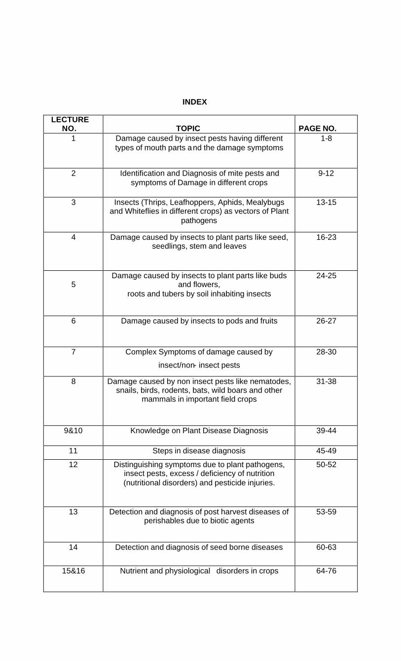
INDEX
LECTURE NO.
TOPIC
PAGE NO.
1 Damage caused by insect pests having different types of mouth parts and the damage symptoms
1-8
2 Identification and Diagnosis of mite pests and symptoms of Damage in different crops
9-12
3 Insects (Thrips, Leafhoppers, Aphids, Mealybugs and Whiteflies in different crops) as vectors of Plant
pathogens
13-15
4 Damage caused by insects to plant parts like seed, seedlings, stem and leaves
16-23
5
Damage caused by insects to plant parts like buds and flowers,
roots and tubers by soil inhabiting insects
24-25
6 Damage caused by insects to pods and fruits 26-27
7 Complex Symptoms of damage caused by
insect/non- insect pests
28-30
8 Damage caused by non insect pests like nematodes, snails, birds, rodents, bats, wild boars and other
mammals in important field crops
31-38
9&10 Knowledge on Plant Disease Diagnosis 39-44
11 Steps in disease diagnosis 45-49
12 Distinguishing symptoms due to plant pathogens, insect pests, excess / deficiency of nutrition (nutritional disorders) and pesticide injuries.
50-52
13 Detection and diagnosis of post harvest diseases of perishables due to biotic agents
53-59
14 Detection and diagnosis of seed borne diseases
60-63
15&16 Nutrient and physiological disorders in crops
64-76

Course No Ento.333
FIELD DIAGNOSIS IN AGRICULTURE
Lecture No: 1. Damage caused by insect pests having different types of mouth parts and the damage symptoms:
Insects utilize the plants to derive their nutrition or as a habitat. The plants sustain injury to satisfy the requirements of insects. Such injury to the plants by the insects is reflected as economic loss to the farmers.
The nature of injury/damage to the plant is related to the feeding habits of the insect. The peculiarity of mouth parts and mechanism/type of feeding determine to a larger extent the pest management strategies including the type of pesticide to be used.
The nature and symptoms of damage caused by insects based on their feeding habits according to the modification of their mouthparts is furnished hereunder.
1. Biting and Chewing type:
They are adapted for biting and chewing of the plant material. They bite leaves, buds, bracts, slender twigs etc, chew the bitten portions and swallow them. Leaves may be eaten up completely leaving only a network of veins.
Eg: Grasshoppers, caterpillars, beetles. They can be controlled effectively with stomach poisons when taken in along with food.
Based on the nature of damage, chewing insects can be classified into different groups as mentioned below.
1. Stem borers:
Larvae enter in to the stem and feed on internal contents. As a result, damaged part is cut off from the main plant and affected part wilts, dries up and exhibits symptoms like dead heart during vegetative stage and white ear during reproductive stage in case of paddy due to larval feeding inside the stem and they can be easily pulled out and bunchy top in case of sugarcane (destruction of growing point results in the activation of side buds, just below the growing point and produces a bunch of side shoots called bunchy top).
Eg: Stem borers of paddy, millets, sugarcane and brinjal
2. Shoot borers:
Larvae attack tender shoots and bore inside during vegetative stage of crop growth and cause wilting, drooping of terminal plant parts which later dry up.
Eg: Shoot borers of brinjal, bhendi, cotton and castor
3. Defoliators/Skeletonizers:
Larvae feed on the leaves completely leaving only midrib/veins or scrape the chlorophyll content of leaves or cause numerous holes.
Eg: Castor semilooper, ash weevils, tobacco caterpillar, epilachna beetle on brinjal.
4. Leaf miners:
Larvae mine leaves/leaflets between the epidermal layers and feed on greenish matter, resulting in the appearance of translucent mines/white patches/zig-zag galleries
Eg: leaf miners of citrus, Cashew and Rice hispa.

5. Leaf Webbers:
Larvae webs leaves/leaflets by means of silken threads and feed on the chlorophyll content by remaining within the web. Often faecal pellets/frass are found within the web.
Eg: Leaf Webbers on gingelly, groundnut, sapota, mango and cashew shoot and blossom webber.
6. Leaf folders:
Larvae fold leaves from tip to base /longitudinally /margin to margin there by giving appearance of a fold/roll and scrape the chlorophyll content remaining within the fold.
Eg: rice leaf folder, Cotton leaf roller (Bell shaped rolling of leaf).
7. Gall makers:
Larvae feeding inside the stem/tiller/leaf/flower bud stimulates excessive growth of cells at the affected portion and distorts normal growth. It results in malformation of plant parts, exhibiting gall formation and gives shelter for the pest.
Examples:
Sunhemp stem borer: caterpillar causes gall like swelling on stem and profuse branching occurs at affected portion of stem.
Tobacco stem borer: caterpillar mines into the leaf axil and then in to stem, bored stems become hollow, swollen and forms a gall.
Cotton stem weevil: Grub tunnels round the stem feeding on the soft tissue and this results in the formation of gall like swelling at the site of injury.
Amaranthus stem weevil: Hypolixus truncatulus twisting and swelling of branches and stems.
8. Pod/capsule borers/boll worms:
During the reproductive stage of the crop larvae enter in to the pods, capsules and feed on the seeds/lint exhibiting symptoms like webbed condition of pods /bolls or web few pods/capsules with frass and excreta or holes of different sizes and shapes/damaged tissues (chilli/lint on Cotton).
Examples:
Spotted pod borer: It enters into pod near the pedicle and feeds on the ripening seeds by remaining inside the pod, at the entrance hole , a mass of dried excreta can be seen.
Capsule borers of
castor and gingelly: Webbing of capsules and holes on pods plugged with excreta.
Tobacco caterpillar: Irregular holes on pods with excreta inside.
Gram caterpillar: Damaged pods with round holes.
Pink bollworm: Rosette flower and double seed.

9. Fruit borers:
Larvae enter into the tender fruits and feed on fresh matter/pulp and plug the larval burrow with excreta.
Eg: Fruit borer of brinjal/bhendi/tomato, mango stone weevil,
Cashew apple and nut borer.
10. Bark borers:
Larvae remain in a small tunnel at the axils of branches, under the bark constructing galleries of frassy web on the stem and near bark/angles of branches and move about, conceal inside the silken gallery and feed on the bark by scraping.
Eg: Bark eating caterpillars of citrus, mango, guava, casuarina, jack etc.
11. Tree borers:
Larvae bore deep into the tree trunk, make the tunnels in zig-zag manner and feed on inner tissues, arresting translocation of sap to top portions of tree, there by the tree exhibits symptoms like yellowing, withering of leaves, drying of twigs or complete drying of tree. Sometimes, gummy material oozes from the affected portion on the tree trunk.
Eg: Tree borers of mango, cashew, coconut red palm weevil etc
12. Root feeders:
Larvae feed on roots/root nodules resulting in stunted growth/poor tillering /drying of plants in isolated patches.
Examples:
Rice root weevil: Grub feeds on epidermis of stem and later enters in to soil and feed on roots. Affected plant turns yellow and stunted. Tillering is poor.
White Grubs: Devour secondary roots leaving supporting root only. As a result leaves of affected plants turn pale, droop down and ultimately wither off. Cut end of affected stem of collapsed plant swells, a characteristic diagnostic symptom .Drying of plants in patches.
Banana Rhizome weevil: grubs tunnel through pseudo stem and rhizome making circular hole, which increase in size with the growth of grubs. Plants break down at tunneled portion/ plant bears few fruits and suckers. Circular holes with black rotten tissue of rhizome plugged with excreta.
Termites in:
Paddy - feed on roots, foliage, stem and fallen heads
Sorghum - feed on roots and stem resulting in wilting and death of plant
Groundnut- feed on main stem which is bored at or just below the ground level. Mature and developing pods are also penetrated and filled with mud.
Sugarcane- enter the sets through buds and cut ends and devour the inner portion, roots are also damaged. Sometimes earthen sheeting at the base of plant, mud filled galleries in shoots, drying of shoots.
Potato- tubers are damaged
Mango- construct mud galleries on tree trunk, if earthen sheet is removed, eaten bark of trees is observed. Young plants will die and dry up.
Coconut –construct mud galleries on trunk. Bark and stem are eaten below the mud galleries. Nursery and transplanted fields show wilting of central shoot and stunted growth.
13. Seed feeders (Stored grain pests):
Grubs/larvae and adults feed on stored seeds either internally /externally by webbing the food particles.
Eg: Rice weevil, red flour beetle, rice moth etc.,
The symptoms of damage caused by biting & chewing insects is furnished below.
a) Defoliation/Skeletonization/ Scraping of leaves:
Early larval instars of large number of Lepidopteron pests with gregarious behavior feed on leaves by scraping the chlorophyll content and gives papery/scorched appearance leaving membranous cuticular layer and stout veins. Such feeding leads to skeletonization .
Eg: Tobacco caterpillar, Bihar hairy caterpillar, Red hairy caterpillar
b) Uneven cuts on leaf margins:
Eg: Grasshoppers on various crops, larvae of mustard saw fly on cruciferous crops
c) Uneven scraping of leaf surface (lace like):
Eg: Grubs and adults of Henosepilachna vigintioctopunctata on cucurbits, solanaceous and leguminous crops.
d) Small white streaks parallel to midrib on rice leaves:
Eg: Adults of Rice Hispa
e) Tubular cases attached to leaf/ floating in water in rice fields:
Eg: Larvae of case worm
f) Shot holes on leaves:
Eg: Larvae of sorghum and sugarcane stem borers, Adults of flea beetle on blackgram/greengram ,Phyllotrea sp on crucifers , Larvae of Anomis sabulifera on jute,
g) Scraping and gnawing of base of stem:
Eg: Plutella xylostella on mustard and rape, Spodoptera litura on potato.
2. Piercing and Sucking Type
Planthoppers, leafhoppers, thrips, paddy gundhy bug, red cotton bug, sorghum ear head bug, aphids, mealy bugs, scales and whiteflies possess piercing and sucking type of mouth parts. However, they cause different types of symptoms on different crops based on their site and extent of feeding. Both nymphs and adults suck sap from base of the plant /leaves /tender terminal plant parts and thereby affect the vigour and growth of plants. In case of severe infestation, sooty mould develops on plant parts covered with honey dew excreted by insects while feeding. Different insects exhibit different symptoms.

These insects cause hopperburn, discolouration, curling of leaves, necrosis on leaf margins and their eventual weakening and death of plant parts. They may also attack young twigs and other parts of the plant and cause them to dry up.
As these insects take their food from inside the plant, stomach poisons are not effective, unless insecticide is a systemic toxicant. Contact poisons are more effective.
a) Hopper burn:
Plant hoppers viz Nilaparvata lugens and Sogatella furcifera of Delphacidae are known to cause hopper burn on Rice, a monocotyledon crop .
Leafhoppers belonging to the genus Empoasca of Cicadellidae are known to cause hopper burn on dicotyledon crops like cotton, okra, castor, brinjal, potato, beans etc.
The general symptoms of hopper burn caused by planthoppers and leaf hoppers is furnished hereunder.
Plant hoppers: - Yellowing of older leaf blades, progressive yellowing of all the plant parts, plants turn brown and die in patches.
Leaf hoppers: -Wilting of leaf tips in very young plants, chlorosis/necrosis on leaf margins, complete drying of leaves & wilting, premature leaf drop, stunted growth of plants.
Differences in feeding behavior between Plant and leafhoppers
Plant Hoppers Leaf hoppers Suck sap from phloem element of monocotyledons
Suck sap from mesophyll paranchymatic cells/phloem elements either from stems/from veins
The damage is mainly through mechanical plugging of sieve elements with salivary sheaths (true sheaths were formed)
No such true salivary sheaths. The damage is mainly through mechanical wounding of cells.
Confined only to stems Change their feeding sites according to the situation
Hopper burn in Groundnut:
Whitening of veins is the first symptom due to feeding from lower surface of leaflets, chlorotic (yellow) patches then appear especially at the tips of leaflets, probably caused by a reaction between jassids salivary secretion and plant sap. Under severe infestation, the leaf tips become necrotic in a typical “V” shape, giving the crop a scorched appearance known as hopper burn.
Eg: Groundnut jassids
b) Curling of leaf margins/with necrotic patches
Starting from leaf margin –Cotton leaf hopper
c) Uniform yellowing of leaves from mid half -Paddy leaf hopper
d) Reduced vigour/sooty mold, squaer/bolldrop -White flies on cotton
e) Yellowing /reduced/stunted growth/sooty mould -Aphids

f) Shriveled/chaffy and discolored grains/sooty
mould on grain -Sorghum ear head bug/rice gundhy bug
g) Mottled appearance with yellow patches on infested
leaves/sooty mould/undeveloped grains on infested
ear heads -Sorghum aphids
i) Gradual wilting and drying of ragi plants in patches -Ragi root aphids
3. Rasping and Sucking / Lacerating and Sucking Type:
Thrips are characterized by this type of mouth parts. Due to the peculiarity of mouth parts and their mechanism of action in rasping the tissues, exudation of juice from inside the plant takes place and it is sucked by thrips. The damaged part of the plant exhibit a whitish mottled/silvery appearance. Such insects can be controlled both by stomach and contact poisons.
a) Groundnut:
Nymphs and Adults suck sap from the surface of the leaf lets. This results initially in white patches on the upper surface and necrotic patches on lower surface of the leaves. It consists of distortions of the young leaflets and patchy areas of necrotic tissue that puncture and split as leaf lets grow. Injury is normally seen in seedlings. In severe infestation, particularly in winter crop (November sown in South India) , leaf distortion causes stunted plants. The effect of such damage on yields is not precisely known, but is not serious.
b) Onion:
Presence of pale white blotches on leaves, gradually change to brown spots followed by gradual drying of leaves from tip down wards. Growth of tubers decreases resulting in yield loss –Onion thrips
c) Chillies:
Infested leaves start curling upward, crumbling and drop down. Wilting and drying of plants under severe infestation
d) Blackgram:
Leaves curl up, crumble, become brittle and plant growth retards .Infested flower buds do not develop in to pods.
e) Rice:
Rolling of leaf terminals/yellow reddish and scorched leaf tips/rolling of entire length of all leaves.
4. Sponging and sucking or Lapping and sucking Type:
Dipterans (Houseflies) possess above mentioned type of mouth parts. These are not pests of Agricultural importance.
5. Chewing and Lapping Type:
Hymenopterans (Honey bees) possess above mentioned type of mouth parts. These are not pests of Agricultural importance.

6. Siphoning/simple sucking Type:
Adult stages of moths and butterflies possess this type of mouth parts, while the caterpillars possess the biting and chewing type of mouth parts.
In larval stage they cause extensive damage. Stomach poisons can effectively control the larval stages. In general, adult stages of moths and butterflies are not harmful. However, adults of certain moths can cause damage to certain fruits.
Fruit feeders: Adults suck juice from ripened fruits with the help of proboscis resulting in minute holes consequently resulting in rotting due to infections whereas larvae feed on the weeds belonging to the family Menispermaceae.
Eg: Adults of Citrus fruit sucking moths and Castor semilooper
7. Degenerate type of mouth parts:
Maggots of Diptera possess above mentioned type of mouth parts
Gall formers:
Paddy Gall Midge: Maggot feeds on growing point which stimulates the leaf sheath to form a hollow pale green cylindrical tube similar to onion leaf/ silver shoot /gall. Affected tiller do not bear panicles. Infestation in early period of crop induces vigorous subsidiary tillering.
Gingelly gall fly: Maggots feed on the ovary which results in the malformation of pod without proper setting of seeds.
Mango inflorescence midge: Three species of midges damage the inflorescence
Procystiphora mangiferae: The maggots feed on stalks of stamens, anthers, and ovary.
Dasineura amaramanjarae: The maggots feed inside the buds and they fail to open and drop down.
Erosomyia indica: The maggots attack the inflorescence stalk, flower buds and small developing fruits. The inflorescence becomes stunted and malformed and the buds do not open.
Mango leaf galls: Procantarina matteriana Small raised wart like galls on tender leaves. Affected leaves deformed and drop prematurely.
Chilli Midge: Asphondylia capsici
Unopened buds are affected. Flowers dry and drop . Pods are deformed.
Coccinia gall fly: Neolasioptera cephalandrae
Elongated galls/swelling of distal stems in between the nodes. If cut open, gall shows maggots presence.
Jasmine blossom midge: Contarinia maculipennis
Swelling at the base of buds. Stunting, finally drying of plant.
Shoot borers:
Larvae attack tender shoots and bore inside during vegetative stage of crop growth and cause wilting, drooping of terminal plant parts which later dry up.
Eg: Shoot fly of sorghum and black gram stem fly

Pod Borers:
Maggot feeds under the epidermis for some time then enters the seed and consumes only part of the seed. Affected seed gets discoloured due to bacterial and fungal infections and becomes unfit for consumption. No visual symptoms are present on pods initially, but only after adult emergence, a minute hole can be seen on pod.
Eg: Redgram Pod fly
Fruit feeders:
Larvae feed on fruits resulting in holes plugged with excreta/ forming necrotic patches /rotting.
Eg: Fruit flies on cucurbits, Mango fruit fly, Ber fruit fly
Rhizome borers:
Maggots mine into mid rib of leaves and enter in to rhizome through petiole resulting in rotting of rhizome and dead hearts.
Eg: Turmeric rhizome fly.

Lecture No: 2 Identification and Diagnosis of mite pests and symptoms of Damage in different crops
Mites belong to the order Acarina of the class Arachnida. The phytophagous mites belong mainly to the families Tetranychidae, Eriophyidae, Tarsonemidae and Tenupalpidae.
Diagnostic features between Tetranychidae and Eriophidae
Tetranychidae Eriophidae
Tetranychid mites are tiny, oval in shape Eriophid mites are elongate, vermiform in shape
Body unsegmented and not divided into cephalothorax and abdomen
Body distinctly divided into cephalothorax and abdomen
These mites possess 4 pairs of legs in the nymphal and adult stages and the larvae have 3 pairs of legs only
These mites possess only 2 pairs of legs and are situated near the anterior end of the body both in nymphal and adult stages
Mouth parts of mites are adapted for biting, piercing and sucking.
Tetranychids penetrate the plant tissue with sharp stylets and remove the cell contents. The chloroplasts disappear and the small amount of remaining cellular material coagulates to form an amber coloured mass. In the palisade layer, only the penetrated cells are damaged and continued feeding leads to irregular spots; transpiration rate accelerates which finally leads to the drying and dropping of leaves. The mite infestation inhibits photosynthesis; and changes the composition of leaf pigments leading to a complex of symptoms like
-yellowing, bronzing, distortion, curling, crinkling, defoliation of leaves, -retardation of growth, -dropping of flowers, reduction in size, quality and quantity of produce.
Eriophids in general, cause no serious mechanical damage to plant tissue by their feeding. Salivary growth regulators, when injected in to plants cause discolouration and growth modifications like galls, erinea, blisters, rust, brooming, leaf edge rolling etc.
Tarsonemids penetrate thin –walled mycelial strands and highly succulent tissues but, incapable of penetrating thick walled lignified tissues. Occasionally toxins are injected which presumably cause alteration of normal tissue.
Plant mites damage the crop plants in a number of ways as given below.
1. They suck the cellular materials by means of their cheliceral stylets resulting in the formation of characteristic white blotches on the leaves and devitalization of plants.
Eg: Tetranychus neocaledonicus
2. The eriophid mites cause severe deformation in plant parts.
Eg: Aceria gossypii produces outgrowths of excessive hairs on cotton leaves Aceria mangiferae crowded buds and also galls in mangoes Phyllocoptruta oleivora pinkish brown blotches on citrus fruits
3. A few mites are known to transmit viral diseases
Eg: Aceria tulipae is a vector of wheat streak mosaic virus Aceria cajani transmits pigeon pea sterility mosaic virus
Predatory mites are the efficient natural enemies of phytophagous mites. Mites of the family Phytoseiidae have been recognized as potential predators of phytophagous mites. Phytoseiidae,Stigmaeidae, Anastidae, Cheyletidae and Erythradidae are the important predatory mite groups. The miscellaneous groups include Bdellidae, Tarsonemidae and Tydeidae .The Phytoseiid mites feed specially on mites of the families Tetranychidae, Eryophidae, Tarsoemidae and Tenupalpidae.
Predators of Tetranychid mites:
Amblyseius gossypii on Tetranychus cinnabarinus, T.urticae,
Eutetranychus orientalis
Phytoseiulus persimilis on T.evansi, T.urticae
Predatory mites:
a)Phytoseiidae mites: Amblyseius longispinosus , A.ovalis, A.tetranychivorus, A.gossipi on red spider mites
b)Stigmaeidae mites : Agistemus fleschmeri on spider mites
c)Cunaxidae mites: Cunaxa sp
1. Rice leaf mite: Oligonychus oryzae Tetranychidae : Acarina
Adult yellowish, Nymphs light yellowish, colonies found underneath fine silken webs
Mites damage symptoms are interveinal necrosis, leaves become whitish between the veins, get shredded and veins remain greenish.
2. Rice panicle mite Stenotarsonemus spiniki Tarsonemidae: Acarina
Whitish mites, colonies are found between stem and leaf sheath and cause damage to leaves, glumes and floral parts.Symptoms of mite damage on leaves can be clearly seen at tillering stage. Infested leaves exhibit elongated, dark and brownish-black necrotic streaks measuring 0.5 to 2.0 cm length. Infested plants show poorly exerted ear heads and necrotic leaf sheaths. Affected glumes bear brown to black lemma and palea and shriveled ovary. The mite reduces panicle size, length of panicle neck, panicle weight.
3. Jowar Mite: Oligonychus indicus, Schizotetranychus andropogoni
Tetranychidae : Acarina
Greyish green mite. Females and nymphs are found in colonies on underside of leaves underneath the fine silken webs, nymphs and female adults suck sap from leaves, mites spin delicate webs on lower surface of leaves and live in the web, red patches develop on leaves which increase in size and spread on entire leaf, leaves wither and dry up and stem dries up in severe cases.
4. Cotton leaf mite: Tetranychus telarius , T.Bimaculatus
Tetranychidae : Acarina
T. telarius is called two spotted spider mite. Adults are oval shaped green/red/amber coloured with two spots on body.
-Feed on lower surface of leaves underneath a web.
-Close observation reveals pin point sized mites on lower surface of leaf.
-Leaves curl up, hard, crisp and shed.

5. Wooly mite of cotton: Aceria gossypii Eriophyiidae : Acarina
White or transparent body
Mites are found on both surfaces of leaves. Growing shoots are attacked. Infested parts including leaves, buds and squares are covered in the outgrowths of dense white hairs. Heavily infested plants show crumpled leaves, distorted growth and lack of fruiting branches. Damage results in felt like outgrowths on leaf surface called “erinium” patches.
6. Red gram mite: Aceria cajani Eriophyiidae : Acarina
-Colonies are found underneath tender leaves
-Causes yellowing of leaves
-Suppression of flowering and fruiting
-Transmits pigeon pea sterility mosaic virus
-Diseased plants look bushy,pale green without flowers or pods
-eaves are small with yellow and green patches.
7. Citrus rust mite: Phyllocoptruta oleivora Eriophyiidae : Acarina
-Specific on citrus, It is a minute, yellowish, wedge shaped, worm like
-Colonies are found both on leaves and fruits.
-Mites puncture the epidermal cells of leaves and tender fruits
-Infestation results in rusty brown patches on leaves as well as fruits after a month
-More on satgudi fruits
-This is known as “Mangu”
8. Brinjal mite: Tetranychus telarius Tetranychidae :Acarina
Adults are ovate reddish brown with four pairs of legs. First instar nymphs are pinkish with 3 pairs of legs while later instars are greenish-red with 4 pairs of legs
Colonies of mites are found feeding on lower surface of leaves by remaining underneath the web, resulting in yellow spots on dorsal surface of leaves, affected leaves gradually curl, get wrinkled and crumpled. In heavy infestation even fruits are affected.
9. Chilli white mite: Polyphagotarsonemus latus, Tarsonemus transulcens
Tarsonemidae : Acarina
Mites are tiny, white and transparent and found mostly under the lower side of leaves. Both nymphs and adults suck sap particularly from terminal /auxillary tender shoots and devitalize the plant.
Infested leaves curl downwards along the margins, petioles of older leaves are elongated, younger leaves reduced in size and form a cluster at the tip of branch and affected leaves turn dark green and become brittle.

Polyphagotarsonemus latus on cotton:
Both nymphs and adults infest the tender shoot and leaves on both sides and cause severe crinkling, downward cupping, brittleness of the leaves and gives a shiny appearance to the plant(without flower of boll formation)
10. Coconut mite: Eriophes guerreronis Eriophyiidae : Acarina
Mite has elongate vermiform body measuring 200-250 microns length and about 40 microns thickness
-The mites inhabit the floral bracts and tender portions and immature nuts covered by perianth
-They suck sap from meristematic tissues
-Initially the damage appears as white later brownish triangular patches at the separation of the floral bracts and extends towards the free part of the nut
-Ultimately longitudinal fissures appears on the nut
-Heavy shedding of the buttons results in the loss of yields
-Reduction in size of nut, kernel content and poor quality of the nut.
11. Sugarcane mite: Schizotetranychus andropogoni
It is found on the under surface of leaves causing discoloration of leaves.
---
Lecture No: 3 Insects (Thrips, Leafhoppers , Aphids, Mealybugs and Whiteflies in different crops) as vectors of Plant pathogens
Transmission of a plant virus from diseased to a healthy susceptible host by a vector is the culmination of several sequential events/steps.
The first step is the acquisition of virus from the infested plant and the last step is successful inoculation of the healthy one. Between these two events, the virus has to be carried in infectious state and many factors determine completion of transmission cycle.
1) Acquisition is not affected even if the cell,
from which virus-laden sap is ingested , is fatally injured.
2) Similarly, cells inoculated with virus have to survive necessarily long enough for the virus to infect adjacent cells.
3) Further, a particular virus is transmitted by a single taxonomic group of vectors that too by a certain spp.
4) Different spp. of the same virus often differs in transmission efficiency.
5) Within the single species active and inactive races of the same vector species were also discovered.
Thus vector transmission of virus is not mere mechanical transfer but far more complex. Therefore there must be potential barriers to transmission and no set of characters are unknown to distinguish vector species from non-vector ones.
That is why, a clear understanding of vector-virus relationship is the first step to find out the various factors involved in “making” a vector. This information may provide a basis for developing techniques to “un make” a vector by affecting its transmission ability.
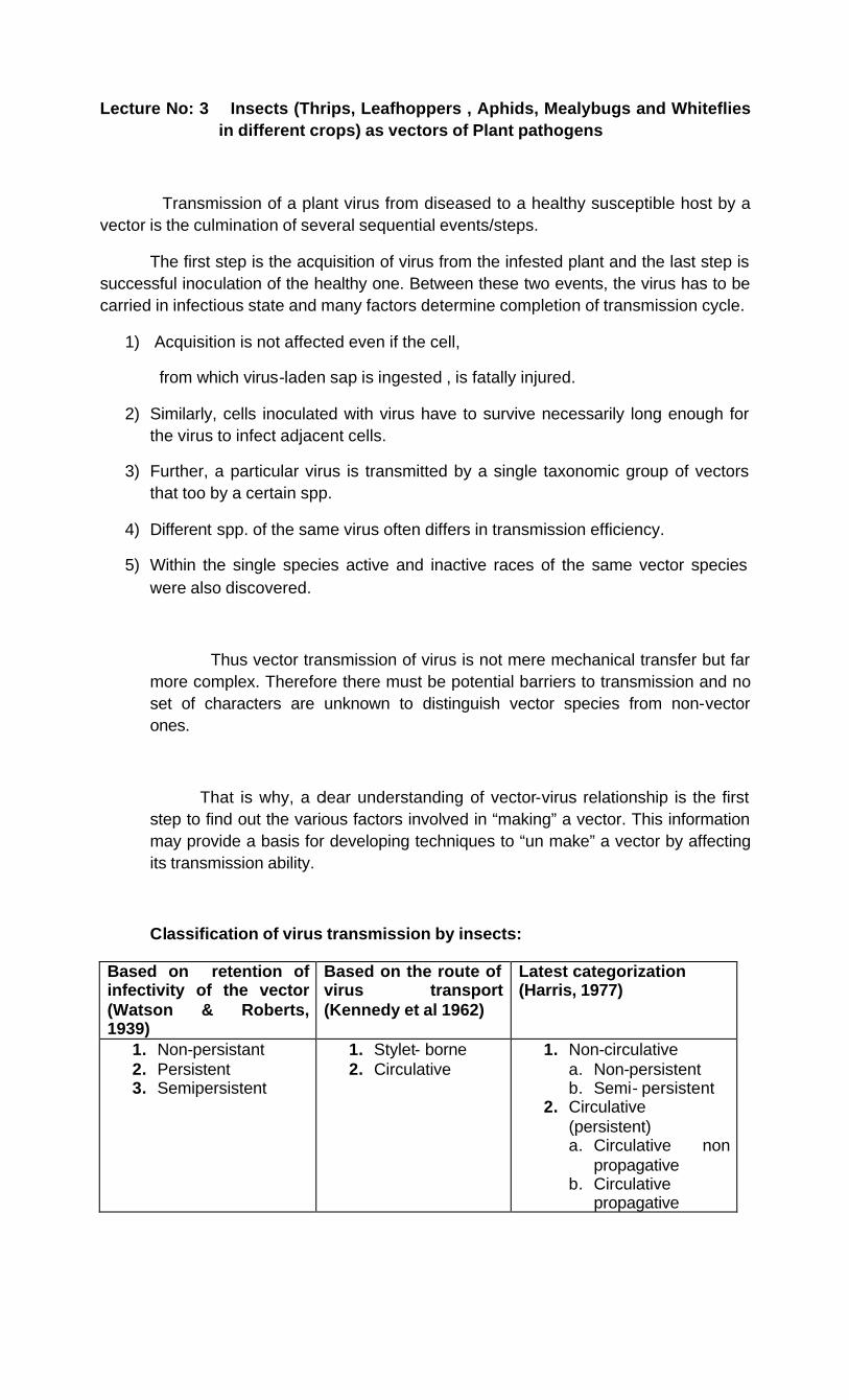
Classification of virus transmission by insects:
Based on retention of infectivity of the vector (Watson & Roberts, 1939)
Based on the route of virus transport (Kennedy et al 1962)
Latest categorization (Harris, 1977)
1. Non-persistant 2. Persistent 3. Semipersistent
1. Stylet- borne 2. Circulative
1. Non-circulative a. Non-persistent b. Semi- persistent
2. Circulative (persistent) a. Circulative non
propagative b. Circulative
propagative
§ Non-circulative viruses
A. Non-persistent viruses
Transmission of non-persistent viruses (stylet borne : viruses adhere to tips of stylets, immediately acquired during feeding and transmitted by vectors soon after acquisition) is virtually a monopoly of aphids. Number of viruses transmitted non-persistently & by aphids far exceed those transmitted semi persistently or persistently by them.
Non persistent viruses are readily sap transmissible due to their presence in relatively superficial tissues. Some viruses such as Onion yellow dwarf and Cucumber mosaic are transmitted by a large no of aphid species.
Non-persistent transmission by hoppers is yet unknown. But Whitefly, Bemisia tabaci is known to transmit non-persistently.
Cowpea mild mottle virus (CMMV) and Tomato pale chlorosis disease virus (TPCDV). But transmission properties are different from typical non persistent transmission by aphids.
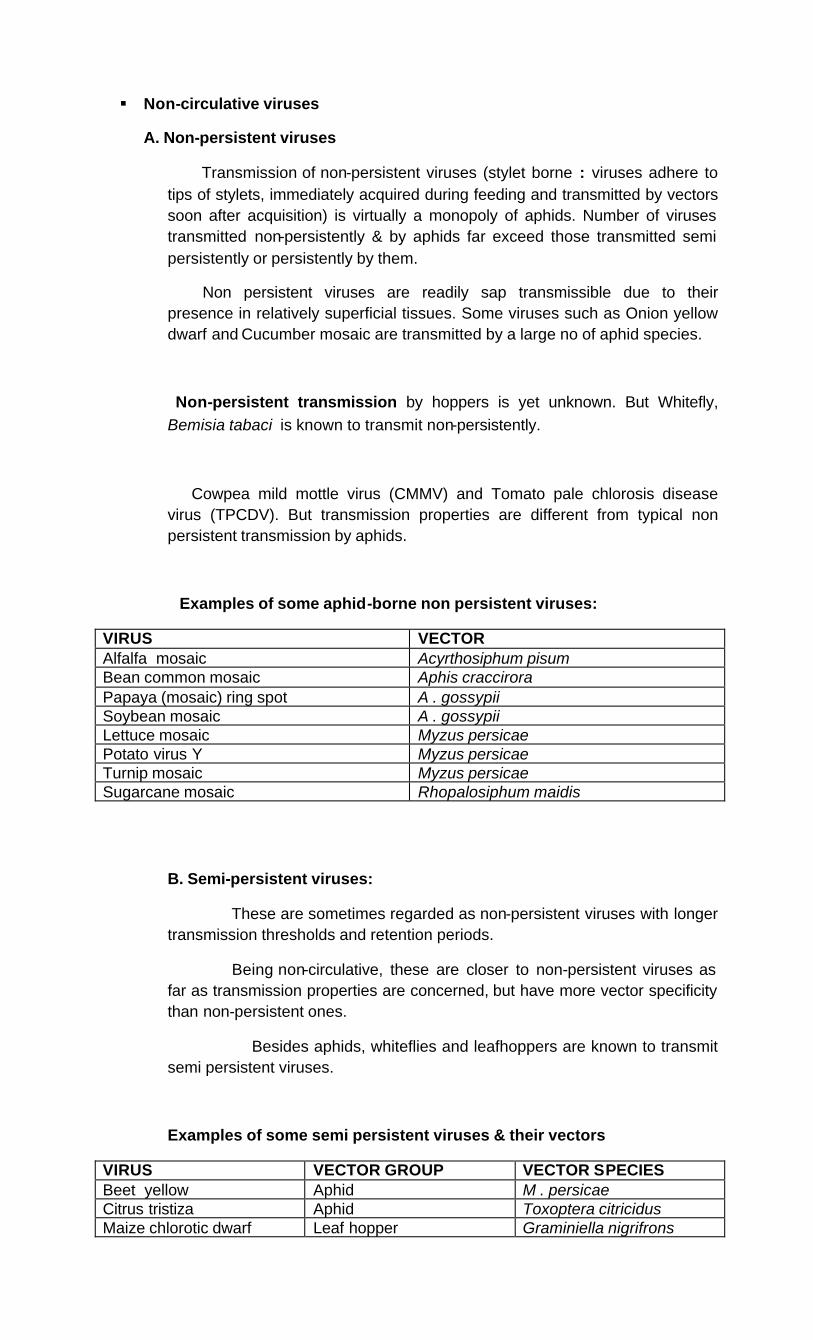
Examples of some aphid-borne non persistent viruses:
VIRUS VECTOR Alfalfa mosaic Acyrthosiphum pisum Bean common mosaic Aphis craccirora Papaya (mosaic) ring spot A . gossypii Soybean mosaic A . gossypii Lettuce mosaic Myzus persicae Potato virus Y Myzus persicae Turnip mosaic Myzus persicae Sugarcane mosaic Rhopalosiphum maidis
B. Semi-persistent viruses:
These are sometimes regarded as non-persistent viruses with longer transmission thresholds and retention periods.
Being non-circulative, these are closer to non-persistent viruses as far as transmission properties are concerned, but have more vector specificity than non-persistent ones.
Besides aphids, whiteflies and leafhoppers are known to transmit semi persistent viruses.
Examples of some semi persistent viruses & their vectors
VIRUS VECTOR GROUP VECTOR SPECIES Beet yellow Aphid M . persicae Citrus tristiza Aphid Toxoptera citricidus Maize chlorotic dwarf Leaf hopper Graminiella nigrifrons
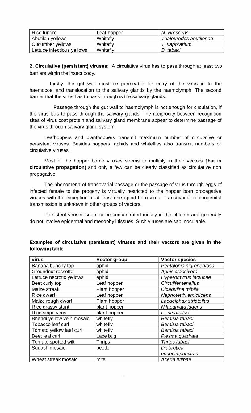
Rice tungro Leaf hopper N. virescens Abutilon yellows Whitefly Trialeurodes abutilonea Cucumber yellows Whitefly T. vaporarium Lettuce infectious yellows Whitefly B. tabaci
2. Circulative (persistent) viruses: A circulative virus has to pass through at least two barriers within the insect body.
Firstly, the gut wall must be permeable for entry of the virus in to the haemocoel and translocation to the salivary glands by the haemolymph. The second barrier that the virus has to pass through is the salivary glands.
Passage through the gut wall to haemolymph is not enough for circulation, if the virus fails to pass through the salivary glands. The reciprocity between recognition sites of virus coat protein and salivary gland membrane appear to determine passage of the virus through salivary gland system.
Leafhoppers and planthoppers transmit maximum number of circulative or persistent viruses. Besides hoppers, aphids and whiteflies also transmit numbers of circulative viruses.
Most of the hopper borne viruses seems to multiply in their vectors (that is circulative propagation) and only a few can be clearly classified as circulative non propagative.
The phenomena of transovarial passage or the passage of virus through eggs of infected female to the progeny is virtually restricted to the hopper born propagative viruses with the exception of at least one aphid born virus. Transovarial or congenital transmission is unknown in other groups of vectors.
Persistent viruses seem to be concentrated mostly in the phloem and generally do not involve epidermal and mesophyll tissues. Such viruses are sap inoculable.
Examples of circulative (persistent) viruses and their vectors are given in the following table
virus Vector group Vector species Banana bunchy top aphid Pentalonia nigronervosa Groundnut rossette aphid Aphis craccivora Lettuce necrotic yellows aphid Hyperomyzus lactucae Beet curly top Leaf hopper Circulifer tenellus Maize streak Plant hopper Cicadulina mibila Rice dwarf Leaf hopper Nephotettix emicticeps Maize rough dwarf Plant hopper Laodelphax striatellus Rice grassy stunt plant hopper Nilaparvata lugens Rice stripe virus plant hopper L . striatellus Bhendi yellow vein mosaic whitefly Bemisia tabaci Tobacco leaf curl whitefly Bemisia tabaci Tomato yellow laef curl whitefly Bemisia tabaci Beet leaf curl Lace bug Piesma quadrata Tomato spotted wilt Thrips Thrips tabaci Squash mosaic beetle Diabrotica
undecimpunctata Wheat streak mosaic mite Aceria tulipae
---

Lecture no.4 Damage caused by insects to plant parts like seed, seedlings, stem and leaves
The insect pests cause damage to the seeds and seedlings during the early stage of the growth period. Symptoms exhibited during the seedling stage on different crops are furnished here under.
A. Damages to seeds and seedlings
No. Symptoms Details Insect group and crops
Examples
1 Dead hearts on seedling
Maggots of various diptera bore into young stem, usually killing the growing point and making the apical leaf turn brown and die; Seedlings before 4 weeks age of the sorghum crop are severely damaged resulting in the formation of dead hearts which can be pulled out easily and emit foul smell.
Sorghum shoot fly Agromyzidae : Diptera
Atherigona soccata
some caterpillars also bore seedling stems of graminaceous plants, but typically they attack older plants
Crambidae, Pyralidae, Noctuidae
2 Seedling stem cut and plant lying on ground
Tobacco cut worms Black cut worm (potato, Tobacco, cabbage)
Spodoptera sp Agrotis ipsilon
3 a. Cotyledons of large seeds bored and eaten
Larvae bore into epicotyl and hypocotyl and prevent germination.
Bean seed fly Ophiomyia phaseoli
b.Stem Larvae bore in the stem of Bean fly

bored, Seedling with swollen hollowed stem.
various seedlings
Ophiomyia phaseoli Agromyzidae
4
Stem severed and plant removed
Several species of termites; leaf cutting ants and harvester ants\
5 Cotyledons or first leaves pitted and eaten
Adult flea beetles (Halticinae) make a shot – hole effect on seedlings of cruciferae, cotton and other crops, frequently stunting and killing the seedlings
6 Seedling or young plant wilting and dying as a result of underground stem being eaten
Root flies, cut worms
B. Damage to stems
Symptoms Details Insect group and crops
Examples
1 Cereals shoots with dead hearts
Severing of the growing part of the tiller in paddy or the stem in sorghum results in dead hearts which can be easily pulled out.
Paddy and sorghum stem borers
Scirpophaga incertulas Chilo partellus
2 Cereal stems galled and distorted
Gall midge or silver shoot or onion leaf in paddy with profuse tillering in the infested hills.
Paddy gall midge
Orseolia oryzae
3 Cereal and grass stem bores
a. Caterpillars of the family Pyralidae generally bore rice and grass stems, while the b. larger caterpillars of the Noctuidae bore stalks of Maize, sorghum; tunnels in sugarcane are usually very short because the stem is solid and pith less.
Sugarcane early shoot borer Pink stem borer on ragi
Chilo infuscatellus Sesamia inferens
4 Drooping of terminal shoots, leaves and death of plant
Caterpillars bore into petioles of leaves, tender shoots
Spotted pod boll worm/Bhendi shoot borer Brinjal shoot and fruit borer
Earias vitella Leucinodes arbonalis
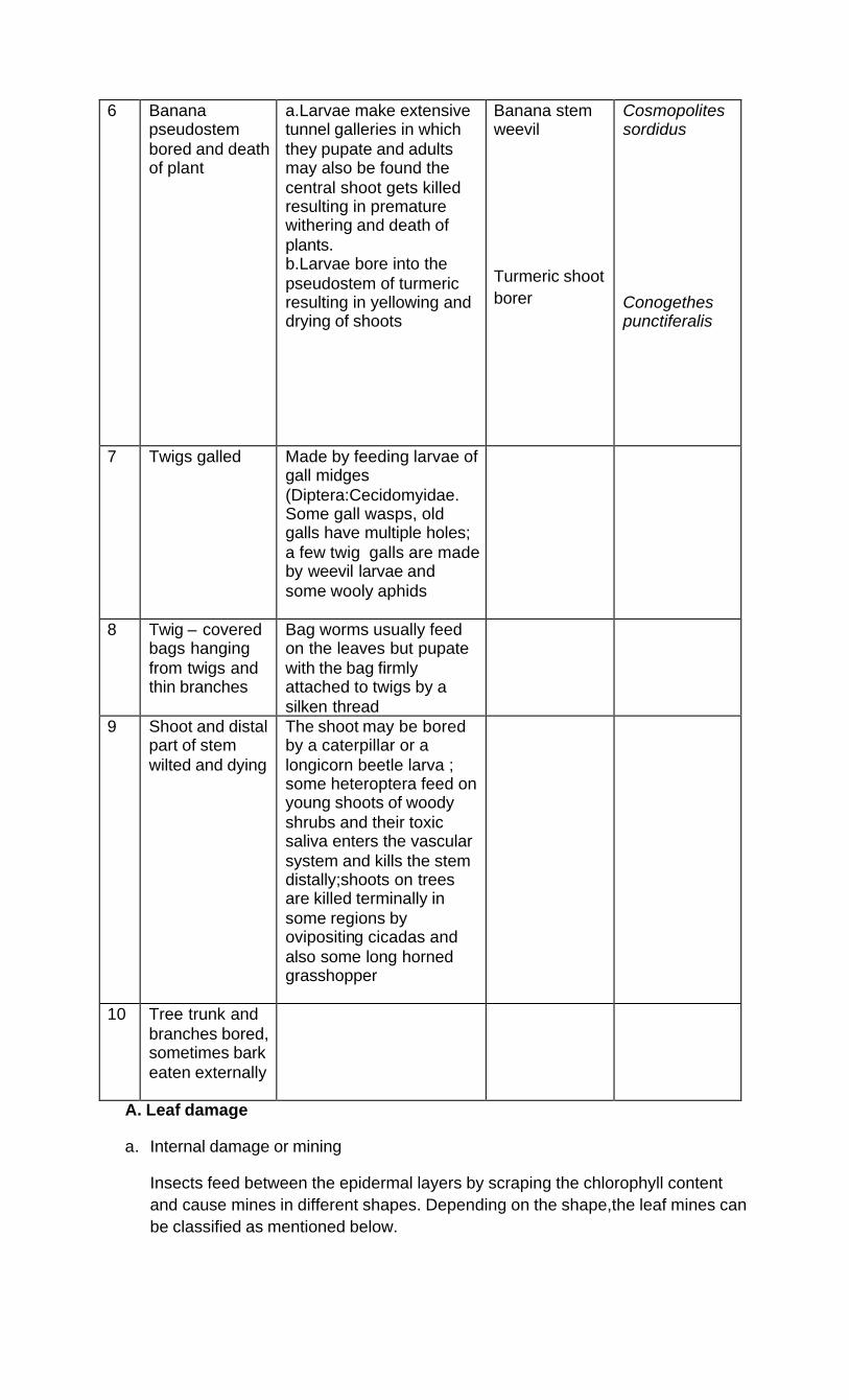
6 Banana pseudostem bored and death of plant
a.Larvae make extensive tunnel galleries in which they pupate and adults may also be found the central shoot gets killed resulting in premature withering and death of plants. b.Larvae bore into the pseudostem of turmeric resulting in yellowing and drying of shoots
Banana stem weevil
Turmeric shoot borer
Cosmopolites sordidus Conogethes punctiferalis
7 Twigs galled Made by feeding larvae of gall midges (Diptera:Cecidomyidae. Some gall wasps, old galls have multiple holes; a few twig galls are made by weevil larvae and some wooly aphids
8 Twig – covered bags hanging from twigs and thin branches
Bag worms usually feed on the leaves but pupate with the bag firmly attached to twigs by a silken thread
9 Shoot and distal part of stem wilted and dying
The shoot may be bored by a caterpillar or a longicorn beetle larva ; some heteroptera feed on young shoots of woody shrubs and their toxic saliva enters the vascular system and kills the stem distally;shoots on trees are killed terminally in some regions by ovipositing cicadas and also some long horned grasshopper
10 Tree trunk and branches bored, sometimes bark eaten externally
A. Leaf damage
a. Internal damage or mining
Insects feed between the epidermal layers by scraping the chlorophyll content and cause mines in different shapes. Depending on the shape,the leaf mines can be classified as mentioned below.

Category Example
1. Linear mine (Fig 1) Bamboo miner
Cosmopteryx bambusae
2. Serpentine mine (Fig 2) Phyllocnistis citrella,
Liriomyza trifoli
3. Blotch mine (Fig 3) Cashew leaf miner-
Acrocercops syngramma,
Rice hispa
Dicladispa armigera
4. Digitate mine (Fig 4)
5. Any combinations of 1-4 (Fig 5) Ber miner- Tischeria ptarmica
6. Needle mine (Fig 6) Casuarina miner-
Metharmositis asphaula

b. External damage on leaf
Insects damage the leaves externally in different ways either by scraping, skeletonization and feeding and as mentioned below

1. Free feeding: Insects feed on part of all of leaf or needle except largest leaf veins which are often left uneaten
e.g Many lepidopterous caterpillars like teak defoliator, Hyblaea puera.
2.Hole feeding: Insect feeds in small patches, rough all layers of a leaf cresting many holes in a leaf.
e.g Ficus leaf feeder, Ocinara varians.
3. Skelitonizing: Insect feeds on the soft material between the veins and leaves the veins as a “skeleton” of the leaf.
e.g Teak Skelitoniser, Eutectoma macheralis
4. Window feeding: Insect feeds on only one surface of leaf that allows the light to penetrate through the remaining leaf layer.
e.g Early instar of many caterpillar.
A. Shelter feeding: insects make a shelter on or within the leaves by webbing with silken threads and then feed on the foliage.
Sometimes abnormal plant growth induced due to insect feeding may also serve as shelters.
The shelter may be in different ways.
1. Web enclosed foliage: Many caterpillars web foliage together and live within this enclosure and feed on the enclosed foliage as Window feeders, Skelitonisers or free feeders.
e.g Ailanthus web worm, Atteva fabriciella
2. Leaf tying or needle tying: Caterpillars lie two to six leaves or needles as the case may be together with silk and feed primarily as window feeders or Skelitonisers.
e.g Casuarina bag worm Clania crameri
3. Leaf folding or rolling: Caterpillars rolls an individual leaf, fastens it together with silk and feeds on it remaining within as a skelitonizer or window feeder.
e.g Tortricid, Bamboo skipper, Baoris cahira
4. Crinkled leaves: Leaves become cupped, crinkled or curled.
e.g Mites, aphids, thrips, mealybugs.
5. Laef and petiole galls: Abnormal growth, if formed around the feeding site forming galls.
e.g Psyllid, Eriophiid mite e.g Erineum leaf gall, petiole gall, pongamia leaf gall midge, Asphondylia pongamiae

D. Stripping damage: Small white or dead spots are formed on the feeding sites due to saliva injected into the plant.
e.g Lace wing bugs, mites ,leafhoppers, scales.
Insect signs
A. Silk shelters are made entirely from silk produced by caterpillars e.g Tent caterpillar, Malacosoma Americana
B. Larval cases are made out of silk, debris of frass e.g bag worm
C. Spittle masses or floating masses produced to enclose the nymphs
e.g Spittle bug.
D. Scale and aphid coverings.
E. Honey dew and sooty mould: The liquid faeces of sap feeders containing sugar is called as honey dews on which black sooty mould fungus grows.
e.g Scales, aphids, whiteflies.
F. Insect remains:
Egg shells, exuviae, pupal cases, cocoons, frass and trails of silk.
Damage to leaves
No. Symptoms Insect group and crop Example Paddy
1 Young terminal leaves drying in nursery
Rice thrips Stenochaetothrips biformis
2 Leaf roll longitudinally Leaf roller Rice leaf roller
Cnephalocrocis medinalis
3 White parallel streaks along long axis
Hispa or spiny beetles Dicladispa armigera
4 Leaf margin notched Ash weevils on cotton and Brinjal Myllocerus sp 5 Leaf lamina scarified Thrips on Cotton Thrips tabaci 6 White parallel sreaks with
cut leaf tip forming leaf tube
Rice case worm Paraponyx stagnalis
7 Graminaceous leaf cut laterally and roll longitudinally
Rice skipper Pelopidas mathias
8 Longitudinal marginal blotching
Rice whorl maggot Hydrillia sasakii
9 Grazing like cutting of seedlings
Rice swarming caterpillar or cut worm
Spodoptera sp.
10 Hopper burn Rice BPH
Nilaparvata lugens
11 Hanging leaf cases Case worm or bag worm Rice case worm 12 Margin irregularly eaten The commonest form of leaf
damage by defoliating pests caused by grasshoppers, locusts.
Hieroglyphus banian, Schistocerca gregaria

Sorghum 13 Parallel or serial holes Sorghum stem borer Chilo partellus 14 Bubble froth or spittle
mass on leaf or leaf axil Spittle bugs on graminaceous plants
Cercopidae bug nymphs
Cotton 15 Leaf roll longitudinally Cotton leaf roller Sylepta derogate
16 Leaf edges curled under, with honey dew, sooty mould and ant movement
Aphids, Jassids, Mealy bugs, Scales, White flies, Psyllids Cotton aphid, Jassid on Cotton and Bhindi, Grape vine Mealy bug, Cocunut scale, Cotton white fly, Curly leaf psyllid
Aphis gossypi, Amrasca devastans, Maconellicoccus hirsutus, Aspidiotus destructor, Bemisia tabaci, Diaphorina citri
17 Irregular shaped holes Many lepidopteran caterpillars and grasshoppers, some polyphagous caterpillar
Helicoverpa armigera
Pulses and oil seeds
18 Leaf folded and mined Ground nut and red gram leaf miner Aproaerema modicella
19 Complete defoliation Hairy caterpillars(Castor) Sphyngids (gingelly)
Euproctis fraterna Orthesia scintillans Acherontia styx
20 phyllody Gingelly Orosius albicinctus Vegetables
21 Lamina with ladder like windowing leaving veins intact
Epilachna beetle on Brinjal and Bitter gourd
Epilachna sp
22 Leaf skeletonised with pappery appearance
Early instars of cut worm on cotton, castor, Cabbage diamond back moth
Spodoptera litura Plutella xylostella
23 Many small shot holes Radish flea beetle Phyllotreta downsei 24 Little leaf Brinjal Cestius phycitis
Spices and condiments 25 Leaves silvered and
wilting Onion thrips Thrips tabaci
26 Young terminal leaves curling upward along margin
Chillies thrips, Pepper marginal gall thrips
Scirtothrips dorsalis, Liothrips karnyi
Fruit crops and tubers
27 Slit like small cut, sometimes T shaped splits
Grapevine flea beetle Sceledonta strigicollis
28 Larger regular shaped holes
Tortoise beetles, Sweet potato, Ber
Aspidomorpha miliaris, Oocasida obscura
29 Leaf lets rolled Coconut skipper Ganagara thyrsis
30 Leaf mine broad with central black faecal pellets and leaf edge folded dorsally for
Citrus leaf miner
Phyllocnistis citrella
pupation Broad and streak like Serpentine mine Blotched pappery mine
Liriomyza trifoli Cyphostiche coerulea
31 Leaves webbed together Mango shoot webber Sappota leaf webber
Orthaga exvinacea Nephopteryx eugraphella
32 Leaves fastened together to form nest
Red tree ant
Oecophylla smaragdina
33 Leaf cut laterally and rolled across
Mango, sappota, leaf twisting weevil
Apoderous tranquebaricus
34 Semi-circular leaf cut Leaf cutting bee on guava Megachile anthracina
35 Round and elongated galls
gall midges (Diptera: Cecidomyidae), gall mites(Acarina: Eryophidae), gall wasps(Hymenoptera: Chalcidoidea, Cynipoidea, and Symphyta), and some Psyllidae Mango leaf galls (gall midges) Pungam leaf gall
Amradiplosis sp. Eryophyid mite
36 Minute yellow specks on leaf
Banana tingid Coconut tingid
Aspidiotus destructor Staphanitis typicus
37 Bunchy top Banana Pentalonia nigronervosa f. typical
38 Frond with V shaped cut Coconut rhinoceros beetle Oryctes rhinoceros
39 Lamina pitted Psyllidae in the group Triazinae cause ventral leaf pits at the sites where the nymphs sit and feed, young leaves sometimes may be considerably deformed
---

Lecture no 5: Damage caused by insects to plant parts like buds and flowers, roots and tubers by soil inhabiting insects
The insect pests that cause damage to floral parts and underground parts like roots and tubers exhibit different symptoms like holes, dropping, distortion, webbing, wilting, withering etc. The different symptoms caused by insect pests on different crops are furnished here under.
Damage to flowers and buds
No.
Symptoms Details Example
1 Flower petals and perianth destroyed
Adult blister beetles ( Meloidae) chew petals of many plants, often common on Malvaceae; adult flower beetles Scarabaeidae)make small holes in petals, Popillia being especially injurious Orange banded blister beetle Brown banded
Mylabris pustulata Gnathopastoides rouxi
2 Flowers partially eaten
petals damage by adults on pulses Mylabris pustulata
3 Petals scarified Flowers of leguminosae, compositae, etc. inhabited by adults and nymphs of thrips (Thripidae) which scarify the bases of the petals
4 Flowers inhabited by tiny black beetles, making feeding scars at the base of the petal
Legume flowers inhabited by Apion weevils
5 Dropping of flowers/ Webbing of flowers and flower buds
Red gram pod borer Gram caterpillar Spotted pod borer
Etiella zinckenella Helicoverpa armigera Maruca vitrata
6 Presence of Webbing and galleries on inflorescence
Castor shoot and capsule borer Conogethes punctiferalis
7 Presence of rosette flowers/ Interlocular damage
Cotton pink boll worm Pectinophora gosypiella
8 Anthers eaten Pollen beetles (Coryna sp)feed on anthers of many flowers, especially Malvaceae, destroying the pollen sacs

9 Maize tassels eaten
By grasshoppers or maize tassel beetle
10 Silk damaged Maize ear worm Helicoverpa armigera
11 Flowers inhabited by tiny maggots
Gall midge larvae (Diptera: Cecidomyidae), either white, yellow, orange or red in colour, usually causing flower drop and deformation
Chilli midge Asphondylia capsici
12 Flower buds bored
Moring bud worm Sappota bud worm Jasmine bud worm
Noorda moringae Anarsia epotias Hendecasis duplifascialis
13 Buds gnawed with large holes
Eaten by large caterpillars; sometimes by long horned grasshoppers
Cotton semilooper: Anomis flava
14 Flower petals with small holes
Cotton flower weevil Amorphoidea arcuata
15 Squares damaged
Cotton spotted boll worm Cotton spiny boll worm
Earias vitella, Earias insulana
16 Capitulum damaged
Sunflower capitulum borer Helicoverpa armigera
17 Aborted flower Moringa midge Stictodiplosis moringae
18 Drying of inflorescence with large scale withering and shedding
Mango hopper Idioscopus niveosparsus I. clypealis Amritodus atkinsoni
19 Inflorescence webbed
Mango flower webber Eublemma versicolor
20
Blighted inflorescence
Cashew mirid bug
Helopeltis antonii
Damage to roots and tubers
No. Symptoms Insect group and crop Example 1 Wilting and drying of
plants in patches due to feeding on roots and rootlets
Rice root weevil Termites in sugarcane, Wheat, Ground nut
Echinocnemus oryzae Odontotermes obesus
2 Wilting and drying of plants and presence of large number of ants at the base of ragi tillers
Ragi root aphid
Tetraneura nigriabdominalis
3 Rhizome extensively bored/ Wilting of plants
Banana rhizome weevil Cosmopolites sordidus
4 Tuber damaged Potato white grubs
Holotrichia sp

5 Wilting of the sweet potato vines and bores into tubers
Sweet potato weevil Cylas formicarius
-----
Lecture no 6: Damage caused by insects to pods and fruits
The insect pests that cause damage to pods and fruits exhibit different symptoms like holes, dropping, distortion or abnormal growth, webbing, shriveling, oozing of brownish fluid, scars on surface and chaffy grains etc. The different symptoms caused by insect pests on different crops are furnished here under.
Damage to fruits, pods and seeds
No. Symptoms Insect group and crop Example 1 Capsule damage Castor capsule borer
Cardamom capsule borer Dichocrocis punctiferalis
2 Bored Fruits Brinjal fruit borer Tomato fruit borer Chilli fruit borer Mango fruit borer
Leucinodes orbanalis Helicoverpa armigera Helicoverpa armigera Bactrocera dorsalis
3 Boll damage Cotton boll worms Helicoverpa armigera
4 Damaged buds and capsules with round holes
Tobacco pod borer Helicoverpa armigera
5 Ear head with chaffy grains
Rice ear head bug Sorghum ear head bug Stink bug
Leptocorisa acuta Calocoris angustatus Nezara viridula
6 Ear head with chaffy grains and protruding pupal cases
Sorghum gall midge
Contarinia sorghiciola
7 Webbing of grains in the ear head
Sorghum web worm Cryptoblabes gnidiella Antoba silicula
8 Cob damaged Maize ear worm Helicoverpa armigera
9 Pod bored Pulse pod borers, Gram pod borers, Plume moth Spotted pod borer Spiny pod borer
Helicoverpa armigera Exelastis atomosa Marucca testulalis Etiella zinckenella
10 Pods shriveled with shriveled grains inside
Pulse pod borer Red gram pod bug
Riptortus pedestris Clavigralla gibbosa
11
Necrotic spots on fruits and pods
Mirid bugs on guava fruit and cocoa pod
Helopeltis antonii
12 Flower and young capsules with galls
Gingelly gall midge Asphondylia ricini
13 Citrus fruits with necrotic lesion, rotting and
Citrus fruit sucking moth Ortheris sp
dropping of fruit 14 Bore hole pomegranate
fruits, feeds on pulp and seed
Anar butterfly Virachola isocrates
15 Maggots bore into fruit and feed on pulp resulting in brown patches
Guava fruit fly Bactrocera sp.
16 Purple discoloration of fruits
San Jose scale Quadraspidiotus perniciosus
17 Premature dropping of tender fruits and oozing of brownish fluid from infested pods
Tea mosquito bug Citrus fruit sucking moth Coconut slug Guava fruit borer
Helopeltis antonii Ortheris sp Contheyla rotunda Conogathes punctiferalis Virachola isocrates
18 Larvae bores into berries and feeds on them
Grape berry borer Conogathes punctiferalis
19 Fruit and berry surface corky
Grapevine berry thrips Banana fruit thrips Cardamom thrips
Scirtothrips dorsalis Chaetonaphothrips sp Scirtothrips cardomomi
20 Berries damaged Pepper pollu beetle Coffee berry borer
Longitarsus nigripennis Hypothenemus hampei
21 T shaped marking on marble sized mango fruits
Mango nut weevil Sternochaetus mangiferae
22 Maggots feed on pulp resulting in rotting and fruit drop
Mango fruit fly Ber fruit fly Cucurbit fruit fly
Bactrocera dorsalis Carepmyia vesuviana Bactrocera cucurbitae
23 Holes on stored cereal grains (Rice, Sorghum, maize, Wheat etc.)
Rice weevil Lesser grain borer
Sitophilus oryzae Rizopertha dominica
24 Cereal grains with exit hole and flap door
Angoumois grain moth (Paddy, sorghum, maize, cumbu)
Sitotroga cerealella
25 Pin head sized holes on processed tobacco
Cigerette beetle Lasioderma serricorne
26 Pin head sized holes on spices
Drug store beetle ( turmeric ,coriander, ginger)
Stegobium panaceum
27 Pulse seeds with circular holes and white eggs cemented on surface
Pulse beetle( All pulse grains)
Callosobruchus maculates
----

Lecture: 7 Complex Symptoms of damage caused by insect/non- insect pests
1.Leaf folder vs. Rice Hispa
2. Rice whorl maggot and yellow stem borer
S.
No
Leaf folder Rice hispa
1 Only the larval stage is damaging Both the grubs and adults
cause damage
2 Longitudinal folding of leaves. Sometimes joins the
leaf tip to the basal part of the leaf blade . When the
foldings are open faecal material can be seen
No folding. Grub mines the
leaf blade near the tip it
results in irregular
translucent white patches
3 White long patches that are parallel to the midrib Adult scrape the upper
surface of leaf blade and
cause small white streaks
parallel to mid rib
Extent of damage is severe if pest incidence
coincides with flowering on flag leaf
In severe cases field gives burnt/scorched
appearance from distance
4 The insect can attack the crop at any stage of crop
growth
Nurseries and young
transplanted seedling are
affected more
S. No Rice whorl maggot Yellow stem borer
1 Maggots are the damaging stage Caterpillars are the
damaging stage
2 No dead hearts Damage leads to dead
hearts at the seedling
stage and white earheads

3. Early shoot borer and internode borer in sugarcane
S. No Early shoot borer Internode borer
1 Damage causes dead
hearts within 1 -3 months
old crop.
Damages the crop after 3 months of age
2 The dead hearts can be
easily pulled and gives
offensive odour
No dead hearts can be seen but the
internodes becomes constricted and short
and the affected tissues become red in
colour
3 A number of bore holes
can be seen at the base of
the shoot just above the
ground level
A number of bore holes can be seen at the
nodal region
4 The damage also induces
production of side tillers
No side tillers are produced
4. Dipteran galls/ psyllid galls
S. No Dipteran galls psyllid galls
1 Gall midges and certain
fruit flies cause swelling
(galls) in the tissues of the
plants they feed on.
Psyllid nymphs (immature Psyllids) can
form unsightly, disfiguring galls on the
leaves of host plants
2 The brightly coloured gall
fly larvae live in leaves
and flowers, usually
causing the formation of
tissue swellings ( galls ). A
As they feed, the nymphs secrete substances that stimulate abnormal plant growth, forming galls over the feeding nymphs.
at milky stage of the crop
3 Yellowish white longitudinal marginal
blotches with hole seen mostly in
emerging leaves
Irregular larval scrapings
can be seen at any stage
of crop growth
4 A row of concentric holes can be seen in
young emerging leaves
No such holes

few live in galls produced
by other.
3 Sooty mould is not seen
on affected parts
Sooty mould is seen on affected parts
5.Rhizome fly and rhizome rot
S. No Rhizome fly Rhizome rot
1 Rhizome and roots are
tunneled by the maggots
Initial disease symptoms appear on
the pseudostem and later spread to
the rhizome
2 The tunneling and feeding
predisposes to attack of
rhizome rot
Rhizomes rot, become soft, bright
orange of the rhizome changes to
brown
3 The affected plants
becomes chlorotic and
dry subsequently
Infected plants show progressive
drying up of the leaves along the
margins, later entire leaf dries up
6. Panicle mite and sheath rot
S. No Panicle mite Sheath rot
1 Panicle rice mites cause
damage to plants by
directly by feeding on leaf
tissue in the leaf sheath
and developing grains at
the milk stage
Generally occurs at booting stage
2 Affected glumes had
brownish to black lemma
and palea and shriveled
ovaries
Initial symptoms are on flag leaf
sheath as oblong or irregular
greyish brown spot. Spots enlarge
and develop grey center with brown
margins

----
Lecture No: 8 Damage caused by non insect pests like nematodes, snails, birds, rodents, bats, wild boars and other mammals in important field crops
Nematodes:
Plant parasitic nematodes are obligate parasites and most of them feed on subterranean plant parts. They are confined to three orders Dorylaimida, Tylenchida and Aphelenchida.
Nematodes feeding on plant tissues may cause either mechanical or biochemical injury which is ultimately responsible for manifestation of disease symptoms. Mechanical injury occurs as a result of continuous thrusting of stylets in to the cells of host plants.
Biochemical injury occurs due to effect of the release of nematode salivary juices in to the plant cells . These juices include hydrolytic enzymes which dissolve cellwalls or act as a digestive enzyme.
The symptoms of injury caused by plan parasitic nematodes are divided in to two categories.
A. Above Ground Symptoms:
1. Distorted and Abnormal Growth: Larvae of Anguina tritici feed on the growing point of wheat seedlings without killing it .The affected plant show twisted and crinkled leaves.
2. Leaf Galls: Some species of Anguina produce galls on leaf surface Eg: Anguina tumafaciens produce galls on Cynodon transvelensis
3 Rice plants that had
poorly exerted earheads
and necrotic leaf sheaths
were found to have
panicle rice mites
between the stem and the
leaf sheath
Depending on early or late
infection, leads to non-emergence
or partial emergence of panicle or
rotting of panicle abundant whitish
powdery growth is formed inside
the leaf sheath.
3. Seed Galls: Eg: Anguina tritici on Wheat. The nematode larvae feed on floral primordia and seed galls become green and soft in initial stage and later turn to black –brown hard structure.
4. Stem Galls: These galls may be greenish or reddish in color. A number of species of Anguina form galls on Cynodon transvelensis.
5. Necrosis and discoloration of foliage and stem: The discoloration may range from light to dark shades and these symptoms are not very specific for Eg: Aphelenchoides ritzemabosi caused interveinal discoloration on chrysanthemum and straw berry.
6. Lesions and Spots: The foliar nematodes cause destruction of leaf parenchyma which may appear in the form of spots and lesions. The spots first appear on the lower side of leaf surface as small yellowish areas, which later turn to brown and finally black in color. These spots may coalesce together and the entire leaf is destroyed.
Eg: Aphelenchoides ritzemabosi on chrysanthemum.
7. Devitalised buds: The infection kills the buds or growing point and stops the further growth of affected tissues. Eg: Aphelenchoides besseyi
B. Below Ground Symptoms:
1. Root Galls and cysts: Galling of roots is the most characteristic symptom produced by root knot nematode (Meloidogyne sp.). The Presence of white or brown cyst projecting on root surface is a characteristic symptom Eg: Heterodera avenae on wheat
2. Root Profliferation : Infection by some species of nematodes result in decay of roots. But due to the injury, plants grow more roots in cluster especially behind the damaged portion. Eg: Heterodera sp and Globodera sp
3. Lesions and Necrosis: Lesions or superficial discoloration and injury due to killing of cells over large area Eg: Pratylenchus sp , Radopholus ps , Xipehinima sp
4. Devitalised root Tips: Due to penetration of roots just behind the root tip results in stoppage of further growth and appearance of stubby roots Eg: Trichodorus and Belonolaimus spp
5. Root Rots: Secondary microorganisms enter through the injuries made by nematodes and cause extensive root tissue destruction. Eg: Ditylenchus destructor
Examples:
1. White tip nematode of Rice /spring dwarf nematode: Aphelenchoides besseyi
Feed on foliage as ectoparasite. Larvae move to panicle and enter grains
Leaf tips turn yellow, brown and finally white, dry up and hang down
Tips of developing leaves become twisted and crinkled
Kernels distorted and in severe cases become chaffed.
2. Wheat Gall nematode: Ear cockle nematode: Anguina tritici
Feeds on tender foliage as ectoparasite
Enter young green grain and converts it in to a gall
Grow and reproduce in the gall
Affected plant become stunted with wrinkled and twisted leaves.

Infested grains ripen slowly, smaller in size with irregular contour
Grains converted in to galls, associated with a bacterium Corynibacterium tritici causing rotting of spikelet with yellow slime oozing (yellow slime disease)
3. Wheat cyst nematode: Heterodera avenae
Second stage larva enter root near tip and feeds on tissues
Shallow root system
Stunted plants with chlorotic leaves
4. Root knot nematode: Meloidogyne incognita, M. javanica
Second stage larva enter the roots
Knot like galls on roots
Stunted plant with chlorotic leaves
5. Citrus nematode: Tylenchulus semipenetrans
Females remain attached to roots with head region buried in tissues
Drying of apical leaves, buds, twigs down wards: this is known as Die Back. Trees show reduced vigour
6. Banana burrowing nematode: Radopholus similis
Endoparasite responsible for panama wilt of Banana is caused
by Fusarium oxysporum f cubens
Nematode enter root at any point, feed on cell contents and migrate through root tissues
Roots are severed from plant
Reduced root system with few short stubs
Affected plants get toppled
7. Rice root nematode: Hirschmanniella oryzae
Endoparasite on rice, bajra, cotton and sugarcane when
they are grown on infested rice fields
Nematode enters the root a little behind the root tip
No visual symptoms above the ground are noticed.
8. Reniform nematode: Rotylenchulus reniformis
Infects Cotton, Bajra, Jowar, Castor, Chillies, Papaya, Bhendi, Tomato and Brinjal
It remains attached to the roots with its anterior end buried into the tissues of roots
Dwarf and unhealthy plants
9. Root lesion nematode, meadow nematode: Pratylenchus sp
Infects Chillies, Coffee, Corn, Cotton, Rice, Pine apple, Rose and Wheat
Both young and adult nematodes enter the roots and feed on the cell contents

Their infection is associated with that of pathogenic fungi and bacteria which enter through the openings made by the nematode.
As a cumulative result brown lesions in the roots develop. Affected fibrous roots die leading to formation of tufts of adventitious roots.
10. Bulb and Stem nematode: Ditylenchus dipsaci
Infects Onion, garlic, potato, tobacco, oats, beans and lucerne more than 400 host species
Nematode enters the host through natural openings and cause rotting of tissues
11. Rice stem nematode: Ditylenchus angustus
It lives in the soil and when seedlings are planted, it becomes active climbs up on the stem and attacks the growing point, stem, leaves and nodes. When young seedlings are attacked, they die. In case of older plants they are severely stunted and leaves withered. When the panicle is attacked, the grains fail to develop and the ear head contains only shriveled grains . The heads may be either twisted and deformed.
12. Lance Nematode: Hoplolaimus sp
It is a very common nematode in all types of soil. It is the most important nematode of Sugarcane. The infected sugarcane plants show stunting of upper internodes, curling of new leaves and withered tip of old leaves, root system is reduced , young lateral roots develop reddish brown lesions .
13. Spiral nematode: Helicotylenchus sp and Rotylenchus sp
Polyphagous and found associated with the roots of sugarcane , Banana, potato, Rice ,maize, wheat, oilseeds and tea .
2. Snail damage
The giant African snail – Achatina fulica, which is a foreign pest got introduced in to
India and is now wide spread. Snails and Slugs live on land and often found feeding on
vegetation.
Slugs and snails are legless creatures that glide along on a path of mucous. This
mucous dries out and can be seen in the daytime as a shiny trail over leaves, fruit and
soil. The detection of these "slime trails" may be the only way of determining their
presence, as slugs and snails generally feed at night. When trails and damage are
observed, the slugs and snails can often be found on the ground near the injured plants,
hiding under decaying plant debris, stones, clods of soil, or logs.
Snails are not regular pests feed on paddy seedlings, slugs feed on betelvine
leaves. They also feed on ornamental plants.

Slugs and snails feed on the lower leaves of many plants especially in the areas
between the veins. Immature slugs and snails damage plants by rasping away the
surface tissue, while adults eat holes through the leaves, nip off tender shoots or cause
complete destruction of seedlings. Damage to the leaves, along with wind, often causes
leaves to shed or in the case of grass and corn, to split lengthwise.
Litter heaps, compost piles, drain pipes, greenhouses, well walls and
uncultivated areas with dense plant growth and provide ideal sites in which the grey
garden slug, grey field slug and snails are capable of overwintering in all developmental
stages.
3. Birds damage
A number of birds feed upon grains from earheads of field crops; fruits and
vegetables. They actually consume very little quantity but often causes more
damage than what they actually eat.
Major bird species affecting different crops are as follows
1. Crow Corvus spp. – Omnivorous , Damage wheat, cobs of maize, jowar,
groundnut, ripe fruits of fig, mulberry and chillies.
2. The parrot Psittacula cyanocephalus – normally frugivorous, It attacks ripening
cereal crops and food grains, cuts and feeds on maize, jowar, bajra, wheat,
barley grains and fruits such as guava, fig, mango, pomegranate etc., (both semi
ripened and ripened fruits are cut and eat leading to fruit drop).
3. The house sparrow – Passer domesticus mainly grainivorous, damages the ear
heads of jowar, maize, bajra and soft and fleshy fruits such as mulberry and fig .
It also feeds on green leafy vegetables.
4. The blue rock pigeon Columba livia mainly grainivorous eat food grains , maize,
pulses and groundnut.
5. The yellow throated sparrow causes heavy damage to wheat and barley.
6. The Mynah Acridotheres tristis – it often damages food grains in fields ,fruits and
vegetables.
7. The Rosy pastor Sturnus roseus- feeds on cereals and nectar of flame of forest.
8. The Baya or Weaver bird –Ploceus philippinus It is a pest of grain crops feed on
Paddy grains.
• Crop damage occurs at various stages of crop production due to birds i.e. seeds
may be removed after sowing, seedlings may be pulled out, grains in milky stage
or at the ripening stage may be fed upon under uprooted conditions.
• The pigeons and crows inflict the damage at the germination and seedling
stages.
• The birds pick up the seed from the field after the post sowing irrigation and feed
on the soaked seeds which were in the process of germination.
• They also pluck out on the developing young seedlings.
• At the flowering stage, the Rose ringed parakeets infest the male inflorescence of
maize (Tassel) and feed on the anthers and pollen grains.
• At the tender maize cob stage, the parakeets damage the cobs with the silky
style and green husk.
• At milky stage of the maize cob when they split and strip away the covering
bracts thereby exposing the grain for easy feeding and further damage.
• In sunflower when the seeds are soft the parrots cause extensive damage by
feeding on the seed thus reducing the yield.
4. Rodent damage:
The popular field rats most widely distributed in the country and causing damage
to crops are Bandicota bengalensis , Rattus meltada , Tatera indica and Mus booduga.
Paddy
Damage in the paddy crop can be observed from the bund by observing the patches
inside the field which are nothing but stems that are cut by rats, causing severe yield
loss.
They also cause severe yield loss in the paddy crop by cutting the ear heads.
Sugarcane
In case of sugarcane, they damage canes at the bottom portion leading to loss of quality
of the juice. Rodent damage in sugarcane is highest when there is heavy lodging.
Groundnut
Rodents cause damage to the fully grown groundnut crop by feeding on their roots and
pods. The damage can be seen as sudden drying of plants in patches.
Vegetables
Damage to most of the vegetables is to compensate water loss during summer
In case of chilli crop, plants were cut and the ripen fruits are damaged.
Coconut and fruits
In case of fruits, they cut the unripe fruits, eat up a portion fruits and vegetables and
causes heavy yield loss.
In case of coconut, these rodents harbour at the crown region and causes the damage
by cutting the unripe fruits, gnawing the developing nuts and swallowing the material
inside it. All these damage, causes severe pre mature nut fall.

Detection of rodent infestation
1. Visual sighting and typical noise. 2. Rat burrows. 3. Rat droppings and urine marks. 4. Feet or tail marks on dustry floors, greasy marks left by rats. 5. Gnawed articles (torn bags and spilled grains etc. or damaged doors and
windows). 6. Pet excitement. 7. Disappearance of bait.
5. Bat damage
Bats are the only mammals that are capable of true flight. Bats belong to the suborder
Microchiroptera consists of microbats Or insect eating bats which produce ultrasound,
with which they locate the host insects. Whereas bats belonging to the suborder
Megachiroptera consists of megabats or fruit eating bats that do not produce any
ultrasound but have clear vision and acute sense of smell with which they feed on fruits.
India is home to about a hundred species of bats, including 12 fruit bats, such as
the fulvous fruit bat Rousettus leschenaulti, Indian flying fox Pteropus giganteus,
Nicobar flying fox P faunulus, island flying fox P hypomelanus, Blyth's flying fox P
melanotus, short-nosed fruit bat Cynopterus sphinx, lesser dog-faced fruit bat C
brachyotis, Ratanaworabhan's fruit bat Megaerops niphanae, Salim Ali's fruit bat
Latidens salimalii, Blanford's fruit bat Sphaerias blanfordi, dawn bat Eonycteris spelaea,
and hill long-tongued fruit bat Macroglossus sobrinus.
All of the Megachiroptera consume fruits such as guava, grapes, mango etc.,
flowers and/or flower products. The grinding teeth of most species are large and flat to
allow them to chew fruit. Nectar and flower feeders have relatively lighter jaws and
smaller teeth, and usually have narrow, elongated muscles and long tongues to allow
them to probe deep into flowers.
The damage symptoms include large compressed pieces of skin and flesh under
the trees which are known as ‘spats’. These are nothing but piece of fruit that is cut and
pressed between tongue and mouth parts and the juice has been extracted and the
remaining skin has been spit out. Either the fruits are eaten completely or part of the
fruit. If a part of the fruit is eaten we can see ‘spats’ under the tree and feeding teeth
marks on the fruit. Bats can also dislodge the fruit while feeding on other fruits in which
case we cannot1 see any markings.
6. Wild boar damage (Sus scrofa)
Sugarcane
The damage in sugarcane is by tearing away the rind on the stalks which are near the
ground. Once the rind is stripped off, boar consumes the soft, juicy part within. Wild

boar damage can be easily differentiated from rodent damage by the prescence of large
pieces of rind.
Groundnut
Wild boar root out groundnuts from underneath the plants, scrapping out a depression
of 5-10 cm deep. Some plants would be uprooted and die whereas on some plants only
the nuts would be removed but the plant will survive. Wild boars prefer groundnuts
when they are soft, fresh-grown stage before the shells harden.
Maize
Wild boars start damaging the maize crop, when the kernels are in milky stage. Stems
are knocked over with their bodies and the kernels are eaten from the cobs. If the cobs
are soft, the whole cob can be consumed. Trampling of the field can be clearly seen in
case of wild boar damage.
Other Mammals:
1. Squirrels: Order: Rodentia Fy: Sciuridae
Funambulus pawarum 3 striped squirrel common in South India
Funambulus pinnati 5 striped squrirrel common in North India
They are diurnal feed on seeds and nuts. Peak activity is observed during morning and
evening.
2. Porcupine: Order: Rodentia Fy:Hystricidae
Hystrix indica: Damage tuber crops potato, sweet potato , Turnip and
carrot.
3. Jackal: Order:Carnivora Fy: Canidae
Canius aureus(Jackal) Omnivorus feed on ripe sugarcane,maize, muskmelon
Vulpus vulpus (Fox) feed on melons, pods of gram
4. Elephant: Order: Proboscidae Fy:Elephentidae
Herbivorus, Feed on bamboo, Sugarcane fields
5. Monkeys: Order:Primates
Brown faced monkey: Macacca muletta
Black face monkey: Presbytis entellus
Damages no of crops like lady’s finger, raddish, chillies,
bittergourd, colacasia are not preferred.
Prefer maize cobs and fruits.
***
Lecture No 9&10: Knowledge on Plant Disease Diagnosis
• The word diagnosis is derived from ‘diagignoskein’ (greek) meaning ‘to distinguish’ (dia – through; gignoskein – to know)
• Diagnosis id mainly an art based on experience, percept and intuitive judgement.
• Purpose of diagnosis is to 1. ascertain the presence and assess quantity of the pathogen (s) 2. certify planting materials for plant quarantine and certification 3. determine the extent of disease incidence and consequent yield loss 4. evaluate the effectiveness of plant protection technologies 5. detect and identify new pathogens rapidly to prevent further spread 6. resolve the complex diseases incited by two or more agents
• Methods of disease diagnosis: In human medicine, symptoms provide the clues for possibilities, but confirmation comes only after performing diagnostic tests. In case of plant diseases, visual observations of the infected plant / parts occupy major share in diagnosis. In recent times microscopy, isolation and identification of biotic agents associated, serological, immunological, physiological and genome analyses are also being used.
• Proper, timely and correct diagnosis in relation to effective disease management is very necessary so as to
1. formulate effective disease management strategies for major and minor diseases 2. predict and link the future disease progress to weather conditions 3. save money and time incurred on plant protection strategies 4. avoid huge losses due to epidemics
• For proper and correct diagnosis, knowledge on two important aspects of plant disease is very important 1. Cause and 2. Symptoms. As the management strategies vary based on the cause, it is important to know the cause of plant disease.
Plant diseases are classified based on cause as
1. Parasitic diseases
2. Nonparasitic diseases
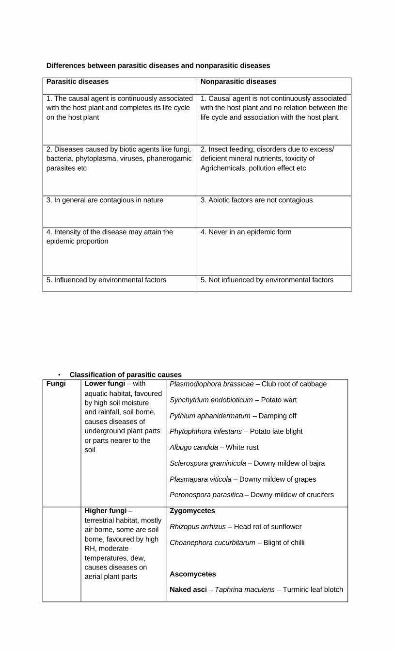
Differences between parasitic diseases and nonparasitic diseases
Parasitic diseases Nonparasitic diseases
1. The causal agent is continuously associated with the host plant and completes its life cycle on the host plant
1. Causal agent is not continuously associated with the host plant and no relation between the life cycle and association with the host plant.
2. Diseases caused by biotic agents like fungi, bacteria, phytoplasma, viruses, phanerogamic parasites etc
2. Insect feeding, disorders due to excess/ deficient mineral nutrients, toxicity of Agrichemicals, pollution effect etc
3. In general are contagious in nature 3. Abiotic factors are not contagious
4. Intensity of the disease may attain the epidemic proportion
4. Never in an epidemic form
5. Influenced by environmental factors 5. Not influenced by environmental factors
• Classification of parasitic causes Fungi Lower fungi – with
aquatic habitat, favoured by high soil moisture and rainfall, soil borne, causes diseases of underground plant parts or parts nearer to the soil
Plasmodiophora brassicae – Club root of cabbage
Synchytrium endobioticum – Potato wart
Pythium aphanidermatum – Damping off
Phytophthora infestans – Potato late blight
Albugo candida – White rust
Sclerospora graminicola – Downy mildew of bajra
Plasmapara viticola – Downy mildew of grapes
Peronospora parasitica – Downy mildew of crucifers
Higher fungi – terrestrial habitat, mostly air borne, some are soil borne, favoured by high RH, moderate temperatures, dew, causes diseases on aerial plant parts
Zygomycetes
Rhizopus arrhizus – Head rot of sunflower
Choanephora cucurbitarum – Blight of chilli
Ascomycetes
Naked asci – Taphrina maculens – Turmiric leaf blotch

Cleistothecia – powdery mildews
Oidium mangiferae – Mango powdery mildew
Erysiphe cichoracearum – PM of crucifers
Leveillula tautrica – PM of tomato
Phyllactinia corylea – PM of mulberry
Sphaerotheca pannosa – PM of rose
Podosphaera luecotricha – PM of apple
Perithecia
Venturia inaequalis – Apple scab
Apothecia
Sclerotinia sclerotiorum – white rot of vegetables
Ascostroma
Claviceps microcephala – Ergot of bajra
Basidiomycetes
Smuts –
Wheat loose smut – Ustilago segatum var tritici
Sorghum grain smut – Sphacelotheca sorghi
Bajra smut – Tolyposporium penicillariae
Karnal bunt – Neovossia indica
Rusts –
Wheat stem rust – Puccinia graminis var tritici (Heteroecious)
Puccinia helianthi – Sunflower rust (autoecious)
Basidiocarps –
Ganoderma lucidum – Coconut root rot and wilt
Deuteromycetes
Conidia directly on conidiophores-
Pyricularia oryzae – rice blast
Alternaria alternata – tobacco brown spot
Cercospora oryzae– rice narrow brown leaf spot
Helminthosporium oryzae – rice brown spot
Cinidia in fruiting bodies –
- pycnidia – Phomopsis vexans – brinjal fruit

rot - - Diplodia natalensis – citrus stem
end rot - Acervulus – Colletotrichum falcatum – red
rot of sugarcane - Sporodichia – Fusariaum udum – redgram
wilt No conidia and only sclerotia
Rhizoctonia solani – rice sheath blight
Sclerotium rolfsii – groundnut collar rot
Bacteria Favoured by high moisture / RH, rainfall, symptoms are watersoaked lesions with yellow halo, angular and necrotic (in dicots), veinal necrosis, on fruits raised shiny black lesions with bacterial ooze
Gram negative – aerobic rods
Polar flagella – yellow pigment – Xanthomonas
- no pigment – Pseudomonas
Gram negative – facultative anaerobes – peritrichous flagella – Erwinia
Gram negative – aerobic – sparsely flagellate – induces tumers – Agrobacterium tumefaciens
Gram positive – Coryneform – Clavibacter tritici
Fastidious – Xylem inhabiting – Leifsonia xyli
- Phloem inhabiting – Candidatus Liberobacter asiaticus
Phytoplasma – wall less prokaryotes – sesamum phyllody, brinjal little leaf, sugarcane grassy shoot
Spiroplasma citri – citrus stubborn
Viruses Nucleocapsid, requires vector / agent for transmission
ssRNA – rigid rods – Tobacco mosaic virus
ssRNA-flexuous rods – Poty virus
ssRNA-spherica – Cucumber mosaic virus
ssRNA-enveloped – Tomato spotted wilt virus
dsRNA-Reoviridae – wound tumer virus
ssDNA – Gemini viruses – Chilli leaf curl virus
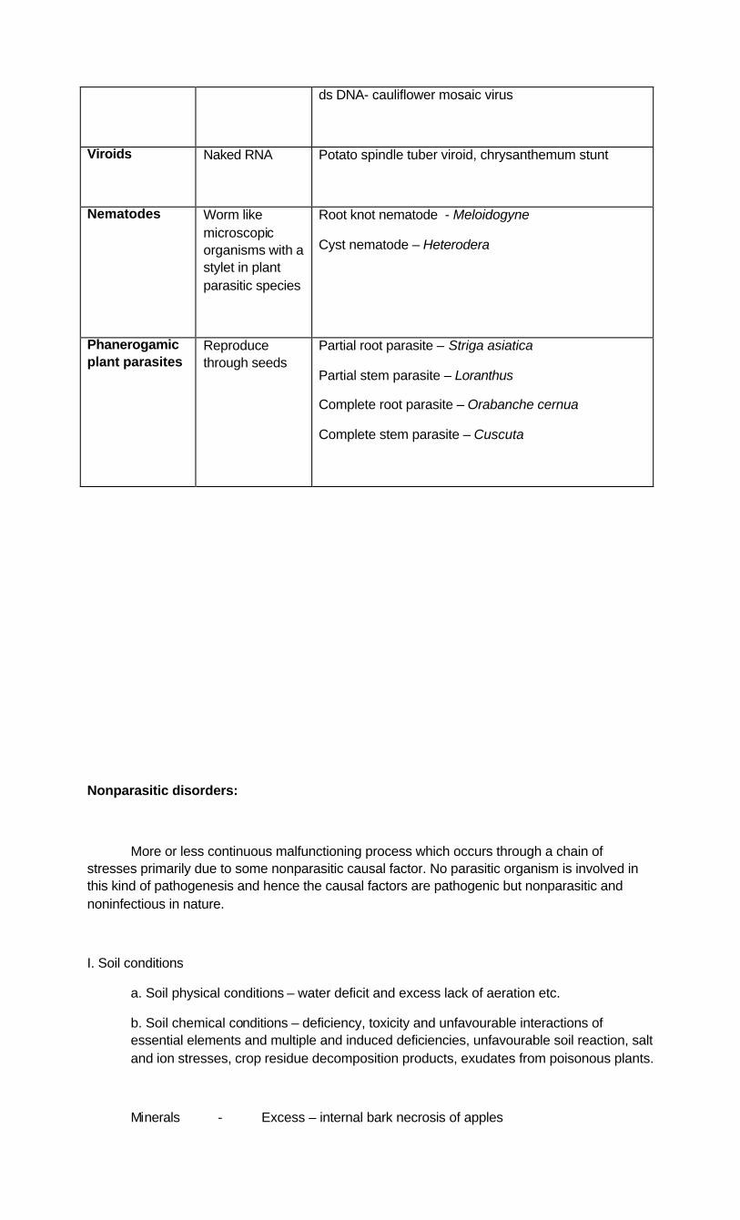
ds DNA- cauliflower mosaic virus
Viroids Naked RNA Potato spindle tuber viroid, chrysanthemum stunt
Nematodes Worm like microscopic organisms with a stylet in plant parasitic species
Root knot nematode - Meloidogyne
Cyst nematode – Heterodera
Phanerogamic plant parasites
Reproduce through seeds
Partial root parasite – Striga asiatica
Partial stem parasite – Loranthus
Complete root parasite – Orabanche cernua
Complete stem parasite – Cuscuta
Nonparasitic disorders:
More or less continuous malfunctioning process which occurs through a chain of stresses primarily due to some nonparasitic causal factor. No parasitic organism is involved in this kind of pathogenesis and hence the causal factors are pathogenic but nonparasitic and noninfectious in nature.
I. Soil conditions
a. Soil physical conditions – water deficit and excess lack of aeration etc.
b. Soil chemical conditions – deficiency, toxicity and unfavourable interactions of essential elements and multiple and induced deficiencies, unfavourable soil reaction, salt and ion stresses, crop residue decomposition products, exudates from poisonous plants.
Minerals - Excess – internal bark necrosis of apples
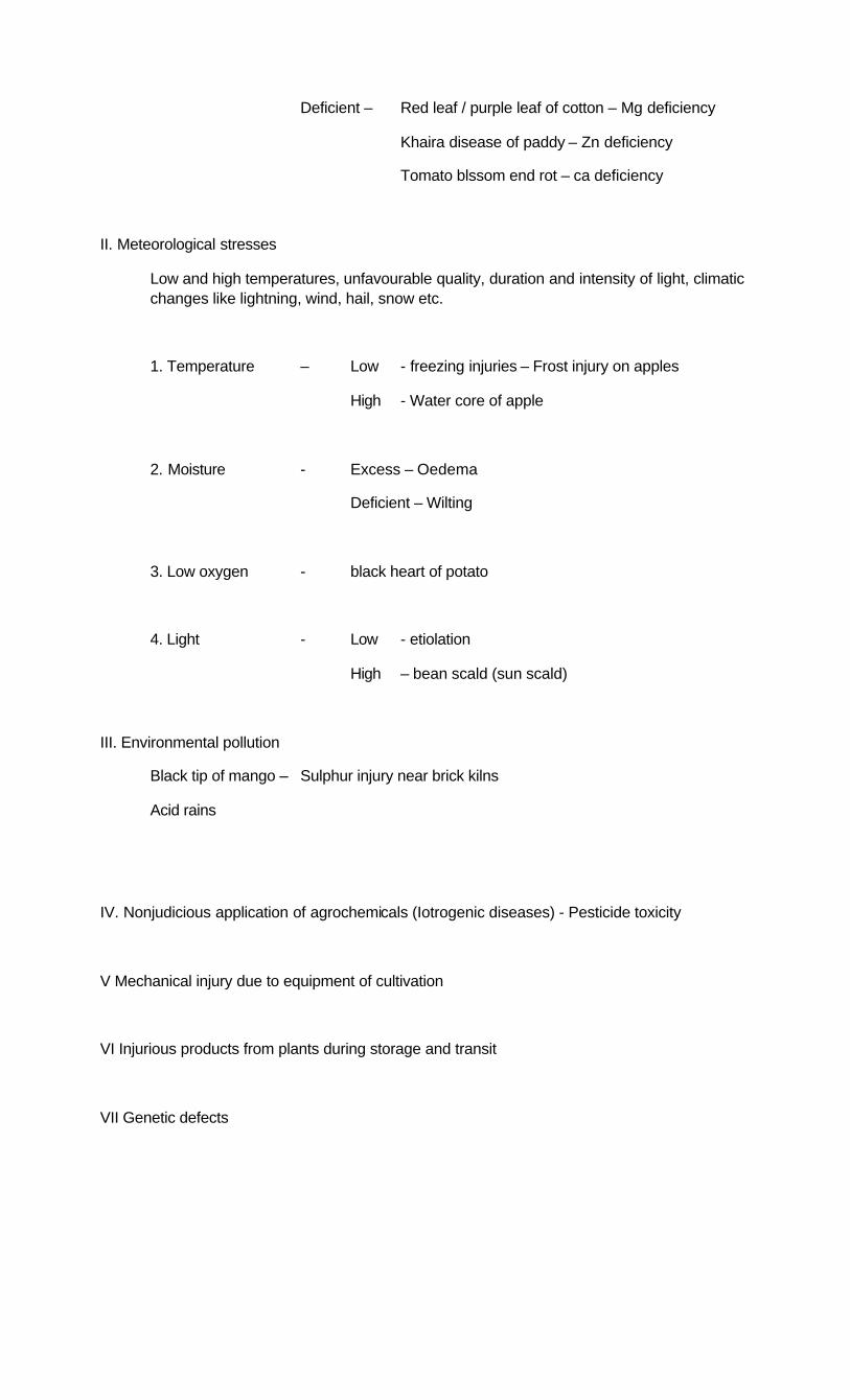
Deficient – Red leaf / purple leaf of cotton – Mg deficiency
Khaira disease of paddy – Zn deficiency
Tomato blssom end rot – ca deficiency
II. Meteorological stresses
Low and high temperatures, unfavourable quality, duration and intensity of light, climatic changes like lightning, wind, hail, snow etc.
1. Temperature – Low - freezing injuries – Frost injury on apples
High - Water core of apple
2. Moisture - Excess – Oedema
Deficient – Wilting
3. Low oxygen - black heart of potato
4. Light - Low - etiolation
High – bean scald (sun scald)
III. Environmental pollution
Black tip of mango – Sulphur injury near brick kilns
Acid rains
IV. Nonjudicious application of agrochemicals (Iotrogenic diseases) - Pesticide toxicity
V Mechanical injury due to equipment of cultivation
VI Injurious products from plants during storage and transit
VII Genetic defects

Classification of plant diseases based on symptoms
Symptoms are visual expression of host-pathogen interaction
Signs are pathogen structures associated with the infected plant part
I. Signs of plant diseases:
Sign Characteristic feature Example
1. Downy
mildew
Sporangiphores and sporangia of the pathogen is seen as velvety downy growth on the under surface of the leaf
Sclerospora graminicola – Downy mildew of bajra
Plasmapara viticola – Downy mildew of grapes
Peronospora parasitica – Downy mildew of crucifers
2. Powdery
mildew
On the infected plant parts ectoparasitic mycelium along with conidiophores and conidia seen as white powdery mass. Sometimes associated with black dot like cleistothecia
Oidium mangiferae – Mango powdery mildew
Erysiphe cichoracearum – PM of crucifers
Leveillula tautrica – PM of tomato
Phyllactinia corylea – PM of mulberry
Sphaerotheca pannosa – PM of rose
Podosphaera luecotricha – PM of apple
3. Rust True rust:
Belong to Basidiomycetes
Pustules of spores usually breaking through the host epidermis. Initially lighter in colour due to uredospores and later darker in colour due
Wheat stem rust – Puccinia graminis var tritici (Heteroecious)
Puccinia helianthi – Sunflower rust (autoecious)

to teliospores
White rust:
Belong to Oomycetes
Blister like pustules breaking open host epidermis and expose powdery mass containing club shaped sporangiophores and hyaline round sporangia
Red rust:
Green algae
Forms vegetative thallus on aerial parts. Filaments are formed between cuticle and epidermis. Green to red in colour. Produce sporangia.
Algugo candida - white rust of crucifers
Cephaleuros virescens – red rust of guava
4.Smut Black dusty mass formed in plant parts
Smutted floral organs with stinking smell – Karnal bunt – Neovossia indica
Smutted floral organs without stinking
smell – systemic and whole inflorescence is converted to smut sori – Loose smut of wheat
systemic but only few grains are converted to smut sori – Sorghum grain smut
Nonsystemic and only few grains are converted in to smut sori – Bajra smut
Smutted leaves
Onion smut – Urocystis cepulae
Paddy leaf smut – Entyloma oryzae
5.Sclerotial bodies
Ascomycetes – develop in to ascocarps
Claviceps – Ascostroma
Sclerotinia - Apothecia
6.Fruiting bodies
Macroscopic
Microscopic
Ganoderma - Bracket fungus
Pycnidia and acervuli
7.Sooty growth
Superficial growth of the fungus
Capnodium mangiferae – Mango sooty mould

II Symptoms – associated with the changes in the host plant
1. Colour changes: eg: Red rot of sugarcane, yellows caused by phytoplamas, and mosaic diseases caused by viruses.
2. Over growth or hypertophy: Hyperplasia (excessive cell division) and hypertrophy(increased cell size). eg: Galls (enlarged portions of plant organs) – Club root of cabbage, Root knot of
vegetables, wart (potato wart), crown gall of stone fruits.
Curl – distortion, thickening and curling – Leaf curl (viral origin)
Mummies, black pockets – grape downy mildew
Enations – abnormal outgrowth from the surface of a stem or leaf – Leaf curl of tobacco
Witches broom – profuse upward branching of twigs – Citrus witche’s broom (Phytoplasma)
Hairy root – Agrobacterium rhizogenes
3. Atrophy / hypoplasia / dwarfing – Negligible or under growth of infected plant organs eg: Sugarcane ratoon stunt (Fastidious bacteria), Corn stunt (Spiroplasma),
Chrysanthemum stunt(Viroid)
4. Necrosis – death of cells / tissues visualized as brown or black Spots – Localised lesions consisting of dead cells - Brown spot of rice
Blights - general and extremely rapid browning and death of leaves, branches, twigs and floral organs – Sorghum Leaf blight
Canker – Sunken, necrotic lesion arising from disintegration of tissues outside xylem cylinder usually associated with development of cork – Citrus canker
Stripes – Long necrotic lesions running parallel to veins in monocots – Wheat stripe rust
Streaks - Linear lesions restricted lengthwise – Rice Bacterial leaf streak
Damping off – rotting of roots and collar region at seedling stage in vegetable nurseries under excess water conditions due to Pythium, Rhizoctonia, Sclerotium etc .
Burn or scald – rapid and quick spread of the disease within and between plants giving burnt appearance to the field – Rice blast
Rot – Rotting of the tissue
Soft rot – Erwinia caratovora sub sp caratovora – soft rot of vegetables due to disintegration of middle lamella and pectin containing tissues.
Wet rot – Appearance of moistened plant parts – Scerotium rolfsii, Sclerotinia sclerotiorum
Dry rot – rotting of plant tissues under dry conditions – Charcoal rot of sorghum
5. Anthracnose – necrotic, black, sunken, irregular lesions on stem, fruit, leaf and flowers Anthracnose of mango, papaya, grapes
6. Die back – excessive necrosis of twigs beginning at the tips and advancing towards their base – Chilli die back, citrus die back
7. Wilt – Generalized loss of turgidity, drooping of leaves and shoots followed by withering and drying – Redgram wilt, Chickpea wilt, Cotton wilt
8. Miscellaneous : Alteration of habit / symmetry – Shoe string effect in Papaya mosaic affected leaves, Sapota flat limb

Premature dropping of leaves – Redgram Cercospora leaf spot
Transformation of plant organs – Sesamum phyllody, Stag head in mustard
---
Lecture No 11: STEPS IN DISEASE DIAGNOSIS
In the field symptoms due to different biotic and abiotic causes overlap several times. Hence it is necessary to distinguish the affects of abiotic and biotic causes in the field so as to correctly diagnose the problem.
A) SYMPTOMS - plant reactions or alterations of a plant’s appearance due to a disease or disorder
1. Identify and classify symptoms
§ Underdevelopment § Overdevelopment § Necrosis or death § Alteration of normal appearance § Wilting
2. Identify plant parts affected
i) Underdevelopment
§ Stunting of plants, leaves § Shortened internodes § Inadequate chlorophyll production § Caused by many types of pathogens
ii) Over-development
a) Biotic causes
§ Root knot nematodes - root galls § Callus formation around cankers § Tissue proliferation -some downy mildews and phytoplasmas § Some insects and mites also cause galls.
b) Abiotic causes
§ Oedema - an abnormal swelling in a plant caused by a large mass of parenchyma or an accumulation of water in the tissues leading to tannish-brown, corky or scabby appearance to leaves. The problem is favored by high humidity, cloudy weather, poor soil drainage, and excessive watering.

§ Fasciation (Malformation of plant parts resulting from disorganized tissue growth) can be physiological or due to biotic cause.
iii) Tissue Necrosis:
a) Fungal leaf spots
§ Usually round, not vein-limited § Elongated on narrow leaves or stems § May have alternating zones of light and dark tissue § Sporulation or mycelia may be evident
b) Bacterial leaf spots
§ Often dark and water-soaked § Often vein-limited, giving angular shape § Bacterial “flow” observed under microscope
c) Fruit rots
§ Firm or soft and watery § Colors vary § Fungal sporulation may be present § Fungal and bacterial causes
d) Cankers = localized necrotic lesions
§ Sunken or swollen or both § Mainly caused by fungi and bacteria § Mechanical injury can cause
e) Blight = rapid death or dieback.
§ Also from coalescing leaf spots, e.g. early blight of tomato § Mainly fungal and bacterial causes
f) Dieback - many causes
§ Girdling cankers. § Root problems § Mechanical, chemical or cold damage
g) Root rots
§ Root lesions § Darkening and softening of roots § Sloughing off of outer tissues § Yellowing of foliage and stunting of plants § Fungal and bacterial causes
h) Damping off
§ Rapid death of seedlings and cuttings § Stem infected at soil line, seedling topples § Spreads rapidly under crowded conditions § Mainly fungal agents
i) Wilting
Biotic factors
§ Root, crown or stem rots § Vascular wilts § Root, crown or stem damage from insects or animals § Mainly fungal and bacterial causes Abiotic factors
§ Damage from weather extremes § Dry or flooded soil § Mechanical damage to roots, crown or stem
j) Alteration of normal appearance
Mosaic
§ Irregular patches of discolored tissue. § Often with distortion § Viruses mainly
Ringspot
§ On leaves and fruits. § Distinct ring shaped lesions, often in concentric zones § Viruses
Abiotic causes
§ Chemical damage § Nutritional deficiencies
Yellowing
§ Often nutritional, cultural or environmental § Root malfunction – biotic or abiotic § Nematode infestation § “Yellows” phytoplasma diseases § Be aware of symptom variability. Ø May have more than one problem. Ø More than one pathogen may be involved. Ø Pathogens have varying levels of virulence. Ø Environmental conditions can affect symptom expression. Ø Host genetics and physiology can effect symptom expression.
B) SIGNS: Signs - actual pathogen, parts or by-products seen on a diseased host plant
Fungal signs
Ø Evidence of pathogen on tissue. Ø Spores, mycelia or fruiting bodies. Ø Use hand lens or knife for field detection.
Fruiting bodies

Ø Shape of fruiting body aids in fungal identification. Ø Tissue location may help differentiate between species. Powdery mildew fungi
Ø Powdery mildew fungi form mycelia and spores on tissue surface. Ø Powdery material rubs off. Ø Leaves often distorted, discolored Ø Dark, round fruiting bodies form in fall. Downy mildew fungi
Ø Downy mildews often produce angular leaf spots on upper surface of leaf Ø Downy mildews develop sporulation on leaf undersides only.
Bacterial signs
Ø “Streaming” from freshly cut stem in water. Ø Bacteria “flow” observed from tissue mount on compound microscope.
Bacterial ooze can be observed on-site with some diseases.
Distribution of symptoms on plant parts:
Ø Biotic disease – symptoms progress and nearby plants become infected. Ø Abiotic disease – generally a lack of symptom progression. Does not spread. Exception – nutritional symptoms progress slowly.
Pattern of symptom development
Ø Determine prevalence of problem: Large area/All plants – generally abiotic Ø Scattered, localized – generally biotic Ø Check for distribution of symptoms
• Uniform – generally abiotic Random – generally biotic
B) Questions to be asked:
Ø When was the problem noticed? Ø Was the damage sudden or gradual? Ø How old are affected plants? Ø Percentage of plants affected? Ø What is the degree of injury?
Review cultural practices.
Ø Proper planting technique Ø Fertilizer and pesticide application Ø Irrigation frequency
Review environmental conditions.
Ø Temperature extremes Ø Drought or excess rain Ø Soil type and conditions Check host specificity – many hosts may mean an abiotic problem
C. Laboratory Examination and Testing
Ø Compound microscope Ø Electron microscope Ø Moist chamber incubation Ø Culturing

Ø Additional tests for biotic agents Ø Tests for abiotic agents
LaboratoryExamination:
Examination using microscope of higher magnification
Ø Identify fungi to genus or to species. Ø View bacterial flow; observe morphology at highest magnification. Ø View virus inclusion bodies. Ø Using electron microscope, view viruses, phytoplasmas
Moist Incubation
Ø Goal – to induce sporulation Ø Important for obligate fungal pathogens Ø Avoid excess moist conditions.
Fungal isolation
Ø Surface sterilize. Ø Use margin of diseased area. Ø PDA media, can be selective Ø Diagnostician can often identify to genus from appearance of culture
Fungal identification – Other methods
Ø Examination and comparison with drawings in literature Ø Utilizing keys in literature Ø Literature sources for host diseases and disorders Ø Serological, molecular, other tests
Bacterial isolations
Ø Grind tissue in sterile water. Ø Streak suspension on bacterial medium. Ø Can use selective media Ø Transfer single, isolated colony
Bacterial identification
ØOoze test ØBiochemical tests ØCarbohydrate utilization (BIOLOG) ØFatty acid methyl ester analysis (FAME) ØMolecular methods ØPolymerase chain reaction (PCR) Ø Serological tests, e.g. ELISA
Identification of other pathogens: Non-culturable pathogens
Ø Viruses, viroids Ø Phytoplasmas Ø Fastidious bacteria
Methods
Ø Serological tests, e.g. ELISA Ø Electron microscopy

Ø Staining for virus-induced inclusion bodies Ø Molecular tests, e.g. PCR Ø Enzyme activity testing Ø Host range testing
Identification of Nematodes
i) Extraction from Soil and Plant Material
Ø Soil extraction using centrifugal flotation Ø Baerman funnel techniques for soil and tissue Ø Mist chamber extraction for nematodes in root or stem tissue ii) Direct examination of nematodes in tissue
Diagnostic tests for abiotic causes
i. Soil, water
Ø pH testing Ø Nutrient analysis Ø Soluble salts analysis Ø Analysis for chemicals ii. Plant tissue tests
Ø Nutrient analysis Ø Analysis for chemicals
Investigate pathogenicity
i. Is identified organism a known pathogen
- of host?
- of hosts in same genus?
ii. If not a host, Koch’s Postulates may be next step
Ø A lengthy process, rarely used in routine diagnoses Ø Problem arises when organism cannot be cultured
iii. Koch’s Postulates
1. Note constant association of organism with diseased plants and consistent observable symptoms.
2. Isolate and characterize organism in pure culture. 3. Inoculate healthy plant with organism, and observe the same disease and symptoms. 4. Re-isolate same organism.
Final Diagnosis: All information compiled and analyzed
Organism identified, found to be pathogen of host
OR
Lab tests, other information reveal an abiotic cause
Develop control recommendations and present to farmer.
___
Lecture No. 12 Distinguishing symptoms due to plant pathogens, insect pests, excess / deficiency of nutrition (nutritional disorders) and pesticide injuries.
• Insect pests and mites: Insects do not cause infection as continuous association of plant and pest does not exist. Hence insect association with the plant is considered only as infestation.
Insect pests damage plants using their mouth parts. Depending upon the mouth parts and type of feeding on the plant apparent damage varies. Some of the important symptoms of pest damage are listed below.
1. Biting and chewing type: Leaves may be eaten up completely leaving only a net work of veins. eg. Grasshoppers, caterpillars, beetles etc. Symptoms associated:
Defoliation or skeletonization / scraping of leaves – Tobacco caterpillar
Uneven cut on leaf margins – Grasshoppers

Uneven scraping of leaf surface – Grubs and adults of beetles
Small white streaks parallel to midrib in monocots – Rice Hispa
Tubular cases attached to leaf / floating in water – Rice case worm
Shot holes on leaves – Flea beetle damage on blackgram / greengram
Scraping and gnawing of base of stem – Plutella xylostella on mustard and rape.
2. Piercing and sucking type: Nymphs and adults suck sap from base of the plants / leaves / tender terminal parts and thereby affect the vigour and growth of plants. In case of severe infestation, sooty mould develops on plant parts covered with honey dew excreted by insects while feeding. This honey dew may be occupied by saprophytes and make the plant parts appear black. eg: feeding due to plant hoppers, leaf hoppers, aphids, white flies, mealy bugs etc.
3. Rasping and sucking type / lacerating and sucking type: eg. Thrips feed by rasping the tissues and suck the juice thereby exuding from inside the plant. The damaged part exhibit a whitish mottled / silvery appearance.
4. Siphoning / simple sucking type: Fruit feeders. Make holes on the fruit consequently resulting in rotting due to infections by microbes.
5. Degenerate type of mouth parts: Gall formers, shoot berers, pod borers
6. Biting, piercing and sucking type: Tetranychid Mites. Penetrate the plant tissue with sharp stylets and remove the cell contents, chloroplasts disappear, formation of irregular spots, drooping and drying of leaves. Yellowing, bronzing, distortion, curling, crinkling, defoliation of leaves, retardation of growth, droping of flowers, reduction in size, quality and quantity of produce are other symptoms associated with mite feeding.
Excess / deficiency of nutrition (nutritional disorders):
Excess: Due to toxic ions present in higher concentrations.
eg. Copper, iron, manganese, boron and zinc. In India, toxicity due to Cu and Fe is more common.
Application of excess Nitrogen also results in toxicity (burning of leaves). However, it results only at a very high dose, i.e., more than 4% of the dry weight
Excess K may not directly cause toxic effect but it results in deficiency Ca, Mg or Fe (K-excess-induced deficiency)
Mn is in general in an unavailable form in the soil. However, if the soil pH falls below 5.5, Mn becomes readily available and cause toxicity.
Deficiency: Red leaf / purple leaf of cotton – Mg deficiency
Khaira disease of paddy – Zn deficiency
Tomato blssom end rot – ca deficiency
(For deficiency symptoms of major and micronutrients refer to Plant Physiology section)
• Pesticide injuries: Symptoms: Chlorosis or yellowing of leaves, bronzing of leaves, necrosis of complte plant or plant parts, scorching, deformation and curling of leaves, white spots on leaves, burning effects on leaves, premature falling of leaves, mottled leaves, poor germination of seeds etc.
REMOTE SENSING
Remote sensing is estimating an object/phenomenon without being in physical contact with it. Remote sensing is a science/art that permits us to obtain information about an object / a phenomenon through analysis of data obtained through sensory devices without being in physical contact with that object. The specific properties of the vegetation, healthy or diseased, influence the amount and quality of the radiation reflected or emitted from the leaves and canopies. Application of this technology to phytopathological research such as phytopathometry is of great interest.
Objectives of remote sensing in plant Pathology
1. Assessment of disease over a vast area without the ground parties visiting those areas.
2. To know the relationship of diseases and environment
3. To know the origin and development of epidemics
4. Quantitative assessment of the disease
Advantages of Remote sensing
1. Reveals pattern of disease incidence, intensity and development over large area
2. Data generated by remote sensing is amenable to multidisciplinary approach
3. Gives synoptic view of large areas
4. Data generated is on a permanent scale and is unbiased
5. Data acquisition is fast compared to traditional methods and data analyzed is effectively utilized
6. Satellite data (ERTS) obtains information of an area periodically so that the information can be updated.

7. It frequently poses questions for ground investigators which cannot be generated by ground parties
Remote sensing techniques of importance to Plant Pathology
1. Aerial photography and 2. Satellite remote sensing
1. Aerial photography: Aerial photography can detect objects on land over a larger area. Colwell (1956) first used remote sensing technique for monitoring stem rust of wheat. He showed that panchromatic colour and especially infrared aerial photography could be used to detect rusts and viral diseases of small grains and certain diseases of citrus. Later, infrared photography was used in England for late blight of potato.
The key to distinguish diseased and healthy parts of a crop is to use appropriate film or filter combinations. The main film types used are panchromatic, infrared, normal colour and colour infrared. The infrared films are preferred because of their superior sensitivity to visible light and to near infrared wavelengths of radiation (700-900 mµ). The colour infrared or Ektachrome Aero Infrared (Camouflage Detection Film) is superior as it can show the difference between diseased and healthy patches of plants in colour. The healthy foliage is highly reflective to the infrared wavelengths and appears red on this film whereas blighted or diseased foliage has low infrared reflectance and does not appear red in the photograph.
2. Satellite Imaging
Weather satellites
Often cyclones create heavy clouds with rains and an anti-cyclone creates a cloudless sky. All these can be effectively monitored by weather satellites. Sequential pictures show the movement of these systems before they arrive in an area. Therefore by monitoring epidemic favouring systems using a satellite, the disease occurrence on the field can be monitored. Ex: The spread and deposition of stem rust pathogen of wheat is influenced by definite synoptic weather conditions called Indian stem rust rules.
Earth resources technology satellites (LANDSAT, 1972, USA)
LANDSAT covers the entire globe every 18 days scanning the same area at a fixed time. The scanned data is compared for any major differences happened within 18 days. Nagarajan utilized LANDSAT infrared spectral bands 6 (0.7-0.8µm) and 7 (0.8-1.1µm) to differentiate healthy wheat crop of India and severe yellow rust affected crop of Pakistan.
Examples: Coconut root rot and wilt, black stem rust of wheat, citrus canker


Lecture No. 13
Detection and diagnosis of post harvest diseases of perishables due to biotic agents.
Causes of post harvest diseases:
1. Physical damage: This cane be divided in to - i) mechanical factors involving damage due to agricultural equipment, branch rubbing and abrasion due to sand particles at the time of high winds (scratches and bruises on fruits and vegetables), damage during loading, unloading and transit
ii) physical factors like hail damage, frost bands (discolored corky tissues in a band or large area of apple fruits), low temperature (cat face on tomato), high temperature (blossom end rot of citrus, water core of apple), excess light (sunscald on bean pods), low oxygen (black heart of potato
2. Physiological deterioration is a common feature in senescing plant parts including fruits and vegetables
3. Pathological rot due to microbes 4. Insect and mite injury 5. Non infectious disorders (abiotic causes)
Magnitude of losses depend upon
1. Initial quality of produce 2. Environmental factors 3. Operational factors 4. Physiological changes during storage 5. Genetical
Importance of studying physiological changes during storage (post harvest diseases)
1. Detecting the deterioration in quality and quantity of produce in nutritional and other substances, which render them unfit or of reduced value for human consumption.
2. Detecting and assaying mycotoxins
Physiological changes in perishables during storage
1. Changes in carbohydrates: • Generally fruits and vegetables are rich in carbohydrates and sugars. • Maturity and ripening leads to rise in amount of carbohydrates particularly soluble
carbohydrates including sugar. • During pathogenesis, usually there is an increase in the loss of complex carbohydrates
and initial increase in simple sugars followed by their decrease too. • Pathogens vary among themselves and on different hosts in terms of varying rates and
degrees of utilization and in preference for sugars.
eg. 1. Infection due to Botryodiplodia theobromae results in decreased glucose and fructose in pineapple and decreased starch in banana
eg. 2. Decreased glucose and maltose content in tomato due to Alternaria alternata
2. Changes in aminoacids, proteins and nucleic acids.
• In general, the amount of both free and protein bound fractions increase during initial stages of pathogenesis on per cent dry weight basis because the proteolysis of the host tissues is more than that of consumption by the pathogen.
• In later stages, the consumption and metabolization by the fungi are higher. • Sudden disappearance or reduction of some aminoacids, glutamic acid in particular and
appearance of new aminoacids like valine is a common feature due to infection perishables.
• Sudden increase of proteins, DNA and RNA in strawberry was observed due to Rhizopus stolonifer due to pathogen directed symthesis of these compounds.
3. Changes in organic acids.
• Organic acids like citric, fumaric, malic, malonic, oxalic, succinic acid etc decrease during pathogenesis as they serve as carbon source.
• Some times organic acids like succinic acid increase.
Oxaloacetic acid malic acid fumaric acid succinic acid
• Sometimes increase in organic acids help the pathogen in pathogenesis. Oxalic acid produced by Penicillium oxalicum acts synergistically with endo-PG by sequestering Ca+2 present in middle lamella there by permitting easy cell wall degradation.
3. Changes in phenols and other secondary metabolic substances. • Phenols and other secondary metabolic substances are assigned a role in resistance. • Plant pathogens are capable of detoxifying such substances, which are regarded as
contrivances for offence. • In general phenol content increase in the initial stages of infection but later decrease as
seen in papaya due to anthracnose pathogen – Colletotrichum gloeosporioides.
5. Reduction in ascorbic acid content
• Fruits particularly the tropical ones are important sources of vitamin C, i. e., ascorbic acid.
• Ascorbic acid content normally diminishes during storage and the diminishing process is enhanced by injury or infection.
• Reduction in ascorbic acid content is found to be to an extent of even up to 100% due to infection by Colletotrichum gloeosporioides in mango, citrus and papaya.
6. Reduction in alkaloid content
• Alkaloids like capsaicin in chillies, responsible for pungency, decreases due to infection by Colletotrichum capsici.
7. Changes in pH
• Varies with kind of fruit Lemon Orange
Total acidity Decrease Increase
Titrable acidity Decrease Increase
Volatile acidity Increase Decrease
+NADH2 H2O +NADH2

• Due to infection – In fruits where acids play a role in economic part – acidity decreases eg. Lemon, mandarine orange
In fruits where conversion of acids in to sugars leads to sweetness, the conversion is affected there by acidity increases. eg. Mango, Sweet orange, Mausambi due to Colletotrichum gloeosporioides and Aspergillus flavus infection.
9. Changes in total soluble solids (TSS) • Reduced TSS observed in citrus due to Colletotrichum gloeosporioides and
Botryodiplodia theobromae
10. Changes in miscellaneous substances • Reduction in oil content and increase in fatty acid content in coconut • In general during storage initially moisture loss due to transpiration is compensated by
moisture released during respiration. However, due to infection, loss of moisture, crude fibre and minerals decrease in case of rotten coconuts.
• A decrease in P content was observed in mango due to Botryodilpodia.
10. Changes in ripening process
• Ethylene production enhances ripening. • Injury and infection enhances ethylene production and there by ripening.In mandarine
oranges infection due to Botryodilpodia enhances ethylene production. • However, where ethylene mediated resistance, i. e., through PAL works in host system –
PAL activity needs to be decreased by the pathogen for successful infection.
11. Mycotoxin production
• Infection of perishables may result in production of mycotoxins there by make the produce unfit for human consumption.
• In vegetables – Aflatoxins by Aspergillus flavus • In Apples – Production of mycotoxin Patulin by Penicllium and Aspergillus • Production of mycotoxin ‘malformin’ in outer scales of onion infected by Aspergillus niger


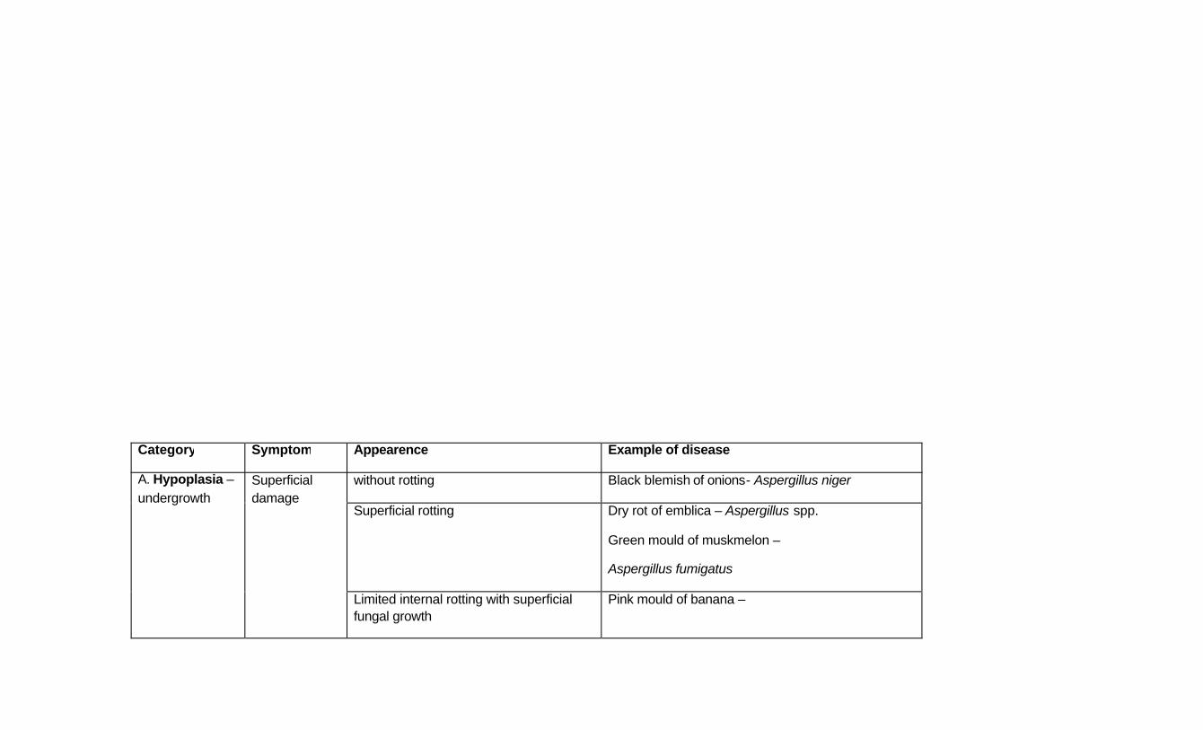
Category Symptom Appearence Example of disease
without rotting Black blemish of onions- Aspergillus niger
Superficial rotting Dry rot of emblica – Aspergillus spp.
Green mould of muskmelon –
Aspergillus fumigatus
A. Hypoplasia – undergrowth
Superficial damage
Limited internal rotting with superficial fungal growth
Pink mould of banana –

Trichothecium roseum
Dry rot Bitter rot of apple – Glomerella cingulata Soft rot
Wet rot Bacterial soft rot of vegetables –
Erwinia caratovora
Mould rots of citrus –
Penicillium sp., Aspergillus sp.
Pin head spots Speckle rot of guava – Phomopsis sp.
Limited spots Black rot of guava – Pestalotia psidi
Lesions
Extensive spots Black ripe rot of chilli –
Alternaria alternata
B. Necrosis –
death of infected tissues
Anthracnose Sunken lesions with raised margins Mango, papaya anthracnose –
Colletotrichum gloeosporioides
Grape anthracnose –
Gloeosporium ampeliphagum
C. Hyperplasia – excess growth
Scab A discrete, superficial lesion involving localized hyperplasia of the surface tissues with or without development of cork
Guava scab – Pestalotia psidi
Potato common scab –
Streptomyces scabies
Apple scab – Venturia inaequalis

Diagnosis of post harvest diseases of perishables
Symptomatology
Canker Sunken, necrotic lesion arising from disintegration of tissues outside xylem cylinder usually associated with development of cork
Citrus canker –
Xanthomonas campestris pv citri

Examples of post harvest diseases
S. No
Mango
1 Stem end rot (Botryodiplodia theobromae)
Symptoms: The disease appears as small brown to black necrotic areas at the stem end of the fruit. Water soaked brown areas progress downwards very quickly with irregular light brown margin. Abundant black tiny fruiting bodoes representing pycnidia. Pulp turns sour and the fruit emits fermentive odour.
2
Anthracnose (Colletotrichum gloeosporioides (IS) / Glomerella cingulata-PS)
Also called blossom blight, fruit rot)
Symptoms: Pathogen remains quiescent on the surface of the fruit and causes disease only upon maturity and turning yellow. Ripe mangoes are most invariably affected by the disease in May – June. Brownish to deep brown irregular sunken spots appear on the fruit. Tear staining is another common symptom of the disease.

Citrus
1 Stem end rot (B. theobromae)
Symptom: Characterized by softening of the rind and underlying pulp. Starts around stem end and remains unnoticed for some time. Then infected rind turns brown to black. Rotting is complete within 3-4 days.
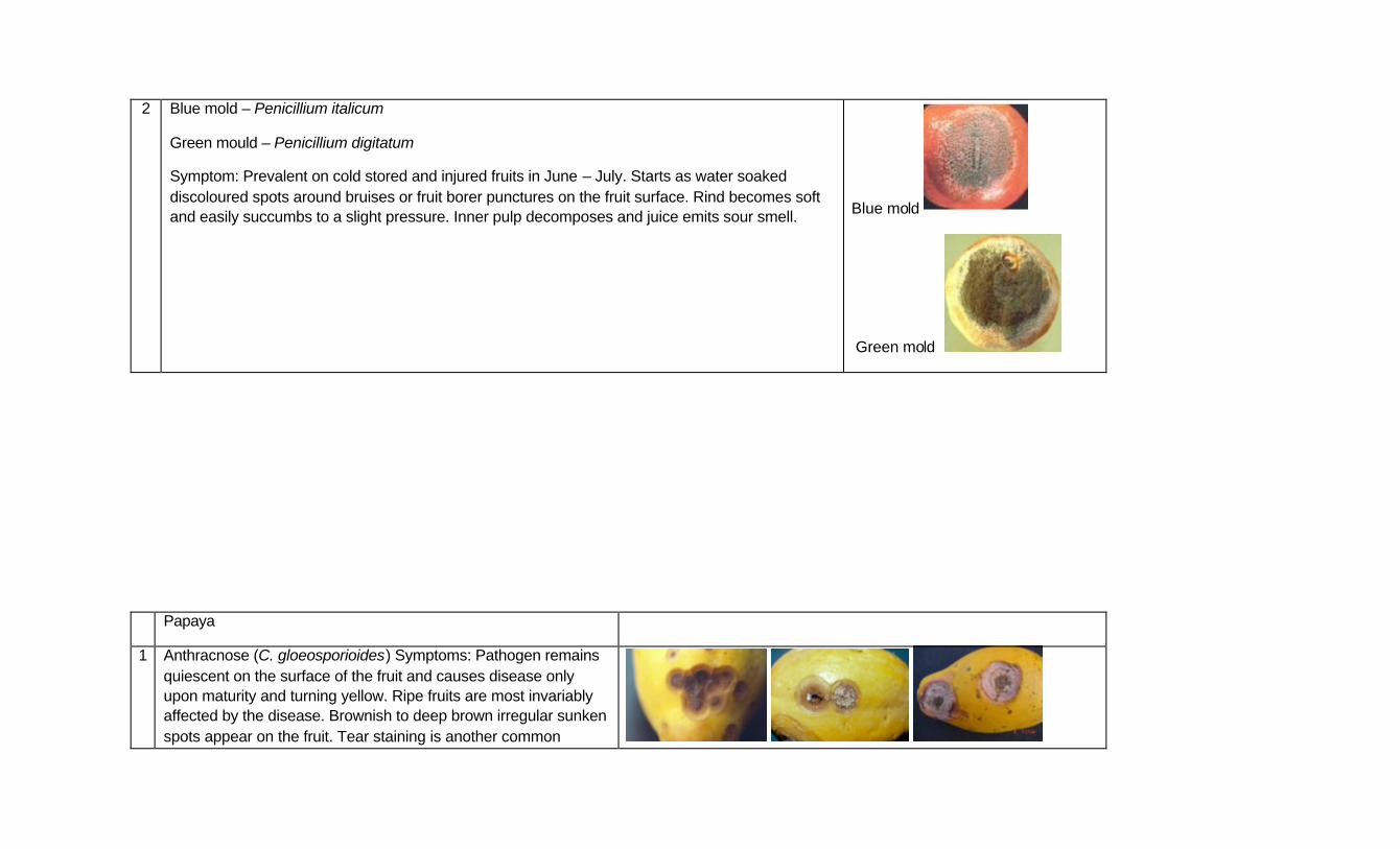
2 Blue mold – Penicillium italicum
Green mould – Penicillium digitatum
Symptom: Prevalent on cold stored and injured fruits in June – July. Starts as water soaked discoloured spots around bruises or fruit borer punctures on the fruit surface. Rind becomes soft and easily succumbs to a slight pressure. Inner pulp decomposes and juice emits sour smell. Blue mold
Green mold
Papaya
1 Anthracnose (C. gloeosporioides) Symptoms: Pathogen remains quiescent on the surface of the fruit and causes disease only upon maturity and turning yellow. Ripe fruits are most invariably affected by the disease. Brownish to deep brown irregular sunken spots appear on the fruit. Tear staining is another common

symptom of the disease.
Banana
1 Brown rot (B. theobromae)
Symptoms: Infection starts from one or both the ends of the finger. Water soaked areas on fruits which later turn brown, pulp becomes soft and fruit emits fermentative smell. Light grey cottony mycelium can be seen on the completely rotten fruit.
2 Anthracnose (C. gloeosporioides)
Symptoms: Disease first appear as small, black, circular specks on the skin, which increases and become sunken and coalesce to form large spots. Under humid conditions mouldy growth can be seen on the fruits. Acervuli are formed on the fruits in later stages.
3 Cigar end rot - Verticillium theobromae
Symptoms: Symptoms appear from the tip of immature fruit and spreads upward. Ashy conidia and conidiophores cover the rotted portion which becomes corrugatd thereby imparting burnt ashy cigar-end appearance with a dark border. Decay may extend up to one third of the fruit but internal tissues develop a dry rot.


Lecture No. 14
DETECTION AND DIAGNOSIS OF SEED BORNE DISEASES
The methods of detection may be general or specific for individual pathogen. The selection of seed health testing method for a particular study is based on certain objectives. The main objectives of seed health testing are
1) for quality assurance in evaluation of planting value of seed in certification schemes and to support decisions about the need for seed treatment and
2) for the detection of plant quarantine organisms, such as to meet phyto-sanitary regulations for the seed that is exported for trade
3) for testing the presence of storage fungi which can affect the storage quality or its value for feed and human consumption
To be worthwhile, seed health testing methods must deliver:
1. Specificity: The ability to recognize the target pathogen from all the organisms present on the seeds
2. Sensitivity that is fit for the purpose of the test 3. Information relating to field performance of the crop 4. Test results that are repeatable within and between samples of the same seed lot,
regardless of who performs the test, within statistical limits and sample variability 5. they must be cost effective
Qualitative and quantitative tests:
There are wide range of seed health testing methodologies, including relatively simple direct inspection methods for macroscopic fungal bodies, incubation tests, grow out tests (or growing –on-tests), bacterial extraction & identification and the more complex imunoassays (ELISA) and molecular assays such as PCR.
1) Qualitative assays establish whether seed is infected with a plant pathogen, with a very low probability of false negatives. Qualitative tests are necessary in the place of plant quarantine, where the aim is to prevent non-indigenous diseases establishing in the importing country.
2) Quantitative tests are designed to estimate the true level of infection in as seed lot, i.e., how many individual seeds are infected, and the level of infection or contamination. Such information can inform decisions about whether or not seed treatment is justified. Most of fungal pathogens are currently determined using these methods. Bacterial and viral pathogens, where severe epidemics result from relatively low numbers of infecting microorganisms and there are few control methods in the field, are commonly determined using qualitative methods.
I. Detection of seed borne fungi:
1) Examination of seeds without incubation or direct examination of seed lot: The seed sample is first examined by naked eye or under the microscope for the presence of discolouration, malformations, ergots, sclerotia, galls, acervuli, perithecium, pycnidia, bacterial ooze, etc. Although this type of testing provides information quickly and may be readily applied in seed testing stations in conjunction with purity testing, only a few diseases are adequately detected in this way.
Ex: Black point of wheat, purple seed stain of soybean, Anguina tritici, ergot fungi, sclerotial bodies of Rhizoctonia and sclerotia, etc.
2. Examination of seed washings: Seed samples may be immersed in water or other liquid to make fungal bodies (Ex: Pycnidia) or symptoms (Ex. Anthracnose on seed coats) more visible, or to encourage the liberation of fungal spores, hyphae and so on that are attached to or carried within the seed.
This method is applicable for some obligate pathogens like downy mildew fungi which don’t sporulate under incubation. A few seeds of the sample are kept in sterilized water and shaken well by mechanical shaker. The washing is centrifuged at 2000-3000 rpm and lactophenol is added to the sediment after removing the supernatant. Further, the sediments can be observed under microscope for presence of spores, fruiting bodies, hypha, etc.
Merits and demerits of direct examination:
1. Direct inspection provides quick information and don’t require much equipment, man power, etc. However, only few fungi producing external symptoms like discolouration, malformation, etc can be detected by this method.
2. In seed washings the amount of spores in a suspension can be quantified by using heamocytometer.
3. Incubation methods:
i. Standard blotter method: This is most convenient and effective of all methods and was first proposed by Doyer. In normal standard blotter method as per International Seed Testing Agency (ISTA) recommendation usually 400 seeds are incubated. The seeds are placed on 2 or 3 layers of moist blotters. Number of seeds per plate may vary from 10-100 depending on size of seed. The seeds are incubated for 7 days under 12 hrs of light and darkness at normal room temperature.
ii. 2,4-D blotter method or modified blotter method: In order to facilitate easy observation of seed samples the blotters are dipped in 200 ppm of 2,4-D solution which helps in killing the seeds and thereby it facilitates easy observation of seed samples and to increase the ability of the pathogen to grow on it.
iii. Deep freezing blotter method: A modification of standard blotter method which was proposed by Limonord. The seeds are sown as in blotter method and inoculated at normal room temperature for 1 or 2 days and further the seeds are subjected to -200C on the third day. After 12 hours of incubation the seeds are re-exposed to normal room

temperature. The dead seeds during freezing acts as a source of nutrients for fungi like Fusarium and Septoria in cereals.
Merits and demerits of Standard blotter and deep freezing blotter methods:
i. Blotter method is widely used where regular seed health testing is carried out especially in circumstances where in agar test is impractical. It is applied to all kinds of seeds and also can be clubbed with the germination test used in seed technology.
ii. The blotter method provides excellent conditions for development of mycelial growth and sporulation of many Hyphomycetes fungi.
4. Agar plate method: The seeds are plated in petridishes on a suitable agar like Potato Dextrose Agar (PDA) or malt extract agar. Prior to plating, a pre-treatment of the seed has to be carried out using sodium hypochlorite or mercuric chloride and then the seeds are incubated under standard temperature. After incubation, colony characters of a particular fungus are recorded for its identification.
Applications: The agar plate method is applicable to those seed samples in which the saprophytic species don’t materially impair quick identification of pathogens, a difficulty often overcome by pre-treatment. The procedure is preferable where the blotter method don’t provide adequate conditions for the mycelial growth, sporulation, etc. But on the other hand characteristic colonies appear on a suitable agar medium.
Limitations: Regular practice with each kind of seed is necessary to acquire and maintain experience. However in agar plate method, slow growing fungi may be suppressed by vigorously growing fungi and thereby suppressing its expression.
II. Detection methods for seed borne Phytopathogenic bacterium: The standardized methods for detecting and identifying seed borne fungi cannot be used to detect the bacteria as the fungi can be induced to sporulate on the infected seeds and they can be identified through their morphological characters whereas the seed borne pathogenic bacteria has to be isolated in pure culture and infected to the host again to prove the pathogenicity and to identify the pathogen. Often percentage of infected seeds in a sample may be so low that detection becomes extremely difficult.
i) Visual separation of infected seeds: Bean seeds infected with Pseudomonas phaseolicola did not show external symptoms unless there was a very massive concentration. Exposure of such discoloured bean seed lots to ultraviolet radiation in subdued light or darkness revealed the percentage of seeds exhibiting fluorescence. The seeds showing bright bluish white fluorescence are found to be infected with bacteria and those infected seeds subsequently showed hollow blight symptoms on the infected plants.
ii) Water agar plate method for detection of bacteria:
Used for detection of Xanthomonas campestris pv. campestris in crucifers (Black rot of crucifers). In this the seeds are first soaked in antibiotic solution like Aureofungin 200ppm solution. After draining off the excess antibiotic solution, the seeds are placed on nutrient agar and incubated at 200C in darkness and recordings are made after 12 days. The diseased seedlings show delayed germination and stunted growth. The hypocotyl and cotyledons become yellowish and pulpy and the seedling collapse on the agar surface. From the infected parts, yellowish bacterial ooze can be seen under microscope.
iii) Grow out / Growing-on tests: These tests are performed on plants grown from seed samples beyond the seedling stage in a green house, controlled environment chamber or the field and the seedlings or plants are observed for symptoms of the pathogen. They have been used for testing the effects of many seed borne bacteria, for ex. Peudomonas syringae pv. phaseolicola. Grow out tests gives an indication of potential transmission from external and internal seed inoculum under specified environmental conditions. However, both fluctuations under those conditions and the viability of the seed stock may influence the test result. The tests are also time consuming and require a considerable amount of space where large numbers of seeds require testing for the presence of low incidence pathogens.
iv) Laboratory tests: Laboratory methods for the detection of seed borne bacteria involve 3 stages
i) Extraction from seeds
ii) Isolation into culture
ii) Identification
Bacteria are extracted from the seed flour in a liquid medium, usually sterile pH buffred saline, though buffered sterile water with various other enrichments can be used. The volume and agitation of the liquid medium together with the duration and temperature of soaking are all critical to the optimum recovery of the target pathogen, which may also be affected by the saprophytic microflora of seeds and inhibitor compounds in the seed. Some seed borne bacteria can be identified directly following isolation on general plating agar media, for ex. Peudomonas syringae pv. phaseolicola is cultured on King’s B medium. These non-selective media methods are most effective when high levels of pathogens and low levels of saprophytes are present in extracts. Where high level of saprophytes and low levels of pathogen are present, semi-selective media can be used to reduce the growth of saprophytes. Further identification may be achieved by morphological and biochemical tests, by immunoassays or other molecular biological methods. All of these tests give presumptive diagnoses and most require conformation by a host pathogenicity test.
v) Host pathogenicity test: Either the pure culture of a bacterium or the crude seed extract medium is inoculated into the host plant or seeds to test the pathogenicity of the bacteria. Plants are inoculated by a number of different methods, including injection, spraying following leaf abrasion and vaccum infiltration.

III. Detection methods for seed borne viruses: i. Growing on tests: They have been used for testing the effects of many seed borne
viruses, for ex. Lettuce mosaic virus, Bean common mosaic, etc.
ii. Infectivity tests: The local lesion host, Chenopodium armanticolor, or indicator host is inoculated with the extract of infected seed samples and observed for the development of symptoms.
iii. Biochemical methods: Viral inclusions can be detected by staining infected seed tissues with 1% phloxin solution. This method is based on fact that viral infections cause biochemical and physiological changes in the host tissues.
iv. Serological methods: ELISA is a very sensitive and accurate tool for the diagnosis of seed borne viral diseases. The abbreviation stands for “Enzyme Linked Immuno Sorbent Assay” and most widely used is DAS-ELISA (Double Antibody Sandwich-ELISA). The basic principle is the utilization of a sensitive enzymatic reaction to detect the binding of an antibody to the antigen.
In DAS-ELISA, a specific antibody is attached to the inner surface of a suitable container usually a small well in a polystyrene plate known as microtiter plate. When a sample to be tested is poured in this well, viral antigen will bind to the antibody and combined units will remain attached to the well. The next step is to add fresh antibody which has been conjugated with an enzyme alkaline phosphatase. The conjugated antibody will bind to the virus and activity of the enzyme is measured by adding a suitable substrate like p-nitrophenol phosphate . During incubation inorganic phosphate will be split and the solution will turn yellow. The intensity of the yellow colour that can be measured at 405nm in a ELISA reader or spectrophotometer is an indicative of strong enzymatic reaction proving the presence of a high amount of viral antigen in the test sample.
v. Electron microscopy: The final confirmation for the presence of plant pathogenic virus can be done by observing through an electron microscope. This can be combined with a serological technique and the procedure is known as Immuno Sorbent Electron Microscopy (ISEM), where the reaction between viral antigens and antibody could be visualized directly to reflect the positive reaction.
Lecture No: 15 and 16
NUTRIENT DISORDERS IN CROPS
In order to complete the life cycle normally, the living organism requires a supply of large number of substances from outside environment, this is called as nutrition. Under normal conditions of growth, all green plants and they require from outside the supply of only inorganic substances. All the inorganic plant requirements are obtained directly or indirectly from the soil. As the sources of these inorganic requirements are mined from soil by plant roots, known as mineral nutrients and the nutrition is called mineral nutrition.
Thus, the plant growth and development can proceed only when the plants are applied with the chemical elements referred as Essential Elements. Chemical analysis of the plant ash (the residue left after the dry matter of the plant has been burnt) has shown that plants contain about 40 different elements. Some of them are indispensable or necessary for the normal growth and development of the plants and they are called as Essential Elements . Rests of the elements are called as Non-essential elements. It is now known that the following 16 elements are essential for majority of the plants: C, H, O, N, P, K, Ca, S, Mg, Fe, Zn, B, Cu, Mn, Cl and Mo. Besides these, Si, Na, and Co may be essential for some plants. Essential elements may be classified into three groups: 1. MAJOR ELEMENTS OR PRIMARY NUTRIENTS The essential elements, which are required by the plants in comparatively larger amounts are called Major Elements or Primary Nutrients. The list includes: C,H,O,N,P and K. 2. SECONDARY ELEMENTS or NUTRIENTS The elements are also requi red by the plant in larger quantity next to primary nutrients. Examples are : Ca, Mg and S. 3. MINOR ELEMENTS or MICRONUTRIENTS or TRACE ELEMENTS The essential elements required in smaller amounts or traces by the plants are called as Minor or Trace Elements. They are : Fe, Mn, Cu, Zn, Mo, B and Cl. Apart from these elements, recently some more elements have also been shown to the essential for the normal growth of some plants such as Na for Atriplex, Si for rice and Cl for coconut and Al, Va and Co for ferns.
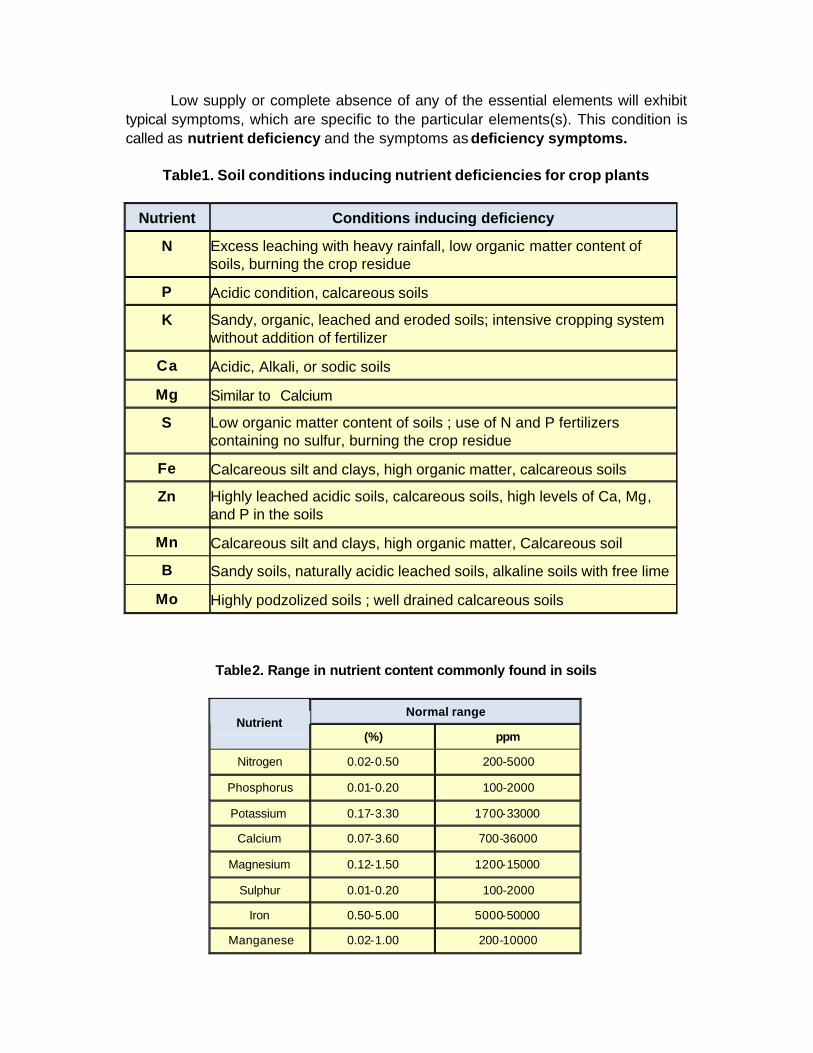
Low supply or complete absence of any of the essential elements will exhibit typical symptoms, which are specific to the particular elements(s). This condition is called as nutrient deficiency and the symptoms as deficiency symptoms.
Table1. Soil conditions inducing nutrient deficiencies for crop plants
Nutrient Conditions inducing deficiency
N Excess leaching with heavy rainfall, low organic matter content of soils, burning the crop residue
P Acidic condition, calcareous soils
K Sandy, organic, leached and eroded soils; intensive cropping system without addition of fertilizer
Ca Acidic, Alkali, or sodic soils
Mg Similar to Calcium
S Low organic matter content of soils ; use of N and P fertilizers containing no sulfur, burning the crop residue
Fe Calcareous silt and clays, high organic matter, calcareous soils
Zn Highly leached acidic soils, calcareous soils, high levels of Ca, Mg, and P in the soils
Mn Calcareous silt and clays, high organic matter, Calcareous soil
B Sandy soils, naturally acidic leached soils, alkaline soils with free lime
Mo Highly podzolized soils ; well drained calcareous soils
Table2. Range in nutrient content commonly found in soils
Normal range Nutrient
(%) ppm
Nitrogen 0.02-0.50 200-5000
Phosphorus 0.01-0.20 100-2000
Potassium 0.17-3.30 1700-33000
Calcium 0.07-3.60 700-36000
Magnesium 0.12-1.50 1200-15000
Sulphur 0.01-0.20 100-2000
Iron 0.50-5.00 5000-50000
Manganese 0.02-1.00 200-10000

Zinc 0.001-0.025 10-250
Boron 0.0005-0.015 5-150
Copper 0.0005-0.015 5-150
Chlorine 0.001 -0.10 10-1000
Cobalt 0.0001-0.005 1-50
Molybdenum 0.00002 -0.0005 0.2-5
Hidden Hunger
Hidden hunger refers to a situation in which a crop needs a nutrient yet has shown no deficiency symptoms. The nutrient content is above the deficiency symptom zone but still considerably needed for optimum crop production. With most nutrients on most crops, significant responses can be obtained even though no recognizable symptoms have appeared.
Figure. Hidden hunger is a term used to describe a plant that shows no obvious symptoms, yet the nutrient content is not sufficient to give the top profitable yield.
In absence or low supply of elements (deficiency), following common
symptoms generally develop in the plants. General Deficiency Symptoms 1. Chlorosis
It is a physiological symptom that occurs due to deficiency of mineral elements.(ex. N,K.Mg,Fe,Zn, Mn,S). Leaves become abnormally yellow or white due to reduction of chlorophyll contents.

2. Mottling It is a condition of plant surface marked with coloured spots due to anthocyanin pigmentation(Example. due to deficiency of N,K,Mg,P,S)
3. Necrosis It refers to the patch of dead tissues, due to deficiency of K,Mg,Zn,Ca, Mo,Mn,Fe,or B.
4. Bronzing Development of bronze/copper colour on the leaves and various parts (eg. K)
5. Die back Collapse of growing tip, affecting the youngest leaves and buds (Example . K, Ca,B or Cu)
6. Scorching Burning of tissues accompanied by light brown colour. (ex. K,Ca,B or Cu)
7. Firing Burning of tissues accompanied with dark brown or reddish brown colour (Example: Mg)
8. Rosetting Clustering of leaves due to reduced leaf size and shorter internodal distance (Example: Cu,K,Zn)
9. Distortion of leaves Irregular shaping of leaves like cupping, twisting, hooking or curling and also wavy margins. Cupping : B or Mo Twisting / Hooking : Zn/Ca/B Curling : Ca,B or Zn Head distortion : Cu (rice), Mn(Sunflower) Wavy : Zn
10. Gummosis Oozing out of cell sap in the form of gummous nature (Example: Cu as in coconut etc.)
Occurrence of Deficiency Symptoms Deficiency symptoms of various nutrient elements will appear either on older or on younger leaves depending on mobility of the nutrient. Thus, the relative mibility of the nutrient influences the site of appearance of the deficiency symptoms. Deficiency symptoms of mobile elements will appear on the older leaves because, these elements will move rapidly from older leaves to younger leaves. Example: N,P,K, Mg, Zn. On the other hand, the deficiency symptoms of the non-mobile elements will appear on the younge leaves because of their accumulation on the older leaves due to their immobile nature. Example: Ca, B, Cu, Mn, Fe and S.
Specific deficiency symptoms of various nutrient elements and their corrective measures are given below : 1. Nitrogen (N)
i. Symptoms first occur on the older leaves due to its mobility. ii. Plant growth is stunted and poorly developed (because protein content, cell division and cell enlargement are decreased) iii. Nitrogen deficiency causes yellowing (chlorosis) of leaves. Older leaves are affected first iv. Flowering and fruiting are reduced v. Protein and starch contents are decreased vi. Prolonged dormancy and early senescence appear vii. Root gets more lengthened as in whea t viii. Veins turn purple or red due to development of abundant anthocyanin pigment (Example: tomato, apple) ix. The angle between stem and leaves is reduced. x. Plants look so sickly and conspicuously pale that the condition is called as general starvation.
Corrective Measures
For correcting N deficiency, fertilizers like ammonium suphate, calcium nitrate, urea etc. are supplied. Foliar spray of 1-2% urea is a quick method of ameliorating N deficiency.
2. Phophorus (P)
i. Young plants remain stunted with dark blue green, or some times purplish leaves. ii. P deficiency may cause premature leaf fall iii. Dead necrotic areas are developed on leaves and fruits. iv. Leaves somtimes develop anthocyanin in veins and may become necrotic; leaves will be dark green in colour. v. Cambial activity is checked vi. Tillering of crops is reduced vii Dormancy is prolonged viii. Growth is retarded ix. Sickle leaf disease is caused in P deficiency, which is characterised by chlorosis adjacent to main veins followed by leaf asymmetry.
Corrective Measures Spray of 2% DAP or application of Phosphatic fertilizers will correct the deficiency. 3. Potassium (K) The deficiency symptoms vary with the degree of shortage of the element. i. In mild deficiency cases,
a. thin shoots may develop b. there may be restricted shoot growth
ii. In acute deficiency cases, a. shoots may die back, eventually plant may die b. Plants may become stunted with numerous tillers
c. there may be little or no flowering iii. Leaf will be dull or bluish green in colour. iv. Chlorosis occurs in interveinal regions (interveinal chlorosis) v. In older leaves, browning of tips (tip burns), marginal scorching (leaf scorch or development of brown spots near the margins occur). vi. Necrotic areas develop at the tip and margins of the leaf which curve downward. vii. In broad leaved plants, shortening of internodes and poor root system are important. viii. Two diseases are common:
a. Rosette : In beet, celery, carrot, pea, potato and cereals, bushy growth or rosette condition develops due to K deficiency. b. Die back : In acute deficiency cases, there is a loss of apical dominance and regeneration of lateral buds, which results in bushy growth. In prolonged cases, die back of laterals is also resulted.(Fruit crops)
Corrective Measures Supply of murate of potash or foliar spray of 1% potassium chloride is commonly used to overcome K deficiency. 4. Magnesium (Mg) i. Mg deficiency causes interveinal chlorosis. The older leaves are affected first and proceeds systematically towards the younger leaves. ii. Dead necrotic spots appear on the leaves. iii. Severely affected leaves may wither and shed or absciss without the withering stage. iv. Defoliation is quite severe v. Carotene content is reduced. vi. Stem becomes yellowish-green, often hard and woody. vii. Sand-drown diseas is common in tobacco due to its deficiency, which is characterised by the loss of colour at the tips of lower leaves and between the veins (interveinal). The veins remain green but in acute cases, entire leaf becomes nearly white. Corrective Measures Magnesium sulphate is usually applied for redressing the deficiency. The malady can be readily corrected as foliar spray @ 2% of MgSO4. 5. Calcium (Ca) Calcium ammonium nitrate (CAN) or superphosphate or gypsum is supplied in deficient soils. In Indian soils, Ca deficiency is not a serious problem. 6. Sulphur (S) i. Sulphur deficiency causes yellowing (Chlorosis) of leaves. Young leaves are affected first. ii. Tips and margins of leaves roll inward. iii. Marked decrease in leaf size, general paling with red or purple pigmentation are general symptoms. iv. Necrosis of young leaf tips develop v. Internodes are shortened’ vi. Apical growth is inhibited and lateral buds develop prematurely
vii. Young leaves develop orange, red or purple pigments. viii. Leaf tips are characteristically bent downwards. The leaf margins and tips roll inwards. (Example: tomato, tobacco and tea) x. Fruit formation is suppressed. xi. Sclerenchyma, xylem and collenchyma formation gets increased and hence the stem becomes unusually thick due to S deficiency. xii. Disease: The Tea Yellow disease is caused in tea plants growing in sulphur dificient soils. Corrective Measures Common fertilizers used for supplying nitrogen and phosphorus contain appreciable amount of sulphur sufficient to meet the crop requirement. In case of severe deficiency, gypsum is added to the soil @ 500Kg/ha. Micronutrients 7. Iron (Fe) i. Interveinal chlorosis of the younger leaves occurs. The veins remain green. ii. Leaf chlorosis may produce a mottled appearance. iii. Leaf may show complete bleaching or often becoming necrotic. iv. In extreme conditions, scorching of leaf margins and tips may occur v. Lime induced chlorosis is the common disease found in fruit trees like citrus. It is also fund in beet, spinach, brassicas and cereals. The younger leaves become white or yellowish white. Corrective Measures Foliar spray of 0.5% ferrous sulphate along with lime (50% requirement) will remove the deficiency in the plant and soil. Chelated iron compounds such as Fe-EDTA, give a very good response in ameliorating Fe deficiency. 8. Manganese (Mn) i. Deficiency causes interveinal chlorosis and necrotic spots of the leaf. ii. Dead tissue spots are found scattered over the leaf. iii. Severely affected tissues turn brown, the brown areas may also twist in the form of spirals and they may wither also. iv. Root system is often poorly developed and badly affected and the plants may die. v. Grain formation is also reduced and the heads may be blind ( as in sulphur) vi. Four diseases are found due to its deficiency :
a. Grey Speck also called as grey stripe, grey spot or dry spot found in oats, barely, rye and maize is the common disease of Mn deficiency. Grey spots or chlorotic spots appear on the lower half of the leaf which fuse together and form elongated brown streaks, found mostly in third or fourth leaves. b. Pahla blight of sugarcane Chlorotic spots develop as long streaks, commonly in young leaves. These chlorontic spots fuse together and turn red and coalesce to form long streaks from which lamina may split. C. Marsh spot of pea Brown, black spot or cavities develop on the internal surface of cotyledons and thus the disease appears in the seeds.

d. Speckled yellow of sugar beet It is characterised by interveinal chlorosis in the leaves and leaf margin may curl upward over the upper surface of leaf. Corrective Measures Foliar spray of 0.5% manganous sulphate plus 50% lime requirement is quite effective and it should be applied in the early stage of the crop. Soil application of 15-30 kg MnSO4 per ha (mixed with sand)is sufficient.
9. Copper (Cu) i. it causes necrosis of the tip of the young leaves. ii. Both vegetative and reproductive growth are retarded. iii. Wilting of terminal shoots occur which is followed by frequent death iv. Leaf colour is often faded due to reduction of carotene and other pigments. v. Foliage shows burning of margins or chlorosis or rosetting and multiple bud formation. vi. Gumming may also occur (gummosis) vii. Younger leaves wither and show marginal chlorosis (yellowish grey) of tips. It is called as Yellow tip or reclamation disease. viii. Following two diseases are common : a. Exanthema or die back of fruit tree : It is commonly found in citrus, plum, apple and pear. The symptoms include formation of strong water-shoots bearing large leaves, gummous tissue or the bark and longitudinal breaks. Fruits become brown, glossy and splitted. Affected shoots loose their leaves and die back and lateral shoots produce bunchy appearance. b. Reclamation disease: It is also called as White Tip disease and is found in legumes, cereals, oats and beet. The tips of leaves become chlorotic followed by a failure of the plants to set seed. Corrective Measures Foliar spray of 0.5% of CuSO4 is recommended. 10. Zinc (Zn) i. Older leaves show chlorosis which sta rts from tips and the margins ii. Leaves become leathery iii. Plants show rosetting due to shortening of internodes and premature shedding. iv. Whitening of upper leaves in monocots and chlorosis of lower leaves in dicots are often found. v. Leaf margins d istorted, become twisted or wavy which later curl and look sickle shaped (Sickle leaf) vi. Seed production and fruits size is greatly reduced. vii. The following diseases are commonly notice : a. Khaira of paddy : The entire older leaves show rusty brown appearance (due to chlorosis) and ultimately die. b. White bud (tip) of maize : Unfolded newer leaves are often pale yellow to while. There is appearance of light yellow streaks between the veins of older leaves followed by white necrotic spots. c. Rosette of fruit trees : It is also called as little leaf disease. Yellow mottling of leaves, reduction of leaf size with rosette appearance (due to reduced internodal distance) and die back of the affected branches are symptoms of the disease.

d. Frenching of citrus : Initially, yellow spots develop between the veins. Leaves become progressively smaller and develop chlorophyll at the basal end of mid rib. Corrective Measures Foliar spray of 0.5% ZnSO4 twice at 7-10 days interval during early stages of growth will alleviate the problem. Also, soil application of 25 kg ZnSO4 per ha is also found beneficial. 11. Molybdenum (Mo) i. Deficiency causes chlorotic interveinal mottling of the older leaves. ii. Leaves often show light yellow chlorosis and leaf blades fail to expand. iii. In acute deficiency cases, necrosis of leaf tissues occurs. iv. Flower formation is inhibited. v. Failure of grain formation occurs (as in oats) vi. Its deficiency causes two diseases :
a. Whiptail of Cauliflower and Brassica : The symptoms begin as appearance of translucent areas near the midrib which become ivory tinted or necrotic. The leaf margins become ragged with upward curling. Before the death of the growing point, the leaf elongates and lamina remains suppressed thus gives a typical whip tail condition. b. Scald of legumes : The leaf shows paling, wilting, marginal rolling or scorching.
Corrective Measures The Mo deficiency is commonly found in cauliflower, legumes, oats and other brassicas which can be corrected by soil application of 0.5 to1.0 Kg/ha sodium or ammonium molybdate or by its foliar spray@ 0.01-0.02% conc. 12. Boron (B) i. It causes death of shoot tip ii. Flower formation is suppressed. iii. Root growth is stunted. iv. Leaves become coppery in texture. v. Plants become dwarf, stunted with apical meristem blacken and die followed by general breakdown of meristematic tissue. vi. Terminal leaves become necrotic and shed prematurely vii. Leaves show symptoms like distortion such as cupping and curling, appearance of white stripe, scorching, pimpling, splitted midrib and reduced growth. viii. Stem shows symptoms like die-back of apex, abnormal tillering, appearance of various forms of deformities such as curling and brittle lesions, pimpling etc. ix. Fruits are severely deformed and develop typical cracking or splitting. x. Following diseases are commonly found due to B deficiency :
a. Heart rot of sugar beet and marigold b. Canker and internal black spot of garden pea c. Browning of cauliflower d. Top sickness of tobacco e. Hard fruit of citrus.

Corrective Measures Foliar spray of 0.2% borax/boric acid will be effective for quick recovery. Liming of soil should be strictly avoided when boron-containing fertilizers are applied. The following table provides information on indicator plants for nutrient deficiency
Nutrient Element
Indicator crop plant for deficiency
Nitrogen Maize , Sorghum ,Leguminous plants Phosphorus Tomato ,Maize, Lucerne, Cereals, Duranta Potassium Maize, Cotton, Potatoes, Banana, Cucurbits
Sulphur Lucerne , Clover , Cereals, Tea Zinc Maize, Tomatoes , Potatoes, Beans, Citrus
Copper Citrus Iron Ornamental plants, Ixora, Acacia, Eucalyptus,
Boron Lucerne, Coconut, Guava Manganese Citrus
Molybdenum Cauliflower, Cabbage
Identification of Visual Leaf and Plant Nutrient Element – Deficiency and Excess Symptoms
Element
/status
Visual symptoms
Nitrogen (N)
Deficiency Light green leaf and plant color with the older leaves turning yellow, leaves that will eventually turn brown and die. Plant growth is slow, plants will be stunted, and will mature early.
Excess Plants will be dark green in color and new growth will be succulent; susceptible if subjected to disease and insect infestation; and subjected to drought stress, plants will easily lodge. Blos som abortion and lack of fruit set will occur.
Ammonium toxicity Plants fertilized with ammonium -nitrogen (NH4- N) may exhibit ammonium-toxicity symptoms, with carbohydrate depletion and reduced plant growth. Lesions may occur on plant stems, there may be a downward cupping of the leaves, and a decay of the conductive tissue at the base of the stem with wilting of the plants under moisture stress. Blossom -end rot of fruit will occur and Mg deficiency symptoms may also occur.
Phosphorus (P) Deficiency Plant growth will be slow and stunted, and the older leaves will have a
purple coloration, particularly on the underside. Excess Phosphorus excess will not have a direct effect on the plant but may show
visual deficiencies of Zn, Fe, and Mn. High P may also interfere with the normal Ca nutrition, with typical Ca deficiency symptoms occurring.
Potassium (K) Deficiency On the older leaves, the edges will look burned, a symptom known as
scorch. Plants will easily lodge and be sensitive to disease infestation. Fruit and seed production will be impaired and of poor quality.
Excess Plants will exhibit typical Mg, and possibly Ca deficiency symptoms due to

a cation imbalance Calcium (Ca)
Deficiency The growing tips of roots and leaves will turn brown and die. The edges of the leaves will look ragged as the edges of emerging leaves stick together. Fruit quality will be affected with the occurrence of blossom-end rot on fruits.
Excess Plants may exhibit typical Mg deficiency symptoms, and when in high excess, K deficiency may also occur.
Magnesium (Mg) Deficiency Older leaves will be yellow in color with interveinal chlorosis (yellowing
between the veins) symptoms. Plant growth will be slow and some plants may be easily infested by disease.
E xcess Results in a cation imbalance showing signs of either a Ca or K deficiency. Sulfur (S)
Deficiency A general overall light green color of the entire plant with the older leaves being light green to yellow in color as the deficiency intensifies.
Exc ess A premature senescence of leaves may occur. Boron (B)
Deficiency Abnormal development of the growing points (meristematic tissue) with the apical growing points eventually becoming stunted and dying. Rowers and fruits will abort. For some grain and fruit crops, yield and quality is significantly reduced.
Excess Leaf tips and margins will turn brown and die. Chlorine (Cl)
Deficiency Younger leaves will be chlorotic and plants will easily wilt. For wheat, a plant disease will infest the plant whe n Cl is deficient.
Excess Premature yellowing of the lower leaves with burning of the leaf margins and tips. Leaf abscission will occur and plants will easily wilt.
Copper (Cu) Deficiency Plant growth will be slow and plants stunted with distortion of the young
leaves and death of the growing point. Excess An Fe deficiency may be induced with very slow growth. Roots may be
stunted. Iron (Fe)
Deficiency Interveinal chlorosis will occur on the emerging and young leaves with eventual bleaching of the new growth. When severe, the entire plant may be light green in color.
Excess A bronzing of leaves with tiny brown spots on the leaves, a typical symptom frequently occurring with rice.
Manganese (Mn) Deficiency Inter-veinal chlorosis of young leaves while the leaves and plants remain
generally green in color. When severe, the plants will be stunted. Excess Older leaves will show brown spots surrounded by a chlorotic zone and
circle. Molybdenum (Mo)
Deficiency Symptoms will frequently appear similar to N deficiency. Older and middle leaves become chlorotic first, and In some instances, leaf margins are rolled and growth and flower formation are restricted.
Excess Not of common occurrence. Zinc (Zn)
Deficiency Upper leaves will show interveinal chlorosis with an eventual whiting of the affected leaves. Leaves may be small and distorted with a rosette form.
Excess Fe deficiency will develop.


PHYSIOLOGICAL DISORDERS IN CROPS Following physiological disorders are often observed in different crops. Symptoms of various disorders, causal factors and remedial measures for redressing different disorders are: 1. MANGO
1. Softnose fruits 2. Malformation of inflorescence 3. Black tip of Fruits. 4. Alternate bearing
2. BANANA 1. Seediness (Kottaivazhai) in fruits 2. Goose flesh of fruits 3. Yellow pulp of fruits 4. Degrain of bunch 5. Finger drop
3. GRAPES Coulure in grapes
4. COCONUT Crown chocking
5. GUAVA Fatio disease
6. CITRUS 1. Die-Back 2. June Drop 3. Fruit Cracking
7. PINE APPLE Crook-Neck Disease
8. APPLE 1. Internal Bark Necrosis (=Apple Measles) 2. Bitter Pit Detailed description of various physiological disorders and their corrective measures are described in the following pages : A. MANGO 1. Soft Nose Fruits This disorder is caused due to excess N application of trees. In acid sandy soil, the incidence is about 7% in trees receiving lower N levels and increases to 78% on trees receiving 10 times more N fertilizer. Remedy Disorder reduced by maintaining higher Ca level in leaf (2.5%) by applying either Calcium Nitrate or Gypsum or Lime Stone. 2. Malformation in Inflorescence: Causes : Malformation disorder increased in trees receiving either P and K or P alone. Remedy Application of N,P and K in the ratio of 9:3 :3 reduces the incidence of floral malformation.

Related Documents