Acetaminophen-mediated cardioprotection via inhibition of the mitochondrial permeability transition pore-induced apoptotic pathway Norell M. Hadzimichalis, Sunanda S. Baliga, Roseli Golfetti, Kathryn M. Jaques, Bonnie L. Firestein, and Gary F. Merrill* Department of Cell Biology and Neuroscience, Division of Life Sciences, Rutgers University, Piscataway, New Jersey 08854 *To whom all correspondence should be addressed: Gary F. Merrill 604 Allison Road Piscataway, NJ 08854 732-445-2320 phone 732-445-5870 fax [email protected] Running head: Mechanisms of acetaminophen-mediated cardioprotection Page 1 of 38 Copyright Information Articles in PresS. Am J Physiol Heart Circ Physiol (October 5, 2007). doi:10.1152/ajpheart.00947.2007 Copyright © 2007 by the American Physiological Society.

Welcome message from author
This document is posted to help you gain knowledge. Please leave a comment to let me know what you think about it! Share it to your friends and learn new things together.
Transcript
Acetaminophen-mediated cardioprotection via inhibition of the mitochondrial permeability transition pore-induced apoptotic pathway
Norell M. Hadzimichalis, Sunanda S. Baliga, Roseli Golfetti, Kathryn M. Jaques, Bonnie L. Firestein, and Gary F. Merrill*
Department of Cell Biology and Neuroscience, Division of Life Sciences, Rutgers University, Piscataway, New Jersey 08854
*To whom all correspondence should be addressed: Gary F. Merrill 604 Allison Road Piscataway, NJ 08854
732-445-2320 phone 732-445-5870 fax [email protected]
Running head: Mechanisms of acetaminophen-mediated cardioprotection
Page 1 of 38
Copyright Information
Articles in PresS. Am J Physiol Heart Circ Physiol (October 5, 2007). doi:10.1152/ajpheart.00947.2007
Copyright © 2007 by the American Physiological Society.

1
ABSTRACT
Our laboratory has previously reported that acetaminophen confers functional
cardioprotection following cardiac insult, including ischemia/reperfusion,
hypoxia/reoxygenation, and exogenous peroxynitrite administration. In the current study,
we further examined the mechanism of acetaminophen-mediated cardioprotection
following ischemia/reperfusion injury. Langendorff-perfused guinea pig hearts were
exposed to acute treatment with acetaminophen (0.35mM) or vehicle beginning at 15
minutes of a 30-minute baseline stabilization period. Low flow global myocardial
ischemia was subsequently induced for 30 minutes followed by 60 minutes of
reperfusion. At the completion of reperfusion, hearts were homogenized and separated
into cytosolic and mitochondrial fractions. Mitochondrial swelling and mitochondrial
cytochrome c release were assessed and found to be significantly and completely reduced
in acetaminophen- versus vehicle-treated hearts following reperfusion. In a separate
group of hearts, ventricular myocytes were isolated and subjected to fluorescence-
activated cell sorting. Acetaminophen-treated hearts showed a significant decrease in late
stage apoptotic myocytes when compared to vehicle-treated hearts following injury
(58±1% vs. 81±5, respectively). These data, together with electron micrograph analysis,
suggest that acetaminophen mediates cardioprotection, in part, via inhibition of the
mitochondrial permeability transition pore and subsequent apoptotic pathway.
Key words: Mitochondrial swelling, apoptosis, cytochrome c, myocardial
ischemia/reperfusion, mitochondrial permeability transition pore
Page 2 of 38
Copyright Information

2
INTRODUCTION
Considerable attention has been given to the potential detrimental effects of Cox-
2 specific analgesics in the mammalian cardiovascular system. However, there have been
few rigorous physiological investigations on the effects of other analgesics on the
cardiovascular system of mammals, including humans. Acetaminophen (paracetamol,
APAP), a popular non-NSAID, has historically been employed as an analgesic antipyretic
agent. In recent investigations, it has also been established as an effective
cardioprotective agent during ischemia/reperfusion, hypoxia/reoxygenation, exogenous
peroxynitrite administration, and experimentally induced myocardial infarction (17, 18,
30-33, 39, 40, 50). We have previously reported both chronic and acute acetaminophen
treatment (0.35mM) to be cardioprotective following ischemia/reperfusion in the isolated
perfused guinea pig myocardium (17, 18). High performance liquid chromatography
analysis shows that treatment with acetaminophen at this molarity yields arterial and
venous concentrations of 45-50 µg/ml (44). These values fall within the range of
therapeutic human plasma concentrations (10-100 µg/ml) and well below those
concentrations resulting in hepatotoxicity (≥300 µg/ml; (21, 36, 37).
Additional studies from our laboratory have demonstrated that acute
acetaminophen treatment also confers protection in a canine model of myocardial
infarction (18, 33). However, while structural, functional, and biochemical evidence of
acetaminophen-mediated cardioprotection exist, mechanistic data are noticeably absent.
It is currently believed that the mechanism of action may involve antioxidant properties
of this drug conveyed by its phenolic structure; however, additional work is required in
order to further delineate the pathway for this observed protection (18, 31, 32).
Page 3 of 38
Copyright Information

3
The goal of the present study was to examine the mechanistic pathway by which
acetaminophen mediates cardioprotection. We found that in our model,
ischemia/reperfusion causes a significant increase in mitochondrial permeability
transition pore (MPTP) opening and mitochondrial cytochrome c release, and that
acetaminophen treatment during injury significantly and completely blocks these effects.
In addition, we report that ischemia/reperfusion causes a significant increase in late stage
apoptotic myocytes in both vehicle- and acetaminophen-treated hearts. However, while
acetaminophen significantly decreases late stage apoptotic myocytes when compared to
vehicle-treated hearts following injury, its inhibition was not complete. The incomplete
attenuation of late stage apoptosis in response to treatment with acetaminophen implies
that although this drug is successful at completely inhibiting MPTP opening and
cytochrome c release, additional pathways of apoptosis are still active in the
ischemia/reperfused Langendorff heart. These results confirm prior reports of
acetaminophen-mediated cardioprotection and suggest a mechanistic pathway to explain
this protection following ischemia/reperfusion.
Page 4 of 38
Copyright Information
4
MATERIALS AND METHODS Animals and Langendorff preparation
Hartley strain male guinea pigs (400 ± 25g) were obtained from Elm Hill
Laboratories (Wilmington, MA, USA) and allowed a minimum of three days to acclimate
to their new environment. Following IACUC review and approval, guinea pigs were
anesthetized using isoflurane in accordance with National Institutes of Health and United
States Department of Agriculture guidelines.
Hearts were isolated and perfused in situ via the cannulated aorta and
subsequently extracted and attached to a Langendorff perfusion apparatus as previously
described (9, 10, 30). Pacing electrodes were placed at the base of the right ventricle to
control heart rate at approximately 200 beats per minute (model S44, Grass-Telefactor;
West Warwick, RI, USA), and physiologic heart temperature was monitored using a
thermistor probe (model BAT-12, Physitemp; Clifton, NJ, USA). Coronary perfusion
pressure was controlled hydrostatically (55 ± 5 mmHg).
Perfusate and ischemia/reperfusion protocol
Hearts were perfused with a modified Krebs-Henseleit physiological salt
solution/buffer (KHB) containing (in mM): 128.0 NaCl, 4.7 KCl, 1.5 MgSO4 • 7H2O, 2.5
CaCl2, 1.2 KH2PO4, 24.9 NaHCO3, 10.0 glucose, 2.0 pyruvate, and 200 µU/ml insulin.
Perfusate was warmed to 37ºC, equilibrated with a 95% O2, 5% CO2 gas mixture (pH
7.40 ± 0.02), and delivered from a water-jacketed perfusion reservoir. Flow was allowed
to vary naturally and continuously monitored ultrasonically (model T106 flow meter,
Transonic Systems; Ithaca, NY, USA).
Page 5 of 38
Copyright Information

5
Guinea pigs were randomly assigned to vehicle (KHB) or acetaminophen (0.35
mM dissolved in KHB) treatment groups. Following extraction and suspension from the
Langendorff apparatus, all hearts remained untreated and were perfused with KHB for
the first 15 minutes of the baseline stabilization period. Subsequently, hearts were treated
with either acetaminophen or vehicle (added to the perfusate reservoir) for the remainder
of the 30-minute baseline period and for the duration of the Langendorff perfusion. Low
flow global myocardial ischemia (1 ml/min) was then induced for 30 minutes followed
by 60 minutes of reperfusion. Animal choice and age and the use of low flow ischemia
were employed in order to be consistent with previous reports from our laboratory
establishing the functional cardioprotective capacity of acetaminophen (31, 32).
Monitored variables included heart rate (HR; beats/minute), coronary perfusate
flow (CPF; ml/min/g), and coronary perfusion pressure (CPP; mmHg). A data
acquisition system (model 214, iWorx/CB Sciences; Dover, NH, USA) in series with a
personal computer (Compaq Evo running LabScribe software version 6.0) was used to
record monitored variables. Metabolic data including pH, PO2 (mmHg), and PCO2
(mmHg) were recorded using a standard blood-gas analyzer (model 248, Chiron
Diagnostics; Norwood, MA, USA).
Myocardial homogenization and fractionation
Hearts were randomly divided into vehicle and acetaminophen treatment groups
and exposed to the ischemia/reperfusion protocol described above. Monitored variables
and metabolic data were collected just prior to perfusion termination (i.e. at 15 minutes
baseline for control hearts or the end of reperfusion). Following termination of
Page 6 of 38
Copyright Information

6
Langendorff perfusion, hearts were crushed and immersed in homogenization buffer (10
ml/g) containing (in mM): 210.0 mannitol, 7.0 sucrose, and 5.0 4-
morpholinopropanesulfonic acid, pH 7.4., 37ºC, 1 tablet/10 ml buffer protease inhibitor
tablets (complete mini, Roche Diagnostics; Indianapolis, IN, USA). Hearts were then
homogenized using both Polytron blade (model PT 2100, Kinematica; Littau-Lucerne,
Switzerland) and Teflon (model JR4000, Arrow Engineering; Hillside, NJ, USA)
homogenizers. Separation of cytosolic and mitochondrial fractions was modified from
previously described procedures (6, 46). The homogenate was centrifuged at 1000 x g
for 10 minutes at 4°C and the resulting supernatant was centrifuged at 7,000 x g for 10
minutes at 4°C. The pellet from the second centrifugation represented the mitochondrial
fraction and was resuspended in 10 mM sodium phosphate, pH 9.0. The supernatant
represented the cytosolic fraction. An additional group of hearts was homogenized
following 15 minutes of baseline perfusion as a control.
Mitochondrial swelling
Mitochondrial suspensions were assayed spectrophotometrically (540 nm) at
25°C for changes in light scattering (8, 15, 25, 41). Mitochondrial fractions of heart
homogenate were assessed following ischemia/reperfusion from both vehicle- and
acetaminophen-treated hearts following a Bradford assay to determine total protein
concentration. Light absorbance values were expressed as a percentage with respect to
the average of baseline hearts.
Myofibrillar ultrastructure
Page 7 of 38
Copyright Information
7
A separate group of hearts was used to assess myofibrillar ultrastructure as
previously described (18). Hearts were randomly assigned to one of two termination
groups (15 minutes baseline or reperfusion) corresponding to the experimental period
following which the Langendorff perfusion would be terminated and heart subjected to
fixation. The reperfusion group was further divided into either vehicle- or
acetaminophen-treatment groups.
Hearts were perfused with Karnovsky’s fixative for two minutes at the end of
baseline or reperfusion conditions. Hearts were then submerged in fixative and 2-3 mm3
blocks of myocardium were removed longitudinally from the anterior free wall of the left
ventricle midway between the left ventricular and left anterior descending branches of the
left main coronary artery, equidistant from base to apex. Blocks were subsequently fixed
using 1% osmium tetroxide and dehydrated in graded ethanol (17, 40). Samples were
embedded in Epon-Araldite cocktail, sectioned with a diamond knife ultramicrotome
(model LKB-2088, LKB; Bromma, Sweden), and viewed with an electron microscope
(model JEM-100CXII, JEOL USA; Peabody, MA), using standard protocols (3).
Mitochondrial cytochrome c release
Following termination of Langendorff perfusion, hearts were freeze-clamped in
liquid nitrogen using a modified Wollenberger clamp and stored at -80ºC until
homogenization (39). A Bradford assay (Biorad Protein Assay, Biorad; Hercules, CA,
USA) was used to determine total protein concentration. Cytosolic and mitochondrial
fractions from baseline, vehicle- and acetaminophen-treated ischemia/reperfused heart
homogenates were then loaded randomly into wells (i.e. the investigator was blinded for
Page 8 of 38
Copyright Information

8
band density quantification). Proteins were resolved on a 15% SDS polyacrylamide gel
and transferred to a polyvinylidene difluoride (PVDF) membrane. The membrane was
then probed with a mouse monoclonal antibody to cytochrome c (clone 7H8.2C12,
1:1000; Stressgen Bioreagents; Victoria, BC, Canada). Cytosolic and mitochondrial
fractions were further probed for rabbit anti-α-actin (1:2500; Sigma-Aldrich; St. Louis,
MO, USA) and rabbit anti-voltage-dependent anion channel/Porin (VDAC, 1:2500;
Sigma-Aldrich; St. Louis, MO, USA), respectively, as loading controls. Film was
scanned, and quantification was carried out through optical density analysis using Scion
image software.
Isolation of ventricular myocytes and fluorescence-activated cell sorting
Hearts were randomly divided into vehicle and acetaminophen treatment groups
and exposed to the ischemia/reperfusion protocol described above. Hemodynamic and
metabolic data were collected at 15 minutes baseline, 30 minutes ischemia, and 60
minutes reperfusion. Isolation of ventricular myocytes was a modification of previously
described methods (24, 35). Briefly, following ischemia/reperfusion, hearts were
perfused with calcium-free KHB for 2-3 minutes to arrest contractions. Hearts were then
perfused with 0.08% collagenase Type 2 (Worthington Biochemical Corporation;
Lakewood, NJ, USA) dissolved in calcium-free KHB in a re-circulating mode for
approximately 10-15 minutes. Subsequently, hearts were removed from the perfusion
apparatus and ventricles cut longitudinally into 6-8 slices and incubated and mildly
agitated with 15 ml of KHB plus 0.08% collagenase at 37ºC for five minutes. Cells were
Page 9 of 38
Copyright Information

9
centrifuged at 10,000 x g for 50 seconds and washed two times in KHB. An additional
group of hearts was digested following 15 minutes of baseline perfusion as a control.
Following isolation, myocytes were re-suspended in annexin V binding buffer,
loaded with annexin V-fluorescein isothiocyanate (FITC) and propidium iodide (PI), and
analyzed using a fluorescence-activated cell sorter (FACS model FC500 flow cytometer,
Beckman Coulter; Fullerton, CA, USA) according to the manufacturer’s protocol
(Vybrant Apoptosis Assay Kit #3, Molecular Probes; Carlsbad, CA, USA). Early and
late stage apoptotic myocytes were characterized as annexin V-FITC or both annexin V-
FITC and PI positive, respectively.
Statistics
Reported values are means ± SEM. Data were analyzed using an ANOVA
followed by Tukey’s Multiple Comparison Test (InStat, GraphPad; San Diego, CA).
Significance was accepted at p < 0.05 in all cases.
Page 10 of 38
Copyright Information

10
RESULTS
Hemodynamic and metabolic parameters
Hemodynamic and metabolic data were collected following 15 minutes baseline,
ischemia, and reperfusion for hearts subjected to myocyte isolation and just prior to
perfusion termination (following baseline or reperfusion) for hearts subjected to freeze-
clamping and homogenization. There were no significant differences between vehicle-
and acetaminophen-treated hearts or between baseline and reperfused hearts during any
sample time. Expected hemodynamic differences in CPF were observed between
baseline and ischemic hearts in the myocyte isolation studies (Table 1).
Acetaminophen treatment inhibits mitochondrial swelling following myocardial
ischemia/reperfusion
Decreases in light absorbance are representative of increases in mitochondrial
matrix volume as a result of the opening of MPTPs, subsequent water influx, and
mitochondrial swelling (27, 42). In the present study, we examined differences in light
absorbance of isolated mitochondria following baseline and ischemia/reperfusion as an
index of MPTP opening. Mitochondrial and cytosolic fractions were each probed for
VDAC, a mitochondrial outer membrane protein, to confirm mitochondrial membrane
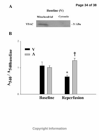
integrity. The lack of VDAC in the cytosolic fraction of vehicle-treated baseline heart
homogenates, compared to its presence in mitochondrial fractions, demonstrates that
mitochondrial membranes were intact (Figure 1A). Our spectrophotometric results
indicate a significant decrease in mitochondrial light absorbance of vehicle-treated
ischemia/reperfused mitochondrial fractions (0.66±0.04) when compared to either
Page 11 of 38
Copyright Information

11
baseline or post-reperfusion acetaminophen-treated mitochondrial fractions (1.27±0.09).
However, there were no significant changes in light absorbance between acetaminophen-
treated mitochondrial fractions following ischemia/reperfusion and baseline values,
suggesting that acetaminophen treatment attenuates ischemia/reperfusion-induced
mitochondrial swelling via inhibition of the mitochondrial permeability transition pore
(Figure 1B).
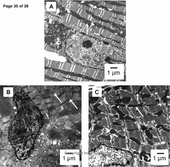
To assess whether acetaminophen preserves myofibrillar ultrastructure during
cardiac ischemia/reperfusion, we examined electron micrographs in both vehicle- and
acetaminophen-treated hearts following injury. As shown in Figure 2, myofibrillar
ultrastructure from vehicle-treated hearts displayed extensive post-reperfusion tissue
damage when compared to myocardial sections from either baseline or acetaminophen-
treated hearts. As indicated by arrows, mitochondria from left ventricular free wall
sections appear dense and intact in baseline and acetaminophen-treated
ischemia/reperfused hearts. However, mitochondria from vehicle-treated
ischemia/reperfused hearts are visually swollen and structurally more rounded. These
data further support the conclusion that acetaminophen treatment inhibits MPTP-induced
mitochondrial swelling following ischemia/reperfusion.
Molecular consistency between vehicle-treated hearts
Previous studies from our laboratory that have examined the effects of
acetaminophen following cardiac injury have reported mostly descriptive and functional
data (17, 18, 30, 31, 40). Drug- and vehicle-treated hearts were considered similar if the
hemodynamic and metabolic parameters collected at baseline were not statistically
Page 12 of 38
Copyright Information

12
different. In the current study, we further explored the molecular consistency between
discrete Langendorff preparations at baseline by comparing cytosolic cytochrome c
content. We found quantitative consistency in cytosolic cytochrome c content between
vehicle-treated hearts following 15 minutes of baseline perfusion (Figure 3A). These
data show the first evidence that our preparations are biochemically consistent.
Acetaminophen treatment inhibits mitochondrial cytochrome c release following
myocardial ischemia/reperfusion
Mitochondrial cytochrome c release is a known trigger for the intrinsic apoptotic
cascade (49). In the current study, we analyzed cytosolic and mitochondrial cytochrome
c content following baseline and ischemia/reperfusion. Our results indicate a significant
increase in cytochrome c release from mitochondria to the cytosol following
ischemia/reperfusion in vehicle-treated hearts (Figure 3B and C). In addition, therapeutic
concentrations of acetaminophen administered beginning at 15 minutes of baseline and
continuing throughout the ischemia/reperfusion protocol result in significant and
complete attenuation of cytochrome c release when compared to vehicle-treated hearts.
Cytosolic cytochrome c levels (normalized to α-actin and baseline hearts) were
11.15±2.18 and 1.21±0.48 in vehicle- and acetaminophen-treated hearts, respectively.
No differences were noted between mitochondrial or cytosolic cytochrome c content of
acetaminophen-treated hearts when compared to corresponding baseline samples (Figure
3B-D). These data show that acetaminophen treatment significantly and completely
inhibits the mitochondrial cytochrome c release normally observed following myocardial
ischemia/reperfusion.
Page 13 of 38
Copyright Information
13
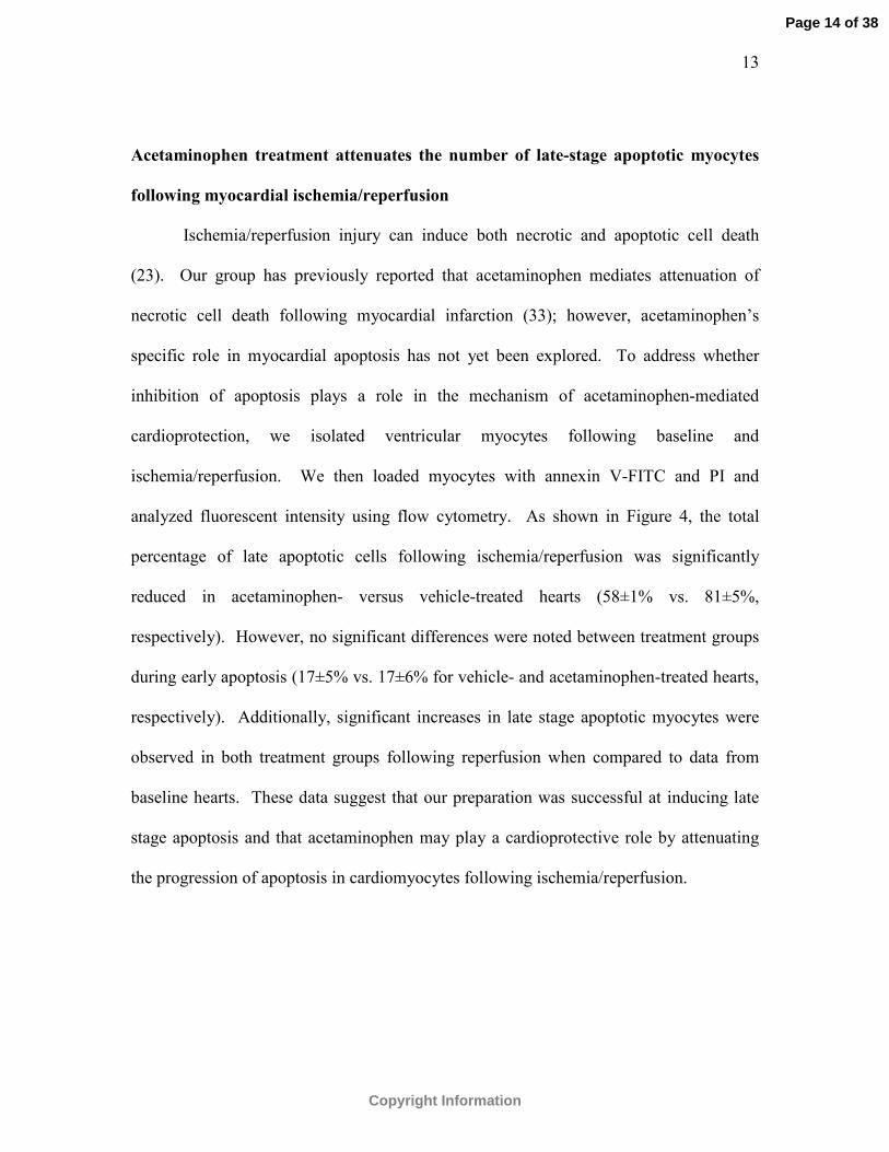
Acetaminophen treatment attenuates the number of late-stage apoptotic myocytes
following myocardial ischemia/reperfusion
Ischemia/reperfusion injury can induce both necrotic and apoptotic cell death
(23). Our group has previously reported that acetaminophen mediates attenuation of
necrotic cell death following myocardial infarction (33); however, acetaminophen’s
specific role in myocardial apoptosis has not yet been explored. To address whether
inhibition of apoptosis plays a role in the mechanism of acetaminophen-mediated
cardioprotection, we isolated ventricular myocytes following baseline and
ischemia/reperfusion. We then loaded myocytes with annexin V-FITC and PI and
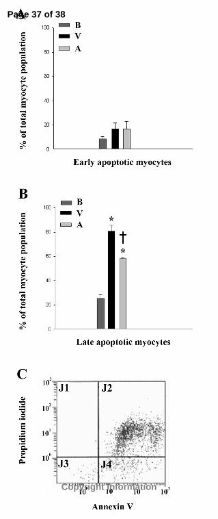
analyzed fluorescent intensity using flow cytometry. As shown in Figure 4, the total
percentage of late apoptotic cells following ischemia/reperfusion was significantly
reduced in acetaminophen- versus vehicle-treated hearts (58±1% vs. 81±5%,
respectively). However, no significant differences were noted between treatment groups
during early apoptosis (17±5% vs. 17±6% for vehicle- and acetaminophen-treated hearts,
respectively). Additionally, significant increases in late stage apoptotic myocytes were
observed in both treatment groups following reperfusion when compared to data from
baseline hearts. These data suggest that our preparation was successful at inducing late
stage apoptosis and that acetaminophen may play a cardioprotective role by attenuating
the progression of apoptosis in cardiomyocytes following ischemia/reperfusion.
Page 14 of 38
Copyright Information

14
DISCUSSION
With the drastic rise in heart disease, the need for preventative cardiac care has
become essential. Many groups have investigated the protective capacity of various
compounds in inhibiting myocardial ischemia/reperfusion-induced injury. Varga and
colleagues (48) investigated the effects of pretreatment with dexamethasone, a potent
glucocorticoid, on post-ischemia/reperfusion. They reported that dexamethasone inhibits
ventricular fibrillation via attenuation of mitochondrial cytochrome c release. Studies by
Kovacs and colleagues (28) used non-specific caspase administration at the onset of
reperfusion to maintain cardiac function and limit both infarct size and apoptosis.
Additional reports from Das et al. (12) examined the cardioprotective effects of
pretreatment with palm tocotrienol, a vitamin E isomer, following myocardial
ischemia/reperfusion. They demonstrated that treatment with tocotrienols, derived from a
tocotrienol-rich fraction of palm oil, results in the ability to attenuate
ischemia/reperfusion-induced damage via inhibition c-Src phosphorylation and
maintenance of proteasomal activity.
In this study, we examined the mechanistic basis for reported acetaminophen-
mediated functional cardioprotection. Previous studies show that in an in vivo canine
preparation of myocardial infarction, acetaminophen treatment results in a significant
reduction of necrotic tissue (33). In the current study, we proposed that acetaminophen
might also have an effect on the mitochondrial pathway of apoptosis following
ischemia/reperfusion. Specifically, we explored whether therapeutic concentrations of
acetaminophen were able to attenuate MPTP opening, cytochrome c release, and
apoptotic cell death. The major finding of our study is that, following myocardial
Page 15 of 38
Copyright Information
15
ischemia/reperfusion, acetaminophen treatment completely blocks opening of the MPTP
and mitochondrial swelling as well as cytochrome c release from mitochondria.
Furthermore, although acetaminophen attenuates late stage apoptosis, it does not
completely block it. These results suggest that acetaminophen inhibits the MPTP-
induced pathway of apoptosis; however, other pathways leading to apoptosis may not be
affected by acetaminophen.
During a procedure to reestablish blood supply to an ischemic myocardium, one
of the main concerns is the oxidative stress that occurs due to the formation of oxygen
radicals and other reactive oxygen species (ROS; 2, 14, 47). When accompanied by
additional post-reperfusion environmental conditions, including mitochondrial matrix
calcium overload, adenine nucleotide depletion, and elevated phosphate concentrations,
permeability transition pores switch to the open conformation thus triggering the intrinsic
apoptotic cascade (19). Although endogenous antioxidant systems are capable of
minimizing ROS-induced tissue injury under physiological conditions, they are
insufficient to neutralize ROS following ischemia/reperfusion insult (4, 38). Previous
reports demonstrate the efficacy of exogenously administered antioxidants in preventing
myocardial ischemia/reperfusion related injury in the Langendorff perfused heart,
including inhibition of MPTP opening (1, 5, 22, 26, 45).
Acetaminophen, when taken at therapeutic concentrations, has been established as
a safe antipyretic and analgesic drug (36). More recently, this compound has also been
established as an effective cardioprotective agent during myocardial ischemia/reperfusion
injury (19, 30-32). Mechanistically, the phenolic hydroxyl group of acetaminophen
likely donates its hydrogen atom to aid in the reported reduction of free radicals, namely
Page 16 of 38
Copyright Information

16
peroxynitrite and hydroxyl radicals, post-reperfusion (30-32, 36). Ischemia/reperfusion-
induced oxidative stress is a well-known trigger for mitochondrial permeability transition
pore opening, mitochondrial cytochrome c release, and downstream apoptotic cell death
pathway activation (16, 19, 34, 49). We hypothesize that acetaminophen-mediated
inhibition of ROS generation results, in part, in attenuation of reperfusion-induced
myocardial injury via a reduction in MPTP opening, mitochondrial cytochrome c release,
and apoptotic cell death. While some investigators report both cytochrome c release and
mitochondrial permeability transition during the ischemic period, many others reserve
this method of damage for the reperfusion period (7, 19, 20, 29). Still, it is possible that
the length and degree of ischemic insult play a crucial part in the onset of pore opening
and mechanism of damage (11).
Acetaminophen-mediated inhibition of mitochondrial swelling and MPTP opening
following ischemia/reperfusion
Reports indicate that mitochondrial swelling is indicative of MPTP opening and
ultimately results in outer mitochondrial membrane (OMM) rupture (13). Increases in
mitochondrial swelling, as assessed by decreases in light absorbance, would therefore
imply downstream cytochrome c release and activation of the mitochondrial-mediated
pathway of apoptosis (13, 27). We found a significant decrease in the light absorbance of
isolated mitochondria from vehicle-treated ischemia/reperfused hearts when compared to
either baseline, and acetaminophen treatment completely reversed this effect (Figure 1B).
These results suggest that our model of ischemia/reperfusion (i.e. 30 minutes low-flow
global ischemia and 60 minutes reperfusion) successfully induced mitochondrial
Page 17 of 38
Copyright Information
17
permeability pore opening at the completion of reperfusion, and that the presence of
acetaminophen resulted in inhibition of this opening (Figure 1B). These data are further
strengthened by electron micrograph analysis showing preserved myofibrillar
ultrastructure and intact mitochondria in acetaminophen-treated hearts, similar to baseline
controls, and visually swollen mitochondria post-ischemia/reperfusion in vehicle-treated
hearts (Figure 2). These data suggest that acetaminophen completely attenuated pore
opening following ischemia/reperfusion in our model.
Acetaminophen-mediated inhibition of mitochondrial cytochrome c release
following ischemia/reperfusion
Following ischemia/reperfusion-induced OMM rupture in response to MPTP
opening and mitochondrial swelling, cytochrome c is released into the cytosol to initiate
the intrinsic pathway of apoptosis (19). We found a significant increase in cytosolic
cytochrome c content, with a concomitant decrease in mitochondrial cytochrome c
content following ischemia/reperfusion in vehicle-treated hearts. This suggests that our
model of ischemia/reperfusion was successful at inducing mitochondrial cytochrome c
release at the completion of reperfusion. In addition, acetaminophen treatment resulted in
a significant and complete inhibition of cytochrome c release following injury when
compared to vehicle-treated hearts. These data suggest that acetaminophen treatment
completely inhibits mitochondrial cytochrome c release following ischemia/reperfusion
in our model. We have shown (Figures 1 and 2) that the observed inhibition of
cytochrome c release is likely a response to the complete upstream inhibition of MPTP
Page 18 of 38
Copyright Information
18
opening, however, it is possible that acetaminophen also exhibits functional
cardioprotection via other pathways upstream to cytochrome c release.
Acetaminophen-mediated attenuation of late stage apoptosis following
ischemia/reperfusion
In our protocol, early stage apoptotic myocytes were defined as those cells that
were stained with annexin V-FITC. This population was comprised of myocytes that had
externalized phosphatidylserine residues and active caspases, but no DNA degradation or
loss of membrane integrity. Late stage apoptotic myocytes were defined as those cells
that were both annexin V-FITC and PI positive. This myocyte population had active
caspases and permeabilized cell membranes (43). We found that acetaminophen
treatment significantly inhibited the number of late stage apoptotic myocytes when
compared to vehicle-treated hearts at the completion of reperfusion. However, there was
also a significant increase in late stage apoptotic myocytes between baseline and
acetaminophen-treated ischemia/reperfused hearts. This increasing index of damage
following ischemia/reperfusion in acetaminophen-treated hearts was not apparent as
mitochondrial swelling or cytochrome c release. It is possible that the changes in
apoptotic cell death noted between treatment groups at reperfusion are due, in part, to
acetaminophen-mediated MPTP inhibition and cytochrome c release. However, these
data also suggest that while acetaminophen may abolish permeability pore transition and
cytochrome c release following ischemia/reperfusion, other pathways of apoptosis,
unaffected by acetaminophen-treatment, are still active during injury. We propose that
Page 19 of 38
Copyright Information
19
acetaminophen-mediated cardioprotection is, at least in part, specific to inhibition of
MPTP-induced cytochrome c release and apoptosis.
Damaging post-reperfusion oxidants, including peroxynitrite and hydroxyl
radical, activate the intrinsic pathway of apoptosis and the efficacy of acetaminophen in
attenuating these compounds makes it a likely inhibitor of this pathway (31). However, it
is possible that the incomplete attenuation of apoptosis in response to treatment is the
result of ischemia/reperfusion-induced activation of other apoptotic cell death pathways,
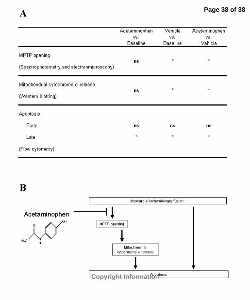
including the extrinsic pathway of apoptosis. Figure 5 is a schematic of the proposed
mechanism of acetaminophen-mediated cardioprotection following ischemia/reperfusion
insult and a model of our findings
These data begin to explain the mechanism of previous reports of acetaminophen-
mediated cardioprotection following ischemia/reperfusion. They suggest that
administration of acetaminophen just prior to an ischemic attack can result in attenuation
of functional damage via inhibition of MPTP opening and cytochrome c release-induced
apoptotic cell death following ischemia/reperfusion. While these data are promising in
that they offer a historically safe alternative to preventative cardiac care, additional
pathway details still need to be elucidated prior to clinical application.
Page 20 of 38
Copyright Information

20
GRANTS
This work was funded in part by Johnson & Johnson COSAT/McNeil CSP (to G.F.M.) and an American Heart Association Grant-in-Aid 0555801T (to B.L.F.).
Page 21 of 38
Copyright Information

21
FIGURE LEGENDS
Figure 1. Spectrophotometric analysis of mitochondrial swelling. (A) Western blot
showing the lack of VDAC in the cytosolic fraction of a representative vehicle-treated
baseline heart following homogenization and centrifugation. (B) Hearts were
homogenized and mitochondria were isolated via centrifugation following baseline and
ischemia/reperfusion in vehicle- and acetaminophen-treated hearts (n=4 per group).
Mitochondrial swelling was measured as a decrease in light scattering at 540 nm and
expressed as a percentage of the average corresponding baseline value (protein density
approximately 1.0 µg/µl). V, vehicle-treated hearts; A, APAP-treated hearts. *p<0.05 as
determined by ANOVA followed by Tukey’s Multiple Comparison Test compared to
corresponding baseline hearts. †p<0.05 as determined by ANOVA followed by Tukey’s
Multiple Comparison Test compared to ischemia/reperfused vehicle-treated hearts.
Figure 2. Electron micrograph analysis of left ventricle free wall. Representative
electron micrographs following (A) baseline, (B) vehicle ischemia/reperfusion, and (C)
acetaminophen ischemia/reperfusion (n=2 per group). Swollen mitochondria (white
arrows in B relative to A and C) imply the opening of mitochondrial permeability
transition pores. The presence of acetaminophen during ischemia/reperfusion appears to
attenuate permeability transition and consequently mitochondrial swelling. Note the
similarity in mitochondrial color and shape between (A) baseline and (C) acetaminophen-
treated ischemia/reperfused hearts.
Page 22 of 38
Copyright Information

22
Figure 3. Western blot analysis of cytosolic and mitochondrial cytochrome c heart
homogenate fractions following ischemia/reperfusion. Hearts were freeze-clamped,
homogenized, and separated into mitochondrial and cytosolic fractions following the
experimental perfusion. Approximately 10 µg of protein from each heart was resolved
on a 15% SDS polyacrylamide gel and probed for cytochrome c and the appropriate
loading control (either α-actin or VDAC) by Western blotting. (A) Hearts were freeze-
clamped and homogenized immediately following 15 minutes of baseline perfusion with
KHB (n=4). Ratio of cytosolic cytochrome c/α-actin band intensity was 0.76±0.03 in
baseline hearts. (B-D) Hearts were freeze-clamped and homogenized immediately
following 15 minutes of baseline perfusion or following ischemia/reperfusion in vehicle-
and acetaminophen-treated hearts (n=4 per group). (B) Representative Western blots
from cytosolic (cyt) and mitochondrial (mito) heart homogenate fractions. (C) Statistical
analysis of cytosolic cytochrome c content normalized to α-actin and baseline. (D)
Statistical analysis of mitochondrial cytochrome c content normalized to VDAC and
baseline. V, vehicle-treated hearts; A, APAP-treated hearts. *p<0.05 as determined by
ANOVA followed by Tukey’s Multiple Comparison Test compared to corresponding
baseline hearts. †p<0.05 as determined by ANOVA followed by Tukey’s Multiple
Comparison Test compared to ischemia/reperfused vehicle-treated hearts.
Figure 4. FACS analysis of post-ischemia/reperfused ventricular myocytes. Hearts were
digested with collagenase and myocytes were isolated and loaded with annexin V-FITC
and propidium iodide following 15 minutes baseline or ischemia/reperfusion in vehicle-
and acetaminophen-treated hearts (n=4 per group). Flow cytometry was used to
Page 23 of 38
Copyright Information

23
determine percentage of (A) early and (B) late stage apoptotic myocytes in vehicle- and
acetaminophen-treated hearts following ischemia/reperfusion. (C) Representative FACS
analysis of vehicle-treated ischemia/reperfused heart. J1, necrotic cells; J2, late apoptotic
cells; J3, viable cells; J4, early apoptotic cells; B, baseline hearts; V, vehicle-treated
hearts; A, APAP-treated hearts. *p<0.05 as determined by ANOVA followed by Tukey’s
Multiple Comparison Test compared to myocytes from baseline hearts in the same stage
of apoptosis. †p<0.05 as determined by ANOVA followed by Tukey’s Multiple
Comparison Test compared to myocytes from vehicle-treated hearts in the same stage of
apoptosis.
Figure 5. Mechanism of acetaminophen-mediated cardioprotection. (A) Summary table
of study findings as they relate to the mechanism of acetaminophen-mediated
cardioprotection following ischemia/reperfusion. Acetaminophen completely inhibits
MPTP opening and mitochondrial cytochrome c release and partially attenuates apoptosis
when compared to vehicle-treated hearts following ischemia/reperfusion. *p<0.05; ns, no
significance. (B) Schematic of proposed mechanism of action of acetaminophen
following myocardial ischemia/reperfusion. Our studies imply that while acetaminophen
completely inhibits MPTP opening and mitochondrial cytochrome c release, apoptosis is
not completely blocked. Thus, additional apoptotic pathways are still active following
insult.
Page 24 of 38
Copyright Information
24
REFERENCES
1. Adlam VJ, Harrison JC, Porteous CM, James AM, Smith RA, Murphy MP,
and Sammut IA. Targeting an antioxidant to mitochondria decreases cardiac ischemia-
reperfusion injury. Faseb J 19: 1088-1095, 2005.
2. Ambrosio G, and Tritto I. Reperfusion injury: experimental evidence and
clinical implications. Am Heart J 138: S69-75, 1999.
3. Bazzola JJ, and Russel LD. Electron microscopy: principles and techniques for
biologists. . Sudbury, MA: Jones and Bartlett Publishers, 1999.
4. Blaustein A, Deneke SM, Stolz RI, Baxter D, Healey N, and Fanburg BL.
Myocardial glutathione depletion impairs recovery after short periods of ischemia.
Circulation 80: 1449-1457, 1989.
5. Bognar Z, Kalai T, Palfi A, Hanto K, Bognar B, Mark L, Szabo Z, Tapodi A,
Radnai B, Sarszegi Z, Szanto A, Gallyas F, Jr., Hideg K, Sumegi B, and Varbiro G.
A novel SOD-mimetic permeability transition inhibitor agent protects ischemic heart by
inhibiting both apoptotic and necrotic cell death. Free Radic Biol Med 41: 835-848, 2006.
6. Bopassa JC, Vandroux D, Ovize M, and Ferrera R. Controlled reperfusion
after hypothermic heart preservation inhibits mitochondrial permeability transition-pore
opening and enhances functional recovery. Am J Physiol Heart Circ Physiol 291: H2265-
2271, 2006.
7. Borutaite V, Jekabsone A, Morkuniene R, and Brown GC. Inhibition of
mitochondrial permeability transition prevents mitochondrial dysfunction, cytochrome c
release and apoptosis induced by heart ischemia. J Mol Cell Cardiol 35: 357-366, 2003.
Page 25 of 38
Copyright Information

25
8. Bosetti F, Baracca A, Lenaz G, and Solaini G. Increased state 4 mitochondrial
respiration and swelling in early post-ischemic reperfusion of rat heart. FEBS Lett 563:
161-164, 2004.
9. Bunger R, Haddy FJ, and Gerlach E. Coronary responses to dilating substances
and competitive inhibition by theophylline in the isolated perfused guinea pig heart.
Pflugers Arch 358: 213-224, 1975.
10. Bunger R, Haddy FJ, Querengasser A, and Gerlach E. An isolated guinea pig
heart preparation with in vivo like features. Pflugers Arch 353: 317-326, 1975.
11. Chen Q, Moghaddas S, Hoppel CL, and Lesnefsky EJ. Reversible blockade of
electron transport during ischemia protects mitochondria and decreases myocardial injury
following reperfusion. J Pharmacol Exp Ther 319: 1405-1412, 2006.
12. Das S, Powell SR, Wang P, Divald A, Nesaretnam K, Tosaki A, Cordis GA,
Maulik N, and Das DK. Cardioprotection with palm tocotrienol: antioxidant activity of
tocotrienol is linked with its ability to stabilize proteasomes. Am J Physiol Heart Circ
Physiol 289: H361-367, 2005.
13. Di Lisa F, and Bernardi P. Mitochondria and ischemia-reperfusion injury of the
heart: fixing a hole. Cardiovasc Res 70: 191-199, 2006.
14. Ferrari R, Alfieri O, Curello S, Ceconi C, Cargnoni A, Marzollo P, Pardini
A, Caradonna E, and Visioli O. Occurrence of oxidative stress during reperfusion of the
human heart. Circulation 81: 201-211, 1990.
15. Gadelha FR, Thomson L, Fagian MM, Costa AD, Radi R, and Vercesi AE.
Ca2+-independent permeabilization of the inner mitochondrial membrane by
Page 26 of 38
Copyright Information

26
peroxynitrite is mediated by membrane protein thiol cross-linking and lipid peroxidation.
Arch Biochem Biophys 345: 243-250, 1997.
16. Gateau-Roesch O, Argaud L, and Ovize M. Mitochondrial permeability
transition pore and postconditioning. Cardiovasc Res 70: 264-273, 2006.
17. Golfetti R, Rork T, and Merrill G. Chronically administered acetaminophen
and the ischemia/reperfused myocardium. Exp Biol Med (Maywood) 228: 674-682, 2003.
18. Golfetti R, VanDyke K, Rork T, Spiler N, and Merrill G. Acetaminophen in
the post-ischemia reperfused myocardium. Exp Biol Med (Maywood) 227: 1031-1037,
2002.
19. Halestrap AP, Clarke SJ, and Javadov SA. Mitochondrial permeability
transition pore opening during myocardial reperfusion--a target for cardioprotection.
Cardiovasc Res 61: 372-385, 2004.
20. Halestrap AP, Connern CP, Griffiths EJ, and Kerr PM. Cyclosporin A
binding to mitochondrial cyclophilin inhibits the permeability transition pore and protects
hearts from ischaemia/reperfusion injury. Mol Cell Biochem 174: 167-172, 1997.
21. Hardman JG, Limbird LL, Molinoff PB, Ruddon RW, and Gilman AG.
Goodman & Gilman's The Pharmacological Basis of Therapeutics. New York: 1996.
22. Hirai M, Hotta Y, Ishikawa N, Wakida Y, Fukuzawa Y, Isobe F, Nakano A,
Chiba T, and Kawamura N. Protective effects of EGCg or GCg, a green tea catechin
epimer, against postischemic myocardial dysfunction in guinea-pig hearts. Life Sci 80:
1020-1032, 2007.
23. Honda HM, Korge P, and Weiss JN. Mitochondria and ischemia/reperfusion
injury. Ann N Y Acad Sci 1047: 248-258, 2005.
Page 27 of 38
Copyright Information

27
24. Huang XD, Horackova M, and Pressler ML. Changes in the expression and
distribution of connexin 43 in isolated cultured adult guinea pig cardiomyocytes. Exp
Cell Res 228: 254-261, 1996.
25. Hunter FE, Jr., Gebicki JM, Hoffsten PE, Weinstein J, and Scott A. Swelling
and lysis of rat liver mitochondria induced by ferrous ions. J Biol Chem 238: 828-835,
1963.
26. Javadov SA, Lim KH, Kerr PM, Suleiman MS, Angelini GD, and Halestrap
AP. Protection of hearts from reperfusion injury by propofol is associated with inhibition
of the mitochondrial permeability transition. Cardiovasc Res 45: 360-369, 2000.
27. Kaasik A, Safiulina D, Zharkovsky A, and Veksler V. Regulation of
mitochondrial matrix volume. Am J Physiol Cell Physiol 292: C157-163, 2007.
28. Kovacs P, Bak I, Szendrei L, Vecsernyes M, Varga E, Blasig IE, and Tosaki
A. Non-specific caspase inhibition reduces infarct size and improves post-ischaemic
recovery in isolated ischaemic/reperfused rat hearts. Naunyn Schmiedebergs Arch
Pharmacol 364: 501-507, 2001.
29. Lundberg KC, and Szweda LI. Initiation of mitochondrial-mediated apoptosis
during cardiac reperfusion. Arch Biochem Biophys 432: 50-57, 2004.
30. Merrill G, McConnell P, Vandyke K, and Powell S. Coronary and myocardial
effects of acetaminophen: protection during ischemia-reperfusion. Am J Physiol Heart
Circ Physiol 280: H2631-2638, 2001.
31. Merrill GF. Acetaminophen and low-flow myocardial ischemia: efficacy and
antioxidant mechanisms. Am J Physiol Heart Circ Physiol 282: H1341-1349, 2002.
Page 28 of 38
Copyright Information

28
32. Merrill GF, and Goldberg E. Antioxidant properties of acetaminophen and
cardioprotection. Basic Res Cardiol 96: 423-430, 2001.
33. Merrill GF, Rork TH, Spiler NM, and Golfetti R. Acetaminophen and
myocardial infarction in dogs. Am J Physiol Heart Circ Physiol 287: H1913-1920, 2004.
34. Orrenius S, Gogvadze V, and Zhivotovsky B. Mitochondrial oxidative stress:
implications for cell death. Annu Rev Pharmacol Toxicol 47: 143-183, 2007.
35. Piper H, and Isenberg G editors. Isolated Adult Cardiomyocytes. Boca Raton,
FL: CRC Press, Inc, 1989.
36. Prescott LF. Paracetamol (Acetaminophen): A Critical Bibliographic Review.
New York: Taylor & Francis, 2001.
37. Prescott LF. Paracetamol: past, present, and future. Am J Ther 7: 143-147, 2000.
38. Przyklenk K, and Kloner RA. Superoxide dismutase plus catalase improve
contractile function in the canine model of the "stunned myocardium". Circ Res 58: 148-
156, 1986.
39. Rork TH, Hadzimichalis NM, Kappil MA, and Merrill GF. Acetaminophen
attenuates peroxynitrite-activated matrix metalloproteinase-2-mediated troponin I
cleavage in the isolated guinea pig myocardium. J Mol Cell Cardiol 40: 553-561, 2006.
40. Rork TH, Van Dyke K, Spiler NM, and Merrill GF. Acetaminophen in the
hypoxic and reoxygenated guinea pig myocardium. Exp Biol Med (Maywood) 229: 1154-
1161, 2004.
41. Rousou AJ, Ericsson M, Federman M, Levitsky S, and McCully JD. Opening
of mitochondrial KATP channels enhances cardioprotection through the modulation of
Page 29 of 38
Copyright Information
29
mitochondrial matrix volume, calcium accumulation, and respiration. Am J Physiol Heart
Circ Physiol 287: H1967-1976, 2004.
42. Ruiz-Meana M, Garcia-Dorado D, Pina P, Inserte J, Agullo L, and Soler-
Soler J. Cariporide preserves mitochondrial proton gradient and delays ATP depletion in
cardiomyocytes during ischemic conditions. Am J Physiol Heart Circ Physiol 285: H999-
1006, 2003.
43. Schmid I, Uittenbogaart C, and Jamieson BD. Live-cell assay for detection of
apoptosis by dual-laser flow cytometry using Hoechst 33342 and 7-amino-actinomycin
D. Nat Protoc 2: 187-190, 2007.
44. Spiler NM, Rork TH, and Merrill GF. An old drug with a new purpose:
cardiovascular actions of acetaminophen (paracetamol). Curr Drug Targets Cardiovasc
Haematol Disord 5: 419-429, 2005.
45. Sztark F, Ichas F, Ouhabi R, Dabadie P, and Mazat JP. Effects of the
anaesthetic propofol on the calcium-induced permeability transition of rat heart
mitochondria: direct pore inhibition and shift of the gating potential. FEBS Lett 368: 101-
104, 1995.
46. Tokarska-Schlattner M, Zaugg M, da Silva R, Lucchinetti E, Schaub MC,
Wallimann T, and Schlattner U. Acute toxicity of doxorubicin on isolated perfused
heart: response of kinases regulating energy supply. Am J Physiol Heart Circ Physiol
289: H37-47, 2005.
47. Tortolani AJ, Powell SR, Misik V, Weglicki WB, Pogo GJ, and Kramer JH.
Detection of alkoxyl and carbon-centered free radicals in coronary sinus blood from
patients undergoing elective cardioplegia. Free Radic Biol Med 14: 421-426, 1993.
Page 30 of 38
Copyright Information

30
48. Varga E, Nagy N, Lazar J, Czifra G, Bak I, Biro T, and Tosaki A. Inhibition
of ischemia/reperfusion-induced damage by dexamethasone in isolated working rat
hearts: the role of cytochrome c release. Life Sci 75: 2411-2423, 2004.
49. Weiss JN, Korge P, Honda HM, and Ping P. Role of the mitochondrial
permeability transition in myocardial disease. Circ Res 93: 292-301, 2003.
50. Zhu YZ, Chong CL, Chuah SC, Huang SH, Nai HS, Tong HT, Whiteman M,
and Moore PK. Cardioprotective effects of nitroparacetamol and paracetamol in acute
phase of myocardial infarction in experimental rats. Am J Physiol Heart Circ Physiol
290: H517-524, 2006.
Page 31 of 38
Copyright Information
A
V A V A V A
pO 2 (mmHg) 515 ± 10 525 ± 8 520 ± 10 534 ± 17 528 ± 15 515 ± 15
pCO 2 (mmHg) 31 ± 1 33 ± 1 32 ± 1 31 ± 1 30 ± 1 30 ± 1
pH 7.40 ± 0.01 7.40 ± 0.01 7.41 ± 0.01 7.41 ± 0.01 7.42 ± 0.01 7.41 ± 0.01
CPF (ml/min/g) 7.0 ± 1.0 7.2 ± 1.0 0.9 ± 0.1* 1.0 ± 0.1* 9.0 ± 2.0 8.5 ± 1.0
B
V A V A
pO 2 (mmHg) 507 ± 9 535 ± 6 514 ± 19 524 ± 15
pCO 2 (mmHg) 30 ± 1 33 ± 1 31 ± 1 31 ± 1
pH 7.41 ± 0.01 7.39 ± 0.01 7.41 ± 0.01 7.41 ± 0.01
CPF (ml/min/g) 7.5 ± 1.0 6.8 ± 0.6 9.5 ± 2.5 8.2 ± 1.0
15 minutes baseline Ischemia Reperfusion
15 minutes baseline Reperfusion
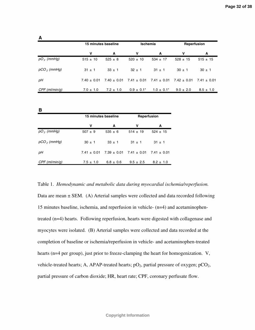
Table 1. Hemodynamic and metabolic data during myocardial ischemia/reperfusion.
Data are mean ± SEM. (A) Arterial samples were collected and data recorded following
15 minutes baseline, ischemia, and reperfusion in vehicle- (n=4) and acetaminophen-
treated (n=4) hearts. Following reperfusion, hearts were digested with collagenase and
myocytes were isolated. (B) Arterial samples were collected and data recorded at the
completion of baseline or ischemia/reperfusion in vehicle- and acetaminophen-treated
hearts (n=4 per group), just prior to freeze-clamping the heart for homogenization. V,
vehicle-treated hearts; A, APAP-treated hearts; pO2, partial pressure of oxygen; pCO2,
partial pressure of carbon dioxide; HR, heart rate; CPF, coronary perfusate flow.
Page 32 of 38
Copyright Information
*p<0.05, as determined by ANOVA followed by Tukey’s Multiple Comparison Test
relative to corresponding baseline value.
Page 33 of 38
Copyright Information
Page 34 of 38
Copyright Information
Page 35 of 38
Copyright Information

Page 36 of 38
Copyright Information
Page 37 of 38
Copyright Information
Page 38 of 38
Copyright Information
Related Documents


![Paracetamol(acetaminophen)ornon-steroidalanti ... (acetaminophen) … · [Intervention Review] Paracetamol (acetaminophen) or non-steroidal anti-inflammatory drugs, alone or combined,](https://static.cupdf.com/doc/110x72/5f387cbd43d51a2eb45648f8/paracetamolacetaminophenornon-steroidalanti-acetaminophen-intervention.jpg)

