1328 JACC Vol. 28, No. 5 November 1, 1996:|328-428 ACC/AHA PRACTICE GUIDELINES ACC/AHA Guidelines for the Management of Patients With Acute Myocardial Infarction A Report of the American College of Cardiology/American Heart Association Task Force on Practice Guidelines (Committee on Management of Acute Myocardial Infarction) COMMITYEE MEMBERS THOMAS J. RYAN, MD, FACC, Chair, JEFFREY L. ANDERSON, MD, FACC, ELLIOTF M. ANTMAN, MD, FACC, BLAINE A. BRANIFF, MD, FACP, NEIL H. BROOKS, MD, FAAFP, ROBERT M. CALIFF, MD, FACC, L. DAVID HILLIS, MD, FACC, LOREN F. HIRATZKA, MD, FACC, ELLIOT RAPAPORT, MD, FACC, BARBARA J. RIEGEL, DNSc, FAAN, RICHARD O. RUSSELL, MD, FACC, EARL E. SMITH III, MD, FACEP, W. DOUGLAS WEAVER, MD, FACC TASK FORCE MEMBERS JAMES L. RITCHIE, MD, FACC, Chair, MELVIN D. CHEITLIN, MD, FACC, KIM A. EAGLE, MD, FACC, TIMOTHY J. GARDNER, MD, FACC, ARTHUR GARSON, JR., MD, MPH, FACC, RAYMOND J. GIBBONS, MD, FACC, RICHARD P. LEWIS, MD, FACC, ROBERT A. O'ROURKE, MD, FACC, THOMAS J. RYAN, MD, FACC Contents Preamble .......................................................................... 1332 Executive Summary. .................................................................... 1333 I. Introduction ..................................................................... 1336 II. Prehospital Issues .................................................................. 1337 Recommendations ................................................................ 1337 Recognition and Management ......................................................... 1338 Intervention Strategies ............................................................ 1338 Emergency Medical Systems ......................................................... 1338 Prehospital-Initiated Thrombolysis ...................................................... 1339 III. Initial Recognition and Management in the Emergency Department ..................................... 1340 Recommendation ................................................................. 1340 Detection/Quantification and Risk Assessment ................................................ 1340 Routine Measures (Oxygen, Nitroglycerin, Aspirin) ............................................. 1342 Recommendations ............................................................... 1342 Oxygen ..................................................................... 1342 Recommendations .............................................................. 1342 "ACC/AHA Guidelines for the Management of Patients With Acute Myo- cardial Infarction" was approved by the American College of Cardiology Board of Trustees on July 10, 1996, and by the American Heart Association Science Advisory and Coordinating Committee on June 20, 1996. When citing this document, the American College of Cardiology and the American Heart Association request that the following format be used: Ryan TJ, Anderson JL, Antman EM, Braniff BA, Brooks NH, Califf RM, Hillis LD, Hiratzka LF, Rapaport E, Riegel BJ, Russell RO, Smith EE III, Weaver WD. ACC/AHA guidelines for the management of patients with acute myocardial infarction: a report of the American College of Cardiology/American Heart Association Task Force on Practice Guidelines (Committee on Management of Acute Myocardial Infarction). JAm Coil Cardiol 1996;28:1328-1428. Address for reprints: A single reprint of this document (the complete Guidelines) is available by calling 800-253-4636 (US only) or writing the American College of Cardiology, Educational Services, 9111 Old Georgetown Road, Bethesda, MD 20814-1699. Ask for reprint No. 71-0094. To obtain a reprint of the shorter version (Executive Summary and Summary of Recommen- dations) published in the November 1 issue of Circulation, ask for reprint No. 71-0092. To purchase additional reprints (specify version and reprint number): Up to 999 copies, call 800-611-6083 (US only) or fax 413-665-2671; 1000 or more copies, call 214-706-1466, fax 214-691-6342, or E-mail [email protected]. 01996 by the American College of Cardiology and the American Heart Association, Inc. 0735-1097/96/$15.00 Published by Elsevier Science Inc. PII S0735-1097(96)00392-0

Welcome message from author
This document is posted to help you gain knowledge. Please leave a comment to let me know what you think about it! Share it to your friends and learn new things together.
Transcript

1328 JACC Vol. 28, No. 5 November 1, 1996:|328-428
ACC/AHA PRACTICE GUIDELINES
ACC/AHA Guidelines for the Management of Patients With Acute Myocardial Infarction
A Report of the American College of Cardiology/American Heart Association
Task Force on Practice Guidelines (Committee on Management of Acute
Myocardial Infarction)
COMMITYEE MEMBERS
THOMAS J. RYAN, MD, FACC, Chair, JEFFREY L. ANDERSON, MD, FACC, ELLIOTF M. ANTMAN, MD, FACC, BLAINE A. BRANIFF, MD, FACP, NEIL H. BROOKS, MD, FAAFP, ROBERT M. CALIFF, MD, FACC, L. DAVID HILLIS, MD, FACC, LOREN F. HIRATZKA, MD, FACC, ELLIOT RAPAPORT, MD, FACC, BARBARA J. RIEGEL, DNSc, FAAN, RICHARD O. RUSSELL, MD, FACC, EARL E. SMITH III, MD, FACEP, W. DOUGLAS WEAVER, MD, FACC
TASK FORCE MEMBERS
JAMES L. RITCHIE, MD, FACC, Chair, MELVIN D. CHEITLIN, MD, FACC, KIM A. EAGLE, MD, FACC, TIMOTHY J. G A R D N E R , M D , F A C C , A R T H U R G A R S O N , JR., M D , M P H , F A C C , R A Y M O N D J. G I B B O N S , M D , F A C C ,
RICHARD P. LEWIS, MD, FACC, ROBERT A. O'ROURKE, MD, FACC, THOMAS J. RYAN, MD, FACC
Contents Preamble . . . . . . . . . . . . . . . . . . . . . . . . . . . . . . . . . . . . . . . . . . . . . . . . . . . . . . . . . . . . . . . . . . . . . . . . . . 1332
Executive Summary. . . . . . . . . . . . . . . . . . . . . . . . . . . . . . . . . . . . . . . . . . . . . . . . . . . . . . . . . . . . . . . . . . . . . 1333
I. Introduction . . . . . . . . . . . . . . . . . . . . . . . . . . . . . . . . . . . . . . . . . . . . . . . . . . . . . . . . . . . . . . . . . . . . . 1336
II. Prehospital Issues . . . . . . . . . . . . . . . . . . . . . . . . . . . . . . . . . . . . . . . . . . . . . . . . . . . . . . . . . . . . . . . . . . 1337
Recommendations . . . . . . . . . . . . . . . . . . . . . . . . . . . . . . . . . . . . . . . . . . . . . . . . . . . . . . . . . . . . . . . . 1337
Recognition and Management . . . . . . . . . . . . . . . . . . . . . . . . . . . . . . . . . . . . . . . . . . . . . . . . . . . . . . . . . 1338
Intervention Strategies . . . . . . . . . . . . . . . . . . . . . . . . . . . . . . . . . . . . . . . . . . . . . . . . . . . . . . . . . . . . 1338
Emergency Medical Systems . . . . . . . . . . . . . . . . . . . . . . . . . . . . . . . . . . . . . . . . . . . . . . . . . . . . . . . . . 1338
Prehospital-Initiated Thrombolysis . . . . . . . . . . . . . . . . . . . . . . . . . . . . . . . . . . . . . . . . . . . . . . . . . . . . . . 1339
III. Initial Recognition and Management in the Emergency Department . . . . . . . . . . . . . . . . . . . . . . . . . . . . . . . . . . . . . 1340
Recommendation . . . . . . . . . . . . . . . . . . . . . . . . . . . . . . . . . . . . . . . . . . . . . . . . . . . . . . . . . . . . . . . . . 1340
Detection/Quantification and Risk Assessment . . . . . . . . . . . . . . . . . . . . . . . . . . . . . . . . . . . . . . . . . . . . . . . . 1340
Routine Measures (Oxygen, Nitroglycerin, Aspirin) . . . . . . . . . . . . . . . . . . . . . . . . . . . . . . . . . . . . . . . . . . . . . 1342
Recommendations . . . . . . . . . . . . . . . . . . . . . . . . . . . . . . . . . . . . . . . . . . . . . . . . . . . . . . . . . . . . . . . 1342
Oxygen . . . . . . . . . . . . . . . . . . . . . . . . . . . . . . . . . . . . . . . . . . . . . . . . . . . . . . . . . . . . . . . . . . . . . 1342
Recommendations . . . . . . . . . . . . . . . . . . . . . . . . . . . . . . . . . . . . . . . . . . . . . . . . . . . . . . . . . . . . . . 1342
"ACC/AHA Guidelines for the Management of Patients With Acute Myo- cardial Infarction" was approved by the American College of Cardiology Board of Trustees on July 10, 1996, and by the American Heart Association Science Advisory and Coordinating Committee on June 20, 1996.
When citing this document, the American College of Cardiology and the American Heart Association request that the following format be used: Ryan TJ, Anderson JL, Antman EM, Braniff BA, Brooks NH, Califf RM, Hillis LD, Hiratzka LF, Rapaport E, Riegel BJ, Russell RO, Smith EE III, Weaver WD. ACC/AHA guidelines for the management of patients with acute myocardial infarction: a report of the American College of Cardiology/American Heart
Association Task Force on Practice Guidelines (Committee on Management of Acute Myocardial Infarction). JAm Coil Cardiol 1996;28:1328-1428.
Address for reprints: A single reprint of this document (the complete Guidelines) is available by calling 800-253-4636 (US only) or writing the American College of Cardiology, Educational Services, 9111 Old Georgetown Road, Bethesda, MD 20814-1699. Ask for reprint No. 71-0094. To obtain a reprint of the shorter version (Executive Summary and Summary of Recommen- dations) published in the November 1 issue of Circulation, ask for reprint No. 71-0092. To purchase additional reprints (specify version and reprint number): Up to 999 copies, call 800-611-6083 (US only) or fax 413-665-2671; 1000 or more copies, call 214-706-1466, fax 214-691-6342, or E-mail [email protected].
01996 by the American College of Cardiology and the American Heart Association, Inc. 0735-1097/96/$15.00 Published by Elsevier Science Inc. PII S0735-1097(96)00392-0

JACC Vol. 28, No. 5 RYAN ET AL. 1329 November 1, 1996:1328-428 MANAGEMENT OF ACUTE MYOCARDIAE INFARCTION
Nitroglycerin . . . . . . . . . . . . . . . . . . . . . . . . . . . . . . . . . . . . . . . . . . . . . . . . . . . . . . . . . . . . . . . . . . 1343
Recommendations for Intravenous Nitroglycerin . . . . . . . . . . . . . . . . . . . . . . . . . . . . . . . . . . . . . . . . . . . . 1343
Analgcsia . . . . . . . . . . . . . . . . . . . . . . . . . . . . . . . . . . . . . . . . . . . . . . . . . . . . . . . . . . . . . . . . . . . . 1343
Aspirin . . . . . . . . . . . . . . . . . . . . . . . . . . . . . . . . . . . . . . . . . . . . . . . . . . . . . . . . . . . . . . . . . . . . . 1344
Recommendations . . . . . . . . . . . . . . . . . . . . . . . . . . . . . . . . . . . . . . . . . . . . . . . . . . . . . . . . . . . . . . 1344
Atropine . . . . . . . . . . . . . . . . . . . . . . . . . . . . . . . . . . . . . . . . . . . . . . . . . . . . . . . . . . . . . . . . . . . . 1344
Rccommcndations . . . . . . . . . . . . . . . . . . . . . . . . . . . . . . . . . . . . . . . . . . . . . . . . . . . . . . . . . . . . . . 1344
Side Effects . . . . . . . . . . . . . . . . . . . . . . . . . . . . . . . . . . . . . . . . . . . . . . . . . . . . . . . . . . . . . . . . . 1345
Risk Stratification and Management of ST-Segment Elevation/Bundle Branch Block Cohort . . . . . . . . . . . . . . . . . . . . . . . 1345
Newer Concepts . . . . . . . . . . . . . . . . . . . . . . . . . . . . . . . . . . . . . . . . . . . . . . . . . . . . . . . . . . . . . . . . . 1345
Noninvasive Imaging in the Emergency Department . . . . . . . . . . . . . . . . . . . . . . . . . . . . . . . . . . . . . . . . . . . . . 1346
Thrombolysis . . . . . . . . . . . . . . . . . . . . . . . . . . . . . . . . . . . . . . . . . . . . . . . . . . . . . . . . . . . . . . . . . . . 1346
Recommendations . . . . . . . . . . . . . . . . . . . . . . . . . . . . . . . . . . . . . . . . . . . . . . . . . . . . . . . . . . . . . . . 1346
Risk of Stroke . . . . . . . . . . . . . . . . . . . . . . . . . . . . . . . . . . . . . . . . . . . . . . . . . . . . . . . . . . . . . . . . . 1348
Net Clinical Benefit . . . . . . . . . . . . . . . . . . . . . . . . . . . . . . . . . . . . . . . . . . . . . . . . . . . . . . . . . . . . . . 1348
Contraindications/Cautions . . . . . . . . . . . . . . . . . . . . . . . . . . . . . . . . . . . . . . . . . . . . . . . . . . . . . . . . . . 1348
Primary Percutaneous Transluminal Coronary, Angioplasty . . . . . . . . . . . . . . . . . . . . . . . . . . . . . . . . . . . . . . . . . . 1348
Recommcndations . . . . . . . . . . . . . . . . . . . . . . . . . . . . . . . . . . . . . . . . . . . . . . . . . . . . . . . . . . . . . . . . 1348
Recommendations for Early Corona~ Angiography in the ST-Segment Elevation or Bundle
Branch Block Cohort Not Undergoing Primary Percutaneous Transluminal Coronary Angioplas~ . . . . . . . . . . . . . . . . 1351
Recommendations for Emergency or Urgent Coronary Artery Bypass Graft Surgery . . . . . . . . . . . . . . . . . . . . . . . . . . 1351
Risk Stratification and Management in Non-ST-Segment Elevation Cohort . . . . . . . . . . . . . . . . . . . . . . . . . . . . . . . . . 1352
Recommendations for Early Coronary Angiography and/or Interventional Therapy . . . . . . . . . . . . . . . . . . . . . . . . . . . 1352
Patient Characteristics . . . . . . . . . . . . . . . . . . . . . . . . . . . . . . . . . . . . . . . . . . . . . . . . . . . . . . . . . . . . . . 1352
Pharmacological Therapy in Patients in the Non-ST-Segment Elevation Cohort . . . . . . . . . . . . . . . . . . . . . . . . . . . . . 1353
Interventional Therapy . . . . . . . . . . . . . . . . . . . . . . . . . . . . . . . . . . . . . . . . . . . . . . . . . . . . . . . . . . . . . . 1353
IV. Hospital Management . . . . . . . . . . . . . . . . . . . . . . . . . . . . . . . . . . . . . . . . . . . . . . . . . . . . . . . . . . . . . . . . 1354
Early, General Measures . . . . . . . . . . . . . . . . . . . . . . . . . . . . . . . . . . . . . . . . . . . . . . . . . . . . . . . . . . . . . 1354
Recommendations . . . . . . . . . . . . . . . . . . . . . . . . . . . . . . . . . . . . . . . . . . . . . . . . . . . . . . . . . . . . . . . 1354
Monitoring for Adverse Events . . . . . . . . . . . . . . . . . . . . . . . . . . . . . . . . . . . . . . . . . . . . . . . . . . . . . . . . 1354
Level of Activity . . . . . . . . . . . . . . . . . . . . . . . . . . . . . . . . . . . . . . . . . . . . . . . . . . . . . . . . . . . . . . . . 1354
Proper Analgesia (Use of Morphine, Anxiolytics, and the Role of Education) . . . . . . . . . . . . . . . . . . . . . . . . . . . . . 1355
Treatment of Adverse Events . . . . . . . . . . . . . . . . . . . . . . . . . . . . . . . . . . . . . . . . . . . . . . . . . . . . . . . . 1356
Identification and Treatment of the Patient at Eow Risk . . . . . . . . . . . . . . . . . . . . . . . . . . . . . . . . . . . . . . . . . . 1356
Triage of Patients With Acute Myocardial Infarction and Other Coronary Syndromes . . . . . . . . . . . . . . . . . . . . . . . . 1357
Summary of Identification and Treatment of the Patient at Low Risk . . . . . . . . . . . . . . . . . . . . . . . . . . . . . . . . . . 1357
Identification and Treatment of thc Patient at High Risk . . . . . . . . . . . . . . . . . . . . . . . . . . . . . . . . . . . . . . . . . . 1358
Recommendations for Management of Recurrent Chest Discomfort . . . . . . . . . . . . . . . . . . . . . . . . . . . . . . . . . . 1358
Recurrent Chest Pain in the Post-MI Patient: Pericarditis and Ischemia . . . . . . . . . . . . . . . . . . . . . . . . . . . . . . . . 1358
Heart Failure and Ix~w-Output Syndromes . . . . . . . . . . . . . . . . . . . . . . . . . . . . . . . . . . . . . . . . . . . . . . . . . 1359
Left Ventricular Dysfunction . . . . . . . . . . . . . . . . . . . . . . . . . . . . . . . . . . . . . . . . . . . . . . . . . . . . . . . . 1359
Right Ventricular Infarction and Dysfunction . . . . . . . . . . . . . . . . . . . . . . . . . . . . . . . . . . . . . . . . . . . . . . 1360
Anatomic and Pathophysiological Considerations . . . . . . . . . . . . . . . . . . . . . . . . . . . . . . . . . . . . . . . . . . . . 1360
Clinical Diagnosis . . . . . . . . . . . . . . . . . . . . . . . . . . . . . . . . . . . . . . . . . . . . . . . . . . . . . . . . . . . . . . 1360
Managemcnt of Right Ventricular Ischemia/lnfarction . . . . . . . . . . . . . . . . . . . . . . . . . . . . . . . . . . . . . . . . . 1361
Prognosis . . . . . . . . . . . . . . . . . . . . . . . . . . . . . . . . . . . . . . . . . . . . . . . . . . . . . . . . . . . . . . . . . . . 1361
Hemodynamic Monitoring . . . . . . . . . . . . . . . . . . . . . . . . . . . . . . . . . . . . . . . . . . . . . . . . . . . . . . . . . . 1361
Recommendations for Balloon Flotation Right-Heart Catheter Monitoring . . . . . . . . . . . . . . . . . . . . . . . . . . . . . 1361
Recommendations for Intra-arterial Pressure Monitoring . . . . . . . . . . . . . . . . . . . . . . . . . . . . . . . . . . . . . . . 1362
Recommendations for Intra-aortic Balloon Counterpulsation . . . . . . . . . . . . . . . . . . . . . . . . . . . . . . . . . . . . . 1362
Rhythm Disturbances . . . . . . . . . . . . . . . . . . . . . . . . . . . . . . . . . . . . . . . . . . . . . . . . . . . . . . . . . . . . . 1363
Atrial Fibrillation . . . . . . . . . . . . . . . . . . . . . . . . . . . . . . . . . . . . . . . . . . . . . . . . . . . . . . . . . . . . . . 1363
Recommendations . . . . . . . . . . . . . . . . . . . . . . . . . . . . . . . . . . . . . . . . . . . . . . . . . . . . . . . . . . . . 1363
Ventricular Tachycardia/Ventricular Fibrillation . . . . . . . . . . . . . . . . . . . . . . . . . . . . . . . . . . . . . . . . . . . . . 1364
Recommendations . . . . . . . . . . . . . . . . . . . . . . . . . . . . . . . . . . . . . . . . . . . . . . . . . . . . . . . . . . . . 1364
Ventricular Fibrillation--Background . . . . . . . . . . . . . . . . . . . . . . . . . . . . . . . . . . . . . . . . . . . . . . . . . 1364

1330 RYAN ET AL. JACC Vo[. 28, No. 5 MANAGEMENT OF ACUTE MYOCARDIAL 1NFARC'I-ION November 1, 1996:K28-428
Management Strategies for Ventricular Fibrillation . . . . . . . . . . . . . . . . . . . . . . . . . . . . . . . . . . . . . . . . . 1365
Ventricular Tachycardia--Background . . . . . . . . . . . . . . . . . . . . . . . . . . . . . . . . . . . . . . . . . . . . . . . . . 1366
Management Strategies for Ventricular Tachycardia . . . . . . . . . . . . . . . . . . . . . . . . . . . . . . . . . . . . . . . . . 1366
Bradyarrhythmias and Heart Block . . . . . . . . . . . . . . . . . . . . . . . . . . . . . . . . . . . . . . . . . . . . . . . . . . . . 1366
Background, Epidemiology, and Importance . . . . . . . . . . . . . . . . . . . . . . . . . . . . . . . . . . . . . . . . . . . . . 1366
Prognosis . . . . . . . . . . . . . . . . . . . . . . . . . . . . . . . . . . . . . . . . . . . . . . . . . . . . . . . . . . . . . . . . . 1366
Treatment . . . . . . . . . . . . . . . . . . . . . . . . . . . . . . . . . . . . . . . . . . . . . . . . . . . . . . . . . . . . . . . . . 1366
Recommendations for Atropine . . . . . . . . . . . . . . . . . . . . . . . . . . . . . . . . . . . . . . . . . . . . . . . . . . . . 1366
Temporary Pacing . . . . . . . . . . . . . . . . . . . . . . . . . . . . . . . . . . . . . . . . . . . . . . . . . . . . . . . . . . . . . . 1367
Recommendations for Placement of Transcutaneous Patches and Active (Demand) Transcutaneous Pacing . . . . . . . . . 1367
Recommendations for Temlzorary Transvenous Pacing . . . . . . . . . . . . . . . . . . . . . . . . . . . . . . . . . . . . . . . 1367
Permanent Pacing After Acute Myocardial Infarction . . . . . . . . . . . . . . . . . . . . . . . . . . . . . . . . . . . . . . . . . 1368
Recommendations . . . . . . . . . . . . . . . . . . . . . . . . . . . . . . . . . . . . . . . . . . . . . . . . . . . . . . . . . . . . 1368
Other Surgical Interventions . . . . . . . . . . . . . . . . . . . . . . . . . . . . . . . . . . . . . . . . . . . . . . . . . . . . . . . . . . 1368
Recommendations for Emergency or Urgent Cardiac Repair of Mechanical Defects . . . . . . . . . . . . . . . . . . . . . . . . . 1368
Clinical Situations Leading Io Coronary Artery Bypass Graft Surgery . . . . . . . . . . . . . . . . . . . . . . . . . . . . . . . . . . 1368
Evolving Myocardial Infalction . . . . . . . . . . . . . . . . . . . . . . . . . . . . . . . . . . . . . . . . . . . . . . . . . . . . . . 1369
Failed Percutaneous Transluminal Coronary Angioplasty . . . . . . . . . . . . . . . . . . . . . . . . . . . . . . . . . . . . . . . 1369
Postthrombolytic Therapy . . . . . . . . . . . . . . . . . . . . . . . . . . . . . . . . . . . . . . . . . . . . . . . . . . . . . . . . . 1369
Recurrent Ischemia . . . . . . . . . . . . . . . . . . . . . . . . . . . . . . . . . . . . . . . . . . . . . . . . . . . . . . . . . . . . . 1369
Elective Coronary Artery Bypass Graft Surgery After Acute Myocardial Infarction . . . . . . . . . . . . . . . . . . . . . . . . . 1369
Ventricular Tachyarrhythmias . . . . . . . . . . . . . . . . . . . . . . . . . . . . . . . . . . . . . . . . . . . . . . . . . . . . . . . 1369
Patients With Prior Coronary Artery Bypass Graft Surgery . . . . . . . . . . . . . . . . . . . . . . . . . . . . . . . . . . . . . . 1369
Patients Undergoing Cardiopulmonary Resuscitation . . . . . . . . . . . . . . . . . . . . . . . . . . . . . . . . . . . . . . . . . . 1369
Intraolzerative Myocardial Protection in the Acutely Injured Heart . . . . . . . . . . . . . . . . . . . . . . . . . . . . . . . . . . 1370
Management of Mechanical Dcfects After Acute Myocardial Infarction . . . . . . . . . . . . . . . . . . . . . . . . . . . . . . . . . . 1370
Diagnosis . . . . . . . . . . . . . . . . . . . . . . . . . . . . . . . . . . . . . . . . . . . . . . . . . . . . . . . . . . . . . . . . . . . . 1370
Acute Mitral Valve Regurgitation . . . . . . . . . . . . . . . . . . . . . . . . . . . . . . . . . . . . . . . . . . . . . . . . . . . . . . 1371
Postinfarction Ventricular Septal Defect . . . . . . . . . . . . . . . . . . . . . . . . . . . . . . . . . . . . . . . . . . . . . . . . . . 1371
Left Ventricular Free Wall Rupture . . . . . . . . . . . . . . . . . . . . . . . . . . . . . . . . . . . . . . . . . . . . . . . . . . . . . 1371
Left Ventricular Aneurysm . . . . . . . . . . . . . . . . . . . . . . . . . . . . . . . . . . . . . . . . . . . . . . . . . . . . . . . . . . 1371
Mechanical Support of the Failing Heart . . . . . . . . . . . . . . . . . . . . . . . . . . . . . . . . . . . . . . . . . . . . . . . . . . 1371
Transplantation After Acute M3ocardial Infarction . . . . . . . . . . . . . . . . . . . . . . . . . . . . . . . . . . . . . . . . . . . . . 1371
Relation Between Volume of Surgery and Outcome . . . . . . . . . . . . . . . . . . . . . . . . . . . . . . . . . . . . . . . . . . . . . 1371
Minimum Operative Caseload . . . . . . . . . . . . . . . . . . . . . . . . . . . . . . . . . . . . . . . . . . . . . . . . . . . . . . . . . 1371
Case Selection Concerns . . . . . . . . . . . . . . . . . . . . . . . . . . . . . . . . . . . . . . . . . . . . . . . . . . . . . . . . . . . . . 1371
V. Rationale and Approach to Pharmacotherapy . . . . . . . . . . . . . . . . . . . . . . . . . . . . . . . . . . . . . . . . . . . . . . . . . . . 1372
Nitroglycerin . . . . . . . . . . . . . . . . . . . . . . . . . . . . . . . . . . . . . . . . . . . . . . . . . . . . . . . . . . . . . . . . . . . 1372
Mechanism of Action . . . . . . . . . . . . . . . . . . . . . . . . . . . . . . . . . . . . . . . . . . . . . . . . . . . . . . . . . . . . . 1372
Pharmacokinetics and Dosage . . . . . . . . . . . . . . . . . . . . . . . . . . . . . . . . . . . . . . . . . . . . . . . . . . . . . . . . 1372
Limitations and Adverse Effecls . . . . . . . . . . . . . . . . . . . . . . . . . . . . . . . . . . . . . . . . . . . . . . . . . . . . . . . 1372
Clinical Trials . . . . . . . . . . . . . . . . . . . . . . . . . . . . . . . . . . . . . . . . . . . . . . . . . . . . . . . . . . . . . . . . . . 1373
Aspirin and Other Platelet-Activc Drags . . . . . . . . . . . . . . . . . . . . . . . . . . . . . . . . . . . . . . . . . . . . . . . . . . . 1373
Mechanism of Action of Aspirin . . . . . . . . . . . . . . . . . . . . . . . . . . . . . . . . . . . . . . . . . . . . . . . . . . . . . . . 1374
Aspirin in Prevention cf Thrombotic Complications cf Atherosclcrosis . . . . . . . . . . . . . . . . . . . . . . . . . . . . . . . . . 1374
Aspirin: Risk cf Hemorrhagic Stroke . . . . . . . . . . . . . . . . . . . . . . . . . . . . . . . . . . . . . . . . . . . . . . . . . . . . 1374
Aspirin: Side Effects and Dosage . . . . . . . . . . . . . . . . . . . . . . . . . . . . . . . . . . . . . . . . . . . . . . . . . . . . . . 1374
Ticlopidine . . . . . . . . . . . . . . . . . . . . . . . . . . . . . . . . . . . . . . . . . . . . . . . . . . . . . . . . . . . . . . . . . . . 1374
Rationale for Thrombolytic Therapy . . . . . . . . . . . . . . . . . . . . . . . . . . . . . . . . . . . . . . . . . . . . . . . . . . . . . . 1374
Ba cl~ground . . . . . . . . . . . . . . . . . . . . . . . . . . . . . . . . . . . . . . . . . . . . . . . . . . . . . . . . . . . . . . . . . . . 1374
Thrombolytic Agents: General Mechanisms ef Action and Pharmacological Frcperties . . . . . . . . . . . . . . . . . . . . . . . . 1375
Efficacy Gf Intravenous Thrombolytic Therapy in Acute Myocardial Infarction . . . . . . . . . . . . . . . . . . . . . . . . . . . . . 1375
Benefits of Thrombolytic Therapy in Specific Patient Subgroups . . . . . . . . . . . . . . . . . . . . . . . . . . . . . . . . . . . . . 1376
Comparative Thrombolytic Efficacy . . . . . . . . . . . . . . . . . . . . . . . . . . . . . . . . . . . . . . . . . . . . . . . . . . . . . 1376
Considerations in Selecting Thrombolytic Regimens . . . . . . . . . . . . . . . . . . . . . . . . . . . . . . . . . . . . . . . . . . . . 1376
Current Use Rates for Thrombolytic Therapy . . . . . . . . . . . . . . . . . . . . . . . . . . . . . . . . . . . . . . . . . . . . . . . 1377

JACC Vol. 28, No. 5 RYAN ET AL. 1331 November 1, 1996:1328 428 MANAGEMENT OF ACUTE MYOCARDIAL INFARCTION
Antithrombotics/Anticoagulants . . . . . . . . . . . . . . . . . . . . . . . . . . . . . . . . . . . . . . . . . . . . . . . . . . . . . . . . . 1377
Heparin . . . . . . . . . . . . . . . . . . . . . . . . . . . . . . . . . . . . . . . . . . . . . . . . . . . . . . . . . . . . . . . . . . . . . 1377
Recommendations and Discussion . . . . . . . . . . . . . . . . . . . . . . . . . . . . . . . . . . . . . . . . . . . . . . . . . . . . 1377
Antiarrhythmics . . . . . . . . . . . . . . . . . . . . . . . . . . . . . . . . . . . . . . . . . . . . . . . . . . . . . . . . . . . . . . . . . . 1380
Lidocaine . . . . . . . . . . . . . . . . . . . . . . . . . . . . . . . . . . . . . . . . . . . . . . . . . . . . . . . . . . . . . . . . . . . . 1381
Bretylium . . . . . . . . . . . . . . . . . . . . . . . . . . . . . . . . . . . . . . . . . . . . . . . . . . . . . . . . . . . . . . . . . . . . 1380
Procainamide . . . . . . . . . . . . . . . . . . . . . . . . . . . . . . . . . . . . . . . . . . . . . . . . . . . . . . . . . . . . . . . . . . 1381
/3-Adrenoceptor Blockers . . . . . . . . . . . . . . . . . . . . . . . . . . . . . . . . . . . . . . . . . . . . . . . . . . . . . . . . . . . 1381
Amiodarone . . . . . . . . . . . . . . . . . . . . . . . . . . . . . . . . . . . . . . . . . . . . . . . . . . . . . . . . . . . . . . . . . . 1381
/3-Adrenoceptor Blocking Agents . . . . . . . . . . . . . . . . . . . . . . . . . . . . . . . . . . . . . . . . . . . . . . . . . . . . . . . . 1381
Recommendations for Early Therapy and Discussion . . . . . . . . . . . . . . . . . . . . . . . . . . . . . . . . . . . . . . . . . . . 1381
Contraindications . . . . . . . . . . . . . . . . . . . . . . . . . . . . . . . . . . . . . . . . . . . . . . . . . . . . . . . . . . . . . . . 1382
Angiotensin Converting Enzyme Inhibitors . . . . . . . . . . . . . . . . . . . . . . . . . . . . . . . . . . . . . . . . . . . . . . . . . . 1382
Recommendations and Discussion . . . . . . . . . . . . . . . . . . . . . . . . . . . . . . . . . . . . . . . . . . . . . . . . . . . . . . 1382
Calcium Channel Blockers . . . . . . . . . . . . . . . . . . . . . . . . . . . . . . . . . . . . . . . . . . . . . . . . . . . . . . . . . . . . 1383
Recommendations . . . . . . . . . . . . . . . . . . . . . . . . . . . . . . . . . . . . . . . . . . . . . . . . . . . . . . . . . . . . . . . 1383
Nifedipinc . . . . . . . . . . . . . . . . . . . . . . . . . . . . . . . . . . . . . . . . . . . . . . . . . . . . . . . . . . . . . . . . . . . . 1383
Verapamil . . . . . . . . . . . . . . . . . . . . . . . . . . . . . . . . . . . . . . . . . . . . . . . . . . . . . . . . . . . . . . . . . . . . 1384
Diltiazem . . . . . . . . . . . . . . . . . . . . . . . . . . . . . . . . . . . . . . . . . . . . . . . . . . . . . . . . . . . . . . . . . . . . 1384
Summary of Calcium Channel Blockers . . . . . . . . . . . . . . . . . . . . . . . . . . . . . . . . . . . . . . . . . . . . . . . . . . . 1384
Magnesium . . . . . . . . . . . . . . . . . . . . . . . . . . . . . . . . . . . . . . . . . . . . . . . . . . . . . . . . . . . . . . . . . . . . 1384
Recommendations . . . . . . . . . . . . . . . . . . . . . . . . . . . . . . . . . . . . . . . . . . . . . . . . . . . . . . . . . . . . . . . 1384
Background . . . . . . . . . . . . . . . . . . . . . . . . . . . . . . . . . . . . . . . . . . . . . . . . . . . . . . . . . . . . . . . . . . . 1384
Inotropic Agents . . . . . . . . . . . . . . . . . . . . . . . . . . . . . . . . . . . . . . . . . . . . . . . . . . . . . . . . . . . . . . . . . 1385
Digitalis . . . . . . . . . . . . . . . . . . . . . . . . . . . . . . . . . . . . . . . . . . . . . . . . . . . . . . . . . . . . . . . . . . . . . 1386
VI. Preparation for Discharge From the Hospital . . . . . . . . . . . . . . . . . . . . . . . . . . . . . . . . . . . . . . . . . . . . . . . . . . . 1386
Noninvasive Evaluation of Low-Risk Patients . . . . . . . . . . . . . . . . . . . . . . . . . . . . . . . . . . . . . . . . . . . . . . . . . 1386
Recommendations . . . . . . . . . . . . . . . . . . . . . . . . . . . . . . . . . . . . . . . . . . . . . . . . . . . . . . . . . . . . . . . 1386
Role of Exercise Testing . . . . . . . . . . . . . . . . . . . . . . . . . . . . . . . . . . . . . . . . . . . . . . . . . . . . . . . . . . . 1387
Supplemental Imaging . . . . . . . . . . . . . . . . . . . . . . . . . . . . . . . . . . . . . . . . . . . . . . . . . . . . . . . . . . . . . 1387
Exercise Myocardial Perfusion Imaging . . . . . . . . . . . . . . . . . . . . . . . . . . . . . . . . . . . . . . . . . . . . . . . . . . 1387
Role of Echocardiography . . . . . . . . . . . . . . . . . . . . . . . . . . . . . . . . . . . . . . . . . . . . . . . . . . . . . . . . . . 1388
Risk Stratification After Myocardial Infarction . . . . . . . . . . . . . . . . . . . . . . . . . . . . . . . . . . . . . . . . . . . . . 1388
Myocardial Viability . . . . . . . . . . . . . . . . . . . . . . . . . . . . . . . . . . . . . . . . . . . . . . . . . . . . . . . . . . . . . 1389
Left Ventricular Function . . . . . . . . . . . . . . . . . . . . . . . . . . . . . . . . . . . . . . . . . . . . . . . . . . . . . . . . . . . 1389
Radionuclidc Testing for the Diagnosis of Acute Myocardial Infarction . . . . . . . . . . . . . . . . . . . . . . . . . . . . . . . . 1389
Measurement of Infarct Size . . . . . . . . . . . . . . . . . . . . . . . . . . . . . . . . . . . . . . . . . . . . . . . . . . . . . . . . . 1390
Summary of Stress Testing After Acute Myocardial Infarction . . . . . . . . . . . . . . . . . . . . . . . . . . . . . . . . . . . . . . 1390
Ambulatory Electrocardiographic Monitoring for Ischemia . . . . . . . . . . . . . . . . . . . . . . . . . . . . . . . . . . . . . . . . 139l
Assessment of Ventricular Arrhythmia (Signal-Averaged Electrocardiography, Ambulatory [Holter]
Monitoring, Heart Rate Variability) . . . . . . . . . . . . . . . . . . . . . . . . . . . . . . . . . . . . . . . . . . . . . . . . . . 1391
Recommendations for Routine Testing and Discussion . . . . . . . . . . . . . . . . . . . . . . . . . . . . . . . . . . . . . . . . . 1391
Summary/Conclusions . . . . . . . . . . . . . . . . . . . . . . . . . . . . . . . . . . . . . . . . . . . . . . . . . . . . . . . . . . . . . 1392
Invasive Evaluation . . . . . . . . . . . . . . . . . . . . . . . . . . . . . . . . . . . . . . . . . . . . . . . . . . . . . . . . . . . . . . . . 1392
Coronary Angiography and Possible Percutaneous Transluminal Coronary Angioplasty After Myocardial Infarction . . . . . . . 1392
Recommendations . . . . . . . . . . . . . . . . . . . . . . . . . . . . . . . . . . . . . . . . . . . . . . . . . . . . . . . . . . . . . . 1392
Coronary Angiography in the Survivor of Myocardial Infarction Not Receiving Thrombolytic Therapy . . . . . . . . . . . . . . . 1392
Coronary Angiography and Possible Percutaneous Transluminal Coronary Angioplasty After Thrombolytic Therapy . . . . . . . 1392
Adjuvant Pcrcutaneous Transluminal Coronary Angioplasty . . . . . . . . . . . . . . . . . . . . . . . . . . . . . . . . . . . . . . . 1393
Immediately After Failed Thrombolysis . . . . . . . . . . . . . . . . . . . . . . . . . . . . . . . . . . . . . . . . . . . . . . . . . 1393
Hours to Days After Failed Thrombolysis . . . . . . . . . . . . . . . . . . . . . . . . . . . . . . . . . . . . . . . . . . . . . . . . 1393
Routine Coronary Angiography and Percutaneous Transluminal Coronary Angioplasty After Successful
Thrombolytic Therapy . . . . . . . . . . . . . . . . . . . . . . . . . . . . . . . . . . . . . . . . . . . . . . . . . . . . . . . . . . . . . 1393
Recommendations . . . . . . . . . . . . . . . . . . . . . . . . . . . . . . . . . . . . . . . . . . . . . . . . . . . . . . . . . . . . . . 1393
Immediately After Successful Thrombolysis . . . . . . . . . . . . . . . . . . . . . . . . . . . . . . . . . . . . . . . . . . . . . . . 1394
Hours to Days After Successful Thrombolysis . . . . . . . . . . . . . . . . . . . . . . . . . . . . . . . . . . . . . . . . . . . . . . 1394
1332 RYAN ET AL. JACC Vol. 28, No. 5 MANAGEMENT OF ACUTE MYOCARDIAL INFARCTION November 1, 1996:1328-428
Days to Weeks After Successful Thrombolysis . . . . . . . . . . . . . . . . . . . . . . . . . . . . . . . . . . . . . . . . . . . . . . 1395
Periprocedural Myocardial Infarction . . . . . . . . . . . . . . . . . . . . . . . . . . . . . . . . . . . . . . . . . . . . . . . . . . . . 1395
Secondary Prevention . . . . . . . . . . . . . . . . . . . . . . . . . . . . . . . . . . . . . . . . . . . . . . . . . . . . . . . . . . . . . . 1395
Management of Lipids . . . . . . . . . . . . . . . . . . . . . . . . . . . . . . . . . . . . . . . . . . . . . . . . . . . . . . . . . . . . 1395
Recommendat ions and Discussion . . . . . . . . . . . . . . . . . . . . . . . . . . . . . . . . . . . . . . . . . . . . . . . . . . . . 1395
Smoking Cessation . . . . . . . . . . . . . . . . . . . . . . . . . . . . . . . . . . . . . . . . . . . . . . . . . . . . . . . . . . . . . . . 1396
Long-Term Use of Aspirin . . . . . . . . . . . . . . . . . . . . . . . . . . . . . . . . . . . . . . . . . . . . . . . . . . . . . . . . . . 1397
Angiotensin Converting Enzyme Inhibitors . . . . . . . . . . . . . . . . . . . . . . . . . . . . . . . . . . . . . . . . . . . . . . . . . 1397
]3-Adrenoceptor Blockers . . . . . . . . . . . . . . . . . . . . . . . . . . . . . . . . . . . . . . . . . . . . . . . . . . . . . . . . . . . 1397
Recommendat ions for D3ng-Term Therapy in Survivors of Myocardial Infarction . . . . . . . . . . . . . . . . . . . . . . . . . 1397
Antioxidants . . . . . . . . . . . . . . . . . . . . . . . . . . . . . . . . . . . . . . . . . . . . . . . . . . . . . . . . . . . . . . . . . . 1398
Anticoagulants . . . . . . . . . . . . . . . . . . . . . . . . . . . . . . . . . . . . . . . . . . . . . . . . . . . . . . . . . . . . . . . . . 1398
Recommendat ions for Long-Term Anticoagulation After Acute Myocardial Infarction . . . . . . . . . . . . . . . . . . . . . . 1398
Calcium Channel Blockers . . . . . . . . . . . . . . . . . . . . . . . . . . . . . . . . . . . . . . . . . . . . . . . . . . . . . . . . . . . . 1399
Estrogen Replacement Therapy and Myocardial Infarction . . . . . . . . . . . . . . . . . . . . . . . . . . . . . . . . . . . . . . . . . 1399
Recommendat ion . . . . . . . . . . . . . . . . . . . . . . . . . . . . . . . . . . . . . . . . . . . . . . . . . . . . . . . . . . . . . . 1399
Antiarrhythmic Agents . . . . . . . . . . . . . . . . . . . . . . . . . . . . . . . . . . . . . . . . . . . . . . . . . . . . . . . . . . . . . . 1399
VII. Long-Term Management . . . . . . . . . . . . . . . . . . . . . . . . . . . . . . . . . . . . . . . . . . . . . . . . . . . . . . . . . . . . . . 1400
Cardiac Rehabil i tat ion . . . . . . . . . . . . . . . . . . . . . . . . . . . . . . . . . . . . . . . . . . . . . . . . . . . . . . . . . . . . . . 1400
Return to Prior Levels of Activity . . . . . . . . . . . . . . . . . . . . . . . . . . . . . . . . . . . . . . . . . . . . . . . . . . . . . . . . 1400
References . . . . . . . . . . . . . . . . . . . . . . . . . . . . . . . . . . . . . . . . . . . . . . . . . . . . . . . . . . . . . . . . . . . . . . . . . 1402
Index . . . . . . . . . . . . . . . . . . . . . . . . . . . . . . . . . . . . . . . . . . . . . . . . . . . . . . . . . . . . . . . . . . . . . . . . . . . . . 1420
Preamble
It is important that the medical profession play a significant role in critically evaluating the use of diagnostic procedures and therapies in the management or prevention of disease. Rigorous and expert analysis of the available data document- ing relative benefits and risks of those procedures and thera- pies can produce helpful guidelines that improve the effective- ness of care, optimize patient outcomes, and impact the overall cost of care favorably by focusing resources on the most effective strategies.
The American College of Cardiology (ACC) and the Amer- ican Heart Association (AHA) have jointly engaged in the preparation of such guidelines in the area of cardiovascular disease since 1980. This effort is directed by the ACC/AHA Task Force on Practice Guidelines, which is charged with developing and revising practice guidelines for important cardiovascular diseases and procedures. Experts in the subject under consideration are selected from both organizations to examine subject-specific data and write guidelines. The process includes additional representatives from other medical pro- vider and specialty groups when appropriate. Writing groups are specifically charged to perform a formal literature review, weigh the strength of evidence for or against a particular
treatment or procedure, and include estimates of expected- health outcomes in areas where data exist. Patient-specific modifiers, comorbidities, and issues of patient preference that might influence the choice of particular tests or therapies are considered, along with frequency of follow-up and cost- effectiveness.
These practice guidelines are intended to assist physicians and other healthcare providers in clinical decision making by describing a range of generally acceptable approaches for the diagnosis, management, or prevention of specific diseases or conditions. These guidelines attempt to define practices that meet the needs of most patients in most circumstances. The ultimate judgment regarding care of a particular patient must be made by the physician and patient in light of circumstances specific to that patient.
These guidelines have been officially endorsed by the American Society of Echocardiography, the American College of Emergency Physicians, and the American Association of Critical-Care Nurses.
James L. Ritchie, MD, FACC Chair, ACC/AHA Task Force on Practice Guidelines

JACC Vol. 28, No. 5 RYAN ET AL. •333 November 1, 1996:1328-428 MANAGEMENT OF ACUTE MYOCARDIAL INFARCTION
Executive Summary Purpose
These guidelines are intended for physicians, nurses, and allied healthcare personnel who care for patients with sus- pected or established acute myocardial infarction (MI).
This executive summary of the guidelines, plus a definition of the classes and a summary of recommendations, appears in the November 1, 1996, issue of Circulation. The guidelines in their entirety, including the ACC/AHA Class I, II, and III recommendations, are published in the November 1996 issue of the Journal of the American College of Cardiology. Beginning with these guidelines, the full text of ACC/AHA guidelines will be published in one journal and the executive summary and summary of recommendations in the other. Reprints of both the full text and the executive summary and summary of recommendations are available from both organizations.
Prehospital Issues
Each year 900 000 people in the United States experience acute myocardial infarction (MI). Of these, roughly 225 000 die, including 125 000 who die "in the field" before obtaining medical care. Most of these deaths are arrhythmic in etiology. Because early reperfusion treatment of patients with acute MI improves left ventricular (LV) systolic function and survival, every effort must be made to minimize prehospital delay. Indeed, efforts are ongoing to promote rapid identification and treatment of patients with acute MI, including (1) patient education about the symptoms of acute MI and appropriate actions to take and (2) prompt initial care of the patient by the community emergency medical system. In treating the patient with chest pain, emergency medical system personnel must act with a sense of urgency.
Initial Recognition and Management in the Emergency Department
When the patient with suspected acute MI reaches the emergency department (ED), evaluation and initial manage- ment should take place promptly, because the benefit of reperfusion therapy is greatest if therapy is initiated early. The initial evaluation of the patient ideally should be accomplished within 10 minutes of his or her arrival in the ED; certainly no more than 20 minutes should elapse before an assessment is made. On arrival in the ED the patient with suspected acute MI should immediately receive (1) oxygen by nasal prongs; (2) sublingual nitroglycerin (unless systolic arterial pressure is less than 90 mm Hg or heart rate is less than 50 or greater than 100 beats per minute [bpm]); (3) adequate analgesia (with mor- phine sulfate or meperidine); and (4) aspirin, 160 to 325 mg orally. A 12-lead electrocardiogram (ECG) should also be performed. ST-segment elevation (equal to or greater than 1 mV) in contiguous leads provides strong evidence of throm- botic coronary arterial occlusion and makes the patient a candidate for immediate reperfusion therapy, either by fibri- nolysis or primary percutaneous transluminal coronary angio-
plasty (PTCA). Symptoms consistent with acute MI and left bundle branch block (LBBB) should be managed like ST- segment elevation. In contrast, the patient without ST-segment elevation should not receive thrombolytic therapy. The benefit of primary PTCA in these patients remains uncertain.
In comparison with standard medical therapy, thrombolytic therapy exerts a highly significant 21% proportional reduction in 35-day mortality among patients with acute MI and ST elevation, corresponding to an overall reduction of 21 deaths per 1000 patients treated. A powerful time-dependent effect on mortality has been observed in the administration of thrombo- lytic agents. The greatest benefit occurs when thrombolysis is initiated within 6 hours of the onset of symptoms, although it exerts definite benefit when begun within 12 hours. An esti- mated 35 lives per 1000 patients treated are saved when thrombolysis is used within the first hour of symptom onset, compared with 16 lives saved per 1000 treated when given 7 to 12 hours after symptom onset. Thrombolysis benefits the patient irrespective of age and gender and the presence of comorbid conditions such as diabetes mellitus, although the degree of benefit varies among patient groups. Thrombolytic therapy is associated with a slightly increased risk of intracra- nial hemorrhage (ICH) that usually occurs within the first day of therapy. Variables that appear to predict an increased risk of 1CH include age greater than 65 years, body weight less than 70 kg, systemic arterial hypertension, and administration of tissue plasminogen activator (TPA).
Primary PTCA may be performed as an alternative to thrombolytic therapy, provided that it can be accomplished in a timely fashion by persons skilled in the procedure and supported by experienced personnel. Prompt access to emer- gency coronary artery bypass graft (CABG) surgery must also be available if primary PTCA is to be undertaken.
Once reperfusion therapy is initiated, the patient with suspected acute MI should be hospitalized. Subsequent short- and long-term management is similar, irrespective of the appearance of the initial ECG. Thus, following the initial triage decision regarding reperfusion therapy, treatment of the patient whose ECG initially showed ST-segment elevation or presumably new LBBB and who received reperfusion therapy is similar to that for the patient whose initial ECG failed to show ST-segment elevation or LBBB and who did not receive reperfusion therapy.
Hospital Management
The First 24 Hours
Once hospitalized, the patient with acute MI should be continuously monitored by electrocardiography and the diag- nosis of acute MI confirmed by serial ECGs and measurements of serum cardiac markers of myocyte necrosis, such as creatine kinase isoenzymes or cardiac specific troponin T or I. The patient should be monitored closely for adverse electrical or mechanical events because reinfarction and death occur most frequently within the first 24 hours. The patient's physical activities should be limited for at least 12 hours, and pain

1 3 3 4 RYAN ET AL. JACC Vol. 28, No. 5 MANAGEMENT OF ACUTE MYOCARDIAL INFARCTION November 1, 1996:1328-428
and/or anxiety should be minimized with appropriate analge- sics. Although the use of prophylactic antiarrhythmic agents in the first 24 hours of hospitalization is not recommended, atropine, lidocaine, transcutaneous pacing patches or a trans- venous pacemaker, a defibrillator, and epinephrine should be immediately available.
Patients who survive a large anterior MI or who have a LV mural thrombus seen on echocardiography are at high risk of having an embolic stroke. Some data suggest that this risk is reduced by early administration of intravenous heparin. For the patient without a large anterior MI or LV mural thrombus who did not receive reperfusion therapy, there are few data on the benefit of heparin beyond that of aspirin,/3-adrenoceptor blocking agents, nitrates, and angiotensin converting enzyme (ACE) inhibitors. For the patient given thrombolytic therapy, the recommendations for subsequent heparin administration are based more on current practice than on evidence and depend on the specific thrombolytic agent. There is only limited evidence that heparin (given intravenously or subcuta- neously) is beneficial in the patient who receives a nonspecific fibrinolytic agent such as streptokinase, anisoylated plasmino- gen streptokinase activator complex (APSAC), or urokinase. When TPA (alteplase) is administered, intravenous heparin increases the likelihood of patency in the infarct-related artery (assessed angiographically), but this may not necessarily lead to improved clinical outcome. Considering the superior per- formance of accelerated TPA plus intravenous heparin in the Global Utilization of Streptokinase and TPA for Occluded Arteries (GUSTO) trial, it seems judicious to give heparin intravenously for at least 48 hours after alteplase is given. When primary PTCA is performed, high-dose intravenous heparin is recommended. Aspirin, 160 to 325 mg daily, initially given in the ED, should be continued indefinitely.
Despite the absence of definitive outcome data, it is rea- sonable to treat the patient with acute MI and without hypotension, bradycardia, or excessive tachycardia with intra- venous nitroglycerin for 24 to 48 hours after hospitalization. Concern exists about oral nitrate preparations in the patient with acute MI because of inability to titrate the dose to effect in an acutely evolving hemodynamic situation, whereas intra- venous infusion of nitroglycerin can be titrated successfully with frequent measurement of heart rate and cuff blood pressure. Nitroglycerin should not be used as a substitute for narcotic analgesics that are often required in the patient with acute MI.
The patient with evolving acute MI should receive early intravenous /Ladrenergic blocker therapy, followed by oral therapy, provided that there is no contraindication. /3- Adrenoceptor blocker therapy should be initiated regardless of whether reperfusion therapy was given, because several studies in the prethrombolytic as well as the thrombolytic era showed that/3-adrenoceptor blockers diminish morbidity and mortal- ity. Calcium channel blockers have not been shown to reduce mortality in patients with acute MI, and in certain persons with cardiovascular disease they appear to be harmful. In the patient without ST-segment elevation or LBBB in whom
pulmonary congestion is absent, diltiazem may reduce the incidence of recurrent ischemic events, but its benefit beyond that of 13-adrenoceptor blockers and aspirin is unclear. Immediate-release dihydropyridines (eg, nifedipine) are con- traindicated in the patient with acute MI.
In the patient with evolving acute MI with ST-segment elevation or LBBB, an ACE inhibitor should be initiated within hours of hospitalization, provided that the patient does not have hypotension or a contraindication. Subsequently, the ACE inhibitor should be continued indefinitely in the patient with impaired LV systolic function (ejection fraction less than 40%) or clinical congestive heart failure (CHF). In patients without complications and no evidence of symptomatic or asymptomatic LV dysfunction by 6 weeks, ACE inhibitors can be stopped. On admission to the hospital, a lipid profile and serum electrolyte concentration (including magnesium) should be measured in all patients.
After the First 24 Hours
After the first day in the hospital, the patient with acute MI should continue to receive aspirin 160 to 325 mg/d indefinitely with a /3-adrenergic blocker; an ACE inhibitor should be administered for at least 6 weeks. Nitroglycerin should be infused intravenously for 24 to 48 hours, and magnesium sulfate should be given as needed to replete magnesium deficits for 24 hours. For the patient receiving alteplase, it is current practice to give intravenous heparin for an additional 48 hours.
Patients with myocardial ischemia that is spontaneous or provoked in the days to weeks after acute MI, irrespective of whether they received thrombolytic therapy, ordinarily should undergo elective angiographic evaluation, with subsequent consideration of percutaneous or surgical revascularization. There is considerable variability in the use of coronary angiog- raphy and catheter interventions among survivors of uncom- plicated acute MI with preserved LV systolic function. Al- though some practitioners routinely perform angiography and PTCA during the days after acute MI in virtually all patients, the available data suggest that such a management strategy does not salvage myocardium nor reduce the incidence of reinfarction or death. Accordingly, coronary angiography and subsequent revascularization should be reserved for survivors of acute MI who have preserved LV systolic function and spontaneous or provoked ischemia.
During hospitalization the patient with acute MI should be closely observed for prompt recognition and management of complications. The patient with recurrent chest pain believed due to pericarditis should receive high-dose aspirin (650 mg every 4 to 6 hours). Recurrent chest discomfort thought to be caused by myocardial ischemia should be treated with intrave- nous nitroglycerin, analgesics, and antithrombotic medications (aspirin, heparin). Coronary angiography with subsequent re- vascularization therapy should be considered. The patient with heart failure should receive a diuretic (usually intravenous furosemide) and an afterload-reducing agent. For the patient in cardiogenic shock, consideration should be given to inser-

JACC Vol. 28, No. 5 RYAN ET AL. 1335 November 1, 1996:1328-428 MANAGEMENT OF ACUTE MYOCARDIAL INFARCTION
tion of an intra-aortic balloon pump and emergency coronary angiography, followed by PTCA or CABG. The patient with right ventricular infarction and dysfunction should be treated vigorously with intravascular volume expansion (using normal saline) and inotropic agents if hypotension persists.
In the patient with acute MI, the appearance of atrial fibrillation is often a manifestation of extensive LV systolic dysfunction. If its occurrence causes hemodynamic compro- mise or ongoing ischemia, direct-current cardioversion should be performed. In the absence of these,/3-adrenoceptor block- ing agents or digitalis should be given to slow the ventricular response. Episodes of ventricular fibrillation should be treated with immediate direct-current countershock; the same is true for episodes of monomorphic ventricular tachycardia associ- ated with angina, pulmonary congestion, or hypotension. If monomorphic ventricular tachycardia is not accompanied by chest pain, pulmonary congestion, or hypotension, it should be treated with intravenous lidocaine, procainamide, or amioda- t o n e .
The patient with acute MI and symptomatic sinus brady- cardia or atrioventricular block should receive atropine. Tem- porary pacing should be performed in the patient with (1) sinus bradycardia unresponsive to drug therapy, (2) Mobitz type II second-degree atrioventricular block, (3) third-degree heart block, (4) bilateral bundle branch block (BBB), (5) newly acquired BBB, and (6) right or left BBB in conjunction with first-degree atrioventricular block.
Immediate surgical intervention is often required for the patient with (1) failed PTCA with persistent chest pain or hemodynamic instability; (2) persistent or recurrent ischemia refractory to medical therapy who is not a candidate for catheter intervention; (3) cardiogenic shock and coronary anatomy not amenable to PTCA; or (4) a mechanical abnor- mality leading to severe pulmonary congestion or hypotension,
such as papillary muscle rupture (with resultant mitral regur- gitation) or ventricular septal defect (VSD).
Preparation for Discharge From the Hospital Before hospital discharge or shortly thereafter, the patient
with recent acute MI should undergo standard exercise testing (submaximal at 4 to 7 days or symptom limited at 10 to 14 days). This is done to (1) assess the patient's functional capacity and ability to perform tasks at home and work, (2) evaluate the efficacy of the patient's current medical regimen, and (3) stratify risk for a subsequent cardiac event. The incremental value of radionuclide imaging or echocardiogra- play during exercise is uncertain. Although markers of electri- cal instability such as abnormal baroreftex stimulation or the presence of late potentials on a signal-averaged ECG are associated with increased risk of death, their positive predictive value is low, and appropriate therapy when these findings are observed is yet to be determined.
Long-Term Management For an indefinite period after acute MI, the patient should
continue to receive aspirin, a/3-adrenoceptor blocker, and a selected dose of an ACE inhibitor. The patient should be instructed to achieve an ideal weight and educated about a diet low in saturated fat and cholesterol. The patient with a low-density lipoprotein (LDL) cholesterol measurement greater than 130 mg/dL despite diet should be given drug therapy with the goal of reducing LDL to less than 100 rag/alL. Smoking cessation is essential. Finally, the patient should be encouraged to participate in a formal rehabilitation program and ultimately to plan to engage in 20 minutes of exercise at the level of brisk walking at least three times a week.

1336 RYAN ET AL. JACC Vol. 28, No. 5 MANAGEMENT OF ACUTE MYOCARDIAL INFARCTION November 1, 1996:1328-428
Guidelines for the Management of Patients With Acute Myocardial Infarction
I. I n t r o d u c t i o n
The ACC/AHA statement "Guidelines for the Early Man- agement of Patients with Acute Myocardial Infarction" was introduced in 1990,1 following a time during which advances in cardiovascular knowledge and therapies proceeded at a pace and scope remarkable even for this century. A substantial body of knowledge and considerable clinical experience was gained in the last decade, and ACC/AHA leaders believed there was a compelling need to summarize this experience and provide guidelines for appropriate management of patients with acute MI. At that time the authors of the guidelines stated that although they believed they were "shooting at a moving target," enough had been established to develop appropriate guidelines. The guidelines were not intended as a rigid pre- scription but rather as a guide to be modified by clinical judgment, individual patient needs, and the findings of new studies. Revision of the original guidelines was clearly envi- sioned.
The current committee was convened by the ACC/AHA Task Force on Practice Guidelines and charged at its first meeting, held November 12, 1994, "to review a critical body of knowledge that has accumulated since 1990 and recommend whatever changes or revisions of the original guidelines that seem appropriate." The committee held seven 2-day meetings, convened 11 conference calls, and concluded its business at a final meeting held March 24, 1996. Pertinent medical literature in the English language was identified by a search of standard library databases for the 5 years preceding guideline develop- ment. An estimated 5000 publications were reviewed by com- mittee members during the course of their deliberations. The committee reviewed many documents on the management or aspects of management of patients with acute MI published by other organizations, such as the American College of Chest Physicians, the American College of Physicians, the Canadian Cardiovascular Society, and the European Society of Cardiol- ogy; in addition, the committee made every effort to adhere to well-established guidelines such as those for advanced cardiac life support (ACLS) and use of automatic defibrillation.
The committee compiled and ranked the evidence, with the weight of evidence ranked highest (A) if the data were derived from multiple randomized clinical trials involving large num- bers of individuals. An intermediate rank (B) was given when the data were derived from a limited number of trials involving comparatively small numbers of patients or from well- conceived data analyses of nonrandomized studies or observa- tional data registries. A lower rank (C) was given when consensus opinion of experts was the primary source of a recommendation. In the interest of ease of use, these evidence ranks are not published in the final document but are available upon request. The analysis of the available evidence, as well as its quality, was critical in making final recommendations and is
developed in the text in detail. Similarly, when no evidence was available, this is noted in the text.
The final recommendations for indications for a diagnostic procedure, a particular therapy, or an intervention summarize both the evidence and expert opinion and are expressed in the ACC/AHA format as follows:
Class I: Conditions for which there is evidence and/or general agreement that a given procedure or treatment is beneficial, useful, and effective.
Class II: Conditions for which there is conflicting evidence and/or a divergence of opinion about the usefulness/efficacy of a procedure or treatment.
Class IIa: Weight of evidence/opinion is in favor of usefulness/efficacy.
Class lib: Usefulness/efficacy is less well estab- lished by evidence/opinion.
Class III" Conditions for which there is evidence and/or general agreement that a procedure/treatment is not useful/ effective and in some cases may be harmful.
Literature citations were generally restricted to published manuscripts appearing in journals listed in Index Medicus. Because of the scope and importance of certain ongoing clinical trials and other emerging information, published ab- stracts (previously refereed) were cited when they were the only published information available.
The emphasis of the committee's review reflected the current trend in the practice of medicine, which is making a transition from practice patterns driven by pathophysiological and nonquantitative reasoning to a broad belief in "evidence- based medicine." Nowhere has this concept been more firmly embraced than in the treatment of cardiovascular disease, and it was greatly influenced by the recent demonstration in clinical trials that concepts seemingly quite rational and widely ac- cepted have been associated with substantial adverse effects on mortality. 2 Despite the recognized importance of empirical evidence to guide therapeutic decisions, it has been only since the advent of computers that computational and organiza- tional capabilities have begun to meet the need. As a conse- quence, the medical community is in the rapid growth phase of learning bow to assimilate and interpret clinical trials and observational databases.
Although these guidelines have been shaped largely within the context of evidence-based medical practice, the committee clearly understands that variations in inclusion and exclusion criteria from one randomized trial to another impose some limitation on the generalizability of their findings. Likewise, in its efforts to reconcile conflicting data, the committee emphasized the importance of properly characterizing the population under study. Not all patients diagnosed with acute MI are alike. For example, those diagnosed with acute MI on entry into the medical care system differ considerably from those whose diagnosis be- comes evident late after admission and appears not as the admission diagnosis but only as the discharge diagnosis. In the former, thrombolytic therapy is feasible, whereas in the latter it is

JACC Vol. 28, No. 5 RYAN ET AL. 1 3 3 7 November 1, 1996:1328-428 MANAGEMENT OF ACUTE MYOCARDIAL INFARCTION
not. Studies examining "processes of care" in acute MI will be greatly influenced by such considerations.
In the first half of this decade rapid changes in the natural history of patients with acute MI have continued, and the committee recognizes the establishment of the reperfusion era. In this era a constellation of therapies in the management of patients with acute MI has been introduced, and therapy is not limited just to the widespread use of thrombolytic agents, PTCA, and emergency CABG surgery in suitable patients. The reperfusion era also embraces the extensive use of aspirin, /3-adrenoceptor blocking agents, vasodilator therapy, and the common use of ACE inhibitors. In addition, this era has witnessed far more aggressive use of cardiac catheterization and revascularization techniques in patients with clinical mark- ers of a poor prognosis (eg, hypotension, CHF, and continuing ischemia). The combined use of all these therapies has resulted in an impressive reduction in early and l-year mortality for patients with acute MI.
As a consequence of this improved survival rate, patients now under observation, such as those enrolled in recent thrombolysis trials, have low rates for subsequent cardiac events. This substantially reduces the predictive accuracy of many tests previously used in risk stratification. Therefore, many gains have resulted in the need to rethink some diagnos- tic and therapeutic strategies.
It is the aim of these revised guidelines to reflect what the committee has identified as the most important changes to be made in thinking about patients with acute MI. Many therapies and procedures in current use are not based on sound scientific evidence. The committee proposes the abandonment of such therapies and procedures that can be identified with confi- dence. On the other hand, new information suggests that a practical division of all patients with acute MI is to classify them as those with ST-segment elevation and those without it. Evidence now shows a distinction in pathoanatomy between the two that demands different therapeutic approaches. Ample evidence exists that persons with suspected MI and ST- segment elevation or BBB should undergo immediate reper- fusion, and those without these findings should not.
Committee members were selected from cardiovascular specialists with broad geographical representation and com- bined involvement in academic medicine and primary practice. The Committee on Management of Acute Myocardial Infarc- tion was also broadened by members of the American Acad- emy of Family Physicians, the American College of Emergency Physicians, the AHA Council on Cardiovascular Nursing, and the American Association of Critical-Care Nurses.
The committee was chaired by Thomas J. Ryan, MD, and included the following members: Jeffrey L. Anderson, MD; Elliott M. Antman, MD; Blaine A. Braniff, MD; Nell H. Brooks, MD; Robert M. Califf, MD; L. David Hillis, MD; Loren F. Hiratzka, MD; Elliot Rapaport, MD; Barbara J. Riegel, DNSc; Richard O. Russell, MD; Earl E. Smith IlI, MD; and W. Douglas Weaver, MD.
This document was reviewed by three outside reviewers nominated by the ACC and three outside reviewers nominated
by the AHA, as well as individuals from the American Acad- emy of Family Physicians, the American College of Emergency Physicians, the American Association of Critical-Care Nurses, the AHA Council on Cardiovascular Nursing, the American Society of Echocardiography, and the American Society of Nuclear Cardiology.
"ACC/AHA Guidelines for the Management of Patients With Acute Myocardial Infarction" was approved for publica- tion by the governing bodies of the American College of Cardiology and the American Heart Association. These guide- lines will be reviewed 2 years after publication and yearly thereafter and considered current unless the task force revises or withdraws them from distribution.
II. P r e h o s p i t a l I s s u e s
Recommendations
Class I
1. Availability of 911 access. 2. Availability of an emergency medical services (EMS)
system staffed by persons trained to treat cardiac arrest with defibrillation if indicated and to triage patients with ischemic- type chest discomfort.
Class Ila
1. Availability of a first-responder defibrillation program in a tiered response system.
2. Heaithcare providers educate patients/families about signs and symptoms of acute MI, accessing EMS, and medi- cations.
Class l ib
1. Twelve-lead telemetry. 2. Prehospital thrombolysis in special circumstances (eg,
transport time greater than 90 minutes).
Each year approximately 900 000 persons in the United States experience acute MI, and about 225 000 of them die. At least one half of these persons die within 1 hour of onset of symptoms and before reaching a hospital emergency depart- ment. 3-4 It has been recognized for more than 3 decades that the majority of these sudden cardiac deaths are the result of fatal arrhythmias that often can be stopped by emergency cardiopulmonary resuscitation (CPR), defibrillation, and prompt ACLS. More recent data regarding the time- dependent benefits of thrombolytic therapy provide added stimulus to develop more effective means of expediting deliv- ery of medical care to persons with acute MI. It has been shown that early treatment results in reductions in mortality, infarct size, and improved LV function? -7 Clearly, delay in treating patients with suspected acute MI is a critical factor in decreasing the overall survival rate. For these reasons the National Heart, Lung, and Blood Institute (NHLBI) has initiated the National Heart Attack Alert Program (NHAAP), a coordinated national program that extends the ACC/AHA

1338 RYAN ET AL. JACC Vol. 28, No. 5 MANAGEMENT OF ACUTE MYOCARDIAL INFARCTION November 1, 1996:1328-428
recommendations promoting rapid identification and treat- ment of patients with acute MI. s,9
Recognition and Management It has been demonstrated that most patients do not seek
medical care for 2 hours or more after symptom onset. A sizable proportion wait 12 hours or more. In general, reperfu- sion therapy beyond 12 hours may offer little benefit. 8,9 The components of delay from symptom onset to treatment are (1) patient related (ie, failure to recognize the seriousness of the problem and delay in seeking emergency care); (2) prehospital evaluation, treatment, and transport times; and (3) time re- quired for diagnosis and initiation of treatment in the hospital. In most cases, patient-related delay is the longest, but each component moves the patient further away from the golden first hour to a time when the effect of treatment is lessened. Effective early intervention cannot occur without appropriate patient and family action early after symptom onset.
Intervention Strategies Interventions to minimize patient delay are primarily edu-
cational in nature and focus on what to do when ischemic-type chest discomfort occurs. Patients with known heart disease or those at high risk of acute MI should be educated by physi- cians, nurses, and staff about common symptoms of acute MI and appropriate actions to take after symptom onset. Patients should be given an action plan that covers (1) prompt use of aspirin and nitroglycerin if available, (2) how to access EMS, and (3) location of the nearest hospital that offers 24-hour emergency cardiac care. Ideally patients should be given a copy of their resting ECG as a baseline to aid physicians in the emergency department. Because chest discomfort is the most common symptom of infarction,~° patients need simple instruc- tions to respond effectively. In addition to being made aware that chest discomfort may be more of a pressure sensation than actual pain, they should understand that the discomfort can be referred to the arm, throat, and lower jaw and can be accom- panied by breathing difficulty, diaphoresis, or a feeling of impending doom. 11.lz Reviewing the description of possible symptoms and the action plan in simple, understandable terms at each visit is extremely important, because studies have indicated that many patients minimize the importance of their symptoms or deny the possibility of acute MI. 12,13 Discussions with patients should emphasize the importance of acting promptly. Family members should be included in these discus- sions and enlisted as advocates for action when symptoms of infarction are apparent. 8.H
The role of medications to be taken at onset of symptoms must be tailored to each individual. Current advice is to take 1 nitroglycerin tablet sublingually at the onset of ischemic-type chest discomfort and another every 5 minutes for a total of 3 doses. If symptoms persist, the patient should call 911 emer- gency services or obtain other emergency transportation to the hospital--not the physician's office. The hospital should be staffed round-the-clock by physicians and nurses competent in
(1) performing an initial evaluation, including an ECG, (2) providing cardiac monitoring and ACLS, and (3) providing reperfusion therapy. Patients who can be identified in the field as being at high risk with signs of shock, pulmonary congestion, heart rate greater than 100 bpm, and systolic blood pressure less than 100 mm Hg ideally should be triaged to facilities capable of cardiac catheterization and revascularization. Al- though it has not yet been demonstrated that initial triage of such patients to tertiary centers results in improved outcome compared with initial management in primary facilities, this approach has the desirable effect of obviating the need of emergency transfer of a critically ill patient from one hospital to another, interrupting intensive nursing care and possibly delaying diagnosis and treatment.
Use of the EMS system almost always decreases delays in initiation of definitive care/Accordingly, the physician should discuss the use of 911 or other local emergency numbers with the patient and should also be aware of the nature and capability of the care that will be rendered. The physician should know whether or not the local EMS system can provide defibrillation and other lifesaving care and should also be familiar with the triage strategy for patients with suspected MI.
Emergency Medical Services Systems Each community prehospital EMS system should develop a
plan to triage and provide rapid initial medical care to patients with ischemic-type chest discomfort. In most cities in the United States trained emergency medical technicians (EMTs) work in several different healthcare settings: (1) the emergency medical section of the fire department, (2) hospital-based ambulance systems, and (3) department of health services. To minimize time to treatment, particularly for cardiopulmonary arrest, many systems incorporate professional first responders to provide CPR and defibrillation. Ideally there should be a sufficient number of trained personnel so that a first responder can be at the victim's side within 5 minutes. Public service personnel such as police, firefighters, public works employees, and other first-aid providers have frequently been trained successfully as first responders. A sense of urgency in managing patients with ischemic-type chest discomfort must be imparted to EMS personnel. Rapid identification and treatment of the acute MI patient is imperative.
Early access to EMS is promoted by a 911 system currently available to 80% of the United States population. 8,9 Enhanced 911 systems provide the caller's location, permitting rapid dispatch of prehospital personnel to locations even if the caller is not capable of verbalizing or the dispatcher cannot under- stand the location of the emergency. Unfortunately the capa- bilities of EMS systems vary considerably among communities, some providing little beyond first aid, whereas others have formal, advanced protocols for the management of patients with suspected MI or ischemic-type chest discomfort. The latter offers promise in favorably influencing outcomes in such patients. Because patients with acute MI are at relatively high risk of sudden death during the first hour after onset of symptoms, a prehospital EMS system that can provide defibril-

JACC Vol. 28, No. 5 RYAN ET AL. 1339 November l, 1996:1328-428 MANAGEMENT OF ACUTE MYOCARDIAL INFARCTION
Table 1. Chest Pain Checklist for Use by EMT/Paramedic for Diagnosis of Acute Myocardial Infarction and Thrombolytic Therapy Screening
Check each finding below. If all [yes] boxes are checked and ECG indicates ST elevation or new BBB, reperfusion therapy with thrombolysis or primary. PTCA may be indicated. Thrombolysis is generally not indicated unless all [no] boxes are checked and BP <-180/110 mm Hg.
Ongoing chest discomfort (->20 rain and <12 h) Oriented, can cooperate Age >35 y (>40 if female) Histo~, of stroke or TIA Known bleeding disorder Active internal bleeding in past 2 weeks Surge~' or trauma in past 2 weeks Terminal illness Jaundice, hepatitis, kidney failure Use of anticoagulants
Yes ZI
No
Systolic/diastolic blood pressure Right arm: , ~ Left arm: /
Yes No
ECG done 21
High-risk profile * Yes No Heart rate ->100 bpm 7_1 BP -<100 mm Hg ~1 Pulmona~' edema (rales greater than one half way' up) 21 _ _ Shock j • Transport to hospital capable of angiography and revascularization if needed.
Pain began AM/PM Arrival time AM/PM Begin transport AM/PM Hospital arrival AM/PM
EMT indicates emergency medical technician: ECG, electrocardiogram; BBB, bundle branch block; PTCA, percutaneous transluminal coronary, anglo- plash; BP, blood pressure; TIA, transient ischemic attack. Adapted from the Seattle/King County. EMS Medical Record.
lation is mandatory. ~,~a The survival of patients who develop ischemia-induced ventricular fibrillation (VF) depends on rapid deployment of defibrillation. The survival rate of prehos- pital treatment for all patients with cardiac arrest (those with and without acute MI) varies from 1% to 25%fl 5d9 If VF occurs under observation and immediate defibrillation is suc- cessful, almost all such patients survive and recover complete- ly. 2° Therefore, the AHA has recommended that every ambu- lance that transports cardiac arrest victims should be equipped with a defibrillator. 2~ However, this goal is yet to be realized.
Automated external defibrillators (AEDs) have been shown to be effective and safe. 18,19.21-23 They can be used by first responders with a minimum of training to quickly and accu- rately analyze rhythms and deliver defibrillation shocks to patients in VF. Systems that incorporate AEDs to shorten response times are highly desirable. Prehospital providers trained and capable of providing ACLS with drugs, intubation, and other therapy further improve the patient's chances for survival.
Undirected prehospital assessments of patients with ischemic-type chest discomfort can lead to excessive evaluation times and can impede rapid delivery of appropriate therapy. 24 Procedures need to be in place for each EMS system so that a targeted history, physical examination, prehospital ECG, and initial treatment take place in 20 minutes or less. Recently, highly skilled prehospital healthcare providers have been trained and equipped to evaluate patients with ischemic-type chest discomfort by using a checklist and performing 12-lead ECGs in the prehospital setting (Table 1). The checklist should be designed to determine the likelihood of MI and the presence or absence of comorbid conditions and underlying conditions in which thrombolytic therapy may be hazardous. The checklist should facilitate detection of patients with sus- pected MI who are at especially high risk, including those with tachycardia (_>100 bpm), hypotension (-<100 mm Hg), or signs of shock or pulmonary edema. If available, prehospital ECGs should be obtained in all patients with ischemic-type chest discomfort and transmitted to the ED physician for interpre- tation and instructions. Such advances accelerate the initial diagnosis and administration of thrombolytic agents after the patient's arrival in the ED. 5,25,26 Active involvement of local healthcare providers--particularly cardiologists and emer- gency physicians--is needed to formulate local EMS protocols for patients with suspected MI, provide training, and secure equipment. Virtually all states have regulations and standards for emergency personnel, training, and equipment. It is useful for those involved in the emergency care of patients with acute MI to be familiar with these regulations.
Prehospital-Initiated Thrombolysis Randomized controlled trials of fibrinolytic therapy have
demonstrated the benefit of initiating thrombolytic therapy as early as possible after onset of ischemic-type chest discomfort. 27-29 It seems rational therefore to expect that if thrombolytic therapy could be started at the time of prehospi- tal evaluation, a greater number of lives could be saved. The value of reducing delay until treatment depends not only on the amount of time saved but when it occurs. Available data suggest that time saved within the first 1 to 2 hours has greater biological importance than time saved during the later stages of acute MI. 5,7,27,28,3° Several randomized trials of prehospital- initiated thrombolysis have advanced our understanding of the impact of early treatment. 5,31-34 Acquisition of ECGs in the field and use of a chest-pain checklist (Table 1) leads to more rapid prehospital and hospital care. 5,26 Although none of the individual trials showed a reduction in mortality with prehospital-initiated thrombolytic therapy, a meta-analysis of all available trials demonstrated a 17% relative improvement in outcome associated with prehospital therapy (95% confi- dence interval [CI], 2% to 29%). 34 The greatest improvement in outcome is observed when treatment can be initiated in the field 60 to 90 minutes earlier than in the hospital. 5,33-35
Although prehospital-initiated thrombolytic therapy results in earlier treatment, the time savings can be offset in most cases by an improved hospital triage with resultant "door-to-
1340 RYAN ET AL. JACC Vol. 28, No. 5 MANAGEMENT OF ACUTE MYOCARDIAL INFARCTION November 1, 1996:1328-428
needle time" reduced to 30 minutes or less. 4 However, only a small percentage (5% to 10%) of patients with chest pain in the prehospital setting have acute MI and are eligible for thrombolytic therapy. 5.2s,3~' Ensuring proper selection of pa- tients for therapy can be dill]cult, and avoiding therapy when it is contraindicated has important medical, legal, and economic implications. For these reasons, a general national policy of prehospital thrombolytic therapy cannot currently be advo- cated. However, in special settings in which physicians are present in the ambulance or prehospital transport times are 90 minutes or longer, this therapeutic strategy should be consid- ered. Observations from prehospital trials suggest that prehos- pital systems should focus on early diagnosis (a relatively minor augmentation in prehospital services) instead of delivery of therapy.
II l . In i t ia l R e c o g n i t i o n a n d M a n a g e m e n t in the E m e r g e n c y D e p a r t m e n t
Recommendation
Class I
1. Emergency department acute MI protocol that yields a targeted clinical examination and a 12-lead ECG within 10 minutes and a door-to-needle time that is less than 30 minutes.
Detection~Quantification and Risk Assessment
Physicians evaluating patients in the ED for possible admis- sion to the coronary care unit (CCU) face the difficult task of avoiding unnecessary admissions but also minimizing the num- ber of patients discharged home inappropriately. Certain sub- groups of patients are known to present with unusual symp- toms of acute MI. Women often experience atypical ischemic- type chest discomforts while the elderly may complain of shortness of breath more frequently than ischemic-type chest discomfortY In addition, with the advent of reperfusion therapy and the desire to minimize door-to-needle time for administration of thrombolytic agents or rapid triage to the catheterization laboratory for primary PTCA, there is a clear need for better methods of prompt identification of patients experiencing a true acute MI as accurately and as soon as possible. The ECG and a history of ischemic-type chest discomfort remain the primary methods for screening patients for myocardial ischemia and infarction. The 12-lead ECG in the ED is at the center of the decision pathway because of the strong evidence that ST-segment elevation identifies patients who benefit from reperfusion therapy. In patients with ischemic-type chest discomfort, ST-segment elevation on the ECG has a specificity of 91% and a sensitivity of 46% for diagnosing acute MI? s Mortality increases with the number of ECG leads showing ST elevation. > Current data do not support administration of thrombolytic agents to patients without ST elevation or BBB, and the benefit of primary PTCA remains uncertain in this population. However, it remains
important to admit such patients to the hospital for medical therapy and possible cardiac catheterization (Fig 1).
Initial errors in ECG interpretation can result in up to 12% of patients being categorized inappropriately (ST elevation versus no elevation), demonstrating a potential benefit of accurate computer-interpreted electrocardiography and fac- simile transmission to an expert. Other decision aids such as high-risk clinical indicators, 4°,41 rapid determination of car- diac serum markers, 42,43 two-dimensional echocardiographic screening for regional wall motion abnormalities, 44 myocardial perfusion imaging, 45 and computer-based diagnostic aids 46,47 are of greatest importance in patients in whom the ECG is nondiagnostic. Two-dimensional echocardiography (trans- thoracic and transesophageal) is of particular value for rapid triage decisions in patients suspected of having an aortic dissection. Because lethal ventricular arrhythmias may develop abruptly in patients with an acute coronary syndrome, all patients should be monitored electrocardiographically on ar- rival in the ED. It is important to examine serial tracings during evaluation in the ED for development of ST elevation, an event that may be detected by intermittent visual inspection of the oscilloscope or auditory alarms in systems with contin- uous ST-monitoring capability.
All patients with complicated infarctions (eg, cardiogenic shock) and/or those requiring sophisticated, labor-intensive treatments (eg, intra-aortic balloon counterpulsation) should be admitted to the CCU. In many hospitals physicians admit low-risk MI patients to a coronary observation unit or tele- metry unit where electrocardiographic monitoring and defibril- lation equipment are available, but other forms of monitoring are not, and staffing is reduced.
According to the World Health Organization (WHO) def- inition, the diagnosis of MI is based on the presence of at least two of the following three criteria: (1) a clinical history of ischemic-type chest discomfort, (2) changes on serially ob- tained electrocardiographic tracings, and (3) a rise and fall in serum cardiac markers. 1°,4s Approximately 70% to 80% of patients with MI present with ischemic-type chest discom- fort. 49,5o Conversely, less than 25% of patients admitted to the hospital with ischemic-type chest discomfort are subsequently diagnosed as having had an acute MI. 51,52 Although ST- segment elevation and/or Q waves on the ECG are highly indicative of MI, about 50% of patients with MI do not exhibit ST elevation 53 but display other or nondiagnostic ECG changes, s4 Thus, for the majority of patients, the laboratory plays an essential role in establishing the diagnosis of MI (Fig 2).
An ideal serum marker of MI should be present early and in high concentration in the myocardium and should be absent from nonmyocardial tissue and serum. 55.56 It should be rapidly released into the blood after myocardial injury, and there should be a stoichiometric relation between the plasma level of the marker and the extent of myocardial injury. The marker should persist in blood for a sufficient length of time to provide a convenient diagnostic time window. Finally, measurement should be easy, inexpensive, and rapid. 57
Creatine kinase-MB (CK-MB) is the current standard

JACC Vol. 28, No. 5 RYAN ET AL. 1341 November 1, 1996:1328-428 MANAGEMENT OF ACUTE MYOCARDIAL INFARCTION
~ atient with ischemic-type chest discomfort)
Triage for rapid care Aspirin 160-325 mg chewed
Obtain Baseline Serum Cardiac Marker Levels Goal = l 0 minu te s
[ ~ ST Elevation or~N~
ew or Presumably J New BBB J
Assess contraindications to thrombolysis
C Assess Initial 12 lead ECG " ) I
( E C G strongly suspicious for i s c h e m i a ~ ( S T Depression. Tw inversion) J ~ o r m a l or Nondiagnostic ECG~
Admit
Initiate Anti-Ischemic Therapy Initiate Therapy / ~ Anti-Ischemic
Initiate Reperfusion Strategy Goal <30 minutes for initiation of thrombolysis and <60 minutes for
I arrival in cath lab / " Y e s for I°P CA , /
Routine Blood Tests to be'e~ obtained on admiss ion: ] Admit
• CBC ] • Lipid profile J ~ Initiate repeffusion strategy
• Electrolyte levels . J if ST elevation develops
Continue evaluation and treatment in ED or monitored bed:
• Obtain follow-up serum cardiac marker levels • Consider 2D Echo
~Evidence of ischemia/infarction?"~
No
Goal = 8-12 hours
Discharge
laboratory test for confirmation of MI, although it is by no means perfect. 55-57 Its drawbacks include lack of specificity for cardiac muscle, resulting in false-positive results and inability to detect MI with sufficient sensitivity in the first 6 to 8 hours, sS,ss There is also uncertainty regarding the meaning of increased levels of CK-MB in the presence of normal total CK levels. In addition, CK-MB is excreted rapidly and usually does not remain elevated in the blood more than 72 hours, s6
CK-MB exists in only one form in myocardial tissue but in different isoforms (or subforms) in the plasma. An absolute level of CK-MB 2 greater than 1 U/L or a ratio of CK-MB 2 to CK-MB1 of 1.5 has improved sensitivity and specificity for diagnosis of MI within the first 6 hours as compared with conventional assays for CK-MB. 5~ Cardiac specific troponin T (cTnT) and I (cTnI) are new markers for acute MI. 5s,6°.6~ Rapid whole blood bedside assays are now available, and increases in serum levels of cTnT and cTnI may therefore occur relatively early after muscle damage and may be present for several days after MI (up to 7 days for cTnI and up to 10 to 14 days for cTnT). 6~ An elevated cTnT level (greater than 0.1 ng/mL) on admission in a patient with an acute coronary syndrome is an important indicator of subsequent cardiac events. ~2,63
Myoglobin, a low molecular weight heme protein found in cardiac and skeletal muscle, is released more rapidly from infarcted myocardium than CK-MB but is also excreted rapidly by renal clearance. Although myoglobin elevations may be seen as early as 2 hours after infarction, the lack of cardiac specificity suggests a need for confirmation of the cardiac
Figure I. Algorithm for management of patients with suspected acute myocardial infarction in the emergency department (ED). All patients with ischemic-type chest discomfort should be evaluated rapidly and receive aspirin. The initial 12-lead electrocardiogram (ECG) is used to define the acute management strategy. Patients with ST-segment eleva- tion or new or presumably new bundle branch block (BBB) should be considered candidates for reperfusion; those without ST-segment eleva- tion but with an ECG and clinical history that are strongly suspicious for ischemia should be admitted for initiation of anti-ischemic therapy (see Fig 4). Patients with a normal or nondiagnostic ECG should undergo further evaluation in the ED or short-term observation until results of serial serum cardiac marker levels are obtained. The following routine blood tests should be obtained in all patients admitted: a complete blood count (CBC), lipid profile, and electrol~e levels. Tw indicates Y wave; PTCA, percutaneous transluminal coronary angioplasty. Adapted from Antman EM, Braunwald E. Acute myocardial infarction. In: Braunwald EB, ed. Heart Disease: A Textbook of Cardiovascular Medicine, 1996, Philadelphia, Pa: WB Saunders.
source of myoglobin by supplementary tests such as CK-MB or cardiac specific troponin (Table 2). 64,65
Assays for biochemical markers of myocardial necrosis must be interpreted in the context of the time-dependent process of MI. Some markers may be more efficient at detecting MI in patients presenting early (eg, myoglobin), while others are useful for detecting patients who present late (eg, cardiac specific troponin T and troponin I). A major difficulty in interpreting the results of clinical trials with biochemical markers is the lack of a

1342 RYAN ET AL. JACC Vol. 28, No. 5 MANAGEMENT OF ACUTE MYOCARDIAL INFARCTION November 1, 1996:1328-428
IschemiCat restDisc°mf°rt ]
No ST-segment L..... ) ST-segment elevation I ~ - - ~ /,4/, 1 elevation
Unstable Non Q-wave Q-wave angina AMI AMI
Figure 2. Nomenclature of acute coronary syndromes. Patients with ischemic discomfort may present with or without ST-segment elevation on the electrocardiogram. The majority (large arrow) of patients with ST-segment elevation ultimately develop a Q wave acute myocardial infarction (AMI), while a minority (small arrow) develop a non-Q wave AMI. Of patients who present without ST-segment elevation, the majority (large arrows) are ultimately diagnosed as either unstable angina or non-Q wave AMI based on the presence or absence of a cardiac marker such as CK-MB detected in the serum; a minority of such patients ultimately develop a Q wave AMI. The spectrum of clinical conditions ranging from unstable angina to non-Q wave AMI and Q wave AMI is referred to as acute coronary syndromes. *Positive serum cardiac marker. Adapted from Antman EM, Braunwald E. Acute myocardial infarction. In: Braunwald EB, ed. Heart Disease: A Textbook of Cardiovascular Medicine, 1996, Philadelphia, Pa: WB Saunders.
clear gold standard. The WHO criteria are inadequate for many cases of MI, especially when CK and CK-MB values are only minimally elevated above the normal range.
Routine Measures (Oxygen, Nitroglycerin, Aspirin)
Recommendations
Class I
1. Supplemental oxygen, intravenous access, and continu- ous electrocardiographic monitoring should be established in all patients with acute ischemic-type chest discomfort.
2. An ECG should be obtained and interpreted within 10 minutes of arrival in the ED in all patients with suspected acute ischemic-type chest discomfort.
Although the specific diagnosis of acute MI can be made with absolute certainty only occasionally at the time of a patient's entry into the healthcare system, the immediate management of all acute coronary syndromes is generally the same. All patients suspected of having an acute MI should have a clinical and electrocardiographic evaluation that is prompt and targeted to estimate the likelihood that the presenting condition is an acute MI as opposed to one of its potentially lethal mimics: aortic dissection, acute pericarditis, acute myo- carditis, spontaneous pneumothorax, or pulmonary embolism.
Although local settings vary widely, the entry process should be completed by a health team member (or members) with the competency to make such an assessment within a very short time of the patient's presentation, ideally within the first 10 minutes and certainly no more than 20 minutes from presen- tation. Only then should specific procedures or therapies be given, except for securing peripheral venous access. At this entry stage it is important that all members of the healthcare team interact with the patient and family in a warm and caring fashion while projecting professionalism and confidence.
Oxygen
Recommendations
Class I
1. Overt pulmonary congestion. 2. Arterial oxygen desaturation (SaO 2 less than 90%).
Class lla
1. Routine administration to all patients with uncompli- cated MI during the first 2 to 3 hours.
Class Ilb
1. Routine administration of supplemental oxygen to pa- tients with uncomplicated MI beyond 3 to 6 hours.
It has become universal practice to administer oxygen, usually by nasal prongs, to virtually all patients suspected of having acute ischemic-type chest discomfort, although it is not known whether this therapy limits myocardial damage or reduces morbidity or mortality. If oxygen saturation monitor- ing is used, therapy with supplemental oxygen is indicated if the saturation is less than 90%. Experimental results indicate that breathing oxygen may limit ischemic myocardial injury, 66
Table 2. Serum Markers of Acute Myocardial Infarction
Cardiac Troponins
Myoglobin cTnI cTnT CK-MB MB-Isoforms
Molecular weight (kD) 17 23 33 86 86 First detectable (h) 1-2 2-4 3-4 2-4 100% sensitivity (h) 4-8 8-12 8-12 6-10 Peak (h) 4-8 10-24 10-24 6-12 Duration (d) (0.5-1.0) 5-10 5-14 2-4 0.5-1.0
cTnI indicates cardiac specific troponin I; cTnT, cardiac specific troponin T. Adapted with permission from Adams J, Abendschein D, Jaffe A. Biochemical markers of myocardial injury: is MB creatine kinase the choice for the 1990s? Circulation. 1993;88:750-763.
JACC Vol. 28, No. 5 RYAN ET AL. 1343 November 1, 1996:1328-428 MANAGEMENT OF ACUTE MYOCARDIAL INFARCTION
and there is evidence oxygen administration reduces ST- segment elevation in patients with MI as wel ls The rationale for use of oxygen is based on the observation that even with uncomplicated MI, some patients are modestly hypoxemic initially, presumably because of ventilation-perfusion mis- match and excessive lung water. 68
In patients with severe CHF, pulmonary edema, or a mechanical complication of acute MI, significant hypoxemia may not be corrected with supplemental oxygen alone. Con- tinuous positive-pressure breathing or endotracheal intubation and mechanical ventilation are often required in such cases and should not be unnecessarily delayed. 69 A variety of me- chanical ventilators are available, and multiple modes are possible. For patients who do not have a depressed sensorium and are capable of initiating spontaneous ventilation, the preferred modes to use include intermittent mandatory venti- lation, assist control, or pressure-support ventilation. 7o
For patients without complications, it should be recalled that excess administration of oxygen can lead to systemic vasoconstriction, and high flow rates can be harmful to patients with chronic obstructive airway disease. On the other hand, because administration of nitroglycerin dilates the pulmonary vascular bed and increases ventilation-perfusion abnormalities, it is reasonable to provide supplemental oxygen, at least in the initial hours, for all patients suspected of having an acute MI. In the absence of compelling evidence for established benefit in uncomplicated cases and in view of its expense, there appears to be little justification for continuing its routine use beyond 2 to 3 hours.
Nitroglycerin
Recommendations for Intravenous Nitroglycerin
Class I
1. For the first 24 to 48 hours in patients with acute MI and CHF, large anterior infarction, persistent ischemia, or hyper- tension.
2. Continued use (beyond 48 hours) in patients with recur. rent angina or persistent pulmonary congestion.
Class lla
None.
Class lib
1. For the first 24 to 48 hours in all patients with acute MI who do not have hypotension, bradycardia, or tachycardia.
2. Continued use (beyond 48 hours)* in patients with a large or complicated infarction.
Class III
1. Patients with systolic pressure less than 90 mm Hg or severe bradycardia (less than 50 bpm).
Considering that the use of nitrates in acute MI was believed to be contraindicated until the early 1970s, 71 it is
*Oral or topical preparations may be substituted.
rather striking that today, with the exception of hypotensive patients, virtually all patients with acute ischemic syndromes will receive at least 1 sublingual nitroglycerin tablet before admission to the hospital. Aside from its known clinical benefit in alleviating ischemic myocardial pain, nitroglycerin is now appreciated as having a dilatory effect on the vascular smooth muscle in vessels throughout the body. Thus, vasodilation of the coronary arteries themselves (especially at or adjacent to sites of recent plaque disruption), the peripheral arteries, and the venous capacitance vessels is particularly beneficial to the patient with acute infarction. However, inadvertent systemic hypotension with resulting worsening of myocardial ischemia is the most serious potential complication of nitroglycerin ther- apy. Thus, patients with ischemic-type chest discomfort should receive sublingual nitroglycerin unless the initial systolic blood pressure is less than 90 mm Hg. It should be avoided in the presence of marked bradycardia (less than 50 bpm) or tachycar- dia 72 and used with extreme caution, if at all, in patients with suspected right ventricular infarction. Patients with right ventric- ular infarction are especially dependent on adequate right ven- tricular preload to maintain cardiac output and can experience profound hypotension during administration of nitrates. 73
Long-acting oral nitrate preparations should be avoided in the early management of acute MI. Sublingual or transdermal nitroglycerin can be used, but intravenous infusion of nitroglyc- erin allows for more precise minute-to-minute control of this agent. Intravenous nitroglycerin can be successfully titrated by frequent measurement of cuff blood pressure and heart rate. Although invasive hemodynamic monitoring is not mandatory, it may be preferable if high doses of vasodilating agents are required, blood pressure instability ensues, or there is clinical doubt about the adequacy of LV filling pressure. Although quite effective in relieving ischemic-type chest discomfort due to acute coronary syndromes, nitroglycerin should not be used as a sub- stitute for narcotic analgesia that is usually required to manage pain associated with acute MI. For a detailed discussion of the pharmacotherapy and relevant clinical studies pertaining to the use of nitroglycerin in acute MI, see "Rationale and Approach to Pharmacotherapy."
Analgesia
The clinical observation of rapid and complete relief of pain after early reperfusion with thrombolytic therapy reinforces the concept that the pain of acute MI is due to continuing ischemia of viable but jeopardized myocardium rather than the effects of completed myocardial necrosis. Efforts to control pain therefore may reasonably involve use of anti-ischemic interventions, including, in addition to reperfusion, oxygen, nitrates,/3-adrenoceptor blocking agents, and, in some circum- stances, intra-aortic balloon counterpulsation. Effective anal- gesia (eg, intravenous morphine) should be administered promptly at the time of diagnosis and should not be delayed on the premise that to do so will obscure ability to evaluate the results of anti-ischemic therapy. See "Hospital Management" for more detailed discussion of proper analgesia.

1344 RYAN ET AU JACC Vol. 28, No. 5 MANAGEMENT OF ACUTE MYOCARDIAL INFARCTION November 1, 1996:1328-428
Aspirin
Recommendations
Class I
1. A dose of 160 to 325 mg should be given on day 1 of acute MI and continued indefinitely on a daily basis thereafter.
Class lib
1. Other antiplatelet agents such as dipyridamole or ticlo- pidine may be substituted if true aspirin allergy is present.
The Second International Study of Infarct Survival (ISIS-2) has shown conclusively the efficacy of aspirin alone for treat- ment of evolving acute MI with a 35-day mortality reduction of 23%. 29 When combined with streptokinase, the reduction in mortality was 42%. A meta-analysis demonstrated that aspirin reduced coronary reocclusion and recurrent ischemic events after thrombolytic therapy with either streptokinase or alte- plase. TM In a dose of 160 mg or more, aspirin produces a rapid clinical antithrombotie effect caused by immediate and near- total inhibition of thromboxane A2 production. Accordingly, aspirin now forms part of the early management of all patients with suspected acute MI and should be given promptly and certainly within the first 24 hours at a dose between 160 and 325 nag and continued daily indefinitely.
Unlike fibrinolytic agents, there is little evidence for a time- dependent effect of aspirin on early mortality. However, data do support the contention that a chewable aspirin is absorbed more quickly than one swallowed in the early hours after infarction, particularly after opiate therapy. The use of aspirin is contraindi- cated in those with a hypersensitivity to salicylate and should be used with caution in patients with active ulcer disease. Aspirin suppositories (325 rag) can be used safely and are the recom- mended route of administration for patients with severe nausea and vomiting or known upper-gastrointestinal disorders. There is currently no evidence that other antiplatelet agents such as dipyridamole, ticlopidine, or sulfinpyrazone have any advantage over aspirin for mortality reduction after acute MI. See "Ratio- nale and Approach to Pharmacotherapy" for additional discus- sion on the use of aspirin in the management of acute MI, and "Preparation for Discharge From the Hospital."
Atropine
Recommendations
The following recommendations are applicable from early after onset of acute MI to 6 or 8 hours afterward:
Class I
1. Sinus bradycardia with evidence of low cardiac output and peripheral hypoperfusion or frequent premature ventric- ular complexes at onset of symptoms of acute MI.
2. Acute inferior infarction with type I second, or third- degree atrioventricular (AV) block associated with symptoms of hypotension, ischemic discomfort, or ventricular arrhythmias.
3. Sustained bradycardia and hypotension after adminis- tration of nitroglycerin.
4. For nausea and vomiting associated with administration of morphine.
5. Ventricular asystole.
Class l la
1. Symptomatic patients with inferior infarction and type I second- or third.degree heart block at the level of the AV node (ie, with narrow QRS complex or with known existing BBB).
Class lib
1. Administration concomitant with (before or after) admin- istration of morphine in the presence of sinus bradycardia.
2. Asymptomatic patients with inferior infarction and type I second-degree heart block or third-degree heart block at the level of the AV node.
3. Second- or third-degree AV block of uncertain mecha- nism when pacing is not available.
Class I I I
1. Sinus bradycardia greater than 40 bpm without signs or symptoms of hypoperfusion or frequent premature ventricular contractions.
2. Type II AV block and third-degree AV block and third- degree AV block with new wide QRS complex presumed due to acute MI.
By its parasympatholytic (anticholinergic) activity, atropine sulfate reduces vagal tone, enhances the rate of discharge of the sinus node, and facilitates AV conduction. 75 It may be given as an adjunct to morphine administration when nausea and vomiting occur. During the early moments to hours of acute ischemia or acute MI, atropine is particularly useful in treating sinus bradycardia associated with reduced cardiac output and signs of peripheral hypoperfusion, including arte- rial hypotension, confusion, faintness, or frequent premature ventricular complexes. 76 In this setting, leg elevation and intravenous administration of atropine may be lifesaving.
Atropine for Atrioventricular Block, Sinus Bradycardia, or Ventricular Asystole
Atropine is the drug of choice for the occasional treatment of type I second-degree AV block, especially when complicat- ing inferior MI. It is occasionally useful in third-degree AV block at the AV node level in either restoring AV conduction or enhancing the junctional response. When AV block or sinus bradycardia is associated with CHF, hypotension, or frequent and complex ventricular arrhythmias, atropine may improve AV conduction, increase the sinus rate, and avoid the need for immediate insertion of a transvenous pacemaker, v7 As a rule, however, in the absence of hemodynamic compromise, treat- ment of sinus bradycardia or first- or second-degree AV block is not indicated. Similarly, atropine is rarely, if ever, the drug of choice for management of type II second-degree AV block. On occasion, while failing to improve AV block, atropine may increase the sinus rate, and, in fact, enhance the block.
The recommended dosage of atropine for bradycardia is 0.5 to 1.0 mg intravenously (IV), repeated if needed every 3 to 5 minutes to a total dose of no more than 2.5 mg (0.03 to

JACC Vol. 28, No. 5 RYAN ET AL. 1345 November 1, 1996:1328-428 MANAGEMENT OF ACUTE MYOCARDIAL INFARCTION
L ST elevation I
ASA beta-
blocker I I ~
Eligible for ] [ Thrombolytic
ront-loaded t-PA / Primary PTCA or SK , I or CABG
I
Not a candidate I for reperfusion
therapy
V•I Other medical therapy: ACE inhibitors
? Nitrates
l Persistent Symptoms?
_ _ ] I
I f , re
Consider Reperfusion
Therapy
0.04 mg/kg), the amount that produces complete vagal block- ade. Atropine may also be therapeutic in ventricular asystole, for which the recommended dose is 1 mg IV, to be repeated every 3 to 5 minutes (while CPR continues) if asystole persists. The total cumulative dose should not exceed 2.5 mg over 2.5 hours. The peak action of atropine given intravenously is observed within 3 minutes. 1
Side Effects
When administered in doses of less than 0.5 mg or other than intravenously, atropine may produce a paradoxic effect (namely, bradycardia and depression of AV conduction), 7s which is due either to central reflex stimulation of the vagus or a peripheral parasympathomimetic effect on the heart. Urinary retention is not uncommon following administration of atropine and can be deleterious to the patient with acute MI. Repeated administration of atropine may produce adverse central nervous system effects, including hallucinations and fever. Careful dosing and observa- tion after administration of atropine is necessary because the sinus tachycardia that follows may increase ischemia. Rarely, ventricular tachycardia and fibrillation occur after intravenous administration of atropine. 79
Pacing is the treatment of choice for symptomatic brady- cardia not responding promptly to atropine administration.
Risk Stratification and Management of ST-Segment Elevation/Bundle Branch Block Cohort
Newer Concepts
The spectrum of myocardial ischemia consists of patients with clinical presentations that cover the following range of diagnoses: stable angina, unstable angina, MI without ST elevation, and MI with ST elevation. Clinical discrimination among unstable angina, Q wave, and non-Q wave MI can only
Figure 3. Recommendations for management of patients with ST elevation. All patients with ST-segment elevation on the electrocardio- gram should receive aspirin (ASA), /3-adrenoceptor blockers (in the absence of contraindications), and an antithrombin (particularly if tissue- type plasminogen activator [t-PA] is used for thrombolytic therapy). Whether heparin is required in patients receiving streptokinase (SK) remains a matter of controversy; the small additional risk for intracranial hemorrhage may not be offset by the survival benefit afforded by adding heparin to SK therapy. Patients treated within 12 hours who are eligible for thrombolytics should expeditiously receive either frontloaded TPA or SK or be considered for primary percutaneous transluminal coronary angioplasty (PTCA). Primary PTCA is also to be considered when thrombolytic therapy is absolutely contraindicated. Coronary artery by- pass graft (CABG) may be considered if the patient is less than 6 hours from onset of symptoms. Individuals treated after 12 hours should receive the initial medical therapy noted above and, on an individual basis, may be candidates for reperfusion therapy or angiotensin-converting enzyme (ACE) inhibitors (particularly if left ventricular function is impaired). Modified from Antman EM. Medical therapy for acute coronary syn- dromes: an overview. In: Califf RM, ed. Atlas of Heart Diseases, Vlll. Philadelphia, Pa: Current Medicine; 1996.
be made retrospectively after serial ECGs and serum cardiac markers have been obtained (Fig 2). Patients with ST-segment elevation have a high likelihood of a coronary thrombus occluding the infarct-related artery, so,s1 However, not every ST-elevation MI evolves into a Q wave MI. Angiographic evidence of occlusive coronary thrombus formation may be seen in more than 90% of patients with ST-elevation MI but in only 1% of patients with stable angina and about 35% to 75% of patients with unstable angina or non-Q wave MI. 8°-s3 Commonly indicated treatment regimens for all acute coronary ischemic syndromes include aspirin, heparin,/3-adrenoceptor blockers, and nitrates. Thrombolytic therapy is highly effective in patients with ST elevation or presumably new LBBB (which obscures the electrocardiographic diagnosis of MI) 27 (Fig 3).

1346 RYAN ET AL. JACC Vol. 28, No. 5 MANAGEMENT OF ACUTE MYOCARDIAL INFARCTION November 1, 1996:1328-428
ST depression/T-wave inversion: Suspected AMI
Patients without prior beta blocker therapy
or who are inadequately treated on current
dose of beta blocker
Establish adequate ] beta blockade
It
Heparin+ASA Nitrates for recurrent angina
I[
4 Pemistent symptoms in patients with prior beta blocker therapy
or who cannot tolerate beta blockers
© Assess clinical status
Add calcium antagonist
Jl
]l
4 High-risk patient: I
1. Recurrent ischemia I 2. Depressed LV function I Clinical stability
3. Widespread ECG changes 4. Pr or M
Catheterization: Anatomy suitable for
revascularization? _ _ ] I
~ Continued observation [ ~ in hospital
Consideration of stress testing
Revasculadzation (PTCA, CABG)
Figure 4. Recommendations for management of patients with acute myocardial infarction (MI) without ST elevation. All patients without ST elevation should be treated with an antithrombin and aspirin (ASA). Nitrates should be administered for recurrent episodes of angina. Adequate /3-adrenoceptor blockade should then be estab- lished; when this is not possible or contraindications exist, a calcium antagonist can be considered. High-risk patients should be triaged to cardiac catheterization with plans for revascularization if they are clinically suitable; patients who are clinically stable can be treated more conservatively, with continued observation in the hospital and consideration of a stress test to screen for myocardial ischemia that can be provoked. LV indicates left ventricular; ECG, electrocardio- graphic; PTCA, percutaneous transluminal coronary angioplasty; CABG, coronary artery bypass graft. Modified from Antman EM. Medical therapy for acute coronary syndromes: an overview. In: Califf RM, ed. Atlas of Heart Diseases, VIII. Philadelphia, Pa: Current Medicine; 1996.
At the same time, evidence now suggests that thrombolytic therapy is ineffective (for normal or nonspecific electrocardio- graphic presentations) and possibly even harmful (for ST- depression presentation) in unstable angina and non-ST- elevation MI subgroups.Z7, 84 Fig 4 presents a suggested schema for management of acute MI without ST-segment elevation.
Noninvasive Imaging in the Emergency Department
Screening patients who present with ischemic-type chest dis- comfort in the ED is an area of clinical and economic importance. Because only 25% or less of patients admitted to the hospital to "rule out" MI actually suffer an MI, accurate screening tech- niques to identify patients with ongoing necrosis is an important goal. The usefulness of echocardiography in the ED as a means of screening for MI has been validated, but small areas of infarction can be missed, and the age of a regional wall motion abnormality cannot be determined. 85-s7a Thallium and sestamibi imaging in the ED are both very good radioisotope screening tech- niques 85,sS.s9 and appear to be quite sensitive. However, their use in the ED is still viewed as experimental and is not recommended. In time, the value of noninvasive imaging may further diminish as rapid assays of specific, earlier, and more sensitive serum markers of myocardial necrosis are developed. 56,5s,~°,6~
Thrombolysis
Recommendations
The constellation of clinical features that must be present (although not necessarily at the same time) to serve as standard indications for administration of thrombolytic ther- apy to patients with acute MI are as follows: (selection of specific thrombolytic agents or regimens is discussed in "Ra- tionale and Approach to Pharmacotherapy.")
Class I
1. ST elevation (greater than 0.1 mV, two or more contig- uous leads),* time to therapy 12 hours or less,t age less than 75 years.
2. Bundle branch block (obscuring ST-segment analysis) and history suggesting acute MI.
Comment: Treatment benefit is present regardless of gender, presence of diabetes, blood pressure (if less than 180 mm Hg systolic), heart rate, or history of previous MI. 27 Benefit is greater in the setting of anterior MI, diabetes, low blood pressure (less than 100 mm Hg systolic), or high heart rate (greater than I00 bpm). The earlier therapy begins, the better the outcome, with the greatest benefit decidedly occurring when therapy is given within the first 3 hours; proven benefit occurs, however, up to at least within 12 hours of the onset of symptoms. Benefit is less with inferior acute MI, except for the subgroup with associated right ventricular infarction (ST eleva- tion RV-4) or anterior-segment depression.
Class lla
1. ST elevation,* age 75 years or older. Comment: In persons older than 75 years, the overall risk of
mortality from infarction is high without and with therapy. Although the proportionate reduction in mortality is less than in patients younger than 75, the absolute reduction results in I0 lives
*Repeat ECGs recommended during medical observation in suggestive clinical settings when initial ECG is nondiagnostic of ST elevation.
tTime of symptom onset is defined as the beginning of continuous, persistent discomfort that brought the patient to the hospital.

JACC Vol. 28, No. 5 RYAN ET AL. 1347 November 1. 1996:1328-428 MANAGEMENT OF ACUTE MYOCARDIAL INFARCTION
saved per I000 patients treated in those over 75. The relative benefit of therapy is reduced. T
Class lib
1. ST elevation,* time to therapy greater than 12 to 24 hours.?
2. Blood pressure on presentation greater than 180 mm Hg systolic and/or greater than 110 mm Hg diastolic associated with high-risk MI.
Comment: Generally there is only a small trend for benefit of therapy' after a delay of more than 12 to 24 hours, but thrombol- ysis may be considered for selected patients with ongoing ischemic pain and extensive ST elevation. Risk of ICH is greater when presenting blood pressure is greater than 180/110 mm Hg, and in this situation the potential benefit of therapy must be weighed carefully against the risk of hemorrhagic stroke. Risk of cardiac rupture appeared to increase with prolonged time to therapy in an earlier meta-analysis 9° but was not associated with increased risk of rupture in a later, larger study. 9j Generally patients presenting more than 12 hours after symptom onset were excluded from some but not all trials. An attempt to lower blood pressure first (with nitrates, f3-adrenoceptor blockers, etc) is recommended but is not of proven benefit in lowering the risk of ICH. Primary PTCA or CABG may be considered if available.
Class III
1. ST elevation,* time to therapy greater than 24 hours,? ischemie pain resolved.
2. ST-segment depression only. Comment: In the absence of ST elevation, there is no evidence
of benefit for patients with normal electrocardiographic or non- specific changes, and, using current thrombolytic regimens, there is some suggestion of harm (including increased bleeding r~k) for patients with ST-segment depression only.2Z 92 When marked ST-segment depression is confined to leads V l through V4, there is a likelihood that this reflects a posterior current of injury and suggests a circumflex artery occlusion for which thrombolytic therapy would be considered appropriate. Very recent retrospective analysis of the Late Assessment of Thrombolytic E ficacy ( LA TE ) Trial 93,94 also casts some uncertainties about withholding throm- bolytic therapy from this heterogenous group of patients.
A collaborative overview from nine trials of thrombolytic therapy (versus control) for acute MI has shown a highly significant (P<.00001) 18% proportional reduction in 35-day mortality (9.6% fibrinolysis versus 11.5% control) correspond- ing to a reduction of 18 deaths per 1000 patients treated when data from all patient groups are pooled. 2v In patients with ST elevation, a proportional mortaliV reduction of 21% occurred. It is now known that this survival benefit can be maintained long term (6 months to at least 4 years). 27.95
Fig 5 summarizes the number of lives saved per 1000
*Repeat ECGs recommended during medical observation in suggestive clinical settings when initial ECG is nondiagnostic of ST elevation.
?Time of symptom onset is defined as the beginning of continuous, persistent discomfort that brought the patient to the hospital.
o a= 3
w
,5
6 "
5
Figure 5. Effect of thrombolytic therapy on mortality according to admission electrocardiogram. Patients with bundle branch block (BBB) and anterior ST-segment elevation (ANT ST I' ) derive the most benefit from thrombolytic therapy. Effects in patients with inferior ST-segment elevation (INF ST ~' ) are much less, while pa- tients with ST-segment depression (ST DEP) do not benefit. Adapted from Fibrinolytic Therapy Trialists' (FTI") Collaborative Group. Indi- cations for fibrinolytic therapy in suspected acute myocardial infarc- tion: collaborative overview of early mortality and major morbidity results from all randomised trials of more than 1000 patients. Lancet Ltd. 1994;343:311-322. Reprinted from Management of Acute Myocar- dial Infarction (Julian D, Braunwald E, eds). Martin GV, Kennedy JW. Choice of thrombolytic agent, p 90, 1994, by permission of the publisher, WB Saunders Co Ltd, London.
patients treated based on the presenting ECG pattern. 96 In general, thrombolytic agents should be administered only to patients with ST-segment elevation greater than 0.1 mV or presumably new LBBB on the ECG. 27,97 However, in the very early phase of acute infarction, giant, hyperacute T waves may be present with no ST-segment elevation. Similarly, direct posterior infarction can result in ST-segment depressions in leads V1 through V4, and in both situations it is appropriate to administer thrombolytic therapy. Thus, it should be clear that certain cases require experienced interpretation of the ECG before withholding reperfusion therapy. Unquestionably, pa- tients with LBBB and anterior ST elevation are at greater inherent risk from MI but also achieve greater benefit with thrombolytic therapy. Although one study 39 suggested that the amount of ST elevation might also predict greater inherent risk and therefore greater benefit, it did not take into account the increased amount of ST elevation seen in patients with ante- rior infarction. Other factors such as collateral flow 98 clearly influence the amount of ST elevation, which may limit its value for predicting therapeutic benefit.
Additional factors that influence the decision to administer thrombolytic therapy include time since onset of symptoms, patient's age, hemodynamic status, and coexisting medical illnesses (Figs 6 and 7). Myocardial salvage increases with progressively earlier administration of thrombolytic therapy, although a reduction in mortality may still be seen in patients treated up to at least 12 hours from onset of definitive symptoms. 27,99.1°° Some patients presenting at more than 12 to 24 hours with persistent ischemic symptoms and ST elevation also may benefit from treatment. Although younger patients

1348 RYAN ET AL. JACC Vol. 28, No. 5 MANAGEMENT OF ACUTE MYOCARDIAL INFARCTION November 1, 1996:1328-428
4°1 35 I
u
0-1 hour 2-3 hour 4-6 hour 7-12 hour
Figure 6. Effect of thrombolytic therapy on mortality according to time from symptom onset. Patients treated early derive the most benefit. Adapted from Fibrinolytic Therapy Trialists' (FIT) Collaborative Group. Indications for fibrinolytic therapy in suspected acute myocar- dial infarction: collaborative overview of early mortality and major morbidity results from all randomised trials of more than 1000 patients. Lancet Ltd. 1994;343:311-322. Reprinted from Management of Acute Myocardial Infarction (Julian D, Braunwald E, eds). Martin GV, Kennedy JW. Choice of thrombolytic agent, p 90, 1994, by permission of the publisher, WB Saunders Co Ltd, London.
achieve a greater relative reduction in mortality compared with older patients, the increasing absolute mortality rates with advancing age result in progressively greater absolute mortality reductions up to age 75. Benefit may also be achieved after age 75 but is less certain than at younger ages. 27,1ol Advanced age does increase risk of stroke after acute MI, both without and with thrombolytic therapy. Given the much greater mortality risk of MI, the elderly should be considered candidates for thrombolytic therapy after careful screening for exclusions. Patients should be considered at higher risk if they have any of the following: female gender, advanced age (greater than 70 years), history of previous infarction, atrial fibrillation, anterior infarction, tales in more than one third of the lung fields, hypotension, and sinus tachycardia or diabetes mellitus.27, w2 Indeed, certain subgroups of patients with an especially high likelihood of benefiting from successful reperfusion include those with hypotension, tachycardia, and a history of diabetes mellitus or prior MI.
Early placebo-controlled trials of thrombolysis for MI raised concern about a paradoxical increase in mortality during the first 24 hours after thrombolysis that was later offset by a greater reduction in mortality in the thrombolytic groupsY More recently conducted thrombolytic trials have confirmed a "high density" of mortality, in the first 24 hours but suggest that this may be attributed primarily to pump failure from unsuc- cessful reperfusion rather than an early hazard of thromboly- sis.l°3
Risk of Stroke
Thrombolytic therapy is associated with a slight but definite excess risk of stroke that occurs predominantly within the first day of therapy. 27 Clinical variables that can be ascertained in the ED that predict an increased risk of ICH are advanced age
(older than 65 years, odds ratio 2.2, 95% CI, 1.4 to 3.5), low body weight* (less than 70 kg, odds ratio 2.1, CI 1.3 to 3.2), hypertension on presentation (odds ratio 2.0, CI 1.2 to 3.2), and use of alteplase (odds ratio 1.6, CI 1.0 to 2.5). l°4-1°6 The number of risk factors at presentation may be used to estimate the probability of ICH and is shown in Fig 8.* Although no firm guidelines have been established, ICH rates less than 1% have generally been regarded as acceptable in clinical trials, considering the overall favorable benefit-risk profiles, whereas rates greater than 1.5% or higher have been viewed as unacceptably high3 °7
Net Clinical Benefit
Clinicians must carefully weigh the risk-benefit ratio of thrombolysis for individual patients. Hesitancy to prescribe thrombolytic therapy arises from concern about intracranial bleeding and uncertainty about eligibility criteria. The gener- ally higher mortality rate among MI patients who do not undergo thrombolysis underscores the need for heightened awareness of current indications for thrombolysis through such projects as the NHAAP. 1°8 Decision analysis methods suggest that appropriate use of thrombolytic therapy in eligible pa- tients would save many additional lives annually in the United States? o9
Contraindications / Cautions
Hemorrhage represents the most important risk of throm- bolytic therapy, especially ICH, which may be fatal in one half to two thirds of patients. Contraindications and cautions to thrombolytic use are given in Table 3.
Summary of Initial Diagnostic and Treatment Strategy
A summary of initial diagnostic and treatment strategies for patients with acute MI with ST elevation or BBB, focusing on emergency management, is provided in Table 4.
Primary Percutaneous Transluminal Coronary Angioplasty
Recommendations
Class I
1. As an alternative to thrombolytie therapy only if per- formed in a timely fashion by individuals skilled in the proce- duret and supported by experienced personnel in high-volume centers.~
*To reduce risk, the dose of 90-minute alteplase should be adjusted downward for low body weight (less than 67 kg). Similarly, the 180-minute regimen should be adjusted downward for patients who weigh less than 65 kg.
tlndividuals who perform more than 75 PTCA procedures per year.H" ~Centers that perform more than 200 PTCA procedures per year. H°

JACC Vol. 28, No. 5 RYAN ET AL. 1349 November 1, 1996:1328-428 MANAGEMENT OF ACUTE MYOCARDIAL INFARCTION
Presentation Percent of patients dead Stratified features Flbrinolyti¢ Control statistics
O-E Variance ECG
B e e 1 8 . 7 % 2 3 . 60/o - 2 4 . 5 6 3 . 3 ST elev, anterior 1 3 . 2 % 1 6 . 9 % * 1 2 2 . 0 4 2 0 . 6 ST elev, inferior 7 . 5 % 8 , 4 % - 27 , 1 2 3 7 . 4 ST elev, other 1 0 . 6 ° / o 1 3 . 4 % - 4 2 . 1 1 5 9 , 6 ST depCe=elon 1 5 . 2 ° / o 1 3 , 8 % 1 2 . 9 1 0 8 . 7 Other abnormality 5 . 2 0 / o 5 . 8 % - 9 . 6 103 . 2 Normal 3 . 0 % 2 . 3 % 3 4 12.9
Hours from onset 0-1 9 . 5 % 1 3 . 0 % -29,3 83.3 2-3 6 ,2°~ 1 0 . 7 % -100,2 364.8 4-6 9.7=/0 11 ,5% - 7 8 , 5 387.6 7-12 1 1 . I % 1 2 . 7 % -51 ,5 3 3 6 , 7 13-24 1 0 . 0 % 1 0 . 5 % -11.1 212,6
Age (years) <66 3 , 4 % 4 , 6 % -45.9 155.6
s.~s4 7 . 2 0 / 0 6 . 9 % - 8 6 . 3 3 6 0 . 0 66-74 1 3 . 5 % 1 6 . 1 % -113.7 533,0 75+ 24.3=/0 2 5 . 3 % -12.6 266.6
Gender Male 8 , 2 % 10,1% -206.1 928.0 Female 1 4 . 1 % 1 6 . 0 % - 6 2 , 2 4 3 6 , 8
Systolic BP (ram Hg) <100 2 8 . 9 % 3 5 . 1% - 3 8 . 7 1 3 2 . 2
lOO-t49 9 . 6 % 1 1 . 5 % -168.9 860.o 150-174 7 , 2 % 8 . 7 % - 5 9 . 2 2 9 0 . 0 176+ 7 . 2 % 8 . 2 % -10.8 74.1
Heart rate <80 7 . 2 % 6 , 5 % - 8 3 , 2 4 6 4 , 9
60-99 9 . 2 % 11 .3% - 65.8 287 . 2 IO0+ 17.4% 20,7=/o -51.7 238.6
Prior MI yes 12 .5% 14.1% -43.7 322.4 No 6 . 9 0 10 ,9% -228.5 1001 .g
Diabetes Yes 1 3 . 6 % 1 7 . 3 % -41 .4 1 4 5 . 7 NO 8 . 7 0 / o 1 0 , 2 ° / o - 1 4 2 , 6 8 3 0 , 4
I A L L P A T I E N T S 2 8 2 0 / 2 9 3 1 E 3 3 5 7 / 2 9 2 8 5 - 2 6 9 . 5 1 3 7 7 , 4 9 . 6 % 1 1 . 5 %
i
0 . 5
Odds ratio & CIs
a:
! ,
: t ,.=
i -
i -
• 1
,=
J :
1 • I - -
1 .0
Chi-square test of odds ratios In different patient categories:
Heterogeneity Trend
21.28 on 6 df
(P< .01)
9.69 on 4 df 9,55 on 1 df (P < .05) (ZP = O.OO2)
8.27 on 3 df 6.58 on 1 dt (P < .05) (2P = 0.01)
1.99 on 1 df (NS}
1.31 on 3 df 0.68 on 1 df (NS) (NS)
0.51 on 2 df 0.31 on 1 dt (NS} (NS)
2.09 on 1 df INS)
1.57 on 1 df (NS}
18% SD 2 odds reduction 2P < 0.OO001
i
1 . 5
Class IIa
1. As a repeffusion strategy in patients who are candidates for repeffusion but who have a risk of bleeding contraindication to thrombolytic therapy (Table 3).
2. Patients in eardiogenic shock.
Class Ilb
1. As a repeffusion strategy in patients who fail to qualify for thrombolytic therapy for reasons other than a risk of bleeding contraindication.
Comment: There is serious concern that a routine policy of primary PTCA for patients with acute MI will result in unaccept- able delays in achieving reperfusion in a substantial number of cases and less than optimal outcomes if performed by less experienced operators. Strict performance criteria must be man- dated for primary angioplasty programs so that such delays in revascularization and performance by low-volume operators/ centers do not occur. Interventional cardiologists and centers must operate within a specified "corridor of outcomes" to include (1) balloon dilation within 60 to 90 minutes" of diagnosis of acute MI; (2) a documented clinical success rate with Thrombolysis in Myocardial Infarction (TIMI) II through III flow attained in more than 90% of patients without emergency CABG, stroke, or death; (3) emergency CABG rate less than 5% among all patients undergoing the procedure," (4) actual performance of angioplasty in a high percentage of patients (85%) brought to the laboratory;
Figure 7. Mortality differences during days 0 through 35 subdivided by presentation features in a collaborative overview of results from nine trials of thrombolytic therapy. At center absolute mortality rates are shown for fibrinolytic and control groups for each clinical feature at presentation listed at left. The odds ratio of death in fibrinolytic group to that in control group is shown for each subdivision (black square) along with 95% confidence interval (horizontal line). The summary odds ratio at bottom corresponds to an 18% proportional reduction in 35-day mortality and is highly statistically significant. This translates to a reduction of 18 deaths per 1000 patients treated with thrombolytic agents. O-E indicates observed versus expected ratio; CIs, confidence intervals; ECG, electrocardiogram; BBB, bundle branch block; ST elev, ST-segment elevation; df, degrees of freedom; BP, blood pres- sure; MI, myocardial infarction; SD, standard deviation. Adapted from Fibrinolytic Therapy Trialists' (FTI') Collaborative Group. Indications for fibrinolytic therapy in suspected acute myocardial infarction: collaborative overview of early mortality and major morbidity results from all randomised trials of more than 1000 patients. Lancet Ltd. 1994;343:311-322. Reprinted from Management of Acute Myocardial Infarction (Julian D, Braunwald E, eds). Antman EM. General hospi- tal management, pp 42-44, 1994, by permission of the publisher, WB Saunders Co Ltd, London.
and (5) mortality rate less than 12%. Otherwise, the focus of treatment should be the early use of thrombolytic therapy.
Since publication of the original report of primary (direct) PTCA as an alternative to thrombolytic therapy in patients with acute MI, 111 its merits have been debated. 112,113 There are

1350 RYAN ET AL. JACC Vol. 28, No. 5 MANAGEMENT OF ACUTE MYOCARDIAL INFARCTION November 1, 1996:1328-428
21 2
o~ 1 .75 "l- L) 1.5
1 .25
~- 1 .¢J
0.75 O
o. 0.5
0 .25
0 1 2 3 N u m b e r of Risk Factors
Incidence 53% 31% 15% 1.%
Likelihood Ratio 0.34 1.28 1.77 2.89
Figure 8. Risk of intracranial hemorrhage (ICH) during thrombolytic therapy. At bottom is estimated incidence of frequency of one or more of the following risk factors: age >65 y, weight <70 kg, hypertension on admission, and use of tissue plasminogen activator (TPA) in patients with acute MI who are potential candidates for thrombolytic therapy. The likelihood ratio describes the probability of finding the risk profile among patients with intracranial bleeding di- vided by the probability of finding the same risk profile among patients without intracranial bleeding. Curves depict estimated probability of ICH assuming an overall incidence of 0.5% and 0.75% (bottom and top curves respectively). Adapted from data in Simoons ML, Maggioni AP, Knat- terud G, et al. Individual risk assessment for intracranial hemorrhage during thrombolytic therapy. Lancet Ltd. 1993; 342:1523-1528.
no randomized controlled trials of primary PTCA versus no
reperfusion. Thus, the recommendations are based on findings from small and moderately sized comparative trials of primary
PTCA and thrombolysis and from indirect evidence. Initial assessments showed that PTCA restored antegrade
flow in the occluded infarct-related artery in more than 90% of
patients and was associated with a 1-year survival rate of 90% to 96%. ]14-117 Subsequently several randomized trials com-
pared PTCA and thrombolytic therapy in patients with acute MI. 118-j2° In these studies PTCA was reported to successfully
restore antegrade coronary flow in approximately 88% to 95%
Table 3. Contraindications and Cautions for Thrombolytic Use in Myocardial Infarction*
Contraindications • Previous hemorrhagic stroke at any time; other strokes or cerebrovascular
events within 1 year • Known intracranial neoplasm • Active internal bleeding (does not include menses) • Suspected aortic dissection Cautions/relative contraindications • Severe uncontrolled hypertension on presentation (blood pressure
>180/110 mm Hg)t • History of prior cerebrovascular accident or known intraeerebral pathology.
not covered in contraindications • Current use of anticoagulants in therapeutic doses (INR >-2-3); known
bleeding diathesis • Recent trauma (within 2-4 weeks), including head trauma or traumatic or
prolonged (>10 min) CPR or major surgery (<3 wk) • Noncompressible vascular punctures • Recent (within 2-4 weeks) internal bleeding • For streptokinase/anistreplase: prior exposure (especially within 5 d-2 y) or
prior allergic reaction • Pregnancy • Active peptic ulcer • History of chronic severe hypertension
INR indicates International Normalized Ratio; CPR, cardiopulmonary resuscitation. *Viewed as advisory for clinical decision making and may not be all-inclusive or definitive, tCould be an absolute contraindication in low-risk patients with myocardial infarction (see text).
of attempts. In the study of Zijlstra et al, ]]8 follow-up angiog- raphy weeks after infarction showed that the infarct-related
artery was patent in 91% of those who had primary PTCA and in 68% of those who received streptokinase (P=.001), and the
residual infarct-related artery stenosis was less in those who underwent PTCA. Those in whom primary PTCA was per-
formed also had fewer in-hospital adverse events (nonfatal
reinfarction or death) and were less likely to have recurrent
ischemia or to require coronary revascularization over the period of follow-up.
Table 4. Diagnostic and Treatment Measures in Patients With ST Elevation or Bundle Branch Block
Initial diagnostic measures 1. Use continuous ECG, automated BP, HR monitoring 2. Take targeted history (for AMI inclusions, thrombolysis exclusions),
check vital signs, perform focused examination 3. Start IV(s), draw blood for serum cardiac markers, hematology,
chemistry, lipid profile 4. Obtain 12-lead ECG 5. Obtain chest x-ray (preferably upright)
General treatment measures 1. Aspirin, 160-325 mg (chew and swallow) 2. Nitroglycerin, sublingual: test for Prinzmetal's angina, reversible spasm;
anti-ischemic, antihypertensive effects 3. Oxygen: sparse data; probably indicated, first 2-3 h in all; continue if low
arterial oxygen saturation (<90%). 4. Adequate analgesia: small doses of morphine (2-4 mg) as needed
Specific treatment measures 1. Reperfusion therapy: goal--door-to-needle time <30 min; door-to-
dilatation time <60 min 2. Conjunctive antithrombotics: aspirin, heparin (especially with TPA) 3. Adjunctive therapies:/3-adrenoceptor blockade if eligible, intravenous
nitroglycerin (for anti-ischemic or antihypertensive effects), ACE inhibitor (especially with large or anterior AMI, heart failure without hypotension [SBP >100 mm Hg], previous MI).
ECG indicates electrocardiogram; BP, blood pressure; HR, heart rate; AMI, acute myocardial infarction; IVs, intravenous administrations; TPA, tissue plasminogen activator; ACE, angiotensin converting en~me; SBP, systolic blood pressure.
JACC Vol. 28, No. 5 RYAN ET AL. 1351 November l, 1996:1328- 428 MANAGEMENT OF ACUTE MYOCARDIAL INFARCTION
Similarly, Gibbons et a1119 found that those who underwent primary PTCA were less likely to require coronary revascular- ization for recurrent ischemia over a 6-month follow-up period than those treated with alteplase. In this study the two groups had similar myocardial salvage (the primary end point), LV ejection fraction, incidence of recurrent MI, and survival. The Primary Angioplasty in Myocardial Infarction (PAMI) Inves- tigators 12o found a significant difference in their primary end point (combined death and nonfatal reinfarction) between patients receiving PTCA (5.1%) or alteplase (12.0%, P = .02) but no significant differences in LV function or mortality. In a post hoc analysis of high-risk patients (ie, older than 70 years, with anterior infarction or tachycardia on presentation), mor- tality was only 2% for those who had primary PTCA and 10% for those who received thrombolysis (P=.01). The survival benefit of PTCA was at least partly due to the fact that those who received thrombolytic therapy had an excessive incidence of cerebrovascular hemorrhage with death; in fact, cardiac- related deaths were similar in the two groups.
A meta-analysis suggests that, in comparison with throm- bolytic therapy, primary PTCA reduces the incidence of sub- sequent hospital morbidity, readmission, and follow-up costs largely by reducing recurrent ischemia following inter- vention, j21 However, this benefit comes at the cost of per- forming PTCA on all patients presenting with infarction (rather than the 20% to 40% who require revascularization for clinical indications following thrombolytic therapy in these trials). 118-120
Before considering PTCA as the preferred therapy for acute MI, several caveats should be kept in mind. Because only about 20% of hospitals in the United States have cardiac catheterization laboratories and even less have the capability of performing emergency PTCA, its applicability as a primary therapy for acute MI is limited. Although transfer of the patient with MI to a facility that can perform PTCA is possible, the necessary, time delay in achieving reperfusion may out- weigh any added benefit.
The excellent results attained in the limited number of patients studied in the randomized trials to date can be attributed to several factors, including (1) the extensive expe- rience of these investigators in performing PTCA in the setting of acute MI; (2) their enthusiastic commitment to all details of the protocol; (3) the resulting dedication of their institutions and support personnel to the project; and (4) the capability to perform PTCA within a short time frame (by 60 to 90 minutes of arrival at hospital). These important considerations may not be reproducible in the community setting and for all acute MI patients not enrolled in specific protocols. For example, there are now several reports from community-based registries in both the United States and Europe showing a greater time delay to primary PTCA (door-to-balloon inflation) compared with thrombolytic therapy (door-to-needle). 122-~26 In these registries in-hospital mortality of patients treated with primary angioplasty ranged from 5% to 10% and was similar to that of patients treated with thrombolysis at the same hospitals.
In the recently completed GUSTO-lib trial results pre-
sented at the 45th Annual Scientific Session of the American College of Cardiology, held in Orlando, Fla, in March 1996, 1138 patients were randomly selected to receive either direct angioplasty or thrombolytic therapy with accelerated alte- plase. 127 Although the mortality (5.7% versus 7.0%) and the composite of death, reinfarction, and disabling stroke (9.6% versus 13.1%) showed a trend toward favoring direct angio- plasty, the magnitude of the effect was less than that observed in the previous small trials, and the cost of each therapy was within several hundred dollars of the other. It is also important to recognize that the results of the randomized trials were achieved only in patients who were eligible for thrombolytic therapy, and the findings do not necessarily apply to persons who are not eligible. In addition, 2% to 5% of patients initially referred for PTCA will require emergency CABG surgery, either because the artery is not suitable for PTCA or failed angioplasty requires further surgical intervention. Accordingly, primary PTCA should be performed in centers with cardiac surgical capability or in those institutions with a proven plan for rapid access to cardiac surgery in a nearby facility.
Recommendations for Early Coronary Angiography in the ST-Segment Elevation or Bundle Branch Block Cohort Not Undergoing Primary Percutaneous Transluminal Coronary Angioplasty
Class I
None.
Class Ila
1. Patients with cardiogenic shock or persistent hemody- namic instability.
Class IIb
1. Patients with evolving large or anterior infarcts treated with thrombolytic agents in whom it is believed that the artery is not patent and adjuvant PTCA is planned.
Class III
1. Routine use of angiography and subsequent PTCA within 24 hours of administration of thrombolytic agents.
Recommendations for Emergency or Urgent Coronary Artery Bypass Graft Surgery
Class I
1. Failed angioplasty with persistent pain or hemodynamic instability in patients with coronary anatomy suitable for surgery.
2. Acute MI with persistent or recurrent ischemia refrac- tory to medical therapy in patients with coronary anatomy suitable for surgery who are not candidates for catheter intervention.
3. At the time of surgical repair of postinfarction VSD or mitral valve insufficiency.
Class IIa
1. Cardiogenic shock with coronary anatomy suitable for surgery.
1352 RYAN ET AL. JACC Vol. 28, No. 5 MANAGEMENT OF ACUTE MYOCARDIAL INFARCTION November 1, 1996:1328-428
Class lib
1. Failed PTCA and small area of myocardium at risk; hemodynamically stable.
Class III
1. When the expected surgical mortality rate equals or exceeds the mortality rate associated with appropriate medical therapy.
Comment: These recommendations are supplementary to those published recently in a more complete set of general guidelines and indications for CABG by another ACC/AHA subcommittee 12~ and are restricted in general to patients with acute MI and associated complications. The basis for recom- mending surgery in emergency circumstances is based on the documented benefits of CABG for severe multivessel disease or left main coronary artery stenosis, particularly with reduced LV function, leS ~Sl with the realization that risk of emergency CABG is greater than that for elective operation.
Previous studies 13~--134 suggested that emergency CABG improved survival and salvaged more myocardium than matched retrospective control groups developed before the widespread use of thrombolytic therapy and primary PTCA. More recently, emergency CABG has been used for acute MI patients when other interventional therapies have failed or have not been indicated.
Risk Stratification and Management in Non-ST-Segmen t Elevation Cohort
Recommendations for Early Coronary Angiography and/or Interventionai Therapy
Class I
1. Patients with recurrent (stuttering) episodes of sponta- neous or induced ischemia or evidence of shock, pulmonary congestion, or LV dysfunction.
Class IIa
1. Patients with persistent ischemic-type discomfort de- spite medical therapy and an abnormal ECG or two or more risk factors for coronary artery disease.
2. Patients with chest discomfort, hemodynamic instabil- ity, and an abnormal ECG.
Class lib
1. Patients with chest discomfort and an unchanged ECG. 2. Patients with ischemic.type chest discomfort and a
normal ECG and more than two risk factors for coronary artery disease.
Patient Characteristics
Ischemic-type chest discomfort in the setting of nondiag- nostic electrocardiographic findings (no ST elevation) repre- sents a continuum between chronic stable angina and typical acute MI. Unstable angina and MI without ST elevation represent two of the most common cardiac emergencies re- quiring hospitalization and account for over 650 000 discharges
per year in the United States. While the optimal treatment regimen or strategy for such patients is under investigation, a proposed schema is presented in Fig 4.
It is believed that acute MI accompanied by nondiagnostic ECG changes is related to acute disruption of an atheroscle- rotic plaque. 82,135-137 Although there are few angiographic and clinical correlations with the syndrome, studies to date have suggested that, unlike MI with ST elevation, total coronary occlusion is much less common, sz,~3s-14° In the initial study by DeWood et al, s2 total coronary occlusion occurred in only 32% of patients studied early by angiography, a greater than 70% stenosis was present in more than 70%, and a few had normal coronary arteries. When total occlusion is present, it most commonly occurs in the circumflex distribution, which is electrocardiographically silent, or in a vessel that is well collateralized.S:. 141
The earlier descriptions of MI patient populations often differ from more contemporary descriptions. Patients with suspected MI are best classified in terms of the initial electro- cardiographic finding: ST-segment elevation and BBB versus other electrocardiographic findings at the time of ischemic- type chest discomfort and admission. Earlier studies of non-Q wave MI described a heterogeneous population that included patients with both ST-segment elevation and nondiagnostic electrocardiographic abnormalities at the time of presenta- tion.142 Many of these studies showed that patients with non-Q wave infarction had a relatively low in-hospital mortality rate. 143,144 Recurrent ischemia, recurrent MI, and death in the weeks after discharge, however, occurred frequently. 145-148 Findings from more recent registries of consecutive patients with acute MI show that nondiagnostic ECGs at the time of admission are more common in the elderly and those with prior MI. 25,149 One study showed that ST elevation occurred in 54% of patients older than 75 years compared with 63% of patients under 55. Fewer elderly patients were eligible for thrombolytic therapy, and invasive means of reperfusion such as primary PTCA were also performed less frequently in this group. 25 The overall incidence of non-Q wave MI may be increasing with the advancing age of the population and the greater use of thrombolytic therapy, aspirin, and /3-adrenoceptor blockers.
Most randomized trials in patients with MI have been conducted in those with ST-segment elevation, although a few early studies were less restrictive and provide some insight into the effect of thrombolysis on outcome in patients with nondi- agnostic electrocardiographic changes. In the first GISSI study (Gruppo Italiano per lo Studio della Streptochinasi nell'Infarto Miocardico) of streptokinase for acute MI, no benefit was associated with thrombolytic therapy in patients with ST-segment depression at the time of admission. Mortal- ity rates in patients with ST-segment depression were in fact higher in those treated with streptokinase (20.5 % versus 16.2% in the control group). 28 Patients with less abnormal or unde- fined electrocardiographic abnormalities had a lower overall mortality rate, averaging about 8%. Again, there was no treatment benefit of thrombolytic therapy. 28 It is important to

JACC Vol. 28, No. 5 RYAN lET AL. 1353 November 1, 1996:1328-428 MANAGEMENT OF ACUTE MYOCARDIAL INFARCTION
realize, however, that only about 10% of all patients randomly assigned in the trial had nondiagnostic electrocardiographic findings. Thus, they likely represent a very select group of patients with nondiagnostic electrocardiographic changes who were deemed eligible and appropriate for thrombolytic ther- apy. In the ISIS-2 trial there was a relatively high mortality rate in patients with MI and ST-segment depression and no treat- ment benefit from thrombolytic therapy. 29 In patients with MI who had only T-wave abnormalities, mortality rates were low (about 5%); in patients with normal ECGs, the mortality rate was 1% to 2%. In a recent overview of the early, large randomized placebo-controlled trials of thrombolytic therapy, 3.6% of the entire group had ST-segment depression. 27 The mortality rate for those receiving thrombolysis was 15.2%, compared with 13.8% for control subjects, a higher rate than in those with ST elevation. Furthermore, two randomized trials of thrombolytic therapy in patients with unstable angina or MI with nondiagnostic electrocardiographic findings showed no benefit of alteplase compared with treatment with aspirin and heparin alone. 92,~5° In summary, the available data do not support the routine use of thrombolytic therapy as a form of reperfusion in patients admitted with ischemic-type chest discomfort and nondiagnostic ECGs.
It should be recognized that relatively few patients with nondiagnostic electrocardiographic findings have been studied to date, and the possibility of benefit, particularly in some subsets of patients, cannot be excluded on the basis of the available data. In the retrospective subgroup analysis of pa- tients enrolled in the LATE study, 93 1-year mortality was significantly reduced by alteplase in patients presenting initially with ST depression greater than 2 mm (20.1% versus 31.9%, P=.006). Thus, although the available data do not support the routine use of thrombolytic therapy in patients with ischemic- type chest discomfort and nondiagnostic ECGs, future pro- spective trials are warranted to better define the role of thrombolytic therapy in such patients. 94
Although few patients with nondiagnostic electrocardio- graphic findings have been treated in trials, it is important to realize that this presentation is not unusual. It was estimated in one consecutive series of patients that almost half of patients with MI were ineligible for acute reperfusion because of a nondiagnostic ECG at the time of admission, yet the mortality rate for this subset was high (14%). s4,151
Pharmacological Therapy in Patients in the Non-ST-Segment Elevation Cohort
Despite the recent realization that at least half of patients with enzymatic evidence of myocardial necrosis do not have ST-segment elevation on the ECG, little is known about the specific response of these patients to pharmacological therapy other than their lack of mortality reduction with thrombolytic therapy as discussed above. On presentation these patients cannot be distinguished from those with unstable angina without myocardial necrosis. The initial pharmacological ther- apy, other than avoidance of thrombolytic therapy, is the same
as for all patients with unstable angina or infarction with ST-segment elevation (Fig 2). It is important to recognize, however, that these recommendations are made in the absence of information specific to this very large group of patients. In patients with recurrent episodes of pain, serial ECGs should be repeated frequently. The development of sustained ST eleva- tion is an indication for thrombolysis or primary PTCA. If the ECG remains nondiagnostic but stuttering symptoms continue, urgent angiography is recommended.
Interventional Therapy
There is considerable variation in use of acute catheteriza- tion, angiography, and catheter or surgical interventions in the management of patients with suspected acute MI and non- diagnostic ECGs. The approach of acute catheterization has been promoted to quickly identify the problem and offer reperfusion therapy and expedite hospital discharge. Although PTCA for non-Q wave MI has been shown to have high success rates and improve myocardial function within the infarct zone, few data exist regarding its effect on clinical outcome. 92.152 To elucidate this issue, the TIMI-IIIB study was undertaken.
T1MI-II1B was the largest (1473 patients) randomized, controlled trial of early intervention versus a conservative strategy in patients with unstable angina/MI and nondiagnostic electrocardiographic changes. 92 Results showed no significant difference in the primary outcome (death, MI, or a positive exercise test at 42 days) in patients receiving early angiography and revascularization versus the conservative approach (16.2% versus 18.1%), 92 although the trend favored PTCA. Hospital mortality rates in the population selected for this trial were low (less than 3%) and considerably lower than the rate observed for patients with nondiagnostic electrocardiographic changes in the large trials. The rate of death and recurrent MI in patients with documented MI and nondiagnostic ECGs treated by early intervention versus conservative therapy was 7.2% versus 9.9%. Similarly, in the subset of patients with unstable angina, these event rates (7.2% versus 6.9%) were not signif- icantly different. In those with ST-segment depression, death and MI occurred in 10.5% in the early intervention group versus 11.8% in the conservative group. All patients were treated with/3-adrenoceptor blockers, calcium channel block- ers, nitrates, heparin, and aspirin. By 42 days, 64% of the conservative-treatment group had received coronary angiogra- phy because of either spontaneous or induced ischemia on provocative testing. Fifty-five percent of the angiograms were done before hospital discharge. The greatest difference be- tween the two treatment strategies was the need for rehospi- talization, which was less in patients undergoing early inter- vention (7.8% versus 14.1%, respectively.) The initial hospitalization was statistically shorter, but the average time saved was only 15 hours and the lengths of hospital stay were much longer than the national average (10 days). There were no economic comparisons of the two strategies, and thus it is not known whether the cost of routine angiography and

1354 RYAN ET AL. JACC Vol. 28, No. 5 MANAGEMENT OF ACUTE MYOCARDIAL INFARCTION November 1, 1996:1328-428
intervention was offset by the reduced need for rehospitaliza- tion.
Many physicians in hospitals with full cardiac facilities routinely perform delayed coronary angiography within 2 to 3 days of admission and then revascularization if appropriate, even if the patient remains asymptomatic, ls3 Other physicians treat such patients conservatively and perform angiography and revascularization only in those with spontaneous or in- duced ischemia during provocative testing in the recovery phase of hospitalization. Proponents for the routine use of coronary angiography soon after admission for patients with suspected MI and nondiagnostic electrocardiographic findings argue that (1) a definitive anatomic diagnosis can be made and prognosis can be stratified, based on the extent of coronary disease and LV dysfunction; (2) a therapeutic plan can be executed early in the hospital, possibly reducing length of stay; and (3) patients with critical coronary obstructions can un- dergo revascularization in the hope that outcome improves and the subsequent need for antianginal medications lessens. ~53 However, there are no trials or empiric data substantiating better outcome using this approach. A conservative strategy of risk stratification and a more selective use of procedures may be more cost-effective with revascularization less frequently performed and targeted to those who would most benefit from it.
More recent data from the TIMI-IIIB study ~54 suggest that patients with unstable angina or non-Q wave MI who have elevations of cTnI on admission have an increased risk of death or nonfatal MI at 6 weeks. Clearly more studies are needed in this area before a guideline for optimal care can be suggested. In general the outcome of patients with ischemic-type chest discomfort and isolated T wave or other minor abnormalities is favorable, and the relative role of interventions in this group is much less clear.
IV. H o s p i t a l M a n a g e m e n t
Early, General Measures
Recommendations
Class I
1. Selection of electrocardiographic monitoring leads based on infarct location and rhythm.
2. Bed rest with bedside commode privileges for initial 12 hours in hemodynamically stable patients free of ischemic- type chest discomfort.
3. Avoidance of Yalsalva. 4. Careful attention to maximum pain relief.
Class lib
1. Routine use of anxiolytics.
Class III
1. Prolonged bed rest (more than 12 to 24 hours) in stable patients without complications.
Monitoring for Adverse Events
Early general measures focus on monitoring for adverse events, preventing such events through protective measures, and treating adverse events when they do occur. Electrocar- diographic monitoring is an essential role of CCU staff, who must be adept at rhythm interpretation, lead selection based on infarct location and rhythm, 82 as well as lead placement for detection of right ventricular involvement. 155 Computer algo- rithms have proved superior to medical personnel for detec- tion of arrhythmias. 156 However, the choice of lead placement and application technique (ie, skin preparation and use of conducting gels) remain essential human skills.
Blood pressure should be measured repeatedly; actual frequency will depend on the severity of the illness. Although invasive arterial monitoring (discussed in "Hospital Manage- ment") is preferred in the hypotensive patient, noninvasive monitoring is adequate for most patients. Monitoring with an automatic device that inflates and deflates at programmed intervals is useful, but it must be recognized that measure- ments may be inaccurate because of inappropriate cuff size or muscle contractions; marked peripheral vasoconstriction can result in falsely low readings. Furthermore, many patients report that the device is irritating and disrupts rest. Pulse oximetry is now routine for continuous monitoring of oxygen saturation and extremely helpful for providing early warning of hypoxemia.
I~vel of Activity
Protection against adverse events involves a variety of measures aimed at minimizing myocardial damage by main- taining a balance of oxygen supply and demand. If oxygen and aspirin therapy have not been initiated in the ED, they should be administered immediately (see "Initial Recognition and Management in the Emergency Department" for dosing), and the need for nitroglycerin should be determined (see "Ratio- nale and Approach to Pharmacotherapy" for dosing). All healthcare providers should communicate quiet confidence.
Limiting early physical exertion and minimizing sympa- thetic stimulation (eg, acute ischemic-type chest discomfort and anxiety) are methods of minimizing myocardial oxygen demand that increases the area of myocardial damage when coronary, blood flow is limited, a57 In an earlier era the duration of bed rest was extended to several weeks until it was known that prolonged immobility is harmful because of the physio- logical deconditioning that occurs after even 6 hours in the supine position. 15s Preload decreases because of plasma vol- ume losses that occur early in the bed rest period. Shifts in ventricular filling activate the body's compensatory mecha- nisms to buffer pressure and volume alterations. Cardiovascu- lar dysfunction after bed rest may be more a function of these fluid shifts than deconditioning from physical inactivity?59
A short period (about 12 hours) of bed rest seems prudent for most patients with acute MI with allowances for bedside commode use. Prolonged bed rest is unnecessary except for patients with acute MI who are hemodynamically unstable.

JACC Vol. 28, No. 5 RYAN ET AL. 1355 November I, 1996:1328-428 MANAGEMENT OF ACUTE MYOCARDIAL INFARCTION
Table 5. Sample Admitting Orders
Condition: IV: Vital signs:
Activity:
Diet:
Medications:
Serious NS or DsW to keep vein open q~'.~ h until stable, then q 4 h and p.r.n. Notify if HR <60
or >110; BP <90 or >150; RR <8 or >22. Pulse oximetry x24 h.
Bed rest with bedside commode and progress as tolerated after approximately 12 h
NPO until pain free, then clear liquids. Progress to a heart-healthy diet (complex carbohydrates=50-55% of kilocalories, monounsaturated and unsaturated fats -<30% of kilocalories), including foods high in potassium (eg, fruits, vegetables, whole grains, dairy products), magnesium (eg, green leafy vegetables, whole grains, beans, seafood), and fiber (eg, fresh fruits and vegetables, whole-grain breads, cereals).
,. Nasal 02 2 L/min × 3 h • Enteric-coated ASA daily (165 rag) • Stool softener daily • /3-adrenoceptor blockers? • Consider need for analgesics, nitroglycerin, anxiolytics
Low-level activities such as toileting, assisted bathing, and light ambulation should be used to prevent physiological decondi- tioning. Sample admitting orders are presented in Table 5.
"Coronary precautions," designed to limit physical exertion and sympathetic stimulation, became the standard of care in the 1960s. Iced and hot fluids were restricted as were stimulant beverages, rectal temperature measurements and examina- tions, and vigorous back rubs; assistance with eating was common, and enforced bed rest was the norm. A recent national survey demonstrates that coronary precautions are still in practice across the United States despite the fact that research does not support their use. ~6°
Avoidance of the Valsalva maneuver is the only coronary precaution of universal significance. Forced expiration against a closed glottis causes sudden and intense changes in systolic blood pressure and heart rate. Changes in ventricular loading during the Valsalva maneuver may influence regional endocar- dial repolarization and predispose the patient to ventricular arrhythmias. L61.~62 Age attenuates autonomic cardiovascular responsiveness/6L163-~65 so avoiding use of the Valsalva ma- neuver may be especially important in persons younger than 45 years. Stool softeners should be prescribed routinely, and a bedside commode rather than a bedpan should be used by all but the most unstable patients.
Blood pressure increases after caffeine intake, t66 but the increase is not clinically significant until 400 mg of caffeine (ie, 2 to 4 cups of coffee, depending on strength and brewing method) is ingested.167 People who drink caffeinated beverages regularly develop a tolerance after 1 to 4 days] 68,t69 regardless of dose. Withdrawal of caffeine is associated with head- ache 17°,171 and increases in heart rate/72 Routine caffeine drinkers can safely drink several cups of coffee daily even while in the CCU. 173
Proper Analgesia (Use of Morphine, Anxiolytics, and the Role of Education)
Patients with acute MI typically exhibit overactivity of the sympathetic nervous system, which adversely increases myo- cardial oxygen demands through acceleration of heart rate, elevation of arterial pressure, augmentation of cardiac contrac- tility, and a heightened tendency to occurrence of ventricular tachyarrhythmias. 97.174 Because this sympathetic drive arises from a combination of ischemic-type chest discomfort and anxiety, a primary objective of therapy is administration of sufficient doses of an analgesic such as morphine sulfate to relieve what many patients have described as a feeling of impending doom. Morphine sulfate can be administered intra- venously at a rate of 2 to 4 mg every 5 minutes, with some patients requiring as much as 25 to 30 mg before pain relief is adequate?V,175 The current practice of administering morphine in small increments to avoid paradoxic augmentation of sym- pathetic nervous system tone and respiratory depression may have a tendency to result in too low a cumulative dose being administered. Fear of inducing hypotension also tends to restrict the amount of morphine sulfate administered. It is important to realize that morphine-induced hypotension typi- cally occurs in volume-depleted, orthostatic patients and is not a particular threat to supine patients. 97 It may be more prudent to avoid concomitant use of other vasodilators such as intra- venous nitroglycerin in patients with severe unremitting pain. Patients should be instructed to noti~ the nurse immediately when discomfort occurs and describe its severity using a numeric scale (eg, 1 to 10).
The depressant effect of morphine on ventilation is cen- trally mediated and widely appreciated. Fortunately, in the setting of acute MI respiratory depression is usually not a significant clinical problem because of the sympathetic dis- charge associated with severe ischemic-type chest discomfort or pulmonary edema. Administration of 0.4 mg naloxone IV at up to 3-minute intervals to a maximum of 3 doses may be used to relieve morphine-induced respiratory depression, should it Occur.
Patient education effectively decreases emotional dis- tress, ~76 increases knowledge, 177 and changes behavior 17s fol- lowing acute MI. Patients want information about risk fac- tors ~78 and self-management techniques (eg, how to treat ischemic-type chest discomfort) rather than information about disease pathophysiology (eg, causes of ischemic-type chest discomfort)/79 Effective educational techniques focus on con- crete, objective information before procedures are per- formed/8° Following are some examples of sensory informa- tion that are helpful to patients before they undergo cardiac catheterization:
• "The room will be dimly lit and may feel cool." • "You will hear us tell you to take a deep breath and hold
it." • "The dye will make you feel hot and flushed for about 15
seconds."
1356 RYAN ET AL. JACC Vol. 28, No. 5 MANAGEMENT OF ACUTE MYOCARDIAL INFARCTION November 1, 1996:1328-428
Materials written at a sixth-grade reading level or below are best.t81
The decreasing length of hospital stays has raised concern about adequate opportunity for appropriate patient educa- tion, ls2 although short educational sequences have been shown to produce outcomes comparable to lengthy sessions. 178 Inno- vative presentation styles (eg, programmed instruction, audio- visual techniques, health education television programs) can produce benefits comparable to individual educational ses- sions. 177A83 All patients may not be ready to learn during hospitalization, and methods of accommodating them until they are ready are greatly needed. Responsibility for some education can be delegated to healthcare professionals who see the patient after discharge (eg, cardiac rehabilitation, home health, or office nurse). Use of a single repository for all educational materials (eg, a binder that travels with the patient) may provide consistency, document material taught, and identify goals that remain. Self-education through per- sonal computer software or videotapes warrants further study. Inclusion of spouses in teaching also increases learning and retention over time. 184
It is important to note that 80% of all sudden cardiac deaths occur in persons with known cardiovascular disease. 185 Accord- ingly, family members of acute MI patients should be taught CPR, 186 because most episodes of cardiac arrest occur within 18 months of hospital discharge. ~87
Symptoms of nicotine withdrawal, anxiety, insomnia, de- pression, difficulty concentrating, irritability, anger, restless- ness, and slowed heart rate ~s8 may occur in hospitalized smokers. Pharmacological therapy can be of benefit to patients experiencing nicotine withdrawal. The proper use of anxiolyt- ics, however, is dependent on a thorough understanding of their pharmacokinetics and pharmacodynamic properties. 29 Agitation and delirium are not uncommon in the CCU, particularly in patients with complicated acute MI and pro- tracted stays in the intensive care setting. In addition, a number of drugs frequently used in the CCU, such as lidocaine, mexiletine, procainamide, atropine, cimetidine, and meperi- dine, are capable of inducing delirium. Intravenous haloperi- dol is a rapidly acting neuroleptic that can be given safely and effectively to cardiac patients with agitation. It rarely produces hypotension or requires assisted ventilation. In selected pa- tients the use of anxiolytics may prove beneficial.
Usually, however, routine use of pharmacological anxiolyt- ics is neither necessary nor recommended. Dixon and col- leagues ~s9 have demonstrated that anxiety, blood pressure, heart rate, and ischemic-type chest discomfort were no differ- ent in patients treated with diazepam compared with those treated with placebo. Conversely, psychological support pro- vided during hospitalization has been shown to decrease anxiety and depression immediately and for up to 6 months after acute MI. 184 Liberalized visiting rules for patients in critical care can be helpful; several studies have demonstrated no harmful physiological effects attributable to unrestricted visiting policies, w°,191
Treatment of Adverse Events
Although the use of prophylactic antiarrhythmic agents in the first 24 hours after MI is not recommended, the availability of atropine, lidocaine, pacing paddles or a pacemaker, a defibrillator, and epinephrine remains prudent for treating important rhythm disorders.* Lidocaine in a dose of 1.0 to 1.5 mg/kg IV may be used for first-line treatment of sustained ventricular tachycardia (VT) associated with hemodynamic instability. See "Rationale and Approach to Pharmacology" for further recommendations.
Epinephrine plays a prominent role in advanced life sup- port following a circulatory arrest associated with VF, asystole, or electromechanical dissociation. 192 Although it is known to have an adverse effect on cardiac rhythm and increases myo- cardial oxygen demand, it does support the peripheral vascular tree and thus enhances circulation during external chest com- pression.
Identification and Treatment o f the Patient at Low Risk
Several methods have been proposed to reduce the cost of caring for acute MI patients: (1) identify true infarcts early; (2) provide early aggressive reperfusion; and (3) streamline the in-hospital phase of management using clinical guidelines and critical pathways, stratifying patients based on risk, and reduc- ing length of CCU stay and total length of stay in hospital.
The ready availability of serum cardiac marker measure- ments in most hospitals, coupled with significant advances in techniques for rapidly measuring markers that rise into the abnormal range in less than 6 hours (eg, myoglobin, 64,65 CK-MB isoforms, 59 cardiac specific troponin T and 156,61) n o w
enable clinicians to diagnose or exclude MI in uncertain cases within 8 to 12 hours from onset of chest discomfort. Use of such rapid biochemical techniques has been shown to reduce length of stay in CCUs, and clinicians are encouraged to assess their current laboratory testing protocols with a goal of more accelerated decision making? 93
Several reports in the literature suggest that reperfusion protocols with thrombolytic agents or PTCA can significantly reduce hospital stay. 194-197 Important independent predictors of freedom from late major complications include absence of early sustained VT or VF, absence of early sustained hypoten- sion or cardiogenic shock, the presence of only one or two coronary arteries with significant (75%) stenosis, and a pre- served LV ejection fraction (greater than 40%). 196
Using clinical variables at presentation, clinicians can esti- mate a patient's risk of mortality before administering throm- bolytic therapy, l°2,tgs Although considerable controversy cen- ters around the relative merits of one thrombolytic agent over another, it is important to realize that several clinical variables
*The committee strongly recommends that physicians and nurses maintain expertise in the correct differentiation of accelerated idioventricular rhythm, bundle branch block, and monomorphic and polymorphic ventricular tachycar- dia.
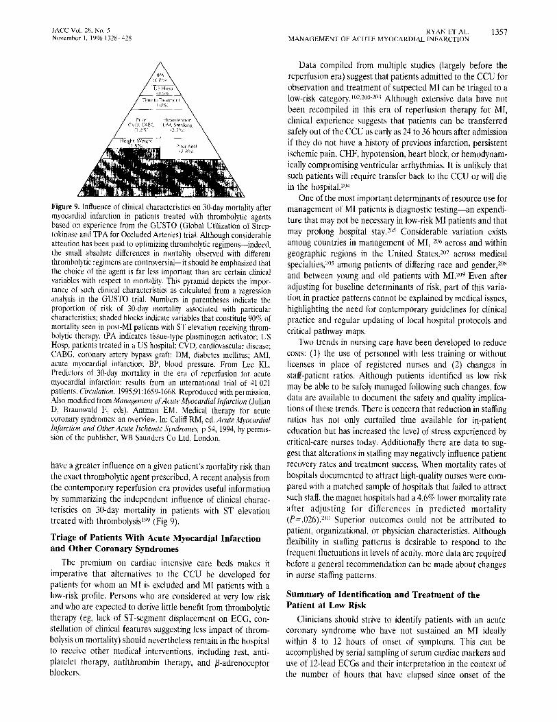
JACC Vol. 28, No. 5 RYAN ET AL. 1357 November 1, 1996:1328-428 MANAGEMENT OF ACUTE MYOCARDIAL INFARCTION
Figure 9. Influence of clinical characteristics on 30-day mortality after myocardial infarction in patients treated with thrombolytic agents based on experience from the GUSTO (Global Utilization of Strep- tokinase and TPA for Occluded Arteries) trial. Although considerable attention has been paid to optimizing thrombolytic regimens--indeed, the small absolute differences in mortality observed with different thrombolytic regimens are controversial--it should be emphasized that the choice of the agent is far less important than are certain clinical variables with respect to mortality. This pyramid depicts the impor- tance of such clinical characteristics as calculated from a regression analysis in the GUSTO trial. Numbers in parentheses indicate the proportion of risk of 30-day mortality associated with particular characteristics; shaded blocks indicate variables that constitute 90% of mortality seen in post-MI patients with ST elevation receiving throm- bolytic therapy, tPA indicates tissue-type plasminogen activator; US Hosp, patients treated in a US hospital; CVD, cardiovascular disease; CABG, coronary artery bypass graft; DM, diabetes mellitus; AMI, acute myocardial infarction; BP, blood pressure. From Lee KL. Predictors of 30-day mortality in the era of reperfusion for acute myocardial infarction: results from an international trial of 41 021 patients. Circulation. 1995;91:1659-1668. Reproduced with permission. Also modified from Management of Acute Myocardial lnfarction (Julian D, Braunwald E, eds). Antman EM. Medical therapy for acute coronary syndromes: an overview. In: Califf RM, ed. Acute Myocardial Infarction and Other Acute Ischemic Syndromes, p 54, 1994, by permis- sion of the publisher, WB Saunders Co Ltd, London.
have a greater influence on a given patient's mortality risk than the exact thrombolytic agent prescribed. A recent analysis from the contemporary reperfusion era provides useful information by summarizing the independent influence of clinical charac- teristics on 30-day mortality in patients with ST elevation treated with thrombolysis 199 (Fig 9).
Triage of Patients With Acute Myocardial Infarction and Other Coronary Syndromes
The premium on cardiac intensive care beds makes it imperative that alternatives to the CCU be developed for patients for whom an M1 is excluded and MI patients with a low-risk profile. Persons who are considered at very low risk and who are expected to derive little benefit from thrombolytic theraw (eg, lack of ST-segment displacement on ECG, con- stellation of clinical features suggesting less impact of throm- bolysis on mortality) should nevertheless remain in the hospital to receive other medical interventions, including rest, anti- platelet therapy, antithrombin therapy, and /3-adrenoceptor blockers.
Data compiled from multiple studies (largely before the reperfusion era) suggest that patients admitted to the CCU for observation and treatment of suspected MI can be triaged to a low-risk category. 1°2.2°°-2°3 Although extensive data have not been recompiled in this era of reperfusion therapy for MI, clinical experience suggests that patients can be transferred safely out of the CCU as early as 24 to 36 hours after admission if they do not have a history of previous infarction, persistent ischemic pain, CHF, hypotension, heart block, or hemodynam- ically compromising ventricular arrhythmias. It is unlikely that such patients will require transfer back to the CCU or will die in the hospital. 2°4
One of the most important determinants of resource use for management of MI patients is diagnostic testing--an expendi- ture that may not be necessary in low-risk MI patients and that may prolong hospital stay. zo5 Considerable variation exists among countries in management of MI, 2o6 across and within geographic regions in the United States, 207 across medical specialties, 205 among patients of differing race and gender, z°8 and between young and old patients with MI. z°9 Even after adjusting for baseline determinants of risk, part of this varia- tion in practice patterns cannot be explained by medical issues, highlighting the need for contemporary guidelines for clinical practice and regular updating of local hospital protocols and critical pathway maps.
Two trends in nursing care have been developed to reduce costs: (1) the use of personnel with less training or without licenses in place of registered nurses and (2) changes in staff-patient ratios. Although patients identified as low risk may be able to be safely managed following such changes, few data are available to document the safety and quality implica- tions of these trends. There is concern that reduction in staffing ratios has not only curtailed time available for in-patient education but has increased the level of stress experienced by critical-care nurses today. Additionally there are data to sug- gest that alterations in staffing may negatively influence patient recovery rates and treatment success. When mortality rates of hospitals documented to attract high-quality nurses were com- pared with a matched sample of hospitals that failed to attract such staff, the magnet hospitals had a 4.6% lower mortality rate after adjusting for differences in predicted mortality (P=.026). 2I~ Superior outcomes could not be attributed to patient, organizational, or physician characteristics. Although flexibility in staffing patterns is desirable to respond to the frequent fluctuations in levels of acuity, more data are required before a general recommendation can be made about changes in nurse staffing patterns.
Summary of Identification and Treatment of the Patient at Low Risk
Clinicians should strive to identify patients with an acute coronary syndrome who have not sustained an MI ideally within 8 to 12 hours of onset of symptoms. This can be accomplished by serial sampling of serum cardiac markers and use of 12-lead ECGs and their interpretation in the context of the number of hours that have elapsed since onset of the

1358 RYAN ET AL. JACC Vol. 28, No. 5 MANAGEMENT OF ACUTE MYOCARDIAL INFARCTION November 1, 1996:1328-428
patient's symptoms rather than adherence to a rigid protocol that requires a specified number of samples be drawn in the hospital. For example, to exclude an MI in a patient presenting to the ED within 4 hours of onset of ischemic-type chest discomfort, blood specimens might be drawn at admission and 8 hours later. A patient presenting 12 hours after onset of discomfort who has a normal ECG and normal serum cardiac marker levels in the ED need not be admitted to the CCU.
The mortality risk of patients who do sustain an MI can be evaluated using an integrated assessment of demographic and clinical variables according to the scheme shown in Fig 9. Low-risk patients include those without a history of previous infarction and who do not experience persistent ischemic pain, CHF, hypotension, heart block, or hemodynamically compro- mising ventricular arrhythmias. Such patients can be safely transferred out of the CCU within 24 to 36 hours of admission and, provided they remain asymptomatic and without compli- cations, constitute a group of patients who can be considered for early discharge in another 24 to 48 hours.
Identification and Treatment of the Patient at High Risk
Recommendations for Management of Recurrent Chest Discomfort
Class I
1. Aspirin for pericarditis. 2. /3-Adrenoceptor blockers intravenously, then orally for
ischemic-type chest discomfort. 3. (Re)administration of thrombolytic therapy (alteplase)
for patients with recurrent ST elevation. 4. Coronary arteriography for ischemic-type chest discom-
fort recurring after hours to days of initial therapy and associated with objective evidence of ischemia in patients who are candidates for revascularization.
Class lla
1. Nitroglycerin intravenously for 24 hours, then topically or orally for ischemie-type chest discomfort.
Class lib
1. Corticosteroids for periearditis. 2. Indomethacin for pericarditis.
Recurrent Chest Pain in the Post-MI Patient: Pericarditis and Ischemia
Recurrent chest pain in the patient still hospitalized after MI requires an evaluation of the cause of the pain while initiating therapy to resolve it, if possible.
The two most common cardiac causes of recurrent chest pain are acute pericarditis and ischemia, with the latter being the more common and potentially more serious. An ECG taken during the recurrent pain and compared with the initial one is clinically helpful, 38 Usually, recurrent pain within the first 12 hours after onset of infarction is considered to be
related to the original infarction itself. Pericarditis is probably not responsible for significant chest discomfort in the first 24 hours.
Pericarditis in acute MI occurs with extension of myocardial necrosis throughout the wall to the epicardium. The Multi- center Investigation of the Limitation of Infarct Size (MILIS) study 211 found that pericarditis (defined as the presence of a pericardial friction rub) occurred in 20% of 703 patients following acute MI. Postinfarction pericarditis occurs in ap- proximately 25% of patients with acute transmural MI not treated with thrombolytic therapy when typical symptoms or a pericardial friction rub are accepted as indicative of pericardi- tis, whereas the average incidence is only 14% when the presence of a friction rub is required for the diagnosis. 212 Patients with pericarditis have larger infarcts (defined by CK-MB), lower ejection fraction (measured with radionuclide ventriculography), and a higher incidence of CHF. 211,2t3 Peri- carditis may appear up to several weeks after acute MI. Anterior chest discomfort mimicking ischemia can occur with pericarditis. However, pericardial pain usually has distinguish- ing characteristics such as pleuritic or positional discomfort, radiation to the left shoulder, scapula or trapezius muscle and a pericardial rub, electrocardiographic J-point elevation with concave upward ST-segment elevation and PR depression. Pericardial effusion is evident echocardiographically in more than 40% of cases z14 but is rarely of hemodynamic conse- quence. A small effusion is not diagnostic of pericarditis as it can be demonstrated in the majority of patients with acute MI.:a
Focal pericarditis can be diagnosed electrocardiographi- cally by either persistently positive T waves or reversal of initially inverted T waves during the first week after acute transmural MI. However, similar T-wave alterations have also been observed when postinfarction pericardial effusion exists in the absence of clinically recognized pericarditis. 215 Pericar- ditis is not associated with re-elevation of CK-MB, and there are data to suggest its incidence has decreased in the reperfu- sion era. 216-21s Interestingly, Dressler syndrome (post-MI syn- drome), an autoimmune-type carditis, has essentially disap- peared 219 in the reperfusion era.
Aspirin (160 to 325 mg daily) is the treatment of choice, but high doses (650 mg every 4 to 6 hours) may be required. 22°,221 Indomethacin provides effective relief of symptoms; however, one study has presented data that suggest it may cause increased coronary vascular resistance 222 and experimentally causes thinning of developing scar. 223 Ibuprofen and cortico- steroids, also efficacious for pain relief, exert a tendency for thinning of scar and myocardial rupture, e24,225 The risk-benefit ratio of continuing antithrombotic therapy such as heparin in the presence of acute pericarditis is always a clinical challenge. Usually such therapy can be continued safely but requires added vigilance for the detection of enlarging pericardial effusion or signs of hemodynamic instability. Any evidence of impending cardiac tamponade is an indication for prompt termination of antithrombotic therapy.
It is important to differentiate between pain due to pericar-

JACC Vol. 28, No. 5 RYAN ET AL. 1359 November 1, 1996:1328-428 MANAGEMENT OF ACUTE MYOCARDIAL INFARCTION
ditis and that due to ischemia. The latter is more likely when the chest pain is similar to the initial ischemic-type chest discomfort, occurring at rest or with limited activity during hospitalization. This may or may not be associated with re-elevation of the CK-MB, ST-segment depression or eleva- tion, or pseudonormalization of inverted T waves (T-wave inversion on baseline ECG becoming upright during isch- emia). 214 Early recurrent angina, especially after successful reperfusion, may occur in up to 58% of patients. 226
Reinfarction has been reported to occur in approximately 10% of patients during the first 10 days but only in up to 3% to 4% of patients who have undergone thrombolytic therapy and received aspirin. 97,~27-23o Reinfarction is associated with re- elevation of serum CK-MB after the initial peak of the index infarction. Diagnosis of reinfarction within 18 hours after initiation of thrombolytic therapy should be based on recur- rence of severe ischemic-type chest discomfort lasting at least 30 minutes, usually, but not always, accompanied by recurrent ST-segment elevation of at least 0.1 mV in at least two contiguous ECG leads and re-elevation of CK-MB to more than the upper limit of normal or increased by at least 50% over the previous value97 Pathological findings of reinfarction show areas of healing myocardium along with the more recent necrosis usually in the same vascular risk region of myocardial tissue perfused by the original infarct-related artery. Death, severe CHF, and arrhythmias are early complications of rein- farction, and there is an increased incidence of cardiogenic shock or cardiac arrest. 227,231
With recurrent suspected ischemic-type chest discomfort, coronary arteriography often clarifies the cause of chest dis- comfort with demonstration of a high-grade coronary obstruc- tion. Prompt reperfusion using PTCA (if available and the lesion is suitable) or additional thrombolysis is appropriate, especially if a thrombus is present. If multiple high-grade lesions are present, more complete revascularization by CABG is appropriate.
Cardiac rupture may account for recurrent pain and occurs in 1% to 4% of all patients admitted with acute MI. 230,232-234 Left ventricular free wall rupture is typically heralded by chest pain and electrocardiographic ST-T wave changes with rapid progression to hemodynamic collapse and electromechanical dissociation. The frequency of cardiac rupture has two peaks: an early peak within 24 hours and a late one from 4 to 7 days after acute MI. Early rupture is related to the initial evolution of infarction before significant collagen deposition, and late rupture is related to expansion of the infarct-related ventricu- lar wan. 9°,232 Cardiac rupture is observed most frequently in patients with the first MI, those with anterior infarction, the elderly, and women. Other risk factors include hypertension during the acute phase of MI, lack of previous angina or MI, lack of collateral blood flow, Q waves on the ECG, use of corticosteroids or nonsteroidal anti-inflammatory drugs, and use of thrombolytic therapy more than 14 hours after on- set. 9°,234 However, thrombolytic therapy early after acute MI, ie, within 14 hours, decreases risk of cardiac rupture. 91,233 The most important determinants in preventing rupture are suc-
cessful early reperfusion and the presence of collateral circu- lationY 2,233 Pseudoaneurysm is a serious complication repre- senting rupture of the free wall. Clot forms in the pericardial space, and an aneurysmal wall containing clot and pericardium prevents exsanguination. The echocardiogram characteristi- cally shows a small neck opening into the body of the aneurysmY ~ Surgical correction is always indicated.
Pericardiocentesis for relief of tamponade and emergency surgical repair may be lifesaving. 235,23~ Transesophageal echo- cardiography is valuable in the diagnosis of free wall rupture and pseudoaneurysm, but for relief of tamponade in this setting, rapid fluid replacement is essential. Ideally the patient should be in the operating room and fully prepared for or already on cardiopulmonary bypass to prevent hemodynamic collapse.
Heart Failure and Low-Output Syndromes
Left Ventricular Dysfunction Pump failure due to acute MI is manifested clinically by a
weak pulse, poor peripheral perfusion with cool and cyanotic limbs, obtundation, and oliguria. Blood pressure (taken by cuff) is usually low, and there are variable degrees of pulmo- nary congestion. A third heart sound may be audible.
The treatment of LV dysfunction is determined by the specific hemodynamic derangements that are present, most importantly (1) pulmonary capillary wedge pressure, (2) car- diac output (measured with a balloon flotation catheter), and (3) systemic arterial pressure (preferably measured with an intra-arterial cannula). Often the patient has a cardiac index less than 2.5 L/min/m 2, a modestly elevated left-sided filling pressure (greater than 18 mm Hg), and a systolic arterial pressure 100 mm Hg or greater. Although this subject has evidence of LV dysfunction, systemic arterial pressure is adequate to allow for (l) modest diuresis (best accomplished with intravenous furosemide) in combination with (2) after- load and preload reduction, using nitroglycerin. Nitroglycerin offers a greater degree of venodilation than sodium nitroprus- side and relieves ischemia by dilating epicardial coronary arteries. In the early hours of acute infarction, when ischemia often contributes substantially to LV dysfunction, nitroglycerin is the more appropriate agent. Its intravenous infusion should be initiated at 5 /xg/min and increased gradually until mean systolic arterial pressure falls by 10% to 15% but not below 90 mm Hg. The institution of ACE inhibitor therapy is also appropriate in this setting.
The patient with more severe LV dysfunction has a de- pressed cardiac output, an abnormally high left-sided filling pressure, and systolic arterial pressure less than 90 mm Hg; this patient has, or is rapidly approaching, cardiogenic shock. If the patient is markedly hypotensive, intravenous norepinephrine should be administered until systolic arterial pressure rises to at least 80 mm Hg, at which time a change to dopamine may be attempted, beginning at 5 to 15 p~g/kg per minute. Once arterial pressure is brought to at least 90 mm Hg, intravenous dobutamine may be given simultaneously in an attempt to reduce the magnitude of the dopamine infusion. In addition,

1360 RYAN ET AL. JACC Vol. 28, No. 5 MANAGEMENT OF ACUTE MYOCARDIAL INFARCTION November 1, 1996:1328-428
consideration should be given to initiating intra-aortic balloon counterpulsation.
Recent nonrandomized and retrospective studies have sug- gested that mechanical reperfusion by PTCA or CABG of occluded coronary arteries may improve survival in patients with MI and cardiogenic shock. In large clinical trials such patients have an in-hospital survival rate ranging from 20% to 50% when treated with intravenous thrombolytic therapy. 237-24° In other case series mechanical reperfusion with PTCA has been reported to result in hospital survival rates as high as 70%, but selection bias may have influenced these findings. Multicenter, prospective, randomized studies are currently under way to verify these promising results. 241
In the setting of cardiogenic shock complicating acute MI, emergency CABG has been used when other interventions have failed or not been indicated. A multicenter trial of surgically controlled repeffusion using total vented cardiopul- monary bypass and substrate-enhanced blood cardioplegia in patients with acute non-PTCA-related coronary occlusion noted 3.4% mortality overall with 9% mortality in patients with preoperative shock. 242,243 Data from the SHOCK (Should We Emergently Revascularize Occluded Coronaries for Cardio- genic Shock?) Registry suggest that, in some patients, emer- gency CABG (without specific recommendations regarding intraoperative myocardial protection strategies) is associated with lower mortality (19%) than emergency PTCA (60%) for patients with cardiogenic shock complicating acute MI. 241 In other nonrandomized studies surgical mortality ranged from 12% 244 to 42% 245 but is generally superior to other treatment modalities. The efficacy of emergency CABG in patients with cardiogenic shock may be better defined by an ongoing clinical trial. 241
Based on these earlier studies, consideration for emergency CABG should be given for acute MI patients with multivessel disease or cardiogenic shock and who are not candidates for or who have undergone unsuccessful thrombolytic therapy and/or PTCA, and who are within 4 to 6 hours of onset of MI.
Right Ventricular Infarction and Dysfunction
Right ventricular infarction encompasses a spectrum of dis- ease states ranging from asymptomatic mild right ventricular (RV) dysfunction through cardiogenic shock. Most patients demonstrate a return of normal RV function over a period of weeks to months, suggesting RV stunning has occurred rather than irreversible necrosis. In this sense RV ischemia can be demonstrated in up to half of all inferior MIs, although only 10% to 15% of patients show classical hemodynamic abnor- malities.24~,,247
Right ventricular infarction accompanying inferior MIs is associated with a significantly higher mortality (25% to 30%) and thus identifies a high-risk subgroup of patients with inferior Mls (6%) who should be considered high-priority candidates for reperfusion. 246 One group of investigators re- cently reported a 31% in-hospital mortality rate in patients with inferior MIs complicated by RV infarction compared with 6% in patients who had an inferior MI without RV involve-
ment. 246 The treatment of patients with RV ischemia is different and occasionally diametrically opposed to manage- ment of LV dysfunction.
Anatomic and Pathophysiological Considerations
The right coronary artery usually supplies most of the RV myocardium; thus, occlusion of this artery proximal to the RV branches will lead to RV ischemia, z4s Hemodynamically sig- nificant RV infarctions occur almost exclusively in the setting of inferior acute MIs. 249 Because the right ventricle has a much smaller muscle mass than the left ventricle, due to the lower vascular resistance of the pulmonary circuit, myocardial oxygen demand is significantly less than that of the left ventricleY ° Coronary perfusion of the right ventricle occurs in both systole and diastole. 25° The right ventricle also has a more favorable oxygen supply-demand ratio than the left ventricle, because of the more extensive collateral flow from left to right351,25z These factors likely explain the absence of hemodynamically significant RV ischemia in most patients with proximal right coronary artery occlusions, as well as improvement in RV function observed in the majority of patients following RV ischemia. 253
The severity of the hemodynamic derangements associated with RV ischemia is related to (1) the extent of ischemia and subsequent RV dysfunction, (2) the restraining effect of the surrounding pericardium, and (3) interventricular dependence related to the shared interventricular septum. When the right ventricle becomes ischemic, it acutely dilates, resulting in an increased intrapericardial pressure caused by the restraining forces of the pericardium. As a consequence, there is a reduction in RV systolic pressure and output, decreased LV preload, a reduction in LV end-diastolic dimension and stroke volume, and a shifting of the interventricular septum toward the left ventricle. 254 Because of this RV systolic and diastolic dysfunction, the pressure gradient between the right and left atria becomes an important driving force for pulmonary per- fusion. Factors that reduce preload (volume depletion, diuret- ics, nitrates) or diminish augmented right atrial contraction (concomitant atrial infarction, loss of AV synchrony), as well as factors that increase RV afterload (concomitant LV dys- function), are likely to have profoundly adverse hemodynamic effects. 255-257 Goldstein and coworkers 256 have demonstrated the importance of a paradoxical interventricular septal motion that bulges in pistonlike fashion into the right ventricle, generating systolic force, which allows pulmonary perfusion. The loss of this compensatory mechanism with concomitant septal infarction may result in further deterioration in patients with RV ischemia.
Clinical Diagnosis
Evidence of RV ischemia should he sought in all patients with acute inferior MI. The clinical triad of hypotension, clear lung fields, and elevated jugular venous pressure in the setting of an inferior MI is characteristic of RV ischemia. Although specific, this triad has a sensitivity of less than 25%. z58 Dis- tended neck veins alone or the presence of Kussmaul's sign

JACC Vol. 28, No. 5 RYAN ET AL. 1361 November 1, 1996:1328-428 MANAGEMENT OF ACUTE MYOCARDIAL INFARCTION
Table 6. Treatment Strategy for Right Ventricular Ischemia/Infarction
Maintain right ventricular preload Volume loading (IV normal saline) Avoid use of nitrates and diuretics Maintain AV synchrony
AV sequential pacing for symptomatic high-degree heart block unresponsive to atropine
Prompt cardioversion for hemodynamically significant SVT Inotropic support
Dobutamine (if cardiac output fails to increase after volume loading) Reduce right ventricular afterload with left ventricular dysfunction
Intra-aortic balloon pump Arterial vasodilators (sodium nitroprusside, hydralazine) ACE inhibitors
Reperfusion Thrombolytic agents Primary PTCA CABG (in selected patients with multivessel disease)
IV indicates intravenous; AV, atrioventricular; SVT, supraventricular tachy- cardia; ACE, angiotensin converting en~me; PTCA, percutaneous transluminal coronary angioplasty: CABG, coronary, arte~, bypass graft.
(distention of the jugular vein on inspiration) are both sensitive and specific for RV ischemia in patients with an inferior MI. 2s9 These findings may be masked in the setting of volume depletion and may only become evident after adequate volume loading. A right atrial pressure of 10 mm Hg or greater and greater than 80% of pulmonary wedge pressure is a relatively sensitive and specific finding in patients with RV ischemia. 26°
Demonstration of 1 mm ST-segment elevation in the right precordial lead V4R is the single most predictive electrocardio- graphic finding in patients with RV ischemia. 261 The finding may be transient; half of patients show resolution of ST elevation within 10 hours of onset of symptoms. 262 It is important for physicians to ensure that hospital personnel (house officer, nurse, technician) recording the ECG in this setting know how to properly record lead V4R, especially in view of the variety of multilead recording systems available. All patients with inferior infarctions should be screened initially for this finding at the time of admission. Critical-care staff should be encouraged to choose routine monitoring leads based on infarct site. Echocardiography can be helpful in patients with suspicious but nondiagnostic findings, sva It can show RV dilation and asynergy, abnormal interventricular and interatrial septal motion, and even right to left shunting through a patent foramen ovale. 2~+26~ This latter finding is unique to RV ischemia and should be suspected when persis- tent hypoxia is not responsive to supplemental oxygen. 265
Management of Right Ventricular Ischemia/Infarction
Treatment of RV infarction includes early maintenance of RV preload, reduction of RV afterload, inotropic support of the dysfunctional right ventricle, and early reperfusion 73 (Ta- ble 6). Because of their influence on preload, drugs routinely used in management of LV infarctions, such as nitrates and diuretics, may reduce cardiac output and produce severe
hypotension when the right ventricle is ischemic. Indeed, a common clinical presentation for RV infarction is profound hypotension following administration of sublingual nitroglyc- erin, with the degree of hypotension often out of proportion to the electrocardiographic severity of the infarct. Volume load- ing with normal saline alone often resolves accompanying hypotension and improves cardiac output, z*6 In other cases, volume loading further elevates the right-sided filling pressure and RV dilatation, resulting in decreased LV output. 267 Al- though volume loading is a critical first step in the management of hypotension associated with RV ischemia, inotropic support (in particular, dobutamine hydrochloride) should be initiated if cardiac output fails to improve after 1 to 2 L of fluid has been given.
Another important factor for sustaining adequate RV pre- load is maintenance of AV synchrony. High-degree heart block is common, occurring in as many as half of these patients. 26~ Atrioventricular sequential pacing leads to a significant in- crease in cardiac output and reversal of shock, even when ventricular pacing alone has not been of benefit. 269 Atrial fibrillation may occur in up to one third of patients with RV ischemia 27° and has profound hemodynamic effects. Prompt cardioversion from atrial fibrillation should be considered at the earliest sign of hemodynamic compromise. When LV dysfunction accompanies RV ischemia, the right ventricle is further compromised because of increased RV afterload and reduction in stroke volume. 271 In such circumstances, the use of afterload-reducing agents such as sodium nitroprusside or an intra-aortic counterpulsation device is often necessary to "unload" the left and subsequently the right ventricle.
Fibrinolytic therapy and primary PTCA with subsequent reperfusion have been shown to improve RV ejection frac- tion 272 and reduce the incidence of complete heart block. 272-274
Prognosis
The mere presence of RV ischemia evident by noninvasive criteria is associated with significantly increased short-term morbidity and mortality and may also influence long-term outcome. 246,275,276 Clinical and hemodynamic recovery even- tually occur even in patients with RV dysfunction 259,277-279 that persists for weeks or months. This return to normal may be due to improvement of concomitant LV dysfunction, resulting in a reduction in RV afterload, or to a gradual stretching of the pericardium with amelioration of its restrain- ing effect. 277
Hemodynamie Monitoring
Recommendations for Balloon Flotation Right-Heart Catheter Monitoring
Class I
1. Severe or progressive CHF or pulmonary edema. 2. Cardiogenic shock or progressive hypotension. 3. Suspected mechanical complications of acute infarction,
ie, VSD, papillary muscle rupture, or pericardial tamponade.

1 3 6 2 RYAN ET AL. JACC Vol. 28, No. 5 MANAGEMENT OF ACUTE MYOCARDIAL INFARCTION November 1, 1996:1328-428
Class Ila
1. Hypotension that does not respond promptly to fluid administration in a patient without pulmonary congestion.
Class III
1. Patients with acute infarction without evidence of car- diac or pulmonary complications.
The balloon flotation catheter is often very helpful in management of acute MI and concomitant hemodynamic instability, including low cardiac output, hypotension, persis- tent tachycardia, pulmonary edema, and apparent cardiogenic shock. In the patient with hypotension and tachycardia, the balloon flotation catheter allows quick and easy differentiation of (1) inadequate intravascular volume, with a resultant low left-sided filling pressure, and (2) adequate intravascular vol- ume and a high left-sided filling pressure due to extensive LV dysfunction. Treatment of the former is prompt expansion of intravascular volume (with normal saline), whereas manage- ment of the latter often includes diuresis, inotropic support, afterload reduction, and/or other supportive measures. In those with extensive LV dysfunction, a balloon flotation cath- eter in the right side of the heart can be used to monitor therapeutic efforts to adjust the left-sided filling pressure so as to maximize cardiac output at the lowest possible filling pressure. These sophisticated manipulations of intracardiac pressures and cardiac output are usually made considerably easier with information provided by a flotation catheter.
Although the balloon flotation catheter is quite safe when used by experienced operators, its use has a recognized association with adverse events, including ventricular tachyar- rhythmias (during its manipulation) and pulmonary hemor- rhage or infarction. In addition, it causes some patient discom- fort and requires that the patient be relatively immobile. Because the pressure waveform recorded from the catheter tip may be distorted, the clinician should routinely examine the actual waveform rather than rely on the digital display of pressure. Because of the risk of infection, balloon flotation catheters generally should not remain in the same site for more than 5 days.
Recommendations for Intra-arteriai Pressure Monitoring
Class I
1. Patients with severe hypotension (systolic arterial pres- sure less than 80 mm Hg) and/or cardiogenic shock.
2. Patients receiving vasopressor agents.
Class lla
1. Patients receiving intravenous sodium nitroprusside or other potent vasodilators.
Class lib
1. Hemodynamically stable patients receiving intravenous nitroglycerin for myocardial ischemia.
2. Patients receiving intravenous inotropic agents.
Class III
1. Patients with acute infarction who are hemodynamically stable.
All CCUs should have the equipment and personnel to monitor intra-arterial pressure. Such monitoring is useful in all hypotensive patients, particularly those with cardiogenic shock. Long-term monitoring is best accomplished through the radial artery, although the brachial or femoral arteries may be used as alternatives. Perfusion of the limb or hand distal to the catheter site must be carefully and periodically examined for evidence of ischemia. Because of risk of arterial thrombosis and infection, intra-arterial catheters generally should not remain in the same arterial site for prolonged periods of time, certainly no longer than 72 hours. Intra-arterial and central catheters can be left in place for this amount of time only if carefully inserted and properly cared for with a sterile occlu- sive dressing. Before insertion, the site should be adequately prepared under sterile conditions. Antibacterial ointments are no longer recommended. 28°
Recommendations for Intra-aortic Balloon Counterpulsation
Class I
1. Cardiogenic shock not quickly reversed with pharmaco- logical therapy as a stabilizing measure for angiography and prompt revascularization.
2. Acute mitral regurgitation or VSD complicating MI as a stabilizing therapy for angiography and repair/revascularization.
3. Recurrent intractable ventricular arrhythmias with he- modynamic instability.
4. Refractory post-MI angina as a bridge to angiography and revascularization.
Class lla
1. Signs of hemodynamic instability, poor LV function, or persistent ischemia in patients with large areas of myocar- dium at risk.
Class lib
1. In patients with successful PTCA after failed thrombo- lysis or those with three-vessel coronary disease to prevent reocclusion.
2. In patients known to have large areas of myocardium at risk with or without active ischemia.
Since its introduction in the late 1960s, intra-aortic balloon counterpulsation has been recognized as an effective treatment for patients with unstable ischemic syndromes and cardiogenic shock. 2sl-286 Reduction of LV afterload by rapid deflation of the balloon in end diastole appears to be the predominant mechanism of the balloon's effect. 287,288 By inflating in diastole, the balloon also raises diastolic coronary and systemic perfu- sion. Studies on the effects of this increased perfusion pressure on coronary blood flow and myocardial oxygen consumption have yielded conflicting results. 2s9,29° Recently Kern et al, z91 using Doppler flow velocity measurements, were able to show
JACC Vol. 28. No. 5 RYAN ET AL. 1363 November 1, 1996:1328-428 MANAGEMENT OF ACUTE MYOCARDIAL INFARCTION
a nearly twofold increase in proximal coronary flow velocity. This combination of decreased myocardial oxygen demand and maintained or improved coronary flow make intra-aortic bal- loon pumping a powerful tool for patients with cardiogenic shock or acute ischemic syndromes.
Counterpulsation was first used as a stand-alone modality to treat patients with post-MI cardiogenic shock. 281 Counter- pulsation stabilized most patients, but in-hospital mortality remained a dismal 83%. 2sl In virtually all shock-management strategies in which counterpulsation is used today, it acts as a stabilizing device or bridge to facilitate diagnostic angiography and revascularization or repair. In selected patient populations survival rates for cardiogenic shock treated in the first 16 to 24 hours with intra-aortic balloon pumping and surgical and angioplasty revascularization range between 60% and 75%. 2~4,2~2 Similarly, intra-aortic balloon pumping and early repair for acute VSD and mitral regurgitation show survival rates of 60% or sometimes higher. 2s5 Patients with severe recurrent ischemia after MI can be stabilized with an intra- aortic balloon pump so that they can undergo angiography and emergency revascularization with PTCA or CABG. 24°
Several early studies, before reperfusion therapy, showed that routine prophylactic use of intra-aortic balloon pumping in acute MI 2s2-293 did not affect infarct size. A retrospective review of the Thrombolysis and Angioplasty in Myocardial Infarction (TAMI) trials suggested that placement of an intra-aortic balloon pump after reperfusion with either throm- bolytic therapy or PTCA reduced the incidence of reocclu- sion. 294 In a subsequent randomized trial, patients with rescue PTCA at 90 minutes or those with three-vessel CAD 295 showed a reduction of reocclusion events from 21% to 8% after intra-aortic balloon pumping. In a second randomized trial of the use of prophylactic placement of these devices in high-risk patients (age greater than 70, ejection fraction less than 45%, three-vessel disease, suboptimal PTCA, saphenous graft occlu- sion, ventricular arrhythmias) undergoing primary PTCA, 437 patients were studied to determine the effect of balloon pumping on resulting LV function and a composite clinical end point (death, reocclusion, reinfarction, CHF, and stroke). There was no significant difference in clinical outcome, includ- ing rate of reocclusion (6.2% versus 8.0%), nor did it influence global or regional LV function. 29~, However, there was a reduction in the incidence of recurrent ischemia, including the need for repeat angiography and PTCA of the infarct-related artery. In summary, for patients without LV dysfunction, the prophylactic and routine use of intra-aortic balloon pumping following either reperfusion strategy cannot be recommended.
Rhythm Disturbances
Atrial Fibrillation
Recommendations
Class I
1. Electrical cardioversion for patients with severe hemo- dynamic compromise or intractable ischemia.
2. Rapid digitalization to slow a rapid ventricular response and improve LV function.
3. Intravenous /]-adrenoceptor blockers to slow a rapid ventricular response in patients without clinical LV dysfunc- tion, bronchospastic disease, or AV block.
4. Heparin should be given.
Class l la
1. Either diltiazem or verapamil intravenously to slow a rapid ventricular response if/3-adrenoceptor blocking agents are contraindicated or ineffective.
Atrial fibrillation (AF) associated with acute MI most often occurs within the first 24 hours and is usually transient but may recur. The incidence of AF in acute MI ranges from 10% to 16%, 2~7,2'~8 whereas atrial flutter or supraventricular tachycar- dia is much less frequent. The consequences and acute treat- ment of all three conditions may be considered together, recognizing that in atrial flutter and supraventricular tachycar- dia, atrial pacing may be effective in terminating the tachycardia. 299-3°5 The incidence of AF increases with age, occurring in 4.2% of patients aged 59 years or less and in 16% of patients aged 70 or older. Atrial fibrillation occurs more often in patients with larger infarcts, those anterior in location, and in patients whose hospital course is complicated by CHF, complex ventricular arrhythmias, advanced AV block, atrial infarction, or pericarditis. Atrial fibrillation may also occur in patients with inferior MI secondary to proximal right coronary artery occlusion due to involvement of the sinoatrial nodal artery, which provides the major blood supply to the atria.
The incidence of AF after acute MI is decreased in patients receiving thrombolytic therapy, 3"°-3°6 and in the GUSTO trial patients treated with accelerated alteplase and intravenous heparin had a significantly lower incidence of AF and atrial flutter compared with other fibrinolytic therapies. 22s The oc- currence of AF is also associated with excess catecholamine release, hypokalemia, hypomagnesemia, hypoxia, underlying chronic lung disease, and ischemia of the sinus node or left atrial circumflex arteries, z7~,297,3°°,307-31°
Systemic embolization is more frequent in patients with paroxysmal AF (1.7%) compared with those without (0.6%), with one half of embolic events occurring on the first day of hospitalization and more than 90% occurring by the fourth day. 29s Because AF can be associated with pericarditis, the development of PR-segment displacement on serial ECGs may predict risk of developing AF during hospitalization? o6
When hemodynamic compromise occurs due to rapid ven- tricular rate or loss of atrial contraction, immediate cardiover- sion is indicated, beginning with 100 J, then 200 to 300 J, then 360 J if lower energies fail. In the conscious patient, support with brief anesthesia is essential.
In the absence of CHF or severe pulmonary disease, one of the most effective means of slowing the ventricular rate in AF is the use of intravenous/3-adrenoceptor blocking agents such as atenolol (2.5 to 5.0 mg over 2 minutes to a total of 10 mg in 10 to 15 minutes) or metoprolol (2.5 to 5.0 mg every 2 to 5

1364 RYAN ET AL. JACC Vol. 28, No. 5 MANAGEMENT OF ACUTE MYOCARDIAL INFARCTION November 1, 1996:1328-428
minutes to a total of 15 mg over 10 to 15 minutes). Heart rate, blood pressure, and the ECG should be monitored, and treatment should be halted when therapeutic efficacy is achieved or if systolic blood pressure falls below 100 mm Hg or heart rate below 50 bpm during treatment.
Rapid administration of digitalis to achieve rate slowing may be accomplished by giving intravenous digoxin (8 to 15 Ixg/kg [0.6 to 1.0 mg in a person weighing 70 kg]) with half the dose administered initially and the additional increment in 4 hours. 221 This method provides a slower response than intravenous/3-adrenoceptor blockade; however, some effect on rate slowing may be detectable in one half to 2 hours.
Rate slowing may also be achieved by intravenous vera- pamil (5 to 10 mg [0.075 to 0.15 mg/kg]) given over 2 minutes with a repeat dose 30 minutes later or similarly by intravenous bolus administration of diltiazem (20 mg [0.25 mg/kg]) over 2 minutes followed by an infusion of 10 mg/h. If rate response is inadequate, a second dose of diltiazem (25 mg [0.35 mg/kg]) may be given over 2 minutes after an interval of 15 minutes. A subsequent infusion is given at a rate of 10 to 15 mg/h. Because of their negative inotropic effect and newer concerns regarding the use of calcium channel blockers in acute MI, these agents are not recommended as first-line drugs despite their effective- ness in slowing heart rate, especially if given to patients also receiving/3-blocking agents. 311 Although AF after acute MI is usually transient, heparin therapy should be given to patients not already receiving it.
Guidelines for use of Class I and Class III antiarrhythmic agents and electric shock for converting persistent AF have not been formulated. It is not clear whether antiarrhythmic agents should be used for prevention of AF if it recurs during hospitalization, although its recurrence portends a worse prog- nosis? °5 For this reason it has become common practice to use antiarrhythmic agents such as quinidine, procainamide, or, preferably, amiodarone or sotalol. 312 Transient AF does not obligate the patient to receive long-term anticoagulation or antiarrhythmic agents, but if such treatment is elected, it is appropriate to limit their use to 6 weeks if sinus rhythm has been restored.
Ventricular Tachycardia/Ventricular Fibrillation
Recommendations
Class I
1. Ventricular fibrillation should be treated with an unsyn. chronized electric shock with an initial energy of 200 J; if unsuccessful, a second shock of 200 to 300 J should be given, and, if necessary, a third shock of 360 J.
2. Sustained (more than 30 seconds or causing hemody- namic collapse) polymorphic VT should be treated with an unsynchronized electric shock using an initial energy of 200 J; if unsuccessful, a second shock of 200 to 300 J should be given, and, if necessary, a third shock of 360 J.
3. Episodes of sustained monomorphic VT associated with angina, pulmonary edema, or hypotension (blood pressure less than 90 mm Hg) should be treated with a synchronized
electric shock of 100 J initial energy. Increasing energies may be used if not initially successful.
4. Sustained monomorphic VT not associated with angina, pulmonary edema, or hypotension (blood pressure less than 90 mm Hg) should be treated with one of the following regimens:
a. Lidocaine: bolus 1.0 to 1.5 mg/kg. Supplemental bo- luses of 0.5 to 0.75 mg/kg every 5 to 10 minutes to a maximum of 3 mg/kg total loading dose may be given as needed. Loading is followed by infusion of 2 to 4 mg/min (30 to 50 pg/kg per minute).
b. Procainamide: 20 to 30 mg/min loading infusion, up to 12 to 17 mg/kg. This may be followed by an infusion of 1 to 4 mg/min.
c. Amiodarone: 150 mg infused over 10 minutes followed by a constant infusion of 1.0 mg/min for 6 hours and then a maintenance infusion of 0.5 mg/min.
d. Synchronized electrical cardioversion starting at 50 J (brief anesthesia is necessary).
Comment: Knowledge of the pharmacokinetics of these agents is important because dosing varies considerably, depending on age, weight, and hepatic and renal function.
Class IIa
1. Infusions of antiarrhythmic drugs may be used after an episode of VTNF but should be discontinued after 6 to 24 hours and the need for further arrhythmia management assessed.
2. Electrolyte and acid-base disturbances should be cor- rected to prevent recurrent episodes of VF when an initial episode of VF has been treated.
Class lib
1. Drug-refractory polymorphic VT should be managed by aggressive attempts to reduce myocardial ischemia, including therapies such as fl-adrenoceptor blockade, intra-aortic bal- loon pumping, and emergency PTCA/CABG surgery. Amioda- rone, 150 mg infused over 10 minutes followed by a constant infusion of 1.0 mg/min for up to 6 hours and then a mainte. nance infusion at 0.5 mg/min, may also be helpful.
Class III
1. Treatment of isolated ventricular premature beats, cou- plets, runs of accelerated idioventricular rhythm, and nonsus- tained VT.
2. Prophylactic administration of antiarrhythmic therapy when using thrombolytic agents.
Ventricular Fibrillation--Background
Disturbances of cardiac rhythm are common during acute MI. Early-phase arrhythmias are probably largely a result of micro reentry. Although other electrophysiological mecha- nisms such as enhanced automaticity and triggered activity have been proposed in experimental models of MI, convincing evidence of their role in human MI is not yet established. 313 Important contributory factors include heightened adrenergic nervous system tone, hypokalemia, hypomagnesemia, intracel-

JACC Vol. 28, No. 5 RYAN ET AL. 1365 November 1, 1996:1328 428 MANAGEMENT OF ACUTE MYOCARDIAL INFARCTION
lular hypercalcemia, acidosis, free fatty acid production from lipolysis, and free radical production from reperfusion of ischemic myocardium? 13-315 The relative importance of each of these factors in the pathogenesis of arrhythmias during acute MI has not been established, nor has it been clearly shown that aggressive measures specifically targeted at one or more of these mechanisms can be relied on clinically to reduce arrhyth- mia frequency in acute MI.
Primary VF should be distinguished from secondary VF, the latter occurring in the presence of severe CHF or cardio- genie shock. 3u' Late VF develops more than 48 hours after onset of infarction. The incidence of primary VF is highest (around 3% to 5%) in the first 4 hours after MI and declines markedly thereafter. 3~7 Epidemiological data suggest that the incidence of primary VF in acute MI may be decreasing in the current era, possibly due to aggressive attempts at infarct-size reduction, correction of electrolyte deficits, and a greater use of/3-adrenoceptor blocking agents? 'a Contrary to prior belief, primary VF appears to be associated with a significantly higher in-hospital mortality, but those persons who survive to hospital discharge have the same long-term prognosis as patients who do not experience primary VF. 3>
Management Strategies for Ventricular Fibrillation PROPHYLAXIS
Primary VF remains an important contributor to risk of mortality during the first 24 hours after MI. Therefore, a reliable method for its prediction and prevention remains desirable but has not been established despite extensive clinical investigation. Classification of ventricular arrhythmias in as- cending order of risk of primary VF ("warning arrhythmias") was proposed, but this approach lacks appropriate specificity and sensitivity? 2°-~-2
Accelerated idioventricular rhythm occurs frequently dur- ing the first 12 hours of infarction. Data from the prereperfu- sion era do not support development of accelerated idioven- tricular rhythm as a risk factor for development of VF. 32~,32~ In patients receiving thrombolysis or undergoing primary PTCA, accelerated idioventricular rhythm may be a reperfusion ar- rhythmia and does not indicate an increased risk of VF? 24 Thus, it is best managed by observation and should not trigger initiation of antiarrhythmic prophylaxis against VF.
Meta-analysis of randomized trials of prophylaxis with lidocaine has shown a reduction in the incidence of primary VF by about 33%, but this was offset by a trend toward increased mortality, probably from fatal episodes of bradycardia and asystole. 32s The prior practice of routine ("prophylactic") administration of lidocaine to all patients with known or suspected MI has been largely abandoned in most contempo- rary CCU protocols because of an unfavorable risk-benefit ratio and a decreased incidence of the target arrhythmia. Thus, its routine use is not recommended, with the possible excep- tion being situations in which a defibrillator is unavailable, provided there is a skilled professional always available who can initiate CPR if asystole occurs. Prophylactic regimens with
other antiarrhythmic drugs have not been evaluated as exten- sively as lidocaine, and no other agents, even including the close structural analogues mexiletine and tocainide, have been shown to decrease the incidence of primary VF when given on a prophylactic basis.
Routine administration of intravenous /3-adrenoceptor blockers to patients without hemodynamic or electrical (AV block) contraindications is associated with a reduction in incidence of early VF. Intravenous followed by oral ,6-adrenoceptor blockers should be given in the absence of contraindications. Suitable regimens include intravenous metoprolol (5 mg every 2 minutes for 3 doses, if tolerated, followed by 50 mg orally twice a day for at least 24 hours and then increased to 100 mg twice a day). An alternative regimen is atenolol (5 to 10 mg intravenously followed by 100 mg orally on a daily basis).
Clinical experience as well as observational data from CCU populations has identified hypokalemia as an arrhythmogenic risk factor for VF. 314-315 Low serum levels of magnesium have not been clearly shown to be associated with an increased risk of VF, -~15 although tissue depletion of magnesium remains a potential risk factor. Although randomized clinical trial data do not exist to confirm the benefits of repletion of potassium and magnesium deficits in preventing VF, it is sound clinical practice to maintain serum potassium levels at greater than 4.0 mEq/L and magnesium levels at greater than 2.0 mEq/L in patients with acute MI.
TREATMENT
Ventrieular fibrillation should be treated with an unsyn- chronized electric shock using an initial energy of 200 J. If this is unsuccessful, a second shock using 200 to 300 J and, if necessary, a third shock using 360 J is indicated? 26 Ventricular fibrillation that is not easily converted by defibrillation may be treated with additional adjunctive measures. No rigorous sci- entific support exists to favor one pharmacological treatment program over another or even to confirm that they improve the likelihood of resuscitation over repeated shocks given alone. The ACLS protocol recommends adjunctive therapy in the following hierarchy, as needed, for resistant VF32~': (1) epi- nephrine (1 mg IV push); (2) lidocaine (1.5 mg/kg); (3) bretylium (5 to 10 mg/kg). Intravenous amiodarone (150 mg bolus), now available, also may be used.
There are no firm data to help define an optimal manage- ment strategy for prevention of recurrent VF in patients who have sustained an initial episode of VF in the setting of MI. It seems prudent to correct any electrolyte and acid-base distur- bances and administer /3-adrenoceptor-blocking agents to inhibit increased sympathetic nervous system tone and prevent ischemia. 313 If infusion of an antiarrhythmic drug is initiated (eg, lidocaine 2 mg/min), it should probably be maintained for only 6 to 24 hours and then discontinued so that the patient's ongoing need for antiarrhythmic treatment can be reassessed.

1366 RYAN ET AL. JACC Vol. 28, No. 5 MANAGEMENT OF ACUTE MYOCARDIAL INFARCTION November 1, 1996:1328-428
Ventricular Tachycardia--Background
Several definitions have been used for VT in the setting of acute MI. Nonsustained VT lasts less than 30 seconds, whereas sustained VT lasts more than 30 seconds and/or causes earlier hemodynamic compromise requiring immediate intervention. Based on electrocardiographic appearance, VT has also been categorized as monomorphic or polymorphic. While short bursts (fewer than 5 beats) of nonsustained VT of either monomorphic or polymorphic configuration may be seen fre- quently, contemporary epidemiological data do not suggest that they are associated with a sufficiently increased risk of sustained VT or VF to warrant a recommendation of prophy- lactic therapy.
The vast majority of post-MI VT and VF occur within the first 48 hours of MI. 31v Sustained VT or VF occurring outside of this time frame deserves careful evaluation, including con- sideration of electrophysiology studies. In addition, monomor- phic VT at rates less than 170 bpm are unusual as a post-MI arrhythmia and suggests a more chronic (mature) arrhythmo- genic substrate. 32v-33°
Management Strategies for Ventricular Tachycardia
1. Only for episodes of sustained hemodynamically com- promising VT is treatment always indicated. 3j3 In the absence of clinical evidence of effective perfusion, urgent electrical conversion of VT is indicated. Rapid, polymorphic-appearing VT should be considered similar to VF and managed with an unsynchronized discharge of 200 J, while monomorphic VT with rates greater than 150 bpm can usually be treated with a 100-J synchronized discharge. 32~ If the patient is hemodynam- ically stable, brief trials of medications (lidocaine or procain- amide) may be given first. Immediate cardioversion is generally not needed for rates under 150 bpm.
2. Episodes of sustained VT that are somewhat better tolerated hemodynamically may initially be treated with one of the following drug regimens:
a. Lidocaine: bolus 1.0 to 1.5 mg/kg. Supplemental boluses of 0.5 to 0.75 mg/kg every 5 to 10 minutes to a maximum of 3 mg/kg total loading dose may be given as needed. Loading is followed by infusion of 2 to 4 mg/min (30 to 50/sg/kg per minute), in older patients and those with CHF or hepatic dysfunction, infusion rates should be reduced to avoid lidocaine toxicity.
b. Procainamide: 20 to 30 rag/rain loading infusion, up to 12 to 17 mg/kg. This may be followed by an infusion of 1 to 4 mg/min. Infusion rates should be lower in the presence of renal dysfunction.
c. Amiodarone: 150 mg infused over 10 minutes fol- lowed by a constant infusion of 1.0 mg/min for 6 hours and then a maintenance infusion at 0.5 mg/min.
3. Rare episodes of drug-refractov sustained polymor- phic VT ("electrical storm") have been reported in cases of acute MI. Anecdotal evidence suggests that these may be related to uncontrolled ischemia and increased sympathetic tone and are best treated by intravenous /3-adrenoceptor
blockade, 331 intravenous amiodarone, 332 intra-aortic balloon pumping, or emergency revascularization.
Bradyarrhythmias and Heart Block
Background, Epidemiology, and Importance
Sinus bradycardia occurs frequently (in 30% to 40% of patients) with acute MI, especially within the first hour of inferior MI and with reperfusion of the right coronary artery (Bezold-Jarish reflex), a result of increased parasympathetic activity (vagal tone). 97 Heart block may develop in approxi- mately 6% to 14% of patients with acute MI and predicts an increased risk of in-hospital mortality but is a poor predictor of long-term mortality in those surviving to discharge. 333-335 In- traventricular conduction delay has been reported in about 10% to 20% of patients with acute MI in past reviews. 336 Of acute MI patients entered in recent thrombolysis trials, BBB was present on admission in only 4% but predicted a substan- tially increased in-hospital mortality, z7
The increased mortality associated with heart block and intraventricular conduction delay is related more to extensive myocardial damage than to heart block as such. Indeed, pacing has not been clearly shown to reduce mortality associated with AV block or intraventricular conduction delay. 334,337 The dif- ficulty in showing benefit may reflect the overriding impact on mortality of extensive infarction that may obscure benefit in a fraction of these patients. 337,338 Thus, pacing to protect against sudden hypotension, acute ischemia, and precipitation of ventricular arrhythmias associated with sudden heart block is still recommended in selected high-risk patients.
Prognosis
Prognosis in AV block is related to the site of infarction (anterior versus inferior), the site of block (intranodal [proxi- mal]--above the His bundle--versus infranodal [distal]- below the His bundle), the nature of the escape rhythm, and the hemodynamic consequences. 221.337-339
The risk of developing heart block with acute MI is increased when one or more of the following are present: first-degree AV block, Mobitz type I AV block, Mobitz type II AV block, left anterior hemiblock, left posterior hemiblock, right bundle branch block (RBBB), and LBBB.
Treatment
Recommendations for Atropine (also see "Initial Recognition and Management in the Emergency Department" for early use)
Class I 1. Symptomatic sinus bradycardia (generally, heart rate
less than 50 bpm associated with hypotension, ischemia, or escape ventricular arrhythmia).
2. Ventricular asystole. 3. Symptomatic AV block occurring at the AV nodal level
(second-degree type I or third degree with a narrow-complex escape rhythm).

JACC Vol. 28, No. 5 RYAN ET AL. 1367 November 1. 1996:1328-428 MANAGEMENT OF ACUTE MYOCARDIAL INFARCTION
Class l la
None.
Class III
1. Atrioventricular block occurring at an infranodal level (usually associated with anterior MI with a wide-complex escape rhythm).
2. Asymptomatic sinus bradycardia.
Atropine reverses decreases in heart rate, systemic vascular resistance, and blood pressure mediated by parasympathetic (cholinergic) activity. Atropine is useful for treating symptom- atic sinus bradycardia, and may be beneficial in the presence of AV block at the AV node level or for ventricular asystole. 32~, Atropine is most effective for sinus bradycardia occurring within 6 hours of onset of symptoms of acute MI. 33~' Sinus bradycardia at this time may be related to ischemia, reperfu- sion (Bezold-Jarish reflex), ischemic-type chest discomfort, or morphine or nitroglycerin therapy. Atropine is also effective for profound sinus bradycardia with hypotension associated with thrombolytic therapy (especially of the right coronary artery). 34° Atropine should be used with caution in the setting of acute MI because of the protective effect of parasympathetic tone against VF and myocardial infarct extension.326, 34~ Doses in increments of 0.5 rag, titrated to achieve minimally effective heart rate (for example, about 60 bpm), up to a maximum of 2.0 rag, may be given. 342 (Doses less than 0.5 nag occasionally may elicit a parasympathomimetic response with a paradoxic slowing of heart rate.)
Temporary Pacing Pacing recommendations in these revised guidelines place
more emphasis on transcutaneous pacing. ~ The newly available transcutaneous pacemaker systems are suitable for providing standby pacing in acute MI, especially for those not requiring immediate pacing and at only moderate risk of progression to AV block, and do not entail the difficulty in application and risk of complications of intravenous systems. 3a3,344 Transcuta- neous technology is also well suited to patients receiving throm- bolytic therapy, reducing the need for vascular interventions.
Recommendations for Placement of Transcutaneous Patches* and Active (Demand) Transcutaneous Pacingt 326
Class I
I. Sinus bradycardia (rate less than 50 bpm) with symp- toms of hypotension (systolic blood pressure less than 80 mm Hg) unresponsive to drug therapy.t
*Transcutaneous patches applied; system may be attached and activated within a brief time if needed. Transcutaneous pacing may be very helpful as an urgent expedient. Because it is associated with significant pain, high-risk patients likely to require pacing should receive a temporary' pacemaker.
tApply patches and attach system; system is in either active or standby mode to allow immediate use on demand as required. In facilities in which transvenous pacing or expertise are not available to place an IV system, consideration should be given to transporting the patient to one equipped and competent in placing transvenous systems.
2. Mobitz type II second.degree A¥ block.t 3. Third-degree heart block.t 4. Bilateral BBB (alternating BBB, or RBBB and alternat-
ing left anterior fascicular block [LAFB], left posterior fascic- ular block [LPFB]) (irrespective of time of onset).*
5. Newly acquired or age indeterminate LBBB, LBBB and LAFBa, RBBB and LPFBa.*
6. RBBB or LBBB and first-degree AV block.*
Class l la
1. Stable bradycardia (systolic blood pressure greater than 90 mm Hg, no hemodynamic compromise, or compromise responsive to initial drug therapy).*
2. Newly acquired or age.indeterminate RBBB.*
Class l lb
1. Newly acquired or age-indeterminate first-degree A¥ block.*
Class III
1. Uncomplicated acute MI without evidence of conduction system disease.
Transcutaneous systems are available that use a single pair of adequately sized, multifunctional electrodes that allow electrogram monitoring, transcutaneous pacing, and defibril- lation as needed. These systems may be used in a standby mode in potentially unstable patients. Because transcutaneous pacing may be uncomfortable, especially when prolonged, it is intended to be prophylactic and temporary. A transvenous pacing electrode should be placed in patients who require ongoing pacing and in those with a very high probability of requiring pacing (risk of AV block of 30% or more). Thus, transcutaneous pacing systems have allowed both the broad- ening of the application of standby pacing and the narrowing of the application of transvenous pacing. Technical aspects of transcutaneous pacing are reviewed elsewhere. 345 The revised recommendations reflect this change.
Recommendations for Temporary Transvenous Pacing:~
Class I
1. Asystole. 2. Symptomatic bradycardia (includes sinus bradycardia
with hypotensiou and type I second-degree AV block with hypotension not responsive to atropine).
3. Bilateral BBB (alternating BBB or RBBB with alternat- ing LAFB/LPFB) (any age).
4. New or indeterminate age bifascicular block (RBBB with LAFB or LPFB, or LBBB) with first.degree AV block.
5. Mobitz type I1 second-degree AV block.
~:It should be noted that in choosing an intravenous pacemaker system, patients with substantially depressed ventricular performance, including RV infarction, may respond better to atrial/AV sequential pacing than ventricular pacing.34~,..~47
1368 RYAN Err AL. JACC Vol. 28, No. 5 MANAGEMENT OF ACUTE MYOCARDIAL INFARCTION November 1, 1996:1328-428
Class lla (note also "Recommendations for Transcutaneous Standby Pacing" above)
1. RBBB and LAFB or LPFB (new or indeterminate). 2. RBBB with first-degree AV block. 3. LBBB, new or indeterminate. 4. Incessant VT, for atrial or ventricular overdrive pacing. 5. Recurrent sinus pauses (greater than 3 seconds) not
responsive to atropine.
Class lib
1. Bifascicular block of indeterminate age. 2. New or age-indeterminate isolated RBBB.
Class III
1. First-degree heart block. 2. Type I second-degree AV block with normal hemody-
namics. 3. Accelerated idioventricular rhythm. 4. Bundle branch block or fascieular block known to exist
before acute MI.
Transvenous access to the right heart (ie, RV apex) with a catheter for temporary pacing can be achieved percutaneously through the internal (or external) jugular, subclavian, or femoral veins and through the brachial veins, percutaneously or by cutdown. 34s Details of pacemaker placement are pro- vided elsewhere. 345 Review of the clinical course of 1022 consecutive patients who received a temporary transvenous pacemaker in the CCU during a 5-year period at Mayo Clinic 34~ suggests that the preferred routes of insertion, espe- cially if fluoroscopy is not immediately available, are the right internal jugular vein (generally first choice) or left subclavian vein (second choice), provided that the operator is well-trained in venous access at these sites. In overall experience, loss of ventricular capture was observed in 18% of patients and complications in 14% (without associated mortality,). The highest rates of loss of capture and pacemaker-related compli- cations occurred with brachial venous pacing.
Choosing between ventricular (single-chamber) and se- quential, AV (dual-chamber) pacing forms part of the decision-making process when proceeding with transvenous pacing. Because of its greater ease and reliability, ventricular pacing with a single lead is usually chosen. However, selected patients may require AV synchrony to maintain adequate hemodynamic compensation, especially those who are pace- maker dependent. In these cases, an atrial J-lead is also placed and guided to the right atrial appendage fluoroscopically. Alternatively, coronary sinus pacing may be used. Patients with RV infarction and other acute MIs with substantially impaired systolic and/or diastolic function are frequently best treated with AV sequential pacing.
Once placed, temporary transvenous pacing may be per- formed in bipolar or unipolar configurations using a variety of commercially available leads. 345 Temporary pacing requires meticulous oversight to ensure safety and efficacy. Temporary pacemaker care is best provided in an intensive care unit setting (generally the CCU). Care includes ensuring sterility of
the venous access site and securely attaching the transvenous lead to the skin; attending to appropriate function and settings of the rate, mode, and threshold functions of the external generator box; continuous monitoring to ensure appropriate pacing and sensing functions and absence of dislodgment; and frequent (eg, at least once per shift) testing of pacing thresholds (pacing energy is usually set at more than three times the threshold).
Permanent Pacing After Acute Myocardial Infarction
Use of permanent pacemakers after acute MI is addressed in the ACC/AHA guidelines for implantation of cardiac pace- makers and antiarrhythmia devices. 34'~ The requirement for temporary, pacing in acute MI does not by itself constitute an indication for permanent pacing. The unfavorable long-term prognosis of patients with acute MI that has caused conduction disturbances is related primarily to the extent of associated myocardial injury. Consequently these patients are at greater risk for death from heart failure and ventricular tachyarrhyth- mia than from progressive heart block. Indications for perma- nent pacing after acute MI in patients experiencing conduction disturbances are related primarily to the degree and type of AV block and do not necessarily depend on the presence of symptoms.
Recommendat ions
Class I
1. Persistent second-degree AV block in the His-Purkinje system with bilateral BBB or complete heart block after acute MI.
2. Transient advanced (second- or third-degree) AV block and associated BBB.*
3. Symptomatic AV block at any level.
Class lib
1. Persistent advanced (second- or third-degree) block at the AV node level.
Class III
1. Transient A¥ conduction disturbances in the absence of intraventricular conduction defects.
2. Transient AV block in the presence of isolated LAFB. 3. Acquired LAFB in the absence of AV block. 4. Persistent first-degree AV block in the presence of BBB
that is old or age indeterminate.
Other Surgical Interventions
Recommendat ions for Emergency or Urgent Cardiac Repair of Mechanical Defects
Class I
1. Papillary muscle rupture with severe acute mitral insuf- ficiency.
*An clectrophysiology study should be considered to assess the site and extent of heart block in uncertain cases.
JACC Vol. 28, No. 5 RYAN ET AL, 1369 November 1, 1996:1328-428 MANAGEMENT OF ACUTE MYOCARDIAL INFARCTION
2. Postinfarction VSD or free wall rupture and pulmonary edema or cardiogenic shock (emergency or urgent).
3. Postinfarction ventricular aneurysm associated with in- tractable ventricular tachyarrhythmias and/or pump failure (urgent).
Class III
1. Acute infarctectomy in hemodynamically stable patients.
Clinical Situations Leading to Coronary Artery Bypass Graft Surgery
Evolving Myocardial Infarction
The role of emergency CABG for evolving MI has been discussed in "Initial Recognition and Management in the Emergency Department." The prevailing opinion at this time is that CABG should be limited to patients who have suitable surgical anatomy and who are not candidates for or who have failed thrombolytic therapy/PTCA and who are within 4 to 6 hours of the onset of MI.
In the setting of cardiogenic shock complicating acute MI, emergency CABG has been used when other interventions have failed or have not been indicated. This topic has been discussed in "Initial Recognition and Management in the Emergency Department."
Failed Pereutaneous Transluminal Coronary Angioplasty Emergency CABG is indicated for most patients with acute
MI who have persistent angina pectoris or hemodynamic instability following failed PTCA. Coronary artery bypass graft surgery, optimally performed within 2 to 3 hours, can limit myocardial necrosis. However, mortality (3.7% to 12.0%) and morbidity rates exceed those for elective CABG, in particular postoperative hemorrhage, the need for blood products, and perioperative MI (21% to 43% in unstable patients). Operative mortality is increased in patients with unstable hemodynamic status, myocardial ischemia, multivessel disease, and prior CABG?5".~51
Postthrombolytic Therapy
For the 3339 patients enrolled in the TIMI-II trial, CABG was used emergently (1.6%) or electively (10% during initial hospitalization), primarily for left main coronary stenosis or coronary anatomy not amenable to PTCA and continuing, recurrent, or exercise-induced ischemia. 352 For the 41021 patients enrolled in the GUSTO trial, CABG was used in 8.6% at a mean of 8.5 days following thrombolytic therapy? 53 Unstable patients undergoing CABG shortly after thrombo- lytic therapy, primarily for continuing myocardial ischemia, have a higher operative mortality (13% to 17%) and increased use of blood products 35a35"*-355 than hemodynamically stable patients operated on within 8 hours of thrombolytic therapy, who have a relatively low (2.8%) mortality. 35(' The only inde- pendent predictor of perioperative mortality in TIMI-II was performance of CABG within 24 hours of entry or PTCA. The low 1-year mortality rate (2.2%) noted for operative survivors in this group may support the use of emergency operation for
selected patients, howeverY 2 The intraoperative use of apro- tinin may reduce hemorrhage related to use of thrombolytic agents? 57
Recurrent Ischemia
Urgent CABG should be considered when recurrent isch- emia occurs in patients who have sustained an acute MI and whose coronary artery anatomy is not suitable for PTCA. Operative mortality in such patients is correlated closely with ejection fraction, and for patients with normal ejection fraction is nearly the same as that of elective CABG. 35s-36° The survival benefit for patients with reduced LV function supports the use of CABG in this situation.
Elective Coronary Artery Bypass Graft Surgery After Acute Myocardial Infarction
Elective CABG would be expected to improve long-term survival in patients with MI who have left main coronary artery stenosis (greater than 50%), three-vessel disease, two-vessel disease with proximal left anterior descending coronary artery stenosis, or two-vessel disease not amenable to PTCA and reduced ejection fraction. ~2s The optimal timing of surgery has not been established in a randomized controlled trial, although recent retrospective reports have suggested that elective CABG may be carried out 3 to 7 days after MI with operative mortality approaching that for other elective CABG. Risk of operation is increased for patients with emergency or urgent surgery, older age, and poor ventricular functionY '°-3~'s
Ventricular Taehyarrhythmias
Ventricular tachyarrhythmia is not an indication for emer- gency CABG except in rare circumstances when refractory ventricular tachyarrhythmia is thought to be due to ischemia. Intra-aortic balloon pump support has been successful in temporarily reducing the incidence of refractory ventricular tachyarrhythmia in some casesY '('
Patients With Prior Coronary Artery Bypass Graft Surgery
Progression of atherosclerosis, particularly in saphenous vein bypass grafts, can result in recurrent myocardial ischemia and the need for reintervention? ~'7 These patients typically have an increased prevalence of unfavorable risk factors, such as previous MI, lower ejection fraction, CHF, and other comorbid conditions as well as risk of atheroembolism from severely diseased bypass grafts, which increase the risk of reoperation in general to approximately 2.0 to 3.5 times the risk of the first operation. 244.363-3~7,~6~ Emergency reoperative CABG has been reported to have a 17% operative mortality with a high rate of recurrent angina in operative survivors (74% at 24.9 months). 24~
Patients Undergoing Cardiopulmonary Resuscitation
Mortality rates in patients who have sustained cardiac arrest in the cardiac catheterization laboratory and who are not responsive to resuscitative measures are reported to be be- tween 43% and 1 0 0 % . 3~¢~'37° Rapid institution of extracorpo-

1370 RYAN ET AL. JACC Vol. 28, No. 5 MANAGEMENT OF ACUTE MYOCARDIAL INFARCTION November 1, 1996:1328-428
Table 7. Clinical Profile of Mechanical Complications of Myocardial Infarction
Free Wall Papillary Muscle Variable VSD Rupture Rupture
Age (mean, y) 63 Days post MI 3-5 Anterior Ml 66% New murmur 90% Palpable thrill Yes Previous MI 25%
Echocardiographic findings Two-dimensional Doppler
PA catheterization
69 65 3-6 3-5 50% 25% 25% 50% No Rare
25% 30%
Visualize defect May have pericardial effusion Flail or prolapsing leaflet Detect shunt Regurgitating jet in LA
Ox'ygen step-up in Equalization of diastolic Prominent V wave in PCW Hi RV pressure tracing
Mortality Medical 9(1% 90% 90% Surgical 50% Case reports 40-90%
VSD indicates ventricular septal defect; MI, myocardial infarction; PA, pulmonary arte~,; LA, left atrium; RV, right vcntricle; PCW, pulmonary capillary wedge. Modified with permission from Labovitz A J, et al. Mechanical complications of acute myocardial infarction. Cardiovasc Rev Rep. 1984;5:948.
real cardiopulmonary bypass with adequate decompression of the heart can limit myocardial injury and provide other organ perfusion during the interval between cardiac arrest and myocardial reperfusion. 371 The decision to proceed with sur- gery in such cases requires careful consideration of whether the patient's condition is reversible.
Intraoperative Myocardial Protection in the Acutely Injured Heart
Acute ischemia following coronary occlusion results in structural, functional, and metabolic derangements not only in the ischemic myocardium but also in adjacent and remote myocardium. The use of intraoperative myocardial preserva- tion strategies may limit and perhaps reverse ischemic injury in all areas. 372 Emergency CABG using substrate-enhanced reperfusate for cardiogenic shock has resulted in reversal of refractory LV dysfunction in 94% (75 of 80 patients) 242 and hospital survival in 91%. Other myocardial protection strate- gies that have been proposed to provide enhanced myocardial protection include normothermic blood cardioplegia without substrate enhancement 373,374 and hypothermic fibrillatory ar- rest without aortic cross-clamping and liberal use of preoper- ative intra-aortic balloon pumping. 375,-~7~' The choice of intra- operative myocardial protection strategy should rest with the individual surgeon.
Previous reports of operation in the setting of acute MI have stressed the use of saphenous vein bypass grafts that permit antegrade delivery of cardioplegia solutions into the ischemic zone? v7 The use of retrograde (coronary sinus) cardioplegia that can perfuse the ischemic zone may permit greater use of internal mammary artery bypass grafts, 37~ with the potential advantage of better long-term patency.
Management of Mechanical Defects After Acute Myocardial Infarction Diagnosis
Mechanical defects can occur after acute MI and include acute mitral valve regurgitation, postinfarction VSD, LV free wall rupture, and LV aneurysm. Sudden and/or progressive hemodynamic deterioration with low cardiac output and/or pulmonary edema should lead to prompt consideration of these defects and rapid institution of diagnostic and therapeu- tic measures. The clinical and hemodynamic profiles of the common mechanical defects that occur after acute MI are summarized in Table 7.
These defects, when they occur, usually present within the first week after acute MI. On physical examination, the pres- ence of a new cardiac murmur indicates the possibility of either VSD, mitral regurgitation, or, occasionally, ventricular rup- ture. A precise diagnosis can usually be established with transthoracic or transesophageal echocardiography.
Use of a balloon flotation catheter is helpful for both diagnosis and monitoring of therapy. With a VSD and left-to- right shunting, oxygen saturation will be higher in the pulmo- nary artery compared with the right atrium; in this instance, thermodilution cardiac output and pulmonary artery samples for mixed venous o~gen saturation will be falsely elevated. With acute mitral regurgitation, a large V wave will often be evident on the pulmonary artery wedge pressure tracing. With ventricular rupture and pericardial tamponade, equalization of diastolic pressure may be seen.
Coronary angiography can delineate the presence of surgi- cally correctable coronary artery disease and should be done unless the patient is hemodynamically severely unstable from the mechanical defect alone. Cardiac catheterization may

JACC Vol. 28, No. 5 RYAN ET AL. 1371 November 1. 1996:1328-428 MANAGEMENT OF ACUTE MYOCARDIAL INFARCTION
better delineate the presence of a mechanical defect if other studies are not clear. Insertion of an intra-aortic balloon pump can help stabilize the patient as noted in "Hospital Manage- ment." Surgical consultation should be obtained when a me- chanical defect is suspected so that preparations for surgical repair can be optimized. In general, prompt surgical repair is indicated, because medical treatment alone is associated with extremely high mortality.
Acute Mitral Valve Regurgitation
With total rupture of a papillary muscle, medical treatment alone is associated with 75% mortality within the first 24 hours. 379 While emergency surgery is being arranged, the patient should receive nitroprusside to help lower pulmonary capillary pressures and improve peripheral perfusion. Al- though emergency mitral valve replacement is associated with relatively high mortality (27% to 55%), both overall mortality and subsequent ventricular function are improved, compared with medical therapy alone. 38°,381 Delay in operation appears to increase the risk of further myocardial injury, other organ injury due to hypoperfusion, and subsequent death. 38° Repair of the mitral valve has also been reported in selected circum- stances of both acute and chronic ischemic mitral insufficiency with good results. 382 When technically possible, the supporting structure of the mitral valve should be retained to more effectively preserve ventricular function.
Postinfarction Ventricular Septal Defect
Increased frequency of acute rupture of the interventricular septum (VSD) as well as earlier presentation may be noted in patients who have undergone thrombolytic therapy. 3s3 Emer- gency surgical repair is indicated when pulmonary edema or cardiogenic shock is present; repair may be deferred in the hemodynamically stable patient. For patients with concomitant cardiogenic shock, only patients who underwent surgery within 48 hours survived. 3~4 Operative mortality is related to early operation (34% in the first week after infarction compared with 11% after the first week), but this is related to differences in the case mix; the presence of cardiogenic shock (39% compared with 8% without shock), site of infarction (32% inferior, 12% anterior), and age (25% older than 65 years, 17% for 65 years and younger). 3s5 Simultaneous CABG, if feasible, is indicated for associated significant coronary disease because long-term survival is improved. 38s.3~6
Left Ventricular Free Wall Rupture
Surgery includes repair of the ventricle using a direct suture technique or patch to cover the ventricular perforation 23s in addition to CABG as needed. Alternatively, the use of cyano- acrylate glue has been described to hold the patch in place over necrotic myocardium. 3~7
Left Ventricular Aneurysm
Left ventricular aneurysm may be associated with refractory CHF, VT, or systemic embolization despite therapeutic anti- coagulation. Surgical techniques designed to retain ventricular geometry using endoventricular patches may maintain better
physiological function with lower (3.3% to 6.5%) mortality than earlier linear repair techniques (11.6% to 12.5% mortal- ity),388'389
Mechanical Support of the Failing Heart
Intra-aortic balloon pump (IABP) support improves diastolic coronary blood flow and reduces myocardial work. Its use is covered in detail in "Hospital Management."
Circulatory support devices include the use of prosthetic ventricles, 39°-392 the LV turbine (Hemopump), 393-395 and per- cutaneous cardiopulmonary bypass circuits. 396 Each has been used in patients with cardiogenic shock after acute MI with improvement in other organ perfusion, in many cases as a bridge to definitive revascularization or cardiac transplanta- tion. Total artificial heart implantation has also been used as a bridge to transplantation. 397 Success rates have varied and are generally correlated with the presence of correctable cardiac disease. Survival has been considered fair (from 20% to 33% at best) for this group of patients generally categorized as at very high risk for death if not otherwise treated. None of these devices has been used in a randomized fashion to assess their comparative efficacy in patients.
Transplantation After Acute Myocardial Infarction
Cardiac transplantation has been reported for patients who sustained irreversible acute myocardial injury with no correct- able lesion and who were otherwise acceptable candidates. 398 Of 15 patients reported, 9 had onset of shock within 3 days of onset of chest pain, and 6 had onset of shock within the first day. Cardiac assist devices were used in 6 patients as a bridge to transplantation. Early post-transplant mortality was 3 of 15 (20%).
Relation Between Volume of Surgery and Outcome
Increasing attention is being directed at the better quality of surgical outcomes as a direct relation to a greater volume of surgical procedures per hospitaP 9~ and per surgeon. 4°° A retrospective review of 18 986 CABG procedures in 77 Cali- fornia hospitals suggested that higher volume hospitals had lower in-hospital mortality, particularly for "nonscheduled" surgery? m This suggests that patients with acute MI who might require emergency CABG should be directed to hospitals with higher surgical volume and acceptable surgical results.
Minimum Operative Caseload
The ACC/AHA guidelines on coronary artery bypass graft surgery 1~8 suggest a minimum caseload of 200 to 300 open- heart operations per institution and 100 to 150 operations per surgeon, with the majority of operations done for coronary artery disease.
Case Selection Concerns
As cardiac surgical programs and individual surgeons come under scrutiny with regard to operative mortality rates, con-
1372 RYAN ET AL. JACC Vol. 28, No. 5 MANAGEMENT OF ACUI 'E MYOCARDIAL INFARCTION November l, 1996:1328 428
cern has been raised about the possibility that salvageable but high-risk patients may not be offered surgery. The committee believes strongly that patients should be offered surgical treat- ment if the treating team believes that the benefits outweigh the risks and that meaningful survival of the patient could result. Furthermore, appropriately validated risk-adjusted out- come measures should be used when evaluating the perfor- mance of an individual surgeon or surgical program.
V. Rationale and Approach to Pharmacotherapy
Nit~vglycerin Mechanism of Action
The primary action of nitrates is vasodilation, which is attributable primarily to nitrate-induced relaxation of vascular smooth muscle in veins, arteries, and arterioles. The metabolic conversion of organic nitrates to nitric oxide at or near the plasma membrane of the vascular smooth muscle cell repre- sents the cellular basis for the vasodilatory action of these compounds. 4"-~ Believed to be an endothelium-derived relaxing factor (EDRF), nitric oxide is an important endogenous modulator of vascular tone. Nitrate administration has been viewed as a means of providing an exogenous source of nitric oxide that may help replenish or restore the actions of EDRF, which are usually impaired in patients with coronary artery atherosclerosis. 403
The reduction in right and left ventricular preload resulting from peripheral vasodilation, particularly in the splanchnic and mesenteric circulations, combined with afterload reduction resulting from arterial vasodilation, decreases cardiac work and lowers myocardial oxy.gen requirements. 4°a As a conse- quence, the ratio of myocardial o~gen demand to myocardial o~gen supply improves, and myocardial ischemia is alleviated. Because of their hemodynamic profile, nitrates are particularly useful in patients with impaired LV systolic function or CHF. Additionally, both direct vasodilator effects of nitrates on the coronary bed and drug-induced prevention of episodic coro- nary artery vasoconstriction can increase global and regional myocardial blood flow, improving the subendocardial- epicardial blood flow ratio. 4°5.4°~ Enlargement of obstructive atherosclerotic lesions containing intact vascular smooth mus- cle can increase the caliber of some stenoses, improving coronary flow? ~v Nitrates also have been shown to dilate coronary collateral vessels, reverse vasoconstriction of small coronary arteries distal to a coronary obstruction, and reduce platelet aggregation. 4"s
Pharmacokinetics and Dosage
As summarized by Abrams, 4~ three nitrate compounds-- nitroglycerin, isosorbide dinitrate (ISDN), and isosorbide-5- mononitrate (ISMN)--are available for clinical use in the United States. Nitroglycerin is characterized by a short half-life of only several minutes. Isosorbide dinitrate is an organic nitrate that is extensively metabolized in the liver to two active
metabolites, isosorbide-2- and ISMN. The half-life of ISDN ranges from 40 to 90 minutes. Isosorbide-5-mononitrate, the principal active metabolite of ISDN, is a synthetic nitrate approved by the Food and Drug Administration (FDA) in 1991. ISMN does not undergo hepatic metabolism and as a result is 100% bioavailable after oral dosing. Its half-life is 4 to 5 hours. Both ISDN and ISMN are available in sustained- release formulas. Nitroglycerin is the only nitrate available for intravenous use in the United States and the preparation of choice in the management of acute MI or unstable angina. Intravenous nitroglycerin can be successfully titrated by fre- quent measurement of blood pressure and heart rate. Al- though invasive hemodynamic monitoring is not mandatory, it may be preferable if high doses of vasodilating agents are required, blood pressure instability or hypotension ensues, or there is clinical doubt about the adequacy of LV filling pressure .4 ~,,
When titrating intravenous nitroglycerin, begin with a bolus injection of 12.5 to 25.0/xg and a pump-controlled infusion of 10 to 20/xg/min, and increase the dosage by 5 to 10/xg every 5 to 10 minutes while carefully monitoring hemodynamic and clinical responses. Titration end points are control of clinical symptoms or decrease in mean arterial pressure of 10% in normotensive patients or 30% in hypertensive patients (but never a systolic pressure less than 90 mm Hg), an increase in heart rate greater than 10 bpm (but not exceeding 110 bpm), or a decrease in pulmonary artery end-diastolic pressure of 10% to 30%. Infusions are slowed or temporarily discontinued when mean blood pressure drops below 80 mm Hg or systolic blood pressure drops below 90 mm Hg. Although there is no absolute upper dosage limit, doses greater than 200/xg/min are associated with increased risk of hypotension, and alternative therapy should be considered.
The combination of intravenous nitroglycerin with a /3-adrenergic blocking agent in appropriate patients is well tolerated and theoretically attractive because the risk of un- desired tachycardia may be reduced. As nitrate tolerance develops, the infusion rate can be increased, but if it becomes necessary to administer more than 200/xg/min, another vaso- dilator such as nitroprusside or an ACE inhibitor should be substituted with the knowledge that effectiveness of nitroglyc- erin usually returns 12 hours after discontinuance.
Limitations and Adverse Effects
In addition to frequently causing headaches, nitroglycerin may also aggravate hypoxemia by increasing ventilation- perfusion mismatch. The most serious side effect is inadvertent systemic hypotension, which may result in reflex tachycardia and worsening myocardial ischemia. Nitroglycerin should be carefully titrated in patients with inferior wall MI because of its frequent association with RV infarction. Such patients are especially dependent on adequate RV preload to maintain cardiac output and can experience profound hypotension during nitrate administration. 73 When nitroglycerin adminis- tration results in bradycardia and hypotension, discontinuation

JACC Vol. 28, No. 5 RYAN ET AL. 1 3 7 3 November I. 1996:1328-428 MANAGEMENT OF ACUTE MYOCARDIAL INFARCTION
of the drug, leg elevation, rapid fluid administration, and atropine are appropriate.
Continuation of the anti-ischemic effects of organic nitrates with repeated dosing is the major limitation in use of these drugs. Nitroglycerin tolerance is a complex multifactorial phenomenon that may partially be explained by a relative depletion of sulfhydryl groups required for conversion of organic nitrates to nitric oxide. 41~ More recently it has been suggested that enhanced vascular superoxide production plays an important role in this phenomenon. 4~2 It is now clear that intermittent dosing regimens that allow for a drug-free interval represent the only practical and effective strategy for avoiding nitrate tolerance. When ISDN is used, anti-ischemic activity is more likely to be maintained with a dosing schedule of 2 or 3 times daily. FDA labeling now indicates a dose-free interval of 14 hours is required to avoid tolerance. An asymmetric ISMN dosing regimen, with administration at 8 AM and 3 PM, has been shown to sustain the anti-ischemic effects of the short-acting preparation of th is agent.4 ~3 When using intravenous nitroglyc- erin for 24 to 48 hours continuously in the early stages of acute MI, it is well to note that drug tolerance is not usually recognized at the bedside. If the desired nitrate effects are lost during this period, it is appropriate to increase the intravenous infusion dose.
Physicians need to be aware of a potential drug interaction between heparin and intravenous nitroglycerin, although as yet unresolved, because these agents are frequently administered at the same time. Several reports have suggested that intrave- nous nitroglycerin may interfere with the actions of heparin on the activated partial thromboplastin and prothrombin time, thereby decreasing sensitivity to heparin. 4H-4jS~, Thus, in addi- tion to requiring increased heparin dosage to achieve a desired anticoagulation end point, patients may be at greater risk for bleeding when nitroglycerin is discontinued and infusion of heparin continues.
Clinical Trials
There is experimental and clinical evidence that intrave- nous nitroglycerin may reduce infarct size and improve re- gional myocardial function. 4",,4w It has also been suggested that nitroglycerin may prevent LV remodeling that frequently occurs after a large transmural MI. 4t7 In the prereperfusion era a number of small studies demonstrated an improvement in mortality and major cardiovascular morbidity following early administration of intravenous nitroglycerin. A meta-analysis of these earlier trials involving 2042 patients suggested that nitrates reduced the odds of death after acute MI by 35% (95% CI, 28 to 49%; P<.001).4~s Similar analyses involving the use of oral nitrates in fewer patients estimated a treatment effect of about 20%, but this was not statistically significant, and the greatest reduction in mortality occurred during the first week or so of follow-up. 4~s,4~'J
The use of nitrate therapy was investigated in the context of routine use of thrombolytic therapy and aspirin with short- term mortality as the primary, end point in two recently completed large trials. The GISSI-3 trial 4~'' randomly assigned
19 394 patients to a 24-hour infusion of nitroglycerin (begin- ning within 24 hours of onset of pain), followed by topical nitroglycerin (10 mg daily) for 6 weeks (with patch removed at bedtime, allowing a 10-hour nitrate-free interval to avoid tolerance), or control. Approximately 50% of patients in the control group received nitrates on the first day or two at the discretion of their physician. There was an insignificant reduc- tion in mortality at 6 weeks in the group randomly assigned to nitrate therapy alone, compared with the control group (6.52% versus 6.92%, respectively). GISSI-3 evaluated lisinopril in a similar fashion; 6-week mortality was reduced slightly. At both 6-week and at 6-month follow-up, the combined use of lisino- pril and nitrates led to a greater reduction in mortality when compared with the group that received no nitrate therapy or lisinopril alone. The other large trial, ISIS-4, 42L compared 28-day treatment of controlled-release oral isosorbide mono- nitrate with placebo control (as well as intravenous magnesium sulfate versus control and the ACE inhibitor captopril versus placebo control) in a 2×2×2 factorial design in 58 050 patients with suspected MI. Nitrate therapy in ISIS-4 was associated with a small, nonsignificant reduction in 35-day mortality compared with the control group (7.34% versus 7.54%) in the overall comparison. All subgroups examined, including those not receiving short-term nonstudy intravenous or oral nitrates at entry, failed to demonstrate a significant mortality benefit with nitrate use. In both GISSI-3 and ISIS-4, the power to detect potential beneficial effects of routine nitrate therapy was reduced by the extensive early use (greater than 50%) of nontrial nitrate in the control subjects. When data from all randomized control trials of nitrate use in the management of acute MI are combined, there is a small relative reduction in mortality that is statistically significant (5.5%_+2.6%; 2P=.03)? 2~ which represents approximately 4 lives saved per 1000 treated.
The totality of evidence from all pertinent randomized clinical trials does not support routine use of long-term nitrate therapy in patients with uncomplicated acute MI. However, it is prudent to use intravenous nitroglycerin for the first 24 to 48 hours in patients with acute MI and recurrent ischemia, CHF, or management of hypertension. It should be continued orally or topically in patients with CHF and large transmural MIs as well. Intravenous administration is recommended in the early stage of acute MI because of its onset of action, ease of titration, and the opportunity for prompt termination in the event of side effects.
Aspirin and Other Platelet-Active Drags
Platelets and thrombosis play important roles in the patho- genesis of acute coronary, artery, syndromes, and the role of antiplatelet agents has been recently reviewed in two publica- tions, the AHA statement "Aspirin as a Therapeutic Agent in Cardiovascular Disease ''4-'~ and the fourth American College of Chest Physicians (ACCP) Consensus Conference on Anti- thrombotic Therapy. 423

1374 RYAN ET AL. JACC Vol. 28, No. 5 MANAGEMENT OF ACUTE MYOCARDIAL INFARCTION November l, 1996:1328-428
Mechanism of Action of Aspirin
In platelets, aspirin prevents formation of thromboxane A 2, a substance that induces platelet aggregation? 24-426 Because platelets are unable to generate new cyclo-oxygenase, enzyme inhibition lasts for the life of the cell, or about 10 days. In vascular endothelial cells aspirin prevents the synthesis of prostacyclin, which inhibits platelet aggregation. 427 Endothe- lial cells can recover cyclo-oxygenase synthesis so that the inhibitory effects of aspirin may be of shorter duration than with platelets) 28,429
Aspirin in Prevention of Thrombotic Complications of Atherosclerosis
As summarized in the fourth ACCP Consensus Conference on Antithrombotic Therapy423:
In the recently reported overview of the Antiplatelet Trial- ists' Collaboration that involved 145 trials, the antiplatelet therapy (mainly aspirin) of 70 000 high-risk patients and 30 000 low-risk patients was found to be protective against vascular events among the following patients: (1) patients with acute MI, 10% versus 14% (at 1 month); (2) a history of MI, 13% versus 17% (2-year follow-up); (3) a history of stroke or transient cerebral ischemia, 18% versus 22% (3-year follow-up); (4) unstable angina, 9% versus 14% (6-month follow-up); and (5) other miscellaneous vascular diseases, 6% versus 8% (1-year follow-up).
When all high-risk patients are considered together, there is about a 30% reduction in nonfatal MI, a 30% reduction in nonfatal stroke, and a 17% reduction in vascular death. For patients with prior infarction or stroke, aspirin is estimated to prevent between 35 and 40 events per 1000 patients treated. In contrast, when used in asymptomatic men, aspirin prevents only 4 events per 1000 subjects treated.
Aspirin: Risk of Hemorrhagic Stroke
A small increase in incidence of stroke in healthy men treated with aspirin was reported in both the American Physician and the British Doctors primary prevention stud- ies. 43° However, there has been no evidence of an increased incidence of stroke in studies in which aspirin was used for secondary prevention of coronary artery disease. These sec- ondary prevention trials clearly indicate that in patients with clinical manifestations of atherosclerotic disease, aspirin re- duces risk of stroke. It is likely that as a consequence of its antihemostatic effect, aspirin produces a small increase in risk of cerebral hemorrhage, which is masked by the beneficial effects of aspirin in patients with an increased risk for throm- boembolic stroke but becomes manifest in healthy individuals at very low risk for this event.
Aspirin: Side Effects and Dosage
The side effects of aspirin are mainly gastrointestinal and dose related. 431 Gastric side effects may also be reduced by administration of diluted solutions of aspirin, 432 treatment with cimetidine, 433 antacids, 432,434 or use of enteric-coated or buf- fered aspirin. 435,436
Aspirin should be avoided in those with a known hypersen- sitivity and used cautiously in those with blood dyscrasias or severe hepatic disease. If the patient has a history of bleeding peptic ulcers, rectal aspirin suppositories can be used safely. Another potentially deleterious effect of aspirin is risk of bleeding from surgical sites. Patients who received aspirin in the Veterans Administration Cooperative Study 437 were noted to have significantly increased postoperative chest drainage and reoperation for bleeding (6.5% for aspirin groups com- pared with 1.7% for nonaspirin groups, P<.01). Others have noted that preoperative aspirin use has been associated with increased postoperative chest drainage but not an increased rate of reoperation for bleeding. 438.439 In another Veterans Administration Cooperative Study, 44° starting aspirin 6 hours after surgery conferred the benefits of improved saphenous vein brass graft patency without the increased postoperative bleeding seen with preoperative administration of aspirin.
Aspirin is an effective antithrombotic agent in doses be- tween 75 mg and 1.2 g/d. It is also possible that 30 mg/d is effective. There is no evidence that low doses are either more or less effective than high doses when used over the long term, although doses less than 160 mg/d may not be effective acutely.
Ticiopifline
Ticlopidine is an antiplatelet drug with a different mecha- nism of action than aspirin. It inhibits platelet aggregation induced by a variety of agonists, including adenosine diphos- phate, possibly by altering the platelet membrane and blocking the interaction between fibrinogen and its membrane glyco- protein receptor, GPIIb/IIIa. 441 The inhibitory effect of ticlo- pidine is delayed for 24 to 48 hours after its administration; thus, ticlopidine may not be useful when a rapid antiplatelet effect is required.
Ticlopidine has been shown to be more effective than a control therapy in reducing vascular death and MI in patients with unstable angina. 44~ The most serious side effect of its use is reversible neutropenia, which has only been observed when treatment is continued for more than 2 weeks. It has been approved for clinical use in patients with cerebral ischemia when aspirin has failed, cannot be tolerated, or is contraindi- cated.
Rationale for Thrombolytic Therapy Background
Herrick 442 in the United States and Obrastzow and Stras- chesko 443 in the Soviet Union first described the clinical features of sudden obstruction of the coronary arteries more than 80 years ago. However, the pathophysiology of acute MI and specifically the role of coronary thrombosis were contro- versial until the early 1980s. The landmark study of DeWood and colleagues, ~° published in 1980, demonstrated complete, presumably thrombotic occlusion of the infarct-related artery in 87% of patients with MI and ST elevation studied anglo- graphically within 4 hours of onset of symptoms and in 65%
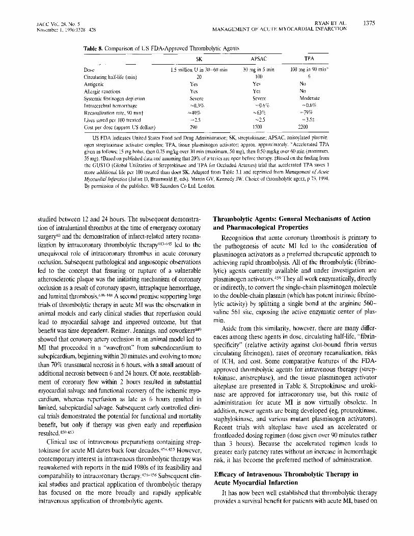
JACC Vol. 28, No. 5 RYAN ET AL. 1375 November 1, 1996:1328 428 MANAGEMENT OF ACUTE MYOCARDIAL INFARCTION
Table 8. Comparison of US FDA-Approved Thrombolytic Agents
SK APSAC TPA
Dose 1.5 million U in 30-60 rain 30 mg in 5 min 100 mg in 90 min* Circulating half-life (rain) 20 100 6 Antigenic Yes Yes No Allergic reactions Yes Yes No Systemic fibrinogen depletion Severe Severe Moderate Intracerebral hemorrhage ~0.3% ~0.6% ~l).6% Recanalization rate, 90 mini" ~40% ~63% ~79% Lives saved per 100 treated ~2.5 ~2.5 ~3.5~ Cost per dose (approx US dollars) 290 1700 2200
US FDA indicates United States Food and Drug Administration; SK, streptokinase; APSAC, anisoylated plasmin- ogen streptokinase activator complex; TPA, tissue plasminogen activator; approx, approximately. *Accelerated TPA given as follows: 15 mg bolus, then 0.75 mg/kg over 30 min (maximum, 50 rag), then 0.50 mg/kg over 60 rain (maximum, 35 rag). iBased on published data and assuming that 20% of arteries are open before therapy. :~Based on the finding from the GUSTO (Global Utilization of Streptokinase and TPA for Occluded Arteries) trial that accelerated TPA saves 1 more additional life per 100 treated than does SK. Adapted from Table 3.1 and reprinted from Management of Acute Myocardial Infamtion (Julian D, Braunwald E, eds). Martin GV, Kennedy JW. Choice of tbrombolytic agent, p 73, 1994. By permission of the publisher, WB Saunders Co Ltd, London.
studied between 12 and 24 hours. The subsequent demonstra- tion of intraluminal thrombus at the time of emergency coronary surgery ~° and the demonstration of infarct-related artery recana- lization by intracoronary thrombolytic therapy 443-445 led to the unequivocal role of intracoronary thrombus in acute coronary occlusion. Subsequent pathological and angioscopic observations led to the concept that fissuring or rupture of a vulnerable atherosclerotic plaque was the initiating mechanism of coronary occlusion as a result of coronary spasm, intraplaque hemorrhage, and luminal thrombosis. 446-448 A second premise supporting large trials of thrombolytic therapy in acute MI was the observation in animal models and early clinical studies that reperfusion could lead to myocardial salvage and improved outcome, but that benefit was time dependent. Reimer, Jennings, and coworkers 449 showed that coronary artery occlusion in an animal model led to MI that proceeded in a "wavefront" from subendocardium to subepicardium, beginning within 20 minutes and evolving to more than 70% transmural necrosis in 6 hours, with a small amount of additional necrosis between 6 and 24 hours. Of note, reestablish- ment of coronary flow within 2 hours resulted in substantial myocardial salvage and functional recovery of the ischemic myo- cardium, whereas reperfusion as late as 6 hours resulted in limited, subepicardial salvage. Subsequent early controlled clini- cal trials demonstrated the potential for functional and mortality benefit, but only if therapy was given early and reperfusion resulted.45o-453
Clinical use of intravenous preparations containing strep- tokinase for acute MI dates back four decades. 454,455 However, contemporary interest in intravenous thrombolytic therapy was reawakened with reports in the mid 1980s of its feasibility and comparability to intracoronary therapy. 456-45s Subsequent clin- ical studies and practical application of thrombolytic therapy has focused on the more broadly and rapidly applicable intravenous application of thrombolytic agents.
Thrombolytic Agents: General Mechanisms of Action and Pharmacological Properties
Recognition that acute coronary thrombosis is primary to the pathogenesis of acute MI led to the consideration of plasminogen activators as a preferred therapeutic approach to achieving rapid thrombolysis. All of the thrombolytic (fibrino- lytic) agents currently available and under investigation are plasminogen activators. 45'~ They all work enzymatically, directly or indirectly, to convert the single-chain plasminogen molecule to the double-chain plasmin (which has potent intrinsic fibrino- lyric activity) by splitting a single bond at the arginine 560- valine 561 site, exposing the active enzymatic center of plas- rain.
Aside from this similarity, however, there are many differ- ences among these agents in dose, circulating half-life, "fibrin- specificity" (relative activity against clot-bound fibrin versus circulating fibrinogen), rates of coronary recanalization, risks of ICH, and cost. Some comparative features of the FDA- approved thrombolytic agents for intravenous therapy (strep- tokinase, anistreplase), and the tissue plasminogen activator alteplase are presented in Table 8. Streptokinase and uroki- nase are approved for intracoronary use, but this route of administration for acute MI is now virtually obsolete. In addition, newer agents are being developed leg, prourokinase, staphylokinase, and various mutant plasminogen activators). Recent trials with alteplase have used an accelerated or frontloaded dosing regimen (dose given over 90 minutes rather than 3 hours). Because the accelerated regimen leads to greater early patency rates without an increase in hemorrhagic risk, it has become the preferred method of administration.
Efficacy of Intravenous Thrombolytic Therapy in Acute Myocardial Infarction
It has now been well established that thrombolytic therapy provides a survival benefit for patients with acute MI, based on
1376 RYAN ET AL. JACC Vol. 28, No. 5 MANAGEMENT OF ACUTE MYOCARDIAL INFARCTION November 1, 1996:1328-428
large, well-controlled clinical trials. Benefit has been shown individually for therapy with streptokinase, anistreplase, and alteplase. 2s,2~,46~',461 In an overview of the nine controlled randomized trials involving more than 1000 patients, a highly significant (P<.00001) 18% proportional reduction in mortal- ity was observed, corresponding to the avoidance of 18 deaths per 1000 patients treated. 27 Furthermore, the largest of these studies (ISIS-2, more than 17 000 patients), showed that when aspirin was combined with streptokinase and treatment was given within 4 hours of onset of symptoms, an odds reduction in mortality of 53% was achieved (control, 13.1%; streptoki- nase plus aspirin, 6.4%) (P<.0001). 2'~ Information from both animal studies as well as clinical trials has provided strong support for the concept that achievement of early, complete, and sustained coronary, patency is primarily responsible for benefit of treatment? ° Mechanisms of benefit include favor- able effects on myocardial salvage as well as postinfarction remodeling.
Benefits of Thrombolytic Therapy in Specific Patient Subgroups
The overview of thrombolysis trials shows that thrombolytic therapy is clearly beneficial in the vast majority of patients. Differences in outcome in subgroups in clinical trials should be interpreted more cautiously than overall differences in out- come with therapy, given the problems of multiple compari- sons and chance deviations from the mean. Sometimes differ- ences in degree (and rarely, direction) of benefit appear among some subgroups, and when these are replicated in independent trials and supported by a clear pathophysiological rationale may reflect valid differences. Implications of overall and sub- group results from the overview of the major randomized, controlled clinical trials 27 for use of thrombolytic therapy in acute M1 are presented in "Initial Recognition and Manage- ment in the Emergency Department."
Comparative Thrombolytic Efficacy Since publication of the first guidelines for the early man-
agement of patients with acute myocardial infarction, ~ results of important trials comparing thrombolytic regimens directly have been published, evaluating relative rates of corona~ patency, functional benefit, and survival. In two large mortality trials (GISSI-2/Internationa146: and ISIS-3463), mortality rates at 4 to 5 weeks were similar (GISSI-2/International: TPA [duteplase]=8.9%, streptokinase=8.5%; ISIS-3: alte- plase = 10.3%, streptokinase = 10.6%, anistreplase = 10.5%). In these studies conjunctive antithrombotic therapy included as- pirin in all patients (160 to 325 mg on admission and daily) and subcutaneous heparin in half (12 500 U twice a day, beginning 4 to 12 hours after thrombolytic therapy). At the time, intravenous heparin was not used in either of these studies because of concerns about increasing the incidence of ICH. The specific failure to use intravenous heparin with TPA in these trials has been the source of some criticism. The GUSTO trial subsequently tested four thrombolytic regimens among 41 021 patients. -~2~ Alteplase was given in an accelerated dose
regimen to further improve early patency rates and concomi- tant heparin administered intravenously to maintain patency. Other regimens included streptokinase with subcutaneous or intravenous heparin and a combination of alteplase and strep- tokinase. Thirty-day mortality was lower with alteplase (6.3%) than streptokinase and subcutaneous heparin (7.2%), strep- tokinase and intravenous heparin (7.4%), and combined strep- tokinase and alteplase plus intravenous heparin therapies (7.0%). Differences were highly significant, although propor- tionately modest, when accelerated alteplase was compared with combined streptokinase groups (14% mortality reduction, P=.001). There was a significant excess of hemorrhagic stroke for accelerated alteplase (P=.03) and the combination strategy (P<.001), compared with streptokinase only. However, net benefit was still achieved with alteplase compared with strep- tokinase, with 9 fewer deaths or disabling strokes per 1000 patients treated. Other complications of acute MI were gen- erally less frequent with alteplase, including allergic reactions, heart failure, cardiogenic shock, and atrial and ventricular arrhythmias.
Other conclusions drawn from GUSTO are (1) intravenous heparin provides no added benefit over aspirin and subcuta- neous heparin when given with streptokinase and in addition increases bleeding risk (the power of this comparison, how- ever, was markedly reduced by the fact that 36% of patients randomly assigned to receive subcutaneous heparin also re- ceived intravenous heparin); (2) combination therapy in- creases bleeding risk (relative to alteplase with intravenous heparin) and provides less benefit; and (3) although the rationale for use of intravenous heparin with alteplase appears sound, other factors, specifically, earlier time to therapy, frontloading alteplase, and requiring ST elevation on entry ECG, likely explain much of the difference in results between GUSTO and ISIS-3. 464 The mechanism of improved benefit with alteplase was assessed in the GUSTO angiographic sub- study, which found differences in early (90-minute) patency among regimens (81%, 56%, 61%, 73%) for alteplase, strep- tokinase-subcutaneous heparin, streptokinase-intravenous heparin, and combination regimens, respectively. 465 These differences in patency in the angiographic substudy closely predicted survival outcomes among the four strategies when applied to the main trial results 4~'~' and furnish a biological explanation for mortality differences among regimens. The data, coupled with that of additional, independent compari- sons showing superior outcomes with accelerated alteplase compared with anistreplases, 4~'7.46s provide a strong impetus for early and complete restoration of infarct artery perfusion as an essential goal of thrombolytic therapy.
Considerations in Selecting Thrombolytic Regimens GUSTO 22s and other recent studies 4~'7,4~s suggest that
accelerated alteplase with intravenous heparin is currently the most effective therapy for achieving early coronary reperfusion and its associated survival benefits but is also substantially more expensive and carries a greater risk of ICH. Thus, the cost-benefit ratio is greatest in patients presenting early after

JACC Vol. 28, No. 5 RYAN ET AL. 1377 November 1. 1996:1328-428 MANAGEMENT OF ACUTE MYOCARDIAL INFARCTION
symptom onset with a large area of injury (eg, anterior acute MI) and at low risk of ICH. In groups with a smaller potential for survival benefit and a greater risk for ICH, streptokinase appears to be the agent of choice, particularly in view of the cost. Other promising thrombolytic agents are under investi- gation (eg, prourokinase, reteplase, staphylokinase, TNK- plasminogen activator).
A number of proposals for selection of thrombolytic regi- mens after GUSTO have been suggested. 9°,469-471 Additional considerations include avoiding reuse of streptokinase or anistreplase for at least 2 years (preferably indefinitely) be- cause of a high prevalence of potentially neutralizing antibody titers. Alternatively, Simoons 47o has proposed considering pri- mary PTCA for those at highest risk (about 10% of patients), alteplase for those at moderate to high risk (40%), streptoki- nase for those at low to moderate risk (40%), and no lytic therapy for those at lowest risk (10%). All of these recommen- dations await prospective testing.
Current Use Rates for Thrombolytic Therapy
The industry-sponsored National Registry of Myocardial Infarction tracks the use of thrombolytic therapy in the United States and has enrolled 220 171 patients treated at 1370 US hospitals during its second phase (NRMI 2) from June 1994 through December 1995. Overall, 37.2% received reperfusion therapy (83% thrombolysis, 15.4% primary angioplasty, 1.4% immediate CABG) (written communication, W. J. Rogers, June 1996). Among a subset of patients presenting with ST elevation or LBBB within 12 hours of symptom onset (n=64211), the use of reperfusion therapy was 70%, with 8.2% of the cohort receiving primary PTCA. 472
Because many patients have contraindications or other exclusions for fibrinolytic agents, it has been ditficult to ascer- tain the proportion of patients with ST elevation who fail to receive fibrinolytic therapy that actually should have received such therapy. 472~' Critical to any such assessment of appropri- ateness of care, however, is whether the diagnosis of acute MI was suspected on entry into the healthcare system or was an "outcomes" diagnosis made after 12 to 24 hours in the hospital or at some later point in time before hospital discharge. Experience to date suggests that in patients younger than 65 years, overall usage of thrombolytic thera W ranges between 40% and 50% (as high as 70% to 75% for patients with ST-elevation MI). In those older than 65, the overall use rate is below 20% and should be higher. Some increase in use rates probably can be achieved, but contraindications prohibit a vast increase in use rates.
Anti thrombot ics /Ant icoagulants
Once fissuring of an atherosclerotic plaque has occurred, whether an epicardial coronary, vessel becomes totally oc- cluded, develops a more severe, flow-limiting stenosis, or heals without incident depends to a large extent on the degree to which thrombus propagates in the vessel lumen. As previously discussed, platelet activation and aggregation are crucial ete-
Table 9. Heparin Adjustment Nomogram for Standard Laboratory Reagents With a Mean Control aPTY of 26-36 s
aPTY Bolus Dose Stop Infusion Rate Change Repeat (s) (U) (rain) (mL/h) aPTI'
<40 3000 0 +2 6 h 40-49 0 0 + 1 6 h 5(1-75 0 0 0 (no change) Next AM
76-85 0 l) - 1 Next AM 86-100 0 30 2 6 h
101-150 0 60 3 6 h
>1511 0 60 - 6 6 h
aPTT indicates activated partial thrornboplastin time. Heparin infusion concentration = 50 U/mL. Target aPT]" = 50-75 s. For aPTI's obtained before 12 h after initiation of thrombolytic therapy: 1, Do not discontinue or decrease infusion unless significant bleeding or aVIT >150 s. 2. Adjust infusion upward if aPTT <50 s. For aPTl's obtained > 12 h after initiation of thrombolytic therapy, use entire nomogram: Deliver bolus, stop infusion, and/or change rate of infusion based on aPTI', as noted on appropriate line of nomogram. Adapted with permission from Hirsh J, Raschke R. Warkentin TE, Dalen JE, Deykin D, Piller L Heparin: mechanism of action, pharmacokinetics, dosing considerations, monitoring, efficacy, and safety. Chest. 1995;108:258S-275S.
ments of the process, but the balance between activation of the coagulation cascade and its inhibition is also critical. The process by which a thrombus is formed is complex, and our understanding of it continues to evolve, 473 but much of the therapeutic effort has focused on inhibiting thrombin and thereby preventing conversion of fibrinogen to fibrin. In addi- tion to having a primary role in this initial process of coronary thrombosis, thrombin also is an important platelet activator; activation of platelets by thrombin is not inhibited by aspirin. Another reason that thrombin is considered critical is that active thrombin becomes bound to a developing clot, and as the clot lyses, either pharmacologically or through endogenous means, the "clot-bound" thrombin can convert fibrinogen to fibrin as it is exposed to the circulating blood.
Heparin
Recommendations
Class I
1. Patients undergoing percutaneous or surgical revascu- larization.
Comment: For PTCA, monitoring of activated clotting time (ACT) is recommended, with a goal of 300 to 350 seconds during the procedure.
Class lla
1. Intravenously in patients undergoing reperfusion ther- apy with alteplase.
Comment: The recommended regimen is 70 U/kg as a bolus at initiation of alteplase infusion, then an initial maintenance dose of approximately 15 ~g/kg per hour, adjusted to maintain aPTT at 1.5 to 2.0 times control (50 to 75 seconds) for 48 hours (Table 9). Continuation of heparin infusion beyond 48 hours should be restricted to patients at high risk for systemic or venous thrombo- embolism.

1378 RYAN ET AL. JACC Vol. 28, No. 5 MANAGEMENT OF ACUTE MYOCARDIAL INFARCTION November 1, 1996:1328-428
2. Subcutaneously (7500 U twice daily) (intravenous hepa- rin is an acceptable alternative) in all patients not treated with thrombolytic therapy who do not have a contraindication to heparin. In patients who are at high risk for systemic emboli (large or anterior MI, AF, previous embolus, or known LV thrombus), intravenous heparin is preferred.
3. Intravenously in patients treated with nonselective thrombolytic agents (streptokinase, anistreplase, urokinase) who are at high risk for systemic emboli (large or anterior MI, AF, previous embolus, or known LV thrombus).
Comment: It is recommended that heparin be withheld for 4 hours and that aPTT testing begin at that time. Heparin should be started when aPTT returns to less than two times control (about 70 seconds), then infused to keep aPTT 1.5 to 2.0 times control (initial infusion rate about I000 U/h). After 48 hours, a change to subcutaneous heparin, warfarin, or aspirin alone should be considered.
Class lib
1. Patients treated with nonselective thrombolytic agents, not at high risk, subcutaneous heparin, 7500 U to 12 500 U twice a day until completely ambulatory.
Class III
1. Routine intravenous heparin within 6 hours to patients receiving a nonselective fibrinolytic agent (streptokinase, anis- treplase, urokinase) who are not at high risk for systemic embolism.
Heparin has been available as an anticoagulant for many years; it was initially described in 1916. The pharmacological entity consists of a mixture of molecules with molecular weights varying between 5000 and 20 000, with different-size molecules having different effects on the coagulation system. After forming a complex with antithrombin III (AT-III), the heparin-AT-III complex has the ability to inactivate both thrombin and activated factor X. When a dose of heparin is given, the actual measured effect on coagulation is modulated by a number of factors, including the particular admixture of heparin molecules in the dose, circulating levels of AT-III, availability of platelet factor IV and other plasma proteins that inactivate heparin, and the ability of heparin to reach thrombin bound to clot. The heparin-AT-III complex is quite large and generally does not appear to be effective against clot-bound thrombin.
In patients who will not be given thrombolytic therapy, there is little evidence about the benefit of heparin in the modern era, in which aspirin, /3-adrenoceptor blockers, ni- trates, and ACE inhibitors are routinely available. Neverthe- less, the best available data emanate from a series of random- ized clinical trials performed before the reperfusion era. A systematic overview of these studies demonstrated a 17% reduction in mortality and a 22% reduction in risk of reinfarc- tion with heparin? 74 The control groups in these trials were not treated with other therapies, particularly aspirin, that are now considered routine. Not withstanding, it is primarily these randomized data from an earlier era that support the recom-
mendation to use heparin in patients not treated with throm- bolytic therapy.
In patients who are treated with thrombolytic therapy, recommendations for heparin therapy depend on the throm- bolytic agent chosen. Streptokinase, anistreplase, and uroki- nase are nonspecific fibrinolytic agents that produce systemic breakdown of the coagulation system, including depletion of factors V and VIII and massive production of fibrin(ogen) degradation products, themselves anticoagulants. From this perspective, the need for conjunctive systemic anticoagulation with these agents is conceptually less. In comparison, relatively fibrin-specific agents, including alteplase and newer agents such as reteplase, produce a variable effect on the systemic coagulation system, and in many patients very little breakdown of fibrinogen or depletion of coagulation factors is evi- dent.475A76
More than 60 000 patients were enrolled in the randomized ISIS-3463 and GISSI-2/InternationaW 7 trials comparing subcu- taneous heparin with no routine heparin in conjunction with streptokinase, anistreplase, and alteplase. During the period in which heparin was given, a small reduction in mortality (4 to 5 lives per 1000 treated) was observed in ISIS-3; however, by 30 days the 2 to 3 lives saved per 1000 treated was no longer statistically significant. A small excess rate of hemorrhagic stroke (1 to 2 per 1000 treated patients) was observed together with a larger excess in systemic bleeding (3 to 5 per 1000 patients), although total stroke rate was not significantly increased. In the GUSTO-I trial, 228 more than 20 000 patients treated with streptokinase were randomly assigned to routine intravenous heparin versus routine subcutaneous heparin. No significant differences were observed in death, reinfarction, or nonhemorrhagic stroke rates, while excess rates of systemic bleeding and hemorrhagic strokes (trend) were observed in the intravenous heparin group.
Several angiographic studies have evaluated coronary per- fusion as a function of heparin therapy. 228,478,479 Two trials have shown more rapid resolution of ST-segment elevation in patients treated with intravenous heparin immediately at the time of streptokinase infusion compared with intravenous heparin started at a later time. 478,479 The OSIRIS study, however, showed no difference in perfusion at 24 hours or in clinical outcomes in the two groups. In the GUSTO-I angio- graphic substudy, patients treated with intravenous heparin had an 88% patency rate at 5 to 7 days compared with 72% in patients treated with subcutaneous heparin (P<.05), although less reinfarction occurred in the subcutaneous heparin group (3.4% versus 4.0%, P<.05). A small group of patients were randomly assigned to anistreplase with or without intravenous heparin in the DUCCS-1 study, 48° and no differences in clinical end points were observed, other than a higher rate of bleeding in heparin-treated patients. Viewing these studies as a whole, intravenous heparin appears to have no advantage over sub- cutaneous heparin when used with a nonspecific thrombolytic agent, and the evidence for use of subcutaneous heparin is equivocal. 4al
The occurrence of a large, anterior infarction, documenta-
JACC Vol. 28, No. 5 RYAN ET AL. 1379 November 1, 1996:1328-428 MANAGEMENT OF ACUTE MYOCARDIAL INFARCTION
tion of thrombus in the left ventricle by echocardiography, history of a previous embolic event, and AF have been associated with a high risk of embolic stroke. Although no randomized trial evidence exists to demonstrate a definite benefit specific to this group, some empirical evidence exists that the risk of systemic emboli in the general population of MI patients can be reduced by early initiation of heparin? 82 In the SCATI trial patients were randomly assigned to a 2000 IU bolus of heparin followed by 12 500 IU subcutaneously twice a day or to placebo. In the subgroup also treated with streptoki- nase, aspirin was withheld. In-hospital mortality was 4.6% in the heparin group and 8.8% in the control group, and a reduction in stroke was observed. Therefore, heparin is rec- ommended for these patients at high risk of systemic arterial emboli, regardless of the thrombolytic agent given.
When alteplase is chosen as the thrombolytic agent, the empirical information to confirm the pathophysiological rea- soning discussed above is primarily inferential. In a series of angiographic trials? 83-4~5 intravenous heparin has been shown to lead to higher rates of infarct-related artery perfusion in conjunction with alteplase. When aPTI" has been evaluated, a direct relation between duration of aP'IT and the likelihood of infarct-related artery perfusion has been observed. 4s4,4s5 A recent overview 4s6 points out, however, that the effects of intravenous heparin on clinical outcomes from these studies are not as convincing; a significant increase in the rate of bleeding and nonsignificant increases in rates of reinfarction and hemorrhagic and nonhemorrhagic stroke are evident. 486 These negative findings are tempered by a point estimate of an 18% reduction in mortality with broad confidence limits. Until the uncertainty is resolved, it seems judicious to use heparin for at least 48 hours with alteplase and to target the aPTI" to a 50- to 75-second range.
When primary angioplasty is chosen as the route of reper- fusion, high-dose heparin therapy is recommended. This rec- ommendation does not come specifically from empirical data in the acute MI setting but from general observations in the setting of angioplasty that an ACT of at least 300 to 350 seconds is associated with a lower rate of complications than lower ACT values. 4s7,488
Very recently abciximab, a Fab fragment of humanized monoclonal antibody to the glycoprotein IIb/IIIa receptor on the platelet surface, has been demonstrated to reduce the risk of adverse outcomes significantly, both at 30 days 489 and at 6 months 49° after high-risk percutaneous intervention. 489.491 Benefit, however, was accomplished at the price of an increase in major bleeding from 13% to 24%. Abciximab, like experi- mental IIb/IIIa antagonists, increases the ACT measurement with a given dose of heparin by an average of 35 seconds. 491 A recent trial with abciximab compared this agent with placebo in the context of standard heparin dosing in the placebo group and two heparin regimens with abciximab: a weight-adjusted standard dose and a lower dose aimed at achieving an ACT of 150 to 300 seconds during routine as well as high-risk percu- taneous procedures. 492 The trial was terminated early when an interim analysis showed a combined rate of death and nonfatal
MI of 8.1% in the placebo group, 3.6% in the weight-adjusted heparin arm, and 2.6% in the low-dose heparin arm. A trend toward less bleeding in the low-dose heparin arm compared with the placebo arm was also reported. A third trial evaluating abciximab in the treatment of refractory unstable angina also was stopped early because of a 40% reduction in the composite end point of death, MI, or need for repeat revasculariza- tion.492.4'~3
The dose of heparin in the thrombolytic-treated patient remains somewhat controversial. Based on the infarct-related artery perfusion results described above, it would be reason- able to recommend an aPTI" value more than threefold higher than the median control value. However, recent information strongly supports a lower aPTT because death, stroke, rein- farction, and bleeding were found to be lowest in the aPTT range of 50 to 75 seconds or approximately 1.5 to 2.0 times the control value. 494 Because of the clear evidence that the mea- sured effect of heparin on the aPTr is important for patient outcome and that the predominant variable mediating the effect of a given dose of heparin is weight, 4'j4 it is important to administer the initial doses of heparin as a weight-adjusted bolus. 481 A 70 U/kg bolus followed by 15 U/kg per hour has been useful, although other mitigating factors including age and gender, require careful aPTI" measurement and dose adjustment. More recent information for both heparin and the novel antithrombin agent hirudin indicate that when used with thrombolytic therapy, an aPTT goal of 60 to 90 seconds is associated with an unacceptably high rate of ICH. 495,496
An algorithm for heparin dosing in the setting of thrombo- lytic therapy or treatment of non-ST-segment elevation is provided in Table 9. It is important to check the aPTI" 4 to 6 hours after initiating therapy or changing dose, given the information about increased risk with a high aPTI'. Consider- ing the substantial delay in reporting aPT? values in many hospitals, the use of bedside coagulation monitoring, 497 if reliably performed, may be helpful.
The previous ACC/AHA guidelines on acute MI recom- mended low-dose subcutaneous heparin (5000 U every 12 hours for 24 to 48 hours) in all MI patients without contrain- dication who were not otherwise being treated with heparin for another reason. Current recommendations call for 7500 U twice a day (ACCP guidelines)? 23 The empirical basis for this recommendation was the demonstration that deep venous thrombosis was reduced from 12% to 4% in an overview of three randomized controlled trials. 498 Continued adherence to this standard is reasonable, although routine earlier mobiliza- tion and use of aspirin may make this treatment unnecessary.
Once heparin has been started, the appropriate duration of therapy is uncertain. Based on the evidence for disruption of the atherosclerotic plaque and the concept that a healed endothelial surface would be salutary, a duration of 3 to 5 days has been standard. The only randomized trial to address this issue found, however, that discontinuation of heparin after 24 hours following thrombolytic therapy with alteplase resulted in no measurable increase in ischemic events, 499 although this study did not have adequate power to detect modest differ-
138(I RYAN ET AL. JACC Vol. 28, No. 5 MANAGEMENT OF ACUTE MYOCARDIAL INFARCTION November 1, 1996:1328-428
ences. A reasonable approach is to use intravenous heparin for 48 hours and then to use heparin according to the clinical characteristics of the patients. Heparin may be discontinued in low-risk patients, given subcutaneously in patients at high risk of systemic embolization, and intravenously in patients at high risk for coronary reocclusion.
Concern is mounting that when heparin is discontinued abruptly, the patient undergoes a high-risk period for recurrent thrombosis. 5°".5°~ Despite this concern, no specific policy has been tested to attempt to reduce this clinical "rebound" effect. Several ongoing studies, however, are reducing heparin infu- sions in a gradual fashion (eg, by one half within 6 hours then discontinuing over the subsequent 12 hours).
Platelet counts should be monitored daily in patients on heparin. Recent evidence suggests the incidence of heparin- induced thrombocytopenia is 3% and is associated with a substantial risk of prothrombotic events. 5°2 If the platelet count drops below 100 000, a test for heparin-induced throm- bocytopenia should be obtained, and the clinician should be vigilant for thrombotic complications as the prognosis in patients with thrombocytopenia is substantially worse. 5°3
The deficiencies of heparin as an antithrombotic agent have been discussed in detail. 5°4 Fractionated heparins have been developed with variable effects on inhibition of thrombin and factor Xa. Although unfractionated heparin and low molecular weight heparin both catalyze the inhibition of thrombin by AT-Ill at clinically administered doses, the higher ratio of anti-Xa:anti IIa activity of low molecular weight heparins offers the potential advantage of inhibiting the coagulation cascade at a more proximal step, leading to reduction in the generation of thrombin. 5"5 The addition of a low molecular weight heparin preparation to a regimen of aspirin,/3-adrenoceptor blockers, and nitrates in patients with unstable angina/non-Q wave MI is superior to placebo for reducing the risk of death and nonfatal MI in hospital, 5°~' although this effect was lessened in longer- term follow-up. Some evidence exists that subcutaneous ad- ministration of a low molecular weight heparin appears to be superior to infusion of unfractionated heparin for reducing episodes of recurrent ischemia in patients with unstable angina. 5''7 These agents are superior in many forms of venous thrombosis, 5°s but their relative value in coronary arterial thrombosis has not been established.
Newer direct antithrombin agents are also in an advanced stage of development. The prototypical direct antithrombin agent hirudin was initially isolated from the saliva of the medicinal leech. Now, synthesized by recombinant technology, this compound has several conceptual advantages: it does not require AT-Ill for its activity, it is not neutralized by plasma proteins, and it is able to inhibit clot-bound thrombin. Its characteristics also yield a stable aPTT value for a given dose, although its predominant renal excretion leads to unpredict- able buildup in patients with significant renal dysfunction. After very promising early phase trials in acute MI s"9,51~' and unstable angina, 51~ large-scale trials were initiated but had to be reconfigured due to an excess rate of ICH in pa- tients treated with thrombolytic agents. 4'~5,4'~, The GUSTO-lib
study comparing hirudin with heparin in conjunction with standard medical therapy in the management of 12 142 pa- tients with acute coronary syndromes recently reported a 30-day death or MI rate (primary end point) of 8.9% for patients randomly assigned to hirudin treatment versus 9.8% for those randomly assigned to heparin (P=.058). 512 The TIMI 9B trial of 3002 patients receiving either TPA or streptokinase for ST-segment elevation MI reported a 30-day rate of death, MI, or severe CHF of 11.9% in patients randomly assigned to heparin compared with 12.9% in patients assigned to hirudin.Sla~
Antiarrhythmics Antiarrhythmic therapy plays an important but more lim-
ited role in acute MI care than in the past, as summarized in "Hospital Management." The use of anticholinergic therapy with atropine for bradyarrhythmias is summarized in "Hospital Management." This section briefly summarizes antiarrhythmic agents in Vaughan-Williams Classes I through III that are appropriate in the acute setting and can be intravenously administered. Use of agents from Classes II 03-adrenoceptor blockers) and IV (calcium-channel entry blockers) have several other mechanisms of action (anti-ischemic, antihypertensive, etc), and their use is primarily summarized in subsequent sections. In general, both acute and long-term antiarrhythmic therapy except with /3-adrenoceptor blocking agents is indi- cated only for life-threatening or severely symptomatic ar- rhythmias and not for risk reduction in patients with non-life- threatening arrhythmias.
Lidoeaine
Lidocaine is a local anesthetic with antiarrhythmic proper- ties, grouped in Class Ib based on its relatively rapid onset and offset kinetics of membrane sodium channel blockade. Lidoo caine is metabolized in the liver; its volume of distribution and rate of clearance are reduced in heart failure. 513 Previous randomized studies have shown that it reduces risk for primary VF in both prehospital and early hospital settings/~4,5~5 De- spite this fact, mortality is not reduced; indeed, VF deaths are offset by deaths associated with asystole and electromechanical dissociation.325.5~
Lidocaine is the drug of choice in the setting of acute MI when treatment is indicated for premature ventricular com- plexes, VT, or VF. It is generally well tolerated, except in patients with shock, in the most recent adult ACLS proto- col, 517 lidocaine is recommended as the first antiarrhythmic agent to be used in cardiac arrest patients with persistent VT/VF despite defibrillation and epinephrine, to prevent recurrence, to control unsustained ventricular ectopy requiring therapy, and to treat wide complex tachycardia of uncertain type.5 ~,5 ~
Lidocaine is given in an initial bolus of 1.0 to 1.5 mg/kg (75 to 100 rag); additional boluses of 0.5 to 0.75 mg/kg (25 to 50 rag) can be given every 5 to 10 minutes if needed up to a total of 3 mg/kg. This is followed by a maintenance infusion of

JACC Vol. 2S, No. 5 RYAN ET AL. 1381 November 1, 1996:1328 428 MANAGEMENT OF ACUTE MYOCARDIAL INFARCTION
1 to 4 mg/min, reduced after 24 hours (to 1 to 2 mg/min) or in the setting of altered metabolism (heart failure, hepatic con- gestion, etc) and as guided by blood level monitoring.
Bretylium
Bretylium is a quaternary ammonium compound with both direct (Class III) and indirect (sympathetic neuronal) actions. Its hemodynamic and electrophysiological profile are biphasic, with initial norepinephrine release from adrenergic nerve endings causing hypertension, tachycardia, shortening of AV nodal refractory periods, and subsequent neuronal blockade leading to hypotension521'; clinical Class III effect (refractory period lengthening) also emerges with some (variable) delay. Experimentally and clinically, bretylium has potent antifibril- latory but weak antiarrhythmic effects.
Clinically bretylium is used in treatment of resistant VF and hemodynamically unstable VT. It is not a first-line agent but is recommended in the current ACLS protocol after defibrilla- tion, epinephrine, and lidocaine have failed to convert VF (or pulseless VT) or after VF has recurred despite epinephrine and lidocaine. It may be used for VT in patients with a pulse, but only after lidocaine and procainamide have failed.
For VF, bretylium is given as a 5 mg/kg bolus; if VF-related cardiac arrest persists, supplemental doses of 10 mg/kg can be given at 5-minute intervals to a maximum dose of 30 to 35 mg/kg. In stable VT the loading dose is diluted to 50 mL with 5% dextrose and given over 8 to 10 minutes. Bretylium therapy is maintained with an infusion rate of 1 to 2 mg/min.
Procainamide
Procainamide is an antiarrhythmic drug grouped in Class Ia because of its intermediate onset and offset kinetics of membrane sodium channel blockade. Procainamide has local anesthetic properties and mild to moderate hypotensive and negative inotropic potential. Its rate of metabolism to N-acetyl- procainamide (NAPA), which has Class III antiarrhythmic activity, is bimodally distributed in the population (fast, slow acetylators).
Procainamide is indicated for life-threatening ventricular arrhythmias but usually not as the drug of first choice. Pro- cainamide suppresses premature ventricular complexes and recurrent VT and may be used when therapy is required when lidocaine has failed or is contraindicated. It may also be used for wide complex tachycardias of uncertain mechanism, al- though it also is usually not the drug of first choice in this setting. ACES guidelines list procainamide as potential therapy for VF and pulseless VT refractory to defibrillation and epinephrine after lidocaine, bretylium, and magnesium have been considered. 517
Intravenous procainamide is initiated with a loading infu- sion of 10 to 15 mg/kg (500 to 1250 rag), given at a rate of 20 mg/min (ie, over 30 to 60 minutes), followed by a mainte- nance infusion of 1 to 4 mg/min. In responding patients, therapy may be continued orally as needed.
Procainamide may cause proarrhythmia, including torsades de pointes. Patients with renal insufficiency may develop high
levels of NAPA and are at increased risk for development of torsades.
~-Adrenoceptor Blockers
/3-Adrenoceptor blockers such as propranolol, metoprolol, and atenolol have been shown to reduce incidence of VF in patients with acute MI in studies preceding the reperfusion era. 521 13-Adrenoceptor blockers also may be of particular value early in the management of "electrical storm" (recurrent, polymorphic VT/VF) in the setting of recent MI, which is often unresponsive to standard antiarrhythmic therapy. 331 Addi- tional rationale for/3-adrenoceptor blocker use in acute MI is provided in the following section.
Amiodarone
Amiodarone is a complex antiarrhythmic with action in each of the four Vaughn-Williams classes. Its mechanisms of action when given over the short term are still poorly defined but may include (1) noncompetitive ¢3-adrenoceptor blockade, (2) calcium channel blockade, (3) blockade of sympathetic efferents, and (4) possible Class la effects. 522 Short-term (intravenous) amiodarone, unlike long-term (oral) administra- tion may have little Class III effect. Intravenous amiodarone is now approved for treatment and prophylaxis of frequently recurring VF and hemodynamically destabilizing VT. If suc- cessful, therapy can be continued orally over the long term. In randomized studies in VF or destabilizing VT refractory to lidocaine, a dose response was observed between larger (500 to 1000 mg/d) and small (125 mg/d) doses of amiodarone in time to first VT/VF recurrence, although not in mortality. 332 Ami- odarone also was equally as effective as bretylium in preventing VT/VF recurrence but was better tolerated (less hypoten- s i o n ) . 52-~
Because of individual variability, dosing of intravenous amiodarone should be titrated according to patient response. The recommended starting dose is 500 mg per 24 hours, given in three stages: (1) rapid infusion of 150 mg over 10 minutes, (2) an early maintenance infusion of 1 mg/min for 6 hours, and (3) later maintenance infusion of 0.50 mg/min. Intrave- nous amiodarone is reasonably well tolerated, but adverse effects such as hypotension, bradycardia, and AV block may occur. With greater experience, amiodarone may become a preferred antiarrhythmic agent for intravenous therapy of life-threatening ventricular tachyarrhythmias in lidocaine fail- ures.
[3-Adrenoceptor Blocking Agents
Recommendations for Early Therapy (see also "Predischarge Preparation")
Class I 1. Patients without a contraindication to /3-adrenoceptor
blocker therapy who can be treated within 12 hours of onset of infarction, irrespective of administration of concomitant thrombolytic therapy.
2. Patients with continuing or recurrent ischemic pain.

1382 RYAN ET AL. JACC Vol. 28, No. 5 MANAGEMENT OF ACUTE MYOCARDIAL INFARCTION November 1, 1996:1328-428
3. Patients with tachyarrhythmias, such as AF with a rapid ventricular response.
Class lib
1. Non-Q wave MI.
Class III
1. Patients with moderate or severe L¥ failure or other contraindications to ~-adrenoceptor blocker therapy.
/3-Adrenoceptor blocking agents may be given to patients with acute MI to reduce morbidity and/or mortality during (1) the initial hours of evolving infarction and (2) the weeks, months, and years after completed infarction (secondary pre- vention).
During the first few hours of infarction, /3-adrenoceptor blocking agents may diminish myocardial oxygen demand by reducing heart rate, systemic arterial pressure, and myocardial contractility. In addition, prolongation of diastole caused by a reduction in heart rate may augment perfusion to injured myocardium, particularly the subendocardiurn. As a result, immediate/3-adrenoceptor blocker therapy appears to reduce (1) the magnitude of infarction and incidence of associated complications in subjects not receiving concomitant thrombo- lyric therapy and (2) the rate of reinfarction in patients receiving thrombolytic therapy.
In subjects not receiving thrombolytic therapy, intrave- nously administered /3-adrenoceptor blocking agents exert a modestly favorable influence on infarct size524). More impor- tant, they diminish short-term mortality. In the First Interna- tional Study of Infarct Survival, -~:5 in which more than 16 000 patients with suspected acute MI were enrolled within 12 hours of onset of symptoms, immediate intravenous atenolol, 5 to 10 rag, followed by oral atenolol, 100 mg daily, reduced 7-day mortality from 4.3% to 3.7% (P<.02; 6 lives saved per 1000 treated). The mortality difference between those receiving and not receiving atenolol was evident by the end of day 1 and was sustained subsequently. In the Metoprolol in Acute Myocar- dial Infarction (MIAMI) trial 526 more than 5700 subjects with evolving MI were randomly assigned to receive placebo or metoprolol, up to 15 mg intravenously in 3 divided doses followed by 50 mg orally every, 6 hours for 48 hours and then 100 mg twice a day thereafter. Fifteen-day mortality was reduced with metoprolol from 4.9% to 4.3%. As in ISIS-l, the mortality difference between those given placebo and those receiving metoprolol was evident by the end of day 1, after which it was sustained.
In subjects receiving concomitant thrombolytic therapy, intravenously administered/3-adrenoceptor blocking drugs di- minish the incidence of subsequent nonfatal reinfarction and recurrent ischemia: in addition, they may reduce mortality if given particularly early (ie, within 2 hours) after onset of symptoms. In the TIMI-II trial, r~7 in which all patients received intravenous alteplase, those randomly assigned to receive intravenous metoprolol, 15 rag, followed by oral metoprolol, 50 mg twice a day for 1 day and then 100 mg twice a day thereafter, had a diminished incidence of subsequent nonfatal
reinfarction and recurrent ischemia when compared with those begun on oral metoprolol 6 days after the acute event. Among those treated especially early, ie, within 2 hours of symptom onset, the composite end point, death or reinfarction, occurred less often in those given immediate intravenous metoprolol than in those who did not receive it.
If intravenous /3-adrenoceptor blockade induces an un- toward effect, such as AV block, excessive bradycardia, or hypotension, the condition is quickly reversed by infusion of a /3-adrenergic agonist (ie, isoproterenol, 1 to 5 gg/min).
Contraindications
The following are relative contraindications to/3-adrenoceptor blocker therapy:
• Heart rate less than 60 bpm • Systolic arterial pressure less than 100 mm Hg • Moderate or severe LV failure • Signs of peripheral hypoperfusion • PR interval greater than 0.24 second • Second- or third-degree AV block • Severe chronic obstructive pulmonary disease • History of asthma • Severe peripheral vascular disease • Insulin-dependent diabetes mellitus
Angiotensin Converting Enzyme Inhibitors
Recommendat ions
Class I
1. Patients within the first 24 hours of a suspected acute MI with ST-segment elevation in two or more anterior precor- dial leads or with clinical heart failure in the absence of significant hypotension or known coutraindications to use of ACE inhibitors.
2. Patients with MI and LV ejection fraction less than 40% or patients with clinical heart failure on the basis of systolic pump dysfunction during and after convalescence from acute MI.
Class lla
1. All other patients within the first 24 hours of a suspected or established acute MI, provided significant hypotension or other clear-cut contraindications are absent.
2. Asymptomatic patients with mildly impaired LV func- tion (ejection fraction 40% to 50%) and a history of old MI,
Class lib
1. Patients who have recently recovered from MI but have normal or mildly abnormal global LV function.
A number of large, randomized clinical trials have assessed the role of ACE inhibitors early in the course of acute MI. All trials in which only oral ACE inhibitors were used demon- strated a benefit in mortality. The only trial not showing benefit using ACE inhibitors was the Cooperative New Scandinavian Enalapril Survival Study (CONSENSUS) II, in which patients were randomly assigned within the first day to receive either
JACC Vol. 28, No. 5 RYAN ET AL. 1383 November l, 1996:1328-428 MANAGEMENT OF ACUTE MYOCARDIAL INFARCTION
intravenous enalaprilat or placebo followed by increasing oral dosages of either enalapril or placebo. This trial was termi- nated early by the Safety Committee because of the high probability that a significant beneficial effect of enalapril over placebo was unlikely to be demonstrated with continuation of the trial, as well as a concern over an adverse effect among elderly patients experiencing an early hypotensive reactionY 7 The 95% confidence limits ranged from showing a 7% benefit to 29% harm.
Clarification of the role of ACE inhibitors early in the course of MI has more recently resulted from large-scale clinical trials in which oral ACE inhibitors were used. In the ISIS-4 trial 58 000 patients with suspected acute MI were randomly assigned within the first 24 hours (median 8 hours) to receive either oral captopril or placebo; a significant 7% proportional reduction was observed in 5-week mortality among those randomly assigned to captopril? 21 The largest benefit was among those with an anterior infarction. 528 Among the 143 fewer deaths in the group allocated captopril, 44 occurred in days 0 through 1 and 37 in days 2 through 7, 529
demonstrating that early therapy is important. The GISSI-3 trial used oral lisinopril in over 19 000 patients with either ST-segment elevation or depression who were randomly as- signed to it or open control. 42o There was a significant reduc- tion in 6-week mortality (odds ratio 0.88; 95% CI, 0.79 to 0.99); 60% of the lives were saved during the first 5 days of treatment. The SMILE (Survival of Myocardial Infarction: Long-Term Evaluation) study involved 1556 patients ran- domly assigned within 24 hours to receive either placebo or zofenopril. 53° The patient population was restricted to those with anterior MI who had not received thrombolytic therapy. Use of an early ACE inhibitor in this trial suggested a strong trend of more lives saved in the first 6 weeks (RR 25%, P=.19). A Chinese captopril pilot study involving more than 13 600 patients with suspected acute MI also revealed an approximate 0.5% absolute mortality benefit among those who were randomly assigned to the ACE inhibitor compared with the control population. 53~ A meta-analysis of these major trials along with 11 smaller trials that involve more than 100 000 patients reveals a 6.5% overall odds reduction (2P=.006) with an absolute benefit of 4.6 fewer deaths per 1000 patients treated among those who received the ACE inhibitor. 529 These data suggest that ACE inhibitors have a role in early management as well as in the convalescent phase of acute MI.
Although detailed subgroup analysis of the ISIS-4 and GISSI-3 trials awaits further publication, it would appear that the benefits of ACE inhibitors are greater among those with an anterior infarct or who have evidence of previous in- farction, heart failure, and tachycardia, ie, those at highest risk. Nevertheless, all trials with oral ACE inhibitors have shown benefit from its early use, including those in which entry criteria included all suspected acute infarctions. Data from these trials indicate that ACE inhibitors should generally be started within the first 24 hours, ideally, after thrombolytic therapy has been completed and blood pressure has sta-
bilized. When there are no patient complications and no evidence of symptomatic or asymptomatic LV dysfunction by 4 to 6 weeks, ACE inhibitors can be stopped. ACE inhibitors should not be used if systolic blood pressure is less than 100 mm Hg, if clinically relevant renal failure is present, if there is a history of bilateral stenosis of the renal arteries, or if there is known allergy to ACE inhibitors. ACE inhibitor therapy should start with low-dose oral admin- istration and increase steadily to achieve a full dose within 24 to 48 hours. For example, in ISIS-4 an initial 6.25 mg dose of captopril was given, followed by 12.5 mg 2 hours later, 25 mg 10 to 12 hours later, and then 50 mg twice a day. GISSI patients received 5 mg oral lisinopril at the time of random- ization, 5 mg after 24 hours, 10 mg after 48 hours, then 10 mg daily for 6 weeks or open control. Similar graded-dose sched- ules should be used with other ACE inhibitors, such as ramipril, zofenopril, enalapril, or quinapril. Intravenous ena- laprilat should be avoided.
C a l c i u m C h a n n e l Blockers
Recommendations
Class I
None.
Class l la
1. Verapamil or diltiazem may be given to patients in whom/3-adrenoceptor blockers are ineffective or contraindi- cated (ie, bronchospastic disease) for relief of ongoing isch- emia or control of a rapid ventricular response with AF after acute MI in the absence of CHF, LV dysfunction, or AV block.
Class l ib
1. In non-ST-elevation infarction, diltiazem may be given to patients without LV dysfunction, pulmonary congestion, or CHF. It may be added to standard therapy after the first 24 hours and continued for 1 year.
Class I I I
1. Nifedipine (short acting) is generally contraindicated in routine treatment of acute MI because of its negative inotropic effects and the reflex sympathetic activation, tachycardia, and hypotension associated with its use.
2. Diltiazem and verapamil are contraindicated in patients with acute MI and associated LV dysfunction or CHF.
Comment: Calcium channel blocking agents have not been shown to reduce mortali O, after acute MI, and in certain patients with cardiovascular disease there are data to suggest they are harmfuU -~2 It is the consensus of this committee that these agents are still used too frequently 84 in patients with acute MI and that [3-adrenoceptor blocking agents are a more appropriate choice across a broad spectrum of patients with acute MI (with excep- tions as noted).
Nifedipine
In patients with acute MI, immediate-release nifedipine does not reduce incidence of reinfarction or mortality when

1384 RYAN ET AL. JACC Vol. 28, No. 5 MANAGEMENT OF ACUTE MYOCARDIAL INFARCTION November 1, 1996:1328-428
given early (less than 24 hours) or late after acute MI. This lack of benefit is found in all patients, irrespective of gender, overall risk, type of infarction (Q wave versus non-Q wave), and presence or absence of concomitant/3-adrenoceptor blocking agents or thrombolytic therapy. Immediate-release nifedipine may be particularly detrimental in patients with hypotension and/or tachycardia; in these patients it may induce a reduction in coronary perfusion pressure, disproportionate dilatation of the coronary arteries adjacent to the ischemic area (so-called "steal"), and/or reflex activation of the sympathetic nervous system, with an increase in myocardial oxygen demands. These findings are based on numerous clinical trials, including the Nifedipine Angina Myocardial Infarction Study (NAMIS), 533 the Norwegian Nifedipine Multicenter Trial, 534 the Trial of Early Nifedipine Treatment in Acute Myocardial Infarction (TRENT), 5-~5 and the Secondary Prevention Reinfarction Is- raeli Nifedipine Trial (SPRINT). 536,537 These studies were performed using first-generation nonsustained-release nifedi- pine. Whether the conclusions are valid for the entire class of agents is unknown. 311-532,53s
Verapamil
Although the overall results of trials with verapamil showed no mortality benefits, subgroup analysis showed that immediate-release verapamil initiated several days after acute MI in patients who were not candidates for a {3-adrenoceptor blocking agent may be useful in reducing the incidence of the composite end point of reinfarction and death, provided LV function is well preserved with no clinical evidence of heart failure. Verapamil is detrimental to patients with heart failure or bradyarrhythmias during the first 24 to 48 hours after acute MI. 539-542 One randomized study of 1700 patients, less than 75 years of age, using verapamil within 2 weeks of acute MI showed a 16.7% reduction in major events (death or MI) over 18 months. 543
Diltiazem
Data from the Multicenter Diltiazem Postinfarction Trial (MDPIT) (Q wave and non-Q wave infarction) 544 and the Diltiazem Reinfarction Study (DRS) (non-Q wave infarc- tion) 54a,541.545-546 suggest that patients with non-O wave MI or those with Q wave infarction, preserved LV function, and no evidence of heart failure may benefit from immediate-release diltiazem. Diltiazem was begun in MDPIT 3 to 15 days after acute MI and in DRS 24 to 72 hours afterward. The results of MDPIT may be confounded by the fact that 53% and 55% of placebo- and diltiazem-treated patients, respectively, received concomitant/3-adrenoceptor blocker therapy. 544 Also, both the MDPIT and DRS projects were conducted in an era when the use of aspirin was not as prevalent as it is today, raising further uncertainty about the relevance of their findings for contem- porary management of acute MI. Of particular clinical impor- tance is the detrimental mortality effect of diltiazem in patients with LV dysfunction.
The INTERCEPT trial (Incomplete Infarction Trial of Euro- pean Research Collaborators Evaluating Prognosis Post Throm-
bolysis) (diltiazem) will test the hypothesis that use of sustained- release diltiazem in patients receiving thrombolytic therapy for a first MI will decrease mortality, reinfarction, and angina. 547
Summary of Calcium Channel Blockers
Calcium channel blockers have not proven beneficial in early treatment or secondary prevention of acute MI, and the possibility of harm has been raised. In patients with first non-Q wave infarction or first inferior infarction without LV dysfunc- tion or pulmonary congestion, verapamil and diltiazem may reduce the incidence of reinfarction, but their benefit beyond that of/3-adrenoceptor blockers and aspirin is unclear. Simi- larly, there are no data to support the use of second-generation dihydropyridines (eg, amlodipine, felodipine) for improving survival in acute MI.
Magnesium
Recommendations
Class I
None.
Class lla
1. Correction of documented magnesium (and/or potas- sium) deficits, especially in patients receiving diuretics before onset of infarction.
2. Episodes of torsades de pointes-type VT associated with a prolonged QT interval should be treated with 1 to 2 g magnesium administered as a bolus over 5 minutes.
Class lib
1. Magnesium bolus and infusion in high-risk patients such as the elderly and/or those for whom reperfusion therapy is not suitable.
Comment: The available data suggest that mortality reduction may be seen in high-risk patients, provided magnesium therapy is administered soon after onset of o,mptoms (preferably less than 6 hours). The optimum dose has not been established, but a bolus of 2 g over 5 to I5 minutes followed by an infusion of 18 g over 24 hours has been used with success.
Background
Supplemental administration of magnesium for reducing morbidity and mortality in patients with acute MI is a reason- able avenue to pursue because of abundant data relating magnesium to cardiovascular disease. 54~ It is the second most abundant intracellular cation and is involved in more than 300 enzymatic processes. Evidence exists that magnesium produces systemic and coronary, vasodilatation, possesses antiplatelet activity, suppresses automaticity in partially depolarized cells, and protects myocytes against calcium overload under condi- tions of ischemia by inhibiting calcium influx especially at the time of reperfusion. 54~-552
Meta-analyses of the seven randomized trials published between 1984 and 1991 suggest a significant mortality benefit of magnesium (odds ratio 0.44, CI 0.27 to 0.71). 553,554 The

JACC Vol. 28, No. 5 RYAN ET AL. 1385 November 1, 1996:1328-428 MANAGEMENT OF ACUTE MYOCARDIAL INFARCTION
Table 10. A Classification of Inotropic Agents
Agent Mechanism Inotropic Vascular Effect Major Use
Isoproterenol /3-1 receptor + + Dilatation
Dobutamine + + Mild dilatation Dopamine + + Renovascular dilation
Norepinephrine
Amrinone
Milrinone Digitalis
Constriction Intense constriction
+ + Intense constriction
+ + Dilatation
+ + Dilatation + Variable
/3-1 receptor Low dose: dopaminergic receptor
Medium dose: 13-1 rcceptor High dose: ot receptor a Receptor
Phosphodiesterase inhibitor
Phosphodiesterase inhibitor Inhibits NA+-K+ATPase pump
Hypotension due to bradycardia; no pacing available
Low output with SBP >90 mm Hg Hypoperfusion with SBP <90 mm Hg
or >30 mm Hg below usual value
Extreme hypotension despite use of dopamine
Second-tier agcnt after failure of dopamine/dobutamine
Established systolic LV dysfunction and symptoms of heart failure for chronic therapy
SBP indicates systolic blood pressure; LV, left ventricular.
Second Leicester Intravenous Magnesium Intervention Trial (LIMIT-2) trial 555 subsequently reported a 24% reduction in mortality with magnesium treatment (P<.04). The magnesium-treated patients in LIMIT-2 had a 25% lower incidence of CHF in the CCU and a 21% lower rate of ischemic heart disease-related mortality over 4 years, con- sistent with the hypothesis that magnesium exerts its ben- eficial effects, at least in part, via a myocardial protective ac t ion .555,556
The results of one large trial were negative. The ISIS-4 investigators enrolled 58050 patients, 29011 allocated to magnesium, and 29 039 to control. There were 2216 deaths (7.64%) by 35 days in the magnesium group and 2103 deaths (7.24%) in the control group (odds ratio 1.06; CI 0.99 to 1.13), suggesting no mortality benefit of magnesium administration and even the possibility of slight harm. 421 When ISIS-4 is added to the preceding randomized trials, meta- analysis indicates no beneficial effect of magnesium. Possible sources of heterogeneity that could explain these differences include:
1. The relatively late administration of magnesium in ISIS-4.55v
2. The control group mortality in ISIS-4 was only 7.2%. Regression analyses of the available data predict a null effect of magnesium when the control mortality is about 7% and increasing benefit of magnesium for higher control mortality rates.55s
Shechter and colleagues 55'3 recently reported a randomized trial of 194 patients with acute MI unsuitable for thrombolysis. There was a significant reduction in mortality in the magne- sium group (4.2% versus 17.3%, P<.01), largely due to a lower incidence of cardiogenic shock and CHF.
An NHLBI-sponsored trial (Magnesium in Coronary Dis- ease [MAGIC]) is planned to further evaluate the role of magnesium in acute MI, especially with early administration before thrombolysis in higher-risk patients. 557
Inotropic Agents It is useful clinically to consider inotropic agents in terms of
three classes (Table 10): inotropic agents with predominant vasoconstrictive properties; catecholamines with predominant inotropic properties with little or no vasoconstriction; and phosphodiesterase inhibitors, inotropic agents with predomi- nant vasodilating properties.
Vasoconstrictor inotropic agents are represented by dopa- mine and norepinephrine. Contractility and heart rate are increased by dopamine through its direct stimulation of a and /3-adrenergic receptors and through release of norepinephrine from nerve endings. When given in low doses (1 to 3 ~g/kg per minute), its major effects are on dopaminergic receptors leading to renovascular dilatation and on /3 adrenoceptors modestly stimulating contractility. At a dose of 5 to 10/xg/kg per minute, the/3-1 receptor effects are dominant, leading to an increase in contractility and heart rate. At higher doses the c~-receptor effects predominate, leading to vasoconstriction. Norepinephrine is almost purely a vasoconstrictive agent with a positive effect on contractility.
The catecholamine inotropic agents that do not cause vasoconstriction are represented by dobutamine. Through its effects on /3-1 receptors, it stimulates contractility; the hope that it would produce less tachycardia and fewer arrhythmias than dopamine has not been realized, lsoproterenol produces increased heart rate and contractility while causing vasodila- tion; therefore, it is not recommended except as an emergency measure when low output is caused by a profound bradycardia and temporary pacing is not available.
Amrinone and milrinone (phosphodiesterase inhibitors) were developed with the hope that their different mechanism of action would lead to improved cardiac output without the risk of arrhythmia engendered by catecholamines. These agents are characterized by both inotropic and vasodilating effects and with a more substantial effect on preload than

1386 RYAN ET AL. JACC Vol. 28, No. 5 MANAGEMENT OF ACUTE MYOCARDIAL INFARCTION November 1, 1996:1328-428
catecholamines. Excessive mortality when oral milrinone was given long term and unacceptable toxicity of long-term use of amrinone 56° have dampened enthusiasm for long-term use of these drugs. Renal elimination of phosphodiesterase inhibitors is a problem in critically ill patients.
In a patient with perceived low output, the clinician must simultaneously assess the patient for the possible cause and institute life-saving therapy. If volume depletion is a possible cause, an intravascular volume-expanding infusion should be initiated. When blood pressure is low (systolic less than 90 mm Hg or 30 points below usual), dopamine is the agent of first choice. If blood pressure remains low with institution of more than 20 /~g/kg, norepinephrine may be substituted in doses of 2 to 20 ~g/kg per minute. In all other situations dobutamine is the agent of first choice. All intravenous cat- echolamines have the advantage of a very short half-life, enabling titration of the dose in a matter of minutes while observing the clinical effect.
Phosphodiesterase inhibitors are reserved for patients who have not responded to catecholamines or who have significant arrhythmias or ischemia-producing tachycardia on catechol- amine therapy. Milrinone is given in a dose of 0.25 to 0.75 /~g/kg per minute. Special caution must be advised in patients with renal dysfunction because the drug will accumu- late.
In general the current concept is that patients requiring intravenous inotropic support should be maintained on these agents for as short a time as possible. These agents are arrhythmogenic and increase myocardial oxygen demand. The only available empirical information on mortality, effects with long-term use are dismal. Whenever possible, afterload reduc- ing agents and intra-aortic balloon pumping should be substi- tuted for inotropic agents.
Digitalis
Despite the initial description of the inotropic properties of digitalis in 1785, its role in the post-MI patient remains controversial. Concern about increased mortality associated with long-term use of milrinone has fueled a reexamination of the empirical information about digitalis from previous obser- vational studies. These studies had mixed results, with some suggesting an increase in mortality and others a neutral effect on mortality. ~6~ Recent studies have demonstrated that in patients with definite systolic LV dysfunction, digitalis im- proves symptomatic status and has a favorable effect on the neurohormonal system. 562,563 The Digitalis Investigator Group (DIG) recently reported a study of 7788 patients in CHF (due to ischemic heart disease in 70% of cases) who were in sinus rhythm. Digoxin was compared with placebo for prevention of all-cause mortality¢ 64 More than 90% of patients were also on ACE inhibitors and/or diuretics. Important secondary objec- tives included hospitalization for CHF, cardiovascular mortal- ity, and death due to CHF. The overall findings of the trial showed no reduction in total mortality with digoxin. However, there were reductions in deaths due to CHF and combined heart failure-related deaths and hospitalizations in digoxin-
treated patients. A trend toward increased deaths due to presumed arrhythmia or MI was observed in the digoxin group. Of note, a recent MI was an exclusion criterion for enrollment in the DIG trial. Thus, the current recommendation, based on previous clinical experience, supports the use of digoxin in selected patients recovering from an MI if they have supraven- tricular arrhythmias or CHF refractory to ACE inhibitors or diuretics. Generally the loading dose is 8 to 15 p.g/kg lean body weight, with half the dose given immediately and the remain- der given in 25% increments 6 hours apart. A maintenance dose of 0.125 to 0.375 mg/d is given, based on renal function and lean body weight.
VI. P r e p a r a t i o n for D i s c h a r g e F r o m
t h e H o s p i t a l
Noninvasive Evaluation of Low-Risk Patients
Recommendations
Class I
1. Stress ECG a. Before discharge for prognostic assessment or func-
tional capacity (submaximal at 4 to 6 days or symptom limited at 10 to 14 days).
b. Early after discharge for prognostic assessment and functional capacity (14 to 21 days).
c. Late after discharge (3 to 6 weeks) for functional capacity and prognosis if early stress was submaximal.
2. Exercise, vasodilator stress nuclear scintigraphy, or exercise stress echocardiography when baseline abnormalities of the ECG compromise interpretation.*
Class lla
1. Dipyridamole or adenosine stress perfusion nuclear scintigraphy or dobutamine echocardiography before dis- charge for prognostic assessment in patients judged to be unable to exercise.
2. Exercise two-dimensional echocardiography or nuclear scintigraphy (before or early after discharge for prognostic assessment).
Class III
1. Stress testing within 2 to 3 days of acute MI. 2. Either exercise or pharmacological stress testing at any
time to evaluate patients with unstable postinfarction angina pectoris.
3. At any time to evaluate patients with acute MI who have uncompensated CHF, cardiac arrhythmia, or noncardiac con- ditions that severely limit their ability to exercise.
*Marked abnormalities in the resting ECG such as LBBB, LV hypertrophy with strain, ventricular pre-excitation, or a ventricular paced rhythm render a displacement of the ST segments virtually uninterpretable. For patients taking digoxin or who have less than 1 mm ST depression on their resting tracing who undergo standard stress electrocardiographic testing, it must be realized that further ST depression with exercise may have minimal diagnostic significance.

JACC Vol. 28, No. 5 RYAN ET AL. 1387 November 1, 1996:1328-428 MANAGEMENT OF ACUTE MYOCARDIAL INFARCTION
4. Before discharge to evaluate patients who have already been selected for cardiac catheterization. In this situation an exercise test may be useful after catheterization to evaluate function or identify ischemia in distribution of a coronary lesion of borderline severity.
Role of Exercise Testing
The role of exercise testing in evaluating patients after MI has been well established 565 and extensively covered in the earlier ACC/AHA guidelines. 1,566,567 The basic aims of early exercise testing after MI are to (1) assess functional capacity and the patient's ability to perform tasks at home and at work; (2) evaluate the efficacy of the patient's current medical regimen; and (3) risk-stratify the post-MI patient according to the likelihood of a subsequent cardiac event. Numerous studies reported throughout the 1980s provided particularly important information about risk stratification and the development of practical algorithms for further management of the post-MI patient. 56~472 The decade of the 1980s also witnessed a dra- matic change in treatment of patients with acute MI, charac- terized most notably by the broad use of thrombolytic therapy beginning in 1988. Equally important has been the widespread use of aspirin, /3-adrenoceptor blocking agents, vasodilator therapy, common use of ACE inhibitors, and a far more aggressive use of revascularization therapy in patients who have clinical markers of a poor prognosis. It is this constella- tion of new therapy and not solely the administration of thrombolytic therapy that marks what is generally referred to as the "reperfusion era."
This period has witnessed an impressive reduction in early and 1-year mortality rates for acute MI patients, which is particularly striking in patients who have received thrombolytic therapy and revascularization during hospitalization. 573
The improvement in 1-year mortality in patients who have received thrombolytic therapy is multifactorial. Such patients are less likely to have severe three-vessel coronary artery diseases 4 Patients who receive thrombol~ic therapy have a smaller infarct size. 575 Coronary angiography is frequently performed during hospitalization due to recurrent chest pain, which identifies many patients with severe disease who subse- quently undergo revascularization. 576 The patient population eligible for predischarge exercise testing in clinical trials of thrombolytic therapy is therefore far different from less se- lected, historical populations. Their low cardiac event rate following discharge is therefore not surprising and substan- tially reduces the predictive accuracy of early exercise testing.
The highest-risk subset of patients are those who are unable to exercises 7,57s Although patients with exercise-induced ST depression have a higher 1-year mortality than patients without exercise-induced depression, their absolute mortality remains low (1.7%) by historical standards. 57s The duration of exercise is also known to be an important predictor of outcomes and the ability to perform at least 5 metabolic equivalents (METs) without early exercise ST depression and show a normal rise in systolic blood pressure is important in constituting a negative predictive value. 57'~,5s~
There is limited evidence on the ability of exercise testing to risk-stratify patients who have not received reperfusion in the current era. Although their subsequent mortality rates are lower because of the constellation of new therapy mentioned earlier, their absolute event rates are higher than in patients who have received thrombolytic therapy, particularly if they have also not undergone revascularization. 573 Although the available evidence is limited, exercise testing presumably can still assist in the risk stratification of such patients.
Low-level exercise testing appears to be safe if patients have undergone in-hospital cardiac rehabilitation, including low- level exercise, have had no symptoms of angina or heart failure, and have a stable baseline ECG 48 to 72 hours before the exercise test. Two different protocols have been used to determine the end points of these very early exercise tests.S~l-5~3
The traditional submaximal exercise test (done at 3 to 5 days in patients without complications) incorporates a series of end points, including a peak heart rate of 120 to 130 bpm or 70% of maximal predicted heart rate for age, a peak work level of 5 METs, or clinical or ECG end points of mild angina or dyspnea, ST-segment depression greater than 2 mm, exertional hypotension, or three or more consecutive premature ventric- ular contractions, whichever end point is reached first. The second protocol is performance of a symptom-limited exercise test (done at 5 days or later) without stopping for target heart rates or MET levels. Although this level appears to be safe and will result in a higher frequency of abnormal exercise tests, the prognostic value of ST depression occurring at higher work levels in deconditioned patients is uncertain and may lead to unnecessary cardiac catheterization.
The optimum time for performing the exercise test after MI remains unresolved. It is argued that a predischarge exercise test provides psychological benefits to the patient and will permit detection of profound ischemia that could be associated with postdischarge cardiac events that might occur before a scheduled 3- to 6-week postdischarge symptom-limited stress test. On the other hand, deferring exercise testing until approx- imately 3 weeks after MI in clinically low-risk patients appears safe and reasonable and enables more optimal assessment of functional capacity. For patients without complications who have not undergone coronary arteriography before discharge, it is the consensus of this committee that patients who might be potential candidates for revascularization procedures should undergo exercise electrocardiography before or just after discharge.
Supplemental Imaging
Exercise Myocardial Perfusion Imaging
In a number of reports from a decade ago, before the use of thrombolytic therapy, the prognostic value of exercise myocar- dial perfusion imaging was found to be superior to that of exercise electrocardiographic testing. 5se-Ss7 Pharmacological stress perfusion imaging 5~-59o was also shown to have value for the prediction of postinfarction cardiac events. The key issues are whether these results apply to current patient populations

1 3 8 8 RYAN ET AL. JACC Vol. 28, No. 5 MANAGEMENT OF ACUTE MYOCARDIAL INFARCTION November 1, 1996:1328-428
Present [
Strategy I
Clinical Indications of High Risk at Predischarge
Absent Absent I
Strategy II I
Symptom-Limited Exercise Test at 14-21 Days
J Markedly Mildly Negative Abnormal Abnormal
I Exercise maging Study
Reversible No Reversible Ischemia Ischemia
, Medical Treatment
I Strategy III
r Submaximal Exercise Test
at 5-7 Days I
I r
_ _ Markedly Mildly N e g l a t i v e - Abnormal Abnormal
C a r d i a c • Catherization ~
i
I Exerc ise Imaging Study j . i
Reversible No Reversible Ischemia Ischemia
Strenuous Le sure Act v t or Occupation
Symptom-Limited Exercise Testing at 3-6 Weeks
_ _ Markedly Mildly Negative Abnormal Abnormal
P Exercise Imaging Study r 1
Reversible No Reversible Ischemia Ischemia
I Medical Treatment --
Figure 10. Strategies for exercise test evaluations soon after myocar- dial infarction (MI). If patients are at high risk for ischemic events, based on clinical criteria, they should undergo invasive evaluation to determine if they are candidates for coronary revascularization proce- dures (Strategy l). For patients initially deemed to be at low risk at time of discharge after MI, two strategies for performing exercise testing can be used. One is a symptom-limited test at 14 to 21 days (Strategy II). If the patient is on digoxin or if baseline electrocardio- gram precludes accurate interpretation of ST-segment changes (eg, baseline left bundle branch block or left ventricular hypertrophy), then an initial exercise imaging study can be performed. Results of exercise testing should be stratified to determine need for additional invasive or exercise perfusion studies. A third strategy is to perform a submaximal exercise test at 5 to 7 days after MI or just before hospital discharge. The exercise test results could be stratified using the guidelines in Strategy I. If exercise test studies are negative, a second symptom- limited exercise test could be repeated at 3 to 6 weeks for patients undergoing vigorous activity during leisure or at work.
in the reperfusion era and whether myocardial perfusion imaging is worth the additional cost for risk stratification. 59~ The same issues outlined previously with respect to exercise electrocardiographic testing also apply to this methodology.
In patients with ST elevation who have received thrombo- lyric therapy, several studies using myocardial perfusion imag- ing have found that it is less valuable than previously for risk stratification, -~'~2-594 primarily because of the low late cardiac event rate.
In patients in the current era who have not received reperfusion therapy, particularly those who have not under- gone revascularization, the same considerations regarding subsequent patient outcome that were outlined above for exercise electrocardiographic testing apply. There is evidence that myocardial perfusion imaging is useful for risk stratifica- tion in such patients, despite their better overall prognosis. 5')5
It seems likely that the previously demonstrated superiority of stress myocardial peffusion imaging probably continues to apply to this population, although there is limited evidence on this point. It must be recognized that prospective studies are ditficult to conduct because clinicians frequently intervene in patients with abnormal predischarge stress perfusion imaging studies.
Myocardial perfusion imaging with either thallium 20P 9f' or technetium 99m sestamibi 5'~v can assess infarct size. The mea- surement of infarct size by either one of these techniques is significantly associated with subsequent patient mortality after thrombolytic therapy. 590,5'~7 Data are also emerging to suggest that vasodilator stress nuclear scintigraphy is safe and can be used for early (48 to 72 hours) risk stratification.
Recommended strategies for exercise test evaluations soon after MI are presented in Fig 10.
Role of Echocardiography
The widespread availability, portability, and relative cost of echocardiography has resulted in its increased use as a practi- cal and reliable means of assessing both global ventricular func- tion and regional wall motion abnormalities. The uses of echo- cardiography in acute MI are discussed in detail in the ACC/ AHA guidelines for clinical application of echocardiography, s7~,
Risk Stratification After Myocardial Infarction The incremental value of exercise echocardiography over
regular exercise testing after MI has also not been established. The usefulness of exercise echocardiography as a means of assessing myocardial ischemia in patients with coronary artery disease has been well established, with overall sensitivity of 81% and specificity of 89%. 59~-~"3 However, its value in pre- dicting cardiac events after MI has not been fully determined. A negative test is, in general, associated with a low risk of cardiac events and death, but it may be higher than that

JACC Vol. 28~ No. 5 RYAN ET AL. 1389 November I, 1996:1328-428 MANAGEMENT OF ACUTE MYOCARDIAL INFARCTION
associated with a negative perfusion scan? '°44'°s The usefulness of pharmacological stress testing with echocardiography or single-photon emission computed tomography (SPECT) imag- ing using agents such as dipyridamole or dobutamine in predicting cardiac events after acute MI is also a subject undergoing intense investigation. A positive dipyridamole echocardiogram after MI is associated with a higher late mortality rate, but a negative test does not preclude cardiac events in the 2-year follow-up period. ~,°u There are few data regarding the prognostic value of a positive or negative dobut- amine stress echocardiogram, but its safety in general and in the 3 to 5 days after MI ~,l~ is acceptably low. This agent, although widely used for pharmacological stress testing, has not been approved for this purpose by the FDA. Like scintig- raphy, there is great variation among institutions in expertise and study quality, and it is this local expertise that should determine the choice of test procedures. Exercise echocardi- ography generally, however, is a less costly procedure than radionuclide perfusion scintigraphy.
Myocardial Viability A significant development since the previous set of recom-
mendations is related to understanding and identifying myo- cardial viability. Up to one third of patients who have signifi- cant LV dysfunction may improve with revascularization? ,~ This usually refers to myocardial hibernation, ('~ in which chronic low flow state is associated with depressed myocardial function. Myocardial stunning ',~2 is more germane to the situation after MI, when depressed ventricular function is present despite adequate restoration of blood flow. Function will subsequently improve. The therapeutic importance of myocardial stunning is perhaps less than hibernation because identification of the former does not in general initiate a change in management of revascu- larization. However, identification of extensive reversible LV dysfunction is of prognostic importance and may help to optimize medical management after MI. '~
Several noninvasive imaging modalities have been estab- lished as accurate predictors of myocardial viability. These include thallium imaging, positron emission tomography (PET), and dobutamine echocardiography. The choice of which technique to use should be dependent on center and regional expertise. Positron emission tomography scanning is most sensitive in detecting viable myocardium, but because of the limitations described above and the expense involved, it has little widespread applicability. Thallium imaging has been well established over time, while dobutamine echocardi- ography seems to have an acceptably high positive predictive accuracy. More important than technique, however, is the ques- tion of whether myocardial viability tests should be used in practice until large-scale outcome data can validate the useful- ness.
Left Ventricular Function
Assessment of LV function after acute MI has been dem- onstrated to be one of the most accurate predictors of future
cardiac events in the risk stratification of patients with acute MI in both the prereperfusion 613 and the reperfusion eras. 614.~'15 Multiple techniques for assessing LV function of patients after infarction have been shown to have important prognostic value and include such basic principles as clinical estimates based on patients' symptoms (eg, exertional dyspnea, functional status), physical findings (eg, rales, elevated jugular venous pressure, cardiomegaly, S 3 gallop), exercise duration (treadmill time) and measurement of ejection fraction by contrast ventriculography, radionuclide ventriculography, and two-dimensional echocardiography. Zaret and colleagues 614 found that an LV ejection fraction less than 0.30 as assessed by radionuclide ventriculography was still predictive of mor- tality in patients surviving infarction treated with thrombo- lytic therapy, despite the significantly reduced mortality of these patients compared with those in the prereperfusion era. White and colleagues ~6 performed contrast left ven- triculography in 605 patients 1 to 2 months after MI. They found postinfarction LV dilation, demonstrated by in- creased end-systolic volume greater than 130 mL, was an even better predictor of mortality after MI than an LV ejection fraction less than 40% or increased end-diastolic volume. In patients with normal ejection fractions, however, end-systolic volume did not provide any further stratifica- tion according to risk.
Radionuclide Testing for the Diagnosis of Acute Myocardial Infarction
Guidelines for cardiac radionuclide imaging have been published recently 5~'7 that indicate the clinical use of radionu- elide imaging for diagnosis of acute MI should be restricted to special limited situations in which the triad of history, electro- cardiographic changes, and laboratory measurements is un- available or less reliable.
In patients who present late (more than 24 hours and less than 7 days) without diagnostic electrocardiographic changes and in patients early after coronary artery bypass surgery, myocardial infarct-avid scintigraphy using 99mTc pyrophos- phate has moderate sensitivity and specificity for the diagnosis of acute MI. ~'~7,6~s More recently infarct-avid scintigraphy with antimyosin antibody has been described as an alternative to pyrophosphate scintigraphy ~j~,62' and has just received FDA approval for use in the United States.
In selected patients with RV infarction, radionuclide imag- ing may also have a role in diagnosis by demonstrating a reduced RV ejection fraction and RV asynergy. 62~
Localized perfusion defects occur in a high percentage of patients with acute LV infarction associated with coronary occlusion. ~':2 However, such perfusion defects do not distin- guish between acute ischemia, acute infarction, or previous infarction. Serial changes on follow-up perfusion images with either 2°1T1 or 99mTc sestamibi suggest an acute process but still do not distinguish between ischemia or infarction.

1390 RYAN ET AL. JACC Vol. 28, No. 5 MANAGEMENT OF ACUTE MYOCARDIAL INFARCTION November l, 1996:1328-428
Table 11. Uses of Radionuclide Testing in Acute Myocardial Infarction
Diagnosis Risk Assessment
Indication Test Class Indication Test Class
1. RV infarction
2. Infarction not diagnosed by standard means--early presentation with successful reperfusion
3. Infarction not diagnosed by standard means--late presentation
4. Routine diagnosis
Rest RNA lla 1. Residual ischemia Stress (exercise/pharmacological) I thallium with redistribution
'~'~'Tc pyrophosphate IIa Stress (exercise/pharmacological) sestamibi with redistribution
Rest myocardial lib 2. Myocardial infarct size Tomographic thallium IIa perfusion imaging Tomographic sestamibi IIa
9~)'Tc pyrophosphate lib
'~mTc pyrophosphate lla 3. Hibernating myocardium Early, late thallium IIa
Any technique III 4. Ventricular function RNA I
RV indicates right ventricular; RNA, radionuclide angiography; 99mTc, technetium 99m. From the ACC/AHA task force. Guidelines for clinical use of cardiac radionuclide imaging: report of the American College of Cardiology/American Heart Association Task Force on Assessment of Diagnostic and Therapeutic Cardiovascular Procedures (Committee on Radionuclide Imaging), developed in collaboration with the American Society, of Nuclear Cardiology. J Am Coil Cardiol 1995;25:521-547.
Measurement of Infarct Size
Technetium 99m sestamibi is uniquely suited to accurate measurement of myocardium at risk in clinical infarction. Because there is minimal redistribution of the radiopharma- ceutical over time, imaging can be delayed for several hours after injection and still provide accurate information about myocardial perfusion at the time of injection. The validity and feasibility of this approach has been well established in animal and clinical studies. ~23-626
As mentioned previously, myocardium at risk is a major determinant of final infarct size. However, final infarct size may be considerably smaller than the initial myocardium at risk, reflecting the effects of reperfusion therapy, spontaneous reperfusion, and collateral blood flow. ~'27 Clinical data have demonstrated the importance of final infarct size as a major determinant of subsequent patient survival and quality of life. Radionuclide techniques are clearly useful for this purpose. In patients who have not received reperfusion therapy, measure- ment of rest ejection fraction and end-systolic volume index before hospital discharge by equilibrium-gated radionuclide angiography is highly associated with subsequent patient out- come. ~'t3.~2s In patients who have received reperfusion therapy, the postdischarge rest ejection fraction by equilibrium radio- nuclide angiography after resolution of myocardial stunning and compensatory hyperkinesia is highly associated with sub- sequent patient outcome. 59~-~29,~3°
Myocardial perfusion imaging with 2°JTI and 99mTc sesta-
mibi can also be used to assess infarct size. 5%,631,632 Most recent ly 9~)mTc sestamibi has been used with tomographic imaging for this purpose? '33,634 Measurement of infarct size with 99mTc sestamibi has been closely correlated with other measurements of infarct size, including ejection fraction, 635 regional wall motion score, ~35 creatine kinase release) 2~ and 2°ITI defect size. 632 Two studies have now shown an association between infarct size and patient outcome. 596.597 Table 11 summarizes the uses for radionuclide testing in acute MI.
Summary of Stress Testing After Acute Myocardial Infarction
It is the consensus of the task force that the current approach to risk stratification of patients after MI requires little change from the recommendations outlined in the origi- nal ACC/AHA task force report "Early Management of Pa- tients With Acute Myocardial Infarction." Patients who have clinically declared themselves to be at high risk should have coronary arteriography to identify those who are candidates for revascularizationS Patients without clinical complications after infarction should have a submaximal exercise stress test before discharge or, alternatively, a symptom-limited stress test 3 weeks after discharge. Patients who can achieve at least 5 METs are treated medically. If there are signs of severe ischemia at a low level of exercise, such as marked ST-segment change or inability to complete stage I, failure to achieve 3 to 4 METs, or if blood pressure falls during exercise, the patient should undergo coronary arteriography.
It must be acknowledged, however, that the positive pre- dictive value of virtually all noninvasive tests has declined as late prognosis improves, particularly those relatively highly selected patients who have received reperfusion therapy. The paradigm for the future will be a new database that examines the benefits, cost-effectiveness, and incremental value of non- invasive tests among lower-risk patients who have received reperfusion therapy.
In patients for whom the resting ECG is uninterpretable because of BBB, major ST-T wave abnormalities, or digitalis therapy, radionuclide myocardial perfusion imaging with exer- cise or stress echocardiography should be performed, depend- ing on local experience and expertise. In the patient who cannot exercise, pharmacological stress agents can be used with either myocardial perfusion imaging or echocardiography. It is the view of the committee that exercise electrocardiogra- phy is a valuable test in assessing prognosis in patients with coronary artery disease. It is generally available, with experi-
JACC Vol. 28, No. 5 RYAN ET AL. 1391 November 1, 1996:1328-428 MANAGEMENT OF ACUTE MYOCARDIAL INFARCTION
enced personnel capable of performing it safely, and it is relatively inexpensive. After uncomplicated MI, patients can be divided into relatively high- and low-risk groups for subse- quent cardiac events if all the information available on the treadmill test is used (Fig 10).
Ambulatory Electrocardiographic Monitoring for Ischemia
The value of ambulatory electrocardiographic monitoring in assessing reversible myocardial ischemia and the risk of a subsequent coronary event early after myocardial infarction have been evaluated in a number of studies. 63°-643 Up to one quarter of patients will show residual ischemia as detected by ambulatory electrocardiographic monitoring. Most episodes of transient myocardial ischemia are silent and occur at rest or during times of low-level physical activity or mental stressJ '44 During long-term follow-up studies, a number of investigators have reported that the presence of ischemia detected by ambulatory electrocardiographic monitoring in the postinfarc- tion period is predictive of a subsequent poor outcome and increases the risk of cardiac events. 636-~43 One recent study found that the odds ratio for the patients with, as compared to those without, ambulatory ischemia was 2.3 for death or nonfatal MI at 1 year) 43
Despite the promising initial results with ambulatory elec- trocardiographic monitoring, the totality of evidence does not support a general statement about its role in all postinfarction patients. Some studies have shown that the results of ambula- tory electrocardiographic monitoring could be predicted from exercise test data, °38,64° while others have found that additional prognostic information could be obtained by ambulatory elec- trocardiographic monitoring in postinfarction patients. 639 At present a cost-effective strategy has not been developed to identify patients who are at increased risk for ambulatory ischemia and in whom ambulatory electrocardiographic mon- itoring might be more helpful for stratification into high- and low-risk subgroups for future coronary events.
Assessment of Ventricular Arrhythmia (Signal- Averaged Electrocardiography, Ambulatory [Holter] Monitoring, Heart Rate Variability)
Recommendations for Routine Testing
Class I
None.
Class Ila
None.
Class l ib
1. Ambulatory (Holter) monitoring, signal-averaged ECG, heart rate variability, baroreflex sensitivity monitoring, alone or in combination with these or other tests, including func- tional tests (ejection fraction, treadmill testing) for risk assessment after MI, especially in patients at higher perceived risk, when findings might influence management issues, or for clinical research purposes.
The risk of malignant ventricular arrhythmias after hospital discharge is greatest in the first year after acute MI. 645-649 Recent data suggest that thrombolytic therapy reduces this risk and also confirm that LV dysfunction remains an important, although diminished, predictor of mortality, including sudden death. 6~4,~50-~53 An open infarct-related artery has emerged as an important predictor of late outcome in other studies. 65~ A number of strategies have been used to try to identify patients at high risk for arrhythmic events. Sustained monomorphic VT induced by electrophysiological study is associated with a high arrhythmic event rate 654 but is invasive and has a low specific- ity. Frequent ventricular premature complexes and higher- grade ventricular ectopy (unsustained VT) on a predischarge Holter monitor also have been associated with a higher mortality among MI survivors, in both the prereperfusion and (less consistently) in the reperfusion eras. 645-653
Recently, newer techniques, including signal-averaged or high-resolution electrocardiography, heart rate variability, and baroreflex sensitivity, have been used to assess patient risk for sudden cardiac death after MI. Signal-averaged electrocardi- ography identifies delayed, fragmented conduction in the infarct zone in the form of late potentials at the terminus of the QRS complex and represents an anatomic substrate that predisposes the patient to reentrant VT. Kuchar et a1655 reported late potentials to predict an increased incidence of sudden death in the post-MI patient population. Gomes et a165f' found late potentials to be the best single predictor among Holter monitoring and ejection fraction and contributed inde- pendently to a combined index, although the positive predic- tive value of each was poor. The filtered QRS duration was the most predictive feature of signal-averaged electrocardiography in a CAST substudy. ~'57 More recent studies have shown reperfusion therapy to reduce the incidence of late potentials after acute MI. (,58 In the setting of frequent use of thrombol- ysis, the predictive value of signal-averaged electrocardiogra- phy has been variable. 65°-65z
Heart rate variability, an analysis of the beat-to-beat varia- tion in cycle length, largely reflects the sympathovagal interac- tion regulating heart rate. Heart rate variability can be quan- tified in a number of ways, using either time or frequency domain parameters. 659 Low heart rate variability, indicative of decreased vagal tone, is a predictor of increased mortality, including sudden death, in patients after MI ~59,6~° and may add significant prognostic information to other parameters. 660 In one study decreased heart rate variability was more predictive of arrhythmic events than the presence of late potentials, Holter-derived data, treadmill test results, or ejection fraction; reduced heart rate variability and a late potential by signal- averaged electrocardiography was the strongest combined pre- dictor. 652 Standards of measurement, physiological interpreta- tion, and clinical use of heart rate variability have been recently published by a task force of the European Society of Cardiol- ogy and the North American Society of Pacing and Electro- physiology. 661 The predictive value of heart rate variability after MI, although significant, is modest when used alone. In combination with other techniques its positive predictive accu-
1392 RYAN ET AL. JACC Vol. 28, No. 5 MANAGEMENT OF ACUTE MYOCARDIAL INFARCTION November 1, 1996:1328-428
racy improves. However, the most practical, feasible, and cost-efficient combination of noninvasive predictive tests with heart rate variability remains to be determined.
Baroreceptor sensitivity also quantifies the influence of parasympathetic tone on the heart. Baroreceptor sensitivity is measured as the slope of a regression line relating beat-to-beat heart rate change in response to a change in blood pressure, often accomplished by giving a small bolus of phenylephrine. 662 Acute MI-associated reductions in baroreflex sensitivity have been associated with an increased susceptibility to arrhythmic events and sudden death in experimental models and initial clinical reports 6~'3-~5 and are being further charac- terized in a multicenter prospective post-MI study (Auto- nomic Tone and Reflexes After Myocardial Infarction [ATRAMI]).
Summary/Conclusions
Although several investigators have reported an increased likelihood of arrhythmic events in patients when one or more noninvasive test is abnormal, two important caveats prevent these techniques from being recommended for routine clinical practice at present. First, although the negative predictive value of most of these tests taken in isolation is high (generally greater than 90%), the positive predictive value is unaccept- ably low (less than 30%). Second, although the positive predictive value of noninvasive testing for future arrhythmic events can be modestly increased by combining several test results, the therapeutic implications of positive findings are unclear. Insufficient data are available to indicate whether general therapies, such as /3-adrenoceptor blockade, ACE inhibition, and revascularization procedures, or specific inter- ventions, such as treatment with amiodarone or an implantable cardioverter-defibrillator, targeted for high-risk patients iden- tified by a combination of noninvasive tests after MI can more favorably impact mortality. ~'6' Moreover, it is difficult to justify the costs of the routine use of these procedures in the absence of therapeutic guidelines or demonstrated clinical benefits associated with a positive test. Until these issues are resolved, use of these tests cannot be recommended in routine manage- ment, although they will continue to be of interest as investi- gational tools for specific risk-assessment protocols.
Invasive Evaluation
Coronary Angiography and Possible Percutaneous Transluminal Coronary Angioplasty After Myocardial Infarction
Recommendations
Class I
1. Patients with spontaneous episodes of myocardial isch- emia or episodes of myocardial ischemia provoked by minimal exertion during recovery from infarction.
2. Before definitive therapy of a mechanical complication of infarction such as acute mitral regurgitation, VSD, pseudo- aneurysm, or LV aneurysm.
3. Patients with persistent hemodynamic instability.
Class IIa
1. When MI is suspected to have occurred by a mechanism other than thrombotic occlusion at an atherosclerotic plaque. This would include coronary embolism, certain metabolic or hematological diseases, or coronary artery spasm.
2. Survivors of acute MI with depressed LV systolic func- tion (LV ejection fraction less than or equal to 40%), CHF, prior revascularization, or malignant ventricular arrhyth- mias.
3. Survivors of acute MI who had clinical heart failure during the acute episode but subsequently demonstrated well-preserved LV function.
Class lib
1. Coronary angiography performed in all patients after infarction to find persistently occluded infarct-related arteries in an attempt to revascularize the artery or identify patients with three-vessel disease.
2. All patients after a non-Q wave MI. 3. Recurrent VT or VF or both, despite antiarrhythmic
therapy in patients without evidence of ongoing myocardial ischemia.
Class III
1. Routine use of coronary angiography and subsequent PTCA of the infarct.related artery within days after receiving thrombolytic therapy.
2. Survivors of MI who are thought not to be candidates for coronary revascularization.
This section discusses indications for coronary angiography and possible angioplasty (PTCA) in patients with acute MI. The use of emergency angiography and primary PTCA in evolving acute MI is considered separately from use of PTCA as an adjunct to thrombolytic therapy (see "Initial Recognition and Management in the Emergency Department").
Coronary Angiography in the Survivor of Myocardial Infarction Not Receiving Thrombolytic Therapy
All survivors of MI who are candidates for revascularization therapy (irrespective of whether they were given thrombolytic therapy) with (1) postinfarction angina, (2) objective evidence of ischemia on stress testing, or (3) noninvasive evidence of LV systolic dysfunction should be considered for coronary angiog- raphy, because PTCA or CABG may be considered in these patients if they are found to have significant coronary artery disease.
Coronary Angiography and Possible Percutaneous Transluminal Coronary Angioplasty After Thrombolytic Therapy
In the immediate period after intravenous administration of thrombolytic therapy, coronary angiography and PTCA have been proposed (1) to restore antegrade coronary flow in the patient in whom thrombolytic therapy is unsuccessful (adjuvant PTCA--a term preferred to "rescue") or (2) to reduce the

JACC Vol. 28, No. 5 RYAN ET AL. 1 3 9 3 November 1, 1996:1328-428 MANAGEMENT OF ACUTE MYOCARDIAL INFARCTION
severity of the residual stenosis of the infarct-related artery in the person in whom thrombolytic therapy is successful.
Adjuvant Percutaneous Transluminal Coronary Angioplasty
Immediately After Failed Thrombolysis
Intravenous thrombolytic therapy successfully restores an- tegrade coronary flow in 75% to 90% of patients with acute MI. 667 In those in whom it is unsuccessful, antegrade coronary flow can usually be restored with PTCA. Several studies have demonstrated the marked beneficial effect of infarct-related artery patency (obtained via endogenous, pharmacological, or mechanical recanalization) on survival in patients with acute MI. 5°,~6s Survivors of infarction with a patent infarct-related artery demonstrated at 90 minutes after treatment have an improved long-term outcome when compared with those with an occluded infarct-related artery, even when LV systolic function is similar. 669,~'7° Therefore, in patients in whom throm- bolytic therapy fails to restore antegrade coronary flow, recan- alization of the infarct-related artery via PTCA has been advocated to (1) establish early infarct-related artery patency, (2) salvage ischemic (but viable) myocardium, and (3) improve long-term survival. Only one relatively small randomized trial 67~ has assessed the effects of early (performed immedi- ately after identification of failed thrombolysis) adjuvant PTCA on LV function, subsequent cardiac events, or mortal- ity. The results showed a trend favoring better outcomes in those assigned to adjuvant PTCA, but the high mortality rate associated with failed PTCA in this setting and the lack of statistical power of the study argue against its routine use.
A major problem in adopting a strategy of adjuvant PTCA lies in the limitation of accurate identification of patients in whom thrombolytic therapy has not restored antegrade coro- nary flow, Unless unsuccessful thrombolysis is recognized and corrected quickly (within 3 to 6 hours of onset of symptoms), salvage of ischemic myocardium is unlikely. Unfortunately, clinical markers of reperfusion, such as relief of ischemic-type chest discomfort, resolution of ST-segment elevation, and reperfusion arrhythmias, have limited predictive value in iden- tifying failure of thrombolysis. 672 Immediate catheterization of all patients following thrombolytic therapy to identify those with an occluded infarct-related artery is impractical, costly, and often associated with bleeding complications. 673,674
Even in the patient with documented failure of thrombo- lysis, it is unknown if adjuvant PTCA should be attempted. First, because extensive myocardial necrosis occurs when cor- onary occlusion has been present for more than 3 hours, 449 PTCA may not salvage a substantial amount of myocardium, considering the time delay associated with presentation of the patient to the hospital after onset of symptoms, infusion of the thrombolytic agent, recognition of failed thrombolysis, and subsequent initiation of PTCA. Second, adjuvant PTCA fails to reestablish antegrade coronary flow in about 10% of pa- tients, and reocclusion of the infarct-related artery occurs in as many as 20% of the remainder. 675 Third, unsuccessful salvage PTCA is associated with a high mortality. -~37.23~ Finally, coro-
nary reperfusion occurs over the subsequent hours in many patients with an infarct-related artery that occluded early after thrombolytic therapy. Although infarct-related artery patency is only 65% to 75% 90 minutes after thrombolytic therapy, it rises to 90% by 24 hoursJ '67 Such "late" reperfusion may improve survival without the risk of invasive procedures cou- pled with thrornbolytic therapy.
Recent nonrandomized and retrospective studies have sug- gested that mechanical reperfusion of occluded coronary ar- teries may improve survival in patients with MI and cardio- genic shock. 238 Such patients have an in-hospital survival rate ranging from 20% to 50% when treated with intravenous thrombolytic therapy. 292 Mechanical restoration of antegrade coronary flow via PTCA can be associated with a hospital survival rate ranging from 40% to 70%. Multicenter, prospec- tive, randomized studies are currently under way to objectively test these promising observations.
Hours to Days After Failed Thrombolysis
Patency of the infarct-related artery is an important predic- tor of mortality in survivors of MI. 668,~'~'9 In comparison with those with a patent infarct-related artery, survivors of infarc- tion with an occluded artery have (1) increased LV dilata- tion/ '76 (2) a greater incidence of spontaneous and inducible ventricular arrhythmias, ~77 and (3) a poorer prognosis. 67s In survivors of infarction, infarct-related artery patency may favor- ably influence LV remodeling and electrical stability even if accomplished at a time when salvage of ischemic myocardium is unlikely (ie, hours to days after unsuccessful thrombolysis). The usefulness of PTCA of a persistently occluded infarct-related artery 7 to 48 hours after symptom onset was assessed in a relatively small number of patients (n=71) in the randomized TAMI-6 Study. ~'79 Angiography 6 months later revealed a high incidence of infarct-related artery patency in those who did not receive PTCA as well as a high incidence of reocclusion in those who did, so that infarct-related artery patency was similar in the two groups. Not surprisingly, the two groups had similar LV ejection fractions, incidence of reinfarction, hospital readmission, and mortality during follow-up. Although other studies in very small numbers of patients 6s° suggested that routine PTCA of occluded infarct-related arteries may improve LV performance, there are no convincing data to support the routine use of adjuvant PTCA within 48 hours of failed thrombolysis.
Routine Coronary Angiography and Percutaneous Transluminai Coronary Angioplasty After Successful Thrombolytic Therapy
Recommendations
Class I
None.
Class Ila
None.

1394 RYAN ET AL. JACC Vol. 28, No. 5 MANAGEMENT OF ACUTE MYOCARDIAL INFARCTION November 1, 1996:1328-428
Class III
1. Routine PTCA of the stenotic infarct-related artery immediately after thrombolytic therapy.
2. Percutaneous transluminal coronary angioplasty of the stenotic infarct.related artery within 48 hours of receiving a thrombolytic agent in asymptomatic patients without evidence of ischemia.
Occlusive coronary thrombus and subsequent MI occur when platelets and fibrin aggregate at sites of endothelial injury or atherosclerotic plaque rupture. For several days after successful fibrinolysis, platelet aggregation and thrombus formation may recur at the site of arterial injury and lead to reocclusion, especially if a severe residual stenosis is present. Hence, many physicians perform catheterization on all patients who have received thrombolysis with the intention of performing PTCA if a high-grade residual stenosis is present to prevent reocclusion, reinfarction, and death. This rationale has led to strategies that include performing PTCA immediately (within hours), early (within 48 hours), or late (up to 2 weeks) after thrombolytic therapy. A number of important clinical trials have addressed each of these strate- gies, and their findings merit special mention and careful consideration.
Immediately After Successful Thrombolysis
Three randomized, prospective trials have examined the efficacy and safety of immediate PTCA after thrombolysis. In the TIMI-IIA study, o73 389 patients received r-TPA, after which they were randomly assigned to immediate (within 2 hours) or delayed (18 to 48 hours) PTCA of the infarct-related artery. Left ventricular function, the primary end point of the study, was similar for the two groups at hospital discharge and 6 weeks. The incidence of exercise-induced ischemia was similar for both groups. However, those who underwent immediate PTCA had an increased incidence of major adverse events (death, recurrent infarction, emergency CABG surgery, or transfusion). In the TAMI study ~74 197 patients underwent routine PTCA of a stenotic infarct-related artery immediately (90 minutes) or 7 to 10 days after thrombolytic therapy. Left ventricular ejection fraction at 1 week was similar for the two groups, as was incidence of reocclusion. Notably, 18% of the patients required a transfusion of 2 or more units of blood as a result of catheterization. A similar outcome was noted in the European Cooperative Study Group VI trial, ~,~ in which 367 patients who received thrombolytic therapy were randomly assigned to immediate PTCA or conser- vative management, with cardiac catheterization and PTCA only for those with spontaneous or provokable ischemia. Immediate PTCA did not influence LV ejection fraction or the subsequent incidence of reinfarction. However, those who underwent imme- diate PTCA had a higher incidence of recurrent ischemia (17% versus 3%), bleeding complications (41% versus 23%), and transfusions (10% versus 4%). The study was prematurely termi- nated because those who underwent immediate PTCA had a higher early (2-week) mortality (7% versus 3%). At l year the differences in outcome persisted.
Taken together, these trials show no benefit of routine PTCA of the stenotic infarct-related artery immediately after thrombolytic therapy. Such a strategy does not appear to salvage myocardium or prevent reinfarction or death, and those subjected to this approach appear to have an increased incidence of adverse events, including bleeding, recurrent ischemia, emergency CABG, and death.
Recent studies have provided insight into why routine PTCA immediately after thrombolysis may be deleterious. In these patients, vascular complications at the site of catheter- ization account for most of the excessive bleeding and trans- fusion requirements. Furthermore, when PTCA is performed after thrombolytic therapy in a patent vessel with some ante- grade flow, there is more extensive hemorrhage into the vessel wall than when either treatment is used alone. 6~2 This may further compromise the lumen of the infarct-related artery and promote rethrombosis and reocclusion.
Hours to Days After Successful Thrombolysis
It has been suggested that elective PTCA of the stenotic infarct-related artery hours to days after thrombolysis may allow sufficient time for development of a more stable hemostatic milieu at the site of previous thrombotic occlusion. In this setting PTCA would be safer and more effective in reducing the inci- dence of reocclusion and improving survival. Two large random- ized, prospective trials have tested this hypothesis, with both concluding that (1) there are fewer complications if PTCA is delayed for several days after thrombolytic therapy, and (2) routine VFCA in the absence of spontaneous or provokable ischemia does not improve LV function or survival. In the British SWIFT (Should We Intervene Following Thrombolysis?) Study, ~3 800 patients with acute MI who received intravenous anistreplase were randomly assigned to PTCA within 2 to 7 days or to conservative management with catheterization and PTCA only for spontaneous or provokable ischemia. There was no difference between the two treatment strategies with regard to LV function, incidence of reinfarction, in-hospital survival, or 1-year survival. The TIMI-IIB triaP °v randomly as- signed 3262 patients who had received r-TPA to routine cathe- terization and PTCA within 18 to 48 hours of thrombolysis or conservative management. At the end of the 6-week follow-up period, the two groups had a similar mortality (5.2% versus 4.7%, respectively), incidence of nonfatal reinfarction (6.4% versus 5.8%, respectively), and LV ejection fraction (0.50 versus 0.50, respectively). At 1 and 3 years, survival, anginal class, and frequency of bypass surgery were similar in the two g r o u p s . 684,685
Thus, in unselected patients receiving thrombolytic therapy, PTCA of the stenotic infarct-related artery in the absence of evidence of recurrent ischemia within 48 hours does not appear to be beneficial.
It is noteworthy that only recently have data been presented to support the policy of performing catheterization and subse- quent revascularization for patients who do have spontaneous or inducible angina after MI. The Danish Acute Myocardial Infarction (DANAMI) Trial 327 randomly assigned 1008 survi- vors of a first acute MI treated with thrombolytic therapy within

JACC Vol. 28, No. 5 RYAN ET AL. 1395 November 1, 1990:1328-428 MANAGEMENT OF ACUTE MYOCARDIAL INFARCTION
12 hours of onset of symptoms to catheterization and subsequent revascularization or standard medical therapy if they showed evidence of spontaneous or inducible angina. Those who under- went revascularization had less unstable angina and fewer nonfa- tal MIs during a 2 1/2-year period of follow-up compared with those patients randomly assigned to medical treatment only (18% and 5.6% versus 30% and 10.5%, respectively).
Days to Weeks After Successful Thrombo!ysis
Continued clot lysis and remodeling of the infarct-related artery stenosis occurs over the days to weeks after success- ful thrombolysis, making the underlying residual coronary stenosis more stable and less prone to rethrombosis and reocclusion. Thus, delaying PTCA for days to weeks after thrombolysis might improve survival, even though earlier routine PTCA does not. To date there have not been adequately sized trials to evaluate this treatment strategy. Barbash et a1686 randomly assigned 201 patients treated with tissue plasminogen activator to (1) catheterization and PTCA of suitable lesions, including occluded vessels, more than 72 hours after admission or (2) conservative management with revascularization only for recurrent ischemia. At 10 months the groups had similar LV function, rates of reinfarction, and mortality.
Ellis et al 6~7 also assessed late PTCA after thrombolytic therapy. Following intravenous thrombolysis, they randomly as- signed 87 asymptomatic patients to PTCA at 4 to 14 days or conservative management. Those with postinfarction angina or ischemia with provocative testing were excluded. Although those having PTCA had less angina at I year, there was no difference in survival in the two groups. Procedure-related infarction occurred in 9.5% of patients, which is similar to that observed when mechanical revascularization is attempted earlier in the postin- farction course. 6s~ In short, these relatively small studies have not suggested that routine PTCA in asymptomatic survivors of acute MI is beneficial. It remains to be established whether the more widespread use of IIb/IIIa antiplatelet drugs or intracoronary stents will alter this apparent lack of benefit.
Periprocedural Myocardial Infarction
A situation meriting special attention is the occurrence of myocardial necrosis in the setting of revascularization proce- dures. Early surgical literature indicated that although eleva- tion of CK and CK-MB was common during bypass surgery and generally inconsequential, substantial elevations or the develop- ment of Q waves 6~9 have been associated with increased mortality and morbidity. Similarly, elevations of CK-MB are common after percutaneous revascularization procedures. Initial reports indi- cated no increase in adverse outcomes in patients with elevations less than 50 IU/L? ~ but subsequent reports have indicated a direct relation between CK-MB elevations and both short- and long-term adverse outcomes with no obvious threshold ef- fect. 691,692 A commonsense guideline based on currently available data is to treat patients with an increase in CK-MB more than fivefold in the same manner as any other patient with an MI. Patients with elevations less than threefold above the upper limit
of normal may be discharged from the hospital in a routine manner, although careful follow-up is indicated because of the higher late event rate. Patients with elevations between three and five times normal are in an uncertain category; especially when the elevation is associated with clinically apparent abrupt closure or side branch occlusion, careful monitoring and routine care for patients with myocardial necrosis would be a conservative route. This area needs considerable further research to determine if enzyme elevations have different meanings as a function of the device used and whether the currently observed adverse progno- sis is due to the enzyme elevation itself or the underlying severity of illness of the patients.
Secondary Prevention
Management of Lipids
Recommendations
Class I
1. The AHA Step II diet, which is low in saturated fat and cholesterol (less than 7% of total calories as saturated fat and less than 200 mg/d cholesterol), should be instituted in all patients after recovery from acute MI.
2. Patients with LDL cholesterol levels greater than 125 mg/dL despite the AlIA Step II diet should be placed on drug therapy with the goal of reducing LDL to less than 100 mg/dL.
3. Patients with normal plasma cholesterol levels who have a high-density iipoprotein (HDL) cholesterol level less than 35 mg/dL should receive nonpharmacological therapy (eg, exercise) designed to raise it.
Class IIa
1. Drug therapy may be added to diet in patients with LDL cholesterol levels less than 130 mg/dL but greater than 100 mg/dL after an appropriate trial of the AHA Step II diet alone.*
2. Patients with normal total cholesterol levels but HDL cholesterol less than 35 mg/dL despite dietary and other nonpharmacological therapy may be started on drugs such as niacin to raise HDL levels.
Class lib
1. Drug therapy using either niacin or gemfibrozil may be added to diet regardless of LDL and HDL levels when triglyceride levels are greater than 400 mg/dL.
Approximately 70% of coronary heart disease deaths and 50% of MIs occur in patients who have previously established coronary artery disease/'~3 It is estimated that the likelihood of fatal and nonfatal MIs is four to seven times higher in patients with apparent coronary disease. Several years ago an overview
*HmG CoA reductase drugs produce the greatest lowering of LDL choles- terol. Niacin is less effective in lowering LDL, although it is more effective in raising HDL levels. Resins arc rarely sutficiently effective to be used alone, but they may be used to supplement lowering LDL with either niacin or HmG CoA reductase drugs, See reference 693.

1 3 9 6 RYAN ET AL. JACC Vol. 28, No. 5 MANAGEMENT OF ACUTE MYOCARDIAL INFARCTION November 1, 1996:1328 428
of secondary prevention trials using both drugs and diet to lower cholesterol demonstrated an approximate 25% reduc- tion in nonfatal and 14% in fatal M I s 9 3 Recently the Scandi- navian Simvastatin Survival Study ~4 reported results in 4444 men and women with coronary heart disease and moderate hypercholesterolemia observed over 5.4 years. Coronary heart disease mortality was reduced by 42% and total mortality by 30% among those receiving simvastatin compared with pla- cebo. It is noteworthy that the relative risk reduction seen in this trial was similar among those with the lowest quartile compared with the highest quartile of serum LDL cholesterol. The Cholesterol and Recurrent events (CARE) trial was a similar study in a population of patients who had recovered from an earlier MI and whose total cholesterol (mean 209 mg/dL) and LDL cholesterol (mean 139 mg/dL) were essentially the same as the average for the general population. In this trial 4159 patients were randomly assigned to either 40 mg of pravastatin a day or placebo. After a median follow-up of 5 years, there was a significant 24% reduction in the primary end point of fatal coronary heart disease and nonfatal confirmed MIs in the prav- astatin cohort. 69-~ These results firmly establish the desirability of lowering atherogenic serum lipids among patients who have recovered from an acute MI.
The effect of cholesterol lowering combined with low-intensity oral anticoagulation on late saphenous vein graft status was also recently reported. 69~ In an angiographic trial attempting to reduce atherosclerosis in saphenous vein grafts, post-coronary bypass graft, aggressive lowering of LDL to less than 100 mg/dL with lovastatin, 80 mg daily, in addition to a Step I AHA diet, achieved a significant 29% reduction in obstructive changes in the vein grafts at 4 to 5 years. There was no additional effect of low-dose warfarin in achieving further reduction.
Approximately 25% of patients who have recovered from an MI demonstrate normal total cholesterol but a low HDL choles- terol fraction on a lipid profile. Low HDL cholesterol is an independent risk factor for development of coronary artery disease, 697 and therefore a rationale exists for attempting to raise HDL cholesterol when it is found to be low in the patient with coronary artery disease. The effect of hypertriglyceridemia is more obscure because in many cases the level varies inversely with HDL cholesterol levels. However, if moderate to severe hyper- triglyceridemia exists in a patient with established coronary disease, it is probably desirable to attempt to lower triglycerides.
The National Cholesterol Education Panel II has recom- mended that a complete blood lipid profile be taken in all patients with established coronary heart disease. 69s In the infarct patient, this should be done at the time of admission or no later than the first 24 hours; othe~'ise, there is a minimum 4-week waiting period after onset of the infarct to allow lipid fractions to stabilize and ensure accuracy. During this interim all patients should be treated with a low-cholesterol, low-saturated fat diet such as the AHA Step II diet. If plasma LDL cholesterol concentrations remain greater than 130 mg/dL, drug therapy should be initiated with the goal of achieving an LDL level less than 100 mg/dL. The drugs available for accomplishing this include HMG CoA reductase
inhibitors, nicotinic acid, and bile acid sequestrants. The use of fibrates in patients with established coronary heart disease should be reserved for patients demonstrating moderate to marked elevations in serum triglycerides as well as low HDL cholesterol. In an adjunct study to the Helsinki Primary Prevention Trial, gemfibrozil given to patients with known or suspected coronary artery disease actually resulted in a trend toward more clinical events than in the control group at the end of 5 years. ~99
Rehabilitation programs stressing nonpharmacological in- terventions have been shown to achieve significant reductions in total cholesterol levels and LDL, with increases in HDL levels. 7~ Exercise, weight management, dietary modification, stress management, and smoking cessation have all been shown to improve blood lipid levels, even without lipid- lowering medications. Because most programs are multifacto- rial, it is difficult to ascertain which of the treatments are most effective. There are data, however, that demonstrate that exercise and moderate consumption of alcohol can effectively raise HDL levels. 7°1-7°3
According to a policy statement on lipids by the Council on Geriatric Cardiology (personal communication, W. Kannel, March 1996):
Diet and drug treatments available for the correction of lipid abnormalities are as effective in the elderly as in the young. Clinical trials have shown that such treatment can reduce total mortality up to age 70 '~4 and the rate of recurrent coronary events up to the age 75. ~'5 In addition, to date, there have been no trials to test the value of lipid control for the prevention of initial coronary events in older persons. Such treatment appears reasonable, however, in those elderly who also have other risk factors such as high blood pressure and diabetes, because their risk of a coronary attack is similar to that of persons who have already survived an attack.
Smoking Cessation Smoking cessation is essential in patients with acute MI.
Smoking triggers coronary spasm, reduces the anti-ischemic effects of /3-adrenoceptor blockers, and doubles mortality after acute MI. 7°4-7°6 Smoking cessation reduces rates of reinfarction and death within a year of quitting, but one third to one half of patients with acute MI relapse within 6 to 12 months. 7o7
Houston-Miller and Taylor 7~'~ advocate a stepped approach to smoking cessation:
• Provide a firm, unequivocal message to quit smoking. • Determine if the patient is willing to quit. • Determine the best quitting method. • Plan for problems associated with withdrawal. • Set a quit date. • Help the patient cope with urges to smoke. • Provide additional help as needed. • Follow up by telephone call or visit, Nicotine gum and patches have been shown to mitigate
symptoms of nicotine withdrawal in recovering patients. 7°~ These agents are not recommended during hospitalization

JACC Vol. 28, No. 5 RYAN ET AL. 1397 November I, 1996:1328 428 MANAGEMENT OF ACUTE MYOCARDIAL INFARCTION
due to the sympathomimetic effects of the active ingredient, nicotine. However, the dose of nicotine in gums and patches is significantly lower than that found in cigarettes and may be preferable to cigarette smoking if the patient is experiencing acute withdrawal. Clonidine has been shown to be effective in women but not menTm; the reason for this finding is unclear. Lobeline has not been shown to have any advantage over placebo 7~-7L~ but is again under investi- gation.
Long-Term Use of Aspirin
The long-term use of aspirin in the postinfarct patient also results in a significant reduction in subsequent mortality. In six randomized, placebo-controlled trials in which patients were randomly selected between 1 week and 7 years after the initial infarct, meta-analysis reveals a reduction in vascular mortality of 13% among those randomly assigned to aspirin with a reduction in nonfatal reinfarction of 31% and nonfatal stroke of 42%. 7~4 Although all of these trials involved the use of aspirin in doses ranging from 300 to 1500 mg/d, a recent trial of patients with chronic stable angina pectoris in which aspirin 75 mg/d was used demonstrated a significant reduction of 34% in the primary end point of nonfatal MI and sudden death. 7~5 This suggests long-term use of aspirin in the postinfarction patient in a dose as low as 75 mg/d can be effective, with the likelihood that side effects can be reduced. Other antiplatelet agents such as sulfinpyrazone and dipyridamole have been used in the postinfarct patient, but there is no evidence from these clinical trials that they were any more elficacious than aspirin alone. 7j~,,717 Ticlopidine, an antiplatelet agent that has been effectively used in unstable angina and cerebrovascular disease, has not been studied in major clinical trials involving patients with acute MI.
Angiotensin Converting Enzyme Inhibitors
The use of ACE inhibitors early in the acute phase of MI has been described earlier. Angiotensin converting enzyme inhibitors are also of value in selected patients who have recovered from an acute infarction through their ability to interfere with ventricular remodeling and thus attenuating ventricular dilatation over time. The clinical result is a lessened likelihood for development of CHF and death. In addition, the likelihood of a recurrent MI may also be reduced.
The expression of tissue ACE within the heart probably arises from vascular endothelium. In the setting of myocardial necrosis and fibrosis, relatively high concentrations of ACE can be found in the myocardium compared with normal ventricular myocardium5 ~s These observations, coupled with experience in both the rat model of MI 7> and large random- ized clinical trials 72°-722 have established that use of ACE inhibitors begun after a patient has recovered from acute MI improves long-term survival, provided the infarct was large, and anterior in location and results in significant impairment of LV contractility. Specifically, in the Survival and Ventricular Enlargement (SAVE) trial, patients received captopril at a mean 11 days after onset of infarction, resulting in an approx-
imate 20% reduction in mortality? 2~ The Acute Infarction Ramipril Efficacy (AIRE) trial, in which patients who had been in clinical heart failure during the first day of their infarct and were then randomly assigned an average of 5 days after onset of infarction to either ramipril or placebo, resulted in an approximate risk reduction of 27% in all-cause mortality. 721 Similarly, the Trandolapril Cardiac Evaluation (TRACE) trial, in which patients with LV dysfunction on echocardiogram were randomly assigned to receive either trandolapril or placebo a median 4 days after onset of infarction, demonstrated a 22% reduction in mortality. 722
The Studies of Left Ventricular Dysfunction (SOLVD) trial evaluated the ACE inhibitor enalapril in 4228 asymptomatic patients with LV ejection fraction less than 0.35, 80% of whom had experienced a prior MI. 723 However, randomization was carried out considerably later on the average than in the SAVE and AIRE trials. This prevention arm of the SOLVD trial revealed a trend toward improved mortality but not a statisti- cally significant difference. 724 On the other hand, SOLVD did demonstrate a significant risk reduction of 20% for the com- bined end points of death or development of CHF requiring hospitalization.
In secondary analyses of the ACE inhibitor trials, the benefit of treatment appears to be primarily in patients with anterior infarctions or LV ejection fraction below 40%. Some rationale exists for the use of these drugs in all patients after MI, based on the observation in the SAVE trial that the likelihood of recurrent MI was reduced by approximately 25% in treated patients. ~'7° However, this finding is based on posthoc analysis and is currently being studied in prospective trials. There is also preliminary evidence that patients who express a homozygous deletional form of the ACE gene (dd) have an increased circulating ACE level and are more likely to develop MI than those with the II allele ACE gene. 725 This reasoning is also supported by recent observations that myo- cardial levels of ACE are also higher in patients expressing the dd gene. 72~'
/3-Adrenoceptor Blockers
Recommendations for Long-Term Therapy in Survivors of Myocardial Infarction
Class I
1. All but low-risk patients without a clear contraindica- tion to ~-adrenoceptor blocker therapy. Treatment should begin within a few days of the event (if not initiated acutely) and continue indefinitely.
Class lla
1. Low.risk patients without a clear contraindication to /3-adrenoceptor blocker therapy.
Class III
1. Patients with a contraindication to /3-adrenoceptor blocker therapy.

1398 RYAN ET AL. JACC Vol. 28, No. 5 MANAGEMENT OF ACUTE MYOCARDIAL INFARCTION November 1, 1996:1328-428
Several placebo-controlled trials, involving a total of more than 35 000 survivors of MI not receiving thrombolytic therapy, have shown that chronic/3-adrenoceptor blocker therapy re- duces mortality through a reduction in incidence of sudden and nonsudden cardiac death. Of the available /3-adrenoceptor blockers, propranolol, 727 timolol, 72~ and metoprolo1729 have been shown to be efficacious in this regard. For example, in the Norwegian trial of timolol conducted in the late 1970s in survivors of infarction, mortality was reduced from 9.8% in those given placebo to 7.2% in those receiving timolol, 10 mg twice daily, over an average observation period of 25 months. Interestingly, the beneficial influence of timolol on survival was sustained for at least 6 years after initiation. 73o Propranolol, 80 mg 3 times daily, and metoprolol, 100 mg twice daily, reduced mortality by 26% and 36%, respectively, in other studies.727.729
The salutary effect of long-term /3-adrenoceptor blocker therapy is greatest in high-risk patients, ie, those with evidence of large or anterior infarction, and there is continued debate about whether low-risk subjects (ie, those without the follow- ing: previous infarction, anterior infarction, advanced age, com- plex ventricular ectopy, or hemodynamic evidence of LV systolic dysfunction) should be treated with /3-adrenoceptor blockers because their long-term prognosis is extremely favorable irrespec- tive of such therapy. Although adverse effects of/3-adrenoceptor blockers, such as fatigue, depression, sexual dysfunction, night- mares, and difficulty with recognition of hypoglycemia in diabetics are known to occur, the frequency and severity of these effects are sufficiently low to warrant their use even in low-risk patients. Although no study has determined if long-term/3-adrenoceptor blocker therapy should be admin- istered to survivors of MI who subsequently have success- fully undergone revascularization, there is no reason to believe that these agents act differently in coronary patients who have undergone revascularization.
Antioxidants
Earlier observational data from epidemiological studies suggest that an increased intake of lipid-soluble antioxidant vitamins (vitamin E and beta carotene) is associated with reduced rates of cardiovascular events, including acute MI. 731-733 In support of these data, one randomized placebo control study of vitamin E treatment in 2002 patients with documented coronary disease indicated a 77% reduction in nonfatal MI but no effect on cardiovascular death or overall mortality.734 However, a mid-study change in the vitamin E dose and some imbalance in the use of/3-adrenoceptor blockers in subjects receiving vitamin E make interpretation of that study problematic. A recent prospective cohort study of over 34 000 postmenopausal women indicated that an increase in dietary vitamin E but not supplemental vitamin E was associated with decreased cardiovascular risk. 735 Regarding beta carotene, sev- eral prospective studies have convincingly shown a lack of bene- ficial effect on cardiovascular disease, 736-%~ and two studies have indicated an increase in lung cancer with beta-carotene treat- merit.736.737
There is even less evidence to support the use of water- soluble enzymatic antioxidants for cardiovascular disease. Al- though one study suggested reduced cardiovascular risk in sub- jects on supplemental vitamin C, 739 the majority of other large epidemiological studies have not indicated a benefit, vBt-v33 Thus, routine use of vitamin C cannot be recommended.
Despite promising experimental studies, recombinant su- peroxide dismutase failed to reduce infarct size in a well- controlled acute PTCA trial. 74o One small study showed a trend for reduced restenosis with vitamin E treatment follow- ing coronary angioplasty (restenosis rate 35.5% for treatment group versus 47.5% placebo; n= 100, P=.06). 741 A larger study evaluating the combination of vitamin E in association with w-3 fatty acids 2 weeks before elective PTCA showed no impact on the restenosis rate. 7~2
Thus, there is no convincing evidence to support lipid- or water-soluble antioxidant supplementation in patients after MI or patients with or without established coronary disease. Because these agents are not harmless, the growing practice of recommending antioxidant supplements in these patients should be discouraged until the results of ongoing, well- controlled studies become available and unequivocally indicate a beneficial effect.
Anticoagulants
Recommendations for Long-Term Anticoagulation After Acute Myocardial Infarction
Class I
1. For secondary prevention of MI in post-MI patients unable to take daily aspirin.*
2. Post-MI patients in persistent AF. 3. Patients with LV thrombus.
Class Ila
1. Post-MI patients with extensive wall motion abnormal- ities.
2. Patients with paroxysmal AF.
Class l ib
1. Post-MI patients with severe LV systolic dysfunction with or without CHF.
The indications for long-term anticoagulation after acute MI remain controversial. A series of studies comparing warfa- rin with conventional therapy has demonstrated a reduction in risk of death of 13% and reduction in relative risk of both stroke and reinfarction of 41%.743 The lack of aspirin use in the control groups in these trials has made it difficult to assess the relative merits of aspirin alone versus warfarin alone. Although a cost-effectiveness analysis demonstrates that warfarin com- pared with standard therapy without aspirin meets the general criteria for cost-effective therapy, the more impressive cost- effectiveness of aspirin 744 makes aspirin alone the current
*See section on "Aspirin, page 13,,14.
JACC Vol. 28, No. 5 RYAN ET AL. 1399 November 1, 1996:1328 428 MANAGEMENT OF ACUTE MYOCARDIAL INFARCTION
standard antithrombotic regimen for secondary prevention, Although an ample theoretical rationale can be developed for using aspirin and warfarin in combination as a secondary preventive strategy, inadequate empirical information cur- rently exists to recommend it at this time. In a recent report evaluating 160 mg aspirin versus 80 mg aspirin plus 3 mg warfarin versus 80 mg aspirin plus 1 mg warfarin, there was no evidence that combined low-dose aspirin and warfarin reduced subsequent events in 8800 patients after MI. Thromboembolic stroke rates tended to be higher in low-dose warfarin-treated patients as well. 745
The previous ACC/AHA guidelines strongly recommended the use of oral anticoagulants with an International Normal- ized Ratio (INR) ratio of 2.0 to 3.0 in patients with a ventricular mural thrombus or a large akinetic region of the left ventricle for at least 3 months. Despite a number of small observational studies demonstrating a higher risk of embolic stroke in patients with large anterior infarction and a better outcome in patients treated with warfarin after demonstration of LV murat thrombus by echocardiography, 746 randomized controlled trials are not available to support this recommen- dation. Concern exists that patients at lower risk were treated in the observational studies, so that a firm recommendation based on empirical information cannot be made. Warfarin is indicated in patients with persistent AF after MI, based on results of multiple trials in other patients with AF.
Calcium Channel Blockers
Calcium channel blockers are not presently recommended for routine treatment or secondary prevention after acute MI. In general, calcium channel blockers should be reserved to treat the subset of patients with angina or hypertension inadequately controlled by other agents. If/3-adrenoceptor blockers are contraindicated or poorly tolerated, calcium antagonists that slow heart rate (such as verapamil or diltiazem) may be appropriate as an alternative for second- ary prevention in those patients with preserved LV func- tion.230.3~ 1.532,538,542,747 755
that combining estrogen with a progestin 76° will ameliorate the potential beneficial effect of ERT on the lipid profile.
In 1993 the American Heart Association and the American Fertility Society sponsored a consensus conference on post- menopausal hormone therapy and the cardiovascular sys- tem. 7~ This conference concluded that the limited data available would indicate that estrogen therapy did reduce mortality in women with moderate and severe coronary artery disease.
Other factors must be considered in recommending ERT. These include beneficial effects on osteoporosis, sexuality, skin tone, and psychological well-being. These must be weighed against the concern of the possible increase in breast cancer rates, although this is highly controver- sial. 7~2,763 A hypothetical population-based analysis by Gor- sky et a1764 concluded that there was a health benefit of ERT that exceeded any risk.
The dose of estrogen for postmenopausal women who have had a hysterectomy is usually 0.625 mg oral conjugated estro- gen or its equivalent once a day. In postmenopausal women with a uterus, two dosing schedules are commonly used: 0.625 mg conjugated estrogen or its equivalent once a day plus 10 mg progestin (medroxy-progesterone) orally per day for 10 to 14 days each month or 2.5 mg progestin orally every day. Screening procedures for women without a uterus who are taking estrogen are no different than for the nontreated population. If women receiving cyclic progestins develop bleeding other than at time of withdrawal, or women receiving continuous progestin develop either heavy, prolonged, fre- quent, or intermittent bleeding lasting longer than 10 months after the start of progestin, they should be evaluated for the bleeding. 7~5
Given the overall uncertainty about the true benefit of ERT in a woman after MI, patient preference is the dominant factor in making any decision. Estrogen replacement therapy is most likely of benefit in both primary and secondary prevention of coronary artery disease.
Recommendation
Estrogen Replacement Therapy and Myocardial Infarction
The issue of estrogen replacement therapy (ERT) for cardiovascular disease in women is far from clear. Observa- tional studies 756,757 have been interpreted as indicating that oral unopposed estrogen is effective in primary prevention of cardiovascular disease. Confounding factors such as compli- ance 758 and baseline health in these studies make it difficult to be certain of the effect of ERT.
Recent clinical trials have shown that estrogen given alone or in combination with progestin improves the lipid profile and lowers fibrinogen. 759 Favorable effects of estrogen on the lipid profile would, theoretically, be expected to produce a favorable result in preventing coronary atherosclerosis. There is concern
Class IIa
1. All postmenopausal patients who have an MI should he carefully counseled about the potential beneficial effects of ERT and offered the option of ERT if they desire it.
Antiarrhythmic Agents
Given the risks of traditional (Class I) antiarrhythmic therapy as observed in CAST, a study that tested suppressive antiarrhythmic therapy targeted to patients with frequent and complex ventricular ectopy,: there is little support at present for the hypothesis that suppression of premature ventricular complexes in post-MI patients will lower mortality. Routine ambulatory (Holter) monitor recordings to identify patients who should receive antiarrhythmic therapy at the time of discharge after an MI is therefore not presently indicated.

1 4 0 0 RYAN ET AL. JACC Vol. 28, No. 5 MANAGEMENT OF ACUTE MYOCARDIAL INFARCTION November 1, 1996:1328-428
Amiodarone, a drug with Class III (as well as Class I, II, and IV action) has shown promise in some but not all post-MI pilot studies. 7~6-76~ These potential benefits of empiric therapy with amiodarone after MI were tested recently in two moderate-size randomized trials involving post-MI patients at high risk due to LV dysfunction (European Myocardial Infarction Amiodarone Trial [EMIAT]) or ventricular arrhythmias (Canadian Amio- darone Myocardial Infarction Arrhythmia Trial [CAMIAT]). In preliminary reports presented at the 1996 ACC Scientific Session, amiodarone appeared to reduce arrhythmia death and cardiac arrest, but effects on total mortality were not signifi- cant. Also, tolerance of long-term amiodarone was poor (40% dropout rate). Thus, amiodarone is safe to use after MI, if necessary for suppression of severe, symptomatic arrhythmias, but /3-adrenoceptor blocker therapy is preferred for general prophylaxis.
VII. Long-Term Management
The majority of patients need to modify their lifestyle after acute M1, Typical recommendations require a change in previous behavior, including exercise, diet, smoking cessation, stress management, and medication adherence. Achievement of these goals is often complicated by denial of the signifi- cance of the event, physical deconditioning that may reflect a lifelong history of sedentary behavior, and emotional distress. Achievement of treatment goals may be facilitated through participation in a formal cardiac rehabilitation program or home rehabilitation if the patient is sufficiently motivated.
Cardiac Rehabilitation
Cardiac rehabilitation combines prescriptive exercise train- ing with education about coronary risk factor modification techniques. Formal rehabilitation programs have been shown to effectively improve functional capacity, 7~ promote compli- ance, decrease emotional distress, improve quality of life, reduce cardiovascular mortality, 77° mitigate ischemic symp- toms, 771 promote reversal of atherosclerosis, 772 and reduce risk of subsequent coronary events. 773 Cardiac rehabilitation may decrease denial, which is known to have a repressive effect and may discourage treatment compliance and recovery after dis- charge. 774
Despite these benefits, only 15% of qualified patients participate in cardiac rehabilitation, possibly because of lack of physician referral, poor motivation, logistical or financial con- straints, or a combination of these factorsS 5 Home exercise training programs have been shown to be beneficial in certain low-risk patient groups, v76 They offer the advantages of con- venience and low cost but lack the valuable elements of education and group interaction.
Social integration and social support have been repeatedly shown to influence outcomes after acute MI. Social integration refers to existence of social ties (eg, spouse, close family
members, or friends) and degree of participation in group activities (eg, family gatherings, religious affiliations). Social support refers to the actual or perceived receipt of informa- tion, materials, and/or emotional support.
Mortality from all causes, including ischemic heart disease, is lower in socially integrated individuals. 777 Recurrent cardiac events are also significantly lower among persons reporting high levels of social integration when compared with socially isolated persons. 778,v7~
The most effective social support interventions occur natu- rally. Family members should be told the importance of their support, including the observation that the need for support has been shown to last longer than most family members realize. 7~° The quality of the support provided is key; support has been shown to facilitate treatment compliance but only when "policing" is minimized. 7~ Telephone follow-up, cardiac rehabilitation, or other group events can be effective methods of support for socially isolated individuals. 7°s Family members should be offered the opportunity to learn CPR because most episodes of cardiac arrest occur within 18 months after hospital discharge for acute MI. 1~7
Return to Prior Levels of Activity
A significant percentage (14%) of the estimated $56 billion cost to society of coronary artery disease in 1994 was due to lost productivity from temporary or permanent disability2 ~2 Return-to-work rates, which currently range from 63% vs3 to 94%, 7s4 are difficult to influence because they are confounded by factors such as job satisfaction, financial stability, and company policies. Return to prior levels of activity is a better outcome indicator than return to paid employment.
The majority of patients who remain asymptomatic after an uncomplicated acute MI can very likely return to prior activi- ties safely within 2 weeks, although few data are available to guide this recommendation. In PAMI-II a study of primary PTCA in low-risk patients with acute MI (ie, age less than 70 years, ejection fraction greater than 45%, one- or two-vessel disease, good PTCA result), patients were encouraged to return to work at 2 weeks. The actual timing of return to work was not reported, but no adverse events occurred as a result of this strategy. 7~5 In patients who desire to return to physically demanding activities early, the safety of activity can be determined by comparing performance on a graded exercise test with the MET level required for the desired activity. Table 12 presents energy levels, expressed in METs, required to perform a variety of common activities. This and similar tables can be helpful in translating a patient's perfor- mance on a graded exercise test into daily activities that may be undertaken with reasonable safety.
The physician should provide explicit advice about when to return to previous levels of physical activity, sexual activity, and employment. Daily walking can be encouraged immediately. 786 In stable patients without complications (Class I), sexual activity with the usual partner can be resumed within a week to

JACC Vol. 28, No, 5 RYAN ET AL. 1401 November 1, 1996:1328- 428 MANAGEMENT OF ACUTE MYOCARDIAL INFARCTION
Table 12. Energy Levels Required to Perform Some Common Activities
<3 METs 3-5 METs 5-7 METs 7-9 METs >9 METs
Self-care
Washing Cleaning windows Easy digging in garden Sawing wood Shaving Raking Level hand lawn mowing Heavy shoveling Dressing Power lawn mowing Climbing stairs (slowly) Climbing stairs Desk work Bedmaking/stripping Carrying objects (30-60 lb) (moderate speed) Washing dishes Carrying objects (15-30 lb) Digging vigorously Carrying objects (60-90 lb) Driving auto Light housekeeping
Carrying loads upstairs (objects >90 lb)
Climbing stairs (quickly) Shoveling heavy snow
Occupational
Sitting (clerical/assembly) Stocking shelves (light Carpentry (exterior) Digging ditches Lumber jack Typing objects) Shoveling dirt (pick and shovel) Heavy laborer Desk work Auto repair Sawing wood Standing (store clerk) Light welding/carpentry Operating pneumatic tools
Recreational
Golf (cart) Dancing (social) Badminton (competitive) Canoeing Handball Knitting Golf (walking) Tennis (singles) Mountain climbing Squash Hand sewing Sailing Snow skiing (downhill) Paddle ball Ski touring
Tennis (doubles) Light backpacking Vigorous basketball Volleyball (6 persons) Basketball
Football Stream fishing
Physical conditioning
Walking (2 mph) Stationary bike Very light calisthenics
Level walking (3-4 mph) Level biking (6-8 mph) Light calisthenics
Level walking (4.5-5.0 mph) Bicycling (9-10 mph) Swimming, breast stroke
Level jogging (5 mph) Swimming (crawl stroke) Rowing machine Heavy calisthenics Bicycling (12 mph)
Running (>6 mph) Bicycling (>13 mph) Rope jumping Walking uphill (5 mph)
METs indicates metabolic equivalents. Adapted from Table 9.2, p 147. Rehabilitation of the coronary patient (Wenger NL, Hellerstein HK, eds). Haskell WL. Design and Implementation of Cardiac Conditioning Program. New York, NY: Churchill Livingstone; 1978.
10 days. Driving can begin a week after discharge if the patient is judged to be in compliance with individual state laws. Each state's Department of Motor Vehicles or its equivalent has mandated certain criteria that vary from state to state and must be met before operation of a motor vehicle after serious illness. 7~7 These include such caveats as the need to be accompanied, to avoid stressful circumstances such as rush hour, inclement weather, night driving, heavy traffic, and high speeds. Because commercial aircraft are pressurized to only 7500 to 8000 feet (personal communication, Federal Aviation Administration, February 14, 1996), air travel should be un- dertaken only by stable patients (without a fear of flying) within the first 2 weeks and then only as long as they travel with companions, carry sublingual nitroglycerin, and request airport transportation to avoid rushing.
For patients who have experienced a complicated MI (requiring CPR, experiencing hypotension, serious arrhyth- rajas, high-degree block, or CHF), driving should be delayed 2 to 3 weeks after symptoms have resolved. Unstable or symp- tomatic patients or patients with complications should also be stabilized for at least 2 weeks before commercial air travel because of the lowered oxygen tension experienced above 5000 feet.
Staff American College of Cardioloff,,
David J. Feild, Executive Vice President Grace D. Ronan, Assistant Director, Clinical Practice and Guidelines Nelle H. Stewart, Document/Guidelines Coordinator, Clinical Practice and
Guidelines Helene B. Goldstein, MLS, Director, Griffith Resource Library Gwen C. Pigman, MLS, Assistant Director, Griffith Resource Library' David P. Bodycombe, ScD, Director, Research and Information Management
American Heart Association Office of Scientific Affairs Rodman D. Starke, MD, FACC, Senior Vice President William H. Yhies, PhD, Science Consultant
Acknowledgments The committee gratefully acknowledges the specific input of the
following individuals for their general review and comments: Douglas Chamberlain, MD; Raymond Gibbons, MD; Robert Jones, MD; Desmond Julian, MD; Hildred Mueller, MD; and Harvey D. White, MB, ChB. The committee also thanks the following individuals for their specific content reviews and comments: Jonathan Abrams, MD; Susan Bennett, DSN, RN; Robert O. Bonow, MD; Garret A. Fitzger- ald, MD; David R. Holmes, MD; William B. Hood, Jr, MD; William B. Kannel, MD; Mirle A. Kellett, MD; Dean Kereiakes, MD; Spencer B. King IlI, MD; Michael D. Klein, MD; Lucien Leape, MD; Melvin Scheinman, MD; Joseph Vita, MD; and Douglas Zipes, MD.
1402 RYAN ET AL. JACC Vol. 28, No. 5 MANAGEMENT OF ACUTE MYOCARDIAL INFARCTION November 1, 1996:1328-428
References 1. Gunnar RM, Bourdillon PDV, Dixon DW, et al. Guidelines for the early
management of patients with acute myocardial infarction: a report of the American College of Cardiology/'Arnerican Heart Association Task Force on Assessment of Diagnostic and Therapeutic Cardiovascular Procedures (Subcommittee to Develop Guidelines for the Early Management of Patients with Acute Myocardial Infarction). J Am Coll Cardiol 1990;16:249- 252.
2. Epstein AE, Hallstrom AP, Rogers W J, et al. Mortality following ventric- ular arrhythmia suppression by cncainide, flecainide, and moricizine after myocardial infarction: the original design concept of the Cardiac Arrhyth- mia Suppression Trial (CAST). JAMA. 1993;270:2451-2455.
3. Herlitz J, Blohm M, Hartfnrd M, Hjalmarsson A, Holmberg S, Karlson BW. Delay time in suspected acute myocardial infarction and the impor- tance of its modification. Clin Cardiol. 1989;12:370-374.
4. National Heart, Lung, and Blood Institute. Morbidity and Mortality: Chart- book on Cardiovascular. Lung. and Blood Diseases. Bethesda, Md: US Department of Health and Human Services, Public Health Service, Na- tional Institutes of Health; May, 1992.
5. Weaver WD, Cerqueira M, Hallstrom AP. ct al. Prehospital-initiated vs hospital-initiated thrombolytic therapy: thc Myocardial Infarction Triage and Intervention Trial. JAMA. 1993:270:1211-1216.
6. Koren G, Weiss AT, Hasin Y, et al. Prevention of myocardial damage in acute myocardial ischemia by early treatment with intravenous streptoki- nase. N Engl J Med. 1985;313:1384-1389.
7. Hermens WT, Willems GM, Nijssen KM, Simoons ML. Effect of throm- bolytic treatment delay on myocardial infarct size. Lancet. 1992;340:1297. Letter.
8. National Heart, Lung, and Blood Institute. 9-1-1: Rapid Identification and Treatment of Acute Myocardial Infarction. Bethesda, Md: US Department of Health and Human Services, Public Health Service, National Institutes of Health; May 1994. NIH Publication 94-3302.
9. National Heart, Lung, and Blood Institute. Patient/Bystander Recognition and Action: Rapid Identification and Treatment of Acute Myocardial Infarc- tion. National Heart Attack Alert Program (NHAAP). Bethesda, Md: Na- tional Institutes of Health: 1993. NIH Publication No. 93-3303.
10. Gillum RF, Fortmann SP, Prineas R J, Kottke TE. International diagnostic criteria fur acute myocardial infarction and acute stroke. Am Heart J. 1984;108:150-158.
11. Alonzo AA. The impact of the family and lay others on care-seeking during life-threatening episodes of suspected coronary, artery, disease. Soc Sci Med. 1986;22:1297-131 l.
12. Reilly A, Dracup K, Dattolo J. Factors influencing prehospital delay in patients experiencing chest pain. Am J Cnt Care. 1994:3:300-306.
13. Ho MT, Eisenberg MS. Litwin PE, Schaeffer SM, Damon SK. Delay between onset of chest pain and seeking medical care: the effect of public education. Ann Emerg Med. 1989;18:727-731.
14. Liberthson RR, Nagel EL, Hirschman JC, Nussenfeld SR, Blackbourne BD, Davis JH. Pathophysiologic observations in prehospital ventricular fibrillation and sudden cardiac death. Circulation. 1974;49:790-798.
15. Becker LB. Ostrander MP, Barrett J, Kondos GT. Outcome of CPR in a large metropolitan area--where are the survivors? Ann Emerg Med. 1991; 20:355-361.
16. Becket LB, Han BH, Mcyer PM, ct al. Racial differences in the incidence of cardiac arrest and subsequent survival: the CPR Chicago Project. N Engl J Med. 1993:329:600-606.
17. Lombardi G, Gallagher J, Gennis P. Outcome of out-of-hospital cardiac arrest in New York City: the Pre-Hospital Arrest Survival Evaluation (PHASE) Study. JAMA. 1994:271:678-683.
18. Eisenberg MS, Horwood BT, Cummins RO, Reynolds-Haertle R, Hearne TR. Cardiac arrest and resuscitation: a tale of 29 cities. Ann Emerg Med. 1990;19:179-186.
19. Cummins RO, Eisenberg MS, LiP, vin PE, Graves JR, Hearne TR, Hall- strom AP. Automatic external defibrillators used by emergency medical technicians: a controlled clinical trial. JAMA 1987;257:1605-1610.
20. Hossack KF, Hartwig R. Cardiac arrest associated with supervised cardiac rehabilitation. J Cardiac Rehabil. 1982;2:402-408.
21. Kerber RE. Statement on early defibrillation: from the Emergency Cardiac Care Committee, American Heart Association. AHA Medical/Scientific Statement. 1991.
22. Weaver WD, Hill D. Fahrenbruch CE, et al. Use of the automatic external
defibrillator in the management of out-of-hospital cardiac arrest. N Engl J Med. 1988;319:661-666.
23. Stults KR, Brown DD, Schug VL, Bean JA. Prehospital defibrillation performed by emergency medical technicians in rural communities. N Engl J Med. 1984;31{):219-223.
24. Kereiakes D J, Weaver WD, Anderson JL, et al. Time delays in the diagnosis and treatment of acute myocardial infarction: a tale of eight cities. Report from the Pre-hospital Study Group and the Cincinnati Heart Project. Am Heart J. 1990;120:773-780.
25. Weaver WD, Litwin PE, Martin JS, et al. Effect of age on use of thrombolytic therapy and mortality in acute myocardial infarction: the MITI Project Group. JAm Coll Cardiol. 1991;18:657-662.
26. Karagounis L, Ipsen SK, Jessop MR, et al. Impact of field-transmitted electrocardiography on time to in-hospital thrombolytic therapy in acute myocardial infarction. Am J Cardiol. 1990;66:786-791.
27. Fibrinolytic Therapy Trialists' (FTT) Collaborative Group. Indications for fibrinolytic therapy in suspected acute myocardial infarction: collaborative overview of early mortality and major morbidity results from all randomised trials of more than 1000 patients. Lancet. 1994;343:311-322.
28. Gruppo Italiano per lo Studio della Streptochinasi nell'Infarto Miocardico (GISSI). Effectiveness of intravenous thrombolytic treatment in acute myocardial infarction. Lancet. 1986;1:397-402.
29. ISIS-2 (Second International Study of Infarct Survival) Collaborative Group. Randomised trial of intravenous streptokinase, oral aspirin, both, or neither among 17 187 cases of suspected acute myocardial infarction: ISIS-2. Lancet. 1988;2:349-360.
30. Gersh BJ, Anderson JL. Thrombolysis and myocardial salvage: results of clinical trials and the animal paradigm--paradoxic or predictable? Circu- lation. 1993;88:296-306.
31. Castaigne AD, Herve C, Duval-Moulin AM, et al. Prehospital use of APSAC: results of a placebo-controlled study. Am J Cardiol. 1989;64(suppl 2):30A-33A.
32. Schofer J, Buttner J, Geng G, et al. Prehospital thrombolysis in acute myocardial infarction. Am J Cardiol. 1990;66:1429-1433.
33. GREAT Group. Feasibility, safety, and efl]cacy of domiciliary, thrombolysis by general practitioners: Grampian Region Early Anistreplase Trial. BMJ. 1992;305:548-553.
34. The European Myocardial Infarction Project Group. Prehospital thrombo- lytic therapy in patients with suspected acute myocardial infarction. N Engl J Med. 1993;329:383-389.
35. Adams J, Trent R, Rawles J. Earliest electrocardiographic evidence of myocardial infarction: implications for thrombolytic treatment. The GREAT Group. BMJ. 1993;307:409-413.
36. Giblet WB, Kereiakes D J, Dean EN, et al. Prehospital diagnosis and treatment of acute myocardial infarction: a North-South perspective. The Cincinnati Heart Project and the Nashville Prehospital TPA trial. Am Heart J. 1991;121:1-11.
37. Maynard C, Litwin PE, Martin JS, Weaver WD. Gender differences in the treatment and outcome of acute myocardial infarction: results from the Myocardial Infarction Triage and Intervention Registry. Arch Intern Med. 1992;152:972-976.
38. Rude RE, Poole WK, Muller JE, et al. Electrocardiographic and clinical criteria for recognition of acute myocardial infarction based on analysis of 3697 patients. Am J Cardiol. 1983;52:936-942.
39. Mauri F, Gasparini M, Barbonaglia L, et al. Prognostic significance of the extent of myocardial injury in acute myocardial infarction treated by streptokinase (the GISSI trial). Am J CardioL 1989;63:1291-1295.
41). Fuchs R, Scheidt S. Improved criteria for admission to cardiac care units. JAMA. 1985;246:2037-2041.
4l. Nattel S, Warnica J, Ogilvie R. Indications for admission to a coronary care unit in patients with unstable angina. Can Med Assoc ,1. 1980;122: 180-184.
42. Eisenberg J, Horowitz L, Busch R, et al. Diagnosis of acute myocardial infarction in the ER: a prospective assessment of clinical decision making and the usefulness of immediate cardiac enzyme determination. J Commu- nity Health. 1979;4:190-198.
43. Seager S. Cardiac enzymes in the evaluation of chest pain. Ann Emerg Med. 1980;9:346-349.
44. Horowitz R, Morganroth J. Immediate detection of early high-risk patients with an acute myocardial infarction using two-dimensional echocardio- graphic evaluation of left ventricular regional wall abnormalities. Am Heart J. 1982;103:814-822.

JACC Vol. 28, No. 5 RYAN ET AL. 1403 November 1, 1996:1328-428 MANAGEMENT OF ACUTE MYOCARDIAL INFARCTION
45. Wackers F, Kie K, Liem K, et al. Potential value of thallium-201 scintigra- phy as a means of selecting patients for the coronary care unit. Br Heart J. 1979;41:111-117.
46. Pozen M, D'Agostino R, Selker H, et aL A predictive instrument to improve coronary-care-unit admission practices in acute ischemic heart disease, N Engl J med. 1984;310:1273-1278.
47. Goldman L, Cook E, Brand D, et al. A computer protocol to predict myocardial infarction in emergency department patients with chest pain. N Engl J Med. 1988;318:797-803.
48. Tunstall-Pedoe H, Kuulasmaa K, Amouyel P, et al. Myocardial infarction and coronary, deaths in the World Health Organization MONICA Project. Circulation. 1994;90:583-612.
49. Kannel W. Prevalence and clinical aspects of unrecognized myocardial infarction and sudden unexpected death. Circulation. 1987;75(suppl II):II- 4-fl-5.
50. Grimm R, Tillingshast S, Daniels K, et al. Unrecognized myocardial infarction: experience in the multiple risk factor intervention trial (MRFIT). Circulation. 1987;75(suppl II):II-6-II-8.
51. Hedges JR, Young GP, Henkel GF, et al. Serial ECGs are less accurate than serial CK-MB results for emergency department diagnosis of myocar- dial infarction. Ann Emerg Med. 1992;21:1445-1450.
52. Gibler WB, Young GP, Hedges JR, et al. Acute myocardial infarction in chest pain patients with nondiagnostic ECGs: serial CK-MB sampling in the emergency department. The Emergency Medicine Cardiac Research Group. Ann Emerg Med. 1992;21:504-512.
53. Goldberg R, Gore J, Alpert J, et al. Incidence and case fatality rates of acute myocardial infarction (1975-1984): the Worcester Heart Attack Study. Am Heart J. 1988;115:761-767.
54. Gibler W, Lewis L, Erb R, et al. Early detection of acute myocardial infarction in patients presenting with chest pain and nondiagnostic ECGs: serial CKMB sampling in the emergency department. Ann Emerg Med. 1990:19:1359-1366.
55. Ellis AK. Serum protein measurements and the diagnosis of acute myocar- dial infarction. Circulation. 1991;83:1107-1109,
56. Adams J, Abendschein D, Jaffe A. Biochemical markers of myocardial injury: is MB creatine kinase the choice for the 1990s? Circulation. 1993;88:750-763.
57. Roberts R, Kleinman N. Earlier diagnosis and treatment of acute myocar- dial infarction necessitates the need for a "new diagnostic mind-set." Circulation. 1994;89:872-881.
58. Mair J, Dienstl F, Puschendorf B. Cardiac troponin T in the diagnosis of myocardial injury. Crit Rev Clin Lab Sci. 1992;29:31-57.
59. Puleo PR, Meyer D, Wathen C, et al. Use of a rapid assay of subforms of creatine kinase MB to diagnose or rule out acute myocardial infarction. N Engl J Med. 1994;331:561-566.
60. Mair J, Artner-I~vorzak E, Lechleitner P, et al. Cardiac troponin T in diagnosis of acute myocardial infarction. Clin Chem. 1991;37:845-852.
61. Antman EM, Grudzien C, Sacks D. Evaluation of a rapid bedside assay for detection of serum cardiac troponin T. JAMA. 1995;273:1279-1282.
62. Ohman EM, Armstrong P, Califf RM, et al, GUSTO IIa investigators: risk stratification in acute ischemic syndromes using serum troponin T. J Am Coll Cardiol. February 1995;25(special issue):148A. Abstract.
63. Ravkilde J, Nissen H, Horder M, Tbygesen K. Independent prognostic value of serum creatine kinase isoenzyme MB mass, cardiac troponin T and myosin light chain levels in suspected acute myocardial infarction: analysis of 28 months of follow-up in 196 patients. JAm Coll Cardiol. 1995;25:574- 581.
64. Ohman EM, Casey C, Bengtson JR, Pryor D, Tormey W, Horgan JH. Early detection of acute myocardial infarction: additional diagnostic information from serum concentrations of myoglobin in patients without ST elevation. Br Heart J. 1990;63:335-338.
65. Zabel M, Hohnloser SH, Koster W, Prinz M, Kasper W, Just H. Analysis of creatine kinase, CK-MB, myoglobin, and troponin T time-activity curves for early assessment of coronary artery reperfusion after intravenous throm- bolysis. Circulation. 1993;87:1542-1550.
66. Maroko PR, Radvany P, Braunwald E, Hale SL. Reduction of infarct size by oxygen inhalation following acute coronary occlusion. Circulation. 1975;52:360-368.
67. Madias JE, Hood WB Jr. Reduction of precordial ST-segment elevation in patients with anterior myocardial infarction by oxygen breathing. Circula- tion. 1976;53(suppl I):I-198-I-200.
68. Fillmore SJ, Shapiro M, Killip T. Arterial oxy. gen tension in acute myoear-
dial infarction: serial analysis of clinical state and blood gas changes. Am Heart J. 1970;79:620-629.
69. Aubier M, Tripperlbach T, Roussos C. Respiratory, muscle fatigue during cardiogenic shock. JAppl Physiol. 1981;51:499-508.
70. Hyzy R, Popovich J. Mechanical ventilation and weaning. In: Carlson RW, Geheb MA, eds. Principles and Practice of Medical Intensive Care. Philadel- phia, Pa: WB Saunders Co Ltd; 1993:924-943.
71. Friedberg CK. Acute coronary, occlusion and myocardial infarction. In: Friedberg CK, ed. Disease of the Heart. 3rd ed. Philadelphia, Pa: WB Saunders Co Ltd; 1966:913-914.
72. Come PC, Pitt B. Nitroglycerin-induced severe hypotension and bradycar- dia in patients with acute myocardial infarction. Circulation. 1976;54:624- 628.
73. Kinch JW, Ryan TJ. Right ventricular infarction. N EnglJ Med. 1994;330: 1211-1217.
74. Roux S, Christeller S, Ludin E. Effects of aspirin on coronary reocclusion and recurrent ischemia after thrombolysis: a meta-ana/ysis. J Am Coil Cardiol. 1992;19:671-677.
75. Das G, Talmers FN, Weissler AM. New observations on the effects of atropine on the sinoatrial and atrioventricular nodes in man. Am J Cardiol, 1975;36:281-285.
76. Pantridge JF, Webb SW, Adgey AA, Geddes JS. The first hour after onset of acute myocardial infarction. In: Yu PN, Goodwin JF, eds. Progress in Cardiology. Philadelphia, Pa: Lea & Febiger; 1974:173-188.
77. Scbeinman MM, Yhorburn D~ Abbott JA. Use of atropine in patients with acute myocardial infarction and sinus bradycardia. Circulation. 1975;52:627- 633.
78. Kottmeier CA, Gravenstein JS. The parasympathomimetic activity of atropine and atropine methylbromide. Anesthesiology. 1968;29:1125-1133.
79. Massumi RA, Mason DT, Amsterdam EA, et al. Ventricular fibrillation and tachycardia after intravenous atropine for treatment of bradycardias. N Engl J Med. 1972;287:336-338.
80. DeWood MA, Spores J, Notske R, et al. Prevalence of total coronary occlusion during the early hours of transmural myocardial infarction. N Engl J .&led. 1980;303:897-902.
81, de Feyter PJ, van den Brand M, Serruys PW, Wijns W. Early angiography after myocardial infarction: what have we learned? Am Heart J. 1985;109: 194-199.
82. DeWood MA, Stifler WF, Simpson CS, et al. Coronary arteriographic findings soon after non-Q-wave myocardial infarction. N Engl J Med. 1986:315:417-423.
83. Early effects of tissue-type plasminogen activator added to conventional therapy on the culprit coronary lesion in patients presenting with ischemic cardiac pain at rest: results of the Tbrombolysis in Myocardial Ischemia (TIMI ILIA) Trial. Circulation. 1993;87:38-52.
84. Rogers WJ, Bowlby IA, Chandra NC, et al. Treatment of myocardial infarction in the United States (1990 to 1993): observations from the National Registry, of Myocardial Infarction. Circulation. 1994;90:2103-2114.
85. Villanueva FS, Sabia P J, Afrookteh A, Pollock SG, Hwang LJ, Kaul S. Value and limitations of current methods of evaluating patients presenting to the emergency room with cardiac-related symptoms for determining long-term prognosis. Am J Cardiol. 1992;69:746-750.
86. Sabia P, Abbott RD, Afrookteh A, Keller MW, Touchstone DA, Kaul S. Importance of two-dimensional echocardiographic assessment of left ven- tricular systolic function in patients presenting to the emergency room with cardiac-related symptoms. Circulation. 1991;84:1615-1624.
87. Sabia P, Afrnoktea A, Touchstone DA, Keller MW, Esquivel L, Kaul S. Value of regional wall motion abnormality in the emergency room diagno- sis of acute myocardial infarction: a prospective study using two- dimensional echocardiography. Circulation. 1991:84(suppl I):I-85-I-92.
87a. Cheitlin MD, Alpert JS, Armstrong WF, Aurigemma GP, Bierman FZ, Belier GA, Davidson TW, Davis JL, Douglas PS, Gillam LD, Lewis RP, Pearlman AS, Philbrick JT, Shah PM, Williams RG. ACC/AHA guidelines for clinical application of echocardiograpby. Circulation. In press.
88. Hilton TC, Thompson RC, Williams HJ, Salors R. Fulmer H, Stowers SA. Yechnetium-99m sestamibi myocardial perfusion imaging in the emer- gency room evaluation of chest pain. J Am Coil Cardiol. 1994;23:1016- 1022.
89. Varetto T, Cantalupi D, Altieri A, Orlandi C, Emergency room technetium-99m sestamibi imaging to rule out acute myocardial ischemic events in patients with nondiagnostic electrocardiograms. JAm Coil Car- diol. 1993;22:1804-1808.

1404 RYAN ET AL. JACC Vol. 28, No. 5 MANAGEMENT OF ACUTE MYOCARDIAL INFARCTION November 1, 1996:1328-428
9(1. Honan MB, Harrcll FE, Reimer KA, el al. Cardiac rupture, mortality and the timing of thrombolytic therapy: a recta-analysis. J Am Coil Cardiol. 1990;16:359-367.
91. Beeker RC, Charlesworth A, Wilcox RG, et al. Cardiac rupture associated with thrombolytic therapy: impact of time to treatment in the Late Assessment of Thrombolytic Efficacy (LATE) Study. J Am Coil Cardiol. 1995:25:1063-11168.
92. Effects of tissue plasminogen activator and a comparison of early invasive and conservative strategies in unstable angina and non-Q-wave myocardial infarction: results of the TIMI IIIB Trial. Thromholysis in Myocardial lschemia. Circulation. 1994;89:1545-1556.
93. Langer A, Goodman SG, Topoi EJ, et al, fnr the LATE Study Investigators. Late Assessment of Thrombolytic Efficacy (LATE) Study: prognosis in patients with non-Q wave myocardial infarction. J Am CoU Cardiol. 1996;27:1327-1332.
94. Braunwald E, Cannon CP. Non-Q wave and ST segment depression myocardial infarction: is there a role for thrombolytic therapy? J Am Coll Cardiol. 1996:27:1333-1334.
95. Van de Werf F. Thrombolysis for acute myocardial infarction: why is there no extra benefit after hospital discharge? Circulation. 1995:91:2862-2864.
96. Martin GV, Kennedy JW. Choice of thrombolytic agent. In: Julian DG, Braunwald E. eds. Management of Acute M~vcardial lnfurction. London, England: WB Saunders Co Ltd: 1994:71-105,
97. Antman EM. Gencral hospital management. In: Julian DG, Braunwald E, cds. Management of Acute Mvocadial Infarction. London, England: WB Saunders Co Ltd: 1994:42-44.
98. Christian TF, Gibbons RJ, Clements IP. Bcrgcr PB, Sylvester RH, Wagner GS. Prediction of myocardium at risk and collateral flow in acute myocardial infarction using electrocardiographic indices with comparison to radionuclide and angiographic measures. J Am Coil Cardiol. 1995;26: 388-393.
99. Late Assessment of Thrombolytic Efficacy (LATE) study with alteplase 6-24 hours after onset of acute myocardial infarctinn. Lancet. 1993;342:759- 766.
100. EMERAS (Estudio Multicentrico Eslreptoquinasa Republicas de America del Sur) Collaborative Group. Randomised trial of late thrombolysis in patients with suspected acute myocardial infarction. Lancet. 1993;342:767- 772.
101. Maggioni AP, Maseri A, Fresco C, el al. Age-related increase in mortality among patients with first myocardial infarctions treated with thrombolysis: the Investigators of the Gruppo Italiano per Io Studio della Sopravvivenza nell'Infarto Miocardico (GISSI-2). N Engl J Med. 1993;329:1442-1448.
102. Hillis LD, Forman S, Braunwald E. Risk stratification before thrombolytic therapy in patients with acute myocardial infarction: the Thrnmbolysis in Myocardial Infarction (TIMI) Phase I1 co-lnvestigators. JAm Coll CardioL 199{/;16:313-315.
1(/3. Kleiman NS, White HD, Ohman EM, ct al. Mortality within 24 hours of thrombolysis for myocardial infarction: the importance of early reperfusion. The GUSTO Investigators, Global Utilization of Strcptokinase and Tissue Plasminogcn Activator for Occluded Coronary Arteries. Circulation. 1994; 90:2658-26t)5.
104. Simoons ML, Maggioni AP, Knatterud G, et al. Individual risk assessment for intracranial haemorrhage during thrombulvtic thcrapy. Lancet. 1993; 342:1523-1528.
1{)5. Mark DB, Hlatky MA, Califf RM, et al. Cost effectiveness of thrombolytic therapy with tissue plasminogen activator as compared with streptokinase for acute myocardial infarction. N Engl J Meal. 1995;332:1418-1424.
106. De Jaegere PP, Arnold AA, Balk AH, Simnons ML. Intracranial hemor- rhage in association with thrombolytic therapy: incidence and clinical predictive factors. JAm CoU CardioL 1992;19:289-294.
107. The TIMI Study Group. Comparison of invasive and conservative strategies after treatment with intravenous tissue plasminogen activator in acute myocardial infarction: results of the thrombolvsis in myocardial infarction (TIMI) phase 11 trial. N Engl J Meal. 1989:320:618-627.
108. National Heart Attack Alert Program Coordinating Committee, 60 Min- utes to Treatment Working Group. Emergency department: rapid identi- fication and trcatmcnt of patients with acute myocardial infarction. Ann Emerg Med. 1994:23:311-329.
1/)9. Fendrick AM, Ridker PM, Bloom BS. Improved health benefits of in- creased use of thrombolytic therapy. Arch Intern Med. 1994;154:1605-1609.
111). Ryan TJ, Bauman WB, Kennedy JW, et al, ACC/AHA guidelines for
percutaneous transluminal coronary, angioplasty: a report of the American College of Cardiology/American Heart Association Task Force on Assess- ment of Diagnostic and Therapeutic Cardiovascular Procedures (Commit- tee on Percutaneous Transluminal Coronary, Angioplasty). J Am Coil CardioL 1993;22:2033-21)54.
I I 1. Meyer J, Merx W, Schmitz H, et al. Percutaneous transluminal coronary angioplasty immediately after intracoronary streptolysis of transmural myocardial infarction. Circulation. 1982;66:905-913.
112. Meier B. Balloon angioplasty for acute myocardial infarction: was it buried alive? Circulation. 1990:82:2243-2245.
113. Brundage BH. Because we can, should we? JAm Coil Cardiol. 1990;15:544- 545.
114. O'Keefe JH, Rutherford BD, McConahay DR, et al. Early and late results of coronary angioplasty without antecedent thrombolytic therapy for acute myocardial infarction. Am J Cardiol. 1989:64:1221-1230.
115. O'Keefe JH, Rutherford BD, McConahay DR, et ah Myocardial salvage with direct coronary, angioplasty for acute infarction. Am Heart J. 1992;123: 1-6.
116. Stone GW, Rutherford BD, McConahay DR, et al. Direct coronary angioplasty in acute myocardial infarction: outcome in patients with single vessel disease. JAm Coll Cardiol. 1990;15:534-543.
117. Kander NH, O'Neill W, Topoi E J, Gallison L, Mileski R, Ellis SG. Long-term follow-up of patients treated with coronary angioplasty for acute myocardial infarction. Am HeanJ. 1989;118:228-233.
118. Zijlstra F, de Boer M J, Hoorntje JC, Reiffers S, Reiber JH, Suryapranata H. A comparison of immediate coronary angioplasty with intravenous streptokinase in acute myocardial infarction. N Engl J Med. 1993;328:680- 684.
119. Gibbons RJ, Holmes DR, Reeder GS, Bailey KR, Hopfenspirger MR, Gersh BJ. Immediate angioplasty compared with the administration of a thrombolytic agent followed by conservative treatment for myocardial infarction: the Mayo Coronary, Care Unit and Catheterization Laboratory, Groups. N Engl J Med. 1993;328:685-691.
1211. Grincs CL, Browne KF, Marco J, et al. A comparison of immediate angioplasty with thrombolytic therapy for acute myocardial infarction: the Primary Angioplasty in Myocardial Infarction Study Group. N Engl J Med. 1993:328:673-679.
121. Michels KB, Yusuf S. Does PTCA in acute myocardial infarction affect mortality and reinfarction rates? A quantitative overview (meta-analysis) of the randomized clinical trials. Circulation. 1995:91:476-485.
122. Cannon CP, Henry TD, Schweiger M J, et al, TIMI 9 Registry, Investigators and Coordinators. Current management of ST elevation myocardial infarc- tion and outcome of thrombolytic ineligible patients: results of the multi- center TIMI 9 registry. J Am Coil Cardiol. 1995;25:231A. Abstract.
123. Rogers WJ, Dean LS, Moore PB, et al. Comparison of primary angioplasty versus thrombolytic therapy for acute myocardial infarction. Am J Cardiol. 1994;74:111-118.
124. Tiefenbrunn AJ, Chandra NC, French WJ, Rogers WJ. Experience with primary. PTCA compared to alteplase in patients with acute myocardial infarction. Circulation. 1995;92(suppl 1):I-138. Abstract.
125. Every N, Weaver WD, Parsons L, Martin JS, for the MITI Project Investigators. Direct PTCA vs thrombolysis: immediate and one year outcome and procedure utilization for the two treatment strategies. Circu- lation. 1995:92(suppl I):1-138. Abstract.
126. Cannon CP, Braunwald E. Time to rcpcrfusion: the critical modulator in thrombolytic and primary angioplas~. J Thrombosis Thrombolysis 1996;3: 109-117.
127. Ellis SG. GUSTO II: primary. PTCA versus thrombolysis substudy. Pre- sented at the American College of Cardiology Annual Scientific Session: March 1996; Orlando, Fla.
128. Kirklin JK, Akins CW, Blackstone EH, ct al. Guidelines and indications for coronary artery bypass graft surgery: a report of the American College of Cardiology/American Heart Association Task Force on Assessment of Diagnostic and Therapeutic Cardiovascular Procedures (Subcommittee on Coronary Artery Bypass Graft Surgery). J Am Coil CardioL 1991;17:543- 589.
129. Caracciolo EA, Davis KB, Sopko G, et al. Comparison of surgical and medical group survival in patients with left main coronary artery disease: long-term CASS experience. Circulation. 1995:91:2335-2344.
130. Caracciolo EA, Davis KB, Sopko G, et ah Comparison of surgical and medical group survival in patients with left main coronary artery disease: long-term CASS experience. Circulation. 1995:91:2325-2334.

JACC Vol. 28, Nu. 5 RYAN ET AL. 1405 November I, 1996:1328 428 MANAGEMENT OF ACUTE MYOCARDIAL INFARCTION
131. Davis KB, Chaitman B, Ryan T, Bittner V, Kennedy JW, Coronary Artery Surgery Study. Comparison of 15-year survival for men and women after initial medical or surgical treatment for coronary artery disease: a CASS registry study. JAm CoU CardioL 1995;25:1000-1009.
132. Berg R Jr, Selinger SL, Leonard J J, Grunwald RP. O'Grady WP. Immedi- ate coronary artery bypass for acute evolving myocardial infarction. J Tho- rac Cardiovasc Surg. 1981;81:493-497.
133. DeWood MA, Spores J, Notske RN, et al. Medical and surgical manage- ment of myocardial infarction.Am J CardioL 1979;44:1356-1364.
134. Phillips SJ, Zeff RH, Skinner JR, Toon RS, Grignon A, Kongtahworn C. Reperfusion protocol and results in 738 patients with evolving myocardial infarction. Ann Thorac Surg. 1986;41:119-125,
135. Gottlieb SO, Weisfeldt ME Ouyang P, Mcllits ED, Gerstenblith G. Silent ischemia prcdicts infarction and death during 2 year follow-up of unstable angina. JAm Coil Cardiol. 1987;10:756-760.
136. Fuster V, Badimon L Badimon JJ, Chcscbro JH. The pathogenesis of coronary artery diseasc and the acute coronary syndromcs (2). N Engl J Med. 1992;326:242-250, 310-318.
137. Willerson JT, Campbell WB, Winniford MD. et al. Conversion from chronic to acute coronary artery diseasc: speculation regarding mecha- nisms. Ant J Cardiol. 1984;54:1349-1354.
138. Ambrose JA, Hjemdahl-Monsen CE, Borrico S, Gorlin R, Fuster V. Angiographic demonstration of a common link between unstable angina pcctoris and non-Q-wave acute myocardial infarction. Am J Cardiol. 1988;61:244-247.
139. Hussain KM, Gould L, Bharathan T, Angirekula M, Choubey S, Karpov Y. Arteriographic morphology, and intracoronary thrombus in patients with unstable angina, non-Q wave myocardial infarction and stable angina pectoris. Angiologv. 1995;46:181-189.
140. Keen WD, Savage MP, Fischman DL, et ah Comparison of coronary angiographic findings during the first six hours of non-Q-wave and Q-wave myocardial infarction. Am J Cardiol. 1994;74:324-328.
141. Dacanay S, Kennedy HL, Uretz E, Parrillo Jg, Klein LW. Morphological and quantitative angiographic analyses of progression of coronary stenoses: a comparison of Q-wave and non-Q-wave myocardial infarction. Circula- t/on. 1994;911:1739-1746.
142. Surawicz B, Ubley H, Borun R, et al. The quest for optimal electrocardi- ography: Task Force I. Standardization of terminology and interpretation. Ant J Cardiol. 1978;41:1311-145.
143. Gibson RS. Non-Q wave myocardial infarction. In: Gersh B J, Rahimtoola SH, eds. Acute Myocardial Infarction. New York, NY: Elsevier Science: 1991:284-3117.
144. Pierard LA. Non-Q wave, incomplete infarction. In: Julian DG, Braunwald E, eds. Management of Acute Myocardial b~farction. London, England: WB Saunders Co Ltd; 1994:315-331).
145. Cannom DS, Le,,y W, Cohen LS. The short- and long-term prognosis of patients with transmural and nontransmural myocardial infarction. Ant J Med. 1976;61:452-458,
146. Szklo M, Goldberg R, Kennedy HL, Tonascia JA. Survival of patients with nontransmural myocardial infraction: a population-based study. Am J Cardiol. 1978;42:648-652.
147. Fabricius-Bjerre N, Munkvad M, Knudsen JB. Subendocardial and trans- mural myocardial infarction: a five year survival study. Am J Med. 1979;66: 986-990.
148. Thanavaro S, Krone R J, Kleiger RE, et al. In-hospital prognosis of patients with first nontransmural and transmural infarctions. Circulation. 198/1:61: 29-33.
149. Kudenchuk PJ. Ho MT, Weaver WD, et al Accuracy of computer- interpreted electrocardiography in selecting patients for thrombolytic ther- apy: MITI Prelect Investigators. J Ant CoU Cardiol. 1991;17:1486-1491.
1511. Karlsson JE, Berglund U, Bjorkholm A, Ohlsson J, Swahn E, Wallentin L, TRIC Study Group. Thrombolysis with recombinant human tissue-type plasminogen activator during instability in coronary' artery' disease: effect on myocardial ischemia and need for coronary rewlscularization. Am Heart J. 1992; 124:1419-1426.
151. Cragg DR, Friedman HZ, Bonema JD, et al. Outcome of patients with acute myocardial infarction who are ineligible for thrombolytic therapy. Ann httem /vled. 199l;115:173-177.
152. Rothbaum DA, Linnemeier TJ, Landin R J, et ah Emergency percutaneous transluminal coronary angioplasty in acute myocardial infarction: a 3 year experience. J Am Coll Cardiol. 1987;11/:264-272.
153. Kulick DL, Rahimtoola SH. Risk stratification in survivors of acute
myocardial infarction: routine cardiac catheterization and angiography is a reasonable approach in most patients, Am Heart J. 1991;121:641-656.
154. Antman EM, Tanasijevic MJ. Cannon CP, et al. Prediction of risk by cardiac-specific troponin I in patients with acute coronary syndromes. N Engl J Meg. 1996;335(18):1342-1349.
155. Drew BJ, Ide B, Sparacino PS. Accuracy of bedside electrocardiographic monitoring: a report on current practices of critical care nurses. Heart Lung. 1991;20:597-607.
156. Romhih DW, Bloomfield SS, Chou TC, Fowler NO. Unreliability of conventional electrocardiographic monitoring for arrhythmia detection in coronary care units. Am J Cardiol. 1973;31:457-461.
157. Vatner SF, McRitehie RJ, Maroko PR, Patrick TA, Braunwald E. Effects of catecholamines, exercise, and nitroglycerin on the normal and ischemic myocardium in conscious dogs. J Clin Invest. 1974;54:563-575.
158. Chobanian AV, Lille RD, Tercyak A, Blevins P. The metabolic and hemodynamic effects of prolonged bed rest in normal subjects. Circulation. 1974:49:551-559.
159. Winslow EH. Cardiovascular consequences of bed rest. Heart Lung 1985; 14:236-246.
160. Riegcl B, Thomason T, Carlson B, Gocia 1. Are nurses still practicing coronary precautions? A national survey of nursing care of acute myocar- dial infarction patients. Am J Crit Care. 1996;5:91-98.
161. Metzger BL, Thcrricn B. Effect of position on cardiovascular response during the Valsalva mancuvcr. Nuts Res. 1990:39:198-202.
162. Taggart P, Sutton P, John R, Lab M, Swanton H. Monophasic action potential recordings during acute changes in vcntricular loading induced by the Valsalva manoeuvre. Br Heart J. 1992;67:221-229.
163. Folta A, Metzger BL, Therrien B. Preexisting physical activity level and cardiovascular responses across the Valsalva maneuver. Nurs Res. 1989;38: 139-143.
164. Porth CJ, Bamrah VS, Yristani FE, Smith JJ. The Valsalva maneuver: mechanisms and clinical implications. Heart Lung. 1984;13:507-518.
165. Storm DS, Metzger BL, Therrien B. Effects of age on autonomic cardio- vascular responsiveness in healthy men and women. Nurs Res. 1989;38:326- 330.
166. Goldstein IB, Shapiro D, Hui KK, Yu JL. Blood pressure responsc to the second cup of coffee. Psychosom Med. 1990;52:337-345.
167. Astrup A, Toubro S, Cannon S, Hein P, Breum L, Madsen J. Dose- dependent effect on serum cholesterol and apoprotein B concentrations by consumption of boiled, non-filtered coffee. Atherosclerosis. 1990;83:257-261.
168. Myers MG, Harris L, Leenen FH, Grant DM. Caffeine as a possible cause of ventricular arrhythmias during the healing phase of acute myocardial infarction. Am J Cardiol. 1987;59:11124-1(128.
169. Pincomb GA, Lovallo WR, Passey RB, Whitsett TL, Silverstein SM, Wilson MF. Effects of caffeine on vascular resistance, cardiac output and myocar- dial contractility in young men. Am J Cardiol. 1985;56:119-122.
170, Hughes JR, Oliveto AH, Bickel WK, Higgins ST, Badger GJ. Caffeine self-administration and withdrawal: incidence, individual differences and interrelationships. Drag Ah.ohol Depend. 1993;32:239-246.
171, van Dusseldorp M, Katan MB. Headache caused by caffeine withdrawal among moderate coffee drinkers switched from ordinary to decaffeinated coffee: a 12 week double blind trial. BMJ. 199(/;30(t:1558-1559.
172, Hofer l, Battig K. Cardiovascular, behavioral, and subjective effects of caffeine under field conditions. Pham2acol Biochem Behav. 1994;48:899- 91)8.
173. Lynn LA, Kissinger JF. Coronary precautions: should caffeine be restricted in patients after myocardial infarction? Heart Lung. 1992;21:365-371.
174, Pasqualucci V. Advances in the management of cardiac pain. In: Benedett C. ed.Advances in Pain Research and Therapy. Ncw York, NY: Raven Press; 1984:501-519.
175, Herlitz J. Analgesia in myocardial infarction. Drags. 1989;37:939-944. 176. Fletcher V. An individualized teaching programme following primary
uncomplicated myocardial infarction. JAdv Nuts. 1987;12:195-200. 177, Maeland JG, Havik OE. The effects of an in-hospital educational pro-
gramme fnr myocardial infarction patients. Sr~nd J Rehabil ivied. 1987;19: 57-65.
178. Durycc R. The efficacy of inpatient education after myocardial infarction. Heart Lung. 1992;21:217-225.
179. Mazzuca SA. Does paticnt cducation in chronic disease have therapeutic value? J Chronic Dis. 1982;35:521-529.

1406 RYAN ET AL. JACC Vol. 28, No. 5 MANAGEMENT OF ACUTE MYOCARDIAL INFARCTION November 1, 1996:1328-428
180. Johnson JE, Christman NJ, Stitt C. Personal control interventions: short- and long-term effects on surgical patients. Res Nuts Health. 1985;8:131-145.
181. Lindeman CA. Patient education. Annu Rev Nurs Res. 1988:6:29-60. 182. Edwardson SR. Outcomes of coronary care in the acute care setting. Res
Nur~ Health. 1988;11:215-222. 183. Lindsay C, Jennrich JA. Biemolt M. Programmed instruction booklet for
cardiac rehabilitation teaching. Heart Lung. 1991;20:648-653. 184. Thompson DR, Meddis R. A prospective evaluation of in-hospital coun-
selling for first time myocardial infarction men. J P, vchosom Res. 1990;34: 237-248.
185. Goldstein S, Bayes-de-Luna A, Soldevila JG. Sudden Cardiac Death. Armonk, NY: Futura Publishing Co [nc: 1994.
186. Dracup K, Moscr DK, Guzy PM, Taylor SE, Marsden C. Is cardiopulmo- nary resuscitation training deleterious for family members of cardiac patients? Am J Public Health. 1994;84:116-118.
187. Myerburg RJ, Kessler KM, Castellanos A. Sudden cardiac death: epidemi- ology, transient risk, and intervention assessment. Ann Intern Med. 1993: 119:1187-1197.
188. Hughes JR, Higgins ST, Bickel WK. Nicotine withdrawal versus other drug withdrawal syndromes: similarities and dissimilarities. Addiction. 1994;89: 1461-1470.
189. Dixon RA, Edwards IR. Pilcher J. Diazcpam in immediate post-myocardial infarct period: a double blind trial. Br Heart J. 1980;43:535-540.
190. Simpson T, Shaver J. Cardiovascular responses to family visits in coronary care unit patients. Heart Lung. 1990:19:344-351.
191. Schulte DA, Burrell LO, Gueldner SH, et al. Pilot study of the relationship between heart rate and ectopy and unrestricted vs restricted visiting hours in the coronary care unit. Am J Crit Care. 1993:2:134-136.
192. Guidelines for advanced life support: a statement by the Advanced Life Support Working Party of the European Resuscitation Council, 1992. Resuscitation. 1992;24:111-121.
193. Collinson PO, Ramhamadamy EM, Stubbs PJ, et al. Rapid enzyme diagnosis of patients with acute chest pain reduces patient stay in the coronary care unit. Ann Clin Biochem. 1993;30:17-22.
194. Hopkins LE, Crabbe SJ, Chase SL. Use of a proprietary database to examine lengths of hospital stay of patients who received drug therapy for acute myocardial infarction. Am J Hosp PhamT. 1989;46:957-961.
195. Topoi EJ, Burek K, O'Neill WW, et al. A randomized controlled trial of hospital discharge three days after myocardial infarction in the era of reperfusion. N Engl J Med. 1988;318:1083-1088.
196. Mark DB, Sigmon K, Topoi EJ, et al. Identification of acute myocardial infarction patients suitable for early hospital discharge after aggressive interventional therapy: results from the Thrombolysis and Angioplasty in Acute Myocardial Infarction Registry. Circulation. 1991;83:1186-1193.
197. Newby LK, Califf RM, Guerei A, et al. Early discharge in the thrombolytic era: an analysis of criteria for uncomplicated infarction from the Global Utilization of Streptokinase and t-PA for Occluded Coronary Axteries (GUSTO) trial. JAm CoU Cardiol. 1996;27:625-632.
198. Normand SL, Glickman ME, Sharma RG, McNeil BJ. Using admission characteristics to predict short-term mortality from myocardial infarction in elderly patients: results from the Cooperative Cardiovascular Project. JAMA. 1996;275:1322-1328.
199. Lee KL, Woodlief LH, Topoi EJ, et al. Predictors of 30-day mortality in the era of reperfusion for acute myocardial infarction: results from an interna- tional trial of 41 021 patients. Circulation. 1995:91:1659-1668.
200. Gheorghiade M, Anderson J, Rosman H, et al. Risk identification at the time of admission to coronary care unit in patients with suspected myocar- dial infarction. Am Heart J. 1988;116:1212-1217.
201. Pozen MW. Stechmiller JK, Voigt GC. Prognostic efficacy of early clinical categorization of myocardial infarction patients. Circulation. 1977;56:816- 819.
202. Krone RJ. The role of risk stratification in the early management of a myocardial infarction. Ann lntem Med. 1992:116:223-237.
203. Kloner RA, Parisi AF. Acute myocardial infarction: diagnostic and prog- nostic applications of two-dimensional echocardiography. Circulation. 1987: 75:521-524.
204. Parsons RW, Jamrozik KD, Hohbs MS, Thompson PL. Early identification of patients at low risk of death after myocardial infarction and potentially suitable for early hospital discharge. BMJ. 1994:308:1006-1010.
205. Ayanian JZ, Hauptman PJ, Guadagnoli E, Antman EM, Pashos CL, McNeil BJ. Knowledge and practices of generalist and specialist physicians
regarding drug therapy for acute myocardial infarction. N Engl J Med. 1994;331:1136-1142.
206. Van der Werf F, Topoi E J, Kerry KL, et al. Variations in patient management and outcomes for acute myocardial infarction in the United States and other countries: results from the GUSTO trial. Global Utiliza- tion of Streptokinase and Tissue Plasminogen Activator for Occluded Coronary Arteries. JAMA. 1995;273:1586-1591.
207. Pilote L, Califf RM, Sapp S, et al. Regional variation across the United States in the management of acute myocardial infarction: GUSTO-1 Investigators. Global Utilization of Streptokinase and Tissue Plasmino- gen Activator for Occluded Coronary, Arteries. N Engl J Med. 1995;333: 565-572.
208. Peterson ED, Wright SM, Daley J, Thibault GE. Racial variation in cardiac procedure use and survival following acute myocardial infarction in the Department of Veterans Affairs. JAMA. 1994;271:1175-1180.
209. Pashos CL, Newhouse JP, McNeil BJ. Temporal changes in the care and outcomes of elderly patients with acute myocardial infarction, 1987 through 1990. JAMA. 1993;270:1832-1836.
210. laken LH, Smith HL, Lake ET. Lower Medicare mortality among a set of hospitals known fur good nursing care. Medical Care. 1994;32:771-787.
211. Toiler GH, Muller JE, Stone PH, et al. Pericarditis in acute myocardial infarction: characterization and clinical significance. Am Heart J. 1989;117: 86-92.
212. Oliva PB, Hammill SC, Talano JV. Effect of definition on incidence of postinfarction pericarditis: is it time to redefine postinfarction pericarditis? Circulation. 1994;90:1537-1541.
213. Wall TC, Califf RM, Harrelson-Woodlief L, et al. Usefulness of a pericar- dial friction rub after thrombolytic therapy during acute myocardial infarc- tion in predicting amount of myocardial damage: the TAMI Study Group. Am J Cardiol. 1990;66:1418-1421.
214. Oliva PB. Hammill SC. The clinical distinction between regional postin- farction pericarditis and other causes of postinfarction chest pain: ancillary observations regarding the effect of lytic therapy upon the frequency of postinfarction pericarditis, postinfarction angina, and reinfarction. Clin Cardiol. 1994;17:471-478.
215. Oliva PB, Hammill SC, Talano JV. T wave changes consistent with epicardial involvement in acute myocardial infarction: observations in patients with a postinfarction pcricardial effusion without clinically recognized postinfarction pericarditis. JAm Coil Cardiol. 1994;24:1073- 1077.
216. Widimsky P, Gregor P. Recent atrial fibrillation in acute myocardial infarction: a sign of pericarditis. Cor Vasa. 1993;35:230-232.
217. Erhardt LR. Clinical and pathological observations in different types of acute myocardial infarction. Acta Med Scand. 1974;560(suppl):l-78.
218. Spodick D. Pericardial complications of myocardial infarction. In: Francis GS, Alpert JS, eds. Modem Coronao' Care. Boston, Mass: Little Brown and Co; 1990:331-339.
219. Shahar A, Hod H, Barahash GM, Kaplinsky E, Morro M. Disappearance of a syndrome: Dressler's syndrome in the era of thrombolysis. Cardiology. 1994;85:255-258.
220. Berman J, Haffajee CI, Alpert JS. Therapy of symptomatic pericarditis after myocardial infarction: retrospective and prospective studies of aspirin, indomethacin, prednisone, and spontaneous resolution. Am Heart Z 1981; 101:750-753.
221. Lilavie CJ, Gersh PJ. Mechanical and electrical complication of acute myocardial infarction. Mayo Clin Proc. 1990;65:709-730.
222. Friedman PL, Brown EJ Jr, Gunther S, et al. Coronary vasoconstrictor effect of indomethacin in patients with coronary-artery disease. N Engl J Med. 1981;305:1171-1175.
223. Hammerman H, Schoen FJ, Braunwald E, Kloner RA. Drug-induced expansion of infarct: morphologic and functional correlations. Circulation. 1984;69:611-617.
224. Bulkley BH, Roberts WC. Steroid therapy during acute myocardial infarc- tion: a cause of delayed healing and of ventricular aneurysm. Am J Med. 1974;56:244-250.
225. Kloner RA, Fishbein MC, Lew H, Maroko PR, Braunwald E. Mummifica- tion of the infarcted myocardium by high dose corticosteroids. Circulation. 1978;57:56-63.
226. Schaer DH, Lieboff RH, Katz RJ, et al. Recurrent early ischemic events after thrombolysis for acute myocardial infarction. Am J Cardiol. 1987;59: 788-792.
227. Weisman HF, Healy B. Myocardial infarct expansion, infarct extension, and

JACC Vol. 28, No. 5 RYAN ET AL. 1407 November l, 1996:1328- 428 MANAGEMENT OF ACUTE MYOCARDIAL INFARCTION
reinfarction: pathophysiologic concepts. Prog Cardiovasc Dis. 1987;30:73- 110.
228. The GUSTO investigators. An international randomized trial comparing four thrombolytic strategies for acute myocardial infarction. N Engl J Med. 1993;329:673-682.
229. Hutchins GM, Bulkley BH. Infarct expansion versus extension: two differ- ent complications of acute myocardial infarction. Am J Cardiol. 1978;41: 1127-1132.
230. Yusuf S, Wittes J, Friedman L. Overview of results of randomized clinical trials in heart disease: I. treatments following myocardial infarction. JAMA. 1988;260:2088-2093.
231. Ohman EM, Califf RM, Topoi EJ, et al. Consequences of reocelusion after successful reperfusion therapy in acute myocardial infarction: the TAMI Study Group. Circulation. 1990;82:781-791.
232. Nakamura F, Minamino T, Higashino Y, et al. Cardiac free wall rupture in acute myocardial infarction: ameliorative effect of coronary reperfusion. Clin Cardiol. 1992;15:244-250.
233. Pollak H, Nobis H, Mlczoch J. Frequency of left ventricular free wall rupture complicating acute myocardial infarction since the advent of thrombolysis. Am J Cardiol. 1994;74:184-186.
234. Becker RC, Gore JM, Lambrew C, et al. A composite view of cardiac rupture in the United States National Registry of Myocardial Infarction. JAm Coil Cardiol. 1996;27:1321-1326.
235. Nunez L, de la Liana R, Lopez Sendon J, Coma I, Gil Aguado M, Larrea JL. Diagnosis and treatment of subacute free wall ventricular rupture after infarction. Ann Thomc Surg. 1983;35:525-529.
236. Balakumaran K, Verbaan CJ, Essed CE, et al. Ventricular free wall rupture: sudden, subacute, slow, sealed and stabilized varieties. Eur Heart J. 1984;5:282-288.
237. Califf RM, Topoi E J, George BS, et al. Characteristics and outcome of patients in whom reperfusion with intravenous tissue-type plasminogen activator fails: results of the Thrombolysis and Angioplasty in Myocardial Infarction (TAMI) I Trial. Circulation. 1988;77:1090-1099.
238. Lee L, Bates ER, Pitt B, Walton JA, Laufer N, O'Neill WW. Percutaneous transluminal coronary angioplasty improves survival in acute myocardial infarction complicated by cardiogenic shock. Circulation. 1988;78:1345- 1351.
239. Goldberg RJ, Gore JM, Alpert JS, et al. Cardiogenic shock after acute myocardial infarction. Incidence and mortality from a community-wide perspective, 1975 to 1988. N Engl J Med. 1991;325:1117-1122.
240. Bengtson JR, Kaplan AJ, Pieper KS, et al. Prognosis in cardiogenic shock after acute myocardial infarction in the interventional era. J Am Coll Cardiol. 1992;2(1:1482-1489.
241. Hochman JS, Boland J, Sleeper LA, et al. Current spectrum of cardiogenic shock and effect of early revascularization on mortality: results of an International Registry. SHOCK Registry Investigators. Circulation. 1995: 91:873-881.
242. Allen BS, Rosenkranz E, Buckberg GD, et al. Studies on prolonged acute regional ischemia, VI: myocardial infarction with left ventricular power failure. A medical/surgical emergency requiring urgent revascularization with maximal protection of remote muscle. J Thorac Cardiovasc Surg. 1989;98:691-703.
243. Allen BS, Buckberg GD, Fontan FM, et al. Superiority of controlled surgical reperfusion versus percutaneous transluminal coronary angio- plasty in acute coronary occlusion. J Thorac Cardiovasc Surg. 1993;105: 864-884.
244. O'Connor GT, Plume SK, Olmstead EM, et al. Multivariate prediction of in-hospital mortality associated with coronary artery bypass graft surgery: Northern New England Cardiovascular Disease Study Group. Circulation. 1992;85:2110-2118.
245. Lemmer JH, Ferguson DW, Rakel BA, Rossi NP. Clinical outcome of emergency repeat coronary artery bypass surgery. J Cardiovasc Surg (Torino). 1990;31:492-497.
246. Zehender M, Kasper W, Kauder E, et al. Right ventricular infarction as an independent predictor of prognosis after acute inferior myocardial infarc- tion. N Engl J Med. 1993;328:981-988.
247. Berger PB, Ryan TJ. Inferior myocardial infarction: high-risk subgroups. Circulation. 1990;81:401-411.
248. Weinshel AJ, Isner JM, Salem DN, Konstam MS. The coronary anatomy of right ventricular infarction: relationship between the site of right coronary artery occlusion and origin of the right ventricular free wall branches. Circulation. 1983;68(suppl III):II1-351. Abstract.
249. Andersen HR, Falk E, Nielsen D. Right ventricular infarction: frequency, size and topography in coronary heart disease: a prospective study com- prising 107 consecutive autopsies from a coronary care unit. J Am Coll CardioL 1987;10:1223-1232.
250. Lee FA. Hemodynamics of the right ventricle in normal and diseased states. Cardiol Clin. 1992;10:59-67.
251. Cross CE. Right ventricular pressure and coronary flow. Am J Physiol. 1962;202:12-16.
252. Haupt HM, Hutchins GM, Moore GW. Right ventricular infarction: role of the moderator band artery in determining infarct size. Circulation. 1983;67: 1268-1272.
253. Setaro JF, Cabin HS. Right ventricular infarction. Cardiol Clin. 1992;10:69- 90.
254. Goldstein JA, Vlahakes G J, Verrier ED, et al. The role of right ventricular systolic dysfunction and elevated intrapericardial pressure in the genesis of low output in experimental right ventricular infarction. Circulation. 1982; 65:513-522.
255. Ferguson J J, Diver D J, Boldt M, Pasternak RC. Significance of nitroglycerin-induced hypotension with inferior wall acute myocardial infarction. Am J Cardiol. 1989;64:311-314.
256. Goldstein JA, Barzilai B, Rosamond TL, Eisenberg PR, Jaffe AS. Deter- minants of hemodynamic compromise with severe right ventricular infarc- tion. Circulation. 1990;82:359-368.
257. Goldstein JA, Tweddell JS, Barzilai B, Yagi Y, Jaffe AS, Cox JL. Impor- tance of left ventricular function and systolic ventricular interaction to right ventricular performance during acute right heart ischemia. J Am Coil Cardiol. 1992;19:704-711.
258. Dell'Italia LJ, Starling MR, O'Rourke RA. Physical examination for exclusion of hemodynamically important right ventricular infarction. Ann Intern Med. 1983;99:608-611.
259. Dell'Italia LJ, Starling MR, Crawford MH, Boros BL, Chaudhuri TK, O'Rourke RA. Right ventricular infarction: identification by hemodynamic measurements before and after volume loading and correlation with noninvasive techniques. J Am Coll Cardiol. 1984;4:931-939.
260. Cohn JN, Guiha NH, Broder MI, Limas CJ. Right ventricular infarction: clinical and hemodynamic features. Am J Cardiol. 1974;33:209-214.
261. Robalino BD, Whitlow PL, Underwood DA, Salcedo EE. Electrocardio- graphic manifestations of right ventricular infarction. Am Heart J. 1989;118: 138-144.
262. Braat SH, Brugada P, De Zwaan C, Coenegracht JM, Wellens HJ. Value of electrocardiogram in diagnosing right ventricular involvement in patients with an acute inferior wall myocardial infarction. Br Heart J. 1983;49:368- 372.
263. Sharkey SW, Shelley W, Carlyle PF, Rysavy J, Cohn JN. M-mode and two-dimensional echocardiographic analysis of the septum in experimental right ventricular infarction: correlation with hemodynamic alterations. Am Heart J. 1985;110:1210-1218.
264. Lopez-Sendon J, Lopez de Sa E, Roldan I, Fernandez de Soria R, Ramos F, Martin Jadraque L. Inversion of the normal interatrial septum convexity in acute myocardial infarction: incidence, clinical relevance and prognostic significance. JAm Coll Cardiol. 1990;15:801-805.
265. Manno BV, Bemis CE, Carver J, Mintz GS. Right ventricular infarction complicated by right to left shunt. JAm Coil Cardiol. 1983;1:554-557.
266. Goldstein JA, Vlahakes G J, Verrier ED, et al. Volume loading improves low cardiac output in experimental right ventricular infarction. JAm Coll Cardiol. 1983;2:270-278.
267. Dell'Italia LI, Starling MR, Blumhardt R, Lasher JC, O'Rourke RA. Comparative effects of volume loading, dobutamine, and nitroprusside in patients with predominant right ventricular infarction. Circulation. 1985;72: 1327-1335.
268. Braat SH, De Zwaan C, Brugada P, Coenegracht JM, Wellens HJ. Right ventricular involvement with acute inferior wall myocardial infarction identifies high risk of developing atrioventricular nodal conduction distur- bances. Am Heart ]. 1984;107:1183-1187.
269. Love JC, Haffajee CI, Gore JM, Alpert JS. Reversibility of hypotension and shock by atrial or atrioventricular sequential pacing in patients with right ventricular infarction. Am Heart ]. 1984;108:5-13.
270. Sugiura T, lwasaka T, Takahashi N, et al. Atrial fibrillation in inferior wall Q-wave acute myocardial infarction. Am J Cardiol. 1991;67:1135-1136.
271. Fantidis P, Castejon R, Fernandez Ruiz A, Madero-Jarabo R, Cordovilla G, Sanz Galeote E. Does a critical hemodynamic situation develop from right ventriculotomy and free wall infarct or from small changes in

1408 RYAN ET AL. JACC Vol. 28, No. 5 MANAGEMENT OF ACUTE MYOCARDIAL INFARCTION November 1, 1996:1328-428
dysfunctional right ventricle aflerload? J Cardiovasc Surg (Torino). 1992;33: 229-234.
272. Braat SH, Ramentol M, Halders S, Wellens HJ. Reperfusion with strep- tokinase of an occluded right coronary artery: effects on early and late right and left ventricular ejection fraction. Am Heart Z 1987;113:257-260,
273. Schuler G, Hofmann M, Schwarz F. et al. Effect of successful thrombolytic therapy on right ventricu/ar function in acute inferior wall myocardial infarction. Am J Cardiol. 1984;54:951-957.
274. Moreyra AE, Suh C, Porway MN, Kostis JB. Rapid hemodynamic improve- ment in right ventricular infarction after coronary angioplasty. Chest. 1988;94:197-199.
275. Berger PB, Ruocco NA Jr, Ryan TL et al. Frequency and significance of right ventricular dysfunction during inferior wall left ventricular myocardial infarction treated with thrombolytic therapy (results from the thrombolysis in myocardial infarction [TIMI] II trial): the TIMI Research Group. Am J Cardiol. 1993;71:1148-1152.
276. Polak JF, Holman BL, Wynne J, Colucci WS. Right ventricular ejection fraction: an indicator of increased mortality in patients with congestive heart failure associated with coronary artery disease. J Am Coil Cardiol. 1983;2:217-224.
277. Lloyd EA, Gersh B J, Kennelly BM. Hemodynamic spectrum of"dominant" right ventricular infarction in 19 patients. Am J Cardiol. 1981;48:1016-1022.
278. Klein HO, Tordjman T, Ninio R. et al. The early recognition of right ventricular infarction: diagnostic accuracy of the electrocardiographic V4R lead. Circulation. 1983;67:558-565.
279. Bellamy GR, Rasmussen HH, Nasser FN, Wiseman JC, Cooper RA. Value of two-dimensional echocardiography, electrocardiography, and clinical signs in detecting right ventricular infarction.Am HeartJ. 1986;112:304-309.
280. Hospital Infection Control Practices Advisory Committee (HICPAC) and the National Center for Infectious Diseases (NCID), CDC. Intravascular device-related infections: an overview (Part 1). Recommendations for prevention of intravascular device-related infections (Part 2). (PB96138102.) Federal Reglster. 1995:49995.
281. Scheidt S, Wilner G, Mueller H, et al. Intra-aortic balloon counterpulsation in cardiogenic shock: report of a co-operative clinical trial. N Engl J Med. I973;288:979-984.
282. Leinbach RC, Gold HK, Harper RW, Buckley M J, Austen WG. Early intraaortic balloon pumping for anterior myocardial infarction without shock. Circulation. 1978;58:204-210.
283. Sammel NL, O'Rourke MF. Arterial counterpulsation in continuing myo- cardial ischaemia after acute myocardial infarction. BrHeartJ. 1979;42:579- 582.
284. DeWood MA, Notske RN, Hensley GR, ct al, lntraaortic balloon counter- pulsation with and without reperfusion for myocardial infarction shock. Circulation. 1980;61:1105-1112.
285. Cohn LH. Surgical management of acute and chronic cardiac mechanical complications due to myocardial infarction. Am Heart Z 1981;102:1049- 1060.
286. Levine FH, Gold HK, Leinbach RC, Daggett WM, Austen WG. Buckle), MJ. Management of acute myocardial ischemia with intraaortic balloon pumping and coronary bypass surgery, Circulation. 1978;58(suppl 1):I-69-I- 72.
287. Urschel CW, Eber L, Forrester J, Matloff J, Carpenter R, Sonnenblick E. Alteration of mechanical performance of the ventricle by intraaortic balloon counterpulsation. Am J Cardiol. 1970;25:546-551.
288. Weber KT, Janicki JS. Intraaortic balloon counterpulsation: a review of physiologic principles, clinical results and device safety. Ann Thorac Surg. 1994;17:602-636.
289. Powell WJ, Daggett WM, Magro AE, et al. Effects of intra-aortic balloon counterpulsation on cardiac performance, oxygen consumption, and coro- nary blood flow in dogs. Circ Res. 1970;26:753-764.
290. Williams DO, Korr KS, Gewirtz H, Most AS. The effect of intraaortic balloon counterpulsation on regional myocardial blood flow and oxy, gen consumption in the presence of coronary artery' stenosis in patients with unstable angina. Circulation. 1982;66:593-597.
291. Kern MJ, Aguirre FV, Tatineni S, et al. Enhanced coronary blood flow velocity during intraaortic balloon counterpulsation in critically ill patients. J Am Coll Cardiol. 1993;21:359-368.
292. Lee L, Erbel R, Brown TM, Laufcr N, Meyer J, O'Neill WW. Multicenter registry of angioplasty therapy of cardiogenic shock: initial and long-term survival. JAm Coll Cardiol. 1991;17:599-603.
293. Flaherty JT, Becket LC, Weiss JL, et al. Results of a randomized
prospective trial of intraaortic balloon counterpulsation and intravenous nitroglycerin in patients with acute myocardial infarction. J Am Coll Cardiol. 1985;6:434-446.
294. Ohman EM, Califf RM, George BS, et al. The use of intraaortic balloon pumping as an adjunct to reperfusion therapy in acute myocardial infarc- tion: the Thrombolysis and Angioplasty in Myocardial Infarction (TAMI) Study Group. Am Heart ). 1991;121:895-901.
295. Ohman EM, George BS, White C J, et al, the Randomized IABP Study Group. Use of aortic counterpulsation to improve sustained coronary artery patency during acute myocardial infarction: results of a randomized trial. Circulation, 1994;90:792-799.
296. Grilfin J, Grines CL, Marsalese D, et al. A prospective, randomized trial evaluating the prophylactic use of balloon pumping in high risk myocardial infarction patients: PAMI-2. J Am Coll Cardiol. 1995;25:86A [715-2]. Abstract.
297. Goldberg RJ, Seeley D, Beeker RC, et al. Impact of atrial fibrillation on the in-hospital and long-term survival of patients with acute myocardial infarc- tion: a community-wide perspective. Am Heart J. 1990;119:996-1001.
298. Behar S, Zahavi Z, Goldbourt U, Reicher-Reiss H. Ixrag-term prognosis of patients with paroxysmal atrial fibrillation complicating acute myocardial infarction: SPRINT Study Group. Eur Heart J. 1992;13:45-50.
299. Nielsen FE, Andersen HH, Gram-Hansen P, Sorensen HT, Klausen IC. The relationship between ECG signs of atrial infarction and the develop- ment of supraventricular arrhythmias in patients with acute myocardial infarction. Am Heart J. 1992;123:69-72.
30(/. Kyriakidis M, Barbetseas J, Antonopoulos A, Skouros C, Tentolouris C, Toutouzas P. Early atrial arrhythmias in acute myocardial infarction: role of the sinus node artery. Chest. 1992;101:944-947.
301. Hildebrandt P, Jensen G, Kober L. Myocardial infarction 1979-1988 in Denmark: secular trends in age-related incidence, in-hospital mortality and complications. Eur Heart J. 1994;15:877-881.
302. Waldo AL. An approach to therapy of supraventricular tachyarrhythmias: an algorithm versus individualized therapy. Clin Cardiol. 1994;17:II-21-II- 26.
303. Gardin JM, Singer DH. Atrial infarction: importance, diagnosis, and localization.Arch Intern Med. 1981;141:1345-1348.
304. Liberthson RR, Salisbury, KW, Hutter AM, Desanctis RW. Atrial tachyar- rhythmias in acute myocardial infarction. Am J Med. 1976;60:956-960.
305. Kobayashi Y, Katoh T, Takano T, Hayakawa H. Paroxysmal atrial fibrilla- tion and flutter associated with acute myocardial infarction: hemodynamic evaluation in relation to the development of arrhythmias and prognosis. Jpn Circ J. 1992;56:1-11.
3(/6. Nielsen FE, Sorensen HT, Christensen JH, Ravn L, Rasmussen SE. Reduced occurrence of atrial fibrillation in acute myocardial infarction treated with streptokinase. Eur Heart J. 1991;12:1081-1083.
307. Hod H, Lew AS, Heltai M, et al. Early atrial fibrillation during evolving myocardial infarction: a consequence of impaired left atrial perfusion. Circulation. 1987;75:146-150.
308. Rechavia E, Strasberg B, Mager A, et al. The incidence of atrial arrhyth- mias during inferior wall myocardial infarction with and without right ventricular involvement. Am HeartJ. 1992;124:387-391.
3(19. Behar S, Tanne D, Zion M, et al. Incidence and prognostic significance of chronic atrial fibrillation among 5839 consecutive patients with acute myocardial infarction: the SPRINT Study Group. Secondary Prevention Reinfarction Israeli Nifedipine Trial. Am ) Cardiol. 1992;70:816-818.
310. James TN. Myocardial infarction and atrial arrhythmias. Circulation. 1961; 24:761-776.
31l. Buring JE, Glynn RJ, Hennekens CH. Calcium channel blockers and myocardial infarction: a hypothesis formulated but not yet tested. JAMA. 1995;274:654-655.
312. Kadish A, Morady F. The use of intravenous amiodarone in the acute thera W of life-threatening tachyarrhythmias. Prqq Cardiovasc Dis. 1989;31: 281-294.
313. Campbell RWF, Arrhythmias. In: Julian DG, Braunwald E, eds. Manage- ment of Acute Myocardial Infarction. London, England: WB Saunders Co Ltd; 1994:223-240.
314. Nordrehaug JE, yon der Lippe G. Hypokalaemia and ventricular fibrillation in acute myocardial infarction. Br Heart J. 1983;50:525-529.
315. Higham PD, Adams PC, Murray A, Campbell RW. Plasma potassium, serum magnesium and ventricular fibrillation: a prospective study. Q J Med. 1993;86:609-617.
316. Volpi A, Cavalli A, Santoro E, Tognoni G. Incidence and prognosis of

JACC Vol. 28, No. 5 RYAN ET AL. 1409 November 1, 1996:1328 428 MANAGEMENT OF ACUTE MYOCARDIAL INFARCTION
secondary ventricular fibrillation in acute myocardial infarction: evidence for a protective effect of thrombolytic therapy. GISSI Investigators. Circu- lation. 1990;82:1279-1288.
317. Campbell RW, Murray A, Julian DG. Ventricular arrhythmias in first 12 hours of acute myocardial infarction: natural history study. Br Heart J. 1981 ;46:351-357.
318. Antman EM, Berlin JA. Declining incidence of ventricular fibrillation in myocardial infarction: implications for the prophylactic use of lidocaine. Circulation. 1992;86:764-773.
319. Bebar S, Goldbourt U, Reicber-Reiss H, Kaplinsky E. Prognosis of acute myocardial infarction complicated by primary ventricular fibrillation: prin- cipal investigators of the SPRINT Study. Am J Cardiol. 1990;66:1208-1211.
320. Lown B, Fakhro AM, Hood WB Jr, Thorn GW. The coronary care unit: new perspectives and directions. JAMA. 1967;199:188-198,
321. Dhurandhar RW, MacMillan RL, Brown KW. Primary ventricular fibrilla- tion complicating acute myocardial infarction. Am J Cardiol. 1971;27:347- 351.
322. EI-Sherif N, Myerburg R J, Scherlag B J, et al. Electrocardiographic ante- cedents of primary ventricular fibrillation: value of the R-on-T phenome- non in myocardial infarction. Br Heart J. 1976;38:415-422.
323. Lie KI, Wellens H J, Duffer D. Characteristics and predicability of primary ventricular fibrillation. Eur J Cardiol. 1974;1:379-384.
324. Solomon SD, Ridker PM, Antman EM. Ventricular arrhythmias in trials of thrombolytic therapy for acute myocardial infarction: a meta-analysis. Circulation. 1993;88:2575-2581.
325. MacMabon S, Collins R, Peto R, Koster RW, Yusuf S. Effects of prophy- lactic lidocaine in suspected acute myocardial infarction: an overview of results from the randomized, controlled trials. JAMA. 1988;260:1910-1916.
326. Emergency Cardiac Care Committee and Subcommittees, American Heart Association. Guidelines for cardiopulmonary resuscitation and emergency cardiac care, part IlI: adult advanced cardiac life support..lAMA. 1992;268: 2199-2241.
327. Grande P. The Danish multicenter randomized study of invasive versus conservative treatment in patients with recurrent ischemia following throm- bolysis in acute myocardial infarction: the Danish Acute Myocardial Infarction Trial (DANAMI). Lancet. In press.
328. Elder M, Sievner Z, Goldbourt U, et al. Primary ventricular taehycardia in acute myocardial infarction: clinical characteristics and mortality. The SPRINT Study Group. Ann lmem Med. 1992;117:31-36.
329. Wolfe CL, Nibley C, Bhandari A. Polymorphic ventricular tachycardia associated with acute myocardial infarction. Circulation. 1991;84:1543-1551.
330. Berger PB, Ruocco NA, Ryan TJ, Frederick MM, Podrid PJ. Incidence and significance of ventricular tachycardia and fibrillation in the absence of hypotension or heart failure in acute myocardial infarction treated with recombinant tissue-type plasminogen activator: results from the Thrombol- ysis in Myocardial Infarction (TIMI) Phase II Trial. J Am Coll Cardiol. 1993 ;22:1773-1779.
331. Nademanee K, Taylor RD, Bailey WM. Management and long-term outcome of patients with electrical storm. JAm Coll CardioL 1995;25:187A. Abstract.
332. Scheinman MM, Levine JH, Cannom DS, et al, for the Intravenous Amiodarone Multicenter Investigators Group. Dose-ranging study of intra- venous amiodarone in patients with life-threatening ventricular tachyar- rhythmias. Circulation. 1995;92:3264-3272.
333. Berger PB, Ruocco NA Jr, Ryan TJ, Frederick MM, Jacobs AK, Faxon DP. Incidence and prognostic implications of heart block complicating inferior myocardial infarction treated with thrombolytic therapy: results from TIMI-II. JAm Coil Cardiol. 1992;20:533-540.
334. Nicod P, Gilpin E, Dittrich H, Polikar R, Henning H, Ross JJ. Long-term outcome in patients with inferior myocardial infarction and complete atrioventricular block. J Am Coil Cardiol. 1988:12:589-594.
335. McDonald K, O'Sullivan J J, Conroy M, Robinson K, Mulcahy R. Heart block as predictor of in-hospital death in both acute inferior and acute anterior myocardial infarction. Am J Med. 1990;74:277-282.
336. Pasternak RC, Braunwald E, Sobel BE. Acute myocardial infarction. In: Braunwald E. ed. Heart Disease: A Textbook of Cardiovascular Medicine. Philadelphia, Pa: WB Saunders Co Ltd; 1992:1240-1249.
337. Fisch GR, Zipes DP, Fisch C. Bundle branch block in sudden death. Prog Cardiovasc Dis. 1980;23:187-224.
338. Hindman MC, Wagner GS, JaRo M, Atkins JM, Scheinman MM, DeSanc- tis RW, Hutter AH Jr, Yeatman L, Rubenfire M, Pujura C, Rubin M, Morris JJ. The clinical significance of bundle branch block complicating
acute myocardial infarction, I: clinical characteristics, hospital mortality., and one-year follow-up. Circulation. 1978;58:679-688.
339. DeGuzman M, Cawanish DT, Rahimtoola SH. AV node-His-Purkinje system disease: AV block (acute). In: Bogan E, Wilcoff K, eds. Clinical Cardiac Pacing. Philadelphia, Pa: WB Saunders Co Ltd; 1995:321-332.
340, Varriale P, Inguaggiato A, David W. Bradyarrhythmias incident to throm- bolysis for acute inferior wall infarction: a caveat. Chest. 1992;101:732-735.
341. Kent KM, Smith ER, Redwood DR, Epstein SE. Electrical stability of acutely ischemic myocardium: influences of heart rate and vagal stimula- tion. Circulation. 1973;47:291-298.
342. Antman EM. General hospital management. In: Julian DG, Braunwald E, eds. Management of Acute Myocardial Infarction. London, England: WB Saunders Co Ltd; 1994:57-59.
343. Zoll PM, Zoll RH, Falk RH, Clinton JE, Eitel DR, Antman EM. External noninvasive temporary cardiac pacing: clinical trials. Circulation. 1985;71: 937-944.
344. Harthorne JW, Barold SS. Atherosclerosis, the conduction system, and cardiac pacing. In: Fuster V. Ross R, Topoi E J, eds. Atherosclerosis and Corona~' Arte~ Disease. Philadelphia, Pa: Lippincott-Raven Publishers; 1996,
345. Wood MA. Temporary transvenous pacing. In: Ellenbogen KA, Kay GN, Wilkoff BL, eds. Clinical Cardiac Pacing. Philadelphia, Pa: WB Saunders Co Ltd; 1995:687-700.
346. Topoi E J, Goldslager N, Ports TA, et aL Hemodynamic benefit of atrial pacing in right ventricular myocardial infarction. Ann Intern Med. 1982;96: 594-597.
347. Maveric Z, Zaputovic L. Matana A, et al. Prognostic significance of complete atrioventricular block in patients with acute inferior myocardial infarction with and without right ventricular involvement. Am Heart J. 1990; 119:823-828.
348. Hynes JK, Holmes DR, Harrison CE. Five-year experience with temporary pacemaker therapy in the coronary care unit. Mayo Clin Proc. 1983;58:122- 126.
349. Dreifus LS, Fisch C, Griffin JC, Gillette PC, Mason JW, Parsonnet V. Guidelines for implantation of cardiac pacemakers and antiarrhythmia devices: a report of the American College of Cardiology/American Heart Association Task Force on Assessment of Diagnostic and Therapeutic Cardiovascular Procedures (Committee on Pacemaker Implantation). JAm Coll Cardiol. 1991;18:1-13.
350. Craver JM, Weintraub WS, Jones EL, Guyton RA, Hatcher CR Jr. Emergency coronary artery bypass surgery for failed percutaneous coronary angioplasty: a 10-year experience. Ann Sung. 1992;215:425-434.
351. Borkon AM, Failing TL, Piehler JM, Killen DA, Hoskins ML, Reed WA. Risk analysis of operative intervention for failed coronary angioplasty. Ann Thomc Sung. 1992;54:884-890.
352. Gersh BJ, Chesebro JH, Braunwald E, et al. Coronary artery bypass graft surgery, after thrombolytic therapy in the Thrombolysis in the Myocardial Infarction Trial, Phase II (TIMI II). J Am Coll Cardiol. 1995;25:395-402.
353. Holmes DR, Califf RM, Topoi EJ. Lessons we have learned from the GUSTO trial: Global Utilization of Streptokinase and Tissue Plasminogen Activator for Occluded Arteries. J Am Coll Cardiol. 1995;251suppl 7):10S- 17S.
354. Kereiakes D J, Topoi EJ, George BS, et al. Favorable early and long-term prognosis following coronary bypass surgery therapy for myocardial infarc- tion: results of a multicenter trial. TAMI Study Group. Am Heart J. 1989;118:199-207.
355. Skinner JR. Phillips SJ, Zeff RH, Kongtahworn C. Immediate coronary bypass following failed streptokinase infusion in evolving myocardial infarc- tion. J Thorac Cardiovasc Sung. 1984;87:567-570.
356. Burner HB, Lea JW IV, Naunheim KS, Stoney WS Jr. Emergency coronary bypass not associated with preoperative cardiogenic shock in failed angio- plasty, after thrombolysis, and for acute myocardial infarction. Circulation. 1989;79(suppl I):I-152-I-159.
357. Efstratiadis T, Munsch C, Crossman D, Taylor K. Aprotinin used in emergency coronary operation after streptokinase treatment. Ann Thomc Sung. 1991;52:1320-1321.
358. Breyer RH, Engelman RM, Rousou JA, Lemeshow S. Postinfarction angina: an expanding subset of patients undergoing coronary artery bypass. J Thomc Curdiovasc Sung. 1985;91):532-540.
359. Hochberg MS, Parsonnet V, Gielchinsky I, Hussain SM, Fisch DA, Norman JC. Timing of coronary revascularization after acute myocardial infarction:

1410 RYAN ET AL. JACC Vol. 28, No. 5 MANAGEMENT OF ACUTE MYOCARDIAL INFARCTION November 1, 1996:1328-428
early and late results in patients revascularized within seven weeks. J Thorac Cardiovasc Surg. 1984;88:914-921.
360. Fremes SE, Goldman BS, Weisel RD, et al. Recent preoperative myocar- dial infarction increases the risk of surgery for unstable angina. J Card Sung. 1991;6:2-12.
361. Kennedy JW, Ivey TD, Misbach G, et al. Coronary' artery bypass graft surgery early after acute myocardial infarction. Circulation. 1989;79(suppl I):I-73-I-78.
362. Floten HS, Ahmad A, Swanson JS, et al. Long-term survival after postin- farction bypass operation: early versus late operation. Ann Thorac Sung. 1989;48:757-762.
363. Sintek CF, Pfeffer TA, Khonsari S. Surgical revascularization after acute myocardial infarction: does timing make a difference? J Thorac Cardiovasc Sung. 1994;107:1317-1321.
364. Creswell LL, Moulton M J, Cox JL, Rosenbloom M. Revascularizatiun after acute myocardial infarction. Ann Thorac Sung. 1995;60:19-26.
365. Kaul TK, Fields BL, Riggins SL, Dacumos GC, Wyatt DA, Jones CR. Coronary artery" bypass grafting within 30 days of an acute myocardial infarction. Ann Thorac Sung. 1995;59:1169-I 176.
366. Gunnar RM, Loeb HS, Scanlon PJ, Moran JF, Johnson SA, Pifarre R. Management of acute myocardial infarction and accelerating angina. Prog Cardiovasc Dis. 1979;22:1-30.
367. Loop FD, Lytlc BW, Cosgrove DM, et al. Reoperation for coronary atherosclerosis: changing practice in 2509 consecutive patients. Ann Sung. 1990;212:378-385.
368. Dittrich HC, Gilpin E, Nicod P, et al. Outcome after acute myocardial infarction in patients with prior coronary artery bypass surgery'. Am J Cardiol, 1993;72:507-513.
369. Reul G J, Cooley DA, Hallman GL, et al. Coronary artery bypass for unsuccessful percutaneous transluminal coronary angioplasty. J Thorac Cardiovasc Sung. 1984;88:685-694.
370. Guiding LA, Loop FD, Hollman JL, et al. Early results of emergency surgery after coronary angioplasty. Circulation. 1986;74(suppl III):III-26- III-29.
371. Mooney MR, Arom KV, Joyce LD, et al. Emergency cardiopulmonary bypass support in patients with cardiac arrest. J Thorac Cardiovasc Sung. 1991;101:450-454.
372. Beyersdorf F, Buckberg GD. Myocardial protection in patients with acute myocardial infarction and cardiogenic shock. Semin Thorac Cardiovasc Sung. 1993;5:151-161.
373. Bottner RK, Wallace RB, Visner MS, et al. Reduction of myocardial infarction after emergency coronary artery bypass grafting for failed coronary angioplasty with use of a normothermic reperfusion cardioplegia protocol. J Thorac Cardiovasc Sung. 1991;101:1069-1075.
374. Lichtenstein SV, Abel JG, Salerno TA. Warm heart surgery and results of operation for recent myocardial infarction. Ann Thorac Sung. 1991;52:455- 458.
375. Akins CW. Early and late results following emergency isolated myocardial revascularization during hypothermic fibrillatory arrest. Ann Thorac Sung. 1987;43:131-137.
376. Akins CW. 1987: early and late results following emergency isolated myocardial revascularization during hypothermic fibrillatory arrest. Up- dated in 1994 by Cary W. Akins, MD. Ann Thorac Sung. 1994;58:1205- 1206.
377. Guyton RA, Arcidi JM Jr, Langford DA, Morris DC, Liberman HA, Hatcher CR Jr. Emergency" coronary bypass fur cardiogenic shock. Circu- lation. 1987;76(suppl V):V-22-V-27.
378. Zapolanski A, Pliam MB, Bronstein MH, ct al. Arterial conduits in emergency coronary artery, surgery. J Card Sung. 1995:10:32-39.
379. Clements SD Jr, Story WE, Hurst JW, Craver JM, Jones EL. Ruptured papillary muscle, a complication of myocardial infarction: clinical presen- tation, diagnosis, and treatment. Clin Cardiol. 1985;8:93-103.
380. Tepe NA, Edmunds LH Jr. Operation for acute postinfarction mitral insulficiency and cardiogenic shock. J Thorac Cardiovasc Sung. 1985;89:525- 530.
381. Kishon Y, Oh JK, Schaff HV, Mullany CJ, Tajik AJ, Gersh BJ. Mitral valve operation in post-infarction rupture of a papillary muscle: immediate results and long-term follow-up of 22 patients. Mayo Clin Proc. 1992;67: 1023-1030.
382. Hendren WG, Nemec J J, Lytle BW, et al. Mitral valve repair for ischemic mitral insulficiency. Ann Thorac Sung. 199I;52:1246-L251.
383. Westaby S, Parry, A, Ormerod O, Gooneratne P, Pillai R. Thrombolysis and postinfarction ventricular septal rupture. J Thorac Cardiovasc Sung. 1992; 104:1506-1509.
384. Lemery R, Smith HC, Giuliani ER, Gersh BJ. Prognosis in rupture of the ventricular septum after acute myocardial infarction and role of early surgical intervention. Am J Cardiol. 1992;70:147-151.
385. Skillington PD, Davies RH, Lull AT, et al. Surgical treatment for infarct- related ventricular septal defects: improved early results combined with analysis of late functional status. J Thorac Cardiovasc Surg. 1990;99:798- 808.
386. Muehrcke DD, Daggett WM Jr, Buckley M J, Akins CW, Hilgenberg AD, Austen WG. Postinfarct ventricular septal defect repair: effect of coronary artery bypass grafting. Ann Thorac Sung. 1992:54:876-882.
387. Padro JM, Mesa JM, Silvestre J, et al. Subacute cardiac rupture: repair with a sutureless technique. Ann Thorac Sung. 1993;55:20-23.
388. Mills NL, Everson CT, Hockmuth DR. Technical advances in the treatment of left ventricular aneurysm. Ann Thorac Sung. 1993;55:792-800.
389. Komeda M, David TE, Malik A, Ivanov J, Sun Z. Operative risks and long-term results of operation for left ventricular aneurysm. Ann Thorac Sung. 1992;53:22-28.
390. Farrar D J, Hill JD. Univentricular and biventricular Thoratec VAD support as a bridge to transplantation. Ann Thorac Sung. 1993;55:276-282.
391. Moritz A, Wolner E. Circulatory support with shock due to acute myocar- dial infarction. Ann Thorae Sung. 1993;55:238-244.
392. Lick S, Copeland JG III, Smith RG, et al. Use of the Symbion biventricular assist device in bridging to transplantation. Ann Thorac Sung. 1993;55:283- 287.
393. Lincoff AM, Popma J J, Bates ER, et al. Successful coronary angioplasty in two patients with cardiogenic shock using the Nimbus Hemopump support device. Am Heart J. 1990;120:970-972.
394. Gacioch GM, Ellis SG, Lee L, et al. Cardiogenic shock complicating acute myocardial infarction: the use of coronary angioplasty and the integration of the new support devices into patient management. J Am Cull Cardiol. 1992;19:647-653.
395. Smalling RW, Sweeney M, Lachterman B, et al. Transvalvular left ventric- ular assistance in cardiogenic shock secondary, to acute myocardial infarc- tion: evidence for recovery from near fatal myocardial stunning. JAm Cull CardioL 1994;23:637-644.
396. Shawl FA, Domanski MJ, Hernandez TJ, Punja S. Emergency percutane- ous cardiopulmonary bypass support in cardiogenic shock from acute myocardial infarction. Am J Cardiol. 1989;64:967-970.
397. Joyce LD, Johnson KE, Toninato CJ, et al. Results of the first 100 patients who received Symbion total artificial hearts as a bridge to cardiac trans- plantation. Circulation. 1989;80(suppl III):I11-192-III-201.
398. Champagnac D, Claudel JP, Chevalier P, et al. Primary cardiogenic shock during acute myocardial infarction: results of emergency cardiac transplan- tation. Eur Heart ~ 1993;14:925-929.
399. Hannan EL, O'Donnell JF, Kilburn H Jr, Bernard HR, Yazici A. Investi- gation of the relationship between volume and mortality for surgical procedures performed in New York State hospitals. JAMA. 1989;262:503- 510.
400. Hannan EL, Siu AL, Kumar D, Kilburn HJ, Chassin MR. The decline in coronary artery bypass graft surgery- mortality in New York State: the role of surgeon volume. JAMA. 1995;273:209-213.
401. Showstack JA, Rosenfeld KE. Garnick DW, Luft HS, Schaffarzick RW, Fowles J. Association of volume with outcome of coronary artery bypass graft surgery: scheduled vs nonscheduled operations. JAMA. 1987;257:785- 789.
402. Fung HL, Chung S J, Bauer JA, Chong S, Kowaluk EA. Biochemical mechanism of organic nitrate action. Am J Cardiol. 1992;70(suppl 5):4B- 10B.
403. Luscher TF. Endothelium-derived nitric oxide: the endogenous nitrovaso- dilator in the human cardiovascular system. Eur Heart J. 1991;12(suppl E):2-11.
404. Abrams J. Hemodynamic effects of nitroglycerin and long-acting nitrates. Am Heart J. 1985;110:216-224.
405. Winbury MM. Redistribution of left ventricular blood flow produced by nitroglycerin: an example of integration of the macro- and microcirculation. Circ Res. 1971;28(suppl I):140-147.
406. Gorman MW, Sparks HV Jr. Nitroglycerin causes vasodilatation within ischaemic myocardium. Cardiovasc Res. 1980;14:515-521.
JACC Vol. 28, No. 5 RYAN ET AL. 1411 November 1~ 1996:1328-428 MANAGEMENT OF ACUTE MYOCARDIAL INFARCTION
407. Brown BG, Bolson E, Petersen RB, Pierce CD, Dodge HT. The mecha- nisms of nitroglycerin action: stenosis vasodilatation as a major component of the drug response. Circulation. 1981;64:1089-1097.
408. Needleman P, Jakschik B, Johnson EM Jr. Sulfhydryl requirement for relaxation of vascular smooth muscle. J Pharmacol Exp Ther. 1973;187:324- 331.
4(19. Abrams J. The role of nitrates in coronary heart disease. Arch Intern Med. 1995; 155:357-364.
410. Gunnar RM, Lambrew CT, Abrams W, et al. Task force IV: pharma- cologic interventions. Emergency cardiac care. Am J Cardiol. 1982;50: 393-408.
411. Needleman P, Johnson EM Jr. Mechanism of tolerance development to organic nitrates. J Pharmacol Exp Ther. 1973; 184:709-715.
412. Munzel T, Sayegh H, Freeman BA, Tarpey MM, Harrison DG. Evidence for enhanced vascular superoxide anion production in nitrate tolerance: a novel mechanism underlying tolerance and cross-tolerance. J Clin Invest. 1995;95:187-194.
413. Yhadani U, Maranda CR, Amsterdam E, et al. Lack of pharmacologic tolerance and rebound angina pectoris during twice-daily therapy with isosorbide-5-mononitrate. Ann Intern Med. 1994;120:353-359.
414. Becket RC, Corrao JM, Bovill EG, et al. Intravenous nitroglycerin-induced heparin resistance: a qualitative antithrombin lI1 abnormality. Am Heart J. 1990;119:1254-1261.
415. Bode V, Welzel D, Franz G, Polensky U. Absence of drug interaction between heparin and nitroglycerin: randomized placebo-controlled cross- over study. Arch Intern Med. 1990;150:2117-2119.
415a.Gonzalez ER, Jones HD, Graham S, Elswick RK. Assessment of the drug interaction between intravenous nitroglycerin and heparin. Ann Pharmaco- ther. i992;26:1512-1514.
416. Bussmann WD, Passek D, Seidel W, Kaltenbach M. Reduction of CK and CK-MB indexes of infarct size by intravenous nitroglycerin, Circulation. 1981;63:615-622.
417. Jugdutt BI, Warnica JW. Intravenous nitroglycerin therapy to limit myo- cardial infarct size, expansion, and complications: effect of timing, dosage, and infarct location. Circulation. 1988:78:906-919.
418. Yusuf S, Collins R, MacMahon S, Peto R. Effect of intravenous nitrates on mortality in acute myocardial infarction: an overview of the randomised trials. Lancet. 1988;1:1088-1092.
419. Fitzgerald LG, Bennett ED. The effects of oral isosorbide mononitrate on mortality following acute myocardial infarction: a multicenter study. Eur Heart J. 1990;11:120-126.
420. GISSI-3: effects of lisinopril and transdermal glyceryl trinitrate singly and together on 6-week mortality and ventricular function after acute myocar- dial infarction: Gruppo Italiano per 1o Studio della Soprawivenza nell'infarto Miocardico. Lancet, 1994;343:1115-1122.
421. ISIS-4: a randomised factorial trial assessing early oral captopril, oral mononitrate, and intravenous magnesium sulphate in 58 050 patients with suspected acute myocardial infarction. Lancet. 1995;345:669-685.
422. Fuster V, Dyken ML, Vokonas PS, Hennekens C. Aspirin as a therapeutic agent in cardiovascular disease. Circulation. 1993;87:659-675.
423. Fourth American College of Chest Physicians Consensus Conference on Antithrombotic Therapy. Chest. 1995;108(suppl):225S-522S.
424. Burch JW, Stanford N, Majerus PW. Inhibition of platelet prostaglandin synthetase by oral aspirin. J Clin Invest. 1978;61:314-319.
425. Roth G J, Majerus PW. The mechanism of the effect of aspirin on human platelets, I: acetylation of a particulate fraction protein. J Clin Invest. 1975;56:624-632.
426. Vane JR. Inhibition of prostaglandin synthesis as a mechanism of action for aspirin-like drugs. Nature New Biol. 1971;231:232-235.
427. Moncada S, Vane JR. The role of prosta~clin in vascular tissue, Fed Proc. 1979;38:66-71.
428. Buchanan MR, Dejana E, Cazenave JP, Richardson M, Mustard JF, Hirsh J. Differences in inhibition of PGI2 production by aspirin in rabbit artery and vein segments. Thromb Res. 1980;20:447-460.
429. Jaffe EA, Weksler BB. Recovery of endothelial cell prostacyclin production after inhibition by low doses of aspirin. J Clin Invest. 1979;63:532-535.
430. Hennekens CH, Peto R, Hutchison GB, Doll R. An overview of the British and American aspirin studies. N Engl J Med. 1988;318:923-924.
431. Hirsh J, Dalen JE, Fuster V, Harker LB, Salzman EW. Aspirin and other platelet-active drugs: the relationship between dose, effectiveness, and side effects. Chest. 1992;102(suppl 4):327S-336S.
432. Leonards JR, Levy. G. Effect of pharmaceutical formulation on gastroin- testinal bleeding from aspirin tablets. Arch Intern Med. 1972;129:457-460.
433. MacKercher PA, Ivey KJ, Baskin WN, Krause WJ. Protective effect of cimetidine on aspirin-induced gastric mucosal damage. Ann Intern Med. 1977;87:676-679.
434. Bowen BK, Krause W J, lvey KJ. Effect of sodium bicarbonate on aspirin- induced damage and potential difference changes in human gastric mucosa. Br Med J. 1977;2:1052-1055.
435. Graham DY, Smith JL. Aspirin and the stomach. Ann Intern Med. 1986;104:390-398.
436. Mielants H, Verbruggen G, Schelstraete K, Veys EM. Salicylate-induced gastrointestinal bleeding: comparison between soluble buffered, enteric- coated, and intravenous administration. J Rheumatol. 1979;6:210-218.
437. Goldman S, Copeland J, Moritz T, et al. Improvement in early saphenous vein graft patency after coronary artery bypass surgery with antiplatelet therapy: results of a Veterans Administration Cooperative Study. Circula- tion. 1988;77:1324-1332.
438. Chesebro JH, Clements IP, Fuster V. et al. A platelet-inhibitor-drug trial in coronary-artery bypass operations: benefit of perioperative dipyridamole and aspirin therapy on early postoperative vein-graft patency. NEnglJMed. 1982;307:73-78.
439. Sanz G, Pajaron A, Alegria E, et al. Prevention of early aortocoronary bypass occlusion by low-dose aspirin and dipyridamole: Grupo Espanol para el Seguimiento del lnjerto Coronario (GESIC). Circulation. 1990;82: 765-773.
440. Goldman S, Copeland J, Moritz T, et al. Starting aspirin therapy after operation: effects on early graft patency: Department of Veterans Affairs Cooperative Study Group. Circulation. 1991;84:520-526.
441. Balsano F. Rizzon P, Violi F, et al. Antiplatelet treatment with ticlopidine in unstable angina: a controlled multicenter clinical trial: the Studio della Tielopidina nell'Angina Instabile Group. Circulation. 1990;82:17-26.
442. Herrick JB. Clinical features of sudden obstruction of the coronary arteries. JAMA. 1912;59:2015.
443. Chazov El, Matveeva LS, Mazaev AV, Sargin KE, Sadovskaia GV, Ruda MI. Intracoronary administration of fibrinolysin in acute myocardial infarct. Ter Arkh. 1976;48:8-19.
444. Rentrop KP, Blanke H, Karsch KR, ct al. Acute myocardial infarction: intracoronary application of nitroglycerin and streptokinase. Clin Cardiol. 1979;2:354-363.
445. Rentrop P, Blanke H, Karsch KR, Kaiser H, Kostering H, Leitz K. Selective intracoronary thrombolysis in acute myocardial infarction and unstable angina pectoris. Circulation. 1981;63:307-317.
446. Falk E. Plaque rupture with severe pre-existing stenosis precipitating coronary thrombosis: characteristics of coronary atherosclerotic plaques underlying fatal occlusive thrombi. Br Heart J. 1983;50:127-134.
447. Davies M J, Thomas AC. Plaque fissuring: the cause of acute myocardial infarction, sudden ischaemie death, and crescendo angina. Br Heart J. 1985;53:363-373.
448. Mizuno IC Satomura K, Miyamoto A, et al. Angioscopic evaluation of coronary-artery thrombi in acute coronary syndromes. N Engl J Med. 1992;326:287-291.
449. Reimer KA, Lowe JE, Rasmussen MM, Jennings RB. The wavefront phenomenon of ischemic cell death, 1: myocardial infarct size vs duration of coronary occlusion in dogs. Circulation. 1977;56:786-794.
450. Anderson JL, Marshall HW, Bray BE, et al. A randomized trial of intracoronary streptokinase in the treatment of acute myocardial infarction. N Engl J Med. 1983;308:1312-1318.
451. Khaja F, Walton JA Jr, Brymer JF, et al. Intracoronary fibrinolytic therapy in acute myocardial infarction: report of a prospective randomized trial. N Engl J Med. 1983;308:1305-1311.
452. Kennedy JW, Ritchie JL, Davis KB, Stadius ML, Maynard C, Fritz JK. The western Washington randomized trial of intracoronary streptokinase in acute myocardial infarction: a 12-month follow-up report. N Engl J Med. 1985;312:1073-1078.
453. Sheehan FH, Mathey DG, Schofer J, Dodge HT, Bolson EL. Factors that determine recovery of left ventricular function after thrombolysis in patients with acute myocardial infarction, Circulation. 1985;71:1121-1128.
454. Sherry, S. Personal reflections on the development of thrombolytic therapy and its application to acute coronary thrombosis. Am Heart J. 1981;102: 1134-1138.
455. Fletcher AP, Alkjaersig N, Smyrniotis FE, Sherry S. The treatment of
1412 RYAN ET AL. JACC Vol. 28, No. 5 MANAGEMENT OF ACUTE MYOCARDIAL INFARCTION November 1, 1990:1328-428
patients suffering from early myocardial infarction with massive and prolonged streptokinasc therapy. Trans Assoc Am Physicians. 1958:71:287.
456. Schroder R, Biamino G. von Leitner ER, et al. Intravenous short-term infusion of streptokinase in acute myocardial infarction. Circulation. 1983; 67:536-548.
457. Rogers WJ, Mantle JA, Hood WP Jr, et al. Prospective randomized trial of intravenous and intracoronary streptokinase in acute myocardial infarction. Circulation. 1983;68:1051-1061.
458. Anderson JL, Marshall HW. Askins JC, ct al. A randomized trial of intravenous and intracoronary streptokinase in patients with acute myocar- dial infarction. Circulation. 1984;70:606-618.
459. Sherry S. Fibinoly,sis, Thrombosis, and Hemosmsis: Concepts, Perspectives. and Clinical Applications. Philadelphia, Pa: Lea & Febiger; 1992:119- 160.
460. Wilcox RG, yon der Lippe G, Olsson CG. Jenscn G, Skene AM, Hampton JR. Trial of tissue plasminogen activator for mortality reduction in acute myocardial infarction: Anglo-Scandinavian Study of Early Thrombolysis (ASSET). Lancet. 1988;2:525-530.
461. AIMS Trial Study Group. Long-term effects of intravenous anistreplase in acute myocardial infarction: final report of the AIMS study. Lancet. 1990;335:427-431.
462. The International Study Group. In-hospital mortality and clinical course of 20 891 patients with suspected acute myocardial infarction randomised between alteplase and streptokinase with or without hcparin. Lancet. 1990;336:71-75.
463. ISIS-3 (Third International Study of Infarct Survival) Collaborative Group. A randomised comparison of streptokinasc vs tissue plasminogen activator vs anistreplase and of aspirin plus heparin vs aspirin alone among 41 299 cases of suspected acute myocardial infarction: ISIS-3. Lancet. 1992:339: 753-770.
464. Anderson JL, Karagounis LA. Does intravenous heparin or time-to- treatment/rcperfusion explain differences between GUSTO and ISIS-3 results? Am J Cardiol. 1994;74:1057-1060.
465. Thc GUSTO Angiographic Investigators. The effects of tissue plasminogen activator, strcptokinasc, or both on coronary-artery patency, ventricular function, and survival after acute myocardial infarction. N Engl J Med. 1993:329:1615-1622.
466. Simes RJ, Topoi EJ, Holmes DR, et al, for the GUSTO-I Investigators. Link between the angiographic subsludy and mortality outcomes in a large randomized trial of myocardial rcperfusion: importance of early and complete infarct artery reperfusion. Circulation. 1995:91:1923- 1928.
467. Neuhaus KL, Feuerer W. Jeep-Tebbe S, Niederer W, Vogt A, Tebbe U. Improved thrombolysis with a modified dose regimen of recombinant tissue-type plasminogen activator. JAm Coll Cardiol. 1989;14:1566-1569.
468. Cannon CP, McCahe CH, Diver D J, ct al. Comparison of front-loaded recombinant tissue-type plasminogen activator, anistreplase and combina- tion thromholytic therapy for acute myocardial infarction: results of the Thrombolysis in Myocardial Infarction (TIMI) 4 trial. J Am Coil Cardiol. 1994;24:1602-1610.
469. Fuster V. Coronary thrombolysis: a perspective for the practicing physician. N Engl J Med. 1993;329:723-725.
470. Simoons ML, Arnold AE. Tailored thrombulytic therapy: a perspective. Circulation. 1993:88:2556-2564.
471. White HD. Selecting a thrombolytic agent. Cardiot CTin. 1995:13:347-354. 472. Rogers W,l, Chandra NC, French W J, Gore JM, Lambrew CT. Tiefenbrunn
AJ. Trends in the use of reperfusion therapy: experience from the Second National Registry of Myocardial Infarction (NRMI 2). Circldation. In press.
472a.French JK, Williams BF, Hart HH, ct al. Prospective evaluation of eligibility, for thrombolytic therapy in acute myocardial infarction. BMJ 1996;312:1637-1641.
473. Hirsh J. Heparin. N Engl J Med. 1991,324:1565-1574. 474. MacMahon S, Collins R, Knight C, Yusuf S, Peto R. Reduction in major
morbidity, and mortality by heparin in acute myocardial infarction. Circu- lation. 1988;78:(supp II)11-98. Abstract.
475. Rao AK, Pratt C, Berke A, ct al. Thrombolysis in Myocardial Infarction (TIMI) Trial--phase I: hemorrhagic manifestations and changes in plasma fibrinogen and the fibrinolytic system in patients treated with recombinant tissue plasminogen activator and streptokinase. J Am Coil Cardiol. 1988;11: 1-11.
476. Popma J J, Califf RM, Ellis SG, George BS. ctal. Mechanism of benefit of combination thrombolytic therapy for acute myocardial infarction: a quan-
titative angiographic and hematologic study. J Am Coil CardioL 1992;20: 1305-1312.
477. Gruppo ltaliano per lo Studio della Soprawivenza nell'Infarto Miocardico. GISSI-2: a factorial randomised trial of alteplase versus streptokinase and heparin versus no heparin among 12 490 patients with acute myocardial infarction. Lancet. 1990;336:65-71.
478. Col J. Decoster O, Hanique G, et al. Infusion of heparin conjunct to streptokinase accelerates reperfusion of acute myocardial infarction: results of a double blind randomized study (OSIRIS). Circulation. 1992;86:(suppl I):I-259. Abstract.
479. Melandri G, Branzi A, Semprini F, Ccrvi V, Galie N, Magnani B. Enhanced thrombolytic efficacy and reduction of infarct size by simutaneous infusion of streptokinase and heparin. Br Heart J. 1990;64:118-12(I.
481/. O'Connor CM, Meese R, Carney R, et al. A randomized trial of intrave- nous heparin in conjunction with anistreplase (anisoylated plasminogen streptokinase activator complex) in acute myocardial infarction: the Duke University Clinical Cardiolo~ Study (DUCCS) 1. J Am Coll Cardiol. 1994;23:11-18.
481. White HD, Yusuf S. Issues regarding the use of heparin following strep- tokinasc therapy. J Thrombosis Thrombolysis. 1995;2:5-10.
482. The SCATI Group. Randomised controlled trial of subcutaneous calcium- heparin in acute myocardial infarction: the SCATI (Studio sulla Calciparina nell-Angina c nella Thrombosi ventricolarc ncll'Infarto) group. Lancet. 1989;2:182-186.
483. Bleich SD, Nichols TC, Schumacher RR, Cooke DH, Tate DA, Teichman SL. Effect of heparin on coronary, arterial patency after thrombolysis with tissue plasminogen activator in acute myocardial infarction. Am J CardioL 1990;66:1412-1417.
484. de Bono DP, Simoons ML, Tijsscn J, ct al. Effect of carl.',, intravenous heparin on coronary patency, infarct size, and bleeding complications after alteplase thrombolysis: results of a randomised double blind European Cooperative Study Group trial. Br Heart J. 1992;67:122-128.
485. Hsia J, Hamilton WP, Kleiman N, Roberts R, Chairman BR, Ross AM. A comparison between heparin and low-dose aspirin as adjunctive thera W with tissue plasminogen activator for acute myocardial infarction: Heparin- Aspirin Reperfusion Trial (HART) Investigators. N Engl J ,fled. 1990;323: 1433-1437.
486. Mahaffey KW, Granger CB, Collins R, et al. Overview of randomized trials of intravenous heparin in patients with acute myocardial infarction treated with thrombolytic therapy. Ant J Cardiol. 1996:77:551-556.
487. Ogilby JD, Kopelman HA, Klein LW, Agarwal JB. Adequatc hepariniza- tion during PTCA: assessment using activated clotting times. Cathet Cardiovasc Dia©l. 1989:18:206-209.
488. Narins CR, Hillegass WB, Nelson CL, et al. Relation between activated clotting time during angioplasty and abrupt closure. Circulation. 1996;93: 667-671.
489. The Epic Investigators. Use of a monoclonal antibody directed against the platclet glycoprotein lib/Ilia receptor in high-risk coronary, angioplasty. N Engl ,l Med. 1994;330:956-961.
490. Topoi EJ, Califf RM, Wcisman HF. Randomised trial of coronary inter- vention with antibody against platelct lib/Ilia integrin for reduction of clinical restenosis: results at six months. Lancet. 1994;343:881-886.
491. Moliterno D J, Califf RM, Aguirre FV, et al. Effect of platelet glycoprotein lIb/IIIa integrin blockade on activated clotting time during percutaneous transluminal coronary angioplasty or directional atherectomy (the EPIC trial). J Am Coil Cardiol. 1995:75:559-562.
492. Lincoff AM. Evaluation of PTCA to improve long-tcrm outcomes by c7E3 glycoprotcin IIb/IIIa receptor blockade (EPILOG). Presented at the Amer- ican College of Cardiology Annual Scientific Session; March 1996; Orlando, Fla.
493. Simoons ML. Refractory' unstable angina: reduction of events by c-7E3: the CAPTURE Study, Presented at the American College of Cardiology Annual Scientific Session; March 1996: Orlando. Fla.
494. Granger CB, Hirsh J, Califf RM, et al. Activated partial thromboplastin time and outcome after thrombolytic therapy for acute myocardial infarc- tion: results from the GUSTO-1 Trial. Circulation. 1996;93:870-878.
495. The Global Use of Strategies to Open Occluded Coronary' Arteries (GUSTO) IIa Investigators. Randomized trial of intravenous heparin versus recombinant hirudin for acute coronary syndromes. Circulation. 1994;9(t:1631-1637.
496. Antman EM. Hirudin in acute myocardial infarction: safety report from the

JACC Vol. 28, No. 5 RYAN ET AL. 1413 November l, 1996:1328-428 MANAGEMENT OF ACUTE MYOCARDIAL INFARCTION
Thrombolysis and Thrombin Inhibition in Myocardial Infarction (TIMI) 9A Trial. Circulation. 1994;90:1624-1630.
497. Zabel KM, Granger CB, Becker RC, Woodlief LH, Chesebro JH, Califf RM. Bedside aP'IT monitoring is associated with less bleeding among GUSTO patients receiving i.v. heparin following thrombolytic administra- tion. JAm Coll Cardiol. In press.
498. Chesebro JH, Fuster V. Antithrombotic therapy for acute myocardial infarction: mechanisms and prevention of deep venous, left ventricular, and coronal 5' artery thromboembolism. Circulation, 1986;74(suppl IIl):III-l-lll- 10.
499. Thompson PL, Aylward PE, Federman J, et al. A randomized comparison of intravenous heparin with oral aspirin and dipyridamole 24 hours after recombinant tissue-type plasminogen activator for acute myocardial infarc- tion. Circulation. 1991;83:1534-1542.
500. Granger CB, Miller JM, Bovill EG, et al. Rebound increase in thrombin generation and activity after cessation of intravenous heparin in patients with acute coronary syndromes. Circulation. 1995;91:1929-1935,
501. Theroux P, Waters D, Lam J, Juneau M, McCans J. Reactivation of unstable angina after the discontinuation of heparin. N Engl J Med. 1992;327:141-145.
502. Warkentin TE, Levine MN, Hirsh J, et al. Heparin-induced thrombocyto- penia in patients treated with low-molecular-weight heparin or unfraction- ated heparin. N Engl J Med. 1995;332:1330-1335.
503. Harrington RA, Sane DC, Califf RM, et al. Clinical importance of thrombocytopenia occurring in the hospital phase after administration of thrombolytic therapy for acute myocardial infarction: the Thrombolysis and Angioplasty in Myocardial Infarction Study Group. J Am Coil Cardiol. 1994;23:891-898.
504. Hirsh J, Raschkc R, Warkcntin TE, Dalen JE, Deykin D, Poller L. Heparin: mechanism of action, pharmacokinetics, dosing considerations, monitoring, etficacy, and safety. Chest. 1995;108(suppl):258S-275S.
505. Hirsh J. Fustcr V. Guide to anticoagulant therapy, part l: heparin. Circulation. 1994;89:1449-1468. AHA medical/scientific statement.
506. FRISC Study Group. Low-molecular-weight heparin during instability in corona~ artery disease: Fragmin during Instability in Coronary Artery Discase (FR1SC) Study Group. Lancet. 1996;347:561-568.
507. Gurfinkel EP, Manos E J, Mejail RI, ct al. Low molecular weight heparin versus regular heparin or aspirin in the treatment of unstable angina and silent ischemia. JAm Coil Cardiol. 1995;26:313-318.
508. Turpie AG, Gent M, Cote R, et al. A low-molecular-weight heparinoid compared with unfractionated heparin in the prevention of deep vein thrombosis in patients with acute ischemic stroke: a randomized, double- blind study. Ann httem Med. 1992;117:353-357.
5119. Cannon CP, McCabe CH, Henry TD, et al. A pilot trial of recombinant desulfatohirudin compared with heparin in conjunction with tissue-type plasminogen activator and aspirin for acute myocardial infarction: results of the Thrombolysis in Myocardial Infarction (TIMI) 5 trial. J Am Coll Cardiol. 1994;23:993-1003.
510. Lee LV. Initial experience with hirudin and streptokinase in acute myocar- dial infarction: results of the Thrombolysis in Myocardial Infarction (TIM1) 6 trial. Am J Cardiol. 1995;75:7-13.
511. Letkovits J, Topol EJ. Direct thrombin inhibitors in cardiovascular medi- cine. Circulation. 1994;90:1522-1536.
512. Topoi EJ. Global use of strategies to open coronary arteries (GUSTO I1): hirudin vs heparin in acute coronary syndromes. Presented at the American College of Cardiology Annual Scientific Session; March 1996; Orlando, Fla.
512a. Antman EM, for the TIMI 9B Investigators. Hirudin in acute myocardial infarction: Thrombolysis and Thrombin Inhibition in Myocardial Infarc- tion (TIM[) 9B Trial. Circulation. 1996;94:911-921.
513. Anderson JL. Antiarrhytbmics. In: Williams RL, Brater DC, Mardenti J, eds. Rational Therapeutk~: A Clinical Pharmacologic Guide for the Health Professional. New York, NY: Marcel Dekker Inc; 1990:339-381.
514. Lie KI, Wellens H J, van Capelle FJ, Durrer D. Lidocaine in the prevention of primals' ventricular fibrillation: a double-blind, randomized study of 212 consecutive patients. N Engl J Med. 1974;291:1324-1326.
515. Valentine PA, Frew JL, Mashford ML, Sioman JG. Lidocaine in the prevention of sudden death in the pre-hospital phase of acute infarction: a double-blind study. N Engl J Med. 1974;291:1327-1331.
516. Too KK, Yusuf S, Furberg CD. Effects of prophylactic antiarrhythmic drug therapy in acute myocardial infarction: an overview of results from ran- domized controlled trials. JAMA. 1993;270:1589-1595.
517. Hazinski MF, Cummins RO, eds. 1996 Handhook of Emergency Cardiac
Care for Healthcare Providers. Dallas. Tex: American Heart Association; 1996.
518. Haynes RE, Chinn TL, Copass MK, Cobb LA. Comparison of bretylium tosylatc and lidocainc in management of out of hospital ventricular fibrillation: a randomized clinical trial. Am J Cardiol. 1981;48:353-356.
519. Olson DW, Thompson BM, Darin JC, Milbrath MH. A randomized comparison study of bretylium to@ate and lidocaine in resuscitation of patients from out-of-hospital ventricular fibrillation in a paramedic system. Ann Emerg Med. 1984;13:807-810.
520, Anderson JL. Sotalol, bretylium, and other class Ill antiarrhythmic agents. In: Podrid P J, Kowey PR, eds. CardiacArrhythmia: Mechanisms, Diagnosis, and Management. Baltimore, Md: Williams & Wilkins; 1995:450-465.
52l. Dcedwania P. Beta-Blockers and Cardiac ArTl~vthmias, New York, NY: Marcel Dekker Inc; 1992.
522. Naccarelli GV, Dougherty AH. Amiodarone: a review of its pharmacologic antiarrhythmic and adverse effects. In: Podrid P J, Kowey PR, eds. Cardiac Arrt~vthmia: Mechanisms, Diagnosis, and Management. Baltimore, Md: Williams & Wilkins; 1995:434-449.
523. Kowey PR. for the IV Amiodarone Investigators. A multicenter random- ized double-blind comparison of intravenous bre~lium with amiodarone in patients with frequent, malignant ventricular arrhythmia. Circulation. 1993; 88(suppl I):I-396. Abstract.
524. Yusuf S, Peto R, Lewis J, Collins R, Sleight P. Bcta blockade during and after myocardial infarction: an overview of the randomized trials. Prog Cardiovasc Dis. 1985;27:335-371.
525. First International Study of Infarct Survival Collaborative Group. Random- ised trial of intravenous atenolol among I6 027 cases of suspected acute myocardial infarction: ISIS-I. Lancet. 1986;2:57-66.
526. The MIAMI Trial Research Group. Metoprolol in acute myocardial infarction: patient population. Am J Cardiol. 1985;56:lG-57G.
527. Sigurdsson A, Swedberg K. Left ventricular remodelling, neurohormonal activation and early treatment with enalapril (CONSENSUS II) following myocardial infarction. Eur Heart J. 1994;15(suppl B):14-19.
528. Pfeffer MA, Hennekens CH. When a question has an answer: rationale for our early termination of the HEART Trial. Am J Cardiol. 1995;75:1173- 1175.
529. Latini R, Maggioni AP, Flather M, Sleight P, Tognoni G. ACE-inhibitor use in patients with myocardial infarction: summa~ of evidence from clinical trials. Circulation. 1995:92:3132-3137.
531/. Ambrosioni E, Borghi C, Magnani B. The effect of the angiotensin- converting-en~me inhibitor zofenopril on mortality and morbidity after anterior myocardial infarction: the Survival of Myocardial Infarction: Long-Term Evaluation (SMILE) Study Investigators. N Engl J Meal 1995;332:80-85.
531. Oral captopril versus placcbo among 13 634 patients with suspected acute myocardial infarction: interim report from the Chinese Cardiac Study (CCS-1). Lancet. I995;345:686-687.
532. Furberg CD, Psaty BM, Mayer JV. Nifedipine: dose-related increase in mortality in patients with coronary heart disease. Circulation. 1995;92:1326- 1331.
533. Muller JE, Morrison J, Stone PH, ct al. Nifedipine therapy for patients with threatened and acute myocardial infarction: a randomized, double-blind, placebo-controlled comparison. Circulation. 1984;69:740-747.
534. Sirnes PA, Overskeid K, Pedersen TR, ct al. Evolution of infarct size during the early use of nifedipine in patients with acute myocardial infarction: the Norwegian Nifedipine Multicenter Trial. Circulation. 1984;70:638-644.
535. Wilcox RG, Hampton JR, Banks DC, et al. Trial of early nifedipine in acute myocardial infarction: the TRENT study. Br Med J (Clin Res). 1986;293: 1204-1208.
536. The Israeli Sprint Study Group. Secondary Prevention Reinfarction Israeli Nifedipine Trial (SPRINT): a randomized intervention trial of nifedipine in patients with acute myocardial infarction. Eur Heart J. 1988;9:354-364.
537. Goldbourt U, Behar S, Reicher-Reiss H, Zion M, Mandelzweig L, Kaplin- sky E. Early administration of nifcdipine in suspected acute myocardial infarction: the Secondary Prevention Rcinfarction Israel Nifedipine Trial 2 Study. Arch Intern Med. 1993;153:345-353.
538. Opic LH, Messerli FH. Nifedipine and mortaliB,: grave defects in the dossier. Circulation. 1995;92:1068-1073.
539. Verapamil in acute myocardial infarction: the Danish Study Group on Vcrapamil in Myocardial Infarction. Eur HeartJ. 1984;5:516-528.
54(I. Gheorghiade M. Calcium channel blockers in the management of myocar- dial infarction patients. Heno; Ford Hosp Med J. 1991;39:210-216.

1414 RYAN ET AL. JACC Vo[. 28, No. 5 MANAGEMENT OF ACUTE MYOCARDIAL INFARCTION November 1, 1996:1328-428
541. Held PH, Yusuf S. Effects of beta-blockers and calcium channel blockers in acute myocardial infarction. Eur Heart J. 1993:14(suppl F):18-25.
542. Hilton TC, Miller DD, Kern MJ. Rational therapy to reduce mortality and reinfarction following myocardial infarction. Am Heart J. 1991;122:1740- 1750.
543. Effect of verapamil on mortality and major events after acute myocardial infarction (the Danish Verapamil Infarction Trial II--DAVIT II). Am J Cardiol. 1990;66:779-785.
544. The Multicenter Diltiazem Postinfarction Trial Research Group. The effect of diltiazem on mortality and reinfarction after myocardial infarction. N Engl J Med. 1988;319:385-392.
545. Gibson RS, Boden WE, Theroux P, et al. Diltiazem and reinfarction in patients with non-Q-wave myocardial infarction: results of a double-blind, randomized, multicenter trial. N Engl J Med. 1986;315:423-429.
546. Boden WE. Non-Q-wave myocardial infarction: a prognostic paradox. Hosp Pract (Ofice). 1992;27:79-92.
547. Boden WE, Scheldewaert R, Waiters EG, et al. Incomplete Infarction Trial of European Research Collaborators Evaluating Prognosis Post- Thrombolysis (diltiazem) (INTERCEPT) Research Group: design of a placebo-controlled clinical trial of long-acting diltiazem and aspirin versus aspirin alone in patients receiving thrombolysis with a first acute myocardial infarction. Am J Cardiol. 1995;75:1120-1123.
548. Arsenian MA. Magnesium and cardiovascular disease. Prog Cardiovasc Dis'. 1993;35:271-310.
549. Woods KL. Possible pharmacological actions of magnesium in acute myocardial infarction. Br J Clin Pharmacol. 1991;32:3-10.
550. [seri LT, French JH. Magnesium: nature's physiologic calcium blocker. Am Heart J. 1984;108:188-193.
551. Altura BM, Altura BT, Carella A, Gebrewold A, Murakawa T, Nishio A. Mg 2 - C a ~+ interaction in contractility of vascular smooth muscle: Mg 2+ versus organic calcium channel blockers on myogenic tone and agonist- induced responsiveness of blood vessels. Can J Physiol Pharmacol. 1987;65: 729-745.
552. du Toit EF, Opie LH. Modulation of severity of reperfusion stunning in the isolated rat heart by agents altering calcium flux at onset of reperfusion. Circ Res. 1992;70:960-967.
553. Teo KK, Yusuf S~ Collins R, Held PH, Peto R. Effects of intravenous magnesium in suspected acute myocardial infarction: overview of random- ised trials. BMJ. 1991;303:1499-1503.
554. Antman EM, Lau J, Kupelnick B, Mnsteller F, Chalmers TC. A comparison of results of meta-analyses of randomized control trials and recommenda- tions of clinical experts: treatments for myocardial infarction. JAMA. 1992;268:240-248.
555. Woods KL, Fletcher S, Roffe C, Haider Y. Intravenous magnesium sulphate in suspected acute myocardial infarction: results of the second Leicester Intravenous Magnesium Intervention Trial (LIMIT-2). Lancet. 1992;339:1553-1558.
556. Woods KL, Fletcher S. Long-term outcome after intravenous magnesium sulphate in suspected acute myocardial infarction: the second Leicester Intravenous Magnesium Intervention Trial (LIMIT-2). Lancet. 1994;343: 816-819.
557. Antman EM. Magnesium in acute MI: timing is critical. Circulation. 1995;92:2367-2372.
558. Antman EM. Randomized trials of magnesium in acute myocardial infarc- tion: big numbers do not tell the whole story. Am J Cardiol. 1995;75:391- 393.
559. Shechter M, Hod H, Chouraqui P, Kaplinsky E, Rabinowitz B. Magnesium therapy in acute myocardial infarction when patients are not candidates for thrombolytic therapy. Am J Cardiol. 1995;75:321-323.
560. Packer M, Carver JR, Rodeheffer R J, et al. Effect of oral milrinone on mortality in severe chronic heart failure: the PROMISE Study Research Group. N Engl J Med. 1991;325:1468-1475.
561. Gheorghiade M. A symposium: management of heart failure in the 1990s: a reassessment of the role of digoxin therapy. Am J Cardiol. June 4, 1992;69(18):lG-154G.
562. Packer M, Gheorghiade M, Young JB, et al. Withdrawal of digoxin from patients with chronic heart failure treated with angiotensin-converting- enzyme inhibitors: RADIANCE study. N Engl J Med. 1993;329:1-7.
563. Uretsky BF, Young JB, Shahidi FE. Yellen LG, Harrison MC, Jolly MK. Randomized study assessing the effect of digoxin withdrawal in patients with mild to moderate chronic congestive heart failure: results of the
PROVED trial: PROVED Investigative Group. J Am Coll Cardiol. 1993; 22:955-962.
564. Gorlin R, Garg R. The effect of digitalis on morbidity and hospitalizations in patients with heart failure. Presented at the American College of Cardiology Annual Scientific Session; March 1996; Orlando, Fla.
565. DeBusk RF. Specialized testing after recent acute myocardial infarction. Ann Intern Med. 1989;110:470-481.
566. Schlant RC, Blomqvist CG, Brandenburg RO, et al. Guidelines for exercise testing: a report of the American College of Cardiology/American Heart Association Task Force on Assessment of Cardiovascular Procedures (Subcommittee on Exercise Testing). J Am Coll Cardiol. 1986;8:725-738.
567. Ritchie JL, Bateman TM, Bonow RO, et al. ACC/AHA guidelines for clinical use of cardiac radionuclide imaging: report of the American College of Cardiology/American Heart Association Task Force on Assessment of Diagnostic and Therapeutic Cardiovascular Procedures (Committee on Radionuclide Imaging), developed in collaboration with the American Society of Nuclear Cardiology. J Am Coll Cardiol. 1995;25:521-547.
568. Theroux P, Waters DD, Halphen C, Debaisieux JC, Mizgala HF. Prognostic value of exercise testing soon after myocardial infarction. N Engl J Med. 1979;301:341-345.
569. DeBusk RF, Kraemer HC, Nash E, Berger WE, Lew H. Stepwise risk stratification soon after acute myocardial infarction. Am J Cardiol. 1983;52: 1161-1166.
570. Krone RJ, Gillespie JA, Weld FM, Miller JP, Moss AJ. Low-level exercise testing after myocardial infarction: usefulness in enhancing clinical risk stratification. Circulation. 1985;71:80-89.
57l. Mark DB, Hlatky MA, Harrell FE, Lee KL, Califf RM, Pryor DB. Exercise treadmill score for predicting prognosis in coronary artery disease. Ann Intern Med. 1987;106:793-800.
572. Ross JJ, Gilpin EA, Madsen EB, et al. A decision scheme for coronary angiography after acute myocardial infarction. Circulation. 1989;79:292- 303.
573. Rouleau JL, Talajic M, Sussex B, et al. Myocardial infarction patients in the 1990s--their risk factors, stratification and survival in Canada: the Cana- dian Assessment of Myocardial Infarction (CAMI) Study. J Am Coil Cardiol. 1996;27:1119-1127.
574. Rogers WJ, Babb JD, Bairn DS, et al. Selective versus routine predischarge coronary arteriography after therapy with recombinant tissue-type plasmin- ogen activator, heparin and aspirin for acute myocardial infarction: TIMI II Investigators. JAm CoU Cardiol. 1991;17:1007-1016.
575. Ritchie JL, Cerqueira M, Maynard C, Davis K, Kennedy JW. Ventricular function and infarct size: the Western Washington Intravenous Streptoki- nase in Myocardial Infarction Trial. J Am Coll Cardiol. 1988;11:689-697.
576. TIMI Study Group. The Thrombolysis in Myocardial Infarction (TIMI) trial: phase I findings. N Engl J Med. 1985;312:932-936.
577. Chaitman BR, McMahon RP, Terrin M, et al. Impact of treatment strategy, on predischarge exercise test in the Thrombolysis in Myocardial Infarction (TIMI) II Trial. Am J CardioL 1993;71:131-138.
578. Villella A, Maggioni AP, Villella M, et al. Prognostic significance of maximal exercise testing after myocardial infarction treated with thrombo- lytic agents: the GISSI-2 data base. Gruppo Italiano per lo Studio della Soprawivenza Nell'Infarto. Lancet. 1995;346:523-529.
579. Mark DB, Shaw L, Harrcll FE, et al. Prognostic value of a treadmill exercise score in outpatients with suspected coronary artery' disease. NEngl J Med. 1991;325:849-853.
580. Piccalo G, Pirelli S, Massa D, Cipriani M, Sarullo FM, De Vita C. Value of negative predischarge exercise testing in identifying patients at low risk after acute myocardial infarction treated by systemic thrombolysis. Am J Cardiol. 1992:70:31-33.
581. Hamm LF, Crow RS, Stull GA, Hannan P. Safety. and characteristics of exercise testing early after acute myocardial infarction. Am J Cardiol. 1989;63:1193-1197.
582. Juneau M, Colles P, Theroux P, et al. Symptom-limited versus low level exercise testing before hospital discharge after myocardial infarction. JAm Coil Cardiol. 1992;20:927-933.
583. Jain A, Myers GH, Sapin PM, O'Rourke RA. Comparison of symptom- limited and low level exercise tolerance tests early after myocardial infarction. J Am Coll Cardiol. 1993;22:1816-1820.
584. Gibson RS, Watson DD, Craddock GB, et al. Prediction of cardiac events after uncomplicated myocardial infarction: a prospective study comparing predischarge exercise thallium-201 scintigraphy and coronary angiography. Circulation. 1983;68:321-336.
JACC Vol. 28, No. 5 RYAN ET AL. 1415 November 1, 1996:1328-428 MANAGEMENT OF ACUTE MYOCARDIAL INFARCTION
585. Hung J, Goris ML, Nash E, et al. Comparative value of maximal treadmill testing, exercise thallium myocardial perfusion scintigraphy and exercise radionuclide ventriculography for distinguishing high- and low-risk patients soon after acute myocardial infarction. Am J Cardiol. 1984;53:1221-1227.
586. Abraham RD, Freedman SB, Dunn RF, et al. Prediction of multivessel coronary artery disease and prognosis early after acute myocardial infarc- tion by exercise electrocardiography and thallium-201 myocardial perfusion scanning. Am J Cardiol. 1986;58:423-427.
587. Wilson WW, Gibson RS, Nygaard TW, et al. Acute myocardial infarction associated with single vessel coronary artery disease: an analysis of clinical outcome and the prognostic importance of vessel patency and residual ischemic myocardium. JAm Coil Cardiol. 1988;11:223-234.
588. Leppo JA, O'Brien J, Rothendler JA, Getchell JD, Lee VW. Dipyridamole- thallium-201 scintigraphy in the prediction of future cardiac events after acute myocardial infarction. NEnglJMed. 1984;310:1014-1018.
589. Pirelli S, Inglese E, Suppa M, Corrada E, Campolo L. Dipyridamole- thallium-201 scintigraphy in the early post-infarction period: safety and accuracy in predicting the extent of coronary disease and future recurrence of angina in patients suffering from their first myocardial infarction. Ear Heart J. 1988;9:1324-1331.
590. Younis LT, Byers S, Shaw L, Barth G, Goodgold H, Chaitman BR. Prognostic value of intravenous dipyridamole thallium scintigraphy after an acute myocardial ischemic event. Am J Cardiol. 1989;64:161-166.
591. Figueredo V, Cheitlin MD. Risk stratification. In: Julian DG, Braunwald E, eds. Management of Acute Myocardial Infarction. London, England: WB Saunders Co Ltd; 1994:361-391.
592. Tilkemeier PL, Guiney TE, LaRaia P J, Boucher CA. Prognostic value of predischarge low-level exercise thallium testing after thrombolytic treat- ment of acute myocardial infarction, Am J Cardiol. 1990;66:1203-1207.
593. Hendel RC, Gore JM, Alpert JS, Leppo JA. Prognosis following interven- tional therapy for acute myocardial infarction: utility of dipyridamole thallium scintigraphy. Cardiology. 1991;79:73-80.
594. Miller TD, Gersh BJ, Christian TF, Bailey KR, Gibbons RJ. Limited prognostic value of thallium-201 exercise treadmill testing early after myocardial infarction in patients treated with thrombolysis. Am Heart J. 1995;130:259-266.
595. Mahmarian JJ, Mahmarian AC, Marks GF, Pratt CM, Verani MS. Role of adenosine thallium-201 tomography for defining long-term risk in patients after acute myocardial infarction. JAm Coll Cardiol. 1995;25:1333-1340.
596. Cerqueira MD, Maynard C, Ritchie JL, Davis KB, Kennedy JW. Long-term survival in 618 patients from the Western Washington Streptokinase in Myocardial Infarction trials. JAm Coil Cardiol. 1992;20:1452-1459.
597. Miller TD, Christian TF, Hopfenspirger MR, Hodge DO, Gersh B J, Gibbons RJ. Infarct size after acute myocardial infarction measured by quantitative tomographic 99mTc sestamibi imaging predicts subsequent mortality. Circulation. 1995;92:334-341.
598. O'Keefe JH, Barnhart CS, Bateman TM. Comparison of stress echocardi- ography and stress myocardial perfusion scintigraphy for diagnosing coro- nary artery disease and assessing its severity'. Am J Cardiol. 1995;75:25D- 34D.
599. Quinones MA, Verani MS, Haichin RM, Mahmarian J J, Suarez J, Zoghbi WA. Exercise echocardiography versus 201TI single-photon emission com- puted tomography in evaluation of coronary artery disease: analysis of 292 patients. Circulation. 1992;85:1026-1031.
600. Marwick TH, Nemec J J, Pashkow FJ, Stewart WJ, Salcedo EE. Accuracy and limitations of exercise echocardiography in a routine clinical setting. JAm Coll Cardiol. 1992;19:74-81.
601. Armstrong WF, O'Donnell J, Ryan T, Feigenbaum H. Effect of prior myocardial infarction and extent and location of coronary disease on accuracy of exercise echocardiography. JAm Coll Cardiol. 1987;10:531-538.
602. Brown KA. Prognostic value of cardiac imaging in patients with known or suspected coronary artery disease: comparison of myocardial perfusion imaging, stress echocardiography, and position emission tomography. Am J Cardiol. 1995;75:35D-41D.
603. Hoffman R, Lethen H, Kleinhaus E, Weiss M, Flachskampf FA, Hanrath P. Comparative evaluation of bicycle and dobutamine stress echocardiography with perfusion scintigraphy and bicycle electrocardiogram for identification of coronary artery disease. Am J Cardiol. 1993;72:555-559.
604. Jaarsma W, Visser CA, Kupper AJ, Res JC, van Eenige MJ, Roos JP. Usefulness of two-dimensional exercise echocardiography shortly after myocardial infarction. Am J Cardiol. 1986;57:86-90.
605. Ryan T, Armstrong WF, O'Donnell JA, Feigenbaum H. Risk stratification
after acute myocardial infarction by means of exercise two-dimensional echocardiography. Am Heart J. 1987;114:1305-1316.
606. Applegate RJ, Dell'Italia LJ, Crawford MH. Usefulness of two-dimensional echocardiography during low-level exercise testing early after uncompli- cated acute myocardial infarction. Am J Cardiol. 1987;60:10-14.
607. Krivokapich J, Child JS, Gerber RS, Lem V, Moser D. Prognostic usefulness of positive or negative exercise stress echocardiography for predicting coronary events in ensuing twelve months. Am J Cardiol. 1993;71:646-651.
608. van Daele ME, McNeill AJ, Fioretti PM, et al. Prognostic value of dipyridamole sestamibi single-photon emission computed tomography and dipyridamole stress echocardiography for new cardiac events after an uncomplicated myocardial infarction. J Am Soc Echocardiogr. 1994;7:370- 380.
609. Picano E, Mathias W J, Pingiture A, Bigi R, Previtali M. Safety and tolerability of dobutamine-atropine stress echocardiography: a prospective, multicentre study. Echo Dobutamine International Cooperative Study Group. Lancet. 1994;344:1190-1192.
610. Dilsizian V, Bonow RO. Current diagnostic techniques of assessing myo- cardial viability in patients with hibernating and stunned myocardium. Circulation. 1993;87:1-20.
611. Rahimtoola SH. A perspective on the three large multicenter randomized clinical trials of coronary bypass surgery for chronic stable angina. Circu- lation. 1985;72(suppl V):V-123-V-135.
612. Braunwald E, Kloner RA. The stunned myocardium: prolonged, postisch- emic ventricular dysfunction. Circulation. 1982;66:1146-1149.
613. The Multicenter Postinfarction Research Group. Risk stratification and survival after myocardial infarction. N Engl J Med. 1983;309:331-336.
614. Zaret BL, Wackers FJ, Yerrin M, et al. Does left ventricular ejection fraction following thrombolytic therapy have the same prognostic impact described in the prethrombolytic era? Results of the TIMI II Trial. JAm Coil Cardiol. 1991;17:214A. Abstract.
615. Roig E, Magrina J, Garcia A, et al. Prognostic value of exercise radionu- clide angiography in low risk acute myocardial infarction survivors. Eur Heart J. 1993;14:213-218.
616. White HD, Norris RM, Brown MA, Brandt PW, Whitlock RM, Wild CJ. Left ventricular end-systolic volume as the major determinant of survival after recovery from myocardial infarction. Circulation. 1987;76:44-51.
617. Coleman RE, Klein MS, Roberts R, Sobel BE. Improved detection of myocardial infarction with technetium-99m stannous pyrophosphate and serum MB creatine phosphokinase. Am J Cardiol. 1976;37:732-735.
618. Corbett JR, Lewis M, Willerson JT, et al. 99mTc-pyrophosphate imaging in patients with acute myocardial infarction: comparison of planar imaging with single-photon tomography with and without blood pool overlay. Circulation. 1984;69:1120-1128.
619. Johnson LL, Seldin DW, Becker LC, et al. Antimyosin imaging in acute transmural myocardial infarctions: results of a multicenter clinical trial. JAm Coil Cardiol. 1989;13:27-35.
620. Khaw BA, Gold HK, Yasuda T, et al. Scintigraphic quantification of myocardial necrosis in patients after intravenous injection of myosin- specific antibody. Circulation. 1986;74:501-508.
621. Reduto L'~, Berger HJ, Cohen LS, Gottschalk A, Zaret BL. Sequential radionuclide assessment of left and right ventricular performance after acute transmural myocardial infarction. Ann Intern Med. 1978;89:441-447.
622. Christian TF, Clements IP, Gibbons RJ. Noninvasive identification of myocardium at risk in patients with acute myocardial infarction and nondiagnostic electrocardiograms with technetium-99m-Sestamibi. Circula- tion. 1991;83:1615-1620.
623. Verani MS, Jeroudi MO, Mahmarian J J, et al. Quantification of myocardial infarction during coronary occlusion and myocardial salvage after reperfu- sion using cardiac imaging with technetium-99m hexakis 2-methoxyisobutyl isonitrile. J Am Coll Cardiol. 1988;12:1573-1581.
624. Sinusas A J, Yrautman KA, Bergin JD, et al. Quantification of area at risk during coronary occlusion and degree of myocardial salvage after reperfu- sion with technetium-99m methoxyisobutyl isonitrile. Circulation. 1990;82: 1424-1437.
625. Christian TF, Gibbons R J, Gersh BJ. Effect of infarct location on myocar- dial salvage assessed by technetium-99m isonitrile, J Am Coll Cardiol. 1991;17:1303-1308.
626. Behrenbeck T, Pellikka PA, Huber KC, Bresnahan JF, Gersh BJ, Gibbons RJ. Primary angioplasty in myocardial infarction: assessment of improved

1416 RYAN ET AL. JACC Vol. 28, No. 5 MANAGEMENT OF ACUTE MYOCARDIAL INFARCTION November 1, 1996:1328-428
myocardial perfusion with technetium-99m isonitrile. J Am Coll CardioL 1991;17:365-372.
627. Reimer KA, Jennings RB. Cobb FR, et al. Animal models for protecting ischemic myocardium: results of the NHLBI Cooperative Study-- comparison of unconscious and conscious dog models. Circ Res. 1985;56: 651-665.
628. Corbett JR, Dehmer GJ, Lewis SE, et al. The prognostic value of submaximal exercise testing with radionuclide ventriculography before hospital discharge in patients with recent myocardial infarction. Circulation. 1981 ;64: 535-544.
629. Christian TF, Behrenbeck T, Pellikka PA, Huber KC, Chesebro JH, Gibbons RJ. Mismatch of left ventricular function and infarct size demon- strated by technetium-99m isonitrile imaging after reperfusion therapy for acute myocardial infarction: identification of myocardial stunning and hyperkinesia. J Am Coll Cardiol. 1991);16:1632-1638.
630. S/moons ME, Vos J, Tijssen JG, et aL Long-term benefit of early throm- bolytic therapy in patients with acute myocardial infarction: 5 year follow-up of a trial conducted by the Interunivcrsity Cardiology Institute of The Netherlands. JAm Coll Cardiol. 1989;14:1609-1615.
631. Hakki AH, Nest/co PF. Heo J, Unwala AA, Iskandrian AS. Relative prognostic value of rest thallium-2fll imaging, radionuclide ventriculogra- phy and 24 hour ambulatory electrocardiographic monitoring after acute myocardial infarction. JAm Coil Cardiol. 1987:10:25-32.
632. Wackers FL Gibbons RJ. Verani MS, et al. Serial quantitative planar technetium-99m isonitrile imaging in acute myocardial infarction: dficacy for noninvasive assessment of thrombolytic therapy. J Am Coil Cardiol. 1989;14:861-873.
633. Gibson WS, Christian TF, Pellikka PA, Behrenbeck T, Gibbons RJ. Serial tomographic imaging with technetium-99m-sestamibi for the assessment of infarct-related arterial patency following reperfusion therapy. J Nucl Med. 1992;33:2080-2085.
634. Christian TF. Schwartz RS. Gibbons RJ. Determinants of infarct size in reperfusion therapy for acute myocardial infarction. Circulation. 1992;86: 81-91).
635. Gibbons RJ, Verani MS, Behrenbeck T, et al. Feasibility of tomographic 99mTc-hexakis-2-methoxy-2-methylprowl-isonitrile imaging for the assess- ment of myocardial area at risk and the effect of treatment in acute myocardial infarction. Circulation. 1989;80:1277-1286.
636. Gottlieb SO, Gottlieb SH, Achuff SC, et al. Silent ischemia on Holter monitoring predicts mortality in high-risk postinfarction patients. JAMA. 1988;259:1030-1035.
637. Tzivoni D, Garish A, Zin D, et al. Prognostic significance of ischemic episodes in patients with previous myocardial infarction. Am J Cardiol. 1988;62:661-664.
638. Bonaduce D, Petretta M, Lanzillo T, et al. Prevalence and prognostic significance of silent myocardial ischaemia detected by exercise test and continuous ECG monitoring after acute myocardial infarction. Eur Heart J. 1991:12:186-193.
639. Langer A, Minkowitz J, Dorian P. et al. Pathophysiology and prognostic significance of Holter-detected ST segment depression after myocardial infarction: the Tissue Plasminogen Activator Toronto (TPAT) Study Group. JAm Coll Cardiol. 1992;20:1313-1317.
640. Petretta M, Bonaduce D, Bianchi V, el al. Characterization and prognostic significance of silent myocardial ischemia on predischarge electrocardio- graphic monitoring in unselected patients with myocardial infarction. Am J Cardiol. 1992:69:579-583.
641. Jereczek M, Andresen D, Schroder J, et al. Prognostic value of ischemia during Holtcr monitoring and exercise testing after acute myocardial infarction. Am J Cardiol. 1993;72:8-13.
642. Currie P, Ashby D, Saltissi S. Prognostic significance of transient myocar- dial ischemia on ambulatory monitoring after acute myocardial infarction. Am J Cardiol. 1993:71:773-777.
643. Gill FB, Cairns JA, Roberts RS, et al. Prognostic importance of myocardial ischemia detected by ambulatory monitoring early after acute myocardial infarction. N Engl J Med. 1996:334:65-70.
644. Deedwania PC. Asymptomatic ischemia during predischarge Holter mon- itoring predicts poor prognosis in the postinfarction period. Am J Cardiol. 1993;71:859-861.
645. Ruberman W. Weinblatt E, Goldberg JD, Frank CW, Shapiro S. Ventric- ular premature beats and mortalitv after myocardial infarction. N Engl J Med. 1977;297:75[)-757.
646. Moss A J, Davis HT, DeCamilla J, Bayer LW. Ventricular ectopic beats and
their relation to sudden and nonsudden cardiac death after myocardial infarction. Circulation. 1979;60:998-11X)3.
647. Bigger JT Jr, Fleiss JL, Kleiger R, Miller JP, Rolnitzky LM. The relation- ships among ventricular arrhythmias, left ventricular dysfunction, and mortality in the 2 years after myocardial infarction. Circulation. 1984;69: 250-258.
648. Mukharji J, Rude RE, Poole WK, et al. Risk factors for sudden death after acute myocardial infarction: two-year follow-up. Am J Cardiol. 1984;54:31- 36.
649. Kostis JB, Byington R, Friedman LM, Goldstein S, Furberg C. Prognostic significance of ventricular ectopic activity in survivors of acute myocardial infarction. JAm CoU Cardiol. 1987;10:231-242.
650. McClements BM, Adgey AA. Value of signal-averaged electrocardiogra- phy, radionuclide ventriculography, Holter monitoring and clinical vari- ables for prediction of arrhythmic events in survivors of acute myocardial infarction in the thrombolytic era. JAm Coll Cardiol. 1993;21:1419-1427.
651. Hohnloser SH, Franck P, Klingenheben T, Zabel M, Just H. Open infarct artery, late potentials, and other prognostic factors in patients after acute myocardial infarction in the thrombolytic era: a prospective trial. Circula- tion. 1994;90:1747-1756.
652. Farrell TG, Bashir Y, Cripps T, et aL Risk stratification for arrhythmic events in postinfarction patients based on heart rate variability, ambulatory electrocardiographic variables and the signal-averaged electrocardiogram. JAm Coll Cardiol. 1991;18:687-697.
653. Califf RM, Topoi EJ, Van der Weft F, Lee KL, Woodlief L, for the GUSTO Investigators. One year followup from the GUSTO I Trial. Circulation. 1994;90(suppl I):I-325. Abstract.
654. R/chards DA, Byth K, Ross DL, Uther JB. What is the best predictor of spontaneous ventricular tachycardia and sudden death after myocardial infarction? Circulation. 1991 ;83:756-763.
655. Kuchar DL, Thorburn CW, Sammel NL. Prediction of serious arrhythmic events after myocardial infarction: signal-averaged electrocardiogram, Holter monitoring and radionuclide ventriculography. J Am Coll Cardiol. 1987;9:531-538.
656. Gomes JA, Winters SL, Martinson M, Machac J, Stewart D, Targonski A. The prognostic significance of quantitative signal-averaged variables rela- tive to clinical variables, site of myocardial infarction, ejection fraction and ventricular premature beats: a prospective study. J Am Coll Cardiol. 1989;13:377-384.
657. EI-Sherif N, Denes P, Katz R, et al. Definition of the best prediction criteria of the time domain signal-averaged electrocardiogram for serious arrhyth- mic events in the postinfarction period: the Cardiac Arrhythmia Suppres- sion Trial/Signal-Averaged Electrocardiogram (CAST/SAECG) Substudy Investigators. JAm Coll Cardiol. 1995;25:908-914.
658. Vatterott PJ, Hammill SC, Bailey KR, Wiltgen CM, Gersh BJ. Late potentials on signal-averaged electrocardiograms and patency of the infarct-related artery in survivors of acute myocardial infarction. J Am Coll Cardiol. 1991;17:330-337.
659. Bigger JT, Fleiss JL, Rolnitzk 5' LM. Steinman RC. The ability of several short-term measures of RR variability to predict mortality after myocardial infarction. Circulation. 1993;88:927-934.
660. Kleiger RE, Miller JP, Bigger JT Jr, Moss AJ. Decreased heart rate variability and its association with increased mortality after acute myocar- dial infarction. Am J Cardiol. 1987;59:256-262.
661. Heart rate variability. Standards of measurement, physiological interpreta- tion, and clinical use: Task Force of the European Society of Cardiolo~' and the North American Society of Pacing and Electrophysiology. Circula- tion. 1996;93:1043-1065.
662. Schwartz P J, La Rovere Mr, Vanoli E. Autonomic nervous system and sudden cardiac death: experimental basis and clinical observations for post-myocardial infarction risk stratification. Circulation. 1992;85(suppl I):I-77-I-91.
663. Schwartz P J, Vanoli E, Stramba-Badiale M, De Ferrari GM, Billman GE, Foreman RD. Autonomic mechanisms and sudden death: new insights from analysis of baroreceptor reflexes in conscious dogs with and without a myocardial infarction. Circulation. 1988;78:969-979.
664. La Rovere MT, Specchia G, Mortara A, Schwartz PJ. Baroreflex sensitivity, clinical correlates, and cardiovascular mortality among patients with a first myocardial infarction: a prospective study. Circulation. 1988;78:816-824.
665. Farrell TG, Paul V, Cripps TR, et al. Baroreflex sensitivity and electro- physiological correlates in patients after acute myocardial infarction. Cir- culation. 1991;83:945-952.

JACC Vol. 28, No. 5 RYAN ET AL. 1417 November 1, 1996:1328-428 MANAGEMENT OF ACUTE MYOCARDIAL INFARCTION
666. Gilman JK, Jalal S, Naccarelli GV. Predicting and preventing sudden death from cardiac causes. Circulation. 1994;90:1083-1092.
667. Neuhaus KL, von Essen R, Tebbe U, et al. Improved thrombolysis in acute myocardial infarction with front-loaded administration of alteplase: results of the rt-PA-APSAC patency study (TAPS). J Am Coll Cardiol. 1992;19: 885-891.
668. Lange RA, Cigarroa RG, Hillis LD. Influence of residual antegrade coronary blood flow on survival after myocardial infarction in patients with multivessel coronary artery disease. CoronaryArte~ Dis. 1990;1:59-63.
669. Cigarroa RG, Lange RA, Hillis LD. Prognosis after acute myocardial infarction in patients with and without residual anterograde coronary blood flow. Am J Cardiol. 1989;64:155-160.
670. Rutherford JD, Pfeffer MA, Moye LA, et al. Effects of captopril on ischemic events after myocardial infarction: results of the Survival and Ventricular Enlargement trial--SAVE Investigators. Circulation. 1994;90: 1731-1738.
671. Ellis SG, da Silva ER, Heyndrickx G, et al. Randomized comparison of rescue angioplasty with conservative management of patients with early failure of thrombolysis for acute anterior myocardial infarction. Circulation. 1994;90:2280-2284.
672. Califf RM, O'Neil W, Stack RS, et al. Failure of simple clinical measure- ments to predict perfusion status after intravenous thrombolysis. Ann Intern Med. 1988;108:658-662.
673. The TIMI Research Group. Immediate vs delayed catheterization and angioplasty following thrombolytic therapy for acute myocardial infarction: TIMI II A results. JAMA. 1988;260:2849-2858.
674. Topol EJ, Califf RM, George BS, et al. A randomized trial of immediate versus delayed elective angioplasty after intravenous tissue plasminogen activator in acute myocardial infarction. N Engl J Med. 1987;317:581- 588.
675. Califf RM, Topoi E J, Stack RS, et al. Evaluation of combination thrombo- lyric therapy and timing of cardiac catheterization in acute myocardial infarction: results of thrombolysis and angioplasty in myocardial infarc- t ion-phase 5 randomized trial. TAMI Study Group. Circulation. 1991;83: 1543-1556.
676. Jeremy RW, Hackworthy RA, Bautovich G, Hutton BF, Harris PJ. Infarct artery perfusion and changes in left ventricular volume in the month after acute myocardial infarction. J Am Coil Cardiol. 1987;9:989-995.
677. Kcrsschot IE, Brugada P, Ramentol M, et al. Effects of early reperfusion in acute myocardial infarction on arrhythmias induced by programmed stim- ulation: a prospective, randomized study. JAm Coll CardioL 1986;7:1234- 1242.
678. Stadius ML, Davis K, Maynard C, Ritchie JL, Kennedy JW. Risk stratifi- cation for 1 year survival based on characteristics identified in the early hours of acute myocardial infarction: the Western Washington lntracoro- nary Streptokinase Trial. Circulation, 1986;74:703-711.
679. Topoi EJ, Califf RM, Vandormael M, et al. A randomized trial of late reperfusion therapy for acute myocardial infarction: Thrombolysis and Angioplasty, in Myocardial Infarction-6 Study Group. Circulation. 1992;85: 2090-2099.
680. Dzavik V, Beanlands DS, Davies RF, et al. Effects of late percutaneous transluminal coronary angioplasty of an occluded infarct-related coronary artery on left ventricular function in patients with a recent (<6 weeks) Q-wave acute myocardial infarction (Total Occlusion Post-Myocardial Infarction Intervention Study [TOMIIS]--a pilot study). Am J Cardiol. 1994;73:856-861.
681. Simoons ML, Arnold AE, Betriu A, et al. Thrombolysis with tissue plasminogen activator in acute myocardial infarction: no additional benefit from immediate percutaneous coronary angioplasty. Lancet. 1988;1:197- 203.
682. Duber C, Jungbluth A, Rumpelt HJ, Erbel R, Meyer J, Thoenes W. Morphology' of the coronary arteries after combined thrombolysis and percutaneous transluminal coronary angioplasty for acute myocardial in- farction. Am J Cardiol. 1986;58:698-703.
683. SWIFT (Should We Intervene Following Thrombolysis?) Trial Study Group. SWIFT trial of delayed elective intervention v conservative treat- ment after thrombolysis with anistreplase in acute myocardial infarction. BMJ. 1991;302:555-560.
684. Williams DO, Braunwald E, Knatterud G, et al. One-year results of the Yhrombolysis in Myocardial Infarction investigation (TIMI) Phase II Trial. Circulation. 1992;85:533-542.
685. Terrin ML, Williams DO, Kleiman NS, et al. Two- and three-year results of
the Thrombolysis in Myocardial Infarction (TIMI) Phase II clinical trial. J Am Coil Cardiol. 1993;22:1763-1772.
686. Barbash GI, Roth A, Hod H, etal. Randomized controlled trial of late in-hospital angiography and angioplasty versus conservative management after treatment with recombinant tissue-type plasminogen activator in acute myocardial infarction. Am J Cardiol. 1990;66:538445.
687. Ellis SG, Mooney MR, George BS, et al. Randomized trial of late elective angioplasty versus conservative management for patients with residual stenoses after thrombolytic treatment of myocardial infarction: Treatment of Post-Thrombolytic Stenoses (TOPS) Study Group. Circulation. 1992;86: 1400-1406.
688. Baim DS, Diver D J, Felt F, ctal, Coronary angioplasty performed within the thrombolysis in Myocardial Infarction II study. Circulation. 1992;85:93- 105.
689. Chaitman BR, Alderman EL, Sheffield LT, et al. Use of survival analysis to determine the clinical significance of new Q waves after coronary bypass surgery. Circulation. 1983;67:302-309.
690. Kugelmass AD, Cohen D J, Moscucci M, et al. Elevation of the creatine kinase myocardial isoform following otherwise successful directional coro- nary atherectomy and stenting. Am J Cardiol. 1994;74:748-754.
691. Abdelmeguid AE, Whitlow PL, Sapp SK, Ellis SG, Topoi EJ. Long-term outcome of transient, uncomplicated in-laboratory coronary artery closure. Circulation. 1995;91:2733-2741.
692. Harrington RA, Lincoff AM, Califf RM, et al. Characteristics and conse- quences of myocardial infarction after percutaneous coronary intervention: insights from the Coronary Angioplasty Versus Excisional Atherectomy Trial (CAVEAT). JAm Coil Cardiol. 1995;25:1693-1699.
693. National Cholesterol Education Program. Second Report of the Expert Panel on Detection, Evaluation, and Treatment of High Blood Cholesterol in Adults (Adult Treatment Panel II). Circulation. 1994;89:1333-1445.
694. Randomised trial of cholesterol lowering in 4444 patients with coronary heart disease: the Scandinavian Simvastatin Survival Study (4S). Lancet. 1994;344:1383-1389.
695. Sacks FM, Pfeffer MA, Braunwald E, et al, for the CARE Investigators. Effect of pravastatin on coronary events after myocardial infarction in patients with average cholesterol levels: preliminary results of the Cholesterol and Recurrent Events (CARE) trial. Presented at the American College of Cardiology Annual Scientific Session; March 1996; Orlando, Fla.
696. Domanski M J, Hunninghake DB, Campeau L. Post CABG trial: effect of cholesterol lowering and low intensity oral anticoagulation on late saphe- nous vein graft status. Presented at the American College of Cardiology Annual Scientific Session; March 1996; Orlando, Fla.
697. Pekkanen J, Linn S, Heiss G, et al. Ten-year mortality from cardiovas- cular disease in relation to cholesterol level among men with and without preexisting cardiovascular disease. NEnglJMed. 1990;322:1700- 1707.
698. Summary of the second report of the National Cholesterol Education Program (NCEP) Expert Panel on Detection, Evaluation, and Treatment of High Blood Cholesterol in Adults (Adult Treatment Panel II). JAMA. 1993;269:3015-3023.
699. Frick MH, Heinonen OP, Huttunen JK, Koskinen P, Manttari M, Manni- non V. Elficacy of gemfibrozil in dyslipidaemic subjects with suspected heart disease: an ancillary study in the Helsinki Heart Study Frame population. Ann Med. 1993;25:41-45.
700. Wenger NK, Froelicher ES, Smith LK, etal . Cardiac Rehabilitation as Secondary Prevention: Clinical Practice Guideline. Quick Reference Guide for Clinicians, No. 1Z Rockville, Md: US Department of Health and Human Services, Public Health Service, Agency for Health Care Policy and Research and National Heart, Lung, and Blood Institute; October 1995. AHCPR publication 96-0673.
701. Paunio M, Heinonen OP, Virtamo J, et al. HDL cholesterol and mortality in Finnish men with special reference to alcohol intake. Circulation. 1994;90:2909-2918.
702. Sagiv M, Goldbourt U. Influence of physical work on high density lipopro- rein cholesterol: implications for the risk of coronary heart disease, lnt J Sport Med. 1994:15:261-266.
703. Gaziano JM, Buring JE, Breslow JL, et al. Moderate alcohol intake, increased levels of high-density lipoprotein and its subfractions, and decreased risk of myocardial infarction. N Engl J Med. 1993;329:1829- 1834.
704. Winniford MD, Jansen DE, Reynolds GA, Apprill P, Black WH, Hillis LD.

[418 RYAN ET AL. JACC Vol. 28, No. 5 MANAGEMENT OF ACUTE MYOCARDIAL INFARCTION November 1, 1996:1328-428
Cigarette smoking-induced coronary vasoconstriction in atherosclerotic coronary artery disease and prevention by calcium antagonists and nitro- glycerin. Am J Cardiol. 1987;59:203-207.
705. Deanfield J, Wright C, Krikler S, Ribeiro P, Fox K. Cigarette smoking and the treatment of angina with propranolol, atenolol, and nifedipine. N Engl J Med. 1984;310:951-954.
706. Barry J, Mead K, Nabel EG, et al. Effect of smoking on the activity of ischemic heart disease. JAMA. 1989;261:398-402.
707. Burling TA, Singleton EG, Bigelow GE, Baile WF, Gottlieb SH. Smoking following myocardial infarction: a critical review of the literature. Health Psychol. 1984;3:83-96.
708. Houston-Miller N, Taylor CB. Lifeso,le Management for Patients With Coronary Heart Disease. Champaigne, Ill: Human Kinetics; 1995.
709. Gourlay SG, McNeil JJ. Antismoking products. Med J Aust. 1990;153:699- 707.
710. Covey LS, Glassman AH. A meta-analysis of double-blind placebo- controlled trials of clonidine for smoking cessation. Br J Addict. 1991;86: 991-998.
711. Bernstein DA. Modification of smoking behavior: an evaluative review. Psychol Bull. 1969;71:418-440.
712. Davison GC, Rosen RC. Lobeline and reduction of cigarette smoking. P~chol Rep. 1972;31:443-456.
713. Ford S J, Ederer F. Breaking the cigarette habit. JAMA. 1965;194:139-142. 714. Becket RC. Antiplatelet therapy in coronau, heart disease: emerging
strategies for the treatment and prevention of acute myocardial infarction. Arch Pathol Lab Med. 1993;117:89-96.
715. Juul-Moller S, Edvardsson N, Jahnmatz B, Rosen A, Sorensen S, Omblus R. Double-blind trial of aspirin in primal' prevention of myocardial infarction in patients with stable chronic angina pectoris: the Swedish Angina Pectoris Aspirin Trial (SAPAT) Group. Lancet. 1992;340:1421- 1425.
716. Secondary' prevention of vascular disease by prolonged antiplatelet treat- ment: Antiplatelet Trialists' Collaboration. Br Med J (Clin Res). 1988;296: 320-331.
717. Collaborative overview of randomised trials of antiplatelet therapy, II: maintenance of vascular graft or arterial patency by antiplatelet therapy. BMJ. 1994;308:159-168.
718. Johnston CI. Franz Volhard Lecture--Renin-angiotensin system: a dual tissue and hormonal system for cardiovascular control. J Hypertens Suppl. 1992;10:$13-$26.
719. Pfeffer MA, Pfeffer JM, Steinberg C, Finn P. Survival after an experimental myocardial infarction: beneficial effects of long-term therapy with captopril. Circulation. 1985;72:406-412.
720. Pfeffer MA, Braunwald E, MoTe LA, et al. Effect of captopril on mortality and morbidity in patients with left ventricular dysfunction after myocardial infarction: results of the survival and ventricular enlargement trial--the SAVE Investigators. N Engl J Med. 1992;327:669-677.
721. The Acute Infarction Ramipril Efficacy (AIRE) Study Investigators. Effect of ramipril on mortality and morbidity of survivors of acute myocardial infarction with clinical evidence of heart failure. Lancet. 1993;342:821-828.
722. Lastini R, Maggioni AP, Flather M, Sleight P, Tognoni G. ACE-inhibitor use in patients with myocardial infarction: summary of evidence from clinical trials. Circulation. 1995;92:3132-3137.
723. Johnstone D, Limacher M, Rousseau M, et al. Clinical characteristics of patients in studies of left ventricular dysfunction (SOLVD). Am J Cardiol. 1992;70:894-900.
724. The SOLVD Investigators. Effect of enalapril on mortality and the devel- opment of heart failure in asymptomatic patients with reduced left ventric- ular ejection fractions. N EnglJ Med. 1992;327:685-691.
725. Camhien F, Poirier O, Lecerf L, et al. Deletion polymorphism in the gene for angiotensin-converting en~me is a potent risk factor for myocardial infarction, Nature. 1992;359:641-644.
726. Danser AH, Schalekamp MA, Bax WA, et al. Angiotensin-converting enzyme in the human heart: effect of the deletion/insertion polymorphism. Circulation. 1995;92:1387-1388.
727. The beta-blocker heart attack trial: Beta-Blocker Heart Attack Study Group. JAMA. 1981;246:2073-2074.
728. Timolol-induced reduction in mortality and reinfarction in patients surviv- ing acute myocardial infarction. N Engl J Med. 1981;304:801-807.
729. Hjalmarson A, Elmfeldt D, Herlitz J, et al. Effect on mortality of metopro-
1ol in acute myocardial infarction: a double-blind randomised trial. Lancet. 1981 ;2:823-827.
730. Pedersen TR. Six-year follow-up of the Norwegian Multicenter Study on Timolol after Acute Myocardial Infarction. N Engl J Med. 1985;313:1055- 1058.
731. Rimm EB. Stampfer M J, Ascherio A, Giovannucci E, Colditz GA, Willett WC. Vitamin E consumption and the risk of coronary heart disease in men. N Engl J Med. 1993;328:1450-1456.
732. Stampfer M J, Hennekens CH, Manson JE, Colditz GA, Rosner B, Willett WC. Vitamin E consumption and the risk of coronary disease in women. N Engl J Med. 1993;328:1444-1449.
733. Gey KF, Puska P, Jordan P, Moser UK. Inverse correlation between plasma vitamin E and mortality from ischemic heart disease in cross-cultural epidemiology. Am J Clin Nutr. 1991;53:326S-334S.
734. Stephens NG, Parsons A, Schofield PM, et al. Randomised controlled trial of vitamin E in patients with coronary disease: Cambridge Heart Antioxi- dant Study (CHAOS). Lancet. 1996;347:781-786.
735. Kushi LH, Folsom AR, Prineas R J, Mink P J, Wu Y, Bostick RM. Dietary antioxidant vitamins and death from coronary heart disease in postmeno- pausal women. N Engl J Med. 1996;334:1156-1162.
736. The Alpha-Tocopherol, Beta Carotene Cancer Prevention Study Group. The effect of vitamin E and beta carotene on the incidence of lung cancer and other cancers in male smokers. N Engl J Med. 1994;330:1029-1035.
737. Omenn GS, Goodman GE, Thornquist MD, et al. Effects of a combination of beta carotene and vitamin A on lung cancer and cardiovascular disease. N Engl J Med. 1996;334:1150-1155.
738. Hennekens CH, Buring JE, Manson JE, et al. Lack of effect of long-term supplementation with beta carotene on the incidence of malignant neo- plasms and cardiovascular disease. N Engl J Med. 1996;334:1145-1149.
739. Enstrom JE, Kanim LE, Klein MA. Vitamin C intake and mortality among a sample of the United States population. Epidemiology. 1992;3:194-202.
740. Flaherty JT, Pitt B, Gruber JW, et al. Recombinant human superoxide dismutase (h-SOD) fails to improve recovery of ventricular function in patients undergoing coronary angioplasty for acute myocardial infarction. Circulation. 1994;89:1982-1991.
741. DeMaio S J, King SB, Lembo N J, et al. Vitamin E supplementation, plasma lipids and incidence of restenosis after percutaneous transluminal coronary angioplasty (PTCA). JAm Coil Nurr. 1992;11:68-73.
742. Leaf A, Jorgensen MB, Jacobs AK, et al. Do fish oils prevent restenosis after coronary angioplasty? Circulation. 1994;90:2248-2257.
743. ASPECT Research Group. Effect of long-term oral anticoagulant treat- ment on mortality and cardiovascular morbidity after myocardial infarction: anticoagulants in the Secondary Prevention of Events in Coronary Throm- bosis (ASPECT) Research Group. Lancet. 1994;343:499-503.
744. Cairns JA, Markham BA. Economics and efficacy in choosing oral anticoagulants or aspirin after myocardial infarction. JAMA. 1995;273: 965-967.
745. Fuster V. Low-dose coumadin plus low-dose aspirin following myocardial infarction (CARS Trial). Presented at the American College of Cardiology Scientific Session; March 1996; Orlando, Fla.
746. Weintraub WS, Ba'albaki HA. Decision analysis concerning the application of echocardiography to the diagnosis and treatment of mural thrombi after anterior wall acute myocardial infarction. Am J CardioL 1989;64:708-716.
747. Hansen JF. Treatment with verapamil after an acute myocardial infarction: review of the Danish studies on verapamil in myocardial infarction (DAVIT I and II). Drugs. 1991;42(suppl 2):43-53.
748. Ralllenbeul W, Ebner F. Myocardial infarction: secondary prevention with nifedipine. Drugs 1991;42(suppl 2):38-42.
749. Frishman WH, Skolnick AE, Miller KP. Secondary prevention post infarc- tion: the rule of ,8-adrenergic blockers, calcium channel blockers, and aspirin. In: Gersh BJ, Rahimtoola SH, eds. Acute Myocardial Infarction. New York, NY: Elsevier Science Publishing Co; 1990:469-492.
750. Yusuf S, Held P, Furgerg C. Update of effects of calcium antagonists in myocardial infarction or angina in light of the second Danish Verapamil Infarction Trial (DAVIT-II) and other recent studies. Am J Cardiol. 1991;67:1295-1297.
751. Hansen JF. Secondary prevention with calcium antagonists after a myocar- dial infarction. Arch Intern Med. 1993;153:2281-2282.
752. The Danish Study Group on Verapamil in Myocardial Infarction. Second- arT prevention with verapamil after myocardial infarction. Am J Cardiol. 1990;66(suppl):331-401.

JACC Vol. 28, No. 5 RYAN ET AL. 1419 November 1, 1996:1328-428 MANAGEMENT OF ACUTE MYOCARDIAL INFARCTION
753. Kloner RA. Nifedipine in ischemic heart disease. Circulation. 1995;92:1074- 1078.
754. Yusuf S. Calcium antagonists in coronary artery disease and hypertension: time for reevaluation? Circulation. 1995;92:1079-1082.
755. Psaty BM, Heckbert SR, Koepsell TD, et al. The risk of myocardial infarction associated with antihypertensive drug therapies. JAMA. 1995;274: 620-625.
756. Stevenson JC, Crook D, Godsland IF, Collins P, Whitehead MI. Hormone replacement therapy and the cardiovascular system: nonlipid effects, Drugs. 1994;47(suppl 2):35-41.
757. Kafonek SD. Postmenopausal hormone replacement therapy and cardio- vascular risk reduction: a review. Drugs. 1994;47(suppl 2):16-24.
758. Petitti DB. Coronary heart disease and estrogen replacement therapy: can compliance bias explain the results of observational studies? Ann Epide- rniol. 1994;4:115-118.
759. Healy B. Effects of estrogen or estrogen/progestin regimes on heart disease risk factors in postmenopausal women: the Postmenopausal Estrogen/ Progestin Interventions (PEPI) Trial. JAMA. 1995;273:199-208.
760. Whitehead M. Progestins and androgens. Fertil Steril. 1994;62(suppl 2): 161S-167S.
761. Lobo RA, Speroff L. International consensus conference on postmeno- pausal hormone therapy and the cardiovascular system. Fertil Steril. 1994: 61:592-595.
762. Stanford JL, Weiss NS. Voight LF, Daling JR. Habel LA, Rossing MA. Combined estrogen and progestin hormone replacement therapy in relation to risk of breast cancer in middle-aged women. JAMA. 1995;274:137-142.
763. Colditz GA, Hankinson SE, Hunter D J, Willett WC. The use of estrogens and progestins and the risk of breast cancer in postmenopausal women. N Engl J Med. 1995;332:1589-1593.
764. Gorsky RD, Koplan JP, Peterson HB, Thacker SB. Relative risks and benefits of long-term estrogen replacement therapy: a decision analysis. Obstet Gynecol. 1994;83:161-166.
765. Guidelines for counseling postmenopausal women about preventive hor- mone therapy: American College of Physicians. Ann Intern Med. 1992:117: 1038-1041.
766. Burkart F, Pfisterer M, Kiowski W, Follath F, Burckhardt D. Effect of antiarrhythmic therapy on mortality in survivors of myocardial infarction with asymptomatic complex ventricular arrhythmias: Basel Antiarrhyth- mic Study of Infarct Survival (BASIS). JAm Coll Cardiol. 1990;16:1711- 1718.
767. Ceremuzynski L, Kleczar E, Krzeminska-Pakula M, et al. Effect of amio- darone on mortality after myocardial infarction: a double-blind, placebo- controlled, pilot study. JAm Coll Cardiol. 1992;20:1056-1062.
768. Singh SN, Fletcher RD, Fisher SG, et al. Amiodarone in patients with congestive heart failure and asymptomatic ventricular arrhythmia: Survival Trial of Antiarrhythmic Therapy in Congestive Heart Failure. NEnglJMed. 1995;333:77-82.
769. Leon AS, Certo C, Comoss P, et al. Scientific evidence of the value of cardiac rehabilitation services with emphasis on patients following myocar- dial infarction. J Cardiopulmonaq Rehabil. 1990;10:79-87.
770. Oldridge NB, Guyatt GH, Fischer ME, Rimm AA. Cardiac rehabilitation
after myocardial infarction: combined experience of randomized clinical trials. JAMA. 1988;260:945-950.
771. Myers J, Ahnve S, Froelicher V, et al. A randomized trial of the effects of 1 year of exercise training on computer-measured ST segment displacement in patients with coronary artery disease. JAm Coll Cardiol. 1984;4:1094-1102.
772. Schuler G, Hambrecht R, Schlierf G, et al. Myocardial perfusion and regression of coronary artery disease in patients on a regimen of intensive physical exercise and low fat diet. JAm Coll Cardiol. 1992;19:34-42.
773. Fletcher GF, Blair SN, Blumenthal J, et al. Statement on exercise: benefits and recommendations for physical activity, programs for all Americans. A statement for health professionals by the Committee on Exercise and Cardiac Rehabilitation of the Council on Clinical Cardiology, American Heart Association. Circulation. 1992;86:340-344.
774. Shaw RE, Cohen F, Doyle B, Palesk) J. The impact of denial and repressive style on information gain and rehabilitation outcomes in myocardial infarction patients. Psychosom Med. 1985;47:262-273.
775. Cardiac rehabilitation programs: a statement for healthcare professionals from the American Heart Association. Circulation. 1994;90:1602-1610.
776. DeBusk RF, Miller NH, Superko HR, et al. A case-management system for coronary risk factor modification after acute myocardial infarction. Ann Intern Med. 1994;120:721-729.
777. Berkman LF, Syme SL. Social networks, host resistance, and mortality: a nine-year follow-up study of Alameda County residents. Am J Epidemiol. 1979;109:186-204.
778, Case RB, Moss AJ, Case N, McDermott M, Eberly S. Living alone after myocardial infarction: impact on prognosis. JAMA. 1992;267:515-519.
779. Ruberman W, Weinblatt E, Goldberg JD. Chaudhary BS. Psychosocial influences on mortality, after myocardial infarction. NEnglJMed. 1984;311: 552-559.
780. Riegel B J, Dracup KA. Does overprotection cause cardiac invalidism after acute myocardial infarction? Heart Lung. 1992;21:529-535.
781. Coppotelli HC, Orleans CT. Partner support and other determinants of smoking cessation maintenance among women. J Consult Clin P~choL 1985;53:455-460.
782. Hodgson TA. Health care expenditures for major diseases in 1980. Heart Care Financing Review. 1984;5.
783. Rost K, Smith GR. Return to work after an initial myocardial infarction and subsequent emotional distress. Arch Intern Med. 1992;152:381-385.
784. Froelicher ES, Kee LL, Newton KM, Lindskog B, Livingston M. Return to work, sexual activity, and other activities after acute myocardial infarction. Heart Lung. 1994;23:423-435.
785. Brodie B, Grines CL, Spain M, et al. A prospective, randomized trial evaluating early discharge (day 3) without non-invasive risk stratification in low risk patients with acute myocardial infarction: PAMI-2. J Am Coll Cardiol. 1995;25:5A. Abstract.
786. Usher MC, Dennis CA, Schwartz RG, Ahn DK, DeBusk RF. Physician influences on timing of return to work after myocardial infarction. Circu- lation. 1986;74(suppl II):II-490. Abstract.
787. US Department of Transportation. Status of Medical Review in Driver Licensing. Policies, Programs and Standards. 1992.
Related Documents