J Neural Transm [GenSect] (1995) 102: I-LIII Journal of Neural Transmission Springer-Verlag 1995 Printed in Austria Abstracts Second Congress of the European Society for Clinical Neuropharmacology Wtirzburg, November 9-11, 1995 Y. Agid INSERM U 289 and F6d6ration de Neurologie, H6pital de la Salp~tri~re, Paris, France Aging, disease and nerve cell death Apoptosis, or programmed cell death, is characterized by an active auto-destruction of ceils. Several proteins inducing (CED-3) or preventing (CED-9) neuronal death have been described in the nematode C. elegans. There is an homology between these proteins and BCL-2 and ICE (Interleukine-l~-Converting (Enzyme) in ver- tebrates. The cascade of biochemical events leading to this active neuronal "suicide" is triggered by initiating factors such as genoto- xicity, growth factors deprivation, cytokines (TNFc0. As the molecular mechanisms of nerve cell death start to be understood, clinicians and neurobiologists are confronted with the difficult problem of pathological aging and neuronal death in pa- tients with neurodegenerative disorders compared to normal aging. In order to distinguish the biochemical abnormalities underlying dysfunction of neurons during aging, neuronal loss during neurode- generation (Parkinson's disease) and nerve cell death, we sear- ched for morphological and biochemical signs of apoptosis in dopa- minergic neurons of the substantia nigra of parkinsonian patients and controls. We found characteristic histopathological features of apoptosis in about 5% of dopaminergic neurons in the brain of pati- ents. In addition, the presence of TNF~ receptors and the expressi- on of the gene bcl-2 was observed in dopaminergic neurons. Thus, apoptosis could represent the ultimate step of dopaminergic neuro- nal degeneration in Parkinon's disease. Whether this is also the case in other neurodegenerative diseases still remains to be proven. In brief, neurons in the human brain could be classified into three categories: those which loose slowly part of their functions but are still spared by the process of neuronal death (senescence); those which are lost more rapidly than similar effects due to aging (neuro- degeneration); a small number of neurons which die rapidly through apoptosis. The consequences of such observation may be important both for neurobiologists and pharmacologists as the basic mecha- nisms which result in senescence, disease and death of neurons could be different. T. Arendt, U. G~irtner, and M. Holzer Paul Flechsig Institute of Brain Research, Department of Neurochemistry, University of Leipzig, Federal Republic of Germany Induetlon of p21ras-dependent signal transduetlon in Alzhelmer's disease Alzheimer's disease (AD) is characterized by the formation of neurofibrillary tangles and neuritic plaques, by a loss of synapses and neurons. Neurofibrillary tangles and dystrophic neurites, surro- unding the amyloid cores of senile plaques contain paired helical filaments (PHF) composed of abnormally high phosphorylated tan. In parallel with these degenerative events, dendritic sprouting occurs in a number of affected brain areas. Furthermore, elevated levels of a variety of mitogenic factors and their binding sites which appa- rently precede the formation of PHF had been observed. It might, therefore, be hypothesized that activation of mitogen-stimulated sig- nal transduction is critically involved in the process of neurodegene- ration and/or proliferation under these conditions. The cascade of protein kinases which links a number of cell surface signals to intra- cellular changes in enzyme activity and gene expression involves the sequential activation of RAS, RAF, the mitogen-activated protein kinase kinase (MAPKK) and the mitogen-activated protein kinase (MAPK). In vitro, the MAPK is able to phosphorylate the rnicrotu- bule-associated protein tau at Ser-Pro and Thr-Pro sites, thereby generating abnormally hyperphosphorylated tau species that are similar to PHF-tau found in AD. In the present study, we investigated changes in the expression of RAS, RAF, MAPKK, and MAPK in the temporal cortex and hip- pocampus of patients with AD by means of enzyme-linked immun- osorbent assays and immunocytochemistry. Expression of RAS, RAF, MAPKK, and MAPK were significantly increased in AD. Changes were pronounced during early stages of AD. Patients carry- ing at least one ApoE epsllon 4 allele were most affected. Pronounced immtmoreactivity of RAS, RAF, MAPKK, and MAPK was present in tangle bearing neurons as well as in unaffected neu- rons. Neurons with strong immunoreactivity were most often locali- zed in the direct vicinity of neuritic plaques. Both MAPKK and MAPK were subcetlularly translocated from the cytoplasmic to the membrane bound compartment which is known to be accompanied by the activation of these kinases. It is suggested that activation of the RAS-dependent signal transduction cascade which appears to be an early feature of AD might be critically involved in self-stimula- ting cascades of neurodegeneration and aberrant repair under these conditions. [Supported by the BMFT: 01ZZ9103-2.7 and 0316914A].

Welcome message from author
This document is posted to help you gain knowledge. Please leave a comment to let me know what you think about it! Share it to your friends and learn new things together.
Transcript
J Neural Transm [GenSect] (1995) 102: I -LI I I Journal of Neural
Transmission �9 Springer-Verlag 1995 Printed in Austria
Abstracts
Second Congress of the European Society for Clinical Neuropharmacology
Wtirzburg, November 9-11, 1995
Y. Agid
INSERM U 289 and F6d6ration de Neurologie, H6pital de la Salp~tri~re, Paris, France
Aging, disease and nerve cell death
Apoptosis, or programmed cell death, is characterized by an active auto-destruction of ceils. Several proteins inducing (CED-3) or preventing (CED-9) neuronal death have been described in the nematode C. elegans. There is an homology between these proteins and BCL-2 and ICE (Interleukine-l~-Converting (Enzyme) in ver- tebrates. The cascade of biochemical events leading to this active neuronal "suicide" is triggered by initiating factors such as genoto- xicity, growth factors deprivation, cytokines (TNFc0.
As the molecular mechanisms of nerve cell death start to be understood, clinicians and neurobiologists are confronted with the difficult problem of pathological aging and neuronal death in pa- tients with neurodegenerative disorders compared to normal aging. In order to distinguish the biochemical abnormalities underlying dysfunction of neurons during aging, neuronal loss during neurode- generation (Parkinson's disease) and nerve cell death, we sear- ched for morphological and biochemical signs of apoptosis in dopa- minergic neurons of the substantia nigra of parkinsonian patients and controls. We found characteristic histopathological features of apoptosis in about 5% of dopaminergic neurons in the brain of pati- ents. In addition, the presence of TNF~ receptors and the expressi- on of the gene bcl-2 was observed in dopaminergic neurons. Thus, apoptosis could represent the ultimate step of dopaminergic neuro- nal degeneration in Parkinon's disease. Whether this is also the case in other neurodegenerative diseases still remains to be proven.
In brief, neurons in the human brain could be classified into three categories: those which loose slowly part of their functions but are still spared by the process of neuronal death (senescence); those which are lost more rapidly than similar effects due to aging (neuro- degeneration); a small number of neurons which die rapidly through apoptosis. The consequences of such observation may be important both for neurobiologists and pharmacologists as the basic mecha- nisms which result in senescence, disease and death of neurons could be different.
T. Arendt, U. G~irtner, and M. Holzer
Paul Flechsig Institute of Brain Research, Department of Neurochemistry, University of Leipzig, Federal Republic of Germany
Induetlon of p21ras-dependent signal transduetlon in Alzhelmer's disease
Alzheimer's disease (AD) is characterized by the formation of neurofibrillary tangles and neuritic plaques, by a loss of synapses and neurons. Neurofibrillary tangles and dystrophic neurites, surro- unding the amyloid cores of senile plaques contain paired helical filaments (PHF) composed of abnormally high phosphorylated tan. In parallel with these degenerative events, dendritic sprouting occurs in a number of affected brain areas. Furthermore, elevated levels of a variety of mitogenic factors and their binding sites which appa- rently precede the formation of PHF had been observed. It might, therefore, be hypothesized that activation of mitogen-stimulated sig- nal transduction is critically involved in the process of neurodegene- ration and/or proliferation under these conditions. The cascade of protein kinases which links a number of cell surface signals to intra- cellular changes in enzyme activity and gene expression involves the sequential activation of RAS, RAF, the mitogen-activated protein kinase kinase (MAPKK) and the mitogen-activated protein kinase (MAPK). In vitro, the MAPK is able to phosphorylate the rnicrotu- bule-associated protein tau at Ser-Pro and Thr-Pro sites, thereby generating abnormally hyperphosphorylated tau species that are similar to PHF-tau found in AD.
In the present study, we investigated changes in the expression of RAS, RAF, MAPKK, and MAPK in the temporal cortex and hip- pocampus of patients with AD by means of enzyme-linked immun- osorbent assays and immunocytochemistry. Expression of RAS, RAF, MAPKK, and MAPK were significantly increased in AD. Changes were pronounced during early stages of AD. Patients carry- ing at least one ApoE epsllon 4 allele were most affected. Pronounced immtmoreactivity of RAS, RAF, MAPKK, and MAPK was present in tangle bearing neurons as well as in unaffected neu- rons. Neurons with strong immunoreactivity were most often locali- zed in the direct vicinity of neuritic plaques. Both MAPKK and MAPK were subcetlularly translocated from the cytoplasmic to the membrane bound compartment which is known to be accompanied by the activation of these kinases. It is suggested that activation of the RAS-dependent signal transduction cascade which appears to be an early feature of AD might be critically involved in self-stimula- ting cascades of neurodegeneration and aberrant repair under these conditions.
[Supported by the BMFT: 01ZZ9103-2.7 and 0316914A].
II
T. Arendt, M. Holzer, P. Fruth, and M. K. Br0ckner
Paul Flechsig Institute of Brain Research, Department of Neurochemistry, University of Leipzig, Federal Republic of Germany
Chronic inhibition of protein phosphatases 1 and 2A: a new animal model of Alzheimer's disease
Aizheimer's disease (AD) is histopathologically characterized by neurofibrillary tangles, formed by the abnormally high phos- phorylated tau protein, and senile plaques which largely consist of the ]3/A4-amyloid peptide. Metabolism of the amyloid precursor protein (APP) and its processing into [3/A4-amyloid is regulated by protein phosphorylation. Thus, an imbalance between protein phos- phorylation and dephosphorylation might be crucial for the develop- ment of both molecular hallmarks of AD. hi the present study, we have investigated the effects of chronic inhibition of phosphatases 1 and 2A in rat brain, induced by continuous infusion of okadaic acid (OA) into lateral ventricles, on the phosphorylation of cytoskeletal proteins, on the expression of APP and its processing into [~/A4- anayloid as well as on learning and memory. After OA treatment for several weeks, we abtained a paired helical filament-like phos- phorylation of tau-protein which was accompanied by its transloca- tion from the axonal into tile somatodendritic compartment. Changes in the phosphorylation pattern of cytoskeletal protein were associa- ted with both an increased expression and an amyloidogenic pro- cessing of APP. Plaque-like depositions immunoreactive for ~/A4- amyloid were detected in the striata~n, neocortex, hippocampus and a number of other areas. This Alzheimer-type pathology was accom- partied by an impaimaent of both working and reference memory as assessed in the eight-arm radial maze. The present expelqmental paradigm demonstrates that a single primary event, a chronic distur- bance of the balance between protein phosphorylation and dephos- phorylation, can lead to both PHF-like state of tau and the formati- on of b/A4-amyloid, the two molecular hallmarks of AD. This model might be suitable to study the chain of molecular events lea- ding to neurofibrillary degeneration, neuronal dysfunction and cognitive impairment in AD.
[Supported by the BMFT: 01ZZ9103-2.7 and 0316914A].
T. Arzberger and A. Weindl
Department of Neurology, Technical University Munich, Federal Republic of Germany
Changes of NMDA receptor and glutamate transporter mRNA expression in the striatum of Huntington's disease
Huntington's disease (HD) is an autosomal dominant progressi- ve neurodegenerative disorder manifesting itself with choreatic hyperkinesia, cognitive and affective impairment due to an increased number of CAG repeats (>38) at chromosome 4pl 6.3. The most pro- nounced neuropathological change is the loss of medium sized GABAergic spiny projection neurons in the striatum. The mecha- nism of how the altered gene product huntington is involved in a degeneration of these neurons is still unknown. A potentiation of NMDA-receptor activation is discussed supporting the glutamate excitotoxin hypothesis. In the framework of an extensive study on the distribution of mRNAs for excitatory and inhibitory amino acid neurntransmitter receptor subunits and their membrane bound trans- porter proteins in the basal ganglia of human early postmortem brain, we have investigated ltD. In-situ hybridization experiments
33 using P-dATP-labelled deoxyoligonucleotides showed a high decrease of NMDA-1 receptor (NR1) snbunit mRNA containing neurons in the striamm of late stages of HD in comparison to con- trols. No reduction of NR1 mRNA containing neurons was found in neighbourmg insular cortex and hypothalamic magnocellular supra-
optic nucleus. The number of cells containing mRNA for the glial glutamate transporter protein GLT1, which is responsible for a rapid removal of glutamate from the synaptic cleft, was increased in the striatum, but the hybridization signals in most of these cells were less intense in comparison to signals in cells of other brain areas such as insular cortex or hypothalamic magnocellular supraoptic nucleus and to signals in stfiatal cells of controls. The increased number of GLT1 mRNA containing cells correlated with an increased number of astrocytes immunohismchemically stained using monoclonal antibodies against glial fibrillary acidic protein (GFAP). Our fin- dings suggest that NR1 expressing neurons degenerate in the stria- turn of HD supporting the theory of a pathological NMDA-receptor activation. The increase of GLT1 mRNA containing cells, which are most probably astmcytes, could be interpreted as an early compen- satory mechanism to remove excitotoxic glutamate from the synap- tic clefts. The decrease of mRNA for GLT1 in most of these cells in late stages of HD suggests a down regulation due to the loss of tar- get neurons for glutamatergic neurons projecting to the striatum. According to these findings glutamate receptor antagonist treatment in early phases of HI) may be a useful strategy for neuroprotection. (Supported by BMFT grant 01 KL 9001).
H. Baas 1, L' Demisch 2, S. Harder 3, F" Biirklinl, and P. A. Fischer l
Departments of 1 Neurology, 2 Psychiatry, and 3 Clinical Pharmacology, University of Frankfurt/M, Federal Republic of Germany
Influence of dopamine-agonists on the pharmacokinetics and pharmacodynamics of levodopa
Background: Despite the broad clinical use of levodopa and dopamine-agonists and their well established clinical efficacy in par- kinsonian patients, little is known about the exact pharmacodyna- mics of both substances and their pharmaeodynamic interactions. However, exact knowledge of pharmacodynarrdcs is essentially for the optimization of therapeutic regimens and maximal clinical effi- cacy especially in fluctuating patients.
Methods: An oral single dose challenge using 100 mg levodopa 25 mg benserazide was performed under standardized conditions in 10 parkinsonian patients with clear-cut wearing-off fluctuations. A continuous s.c. infusion of apomorphine in a clinical subthreshold dosage was coadmimstered to the levodopa challenge under double blind conditions vs. NaC1 in each patient. Levodopa serum-concen- trations (LSC) and the actual motor disability (AMD) as the efficacy parameter were measured in 15 rain intervalls over 4 h. AMD was semiquantitatively measured by Columbia-University-Rating-Scale (CURS) sum-scoring, LSC was measured by HPLC. Calculation of main pharmacodynamic parameters (LPC-effect analysis) was per- formed individually using a semiparametric approach. Data were fit- ted by an Emax model.
Results: Levodopa pharmacokinetics were not significantly influenced by the coadministration of apomorphine with exception of a slight increase of AUC. Pharmacodynamics were significantly altered by apomorphine. Starting from a similar level of basic disa- bility Emax (19.9 _+ 7.7 vs. 19.7 -+ 7.7 pts.) was similar under both regimen but clear differences were seen in Teq (26-+ 8 vs. 19 + 10 min), MRTe (1.9 + 0.5 vs. 3.0 + 0.9 h) and EC50 (430 _+ 163 vs. 315 + 123 ng/ml). The slope factor N showed some decrease under apomorphine but remained still on a high level (17 vs. 7). Our data have shown that the "all or nothing" character of the motor res- ponse to levodopa ist preserved under coadministration of a dopa- mine-agonist and that the mnplitude of motor improvement remains unchanged whereas the duration of the on phase is clearly increased.
M. Bagli, M.L. Rao, T. Sobanski, and G. Laux
Department of Psychiatry, University of Bonn, Federal Republic of Germany
Influence of co-medication on the fluvoxamine serum concentration in psychiatric inpatients
Fluvoxamine is a selective serotonin reuptake inhibitor (SSRI) with antidepressant properties. Therapeutic doses of fluvoxamine range between 100 to 300 mJday. Several investigations on the pharmacokinetics of fluvoxamine show (1) a non-linear relationship between dose and serum concentration, (2) altered pharmacokinetics In patients with liver disease, and (3) no clear relationship between serum concentration and antidepressant response. Fluvoxamine is often administered in combination with other psychotropic drugs. In phannacokinetic studies with fluvoxamine concerning drug interac- tions the attention is mainly focused on changes of the pharmacoki- netics of the co-administered drug, e.g., tricyclic antidepressants, neuroleptics, benzodiazepines, and carbanmzepine. The influence of the co-medication on the pharmacokinetics of fluvoxamine is rarely reported.
We analysed 232 serum samples of psychialaJc inpatients during steady-state of 50-300 mg fluvoxamine/day with an optimized high- perfomaance liquid chromatographic method and ultraviolet detec- tion. We noted that the comedication occasionally influenced fluvo- xamine serum levels, but a statistical significant difference in the concentration/dose ratios of fluvoxamine between the groups with and without comedication could not be detected. This might be due to the heterogeneity between treatment groups (without and with co- medication). On inspection of individual fluvoxamine serum con- centration-time profiles we found in single observations that diaze- pare or haloperidol did not affect the fiuvoxamine serum levels; however, the treatment with chlorprothixene resulted in an increase and that of carbamazepine in a decrease of the fluvoxamine serum levels. Whether these observations on the interaction of co-medica- tion on the fluvoxamine serum levels were due to enzyme-induction or to an inhibition of fluvoxamine metabolism must be confirmed in timber studies.
P. Barbier I , F. Fumagalli 2, E. Donati 2, R. Maggio 1, G. Racagni 2, G. U. Corsini 1, and M. Riva 3
1 Institute of Pharmacology, School of Medicine, University of Pisa, 2 Centre for Neuropharmacology, Institute of Pharmacological Sciences, University of Milan, and 3 DIBIT, San Raffaele Hospital, Milan, Italy
Inhibition of nitric oxide synthase dramatically affects seizures induced by kainic acid and pilocarpine in rats
Nitrogen monoxide (NO) is one of the major neuronal messen- gers in brain and it has been implicated in some fonns of synaptic plasticity as well as in pathological conditions involving an over- stimuIation of glutamate receptors, mainly of the NMDA type. Epilepsy is one of the diseases in which excitatory aminoacids me implicated, and NO could be an important pathogenetic componeut in the mechanism that regulates seizure induction, propagation and progression. In the present study we have investigated whether chro- nic inhibition of NO-synthase by nitro-L-arginine (50 mg/kg i.p., twice daily, for four days) would affect limbic motor seizures Indu- ced by intraperitoneal injection of kainic acid (10 mg/kg) or pilocar- pine (200 or 400 mg/kg).
Animals chronically injected with saline (control animals) and then given 10 mg/kg of kainic acid showed a classical pattern of lim- bic motor seizures with facial and forelimb clonus, rearing and fal- ling. Wet dog shake (WDS) appeared 27 + 4 minutes after the con-
III
vulsant administration, while the clonic seizures reached their maxi- mum at 68 +_ 3 minutes. None of the animals went in tonic clonic sei- zures or died within the 3 horns observation period.
Pretreatment of animals with nitro-L-arginine resulted In a dra- matic potentiation of the convulsive activity induced by kainic acid. SL'~ out of seven animals went in tonic clonic seizures and entered into status epilepticus; three of them died after a mean time of 110 minutes. The latency of WDS was not affected compared to the con- trol animals while the time required to reach clonic seizure state was significantly reduced (43 + 3 minutes).
All animals chronically injected with saline (control animals) and then treated with 200 mg/kg of pilocarpine showed only mild signs of convulsive activity. Increasing the dose to 400 mg/kg of pilocarpine resulted in a full pattern of limbic motor seizures. One out of five animals went in status epilepficus, none died after the 3 hours observation period. Pilocarpine-induced seizures were also greatly potentiated by pretreatment with nitro-L-arginine. All ani- mals treated with 200 mg/kg of pilocarpine reached the maximum score and went in status epilepticus; three of them died before the end of the 3 hours observation period (mean time 120 minutes). Animals treated with 400 mg/kg of l~qlocarpine died all briefly after the administration of the convulsant of severe tonic clonic seizmes.
Our results demonstrate that nitro-L-arginine pretreatment dra- matically potentiates seizures induced by kainic acid and pilocarpi- ne. From our data, it could be speculated that NO formation plays an important rule in the regulation of seizures. Our data are in line with recent evidences suggesting a potential anticonvulsant action of NO in vivo.
[The support of the EC BIOMED pro~amme (BMH1-CT94- 1563) is gratefully acknowledged.]
J. Berger 1, B. L6schl 1, H. Bernheimer 1, A. Lugowska 2, A. Tylki-Szymanska 2, V. Gieselmann 3, and B. Molzer 1
1 Institute of Neurology, University of Vienna, Austria 2 Centrum Zdrowia Dziecka, Warszawa, Poland 3 Biochemical Institute, University of Kiel, Federal Republic of Germany
Occurrence, distribution, and phenotype relations of arylsulfatase A mutations in patients with metachromatic leukodystrophy
Metachromatic leukodystrophy (MLD) is an autosomal recessi- vely inherited neurometabolic disease, presenting with three major variants according to age of onset and clinical course: late Infantile, juvenile, and adult MLD. MLD is caused by the genetic deficiency of the Iysosomal enzyme arylsulfatase A (ASA; EC 3.1.6.1); this results In accumulation of the enzyme substrate sulfatide in the cen- tral and peripheral nervous system, bringing about severe myelin destruction. By now >29 MLD related ASA mutations have been described; their occurrence and frequency differ within different eth- nic groups. MLD mutations can be divided functionally into alleles resulting in enzymatically inactive ASA (0 alleles) and in ASA with (low) residual enzyme activity (R alleles). There is a genotype-phe- notype relation: homozygosity for 0 alleles leads to late infantile MLD, presence of two R alleles predominantly to adult MLD and less frequently to juvenile MLD; compound heterozygosity for 0/R is most frequent in juvenile MLD.
Occurrence, distribution, and phenotype relations of arylsulfata- se A (ASA) mutations were investigated in 26 patients with metachromatic leukodystrophy (MLD) from Central Europe, main- ly from Austria (n = 14) and from Poland (n = 9). Genomic DNA from leukocytes, fibroblasts, or paraffin embedded fol~nalin fixed brain or nerve tissue, respectively, was tested by natural or mutated primer-modulated PCR restriction fragment length polymorphism for the eight most conmaon mutations in Europe: E2P400, E2P436, E2S609, E8P2381, E3P799, E7S2194, E3P898, and E8D2506.

IV
Overall identification rate was highest (92%) in adult, medium (50%) in juvenile, and lowest (25%) in late infantile MLD patients. Of all mutations tested only three were detected: E8P2381 (13/52 alleles), E2S609 (8/52), and E3P799 (7/52). The two common alle- les E8P2381 and E2S609 accounted for 13 and 8 (together 40%) of all 52 MLD aiIeles investigated, and E3P799 was observed in 7 of 52 MiLD alleles (13%). Thus, E3P799 was far more frequent than hitherto believed, and appears to be a third common mutation in Europe. Moreover, a different allelic distribution between Austrian and Polish juvenile patients was disclosed, indicating genetic hete- rogeneity of MLD even within Central Europe.
The genotype-phenotype relation mentioned above could be confirmed in 6/8 patients (including 2 siblings) with completely identified genotype. Other MLD patients, however, did not follow this relation; moreover, some MLD patients with identical ASA mutations, including dizygotic twins, presented with different phe- notypes. This may be due, at least in some cases, to the presence of an additional mutation on individual mutmat alleles. Therefore, single mutation analysis may not be sufficient for prediction of the clinical course of the disease.
J. Berger 1, B. Molzer t, L Fa~ 2, and H. Bernheimer ~
1 Institate of Neurology and 2 Department of Blood Group Serology, University of Vienna, Austria
X-linked adrenoleukodystrophy (ALD): mutation of the ALD gene generates the metabolic defect but not clinical variability
X-linked adrenolettkodystrophy (X-ALD) is a phenotypically heterogenous genetic disease, characterized biochemically by accu- mulation of very long chain fatty acids (VLCFA; C > 24) in lipids of brain, adrenal cortex, as well as of other tissues and blood. This is due to deficient 13-oxidation of VLCFA in peroxisomes. Genetically, X-ALD is believed to result from defects of the X-chiomosomal ALD gene, leading to deficiency of a peroxisomal transmembrane protein (ALD protein, ALDP). More than 100 mutations of the ALD gene have been detected in X-ALD patients so far. A close relation between ALDP and VLCFA metabolism seems likely, but neither the mechanisms of interaction nor the pathobinchemical role of ALDP in X-ALD have been clarified.
X-ALD phenotypes are widely different: Childhood cerebral adrenoleukodystrophy (CALD) and related adult cases (adolescent and adult cerebral X-ALD) show central nervous system demyelina- tion associated with a marked inflammatory response. Adrenomyelonemopathy (AMN) is a milder form of the disease in young adults. It involves predominantly spinal cord, shows fibre los- ses consistent with distal axonopathy and very little or no inflam- mation. CALD as weU as AMN often present with adrenocortical insufficiency. Adrenal insufficiency only (Addison only, ADD) is another ALD phenotype characterized by adrenocortical insufficien- cy without neurological involvement. Different clinical phenotypes may be obselwed even within the same kindred. The origin of the dif- ferent X-ALD phenotypes is not clear.
We investigated an X-ALD family with a symptomatic mother and 10 children (2 died of CALD, 1 is affected by adolescent cere- bral X-ALD, 1 by AMN, and 1 by ADD). Fragments of the ALD gene from the adolescent patient were amplified by reverse tran- scriptase polymerase chain reaction and subcloned. Bidirectional sequencing of the entire coding ALD gene disclosed a cytosine to guanine transversion at nncleotide 1451 in exon five, resulting in substitution of proline 484 by arginine. No other mutation was found. Four of nine siblings of the patient (comprising 2 CALD, 1 AMN, 1 ADD) as well as the symptomatic mother, all accumulating very long chain fatty acids, carried this mutation, which was not
detected in the unaffected relatives, in 5 unrelated X-ALD patients and in 20 X-ALD unrelated controls. We propose that this new mutation was indeed the cause of the disease in our family, since it was found in all affected members as the only mutation and was lacking in unaffected members and controls. Thus, cerebral X-ALD, AMN, ADD as well as symptomatic heterozygosity obviously origi- nated from the same mutation. We propose that this missense muta- tion generated the disease per se as well as the metabolic defect; the different phenotypes, however, must have originated by means of additional pathogenetic factors.
G. Bernocchi, E. Scherini, and D. Necchi
Dipartimento di Biologia Animate e Centro di Studio per I'Istochimica del CNR, Universith di Pavia, Italy
Effects of alpha-dihydroergocryptine in a MPTP-model of Parkinson disease: morphocytochemical studies in the monkey substantia nigra and cerebellum
Several papers have described that the treatment with MPTP induces in mammals degeneration of neurons, primarily in the sub- stantia nigra, though damages are also found in other brain areas, e.g., stfiatum, locus coeruleus, cerebellum.
Alpha-dihydroergocryptine (DEK) is an ergot alkaloid with strong dopaminomimetic activity in vitro and in vivo. When admi- nistered to patients with prolactin-secreting pituitary turnouts, DEK is able to lower prolactin levels. In addition, DEK decreases tremor, rigidity and akinesia in parkinsonian patients.
We have analysed the changes that occur in the monkey sub- stantia nigra after administration of DEK, in response to MPTP- induced brain damages (Bemocchi et al., 1993). Changes in the cere- bellum were also taken into consideration.
To do this, some immunocytochemical markers, indicative of morphological and functional integrity of the nervous tissue, i.e., immunoreactivity for GFAP (glial fibrillar acidic protein), pNF (phosphorylated neurofilament) and calbindin, were considered.
After preventive treatment with DEK and simultaneous treat- ment with DEK and MPTP (4 animals), in comparison with controls (3 animals) and monkeys that received MPTP alone (3 animals), the following results were found.
i. In the substantia nigra (pars compacta), there was loss of neu- rons. However, this was drastically reduced in 3 animals in compa- rison with MPTP alone treated animals. The pNF immunoreactive fibers were numerous with trend similar to that of controls; the num- bre of GFAP immunopositive glial cells was higher than that of both controls and MPTP alone treated monkey.
ii. In the cerebellar cortex, the number of Purkinje cells with a marked alteration of the pNF-immunopositive pinceau at the axon hillock was lowered in 3 animals and Purkinje neurons with somata and stem dendrites immunonegative for calbindin were decreased. The number and distribution of GFAP positive cells was similar to that of controls, while in MPTP alone treated monkeys, it was lowe- red.
The data seem to indicate that DEK is neuroprotective from MPTP-induced damages, in terms of preservation of neuronal mor- phology and brain architecture. The role of glial cells in degenerative and repair processes must be considered.
Finally, the strict correlation between the findings in the sub- stantia uigra and those in the cerebellum might explain the lower impairment of posture and motor activity in DEK-treated monkeys.
M. Bigl 1, D. Bleyl 1, V. Bigl 2, and K. Eschrich
1 Institute of Biochemistry, School of Medicine, and 2 Paul-Flechsig-Institute of Brain Research, School of Medicine, University of Leipzig, Federal Republic of Germany
Phosphofructokinase activity is elevated in brains from patients with AIzheimer's disease
In vivo studies revealed a severe reduction of cerebral glucose consumption as one of the predominant metabolic abnormalities generally found in the brain of patients with Alzheimer's disease. Concomitantly, changes of the activity of key regulatory glycolytic enzymes including phospho-fructokinase (6-phosphofi~ucto-l-kina - se, EC 2.7.1.1l; PFK) have been reported. However, the results obtained in vitro are inconsistent and not yet fully understood.
We studied the activity of PFK using optimized tissue disinte- gration and assay methods and determined the PFK isozyme pattern in seven brain areas of fifteen Alzheimer brains and twenty-one age matched controls. Isozyme patterns were estimated by Western blot- ring and semi-quantitatively assayed after PAGE and silver staining. Clinical severity of Alzheinler's disease was rated using the "Global Deterioration Scale for Assessment of Primary Degenerative Dementia (GDS)'. Neuropathologically each case met the accepted criteria for Alzbeimer's disease. In addition, the activity of choline acetyltransferase was determined and used as a biochemical indica- tor for the degeneration of cholinergic neurons. Within control brains the highest activity of PFK was found in cortical areas with maximum values in temporal cortex. PFK activity in brain of Alzheimer's disease was significantly increased in frontal, occipital and temporal cortex and unchanged in the other brain areas studied when compared with control brains. All three PFK isozyme subunits were found in all brain areas studied. In brains from Alzheimer pati- ents C-type PFK was slightly reduced at the expense of M- and L- type subunits.
The presented data do not support the results of other groups [1, 2] which reported an up to 90% reduction of PFK activity in Alzheimer's disease. In contrast the presented data clearly note out changes of PFK activity concentration to be the cause for the re- duced glucose consumption in Alzheimer brains.
References [1] Brown DM, Spinane J, Curzon G, Meier-Ruge W, White P,
Goodhardt MJ, Iwangoff P, Davidson AN (1979) Accelerated aging or selective neuronal loss as an important cause of demintia. Lancet i: 11-14
[2] Iwangoff P, Armbruster R, Enz A, Meier-Ruge W (1980) Glycolytic enzymes from human autoptic brain cortex: normal aged and demented cases. Mech Aging Dee 14:203-209
[Supported by EL BIOMED programme (BlVIH-CT-1563).]
F. Block and M. Schwarz
Department of Neurology, RWTH Aachen, Federal Republic of Germany
Protection of functional and morphological consequences induced by global ischaemia in rats
Global cerebral ischaemia in rats induces selective neuronal damage in the CA1 sector of the hippocampus and in the striatum. Massive release of glutamate and intracellular overload with calci- um are two major pathogenetic factors contributing to neuronal damage due to global cerebral ischaemia. Ischaemic damage in rats has been demonstrated to affect spatial learning and memory. Protection of ischaemia-induced neuronal damage may represent a valid target for drug intervention. However, demonstration of func- tional drug efficacy, like amelioration of deficits in spatial learning and memory produced by global ischaemia, may represent a clini-
V
cally more relevant measure of therapeutic outcome. Thus, the pre- sent study was designed to investigate the protective effects of flu- pirtine and GYK152466, two glutamate antagonists, and of levemo- pamil, a calcium antagonist, in rats subjected to forebrain ischaemia by using behavioural as well as morphological parameters. Global cerebral ischaemia was induced by four-vessel occlusion for twenty minutes. Flupirtine (10 mg/kg), GYKI 52466 (30 mg/kg) or leve- mopamil (30 mg/kg) were administered either 20 min before ischae- mia (pretreatment) or at the onset of reperfusion (posttreatment). One week after surgery spatial learning and memory was tested in the water maze. After behavioural testing the animals were killed and the brains were processed for conventional histology.
Pretreatment with flupirtine, GYKI 52466 or levemopamil reduced increase in latency and in swim distance induced by 4VO. Finpirtine and levemopamil both increased the reduction in spatial bias following ischaemia whereas G~UK152466 had no effect on the deficit in the probe trial. Neuronal damage in the hippocampus pro- duced by 4VO was significantly attenuated by each of the substances given. Striatal damage, however, was only reduced by flupirtine and levemopamil. Posttreatment with any of these substances did not influence the functional and morphological sequelae induced by ischaemia.
The present data demonstrate that pretreatment with the gluta- mate antagonists flupirtine and GYYd 52466 and with the calcium antagonist levemopamil exerts a protective effect on neuronal dama- ge and deficits in spatial learning induced 4VO. With respect to the possible field of application of these substances the present results suggest that they may be of beneficial effect in case of neuropsy- chological deficits which occur in patients undergoing open-heart surgery.
D. Blum-Degen 1, Th. Miiller 2, W. Kuhn 1, M. Gerlach 1, 2, H. Przuntek 2, and P. Riederer 1
l Department of Psychiatry, Wtirzburg, and 2 St. Josef Hospital, Deparm~ent of Neurology, Bochum, Federal Republic of Germany
Neuroimmunological changes in the CSF of de novo parkinsonian and AIzheimer's patients
Alzheimer's disease (AD) and Parkinson's disease (PD) are the most common neurodegenerative disorders in the elderly. Although many different hypotheses have been advanced, the cause of chronic neural cell death and the underlying mechanisms remain elusive. Cytokines play a key role in the interaction between nervous and immune system. Changes in the content of several cytokines in the cerebrospinal fluid (CSF) may indicate inflammational processes involved in the pathogenesis of AD and PD.
In the present study were measured interleukin-6 (IL-6) and IL- 2 concentrations in the CSF of de novo PD and AD patients for the first time. In addition we checked the IL-1 ~ levels because there exi- sted contradictionary publications about the IL-I~ contents in the CSF of AD patients. In parallelism we also determined the levels of IL-I[3,1i,-2, and IL-6 in the plasma of the same patients in order to compare the immunological status between periphery and CNS.
IL-I~, IL-2, and IL-6 were measured in the CSF and plasma of 12 control subjects, 10 AD and 29 de novo PD patients using com- mercially high sensitivity enzyme linked immunosorbent assays (ELISA). Statistical differences between control and de novo PD/AD patients were calculated using the Mann-Whitney U-test for non-parametric distributed values.
IL-I~ and IL-6 contents were significantly (P < 0.05) elevated in the CSF of de novo PD and AD patients in comparison to the con- trol group. These findings support the hypothesis of an IL-I[~/IL-6 mediated event involved in pathology of neurodegeneration, e.g., triggering an acute phase response which can contribute to a lysis of affected neurons and so-called "bystander" cells. In contrast, the plasma levels were unaffected. The unchanged IL-2 levels in CSF
VI
and plasma of de novo PD and AD patients in comparison to the control group indicate no remarkable involvement of this T-cell spe- cific cyto~ne in the immunological scenario of neurodegeneration.
In conclusion, the neuroimmune dysfunctions found in de novo PD and AD patients seem to be limited to the CNS, with only mar- ginally effects on the peripheral immune system.
U. Bonuccelli I, R. Ceravolo 1, A. Nuti 1, C. D'Avino 1, G. Placidi 2, G. Perugi 2, and G. B. Cassano 2
Institutes of 1 Clinical Neurology, and 2 Psychiatry, University of Pisa, Italy
CIozapinc in tardive dystonia
Tardive dystonia (TD) represents a distinct entity that may com- plicate chronic use of dopamine receptors antagonists, such as neu- roleptics. TD is difficult to treat and therapy is often ineffective; in fact only 50% of cases respond favourably to the administration of dopamine depleting drugs, such as reserpine and tetrabenazine, and this percentage is even lower for anticholinergic drugs. Encouraging results in two single cases of TD treated with clozapine (CLO), an atypical neuroleptic, have been separately reported by Trngman (1994) and Wolf (1994). In this report we describe three cases in which TD is abolished by CLO. The patient 1 is a 28-year-old man with manic-depressive psychosis, who developed TD after 10 years of discontinuous and irregular treatment with neuroleptics and various other drugs. Neurological examination revealed retrocollis and back-arching movements of the trunk, and dystonic movements of the upper left limb. Extensive laboratory evaluations were negati- ve as well as neurophysiological and nenroradiological investigati- ons, The pt was treated with CLO at increasing doses up to 600 rag/day with clear-cut reduction in dystonia with significant side-effects. The patient 2, a 36-year-old man, with schizophrenia developed dystonic movements of the neck and upper limbs, and blepharospasm, after 6 years of continuous neuroleptic treatment. We observed a dramatic improvement of dystonia with CLO at dosage of 200 mg/day. Unfortunately, fever appeared after 1 month of therapy implying the drug withdrawal. The patient 3, a 30-year- old man with schizoaffective disorder, presented right laterocollis and dystonic movements of trunk and right upper limb after 8 years of in'egular neuroleptic treatment. CLO was instituted at 25 rag/day and increased up to 600 rag/day over the next 8 weeks. We reported a marked dose-dependent improvement of dystonia, without any sig- nificant side-effect. Our observations confiml previous single case reports and extend the spectrum of movement disorders in which CLO could be utilized through at very different doses.
U. Bonuccelli I, P. Del Dotto l, P. Piccini 1, A. Colzi 1, G. U. Corsini 2, and A. Muratorio 1
Institutes of 1 Clinical Neurology and 2 Pharmacology, University of Pisa, Italy
Possible short-term tolerance to repeated doses of levoflopa during the day in Parkinson's disease
Parkinson's disease (PD) patients frequently report their mor- ning levodopa doses to be more effective than those received later in the day, and even complete lack of response to some individual levo- dopa dose may occur in severe fluctuating patients.
Motor response to repeated doses of levodopa was studied hour- ly, over a twelve-hour period, in 14 de novo PD patients and in pati- ents under long-term treatment with levodopa ("stable", No. 20; "wearing-off', No. 13), evaluating tapping test, walking time and tremor. On the day of the study all patients received standard doses of levodopa/carbidopa at 8 am, 12 and 4 pro. Blood samples were collected hourly for plasma levodopa and 3OMD analysis.
In "de novo" PD patients we did not observe dinmal changes in
motor performance, whereas a progressive daytime worsening was visible in "stable" and "wearing-off" patients. In all groups a dayti- me significant increase in 3OMD plasma levels was found, while ievodopa pharmacokinetic parameters were similar in a]l patients. Though the progressive increase in 3OMD levels might play a role in this decremental response, no significant statistical correlation resulted between motor score and 3OMD plasma levels either in "stable" or "fluctuating" PD patients. A possible explanation of the observed progressive daytime worsening is the phenomenon of short-term tolerance to repeated doses of levodopa. This finding may have therapeutic implications; in fact, no more than 3M- daily doses of levodopa should be administered even to advanced PD patients to avoid a further worsening of the motor fluctuations.
H. Braak 1, E. Braak 1, D. M. Yilmazer 1, R. A. I. de Vos 2, and E. N. H. Jansen 2
t Zentmm der Morphologie, J. W. Goethe-Universit/it Frankfurt, Frankfurt/Main, Federal Republic of Germany 2 Streeklaboratoria voor pathologic, Burg. Edo bergsmalaan, Enschede, Netherlands
Lesional pattern in Parkinson's and Alzheimer's diseases
Alzheimer's disease and Parkinson's disease are the most com- mon age-related degenerative disorders of the human brain. Both diseases are the consequences of cytoskeletal abnormalities develo- ping in only a few neuronal types. Both diseases involve multiple neuronal systems with specific lesional patterns remaining remarka- bly consistent across cases. In the normal human brain, somato-sen- sory, visual, and auditory data proceed through the respective core, belt, and association areas of the parietal, occipital, and temporal neocortex and are then conveyed by long cortico-corticaI projections to the prefrontal cortex. Part of this stream of data branches off and converges upon the entorhinal region and amygdala (afferent leg of the limbic loop). Projections from the entorhinal region, hippocam- pus, and amygdala (efferent leg of the limbic loop) are directed toward and exert important influence upon the prefrontal cortex, the highest organisational level of the brain. Tracts then guide the data via the striatal loop to the frontal belt (premotor areas), and via the cerebellar loop to the frontal core (primary motor area). In Alzheimer's disease, six developmental stages can be distinguished on account of the predictable manner in which the neumfibrillary changes spread across the cerebral cortex. The pathologic process commences in the transentorl~nal region (clinically silent stages I and I), then proceeds into other cortical and subcortical limbic cent- res (stages III and IV - incipient Alzheimer's disease), and eventu- ally extends into the neocorticai association areas (stages V and VI - full blown Alzheimer's disease). Parkinson's disease involves important components of the Iimbic system including the entorhinal region, the hippocampus (CA2-Lewy neurites), limbic nuclei of the thalamus, anterior cingulate areas, agranular insular cortex (layer VI), the accessory cortical nucleus, the ventromedial divisions both of the basal and accessory basal nuclei, and the central nucleus of the amygdala. The amygdala has a strong impact on all non-thalamic nuclei which in a non-specific manner project upon the cerebral cor- tex mad on major nuclei regulating endocrine and autonomic fusions. All these amygdala-dependent structures themselves exhibit severe Parkinson-speciflc lesions. The extranigral pathology usually does not lead to intellectual deterioration. Similarly, Alzheimer-related pathology up to stage HI may be asymptomatic as well. Full blown Parkinson's disease with concurring incipient Alzheimer's disease, however, is likely to cause impaired cognition. Presently available data support the view that this is the most common cause of intellec- tual decline in Parkinson's disease.
(This study was kindly supported by the Deutsche Forschungsgemeinshaft and the Btmdesministerium fiir Forschung und Teclmologie.)
G. Bringmann t, H. W. Clement 2, C. Grote 2, F. Rausch 4, H. Reichmann 1, P. Riederer 1, K.-H. Sontag 3, and W. Wesemann 2
1 Institute of Organic Chemistry, Wti~burg, 2 Institute of Physiological Chemistry, Department of Neurochemistry, Marburg, and 3 Max-Planck-institute for Experimental Medicine, Grttingen, Federal Republic of Germany 4 Institute of Medical Chemistry, Vienna, Austria
Possible neurotoxic hazards of trichloroethylene-derived 13-carbolines
Harmans (~-carbolines) as structural analogs of the neurotoxin N-methyl-4-phenyl-l,2,3,6-tetrahydropyridine (MPTP) are discus- sed as potential natural inducers of Parkinson's disease (PD) [1]. The present paper addresses the question whether it is feasible to assume that neurotoxic ~-carbolines can originate from endogenous tryptu- mine (derivatives) and chlorinated hydrocarbones as xenobintics. Chloral hydrate ("Clo") was used as a candidate to test this hypo- thesis since tiffs aldehyde readily reacts with tryptamine ("Ta") under quasi physiological conditions (water, pH 7.4, 37 ~ to give "TaClo" (1-trichlormethyl-l,2,3,4-tetrahydro-13-carboline) [2].
"TaClo" as a fh'st representative of chloral-derived heterocycles could now be demonstrated to be formed in vivo after application of its putative precursors tryptamine and chloral to rats. Its neurotoxic potential on the dopaminergic system was revealed by in vivo vol- tammetric analysis of the nigrostriatal dopamine metabolism, by stu- dies of the behavioural activities of rats as well as by histochemical examination of rat brain slices and of mesencephalic cell cultures after "TaCIo" treatment. Moreover, "TaClo" strongly inhibited complex I of the mitochondrial respiratory chain.
In conclusion, de novo formation of the neurotoxin "TaClo" in man has to be taken into consideration. Tryptamine is present in the organism and chloral hydrate - beside its use as drug - is a metabo- lite of trichloroethylene which may be inhaled at the working place but also by addiction ("m-addiction"). Hence, the toxic potential of "tri" has to be re-evaluated.
References [1] Collins MA, Neafsey F_J (1985) Neurosci Lett 55:179-184 [2] Bringmann G, Hille A (1990) Arch Pharm 323:567-569
[This work has been supported by BM~-T (01 9405).]
G. Bringmann 1, R. God 1, D. Feineis t, R. Briickner 1, J.-A. Protzen t, S. Ffihr l, W. Wesemann 2, P. Riederer 3, W.-D. Rausch 4, H. Reichmann 5, and K.-H. Sontag 6 1 Institute of Oroanic Chemistry, University of Wtirzburg, 2 Institute of Physiological Chemistry, Department of Neurochemistry, University of Marburg, and 3 Clinical Neurochemistry, Department of Psychiatry, University of Wth'zburg, Federal Republic of Germany 4 Institute of Medical Chemistry, Veterinary Medical University, Vienna, Austria 5 o University Hospital of Neurol%y, University of Wtirzburg, Wiirzburg, and 6 Max Planck Institute for Experimental Medicine, G0ttingen, Federal Republic of Germany
1-Trichloromethyl-l,2,3,4-tetrahydro-~-carboline (TaCIo), a chloral-derived potentially in vivo occurring toxin for dopaminergic neurons
Since the discovery of the linkage between the presence of MPTP in illicitly synthesized meperidine and the appearance of par- kinsonian symptoms in drug-abusing humans, an intensive search for MPTP-related, yet naturalJy occurring substances with dopamin- ergic neurotoxicity has begun.
VII
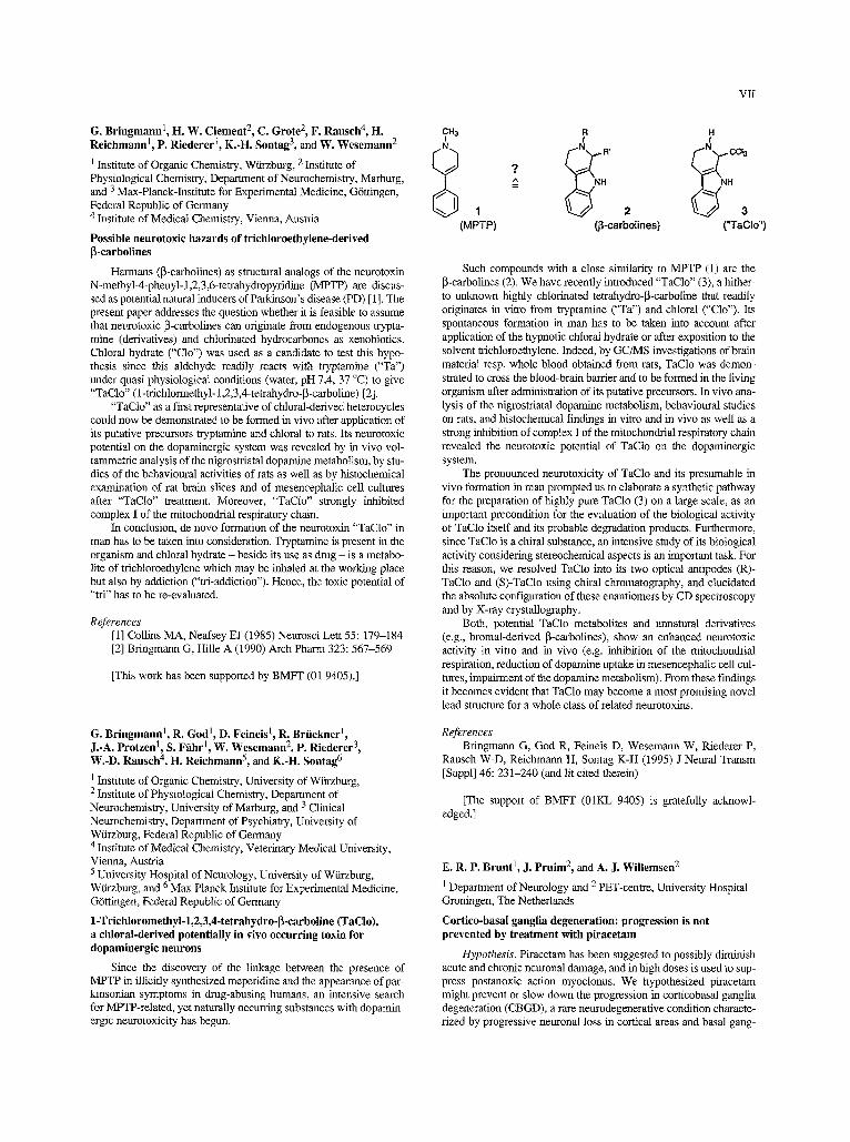
CH3 R H I I
1 2 - . . ~ 3 (MPTP) (~-carbolines) ( 'TaClo")
Such compounds with a close similarity to MPTP (1) are the 13-carbolines (2). We have recently introduced "TaClo" (3), a hither- to unknown highly chlorinated tetrahydro-~l-carboline that readily originates in vitro from tryptumine ("Ta") and chloral ("Clo"). Its spontaneous formation in man has to be taken into account after application of the hypnotic chloral hydrate or after exposition to the solvent trichloroethylene. Indeed, by GC/MS investigations of brain material resp. whole blood obtained from rats, TaClo was demon- strated to cross the blood-brain barrier and to be formed in the living organism after administration of its putative precursors. In vivo ana- lysis of the nigrostriatul dopamine metabolism, behavioural studies on rats, and histochemical findings in vitro and in vivo as well as a strong inhibition of complex I of the mitochondria] respiratory chain revealed the neurotoxic potential of TaCIo on the dopaminergic system.
The pronounced neurotoxicity of TaClo and its presumable in vivo formation in man prompted us to elaborate a synthetic pathway for the preparation of highly pure TaClo (3) on a large scale, as an important precondition for the evaluation of the biological activity of TaClo itself and its probable degradation products. Furthermore, since TaClo is a chiral substance, an intensive study of its biological activity considering stereochemical aspects is an important tusk. For this reason, we resolved TaClo into its two optical antipodes (R)- TaClo and (S)-TaCIo using chiral chromatography, and elucidated the absolute configuration of these enantiomers by CD spectroscopy and by X-ray crystallography.
Both, potential TaClo metabolites and unnatural derivatives (e.g., bromal-derived ~-carbolines), show an enhanced neurotoxic activity in vitro and in vivo (e.g. inhibition of the mitochondrial respiration, reduction of dopamine uptake in mesencephalic cell cul- tures, impairment of the dopamine metabolism). From these findings it becomes evident that TaClo may become a most promising novel lead structure for a whole class of related neurotoxins.
References Bringmann G, God R, Feineis D, Wesemann W, Riederer P,
Rausch W-D, Reichmann H, Sontag K-H (1995) J Neural Transm [Suppl] 46:231-240 (and lit cited therein)
[The support of BMFT (01KL 9405) is gratefully acknowl- edged.]
E. R. P. Brunt l, J. Pruim 2, and A. J. WiUemsen 2
1 Department of Neurology and 2 PET-centre, Universit 3, Hospital Groningen, The Netherlands
Cortico-basal ganglia degeneration: progression is not prevented by treatment with piracetam
Hypothesis. Piracetam has been suggested to possibly diminish acute and chronic neuronal damage, and in high doses is used to sup- press postanoxic action myoclonus. We hypothesized piracetum might prevent or slow down the progression in corticobasal ganglia degeneration (CBGD), a rare neurodegenerative condition characte- rized by progressive neuronal loss in cortical areas and basal gang-
VIII
lia, and often the occurrence of a distinguished type of cortical reflex myoclonus [1]. This study extends a previous report [2].
Aims of the study. To judge possible therapeutic aspects of pira- cetam in CBGD.
Methods. A single patient with marked segmental action- and reflex myoclonus and asymmetrically impaired motor performance was studied in an open label situation. Ph.acetam was given orally during a 30 month period in a daily dose of 12 grams, and clinical examination, electromyography and videorecording was performed at 3-6 months intervals. Before the treatment, and subsequently at approximately yearly intervals, a brain ISF-FDG PET scan was made. The metabolic activity in frontal, parietal mad thalamic regi- ons was compared with the metabolic activity in the -unaffected- occipital region.
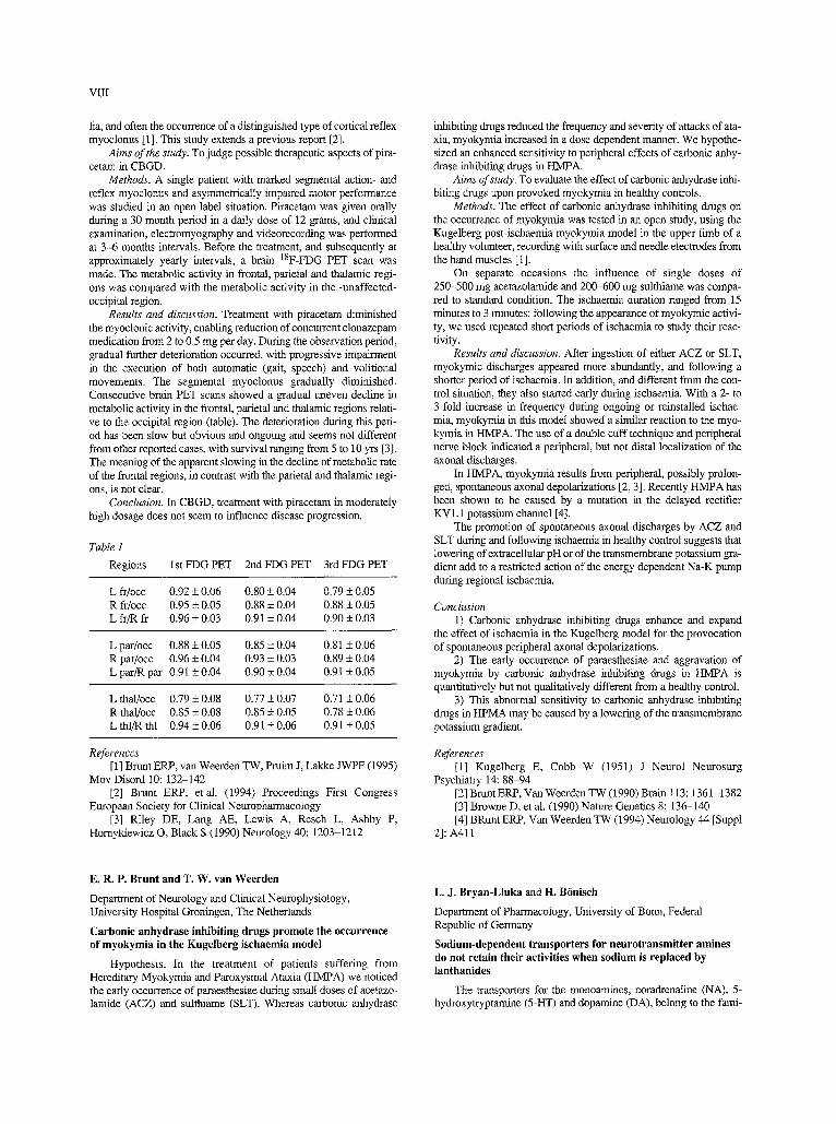
Results and discussion. Treatment with piracetam diminished the myoclonic activity, enabling reduction of concurrent clonazepam medication from 2 to 0.5 mg per day. Dming the observation period, gradual filrther deterioration occurred, with progressive impairment in the execution of both automatic (gait, speech) and volitional movements. The segmental myoclonus gradually diminished. Consecutive brain PET scans showed a gradual uneven decline in metabolic activity in the frontal, parietal and thalamic regions relati- ve to the occipital region (table). The deterioration during this peri- od has been slow but obvious and ongoing and seems not different from other reported cases, with survival ranging from 5 to 10 yrs [3]. The meaning of the apparent slowing in the decline of metabolic rate of the frontal regions, in contrast with the parietal and thalamic regi- ons, is not clear.
Conclusion. In CBGD, treatment with piracetam in moderately high dosage does not seem to influence disease progression.
Table 1
Regions 1st FDG PET 2nd FDG PET 3rd FDG PET
L fr/occ 0.92 _+ 0.06 0.80 -+ 0.04 0.79 -+ 0.05 R fr/occ 0.95 -+ 0.05 0.88 -+ 0.04 0.88 _+ 0.05 L fr/R fr 0.96 _+ 0.03 0,91 + 0.04 0.90 _+ 0.03
L par/occ 0.88 _+ 0.05 0.85 _+ 0.04 0.81 + 0.06 R par/occ 0.96 _+ 0.04 0.93 _+ 0.03 0.89 + 0.04 L par/R par 0.91_+0.04 0.90_+0.04 0.91_+0.05
L thal/occ 0.79 + 0.08 0.77 _+ 0.07 0.71 _+ 0.06 R thal/occ 0.85 + 0.08 0.85 + 0.05 0.78 + 0.06 L thl/R thl 0.94+0.06 0.91_+0.06 0.91_+0.05
References [1] Brant ERP, van Weerden TW, Pruim J, Lakke JWPF (1995)
Mov Disord 10:132-142 [2] Brunt ERP, etal. (1994) Proceedings First Congress
European Society for Clinical Neuropbarmacology [3] Riley DE, Lang AE, Lewis A, Resch L, Ashby P,
Homykiewicz O, Black S (1990) Neurology 40:1203-1212
inhibiting drugs reduced the frequency and severity of attacks of ata- xia, myokymia increased in a dose dependent manner. We hypothe- sized an enhanced sensitivity to peripheral effects of carbonic anhy- drase inhibiting drugs in HMPA.
Aims of study. To evaluate the effect of carbonic anhydrase inhi- biting drugs upon provoked myokymia in healthy controls.
Methods. The effect of carbonic anhydzase inhibiting drugs on the occurrence of myokymia was tested in an open study, using the Kugelberg post-ischaemia myokymia model in the upper limb of a healthy volunteer, recording with surface and needle electrodes from the hand muscles [1].
On separate occasions the influence of single doses of 250-500 mg acetazolamide and 200-600 mg sulthiame was compa- red to standard condition. The ischaemia duration ranged from 15 minutes to 3 minutes; following the appearance of myokymic activi- ty, we used repeated short periods of ischaemia to study their reac- tivity.
Results and discussion. After ingestion of either ACZ or SLT, myokymic discharges appeared more abmadantly, and following a shorter period of ischaemia. In addition, and different from the con- trol situation, they also started early during ischaemia. With a 2- to 3-fold increase in frequency during ongoing or reinstalled ischae- mia, myokymia in this model showed a similar reaction to the myo- kymia in HMPA. The use of a double cuff technique and peripheral nerve block indicated a peripheral, but not distal localization of the axonal discharges.
In HMPA, myokymia results from peripheral, possibly prolon- ged, spontaneous axonal depolarizations [2, 3J. Recently HMPA has been shown to be caused by a mutation in the delayed rectifier KVI.1 potassium channel [4].
The promotion of spontaneous axonal discharges by ACZ and SLT during and following ischaemia in healthy control suggests that lowering of extracellniar pH or of the transmembrane potassium ga- dient add to a restricted action of the energy dependent Na-K pump during regional ischaemia.
Conclusion 1) Carbonic anhydrase inhibiting drugs enhance and expand
the effect of ischaemia in the Kugelberg model for the provocation of spontaneous peripheral axonal depolarizations.
2) The early occmrence of paraesthesiae and aggravation of myokymia by carbonic anhydrase izthibiting drugs in HMPA is quantitatively but not qualitatively different from a healthy control.
3) This abnormal sensitivity to carbonic anhydrase inhibiting drugs in HPMA may be caused by a lowering of the transmembrane potassium gradient.
References [1] Kugelberg E, Cobb W (1951) J Neurol Neurosurg
Psychiatry 14:88-94 [2] Brunt ERP, Van Weerden TW (1990) Brain 113:1361-1382 [3] Browne D, et al. (1990) Nature Genetics 8:136-140 [4] BRunt ERP, Van Weerden TW (1994) Neurology 44 [SuppI
2]: A411
E. R. P. Brunt and T. W. van Weerden
Depamnent of Neurology and Clinical Neurophysiology, University Hospital Groningen, The Netherlands
Carbonic anhydrase inhibiting drugs promote the occurrence of myokymia in the Kugelberg ischaemia model
Hypothesis. In the treatment of patients suffering from Hereditary Myokymia and Paroxysmal Ataxia (HMPA) we noticed the early occurrence of paraesthesiae dm-ing small doses of acetazo- lamide (ACZ) and sulthianae (SLT). Whereas carbonic anhydrase
L. J. Bryan-Lluka and H. B6nisch
Department of Pharmacology, University of Bonn, Federal Republic of Germany
Sodium-dependent transporters for neurotransmitter amines do not retain their activities when sodium is replaced by lanthanides
The transporters for the monoamines, noradrenaline (NA), 5- hydroxytryptarnme (5-HT) and dopamine (DA), belong to the faim-
ly of twelve transmembrane region, Na +- and C1--dependent neuro- transmitter transporters [1]. In epithelial cells, uptake of sugars such as glucose and amino acids such as proline also occur by Na+-co - transport systems [2, 3]. Although no other monovalent or divalent cations can substitute for Na + for transport of proline or glucose, the trivalent lanthanides have been shown to substitute for Na + [2, 3]. The aim of this study was to examine whether the lanthanides can also substitute for Na + to retain the activity of the human neuro- transmitter transporters for NA, DA, and 5-HT.
The study was carried out using human neuroblastoma SK-N- SH-SY5Y (SY5Y) cells which express the NA uptakel transporter, human placental choriocarcinoma JAR cells which express the 5-HT transporter and transfected COS-7 cells expressing the human DA transporter (hDAT). The SY5Y and JAR cells were subcultured into 12-well culture plates 3 and 2 days, respectively, prior to the experi- ment, and the COS-7 ceils were transfected with hDAT cDNA on day 1 and subcultured into 12-well culture plates on day 3 for an experiment on day 4. On the day of the experiment, the culture medi- um was replaced by buffer (pH 7.4) containing 5 mM KCI, 2 mM MgCI2, 5.6 mM glucose, 1 mM ascorbic acid, 10 mM HEPES buf- fer and, for isotonic solutions, either 150 mM NaC1 or LiC1 or 90 mM EuC13, SmCI3 or LaCI3 or, for hypertonic solutions, either 150 mM NaC1 + 100 mM LiC1 or 250 mM LiCI or 150 mM EuC13, SmC13 or LaCI3. Amine metabolizing enzymes were inhibited. The cells were initially incubated for 15 min at 37 ~ with buffer as indi- cated above, In the absence or preseuce of the selective uptake inhi- bitors, 1 gM desipramme (SY5Y cells), 1 gM 6-nitroquipazine (JAR cells) or 1 gM GBR 12909 (hDAT-transfected COS-7 cells). The cells were then incubated for 2 rain after adding 10 nM 3H-NA (SY5Y ceils), 3H-5-HT (JAR cells) or 3H-DA (hDAT-transfected COS-7 cells). After washing the cells with ice-cold buffer and ce11 lysis, their 3H and protein contents were determined. The specific amine uptake by the appropriate transporter in the cells was calculat- ed as the difference between uptake of tritiated amine In the absence and presence of inhibitor. Data were analysed by analysis of variance and Tukey-Ka-amer post hoc t-tests.
Specific uptake values under isotonic and hypertonic conditions were, respectively, 61% and 77% of total uptake in SY5Y cells, 66% and 58% in JAR cells, and 92% and 87% in the hDAT-transfected COS-7 cells. In SY5Y cells, specific NA uptake was greater (P<0.01) under hypertonic (62.1 +3.7fmol/mg protein, n = 13) than isotonic (48.0 _+ 2.2 fmol/mg protein, n = 16) conditions, and was abolished (P < 0.001) when Na + ions were replaced by Li +, La 3+, Eu 3+ or Sm 3+ ions (n = 6-8). In JAR ceils, specific 5-HT uptake (isotonic: 25.4 + 2.5 fmol/mg protein, n = 6; hypertonic: 24.8 +2.3 fmol/mg protein, n = 6 ) was reduced by 94-100% (P<0.001) when Na + ions were replaced by Li + or Eu 3+ ions (n = 5-6). In hDAT-transfected COS-7 cells, specific DA uptake was less (P<0.001) under hypertonic (173 + 14 fmol/mg protein, n = 6) than isotonic (265 + 20 fmol/mg protein, n = 6) conditions, and was reduced by 95-99% (P < 0.001) when Na + ions were repla- ced by Li + or Eu 3+ ions (n = 6). These results show that the activi- ties of the Na+-dependent amine transporters for NA, DA, and 5-HT were not maintained when Li + or lanthanide ions were present instead of Na + ions. This contrast with the increased activity of the
+ + Na -coupled glucose and proline traospolters when Na ions were 3+ 3+ replaced by lanthanides such as Eu or Sm [2, 3]. It can be con-
cluded from this study that the sites of Na + ion Interaction in the amine transporters for NA, 5-HT and DA and those In the Na +- dependent transporters for sugars and amino acids must be different.
References [1] Blakely RD (1992) Curt Opin Psychiat 5:69-73 [2] Bimir B, Hirayama B, Wright EM (1987) J Memb Biol 100:
221-227 [3] Stevens B, Kneer C (1988) Biochem Biophys Acta 942:
205-208
IX
Th. Biittner, W. Kuhn, Th. MOiler, U. McMonagle, and H. Przuntek
Department of Neurology, Ruhr-University, St. Josef-Hospital Bochum, Federal Republic of Germany
Pharmacological effects of dopaminergies and amantadine on eolour discrimination in Parkinson's disease
AbnolTnalities in colour perception have been recently reported in Parkinson's disease (PD). Up to now the origin of visual disorders in PD and the effect of anti-parkinsonian drugs are unclear. The aim of our study was to evaluate the effect of dopaminergics and aman- tadine on the colour perception in PD. We therefore performed the Famsworth-Munsell 100 hue test (FM) In 19 patients before and after the oral application of the morning medication with L-DOPA. 24 further patients underwent the colour vision test before and after the subcutaneous application of apomorphine. 16 patients were tested initially mad after an infusion therapy with amantadine (200 rag/d) over 3 days. Under those treamaent conditions the motor symptoms of Parkinsonism significantly improved in all three groups as assessed by the part "motor examination" (part ILl) of the Unified Parkinson's Disease Rating Scale (UPDRS). Before the morning medication with L-DOPA the mean total error score (MTES) of the FM was 106.3 (SD 62.5). After the ingestion of the individual L-DOPA medication the MTES improved to 71.8 (SD 51.0) (p<0.001). There was no different effect of L-DOPA on a spe- cific colour axis. After subcutaneous application of apomorphIne, MTES improved from 100.4 (SD 41.3) to 93.4 (SD 58.4). In con- trast, the MTES was unchanged after an Infusion therapy with aman- tadine [MTES before amantadine-infusions: 94.4 (SD 55.1); after therapy: 98.9 (SD 55.0)].
We conclude that the distorted colota" vision in Parkinson's disease in due to a dopamine deficiency Involving the visual system. As far as the visual system is concerned, amantadine appears not to act as an effective dopamine agnnist. We suppose that antiglutama- tergic properties of amantadine are a possible explanation for this phenomenon, because glutamate is regarded as the main neurotrans- mitter of retinal photoreceptors.
References [1] BiJttner Th, et al. (1993) J Neural Transm [PD-Sect] 6:
11-15 [2] Price MJ, et al. (1992) Neurology 42:887-890
L. Calzh 1, 2, M. Pozza 1, F. Coraddu 2, and G. Farci 3
1 Institute of Human Physiology, University of Cagliari, 2 Pathophysiology Centre for the Nervous System, Modena, and 3 Pathology Depm'tment, Brotzu Hospital, Caglim'i, Italy
Age-related modifications of human paraventrieular nucleus: focus on corticotrophin releasing hormone- (CRH), vasopressin- (VAS) and oxytocin- (OXY) immunoreactive (IR) neurons
The paraventricular nucleus of the hypothalamus (PVN) plays a key role in the integration of autonomic, endocrine and limbic sig- nals responsible for body homeostasis control. The homeostatic regulation becomes less reliable with aging. In spite to the variabil- ity due to different animal species, ages and experimental ap- proaches, it is generally accepted that the basal levels of hypotha- malus-pituitary-adrenal (HPA) axis tend to increase with age. Moreover, old subjects have a different responsiveness to acute stressful situations. CRH, VAS and OXY are responsible for hypo- thalamic regulation of the pituitary-adrenal axis. In this study, we investigated the distribution of CRH-, VAS- and OXY-IR neurons in the PVN of human brains. 5, male and female "old" (age 78.0 _+ 2.3 years) and 3 male and female "adult" (age 48.3 _+ 6.8

X
years) subjects were included in the study. In this subjects, no sig- nals of major neurological or psychiatric disorders were present in the clinical history. The pre-mortem conditions were recorded in order to exclude severe hypoxic or dismetabolic conditions from the study. Post-mortem delay ranged from 24 and 48 hours. The hypo- thalami were dissected out and fixed in paraformaldehyde 4% for 4-6 hours. The brains were then rinsed in a sucrose/buffered soluti- on. The brain blocks were then quickly frozen using CO2 and sec- tioned in a cryostat ( 2 0 ~ 14 gm thickness). The hypothalamic area was carefully sampled allover 4500 gm rostro-caudal extension and series of sections including the PVN were collected on gelatine- coated slides and processed for the indirect inmmnofluorescence visualization of CRH-, VAS-, and OXY-IR elements. Primary anti- sera were purchased from Incstar. Fluorescin-isothiocyanate and tetramethylrhodamine-conjugated secondary antibodies (DAKO) were used. Alternative sections were also processed as "control" sli- des, in order to monitor tissue autofluorescence sasnd antisera spe- cificity. The sampling strategy assured the analysis of the entire PVN. We found a great increase of CRH-IR elements in the PVN of old subjects. In fact, in the anterior PVN we found up to 50 CRH- positive cells/section in old samples, whereas it was hard to visuali- ze up to 10 CRH-IR elements in adult samples. Moreover, in old samples staining intensity was higher than in adult ones and a num- ber of strongly stained dentrites was evident. Long fibres with vari- cosities were also present. Also VAS- and OXY-1R were modified in old subjects compared to adult ones. In particular, the staining intensity of VAS-positive cells was lower in old samples, whereas the cell density was similar to those of adult samples. Moreover, in old subjects we also observed a number of weakly stained neurons which was not present in adult ones. OXY-IR was instead reduced in old samples, with regard to both number and staining intensity of neurons. When possible, the infundibular area was also examined. A great number of CRH- and VAS-IR fibres converging to the hypo- physeal stalk was observed in old subjects. These data indicate an upregulatinn of CRH- and VAS-IR system in the PVN of old sub- jects, supporting an hypothalamic involvement in age-dependent increase of basal level of HPA axis fimction.
[Supported by EU grand BMH1-CT-94-1563.]
A. Carlsson
Department of Pharmacology, University of Gothenburg, Sweden
Recent advances in molecular neuropharmacology: a challenge for drug development
Drug research is facing an enormous challenge at present, thanks to the advent of a variety of powerful methodology, encom- passing molecular biology, biochemical and behavioural pharmaco- logy, and modem imaging techniques. This is illustrated by the ongoing research on receptors, transporters etc. in the brain. The subclassification of receptors, which started out from classical phar- macology, is now receiving an additional input from molecular bio- logy. To characterize the function of the various subtypes at the molecular level, to study their cellular and regional distribution, and ultimately to define their physiological role in the intact system, pla- ces great demands on biochemists, physiologists, pharmacologists, and medicinal chemists.
The ideal approach towards solution of this kind of problem is to synthesize agonists as well as antagonists with the highest possi- ble specificity for a given receptor subtype, to asses their distributi- on, administered as labelled ligands, and to study their effects on various brain functions. In real life, given the close similarity of the various subtypes, it will often prove impossible to achieve a full separation in terms of affinities, and thus the pharmacologist will have to resort to a number of less than ideal tools, and then try the kind of jig-saw puzzle pharmacologists are used too. In any event, the investments necessary to reach the goals are considerable, but so
are often the gains to be expected from a successful enterprise. Consequently, substantial investments are made in this area today, despite the high risks involved in many cases.
As an example of the kind of problem encountered in this rese- arch, the dopamine D3 receptor subtype is taken.
As to the availability of D3-selective ligands, there is some con- troversy concerning agonists but fairly general agreement concer- ning antagonists. Although several antagonists with a high degree of D3 selectivity are in the making at present, it is possible at this time to discuss only agents with some limited selectivity for this subtype. The only agent among these undergoing clinical testing is (+)- UH232, an aminotetralin developed in our research group and found to have a peculiars pharmacological profile (preferential dopamine autoreceptor antagonist, Svensson etal., 1986). Only later did Sokoloff et al. (1990) discover its D3 selectivity, which shows up as a D3/D2 affinity ratio of 4 to 5 in different measurements. This level of selectivity cannot be regarded as satisfactory but may provide important clues.
Recently more selective D3 receptor ligands have been de- veloped. Studies with these compounds have revealed that post- synaptic D3 receptors exert a behaviottrally inhibitory function, in contrast to the stimulant functions of other dopamine receptors. The possible implications of this observation will be discussed.
R. Ceravolo, A. Napolitano, S. Salvetti, G. Dell'Agnello, M. Renna, and U. Bonuccelli
Institute of Clinical Neurology, University of Pisa, Italy
Clozapine, apomorphine, and levodopa in essential tremor
Essential tremor (ET), the most common movement disorder, may affect hands, voice, legs, and trunk resulting in significant dis- ability in some patients. The parmacological treatment of ET is often disappointing, owing to a lack of insight on its pathogenesis. We haves carried out a comparative study of the effects of two dopa- minergic agents, i.e., levodopa and apomormphine, and the atypical neuroleptic clozapine on 15 ET patients. Study subjects received randomly, on separate days, levodopa/carbidopa (250/25 mg p.o.), clozapine (12.5 mg p.o.), apomorphine (50 gg/kg s.c.) or saline s.c. as placebo. Tremor was scored by means of an arbitrary four paint- scale (0 = absent; 1 = mild; 2 = moderate; 3 = severe) every ten minutes for the first two hours, and every thirty minutes for the fol- lowing three hours. At the same time we monitored cardiac frequen- cy, blood pressure and sedation quantified by an arbitrary four point- scale. The outline of the antitremor effect was as follows: a) magni- tude: clozapine > apomorphine >> levodopa > placebo; latency: cloza- pine = levodopa >> apomorphine; duration: clozapine >> levo- dopa > apomorphine; sedation: clozapine = apomorphine > levodo- pa = placebo. After fixing a cut-off of 50% of improvement to con- sider positive the test, we reported positive response in 88% out of patients by clozapine, 53% by apomorphine, 25% by levodopa and 17% by placebo. The anti-tremor effect of clozapine and apomor- phine was not dependent on sedation.
On the basis of our results clozapine was chronically administe- red to 10 ET patients for 10.7 _+ 4.8 months reporting a moderate to marked and persisting in time reduction of tremor in the whole group, without any significant side-effect. Our data suggest that a marked antitremor effect can be achieved by both blocking (cloza- pine) or, to as lesser degree, stimulating (apomorphine, levodopa) dopamine receptors. On the other band, the atypical profile of cloza- pine, in particular anti-muscarinie, anti-serotonergic and anti-adren- ergic properties, suggests that the modulation of neurochemical systems, other than the dopaminergic one, may be responsible of its efficacy in ET.
F. Conquet 1, Z. Bashir 2, H. Daniel 3, F. Ferraguti 4, G. Collingrldge 2, and F. Crtpel 3
1 Glaxo IMB, Plan-les-Quates, Switzerland 2 University of Birmingham, United Kingdom 3 University of Paris-Sud, Orsay, France 4 Glaxo Research Laboratory, Verona, Italy
The knock-out technology as a novel approach for studying the role of Glutamate receptors in synaptic plasticity
Gene knockout technology is becoming a major tool for the fun- ctional study of molecules which are thought to be involved in syna- ptic plasticity. Some recent studies have reported the implication of kinases in learning and memory with the use of this strategy. The generation of such an animal becomes crucial when neither selective nor potent antagonists are available to block the activity of the mole- cule.
Metabotropic glutamate receptor 1 (mGluR1) is a member of a large family of G protein coupled-glutamate receptors, the physiolo- gical functions of which are largely unknown. Moreover, neither potent nor selective antagonist is available to assess the contribution of each of the members of the mGluR family. To overcome this pro- blem, we have generated mutant mice for MGluRI by the technique of gene knockout. We report that mGlnRl-deficient mice show severe motor coordination defect which can be referred to as ataxia and which is likely to be due to cerebellar dysfunction. Furthermore, mutant mice showed spatial learning defects. Histological studies revealed no gross anatomical abnormalities in either the cerebellum or hippocampus. Electrophysiological analysis of the mutant mice showed normal basal synaptic response of Purkinje cells as well as in the different areas of the hippocanapus. They do, however, show impaired cerebellar LTD and hippocampal mossy fibre LTP. Therefore, these results strongly suggest that mGiuR1 is implicated in these two forms of long-lasting electrophysiological events pro- bably with different signal transduction pathways according to its site of expression. In addition, deletion of a single member of the mGluR family creates a striking phenotype which should be valua- ble for the study of synapfic plasticity.
J. Coos Verhoef, F. W. H. M. Merkus, and H. E. Junginger
Laiden/Amsterdam Centre for Drag Research, Leiden University, The Netherlands
Novel delivery systems for peptide drugs
Recent years have seen enormous advances in the field of pro- tein and (poly)peptide engineering by means of biotechnology and recombinant DNA techniques, ,and todays possibilities are to produ- ce significant quantities of a wide variety of biologically active pept- ides mad proteins that are therapeutically applicable. In most cases such compounds are indicated for chronic therapy, where they will need to be administered by an appropriate delivery system. At the time being all possible alternative routes of peptide and protein administration - avoiding the parenteral route - are investigated with great efforts both in industry and academia. They include the nasal, transdermal (using iontophoresis), pulmonal, rectal, buccal and per- oral routes, and for each route's requirements special drug delivery systems have been or are being designed. However, particular diffi- culties are met in designing effective delivery systems, i.e., to achie- ve a predictable and reproducible absorption in therapeutic doses.
In general, poor absorption of peptide and protein drugs across mucosal surfaces is caused by the high polarity and high molecular weight of these compounds and their susceptibility to proteolytic enzymes. In order to improve their mucosai transport properties and thereby enhancing their systemic bioavailabilities, several strategies
XI
can be used, such as the synthesis of stabilized and more lipophilic analogues, co-administration of absorption enhancers (e.g., bile salts, surfactants, cyclodextrins) and protease inhibiturs, and the design of appropriate dosage forms (e.g., the use of solid versus liquid preparations; encapsulation of the peptide/protein di-ag in par- ticular or vesicular carriers such as biodegradable nanoparticles, bio- adhesive microspheres and liposomes). Also transdermal iontopho- resis, a non-invasive technique using a mild electric current (0.1-O.5 mA/cm 2) to facilitate percutaneous drug transport, offers the potential to deliver peptide drugs systemically in a continuous or pnlsatile fashion.
This presentation will focus on recent results in this challenging field of research, as obtained in our Centre with two ACTH- and vasopressin-related neuropepfides, the LHRH analoque buserelin, and the polypeptide hormone insulin.
F. F. Cruz-Sfinchez
Neurological Tissue Bank, Hospital Clinic, University of Barcelona, Spain
Brain research in Europe
To investigate may generate a sensation akin to well-being, comfort and prowess to society at large which in turn spills over to its conu-ibutors. Societies, power groups and states fight for pres- tigious investigative projects which ultimately extrinsecate in the eternal spell of supremacy. At this this junction, please pause mad remember the rush to the moon and space.
In this context, investigators may befall the risk of scientific nationalism which propounded in other fortunately forgotten histor- ical times; recaII here the history of our century in the Europe of the 30s. Scientific nationalism is a principle for which investigators pur- posely avod to understand what they are doing in the culture to which they appertain. In these case, investigators are infused only of their own self, in their subjective aims. Researchers must be open to diverse horizons, to social interests and to the universitality.
Brain disease affects around 20% of the population and, for example, more than 50 million Americans are sufferer in any one year, thus costing the government 120 b$. The 20% of the health budget of advanced nations is spent on brain-ages, especialIy with a tendency to increase as the population ages, especially with the increase in the number of neurodegenerative afflictions.
To this economic dimension one must add the consequences of the sufferings not only of those who are affected but also of the fami- ly. Probably the illnesses which have a greater economic and social impact are the age dependant neurodegenerative ones, autoimmune and those which present in a paroxystic form. The future of neuros- cience lies in facilitating the improvement of clinical diagnosis, in lending support to the development of tfigh quality investigative approach of brain and in joining related sciences.
With the clear intention of stimulating further progress in the study of the brain, private and public urganisations devote large sums for research so that new methods of diagnosis, prevention and cure can be devised whilst at the same time more information is gat- hered on the very pathogenetic mechanisms of the various conditi- ons.
The European Parliament proposed in 1992 to establish a speci- fic program of brain research within the fourth Framework Program of Research mad Technology Development, underlying the need of promotion of neuroscience by the combined support of several dis- ciplines. The cormnission of the European Communities throughout the collaboration of experts from several European countries produ- ced the European Decade of the Brain Research Report proposing a project for 5 years. This project is based on 2 specific programs, Biotechnology and Biomed. The aim of both programs is to contri- bute to the development of the health of European citizen and the promotion of the relationship between health industry and reseat-
XII
chers. But the most important objectives of such programs is to esti- mulate and develop collaboration across European researchers try- ing to establish joint task forces for common problems such as neu- rological and psychiatric disorders.
[Supported by the EC BIOMED programme; PL931359.1
L. Demisch 1, H. Baas 2, T. Kutschka 2, M. Beeg 2, and P.-A. Fischer 2
1 Clinical Neurochemistry Laboratory and 2 Clinic of Neurology, University of Frm~zflkrt, Frankfurt am Main, Federal Republic of Germany
Influence of chronic levodopa treatment on the amount of lipid peroxides and the activity of cytosolie superoxide dismutase in platelets from patients with Parkinson's disease
Aim of the study. There is a controversial debate whether levo- dopa medication contributes to oxidative stress in patients with Parkinson's disease (PD). Whereas most clinical studies did not con- ftrm the conclusion of Lesser et al. published in 1979 about an enhanced deterioration of motor symptoms following levodopa therapy there are arguments from biochemical studies about the above suggestion. Aim of the study was to demonstrate effects of chronic levodopa medication on biochemical indices related to radi- cal scavenging and oxidative stress in parkinsonian patients. Blood platelets were used as ex vivo models since increased levels of pla- telet lipid peroxides and decreased activities of cytosolic superoxide dismutase (SOD-l) were found in platelets of defined density from untreated PD patients.
Methods. The study sample consisted of 16 patients (9 fe- males/7 males, aged 60 .+ 10 yrs) never treated with levodopa (de- novo patients) and 28 patients (15 f/13 m, aged 62 + 12 yrs) medi- cated with L-dopa (mean: 433 mg/day) and a peripheral decarboxy- lase inhibitor since 6.3 .+ 5.8 yrs. In de-novo PD patients biochemi- cal determinations and clinical evaluations were carried out before and 6_+2 weeks after starting medication with levodopa (356 mg/day; range 150-600 mg/day) and benserazid. Platelets were prepared from citrated blood samples and purified by means of den- sity gradient centxJfugation. Lipid peroxides were measured as malondialdehyde-thiobarbituric acid product ((MDA-(TBA)2) after HPLC separation. Cytosolic SOD (Cu/Zn-SOD; SOD-l) was mea- sured as described by McCord and Fridovich. Data from 11 de-novo patients were available for statistical analysis of SOD-1.
Results and Discussion. Following 6 weeks of Ievodopa treat- ment there were no significant changes of platelet MDA levels ((before: 42.6 + 24.2; after: 37.8 + 16 pmol MDA(TBA)2/10 a pit)). L-DOPA treatment did not alter SOD-1 activity (before: 0.46 + 0.30 U/108 plt; after: 0.47 _+ 0.31 U/108 pit). A comparison of the group of de-novo patients and patients medicated for about 6 years with L- DOPA did not show significant differences among the above men- tioned biochemical parameters. In the present study levodopa was administered together with a peripheral decarboxylase inhibitor. Thus marked increases of plasma dopamine levels and an enhanced deamination of dopamine via platelet MAO-B does not take place. Therefore the above results are limited to effects of L-DOPA.
Conclusion. Chronic adminstration of levodopa has no effect on some biochemical indices related to oxidative stress in platelets from parkinsonian patients such as lipid peroxidatiun and SOD-1 activity.
[Supported by a grant from BMFT (01KL9011).]
L. Demisch 1, T. Kutschka 2, H. Baas 2, M. Beeg 2, and P.-A. Fischer 2
1 Clinical Neurochemistry Laboratory and 2 Clinic of Neurology, University of Frankfurt, Frankfurt am Main, Federal Republic of Germany
Effect of chronic L-deprenyl treatment on lipid peroxide concentrations and cytosolic superoxide dismutase activity in platelets from patients with Parkinson's disease
Aim of the study. L-deprenyl is the first drug which appears to slow the progression of Parkinson's disease (PD). The substance was selected for clinical use because it inhibits selectively MAO-B and decreases peroxides generated from MAO B oxidation of dopa- mine. The exact mechanism of delaying the development of disabi- lity in parkinsonian patients remains, however, to be defined. Aim of the present study was to show alterations of biochemical indices related to oxidative stress in platelets from PD patients after long- term L-deprenyl treatment (up to 2 years).
Methods. The study sample consisted of 20 levodopa treated PD patients (9 females, 11 males; aged 62 + 11 yrs), which never recei- ved L-deprenyl before. Clinical and biochemical investigations were carried out before L-deprenyl administration and every six month up to two years later. After inclusion into the study sample all patients were medicated with 5-10 mg L-deprenyl/day in addition to levo- dopa. Presently the data of one-year follow-up are shown. Platelets were prepared and purified by means of density gradient centrifuga- tion. Lipid peroxides (LPO) were measured as malondialdehyde- thiobarbituric acid product after HPLC separation. Cytosolic SOD (Cu/Zn-SOD; SOD-l) was measured as described by McCord and Fridovich.
Results and discussion. Platelet MDA levels and SOD-1 activi- ties were analysed by means of Repeated Measures Analysis of Variance. For both biochemical parameters a significant differen- ce between groups was found ((SOD-l: before L-deprenyh 0.57 .+ 0.42; after six month: 0.45 _+ 0.25; after one year: 0.31 .+ 0.25 U/10 s pit.; p = 0.049; F = 3.257; MDA-(TBA)2 : before L-deprenyl 41.3 _+ 19.5; after six month 36.7 + 20.3; after one year 29.2 + 18.1; Fr = 8.68; p = 0.013)). Post hoc tests showed a significant differen- ce between time points of one year of L-deprenyl medication and before starting treatment. The results demonstrate that chronic L- deprenyl treatment alters biochemical indices of radical scavenging (SOD-1 activity) and oxidative stress (lipid peroxide levels) in levo- dopa medicated parkinsonian patients.
[Supported by a grant from BMFT (01KL9011).]
M. Deuschle, B. Weber, A. K6rner, H. Standhardt, C.-H. Lammers, T. Motzek-No6, and I. Heuser
Max Planck Institute of Psychiatry, Clinical Institute, Munich, Federal Republic of Germany
Doxepine in conventional and pulse-loading application: effects upon psychopathology and HPA-system activity
It was shown, that a single pulseqoading (PL) dose of clomi- pramine improves depressive symptoms. However, so far PL and conventional (CONV) application of antidepressants were never directly compared. We performed a double-blind study of PL and CONV application of doxepine in depressed patients. After a one- week placebo treatment, nine patients in the PL group (age 45.7 + 12.2 yrs) received 250 mg doxepine every 6 days and place- bo on the other days for 40 days. Ten patients in the CONV group (age 44.8 + 12.4 yrs) received increasing doses tmtil day 7 and 250 mg doxepine until day 40. HAMD rating scales were done twice weekly. Three dexamethasone (DEX)-suppression/CRH-stimulation tests were done: 1) during the initial placebo treatment, 2) on day 9 and 3) on day 21. In the PL group, HAM'D scores were significant- ly reduced after 29 days of treatment compared to HAMD scores
after placebo (17.1 -+ 4.9 vs. 22.7 _+ 2.8, p < 0.03). In the CONV group, after 2 days already HAMD scores were significantly impro- ved (22.8 5:7.2 vs. 26.5 5: 5.7, p < 0.03) and improved further until day 40 (HAMD 7.3 5: 5.8). On day 25, there were significant diffe- rence between the two groups (HAMD 12.5 + 8.9 vs. 20.3 + 6.5, p < 0.05). Post-Dex cortisol declined non-significantly in the PL group (35.9 5:40.7 vs. 24.0 + 20.7 vs. 23.6 + 26.6 gg/ml, n.s.), while there was a significant decline in the CONV group at the second test (111.35:159.4 vs. 10.75:4.2 vs. 19.85:19.0 gg/ml, p<0 .05 resp. n.s.). The AUC cortisol response after CRH was attenuated earlier in the PL group (5667 + 2910 vs. 1883 + 2178 vs. 2239 + 2583, p<0 .001 resp. p<0 .001) compared to the CONV group (5283 5:4609 vs. 1267 _+ 2053 vs. 445 + 1016, n.s. resp. p < 0.02). We conclude that 1) compared to PL, CONV application of doxepi- ne is clinically superior and 2) both modes of application have attenuating effects on HPA-system activity, but with different time courses.
C. D. Earl 1, T. Reum 2, J. Sautter 1, J.-X Xie 1, A. Kupsch 1, W. H. Oertel 1, and R. Morgenstern 1
1 Neurologische Klinik, Klinikum Grosshadem, Munich, and 2 Institute of Pharmacology and Toxicology, Humboldt University Berlin, Federal Republic of Germany
Foetal ventral mesencephalic cell suspension grafts influence dopamine overflow in the non-grafted caudate putamen of 6-hydroxydopamine-lesioned rats: in vivo voltammetric data
The effects of intrastriatal ventral mesencephalic cell suspensi- on grafts on the dopamine system of the non-lesioned candate puta- men are largely unknown. The present study employed differential pulse voltammetry to monitor and compare extracellular levels of dopamine in the lesioned/non-grafted, in the lesioned/grafted, the respective non-lesioned/non-grafted contralateral rat candata puta- men and the normal caudate putamen of control rats after pharma- cological stimulation of dopamine overflow.
Cell suspensions of foetal rat ventral mesencephalic tissue were grafted into the dopamine-depleted caudate pntamen of unilaterally 6-hydroxydopamine-lesioned rats. At six weeks, animals with func- tional, mature grafts were pretreated with pargyline (75 mg/kg, i.p.) and both striatal sides were monitored for dopamine overflow for 90 min following amphetamine sulphate administration (5 mg/kg, i.p.).
The time course of dopamine overflow inside the graft was similar to that in the non-lesioned/non-grafted contralateral caudate putamen of the same animal and to the normal control animal. However, in grafted animals the mean dopamine overflow detected in the non-lesioned/non-grafted caudate putamen was approximate- ly 34% lower than the concentration of dopamine detected in the non-lesioned/non-grafted caudate putamen of 6-hydroxydopamine- lesioned (non-grafted) control animals and approximately 39% lower than the concentration of dopamine detected in the caudate putamen of the normal control animal. In contrast, no voltammetric signals could be detected in the candate pntamen ipsilateral to the 6- hydroxydopamine lesion. There was no statistical difference in the concentration of amphetamine-induced dopamine overflow between the caudate putamen contralateral to the 6-hydroxydopamine lesion and the candate pntamen of the normal control animal. The mecha- nism by which grafted cells influence the contralateral caudate pnta- men may be related to a reduction in the fimction of the dopoamine transporter system and hence a decrease in the overflow of cyto- plasmic dopamine. Alternatively the decrease in dopamine overflow may be secondary to a modification in gamma-aminobutyric acid neurotransmission in caudate pntamen contralateral to the grafted cells.
XIII
G. M. Emilien 1, J. M. Maloteaux 2, A. Seghers 2, and G. Charles 1
1 Bristol Myers Squibb, and 2 Department of Neurology, Universit~ Catholiques de Louvaln, Brussels, Belgium
Lithium compared to valproic acid and carbamazepine in manic depressive illness. A statistical meta analysis
Lithium has been used for nearly a quarter century in the phar- macotherapy of bipolar disorders. It is the only medicine currently approved by U.S. Food and Drug Association (FDA) for the treat- ment of mania although the anticonvulsants valproate and carbama- zepine have also been used to treat this disorder. However, the use of hthium is hindered by its toxic effects. Lithium has a narrow the- rapeutic/toxic ratio. Therapeutic monitoring of serum concentrations must be carried out. Although, its mechanism of action is still uncle- ar, there is much evidence to suggest that lithium exerts its thera- peutic action by interfering with the metabolism of the phosphoino- sitides which play an important function in synaptic transmission.
In many studies, it is suggested that the anticonvulsants carba- mazepine and valproate also share the same components of spectrum of efficacy like lithium in the treatment of affective disorders. In addition, they are clinically effective in the same lithium nom'espon- ders (Jefferson, 1995). The aim of this study is to compare the bene- fit/risk ratio of lithium to valproic acid and carbamazepine in a meta analysis.
Meta analysis (Glass, 1976) is a statistical procedure to reanaly- se published data to answer new research questions. Interest in meta analysis of randomized clinical studies grew out of recognition of problems resulting from the conduct of small randomized trials which individually showed no effect although the results pointed in the same direction. The results may turn out to be significant on meta analysis.
Data sources included the MEDLINE database and relevant references from articles obtained in this search and in major reviews. Only randomized, placebo controlled studies are included. They are required to have a double blind evaluation of outcome. Serum lithi- um levels should be within 0.4-1.5 mmol]l.
The results are discussed in terms of benefit/risk ratio of the three drugs involved. The importance and contribution of the meta analysis as a useful statistical technique in psychoactive drug re- search is also underlined.
RefeFeFtces Glass GV (1976) Primary, secondary and recta-analysis of rese-
arch, Educ Res 5 :3 -8 Jefferson JWW (1995) Lithium: the present and the future.
J Clin Psychiatry 56:41-48
R. Erdmann and D. Hrgemann
Department of Clinical Psychiatry and Psychotherapy, Medizinische Hochschnle Hannover, Federal Republic of Germany
Nicotine and neuropsychiatric movement disorders
Neuropsychiatric movement disorders such as Tourette Syndrome (TS), Tardive Dyskinesia (TD), Neuroleptic Induced Parkinson (NIP), and Parkinsun's Disease (IPD) are due to a dys- function within the nigrostriatal system: hypersensibility of dopa- minergic receptors in TD and TS; hyposensibility in NIP and hypo- activity in IPD. Former investigations have shown that nicotine exerts different effects in the nigro-striatal system after acute or chronic applications and also it is well known that patients with movement disorders show a different smoking behavioural pattern (the usual way to take nicotine): chronic use in patients with TD, TS, and NIP, whereas patients with IPD seldom smoke.
The aim of the study was to find out whether nicotine inf!uen-
XIV
ces the symptomatology of the movement disorders. The patients with movement disorders received nicotine and were rated before and after application to decide if there were any change in the seve- rity of the disorder.
Preliminary results are, that nicotine has positive effects in TS and perhaps in TD and NIP. But no results in IPD.
In respect of epidemiological, electrophysiological, pathobio- chemical, and pathophysiological studies and our own resuits a hypothetical model of the nicotine effects ha neuropsychiatric move- ment disorders could represent that acute nicotine application leads to a higher activity of the frontal cortex consequent an amelioration of the symptomatology, especially in TS. Chronic application of nicotine desensibilizes the dopaminergic receptors in the nigro-stria- tal system with positive results in TS and TD, but negative results in IPD and NIP.
Nicotine is possibly helpful in the treatment of some movement disorders.
N. Fichter, C. H. Liicking, G. B. Landwehrmeyer, T. Winter, and T. J. Feuerstein
Sektion Klinische Neuropharmakologie, Neurologische Universit~itsklinik, Neurozentmm, Freiburg, Federal Republic of Germany
Nipecotate-induced GABA release from slices of the rat caudato-putamen: effects of gabapentin
The precise mechanism of action of the anticonvulsant amino acid gabapentin is unknown. In normal brain tissue, gabapentin is inactive at GABA receptors and GABA uptake carrieres and does not imfluence the quasi-physiological, action potential-induced, exocytotic release of GABA from brain slices. We hypothesized that this drug is therapeutically active only under pathophysiological conditions, i.e., in hyperactive neuronal circuits. The microenviron- ment of hyperactive neurons is characterized by elevated extracellu- lar potassium levels which may alter the sodium gradient essential for GABA transport. Under these conditions a reversal of the GABA cartier may increase extracellular GABA levels. Gabapentin could enhance this mechanism thereby strengthening GABA receptor- mediated effects which in turn could counteract neuronal hyperac- tivity. Therefore, we developed an m-vitro model of GABA release through reversed GABA transport using nipecotate an inhibitor and a subs~'ate of the GABA transporter and studied the effects of gaba- pentin in this model.
Slices of the caudato-putamen of rats were prelabelled with eit- her [3H]-GABA or [3H]-glutamine and superfused with physiologi- cal buffer. The incubation with [3H]-GABA allowed to measure [3H]-GABA directly as tritium in the supeffusate fractions and in the slices. Aminooxyacetic acid (AOAA, 10 ~tM) was present in the supeffusion fluid to prevent the metabolic breakdown of GABA and to increase the cytosolic pool of the transmitter. When [3H]-gluta-
w 3 3 mine as used instead of [ H]-GABA, [ H]-GABA was synthesized by endogenous glutamic acid decarboxylase (GAD) in GABAergic neurons thus labelling preferentially the neuronal transmitterpool of GABA. [3H]-GABA was separated from other tritiated compounds by cation exchange chromatography. AOAA was not used in expe- riments employing [3H]-glutamine. Brain slices were stimulated by a pulse of 2 rain of nipecotate (1 raM) to release GABA. Release- modulating drugs were added to the superfusion fluid before stimu- lation. The nipecotate-induced [3H]-GABA release was neither tetrodotoxin-sensitive nor dependent on extracellular calcium sug- gesting action potential-independent, non-exocytotic release. It was diminished, however, by the GABA uptake inhibitor NNC-711 (1, 3, 10 gM) in a concentration-dependent fashion suggesting a rever- sal of GABA transport. Gabapentin (100pM) significantly (p < 0.05) enhanced the nipecotate-evoked release to 155.1 + 26.9% of the corresponding control mean. This effect was observed only
provided that the neuronal transmitter pool of GABA was preferen- tially labelled following incubation with [3H]-glutamine. This effect of gabapentin (100 gM) was not detectable when [3H]-GABA itself was used to label neuronal and extraneuronal GABA pools.
This finding of an increase in nipecotate-induced GABA relea- se by gabapentin suggests that a novel mechanism to enhance GABA activity may underly the anticonvulsant action of this drag. Moreover, the fact that gabapentin was effective when [3H]-GABA was synthesized within GABAergic neurons from its precursor [3H]- glutamine, but not when AOAA inhibited the activity of intracellu- lar enzymes as GABA aminotransferase and GAD, points to bio- chemical changes that require gabapentin to be present in the neuro- nal cytosol, perhaps to interact with a cytosolic enzyme. The enhan- cement of GABA activity by gabapentin seems to be pathophysiolo- gically relevant since the underlying reversal of the GABA canier contributes to a non-synaptic release of GABA which may become important under conditions of neuronal hyperactivity.
Apart from seizures neuronal hyperactivity may also cause other CNS disturbances, like certain forms of pathological tremor. Therefore, tremor could also be a field of therapeutic application of gabapentin. Preliminary results are consistent with this hypothesis as gabapentin markedly reduced essential postural tremor in two pati- ents.
D. Fitzgeral l, M. C. Anderson 2, B. Lawlor 2, and K. F. Tipton 1
1 Department of Biochemistry, Trinity College. and 2 Department of Psychiatry, St Patrick's Hospital, Dublin, Ireland
Substrate specificity of human platelet monoamine oxidase-B in Parkinsnn's and AIzheimer's disease
The activity of human platelet monoamine oxidase-B (Amine : Oxygen oxidoreductase (deaminating) (fiavin-containing), EC 1.4.3.4; MAO-B) has been reported to be altered in a munber of diseases of the central nervous system, such as Alzheimer's disease (AD), depression, epilepsy, schizophrenia, and Parkinson's disease (PD). However, the data obtained in different published studies have been contradictory, perhaps partly because different preparative and assay procedures have been used. Humfrey et at. (Biochem Pharmaco140: 2562-2564, 1990) reported that the substrate used to determine the platelet MAO-B activity in subjects with PD (both treated and untreated) affected the changes in activity seen, with sug- gesting that the specificity of the enzyme may be altered in this con- dition. They found that activity towards 2-phenylethylamine (PEA) was higher in PD than in control subjects, whereas it was lower in the former ~oup when tyramine was used as substrate and the decrease was even more marked with dopamine. Thus it might appe- ar that effectiveness of platelet MAO-B towards hydroxyl-substitu- ted amine substrates, such as dopamine, is differentially lowered in PD. If such differences were an in~insic property of the enzyme its- elf, they should be independent of the preparative and assay proce- dures and, therefore, might constitute a robust marker for PD. Furthermore, although there is no information to indicate whether this situation also applies in brain, such a change could have far-lea- ching consequences in terms of metabolic capacity towards dopami- lae.
In order to investigate this fimher the activities of ptatelet MAO-B were studied in a group of 32 subjects comprising: 7 clin- ically-defined untreated PD subjects, 16 clinically-defined untreated AD subjects and 9 age-matched control subjects. Enzyme activities were determined with benzylamine, 2-phenylethylamine (each con- taining no ring hydroxy-group), tyramine (4-hydroxy-2-phenylethy- lamine) and dopamine (3,4-dihychoxy-2-phenylethylamine) as sub- strates. No differential alterations in the activities towards these sub- strates was seen in either PD or AD as compared to controls, or to one another. The possibility that the earlier results were affected by the presence of an endogneous modulator that affects the specificity
of MAO-B in PD cannot be excluded, but the present results would indicate that there is no change in the substrate specificity of the enzyme itself, either in this condition or in Alzheimer's disease.
[The support of the EC BIOMED programme is gratefully ack- nowldeged (BMH1-CT94-1563).]
R. S. J. Frackowiak
Wellcome Department of Cognitive Neurology, Institute of Neurology, London, United Kingdom
Imaging neuropharmacological function in the human brain
Non-invasive monitoring of radioactivity using positron emissi- on tomography has been used to investigate issues in psychophar- macology over the last 10 years. There are two main areas of rese- arch. The first is based on tagging radio pharmaceuticals of interest with positron emitters and imaging the distribution of radioactivity at various times folowing administration. Tagged tracers can be used to directly measure the affinity or the concentration of specific receptors. They can also be used as tracers to measure the pharma- cokinetic properties of previous, or subsequent, pharmacological doses of cold molecules of interest that interact with the same recep- tors. Examples of these approaches will be presented.
The second major area of interest lies in the use of tracers of cerebral perfusion which act as an index of local synaptic activity in the brain. Such tracers can be used to identify distributed brain systems that underlie particular functions. Through experiments that are usually factorial in design, the activity in such distributed systems can then be modified by pharmacological doses of relevant chemicals. The interaction between behaviourally induced activity and its modulation by the pharmacological agent can then be moni- tored. Examples of this approach will also be given. It is possible that ha the future this type of experimentation may be carried out using non-invasive MRI based techniques.
U. Freo 1, M, Dam 1, G. Pizzolato 1, A. Merico 1, C. Ori l, E. Sale 2, and L. Battistin 1
I Neurology and 2 Anesthesiology, University of Padova, Italy
The effects of fluoxetine on local cerebral glucose utllisation of awake rats
Fluoxetine is a widely used antidepressant that has been re- garded as a selective serotonin (5-HT) reuptake inhibitor. However, recent reports indicate that some therapeutical effects of the drug may depend upon non 5-HT mechanisms, such as inhibition of dopamine and noradrenaline uptake and enhancement of dopamine release.
The aim of this study was to assess the specificity of regional cerebral metabolic (rCMR glc) patterns produced by fluoxetine in awake rats.
Experiments were performed ha male 3-too-old Fischer 344 rats. according to the [14C]2-deoxy-D-ghicose antoradiographic techni- que. Fluoxetine hydrocloride (40 mg/kg) was injected i.p. 30 min before the radiotracer, rCMR glc was assessed in 50 brain areas; values of each region were analysed for statistical significance by comparing the mean of fluoxetine treated group to the control group using an unpaired Student-t-test.
Fluoxetine significantly reduced rCMR glc in 40% of the brain areas including sensory systems, limbic areas and basal ganglia regi- ons, the metabolic alterations produced by the antidepressant differ from tllose produced by noradrenaline or dopamine reuptake inhibi- tots, but are very similar to those produced by the tryciclic 5-HT reuptake blocker clomipramine.
These results suggest that the effects fluoxetine on brain func- tional activity are due almost exclusively to its property of inhibiting 5-HT reuptake system.
XV
J. Fritze
Department of Psychiatry, University of Frankfurt am Main, Federal Republic of Germany
Drug targeting in CNS pathology
Drug development has to comprise the full range of aspects of drug targeting, i.e., chemicostmctural, pharmacokinetic, neuropa- thobiochemical, sociopsychological~ and economic. Interestingly, the hierarchy of considerations in this sequence of aspects in reality has to be and in fact is the other way around. The economic aspects comprise the medical need including the competitor situation, the actual therapeutic standard and the prices achievable (generics!), the life-tinae prevalence and course of the disorder in question, i.e., the expected market size in terms of expected sales worldwide. Thus, a high price niche in a small orphan indication might be attractive but also risky. In a world of changing values, changes in the sociopsy- chological acceptance of a new treatment have to be anticipated (e.g., non-acceptance of genetic approaches). There is a shift from treatment to prevention e.g., by non-drng procedures like psychoe- ducation. This trend is anticipated to be especially relevant in psy- chiatric disorders where most probably stressors interact with vul- nerabilities in the pathogenesis. Finally, the drug developer has to consider how much society is willing to pay for health. - The dra- matically growing understanding of the genetic mechanisms of neu- rologic disorders offer new approaches to treatment and prevention, be it by some gene therapy or by manipulating the gene product. These, however, are all small niche indications. This is one of the major reasons why the standard of the neurological therapeutic arse- nal presently is rather low. In contrast, psychiatric disorders mostly chronically affect huge populations with only a few indications with largely unknown pathomechanisms based on the mechanisms of action of a highly competetlve, low price, medium-to-low quality pharmaceutical market. One extreme is the dementias, where many clinicians think an effective treatment is completely lacking. It is anticipated that diagnoses will diversify in the progress to more spe- cific drugs. In the interest of compliance only drugs with reasonably long half-lives allowing for o.d. dosing are recommended. Otherwise, oniy complicated galenic staategies can help. The increa- sing multimorbidity calls for considering multiple pharmacokinetic interactions. The final chemical development starts with the search for some lead structures. These have to be free of patent protection. The screening test battery depends on the indication. If a specific binding site (e.g., an enzyme, a receptor) relevant to the disorder has been identified and its structure is kaaown computer modelling may help identifying a lead s~ucture. Alternatively, the target protein may be expressed in tissue culture to allow for high throughput scre- ening. The supplementary psychopharmacological screening proce- dures include a variety of behavioural animal models the predictive validity of which, however, is disputable. Neurological disorders with identified gene defects as well as Alzheimer's disease offer the opportunity to develop transgenic animal models. The major psy- chiatric disorders are far from this. Interesting drug targets in Alzheimer's disease include the full range of genes and gene pro- ducts involved in the synthesis, deposition and cleavage of e.g. amy- loid and'c-protein. Even more interesting are approaches to the gene- tics of Apo-E and the recently identified gene locus on chromosome 14. Drug targets in psychopharmacology include the full range of neurotransmitter transporters, metabolizing enzymes as well as receptors and their effector systems. New impetus to the develop- ment of antipsychotics comes from the cloning of dopamine recep- tor subtypes but still no breakthrough is in sight, and the role of dopamine in schizophrenia is questionable. Noradrenergic and sero- tonergic systems are probably not directly involved in depression, and antidepressants work unspecifically. Thus, it would be arbitrary and counterproductive for innovation to prefer one transmitter system over others. In affective disorders, an interesting target might
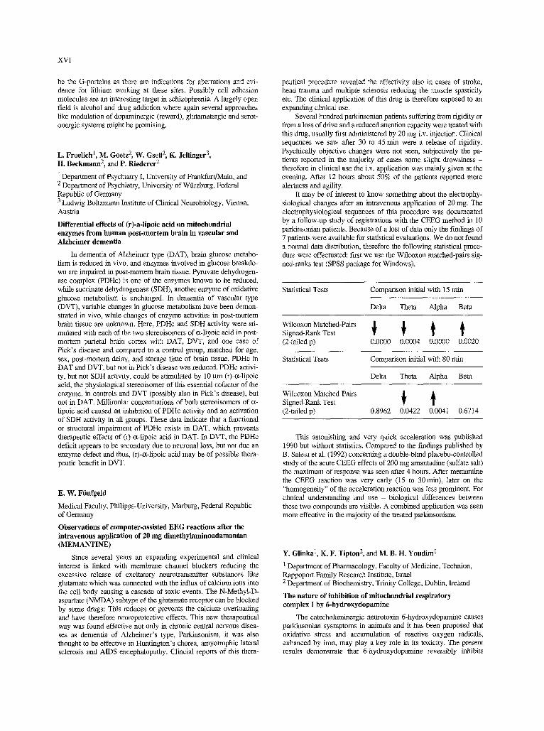
XVI
be the G-porteins as there are indications for aberrations and evi- dence for lithium working at these sites. Possibly cell adhesion molecules are an interesting target in schizophrenia. A largely open field is alcohol and diaag addiction where again several approaches like modulation of dopaminergic (reward), glutamatergic and serot- onergic systems might be promising.
L. Froelich 1, M. Goetz 2, W. Gsell 2, K. Jellinger 3, H. Beckmann 2, and P. Riederer 2
t Department of Psychiatry I, University of Frankfurt/Main, and 2 Department of Psychiatry, University of Wiirzbarg, Federal Republic of Germany 3 Ludwig Boltzmann Institute of Clinical Neurobiology, Vienna, Austria
Differential effects of (r)-a-lipoic acid on mitoehondrial enzymes from human post-mortem brain in vascular and Alzheimer dementia
In dementia of Alzheimer type (DAT), brain glucose metabo- lism is reduced in vivo, and enzymes involved in glucose breakdo- wn are impaired in post-mortem brain tissue. Pyruvate dehydrogen- ase complex (PDHc) is one of the enzymes known to be reduced, while succinate dehydrogenase (SDH), another enzyme of oxidative glucose metabolism is unchanged. In dementia of vascular type (DVT), variable changes in glucose metabolism have been demon- strated in vivo, while changes of enzyme activities in post-mortem brain tissue are unknown. Here, PDHc and SDH activity were sti- mulated with each of the two stereoisomers of c~-iipoic acid in post- mortem parietal brain cortex with DAT, DVT, and one case of Pick's disease and compared to a control ~oup, matched for age, sex, post-mortem delay, and storage time of brain tissue. PDHc in DAT and DVT, but not in Pick's disease was reduced. PDHc activi- ty, but not SDH activity, could be stimulated by 10 gm (r)-o~-lipoic acid, the physiological stereoisomer of this essential cofactor of the enzyme, in controls and DVT (possibly also in Pick's disease), but not in DAT. Millimolar concentrations of both stereoisomcrs of c~- lipoic acid caused an inhibition of PDHc activity and an activation of SDH activity in all groups. These data indicate that a ftmctional or structural impairment of PDHc exists in DAT, which prevents therapeutic effects of (r)-c~-lipdic acid in DAT. In DVT, the PDHc deficit appears to be secondary due to neuronal loss, but not due an enzyme defect and thus, (r)-c~-lipoic acid may be of possible thera- peutic benefit in DVT.
E. W. Fiinfgeld
Medical Faculty, Philipps-University, Marburg, Federal Republic of Germany
Observations of computer-assisted EEG reactions after the intravenous application of 20 mg dimethylaminoadamantan (MEMANTINE)
Since several years an expanding experimental and clinical interest is linked with membrane channel blockers reducing the excessive release of excitatory neurotransmitter substances like glutamate which was connected with the influx of calcium ions into the cell body causing a cascade of toxic events. The N-Methyl-D- aspartate (NMDA) subtype of the gtummate receptor can be blocked by some drugs: This reduces or prevents the calcium overloading and have therefore neuroprotective effects. This new therapeutical way was found effective not only in chronic central nervous disea- ses as dementia of Alzheimer's type, Parkinsorfism, it was also thought to he effective in Huntington's chorea, amyotrophic lateral sclerosis and AIDS-encephalopathy. Clincial reports of this thera-
peutical procedure revealed the effectivity also in cases of stroke, head trauma and multiple sclerosis reducing the muscle spasticity etc. The clinical application of this drag is therefore exposed to an expanding clinical use.
Several hundred parkinsonian patients suffering from rigidity or from a loss of chive and a reduced attention capacity were treated with this drug, usually first administered by 20 mg i.v. injection. Clinical sequences we saw after 30 to 45 rain were a release of rigidity. Psychically objective changes were not seen, subjectively the pa- tients reported in the majority of cases some slight drowsiness - therefore in clinical use the i.v. application was mainly given at the evening. After 12 hours about 50% of the patients reported more alertness and agility.
It may be of interest to know something about the electrophy- siological changes after an intravenous application of 20 mg. The electrophysiological sequences of this procedure was documented by a follow-up study of registrations with the CEEG method in 10 parkinsonian patients. Because of a lost of data only the findings of 7 patients were available for statistical evaluations. We do not found a normal data distribution, therefore the following statistical proce- dure were effectuated: first we use the Wilcoxon matched-pairs sig- ned-ranks test (SPSS package for Windows).
Statistical Tests Comparison initial with 15 rain
Delta Theta Alpha Beta
Wilcoxon Matched-Pairs ~ I~ ~ Signed-Rank Test (2-taiIed p) 0.0000 0.0004 0.0000 0.0020
Statistical Tests Comparison initial with 80 ruin
Delta Theta Alpha Beta
Wilcoxon Matched-Pairs ~ Signed-Rank Test (2-tailed p) 0.8962 0.0422 0.0041 0.6714
This astonishing and very quick acceleration was published 1990 but without statistics. Compared to the findings published by B. Saletu etal. (1992) concerning a double-blind placebo-controlled study of the acute CEEG effects of 200 mg amantadine (sulfate salt) the maximum of response was seen after 4 hours. After memantine the CEEG reaction was very early (15 to 30 min), later on the "homogeneity" of the acceleration reaction was less prominent. For clinical understanding and use - biological differences between these two compounds are visible. A combined application was seen more effective in the majority of the treated parkinsonians.
Y. Glinka t, K. F. Tipton 2, and M. B. H. Youdim I
1 Department of Pharmacology, Faculty of Medicine, Technion, Rappoport Family Research Institute, Israel 2 Department of BiochemisttS,, Trinity College, Dublin, heland
The nature of inhibition of mitoehondrial respiratory complex I by 6-hydroxydopamine
The catecholaminergic neurotoxin 6-hydroxydopamine causes parkinsoinan sysmptoms in animals and it has been proposed that oxidative stress and accumulation of reactive oxygen radicals, enhanced by iron, may play a key role in its toxicity. The present results demonstrate that 6-hydroxydopamine reversibly inhibits
brain mitochondrial respiratory complex I. 6-hydroxydopamine its- elf rather than its oxidative products was responsible for the inhibi- tion. Iron(Ill) did not enhance inhibition but decreased it by stimu- lating the non-enzyme oxidation of 6-hytkoxydopamine. Inhibition was potentiated to some extent by calcium ions.
6-Hydroxydopamine inhibition of complex I was non-selective: both brain and liver enzymes were affected by this inhibitor in two different assay systems involving either ferrycyanide or nhiquinone Q reduction. Brain NADH-ubiquinone Q reductase was inhibited by 6-hydroxydopamine partially uncompetitively in respect to NADH with Ki. = 0.051 + 0.014 raM.
Desferrioxamine protected brain complex I against inhibition by increasing K i tenfold and by increasing the residual activity of the enzyme-substrate-inhibitor complex. It did not compete directly with 6-hydroxydopamine for binding to the inhibitory site, but rather induced a conformation urlfavourable for the inhibitor binding to the protein. It also protected liver complex I. Desferrioxarnine "compe- titively" activated complex I with Ka = 0.02 and 2 mM for brain and liver enzymes.
[Supported by EU grant (BMttl-CT 94-1563).]
M. E. G6tz 1, W. Breithaupt t, M. Gerlach 1, 4, j. Sautter 2, A. Kupsch 2, R. Burger 1, W. H. Oertel 2, M. B. H.Youdim 3, and P. Riederer t
! Department of Psychiatry, University of Warzburg, and 2 Department of Neurology, Grog-Hadem, Munich, Federal Republic of Germany 3 Department of Pharmacology, Faculty of Medicine, Rappaport Family Research Institute, Haifa, Israel 4 Department of Neurology, University of Bochum, Federal Republic of Germany
Chronic TVP1012 (Rasagiline) dose - activity response of monoamine oxidases A and B in brain of the common marmoset
The reasons for the beneficial effects of selegiline, an irreversi- ble MAO inhibitor, described for Parkinson's disease are still a mat- ter of debate. It is often difficult to discern between a symptomatic and a neuroprotective mode of action of selegiline. Now, an acety- lenic mechanism-based inhibitor (Rasagiline, TVP1012) very simi- lar to selegiline has been developed which is devoid of either the cheese effect or sympathomimetic amphetamine-like properties. To evaluate the efficiency and selectivity of TVP1012 the activities of monoamine oxidases A and B (MAO-A, -B) were determined in dif- ferent brain regions of the common marmoset following its subcuta- neous administration from 0.01 to 1 mg/kg for 7 days. At a dose of 0.1 mg/kg TVP1012 almost 80% of MAO-B activity is inhibited in all brain regions investigated, whilst MAO-A is not inhibited in putamen and nucleus accumbens. However, by increasing the TVP1012 dose to 0.5 mg/kg, MAO-A will also be inhibited to a sig- nificant extent cnncomitant to total inhibition of MAO-B. The results obtained indicate that TVP1012 irreversibly hlhibits both types of MAO in the common marmoset with selectivity for MAO- B at doses less than 0.5 mg/kg. Thus, TVP1012 could be useful for el/citing possible neuroprotective effects of MAO-inhibition in ani- mal models of neurodegeneration and even be of therapeutic interest in Parkinson's disease.
[The support of BMFT (01 KL 9405) is gratefully acknowl- edged.]
XVII
M. E. G6tz 1, P. Fischer 2, W. Gsell t, P. Riederer 1, M. Streifler 3, M. Simanyi 3, F. Mitller 3, and W. Danielczyk 3
1 Departlnent of Psychiatry, Clinical Neurochemistry, University of Wiirzburg, Federal Republic of Germany 2 Psychiatric Clinic, University of Vienna, and 3 Ludwig Bohzmarm Institute for Geriatric Research, Research Group Alzheimer Dementia, Vienna, Austria
Platelet monoamine oxidase B activity in dementia: a 4 year follow-up
Platelet monoamine oxidase B (MAO-B) activity has been fotmd to increase significantly in geriatric patients with dementia. For the first time a 4 year follow-up of platelet MAO-B activity and Mini-Mental State (MMS) was performed in patients with probable dementia of the Alzheimer type (DAT) and age-matched controls. Unpaired comparison of DAT patients using Fischer's PLSD post hoc ANOVA calculation resulted in a significant increase in MAO- B activity two years after the beginning of the study and remained significantly higher for the entire period of examinations (p < 0.0001). In addition, unpaired compatison of platelet MAO-B activity of DAT patients to that of controls was highly significant (p < 0.002 U-test). The decline of the MMS of DAT patients prece- ded the elevation of MAO-B activity. However, the decrease in the MMS did not correlate to the time course of increase in platelet MAO-B activity (Speatrnan rank correlation test). Therefore it seems reasonable to assume that degenerative processes in brain areas responsible for cognitive functions are coveting non-aminergic systems rather than aminergic ones. Degeneration of brain mono- aminergic systems at a later stage of DAT are suggested to be reflec- ted by changes in platelet MAO-B. It is likely that our data reflect changes in the activity and vigilance of the patients' parameters that are important in cognitive performance.
[The support of BMPT (01 KL 9405) is gratefully acknowl- edged.]
C. Grote t, H.-W. Clement I, G. Bringmann 2, D. Feineis 2, and W. Wesemann 1
1 Institute of Physiological Chemislry, University of Marburg, and 2 Institute of Organic Chemistry, University of Wtirzburg, Federal Republic of Germany
In vivo studies of the activity of the dopaminergie system after the application of TaCIo (1-trichloromethyM,2,3,4,-tetrahydro- 13-earboline) and derivatives in the rat brain
For the investigation of the etiology of Parkinsonism it is very interesting to study substances which occur in the environment and with a structurally similarity with the well known dopaminergic neu- rotoxin MPTP (N-methyl-4-phenyl-l,2,3,6-tetrahydropyridine). In this study chlorinated 13-carbolines, which can be endogenously for- med in the organism after a supply of chloral [1], and some deriva- tives were unilaterally injected into the substantia nigra pars com- pacta (SNC) of the rat. One week later the dopaminergic metabolism of the striatum was measured monitoring the extracellular content of dihydroxyphenylacetic acid (DOPAC) in both sides of the striatum using in vivo pulse voltammetry.
The intranigral application of 10 I.tg TaClo (1-trichloromethyl- 1,2,3,4-tetrahydro-ls resulted in reduced a DOPAC signal in the ipsilateral sttiamm after one week. The systemic injection of the dopamine (DA) precursor L-DOPA (L-dihydroxyphenylalanine) caused a restauration of the DA metabolism compared to the con- tralateral side of the stTiatum. The N-methylated derivative of TaClo, the substance with the most distinct similarity to MPTP, produced a nearly total lesion of the doparninergic system; the DOPAC signal was reduced to less than 10% compared m the intact side but resto-

XVIII
red by L-DOPA. The intracerebral application of brominated (TaBm) or fluorinated (DDH-TaFIu) compounds caused also a large impatrment of the dopaminergic neurotransmission in the nigrostria- tal system. After the injection of TaBro the DOPAC signal was not fully restored.
The experiments demonstrate the toxicity of highly halogenated [3-carbolines as TaClo and several derivatives to the dopaminergic neurones in the SNC. It could be shown that N-methylation, which may occur in the organism, enhanced the neurotoxic potential of this substance. The results support the assumption that these possibly endogenously formed substances can play a role for the degenera- tion of the nigrostriatal system.
References [1] BNlgmmm G, t-lille A (1990) Arch Pharm 323:567-569
[Supported by BMFT (01KL 9405)].
C. Grote, H.-W. Clement, T. Hirning, M. Sohibaeh, and W. Wesemann
Institute of Physiological Chemistry, University of Marburg, Federal Republic of Germany
Effect of intranlgral injection of iron and aluminium on the dopaminergic neurotransmission of the rat measured by in vivo differential pulse voltammetry
The involvement of transition metals in the etiology of Parkinson's disease and other neurodegenerative disorders by for- mation of free radicals is widely discussed. It is described that the toxic effect of iron can be enhanced by aluminium which is not a transition metal and cannot serve as a catalyst for radical reactions. Because of that the effect of iron or alurninium on the nigrostriatal system was studied in an in vivo model of Parkinson's disease.
In vivo pulse voltammetry with carbon fibre electrodes which allows to monitor the extraceltular concentration of dthydroxy- phenylacetic acid (DOPAC) as a measure for the metabolism of dopamine (DA) was used to determine the DA activity in both sides of the striatum.
One, three or six weeks before the voltammetric measurement Fe 3+ or A13+ was injected unilaterally in the substantia nigra pars compacta (SNC).
An intranigral application of 1.5 gg Fe 3+ resulted in a reduced DOPAC signal in the ipsilateral striatum compared with the con- tralateml side after I and 3 weeks. The DA activity measured after 6 weeks showed an even more pronounced lesion of the nigroslriatal system. The intracerehral injection of alnminium resulted 6 weeks later in an impairment of the DA system which was in a similar extent as after iron injection. A coinjection of 1.5 gg Fe 3+ and A13+ enhanced the neurotoxic effect of iron or aluminium alone.
The results obtained show a progressively developing neurode- generation induced by iron. This neurotoxic effect can be enhanced by alnmininm, which is also toxic to the nigrostriatal system of the rat.
[Supported by BMFT (01 KL 9405).]
C. Grote, H.-W. Clement, R. Nafc, and W. Wescmann
Institute of Physiological Chemistry, University of Marburg, Federal Republic of Germany
Neuroprotective effect of the lazaroid U-74389G using iron-treated rats as an animal model of neurodegeneration
Apparently oxidation stress plays an important role in cytotoxi- city and the development of neurodegenerative disorders. Iron and other transition metals are involved in radical mechanisms as well as
in lipid and protein oxidation. Further, increased iron levels in brain tissue are observed, e.g., after haemorrhage or in M. Parldnson. In addition, the rather high ascorbate concentrations found in brain tis- sue favour iron-induced radical generation.
Hence, in our study iron was injected unilaterally in the sub- stantia nigra pars compacta (SNC) of the rat producing a progressi- vely developing degeneration of the nigrostriatal dopanainergic (DA) system. The DA activity in both sides of the striatum was monitored by in vivo pulse voltammetry with carbon fibre elec- trodes. Three weeks after the intranigral injection of 1.5 gg Fe z+ the extracellular concentration of dihydroxyphenylacetic acid (DOPAC) as a measure of the DA metabolism was significantly reduced in the ipsilateral striatum.
The lazaroid U-74389G was tested to prevent the iron effects since it is known that 21-aminosteroids can serve as lnhibitors of lipid peroxidation and may be used in the treatment and prevention of neurodegeneration. After pretreatment with the lazaroid U- 74389G (inl*aperitoneally, 10 mg/kg) the DA activity of the ipsila- teral strianma showed no significant differences compared to the contralateral side after an injection of Fe 3+. This finding suggests that iron-mediated impairment can be prevented by the lazaroid U- 74389G.
Our study confirms (1) that intracerebral iron application provi- des a model of neurodegenerative processes and (2) that radical mechanisms are involved. It could be shown that the injection of U- 74389G, an aminosteroid acting as a radical scavenger, inhibits iron cytotoxicity and may be used for neuroprotection.
W. Gsell, H. Sternadl, and P. Riederer
Department of Psychiatry, Clinical Neurochemisn'y, University of Wiirzburg, Federal Republic of Germany
The neurochemistry of depression. A meta-analysis
Hypothesis. We want to test, whether the main hypotheses of depression on serotonergic and noradrenergic deficits, gained by pharmacotherapy, are reflected by post-mortem findings.
Aims. We present the results of a recta-analysis reflecting neu- rochemical changes in post-mortem brains of depressive patients, based on scientific investigations published between 1960 and 1993 (Medline). These results might help to improve the scientific basis to understand the pathochemistry of depression and to develop better therapeutic intervention.
Methods. All data were transfomled to pelvent of controls to avoid problems caused by different units of measurement. Data reported by various authors on one parameter in a specific region were weighted by number of cases.
Results and Discussion. In 44 publications 43 regions and 34 neurochemical parameters have been investigated. However, in many publications only a small number of neurochemical parame- ters in only few regions have been looked at.
Within the serotonergic system, although representing a major hypothesis of depressive illness, only a small number of parameters obtained with a sufficient number of cases show great differences without strong scattering of data when compared with control values. 5-HT contents in raphe nuclei and substantia nigra seem to be decreased. In all other areas neither 5-HT nor tryptophane and the serotonin receptors (subtype mostly notspecified) are changed.
Within the noradrenergic system, representing another major hypothesis of depressive illness, NA content seems to decline within the basal ganglia. However, lower number of cases and therefore increased scattering of data restricts further conclusions.
The same is true for the dopaminergic system. Dopamme con- tents seem to be decreased in hypothalamus, red nucleus and amyg- daloid body, while ha all other areas and investigated dopaminergic
parameters (precursor amino acids, DA, HVA, receptors) changes are marginal.
Conclusion. Neurochemical post-mortem findings do not reflect therapeutic main streams underlying most selective drug targeting. From this recta-analysis it is suggested that a regionally specific dec- line and imbalance of different neurotransmitters in depression must be considered.
[The suppot~ of BMFT (0IKL 9405) is gratefully acknowl- edged.]
W. Gsell 1, M. Winter l, U. N6th 1, C. Heim 2, K.-H. Sontag 2, and P. Riederer t
1 Department of Psychiatry, University of Wtirzburg, and 2 IVIPI Experimental Medicine, Gtttingen, Federal Republic of Germany
Changes in the antioxidative defense system in 6-hydroxy-dopamine- and vitamin E-treated rats
Hypothesis. There is a lot of evidence, that reactive oxygen spe- cies contribute to the etiopathogenesis of Parkinson's disease (PD). We want to test whether this is true also for one of the major animal models of PD, the 6-hydroxydopamine (6-OHDA) model for rats.
Aims. 6-OHDA is known to cause a neuronal toss in the nigra- striatal dopaminergic system in brains of mammals. We investigat- ed the activities of reactive oxygen species detoxicating enzymes superoxide dismutase (SOD) and catalase (CAT), the glutathione content (GSH) and the activity of the redox-equivalent regenerating glucose-6-phosphate dehydrogenase (G6P-DH), which provides NAPD+ for the glutathione enzymes, in 6-OHDA (6 gg/2 gl ascor- bate; injected into the ventrolateral striatum) and/or vitamin E (V1T E; 100 mg/kg i.p.) treated rats (n = 12-14 animals each). Changes in enzyme activities or ratios and GSH content should indicate oxida- tive stress in the 6-OHDA animal model of PD. This might lead to therapeutic strategies that resemble findings in PD more closely than symptomatic therapies do.
Methods. We investigated the striatum of rats on both sides. SOD and G6P-DH were measured spectrophotometrically, CAT permanganometrically by convenient methods. GSH was deter- mined spectrophotometrically with the GSH-400 kit of Bioxytech. Data were analysed with ANOVA (Fisher's PLSD-test).
Results. In 6-OI-IDA treated animals CuZn-SOD and GSH decreased significantly, while activity of G6P-DH increases when compared to SHAM-treated animals. Mn-SOD and CAT are unchanged. In VIT E-treated rats CuZn-SOD also decreases, while CAT and Mn-SOD are unchanged and G6P-DH decreases. GSH values scattered strongly in this experiment. In a second experiment with completely untreated control animals (19 controls/15 VIT E i.p.) CuZn-SOD strongly increases and GSH mad G6P-DH strongly decrease. When animals were co-treated with 6-OHDA and V1T E, CuZn-SOD activity recovers, while GSH decline and G6P-DH increase are not as strong as during 6-OIJDA treatment.
Discussion and conclusion. As 6-OHD treatment inhibits cyto- solic CuZn-SOD and GSH, while it stimulates G6P-DH activity, that recovers redox-equivalents for the glutathione enzymes, interaction with antioxidative defense mechanisms and thus oxidative stress as in PD is evident. It is astonishing that VIT E seems to be as toxic as 6-OHDA with respect to CuZn-SOD activity. However, the co-tre- atment of 6-OHDA and VIT E findings with regeneration of CuZn- SOD suggest, that CuZn-SOD might be downregulated due to a con- cornmittant protective effect of VIT E. Our findings indicate that the 6-OHDA rat model of PD resemble the findings in post-mortem Parkinsonian brains very well and thus provide the best animal model of PD with respect to oxidative stress.
[The support of BMFF (01 KL 9405) is gratefully acknowl- edged.]
XIX
J. Hartmann 1, G. Kiinig 2, B. Niedermeyer 2, W. Berger 3, J. Deckert 2, F. Abel 2, H. Heinsen 2, D. Senitz 2, J. Mayr 3, G. Ransmayr 3, H, Beckmann 2, and P. Riederer 2
1 Department of Neurology, Regional Hospital, Offenburg, and 2 Department of Psychiatry, University of Wtirzburg, Federal Repubfic of Germany 3 Department of Neurology, University of Innsbruck, Austria
Increased glutamatergie activity in Alzheimer disease and ischemic cerebrovascular disease in human hippocampus
Glutamatergic neurotransmission is tbought to be one of the main reasons for excitotoxity. Activation of the NMDA receptor leads to Ca influx into the nerve cells in the CNS and subsequent cell damage. In Alzheimer disease the reason for the neurodegenerative process is still unknown. Involvement of neurotransmission by exci- tatory amino acids is postulated. In ischaemia cell damage by Ca influx is well known, therapy by NMDA antagonists in animal modells is published.
Method. We investigated the NMDA receptor in human hippo- campus tissue by quantitative autoradiography, using [3H] MK-801 as ligand. KD and Bmax values were calculated using the GraphPAD-Inplot software, Bmax 'values were quench corrected according to Zilles et at. Five regions in human hippocampus tissue were differentiated: pyramidal cell layer of subiculum, CA 1, CA 2/3, CA 4, and molecular cell layer of gyrus dentatus. 5 control brains were compared to 6 brains of patients with Alzheimer disea- se and 6 brains of patients with ischaemic brain damage.
Results. The affinity (KD) of MK-801 to the NMDA receptor was normal in the group of brains with Alzheimer disease but signi- ficantly reduced in the group of brains with ischaemic damage. The amount of receptors (Bmax-quench) were significantly elevated in both groups, in Alzheimer disease as well as in ischaemic brain damage.
Discussion. Excitotoxity by glutamatergic neurotransmission may be one factor for cell death in nemodegenerative diseases, ha our investigation in human hippocampus tissue of patients with Alzheimer disease the cell damage is associated by an increase of NMDA receptors so that NMDA antagonism may be a therapeutical strategy in this group of neurodegenerative disorders.
The reduced affinity of the NMDA receptor in the group of ischaemic brain damage may point to a possible down regulation of the intact brain tissue as a consequence of elevated glutamatergic neurotransmissinn.
Conclusion 1. Patients with Alzheimer disease are not able to react to the
increased glutamatergic activity in the brain tissue. 2. NMDA antagonists may be of beneficial effect in
Alzheimer's disease. 3. The intact brain tissue of patients suffering from cerebral
ischaemia react to the increased glutamatergic activity by down regulation of the affinity of the NMDA receptor.
4. Also in ischaemia NMDA antagonists may have a therapeu- tic effect in human brain.
H.-P. Hartung
Neurologische Klinik und Poliklinik, Universit~it Wfirzburg, Federal Republic of Germany
New strategies to treat immune-mediated diseases of the CNS and PNS
Multiple sclerosis (MS) and Guillain-Barr4 syndrome (GBS), prototypic immune-mediated disorders of the nervous system, result from abeaxant immune responses to neural antigens. Active lesions are histologically characterized by inflammatory infiltrates of T cells
XX
mad macrophages. Autoreactive T cells directed at components of CNS/PNS myelin circulate in blood. Animal models, EAE and EAN can be induced in naive rodents by adoptive transfer of myelin-spe- cific T cells. The mechanism of initial autosensitisatiun of T cells remains elusive although autorective T cells forming part of the nol~ real immune repertoire may be activated by molecular mimicry or bacterial or viral superantigens.
Much recent research has focused on the mechanisms by which a systemic immune response is directed into the ne~wous system. Adhesion molecules such as ICAM-1, VCAM, L-selectin are criti- cally involved in the homing and migration of T cells. Upon encoun- tering the appropriate autoantigen(s) on antigen presenting cells in the context of MHC class II gene products and accessory molecules, re-activation of autoreactive T ceils occurs in situ with subsequent clonal proliferation.
CD4+ T cells of the TH1 inflammatory phenotype by releasing cytokines (TNF c~, IFN 7) recruit macrophages and microglia to effect myelin damage. Activated T cells may signal B ceils to pro- duce myelin autoantibodies. Autoreactive T cells may also serve to breach the BBB/BNB allowing circulating autoantibodies access to the CNS or PNS where, upon initiation of the complement cascade they cause demyelination. Microglia macrophages damage myelin by phagocytic attack or by synthesis and release of inflammatory and noxious molecules such as TNF ~z, oxygen radicals, nitric oxide metabolkes.
Acute immune responses may be telminated by suppressor cells, down-regulatory Th2 cytokines (IL-4, IL-10, TGF[3), eicosa- noid or by apoptosis of immigrant autoreactive T cells.
A better unerstanding of the pathogenesis of MS and GBS has opened several avenues for future immunotherapy:
- interference with T cell activation by monoclonal antibodies - T ceil vaccination - oral tolerization to myelin antigens - interference with T cell immigration into brain or nerve - neutralization of proinflammatory cytokines oi inhibition of
their synthesis - administration of down-regulatory cytokines
use of anti-inflammatory drugs. Furthermore, promotion of repair mechanisms thi'ough the
administration of neurotrophic factors or transplantation of glial cell precursors may become feasible.
A. Hells, A. Teufel, S. Petri, M. Seemann, D. Bengel, H. J. Degen, P. Riederer, and K. P. Lesch
Department of Psychiatry, University of Wtirzburg, Federal Republic of Germany
Transcriptional regulation of the serotonin transporter gene
The serotonin (5-HT) transporter plays a central role in the ter- mination of serotonergic neurotransmission by Na+-driven uptake of 5-HT into the presynaptic neuron and is regarded as an initial sites of action of antidepressant drugs, of several presumably neurotoxic agents including the amphetamine derivatives 3,4-methylenedioxy- methamphetamine [MDMA, "ecstasy"] and non-stimulant fenflura- mine, as well as the novel MPTP analog 2'-NH2-MPTP. Tricyclic antidepressants, such as imipramine, and the selective 5-HT uptake inhibitors, fluoxetine, paroxetine, citalopram, and sertraline, occupy sites overlapping at least partially the substrate binding site and are widely used in the treatment of depression, anxiety and impulse con- trol disorders, as well as substance abuse including alcoholism. Several lines of evidence indicate that alterations in serotonin trans- porter (5-HTT) function may be associated with disorders of the affective spectrum, such as depression and manic-depressive illness. Neurotoxins, such as MDMA, are concentrated ha serotonergic neu- rons by this transporter. Paralleling the recognition of these clinical
finks, research has increasingly focused on the structure and function of the human 5-HTT. Cloning of the rat and human 5-HT has iden- tified a protein with twelve putative transmembrane domains and studies using site-directed mutagenesis and deletion mutants indica- te that an aspartate residue and a possibIy serine cluster ha trans- membrane I and VII, respectiveIy, participate in substrate transloca- tion and competitive antagonist binding. Evidence from studies with deletion mutants indicates that 5-HTT function may be dependent on the formation of quaternary structures, such as dimers and tetramers. Several conducting states have been reported for the 5-HTT: 1) during gating a transport-associated current caused by a flux of ions which are not involved in the transport but can pass through the channel, 2) a transient ctment triggered by extreme negative poten- tials, and 3) a leakage current.
The primary structure of the human pIatelet 5-HT uptake site is identical with the human brain 5-HTT and thus validates the human platelet 5-HT uptake site as a peripheral model of the brain 5-HTT in molecular pharmacologic and neurobiologic research. The 5-HTT gene has been mapped on the human chromosome 17q11.2 and is composed of 14 exons spanning >35 kb. While only a single mRNA species occurs in rats and mice, the most proximal putative signal for polyadenyIation in the human gene is highly degenerate in compari- son to the rat and routine motif. This proximal polyadenylation sig- nal-like motif may lead to alternate usage of additional polyadeny- lation sites resulting in additional mRNA species in humans. The promotor is defined by a TATA-like motif and several potential bin- ding sites for transcription factors including AP1, AP2, SP1, and a CRE-like motif are present hi the 5'-flanking region. The role of these transcription factor motifs in the regulation of 5-HTT gene expression have been established in gel mobility shift assays and by transfection studies using reporter gene fusion constructs of 5-HTT 5'-flanking sequences. It is anticipated that characterization of the 5- HTT 5-flanking sequence will identify a promoter/enhancer unit specific for serotonergic neurons. The fusion of this promoter/enhancer unit to similar or unrelated genes may aid their expression in a specific neuronal system and open the way to novel therapeutic strategies.
C. Heim 1, 2, T. Sontag t, and K.-H. Sontag I
1 Max-Planck-Institute for Experimental Medicine, and 2 Department of Psychiatry, University of G6ttingen, Federal Republic of Germany
Progressive neurodegeneration of the dopaminergie system induced by TaClo
TaClo (1-trichloromethyl- 1,2,3,4-tetrahydro-[~-carboline) appears to be a neurotoxic drug able to induce a slow developing degenerative process of the dopaminergic system. This is suggested from measurements of the apomorphine-induced locomotor activity expressed as percentage of time spent nmning and distance travel- led. These experiments were carried out on female rats in a compu- terized open field system 4-9 days, and 9 months after the end of a 7 week injection period of 0.2 mg/kg TaCIo.
Animals were more restless during 60 rain of observation in a new environment 4-9 days after the end of the injection period (group differences: % of time spent running: p = 0.0016; distance travelled: p = 0.0002); apomorphine in a dose of 0.4 mg/kg in- creased the locomotor activity expressed as distance travelled to a lesser degree in these TaClo-treated rats than in controls (group dif- ferences: p = 0.0166).
However, after 9 months, the apomorphine-induced increase in locomotor activity was significantly higher in the group of TaClo- treated animals (group differences: time spent rulming: p = 0.0032; distance travelled: p = 0.0068) while their spontaneous locomotor activity during 60 rain of habituation showed no differences between the groups.
The increased reactivity of the rats' motor system in response to apomorphine suggests a hypersensitivity of the postsynaptic dopa- minergic receptors as a delayed consequence of subchronic treat- ment with TaClo 9 months before. It is possible, therefore, that TaClo triggers a neurodegenerative process involving the dopamin- ergic system which slowly advances during the following period causing an up-regulation of postsynaptic receptors. This contrasts with the severe symptoms observed shortly after MPTP or MPP + [1] where a comparison with the slowly progressive development of Parkinson's disease is difficult or even impossible to assess [2, 3], the gradual degeneration of the dopaminergic system after TaClo treamaent allows more detailed anaiysis of the mechanisms involved in the progression of degenerative processes. This should enhance the chance to develop neuroprotective substances to stop or at least retard the progression of the illness.
References [1] Bums RS, Chinch CC, Markey SP, Ebert MH, Jacobowitz
DM, Kopin I (1983) A primate model of par/dnsonism. Selective destruction of dopaminergic neurons in the pars compacta of the substantia nigra by N-methyl-4-phenyl-l,2,3,6-tetrahydropyridine. Proc Natl Acad Sci USA 80:4546-4550
[2] Jenner P, Marsden CD, Costall B, Naylor RJ (1986) MPTP and MPP+ induced neurotoxicity in rodents and the common mar- moset as experimental models for investigating Parkinson's disease. In: Markey SP, Castagnoli N Jr, Trevor AJ, Kopin IJ (eds) MPTP: a neurotoxin producing a Parkinsonian syndrome. Academic Press, Orlando San Diego New York, pp 45~68
[3] Mohanakumar KP, de Bartolomeis A, Wu RM, Yeh KJ, Stemberger LM, Peng SY, Murphy DL, Chinch CC (1994) Ferrous- citrate complex and nigral degeneration: evidence for free radical formation and lipid peroxidation. Ann NY Acad Sci 738:392-339
[The support of BMFr (01 KL 9405) is gratefully acknowl- edged.]
C. Heim 1, 2, T. Sontag 1, and K.-H. Sontag t
1 Max-Planck-Institute for Experimental Medicine, and e Department of Psychiatry, University of GOttingen, Federal Republic of Germany
Progressive neurodegeneration of the dopaminergic system after cerebral oligemic hypoxia
Bilateral clamping of the carotid arteries (BCCA) for 60 min reduces the cerebral blood flow within the rat's brain to oligermic levels [1] mad increases lipid peroxidation in vulnerable structures without producing any delayed neuronal death [2]. 8-10 days after BCCA for 60 rain rats had no obvious deficiencies to find a hidden platform in a Morris water maze. However, 6 and 12 months after BCCA severe progressive deficiencies to perform the spatial memory task occur (see also [3]). Studies were therefore conducted to see whether the observed deficiencies in BCCA animals are accompanied by progressive alteration in the dopaminergic system. Dopamine has been implicated in many central and peripheral func- tions, among which are motor control, cognitive and affective func- tions.
Analysis of the animals' locomotor activity in a computerized open field system during 60 min of observation in a new environ- ment (habituation) and in response to 0.4 mg/kg apomorphine s.c. injected in the neck could show that 7 months after cerebral oliger- mia rats run more slowly, spent less time running and run shorter distances compared with sham-operated controls during the habitua- tion phase. After the injection of apomorphine a shorter running time and more stereotypic movements were observed in the group of BCCA-treated rats than in controls. 12 months after surgery no dif-
XXI
ferences between the groups could be observed during the habituati- on phase. Injection of apomorphine, however, increased stereotypic movements, running time, running speed, and running distances of BCCA animals to a higher extend than in controls.
The present experiments suggest that oligemic hypoxia produ- ced by BCCA- similar to hypoxic hypoxia [4] - affects the animals' dopaminergic system, leading to hypersensitivity DA receptors. This BCCA-effect seems not only to be long-lasting but even to progress with increasing time interval after BCCA and/or with the age of the animals. While 7 months after surgery only dopaminergic receptors seem to be affected which are primariiy involved in stereotypic behaviottr, 12 months after BCCA both, the mesolimbic and the striatonigral system seem to be affected by the treatment (see [5, 6]). Progressive neurodegeneration of the physiologically balanced DA system seems to be triggered during BCCA by increased glutamate- (Gerlach, personal communication) and DA-release [7] leading to enhanced Ca2+-uptake caused by postsynaptically acting (non-) NMDA-receptors and autooxidation of DA during oxygen deficits.
References [1] Block et al. (1993) Acta Neurol Scand 88:35-40 [2] Melzacka et al. (1994) Neurochem Int 25:161-168 [3] Heim, Sontag (1995) J Neural Transm [Suppl] (in press) [4] Speiser et al. (1990) Behav Brain Res 37:19-27 [5] Mogenson, Nielsen (1984) Behav Neurol Bio142:38-51 [6] Mandel et al. (1990) Brain Res Bui125:285-292 [7] L~ier et al. (1993) J Neural Transm [GenSect] 92:203-211
[The support of BIVIFE (01 KL 9405) is gratefully acknowl- edged.]
F. Heinen and R. Korinthenberg
Kinderklinik, Universit~it Freiburg, l:ederal Republic of Germany
Botulinum toxin in pediatric indications
Treatment with botulinum toxin is established for the focal dystonias and for spastic conditions in adults. The use of botulinum toxin in children, however, has only recently gained acceptance.
We report on the effect of localized treatment with botulinum toxin in 20 children (age: 0.5 to 18 years) with dystonie (n = 6) and spastic motor problems (n = 14). The doses of botulinum toxin used per session varied between 8 and 28 Units (Dyspolt| per kilo- gramm body weight.
The effect was scored for joint mobility and functional ap- provement. All patients showed significant improvement for joint mobility 17/20 showed a significant functional benefit. The best effects were seen in the group of dystonic patients (6/20), followed by those children with severe spastic quadriplegia (5/20). Minor improvement were seen in a group of patients (5/20) with spastic diplegia and associated gait problems.
The mean response latency was around 1 week (3 to 14 days), the average duration of effect was about 3 months (2 weeks to 6 months). Only in one patient extended local paresis was observed as a side effect. There were no primary or secndary non-responders.
In conclusion, local injection of botulinum toxin is a safe and promising option for dystonie and spastic motor problems in child- ren.
XXII
W.-D. Heiss
Max-Planck-Institut for neurologische Forschung, Cologne, Federal Republic of Germany
Pathophysiology of ischaemic stroke assessed by positron emission tomography
In stroke patients, multitracer positron emission tomography (PET) permits the assessment of acute changes in regional cerebral blood flow (rCBF), blood volmne (rCBV), oxygen consumption (rCMRO2), and glucose metabolism (rCMRgl), which are the initi- al steps in the complex molecular and biochemical process leading to ischaemic cell damage. While early infarcts exhibit low flow and oxygen consumption, increased oxygen extraction fraction (OEF) due to preserved metabolism at reduced flow suggests liability of tis- sue. However, most initially "viable" tissue will be metabolically deranged and will become necrotic hi the further course; only in a few instances do these tissue compartments recover to normal func- tion. Increased glucose uptake at reduced oxygen supply induces non-oxidative glycolysis with noxious lactacidosis, whereas hyper- perfusion beyond the metabolic demand is of controversial effect. In subacute or cbronic states after ischaemia reduced flow can be com- pensated by increased blood volume; when perfusional reserve is exhausted, oxygen extraction increases. Such findings may guide therapeutic decisions and predict the severity of permanent deficits. Functional deactivation of tissue remote from the lesion is found regularly as a sign of damaged connecting pathways. Flow and metabolic studies during the performance of specific tasks help to detect alternative functional loops and may yield prognostic infor- mation. Repeat studies in the course of stroke are employed for the evaluation of therapeutic strategies targeted to improve reperfusion or to effect metabolic or biochemical alterations. In the future PET may gain additional clinical importance wheu patients are selected for elective treatment accordmg to the prevailing pathophysiological pattem.
H. Heinsen I , U. Riib 2, B. Gangus 2, G. Jungknnz 2, M. Bauer 3, G. UImar 3, F. B6cker 4, M. Schiiler 4, B. Bethke 5, and U. Lockemann 6
1 Psychiatrische Klinik der Universit~it, Morphologische Hirnforschung, 2 Nervenkrankenhaus des Bezirks Unterfranken, Lobr, 3 Psychiatrische Landeskrankenhaus, Wiesloch, 4 Nerven- krankenhaus des Bezifks Oberfranken, Bayreuth, 5 Institut fox Pathologie, Klinikum Bayreuth, Bayreuth, and 6 Institut far Rechtsmedizin, Hamburg, Federal Republic of Germany
The centromedian-parafascicular complex in Huntington's disease: a nodal point with severe nerve cell loss
Late stages of Huntington's disease are characterized by a mas- sive striatal neuron loss. The striatum is intercalated in cortical and subcortical neuronal loops and k can be expected that anterograde, retrograde or transneuronal nerve cell degenerations will occur eit- her in cortical or subcorticaI grisea that are interconnected with the striamm. Recently we could demonst~zte a 33% neuronal loss in the cortex. Projection nem'ones in neocortical layers IIIc, V, and VI were most severely affected but the majority of cortical neurones appea- red intact. The centromedian-parafascicular complex represents a thalanfic relay station that receives striatal inputs and projects feed- back fibres to the striatum. Thus, the centromedian-parafascicular complex is a nodal point in subcortical striatal loops. We estimated the total nerve cell number in the ceatromedian parafascicular com- plex in 6 cases with Huntington's disease and compared this number with 6 age- and sex-matched control cases. Cavalieri's principle and the optical disector were applied to achieve an unbiased estimation. The mean nem'one number in the centromedian-parafascicular corn-
plex in cases with Huntington's disease was reduced by 55% (p<0.001; Mann-Whitney-U-test). Thus, the centromedian-parafas- cicular complex is heavily afflicted in Huntington's disease. The functional significance of this pronounced nerve cell loss remained to be established.
E. Hermans, P. Vanhoorde, and J.-M. Maloteaux
Laboratoire de Neurochhnie, Universit6 Catholique de Louvain, Brussels, Belgium
Desensitization of the neurotensin-induced phosphoinositide hydrolysis in transfected cho cells
Neurotensin (NT) elicits a broad range of biological activities after specific interaction with high affinity NT receptors. This recep- tor belongs to the broad family of G-protein coupled receptors and the phosphoinositide hydrolysis constitutes the major second mes- senger pathway associated with its activation. Despite an abundant literature concerning the regulation of the NT receptor by means of receptor internalization, little is know about the desensitization of the NT-induced inositol phosphates production.
The regulation of the neurotensin-induced phosphoinositide tur- nover was studied in transfected CHO cells expressing the rat neu- rotensin receptor. Incubation of these cells with neurotensin in the presence of lithium ions resulted in an important increase in the ino- sitol phosphates cell content. After prolonged stimulations of the cells with NT, the accumulation of inositol phosphates tend to satu- rate progressively. This effect was not related to the loss of phos- pholipase C substrate during prolonged stimulations.
The present results cuntrast with our previous works (Hermans et aL, J Neurochem 64: 2518, 1995) where no desensitization of the NT-induced calcium mobilization was reported in the same model of transfected CHO ceils. Together, these results indicate, that the lack of desensitization of the calcium mobilization does not result from the absence of receptor desensitization by itself, but is more likely related to the high efficiency of the NTR signalling pathway in the transfected CHO cells.
S. Hesse
Berlin, Federal Republic of Germany
The role of botulinum toxin in the rehabilitation of poststroke spastieity
In 40 hemiparetic patients with severe lower limb extensor spa- sticity, we have injected EMG-guided 400 units BTX-A into the soleus, tibialis posterior and both heads of the gastrocnemius muscles. Spasticity, as assessed by the Asbworth Score, diminished, the functional gait level improved and the kinematic EMG revealed a diminuition particularly of the so-called premature activity of the M. soleus. Recently, we applied functional electrical stimulation after the injection to increase the activity level of the muscles and thereby enhance the effectiveness of the toxin. Animal experiments indicate a relation between muscle activity and potency of the toxin.
M. Hiill 1, B. Fiebich 1, K. Lieb 1 , S. Strauss 2, M. Berger 1, B. Volk g, and J. Bauer 1
Departments of I Psychiatry and 2 Neuropathology, Freiburg University Medical School, Freiburg, Federal Republic of Germany
Participation of interleukin-6 in the pathogenesis of AIzheimer's disease
Inflm~amatory pmarneters such as activated microglial cells, acute-phase proteins, complement proteins and the cytokine inter- leukin-6 (IL-6) are consistently detected in the brains of Alzheimer's disease (AD) patients. IL-6 and other inflammatory markers are absent in the brains of nondemented elderly persons. We therefore suggest a specific role of an IL-6-associated inflammatory process in the pathogenesis of Alzheimer's disease.
Until recently it remained unclear, whether an IL-6-associated inflammatory mechanism is an early or late event in the cascade of pathological changes in AD. In order to approach the question, we nvestigated whether IL-6 could be detected in plaques of AD pa- tients prior to the onset of neuritic degeneration. We analysed tissu- es from autopsy cases with clinically diagnosed and neuropatho- logically confirmed AD and from control persons without a history of neurological disease. Plaque stages were determined by the Bielschowski silver staining while the presence of IL-6 was detected by immunohistochemistry. We found IL-6 both in early and neuritic plaques but predominantly in plaques where neuritic pathology has not yet developed. This indicates that the appearance of IL-6 may precede neuritic changes and is not just a consequence of neuritic degeneration. Therefore, one may hypothesize that the activation of an inflammatory mechanism is involved in the generation of neuri- tic changes inside plaques.
We suggest that a suppression of IL-6 synthesis could be of therapeutical value. Therefore, we screened non-steriodal anti- inflammatory substances (NSAI) for their ability to suppress the pro- duction of IL-6 in cultured human as~'ocytoma ceils. We analysed the suppression of IL-6 production by NSAI on the mRNA level by northern blot analyses and on the protein level by ELISA. We found that the recently developed NSAI tenidap was able to inhibit IL-6 synthesis in cultured human astrocytoma cells. Substances such as tenidap may be therapeutically useful in AD and should be evalua- ted in clinical studies.
L. L. Iversen
Department of Pharmacology, University of Oxford, United Kingdom
Non-peptide antagonists for peptide receptors
Until recently morphine and related opiates were the only non- peptide drugs known to act on peptide receptors, but in recent years a number of non-peptide antagonists has been discovered which act on receptors for neuropeptides.
The first such compounds, acting on receptors for cholecystoki- inn, were developed in Merck Research Laboratories from the natu- ral product lead asperlicin. Devazepide (MK-329) is a potent and selective antagonist acting at the CCK-A subtype which predomina- te in the gastrointestinal tract, whereas L-355,250 was found to be a selective antagonist for the CCK B receptors which predominate in CNS. Other laboratories have since developed a number of other potent non-peptide antagonists targeting these receptors. The CCK- B receptor is identical to the gastrin receptor in stomach, so CCK-B antagonists also act as a gastrin antagonist. Neuropharmacologicai applications of CCK-B antagonists have focused on the evidence that the C-terminal tetrapeptide CCK-4, and pentagastrin are able to
XXIII
induce panic when administered intravenously to normal subjects, or to patients who suffer from panic disorders. Using a single oral dose of 50 mg of L-365, 260 it was possible to show complete blockade of the CCK-4 or pentagasWin-induced panic. However, a prelimi- nary clinical trial with L-365, 260 in patients with panic disorder fai- led to show any beneficial therapeutic effects, so the rote of CCK in the generation of spontaneous panic attacks remains unclear. Other possible uses of CCK antagonists inchide the enhancement of opia- te-induced analgesia and the treaunent of gastrointestinal disorders.
A breakthrough has occurred in the last five years in the devel- opment of potent and selective non-peptide antagonists acting selec- tively on the various receptor subtypes which mediated the actions of substance P and the related neurokinins A and B. Drugs which tar- get the NK-1 subtype, for which substance P is the natural agonist iigand, have a variety of potential utilities. The NK-1 antagonists are able to inhibit components of the neurogenic inflammatory respon- se, in which substance P plays an important role; they also have the potental to suppress components of the CNS response to chronic noxious stimulation and could thus have analgesic properties. NK-1 antagonists act as potent anti-emetic agents against a broad range of emetic stimuli, and this may represent an early and important thera- peutic target. Agents acting selectively on NK-2 and NK-3 receptors are also now available, although the physiological role of these receptors remains less clear.
Non-peptide antagonists are now known for a variety of other neuropeptide receptors, these include those for angiotensin, oxyto- cin, vasopressin and neurotensin. Much remains to be learned of the neurobiological functions of neuropeptides in the mammalian ner- vous system, and the newly available antagonists should help to reveal these. A feature of all of the antagonists so far available is that they have remarkably little effect on normal animal or human beha- viour or function - suggesting that peptidergic systems may nomlal- ly operate with only low tonic activity. At the molecular level the non-peptide antagonists interact with domains on the receptors which overlap with those involved in binding the peptide agonists, but are not identical. This gives rise to Iarge species differences in responsiveness to the antagonists, and means that predicting antago- nist structures from modelling of peptide smmtures is unlikely to prove fruitful.
B. Janetzky 1, S. Hauck 1, R. God 2, G. Bringmann 2, and H. Reichmann 1
Departments of t Neurology and 2 Organic Chemistry, University of Wtil'zburg, Federal Republic of Gerulany
1-Trichloromethyl-l,2,3,4-tetrahydro-~-carholine, a new inhibitor of complex I
The neurotoxic agent MPP + is an artificial substance producing a syndrome very similar to that of idiopathic Parkinson's disease. There are also naturally occurring neurotoxic substances under dis- cussion like the group of isoquinoline and ~-carboline alkaloids. All these substances are more or less powerfull inhibitors of complex I of the mitochondrial oxidative phosphorylation. This study exami- ned the effect of 1-trichloromethyl-l,2,3,4-tetrahydro-~-carboline (TaClo), a putative in vivo condensation product of chloral hydrate and tryptamine, on the oxidative phosphorylation system compared to MPP +. Similar to MPP +, TaClo inhibits only the electron transfer from complex I towards ubiquinone. Demonstrating a 10-times more effective inhibition than MPP +, complex I activity is fully bin- hinted by 800 gM TaClo in brain homogenates and submitochon- drial pm'ticles. By extending the preincubation time from 5 to 30 rain complex I is already inhibited by 400 gM TaClo. Other derivatives of TaClo as N-methyl-TaClo demonstrate an even greater inhibitory effect on complex I and especially on complex 1I activities.
XXIV
Detailed investigations concenting the inhibitory effect of chlo- rinated [3-carbolines on mitochondi'ial respiratory enzymes may carry important future implications for our understanding of the neu- rotoxic origin of Parkinson's disease.
[This work has been supported by BMFT (01 KL 9405).]
A. P. Jeanjean, E. C. Laterre, and J. M. Maloteaux
Service de neurologie et laboratoire de neurochimie, Universit6 Catholique de Louvain, Bmxelles, Belgium
Binding properties and regional distribution of MU and KAPPA receptors in human brain
The anatomical localization of opioid receptors has been exten- sively described in the central nervous system of the rat, rabbit, and guinea pig. However, there are species differences in the concentra- tions and distribution of these sites, that make difficult to extrapola- te results from mmnal to human. In the present study, we report on the distribution of ~t opiate receptors characterized with [3H]lofenta- nil and [3H]sufentanil, K: opiate receptors characterized with [3H]bremazocine and [3H]CI-977 in human brain.
[3H]lofentanil and [3H]sufentanil stereospecific high-affinity bindings in human brain were saturable and reversible. Saturation experiments, performed in limbic cerebral cortex with [3H]lofenta- nil showed a Bmax value of 38 fmol/mg of tissue, while the KD value was 0.81 nM. In the presence of 10 4 M GppNHp, the KD value of [3H]sufentanil was 0.63 nM while the KD value in the absence of GppNHp was 0.24 nM. ICs0 values obtained from com- petition curves performed with [3H]sufentanil or [3H]lofentanil were those of a g opiate binding site.
Regional distributions of [3H]lofentanil binding sites and [3H]sufentanil binding sites were comparable and showed a consid- erable enrichment in frontal and anterior temporal cortex. [3H]lofen- tanil binding (0.1 nM), when compared to frontal cortex, was more than three times higher in the thalamus, while it was about one half of that value in the cerebellar cortex.
[3H]bremazocine high affinity binding was saturable and rever- sible in human cerebral cortex. The regional pattern of distribution of [3H]bremazocine displayed a frontal enrichment as well as an anteroposterior decreasing gradient. The binding of [3H]CI-977 in human frontal cortex was saturable and reversible, the KD was 0.39 nM and the Bmax 8.7 fmol/mg tissue. In the putamen, K D was 0.92 nM and Bmax 6.9 fmol/tissue. ICs0 values obtained from com- petition experiments of [3H]CI-977 binding are the following: etor- phine : 4.8 nM, U50488 : 25 nM, RP60180 : 40 nM. [3H]CI-977 specific binding was sensitive to sodimn, falling to 16% of control values when 320 mM of sodium was added to the incubation buffer.
The regional pattern of distribution of [3H]CI-977 binding sites 3 in human cerebral cortex is comparable to that of [ H]bremazocine.
3 In the other cerebral regions, [ H]CI-977 binding sites are slightly less represented than in the cortex: 2.98 fmol/mg of tissue in the cerebellar cortex; 4.8 fmol/mg of tissue in the substantia nigra; 4.5, 3.5, mad 3.9 finol/mg of tissue in the caudate, the putanren and the pallidum, respectively.
It is noteworthy that the cerebral areas involved in sensory pro- cesses (thalamus) as well as in effective processes (frontal and tem- poral cortex) are relatively rich in g opiate receptors. When compa- red to ~. opiate receptors in the human frontal cerebral cortex, values obtained for ~ opiate receptors are lower. The distribution of ~ opia- te receptors in the cerebral cortex globally fits the one of g opiate receptors, with an enrichment in the frontal cortical regions. This high concentration of ~ opiate receptors in the frontal regions might be localized on the terminals of the dopaminergic mesocortical pathway and play a role in the dysphoric and psychotomimetric effects of the ~ opioid agonists, that are clearly different from the reinforcing effects of the ,u opioid agnnists. The regional distribution
of receptors is important to precise in order to discuss the role of each subtype as well as to interprete modifications observed in pathological conditions.
K. A. Jellinger t, C. Bancher 1, and P. Fischer 2
1 L. Boltzmaun Institute of Clinical Neurobiology, Lainz-Hospital, Vienna, and 2 Department of Psychiatry, University of Vienna School of Medicine, Vienna, Austria
Correlation between mental status and AIzheimer lesions in elderly subjects, Parkinson's and Lewy body diseases. A clinico-pathological correlation study
Psychometrically assessed psychostatus using Mini-Mental- State (MMS) scores was compared with neuritic Alzheimer stages in 83 consecutive elderly individuals and in 39 patients with idiopathic Parkinson's disease (PD) (median age 79 years). Both cohorts sho- wed a significantly negative correlation between MMS and neuritic lesions; the majority of demented subjects had extensive isocortical changes being less frequent in demented PD. Relations between cli- nically assessed mental status (severe vs. non-demented), Alzheimer lesions (CERAD criteria and Braak stages), and age were studied in 100 consecutive autopsy cases of PD and in 31 cases of cortical Lewy body disease (CLBD). In the majority of nondemented PD cases neuritic changes were absent o1" restricted to limbic areas; 73 percent of the severely demented subjects fulfilled the CERAD cri- teria of probable or definite Alzheimer disease (AD) with isocortical neuritic lesions in 61 percent. 10 of the 18 severely demented PD cases without neuritic changes showed abundant cortical Lewy bodies suggesting CLBD or LB variant of "plaque only" AD. Among 31 instances of CLBD (mean age 76 years), 26 with demen- tia, all nondemented mad 11 severely demented ones showed neuri- tic changes restricted to the allocortex, only 3 satisfying the CERAD criteria fnr probable AD, while 15 brains showed extensive isocorti- cal neuritic pathology (Braak stages V-VI). In contrast to PD- and non-PD-subjects, there was no relationship between age and severi- ty of neuritic changes. These data suggest that neuritic Alzheimer pathology is no the only determinant factor of severe mental decline in CLBD.
W. H. Jost
Department of Neurology, German Clinic for Diagnostics, Wiesbaden, Federal Republic of Germany
The use of botulinum toxin in sphincteral disturbances - anal fissures
In the treatment of uncomplicated chronic anal fissure, sphinc- terotomy still is considered the therapy of choice in order to elim- inate sphincter spasm. This breaks the vicious circle of inflammati- on-pain-spasm. This obvious success is weighed against the possible risk of the operation and the risk of subsequent fecal incontinence. Already in 1992 we inaugurated an injection therapy as an alternati- ve treatment for anal fissure. This is made by injecting botulin toxin (2.5 to 5 U Botox| each) bilateral to the fissure which causes a paresis of the sphincters for about 3 months and allows the fissure to heal. Until now we have treated 65 patients (34 males, 31 females). In 59 cases (90.8%) we saw reduced complaints, and in 36 cases (55.4%) no more complaints after one week. 57 patients (87.7%) reported that they were free from any pain. Clinical improvement could be seen in 52 patients (80%). Unwanted side-effects are incon- tinence for flatus in 3 cases (4.6%), lasting one week, and a proba- ble perianal thrombosis. After 3 months we found complete healing of the fissure in 51 cases, resp. 78.5% (without any disturbance of continence), glaree of the patients had a relapse within the first six
months of therapy. In 14 patients (21.5%) no healing occurred and they had to undergo surgery.
Injection of botulin toxin gives us a possible new mode of thera- py in the treatment of chronic, uncomplicated anal fissures with increased sphincter tone. It is well-tolerated, can be performed as an outpatient, and does not cause any lesion of the continence organ and subsequently does not lead to any permanent latent or apparent fecal incontinence. Until now, this new therapy can only be carried out in a few clinics and should remain in the hands of a physician experi- enced with botulin.
P. Kalus 1, D. Senitz 1, M. Bauer 2, and H. Beckmann 1
1 Neurobiologicai Research Laboratory, Department of Psychiatry, University of Wtirzburg, and 2 Neuropathologicai Department, Psychiatric Hospital, Wiesloch, Federal Republic of Germany
Calcium-binding protein parvalbumin characterizes morphologically heterogeneous classes of local circuit neurons in the human anterior cingulate cortex
Parvalbumin (PV) is a caicinm-binding protein, which was pre- viously shown to selectively label a subpopulation of cortical GABAergic intemeurons. Although the exact physiological function of PV is yet unknown, there is strong evidence for PV to be invol- ved in calcium homeostasis of neurons, thus possibly playing a pro- tective role in neuronal degeneration processes. Hence, PV-contai- ning neurons have been examined in several neurodegenerative dis- orders, including Alzheimer's disease, Parkinson's disease, and epi- lepsy. However, the results from these studies, which mostly are confined to the assessment of overall numbers of PV positive neu- rons, are inconsistent. Until now, there are not many data concerning the morphological properties and distribution of PV positive neurons and fibres in healthy human cortex, which is a necessary prerequisi- te for the examination of pathological changes with PV immuno- cytochemistry. The aim of the present study was to classify the mor- phological phenotypes of PV immunoreactive structures and to cha- racterize their regional distribution pattern in the anterior cingulate cortex, an important relay centre of the limbic system, which is affected in numerous neurodegenerative disorders.
Indirect immunocytochemistry with a monoclonal antibody rai- sed against PV (clone no. PA-235) was performed on serial frozen sections of adult human anterior cingulate cortex (Brodmann's area 24). For cytoarchitectonical delimitation, adjacent sections were stained with toluidine blue. PV immunoreactive smacmres were assessed qualitatively and quantitatively.
PV immunoreactivity is found in the somata of intemeurons, dendrites and axonal smactures. Lamina I is devoid of PV positive neurons, while layers II to VI contain different amounts of immun- oreactive cells. According to morphological characteristics of their perikarya and dendritic arborization and to their location within the cortex, PV positive neurons can bes classified into eight types. Furthermore, PV immunoreactive fibres within the neurophil are distributed in a lamina-specific manner. Layers III to VI contain axon cartridges, specific terminal structures of chandelier neurons, which are known to heavily influence the cortical output in diverse brain areas.
The presented results reveal a distinct lamina- and cell-type spe- cific distribution pattern for PV immunoreactivity in the human anterior cingulate cortex.
Neurodegenerative diseases are known to deslroy cortical tar- gets in very selective ways. If they really affect PV immunoreactive structures, a diffuse loss of PV positive cells throughout the cortex is not likely, already because of their morphological and, therefore, - in terms of cortical circuitry - probably functional manifoldness. For that reason, the present study may provide a basis for further
XXV
examinations of PV immunoreactivity in neurodegenerative disor- ders, which should take into consideration also qualitative aspects. However, if future research will improve our knowledge about the (patho-)physiological function of calcium-binding proteins, inte- resting therapeutical implications for preventing neuronal degenera- tion can be awaited.
B. Kanner
Department of Biochemistry, The Hebrew University - Hadassah Medical School, Jerusalem, Israel
Structure and function of neurotransmitter transporters
High-affimty sodium-dependent neurotransmitter transport is designed to maintain a low basal concentration of the transmitters in the synaptic clef and thereby ensures efficient synaptic transmission. We have purified and cloned a GABA and a L-glutamate transpor- ter. The (Na + + C1-)-coupled GABA transporter is the first described member of a large family of neurotransmitter transporters, all of wich are sodium- and chloride-dependent. The glutamate transporter is not dependent on chloride: rather it is coupled to sodium and potassium. The three glutamate transporters cloned are not related to the above superfamily, but are related to each other with N55% homology. Analysis of the GABA transporter domains reveals that while amino- and carboxyl-terminal tails are not required for func- tion, the 12 putative membrane spanning an helices are esssentiai. Several critical amino acids located in putative helices 1 and 4 have been identified. The loops consecuting these helices are also crucial. It appears that in some regions it is the number and not the chemical nature of the amino acid residues which is the important parameter. Critical amino acid residues have also been detected in the glutama- te transporter, Using proteolytic enzymes and antibodies generated against peptides located in this transporter, we have been able to detect substrate-induced conformational changes. These studies have also provided the first experimental topological data on this transporter.
E. V. Khrapova and O. S. Brusov
Moscow, Russia
PLatelet serotonergic system in patients with discirculatory encephaiopathy and antloxidant
Hypothesis about the involvement of serotonergic system (SS) in affective disorders initiated the works in development of selective inhibitors of serotonin (5-HT) uptake. The aim of the present work to determine the influence of antioxidant-emoxipine (E) on functio- nal condition of SS in patients with discirculatory encephalopathy (DE), who also had nonendogenic depression. We used radioligand method and Hamilton scale. 18 patients (age 46~4) with DE were included in this study. 15 patients had moderate or mild degree of depression, 3 had not. The results were compared with close-mar- ched group of control donors (49). The patients got E in 1% soluti- on i.v. during 7 days, the course doses 0.54).6 g. Good effect was observed in 10 patients, satisfactory or no effect in 7, became worse 1. The side-effects were in 5 cases. The results showed the signifi- cant redncement of activity of monoaminoxidase (MAO) in platelets before and after treatment. The platelet 3H-5-HT uptake velosity (Vmax) was also significantly reduced before and after treatment and did not depend upon the degree of depression or effect of treat- ment. The inhibitory potency of Transdome on platelet 3H-5-uptake (Ie 50, traz) had tendency to increase after treatment, and was nor- mal before treatment. The density of 3H-Imipramine binding sites

XXVI
(Bmax) had tendency to increase before treatment, after treatment it was normal. It was revealed that patients with DE and depression had changes in the functional condition of SS. The mechanism of action of E may consist in supply of the high level of seromnin in the blood and platelets and may stimuiate releasing of serotonin while the uptake effectiveness is low. Apparently other mechanisms exist in realisation of the action of E in depression in patients besides sero- tonin as we can see the absent of clear clinico-biochemical CO~Tela- tions. So changes take place in active 3H-5-HT uptake in platelets in patients with DE and depression. The treatment during 7 days did not change Vmax, activity of MAO, but promoted tendency in activation of regulatory process of serotonin uptake.
J. Knauber and W. E. Miiller
Central Institute of Mental Health, Department of Psychophannacology, J5, MannSeim, Federal Republic of Germany
Effects of central cq-receptor blockade on passive avoidance learning
There is growing evidence that a dysfunction of the central noradrenergic system is involved in age-related impairment of cognitive performance and the pathophysiology of Alzheimer's disease (AD). A reduction of central czl-receptor density has been shown in aging and AD brains. On the other hand, preliminary evi- dence suggests that KA-672 (7-methoxy-6-[3-[4-(2-methoxy- phenyl)- 1 -piperazinyl] propoxy }-3,4-dimethyt-2H- 1-benzopyran-2- one) has czl-adrenergic antagonistic properties and also cognition enhancing effects in animals. The drug also shows high affinity for central 5-HT1A-receptors and inhibits acetylcholinesterase. The rele- vance of central at-receptor blockade by KA-672 for its effects on cognitive functions was investigated. Prazosin was used for compa- rison, since it does not bind to 5-HTiA-receptors. In the present study we investigated the time course (2-120 minutes after drug administration) and potency of receptor occupation after oral admi- nistration of these drugs in the mouse brain using an ex-vivo recep- tor binding technique. Maximal inhibition of specific [3H]-prazosin binding by prazosin (1 mg/kg) is slightly higher than that of the same dose of KA-672. This is in accordance with the in vitro IC50 values indicating a higher in vivo affinity of prazosin. Passive avoidance learning: Young female NMRI-mice (n = 15 each group) acutely treated with 0.1-10 mg/kg prazosin showed a tendency to a dose- dependant improvement of short term memory (p < 0.1). Similarly a significant improvement of short-term memory (p < 0.05) by acute KA-672-treatment (0.1-10mg/kg) was found only at the highest dose. In both groups, there was no significant improvement of long- term memory. Subchronic treatment (3 weeks, last treatment 24 h before test) of young mice with KA-672 had no effect on cognitive functions. However, there was a significant improvement of long- term-memory in subchronically KA-672 (0.3-3 mg/kg) treated aged mice, but no effect in short-term-memory. The relationship between the improvement of passive avoidance learning and possible up- regulation of Otl-receptor density is under investigation. Our findings indicate that KA-672 has beneficial effects on age-related disturban- ces of cognitive function which are probably not solely explainable by its interaction with the central adrenergic system.
A. D. Korczyn
Sackler School of Medicine, University of Tel Aviv. Israel
Multiple system atrophy
Multiple system atrophy, MSA, designates a clinical syn&ome which includes parkinsonian, autonomic, cerebellar and pyramidal manifestations. The various features may appear at any order, making a diagnosis difficult at the initial stages. The clinical entity must exclude Parkinson's disease, progressive supranuclear palsy, and corticobasal degeneration, as well as other diagnoses.
The clinical differential diagnosis will be reviewed, and spec- ulations about the pathogenesis offered. Treatment is primarily sym- ptomatic.
J. Kornhuber t, C. G. Parsons 2, S. Hartmann 2, W. Retz 1, S. Kamolz t, J. Thome 1, and P. Riederer t
1 Department of Psychiatry, University of Wtirzburg, and 2 Department of Pharmacology, Merz + Co., Frankfurt am Main, Federal Republic of Germany
Orphenadrine is an uncompetitive NMDA receptor antagonist
The development of neuroprotective agents for the prevention of neuronal loss in acute conditions like stroke and epilepsy or chro- nic neurodegenerative disorders including Alzheimer's disease and Huntington's chorea is currently focusing, among others, on drugs that inhibit excitatory amino acid neurotransmission. Unfortunately, potent antagonists at the N-methyl-D-aspm'tate (NMDA) type gluta- mate receptor, e.g., MK-801 or phencyclidine (PCP), share a high probability of inducing psychotomimetic side effects. Further, these drugs have been associated with acute neurotoxicity in vitro and in vivo, precluding their clinical use. Orphenadfne [N,N-dimethyl-2(o- methyl-ct-phenylbenzyloxy)ethylamine] has been widely used for the control of Parkinson's disease and acute phases of drag-induced Parkinsonism. Furthermore, orphenadrine has muscle relaxant and analgesic properties. While the antiparkinsonian effects have been explained by its anticholinergic properties, the mechanisms of the analgesic effects of orphenadrine remained elusive. Two other anti- parkinsnnian agents, namely an~antadine and budipine, may media- te their symptomatological effects at least partially via open channel blockade of M D A receptors. This raises the possibility tbat at least part of the antiparkinsonian effects of orphena&'ine are also media- ted by blockade of hyperactive glutamateric pathways in the basal ganglia.
Orphenadrine inhibited [3H]MK-801 binding to the PCP bind- ing site of the NMDA receptor in homogenates of postmortem human frontal cortex with a Ki-value of 6.0 + 0.7 ~tM (n = 3, SD). The NMDA receptor antagonistic effects of orphenadrine were assessed using concentration- and patch-clamp techniques on cultu- red superior colliculus neurones. Orphenadrine blocked open NMDA receptor channels with fast kinetics and in a strongly volt- age-dependent manner. The IC50-value against steady state currents at-70 mV was 16.2 + 1.6 gM (n = 6, SEM). Orphenadrine exhibited relatively fast, concentration-dependent open channel blocking kine- tics (Kon 0.013 + 0.002 106 M -1 S -t) whereas the offset rate was concentration-independent (Koff 0.230 4- 0.004 s-t). Calculation of the ratio Koff/K~n revealed an apparent Kd value of 17.2 gM which is nearly identical to the ICs0 calculated at equilibrium.
The serum concentrations of orphenadrine are in the low micro- molar range under therapeutic conditions. Thus, therapeutic concen- trations are much higher than the Ki-value of orphenadrine at central muscarinic receptors (0.1 gM) but are close to the Ki-vaiue at the PCP binding site (6.0/aM). This suggests that orphertadrine has, in addition to anticholinergic effects, NMDA receptor antagonistic pro- perties. While NMDA receptor antagonism alone might explain
antiparkinsonian, antispastic and antinociceptive properties of orphenadrine, the additional interference with cholinergic transmis- sion might have synergistic effects, e.g., in the treatment of Parkinson's disease. The present results not only provide an alterna- tive explanation for the antiparkinson, antispastic and antinocicepti- ve effects but also suggest neuroprotective properties of orphenadri- ne similar to those of amantadine and memantine.
E. Koutsilieri 1, T.-S. Chen, W.-D. Rausch e, and P. Riederer 1
I Department of Psychiatry, Clinical Neurochemistry, University of Wtirzbttrg, Federal Republic of Germany 2 Institute of Medical Chemistry, Veterinary University of Vienna, Vienna, Austria
Selegiline increases the number of MPP+-treated TH positive neurons and astrocytes in primary brain cell cultures
The ability of selegiline to protect against the neurotoxin 1- methyl-4-phenyl-l,2,3,6-tetrahydropyridine (MPTP) has been attri- buted to the inhibition of conversion of MPTP to 1-methyl-4-pheny- lpyridininm (MPP +) catalyzed by monoamine oxidase-B (MAO-B). Selegiline, however, has been found to rescue neurons in MPP +- treated mice after they have sustained lethal damage independently of MAO-B inhibition. In our present study we investigate whether selegiline can protect or/and rescue MPP+-injured dopaminergic (DA) neurons in co-cultures of mesencephalic and striatal cells of embryonic C57BI/6 mouse brains.
Cultures were prepared, in a serum containing medium, at a density of 106/ml medium, consisted of 0.5 • 106 mesencephalic and 0.5 X 10⁶ striatal ceils. The medium was changed three times a week. After the first week a serum free supplemented medium was provided consisting of MEM Dulbecco supplemented with hormo- nes. Cells were exposed to selegiline (1, 10, 100 gM) in three diffe- rent schemes: (i) in control cultures on the 8th day for 48 hours. (ii) pretreatment: on the 8th day for 48 hours, followed by administrati- on of MPP + (0.5 gM) on the 9th day for 24 hours. (iii) delayed tre- atment: on the 9th day for 48 hours while MPP + was administered on the 8th day and remained in culture during treatment with selegi- line.
In the delayed scheme selegiline (1 gM) increased DA content, number of tyrosine hydroxylase immunoreactive cells and astrocytes in the cultures. We question whether selegiline protects the cells injured by a toxic stressor via astrocyte involvement.
E. Koutsilieri 1, M. E. G6tz 1, P. Riederer 1, and W.-D. Rausch e
1 Department of Psychiatry, Clinical Neurochemistry, University of Wiirzburg, Federal Republic of Germany
Effect of lipoic acid on primary neuronal cultures treated with 1-methyl-4-phenylpyridinium
Low molecular weight thiols (e.g., lipoic acid) are suggested to protect tissue against free radical mediated damage, either via direc- tly scavenging radicals or as cosubstrates for enzymes degrading reactive oxygen species. Lipoic acid is an essential cofactor for mit- ochondrial alpha-keto-dehydrogenase complexes and both its redu- ced (dihydrolipoic acid) and oxidized (thiotic acid) forms have been used as therapeutic agents to treat diseases in which free radicals are involved. 1-methyl-4-phenylpyridinium (MPP +) is a metabolite of 1-methyl-4-phenyl-l,2,3,6-tetrahydropyridine, used in the animal model of Parkinson's disease. The toxicity of this substance has been postulated, among others, to be accompanied by increased oxi- dative stress. Therefore, it was logical to investigate whether lipoic acid exerts any protective effect against the damage caused by MPP +. Cell cultures were used from mesencephalon of C57BL/6
XXVII
mice to evaluate the effects of hpoic acid on MPP+-treated or untreated cells. Cells were incubated with 1, 10, and 100 gM thioc- tic acid on the 9th day in vitro for 24 hours. Cell survival, 3H-dopa- mine uptake and dopamine content in the cultures were analysed. In cultures thioctic acid is readily converted to dihydrolipoic acid, which is a potent scavenger of reactive oxygen species such as hydroxyl, superoxide anion, peroxyl radicals, and hydrogen peroxi- de. Only the R-configuration is covalently incorporated as a cofac- tot into the dihydrolipoamide acetyltransferase component of the pyrnvate dehydrogenase complex (naturally occurring enantiomer). our results show indeed that only the R-enantiomer of thioctic acid was able to partially restore the functional cell machinery, since an increase in both biochemical parameters was apparent. However, toxicity of MPP+ was not prevented as far as cell survival is con- cemed. Nevertheless, the increase of function of dopaminergic cells injured by the treatment with MPP+ indicates a protective mecha- nism exerted by thiotic acid.
G. W. Kreutzberg
Max-Planck-Institute of Psychiatry, Martinsried, Federal Republic of Germany
Microglia - first line of defenee of brain tissue
Microglial cells account for approximately 20% of the total glial population in the central nervous system. They are distributed with no significant local differences in the white and grey matters. In con- trast to astrocytes they cover non-overlapping territories. They belong to the mononuclear phagocyte system and form the resident macrophages in the brain tissue, the spinal cord and the retina. Their function in the normal neural parenchyma is unknown. However, in various pathologies they form a most reactive sensor to threats to the nervous system. Within a few hours they exhibit an activation pro- gram that we have studied in seven different experimental para- digms, e.g., following nerve section, direct brain trauma, toxic iesi- on, spreading depression, ischemic lesion, fibre degeneration, autoimmune diseases.
Activated microglial celIs become immuno-competent and are MHC class 1- and class 2-positive. They express the amyloid pre- cursor protein, APP. The complement receptor CR3bi is quickly upregulated. The mitotic activity depends on the colony stimulating factors M-CSF mad GM-CSF and the appropriate receptors. Molecules discussed as signals in the activation process of microglia are cytokines such as IL-1, IL-2, IL-6, TGF[31. An important role could also be attributed to the unique potassium channel of micro- glia. Brain macrophages of microglial origin have a strong respira- tory burst activity, meaning that they produce oxygen radicals. They also possess Cathepsin B and L and thus are potentially cytotoxic.
Taken together, microglia are highly reactive, mobile and mul- tifunctional immune cells of the CNS that can play a universal role in the defense of the neural parenchyma.
J. Krieglstein
Institut ftir Pharmakologie und Toxikologie, Philipps-Universit~it Marburg, Federal Republic of Germany
Neuroprotective effects of adrenergic and serotonergic drugs - mechanisms of action
The neuroprotective potency of serotonergic and adrenergic drugs against damage caused by cerebral ischaemia has already been demonstrated in rodents. The purpose of the present study is to shed some light on the putative mechanisms of neuroprotection by these drugs.
Roxindole and 8-OH-DPAT were used as 5-HTIA agonists and clenbuterol as a lipophilic [32-mimetic drug. Primary cultures of hip- pocampal neurons obtained from newborn Fisher rats were used.
XXVIII
Using the patch-clamp technique we could demonstrate that the 5-HTIA agonists opened inwardly rectifying K +channeis and sub- sequently hyperpolarized the neuronal membrane. In addition, these drugs reduced the HVA Ca 2+ currents. These data were consistent with the effect of 5-HT1A agonists to inhibit the glutamate-induced increase in the intracellular Ca 2+ concentration measured with the Fura-2 technique. Consequently, the neuroprotective potency of 5- HT1A agonists was interpreted as a stabilizing effect on the neuronal Ca 2+ homeostasis leading to neuroprotection.
It has already been reported that the adrenergic system is invol- ved in the regulation of NGF expression in the brain. Therefore, we tested the capacity of clenbuterol to induce NGF. Using the primary cultures of hippocampal ceils we could demonstrate that clenbuterol was capable of inducing NGF synthesis and of protecting the neu- rons against glutamate-induced damage. However, when an NGF- antibody was added to the culture medium the neuroprotective effect of clenbuterol was abolished. These results suggest that the neuro- protective effect of clenbuterol is mediated by NGF.
W. Kuhn 1, Th. Mfiller ~, R. Winkel 1, S. Danielcyk 1, A. Gerstner 1, C. Mattern 2, R. H~icker 2, and H. Przuntek 1
1 Department of Nemology, St. Josef Hospital, Ruhr-University of Bochum, and 2 Department of Research & Development, Mattem et Pm'tner GmbH, Stamberg, Federal Republic of Germany
Clineial improvement of parkinsonian symptoms after parenteral administration of NADH
It has been postulated that the reduced coenzyme nicotinamide adenine dinucleotide (NADH) may stimulate the endogenous bio- synthesis of L-Dopa. To test the clinical potential of NADH (Mattem et Paa"mer CmabH, Germany) we intravenously administe- red NADH over 7 days (10 m j 5 0 0 ml 0,9% NaC1 per day, hffusion time 8.00 till 9.00 a.m.) in 14 patients (age 61.0 _+ 10.5 (mean + SD) years), 10 male, 4 female in advanced stages (Hoehn and Yahr scale: 3.0 _+ 0.7) of Parkinson's disease (PD). The mean duration of PD was 9.0 + 7.5 years. The clinical symptoms were scored by using the Unified Parkinson's Disease Rating Scale (UPDRS). The mean UPDRS at day 0 (57.3 points, range 33 to 84.9) was significantly higher (p = 0.047; Wilcoxion test) than after the administration of i.v. NADH over 7 days (50.9; range 32 to 73).
These data correspond with the results of a double-blind, place- bo-controlled trial with peroral administration of NADH (capsules with 5 mg; one capsule every 2nd day for 4 weeks) on 60 patients (39 male, 21 female, age 57.4 + 6.6 years, Hoehn and Yahr scale: 2.0 + 0.7, mean UPDRS scores at day 0:18.4 + 5.4). In patients with a PD duration > I0 years the improvement in the NADH group was 2.9 points (UPDRS), compared to 0.7 points in the placebo group (p = < 0.05; ANOVA).
Both trials confirm results of an earlier retrospective study on 60 patients with parenteral administration of NADH, leading to an improvement from 30.1 + 17.6 to 17.4+ 11.5 points (UPDRS, p < 0.01; Wilcoxon test).
From these results, we conclude that parenteral administration of NADH may be of therapeutic benefit in PD mad should be further investigated in double-blind, placebo-controlled trials.
D. Labunsky, I. Zhirnova, L. Komelkova, L. Popova, and I. Avdiunina
Institute of Neurology, Moscow, Russia
Dynamics of immunological state in diphteric polyneuropathy patients treated with sandoglobuline
The diphteritic polyneuropathy (DP) is a heavy disease char- acterised by high letality. Respiratory insufficiency is stated as pri- mary life jeopardising syndrome: airway obstruction coincide with
respiratory muscles paralysis and massive bronchopneumonia. The bacterial toxin (Corynebacterium diphteria) has a great importance in heavy complications of dipthetia, so immunological function dis- orders need many-sides drag correction in this disease.
The aim of the study consists in analysis of some clinical and immunological parameters at the course of treatment with Sandoglobulin in DP patients. Sandoglobulin (Sandoz, Switzerland) is a parenteral form of the human polyvalent immunoglobulin (the main fraction is IgG) with wide spectrum of opsonize and neutrali- ze antibodies against bacteria and viruses. The therapy began at third month from the beginning of illness because noneffectivity of antib- acterial and detoxicate treatment and DP progression. The drug introduced intravenous slow in proportion 0.4 g/kg of patients body mass, commondose - 21 grams.
General immunological blood investigation revealed immtmo- deficit in 50% (decrease of lymphocytes and T-cells (especially supressors), decline immunoglobulins G and M and circulate immu- ne complexes (CIC) in all patients. Complete regression of neurolo- gic deficit was stated already after 2.5 weeks of start of treatment; signs of immunodeficit were vanished; the number of B-lym- phocytes were normalised; 4 times increase of CIC was stated. The authors express the opinion of positive significant role of CIC in DP.
J. P. W. F. Lakke
University of Groningen, The Netherlands
Von Humboldt and Parkinson. Oldest autobiographic account of the shaking palsy
wilhelm von Humboldt (t767-1835) was an educational refor- mer, a statesman serving Frederick William III King of Prussia and a philologist, who influenced the present linguist Chomsky. Moreover a prolific writer, he kept up a substantial correspondence with family, friends and contemporaries. His letters comprise also references to his physical fitness. In the year of James Parkinson's death, 1824, yon Humboldt mentioned for the first time difficulties writing. From his correspondence of the last I i years of his life we can reconstruct a classic development of the shaking palsy based on clever selfobservations. Von Humboldt supplemented James Parkinson's description with micrography, dysdiadochokinesia. Moreover, he observed that mining around in bed was impeded, that after 9 years of misery his tremor subsided, and that writing was pos- sible using latin plint lettering in stead of his usual gothic handwrit- ing. The 8th April 1835 yon Humboldt died due to pneumonia com- plicating his sufferings.
K. W. Lange, A. Steup, O. Tucha, W. Gsell, and M. Naumann
Research Programme in Neuropsychology and Neurolinguistics, Institute of Psychology, University of Freiburg, Federal Republic of Germany
Dopaminergic effects on cognitive performance in patients with Parkinson's disease
While the importance of dopmninergic dysfunction in the brain is well established in connection with the motor symptoms in Parldnson's disease, the involvement of central dopaminergic systems in the cognitive deficits observed in parkinsonian patients is less clear. In the present study patients with idiopathic Parkinson's disease were assessed both on and off L-Dopa medication on a range of tests of cognitive performance. Withdrawal of L-Dopa produced impairments in tests sensitive to frontal lobe dysfunction such as verbal fluency and the Tower of London task. Another test of fron- tal lobe function, the Wisconsin Card Sortirlg Test, was not affected in the absence of L-Dopa. Memory functions such as short-telrn
span and verbal paired associate memory were not impaired by L- Dopa withdrawal. These results suggest that some of the cognitive impairments observed in Parkinson's disease, namely frontal lobe deficits, are related to a loss of central dopaminergic function.
H. Lassmann
University of Vienna, Austria
Patterns of leucocyte migration in brain inflammation
The brain has for long time been considered an immunoprivile- ged organ. Due to the existence of a blood brain barrier the exchan- ge of inflammatory cells and mediators between the circulation and the nervous system is limited. Yet, in brain inflammation leucocytes as wesll as serum proteins, including antibodies and immunoregula- tory cytokines, accumulate in the lesions. Thus, the mechanisms, that regulate the traffic of leucocytes through the blood brain barri- er, are of critical importance for the pathogenesis of inflmnmatory brain diseases.
Recent studies have shown that already the normal brain is con- tinuously surveyed by the immune system by the migration of activated T-lymphocytes through the blood brain barrier. In additi- on, most meningeal and perivascular monocytes as well as excep- tional microglia cells are continually replaced by leucocytes, newly recruited from the periphery. When activated T-cells, that have ente- red the brain during immune surveillance, recognize their specific antigen, an inflammatory focus is initiated. This leads to local pro- duction of cylokines, upregulation of endothelial adhesion molecu- les and secondary recruitment of effector cells (macrophages, other T-cells and B-cells) into the lesions.
Within the lesions the antigen specific T-cell population is rapidly destroyed by programmed cell death (apoptosis). Thus active brain inflammation is only sustained, as long as new antigen speci- fic T-cells are recruited from the peripheral immune system. Local factors, such as liberation of antigen and/or immunosuppressive cytokines, as well as systemic endocrine factors (corticosteroids), appear to be involved in the destruction of T-cells by apoptosis.
Secondarily recruited effector ceils are destroyed by pro- grammed cell death to a very limited degree. The majority of the cells leave the brain either through blood vessels or through the meninges. The latter route involves migration of the cells through special meningeal sites at the root pockets. There the cells enter the lymphatic channels in the epidural space and are further transported into the regional lymph notes.
[The study was supported by the Austrian Science Foundation, Project P 10608.]
J. Leszek 1 , K. Gasinrowski 2, and D. Inglot 3
t Psychiatric Clinic, Medical Academy, 2 Department of Basic Medical Science, and 3 lilstitute of Immunol Exp Ther, Polish Academy of Sciences, Wroctaw, Poland
Anti-inflammatory therapy of Alzheimer's disease - current concepts
Alzheimer's disease (AD) is the progressive neurodegeneratory syndrome which comprises different pathogenetic contributors. Recent evidence demonstrated that activation of inflammatory and irmnune mechanisms accompanied the neurodegenerative process of AD and could contribute to the destruction of brain tissue. Once trig- gered, the inflammation could become self-propagating, by overpro- duction of various inflammatory proteins like cytokines and com- plement factors. It was well documented that at senile plaques as well as in peripheral blood of AD patients the levels of IL-1, IL-2, IL-6, TNF-alpha and complement factors were significantly eleva-
XXIX
ted. It seems probable that inflammatory process may augment the amyloidosis and the subsequent pathological cascade resulting in neuronal death. The expression of the I-ILA-DR antigen was enhan- ced by 50-fold at senile plaques in comparison to the plaque margin. The HLA-DR is expressed mainly by astrocytes and microglia cells, which infiltrate the senile plaques.
The local, restricted to brain tissue. Inflammatory mechanisms in AD could be responsible for clinical progression of the disease, therefore anti-inflammatory drugs are the rational choice of the attempts to obtain a long-lasting remission of the disease. Several drugs with anti-inflammatory (immunosupressive) activity are now being tested for their utility in AD treatment, some of them lead to the stabilisation of the disease clinical state, and even the remission of several disease symptoms.
An early diagnosis of AD is important because changes in cen- tral nervous system respond better to anti-inflammatory treatment in the early course of disease.
[The support of the EC BIOMED programme (BMH1-CT94- 1563) is gratefully acknowledged.]
M. J. Lohse
Institut Ftir Pharmakologie und Toxikologie, Universitht Wiirzburg, Federal Republic of Germany
Signal transdnction via adrenergic receptors
Signal transmission via G-protein-coupled receptors is a highly regulated process that is initiated by agonist binding, results in activation of G-proteins, and is terminated either by dissociation of the ligand or by a variety of intracellutar processes that reduce recep- tor function.
Receptor activation occurs by agonist binding to a defined set of amino acids within the receptor structure. The mode of (-)isopro- terenol binding to the human 132-adrenergic receptor will be discus- sed as a prototype for such a binding and activation reaction. This binding occurs via at least 4 attachment points, winch correspond to the NH3-function, the two catechol OH-groups and the stereospeci- fic p-OH group in isoproterenol. Interactions of the I3-OH group with the 6th transmemhrane helix may be relevant both for stereo- selective agonist recognition and for receptor activation. The tlTms- mission of such agonist binding to the cytosolic loops of the recep- tor, which interact with the G-protein, will be discussed.
Receptor desensitization is triggered by phosphorylation of the receptor which can be catalyzed by the effector kinases, i.e., by pro- tein kinase A and protein kinases C, or by specific kinases termed the 13-adrenergic receptor kinases (13ARK). The 13ARKs require spe- cific membrane targeting by G-protein 13- and 7-subunits. There appear to be specific interactions between some of these kinases and defined G-protein [~- and '/-subunits. Phosphorylation of receptors by the GRKs enhances the affinity of the receptors for a set of cytoso- lic inhibitor proteins called arrestins. Binding of an arrestin molecu- le to a phosphorylated receptor causes disruption of receptor/G-pro- tein-coupling. The reversal of receptor desensitization seems to involve agonist-induced intemalization of the receptors, followed by dephosphorylation and finally recycling to the cell sttrface.
Regulation of signal transduction at the G-protein level can be exerted by phosducin, a cytosolic protein that also binds to G-protein 13- and y-subtmits. Binding of phosducin to G-proteins is itself con- trolled via phosphorylation of phosducin by PKA. Phosducin can compete with BARK for G-protein binding and can thereby interfe- re with the desensitization process.
XXX
P.-A. L6schmann, F. Eblen, U. Wiillner, T. Klockgether, and J. Dichgans
Department of Neurology, University of Ttibingen, Federal Republic of Germany
Lack of antiparkinsonian activity of lamotrigine in rat models of Parkinson's disease
Degeneration of dopaminergic nigrostiratal neurons is the pri- mary histopathological finding in Parkinson's disease. Replacement therapy with the dopamine precursor L-DOPA remains the most effective treatment available. Limiting factors of L-DOPA therapy are loss of efficacy and complications such as motor fluctuations, dyskinesia and hallucinations. Therefore, alternative therapeutic strategies that reduce the requirement of dopaminergic stimulation would be useful in the management of Parkinson's disease. Advances in our understanding of basal ganglia organization over the past decade revealed a functional relationship between excitatory glutamatergic and dopaminergic transmitter systems which could serve as a target for pharmacological interventions in Parkinson's. Cortical excitatory glutamatergic pathways innervate the caudate nucleus, putamen and the subthalamic nucleus of the basal ganglia. Dopaminergic projections arising from the substantia nigra pars compacta terminate in the caudate nucleus and putamen. Studies in primates with MPTP lesions indicate that degeneration of nigral dopamine neurons results in glutamatergic overactivity in the can- date nucleus and putamen and disinhibition of the subthalamic nucleus. Akinesia and rigidity are thought to be the consequence of overactivity of excitatory glutamatergic projections from the subtha- lamic nucleus to the internal segment of the pallidum and the sub- stantia nigra pars reticulata, resulting in increased inhibitory output of the basal ganglia to the thalamocortical system. Experimental evi- dence indicates that inhibition of glutamatergic systems could be beneficial ha Parkinson's disease. Blockade of glutamate receptors restores locomotor activity and reduces muscular rigidity in cate- cholamine-depleted mice, rats and primates. This effect is enhanced by concomitant treatment with L-DOPA. An alternative approach would involve the inhibition of glutamate release resulting in func- tional glutamate antagonism. The novel antiepileptic drug lamotrigi- ne blocks the veratridine-evoked release of the excitatory transmit- ters L-glutamate and L-aspartate. Due to its presumed antiglutama- tergic action it has been suggested that lamotrigine may be useful in the treatment of Parkinson's disease. In a preliminm'y open-label study in patients with Parldnson's disease some favourable effects were reported. The present study was undertaken to systematically investigate the effects of lamotrigine in rat models of Parkinson's disease. However, lamotrigine failed to exert antiparkinsonian activity in reserpmized rats when administered alone or in combina- tion with the dopamine receptor agonist apomorphine. In rats bea- ring 6-OHDA lesions of the substantia nigra lamotrigine did not induce rotations when given alone and did not modify rotations induced by apomorphine or the preferential dopamine D2 receptor agonist lisuride. On basis of these negative results it is predicted that lamotrigine will not have significant favourable effects on akinesia and rigidity in Parkinson's disease patients.
I. M. Macrae
Wellcome Surgical Institute, University of Glasgow, United Kingdom
Brain injury and potential in experimental stroke
The last 10 years has seen huge advances in our knowledge of the pathophysiology of cerebral ischaemia; the identification of mechanisms responsible for ischaemia-induced brain damage has led to the development of a range of potential therapeutic agents for the treatment of stroke and severe head injury. A number of these
agents have now been thoroughly investigated in the laboratory using animal models of focal cerebral ischaemia, and a limited num- ber have progressed as far as Phase III trials. Although the relevan- ce of data from animal models is often questioned, recent studies have provided invaluable information on neuroprotective efficacy and in addition highlighted potentially serious adverse effects of anti-ischaemic agents.
In occlusive stroke and severe head injury, the initial ischaemic or traumatic insult triggers a modifiable secondary neurodegenerati- ve process composed of a complex interplay of mechanisms. Principal players are excessive release of the excitatory amino acid glutamate; intercellular calcium overload; activation of the arachi- donic acid cascade; free-radical production and consequent induc- tion of lipid peroxidation and local inflammatory responses. All these processes are interwoven and, via positive feedback loops, amplify the preceding event.
One of the major branches of neuroprotection research has been the study of agents designed to attenuate glutamate-induced excito- toxic neuronal injury. The main classes of drug within this group are antagonists of the NMDA and AMPA glutamate receptors along with glutamate release inhibitors. Drags within each class have been shown to possess significant neuroprotective efficacy in animal models but a range of adverse effects have also been reported (e.g., cardiovascular, respiratory, psychotomimetic and morphological). However, non-competitive NMDA antagonists with "low affinity" for their binding site within the receptor complex have been propo- sed to exhibit fewer adverse effects than "high affinity" blockers.
Following an ischaemic insult, the reintroduction of oxygen in partially perfused or reperfused tissue can result in the generation of oxygen-free radicals such as superoxide and hydroxyl radical. These radicals are transient, highly reactive species capable of causing widespread tissue injury by attacking lipid membranes, proteins, enzymes, and DNA. They also promote glutamate release thereby providing a positive feedback for excitotoxic injury. Inhibitors of free radical injury act by scavenging and inhibiting formation of these reactive species and protecting membranes from free radical attack. A number of these agents have displayed significant neuro- protective efficacy in animal stroke models (particularly those invol- ving transient ischaemia with repeffusion) and several, including the 21-aminosteroid Tirilazad and the glutathione peroxidase mimic Ebselen have progressed to Phase lII trials.
Completion of large scale Irials in the near future will hopeful- ly provide clinicians with the first proven neuroprotective agent for stroke in man. However, in view of the variety and complexity of potentially deleterious mechanisms in the ischaemic cascade, mono- therapy is unlikely to be the best therapeutic strategy. Treatment of cerebral ischaemia will probably require several different interventi- ons given concurrently or sequentially. The next challenge is there- fore to determine the potential benefits and risks of appropriate com- bination therapy.
R. Maggio 1, P. Barbier 1, M. L. Mimmack 2, P. Emson 2, and G. U. Corsini I
1 Institute of Pharmacology, School of Medicine, University of Pisa, Italy 2 MRC, The Babraham Institute, Bahraharn, Cambridge, United Kingdom
Reconstitution of functional dopanrine receptors by coexpression of N- and C-terminal receptor domains
An N-terminal D2 dopamine receptor clone was constructed and coexpressed in cell cultures expressing a separate gene fragment encoding the C-terminal sequence of the D2 receptor. The mmcated receptor (referred to as D2-trunc) contalnded transmembrane
domains (TMD) I-V and the N-terminal portion of the third cyto- plasmic loop (i3), whereas the C-terminal receptor fragment (refer- red to as D2-tail) possessed TMD VI and VII and the adjacent intra- and extracellular sequences. Expression in COS-7 cells of any of these two polypeptides alone did not result in any detectable [3H]methylspiperone binding activity. However, specific [3H]me- thylspiperone binding could be observed after coexpression of the D2-mmc and D2-tail gene cons~ucts. The reconstituted dopamine receptor displayed binding properties similar to those of the wild type D2 receptor. Taking advantage of the capability of the two coexpressed fragments to bind [3H]methylspiperone we have esta- blished CHO cell lines that coexpress the N- and C-terminal D2 receptor gene fragments. Initial pharmacological characterization indicated that the reconstituted receptor behaved in a similar manner to the wild type D2 receptor.
This data raises the intriguing possibility that dopaminergic receptors can behave in a fashion analogous to multiple subunit receptors. It appears that the i3 loop can function as a linker between the two functional domains. Our findings confirm and extend analo- gous data for muscarinic and adrenergic receptors indicating that this phenomenon could be of general importance for the entire superfa- mily of G protein-coupled receptors.
The functional imphcations of this phenomenon will be discus- sed further.
[The support of the EC Biomed programme (BMH1-CT94- 1563) is gratefully acknowledged.]
C. Mattern l, M. Norta 2, R. H/icker l, and H.-H. Borchert 2
1 Department of Research & Development, Mattem et Partner GmbH, Stamberg, and 2 Humboldt-Universit~tt zu Berlin, Fachbereich Pharmazie, Federal Republic of Germany
NADH - evaluation of the possibilities of permeation through biological membranes
Reduced nicotinamide adenine dinucleotide (NADH) has, up to now, not been used for therapeutical purposes. Recently, clinical results have been reported on the treatment of Parkinson's disease by parenteral or peroral administration of NADH (Kulm et al., 1995). There are no data on a possible bioavailability. Therefore the possi- bilities of a permeation of NADH through biological membranes were studied in vitro.
As distribution model, the equipment described by Koch was used. An arrangement with segments of rat intestine similar to that published by Wilson and Wiseman was used as membrane model, but with a non-everted gut (Norm et al., 1994). For the distribution constants values of 3.7 • 10 -4 to 8.1 • 10-4 were found for the inflow (kl) and for the outflow (k2) values of 4 to 9 • 10-4 were found (5 mmol NADH, pH 7.4, octanol or octanol/butanol mixtures as membrane phase). The values depend on the lipophilic character of the membrane phase, a lesser lipophilic medium leads to an increase of rate constants. It is obvious that NADH, despite its ioni- sation, is in principle able to get through a more or less lipophilic barrier.
The membrane model shows that NADH permeates as an unchanged substance through biological membranes. Within 120minutes, 3% to 4.8% (conc. range donator 3-56mmol/1, pH 7.4) of the initial amount of NADH was permeated. The kinetics of NADH permeation seems to be of first order and there is no indi- cation of an active transport through the membrane.
From the results it can be assumed that NADH is able to per- meate through biological membranes also in vivo, exhibiting a satis- fying bioavailability following administration for therapeutical pur- poses.
XXXI
R. Medori I, W.-D. Rausch 2, W. W. Chan 2, M. E. Grtz 3, R. Burger 3, T. Heinemann 3, M. Gerlach 3, and P. Riederer 3
1 Lilly, Bad Homburg, Federal Republic of Germany 2 Institute of Medical Chemistry, University of Veterinary Medicine, Vielma, Austria 3 Department of Psychiatry, University of Wtirzburg, Federal Republic of Germany
Neuroprotective efficacy of pergolide in the MPTP mouse
We investigated the neuroprotective potential of pergolide com- pared to bromocriptine and selegiline in the MIrI'P mouse model of Parkinson's disease. It is known that the subacute treatment with MPTP selectively destroys the dopaminergic neurons in the sub- stantia nigra, thus causing dopaminergic deficits in experimental animals. The MPTP toxicity is due to the glial production of the MPP + as final metabolic product which accumulates in dopamine neurons through the dopaminergic reuptake system. MPP+ concen- trated in the mitochondria impairs the respiratory chain and ATP production. The superoxide radicals generated through redox reac- tions between intermediate metabolites may lead to oxidative stress and contribute to cell damage. Substances like the dopamine agonist pergolide and MAO-inhibitor like selegiline should protect the cells from the MPTP toxicity acting on the regulation of dopamine meta- bolism, the superoxide dismutase or MAO-B. The experimental design Included three groups of MPTP animals. In each group, per- golide, bromocriptine, or selegiline was administered orally with the food (n = 5-8 each).
Treatment day 1-7 day 8 day 9 day 10
0.9% NaC1 X X X X
IVIPTP/DDC - 24/800 24/800 24/800
Pergolid X - - - MPTP/DDC - 24/800 24/800 24/800
Bromocriptin X - - - MPTPB)DC - 24/800 24/800 24/800
Selegiline X - - - MPTP/DDC - 24/800 24/800 24/800
Pergolide, Bromocriptine and Selegiline enhanced reduced glutha- tione significantly vs. MPTP and controls, indicating neuroprotec- tive potential
M. Melzacka 1, 3, C. Heim 2, 4 W. Kolasiewicz 1, 4, M. Sieklucka 5, 4, T. Jaros 1, 4, W. Wesemann 3, and K.-H. Sontag 4
t Institute of Pharmacology, Polish Academy of Sciences, Krakow, Poland 2 Department of Psychiatry, University of Gtittingen, 3 Department of Neurochemistry, Institute for Physiological Chemistry, Marburg, and 4 Max-Planck-Iaastitute for Experimental Medicine, G6ttingen, Federal Republic of Germany 5 Institute of Pharmacology, Medical Academy, Lublin, Poland
Cerebral oligemic hypoxia during bilateral clamping of the carotid arteries prolongates iron-induced lipid peroxidation
Reduction of cerebral blood flow to oligemic levels produced by bilateral clamping of rats' carotid arteries (BCCA) induces lipid peroxidation in the striatum and cortex 2 weeks after surgery- [l]. At this time detectable neuronal necrosis in vulnerable brain structures could not be observed by lightmicroscopy [ 1]. However, late spatial memory deficiencies accompanied by a progressive alteration of the dopaminergic system does occur (see [2]).
Oligemic episodes in humans occur quite often with increase in
XXXII
age. Moreover, increased iron levels are observed in different brain structures of elderly people, and are thought to be involved in neu- rodegenerative diseases [3, 4]. It is, however, not known, whether additional intrastriatal iron in animals could alter the BCCA-effect on striatal lipid peroxidation. The combined pathophysiological situation with transient cerebral blood flow reduction and increased iron levels in distinct brain structures of rats might reflect clinical pathology in patients.
1 5 gg FeC13, was injected unilaterally into the ventrolateral striatum either 1 week after sham- or BCCA-treatment, or without pretreatment. Lipid peroxidation was determined according to Ohkawa et al. (1979) [5] 2, 4, or 6 weeks after BCCA, or 1, 3, or 5 weeks after FeC13, injection Malondialdehyde (MDA) concentration was taken as an index for lipid peroxidation.
Either iron-injection or BBCA-treatment induced increased lipid peroxidation bilaterally in striatal tissues 1 or 2 weeks later. An additional iron-injected 1 week after BCCa did not have an accumu- lative effect. However, while 4 weeks after BBCA or 3 weks after FeC13, no fitrther differences occurred when compared with con- trols, the combination of both (BCCA and injected FeC13 1 week later) resulted in a bilateral increase in Lipid peroxidation associated sites striatal tissues. This effect was still present in the ipsilateraI striatum 6 weeks after BCCA and injected FeC13 1 week later.
It may be concluded from the present studies that a transient cerebral ofigemic event can increase the susceptibility of brain tissue to the free radical-inducing effects of ionic iron. Alterations in NMDA-receptor gene expression following hypoxia [6] might increase calcium-influx leading to activation of NO-synthase [7], the elevation of NO is able to dissociate free iron from ferritin [8]. Ionic iron plays a crucial role in the production of hydroxyl radical [9]. Such processes may be importantly involved in progressive neuro- degenerative processes in patients suffering from recurred transient ischaemic attacks (TIAs) or reversible ischaemic neurological defi- cits (RINDs).
References [1] Meizacka M, et al. (1994) Neurochem Int 25:161-168 [2] Helm C, Sontag K-H (1995) J Neural Transm [Suppl] (in
press) [3] Mohanakumar KP, et al. (1994) Ann NY Acad Sci 738:
392-399 [4] Lauffer RB (1992) In: Lanffer RB (ed) Iron and Auman
disease. CRC Press, Boca Raton [5] Ohkawa H, et al. (1979) Ann Biochem 95:351-358 [6] Perez-Velazquez JL, Zang L (1994) J Neurochem 63:
1171-1173 [7] Bredt DS, Snider SH (1990) Proc Natl Acad Sci 87:
682~585 [8] Reif DW, Simmons RD (1990) Arch Biochem Biophys 283:
537-541 [9] Halliwell B, Gutteridge JMC (1989) Free radicals in biology
and medicine, 2nd edn. Oxford University Press, pp 86-276 [The support of BMFT (01 KL 9405) is gratefully acknowl-
eged.]
A. Mesec, S. Sega, and T. Kiauta
Medical Centre, Department of Neurology, Ljubljana, Slovenia
Cardiovascular reflexes in patients on different antiparkinsonian drug regimens
Autonomic dysfunction is an integral part of Parkinson's disea- se (PD) and antiparkinsonian drugs might have some influence on it. To determine the effects of antiparkinsonian drugs on the autonomic dysfunction in PD patients, we compared three groups of patients treated with different drugs. Group I consisted of 11 the de-novo patients with mean CURS 20.5 and mean duration of the disease of
1.7 years. Group 1I consisted of 14 levodopa (mean daily dosage 401.8 rag) treated patients with mean CURS 27.6 and mean durati- on of the disease of 3.7 years. Group lII consisted of 9 levodopa (mean daily dosage 522.1 mg) and bromocriptine (mean daily dosa- ge 11.8 rag) treated patients with mean CURS 33.1 and mean dura- tion of the disease of 4.1 years. We also compared all patients poo- led together with a control group of 38 healthy subjects. There was no statistically significant difference of the age concerning the groups.
The following 4 cardiovascular reflex (CVR) tests were perfor- med: Valsalva manoeuvre, deep breathing test, hand grip test, and orthostatic test.
Between healthy controls and the patients pooled together (n = 34) results differed significantly in all CVR tests except in Valsalva manoeuvre.
Comparison of de-novo patients and patients treated with levo- dopa showed no significant differences in CVRs. PD lasted longer and was more severe in the treated patients. Comparison of de-novo patients and those on combined treatment showed only significantly more pronounced decrease of diastolic blood pressure during orthostatic test in the later group. Comparing both treated groups did not reveal any significant difference.
We speculate that antiparkinsonian drugs in moderate dosage do not increase the autonomic dysfunction.
A. Moser, P. Vieregge, and F. Siebecker
Department of Neurology, Medical University of Ltibeck, Federal Republic of Germany
Do tetrahydroisoquinoline derivatives that bind to opoid recep- tors produce hallucinations in patients with Parkinson's disease?
In earlier studies it was suggested that the 1,2,3,4-tetrahydroiso- quinoline derivatives N-methyl-norsalsolinol (2-MDTIQ) and salso- linol are probably toxic to the dopaminergic system of the brain. Additionally, these compounds that are described to bind on opoid receptors (Stammel and Tomas, 1991), were identified in lumbar cerebrospinal fluid by means of high performance liquid chromato- graphy (HPLC) with electrochemical detection (ECD) (Moser and K6mpf, 1992). In order to study the presence of 2-MDTIQ and hal- lucinations in L-dopa treated patients with Parkinson's disease (PD), lumbar CSF were obtained from 10 normal control subjects and 20 patients with Parkinson's disease. All patients had symptoms of bra- dykinesia, resting tremor and rigidity. 10 patients suffered on visual hallucinations. One day before collection of CSF, severity of Parkinson's disease was rated by the Webster rating scale (mean 16.5, range from 5-25). No difference was made in the nutritional regime for both patients and control subjects.
After hydrolysis by arylsulfatase IV (EC 3.1.6.1) and ~-glucu- ronidase (EC 3.2.1.31), CSF was injected into HPL system. A C18 column (Eurospher RP 18, 5 ~tm, column size 250 • 4.0 mm with a precolumn) was used. The mobile phase was a degassed solution of 0.1 M citrate buffer, pH 3.0, with 14.5% methanol (v/v), 0.3 mM Na2EDTA and 0.52 mM sodium 1-octansulfonate and pumped at a flow rate of 1 ml/min. The CSF levels of 2-MDTIQ, 3-methoxy- dopa (MDOPA), as long-tema parameter of L-dopa treatment, and 5- hydroxyindolacetic acid (HIAA) were quantified by ECD.
2-MDTIQ could be detected in 11 patients with Parkinson's disease. However, no correlation between presence of 2-MDTIQ and visual hallucinations was found (p = 0.91). In patients withouth hallucinations a clear relationship between CSF levels of HIAA and the [daily L-dopa dose]/[MDOPA] ratio exists (R = 0.59). In con- trast to this finding, in the patient group with hallucinations either CSF levels of HIAA or L-dopa dose/MDOPA ratios were strongly enhanced when compared to patients without hallucinations.
These results suggest that (1) hallucinations in patients with Parkinson's disease are probably not due to the presence of N- methyl-nursalsolinol in CSF. (2) Central serotonergic hyperactivity as well as a diminished capacity to inactivate exogenous L-dopa may participate in pathogenesis of PD hallucinosis.
Th. Miiller, W. Kuhn, Th. Bfittner, and M. Gerlach
Department of Neurology, St. Josef Hospital, Ruhr-University, Bochmn, Federal Republic of Germany
Acetylsalicylic acid as free radical scavenger
The mechanism of action of aspirin (A) in the treatment of stro- ke is generally attributed to its antiplatelet action by inhibiting (a) the cyclooxygenase-dependent pathway and (b) the release of thrombo- xane A2 in platelets. However, a further important property of A has not gained attention up to now. In vivo and in vitro studies have demonstrated the efficacy of A in reducing oxidative stress. This possible effect of A is due to (1) inhibition of cyclooxygenase and (2) its metabolic derivative, salicyclic acid (S), which acts as radical scavenger. Oxygen free radicals are generated during arachidonic acid metabolism via cyclooxygenase and lipooxygenase, Because lipid peroxides stimulate both enzymes, this will lead to additional production of free radicals. At this step treatment with radical sca- vengers, like e.g., A, may provide an efficient neuroprotective thera- py. On the one hand A may act as indirect radical scavenger by inhi- biting cyclooxygenase, because cyclooxygenase metabolism of ara- chidonic acid but not lipooxygenase appears to be a major source of superoxide anion radicals. On the other hand by hydrolisation of A by esterases in the gastrointestinal tract, in the liver and to a smaller extent in the serum antioxidative S is quickly formed. Water-soluble S is able to penetrate the ischaemic brain tissue, because in partially perfusion and reperfused states of stroke the blood-brain barrier is damaged. In part S is metabolized by conjugation with glycine and glucuronic acid and by hydroxylation to 2.5 dihydroxybenzoic acid (DHB) by the cytochrnme P-450 system. However, about 60% of S is attacked by high reactive free radicals, such as hydroxyl radicals (OH.). Thereby 2,3-DHB and in negligible amounts catechols and 2,5-DHB are generated. Therefore, 2,3-DHB is looked upon as use- ful marker of in vivo and in vitro OH. synthesis. To prove this anti- oxidative potency of S in vivo we orally administered 6 healthy voI- unteers (2 females, 4 males, mean age 27.66 + 7.78 (SD) years, min. 21, max. 41 years) 1000 mg of A (Aspirin Direkt Bayer | and mea- sured S, 2,3-DHB and 2,5-DHB by HPLC within the next three hours. Two hours after intake of A a steady state with peak doses of plasma concentrations of S, 2,3-DHB and 2,5-DHB were found. The observed increase of 2,3-DHB demonstrates the in vivo potential of S as a free radical scavenger. In conclusion, we postulate, that stro- ke prevention as well as acute management of cerebrovascular ischaemia with A will at least partially enhance the capacity of the living brain to reduce oxidative damage by formation of free radi- cals.
G. Miinch l, R. Schinzel I, P. Riederer I , J. Michaelis 3, and A. Cunningham 2
l Physiological Chemistry and Clinical Neurochemistry, University of Wiirzburg, Federal Republic of Gemany 2 Garvan Institute of Medical Research, and 3 Peptide Technologies Ltd, Sydney, Australia
Age-inhibitors: new drugs for the treatment of Alzheimer's disease
Advanced Glycosylation Endproducts (AGEs) have been shown in histochemical and ilnmunoquantitative studies on post mortem tissue to be major components of amyloid plaques, which
XXXIII
accumulate in the brain of patients with Alzheimer's disease [1, 2]. Aggregation of [3-amyloid peptide, the major component of plaques in patients with Alzheimer's disease, occurs in a nucleation depen- dent polymerisation process. Both steps of this process, formation of the nucleus as well as the growth of the aggregate on the preformed seed, are accelerated in a concentration dependent manner by gluco- se mid fructose, probably through AGE induced peptide crosslin- king.
AGE mediated crosslinking of model proteins as well as foma- tion of the amyloid peptide aggregates can be attenuated by several AGE-inhibiturs such as Tenilsetam, aminoguanidine and camosine [3, 4]. Interestingly, clinical studies with Tenilsetam show a slow, but marked improvement in cognition and memory in patients with Alzheimer's disease [5]. These results suggest, that AGEs might play an important role in etiology or progression of the disease and that AGE-inhibitors can be promising drugs for the causative treat- ment of the disease.
References [1] Vitek MP, Bhattacharya K, Glendening JM, Stopa E,
Vlassara H, Bucala R, Manogue, Cerami A (1994) Proc Natl Acad Sci USA 91:4766
[2] Smith MA, Taneda S, Richey PL, Miyata S, Yan SD, Stem D, Sayre LM, Mormier VM, Perry G (1994) Proc Natl Acad Sci USA 91:5710
[3] Mtinch G, Taneli Y, Schraven E, Schindler U, Schinzel R, Palm D, Riederer P (1994) J Neural Transm [P-D-Sect] 8:193
[4] Mtinch G, Mayer S, Michaelis J, Hipkiss AR, Schinzel R, Riederer P, Cunningham AM (1995) Influence of advanced glyco- sylation endproducts and AGE-inhibitors on nucleation dependent polymerization of b-amyloid peptide (submitted)
[5] IN R, Perisic I, Maurer K, Dierks T (1989) J Neural Transm [P-D Sect] h 84
A. Napolitano 1, M. Da Prada 2, +, E. Borroni 2, G. Ztircher 2, and U. Bonuccelli 1
t Institute of Clinical Neurology, University of Pisa, Italy 2 Pharma Division, Preclinical Research, F. Hoffmaun-La Roche Ltd, Basel, Switzerland
An in vivo comparison of the effects of two novel catechol-O- methyltransferase inhibitors on extracellular levels of L-DOPA and dopamine in the rat striatum
Catechol-O-methyltransferase (COMT) inhibitors are very pro- mising drags for the add-on therapy of Parkinson's disease patients. COMT inhibitors act through blockade of extracerebral conversion of 3,4-dthydroxyphenyl-L-alanine (L-DOPA) into 3-O-methyldopa (3-OMD), thus increasing both peripheral and hrain bioavailability of L-DOPA. The intfibition of cerebral COMT activity may further potentiate dopaminergic neurotransmission by impairing centrally the O-methylation of L-DOPA, as well as the conversion of dopa- mine (DA) into 3-methoxytyramme (3-MT) and homovanillic acid (HVA). We have used the technique of microdialysis in freely- moving rats in order to compare the effects of tolcapone, a peripheral and central COMT inhibitor, and entacapone, a peripheral COMT inhibitor, on striataI extracellular levels of L-DOPA, DA, and their metabolites. Tolcapone (15 mg/kg), entacapone (15 mg/kg) or sali- ne were administered orally to rats 80 min before L-DOPA/bensera- zide (50/15 mg/kg). Tolcapone considerably decreased the levels of HVA, a methylated metabolite of DA, hence confirming its ability to potently inhibit brain COMT activity, whereas entacapone did not alter the efflux of HVA. The striatal levels of L-DOPA measured after the administration of L-DOPA/benserazide were increased to the same extent by pre-treatment with either entacapone or tolcapo- ne, although tolcapone attenuated the increase of extracellular 3-

XXXIV
OMD more markedly than entacapone. Nevertheless, rats given tolcapone followed by L-DOPA/benserazide showed a greater and longer-lasting increase of DA efflux than animals which received entacapone. Since brain L-DOPA Ievels after both COMT inhibitors were comparable, this effect is likely the aftermath of a reduced cata- bolism of DA, cen~al O-methylation pathway being blocked by toI- capone. In conclusion, these results show that a sustained increase in brain L-DOPA levels can be achieved by merely inhibiting peri- pheral COMT (e.g., with entacapone). However, the greater capaci- ty of tolcapone to elicit a prolonged increase of striatal DA output, presumably via inhibition of brain COMT activity, may offer signi- ficant therapeutic advantages.
M. Naumann t , M. G6tz 2, K. Reiners 1, K. W. Lange 3, and P. Riederer 2
Departments of 1 Neuroiogy and 2 Psychiatry, University of Wfirzburg, and 3 Department of Neuropsychology, University of Freiburg, Federal Republic of Germany
Neurotransmitters in CSF of idiopathic adult-onset dystonia: reduced 5-HIAA levels as evidence of impaired serotonergic metabolism
Objective. To assess serotonin and dopamine metabolism in cerebrospinal fluid of adult-onset idiopathic dystonia
Background. While several radiological findings point towards the basal ganglia as a possible anatomical site of the lesion in dysto- nia patients the biochemical basis of the disorder is stiil unknown. As yet there is no specific pharmacological treatment for idiopathic dystonia patients.
Patients and methods. 5-hydroxyindoleacetic acid (5-HIAA) and bomovanillic acid (HVA) levels - the respective metabolites of serotonin and dopamine - were measured in lumbar cerebrospinal fluid (ICSF) of 15 patients with idiopathic adult-onset focal dystonia and in 1CSF of 11 controls. CSF was centrifuged through molecular weight filters. 100 ~tl of the filtrate were injected into a Rheodyne injector and analysed for 5-HIAA and HVA by reversed-phase HPLC with electrochemical detection. Qualtitative and quantitative analysis of metabolites was performed by compairing retention times and peak heights with those obtained with commerically avai- lable standards.
Results. 5-HIAA levels were significantly reduced in dystonia patients (11.4 ng/ml) compared to controls (18.4 ng/ml) but (p < 0.02). HVA levels in dystonia patients (30.3 ng/ml) were below control values (41.6 ng/ml) this finding did not reach statistical sig- nificance.
Conclusion. Decreased 1CSF levels of 5-HIAA suggest a patho- genic role of serotonin in idiopathic adult-onset dystonia. This observation may provide a biochemical basis for a more specific pharmacotherapy in dystonia patients.
P. J. Neveu
Inserm U394, Bordeaux, France
Cytokines and sickness behaviour
During infectious diseases and inflammatory processes, immu- ne cells produce cytokines including interleukin-1 (IL-1), tumour necrosis factor (TNF) and interleukin-6 (IL-6). Cytokines activate brain metabolism as measured by enhanced catecholaminergic and serotoninergic metabolism in various brain structures, enhancement which may be asymmetrically expressed. Cytokines also stimulate the hypothatamic-pimitary-adrenal (HPA) axis and therefore, increa- se tke plasma levels of ACTH and glucocorticoids. Interestingly, these modifications depend on lateralization [ 1 ]. Indeed in mice, the
neurochemical and neuroendocrine responses to lipopolysaccharides (LPS) which are good inducers of cytokine production, are related to behavioural lateralization which is supposed to reflect differences in brain organization.
Cytokines which induce fever, also cause behavioural alterati- ons collectively termed "sickness behaviour". These alternations include hypersomnia, anorexia, depressed activity and loss of inter- est in usual activities including social contacts. Indeed, peripheral or central injections of IL-1 (c~ or [3) and TNF (~x) induced sickness behaviour in mice and rats, in the form of reduced social interest and decrease food-motivated behaviour [2]. The similarity of response to IL-1]3 and TNF-c~ is due to the fact that, at least in the case of soci- al exploration, TNF-c~ acts by inducing the synthesis of IL-1 since pretreatment with the specific antogonist of IL-1 receptors, IL-lra, blocks the reduction in social exploration by exogenous TNF-~. The behaviouml effects of LPS and IL-1 are regulated by a number of endogenous factors. Among them, glucocorticoids which synthesis is enhanced by cytokines, are the most prominent. In fact a&'enalec- tomy sensitizes mice to the behavioural effects of central and peri- pheral injections of LPS or IL-1 ~. This phenomenon is mimicked by acute administration of the glucocorticoid receptor antagonist RU- 38486 and antagonized by subcutaneous implantation of a cortico- sterone pellet into adrenalectomized animals [3].
Although many data have been obtained concerning the neuro- chemical, neuroendocrine and behavioural effects of cytokines during recent years, much still remains to be done in order to eluci- date the underlying mechanisms. In addition, the pathophysiological effects of cytokines still need to be evaluated in pathology and the- rapeutics.
References [1] Delrue C, Deleplanque B, Rouge-Pont F, Vitiello S, Neveu
PJ (1994) Brain monoaminergic, neuroendocrine, and immune res- ponses to an immune challenge in relation to brain and behavioural lateralization. Brain Behav Immun 8:137-157
[2] Bluth6 RM, Pawlowski M, Suarez S, Parnet P, Pittman Q, Kelley KW, Dantzer R (1994) Synergy between rumor necrosis fac- tor c~ and interleukin- 1 in the induction of sickness behavior in mice. Psychoneuroendocrinology 2:197-207
[3] Goujon E, Parnet P, Auben A, Goodall G, Dantzer R (1995) Corticosterone regulates behavioral effects of lipopolysaccharide and interleukin-l[3 in mice. Am J Physiol Regul Integr Comp Physiol (in press)
R. M. Nitsch
Centre for Molecular NeurobioIogy, University of Hamburg, Federal Republic of Germany
Reculation of APP processing by neurotransmitters and second messengers
The amyloid ~-protein precursor (APP) is revolved in the patho- genesis of at least some forms of Alzheimer's disease (AD) inas- much as disease-causing APP-mutations are associated with either increases in amyloidogenic A[3 peptides or the generation of longer All peptides that are likely to aggregate more readily. Transgenic mice that overexpress human APP in brain develop congophllic amyloid plaques similar to these in AD brains. Cm-rent approaches to the treatment of AD include the reduction of brain levels of potentially amyloidogenic APP derivatives including A[3. Posttranslational APP processing is highly regulated by neuronal activity, conceivably via a large family of G protein-coupled cell surface receptors that include muscarinic, glutamatergic, sero- toninergic and peptidergic receptor subtypes. Activation of these receptors by either physiologicai transmitters or by pharmacoIogicai ligands rapidly increases proteolytic cleavage of APP within the
amyloidogenic A~3 domain, and generateas two non-amyloidogenic cleavage products. Consequently, stimulation of these receptor sub- types reduces A[3 secretion in several cell lines. Reduced neurot- ransmission in AD brain may contribute to misprocessing of APP, accelerate the formation of amyloidogenic derivatives, and promote amyloid folrnation. Activation of cell surface receptors by selective agonists and stimulation of the associated signalling pathways coup- led to increases in non-amyloidogenic APP processing may provide an interesting novel approach for the therapeutic reduction of brain levels of A[3 in AD.
A. Nuti 1 , N. Pavese 1, C. Lucetti 1, G. Rossi 2, and U. Bonuccelli 1
t Institute of Clinical Neurology, University of Pisa, and i Biostatistic Unit, Institute of Clinical Physiology, CNR, Italy
Flunarizine prophylaxis in migraine: predictive factors of a positive response
Flunarizine (Flun) efficacy in migraine prophylaxis is con- filmed by both open and controlled trims. It is, however, unknown what factors may influence a good response to a long-term therapy with Flun. The aim of this study is to determine the possible predic- tive factors for the therapeutic responsiveness to a Flun treatment.
In a retrospective study we evaluated 100 migration patients (diagnosis made according to International Headache Society crite- ria) refened to the Institute of Clinical Neurology of Pisa. After a 4- week ruin-in period for baseline recording, they were treated with Flun (10 mg daily) for 6 months. During this period frequency, intensity, duration of headaches, occurrence of concomitant sym- ptoms, and analgesic consumption were recorded by each patient on a headache diary. We considered "respunders" those patients that had showed a reduction in migraine frequency of 50% after treat- mere, and "non-responders" those patients that had not reached this goal.
Fire1 was effective in migraine prophylaxis, determining a posi- tive response in 71% of migraineurs (responders). Statistical analy- sis revealed four factors which might influence the therapeutic res- ponsiveness in our patients. Positive factors were a family history and high intensity of pain; negative factors were frequent attacks and the presence of phonofobia during migraine. Nun-responders were patients with high frequency of attacks at baseline and with a positi- ve history of analgesic abuse. The values of sensitivity and specifi- city of the analysis were 87% and 62%, respectively. These data confirm Flun efficacy in migraine prophylaxis and seems to indica- te that the patients with low frequency of attacks at baseline and hig- her levels of migraineons pain, constitute the prototype of Flun long- term treatment responders.
D. Often t, I. Ziv I, R. Stein 2, A. Barzilai 2, A. Hochman 2, and E. Melamed 1, 2
I Beitinson Medical Centre, Petah-Tiqva, and 2 Tel Aviv University, Tel Aviv, Israel
The Bcl-2 oneo-protein protection against dopamine-induced apoptosls is associated with anti-oxidant pathways
The cause for the selective degeneration of dopaminergic (DA) nigrostriatal neurons in Parkinson's disease is still enigmatic. We recently showed that dopamine, the endogenous neurotransmitter, can trigger apoptosis, an active program of cellular self-destruction, in cultured, postmitotic chick sympathetic neurons and also in rat pheochromocytoma cells (PC12), thus suggesting a role for inap- propirate activation of apoptosis in the pathogenesis of Parkinson's disease. Bcl-2 is a onco-protein which blocks apoptosis and is expressed in neuronal cells. We therefore examined the potential of
XXXV
Bci-2 and its mode of action as a possible inhibitor of dopamine toxicity. PC-12 cells were induced to overexpress Bcl-2 by transfec- tion with recombinant pCMV-5 expression vector, containing monse-bcl-2 coding sequence cDNA. Bcl-2 expressing [Bcl-2(+)] cells manifested marked resistance to otherwise lethal in vitro con- centration of DA (0.3 mM), evident by the trypan blue exclusion, 3H-thymidine incorporation, flow cytometry and electron-microsco- py. Furthermore, cell extracts from Bcl-2(+) cells significantly inhi- bited in vitro oxidation of DA. Expression of Bcl-2 was also found to inhibit the reduction in intracellular reduced-thiol (-SH) groups which is normally encountered following exposure to DA. These observations were in accordance with our findings of inhibition of DA-induced apoptosis by thiol-containing anti-oxidants agents (i.e., N-acetyl cysteine, reduced glutathione and dithiothreitol). In conclu- sion, we found Bcl-2 as a powerful intrinsic protective mechanism against DA toxicity, which acts in association with intracellular anti- oxidant systems. This may have implications for future therapeutic approaches for Parkinson's disease.
H. Ozawa 1, E. Hashimoto 1, T. Saito 1, M. Ymamoto 1, N. Takahata t, W. Gsell 1, L. Fr61ich 3, and P. Riederer 2
1 Department of Neuropsychiatry, Sapporo Medical University, Sapporo, Japan 2 Department of Psychiatry, Wiirzburg University, mad 3 Department of Psychiatry, University of Frankflm am Main, Federal Republic of Germany
Cyclic AMP production and degradation systems in postmortem cortex of depressed patients
In this study, we examined cAMP production and degradation systems in the levels of adenylate cyclase activity and phosphodie- sterase type 4 binding sites in membrane preparations from post- mortem parietal cortex from monopolar depressive patients and aged-matched controls. Basal, Gppl~VHp, forskolin and manganese stimulated adenylate cyclase activity were significantly decreased in depressives compare to controls. However, the percentage of inhibi- tion of adenylate cyclase activity by GppNHp (0.1 ]tM) was higher in depressives than controls. Moreover, the maximal binding num- bers of phosphodiesterase type 4 labelled by [3H)-rolipram showed a tendency to be increased in depressives compared to controls in parietal cortex membranes without alterations of the affmity. These results suggest that disturbances of post-receptor transduction in depression may be related with decreased cAMP production and increased degradation systems.
W. Paulus 1, E. Hermsteiner 1, B. Haug 1, and B. Bandelow 2
Department of 1 Clinical Neurophysiology and 2 Psychiatry, University of G6ttingen, Federal Republic of Germany
Dopaminergic modulation of GABA and glutamate in the retina in Parkinson's disease
Hypothesis and aim of the study. Previously we have found ele- vated increment thresholds in Huntington's disease (Ann Neuro134: 547-578, 1993). The present study was designed in order to find possible threshold changes in patients with dopaminergic deficit.
Methods. With a modified Maxwellian view instrument (Nidek, Japan, Retinal Function Test) we have investigated retinal increment thresholds and postadaptation thresholds (0, 0.1, 0.5, 1, 1.5, and 1.92 s after switching off the adaptation light) for blue test lights (3~ 50 ms and 50 ms duration on a yellow adaptation field (10 ~ diameter). We compared 15 patients with Parkinson's disease and 15 patients taking neuroleptic medication with 15 age-matched normals each.

XXXVI
Results. Increment threshold (p <0.001) and postadaptation thresholds were increased in Parkinson's disease (p < 0.05- p < 0.005) and to a somewhat lesser degree under neuroleptic medication (p < 0.01, respectively, p < 0.05 to 0.005).
Conclusion. We conclude that in both groups of patients incre- ment thresholds and postadaptation thresholds are elevated, whereas as a consequence transient tritanopia as the difference between both groups is not affected itself. According to the results of Zrenner (t983) transient tritanopia is probably generated in the outer plexi- form layer. Here GABA and gintamate are involved in the neurot- ransmission of the cone triade. We speculate that the threshold increases of both increment threshold and postadaptation threshold with dopaminergic deficit may be due to impairment of the modu- lary function of dopamine on both GABA and glutamate in contrast to degenerative process in Huntington's disease which only affects increment threshold.
D. Peckys 1, O. Gleichauf 1, R. Jackisch 2, B. Landwehrmeyer 1, H. G. Blog 3, and T. J. Feuerstein 1
1 Division of Clinical Neuropharmacology, Depmlment of Neurology, 2 Department of Pharmacology, and 3 Department of Neurosurgery, University of Freiburg, Federal Republic of Germany
Modulation of acetylcholine release by opioid receptors in human neocortex
Memory and cognitive functions in human cerebral cortex are thought to be influenced by both cholinergic neurotransmission and opioid peptides. The objectives of the present studies were 1) to examine the role of opioid receptors in the regulating of physiologi- cal acetylcholine release and 2) to characterize pharmacologically the opioid receptors involved. We studied the modulation of acetylcholine (ACh) release by opioid receptors in superfused sli- ces of surgically removed human neocortex. Tissue slices from 33 patients were prelabelled with [3H]choline and stimulated electrical- ly or by K + (20 retool/l) to evoke [3H]ACh overflow. Electrically evoked [3HJACh release was tetrodotoxine-(TTX)-sensitive and dependent on the presence of extraceltular Ca ++. Evoked [3H]ACh release was significantly increased by the 6-opioid receptor antago- nist naltrindole (10-7mol/l) in the presence of peptidase inhibitors suggesting an endogenous, 5-opioid receptor mediated inhibition of ACh-release by enkephalin. The g-opioid receptor agonist DPDPE and the K-opioid receptor agonist U-50488H in nanomolar concen- trations reduced evoked [3H]ACh release in a concentration-depen- dent fashion; naltrindole (10 .7 tool/l) and norbinaltorphimine (10 .9 mol/l), respectively, antagonized threse effects. Application of the g-opioid receptor agonist DAGO also resulted in an inhibition of [3H]ACh release; however, 5- and ~:-receptor antagonists were able to block the effect of DAGO. The g-receptor agonists morphine and (+)-nortilidine had no effect on evoked [3H]ACh release. Together these results indicate that ACh release in human neocortex is inhibi- ted by agonists on 5- and on l~-receptors, but not by agonists on bt- receptors. As the K+-evoked [3H]ACh overflow in the presence of TTX was only inhibited by U-50488H (10 4 mol/1) but not by DPDPE (10 .7 tool/l) we propose that s-receptors are directly local- ized on cholinergic terminals, whereas k-receptors are expressed on cortical interneurons. The potent effect of the g-receptor agonist on cortical ACh release is therefore likely to be indirect, by modulation of intrinsic cortical neurons.
K. Plaschke, D. Miiller, and S. Hoyer
Department of Pathochemistry and General Neurochemistry, University of Heidelberg, Federal Republic of Germany
Effect of adrenalectomy and subsequent glucocortieoid substitution on glucose and glycogen metabolism in rat brain
In non-nervous tissues, glucocorticoids (GCs) counteract the effects of insulin and stimulate gluconeogenesis. With respect to the brain, insulin stimulates the activities of glycolytic key enzymes, whereas GCs have an adverse effect. Additionally, GCs have been shown to potentiate the effects of hypoxia and ischaemia in brain. Otherwise, adrenalectomy (ADX) has been found to protect pyrami- dal neurons from ischaenfic damage.
The present study was designed to investigate whether or not ADX and GC substitution influence the pathway of both glucose and glycogen metabolism in rat brain by using spectrophotometric methods.
To our knowledge, this is the first experiment to show that GCs influence the glycolytic flux and the activities of key enzymes in glycolysis, and also in glycogen metabolism in rat brain. An increa- se in both glycolytic flux mad glycogen breakdown and a decrease in gluconeogenesis in parietotemporal cerebral cortex bat not in hippo- campus were observed after ADX. After substitution with cortico- sterone (CST) in adrenalectomized rats the effect of ADX on enzy- me activities was reversed (e.g., significant differences for the key enzyme of glycolysis pymvate kinase and the glycogenolytic and gluconeogenetic enzyme glucose-6-phosphatase). So, the results of this study suggest that the concentration of circulating stress hormo- ne CST contributes to the regulation of cerebral glucose metabolism.
Since there is increasing evidence for a pivotal role of pertubed cerebral glucose/energy metabolism in the pathogenesis of sporadic late-onset dementia of Alzheimer type and of amyloid formation, the possibility cannot be excluded that variations in the GC metabolism during aging and even more prominently in Alzheimer's disease may contribute decisively the causation and maintenance of this pre- dominant pathogenetic factor. If this is so, sporadic late-onset dementia of Alzheimer type can be better understood with respect to the pathogenesis and therapeutic strategies.
L. M. Popova and L. A. Avdyuna
Moscow, Russia
Small-dose of immunoglobulin G (Ig G) in the treatment of demyelinating polyneuropathy
Modem treatment of demyelinating polyneuropathies is based upon methods influencing immunologic processes in organism. Among them: supression of immunologic reactions by corticoste- roids: removal of immune complexes by plasmapharesis (PA). Clinical experience showed the least efficiency of corticosteroids with acute inflammatory demyelinating polyneuropathy (AIDP), which do not reduce illness period but may provoke different com- plications. PA proved to be more effective: motor functions are restored faster, duration of m'tificial lung ventilation (ALV) is redu- ced. Recently isolated facts of successful treatment of AIDP and chronic inflarmnatory demyelinating polyneuropathy (CIDP) by high doses of IgG (0.4 g/kg of patients weight on each injection during 5-6 days) have been described. The cost of such treatment is extremely high. Furthermore, last years appeared descriptions of thrombotic complications after high-dose IgG treatment.
The present study is devoted to results of small-dose lgG (Sandoglobulin) treatment of 6 patients with different etiology demyelinating polyneuropathies (from 0.16 to 0.98 g/kg of patient's weight on complete course). Two of them suffered from Aft)P, two from diphtheric polyneuropathy. All patients have a severe form of disease with disturbances of life important functions (glossopbaryn-
golaryngeal paralysis, respiratory insufficiency, tetraplegy). Treatment effectiveness was evaluated by function restoration of deglutition, phonation, respiratory muscles, shortening of ALV duration. Rapid (in comparison with other patients) renewal of pho- nation, deglutition increase of vital lung capacity, regress of motor and sensitive disturbances were observed in all cases. Small-dose of the drug was ineffective with two patients with CIDP grew progres- sively worse during 1.5-3 years. In both cases the polyneuropathy was caused by lymphoproliferative process with dysgammaglobuli- nemia, high IgG content (in one - there was lambda-paraproteine- mia). The treatment was endured well.
The mechanism of IgG action with demyelinating polyneuropa- thies is not well-studied yet. Perhaps it intensifies T-suppressors fun- ctions or influences nonspecific on natural kilkers which have gene- ral antigen determinant with myelin associated with glycoprotein. It is supposed that FC-receptors satiation by IgG blockade demyelini- sation process.
Thus, the use of small-dose IgG proved to be effective with severe forms of AIDP and diphtheric polyneuropathy. Method is the most safe for immediate use. Experience accumulation is advisable for most optimum dose application.
J. Putzke, R. Spanagel, T. R. T611e, and W. Zieglg~insberger
Max-Planck-Institute of Psychiatry, Clinical Institute, Clinical Neurophatmacology, Munich, Federal Republic of Germany
Ethanol withdrawal differentially increases the expression of NMDAR1 splice variant mRNA in the rat brain
The present study investigated effects of long-time ethanol con- sumption and of ethanol withdrawal on the expression of alternati- vely spliced messenger RNA (mRNA) encoding NMDAR1 receptor subunit variants by in situ or dot blot hybridization in various brain structures. For chronic ethanol consumption AA rats (Alko, F 61 of the alcohol preferring rat strain, n = 6), obtained from TROPON (Colonia, 6.4 g ethanol&g/day), were compared with control rats (Wistar, n = 6). For in situ hybridization the brains were removed after decapitation and 14 gm coronal slices were hybridized with anti-sense oligonucleotides of amino- and carboxy-terminal spliced variants of the NMDAR1 gene. For dot blotting after sacrificing the brains were removed, hippocampi dissected, total RNAs were extracted and blotted onto nylon membranes. The relative abundan- ce of the various mRNAs obtained with the 35S-labelled (for in situ) or 32p-labelled oligonucleotides (for dot blotting) was detected and quantified by optical density measurements of X-ray autoradio- graphs.
Our experiments revealed slight differences in abundance of splice variant mRNA between rats which chronically ingested etha- nol and controls and marked alterations in animals which underwent ethanol withdrawal. In the chronic ethanol consumption study, in most cases significant decreases of NMDAR1 mRNA levels (on average 10-20%) were observed depending on the different splice variants and brain regions. In contrast, during ethanol withdrawal and enormous elevation of, e.g., NMDAR1-4 and NMDARI-b spli- ce variant mRNA could be observed.
Our results suggest that chronic ethanol consumption and with- drawal from ethanol induces differential alterations in NMDA receptor mRNA levels in several brains structures.
XXXVII
J.-M. Rabey 1, E. Orlov 2, and A. D. Korczyn 2
1 Assaf Harofeh Medical Centre, Zerifin, and 2 Tel-Aviv Medical Centre, Tel-Aviv, Israel
Comparison of fluvoxamine (FLU) versus amitriptyline (AMI) for the treatment of depression (DEP) in Parkinson's disease (PD)
Objective. To compare the efficacy of FLU versus AMI for the treatment of DPR in PD.
Background. DPR commonly coexists with PD and does not consistently respond to any of the usual forms of therapy including ECT.
Patients and methods. Forty-seven PD patients (17 with demen- tia, DSM IV criteria) (mean age 75 years; mean disease duration 7 years; mean levodopa dose 500 mg) suffering from DPR (DSM IV criteria; mean Hamilton score 17) were randomly allocated (open way) to FLU (mean dose 78 mg; mean duration 17 months; 20 pa- tients) or AMI (mean dose 69 mg; mean duration 16 months; 27 patients).
Results. Twelve patients (60%) under FLU and 15 patients (55%) under AMI improved (clinically and 50% decrease in Hamilton score). Eight patients (40%) treated with FLU and 12 (45%) treated with AM1 dropped-out, due to: confusion and visual hallucinations in 7 FLU and 10 AMI patients, respectively; somno- lence in one AMI patient; tremor in 1 FLU patient; mouth dryness in 1 AMI patient.
Conclusion. 1) FLU and AMI were equally effective in the tre- atment of DPR in PD; 2) although "a priori" AMI may look more deleterious (due to its anticholinergic effect), the dropout rate in both groups was similar; 3) the frequent occurrence of confusion as the main cause of treatment discontinuation may be explained by the combined central effect of dopamine and serotonin in this particular population.
F. von Raison 1, K. Lehmann 2, and U. Havemann-Reinecke 2
1 Department of Neurosurgery, University Hospital Salp~tri~re, Paris, France 2 Psychiatric Hospital, University of Gtttingen, Federal Republic of Germany
Psychic dependence of drugs and Parkinson's disease (PD)
Introduction. Drugs abused by humans (e.g., alcohol, nicotine, opioids, cocaine) may lead to psychic and/or physical dependence. From literature and several own studies it is assumed that the increa- se to nigrostriatal and mesolimbic dopaminergic activity produced by the drugs mentioned above, might be critically involved in the development of psychic dependence.
Degeneration of mesolimbic and nigrostiratal dopaminergic neurons is the most characteristic pathological feature in PD. A number of studies have reproted lower cigarette and alcohol con- smption in patients with PD previous to onset of the disease.
Patients and methods. In this study we investigate by a ques- tionnaire the consumption of drugs, and the development of psychic and physical dependence in 46 PD subjects, 46 controls and 46 addict subjects. Patients and control groups were compared using a multivariate discriminant analysis.
Results. We found significantly lower consumption of alcohol and nicotine and a lower tendence for PD patients to develop an addiction (psychic dependence) from this drugs than other controls. (p < 0.001).
The hypodopaminergic transmission in PD could be the factor to prevent from drug abuse.
XXXVIII
G. Ransmayr 1, M. ButUt 1, W. Berger 1, S. Federspiel 1, and H. Maier 2
Departments of 1 Neurology and 2 Pathological Anatomy, University Hospital, Innsbmck, Austria
Brain-stem pathology in dementia of the AIzheimer type
The neuropathological hallmarks of dementia of the Alzheimer type (DAT) are neuronal loss, neurofibrinary tangles (NFTs) and amyldid deposition (plaques) in the entorhinal cortex, the hippo- campus and various cortical areas, in particular the temporoparietal neocortex. Several studies have demonstrated that the basal ganglia and aminergic nuclei of the brain-stem are also involved in DAT. To elucidate motor, sensory and vegetative manifestations of DAT, brain-stems (mesencephalon, puns and medulla) or five patients with DAT (age 82.8 + 6.5 years) were screened for Alzheimer pathology. The brains of four patients without cerebral disorder served as con- trols (age 72 + years). Serial transverse 40 gm thick sections through the brain-stem were stained with silver (Palmgren techni- que) and immunohistochemistry with antibodies (ABs) to phos- phorylated neurofllament (PNF) and ubiquitin to visualise NFTs and plaques. Astrocytic glial reaction was demonstrated with IHC (AB to glial fibrillary acidic protein, GFAP) and gallocyanin. In accor- dance with previous studies, aminergic and melanized nuclei, such as locus coeruleus, substantia nigra, nucleus (n.) dorsalis X, and the raphes nuclei complex, contained medium to high densities of NFTs, and astrocytic proliferation was found. Moreover, low to medium densities of NFrs were observed in the IIIrd nerve complex, n. pedunculopontinus, n. parabrachialis medialis and lateralis, griseum centrale mesencephali and pontis, n. papillioformis, n. reticularis pontis oralis and candalis, n. gigantocellularis, n. vestibularis medfa- lis and lateralis, n. parvocellularis, n. prepositus hypoglossi and olive nuclei. Only few plaques were found in the brain-stem of DAT pati- ents. The study demonstrates, that DAT involves, to a variable extent, also non-aminergic brain-stem uuclei, which are involved in motor, vegetative and sensory functions.
[The support of the EC BIOMED programme is gratefully ack- nowledged (BMH1-CT94-1563).]
W.-D. Rausch 1 , M. Abdel-mohsen 1, M. Abdel-moneim 1, E. Koutsilieri 3, W. W. Chan 1, and G. Bringmann 2
1 Institute of Medical Chemistry, University of Veterinary Medicine, Vienna, Austria 2 Institute of Organic Chemistry, University of Wttrzburg, and
Clinical Neurochemistry, Department of Psychiatry, University of Wiirzburg, Federal Republic of Germany
Dopaminergic neurotoxicity and gliatoxicity of the potentially endogenous toxin TaCIo(1-trichloromethyl-l,2,3,4-tetrahydro- [~-carboline) in cell culture
The similarity of the ~-carboline framework of 1-trichlorome- thyl-l,2,3,4-tetrahydro-13-carboline (TaClo) to the chemical structu- re of the dopamhiergic neurotoxin 1-methyl-4-phenyl-l,2,3,6- tetrahydropyridine (MPTP) prompted us to investigate the neuroto- xic potential of this compound and derivatives.
Primary cell cultures from mesencephalon containing dopamin- ergic neurons and glial cell cultures from C57/B6 mice were used. Cells were exposed to the toxins for 24 hours. Tyrosine hydroxyla- se positive neurons and gral cells stained with GFAP were evalua- ted. Dopamine (DA) content (HPLC), DA- and GABA-uptake were determined.
Morphological changes included swollen dendrites and soma, loss of axons and dentrites and were quantified by a video imaging technque. At a TaClo concentration of 100 gM, neuronal survival was decreased by 50%. In case of astrocyte cultures, changes be-
come evident at 10 gM and at concentrations between 50-100 gM, a cell loss of 50% was observed. Toxicity of N-methyl-TaClo appea- red more pronounced. Furthermore, uptake of DA was reduced by 43% at 100 pM TaClo. Simultaneously, the DA content was signifi- cantly reduced to 50% at 50 gM.
Thus generally lower toxicity compared to MPTP and MPP + was found, yet also with pronounced toxic effects on the astroglia.
[This work has been supported by BMFT (01 KL 9405).]
G. P. Reynolds
Department of Biomedical Science, The University of Sheffield, Sheffield, United Kingdom
Dopamine D4 receptors and schizophrenia
A novel D2-1ike dopamine receptor, termed D4, recently identi- fied by molecular biological techniques, is expressed in areas of the brain implicated in the pathophysiology of schizophrenia. Although having a lower affinity than the D2 receptor for most antipsychotic drags, D4 has a higher affinity for clozapine, and thus has been pro- posed to mediate the unique efficacy of this drag in the treatment of schizophrenia. A report that D4 receptor density was increased in the brain in schizophrenia added to the excitement these findings gene- rated, even though the increase was only reported in the striatum. In addition, the receptor gene provided the basis for novel hypotheses addressing the genetic bases of schizophrenia or antipsychotic drug response.
However, there is found to be no association of genetic variants of the D4 gene with either schizophrenia or clozapine response, nor is the absence of the receptor in subject homozygous for a null muta- tion related to major neuropsychiatric deficits. Doubt has also been cast on the studies purporting to demonstrate elevated D4 receptor density in schizophrenia. Thus we have been unable to identify a D4 component to the D2-1ike receptors in the striatum in either controls or schizophrenic patients (Reynolds and Mason (1994) J Neurochem 63, 15767). Recently we have used another approach: the differen- ce in saturable binding of emonapride (to D2, D3, and D4 receptors) and epidepride (to D2 and D3 receptors) to human striatum, again finding no significant D4 component in controls or schizophrenics. We conclude that no detectable concentrations of D4 receptors occur in human brain, including that of schizophrenic patients, that the increase in striatal D2-1ike sites in schizophrenia is due to treatment- induced up-regulation of D2 and/or D3 receptors, and that it is most unlikely that D4 receptors provide a target for antipsychotic drug action.
[The support of the EC BIOMED programme (BMH1-CT94- 1563) is gratefully acknowledged.]
G. P. Reynolds, A. M. Sardar, and C. J. Eggett
Department of Biomedical Science, Tile University of Sheffield, United Kingdom
Neurodegeneration in AIDS: neurotransmitter pathology and neurotoxic mechanisms
HIV-association dementia is a frequent consequence of HIV infection, particularly in the later stages of AIDS. Although some neurological problems may result from oppommistic infection, it is considered that neuronal damage can occur in AIDS as a direct con- sequence of HIV infection, independent of metabolic encephalopa- thy and focal lesions. Losses of cortical neurons have been observed in AIDS and are likely to contribute to the neurological deficits. We have observed deficits of striatal dopamine which may reflect losses of nigrostriatal cells and contribute to motor dysfunction in AIDS patients.

HIV invasion of the brain is considered to be primarily via macrophages/microglia, that when activated produce soluble neuro- toxic factors. Macrophage activation can lead to the induction of indoleamine-2,3-dioxygenase (IDO) that regulates tryptophan/kyun- renine metabolism, and thus to the overproduction of two neuroto- xins, 3-hydroxykynurenine (3HK) and quinolinic acid. The latter toxin has been reported to be increased in concentration in blood and CSF from AIDS patients.
We have determined 3HK concentration and the activity of IDO in brain tissue taken post-mortem from the frontal cortex of HIV- positive individuals and matched control subjects. 3HK was increa- sed in the AIDS group, an effect which is greatest in patients with dementia. IDO was found to be increased in AIDS patients, but only reached significance in the group of patients with dementia. Thus an activation of the kynurenine pathway in the brain in AIDS, as a con- sequence of the increase in IDO activity, is likely to resuk in increa- sed formation of 3HK and, presumably, quinolinic acid. These neu- rotoxins may well be responsible for the neuronal losses underlying H/V-associated dementia.
The chick embryo retina has been shown to provide a valuable neuronal preparation for the qualitative assessment of neurotoxic damage, and particularly excitotoxicity. We find that 3HK, like qul- nolinic acid, is able to induce neuronal cell damage in this prepara- tion, the effect being blocked by the antagonist AP5, implicating glutamate/NMDA receptors in the final mechanism of 3HK-induced toxicity. Although we find no evidence for 3HK as a direct agonist at glutamate/NMDA receptors, the fact that its ability to induce neu- ronal damage may be inhibited by antagonists at this receptor indi- cates the potential for such drugs in neuroprotection from 3HK and related neurotoxins produced in these disorders. Finally, the fact that 3HK and other neurotoxins may be produced from macrophages/microglia, that proliferate in the brain in AIDS, leads one to speculate on the role of these cells in other neurodegenerati- ve processes involving excitatory neurotoxicity.
[The support of the EC BIOMED programme (BMH1-CT94- 1563) is gratefully acknowledged.]
P. Rosario, E. de la Morena, and M. Jos~ Barro
Department of Biochemistry and Enzymology, Fundaci6n Jim6nez Dfaz, Madrid, Spain
Chronic CDP-choline treatment reduces ethanol induced phos- pholipase A2 and phospholipase D activities and improves high-affinity choline uptake in rat hippocampus of our alcoholic colony
Chronic intake of ethanol both in haman and rat results in a sub- stantial impairment in memory function associated with a reduction in the number of cholinergic neurons in the basal forebraln. These degenerative changes are paralleled by the concomitant reduction of presynaptic cholinergic markers in the neocortex and hippocampus. In the mammalian CNS, the main molecular target of ethanol is like- ly to be the excitable membranes, producing alterations in the func- tioning of neurotransmitter systems. Phospholipids, especially phos- phatidylcholine, have mayor structural functions in biological mem- branes. They can also influence cell growth and serve as reservoirs for fatty acids, and for other substituents that, with or without furt- her transformation, are important mediators and modulators of trans- membrane signalling. It has been shown that chronic exposure of animals to ethanol increases Phospholipase A2 (PLA2) and Phospholipase D (PLD) activities and impairs High-Affinity Choline Uptake (HACU). Although the proximate mechanisms involved in the regulation of HACU are unclear, HACU is conside- red the rate-limiting step of acetylcholine (ACh) synthesis. It would be of interest to know what is the signal that activates the hydrolysis of lipids to produce choline for ACh synthesis and the specific phos-
XXXIX
pholipase enzyme that initiates the "harvesting" of membrane choli- ne for ACh synthesis. Our aim was to elucidate how these signal- transduction mechanisms associated with breakdown of membrane phospholipids are affected by our experimental model of ethanol intake and if chronic CDP-choline may reverse them. We have examined the effects of oral administration of CDP-choline (500 mg/kg/day) to male normal rats and ethanol-treated rats (liquid diet, 10% v/v) for 10 weeks with their respective controls. We have studied PLA2 (Katsumata etal., Anal Biochem 134: 676-681, 1986), and PLD (Lundqvist et al., Neurosci Lett 179: 127-131, 1994) activities, HACU and [3H]Hemicholinium-3 ([3H]HCh-3) binding (Saltarelli et al., Eur J Pharmacol 135: 35-40, 1987). We developed these experiments in hippocampal slices (PLA2 and PLD), synaptosomes (HACU) and membranes ([3H]HCh-3) from rat brain. The ethanol-treated rats showed a significant increase of PLA2 (p < 0.01) and PLD (p < 0.01) activities that it was reduced after CDP-choline treatment (p < 0.05; PLA2) p < 0.01; PLD). The same treatment improved HACU (p < 0.01) and decreased the etha- nol induced [3H]HCh-3 binding at levels closed to control values. Our results show that CDP-choline inhibits phosphollpids degrada- tion reversing neuronal membrane damage and facilitates choline uptake into these surviving neurons reducing the inhibitory effect of arachidonic acid.
P. M. Rossini 1, 2
10spedale "Fatebenefratelh", Isola Tiberina, and 2 I.R.C.C.S. "Santa Lucia", Roma, Italy
Diagnostic methods in early stages in Parkinson's disease: neurophysiological techniques
Neurophysiological protocols combining Somatosensory Evoked Potentials (SEPs), muscular reflexes as well as Motor Evoked Potentials to magnetic brain stimulation in healthy humans during different motor performances and in patients suffering by Parkinson's disease (PD) have indicated some diagnostic parameters labelling sensorimotor integration processes disorders.
First of all a depression of frontal N30 SEP component was found in PD, ascribing both to dysfunction of the cortico-subcorti- cal-cortical reentry loops connecting basal ganglia, anterior thalamic nuclei and supplementary motor and premotor areas and to 'gating' effects exerted at different cortical and subcortical levels by motor outflow on incoming sensory signals. Moreover, an inverse correla- tion between N30 depression and the exaggeration of the late mus- cular responses to nerve stimulation are present, suggesting in PD alterations in cortical motor output in response to sensory input. Confirmation of this finding came by studying motor evoked poten- tials from thumb flexor muscles, whilst a conditioning stimulation of median nerve randomly preceded (10M8 ms interval) magnetic brain transcraulal stimulation (TCS). It appeared that the conditio- ning stimuli did not influence the excitability threshold to TCS, whe- never the patient frontal N30 was depressed. This nicely integrate with abnormalities of excitability threshold of motor areas as of Silent Periods observed during TCS in PD patients.
When studying apomorphine chloride effects as a diagnostic test in early onset suspected PD, a remarkable amplitude potentiati- on of the frontal SEP was evident, fading away nearly in parallel with apomorphine clinical efficacy. Lowest rate of N30 abnormal- ities in baseline recordings as well as minimal amount of post-apo- morphine potentiation was displayed by long history patients.
Increased latencies of pattern-reversal visual evoked potentials (VEPs) and electroretinograms (PERGs) are present in de novo PD who have not received therapy. Both PERG P50 and VEP P100 latencies are dopamine-sensitive, although PERG is more sensitive to pharmacological manipulation if low contrast stimulation is used.
XL
J. Roth, E. R~i~.ka, I. Svobodov~i, and P. Me~ff
Department of Neurology, 1st Medical Faculty, Charles University, Prague, Czech Republic
Effects of apomorphine on cardinal symptoms of Parkinson's disease
Apomorphine (APO) is known as a potent short-acting dopami- ne agonist drug. In Parkinson's disease (PD), APO transiently improves the main motor symptoms, i.e., aldnesia (A), rigidity (R), and tremor (T). We aimed to assess the efficacy of APO in various stages of PD (I) and to determine whether the improvement of sepa- rate symptoms is proportional (II).
We investigated 36 PD patients (12 f, 24 m, mean age (SD) 59.1 y (7.5), mean duration of PD 9.5 years (5.9), mean duration of L-DOPA therapy 6,3 years (5.2) mean Hoehn and Yahr stage 2.9 (1.9)), who showed a remarkable clinical improvement (at least 30% decrease of Columbia University Rating Scale (CURS) score) afte sc. administration of APO (0.05 mg/kg).
(I) The patients were distributed into two groups: 18 patients with mild PD (basal CURS scores under 30) and 18 patients with moderate to severe PD (CURS scores above 30), and we compared the APO effects on T, R, mad A in both groups using the respective subscores of CURS. The relative improvement in global CURS and in subscore values for T, R, and A showed no significant differences between patients with mild and severe PD.
(]1) The CURS subscores ofT, R, and A were separately analy- zed in the whole PD group mad, in addition, in selected 10 patients with equivalently pronounced symptoms to determine whether T, R, and A are equally sensitive to APO stimulation. The absointe postt- reatment score changes in T, R, and A were not significantly diffe- rent, either in the whole PD group, or in the patients with equivalent symptomatology.
In conclusion, dopaminergic responsiveness to APO stimulati- on is conserved matil the advanced stages of PD. The cardinal sym- ptoms of PD appear to be equally sensitive to APO.
E. R~i~ka, R. Jech, J. Roth, and 17. Reme~
Clinic of Neurology, Charles University, Prague, Czech Republic
Distant effects of local treatment with botulinmn toxin
Local injections of botulinum toxin (BTX) have been recog- nised as treatment of choice for focal and segmental dystonias, hemifacial spasm, spinal spasticity, etc. By long-term blockade of cholinergic neuromuscular transmission, BTX reduces localised muscle spasms, alleviates secondary pain, and can also produce effects at a distance from the injection site such as resolution of asso- ciated facial and oromandibular muscle spasms in craniocervical dystonias. In the present study, we aimed to further investigate the mechanisms of BTX effects.
Methods. We studied brainstem auditory evoked potentials (BAEP) and median nerve somatosensory evoked potentials (SEEP) in 17 patients (11 f, 6 m, mean age 40, range 23-62 years) with spas- modic torticollis (ST), in 7 patients (4 f, 3 m, mean age 70.1, range 57-79 years) with idiopathic blepharospasm (IB), and in 4 patients (4 f, mean age 74, range 65-85 years) with hemifacial spasm (HF). Evoked potentials were recorded twice: before, and two weeks after the first injection of BTX to hyperactive orbicularis oculi or neck muscles.
Results. After BTX, mean amplitude values of the BAEP wave I increased ha IB (p < 0.05), and wave V amplitudes increased in the ST group (p < 0.01). In both groups, significant delays of the N9 SSEP were found after BTX in comparison with the pre-treatment results.
Conclusions. Latency shifts of the SSEP reflecting brachial ple- xus involvement suggest general systemic effects of local BTX
injections. On the other hand, the BAEP changes seem also to reflect afferent feedback to the extrapyramidal system and direct or indirect action of BTX on the brain stem intemeurones. Both mechanisms can explain the distant consequences of the treatment.
J. Sautter, A. Kleinschroth, C. D. Earl, A. Kupsch, and W. H. Oertel
Klimkum Grosshadem, Department of Neurology, University of Munich, Federal Republic of Germany
Vitamin E fails to protect nigral neurons from progressive degeneration induced by intrastriatal 6-hydroxydopamine
Oxidative stress caused by increased production of cytotoxic free radicals and/or impairment of cellular antioxidant mechanisms may contribute to the loss of dopaminergic nigral neurons in Parkinson's disease. The clinical situation might be simulated expe- rimentally by inducing free radical generated neurotoxicity in the nigrostriatal system through 6-hydroxydopamine. We therefore investigated whether the antioxidant vitamin E which can interrupt free radical-induced lipid peroxidation and function as a circulating antioxidant can protect nigral neurons and restore striatal dopamine levels following intrastriatal application of 6-OHDA in the rat, an insult which leads to the progressive degeneration of pre-labelled nigral neurons.
Adult rats received bilateral intrastriatal injections of the flu- orescent tracer Fluorogold (FG; 0.2 p.l, 4%) followed by an unilat- eral injection of 6-OHDA (20 p.g) at the same site one week later. Immediately after the lesion rats were treated daily with 600 mg/kg ~-tocopherolacetate (i.p.) or with vehicle for 4 weeks. Four weeks post-lesion FG-iabelled and tyrosine hydroxylase-immunoreactive cells in the substantia nigm pars compacta were significantly redu- ced (p < 0.0001, unpaired t-test) on the lesioned side to 25% and 54% in the vehicle and to 24% and 52% in the vitamin E group, res- pectively, when compared to the non-lesioned side. The toxin also induced cellular atrophy leading to a significant (p < 0.01, unpaired t-test) reduction of cell size of 82% and 80% of the size of labelled neurons on the contralateral side (vehicle and vitamin E, respective- ly). Vitmnin E did not affect these parameters significantly.
Biochemical analysis of striatal tissue revealed a significant (p < 0.001, unpaired t-test) loss of dopamine and of the DA-metabo- lites DOPA and HVA on the side ipsilateral to the lesion compared to contralateral. DA: 25%/36%, DOPA: 31%/45%, HVA: 35%/50% (velticle and vitamin E, respectively). The differences between treat- ment groups were, however, statistically not significant. DA- and DA-metabolic levels in the nucleus accumbens were not altered sig- nificantly.
It isnot clear whether 6-OHDA-generated free radicals are directly responsible for the progressive nigral cell loss as seen in this "subacute" animal model or whether the toxin mediates a rapid destruction of striatal DA-terminals and the progressive nigral cell degeneration is caused by another mechanism. Limited availability of vitamin E in the brain during the acute phase of 6-OHDA-toxicity might prevent any neuroprotective effects and we therefore might have missed the therapeutic window in our study. Further studies in which rats are pre-treated with vitamin E might thus be necessary to demonstrate protective effects of vitamin E in this animal model.
R. Schliebs, S. Rogner, M. Heider, and V. Bigl
Paul Flechsig Institute for Brain Research, University of Leipzig, Federal Republic of Germany
Rat basal forebrain cholinergic immunolesion by 192IgG-saporin - a novel tool to study the cholinergic impact on neurotransmission in cholinoceptive cortical target regions
Besides some morphological alterations Alzheimer's disease is also associated with a progressive loss of cholinergic cells in the nucleus basalis of Meynert which is accompanied by a number of changes in other cortical transmitter systems. To study whether the changes in cortical neurotransmission observed in patients with Alzheimer's disease are the consequence of reduced cortical cholin- ergic activity, a novel cholinergic immtmotoxin (conjugate of the monoclonal antibody 192IgG against the rat low-affinity nerve gro- wth factor receptor with the cytotoxic protein saporin) was used to produce a specific and selective loss of cholinergic cells in rat basal forebrain nuclei. Already a single intracerebroventricular injection of 4 gg 192IgG-saporin conjugate selectively destroys nearly all cholinergic but no GABAergic cells in rat basal forebrain nuclei. The loss of cholinergic cells was accompanied by a strong activati- on of microglia in these regions. Histochemistry for acetylcholine- sterase (ACHE) revealed a dramatic loss in enzyme staining folio- wing immunolesion in all regions of the cerebral cortex, whereas corpus striatum, cerebellum and the brain stem were unaffected. A number of cortical cholinergic markers including choline uptake, muscarinic acetylcholine receptor as well as release of acetylcholine were found to be altered following immunolesion. Thus it appears that 192IgG-saporin represents an appropriate tool to produce an animal model which mimics some aspects of reduced cortical choli- nergic activity. 7, 15, 30, and 90 days after a single injection of 4 gg 192IgG-apurin into the left lateral ventricle of male adult rats, twel- ve ~tm thick serial coronal brain sections were cut, thaw-monnted on gelatin coated slides and used for radioligand receptor autoradiogra- phy. Quantitative analysis of the aturadiograms obtained was done by image analysis.
NMDA glutamate receptor binding was markedly reduced in cortical regions displaying a reduced activity of ACHE, whereas AMPA and kainate binding sites were significantly increased in these regions, cO-Adrenoceptor binding sites were seemingly not affected by cholinergic immunotesion in any of the cortical regions studied, whereas the levels of c~2-and [3-adrenoceptor binding were decreased in a number of cortical regions and hippocampal formati- on after lesion. [3H]ketanserin binding of 5-HT2 serotonin receptors was markedly reduced in cortical regions displaying a reduced activity of acetylcholinesterase, while 5-HT1A receptor binding was found to be transiently reduced in some anterior cortical regions but not in the hippocampal areas following immunolesion.
Similar receptor changes were reported in brains from patients with Alzheimer's disease suggesting that 192IgG-saporin might represent a useful tool to mimic some neurochemical aspects of Alzheimer's disease. The differential changes in receptor subtypes following cholinergic lesion might be regarded as the consequence of a cortical reorganization and could further be of particular value to elaborate therapeutical strategies compensating for the reduced cholinergic presynaptic input.
H. Schubert 1, P. K6nig 2, H. Schultes 3, and L. Havelec 4
1 Psychiatric Hospital, Hall, 2 Psychiatric Hospital, RankweiI, 3 Krems, and 4 Medical Statistics, University of Vienna, Austria
Amantadine versus biperiden: a double-blind study of treatment-efficacy in neuroleptic EPS
Anticholinergic treatment of neuroleptic EPS has been associa- ted with induction of tardive dyskinesia. Also an increasing abuse of
XLI
anticholinergics by schizophrenic patients is noted. Since positive effects of amantadine on neuroleptic EPS have been described as early as 1976, a controlled study of these reports seemed wos- rthwhile. Thirty-five schizophrenic patients in three centres were treated for EPS in a double-blind design: 18 (11 m, 7 f) with aman- tadine (A) and 17 (8 m, 9 f) with biperiden (B). Identical preparati- ons of A (100 mg, rid) and B (2 mg tid) were used in treatment of haloperidol induced EPS (A: mean 22.4 mg haloperidol, B: mean 19.6 mg haloperidol). Effects of treatment and possible side effects were rated: EPS for the intensity of EPS, BPRS for quantification of psychotic symptoms, FSUCL for rating the side effects and KUSTA to document patients mood. Ratings were recorded on days 0, 3, 7, 14, 28 and at discontinuation, respectively. All patients were treated with haloperidol and levomepromazine (for tranquilization/sleep- induction) and the respective anti-parkinsonian agent for 4 weeks. Patient characteristics did not differ significantly in either group. In treatment-group A (amantadine) 2 patients dropped out for non- compliance, 1 in group B 5 (3 no effect, 1 non-complicance, agitati- on). There were no significant differences in results as recorded with the different rating-instruments, notably the treatment-effect of both drugs on EPS was similar. Thus, the application of amantadine in cases of neumleptic EPS seems justified and is a useful alternative to anticholinergic drugs. Certain advantageous aspects of amantadi- he-treatment of EPS with regard to the glutamate-hypothesis of schi- zophrenia and tardive dyskinesia arc discussed.
J.-C. Schwartz
Unit6 de Neurobiologie et Pharmacologic (U. 109) INSERM, Centre Paul Broca, Paris, France
Molecular pharmacology and clinical implications of the dopamine D 3 receptor
After the discovery of the existence of a third dopamine recep- tor subtype by purely molecular biology approaches [1], studies about the functional significance of the D3 receptor have been (and are still) following several complementary approaches.
First, localisation studies have identified the phenotype of groups of neurons expressing the D3 receptor and its gene tran- scripts, particularly in discrete limbic areas and in comparison with other dopamine receptor subtypes.
Second, in vitro studies on permanently transfected cell lines (CHO, NG 108-15) have identified signalling pathways (cAMP, mitogenesis) as well as partially selective ligands, e.g., quinelorane or 7-0HDPAT as agonists, UH 232 as partial agonist and nafadotri- de as antagonist. Such drugs are now used to establish the ftmctional implication of the D5 receptor in behavioral paradigms, e.g., in moti- vational processes. They also suggested that the brain D3 receptor is largely occupied during successful treatment of schizophrenics by available neuroleptics.
Third, neurotoxin-induced lesions of dopamine neurons have evidenced a paradoxical regulation of the dopamine D3-receptor expression, which might be relevant in Parkinson's disease.
These various experimental approaches tend to progressively uncover the clinical implications of the dopamine D3 receptor in drag abuse, neurology and psychiatry.
References [1] Sokoloff P, Giros B, Martres MP, Bouthenet ML, Schwartz
J-C (1990) Molecular cloning and characterisation of a novel dopa- mine receptor (D3) as a target for neuroleptics. Nature 347:146-151
XLII
M. Sendtner l, A. Smith 3, M. Li 3, O. Griesbeck 2, A. Parsadanian 2, B. Holtmann 2, P. Carroll 2, K. V. Toyka 1, and H. Thoenen 2
1 Department of Neurology, University of Wfil'zburg, and 2 Max-Planck Institute for Psychiatry, Martinsried, Federal Republic of Germany 3 Centre for Genome Research, The University of Edinburgh, Edinburgh, United Kingdom
Neurotrophic factors for motor neurons: evidence from transgenic and knock-out mice
Using cell cultures of embryonic motoneurons and in vivo models of motoneuron degeneration, a variety of neurotrnphic fac- tors has been identified so far, including Ciliary neurotropic factor (CNTF), Leukemia inhibitory factor (LIF), Brain-derived neurotro- pinc (BDNF), Neurotrophin 4/5, Insulin-like growth factor I and II and, most recently, Glial-derived neurotrophic factor. Quantitative Northern blot analysis and in situ hybridization have shown that in skeletal muscle, the target tissue of motoneurons, Neurotropinn-3 and NT-4/5 are most abundantly expressed. BDNY levels are very low, in particular after birth. After nerve lesion in postnatal rats, NT- 4 mRNA expression is rapidly dowuregulated, whereas the slow increase of BDNF expression occurs in Schwann cells rather than skeletal muscle, suggesting that during postnatal life BDNF expres- sion might only play a role for motoneurons after nerve lesion.
We have established and analysed mice in which the genes for CNTF and other neurotrophic factors have been disrupted. Whereas CNTF deficient mice develop only relatively mild symptoms of motoneuron degeneration during the postnatal period, mad motoneu- rons are apparently normal in LIF deficient mice, mice with double gene defect for CNTF and LIF show higher rates of motoneuron loss, both during normal postnatal development and after peripheral nerve lesion in 4 week old mice, when motoneuron loss does not occur in control animals during 2 weeks after lesion. In contrast to mice lacking CNTF or LIF, mice with targeted disruption of the LIFR[3 gene die within 24 h after birth. Histological analysis reveals a significant reduction of neuronal cell numbers in the facial nucle- us and the nucleus ambiguns. Neurons of the nucleus ambiguus are involved in complex body function such as swallowing and regula- tion of heart stroke. Therefore, functional deficits of this neuron might play a role in the early death of these mice. These findings point to the existence of a ligand for LIFR which is required for the development of motor neurons and neurons of the nucleus ambigu- US.
D. Senitz, P. Kalus, and H. Beckmann
Neurobiological Research Laboratory, Department of Psychiatry, University of Wtirzburg, Federal Republic of Germany
Gliogenesis in the human neocortex - one basis for explanation of neurodegenerative disorders
In the human neocortex, gliogenesis is inseparably connected with the development of the intracortical system of blood vessels and the migration and differentiation of neurons. Toxic influences on developmental stages of the protoplasmatic astrocytes (PA) lead to structural anomalies (heterotopias) in the mature brain, as has been found in animal experiments following the application of methylazoxymethanolacide (MAMac) [Collier and Ashwell; Neurotoxicol Teratol 15:339-444 (1993)]
Exact knowledge of the gliogenesis is important and may be regarded as the basis for an explanation of neurodegenerative disor- ders.
The examinations were performed on human brains of newborn individuals, comprising immature orbitofrontal cortex (OFC), motor
cortex (MC) and visual cortex (VC). Golgi technique and DiI tracer technique were employed for the demonstration of cellular elements. These techniques show the developmental stages and mature PA more exactly than do file silver impregnation of Hortega, GFAP and vimentin [Hama etal.; Microsc Res Techniques 29:357-367 (1994)].
In the OFC, two developmental strains of PA can be recognised: 1. Development of PA from radial glial cells, which serve as
guide-rails for the migrating neuroblasts. Different developmental stages can be observed within the laminae: Vertically orientated radial glia with no connection to the ventricle, respectively, the pial surface in layers II and IlL Spider-like forms in layer IV and hori- zontal orientated ones in layer VI. Furthermore, there are neuron- like cells in layer VI, which could have developed from radial glia.
2. Glial cells develop from glioblasts. These are always located in the vicinity of blood vessels.
It can be concluded hypothetically that in the mature neocortex two types of PA with two different functional valences may be distinguished winch cannot be yet be differentiated morphological- ly: I. PA, which always had a tight contact to neuronal cells having developed from radial glia; 2. PA, which developed from glioblasts and might perform predominantly metabolic functions.
Neurodegenerative changes in the neocortex as observed in Alzheimer's disease, may primarily be induced by pathological PA [Senitz; Wiss Z Univ Leipz 39:538-545 (1990)].
A. A. Sharkawy 1, T. A. Ibrahim 2, and W.-D. Rausch 1
1 Institute of Medical Chemistry, University O f Veterinary Medicine, Vienna, Austria 2 Department of Forensic Medicine and Toxicology, Assiut University, Assiut, Egypt
Lead toxicity in astroeyte cultures
Lead is known from early times to be neurotoxic. Mechanisms of its action involve interference with Ca-metabolism, mitochondri- al respiration and neurotransmitter release. Astroglia can accumula- te lead in high concentrations and thus- though more resistant than other neuronal cells - is particularly exposed.
Primary cell cultures of astrocytes were prepared from newborn C57BI6 mice (cerebral cortex) and cultured until 14 DIV. Cells were exposed to inorganic lead at concentrations ranging from 0.001 to 100 pM for 24 hours. Astroglial cells were characterized by GFAP- staining. Morphological changes were evaluated by video imaging techniques and cellular damage was measured by LDH-release and glutamine synthetase.
Dose dependent degenerative changes in the cytoskeletnn (cell size, cell number, and length of processes either primary or secon- dary) were detected from 0.01 gM lead.
Pretreatment with Ca-chelating agents was protective or partial- ly protective for dithiotreitol (5 mM), EDTA (20 gM) and BAL as shown by LDH-release.
In conclusion, astrocytes in culture present a valuable model to study lead toxicity at low concentrations and effects of protective agents to the nervous system.
[This work has been supported by BMYT (01 KL 9405).]

F. Siebecker, A. Moser, and U. Pulkowski
Department of Neurology, Medical University of Ltibeck, Federal Repubfic of Germany
Presence of dihydroxylated tetrahydroisoquinoline derivatives in urine and eerebrospinal fluid of patients with Parkinson's disease
The dihydroxylated tetrahydroisoquinoline derivatives salsoli- nol mad N-methyl-norsalsolinol are MPTP-like substances discover- ed in the brain and cerebrospinal fluid (CSF) of patients with par- kinson's disease (PD). Since these compounds are probably invol- ved in the pathogenesis of PD we examine the relationship between urine salsolinol and dopamine metabolites in CSF. In this study 30 patients were included, 18 males and 12 females. All patients had symptoms of bradykinesia, resting tremor and rigidity with a mean Webster rating scale score of 16.3 (range 8-25). The mean duration of disease was 9.5 years (range 1.5-2.5 years) and the mean daily L- dopa dose was 375 mg/d (range 100-700 mg L-dopa/d). CSF was collected via lumbar puncture 2 hours after last L-dopa medication. The patients were instructed to avoid the intake of banana, chocola- te, soya sauce, and alcohol within 48 hours before urine collection. With respect to the treatment patients were divided into two groups. Group I was treated with L-dopa/L-dopa decarboxylase inhibitor monotherapy, group lI with L-dopa/L-dopa decarboxylase inhibitor plus dopamine agonist (pergolide).
After hydrolysis with arylsulfatase IV and 13-glucoronidase, affinity chromatography was performed using m-aminophenylboro- nic acid agarose cartridges. For quantification of salsolinol, homo- vanillic acid (HVA) and 3-methoxydopa (MDOPA) RP-high perfor- mance liquid chromatography with electrochemical detection was employed. Concentrations of urine salsolinol correlated with those found inCSF (R = 0,56; p = 0,045). There was neither a correlation between urine levels of salsolinol and daily L-dopa doses (R = 0,27; p = 0,14) nor Webster rating scale scores (R = 0,2; p = 0,16). Urine levels of salsolinol negatively correlated with disease duration (R = 0,56; p = 0,015) but increased with an enhancement of the CSF HVA/MDOPA ratio (R = 0,54, p = 0,034). In group II (L-dopa plus pergolide) the mean dally salsolinol of group II was enhanced in comparison to group I.
These results suggest that salsolinol may be a result of an increased dopamine metabolism, probably due to a compensating alteration of the dopaminergic pathway in early stages of PD. The results on pergolide are unexpected and might indicate a neuropro- tective effect of dopamine agonists.
[This study was supported by a grant of Lilly Deutschland GmbH 768-Ne/Mo.]
B. K. Siesj6
Laboratory for Experimental Brain Research, Lurid University Hospital, Lund, Sweden
Mechanisms of reperfusion injury in the brain
Although free radicals are believed to contribute to ischaemic brain damage, particularly if ischaemia is followed by recirculation, definitive evidence demonstrating a burst of free radical production during reoxygenatinn was reported relatively recently. In the same period, data were published showing that free radical scavengers can ameliorate damage due to transient or permanent ischaemia.
Two main questions remain unanswered. First, under what ischaemic conditions do free radicals play a pathogenetically role? Second, which are the targets of attack: neurons, glial cells, or microvessels? To provide answers to these questions, it is justified to distinguish between two major forms of ischaemia: global/fore- brain ischaemia of "cardial arrest" type and focal ischaemia of the "stroke" type. In the former, the ischaemia is usually severe and
XLIII
shortlasting and, if recirculation is prompt and adequate, damage is confined to neurons. Focal ischaemia is either permanent or, if reper- fusion occurs, relatively longlasting. The densely ischaemic core of the ischaemic lesion is usually considered doomed unless reperfusi- on is quickly instituted. In contrast, ceils in the perifocal "penumbra" zone are at risk, but potentially salvageable.
Evidence exists that reactive oxygen species (ROS) such as -02- and H2Oa have microvessels as one of their main targets. This would explain why pan-necrotic lesions (infarcts) result from focal ischae- mia of long duration. The question arises: is selective neuronal necrosis also a free radical-mediated lesion, i.e., are neurons addi- tional targets of attack for ROS? Recent results support this notion since some free radicals scavengers ameliorate selective neuronal damage due to ischaemia. For example, pretretment of rats subjec- ted to 15 min of forebrain ischaemia with the freely diffusible free radical scavenger dimethylthiourea (DMTU) reduces neronal necro- sis in hippocampus, neocortex and candoputamen.
Several free radical scavengers, including DMTU, allopurinol and tiralazad mesylate, as well as the enzymes superoxide dismuta- se and catalase, reduce infarct size in focal ischaemia. Recently, the spin trap a-phenyl-N-tert-butyl nitrone (PBN) was shown to drama- tically reduce infarct size in rats when give up to 12 hours following permanent MCA occlusion, or up to 3 hours after the start of recir- culation, following 2 h of MCA occlusion. This remarkable window of opportunity suggests that the gradual involvement of the penum- bra zone in the infarction process represents secondary damage. Such secondary damage could affect the microvessels. Since reper- fusion after long periods of ischaelnia is known to be accompanied by microvascular obstruction mainly due to "plugging" with PMN leucocytes, one can envisage a scenario in which ischaemia triggers the production of chemical mediators which then cause expression and synthesis of adhesion molecules such as ICAM-1. The thera- peutic window observed may thus reflect the time taken for the expression/synthesis of adhesion molecules. Alternatively, reperfu- sion after long periods of ischaemia leads to progressive mitochon- drial failure. Although damage to mitochondrial components (e.g., complex I and I I -m) is known to result from both ischaemia and addition of ROS to respiratory suspensions, a delayed form of mito- chondrial damage has only recently been described, one which affects cytochrome c oxidase (complex IV). Conceivably, therefore, continuous production of ROS (or of peroxynitrate) during recircu- lations lead to gradual mitochondrial failure.
Whatever is the target of attack one must postulate that PBN and other nitrone spin traps exert their effect by being concentrated at sites of critical free radical attack.
T. Sobanski, M. Bagli, M. Klessaschek, M. L. Rao, and G. Laux
Psychiatrische Klinik, Rheinische Friedrich-Withelms-Universit~it, Bonn, Federal Republic of Germany
Case report: high serum concentrations and commencing serotonin syndrome under treatment with paroxetine
Paroxetine is the selective serotonin reuptake inhibitor (SSRI) with the highest affinity to the 5-HT reuptake system. Within a num- ber of clinical trials the substance has proved to be equally effective in the treatment of major depression as tficyclic antidepressants (TCAs) while causing fewer and less severe side effects. On the other hand the serotonin syndrome, that occurred after co-medicati- on with monoamine oxidase inhibitors (MAOIs) is conceivably a lethal complication in the therapy with SSRIs. Currently we screen patients on paroxetine with a newly developed method (HPLC) for the quantification of paroxetine in serum. In the course of these stu- dies we came across two patients who exhibited very high serum concentrations (693 ng/ml; 668 ng/ml) after administration of low to

XLIV
moderate doses (30 mg; 40 mg). One of these patients, who received a concomitant lithium therapy, developed severe agitation, rigor and tremor of the musculature and a skin flush. We intelprete these sym- ptoms, that led to discontinuation of paroxetine treatment, as signs of a beginning serotonin syndrome. Since a potential toxicity of SSRIs in combination with lithium is established, careful clinical observation and therapeutic drug monitoring are indicated for the management of all patients treated with both drugs.
S. Sopper I, M. Denmth t, R. D6rries 1, S. Hemm t, C. StahI-Hennig 2, R. Brinkmann t, and V. ter Meulen t
t Institut fox Vlrologie und Immunobiologie, Wtirzburg, and 2 Deutsches Primatenzentrum, G6~ingen, Federal Republic of Germany
Microglia and the immune system in HIV induced neurological disease
Infection with immunodeficiency viruses often leads to impair- ment of neurological functions. Pathological investigations show diffuse degenerations of the white matter of brain although only few cells of monocytic/microglial origin are infected. Little is known about the mechanisms resulting in neurological disorders. At the moment infection of rhesus monkeys with simian immuno- deficiency virus (SIV) is the best model to study the pathogenesis of HIV induced disases. SIV is closely related to HIV-2 and macaques infected with SIV display similar symptoms. For this study both CSF and blood were obtained at regular intervals and microglia iso- lated at necropsy from uninfected and SIV infected macaques.
There are currently three concepts concerning the pathogenesis of neuro-AIDS, which are addressed in the present study:
1) This phenomenon could be caused directly by the virus or toxic viral products. For this reason we evaluated the concentration of viral antigen in CSF and plasma.
2) Viral infection could alter the metabolism of cells resulting in production of potentially neurotoxic substances. Therefore we quan- tiffed cytokines such as IL-6 and TNrFc~ in CSF, blood and supema- tants of microglia cultures.
3) Immunological events supposedly contribute to neurological dysfunction. Thus the condition of the blood-brain-barrier (BBB) and number, phenotype, as well as functional parameters of CSF and brain derived cells, were analysed.
Regardless of the phenotype of SIV used for infection macaques display two different courses of disease, which are reflected by the parameters tested. Some monkeys mount a strong virus specific immune response and develop AIDS only after a prolonged period of more than one year but remain neurologically inconspicious. In these animals, concomitant with lymphocyte sequestration into the CNS, a brief increase of p27 and IL-6 in CSF is found. Further in the course of infection virus production is suppressed by intrathecal virus specific cotytoxicity and antibody synthesis, thus p27 and IL- 6 are no longer detectable.
In contrast other macaques succumb to disease with six months and show only a poor virus specific immune response. In CSF of these animals a steadily increasing amount of IL-6 can be detected. Only rapid disease progressing animals which have been infected with the macrophage tropic virus strain SIV 251 MPBMC eventual- ly develop neurological disease. In the CSF of these macaques high levels of viral antigen can be found. In addition only microglia of such an animal released high amounts of viral antigen in vitro, whe- reas in the supernatunt of cells of SIV 239 infected macaques, and of asymptomatic animals, only marginal concentrations of p27 can be found.
Moreover, in vitro and in vivo infection of microglia changes the pattem of cytokine synthesis and the ability to respond to LPS stimulation. The most prominent finding of cells isolated fiom
asymptomatic macaques was the high baseline production of IL-6 and the high values of TNFc~ synthesized upon LPS stimulation.
These findings suggest a protective role of the virus specific irmnune response rather than possible detrimental effects of immune effector functions in HIV induced central nervous disease.
G. Sperk, and C. Schwarzer
Department of Pharmacology, University of Innsbmck, Austria
Differential expression of the two genes encoding glutamate decarboxylase in interneurons and granule cells of the dentate gyrus in epileptic rats
There is increasing evidence for altered GABA-ergic neuro- transmission in epilepsy. Thus, several years ago we have observed increased activity of glutamate decarboxylase (GAD) in the cortex and hippocampus of animals previously subjected to limbic seizures induced by i.p. injection of kainic acid. Recently two different genes encoding for two different forms of GAD, GAD-65 and GAD-67 have been cloned. By using specific radiolabelled oligonucleotide probes we now investigated the expression of mRNAs encoding these two GAD species in the hippocampus of rats previously trea- ted with KA.
In naive rats, both GAD species are found within intemeurons of all subfields of the hippocampal formation. These neurons are especially densely packed within the hilus of the dentate gyrus and in the stratum oriens. No GAD mRNA is found in granule or pyra- midal cells which contain glutamate as neurotransmitter. In kainic acid-treated rats, already after 6 hours art increase in GAD-65 mRNA was observed in interneurons of the hilus of the dentate gyrus (especially in type 1 basket cells). GAD-65 mP, NA concen- tration continuously increases in these neurons up to 30 days after kainic acid injection (by 150%). In spite of considerable neurodege- neration occurring at the latter time point, also an apparent increase in number of GAD-65 positive cells was seen_ On contrast, GAD-67 mRNA was markedly enhanced (by up to 250%) throughout the gra- nule cell layer 6 to 12 hours after kainate injection. This increase of GAD-67 mRNA was ameliorated at later interval but it was still detectable after 30 days. GAD-67 mRNA expression was also enhanced in hilar intemeurons, but not to that great extent as GAD- 65. With the exception of a few clustered cells, GAD-65 mRNA is not expressed in granule or pyramidal cells. By using probes speci- fically labelling the embronic and the adult form of GAD-67 mRNA, respectively, revealed the expression of the functionally active adult gene. Correspondingly GAD-immunoreactivity was found in the ter- minal field of mossy fibres 24 hours after kamic acid injection.
Our results suggest expression of the rate limiting enzyme of GABA synthesis, glutamate decarboxylase, in otherwise excitatory neurons of epileptic rats. It is still questionable whether these neu- rons may be able to use GABA also as (inhibitory) transmitter under epileptic conditions. Although the snbstrates for the enzyme (gluta- mate and pyridoxal phospate) may be sufficiently available within these neurons, it is questionable whether GABA can also be trans- ported into synaptic vesicles by the available carrier.
G. Stern
Department of Clinical Neurology, University of College London, United Kingdom
Clinical diagnosis in early stages of Parkinson's disease
Many methods - clinical and investigational - have been pro- posed to diagnose early Parkinson's disease. Some of these will be briefly reviewed. As clinical diagnosis in established long-standing disease carries an en'or of about 20% it is not too surprising that the
proposed early diagnostic markers are even less reliable. However, neurologists should not despair until effective safe neuroprotective agents become available there seems little advantage to patient or physician to be able to diagnose early Parkinson's disease.
G. Storm
Department of Pharmaceutics, Utrecht Institute of Pharmaceutical Sciences, Utrecht University, The Netherlands
Utility of liposomes for improved drug delivery to the central nervous system
So far, only very limited means are available to enhance the delivery of hydrophylic drugs to the central nervous system (CNS). Several recent reports suggest that the use of liposomes might be a useful in this regard. Liposomes (usually submicrometer in size) are composed of one or more concentric bilayers of (phospho)hpids separated by aqueous compartments. These vesicles can entrap hydrophilic drugs in the Internal aqueous phase or accommodate more lipophilic drugs in the lipid phase. Recent advances in liposo- me developement have yielded "'sterically stabilised" or 'stealth'" liposomes, which are less susceptible m reticuloendothelial uptake, resulting in prolonged circulation and enhanced extravasation at sites of pathology. Immunoliposomes represent another strategy to enhance liposomal drug delivery by linking liposomes to monoclo- nal antibodies directed against certain antigenic receptor structures to provide targeting.
Considering their particulate nature, it is unlikely that liposomes can cross the BBB. However, liposomes can act as (long-circulating) microreservoirs which slowly release entrapped drugs that can pene- trate from plasma into the brain [1]. Also, the possibility that smalI- sized liposomes, in particular when they show long circulation times, can bypass the blood-brain barrier at sites of trauma should not be completely neglected [2]. Furthermore, the effectiveness of liposomes as drug deiivery system to the CNS might be enhanced if liposmnes could be targeted to the blood-brain banier [3, 4]. Drag- containing liposomes attached to receptors expressed in brain capil- lary endothelium might enhance brain uptake of drug by allowing diffusion of drug from the liposomes across the capillary endotheli- um into brain tissue. Examples of these various strategies will be given.
References [1] Woodle MC, Storm G, Newman MS, Jekot J, Collins LR,
Martin F J, Szoka FC (1992) Prolonged systemic delivery of peptide drugs by long circulating Iiposomes. Illustration with vasopressin in the Brattleboro rat. Pharm Res 9:260-265
[2] Siegal T (1993) Doxornbicin encapsulated in long circulat- ing liposomes in an experimental brain tumor model: tissue distri- bution and efficacy studies. In: Liposomes in Drug Delivery, Kinetics and Beyond. London, December 1993 (Abstracts)
[3] Micklus MJ, Greig NH, Tung J, Rapoport SI (1993) Targeting of liposomes to blood-brain barrier in rats. Drug Delivery 1:21-26
[4] Bloemen PGM, Henricks PAI, Van Bloois L, Van den Tweel MC, BLoem AC, Nijkamp FP, Crommelin DJASC, Storm G (1995) Adhesion molecules: a new target for immunoliposome- mediated drug delivery. FEBS Lett
XLV
I. Strein, W. Gsell, and P. Riederer
Department of Psychiatry, Clinical Neurochemistry, University of Wttrzburg, Federal Republic of Germany
The neurochemistry of Alzheimer type, vascular type, and mixed type dementias compared
Aims. We present the results of a meta-analysis of neurochem- ical changes in post mortem brains of Alzheimer type (AD), vascu- lar type (VD), and mixed type (MD) dementias, based on the litera- ture published between 1980 and 2/1994 (Medline). The results might help to increase our knowledge for better therapeutic inter- vention
Methods. All data were transformed to percent of controls to avoid problems caused by different units of measurement. The each overall mean is shown for cortical (CO), limbic (LI), and motor regi- ons (MO) in this order for the cholinergic system, biogenic amines (serotonin, dopamine and noradrenaline), the GABAergic system, excitatory amino acids, neuropeptides, second messenger systems, energy metabolism and oxidative stress.
Results and discussion. Within the cholinergic system choline acetyltransferase (CHAT) is the best examined parameter. In AD data for ChAT activity exist for 8081 patients. The activity declines to 54%, 48%, and 77% of control values, while changes in VD are not as prominent with 83%, 63%, and 83% and MD is in between with 55% (CO) and 44% (LI). Acetylcholineesterase (ACHE) was not examined in MD. In AD ACHE activity declines to 62%, 36%, and 67%. Again changes in VD are smaller with 75% (CO) and 70% (MO). On the cholinergic receptor site, the muscarinic system (MUSC) is better examined than the nicotinergic (NIC) one. In AD unspecific MUSC binding increases slightly to 110%, 108%, 108%, and 97%, while in VD values are 87%, 90%, and 89%. Nicotinergic binding decreases in AD with 52%, 77%, and 75% and in VD with 45% (CO) and 106% (LI). The serotonergic system is not afflicted inasmuch. In AD 5-HT content decreases to 63%, 59%, and 62% of controls and 5-HIAA content changes to 73%, 82%, and 150% of control values, ia~ VD corresponding values for 5-HT are 79%, 75%, and 82%, and for 5-HIAA 81%, 66% and 59%. On the receptor/uptake site, values in AD are 69%, 65%, 76%, and 66% of controls. Here data in VD do not exist. In MD corresponding values are 141% (CO), 125% (LI) and 83% (BG). Within the dopaminer- gic and noradrenergic systems changes are less pronounced.
Conclusion. The cholinergic system is more degenerated in all types of dementias investigated than the serotonergic system. However, the receptor site within the cholinergic system seems to be more intact than in the serotonergic system. Generally, degeneration hi AD seems to be more severe than in VD. In MD number of data must be increased to ascertain any conclusion.
[The support of BMFT (01 KL 9405) is gratefully acknowl- edged.]
M. Struck
Ciba-Geigy Limited, International Clinical Research, CNS Clinical Research, Basel, Switzerland
CNS clinical research: why should we think about costs first?
Health care expenditures as defined by the percentage of the Gross Domestic Product have dramatically increased throughout the last three decades in practically all industrialised countries. Cost containment initiatives, even if not yet pushed through by health authorities and/or health care providers, will be common in major markets by the year 2000.
As a result pharmaco-economical considerations fotrn one of the first steps within CNS clinical research when evaluating a new potential development project (project selection and pioritisation).
XLVI
At a later stage of the development cycle, economic analyses may form the basis of pricing considerations and marketing strategies.
In addition to the more traditional loci of both physicians and regulators, which were efficacy and tolerability of any new compo- und, cost effectiveness and compliance measures will play an increa- singly important role in an environment where managed care orga- nisations and other alternative health care providers will lead to a foreseeable change in the way medicine is being practised.
In this presentation a fictitious anxiolytic, which is supposed to enter phase I, will be evaluated economically on the bases of its spe- cific anticipated target profile in comparison to existing treatments in two different anxiety disorders leading to the exclusion of one indication. A Go/No go decision tree for the "economically viable" anxiety disorder will be presented.
The importance for pharmaceuticals to reach out for so-called unmet medical needs whithin neuropsychiatry as well as the metho- dological problems associated with additional anticipated phase III trials will be highlighted. The terms efficacy, effectiveness and effi- ciency in relation to the suggested new treatment approach will be discussed.
H. J. Stiirenburg and K. Kunze
Department of Neurology, University Hospital Hamburg- Eppendorf, Federal Republic of Germany
Inhibition of muscle 3~i-hydroxysteroiddehydrogenase activity by anabolic-androgenic steroids, indomethacin and acetylsalicyelic acid
Catabolic and atrophic processes in the musculature of patients with neuromuscular disease are connected through various molecu- lar mechanisms, with endogenous glucocorticoids and androgens playing a role. The activation and deactivation of steroid hormones in target tissues through enzymatic transformation represents an important regulating mechanism in the effects of steroid hormones. The androgen receptor binds with high affinity two natural androgen derivates, testosterone and 5ft-dihydrotestosterone (DHT). DHT is bound with a higher affinity and therefore is the more active andro- gen metabolite, the metabolisation of DHT through 3~-hydroxy- steroiddehydrogenase leads to 3fi-Dioi. 3~-Diol does not react with the androgen receptor system. We examined the inhibition of human muscle 3~-hydoxysteroiddehydrogenase activity in vitro by various anabolic-androgenic steroids, indomethacin and acetylsalicylic acid.
Results. Vmax (3~-hydroxysteroiddehydrogenase) was: 8.23* 10-12Mol/mg Prot, *min. The Michaelis-Menten constant (Km) was: 16.4 IxM. The inhibition constants (Ki-values) were for meste- rolon: 15.0 gM, clenbuterol: 17.0 gM, nortestosterone: 25.0 gM, metenolone: 25.0gM, 17[~-estrone: 27.0gM, fluoxymesterone: 35.0 gM, stanzolol: 45.0, oxymetolone: 62.0 gM, indomethacin: 5.0 gM, acetylsalicylic acid: 500.0 gM.
Therefore we could determine potent inhibition of human muscle 3gt-hydroxysteroiddehydrogenase activity in vitro by anabo- lie-androgenic steroids, indomethacin and acetylaslicylic acid, inhi- bition of 3gl-hydroxysteroiddehydrogenase in the human musculatu- re possibly leads to an increase of endogenous DHT concentrations and decrease of 3fi-Diol concentrations. This mechanism could be of importance for substances like oxymetolone, which does not bind to the androgen receptor, indomethacin and acetylsalicylic acid might provide anabolic effects in human musculature of patients with neu- romuscular diseases via inhibition of 3ft-hydroxysteroiddehydrogen- ase.
A. I. Svadovsky 1, K. V. Morgunov 2, V. V. Peresedov 1, and A. V. Moshkin 3
Institutes of 1 Neurology, 2 Virology, and 3 Neurosurgery, Moscow, Russia
The properties and peculiarities of action of yeast recombinant IL-2 in combined treatment of brain gliomas
Hypothesis. During the last time in neurology the interest in pro- perties and possibilities of interleukin-2 bases on ones ability increa- se permeability Blood Brain Barrier (BBB), hlfluence on the diffe- rentiation and growth of T- and B-cells and growth of natural killers is ever increasing.
Aim of the study. To investigate the properties of recombinant interleukin-2 (rIL-2) and effect of this therapy in combined treat- ment (surgery + immunotherapy) of brain glioma patients.
Brief methodology. We treated by yeast recombinant inter- leukin-2 (NPO "Biotech", Russia) 19 patients with brain gliomas. Female was 7, male 12. Age was 10-62 years, rlL-2 was employed after open surgery in patients of group "A" (n = 12) and after biop- sy but before open surgery in group "B" (n = 7). All patients under- went total or subtotal debulking. According to classification we deft- ned 6 patients with II grade malignancy (astrocytomas) and 13 pati- ents with high grade gliomas (III-IV grae malignancy: anaplastic astrocytoma and glioblastomas). Pathomorphological study of turnout tissue were made after open surgery in ~oup "A" and in dynamics in group "B". We also investigated subsets lymphncytes in bioptates after open surgery ill 4 patients of group "B" by indirect immunoftuorescence using monoclonal technique to CD3, CD4, CD8, CD16, CD25, CD72 lymphocytes. We examined BBB per- meability on albumin and IgG by Felgenhaner coefficient (Q Alb + Q IgG). Gammaencephalography (isotope Tc99) and EEG mapping were analysed before and after rlL-2 therapy.
Results and discussion. Base-line Q Alb showed in general nor- real and lower values that may indicate unimportant involvement of albumin components in peritumoural oedema and oedematous fluid. However, after rlL-2 therapy Q Alb was not out the border normal values. In 50% patients we observed the initial local synthesis IgG (LS-IgG), in some cases after rlL-2 therapy ones increased. In sepa- rate cases when LS-IgG absence on base-line ones appear after tre- atment. Gammaencephalography patterns confmned as a rule signi- ficant increased BBB disturbances after rIL-2 using, in this we noted specifical immunological perifocal inflammation (especially in group "A"). EEG mapping revealed functional activity in the brain: appearance and increased -rythm, decreased focal paroxysmal activity (even before surgery). Light microscopy in dynamics sho- wed a big lymphocytic infiltration and lysis tumour tissue signs. At the same time electron microscopy showed activited lymphocytes in bioptates, which have the phagocytosis signs also. Studies of cells subsets in tumour tissue using monoclonal technique showed: T- lymphocytes ranged 1232%, including helpers/inductors ranged 1-16%, supressors/cytotaxic ranged 2-15%, natural killers ranged 1-20%. Lymphocytes with expressed receptors to IL-2 were revea- led 3-22%. We noted very small amount of B-lymphocytes - 0-2%.
Conclusions. Our data showed that: 1) BBB to be able selective permeability for lymphocytes and proteins in brain and tumour tis- sue and CSF, 2) we considered that needed use term "immunologi- cal autonomy of the brain" instead of so-called "immunological pri- vileged site", since specific immunological response was noted pre- and after rIL-2 therapy; another data for such conception existed too, 3) rIL-2 action looked as nootropic and metabolic drug, 4) indirect mmour-destruction effect of rIL-2 was observed according to light and electron microscopy data.
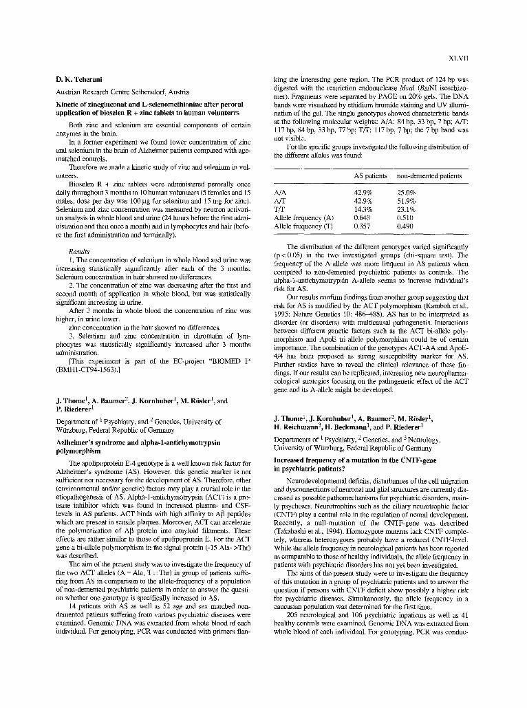
D. K. Teherani
Austrian Research Centre Seibersdorf, Austria
Kinetic of zincgluconat and L-selenomethionine after peroral application of bioselen R + zinc tablets to human volunterrs
Both zinc and selenium are essential components of certain enzymes in the brain.
In a former experiment we found lower concentration of zinc and selenium in the brain of Alzbeimer patients compared with age- matched controls.
Therefore we made a kinetic study of zinc mad selenium in vol- unteers.
Bioselen R + zinc tablets were administered perorally once daily throughout 3 months to 10 human volunteers (5 females and 15 males, dose per day was 100 gg for selenium and 15 mg for zinc). Selenium and zinc concentration was measured by neutron activati- on analysis in whole blood and urine (24 hours before the first admi- nistration and then once a month) and in lymphocytes and hair (befo- re the first administration and terminally).
Results 1. The concentration of selenium in whole blood and urine was
increasing statistically significantly after each of the 3 months. Selenium concentration in hair showed no differences.
2. The concentration of zinc was decreasing after the first and second month of application in whole blood, but was statistically significant increasing in urine.
After 3 months in whole blood the concentration of zinc was higher, in urine lower.
zinc concentration in the hair showed no differences. 3. Selenium and zinc concentration in chromatin of lym-
phocytes was statistically significantly increased after 3 months administration.
[This experiment is part of the EC-project "BIOMED I" (BMH1-CT94-1563).]
J. Thome 1, A. Baumer 2, J. Kornhuber 1, M. R6sler l, and P. Riederer 1
Department of 1 Psychiatry, and 2 Genetics, University of Wttrzburg, Federal Republic of Germany
Azlheimer's syndrome and alpha-l-antichymotrypsin polymorphism
The apohpoprotein E-4 genotype is a well known risk factor for Alzheimer's syndrome (AS). However, this genetic marker is not sufficient nor necessary for the development of AS. Therefore, other (environmental and/or genetic) factors may play a crucial role in the etiopadiogenesis of AS. Alpha-1-antichymotrypsin (ACT) is a pro- tease inhibitor which was found in increased plasma- and CSF- levels in AS patients. ACT binds with high affinity to A~ peptides which are present in sensile plaques. Moreover, ACT can accelerate the polymerization of A[3 protein into anlyloid filaments. These effects are rather similar to those of apolipoprotein E. For the ACT gene a bi-allele polymorphism in the signal protein (-15 Ala- >Tin) was described.
The aim of the present study was to investigate the frequency of the two ACT alleles (A = Ala, T = Thr) in group of patients suffe- ring from AS in comparison to the allele-frequency of a population of non-demented psychiatric patients in order to answer the questi- on whether one genotype is specifically increased in AS.
14 patients with AS as well as 52 age and sex matched non- demented patients suffering from various psychiatric diseases were examined. Genomic DNA was extracted from whole blood of each individual. For genotyping, PCR was conducted with primers dan-
XLVII
king the interesting gene region. The PCR product of 124 bp was digested with the restriction endonuclease MvaI (BstNI isoschizo- mer). Fragments were separated by PAGE on 20% gels. The DNA bands were visualized by ethidium bromide staining and UV illumi- nation of the gel. The single genotypes showed characteristic bands at the following molecular weights: A/A: 84 bp, 33 bp, 7 bp; A/T: 117 bp, 84bp, 33bp, 77bp; T/T: 117 bp, 7bp; the 7bp band was not visible.
For the specific groups investigated the following distribution of the different alleles was found:
AS patients non-demented patients
A/A 42.9% 25.0% A/T 42.9% 51.9% T/T 14.3% 23.1% Allele frequency (A) 0.643 0.510 Allele frequency (T) 0.357 0.490
The distribution of the different genotypes varied significantly (p < 0.05) in the two investigated groups (chi-square test). The frequency of the A-allele was more frequent in AS patients when compared to non-demented psychiatric patients as controls. The alpha-l-antichymotrypsin A-allele seems to increase individual's risk for AS.
Our results confirm findings from another group suggesting that risk for AS is modified by the ACT polymorphism (Kamboh et al., 1995; Nature Genetics 10: 48G488). AS has to be interpreted as disorder (or disorders) with multicausal pathogenesis. Interactions between different genetic factors such as the ACT bi-allele poly- morphism and ApoE tri-allele polymorphism could be of certain importance. The combination of the genotypes ACT-AA and ApoE- 4/4 has been proposed as strong susceptibility marker for AS. Further studies have to reveal the clinical relevance of these fm- dings. If our results can be replicated, interesting new neuropharma- cological strategies focusing on the pathogenetic effect of the ACT gene and its A-allele might be developed.
J. Thome 1, J. Kornhuber 1, A. Baumer 2, M. R6sler 1, H. Reichmann 3, H. Beckmann t, and P. Riederer 1
Departments of 1 Psychiatry, 2 Genetics, and 3 Neurology, University of Wtirzburg, Federal Republic of Germany
Increased frequency of a mutation in the CNTF-gene in psychiatric patients?
Neurodevelopmental deficits, disturbances of the cell migration and dysconnections of neuronal and glial structures are currently dis- cussed as possible pathomechanisms for psychiatric disorders, main- ly psychoses. Neurotrophins such as the ciliary neurotrophic factor (CNTF) play a central role in the regulation of neural development. Recently, a null-mutation of the CNTF-gene was described (Takahashi et al., 1994). Homozygote mutants lack CNTF comple- tely, whereas heterozygotes probably have a reduced CNTF-level. While the allele frequency in neurological patients has been reported as comparable to those of healthy individuals, the allele frequency in patients with psychiatric disorders has not yet been investigated.
The aims of the present study were to investigate the frequency of this mutation in a group of psychiatric patients and to answer the question if persons with CNTF deficit show possibly a higher risk for psychiatric diseases. Simultanously, the allele frequency in a caucasian population was determined for the first time.
205 neurological and 106 psychiatric inpatients as well as 41 healthy controls were examined. Genomic DNA was extracted from whole blood of each individual. For genotyping, PCR was conduc-
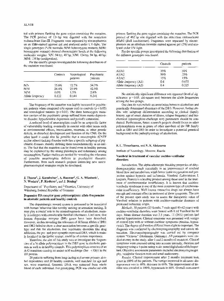
XLVIII
ted with primers flanking the gene region containing the mutation. The PCR product of 134 bp was digested with the restriction endonuclease Hae-lII. Fragments were separated by electrophoresis on an EtBr-stained agarose gel and analysed under UV-light. The single genotypes (N/N: normals; N/M: heterozygote mutants; M/M: homozygote mutants) showed characteristic bands at the following molecular weights: N/N: 94 bp, 40 bp; N/M: 134 bp, 94 bp, 40 bp; M/M: 134 bp (nodigestion).
For the specific groups investigated the following distribution of the mutation was found:
Controls Neurological Psychiatric patients patients
N/N 75.6% 72.7% 54.7% N/M 24.4% 25.9% 42.5% M/M 0.0% 1.5% 2.8% Allele frequency 0.122 0.144 0.241
The frequency of the mutation was highly increased in psychia- tric patients when compared (chi-square test) to controls (p < 0.05) and neurological patients (p < 0.01). The three homozygote muta- tion carriers of the psychianic group suffered from manic-depressi- ve disorder, hypochondric depression and periodic catatonina.
A reduced level of neurotrophins could be an unspecific factor of vulnerability leading, eventually in concert with other noxes such as environmental effects, intoxications, traumata, or other genetic defects, to disturbed development and function of the CNS. On the other hand it could also be possible that a neurotrophin (CNTF and/or others)-lacking disorder underlies a specific subgroup of psy- chiatric diseases, thereby defining them neurochemically as an enti- ty. The fact that the mutation can be found even in healthy persons may be explained by the strong pleiotropia and redundancy among neurotrophins. Further studies are needed for revealing the relevance of possible neurotrophin deficits in psychiatric diseases. Furthermore, from such research projects interesting new neuro- pharmacological strategies might be developed.
J. Thome l, J. Kornhuber l, A. Baumer 2, G. A. Wiesbeck 1, N. Wodarz ~, P. Riederer 1, and J. B6ning l
Department of 1 Psychiatry and 2 Genetics, University of Wtkzburg, Federal Republic of Germany
Dopamine D3 receptor gene polymorphism: allele frequencies in alcoholic patients and healthy controls
The dopaminergic reward system is presumed to be associated with human behaviour like novelty seeking or sensation seeking. It may play a crucial role in the etiopathogenesis of alcoholism, main- ly in subtypes with considerable familial inheritance. Until now, five human dopamine receptor (DR) genes have been described. However, studies revealing the relevance of different alleles of DR1 and DR2 failed to show a clear association between a specific geno- type and risk for alcoholism. For psychiatric disorders like drug addiction, the pre- and post-synaptic expressed DR3, which is main- ly localized in the limbic system, could be of certain importance.
Therefore, the aim of this study was to investigate the frequen- cies of a bi-allele polymorphism in the DR3 gene in alcoholic pati- ents as well as in healthy controls. This polymorphism consists of an A-G transition causing an amino acid exchange (Ser -< Gly) in the D3 protein.
20 patients suffering from long-lasting and severe primary alco- hol dependence and 20 healthy controls, well matched for age and sex, were examined. Genomic DNA was extracted from whole blood of each individual. For genotyping, PCR was conducted with
primers flanking the gene region containing the mutation. The PCR product of 462 bp was digested with the restriction endonuclease MluNI (Bali isoclnzomer). Fragments were separated by electro- phoresis on an ethidium-bromide stained agarose gel (2%) and ana- lysed under UV-light.
For the specific groups investigated the following distribution of the different genotypes was found:
Controls patients
A1/A1 35% 50% A1/A2 50% 35% A2/A2 15% 15% Allele frequency (A1) 0.6 0.675 Allele frequency (A2) 0.4 0.325
No statistically significant difference was apparent (level of sig- nificance: p = 0.05, chi-square test) between the allele frequencies among the two groups.
Our data do not exclude an association between alcoholism and genetically determined alterations of the DR3. However, further stu- dies with subgroup differentiation according t o 91inical (faj~nily history, age of onset, duration of illness, relapse frequency) and bio- chemical (apomorphine-challenge test) parameters should be con- ducted. Furthermore, future research projects should focus on other polymorphisms even in genes of other members of the DR family such as DR4 and DR5 in order to investigate a potential genetical background in the pathophysiology of alcoholism.
S. L. Timerbaeva, and N. S. Alekseeva
institute of Neurology, Moscow. Russia
Vasobral in treatment of vascular cochleo-vestibular disorders
Introduction. The alpha-adrenergic blocking properties of dihy- droergocryptine result essentially in an improvement of cerebral blood flow and metabolism, whith better tissue oxygenation and pro- tection against hypoxia and ischaemia. Vasobral (Laboratoires J. Logeals, France) is a leading product among those used in the treat- ment of cerebrovascular insufficiency. It is known that cochleo- vestibular syndrome is one of the most common type of cerebrovas- cular insufficiency. Well-known vasoactive drugs not always have enough and constant effect in treatment of these symptoms. The aim of the present open study was to assess the therapeutic value of Vasobral solution in patients with cochleo-vestibular disorders of presumed ischaemic origin.
Methods. 30 patients (23 female, 7 male aged 40~2 years) with cochleo-vestibular disorders were treated with 8 ml Vasobral for 60 days. Mean disease duration was 2.5 years. 11 (36%) patients had arterial hypertension. Clinical syndrome was presented with vertigo of central type with or without cochlear symptoms (tinnitus, hypoa- cusis). The degree of disorders differed from slight to important. The diagnosis was confirmed by electronystagmography and caloric sti- mulation. Electronystagmography was carried out by computer system "Octavus" (Hortmann, Germany). All parameters were stu- died at entry in the study, after 1 and 2 months of treatment. Clinical symptoms were assessed taking into account intensity, duration and frequency using a 4-point rating scale: none/slight/noticeable/impor- tant. Objective assessment parameters included electronystagmogra- phy, vestibular function tests and tonal audiometry.
Results. Clinical improvement after 2 months treatment took place in 100% of the patients. The vertigo improved in all cases: dis- appearance was in 40%, decrease in 60%. The improvement of tin- nitus was revealed in 100%, hypoacusis in 88%. Overall assessment
of clinical efficacy was "good" (disappearance of all disabling sym- ptoms) in 5 (17%) patients and "fair" (good improvement of the more disabling symptoms) in 25 (83%) cases. It should be noted that the effect of Vasobral was more prominent after 2 months than after 1 month of treatment. The changes of electronystagmography para- meters and vestibular tests were also positive. Severe adverse effec- ts were not recorded.
Discussion. Effect of Vasobral was studied in cases with a long period of treatment by other vasoactive drugs without stable effect. Vasobral was effective in treatment of all cochleo-vestibular sym- ptoms: vertigo (100%), tinnitus (100%), hearing loss (88%). The improvement of clinical symptoms was more prominent and stable after 2 months of treatment. We may conclude that Vasobral is an effective vasoactive drug and may be recommended in treatment of cochleo-vestibular disorders due to vascular insufficiency.
A. Toso, R. Maggio, P. Barbier, D. Barletta, and G. U. Corsini
Institute of Pharmacology, School of Medicine, University of Pisa, Italy
Sodium nitroprusside induces internalization of muscarinic receptors
In the present study we have extensively characterized the inter- nalization of muscarinic acethylcholine receptors induced by the nitrogen monoxide (NO) generating compound sodium nitroprussi- de (SNP).
When CHO cells stably transfected with the m4 muscarinic receptor subtype were incubated for 1 h in the presence of 700 ~I.M SNP, the number of receptors, measured in intact cells with the hydrophilic ligand [3H]N-methylscopolamine ([3H]NMS), were reduced by 30%. This effect was dose dependent, beginning with concentration of SNP as low as 45 gM. Receptor diminution in- duced by SNP was very fast (t12 ranged between 10 and 20 rain). Removal of SNP from the incubation medium did not result in a recovery of the binding sites measured with [3H]NMS. The pheno- menon was temperature dependent (it did not occur at 4 ~ and was blocked by the muscarinic antagonist atropine. Moreover, the effect of SNP was not observed in cell homogenate indicating that the inte- grity of the cell was required. Receptor diminution was not detected when the number of binding sites was evaluated with the lipophilic antagonist [3H]quinuclidynil benzilate. This indicates that SNP induces a redistribution of the muscarinic receptors between the plasma membrane and an internal compartment of the cell.
Receptor loss was readily reversed by the treatment with the sul- phydryl reducing agent diethyldithiocarbamate (10 gM). Our data indicate that muscarinic receptors are internalized by SNP through the oxidation of sulphydryl groups; moreover, they suggest that NO could play a role in muscarinic receptor desensitization.
[The support of the EC BIOMED programme (BMIt1-CT94- 1563) is gratefully acknowledged.]
V. Tuulik l, E. Lossmann 2, A. Raja;, and A. Meister 2
1 Institute of Clinical and Experimental Medicine, and 2 Institute of Radio and Communication, Tallinn Technical University, Tallinn, Estonia
On diagnosing the functional state of the CNS in case of neurotoxic diseases
The functional state (FS) of the human central nervous system (CNS) is an integral concept, therefore it cannot be described by a single feature. Application of the quantitative EEG and the psycho- metric methods have made it possible to introduce the quantitative characteristics to determine the FS of the CNS. According to P. K.
XLIX
Anokhin and R. Ashby, the central nervous system is complex inte- gral self-regulatory. The psychosocial and psychical factors, as well as the chemical ones, which affect the human in industry, are consi- dered as stress factors.
We aimed to ascertain the correlations between the clinical, psy- chophysiological and electrophysiological data (quantitative EEG, QEEG) at subjects, who have exposed to long-term effect of chemi- cal substances.
Applying the integral parameters for different EEG rhythms (QEEG data) and a classification algorithm, based on neural net- works, we attempted to classify the FS of the CNS of 97 persons. 53 subjects were exposed to heavy metals at their work-place, 24 sub- jects to organic solvents. 52 persons have worked under influence of toxic substances more than 5 years. Reference group consisted of 20 healthy subjects.
Using the classification algorithm, all healthy subjects and sub- jects, who had contact with neurotoxic factors on their work-places, were correctly classified to 2 classes. The algorithm did not perform well, when classifying the subjects with neurotoxic diseases to 3 classes, corresponding to light, medium, and heavy changes of FS of the CNS.
To study the neurotoxic effect reflected in the QEEG, we analzysed the CNS functional state change data at the syndrome level. We calculated the correlation between changes in the FS of the CNS and the general bioelectric activity together with expression of the alpha-activity (C=0 .42-0 .60 at different EEG leads). Correlation between the FS and the speed of performing the psy- chometric test was C = 0.464).57. It should be noted that we obser- ved positive correlation also between the percentage of correct answers during the 1st and the number of years, spent in toxic envi- ronment (C = 0.45-0.53).
The applied psychometrical methods and algorithms for QEEG analysis enable to determine the changes in functional state of the CNS, caused by neurotoxic factors. Different analysis methods, applied on QEEG, show the possibility to use the QEEG as adequa- te method for diagnosing the neurotoxic effect on CNS.
R. J. Uitti, A. H. Rajput, J. E. Ahlskog, K. P. Offord, D. R. Schroeder, and P. C. O'Brien
Mayo Clinic, Jacksonsville, FL, Saskatoon, SK, and Rochester, MN, U.S.A.
Improved survival in Parkinson's disease with use of amantadine
Hypothesis. Amantadine use in parkinsonism provides symp- tomatic benefit and may also afford neuroprotection through N- methyl-D-aspartate (NMDA) receptor antagonism, dopamine upta- ke blockade activity, or other mechanisms.
Aims of the study. By employing specific statistical methodol- ogies, we sought to determine retrospectively which variables inde- pendently predicted improved survival in large parkinsonian cohort.
Brief methodology. We evaluated survival in all parkinsonism patients attending a single clinic over a 23-year period, employing standard survival curves as well as a Cox regression model, to iden- tify independent predictive variables for survival (while taking into account factors potentially associated with both outcome and treat- ment selection).
Results and discussion. The cohort consisted of 836 patients, 250 of whom were treated with amantadine (> 2 months). Improved survival was independently associated with early age at onset, fema- le gender, absence of dementia, type of parkinsonism = Parkinson's disease, low Hoehn & Yahr stage (I or II) at initial visit, and use of amantadine (all p < 0.01). Amantadine treated patients were similar to those who had not received amantadine in terms of the other inde- pendent predictors of survival.
L
Conclusion. Use of amantadine and improved survival in par- kinsonism may result from symptomatic benefits of the drug and/or "neuroprotection". We speculate that NMDA receptor antagonism, dopamine uptake blockade activity, or other mechanisms may lead to improved survival by influencing the usual pathogenic course of idiopathic parkinsonism. A prospective, controlled trial evaluating amantadine's effects on Parkinson's disease progression is warran- ted.
F. Vaglini, F. Fascetti, C. Pardini, L. Mancino, and G. U. Corsini
Institute of Pharmacology, School of Medicine, University of Pisa, Italy
Nicotine prevents MPTP experimental parkinsonism in rodents
We have demonstrated that diethyldithiocarbamate (DDC) enhances MPTP toxicity in mice. This combined treatment induces a more marked depletion of striatal dopamine (DA) and a severe loss of tyrosine hydroxylase (TH) positive perikaria in Substantia Nigra pars compacta (SNpc) (Corsini et al., Eur J Phannacol 19: 127, 1985). This represents a reliable model of experimental parkinso- nism in rodents on which neuroprotective molecules can be evalua- ted. For instance, it is now well established that non-competitive NMDA antagonists (MK-801, dextromethorphan) provide a com- plete protection on DDC enhancement of MPTP toxicity.
In the present study we used this model in order to evaluate the effect of nicotine, the major smoke component, on experimental par- kinsonism. (-) Nicotine (1 mg/kg, s.c.) administered three times (90 and 30 rain before and 30 min after MPTP) completely prevented both the marked depletion of striatal dopamine and the severe loss of TH positive perikaria in SNpc induced by DDC + MPTP.
We are now studying the mechanism of nicotine-induced pro- tection, this could be related to NMDA antagonism as well as an activation of nicotinic cholinergic receptors. In any case, our data suggest that nicotine could be responsible of the reduced prevalence of Parkinson's disease among smokers.
[The support of the EC BIOMED programme (BMH1-CT94- 1563) is gratefully acknowledged.]
K. Velbinger, H. Har tmann, A. Eckert, and W. E. Miiiler
Zentralinstitut ftir Seelische Gesundheit, J 5, Mannheim, Federal Republic of Germany
Region specific down-regulation of free intracellular calcium in the aged rat brain
Aged-related changes in resting levels of the free intracellular calcium concentration ([Ca2+]i) as well as alterations of the rise [Ca2+]i following depolarization have been investigated in acutely isolated neurons of various regions of the rat brain. Characterization of the Ca 2+ responses following KC1 depolarization in the hippo- campus, cortex, striatum, and cerebellum of young rats revealed sig- nificant regional differences in the basal [Ca2+]i level as well as in the KCl-induced rise in [Ca2+]i as well as Ca 2+ responses after depo- larization were lower in the hippocampus and cortex of the aged ani- mals, but not in the striatum or cerebellum. It is concluded that the Ca 2+ homeostasis in the first two regions is specially susceptible to the aging process, resulting in a down-regulation of [Ca2+]i, probab- ly as a consequence of an enhanced sensitivity of mechanisms regu- lating [Ca2+]i. These alterations of the central Ca 2+ homeostasis were accompanied by parallel changes of [Ca2+]i in rat spleeno- cytes. The rise in [Ca2+]i in the aged animals following stimulation of lymphocytes with the mitogen phytohemagglutinin (PHA) was significantly reduced in the plateau phase, which is maintained by
Ca 2+ influx mechanisms. Moreover, a reduced Ca 2+ response was also found after stimulation of the cells with the Ca 2+ ionophore A23187. The data indicate that comparable disturbances of the Ca 2+ homeostasis occur in central neurons and peripheral cells and that these alterations mainly affect transmembraneous Ca 2+ fluxes rather than Ca 2+ release from intracellular stores.
R. Winkel, Th. Mfiller, S. Griiter, S. Behrens, and W. Kuhn
Department of Neurology, Ruhr-University of Bochum, St. Josef-Hospital, Bochum, Federal Republic of Germany
The effect of tyrosine in combination with thyreotropin-releasing hormone on motor performances in parkinsonian patients
Systemic administration of thyreotropin-releasing hormone (TRH) in combination with tyrosine enhances striatal dopamine release in rats (Kreutz, Lehnert et al., Brain Res 536: 347-352, 1990). We studied the effect of TRH in combination with tyrosine in 14 parkinsunian patients (10 male, 4 female) belonging to Hoehn and Yahr stage III-1V, under double-blind conditions in a placebo- controlled study design. The study was performed on two days from 8.00 to 11.00 a.m. Parkinson-specific drugs were withdrawn 12 hours before and during the study. Under double-blind conditions the patients received on day 1 or on day 2 at 8.00 a.m. either TRH (200 gg) i.v. in combination with orally administered tyrosine or placebo. Tyrosine was given in two different doses: 7 patients were treated with 50mg/kg tyrosine (group 1); 7 patients received 100 mg/kg tyrosine (group 2). Motor skills were examined perfor- ming the UPDRS (Unified Parkinson's Disease Rating Scale) at every hour. Data were analysed by using the Wilcoxon signed rank test. The UPDRS-score performed at 8.00 a.m. did not differ signi- ficantly from the score at 11.00 a.m., both in the group of patients treated with 50 mg/kg tyrosine and TRH (p = 0.6250) and in the group of patients receiving 100mg/kg tyrosine and TRH (p=0.0625) or change in the placebo group (p=0.9958). Comparing the UPDRS at 11.00 a.m. of group 1 and of group 2 with the corresponding placebo group, no significant differences were found (placebo vs. tyrosine 100 mg/kg and TRH p = 0.6875; place- bo vs. tyrosine 50 mg/kg and TRH p = 0.4375). We conclude, that TRH and orally given tyrosine in these doses have no significant benefit in the treatment of parkinsonian patients.
T. M. Winter, J. Niemann, T. J. Feuerstein, B. Guschelbauer, G. B. Landwehrmeyer, M. Lauk, and C. H. Liicking
Neurologische Klinik, Albert-Ludwigs-Universit~it Freibttrg, and Neurozentmm, Freiburg, Federal Republic of Germany
Does gabapentin suppress essential tremor? A case report
Essential Tremor (ET) is a benign, but socially and at times phy- sically disabling hyperkinetic movement disorder of unknown cause. In some families, ET appears to be an autosomal dominant trait; small quantities of ethanol reduce tremor amplitudes ha about 60% of people affected by ET. The pathophysiological mechanisms underlying ET are unknown. Therefore the treatment of ET is empi- rical and compounds suppressing ET have been discovered by serendipenty. Betablockers such as propranolol o1 metoprolol and antiepileptic drugs (AED) like primidone are the most commonly used drugs in the symptomatic treatment of ET.
We observed that gabapentin, a recently introduced AED with unknown mechanism of action, can have a beneficial effect on ET. A 77 year old woman (H.S.) with posmral and action tremor of the upper extremities of both sides with onset at the age of 15 years, developed within the past three years a disabling, predominantly
"no"-tremor of the head. Initial tremor treatment with primidone, up to a maximum dose of 500 mg/day had no beneficial effect. Under medication with propranolol (240 mg/d) a reduction of tremor could be noticed, but treatment had to be discontinued because of side effects (vertigo). H.S. suffered in addition from a depression, which was treated with mianserin (70 rag/d).
Before starting tremor treatment with gabapentin, we evaluated tremor activity of the upper extremities by computer assisted acce- lerometric tremor analysis. Simultaneous computer assisted EMG recording of hand flexor and extensor muscles showed EMG syn- chronisation at a frequency of 6 Hz. An identical peak frequency was recorded by accelerometric measurements. The pattern of neck muscle activity was recorded by polyelectromyography of the sple- nius capitis, stemocleidomastoideus an trapezins muscle of both sides, evaluated by clinical criteria and recorded on video tape. Accelerometric measurement of hand tremor showed an amplitude of 7,2 [milfi gravity] 2 before treatment. After treatment with gaba- pentin at a dose of 3 x 400 mg/d we found serum levels of gabapen- tin at 4,9 mg/1, but no reduction of tremor (amplitude 7,9 [milli gra- vity]2). Head tremor was also unchanged. At a gabapentin dose of 3 x 800 mg/d, corresponding to a gabapentin serum level of 12,7 mg/1, we observed a marked tremor reduction. Accelerometric measurement of hand tremor showed an amplitude of 2,3 [milli gra- vity] 2, head tremor was also markedly reduced as judged by clinical criteria. Tremor frequencies remained constant throughout the entire treatment at 6 Hz for tremor of the upper extremities and at 4 Hz for head tremor. There was no change in pattern of neck muscle activi- ty as recorded by polyelectromyography.
We suggest that the reduction of tremor amplitudes observed in H.S. was due to gabapentin. As gabapentin is able to reduce neuro- nal hyperactivity in patients with seizure disorders, an analogue mechanism may underly the therapeutic effect of gabapentin on tre- mor.
J. Wissel 1 and W. Poewe 2
I Department of Neurology, Virchow Klinikum, Berlin, Federal Republic of Germany 2 Department of Neurology, University of Innsbmck, Austria
Functional outcome of botulinum toxin treatment of task-specific limb dystonias
Local injections of botulinum toxin A have developed into a first line treatment for focal dystonias. Such therapy may lead to complete remissions in essential blepharospasm, certain types of cervical dystonia and spasmodic dysphonia. This is in contrast to the effects of localized botulinuna toxin injections in patients with task specific dystonias involving highly skilled motor acts. So none of our own patients with musicans cramp or those published in the lite- rature has reached the premorbid performance level. In patients with writer's cramp several open-label and three double blind studies have demonstrated efficacy of botulinum toxin treatment. However, most of the published reports lack objective or quantifiable functio- nal response parameters. In order to further analyse botufinum indu- ced improvements of motor performance in patients with task speci- fic dystonia we analysed baseline and post-treatment data in 31 pati- ents with writer's cramp. Assessments included a blind videorating of writing performance on a newly developed scale (Writer's Cramp Rating Scale = WCRS), surface poly-EMG recordings of compound muscle activity in forearm flexors and extensors as well as elbow flexors and extensors, speed of pen movements, number of chan- ges of pen acceleration during a standard writing task and subjective response gradings. Writing movements were recorded by a digiti- zing tablet connected to a computer (sampling frequency 166 Hz, accuracy 0.25 mm). More than 80% of the patient experienced a subjective improvement following botulinnm toxin injections and
LI
the WCRS-score decreased significantly (p < 0.05) post treatment. While there were improvements in kinematic parameters (speed of pen movement and number of acceleration changes) in all patients only a subgroup reached near normal pen movement parameters. These patients also had the best subjective response ratings and their baseline data on the digitizing tablet recordings were the least abnor- mal. These results may suggest that restoration of normal motor fun- ction in focal task specific dystonia could be possible when treat- ment is initiated as early as possible and when motor performance deterioration is still mild.
[This study has been supported by BMFT 01KL92081.]
C, Wurthman
Department of Psychiatry, University of Magdeburg, Federal Republic of Germany
Differential therapy in generalized anxiety disorders - results of a study with 30 one-subject experiments
In pharmacotherapy of generalized anxiety disorders (GAD) different psychopharmacological agents (neuroleptics, diazepines, antidepressants) proved to be effective.
One issue in pharmacotherapy of GAD is, that more research needs to be done to investigate whether subtypes of GAD can be defined that respond preferentially to different classes of anxiolytic agents. Thus far, rationally grounded guidelines which treatment is to choose in the individual case are not evident in the literature. Therefore psychiatrists often detelanine simply on the background of personal beliefs and general conventions. First of all, this procedure has to be considered questionable in the treatment of chronic GAD, because particularly in this indication even small differences in effi- cacy between different classes of anxiolytic agents are of impor- tance.
The present study was designed to determine individually the most effective anxiolytic agent (diazepine, antidepressant, neurolep- tic, placebo) in 30 patients with chronic GAD. For this purpose an intensive, self-controlled design (one-subject experiment) was used. Based on a systematic arrangement of treatment and wash-out peri- ods patients served as their own controls.
Amitriptyline 30rag/d, flupentixole 1.5 mffd, clotiazepam 15 mg/d, and placebo were administered double-blind and random- iy.
Each agent was given 4 times for one week. To avoid carry-over effects, all treatment weeks were interrupted by one week's wash- out periods. Thus each single-case experiment comprised 31 weeks. Primary efficacy criterium was Hamilton-total-score at the end of each treatment week. Statistical analysis (U-test) showed that in 19 patients one agent was significantly (p < 0.05) superior to the other substances (clotiazepam n = 11, flupentixol n = 3, amitriptyline n = 5). Placebo was not superior in any of the 30 patients. There was no significant difference between the drugs in 11 patients. However, metaanalysis showed that in chronic GAD by means of one-subject experiments differences in efficacy between different drugs can be found (p < 0.01). ANOVA showed no drug • time interaction. In an approach to optimize pharmacological treatment of individual pati- ents with cronic GAD complex and time-consuming design may be useful and provide rationally grounded guidelines which treatment is to choose in the individual case.
LII
D. M. Yilmazer t, H. Braak 1, R. A. I. de Vos 2, and E. N. H. Janzen 2
1 Zenmma der Morphologie, J. W. Goethe-Universit~it Frankfurt, Frankfurt/Main, Federal Republic of Germany 2 Streeklaboratoria voor pathologie, Enschede, Netherlands
Pathology of the limbic system in Parkinson's disease
Projections, originating in the parietal, occipital, and temporal neocortex head towards limbic centres and establish the afferent leg of the limbic loop. The transentorhinai region and the lateral nucle- us of the amygdala are the major ports of entry of this important sen- sory input to the limbic system. The entorhinal region, hippocampus and the basal and accessory basal nuclei of the amygdala generate projections to the prefrontal cortex - the efferent leg of the limbic loop - and, thus, exert influence upon the most important associati- on cortex of the human brain. The central nucleus of the amygdala plays a key role in conveying processed sensory information to endocrine and autonomic centres of the hypothalamus and lower brain stem. Moreover, it is tightly interconnected with nuclei which in a non-specific manner project upon the cerebral cortex. In Parkinson's disease, severe pathologic alterations consistently occur at many extranigral locations. Lewy bodies and Lewy neurites deve- lop in one of the deep layers of the entorhinai region responsible for a feed back transfer of data from the hippocampus to the neocortex. The pattern of Parkinson-specific amygdala pathology includes the parvicellular portions of the basal and accessory basal nuclei, the accessory cortical nucleus, and the periamygdaloid cortex. Severe changes in the central nucleus of the amygdala add a high level con- trol centre to the well-known desmaction of other autonomic centres in Parkinson's disease. Within the hippocampus, many Lewy neu~- tes are displayed in the CA2 region which is likely to receive input from the tuberomammilary nucleus of the hypothalamus. In sum- mary, apart from the conspicuous pathology of the substantia nigra, there is a substantial degree of extranigral lesions in Parkinson's disease which gradually results in the dstruction of important relay stations in the limbic system. These lesions are in themselves not sufficient to produce overt intellectual impairment but pave the way for the appearance of cognitive decline.
[This study was kindly supported by the Deutsche Forschungsgemeinshafl and the Bundesministerium far Forschung und Technologic.]
M. B. H. Youdim 1, G. Goping 2, O. M. Adegemo 2, A. Gemma 2, J. Kuijpers 2, and H. B. Pollard 2
1 Technion-Faculty of Medicine, Haifa, Israel 2 Laboratory of Cell Biology and Genetics, NIDDK, NIH, Bethesda, USA
Goldfish as an animal for studying degeneration and regeneration of dopamine neurons in Parkinson's disease
Parkinson's disease has been modelled in humans, lower pri- mates and to a lesser extent in some other vertebrates, by adminis- tration of the potent neurotoxin, MPTP (N-methyl-4-phenyl-l,2,3,6- tetrahydropyridine). We now report that a parkinsonian syndrome can be elicited in tile common goldfish (Carassium auratus) by a single i.p. dose of MPTP (20 mg/kg). The syndrome is characterized by profound bradykinesia, the full extent of which is reached 6 days after MPTP administration. The reduction in movement is paralleled by loss of dopamine from the forebrain and midbrain. The toxic monoamine oxidase oxidation product of MtYrP, MPP +, is also accumulated predominantly in forebrain, midbrain and eyes and pre- treatment with the monoanaine oxidase inhibitors substantially redu- ces accumulation of the toxic metabolite. By contrast, no change in dopamine metabolism in the eyes of the MPTP treated goldfish was
noted, even though MPP + accumulation was greater than that obser- ved in the forebrain and midbrain. A barely perceptible coarseness in balance adjustment also occurs in treated animals with extensive beating of the fans. The MPTP-treated goldfish recover normal movement and normal brain dopamine levels within 10-13 days after administration of the drug, suggesting regeneration of the cate- cholaminergic neurons. We interpret these and other data to indica- te that MPTP can induce a Parkinson's disease-like syndrome in the goldfish that is similar in many aspects to the syndrome induced by MPTP in humans and other primates by lesioning the "dopamine" (tyrosine hydroxylase staining) neurons in the forebrain. This can be prevented by prior treatment with 1-selegiline. Motor performance in MPTP treated goldfish can be improved via L-dopa treatment (25 mg/kg). This remarkable parallel may permit the goldfish to sup- plement expensive and scarce primates for the purpose of searching and screening neuroprotective drugs with specific relevance to Parkinson's disease.
[The support of the EC programme BIOMED (BMH1-CT94- 1563) is gratefully acknowledged.]
B. Zielke 1, C. Grote 2, W. Wesemann 2, G. Bringmann 3, and P. Riederer 1
1 Clinical Neurochemistry, Department of Psychiatry, Warzburg, 2 Institute of Physiological Chemistry, Department of Neurochemistry, Marburg, and 3 Institute of Organic Chemistry, Wtirzburg, Federal Republic of Germany
TaClo-induced lesion of the substantia nigra pars compacta (SNpc) of the rat: a histological study
The structural similarity of the [3-carboline TaClo (1-trichloro- methyl-l,2,3,4-tetrahydro-13-carboline) to the dopaminergic neuro- toxin MPTP (N-methyl-4-phenyl-l,2,3,6-tetrahydropylidine) has recently focused attention with respect to its possible neurotoxic potential.
In a preliminary study a single dosis of 0.03 gmol TaClo in 2 gl of 0.1 M HC1 was injected into the SNpc of three anaesthetized Han- Wistar rats. The control group was treated with 2 gl of 0.1 M HC1. After one week the rats were sacrificed under anaesthesia. For a quantitative estimation of drug-induced neuronal loss on the lesio- ned side of the SNpc after Nissl staining neuron countings with the optical disector and volume estimate were carried out on systemati- cally sampled sections (West and Gundersen 1990) Furthermore, a semiquantitative analysis of the neuronal loss of tyrosine hydroxyla- se - immunoreactive (TH-IR) neurons was achieved.
By comparing the lesioned to the unlesioned side in each ani- mal, injection of TaClo into the SNpc reduced the neuronal density by 15.9 + 2.3% as well as the neuronal number by 13.4 _+ 0.4% (mean + SEM, n = 3, p < 0.05%). No shrinkage or edematous increase of volume was apparent. Histologically TaClo exhibited a cystic necrosis covering extensive parts of the ventromedial SNpc. The surrounding area of this necrosis showed a mild loss of TH-IR neurons, reduced TH-IR staining of the remaining ones and reduced fibre density.
In conclusion, TaClo exhibits neurotoxic effects in the SNpc of rats after a single injection.
References West, Gundersen (1990) J Comp Neurol 296:1-22
[This work has been supported by BMFT (01 KL 9405).]
U. Ziemann, D. Bruns, and W. Paulus
Department of Clinical Neurophysiology, University of G&tingen, Federal Republic of Germany
Enhancement of inhibition in motor cortex of normal subjects by the dopamine receptor agonist pergolide: a transcrauial magnetic stimulation study
Hypothesis and aim of the study. Pergolide mesylate is a direct- acting doparnine agonist which is now used as an effective adjunc- tive drug in the levodopa treatment of Parkinson's disease (PD). The present study investigates whether pergolide can enhance motor cor- tex inhibition which is known to be deficient in PD.
Brief methodology. The effect of a single oral dose of 0.25 mg pergolide on the excitability of the motor cortex was investigated by means of focal transcranial magnetic stimulation (TMS) in six healthy volunteers (mean age 36 years). Motor thresholds in the resting and active abductor digiti minimi muscle, the duration of the
LIII
cortical silent period (CSP), and the cortico-cortical inhibition and facilitation after paired cortical stimulation were investigated as dif- ferent parameters of motor system excitability. Measurements were done baseline and 1, 3, and 24 hours after pergohde intake.
Results. Resting and active motor thresholds remained unchan- ged throughout. The mean CSP was siightly lengthened by up to 15 msec. The cortico-corticat inhibition was enhanced at intersthnulus intervals of 2 and 4 msec, and the cortico-cortical facilitation sup- pressed at intervals of 5-7 msec. THis effect peaked at 3 hours and returned to baseline at 24 hours. The cortico-cortical facilitation at longer intervals (8-30 msec) was not affected.
Conclusion. TMS is a non-invasive technique which allows the investigation of the modulation of human cortex excitability by anti- parkinsonian drugs in vivo. The enhancement of cortico-cortical inhibition by pergolide is exactly the reverse of the reduction of inhi- bition seen in PD during the OFF-phase. This supports the notion that pergolide is capable of restoring deficient motor cortical inhibi- tion in PD.
Related Documents


