ABSTRACT Title of Document: A COMPARATIVE STUDY OF FGFR3 SIGNALING DURING THE DEVELOPMENT OF THE ORGAN OF CORTI AND BASILAR PAPILLA Bonnie Elizabeth Jacques PhD 2008 Directed By: Dr. Matthew W. Kelley Dr. William R. Jeffery, Department of Biology Most age-related hearing loss is the result of the accumulated death of inner ear hair cells over a life span. Human hair cells lack the ability to be regenerated once they die and thus there is a need to understand the processes which regulate hair cell formation. Unlike the mammalian ear, the avian cochlea has the ability to regenerate lost hair cells and thus there exists an ongoing race to find the key to regeneration in the mammalian ear. Human hearing is dependent on the interactions between numerous cell types yet very little is known about the pathways which regulate the development of the functionally essential support cells of the mammalian cochlear sensory epithelium. This study aims to elucidate some of the genetic pathways involved in hair cell and support cell differentiation in the developing cochlea. Specifically, the role of Fgfr3 signaling in pillar cell and hair cell differentiation will be revealed through the use of an in vivo mutant mouse model containing a null Fgf8 gene and in vitro whole organ culturing of

Welcome message from author
This document is posted to help you gain knowledge. Please leave a comment to let me know what you think about it! Share it to your friends and learn new things together.
Transcript

ABSTRACT
Title of Document: A COMPARATIVE STUDY OF FGFR3 SIGNALING DURING THE DEVELOPMENT OF THE ORGAN OF CORTI AND BASILAR PAPILLA
Bonnie Elizabeth Jacques PhD 2008 Directed By: Dr. Matthew W. Kelley Dr. William R. Jeffery, Department of Biology Most age-related hearing loss is the result of the accumulated death of inner ear hair cells
over a life span. Human hair cells lack the ability to be regenerated once they die and
thus there is a need to understand the processes which regulate hair cell formation.
Unlike the mammalian ear, the avian cochlea has the ability to regenerate lost hair cells
and thus there exists an ongoing race to find the key to regeneration in the mammalian
ear. Human hearing is dependent on the interactions between numerous cell types yet
very little is known about the pathways which regulate the development of the
functionally essential support cells of the mammalian cochlear sensory epithelium. This
study aims to elucidate some of the genetic pathways involved in hair cell and support
cell differentiation in the developing cochlea. Specifically, the role of Fgfr3 signaling in
pillar cell and hair cell differentiation will be revealed through the use of an in vivo
mutant mouse model containing a null Fgf8 gene and in vitro whole organ culturing of

the embryonic cochlear sensory epithelia of mice and chickens. The classic localize,
activate, inhibit scheme will be employed. This study will demonstrate that Fgf8 and
Fgfr3 are expressed by inner hair cells and pillar cells, respectively, and are required
throughout development for normal differentiation and pattern formation of the organ of
Corti. Inhibition of the receptor or ligand results in the loss of pillar cells and ectopic
formation of hair cells, while activation of this pathway inhibits hair cell formation and
induces pillar cells or activation of these genes and their proteins have on the formation
of hair cell and support cell types. This study also takes a comparative approach by
addressing the similarities and differences of the Fgfr3 signaling pathway in the
mammalian organ of Corti and the avian basilar papilla. Fgfr inhibition in the developing
basilar papilla causes an increase in hair cell density via the direct transdifferentiation of
support cells into hair cells suggesting a role for this signaling pathway in the ability to
regenerate hair cells.

A COMPARATIVE STUDY OF FGFR3 SIGNALING DURING THE DEVELOPMENT OF THE ORGAN OF CORTI AND BASILAR PAPILLA
By
Bonnie Elizabeth Jacques
Dissertation submitted to the Faculty of the Graduate School of the University of Maryland, College Park, in partial fulfillment
of the requirements for the degree of Doctor of Philosophy
2008
Advisory Committee: Dr. William R. Jeffery, Chair Dr. Matthew W. Kelley Dr. Catherine E. Carr Dr. Doris K. Wu Dr. Hey-Kyoung Lee Dr. Steven E. Brauth

© Copyright by Bonnie Elizabeth Jacques
2008

ii
Dedication
This dissertation is dedicated to Kahuku High School
without which I would not be here.

iii
Acknowledgements
I would foremast like to thank Dr. Matthew Kelley for all of his hard work and
efforts in seeing this project through to completion, for providing guidance when it was
needed and for having the keen eye to step back and give me the freedom to think and do.
I would also like to thank him for understanding why rubber slippers are OK lab attire.
I would like to thank the members of my committee and Dr. William Jeffery for
their willingness to be flexible and for being available when I needed them. I would also
like to thank Lois Reid for pushing me through deadlines and keeping me on track.
I would especially like to thank the members of the Kelley lab both past and
present for all of their help in scientific discussions, hands on tutoring, and moral support,
and especially Drs. Alain Dabdoub, Chandrakala Puligilla, Elizabeth Driver, Weise
Chang, Jennifer Jones and Helen May-Simera for teaching me everything I know about
bench work. I would like to extend my most heartfelt thanks to Dr. Mireille
Montcouquiol who first showed me how to be an independent scientist and thinker, and
for pointing out that I was doing my dissections upside down. I would also like to thank
Erynn Layman for her technical and not so technical help. Additionally, I would like to
thank all of the lab members for switching from pizza to sushi for lunch on Fridays, the
only thing that got me through some weeks.
For contributions to chapter one I would like to thank Drs. Alain Dabdoub,
Mireille Montcouquiol, Matthew Holley, Karen Avraham and Katy McCabe for

iv
intellectual and/or technical advice, and Drs. Thomas Friedman, Doris Wu, Weise Chang
and Thomas Reh for reading an earlier version of the manuscript. The authors would also
like to thank Kasey Heintz for technical support.
For contributions to chapter two I would like to thank Mireille E. Montcouquiol,
Erynn M. Layman, and Mark Lewandoski, my co-authors. I’d also like to thank Dr.
Elizabeth Grove for providing the Fgf8b-expression construct, as well as Chad Woods for
technical assistance and Drs. Alain Dabdoub and Jennifer Jones for scientific advice.
I must also thank my parents John and Kathryn Jacques for their love and
encouragement, my sister Kerry for being such an excellent role model and friend, as
well as my grandparents David and Evelyn McPherson and Isabelle Gary for giving me a
reason to make them proud.
Most of all I would like to thank William Risser, without whom I would not have been
able to complete this task.
Mahalo.

v
Table of Contents Dedication………………………………………………………………………………...ii Acknowledgements………………………………………………………………………iii Table of Contents………………………………………………………………………...iv List of Figures …………………………………………………………………………….v Chapter 1: Introduction…………………………………………………………………...1 Chapter Summaries……………………………………………………………...24 Chapter 2: Fgf Signaling Regulates Pillar Cell Development in the Organ of Corti……………………………………………………………..….28
Abstract………………………………………………………………………….28 Introduction……………………………………………………………………...30 Materials and Methods…………………………………………………………..34 Results…………………………………………………………………………...39 Discussion……………………………………………………………………….63
Chapter 3: Fgf8 induces pillar cell fate and regulates cellular patterning in the mammalian cochlea……………………………………………………...……...70
Abstract…………………………………………………………………………70 Introduction………………………………………………………………..……71 Materials and Methods……………………………………………………….....75 Results…………………………………………………………………………..79 Discussion…...………………………………………………………………....101
Chapter 4: Fgf signaling regulates development and transdifferentiation of hair cells and support cells in the basilar papilla…………………...………..………….106
Abstract…...………………………………………………………..……….….106 Introduction………………………………………………………….………....108 Materials and Methods……………………………………………….………...114 Results……………………………………………………………………….....117 Discussion…………………………………………………………...………….139
Chapter 5: Summary and Discussion…………………………………………………...144 Appendix: Complete Guide to Mouse Cochlear Dissection……………………………163 References…………………………………………………………………………....…172

vi
List of Figures
Chapter 1
Figure 1.1. Development and structure of the organ of Corti……………………………..4
Figure 1.2. Development and structure of the basilar papilla……………………………..6
Figure 1.3. Model of the Fgf signaling cascade………………………………………….14
Chapter 2
Figure 2.1. Cellular pattern in the organ of Corti……………………………………….31
Figure 2.2. Expression of Fgfr3 in the embryonic cochlea……………………………...40
Figure 2.3. Developing pillar cells express p75ntr……………………………………….41
Figure 2.4. Inhibition of Fgfr3 disrupts development of pillar cells…………………….45
Figure 2.5. Effects of SU5402 on pillar cell development are dependent
on dose, duration, and time of addition…………………………………………..50
Figure 2.6. Exposure to SU5402 does not affect development of hair cells…………….52
Figure 2.7. Exogenous treatment with Fgf induces an increase in the
number of cells that develop as pillar cells………………………………………56
Figure 2.8. Treatment with Fgf2 induces an increase in the number of
pillar cells and inner hair cells…………………………………………………...58
Figure 2.9. Effects of Fgf2 on pillar cell development are independent
of changes in inner hair cells…………………………………………………….61

vii
Chapter 3
Figure 3.1. Anatomy of the organ of Corti……………………………………………...72
Figure 3.2. Fgf8 and Fgfr3 are expressed in the developing organ of Corti…………….80
Figure 3.3. Targeted deletion of Fgf8 leads to a disruption in pillar
cell development…………………………………………………………………83
Figure 3.4. Inhibition of Fgf8 signaling disrupts pillar cell development………………87
Figure 3.5. Over-expression of Fgf8 induces an increase in p75ntr-positive cells……....89
Figure 3.6. Fgf17 inhibits development of outer hair cells and Deiters cells…………...93
Figure 3.7. Fgf17 treatment promotes PC differentiation and induces ectopic PCs……96
Figure 3.8. Expression of β-actin in the embryonic organ of Corti……………………...98
Chapter 4
Figure 4.1. Structure and development of the basilar papilla…………………………..109
Figure 4.2. Early-stage (E6) inhibition with SU5402 results in an increased
hair cell density…………………………………………………………………118
Figure 4.3. Graph of hair cell density in E6 control and SU5402-treated
explant cultures…………………………………………………………………121
Figure 4.4. SU5402 treatment does not increase the level of BrdU
incorporation in developing HCs……………………………………………….122
Figure 4.5. SU5402 treatment creates small HCs adjacent to larger ones
and alters cellular patterning……………………………………………………125
Figure 4.6. Distributions of hair cell sizes in the basilar papilla following
SU5402 treatment………………………………………………………………129

viii
Figure 4.7. Sox2-positive cells are found in the hair cell layer in
SU5402-treated samples………………………………………………………..133
Figure 4.8 Inhibition of Fgfr activity promotes HC formation in mature
basilar papillae………………………………………………………………….136
Chapter 5
Figure 5.1. Model of Fgfr3 signaling in the developing organ of Corti………………..150
Figure 5.2. Working model of Fgfr3 signaling during development and
regeneration in the BP…………………………………………………………..157

1
Chapter 1: Introduction
Hearing is a fundamental sense. The ability to detect sound can be found in a
variety of animals from insects to humans and has played crucial roles in adaptation. In
the human species, hearing has enabled the evolution of complex communication, speech,
and language and as a result we are the only animals with a written recorded history. We
now have an even more elaborate recorded history in the music, movies, and television so
prevalent in our society. This love of auditory stimulation, however, may have come at a
price, and we are now at risk of losing this, one of our most cherished senses. Humans
are now experiencing a greater risk for sound-induced hearing damage than ever before.
The prevalence of portable music devices with endless amounts of playback time, the
accessibility of rock shows and bands with killer amps as well as our love of high speed
travel are all hazards for our delicate sense of hearing. It is for this reason, now more than
ever, that we must target efforts to find therapies for hearing loss. In order to understand
how hearing can be repaired, however, we must first understand how it is created.
Transduction of sound
What we hear as sound is actually oscillations in the density of air or water caused
by a vibrating object. Movement of an object causes the compression of atmospheric
particles around it which are propagated in a wave away from the object in the same way
a boat makes a wake as it moves through and displaces water. These micro-scale “wakes”
or changes in atmospheric pressure are what we detect as sound. The volume of a sound
2
is determined by the height of the wave generated and is referred to as amplitude, while
the rate at which waves are produced define the frequency of sound.
Detection, decoding and processing of sound occurs within the inner ear of
vertebrates. The outer ear, including the pinna, concha and auditory meatus, collects and
amplifies the sound. In humans it amplifies those frequencies important for human
speech and funnels it to the ear drum which is sensitive to the tiny pressure changes
created in the air by the sound. The tympanic membrane connects to the ossicles of the
middle ear. These tiny bones act to translate the vibrations in the air into fluid vibrations
of the inner ear via contacts with the oval window of the coiled cochlea. In the cochlea
the mechanical vibratory signals are converted into electrical nerve impulses which are
sent via the auditory neurons to the central nervous system for processing. The
transduced sound travels through multiple regions of the brain where, at each step, the
components of sound are further integrated. In ascending order, these include the
cochlear nucleus, superior olivary nucleus, lateral lemniscus, inferior colliculus, and
medial geniculate nucleus, before the signal reaches the auditory cortex where final
processing occurs. In the cortex the auditory scene is created and auditory signals
intermix with input from other senses, this is the basis of complex communication and
awareness (central auditory processing reviewed by Demanez and Demanez, 2003).
Auditory hair cells
This fine-tuned processing would not be possible, however, without highly
sensitive receivers to pick up the detailed components of sound. The primary receivers of
the incoming stimuli are mechanosensory hair cells (HCs) found in the cochlea of the

3
inner ear. Hair cells sit on a basilar membrane that vibrates in response to sound pressure
waves entering the cochlea through the oval window. Hair cells are responsible for
decoding and transmitting this fluid motion in the cochlea into electrical nerve impulses.
Each hair cell has a complex stereocilia bundle at its luminal surface that responds to
changes in sound waves by opening and closing ion channels found at their tips (cochlear
structure reviewed in Raphael and Altschuler 2003). Hair cells turn the mechanical
shearing of the stereocilia relative to one another into a nerve impulse in a process known
as mechanotransduction. The individual stereocilia within a bundle are linked together by
spring-gated ion channels (Pickles, Comis et al. 1984; Kachar, Parakkal et al. 2000).
When incoming vibrations cause the flexion of a stereocilium the force exerted on the tip
link causes the ion channels to open or close depending on the direction of the stimulus.
When a channel is pulled opened on the cell membrane potassium ions flow in triggering
the depolarization of the cell and an influx of calcium ions into the base of the cell.
Calcium intake causes the release of neurotransmitters which stimulate the afferent
neurons of the spiral ganglion. This signal is then propagated up to the brain.
Decoding begins with the type of hair cell detecting the sound. Hair cells are
grouped into a large field with other hair cells in the auditory sensory epithelium. This
inner ear epithelium of lizards, birds (Figure 1.2) and mammals (Figure 1.1) is elongate
and tonotopically arranged (Fay and Popper 2000). In mammals the cochlear duct forms
a tight spiral (Figure 1.1B) while in birds it is relatively straight with a slight curve to it
(Figure 1.2B). Tonotopicity means that within the epithelia there are regions that are
specifically tuned to have the highest sensitivity to one specific frequency range. The
proximal region (referred to as the base) of these epithelia decodes high frequency sound

4
Figure 1.1

5
Figure 1.1. Development and structure of the organ of Corti. At E13, the cochlear
duct has just begun to form and is a partial turn (A), by P0 it has fully formed its 1.75
turns (B); the vestibular system is colored in gray and the cochlear duct is highlighted in
yellow. C and D are cross-sections through an E14.5 (C) and P0 (D) organ of Corti. E and
F are the lumenal surfaces of the OC at the same ages, respectively. The large pool of
progenitor cells is indicated in purple representing the expression of Fgfr3 in these cells
at this age(C). In D and F the unique differentiated cell types of the OC can be identified:
inner hair cells (IHC, yellow), inner pillar cells (IPC, fuscia), outer pillar cells (OPC,
red), outer hair cells (OHC, green), Deiter’s cells (DC, pink) and Hensen’s cells (HeC,
orange). G and H are images of the lumenal surface of the OC at E14.5 (G) and P0 (H).
p75ntr is a marker of pillar cells late in development (H) and is expressed broadly in the
developing epithelium early in development (G), at this stage IHC differentiation has just
begun (arrows). Hair cells and bundles can be seen with Phalloidin stain (H).

6
Figure 1.2
7
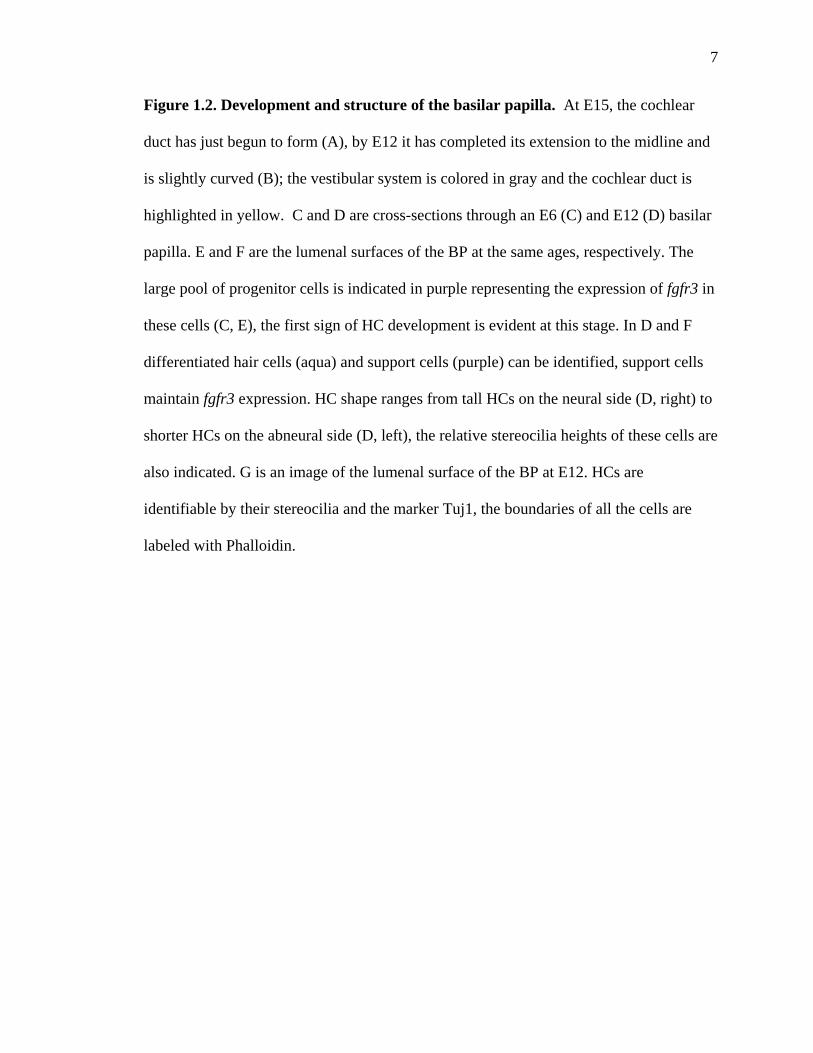
Figure 1.2. Development and structure of the basilar papilla. At E15, the cochlear
duct has just begun to form (A), by E12 it has completed its extension to the midline and
is slightly curved (B); the vestibular system is colored in gray and the cochlear duct is
highlighted in yellow. C and D are cross-sections through an E6 (C) and E12 (D) basilar
papilla. E and F are the lumenal surfaces of the BP at the same ages, respectively. The
large pool of progenitor cells is indicated in purple representing the expression of fgfr3 in
these cells (C, E), the first sign of HC development is evident at this stage. In D and F
differentiated hair cells (aqua) and support cells (purple) can be identified, support cells
maintain fgfr3 expression. HC shape ranges from tall HCs on the neural side (D, right) to
shorter HCs on the abneural side (D, left), the relative stereocilia heights of these cells are
also indicated. G is an image of the lumenal surface of the BP at E12. HCs are
identifiable by their stereocilia and the marker Tuj1, the boundaries of all the cells are
labeled with Phalloidin.

8
while the distal end (known as the apex) decodes low frequency with a gradient of
frequencies between. Tonotopic variations include hair cell body size, stereocilia length,
mechanotransduction channel composition as well as the thickness of extracellular
membranes (Koppl, Manley et al. 2000); these specializations enable the decoding of
sound by breaking it up into its component parts.
Most age-related hearing loss is the result of the accumulated death of these inner
ear hair cells over a life span. Human hair cells lack the ability to be regenerated once
they die and this has spurred much research into hair cell development. Unlike the
mammalian inner ear, the avian cochlea has the ability to regenerate lost hair cells.
Difference in structure and development of these organs may help explain why mammals
lack the ability to repair their hearing (Matsui and Cotanche, 2004). Many of the
mutations that cause hair cell abnormalities and consequently deafness have been well
characterized (Schrijver, 2004). Hearing however, is dependent on the interactions
between numerous cell types.
Cochlear structure
The hair cell containing epithelium of the mammalian cochlea is called the organ of
Corti (OC). It is highly specialized and housed deep within the bony coiled labyrinth of
the cochlea (Figure 1.1B). The OC is comprised of over six distinct cell types which must
develop in a specific pattern in order for normal auditory function to occur (Echteler, Fay
et al. 1994)(Figure 1.1D, F). Typically, there are four rows of hair cells that spiral in
parallel up the cochlear duct. Inner hair cells (IHCs) are the primary receptors for
incoming stimuli and comprise the first row; they are supported by more basally situated

9
phalangeal cells. Outer hair cells (OHCs) comprise the last three rows and have
underlying Deiter’s cells for support. The stereocilia of OHCs are embedded in the
tectorial membrane which allows them to better sense and respond to stimuli. Separating
the IHCs from OHCs are two rows of pillar cells (PCs), the inner pillar cell and outer
pillar cell, which form a triangular fluid-filled space between them, the tunnel of Corti
(cochlear structure reviewed inRaphael and Altschuler 2003). Pillar cells have large
processes constructed of dense networks of microtubules which give them a rigid
structure; these pillar heads extend from the basal lamina up to the lumenal surface of the
epithelium and act to separate the inner hair cells from the outer hair cells. Their exact
function however is unknown.
The tightly defined rows of hair cells in the OC are flanked by two fields of
relatively undifferentiated cells, one of these is Kolliker’s organ that lays between the
IHCs and the spiralganglion and the other is referred to as the lesser epithelial ridge
(LER) which extends beyond the last OHC. These fields of undifferentiated cells have the
potential to form hair cells when experimentally manipulated to express hair cell specific
genes (Woods, Montcouquiol et al. 2004; Jones, Montcouquiol et al. 2006; Driver, Pryor
et al. 2008). Retention of the ability to become a hair cell may, in part, be explained by
looking at a homologous organ such as the basilar papilla of the avian cochlea. The
basilar papilla (BP), is much broader than the mammalian cochlea and the hair cells are
more loosely organized(Koppl, Manley et al. 2000) (Figure 1-2F). The field of hair cells
almost entirely fills up the cochlear duct, in contrast to the small strip they occupy in the
OC. This suggests that development of the organ of Corti may have evolved to produce
hair cells only in a subset of cells within a broader ancestral organ.

10
Other differences suggest that the OC is the byproduct of a significant amount of
evolutionary remodeling. For example, there are two types of hair cells in the BP, short
and tall that are arranged in a gradient across the width of the basilar papilla. Shorter hair
cells cluster along the abneural edge, while taller hair cells are found more on the neural
edge (Koppl, Manley et al. 2000) (Figure 1.2B). There is no distinct boundary line
between the two types as is found between the inner and outer hair cells of the OC. Also,
avian hair cells perform both amplification and detection roles while in the OC, detection
is done mostly by the IHCs and amplification is achieved via membrane-bound motor
proteins of the OHCs (Dallos, Wu et al. 2008). The hair cells of the BP are surrounded
by a uniform type of support cell (Figure 1.2D, F) that is morphologically different from
either pillar cells or Deiter’s cells. The specializations seen in the OC can be attributed to
changes in genes that regulate the development and differentiation of hair cells and
support cells from a common progenitor pool.
Development of the inner ear
The inner ear is derived from a thickening of the head ectoderm called the otic
placode which is induced by signals arising from the hindbrain and adjacent mesoderm.
In mice, the otic placode is formed around embryonic day 8 (E8) (Ohyama, Groves et al.
2007)1
1 For the sake of consistency, development of the inner ear will be described as it occurs in the mouse in which parturition occurs around E19.
. This structure invaginates to form the otic vesicle, which eventually divides into
two regions: a dorsal region which forms the gravistatic vestibular system and a ventral
region which forms the saccule and cochlea (Bok, Chang et al. 2007), both of which are
composed of hair cells that respond to vibratory stimuli.

11
The cochlea can be distinguished as a medially-oriented ventral out-pocketing of
the otic vesicle around E10 (Figure 1.1A, shown slightly later). As development
proceeds, the cochlea continues to grow outward and distinctive coiling is evident by
E15; at P0, formation of the 1.75 turns characteristic of the mouse cochlea is complete
(McKenzie, Krupin et al. 2004) (Figure 1.1B). In chickens, ventral out-pocketing of the
basilar papilla is evident by E5 (Figure 1.2A), after which the cochlea continues to grow
out medially and elongate; by E10 the lengthening of the duct is complete (Figure 1.2B).
Sensory structures in the mouse otocyst first begin to differentiate at E11, as
indicated by a thickening of the otic epithelium in patches; at this point spiral ganglion
neurons are first detectable (Bok, Chang et al. 2007). Innervation to the inner ear arises
from the cochleovestibular ganglion which is comprised of two distinct populations of
neurons, those in the lateral region (vestibular ganglia) that will innervate the vestibular
portion of the inner ear and its hair cells and those situated medially (spiral ganglia)
which will go on to innervate the cochlear hair cells (Sanchez-Calderon, Milo et al.
2007).
Development of the organ of Corti
Development of the organ of Corti occurs in two waves. The first wave is a
mitotic front which progresses from apex to base beginning around E11, with terminal
mitosis occurring lastly in the base by E14. A second wave of differentiation begins at in
the base and extends towards the apex to initiate the formation of specific cell types in the
OC. In addition to the axial gradient of differentiation there is also a planar gradient
beginning with the IHCs and spreading laterally to the Hensen’s cells which sit right
12
beyond the OHCs. One of the first cues that differentiation has begun is a thickening of
the actin ring that surrounds the lateral boundaries of future IHCs; this occurs along with
an increase in size and more lumenal positioning of their nuclei (McKenzie, Krupin et al.
2004) . This can be seen as a line of cells that extends from the base which have more
pronounced labeling by Phalloidin F-actin than the surrounding cells (Figure 1.1E, G).
Adjacent to this row of presumptive IHCs is a large pool of cells that are positive for the
neurotrophin receptor p75ntr. This pool of p75ntr-positive cells begins in the lesser
epithelial ridge and terminates at the line of IHCs (Figure 1.1G). These cells will
eventually become the pillar cells, OHCs, Deiter’s cells and Hensen’s cells (Mueller,
Jacques et al. 2002) (Figure 1.1H). Differentiation is complete by E17, after which cells
grow and attain their proper orientation (Kelley 2007).
Deletions or mutations of any of these cell types during development can result in
mild to severe hearing loss (Friedman, Dror et al. 2007). Given the specialized nature of
hair cells and support cells of the organ of Corti, it is reasonable to assume that their
development and maintenance is dependent on variable gene expression patterns. The
highly specialized support cells of the OC are just as vital to normal hearing function as
are hair cells, yet very little is known about these genetic pathways which regulate their
development, the elucidation of which would greatly aid the understanding of the
molecular basis of deafness as well as provide clues for potential therapies.
Fibroblast growth factor signaling
There are many families of genes whose products are secreted that play important
conserved roles in the development and morphogenesis of the body’s systems. Some of
13
these include the Fgf, Wnt, Bmp, and Hh signaling pathways. All of these gene networks
have been shown to play a role in auditory development as well (Kelley 2007). Of
particular interest to developmental auditory research, the Fgf family has been shown to
be the prime signaling pathway involved in the early induction of the inner ear
(Schimmang 2007).
Fibroblast growth factors (Fgf) are a family of 23 secreted proteins that mediate
their response by binding with and activating one of four transmembrane Fgf receptors
(Fgfrs). Fgfrs are tyrosine kinase receptor proteins made up of three extracellular
immunoglobulin-like domains and a heparan sulfate proteoglycan (HSPG) binding
region(reviewed by (Thisse and Thisse 2005)). Each transcribed Fgfr can be alternately
spliced in the third immunoglobulin domain to produce multiple variants of the same
gene (Miki, Bottaro et al. 1992). Each splice variant shows unique binding properties and
affinities for the various Fgf ligands and are differentially expressed by different tissues.
Two Fgf ligands bound to an HSPG are required for activation of an Fgfr. Once the Fgf-
HSPG binds to the extracellular domain of the Fgfr the receptor undergoes homo-
dimerization, bringing together the intracellular TrK regions of the receptors. This
triggers cross-activation of the tyrosine kinase domains, which are auto-phosphorylated,
and actives down-stream signaling cascades via the MAP kinase signaling pathway
(Mohammadi, Olsen et al. 2005) (Figure 1.3).
Fgf signaling also functions in many feedback loops, one of which is the Sprouty
pathway. Sprouty’s are genes that negatively regulate the activity of Fgf signaling by
binding to and blocking the activity of Grb2, a binding partner required during Fgf
receptor activation (Hanafusa, Torii et al. 2002). Tyrosine phosphorylation during Fgf

14
Figure 1.3

15
Figure 1.3. Model of the Fgf signaling cascade. Fgf receptors are transmembrane
proteins made up of three extracellular immunoglobulin-like domains (Ig, light green), a
transmembrane domain (dark green) and intracellular tyrosine kinase domains (TRK,
teal). Two Fgf ligands (yellow) bound to an HSPG (orange) can bind to the Fgfr in the
IgII and IgIII region. Once the Fgf-HSPG binds to the extracellular domain of the Fgfr
the receptor undergoes homodimerization, bringing together the intracellular TRK
regions of two receptors. This triggers cross-activation of the tyrosine kinase domains,
which are auto-phosphorylated (light blue, P) causing recruitment of multiple signaling
modulators (FRS2, Grb2, Sos, and Ras). Activation of Ras starts the MAP kinase
signaling cascade (red), which targets and binds to DNA promoter regions influencing
transcription of members of multiple gene pathways such as the Sprouty’s. Sprouty’s
negatively regulate Fgf signaling by binding to Grb2 and effectively blocking
downstream, activation of MAPK.

16
signaling induces the expression of Sprouty’s thus activating this negative feedback loop
(Tefft, Lee et al. 1999).
Fgf signaling in inner ear development and differentiation
Ligands of the Fgf signaling pathway are known to regulate the early
development of the inner ear (Schimmang 2007). Of particular interest, Fgf8 has been
shown to play a key regulatory role during otic placode induction (Maroon, Walshe et al.
2002; Sanchez-Calderon, Martin-Partido et al. 2004; Ohyama, Groves et al. 2007) and
differentiation of the otocyst (Adamska, Herbrand et al. 2001; Sanchez-Calderon, Martin-
Partido et al. 2004; Schimmang 2007). Fgf8 secreted by the developing hindbrain is
required for induction of otic pre-placodal cells (Phillips, Storch et al. 2004). Fgf3 and
Fgf8 are required together for normal formation of the otocyst (Maroon, Walshe et al.
2002) and, independently, Fgf8 is required for differentiation of the various sensory
epithelia within the otocyst (Sanchez-Calderon, Martin-Partido et al. 2004).
While much is known about the factors which regulate otic induction and
formation of the gross morphology of the ear there remains much to be learned about the
gene networks responsible for hair cell and support cell formation in the organ of Corti.
Some key evidence has demonstrated that Atoh1 is required for specification of HCs
(Bermingham, Hassan et al. 1999; Woods, Montcouquiol et al. 2004). While hair cells
are directly responsible for the detection of vibratory signals, support cells have been
shown to have a vital role in the maintenance of these hair cells thus understanding how
they develop is crucial to understanding hair cell viability and cochlear function.

17
Characterization of mice deficient in the gene encoding fibroblast growth factor
receptor-3 (Fgfr3) revealed that these animals have relatively normal hair cells yet were
entirely deaf (up to and greater than 60 dB at all frequency ranges tested)(Colvin, Bohne
et al. 1996; Puligilla, Feng et al. 2007). The most prominent malformation in the
cochleae of these animals, besides an extra row of hair cells, is a complete inhibition of
pillar cell development, and consequently the absence of the tunnel of Corti (Colvin,
Bohne et al. 1996; Hayashi, Cunningham et al. 2007; Puligilla, Feng et al. 2007).
Fgfr3 is one of the four Fgfrs and is a member of the Fgf8 synexpression group
(Tsang, Friesel et al. 2002) (a family of co-expressed ligands, receptors, inhibitors and
signal modulators that work in tandem within developing tissues to regulate
development). Fgfr3 is most highly activated by the Fgf8 ligand and this signaling
pathway has been shown to have unique effects during pattern formation, differentiation,
and cell growth throughout the developing embryo and nervous system (Olsen, Li et al.
2006). Deletion of Fgfr3, as discussed above, has been shown to prevent the formation
of a specific type of support cell, the pillar cell (Colvin, Bohne et al. 1996; Hayashi,
Cunningham et al. 2007; Puligilla, Feng et al. 2007). Concurrently, Fgfr3 has been
reported to be expressed early in the domain of the developing OC which will become the
pillar cells and outer hair cells (Peters, Ornitz et al. 1993) (Figure 1-1C, E) and then is
down-regulated in all but the pillar cells (Pirvola, Cao et al. 1995; Pirvola, Ylikoski et al.
2002) (Figure 1-1D, F). These two lines of evidence point to a direct role for Fgfr3 in
pillar cell formation and differentiation, which would make Fgfr3 the only gene
identified to specifically regulate the differentiation of a support cell.

18
Quantitative RT-PCR analysis has also indicated that Fgf8 is strongly expressed
in the embryonic cochlear sensory epithelium (Pickles 2001). More specifically it has
been suggested that Fgf8 may be expressed in the inner hair cells of the cochlea
(unpublished data by Ulla Pirvola), a location in which it would have the potential to play
a role in the Fgfr3 signaling cascade. Fgf8 is a non-autonomously acting ligand, meaning
it exerts its effects on cells adjacent to those secreting it rather than influencing the cells
in which it is transcribed. The positioning of pillar cells directly adjacent to Fgf8-
expressing IHCs, and the high affinity binding Fgf8 has with Fgfr3 (Ornitz, Xu et al.
1996; Olsen, Li et al. 2006), shown to be expressed within the pillar cell region (Pickles
2001), suggest that this ligand-receptor pair may act in tandem to regulate the
differentiation of pillar cells.
The epithelium of the chicken basilar papilla, however, also expresses fgfr3 in a
pattern similar to that of OC yet the BP lacks pillar cells (reviewed by Manley and Koppl,
1998). Specifically, fgfr3 is expressed in all cells of the BP prior to differentiation at
E5.5 (Figure 1.2C, E). During hair cell specification (E6-E8) fgfr3 is down-regulated in
cells that will become hair cells and is expressed only by support cells of the adult
chicken (Bermingham-McDonogh, Stone et al. 2001) (Figure 1.2D, F). The fact that
Fgfr3 is down-regulated during organ of Corti development yet is maintained in the
basilar papilla suggests that this signaling pathway may have contributed to the
divergence of the unique support cell types of the OC which may also explain some
functional differences between these two ears.
For example, while avian hair cells are as vulnerable to acoustic or toxic damage
as are mammalian hair cells, unlike mammals, birds are able to regenerate and repair the

19
auditory sensory epithelium when hair cells are lost. The basilar papilla is able to
produce new hair cells to replace ones that are damaged and thus hair cell loss in the
adult BP is not permanent (Cruz, Lambert et al. 1987; Corwin and Cotanche 1988).
Replacement of dead HCs initially involves the direct transdifferentiation of a support
cell into a hair cell following HC death which is followed by a round of mitosis in the
support cell layer (Cruz, Lambert et al. 1987; Corwin and Cotanche 1988; Cotanche, Lee
et al. 1994). Interestingly, Fgfr3 has been shown to be down-regulated in support cells
following HC death, suggesting it may play a role during HC regeneration (Bermingham-
McDonogh, Stone et al. 2001).
For reasons unknown, perhaps owing to their highly differentiated state, support
cells in the mammalian OC are unable to transdifferentiate or re-induce mitosis following
HC damage. It appears that the OC has given up the ability to repair itself in exchange
for the high frequency tuning and detection made possible by the presence of these
unique support cells (Bermingham-McDonogh and Rubel 2003). The deafness that
occurs in mammals when Fgfr3 is knocked out and pillar cells fail to form suggests that
these support cells have evolved a vital role in hearing. Given the similarities of fgfr3
expression during development and regeneration in the BP, and the concurrence of Fgfr3
expression in the developing OC, there is a strong potential that this gene may have a
conserved role in the development of both the avian and mammalian cochleae and may
also play a role in the regenerative process. Thus there existed a clear need to study the
role of Fgfr3 signaling within these organs.

20
Aims and hypotheses
This project aims to elucidate the role of Fgfr3 in the regulation, development and
differentiation of hair cells, pillar cells and other support cells from a common progenitor
pool. A comparative approach will be taken by analyzing cochlear development in both
birds and mammals. Addressing the signaling in two different types of cochleae will
provide insights into the conserved mechanisms of hair cell development and formation.
Additionally, identifying differences between a regenerative and non-regenerative ear
may elucidate some of the molecular requirements for hair cell regeneration. The scope
of this research will attempt to experimentally address three main hypotheses.
Hypothesis 1: Fgfr3 regulates the differentiation of pillar cells in the developing organ of
Corti.
Hypothesis 2: Fgf8 is responsible for the activity of Fgfr3 in the developing organ of
Corti.
Hypothesis 3: Fgfr3 regulates the transition of support cells into hair cells in the basilar
papilla.

21
Experimental Design
In this set of experiments the classic localize, inhibit, activate scheme was used to
test the general hypothesis that inductive interactions between Fgf8 and Fgfr3 regulate
the formation and differentiation of support cells and hair cells in the mouse organ of
Corti and to determine if Fgfr3 plays a similar role in the avian basilar papilla. Two
model systems were used to address these underlying questions. An in vivo approach was
taken by analyzing Fgf pathway-specific knockout mice for defects in the developing
organ of Corti. The available data on the fully characterized Fgfr3 deletion (Colvin,
Bohne et al. 1996; Hayashi, Cunningham et al. 2007; Puligilla, Feng et al. 2007) was
used as a basis of comparison for analysis performed on mice with an inner ear-specific
conditional knockout mutation of Fgf8 (Meyers, Lewandoski et al. 1998; Hebert and
McConnell 2000) the generation of this mutation is described in chapter 3).
The second approach involved an in vitro organ culturing technique in which
intact cochlear sensory epithelia are isolated from embryonic mice between E13 and E16.
These epithelia were grown in culture for 3-7 days during which time various modulators
of Fgf signaling can be applied to the culture media to both inhibit and over activate
Fgfr3 signaling. After incubation these explant cultures are fixed and labeled with
markers for various cochlear cell types. Additionally, square-wave electroporation was
used to transfect cochlear cultures with Fgf-expression vectors to drive ectopic
expression of transgenes within the sensory epithelium.
The use of tissue culture in developmental studies has a long-standing history. As
early as 1949 (Werthessen 1949) in vitro culture systems have been published as viable
models for understanding how biological processes work in vitro. While definite

22
differences exist between in vivo and in vitro systems, tissue culture offers the ability to
do experiments not possible in living organisms. In particular, the use of whole organ
explants for inner ear research has been vital. Culturing offers more precise control over
potential variables that are much harder to control for in living animals. The location of
the inner ear deep within the bony labyrinth of the cochlea provides a further obstacle by
hindering its accessibility in living animals. Embryonic cochlear explants have thus been
widely utilized in developmental auditory research (Kelley, Xu et al. 1993; Raz and
Kelley 1999; Woods, Montcouquiol et al. 2004; Gross, Machulik et al. 2005; Jones,
Montcouquiol et al. 2006; Montcouquiol, Sans et al. 2006; Coleman, Fallon et al. 2007;
Hayashi, Kokubo et al. 2008).
Expression analysis
The expression patterns of the components of the Fgf pathway that were
hypothesized to play a role in OC development were analyzed by in situ hybridization
and immunohistochemistry. These included the related ligands Fgf8 and Fgf17 as well as
Fgfr3. Expression of these genes was analyzed at stages of development between E13
and P1.
Additionally, markers for assaying pillar cell growth and development were
identified and used to analyze the effects of experiments pertaining to the functional
modulation of Fgfr3 signaling such as p75ntr, the low-affinity neurotrophin receptor.
Another useful indicator of PC development is the ITO distance: the “Inner-to-Outer hair
cell” distance, or the distance between the IHC and first row of OHCs. Larger ITOs are
reflective of more mature PCs. A third marker, β-actin, was also used given that it is
23
expressed strongly in PCs and more weakly in OHCs, Deiter’s cells, and Hensen’s cells
at P0 but is not expressed in any cell types within the OC prior to E17.
Fgfr3 Pathway Inhibition
In order to address the effects of the inhibition of the Fgfr3 signaling pathway,
signaling modulation was done in vivo and in vitro at two different levels: (1) by
inhibition of receptor activation and (2) by the functional inhibition of specific ligands.
Receptor inhibition will be carried out by treatment of whole organ embryonic cochlear
explant cultures with SU5402, a pharmacological agent that inhibits the receptor tyrosine
kinase activity of all Fgf receptors. In order to inhibit the specific function of the
identified Fgfr3 ligand, Fgf8, cochleae from an inner-ear specific Fgf8 conditional knock-
out mouse (Meyers, Lewandoski et al. 1998) were analyzed. Cultured developing OC
sensory epithelia were also exposed to an Fgf8 function-blocking antibody to determine if
inhibition at the protein level mimics the activity of genetic inhibition.
Fgf pathway ectopic activation
The second major experimental goal is to characterize the effects of the Fgfr3
signaling pathway on pillar cells by looking at the effects of over-activation of the Fgf
pathway components. Receptor activation using Fgf2, a non-endogenous broad-acting
promiscuous Fgf ligand with high affinity for all Fgf receptors (Ornitz, Xu et al. 1996),
was used. In order to identify the endogenous ligands that play a role in activating Fgfr3
and pillar cell development purified protein of the members of the Fgf8 synexpression
group were applied to embryonic cultures, including Fgf8, Fgf17 and Fgf18 protein.

24
Fgf17 protein is known to bind to and activate Fgfr3 in the same domains that bind Fgf8
(Olsen, Li et al. 2006). However, Fgf17 binds Fgfr3 with slightly higher affinity than
Fgf8 and thus can function as a strong ectopic activator of the receptor. Transfection of
Fgf8 into the sensory epithelium was also done using a viral construct expressing Fgf8.
Comparative development
To identify any conserved role Fgfr3 may play in the avian basilar papilla, many
of the same experiments described above for the OC were performed on embryonic BP
explant cultures. These included inhibition of Fgfr3 with SU5402 and ectopic activation
using Fgf17 and Fgf2. Immunocytochemistry was used to identify changes in the cultured
versus control epithelia. The antigens HCA and SCA were used to distinguish HCs from
SCs, respectively. Serrate-1 (Adam, Myat et al. 1998) and Sox2 (Neves, Kamaid et al.
2007) expression were also used as indicators of SC phenotypes. Tuj1, Myosin VIIa and
Math1 antibodies were used to identify developing hair cells.
Chapter Summaries
The results of these experiments will be presented in three main chapters, the
summaries of which are below.
Chapter 2: Fibroblast Growth Factor Signaling Regulates Pillar Cell Development in the
Organ of Corti
Chapter two is published in its entirety in the Journal of Neuroscience volume
22(21), November 1, 2002, page 9368-9377(Mueller, Jacques et al. 2002). In this chapter

25
Fgfr3 is identified as a gene that is expressed during early cochlear development in the
mouse; this is shown by immunohistochemistry and in situ hybridization. In vitro
functional studies using mouse whole-organ embryonic cochlear explant cultures
demonstrate that inhibition of the receptor with SU5402 significantly reduces the ability
of pillar cells to develop and differentiate. Conversely, over-activation using ectopic Fgf2
caused a significant increase in the number of cells that developed as pillar cells. This
first chapter thus demonstrates that Fgfr3 is important and necessary for normal organ of
Corti development and specifically shows that it plays a role in pillar cell formation. It
also defines a developmental window between E15 and P0 in which Fgfr signaling is
required in the OC.
Chapter 3: Fgf8 induces pillar cell fate and regulates cellular patterning in the
mammalian cochlea
Chapter three is published in its entirety in the journal Development volume
134(16), August 2007, page 3021-3029(Jacques, Montcouquiol et al. 2007). This chapter
delves further into the role that Fgfr3 plays in the cochlea. Fgf8 expression by inner hair
cells is shown to begin at E15, the same time as Fgfr3 and adjacent to the pool of Fgfr3
expressing cells and suggests they may be interacting. Fgf8 conditional knock out mice
are shown to lack pillar cells, very similar to the phenotype of the Fgfr3 knockouts, a
good indication that they are operating in the same pathway. Fgf8 is inhibited in vitro
using an Fgf8 function-blocking antibody in the media of embryonic OC cultures. As
with Fgfr inhibition, and identical to the mutants, inhibition at the protein level causes a
complete inhibition of pillar cell development, and demonstrates the need for Fgf8-Fgfr3

26
signaling to enable formation of pillar cells. Ectopically activating Fgfr3 with Fgf8-
expressing constructs via electroporation significantly increases the number of pillar cells
adjacent to transfected regions. There is a significant decrease in the number of outer
hair cells at these sites as well as in cultures exposed to Fgf17 protein. This chapter thus
supports the hypothesis that Fgf8 signaling plays a role in promoting pillar cell formation
and activation of Fgfr3 prevents the differentiation of hair cells.
Chapter 4: Fgf signaling regulates development and transdifferentiation of hair cells and
support cells in the basilar papilla
Chapter four is a manuscript that is currently being prepared for journal
submission. In this final chapter, the role of Fgfr3 signaling in the avian basilar papilla is
addressed. Expression of Fgfr3 in the developing BP suggests that it may play a similar
role in support cell and hair cell differentiation as it does in the organ of Corti, given the
homologous nature of these organs. Whole-organ embryonic chick cultures are used to
understand the effect of inhibiting Fgfr3. Inhibition with SU5402 prior to hair cell
differentiation at E6 results in a significant increase in hair cell density. This increase is
shown to be the result of direct transdifferentiation of support cells into hair cells. The
increase in the number of hair cells following inhibition of Fgfr3 is similar to that seen in
mice with mutations in Fgfr3 or Fgf8. In more mature BP cultures SU5402 also causes an
increase in hair cell density. In older explants, small young hair cells are identified
forming directly adjacent to more mature hair cells. These new hair cells disrupt the
regular patterning and ratio of hair cells to support cells and a specific pool of support
cells are identified that have the ability to undergo transdifferentiation. The conversion

27
of support cells into hair cells without a prior round of hair cell death suggests that Fgfr3
is directly responsible for inhibiting hair cell formation and promoting the supportive cell
phenotype.
28
Chapter 2: Fgf Signaling Regulates Pillar Cell Development in the Organ of
Corti
These results indicate that Fgf signaling plays a critical role in the commitment
and differentiation of pillar cells. Moreover, the position of the pillar cells appears to be
Abstract
One of the most striking aspects of the cellular pattern within the sensory
epithelium of the mammalian cochlea is the presence of two rows of pillar cells in the
region between the single row of inner hair cells and the first row of outer hair cells. The
factors that regulate pillar cell development have not been determined, however, previous
results suggested a key role for fibroblast growth factor receptor 3 (Fgfr3).
To examine the specific effects of Fgfr3 on pillar cell development, receptor
activation was inhibited in embryonic cochlear explant cultures. Results indicated that
differentiation of pillar cells is dependent on continuous activation of Fgfr3. Moreover,
transient inhibition of Fgfr3 did not permanently inhibit the pillar cell fate, since
reactivation of Fgfr3 resulted in the resumption of pillar cell differentiation. The effects
of increased Fgfr3 activation were determined by exposing cochlear explants to Fgf2, a
strong ligand for several Fgf receptors. Treatment with Fgf2 leads to a significant
increase in the number of pillar cells and to a small increase in the number of inner hair
cells. These effects were not dependent on cellular proliferation, suggesting that
additional pillar cells and inner hair cells were a result of increased recruitment into the
prosensory domain.

29
determined by the activation of Fgfr3 in a subset of the progenitor cells that initially
express this receptor.

30
One signaling pathway that has been implicated in pillar cell development is the
fibroblast growth factor (Fgf) signaling pathway(Ornitz 2000; Ornitz and Itoh 2001).
Mice containing a targeted disruption of the fibroblast growth factor receptor 3 (Fgfr3)
gene are profoundly deaf; however, the only obvious defect in the auditory system of
these mice is the incomplete development of the pillar cells and the tunnel of
Corti(Colvin, Bohne et al. 1996). In addition, expression of messenger RNA for Fgfr3 in
the developing organ of Corti has been localized to a region of the cochlea that
corresponds to the developing sensory epithelium(Peters, Ornitz et al. 1993). These
Introduction
The sensory epithelium of the mammalian cochlea (the organ of Corti) is
comprised of at least six distinct cell types that are arranged in a rigorous cellular pattern
(Figure 2.1). One of the most intriguing aspects of this pattern is the presence of the
tunnel of Corti, an extracellular space that extends along the basal-to-apical axis of the
organ of Corti between the single row of inner hair cells and the first row of outer hair
cells(Lim 1986). The walls of the tunnel of Corti are formed by single rows of inner and
outer pillar cells that also extend along the length of the cochlea (Figure 2.1). Pillar cells
and the existence of a tunnel within the hair cell sensory epithelium appear to be unique
to mammalian cochleae (reviewed in(Slepecky and Ogata 1996; Manley and Koppl
1998), suggesting that pillar cells represent a derived cell type. The results of previous
studies have demonstrated that the development of pillar cells and the formation of a
normal tunnel of Corti are required for normal hearing(Colvin, Bohne et al. 1996; Chen
and Segil 1999), however, the factors that play a role in pillar cell development are
largely unknown.

31
Figure 2.1
Figure 2.1. Cellular pattern in the organ of Corti. Schematic drawings of tangential
(A) and cross-sectional (B) views of the organ of Corti in a mouse at P0. A.
Mechanosensory hair cells are arranged in a single row of inner hair cells (IHC) and three
or four rows of outer hair cells (OHC1, 2, 3). A single row of inner pillar cells (IPC, dark
gray) and a single row of outer pillar cells (OPC, lighter gray) are located in the region
between the inner hair cells and the first row of outer hair cells (OHC1). Outer pillar
cells also extend cytoplasmic processes that interdigitate between the first row outer hair
cells. B. The pillar head is comprised of apical projections from both the inner (IP) and
outer (OP) pillar cells. Note that the lumenal surface of the pillar head (PH) is derived

32
primarily from the inner pillar cell. However, as illustrated in A, the lumenal surfaces of
outer pillar cells can be visualized in the interdigitations between first row outer hair
cells. As development continues, the tunnel of Corti will form in the region between the
inner and outer pillar cells. Abbreviations: IH: inner hair cell, PC: phalangeal cell, OH:
outer hair cells, DC: Deiter’s cells.

33
results suggest that Fgfr3 is required for the development of pillar cells; however, the
specific effects of Fgfr3 and the Fgf signaling pathway have not been determined. The
results presented here demonstrate that activation of Fgfr3 is required throughout the
embryonic period for the ongoing differentiation of the pillar cells. Moreover, increased
activation of Fgfr3 by treatment with fibroblast growth factor 2 (Fgf2) leads to an
increase in the number of cells that develop as pillar cells. These results demonstrate
roles for the Fgf signaling pathway in both the commitment and differentiation of cells as
pillar cells.
34
Immunohistochemistry
Cochleae were dissected from mouse embryos of specific ages between E13 and P1 and
fixed in 4% paraformaldehyde (PFA) overnight at 4°C. Some cochleae were then
cryoprotected in sucrose, embedded in TissueTek OCT (Ted Pella, Inc, Redding, CA),
and sectioned in a cryostat at a thickness of 12µm. Expression of p75ntr (Chemicon,
Temecula, CA) or Fgfr3 (Santa Cruz Biotechnology, Santa Cruz, CA) at different
developmental time points was determined in whole-mounts and, for p75ntr, cryosections.
Briefly, developing scala vestibuli, scala tympani and Reissner’s membrane were
dissected to expose the developing cochlear sensory epithelium. Samples were then
permeabilized with either 0.1% Triton-X 100 in phosphate buffered saline (PBS; p75ntr)
or 100% acetone (Fgfr3), followed by overnight incubation in the primary antibody at
4oC with rocking. Antibody binding was detected using either a biotin-linked secondary
antibody and the Vector Elite ABC (peroxidase) kit (Vector laboratories, Burlingame,
CA) or an Alexa-568-conjugated secondary antibody (Molecular Probes, Eugene, OR).
Cochlear explant cultures
Materials and Methods
Cochlear explant cultures were prepared as described previously (Kelley, Xu et al. 1993;
Raz and Kelley 1999). Briefly, timed-pregnant ICR strain mice were deeply anesthetized
by inhalation of CO2 gas and then euthanized by creating a double pneumothorax on
gestational day 13 (E13) or 14 (E14). Embryos were removed from the uterus and staged
according to Kaufman (1992). All procedures involving animals met the guidelines

35
described in the NIH Guide for the care and Use of Laboratory Animals and had been
previously approved by the NIH IACUC.
After removal of embryos, cochleae were dissected and oriented with the lumenal
surface of the sensory epithelium facing upwards onto MatTek dishes (MatTek
Corporation, Ashland, MA) that had been coated with a 0.01% layer of poly-lysine
(Sigma Chemical, St. Louis, MO) followed by a layer of Matrigel (1:70 dilution)(BD
Biosciences, San Jose, CA). Cultures were maintained in media comprised of minimum
essential medium (MEM), glucose, HEPES, sodium bicarbonate, N1 supplements and
10% fetal bovine serum.
Inhibition of Fgfr3
Activation of Fgfr3 was inhibited using SU5402 (Calbiochem, San Diego, CA), a
member of a new class of Fgfr antagonists that block the tyrosine kinase activity of the
receptor by interacting with the catalytic domain (Mohammadi, McMahon et al. 1997).
A stock solution of SU5402 was dissolved in DMSO and then diluted to specific
concentrations between 1 and 50 µM in culture medium. Medium containing either
SU5402 (experimental) or an equivalent amount of DMSO (control) was added to explant
cultures at specific time points that corresponded with embryonic ages between E14 and
E18. Cultures were maintained until hair cells could be identified along the entire length
of the developing sensory epithelium, typically six days in vitro (DIV) for cultures
established on E13. At the end of each experiment, cultures were fixed in either 4%
paraformaldehyde (PFA) or 3% glutaraldehyde, 2% paraformaldehyde for 20 minutes at
room temperature.
36
After fixation, pillar cells were labeled with an antibody against p75ntr (Chemicon,
Temecula, CA) and hair cells were labeled with either an antibody against myosin VI (a
gift from Tama Hasson, UCSD, Hasson et al., 1997) or VIIa (antibodies kindly provided
by both Tama Hasson, UCSD and Christine Petit, Institut Pasteur, (Hasson, Gillespie et
al. 1997) (Sahly, El-Amraoui et al. 1997) or with Griffonia simplicifolia lectin (Vector
Laboratories, Burlingame, CA)(Lanford, Lan et al. 1999; Warchol 2001). Primary
antibody labeling was detected using appropriate secondary antibodies conjugated to
Alexa-488 (Molecular Probes), Alexa-568 (Molecular Probes, Eugene, OR) or biotin
(Vector Laboratories, Burlingame, CA). Binding of secondary antibodies conjugated to
biotin was detected using the Vector Elite ABC peroxidase staining kit (Vector
Laboratories, Burlingame, CA). G. simplicifolia labeling was detected by direct
fluorescence or with the Elite ABC alkaline phosphatase staining kit (Vector
Laboratories, Burlingame, CA). To visualize cellular borders, filamentous actin was
stained with Alexa-488 conjugated-phalloidin (Molecular Probes, Eugene, OR). To
examine cellular histology, some cultures were imbedded in immunobed (Polysciences,
Warrington, PA) and sectioned at a thickness of 3 µm.
Activation of Fgfr3
Fgf2 (R&D Systems, Minneapolis, MN) was dissolved in culture media containing 2
µg/ml heparin and 1% DMSO (to improve penetration into the epithelium) and then
added to cochlear cultures at specific time points. Controls received media containing 2
µg/ml heparin and 1% DMSO. Cultures were maintained until hair cells could be
identified along the complete length of the developing organ of Corti, a total of 6 DIV for

37
cultures established on E13. At the end of the experiment cultures were fixed in 4%
PFA. Pillar cells and hair cells were labeled as described above.
Detection of proliferating cells
To identify proliferating cells within cochlear explants, the thymidine analog 5-
Bromodeoxyuridine (BrdU)(Sigma Chemical, St. Louis, MO), was added to culture
media at a concentration of 3 µg/ml (Montcouquiol and Corwin 2001; Montcouquiol and
Corwin 2001). Uptake of BrdU was determined by labeling with an anti-BrdU antibody
(BD Biosciences, San Jose, CA), followed by a biotinylated secondary antibody and the
Vector Elite ABC staining kit (Vector Laboratories, Burlingame, CA). Since the BrdU
antibody used for these studies was generated in a mouse, non-specific labeling was
inhibited using the Mouse-On-Mouse kit (Vector Laboratories, Burlingame, CA) prior to
addition of the primary antibody.
Determination of numbers of pillar heads, hair cells and ITO distances
Changes in the number of pillar heads (the combined apical extensions of both the inner
and outer pillar cells that gives rise to the roof of the organ of Corti), inner and outer hair
cells and distances between inner and outer hair cells (ITO (Inner To Outer) distances)
were determined as follows: first, the total length of the sensory epithelium was
determined based on the extent of inner and outer hair cells. Based on this length,
positions that were equivalent to distances of 10%, 20%, 30%, and 40% from the extreme
basal end of the epithelium were identified. For each position, the number of inner hair
cells, outer hair cells, pillar heads, and ITO distances were determined. The length of

38
region to be counted always included a minimum of 10 inner hair cells, as determined by
counting the number of myosin VI or myosin VIIa positive cells along the inner curve of
the sensory epithelium. Outer hair cells within the same region were also counted based
on the number of cells that expressed myosin VI or myosin VIIa. The number of pillar
heads was determined using one of the two following methods: Cultures were labeled
with anti-p75ntr and individual labeled pillar heads were counted at the apical surface of
the sensory epithelium. Alternatively, filamentous actin was labeled with phalloidin. As
a result, individual pillar heads were outlined in the region between the inner and outer
hair cells. For all experiments, a minimum of three independent samples from at least
two separate experiments were analyzed.

39
Expression of Fgfr3 in the organ of Corti
Results of immunolocalization studies indicated no expression of Fgfr3 in the
cochlea prior to E16 (data not shown). However, by E16, Fgfr3 is expressed in a band of
cells that extends along the length of the cochlear duct (Figure 2.2A). As was reported in
Peters et al. (1993), cells that express Fgfr3 are located in the region of the cochlear duct
that will develop as the pillar cells, outer hair cells, and Deiter’s cells (Figure 2.2A). By
P0, expression of Fgfr3 in the sensory epithelium is restricted to pillar cells (Figure
2.2B).
Developing pillar cells express p75ntr
Results
The results of previous studies have demonstrated that p75ntr is a specific marker
for pillar cells in the organ of Corti of neonatal (P0-P3) mice (von Bartheld, Patterson et
al. 1991; Gestwa, Wiechers et al. 1999; Sano, Mukai et al. 2001). At later developmental
time points expression of p75ntr is apparently down regulated in pillar cells; however, two
published results disagree regarding the timing of this down regulation(Gestwa, Wiechers
et al. 1999; Sano, Mukai et al. 2001). To examine the embryonic expression of p75ntr ,
cochleae were dissected from embryos at E15 and expression of p75ntr was determined
by immunolabeling. Results indicated that at E15, p75ntr is expressed in a relatively
broad band of cells (Figure 2.3A) that extends along the length of the basal-to-apical axis
of the cochlea (data not shown). In cross-section, the band of expression of p75ntr
correlates with the region of the epithelium that will develop as pillar cells, however, the
number of cells expressing p75ntr appears greater than the number of cells that will

40
Figure 2.2
Figure 2.2. Expression of Fgfr3 in the embryonic cochlea. A. Lumenal surface of the
developing organ of Corti in the middle turn of the cochlea at E16. The band of
expression of Fgfr3 (dark region) is located adjacent to the row of developing inner hair
cells (IHC) and appears to correspond with the region of the epithelium that will develop
as pillar cells, outer hair cells and Deiter’s cells. B. Lumenal surface of the developing
organ of Corti in the middle turn of the cochlea at P0. Fgfr3 expression is restricted to
the developing pillar cells (PC) located between the single row of inner hair cells (IHC)
and the first row of outer hair cells (OHC). Stereociliary bundles are evident in the outer
hair cell region (arrows) but are not in the plane of focus for inner hair cells. Scale bar in
A (same in B), 50 µm.

41
Figure 2.3
Figure 2.3. Developing pillar cells express p75ntr. Expression of p75ntr at E15 (A and
C) and P0 (B and D). A. Low magnification image of the lumenal surface in a whole
mount of the cochlear at E15. p75ntr is expressed broadly in a band of cells that correlates
with the position of developing pillar cells, outer hair cells and Deiter’s cell (arrows). B.
Low magnification image of the lumenal surface of the organ of Corti in the basal turn at
P0. At this stage, p75ntr is expressed intensely in the row of pillar heads (arrows). A
second band of p75ntr expression is present at the lateral edge of the sensory epithelium
(arrowheads). C. Cross section of the developing sensory epithelium from the middle
turn of the cochlea at E15. p75ntr is expressed diffusely within a group of cells that
correlates with the position of developing pillar cells (arrow). p75ntr is also expressed in
a more lateral region of the epithelium that appears to correlate with the development of
Hensen’s cells (arrowhead). D. Cross section of the organ of Corti from the middle turn

42
of the cochlea at P0. p75ntr is strongly expressed in the inner and outer pillar cells (arrow,
PC) and more diffusely in the Hensen’s cells (arrowhead, HeC). There is also expression
of p75ntr in neurites extending from the spiral ganglion to the sensory epithelium
(arrowhead). E. Lumenal surface of the sensory epithelium in a cochlear explant culture
established on E14 and fixed after 5 DIV. A single line of P75ntr-positive pillar cells (red,
PC) are located between the single row of myosin VI-positive inner hair cells (green,
IHC) and the first row of myosin VI-positive outer hair cells (green, OHC). Spiral
ganglion neurites that innervate inner hair cells are also labeled with p75ntr. Scale bar in
A, 500 µm, scale bar in B, 500 µm, scale bar C (same in D), 50 µm scale bar in E, 50 µm.

43
ultimately develop as pillar cells (Figure 2.3C). In addition, a second, less intense, region
of p75ntr expression is present in a position within the epithelium that correlates with
developing Hensen’s cells (arrowhead in Figure 2.3C). By P0, as has been reported
previously, p75ntr is expressed intensely in a narrow band (Figure 2.3C) that extends
along the length of the cochlea (data not shown). Analysis of cross-sections indicates
that this intense band of expression of p75ntr correlates with expression in both the inner
and outer pillar cells (Figure 2.3D); however, a second, less intense band of staining is
also apparent in the region of the Hensen’s cells (arrowheads in Figure 2.3B, HeC,
arrowhead in Figure 2.3D). Finally, double-labeling of cochlear explant cultures with
antibodies against myosin VI and p75ntr demonstrates that the band of p75ntr expression is
located between the single row of inner hair cells and the first row of outer hair cells
(Figure 2.3E). Based on these results, expression of p75ntr in the region between the row
of inner hair cells and the first row of outer hair cells was used as a marker for pillar cell
development at P0.
Inhibition of Fgfr3 disrupts pillar cell development
To determine the effects of Fgfr3 during the development of the embryonic organ
of Corti, cochlear explant cultures were established on E13 or E14. After 18 hours in
vitro, either 10 µM SU5402 or a vehicle control was added to the culture medium.
SU5402 was maintained in the culture medium for the duration of the experiment.
SU5402 has been shown to inhibit the tyrosine kinase activity of all four Fgfrs by
interacting with the catalytic domain(Mohammadi, McMahon et al. 1997). The results of
previous studies have suggested that Fgfrs 1, 2 and 4 are not expressed in the cochlear

44
sensory epithelium(Pirvola, Cao et al. 1995), however, recent unpublished findings have
suggested that Fgfr1 may be present in the developing cochlea (U Pirvola, personal
communication).
Explants maintained in control media developed a single row of p75ntr -positive
heads that appeared similar to the pattern of p75ntr expression at P0 in vivo (Figure 2.4A).
Analysis of double-labeled samples indicated that the row of p75ntr -positive cells was
located between the single row of inner hair cells and the first row of outer hair cells
(Figure 2.4C). Although expression of p75ntr at the apical surface appeared as a single
line, analysis using confocal microscopy indicated that both inner and outer pillar cells
expressed p75ntr (data not shown). In contrast with controls, p75ntr -positive cells were
absent in explants exposed to SU5402 (Figure 2.4B). A single row of inner hair cells and
three to four rows of outer hair cells were still present in these cultures; however, the
distance between the row of inner hair cells and the first row of outer hair cells was
reduced (Figure 2.4D).
To determine whether exposure to SU5402 lead to the elimination of developing
pillar cells, cross-sections from the basal turn of control and SU5402-treated explants
were analyzed. In sections from controls, a pair of inner and outer pillar cells was present
in the region between the inner hair cell and first row outer hair cell in most sections
(Figure 2.4E). In addition, projections from the two pillar cells extended to the apical
surface to form a developing pillar head process. Two cells were also present in the
region between the inner hair cell and first row outer hair cell in explants that had been
exposed to SU5402, however these cells did not give rise to a developing head process

45
Figure 2.4

46
Figure 2.4. Inhibition of Fgfr3 disrupts development of pillar cells. A. Low
magnification image of an E14.5 control culture after 5 DIV. Expression of p75ntr is
present in the pillar cells (PC, arrows) and spiral ganglion neurons (GC). B. Low
magnification image of an E14.5 cochlear culture treated with 10 µM SU5402 beginning
after 18 hours in vitro. p75ntr expression is still present in the spiral ganglion neurons
(GC), but no expression is detected in the region of the pillar cells. C. High
magnification image of the apical surface of the sensory epithelium from an E13 control
explant after 6 DIV. The row of pillar cells (red, PC) is located between the single row of
inner hair cells (green, IHC) and the first row of outer hair cells (green, OHC). D. High
magnification image of an E13 explant treated with 10 µM SU5402 beginning after 18
hours in vitro and maintained for a total of 6 DIV. Cells are labeled as in (C). Note the
absence of pillar cell labeling and the close apposition of the row of inner hair cells and
the first row of outer hair cells. E. Cross-section of the sensory epithelium from an E13
control explant after 6 DIV. A single inner hair cell (IHC), three outer hair cells
(numbered) and single inner and outer pillar cells (arrow and arrowhead, respectively) are
present. Note that a pillar head (PH) is present in the space between the inner and first
row outer hair cells. F. Cross-section through the sensory epithelium from an E13
explant treated with 10 µM SU5402 beginning after 18 hours in vitro and maintained for
a total of 6 DIV. A single inner hair cell (IHC) and three outer hair cells (numbered) are
present. Two cell nuclei (arrow and arrowhead) are present in the region between the
inner hair cell and first outer hair cell, however, no pillar head is present and neither of
these cells appears to contact the lumenal surface. G. Apical surface of the sensory

47
epithelium from an E13 control explant after 6 DIV. Cell-cell junctions and stereociliary
bundles have been stained with phalloidin. A row of pillar heads (asterisks) is present in
the region between the row of inner hair cells (IHC) and first row of outer hair cells
(OHC). H. Apical surface of the sensory epithelium from an E13 explant treated with 10
µM SU5402 beginning after 18 hours in vitro and maintained for a total of 6 DIV. Cell-
cell junctions are labeled as in G. The separation between the single row of inner hair
cells (IHC) and first row of outer hair cells (OHC) is noticeably decreased as compared
with the control, however a limited number of apical projections (asterisks) are present in
the region between the IHC and OHC. Scale bar in A (same in B), 200 µm, scale bar in
C (same in D), 50 µm, scale bar in E (same in F), 10 µm, scale bar in G (same in H), 10.

48
(Figure 2.4F). To determine whether these cells extended apical process, cell-cell
junctions at the surface of the organ of Corti were analyzed in control explants and
explants that had been exposed to SU5402. In control explants, a row of roughly
cuboidal pillar heads was present in the region between the row of inner hair cells and
first row of outer hair cells (Figure 2.4G). In contrast, in explants exposed to SU5402 a
clear row of pillar heads was not evident and, and as discussed, the distance between the
inner and outer hair cells was decreased. However, a small number of apical projections
were observed in the region between the inner and outer hair cells (Figure 2.4H),
suggesting that some cells within this region did extend limited apical projections. These
results are consistent with previous observations from Fgfr3 mutant mice and support the
hypothesis that Fgfr3 is necessary for pillar cell commitment and/or differentiation. In
addition, since the overall phenotype in explants exposed to SU5402 appeared to match
the phenotype in Fgfr3 mutants, it seems likely that the effects of treatment with SU5402
in the developing organ of Corti are restricted to inhibition of Fgfr3.
Effects of SU5402 on pillar cell development are dependent on dosage
If Fgfr3 activation is required for the development of cells as pillar cells, then
changes in the level of activity of Fgfr3 should lead to proportional changes in the
number of cells that develop as pillar cells. In order to examine this hypothesis, cochlear
explants were exposed to specific concentrations of SU5402 between 1 µM and 50 µM.
As discussed, addition of 10 µM SU5402 lead to the loss of expression of p75ntr along the
entire length of the sensory epithelium. In explants exposed to 5 µM SU5402, a 63%
reduction (± 17%) in the length of the sensory epithelium with expression of p75ntr was

49
observed, while exposure to 1 µM SU5402 did not affect the number of cells that
developed as pillar cells (Figure 2.5A). Exposure to SU5402 at concentrations greater
than 10 µM resulted in general toxicity as determined by the observation of large scale
death of multiple cell types including epithelial, neuronal and mesenchymal cells. Most
cells appeared rounded and were no longer in contact with substrate. In addition,
extensive cell debris was observed in many samples.
Exposure to SU5402 does not change the number of cells that develop as hair cells
Although two cells were consistently observed in the space between the inner and
first row outer hair cells in explants treated with 10 µM SU5402, it seemed possible that
progenitor cells that were inhibited from developing as pillar cells might develop as other
cell types within the organ of Corti. To examine this possibility, the number of inner and
outer hair cells was determined in both control explants and explants exposed to 10 µM
SU5402. Results indicated no significant change in the number of inner or outer hair
cells in cultures treated with SU5402 (Figure 2.6). This result suggests that inhibition of
FGFR3 does not lead to changes in cell fate.
Fgfr3 regulates pillar cell differentiation
The results of the initial experiments in this study confirmed a requirement for
Fgfr3 during pillar cell development. To determine whether there is a critical period for
Fgfr3 activation during pillar cell development, cochlear explants were established from
embryos at E13 as described. SU5402 was then added after 18 hours (equivalent to E14),
36 hours (equivalent to E15), or 60 hours (equivalent to E16) in vitro. All explants were
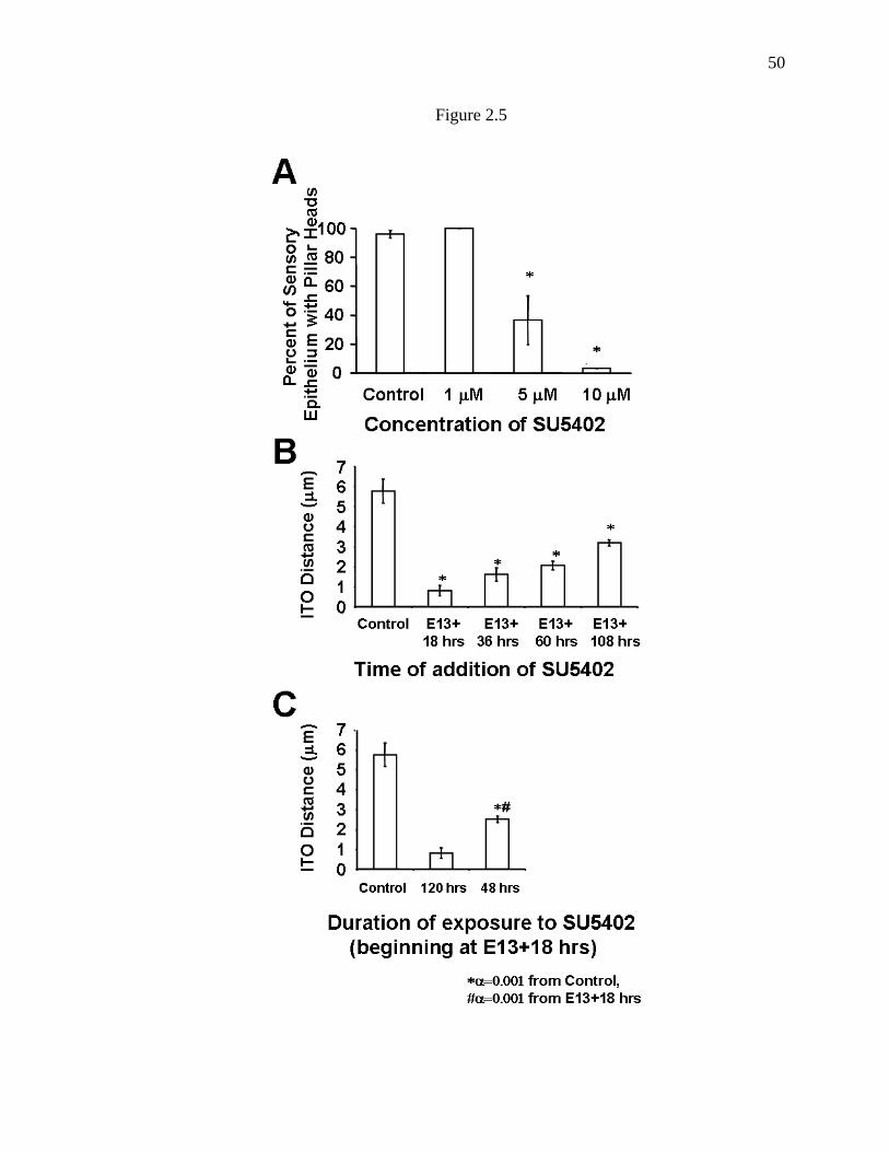
50
Figure 2.5
51
Figure 2.5. Effects of SU5402 on pillar cell development are dependent on dose,
duration, and time of addition. A. E13 cochlear explants were exposed to different
doses of SU5402 for 6 days beginning after 18 hours in vitro. Results indicate a dose
dependent decrease in the percentage of the sensory epithelium in which p75ntr-positive
pillar heads were present. B. E13 cochlear explants were exposed to 10 µM SU5402
beginning at the time points indicated. All explants were maintained for a total of 6 DIV.
Exposure to SU5402 induced a significant change in the distance between the row of
inner hair cells and the first row of outer hair cells (ITO distance) regardless of the time
of addition. However, the effects of SU5402 were clearly dependent on the timing of
addition, suggesting an ongoing requirement for activation of Fgfr3. C. E13 cochlear
explants were exposed to 10 µM SU5402 beginning after 18 hours in vitro. Removal of
SU5402 after 48 hours resulted in a significant increase in the ITO distance as compared
with explants maintained in SU5402 for the entire culture period (120 hrs). All explants
were maintained for a total of 6 DIV. Values for control and SU5402 exposure for 120
hrs are from the same experiment as in B. Error bars are standard error of the mean
(SEM).

52
Figure 2.6
Figure 2.6. Exposure to SU5402 does not affect development of hair cells. The total
number of hair cells in the basal 50% of the sensory epithelium was determined for
control and SU5402-treated cochlear explants established on E13 and maintained for 6
DIV. Error bars are SEM.

53
maintained for a total of 6 DIV. Results indicated that expression of p75ntr was disrupted
along the length of the sensory epithelium regardless of the timing of addition of SU5402
(data not shown). These results suggest that activation of Fgfr3 is required for pillar cell
development throughout most of the embryonic period.
To determine whether the effects of inhibition of SU5402 changed over
developmental time, the distance between the row of inner hair cells and the first row of
outer hair cells (referred to as the ITO (Inner To Outer) distance) was determined for
explants from each time points listed above. In addition, a final time point, 108 hours
(equivalent to E18) was also added. Previous morphological studies have demonstrated
that the ITO distance progressively increases as pillar cells develop(Ito, Spicer et al.
1995; Kaltenbach and Falzarano 1997), therefore changes in the ITO distance should be
reflective of changes in pillar cell development. In addition, since hair cell differentiation
along the cochlear duct progresses in a gradient that begins in the mid-basal turn and
extends both towards the apex and base, ITO distances for each experimental condition
were determined at specific positions along the length of the sensory epithelium. Results
for control explants indicated that after 6 DIV (P0 equivalent) the overall average ITO
distance from positions located 10%, 20%, 30% and 40% from the basal end of the
sensory epithelium was 5.77 µm (Figure 2.5B). Addition of SU5402 beginning after 18
hours in vitro resulted in a mean ITO distance of 0.81 µm (Figure 2.5B), a significant
decrease from control. Addition of SU5402 after 36, 60 or 108 hours in vitro also
resulted in significant decreases in the mean ITO distance (Figure 2.5B). However, the
average change in ITO distance decreased progressively depending on the timing of

54
addition of SU5402. These results strongly suggest that continuous activation of Fgfr3 is
required for ongoing pillar cell differentiation.
To determine whether inhibition of Fgfr3 results in a permanent disruption of
pillar cell differentiation, cochlear explants established at E13 were maintained in
SU5402 for 48 hours beginning after 18 hours in vitro. At the end of the 48-hour time
period, SU5402 was washed out of the culture medium and the cultures were maintained
for an additional 4 DIV (P0 equivalent) prior to fixation. Results indicated normal
expression of p75ntr along the length of the sensory epithelium (data not shown). In
addition, analysis of the mean ITO distance indicated a significant increase by
comparison with explants that had been exposed to SU5402 continuously beginning after
18 hours in vitro (Figure 2.5C). These results demonstrate that transient inhibition of
Fgfr3 results in a transient disruption of pillar cell differentiation. While the mean ITO
distance in explants exposed to SU5402 for 48 hours were significantly greater than in
continuously exposed cultures, the distances were approximately half of those in control
explants, suggesting that removal of the Fgfr3 antagonist resulted in a resumption of
pillar cell differentiation. However, the degree of decrease in ITO distance
(approximately 50% by comparison with control) is consistent with a 48 hour inhibition,
suggesting that pillar cell differentiation may proceed at a relatively constant rate.
Exogenous Fgf induces an increase in the number of cells that develop as pillar cells
To determine whether increased activation of Fgfr3 would also influence pillar
cell development, cochlear explants were treated with exogenous Fgf2. Fgf2 was used
because it has been shown to be a strong activator of Fgfr3c(Ornitz, Xu et al. 1996), the

55
Fgfr3 splice variant that is thought to be expressed in the developing cochlea(Pickles
2001). Analysis of explants established on E13 and exposed to 300 ng/ml Fgf2 for 6 DIV
beginning after 18 hours in vitro indicated a marked increase in the number of pillar
heads in the region between the row of inner hair cells and first row outer hair cells
(Figure 2.7A, B). A similar increase in the expression of p75ntr was also observed in the
pillar cell region (Figure 2.7C, D). The effects of Fgf2 were quantified by determining
the average number of pillar heads and hair cells at specific positions along the basal half
of the sensory epithelium. Results indicated that exposure to Fgf2 induced a significant
increase in the number of pillar heads (Figure 2.8A). A significant increase in the
number of inner hair cells was also observed (Figure 2.7B), however, the magnitude of
this increase was considerably less by comparison with the change in the number of pillar
heads (Figure 2.8A). Finally, the number of outer hair cells was unaffected in explants
treated with Fgf2 and outer hair cells were separated from one another by supporting cells
suggesting that there was no change in the number of Deiter’s cells.
To determine whether changes in the number of pillar heads and inner hair cells
were also dependent on the level and timing of activation of Fgfr3, the concentration and
time of addition of Fgf2 were varied (Figure 2.8B, C). A concentration of 150ng/ml
induced an increase of approximately 50% in the density of pillar heads, while explants
exposed to 500 ng/ml developed almost six times as many pillar heads as controls (Figure
2.8B). In contrast, inner hair cells were not affected in explants exposed to 150 ng/ml
Fgf2 but were significantly increased in explants treated with a concentration of 300
ng/ml or greater (Figure 2.8B). A graded decrease in the density of inner hair cells and
pillar heads was observed when the addition of 300 ng/ml of Fgf2 was delayed by either

56
Figure 2.7
Figure 2.7. Exogenous treatment with Fgf induces an increase in the number of cells
that develop as pillar cells. A. Lumenal surface of the organ of Corti from an E13
control explant after 6 DIV. Cell boundaries are labeled with phalloidin in green and hair
cells are labeled with anti-myosin VIIA in red. A single row of pillar heads (PH) is
present in the region between the single row of inner hair cells (IHC) and the first row of
outer hair cells (OHC). B. Lumenal surface of the organ of Corti from an E13 explant
exposed to 300 ng/ml of Fgf2 beginning after 18 hours in vitro and maintained for a total
of 6 DIV. Labeling is as in A. There is a marked increase in the number of pillar heads
(PH) in the region between the inner (IHC) and outer (OHC) hair cells. An increased
number of inner hair cells are also present. C. Lumenal surface of the organ of Corti
from an E13 control explant after 6 DIV. Pillar cells have been labeled with anti-p75ntr.
A single band of pillar heads is present. D. Lumenal surface of the organ of Corti from

57
an E13 explant exposed to 300 ng/ml of Fgf2 beginning after 18 hours in vitro and
maintained for a total of 6 DIV. Labeling is as in D. There is a marked increase in the
number of individual pillar heads. Scale bar in A (same in B, C, D), 50 µm.
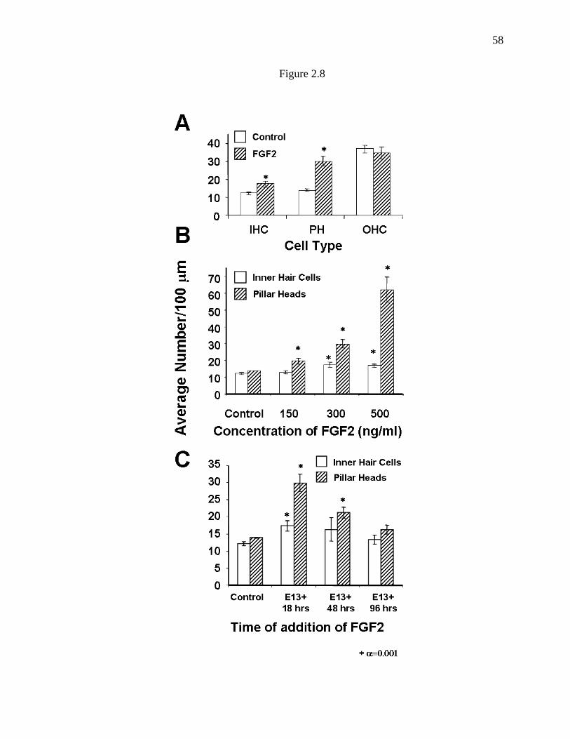
58
Figure 2.8

59
Figure 2.8. Treatment with Fgf2 induces an increase in the number of pillar cells
and inner hair cells. A. The average number of pillar heads and inner and outer hair
cells was determined at specific locations along the basal half of the sensory epithelium
in E13 explants maintained in either control media or media containing 300 ng/ml Fgf2
for 6 DIV. Exposure to Fgf2 leads to an approximate doubling in the number of pillar
heads. A significant increase in the number of inner hair cells was also observed,
however the magnitude of this increase was considerably lower by comparison with the
change in the number of pillar heads. Outer hair cell number was unchanged. IHC: Inner
hair cells, PH: Pillar heads, OHC: Outer hair cells. B. Average number of inner hair
cells and pillar heads in E13 explants exposed to different concentrations of Fgf2 for 6
DIV. Increased concentrations of Fgf2 lead to a graded increase in the average number
of inner hair cells and pillar heads. C. Average number of inner hair cells and pillar
heads in E13 explants exposed to 300 ng/ml FGF2 beginning on E14, E15 or E17 and
maintained for a total of 6 DIV. The effects of FGF2 on both the number of inner hair
cells and the number of pillar heads was dependent on time of administration. Significant
increases in inner hair cells were only observed when FGF2 was added on E14. Pillar
heads were significantly increased when FGF2 was added on E14 or E15. By E17,
treatment with FGF2 did not induce a significant change in either the number of inner
hair cells or the number of pillar heads. Error bars are SEM.

60
48 hours or 96 hours (Figure 2.8C). To determine whether the decreased effects of Fgf2
at later time points were a result of the maturation of the organ of Corti, the changes in
the number of pillar cells and inner hair cells were determined for specific positions along
the basal half of the sensory epithelium. Results indicated that the effects of addition of
Fgf2 after either 48 or 96 hours in vitro were restricted to more apical positions along the
sensory epithelium (data not shown). These results are consistent with the hypothesis
that there is a critical period for the effects of the Fgf signaling pathway and that this
period is related to the developmental progression of the organ of Corti.
The observation that treatment with Fgf2 induced an increase in both inner hair
cells and pillar cells suggested that the effects of Fgf2 on pillar cell development could be
mediated indirectly through the increased number of inner hair cells. To determine
whether an increase in inner hair cells was required to induce an increase in the number
of pillar cells, the effects of Fgf2 on inner hair cells and pillar heads were analyzed for
different positions along the length of the sensory epithelium (Figure 2.9). Results
indicated that Fgf2 induced a significant increase in the number of pillar heads at all 4
positions along the length of the sensory epithelium (Figure 2.9). In contrast, the number
of inner hair cells was only significantly increased at the 30% and 40% positions (Figure
2.9). Therefore, at the 10% and 20% positions, Fgf2 induced a significant increase in the
number of pillar heads independent of an increase in the number of inner hair cells.
These results suggest that the Fgf signaling pathway may mediate multiple events during
the development of the organ of Corti including the development of both inner hair cells
and pillar cells.

61
Figure 2.9
Figure 2.9. Effects of Fgf2 on pillar cell development are independent of changes in
inner hair cells. Effects of exposure to 300 ng/ml Ffg2 on E13 explants after 6 DIV
were determined at different positions along the length of the cochlea. Significant
increases in the number of pillar heads were observed at all four locations. In contrast,
inner hair cells were only increased significantly at the 30% and 40% positions. These
results indicate that the increase in pillar heads is not dependent on an increase in inner
hair cells. IHC: Inner hair cells, PH: Pillar cells, OHC: Outer hair cells.

62
Fgf2 does not induce proliferation within the developing organ of Corti
To determine whether the effects of Fgf2 on the production of supernumerary
inner hair cells and pillar cells were dependent on mitotic proliferation, explant cultures
were treated with 300 ng/ml of Fgf2 and maintained in culture media containing 3 µg/ml
of BrdU. There was extensive incorporation of BrdU in the nuclei of fibroblasts and
other cell types in both control and Fgf2-treated explants. However no BrdU labeling
was observed in the nuclei of either inner hair cells or pillar cells in either control or
Fgf2-treated cultures (data not shown).
63
Deletion of Fgfr3 was known to lead to defects in pillar cell development, but the
specific role of Fgf signaling had not been determined (Colvin, Bohne et al. 1996). Two
rows of cells were observed in the region between the row of inner hair cells and the first
row of outer hair cells in Fgfr3 mutants (Colvin, Bohne et al. 1996), however, whether
these cells represented uncommitted progenitor cells or pillar cells that had become
arrested in development was not clear. The results presented here support the hypothesis
that activation of Fgfr3 is required for pillar cell differentiation. As in Fgfr3 mutant
mice, pillar cell development was disrupted in cochlear explants exposed to the Fgfr
inhibitor, SU5402; however, the cells that would have developed as pillar cells did not
undergo a change in fate, and instead remained in the epithelium in an apparently
Discussion
Fgfr3 is expressed in the embryonic cochlea
The results of previous studies have demonstrated that Fgfr3 is expressed in the
organ of Corti (Peters, Ornitz et al. 1993; Pirvola, Cao et al. 1995); however, the time
course for expression of Fgfr3 protein during embryonic development had not been
determined. Consistent with previous findings, the results presented demonstrate that
Fgfr3 is initially expressed broadly in progenitor cells that will develop as pillar cells,
outer hair cells and Deiter’s cells. In contrast, by P0, Fgfr3 was only detected in inner
and outer pillar cells, suggesting that activation of Fgfr3 may be required for maintenance
of expression.
Fgfr3 activation is required for pillar cell differentiation

64
undifferentiated state. Following removal of SU5402, these cells were able to resume
their development as pillar cells, but ongoing activation of Fgfr3 was required for their
continued differentiation. Fgfr3 has also been shown to control the rate of differentiation
in developing lens fibers (Govindarajan and Overbeek 2001) and osteoblasts (Chen, Adar
et al. 1999; Funato, Ohtani et al. 2001), suggesting that this may be a conserved function
of this receptor.
It is important to consider that Fgfr3 may also play a role in the commitment of
progenitor cells to the pillar cell fate. While inhibition of Fgfr3 did not lead to a change
in the fates of the cells that would have developed as pillar cells, these cells may have
remained in an uncommitted state until Fgfr3 activity was restored. As will be discussed,
the effects of treatment with Fgf2 are consistent with a role for Fgf signaling in cell fate.
The Fgf signaling pathway regulates the number of cells that will develop as pillar
cells
Treatment with Fgf2 lead to a dose dependent increase in the number of cells that
developed as pillar cells, suggesting that Fgf signaling plays a key role in the regulation
of pillar cell number. Moreover, the overproduction of pillar cells in response to
exogenous Fgf2 demonstrates that the number of cells with the potential to develop as
pillar cells is significantly greater than the number of cells that normally assume this fate.
These results suggest that the abundance of ligands for Fgfr3 within the developing organ
of Corti may be a limiting factor for the determination of cells as pillar cells.
The source of supernumerary pillar cells is not clear. Since BrdU-labeling did not
indicate an increase in cellular proliferation, the most likely source of additional pillar

65
cells appears to be the population of Fgfr3 positive cells that is present in the cochlear
duct at E16. As discussed, during normal development of the organ of Corti, the number
of cells that express Fgfr3 at E16 is considerably larger than the number of cells that
either maintain expression of Fgfr3 or that develop as pillar cells. Increased activation of
Fgfr3 in these cells by addition of Fgf2 is apparently sufficient to increase the number of
these cells that will become committed to develop as pillar cells.
The population of progenitor cells that expresses Fgfr3 at E16 appears to include
cells that will develop as both pillar cells and hair cells(Peters, Ornitz et al. 1993) (data
presented here, Figure 4.2). Based on this observation, a second expected outcome of
exposure to Fgf2 might be a decrease in the number of cells that develop as outer hair
cells. Surprisingly, the number of outer hair cells in explants exposed to Fgf2 did not
differ from control. This result suggests that the increased number of pillar cells,
treatment with Fgf2, or a combination of these factors, resulted in the recruitment of
additional cells into the prosensory domain. In fact, there have been several reports
suggesting that the position of the boundary of the prosensory domain at its outer hair cell
edge may be variable. Supernumerary outer hair cells have been observed both in vivo
and in vitro(Lavigne-Rebillard and Pujol 1986; Lavigne-Rebillard and Pujol 1987;
Abdouh, Despres et al. 1993; Abdouh, Despres et al. 1994), and treatment with retinoic
acid induces a significant increase in the number of cells that develop as outer hair cells
presumably through increased recruitment(Kelley, Xu et al. 1993).
An alternative hypothesis for the source of supernumerary pillar cells could be
related to the expression of p75ntr in this cell type. The results of recent experiments have
demonstrated that p75ntr can act as a mediator of apoptotic cell death(Chao and Bothwell

66
2002) and therefore, it seems possible that cell death could play a role in the number of
cells that develop as pillar cells. However preliminary results indicate that neither
activation of p75ntr by NGF or NT-3 or loss of p75ntr activity by genetic manipulation
results in a change in pillar cell number (B.E. Jacques and M.W. Kelley, unpublished
results).
Fgf2 also induces an increase in inner hair cells
In addition to an increase in the number of pillar cells, a more limited increase in
the number of inner hair cells was also observed in explants exposed to Fgf2. The basis
for this effect is not clear. Fgfr3 is not expressed in the region of the cochlear duct that
will develop as inner hair cells, suggesting that the effects of Fgf2 on inner hair cell
number are probably regulated through a different Fgf receptor. One possibility would be
Fgfr1, which has been reported to be expressed in the embryonic cochlear duct (U.
Pirvola, personal communication) and would be activated by treatment with Fgf2 (Ornitz,
Xu et al. 1996). However, treatment with SU5402, which antagonizes all Fgfrs
(Mohammadi, McMahon et al. 1997), did not lead to a change in inner hair cell number
suggesting that the Fgf signaling pathway is not required for inner hair cell development.
This result raises the possibility that the effects of Fgf2 on inner hair cell development
could be indirect. A potential source of this interaction could be the increased number of
pillar cells. If reciprocal signaling interactions between inner hair cells and pillar cells
play a role in regulating the ratio of inner hair cells to pillar cells, then an increase in the
number of pillar cells could potentially lead to a subsequent increase in the number of
inner hair cells. In fact decreases in the number of pillar cells have been reported in

67
response to the loss of inner hair cells in the Bronx waltzer mutant mouse (Tucker,
Mackie et al. 1999). While this does not demonstrate a link in the ratio of the two cell
types, it does support the potential for cell-cell interactions.
It is important to consider that the addition of a relatively high concentration of
Fgf2, as compared with the physiological concentration, was required to elicit an effect
on pillar cell development. The basis for this disparity is not clear. However previous
results have suggested that the presence of the developing tectorial membrane(Rau,
Legan et al. 1999) along with robust expression of different cadherins (Whitlon 1993)
may inhibit the penetration of some molecules into the developing sensory epithelium
(Zheng and Gao 2000). In addition, a number of endogenous inhibitors of Fgfr activation
have recently been identified (Tefft, Lee et al. 1999; Wakioka, Sasaki et al. 2001;
Furthauer, Lin et al. 2002; Tsang, Friesel et al. 2002) and the presence or some of these
molecules within the developing cochlea could also play a role in modulating the
effective concentration of Fgf2 within individual explants.
Fgf signaling and development of pillar cells
It is not clear which Fgfs might act as endogenous ligands for Fgfr3 in the
developing cochlea. Previous reports (U. Pirvola, personal communication) and
preliminary data from our laboratory indicate that Fgf8 is expressed specifically in
developing inner hair cells, suggesting that it could act as an endogenous ligand for
Fgfr3. As discussed, on E16, Fgfr3 is expressed in a band of cells that appears to
correspond with the region of developing pillar cells and outer hair cells. Therefore,
inner hair cells expressing Fgf8 would be located directly adjacent to the domain of Fgfr3

68
expression. Presumably, the effective concentration of Fgf8 would be limited to one or
two cell diameters from each inner hair cell leading to activation of Fgfr3 and placement
of the pillar cells adjacent to inner hair cells. It is not clear why the number of cells that
initially express Fgfr3 is considerably larger than the number of cells that ultimately
develop as pillar cells, however, the group of Fgfr3-positive cells could serve as a pool of
potential pillar cells. This pool could represent a fail-safe mechanism to ensure pillar cell
development.
The spatial expression of Fgf8 suggests that it is a candidate for the endogenous
ligand for Fgfr3, however, existing data also suggests that other Fgfs are present in the
epithelium as well. In particular, Fgf1 and Fgf3 have been reported to be present in the
developing cochlea (Luo, Koutnouyan et al. 1993; Pickles 2001) although the specific
cellular pattern of expression has not been determined. In addition, preliminary results
from our laboratory indicate that Fgf17 is also present in the sensory epithelium and that
treatment of cochlear explants with a mixture of Fgf8/17/18 leads to an increase in the
number of pillar cells (K. Mueller, B. Jacques, M. Kelley, unpublished results). In
contrast, Fgf2 is not expressed in the embryonic cochlea (Luo, Koutnouyan et al. 1993),
suggesting that it is unlikely to be an endogenous ligand for Fgfr3.
In summary, the results presented here indicate that the number of cells that
develop as pillar cells is regulated through the Fgf-signaling pathway, and specifically
through activation of Fgfr3. It seems likely that developing inner hair cells express one
or more Fgfs, leading to local activation of Fgfr3 in adjacent cells. Since the number of
cells with the potential to develop as pillar cells is considerably greater than the normal

69
complement, we hypothesize that activation of Fgfr3 is limited to the cells located nearest
to the developing inner hair cells.

70
Chapter 3: Fgf8 induces pillar cell fate and regulates cellular patterning in
the mammalian cochlea
Abstract The mammalian auditory sensory epithelium (the organ of Corti) contains a
number of unique cell types that are arranged in ordered rows. Two of these cell types,
inner and outer pillar cells, are arranged in adjacent rows that form a boundary between a
single row of inner hair cells and three rows of outer hair cells. Pillar cells (PCs) are
required for auditory function, as mice lacking PCs due to a mutation in Fgfr3, are deaf.
Here, using in vitro and in vivo techniques, we demonstrate that an Fgf8 signal arising
from the inner hair cells is the key component in an inductive pathway that regulates the
number, position, and rate of development of PCs. Deletion of Fgf8 or inhibition of
binding between Fgf8 and Fgfr3 leads to defects in PC development whereas over-
expression of Fgf8 or exogenous Fgfr3 activation induces ectopic PC formation and
inhibits outer hair cell development. These results suggest that Fgf8/Fgfr3 interactions
regulate cellular patterning within the OC through the induction of one cell fate (pillar
cell) and simultaneous inhibition of an alternate fate (outer hair cell) in separate
progenitor cells. Some of the effects of both inhibition and over-activation of the Fgf8-
Fgfr3 signaling pathway are reversible, suggesting that PC differentiation is dependent
upon constant activation of Fgfr3 by Fgf8. These results suggest that pillar cells may
exist in a transient state of differentiation that makes them potential targets for the
development of regenerative therapies.

71
Introduction
The sensory epithelium of the mammalian cochlea, the organ of Corti (OC), is
comprised of at least six distinct cell types arranged into precise rows that extend along
the entire length of the cochlear spiral. The OC contains four rows of hair cells (Figure
3.1A): three rows of outer hair cells (OHCs) supported by underlying Deiter’s cells (DCs)
and flanked on the lateral edge by a several rows of Hensen’s cells (HeCs), and one row
of inner hair cells (IHCs) with underlying phallangeal cells. Separating the two types of
hair cells are parallel rows of non-sensory pillar cells (PCs) (Figure 3.1B). When mature,
PCs form the boundaries of a triangular fluid-filled space referred to as the tunnel of
Corti (Figure 3.1C) (Raphael and Altschuler 2003). The tunnel of Corti and the pillar
cells that form this structure are unique to the mammalian auditory system and are found
in no other vertebrate class. Defects in PC development result in significant hearing
impairment (Colvin, Bohne et al. 1996).
Despite their crucial role in cochlear function, the factors that regulate pillar cell
formation are poorly understood. Previous work has demonstrated that ongoing
activation of one of the fibroblast growth factor receptors, Fgfr3, is required for pillar cell
development (Colvin, Bohne et al. 1996; Mueller, Jacques et al. 2002). Ectopic
activation of Fgfr3 in vitro by treatment with Fgf2 induces an over-production of PCs,
suggesting that the relative level of ligand available for Fgfr3 activation plays a key role
in regulating PC number and position within the OC (Mueller, Jacques et al. 2002).
Fgfr3 is one of four related receptors that bind to members of the fibroblast growth factor
family. All Fgf receptors are transmembrane proteins that contain a tyrosine kinase (TK)
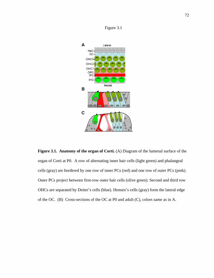
72
Figure 3.1
Figure 3.1. Anatomy of the organ of Corti. (A) Diagram of the lumenal surface of the
organ of Corti at P0. A row of alternating inner hair cells (light green) and phalangeal
cells (gray) are bordered by one row of inner PCs (red) and one row of outer PCs (pink).
Outer PCs project between first-row outer hair cells (olive green). Second and third row
OHCs are separated by Deiter’s cells (blue). Hensen’s cells (gray) form the lateral edge
of the OC. (B) Cross-sections of the OC at P0 and adult (C), colors same as in A.

73
domain in their intracellular region. Fgfr activation is mediated through binding of one
of at least 23 known Fgfs and a sulphated glycosaminoglycan such as heparin sulphate.
Fgf ligand and heparin binding leads to receptor dimerization, cross-activation of the TK
domains and down-stream signaling through the MAP kinase signaling pathway
(Mohammadi, Olsen et al. 2005).
Within the developing cochlea, Fgfr3 is initially expressed around E16 in a broad
pool of progenitor cells located directly adjacent to developing IHCs (Peters, Ornitz et al.
1993; Mueller, Jacques et al. 2002), the first cells to differentiate within the epithelium
(Sobin and Anniko 1984). Based on the spatiotemporal pattern of expression, it seems
likely that Fgfr3 is expressed in progenitors that will ultimately develop as pillar cells and
OHCs, as well as Hensen’s cells and Deiter’s cells. As development proceeds, Fgfr3 is
down-regulated in progenitors that develop as OHCs, HeCs and DCs but is maintained in
PCs (Pirvola, Cao et al. 1995; Mueller, Jacques et al. 2002). RNA expression analysis
using quantitative PCR has suggested that the Fgfr3c splice variant is the predominant
isoform expressed in the cochlea (Pickles 2001). In addition, ligand binding assays
indicate that the “c” isoform of Fgfr3 binds with the Fgf8b isoform with high affinity
(MacArthur, Lawshe et al. 1995; Ornitz, Xu et al. 1996; Olsen, Li et al. 2006). The
Fgf8b ligand has been shown to have important regulatory roles during pattern formation,
differentiation, and cell growth throughout the developing embryo and nervous system
(Olsen, Li et al. 2006). Quantitative RT-PCR analysis has indicated that it is expressed in
the embryonic cochlear sensory epithelium (Pickles 2001). Here we demonstrate that
Fgf8 is expressed in a pattern that is consistent with an inductive role in PC development

74
and that changes in the levels of Fgf8, or in Fgfr3 activation, lead to corresponding
changes in the number and differentiation of pillar cells.

75
Cochleae were dissected from mutants and littermate controls at E15.5, E16 and
E19, and fixed in either 4% paraformaldehyde (PFA) or 3% glutaraldehyde/2% PFA
overnight. Following fixation, the cochleae were dissected and the OC were exposed and
labeled with cell type-specific antibodies: anti-Myosin VI (Proteus Biosciences) 1:1000,
anti-p75ntr (Chemicon) 1:1000, anti-β-actin (Sigma) 1:200. Secondary antibodies were
Materials and methods
In situ hybridization
In situ hybridization (ISH) was performed as described previously (Wu and Oh
1996) for Fgf8 and Fgfr3 on 12 µm frozen sections or whole organs from cochleae
isolated at E15, E16 and P0. A probe specific to exons 2 and 3 of Fgf8 (the region
excised by Cre in the Fgf8∆2,3n/flox; FoxG1cre/+ mutants) was also used on E16-E18
cochleae from Fgf8∆2,3n/flox; FoxG1cre/+ mutants and their wild type littermates to
demonstrate excision of the targeted region.
Generation of Fgf8∆2,3n/flox; FoxG1cre/+ mutants and analysis of pillar cell defects
Animals with a targeted deletion of Fgf8 in the forebrain, retina and inner ear
were generated by crossing Fgf8flox/flox females with Fgf8∆2,3n/+; FoxG1cre/+ males. Mice
carrying these alleles have been previously described (Meyers, Lewandoski et al. 1998);
(Hebert and McConnell 2000). Mutant progeny of the genotype Fgf8∆2,3n/flox; FoxG1cre/+
were visually identified based on obvious defects in the development of the forebrain
(Storm, Rubenstein et al. 2003) Siblings were of the genotypes Fgf8+ /flox; FoxG1cre/+ ,
Fgf8+ /flox; FoxG1+/+ or Fgf8∆2,3n/flox; FoxG1+/+ and served as normal littermate controls.

76
conjugated to one of the following: Alexa 350, Alexa 488, Alexa 546 or Alexa 633
(Molecular Probes). In addition, filamentous actin was labeled using phalloidin at 1:200
conjugated to either Alexa 488 or Alexa 633 (Molecular Probes). Specimens were then
flat mounted and the total length of the cochlear duct was measured. The cochlea was
then divided into four equal sections, each representing one fourth of the total length of
the cochlear duct, and the distances between the inner hair cells and first row of outer hair
cells (ITO distances) were determined in each region (n=5 animals; >50 cells counted per
region). All animal care and procedures were approved by the Animal Care and Use
Committee at NIH and complied with the NIH guidelines for care and use of animals.
Measurement of ITO distance
The Inner-to-Outer (ITO) distance is defined as the distance between the lateral
edge of the IHC and the medial edge of the first row OHC. This is the distance
encompassed by the inner pillar head. Digital images of the OC were captured for each
sample using a Zeiss 510 LSM confocal laser scanning microscope. Measurements of
ITO distances were taken at 3 specific points along the length of the cochlear duct of each
sample, roughly at 25%, 50% and 75% of the distance from the most basal region and
moving towards the apex. A minimum of 15 ITO measurements were made at each of
the three locations. Cell counts were also taken of each cell type in the measured
quadrants.

77
Histological Sections
Temporal bones from control and Fgf8∆2,3n/+; FoxG1cre/+ littermates were fixed in
3% glutaraldehyde/2% paraformaldehyde, tissue was dehydrated in ethanol and then
embedded in Immunobed (Polysciences Inc.). Cochleae were oriented to generate mid-
modiolar sections, cut at 5µm thickness and stained with thionin.
Explant cultures
Explant cultures of embryonic cochleae were established as described previously
(Montcouquiol and Kelley 2003) and maintained for 6 DIV. E13.5 explants were
incubated for 24 hours before exposure to growth factors or antibodies that were diluted
in culture medium to the stated final concentrations along with 0.1% DMSO and 1µg/µl
of heparin. Anti-Fgf8b: 75-150 µg/ml; anti-Fgf5: 75-150 µg/ml; Fgf17 (all from R&D
systems): 300ng/ml. Antibodies and proteins were used at 100 times the ND50 and ED50,
to ensure penetration through the reticular lamina, a strong ionic barrier that exists at the
lumenal surface of the OC.
Electroporation
Full-length cDNA for murine Fgf8b was kindly provided by Elizabeth Grove
{Fukuchi-Shimogori, 2001 #38}, University of Chicago. Fgf8b was excised from its
original vector using BamH1 and then directly ligated into the pAM/CAG-IRES_EGFP
vector at the BamH1 site. Orientation was determined by sequencing. Empty pAM/CAG-
IRES_EGFP vector and pAM/CAG-IRES_EGFP containing full-length Fgf8b in the

78
reverse order were used as controls. Electroporation of cochlear explants was carried out
as previously described (Jones et al, 2006); n>30 for each vector type.
Luminosity measurements
Images of electroporated explants were obtained using a Zeiss LSM510 confocal
microscope. All samples were obtained during the same session using the same laser
power and detection settings. To quantify the effects of over-expression of Fgf8, a
rectangle with a long dimension of 225 µm and a short dimension of 110 µm was
oriented such that the short dimension of the rectangle was parallel with the line of PCs in
the region being measured. The rectangle was positioned so its strial edge aligned with
third row OHCs. Thus, the rectangle included a 110 µm stretch of the OC with the
adjacent region of the greater epithelial ridge containing transfected cells. Control and
experimental regions were obtained and then thresholded for both green and red pixels.
The total number of pixels of each color was then determined as a percentage of the total
of number of pixels within the entire rectangle.

79
Results
Expression of Fgf8 and Fgfr3 in the organ of Corti
Pillar cells develop on the medial edge of an Fgfr3-expression domain located
directly adjacent to the inner hair cells. Given the crucial role of Fgfr3 in PC formation
(Pirvola, Cao et al. 1995; Mueller, Jacques et al. 2002), it seemed likely that hair cells
located adjacent to developing PCs might act as a source of ligand for Fgfr3.
Furthermore, Fgf8, a high affinity ligand for Fgfr3, has been reported to be expressed by
the IHCs of the adult cochlea (Pirvola, Ylikoski et al. 2002). To determine whether Fgf8
could play a role in PC development, the expression pattern for Fgf8 in the embryonic
cochlea was compared with that of Fgfr3 by in situ hybridization. Cochlear development
proceeds in a spatially conserved pattern in which cells in the basal region mature prior to
those in the apical region, allowing one to visualize multiple developmental stages within
the same ear (Sobin and Anniko 1984). No expression of Fgf8 was observed between
E13-E15 (data not shown) or at E16 in the less mature apex (Figure 3.2A). However,
Fgf8 expression was observed in a single cell in the more mature basal region at E16
(Figure 3.2B). Fgfr3 expression was also first observed in the basal region of the cochlea
at E16 in a group of cells that correlates with developing PCs, OHCs, HeCs and DCs
(Figure 3.2D). Weak expression of Fgfr3 was observed in the apex of the same E16
cochlea (Figure 3.2C), suggesting that its onset may slightly precede that of Fgf8. By P0,
Fgf8 expression is clearly present in the single IHC (Figure 3.2E, G) while expression of
Fgfr3 has become largely restricted to the developing PCs (Figure 3.2F). Some
expression of Fgfr3 persists in the developing DC and HeC region (Figure 3.2F),

80
Figure 3.2
Figure 3.2. Fgf8 and Fgfr3 are expressed in the developing organ of Corti. In situ
hybridizations for Fgf8 and Fgfr3 were performed at E16 (A-D) and P0 (E-G). Fgf8 is
not detected at E16 in the apical region (A) but is seen in developing IHCs at the base (B,
arrow). (C) At E16 Fgfr3 is faintly detected in a group of cells near the apex (indicated
by brackets) and shows more robust expression within a similar region at the base (D).
(E) Expression of Fgf8 at P0 is in the IHC (arrow), while Fgfr3 (F) is restricted to PCs
(arrow) and DCs. (G) Whole-mount of the lumenal surface at P0 illustrating expression
of Fgf8 in all IHCs. Scale bar in A-F: 30 µm. Scale bar in G: 20 µm.

81
however based on immunolocalization, this expression appears to be down-regulated as
development continues (Mueller, Jacques et al. 2002). These expression patterns
demonstrate that the timing of the onset of Fgf8 expression correlates strongly with the
onset of Fgfr3 expression and subsequent differentiation of PCs.
Deletion of Fgf8 in vivo results in defects in pillar cell development
To determine whether Fgf8 is required for pillar cell development, a tissue-
specific deletion of Fgf8 was generated using a Cre-loxP strategy. Briefly, a floxed
version of Fgf8 (Fgf8flox) (Meyers, Lewandoski et al. 1998) and the FoxG1cre line (Hebert
and McConnell 2000) were used to delete Fgf8 in a subset of tissues. It has been
demonstrated that Cre-mediated excision of the second and third exons of the Fgf8flox
allele results in complete inactivation (Meyers, Lewandoski et al. 1998). The FoxG1
promoter induces expression of Cre in a relatively small number of tissues, including the
developing otocyst, beginning at E8.5. By E9, the expression of Cre is strong in virtually
all cells within the otocyst (Hebert and McConnell 2000), well before the normal onset of
Fgf8 expression.
Embryos that were Fgf8∆2,3n/flox; FoxG1cre/+ (see Material and Methods for specific
genetic cross) died at birth as a result of defects in the development of the forebrain
(Storm, Rubenstein et al. 2003), however overall development of the inner ear and
cochlea appeared normal. To examine the effects of inactivation of Fgf8 on PC
development, cochleae were obtained from mutants at E15 and E18.5. Consistent with
the timing of the onset of Fgf8, there were no obvious differences in cellular patterning or
in the expression of p75ntr (a marker that is co-expressed with Fgfr3 during cochlear

82
development) (Mueller, Jacques et al. 2002) between mutants and controls at E15 (data
not shown). However, while development of inner and outer hair cells appeared normal
at E18.5 (Figure 3.3A, 3B), there was a marked decrease in the size and number of PCs
as well as a decrease in the overall levels of expression of p75ntr both in the PCs and
HeCs (Figure 3.3C, 3D). The effects of deletion of Fgf8 were quantified by determining
the distance between inner and outer hair cells, referred to as the ITO distance (Mueller,
Jacques et al. 2002), at different positions along the basal-to-apical axis of the OC. The
developing pillar heads expand as the cells differentiate and thus larger ITO distances are
reflective of more advanced PC development (Mueller, Jacques et al. 2002). Deletion of
Fgf8 resulted in a significant decrease in ITO distances along the length of the cochlea
(Figure 3.3G), similar to that seen in the Fgfr3 knockout mice (Jacques, Puligilla, and
Kelley, unpublished).
Rather than a complete absence of p75ntr-positive PCs, as seen when Fgfr3 is
pharmacologically inhibited (Colvin, Bohne et al. 1996; Mueller, Jacques et al. 2002),
some p75ntr-positive cells were clearly present in the PC space in Fgf8∆2,3n/flox; FoxG1cre/+
mutants (Figure 3.3D). Therefore, semi-thin plastic cross-sections of the cochlea from
Fgf8∆2,3n/flox; FoxG1cre/+ mice were examined at E15 (n=4) and E18.5 (n=5). As expected
based on the timing of onset of Fgf8 expression, overall structure of the epithelium and
putative developing PCs appeared normal in cross-sections from E15 (data not shown).
In contrast, at E18.5, two cells were present in the region of the epithelium between the
IHCs and OHCs (Figure 3.3E, 3F), but these cells had either weak lumenal projections, or
no projections at all. In some sections, the IHCs and OHCs appeared to be in direct
contact with one another (Figure 3.3F, magnified in inset with a red line to show the IHC

83
Figure 3.3
Figure 3.3. Targeted deletion of Fgf8 leads to a disruption in pillar cell
development. (A) Lumenal suface of the OC from a littermate control animal at E18.5.
Hair cell stereocilia and cell boundaries labeled with phalloidin (Phal, green) and PCs
labeled with anti-p75ntr (red). The row of IHCs and first row of OHCs (OHC1) are
indicated. (B) Lumenal surface from an Fgf8∆2,3n/flox; FoxG1cre/+ mouse. Note the
disrupted growth of the PCs and close approximation of the IHCs to OHCs. (C, D) Red
channels from A and B, respectively, to illustrate PC morphology. (D) PCs are missing
or under-developed in Fgf8∆2,3n/flox; FoxG1cre/+ mice. (E) Cross-section through a control
OC at E18.5 showing two PCs (asterisks) extending a lumenal projection between the

84
IHC and first row OHC (numbered). The morphology of the projection is illustrated in
the magnified inset with a red line to indicate the lateral boundary of the IHC and a green
line to indicate the medial boundary of the first row OHC. (F) Cross-section through an
Fgf8∆2,3n/flox; FoxG1cre/+ OC illustrating a stunted lumenal PC projection (magnified in
inset). (G) Average ITO distances (see text for details), as a measure of the degree of PC
development, in control and Fgf8∆2,3n/flox; FoxG1cre/+ cochleae. Error bars indicate
standard error of the mean. (H) In situ hybridization using a probe specific to the deleted
region of Fgf8 in control and Fgf8∆2,3n/flox; FoxG1cre/+ cochleae. Arrowhead points to the
row of labeled IHCs in the control. No similar labeling is present in the Fgf8∆2,3n/flox;
FoxG1cre/+ cochlea. Both cochleae have been intentionally over-reacted to ensure
complete detection of Fgf8 expression. Scale bar in A-D: 20 µm. Scale bar in E-F: 10
µm.

85
boundary and a green line to show the OHC boundary). Pillar cells with weak or no
lumenal projections were observed along the entire length of the cochlea with no region-
specific variations. To ensure that Cre-mediated excision of Fgf8 exons 2 and 3 was
complete in these mutants, ISH was performed on Fgf8∆2,3n/flox; FoxG1cre/+ mutants and
their control littermates (n>3 for each genotype analyzed) at ages E16-E18 using a probe
generated from exons 2 and 3 of the Fgf8 gene. Control and mutant cochleae were
processed together and the colorization step was deliberately extended to ensure detection
of any residual Fgf8. A single row of Fgf8-positive IHCs was clearly present in control
cochleae, however no expression of Fgf8 was observed in the mutant cochleae (Figure
3.3H). These results indicate a complete deletion of Fgf8 in Fgf8∆2,3n/flox; FoxG1cre/+
mutants.
In vitro treatment with an anti-Fgf8 antibody results in complete inhibition of pillar
cell development
The presence of some p75ntr-positive inner pillar cells in Fgf8∆2,3n/flox; FoxG1cre/+
mutants, compared to the complete absence of these cells in cochlear explants in which
Fgfr3 activation has been inhibited (Mueller, Jacques et al. 2002), suggest possible
residual Fgfr3 activity in Fgf8∆2,3n/flox; FoxG1cre/+ mutants. This could be the result of
functional compensation within the Fgf8-deficient mutant cochleae whereby another
endogenously-expressed Fgf may bind to and activate Fgfr3 when no Fgf8 ligand is
present. Therefore, we sought to inhibit Fgf8 signaling at the protein level by using an
Fgf8-function-blocking antibody (R&D Systems) on cochlear explant cultures. As a
control, similar explants were exposed to an antibody that specifically blocks the function
86
of Fgf5, a ligand not reported to be endogenously expressed within the OC. Explants
were established on E13, exposed to anti-Fgf8 beginning after 24-36 hours, maintained
for 4 to 6 days in vitro (DIV), then fixed and stained to examine PC development.
Exposure to anti-Fgf8 resulted in a complete loss of p75ntr labeling, a lack of obvious
pillar heads (Figure 3.4B) and ITO distances of nearly zero (Figure 3.4C) indicating a
complete inhibition of PC development. This effect phenocopies that observed in
explants exposed to the Fgfr antagonist SU5402 (Mueller, Jacques et al. 2002). In
contrast, exposure to anti-Fgf5 had no obvious effect on PC development (Figure 3.4A,
C). If addition of anti-Fgf8 was delayed until the equivalent of E17, a reduction in p75ntr
staining and ITO distances was observed, however the magnitude of both was decreased
compared to exposure with anti-Fgf8 for 6 DIV (data not shown). Similarly, a 72 hour
transient exposure to anti-Fgf8 beginning at E14.5 resulted in a decrease in ITO distances
that was consistent with a delay in the onset of PC differentiation (data not shown).
Based on these results, there appears to be an ongoing requirement for Fgf8 throughout
the five day period of embryonic PC development. The complete inhibition of pillar head
formation in these explants, in contrast with a small residual development in Fgf8∆2,3n/flox;
FoxG1cre/+ mutants, suggests that either Fgf8 was not completely deleted in the
Fgf8∆2,3n/flox; FoxG1cre/+ mutants or that the anti-Fgf8 antibody recognizes and inhibits
other Fgfs within the epithelium that also activate Fgfr3. The first explanation seems less
likely, considering that in situ hybridization indicated no expression of mRNA for Fgf8 at
E16.5. In contrast, Fgf10 is expressed in the developing inner sulcus (Pauley, Wright et
al. 2003), and preliminary results indicate that Fgf17 is also expressed in the cochlea
(Jaques, Mueller and Kelley, unpublished).

87
Figure 3.4
Figure 3.4. Inhibition of Fgf8 signaling disrupts pillar cell development. (A) A
control cochlear explant that was treated with a function-blocking antibody directed
against Fgf5. Stereocilia and cell boundaries are labeled with phalloidin (green) and PCs
with anti-p75ntr (red). IHCs and first row OHCs are indicated. (B) Cochlear explant
treated with a function blocking antibody that binds to Fgf8 (labeling as in B). (C) ITO
distances for control (anti-Fgf5) and anti-Fgf8-treated explants at basal and midbasal
regions of the cochlear duct. Scale bar in B-C: 20 µm.
88
Ectopic Fgf8 expression results in over-expression of pillar cell markers
It has been shown that addition of exogenous Fgf2 results in the formation of
additional rows of PCs (Mueller, Jacques et al. 2002), suggesting that the amount of Fgf
within the epithelium could be a limiting factor in PC formation. Therefore, the effects
of increased Fgf8 within the cochlea were determined by transfecting cochlear explants
with an Fgf8 expression vector containing EGFP as an independent transcript to identify
transfected cells (Zheng and Gao 2000; Fukuchi-Shimogori and Grove 2003; Jones,
Montcouquiol et al. 2006). For controls, explants were electroporated with a vector that
expressed either EGFP alone or EGFP with Fgf8 in the reverse orientation.
Electroporated explants typically contained large clusters of transfected cells in
Kolliker’s organ, a population of epithelial cells located adjacent to the developing OC
(Figure 3.5A, 5B). In control electroporated explants, no changes in PC number, as
determined by expression of p75ntr, were observed in regions of the OC located adjacent
to large clusters of transfected cells (Figure 3.5A, C). In contrast, there was a marked
increase in the number of p75ntr-positive cells, and a decrease in the number of OHCs, in
regions of the sensory epithelium located adjacent to large clusters of Fgf8-transfected
cells (Figure 3.5B, D and data not shown). Pillar cells located at a distance from large
clusters of Fgf8-expressing cells were unaffected, suggesting that Fgf8 has a limited
diffusion radius within the cochlear epithelium (Figure 3.5B). However it is also possible
that the gradient of Fgf8 is more rapidly decreased because of diffusion into the culture
media.

89
Figure 3.5
Figure 3.5. Over-expression of Fgf8 induces an increase in p75ntr-positive cells. (A)
Low-magnification image of a cochlear explant transfected with a control vector
expressing GFP (green). The sensory epithelium (SE) is indicated by expression of p75ntr
(red) in the PCs. Kolliker’s organ (KO) is located medially to the SE. (B) Similar view
as in A, from an explant transfected with an Fgf8/GFP-expressing vector (green). p75ntr-
positive PCs appear normal except in regions located near a cluster of Fgf8-transfected
cells (arrows). (C) High-magnification view from a control with a large number of
transfected cells. (D) High-magnification view of an Fgf8-transfected cochlea. Note the
increased number of p75ntr-positive cells. (G) Quantification of effects of expression of

90
GFP or Fgf8 (green pixels) on p75ntr expression (red pixels, see text for details). Control
data represents regions with no electroporation in comparison to regions with high GFP
or Fgf8/GFP electroporation. Error bars indicate standard error of the mean. (H) A
scatter plot of the relationship between the overall level of transfection (% Green Pixels)
and the overall increase in p75ntr expression (% Red Pixels). Best fit linear regression line
and correlation coefficient (R2) value are indicated. Scale bar in A-B: 100 µm. Scale bar
in C-D: 20 µm.
91
To quantify the effects of over-expression of Fgf8 on p75ntr expression, the
relative level of p75ntr (as a measure of number of PCs) was determined for measured
sections of the sensory epithelium located adjacent to regions of Kolliker’s organ that
were comparably transfected with either the Fgf8 or control vectors. As a further control,
the level of p75ntr in comparable sections of the sensory epithelium was also determined
in untransfected explants, or in untransfected regions of transfected explants.
Untransfected regions and regions transfected with the control plasmid had similar levels
of expression of p75ntr, indicating no effect of transfection, or of expression of EGFP, on
PC development (Figure 3.5G). In contrast, there was a significant increase in the level
of expression of p75ntr in regions of the OC located adjacent to Fgf8-transfected cells
(Figure 3.5G). To determine whether a direct correlation exists between the level of Fgf8
transfection and an increase in p75ntr expression, relative levels of p75ntr were determined
for sections of the OC located adjacent to regions of Kolliker’s organ with variable levels
of transfection. The results indicate a strong positive correlation between increasing
levels of Fgf8-transfection and the number of p75ntr-positive cells (R2= 0.7405; Fig. 5H),
consistent with a dose effect for Fgf8.
Ectopic activation of Fgfr3 increases the expression of pillar cell markers at the
expense of outer hair cells
The results presented above are consistent with the hypothesis that an increased
level of Fgfr3 activation leads to a greater number of Fgfr3-positive progenitors
becoming committed to develop as PCs. Therefore, we sought to fully activate Fgfr3
throughout the developing OC by exposing cochlear explants to Fgf protein. Treatment

92
with Fgf8 protein had no apparent effect on the development of the OC. While the basis
for this is unknown, we were able to obtain a strong effect with Fgf17 protein. To initially
confirm that Fgf17 activates Fgfr3 in cochlear explants, explants were exposed to the
anti-Fgf8 function blocking antibody and Fgf17 protein. The presence of Fgf17 was
sufficient to rescue pillar cell development in these explants (data not shown).
Treatment with Fgf17 resulted in a conversion of the OHC region of the OC into a
band of cells that were positive for p75ntr (Figure 3.6A-D). Absence of expression of the
hair cell marker myosin VI (Figure 3.6E, F) and lack of stereocilia (Figure 3.6C, D)
indicated that these p75ntr–positive cells were inhibited from developing as OHCs. When
a few OHCs were present in Fgf17-treated explants, each was surrounded by a group of
p75ntr-negative cells, suggesting that the presence of an OHC was sufficient to cause a
local down-regulation of p75ntr, even in the presence of Fgf17 (Figure 3.6G-I). The
effects of Fgf17-treatment appeared to be restricted to the PC/OHC region as Myosin VI-
positive IHCs were still present in all explants (data not shown). In addition, Hensen’s
cells located lateral to the OHC domain were also present in Fgf17-treated explants.
Considering that expression of p75ntr is a marker for undifferentiated cells at very
early stages of OC development we wanted to determine whether the large number of
p75ntr-positive cells in Fgf17-treated explants represented induction of ectopic pillar cells,
maintenance of undifferentiated cells, or both. β-actin is expressed strongly in PCs and
more weakly in OHCs, DCs, and HeCs at P0 (Figure 3.8A-F) but is not expressed in any
cell types within the OC prior to E16. Cells which are positive for both p75ntr and β-actin
can thus be classified as differentiated PCs. In control explants (established on E13 and
maintained for 7 DIV) only the cells located directly adjacent to the IHCs were positive

93
Figure 3.6

94
Figure 3.6. Fgf17 inhibits development of outer hair cells and Deiters cells. (A)
Low-magnification view of p75ntr expression (red) in a control explant and (B) in an
explant treated with 300 ng/ml Fgf17 for 5 days. (C) High-magnification of a control
OC. Cell surfaces and stereocilia labeled with 94halloidin (green), p75ntr-positive PCs
(red). (D) High-magnification of an Fgf17-treated explant, IHCs are present, but the
OHC region appears undifferentiated and is positive for p75ntr (red). (E-F’) Lumenal
views of the OC from E14 cochlear explants after 6 DIV. (E) PCs, positive for both
p75ntr (green) and β-actin (red), appear as a yellow line between IHCs and OHCs (Myo6
in blue). (E’) β-actin alone. (F) OHC numbers are reduced and p75ntr expression is
increased (green) in the OHC region in Fgf17-treated explants. At the extreme lateral
edge a second band of β-actin expression (red in F, arrow) appears. (F’) β-actin alone.
(G) An Fgf17-treated explant with few OHCs (numbered). Most cells in the OHC region
are positive for p75ntr (red). (G’) p75ntr expression alone. OHC positions are indicated
with numbered circles. (H, I) High-magnification images of individual examples of
OHCs from Fgf17-treated explants. Stereocilia and cell boundaries are indicated in green,
p75ntr is indicated in red. Asterisks indicate cells adjacent to OHCs that have down-
regulated expression of p75ntr. Scale bar in A,B: 200 µm. Scale bar in C,D: 50 µm. Scale
bar in E-F’: 20 µm. Scale bar in G, G’: 25 µm. Scale bar in H-I’: 20 µm.
95
for both p75ntr and β-actin (Figure 3.6E). Treatment with Fgf17 resulted in a marked
increase in the number of p75ntr-positive cells with many located throughout the OHC
region, however, expression of β-actin was restricted to the cells directly adjacent to the
IHCs (Figure 3.6F, F’) and to a band of cells located on the lateral edge of the OC, which
normally develop as Hensen’s cells (Figure 3.6F, arrows). The presence of strong p75ntr
expression in the absence of β-actin suggests that the effect of activation of Fgfr3 within
the OHC region is to inhibit differentiation rather than to induce a PC fate. To confirm
this, explants were treated with Fgf17 for 72 hours followed by a 72 hour recovery
period. In contrast with continuous application of Fgf17(Figure 3.8G), the patterning of
the OC developed normally in explants in which Fgfr3 had been transiently activated,
however OHC development in these explants appeared to be delayed by approximately
72 hours based on the differentiation of OHCs in the second and third rows (Figure 3.8H,
I). These results strongly suggest that activation of Fgfr3 inhibits progenitor cells from
developing as hair cells.
To determine whether Fgfr3 activation also promotes PC commitment or
differentiation, we examined the effects of treatment with Fgf17 on the development of
endogenous PCs. In the presence of Fgf17, PCs developed more rapidly as indicated by
larger pillar heads, wider foot plates (compare Figure 3.7A to B, and 3.7C to F), and a
significant increase in ITO distances versus controls (Control, 3.985 ± 0.124 µm; Fgf17-
Treated, 5.046 ± 0.202 µm, α=0.0004).
Treatment with Fgf17 also induced a marked increase in the expression of β-actin
(Figure 3.6F) and p75ntr (Figure 3.7B) in a lateral band of cells that would normally
develop as HeCs, suggesting that these cells might assume a PC fate in response to

96
Figure 3.7
Figure 3.7. Fgf17 treatment promotes PC differentiation and induces ectopic PCs.
Cross-sections were generated using confocal reconstruction. (A) Cross-section through
a control explant. Cell boundaries labeled with phalloidin (green), developing PCs (red)
and pillar head (asterisk); OHCs are numbered. HeCs located adjacent to OHC4 are short
and have weak p75ntr expression. (B) An Fgf17-treated explant has a broader pillar head
(asterisk), reduced OHC numbers, and three rows of PC-like cells in the HeC position
(red asterisks). (C, C’) High-magnification cross-section of an inner PC (red in C, and
shown alone in C’) from a control explant. The pillar head narrows as it approaches the
lumenal surface. (D, D’) Cross-section along the mediolateral plane illustrating the row

97
of inner PCs in a control explant. (E, E’) Cross-section along the mediolateral plane
illustrating the row of HeCs in the same control explant. Note the shortness of the HeCs
and lack of p75ntr expression. (F) Cross-section through an inner PC from an Fgf17-
treated explant. Note the increased width of the pillar head. (G, G’) Cross-section of
PCs from an Fgf17-treated explant, same view as in D. (H, H’) Cross section of the HeC
region from an Fgf17-treated explant. HeCs are taller, are strongly positive for p75ntr and
are morphologically similar to PCs (compare with D and G). (I-K) Examples of PC (I, J)
and HeC (K, L) morphology in control (I) and Fgf17-treated (J-L) explants. All images
illustrate expression of p75ntr. Note the increased thickness of the pillar head in J versus I
and the similarity between endogenous PCs (I, J) and ectopic PCs located in the HeC
region (K, L). Asterisks indicate pillar head-like structures. Scale bars in A-B, C-H, and
I-L all 10 µm.

98
Figure 3.8

99
Figure 3.8. Expression of β-actin in the embryonic organ of Corti. Development of
the organ of Corti after transient exposure to Fgf17. (A-C) β-actin (red) expression is first
observed at E17 and initially co-localizes with p75ntr (green) in the pillar cells. (D-F) At
P0, expression of β-actin is still present in the pillar cells but has also expanded to inclue
Deiters’ cells. Expression of p75ntr (green) is still largely restricted to pillar cells. Merge
(F) illustrates strong overlap (yellow) in pillar cells. Scale bar in B, 20 mm. (G-J)
Lumenal surfaces of E14 cochlear explant cultures maintained in either control media or
in media containing Fgf17 for the times indicated. All explants were fixed after 6 DIV.
Hair cell stereocilia and cell boundaries are illustrated in green and p75ntr is shown in red.
(G) Explant treated with Fgf17 continuously. The number of OHCs is markedly reduced
and there are high levels of p75ntr expression in the OHC region. (H) Explant treated for
4 days with Fgf17, followed by a 48 hour recovery period. First and second row OHCs
have begun to develop. (I) Explant treated with Fgf17 for 3 days followed by a three day
recovery period. Complete first and second rows of OHCs are present, and p75ntr
expression is being down-regulated throughout the outer hair cell region. (J) A control
explant maintained 6 DIV with no exposure to Fgf17. The normal pattern of IHCs, PCs
and OHCs is present. p75ntr expression is restricted to PCs with low levels of expression
in the Hensen’s cells.

100
increased activation of Fgfr3. Confocal analysis indicated a marked change in these
cells, including increased height, decreased width and maintenance of expression of
p75ntr (Figure 3.7E, H). Many of these cells developed lumenal projections similar to
those of PCs (Figure 3.7I-L). Thus it seems that increased activation of Fgfr3 induces
progenitor cells that would have developed as HeCs to form as PCs instead.

101
The tunnel of Corti and pillar cells are unique mammalian structures not found in
the elongated cochleae of either birds or reptiles. Although the specific role of the tunnel
has not been determined, the shape of the pillar cells and the position of the tunnel have
led to the suggestion that it plays a role in the vibrational isolation of the inner hair cells.
Since a tunnel is only present in the inner ears of mammals, it is generally assumed that
the evolution of this structure occurred in response to selective pressures related to
increased auditory acuity and perception of high frequencies. The results presented here
demonstrate that the development and placement of pillar cells, and therefore of the
tunnel of Corti, is dependent on an inductive interaction between Fgf8, expressed
exclusively in inner hair cells, and Fgfr3, expressed in a domain of progenitor cells
located directly adjacent to inner hair cells. Although Fgfr3 is expressed in progenitors
that will develop as OHCs, Deiter’s cells and Hensen’s cells, as well as cells that will
develop as pillar cells, the existing data suggest that the normal range of Fgf8-dependent
activation of Fgfr3 is probably limited to the one or two progenitor cells located directly
adjacent to the inner hair cell. In addition to the effects of ectopic activation of Fgfr3 by
Fgf2(Mueller, Jacques et al. 2002), Fgf8 or Fgf17, deletion of Sprouty-2, an Fgf signaling
pathway antagonist that is expressed in a similar domain to Fgfr3, results in an extra row
of pillar cells(Shim, Minowada et al. 2005). These results are consistent with the
hypotheses that normal Fgfr3 activation is limited to the cells located directly adjacent to
the IHCs and changes in the spatial activation of Fgfr3, either through increased
Discussion

102
availability of ligand or through decreased receptor antagonism results in defects in
cellular patterning.
The results presented here coupled with previous findings suggest that Fgfr3
mediates two different aspects of the development of the OC. First, activation of Fgfr3
inhibits the differentiation of cells as outer hair cells. As discussed, the presence of pillar
cells creates a disruption in the normal alternating cellular mosaic of hair cells and
supporting cells. The developmental and evolutionary mechanisms that generate this
disruption are unknown, but the data presented here suggest that inhibition of OHC
formation through activation of Fgfr3 could represent an import aspect of this regulatory
mechanism. This hypothesis is supported by the recent demonstration of an increase in
the number of OHCs in Fgfr3 mutants (Hayashi, Cunningham et al. 2007); Puligilla et al.,
In Press). Fgfr3 is also expressed in the developing chick basilar papilla (cochlea) but is
down-regulated in developing hair cells (Bermingham-McDonogh, Stone et al. 2001).
Fgfr3 expression is maintained in basilar papilla supporting cells throughout life, and
only becomes down-regulated during a regenerative response to hair cell
loss(Bermingham-McDonogh, Stone et al. 2001). Once hair cells have been replaced,
Fgfr3 expression returns, suggesting that down regulation of Fgfr3 may be necessary to
allow new hair cell formation.
The observation that Fgfr3 expression is maintained in avian supporting cells that
retain the ability to develop as hair cells, suggests that expression of Fgfr3 may be an
indicator of cells that retain a greater degree of developmental plasticity. Within the
adult mammalian cochlea, Fgfr3 expression is only retained in pillar cells(Pirvola, Cao et
al. 1995), suggesting the possibility that these cells may retain a higher degree of cellular

103
plasticity and that ongoing activation of Fgfr3 may be required to maintain PCs in a
differentiated state. This hypothesis is supported by the recent demonstration that, in
comparison with other cells in the OC, pillar cells appear to possess a comparatively
higher level of plasticity (Izumikawa, Minoda et al. 2005; Kiernan, Cordes et al. 2005;
White, Doetzlhofer et al. 2006), and may even be able to differentiate into hair cells
under some circumstances(White, Doetzlhofer et al. 2006). Based on these results, it
seems possible that inhibition of Fgfr3 activation in mature pillar cells could cause these
cells to revert to a less differentiated state. Under some circumstances, it might then be
possible to induce these cells to adopt an alternative fate, such as differentiation as a hair
cell.
The second role of Fgfr3 appears to be to specify the fate and/or subsequent
differentiation of pillar cells. Deletion of either Fgfr3 or its endogenous ligand Fgf8
leads to a disruption in pillar cell differentiation, while increased availability of Fgfs
enhances the pace of pillar cell differentiation. The induction of ectopic PCs is
apparently restricted to a band of cells located on the lateral edge of the OC. The reasons
for this restriction are unclear, however the position of these cells in somewhat similar to
the position of the endogenous PCs in that they are located on a border of the OHC
domain. Therefore, it seems possible that cells within the OHC domain might be
prevented from developing as PCs. This hypothesis is supported by the observation that
OHCs were capable of inducing a local decrease in p75ntr even in the presence of Fgf17,
suggesting that OHCs or OHC progenitors might exert a local influence that is not
compatible with PC development. This type of interaction, along with the limited

104
expression of Fgf8, might play a role in ensuring that PCs only develop between the IHCs
and first row OHCs.
The limited expression of Fgf8 in the single row of inner hair cells, along with the
demonstrated roles of Fgfs in the development of PCs and OHCs, suggest that IHCs act
as a global organizing center for the development of the OC. Cellular differentiation
proceeds in a gradient that begins with the IHCs and moves laterally through the pillar
cells and OHCs. A similar role for Fgf8 has been described during neurogenesis of the
chick spinal cord. During development of the neural tube, Fgf8 signals arising from the
caudal neural plate act to regulate the timing of neural development within the spinal cord
by inhibiting differentiation {DiezdelCorral, 2002 #33; DiezdelCorral, 2003 #34}, thus
maintaining a balance between neuronal and glial cell types. When the Fgf8 signal is
removed or inhibited, precocious differentiation of spinal cord neurons is observed
{DiezdelCorral, 2002 #33}. Based on the above results it seems likely that Fgf8 signaling
may prolong the ability of pillar cells to switch fates and undergo mitosis. In addition,
the expression of multiple sprouty genes within the DCs (Shim, Minowada et al. 2005)
can act to inhibit Fgf8 signaling and thus also inhibit the ability of DCs to assume a more
plastic role within the organ of Corti. However, the Hensen’s cell region never expresses
sprouty genes and thus, when exposed to ectopic Fgf8 or Fgf17, can develop as PCs.
Thus, it seems likely that Fgf8 expression by IHCs acts to organize the next step
in cellular patterning by inducing the development of pillar cells and simultaneously
preventing the development of hair cells in directly adjacent cells. Following this
induction, subsequent signaling interactions must play a role in specifying and patterning
the OHCs and associated Deiter’s cells. Whether these events are regulated through long

105
range signals generated by IHCs or as a result of shorter range signals originating in pillar
cells remains to be determined.

106
Chapter 4: Fgf signaling regulates development and transdifferentiation of
hair cells and support cells in the basilar papilla
The avian basilar papilla (BP) is a homolog of the mammalian cochlea’s auditory
sensory epithelium, the organ of Corti. Fibroblast growth factor receptor-3 is known to
regulate the differentiation of hair cells and support cells during mammalian
development. Fgfr3 is expressed in developing support cells of both the BP and the organ
of Corti however its role in BP development in unknown. We utilize an in vitro whole
organ embryonic culture system to look at the role Fgfr3 plays in the developing avian
cochlea. SU5402 (an antagonist of Fgfr signaling) is applied to developing and mature
BP cultures and the effects it has on HC growth is assayed. Similar to blocking the Fgfr3
pathway in mice, SU5402 causes an increase in the amount of HCs seen in the BP,
measured as a function of density. With this treatment new young hair cells can be seen
intermixed with older more mature hair cells. Lack of any BrdU-positive HCs suggests
that the Fgf pathway is involved in the direct conversion of support cells into hair cells, a
process known as transdifferentiation, rather than having a mitotic role. Given this role, it
suggests that Fgfr3 is a likely candidate to play a central role in the regeneration of hair
cells. Here we demonstrate for the first time a signaling pathway which regulates the
conversion of support cells into hair cells; Fgfr signaling is needed to maintain the
support cell phenotype and inhibit the formation of hair cells. When this signaling
pathway is interrupted, inhibition is lifted and SCs are converted into hair cells via direct
Abstract

107
transdifferentiation. Recent research has pointed to a missing pathway that acts
complementary too and probably upstream of the Notch pathway during regeneration.
We suggest that this is the Fgfr3 pathway. This is the first description of a signaling
pathway shown to directly regulate the conversion of support cells into hair cells in the
basilar papilla.

108
Introduction
The avian auditory periphery is comprised of the auditory portion of the
cochleovestibular ganglion and an elongated mechanosensory epithelium, the basilar
papilla (BP). The basilar papilla is an elongated cochlea that projects medially from the
external auditory meatus. While mostly straight, there is a noticeable curve to the
cochlear duct (Figure 4.1C,E), the superior outer edge is the neural edge as that is where
the auditory neurons are clustered, while the inferior edge is referred to as abneural
(Figure 4.1E).
The primary detectors of incoming sound are mechanosensory hair cells (HCs);
they are arranged across the surface of the cochlear sensory epithelium. Hair cells have
bundles of stereocilia on their lumenal surfaces and these bundles are embedded in the
mesh of an overlying tectorial membrane. The nuclei of the hair cells sit atop a group of
uniform supportive cells whose bases form contacts with a basilar membrane below
(Figure 4.1D) and whose apical surfaces articulate between the hair cells at the surface of
the epithelium. When sound enters the BP, it causes a flexion of the basilar membrane
(BM) creating a shearing force that causes the hair bundles to deflect. Deflection opens
spring-gated ion channels at the tips of the stereocilia which depolarizes the cell and
triggers an electrical response.
In the BP there are two basic types of hair cells, tall and short, referring to both
the height of the stereocilia bundle as well as the length of the hair cell’s body. Tall hair
cells are arranged along the superior edge, while short hair cells are located on the
inferior edge (Figure 4.1D,E), however a gradient of hair cell types exists between the

109
Figure 4.1

110
Figure 4.1. Structure and development of the basilar papilla. The gross morphology
of the developing avian cochlea at E5 (A) and E12 (B) demonstrates the elongation of the
BP during development, the cochlear regions are highlighted in yellow. Schematics of
cross sections through the E6 (B) and E12 (D) BP show the delamination of HCs early in
development from a large progenitor pool (B, light green) and differentiation of Tall HCs
(D, green) and Short HCs (D, blue) positioned above the support cells (B, D, yellow). (E)
This schematic demonstrates the two axes of the BP, shading represents the gradient of
HC density (darker regions are higher density) the positioning of the different types of
HCs across the superior/inferior axis is shown. A basilar papilla from an E6 chicken
embryo plated onto a Millipore tissue-tek membrane and maintained for 6 DIV develops
relatively normal cellular morphology and patterning (F). The three sensory organs
derived from the ventral out-pocketing of the otocyst (A, yellow) can be seen in culture
and are labeled for expression of Tuj1 (F, green). The saccule (F, yellow outline), the
basilar papilla (F, red outline) and the lagena (F, blue outline). There is a normal
distribution of lower HC density in the base (arrow head) and higher HC density in the
apical regions (arrow). Scale bar is 200 um.

111
two extremes. There is also a noticeable difference in distribution of hair cells across the
proximal distal axis, with a lower density of hair cells in the proximal region and a higher
density in the distal region (Figure 4.1E,F)
During development hair cells begin to differentiate around E6, just proceeding
the out-pocketing of the cochlear duct from the otocyst at E5 (Figure 4.1A). Hair cells
and support cells are derived from the same progenitor pool of cells that initially have
projections that extend from the basal to lumenal surface of the epithelium(Fekete,
Muthukumar et al. 1998) (Figure 4.1B). As hair cells differentiate, they delaminate from
the basilar membrane and their nuclei shift upward(Daudet and Lewis 2005) above the
nuclei of the support cells which remain situated more basally.
The basilar papilla and the mammalian auditory sensory epithelium, the organ of
Corti, are considered to be homologous and as such, share many key features (Smith and
Takasaka 1971). In particular they both have an elongated cochlear duct with
tonotopically organized hair cells, in each cochlea hair cells and support cells are derived
from a common progenitor pool(Fekete, Muthukumar et al. 1998) and a similar
developmental gradient proceeding along the proximal-to-distal axis(Katayama and
Corwin 1989) are among the most striking commonalities between these structures. One
notable difference, however, is that mammals are much more susceptible to hearing loss
than birds because they lack the ability to regenerate auditory hair cells that are killed by
acoustic or ototoxic trauma (reviewed by (Matsui, Parker et al. 2005).
The basilar papilla is able to regenerate hair cells and restore normal hearing
function following hair cell loss (Cotanche 1987; Corwin and Cotanche 1988). New hair
cells are formed by one of two processes. A support cell can directly transdifferentiate

112
into a hair cell through up-regulation of Atoh1 and other hair cell factors (Cotanche, Lee
et al. 1994; Cotanche 1997; Stone, Shang et al. 2004) or new hair cells can form via a
mitotic route whereby a support cell first undergoes a round of division after which one
or both daughter cells can either become hair cells or new support cells(Cotanche, Lee et
al. 1994; Stone, Shang et al. 2004). What remains to be known are the factors and
pathways that regulate the conversion of support cells into hair cells. It is predicted that
HC formation during regeneration occurs in the same way that HCs are formed during
development, and recent evidence has shown that at least a few aspects of HC
development are recapitulated during regeneration(Levic, Nie et al. 2007; Stone and
Cotanche 2007). Thus, fully understanding how hair cells and support cells are formed
and which genes are responsible for their differentiation from a common precursor pool
will provide much insight into the process of regeneration.
During development of the mammalian organ of Corti, fibroblast growth factor
signaling has been shown to regulate the differentiation of hair cells and support cells
from a common progenitor pool(Mueller, Jacques et al. 2002; Hayashi, Cunningham et
al. 2007; Jacques, Montcouquiol et al. 2007; Puligilla, Feng et al. 2007). Fibroblast
growth factor receptor-3 (Fgfr3) is initially expressed in a broad pool of cells found
adjacent to the developing inner hair cells at E15.5 beginning in the base of the cochlea.
As development proceeds, Fgfr3 is down-regulated in developing hair cells but is
maintained in pillar cells and Deiters’ cells, two specific types of support cells unique to
the organ of Corti. By birth, Fgfr3 expression is lost from all cells except the pillar cells
(Mueller, Jacques et al. 2002). If Fgfr3 function is inhibited, either in vivo through
genetic deletion of Fgfr3 (Puligilla, Feng et al. 2007; Hayashi, Ray et al. 2008) or in vitro

113
using SU5402, a pharmacological inhibitor of Fgfr signaling, an extra row of outer hair
cells forms while the number of pillar cells is decreased and remaining pillar cells fail to
develop a mature phenotype (Mueller, Jacques et al. 2002). If Fgfr3 is ectopically
activated in vitro using Fgf17, Fgf8 or Fgf2, outer hair cells fail to form resulting in
ectopic Deiter’s cells and pillar cells (Jacques, Montcouquiol et al. 2007). Interestingly, if
Fgfr3 is transiently activated for a few days followed by a recovery period, a normal
complement of outer hair cells and pillar cells develops (Jacques, Montcouquiol et al.
2007). These data suggest that activated Fgfr3 prevents cells from developing as hair
cells while at the same time promoting the differentiation of supporting cells.
The pattern of fgfr3 expression in the developing and regenerating avian basilar
papilla largely parallels the pattern seen in the mammalian cochlea. All precursor cells
initially express fgfr3 at high levels at E5.5 (Bermingham-McDonogh, Stone et al. 2001),
it then becomes down-regulated in those cells that will develop as hair cells between E6
and E8. In contrast with the mammalian cochlea, fgfr3 expression is maintained by all
support cells of the BP throughout life. The only exception is during regeneration at
which point fgfr3 is down-regulated by those support cells that are undergoing
transdifferentiation into hair cells (Bermingham-McDonogh, Stone et al. 2001). Once
regeneration is complete, expression of fgfr3 returns in all supporting cells. These results
are consistent with the hypothesis that Fgfr3 acts to block the development of cochlear
progenitor cells as hair cells. The aim of this study is to understand if fgfr3 is responsible
for the differentiation of hair cells and support cells as in the mammalian cochlea and to
determine if it also plays a role in the transdifferentiation of support cells.

114
E5-E8 stage chick embryos were obtained and the sensory epithelia were
dissected in HBSS as follows. The cartilaginous capsule surrounding the duct of the
basilar papilla is removed, after which the tegmentum vasculosum is peeled away to
expose the sensory epithelium. The underlying mesenchyme is then removed and the
innervating ganglion is severed in order to free the epithelia and allow flat-plating and
proper orientation of the tissue in the culture dish. The dissected sensory epithelia are
then placed onto Matrigel-coated (matrigel is diluted to 5% to prevent activity of
endogenous aminoglycosides) Millipore tissue-tek inserts that have previously been
placed into dishes with 0.8 ml of culture media (Medium-199(Sigma-Aldrich) with 10%
FBS, 1% N2 supplement (Gibco) and cipro). This set-up allows tissue to wick media
through the porous membrane while maintaining a high surface tension to enable proper
growth and orientation of the sensory epithelium. Cultures are maintained for 3-10 days
in vitro (DIV). Some cultures are incubated in media that has 10 uM SU5402
Materials and Methods
Animals
Fertilized Gallus gallus eggs were obtained from CBT Farms (Chestertown,
Maryland) at zero, three or six days post-fertilization and incubated until stages E5-E8.
Embryos were hatched from their eggs, developmentally staged then decapitated prior to
cochlear dissection. Protocols for animal use were approved by the University of
Maryland and the NIH ACUC.
Basilar papilla cultures

115
(Calbiochem), control media has a proportional amount of DMSO. Tissue is fixed in 4%
PFA for 30 minutes then rinsed in PBS. To identify cells undergoing mitosis, some
culture sets had BrdU added to the media [1:3000].
Immunohistochemistry
Immunohistochemistry was performed the same for whole mount tissue or fixed
explants. First, tissue is incubated in 1% triton-PBS for one hour then blocked in 10%
NGS or NHS in PBST. The primary antibodies were used at the following concentrations
for 3 hours at room temperature or over night at 4 C: Myosin VIIa [1:1000], HCA and
SCA [1:10] (gifts of Guy Richardson, University College, UK), TUJ1 [1:500], anti-goat
Sox2 [1:200] (Santa Cruz Biotech), anti-activated caspase 9 [1:100] (Cell Signaling
Technology), polyclonal Math1 [1:100] (a gift from Jane Johnson, University of Texas)
Phalloidin [1:500]. Secondary anti-mouse, anti-rabbit, or anti-goat antibodies conjugated
with 546, 488 or 633 were used at [1:1000]. For identification of BrdU-positive cells,
tissue was placed into 1N HCl for 30 minutes following fixation. Anti-BrdU antibody
was used at [1:50] in PBST over night.
Quantification of hair cell density
Embryonic BP cultures were established such that one ear from each embryo was
maintained under control conditions while the contralateral ear was treated with SU5402.
Once fixed, explants were labeled for expression of TUJ1 to mark HCs and Sox2 to
identify support cells. Z-stack confocal images were obtained from the base, middle and
apex of each sample using a Zeiss LSM510 with the Z-plane beginning at the tips of the
116
stereocilia and ending below the level of support cell nuclei. A scaled box 50x100 um
was then digitally placed over the images that had been compressed in the Z-plane. Three
non-overlapping boxes were placed onto each imaged region and the number of HCs and
SCs were counted and recorded, n > 3 independent culture sets with at least 5 samples per
condition for each set. HC and SC numbers were then pooled for each condition and
averaged.
For the quantification of BrdU-incorporation by HCs, samples were labeled for
expression of HCA (a HC-specific antigen) and Sox2 as well as for BrdU incorporation
with an anti-BrdU antibody. The same imaging process was used as described above.
Hair cells were counted to ensure SU5402 treatment had an effect and then the total
number of HCs per sample that were both HCA and BrdU-positive were counted.
Measurement of hair cell diameters and surface areas
To determine the cell diameter of HCs in control and SU5402-treated cultures, BP
cultures were labeled with Phalloidin to mark the cell boundaries and Tuj1 or HCA to
identify them as HCs. Z-stack confocal images were taken beginning at the lumenal
surface and extending down to the level of support cell nuclei. For each sample the
composed Z-stack was analyzed to find the depth in the epithelium at which hair cell
diameters were greatest. Measurements were then taken using software (Zeiss LSM
Image Browser) that automatically calculates the diameter of a circle (as the HCs appear
in Z-stack) that is superimposed over the Phalloidin-stained perimeter of the HC.
Measurements were obtained for multiple regions of the BP in control and SU5402-
treated samples.

117
Results
Inhibition of Fgfr signaling in the developing basilar papilla results in
supernumerary hair cells
As discussed, the pattern of expression of fgfr3 in the developing basilar papilla,
along with its known role in the mammalian cochlea suggests that it may play a role in
inhibiting progenitor cells from developing as hair cells. To test this hypothesis, a
modified in vitro culture system for the embryonic basilar papilla was used(Stone and
Cotanche 1991). Briefly, the ventral out-pocketing of the otocyst is dissected from E5 or
E6 chick embryos and placed in a Millipore filter cup with the lumenal surface oriented
up (see Methods and Materials for details). After 5 or 6 days in culture, explants are
fixed and hair cells are visualized with either ant-β-tubulin (Tuj1) or other hair cell
markers. Tuj1-labeling indicates the development of a relatively normal pattern of hair
cells (Figure 4.1F). The sacculus, located at the proximal end, the basilar papilla, and
distally-situated macula lagena are easily distinguished from one another (Figure 4.1F).
In some explants, such as the one illustrated in Figure 4.1F, the BP does not fully
elongate by comparison with in vivo development, however hair cells are clearly present
along the entire length and the normal proximal-to-distal gradient of hair cell
morphologies is evident (Figure 4.1F, arrow; Figure 4.2).
At E6, hair cells and support cells have just barely begun to differentiate from the
pool of progenitor cells in the sensory epithelium. To determine whether Fgfr signaling
plays a role in the number of cells that develop as hair cells, explants were maintained in
media containing 10 µM SU5402, a potent and specific inhibitor of Fgfr signaling.

118
Figure 4.2

119
Figure 4.2. Early-stage (E6) inhibition with SU5402 results in an increased hair cell
density. Low-mag images of control (A) and SU5402 treated (B) BP explants, started at
E6 and maintained for 5 DIV, HCs are labeled with Tuj1 (red). C and D are slightly
higher-magnification images of control (C) and SU5402-treated (D) explants. (C-H)
High-mag images from basal, middle and apical regions of control (E-G) and SU5402-
treated (H-J) BPs. Greater hair cell density is seen in apical regions compared to basal
within the same explant, and overall 30% higher HC density is seen in SU5402-treated
versus controls (K). Scale bar in A same for B: 200 um, scale bar in C same for D: 50
um.

120
Treatment with SU5402 beginning on E5 or E6, prior to the onset of HC differentiation,
results in a significant increase in HC density along the length of the BP (Figure 4.2B, D,
H-J) compared to control explants (Figure 4.2A, C, E-G). Control explants showed a
normal range of HC density distribution in the base, middle, and apical regions (Figure
4.2E, F, G). In SU5402 treated cultures HCs were clearly smaller and more closely
grouped than in controls in all three regions of the epithelium (Figure 4.2C-J).
Quantification of hair cell density indicated an approximately 30% increase in the
number of hair cells at each position along the BP (Figure 4.3). All of these increases
were significant. Moreover, similar increases were observed when exposure to SU5402
was initiated on E9, a point after which most hair cells have begun differentiation (data
not shown).
Supernumerary hair cells do not have mitotic origins
To determine if the increase in hair cell density is the result of renewed mitosis or
the result of a greater number of progenitor cells adopting a hair cell fate, the mitotic
tracer BrdU was added to the culture media of E6 control and SU5402-treated BP
explants. Tissue was fixed after 48, 72 or 120 hours and then stained for incorporation of
BrdU into cell nuclei. Results indicate no difference in the number of BrdU-positive hair
cells between the control and experimental conditions (Figure 4.4). Many BrdU-positive
cells can be seen in the mesenchyme surrounding the BP (Figure 4.4A) however the
number of BrdU-positive cells within the region of the sensory epithelium is significantly
less. The majority of BrdU-positive nuclei in the sensory regions are not hair cells as
they are not found in cells co-expressing Tuj1, a hair cell marker. There is no difference

121
Figure 4.3
Figure 4.3. Graph of hair cell density in E6 control and SU5402-treated explant
cultures. HC density is measured as number of hair cells per 50x100 µm area per region
of the BP (base, middle, apical). There is a 30% or greater increase in the number of hair
cells per unit area in each region of the BP that was counted in SU5402-treated samples
compared to controls.

122
Figure 4.4

123
Figure 4.4. SU5402 treatment does not increase the level of BrdU incorporation in
developing HCs. A low-magnification view of an embryonic BP culture begun at E6
and maintained for 5 DIV with SU5402 shows high levels of BrdU incorporation (green)
in the nuclei of mesenchymal cells outside the epithelium (A), with few BrdU-positive
cells in the HC region (HCs identified by Tuj1 expression, red). High magnification
images of two control (B, B’) and two SU5402-treated (C, C’) BP cultures shows that HC
nuclei are not positive for BrdU incorporation. Lumenal surfaces are shown above, while
confocal generated Z-stack cross sections of the same BrdU-positive cells are shown
below.

124
in control (Figure 4.4B, B’) or SU5402-treated (Figure 4.4C, C’) cultures in which HCs
labeled with Tuj1 can be seen from the lumenal as well as cross-sectional planes. Control
samples had an average of only 2.4 brdU-positive HCs per culture, and SU5402-treated
samples had an average of 2.2, n>5 samples per condition.
The hair cell and support cell mosaic pattern is altered by inhibition of Fgfr
signaling
The normal pattern of hair cells and support cells (as viewed in respect to one hair
cell) is a rosette of three concentric rings of support cells (Figure 4.5E). One hair cell
(HC) is contacted by approximately six support cells with very small apical projections
(Figure 4.5A’,E-yellow 1), these projections make complete contact with the HC along
one edge. Next is another ring of approximately six support cells with larger apical
projections but with very small contact points with the HC (Figure 4.5 A’-dots, E-red 2).
A final row of support cells with very small apical projections occurs staggered between
the larger support cells (Figure 4.5 E-blue 3). These support cells are completely
contacted on one edge by a row of support cells and on the other edge by a hair cell
existing outside the original rosette. In control E9 plus 5 DIV BP cultures, this pattern is
readily identifiable (Figure 4.5 A’-D’) however, in SU5402-treated samples this rosette
pattern is disrupted and one hair cell is seen to be completely surrounded by other smaller
hair cells (Figure 4.5B’,D’, F-aqua) and apical support cell projections (from rows 1 and
3 of the diagram in Figure 4.5F) become hard to see. At E9 most hair cells have been
formed and the patterning of the cochlea is fairly well established. In SU5402 cultures,
new hair cells can be seen in the exact position in which row 2 support cells normally

125
Figure 4.5

126
Figure 4.5. SU5402 treatment creates small HCs adjacent to larger ones and alters
cellular patterning. Control (A,C) E9 +5DIV BP cultures have lower hair cell density
that SU5402-treated (B,D) in both the basal (A-B) and apical (C-D) regions. Support cell
apical projections outlined by phalloidin labeled actin (green) are significantly reduced
by SU5402 treatment, viewed from the apical surface (B,D) compared to controls (A,C).
A’-D’ show the same cells as in A-D viewed at a deeper point in the Z-plane of the
epithelia (at the point where hair cell diameter is greatest). At this level a reduction in the
number of support cells and an increase in the number of small hair cells can be seen in
the base of SU5402-treated (B’) compared to control (A’) explants. The same is true for
the apical regions (D’ and C’, respectively). Hair cells are indicated by asterisks (*) and
support cells by dots (.). Scale bar in A’ is the same for all: 10 µm. E and F are
schematics representing packing and distribution of HCs in control (E) and SU5402-
treated (F) explants. In the normal BP (E), HCs (green) are surrounded by three
concentric rings of support cells as numbered (1, yellow; 2, red; and 3, blue). Following
SU5402 treatment HCs are surround by only two types of support cells and an additional
row of HCs (F, aqua).

127
would be, suggesting these HCs are arising in situ and may be the result of
transdifferentiation of support cells in this position. This same effect is seen in both basal
(Figure 4.5A-B) and apical (Figure 4.5C-D) regions of BP explants.
The positioning of these smaller HCs occurs uniformly throughout the epithelium
suggesting that rather than the conversion of HCs occurring in one particular region and
having new HCs migrate into place, new HCs are arising in situ. Some explants were
fixed 48 hours, 72, 96 or 120 hours post application of SU5402 and at all times younger
cells are seen throughout the epithelium and never concentrated in one region. At E9, the
epithelium is relatively immature however, and it is possible that support cells and hair
cells have not fully completed differentiation from the progenitor pool of cells.
SU5402 treatment increases the number of small hair cells in developing BP cultures
While the density of hair cells is significantly greater in SU5402-treated samples,
based on qualitative assessment, the overall surface area of the sensory epithelia of the
BP was unchanged between control and SU5402-treated explants.
To determine if there were changes in individual cell size or distribution of
support cells and HCs, the cellular boundaries of E9 plus 5 DIV epithelia were labeled
using Phalloidin. As viewed from the lumenal surface, the surface area of both hair cells
and supporting cells seemed reduced in SU5402-treated versus control samples (Figure
4.5). To determine whether the decrease in hair cell lumenal surface area was a reflection
of an overall decrease in the size of the hair cells, confocal-generated Z-stack images
were generated and used to measure the surface area through the widest region of the hair
cells (a few microns below the surface of the epithelium) in SU4502 (Figure 4.5 B, B’,
128
D’, D’) and control treated samples (Figure 4.5 A, A’, C’, C’). Because hair cells located
in the distal third of the BP are significantly smaller than hair cells located in the middle
and proximal thirds, data from distal regions was examined separately (Figure 4.5 B, D).
Hair cell bodies located in the middle and proximal thirds of BP explants had an average
diameter of 35.49 microns while the average hair cell diameter for cells in equivalent
regions in SU5402-treated explants was 23.11, a significant decrease. Similarly, the
average diameter of hair cells in the distal region of control explants was 27.80 while in
SU5402-treated explants the diameter was 18.79, also significantly decreased. When the
data for hair cells located in the proximal and middle regions of the BP was graphed as a
frequency histogram, hair cell diameters in control explants showed a relatively normal
distribution around the mean value (Figure 4.6 A). In contrast, hair cell diameters in
SU5402-treated explants showed a bimodal distribution with approximately 50% of the
cells normally distributed around the same mean as in control, but with the remaining
50% of cells normally distributed around a lower mean centered between 10 and 15
microns (Figure 4.6A). The smaller population of cells could be a result of crowding, or
alternatively could represent younger, newly formed HCs that have arisen over the course
of the experiment and are therefore delayed in their development. Consistent with this
hypothesis, cells with transitional diameters located between the two diameter means
were observed as well.
Inhibition of Fgfr activity produces new hair cells in the developing BP
To determine whether these smaller cells represented immature hair cells that had
formed later in the experiment, we examined the expression of myosin VIIa, a HC marker

129
Figure 4.6

130
Figure 4.6. Distributions of hair cell sizes in the basilar papilla following SU5402
treatment. HC sizes were measured as the cross sectional surface area through the
widest point of the hair cells. HC sizes were measured in control and SU5402 treated
samples that were begun on day E9 and maintained for 5 DIV. Hair cells were binned in
groups based on the size of their cross sectional surface area, each bin represents a range
of 5 µm2. The number of HCs was then plotted as a percentage of the total number of
HCs measured (n= 90 hair cells per region of the cochlea). Distribution of HC sizes in
the proximal 2/3 of the cochlea (A) and distal 1/3 (B) are shown.

131
which turns on approximately 6 hours later than the early hair cell marker Tuj1(Duncan,
Mangiardi et al. 2006). BP explants were begun at E6, treated with SU5402 or control
media and then a subset of explants were fixed after 24, 48, 72 or 96 hours in vitro in
hopes of catching newly formed HCs if present. Myosin VIIa expression was not
observed in some HCs of SU5402-treated explants while all control HCs were positive
for MyosinVIIa (data not shown), this would suggest that they are indeed younger hair
cells. However, given the small window of time that exists prior to HCs turning on
MyosinVIIa expression, the number of cells negative for this HC marker were very few.
As another indicator of HC age the depth of HC projections were analyzed using
confocal Z-stack projections. During normal HC development, undifferentiated cells have
projections contacting both the lumenal and basal surfaces and have centrally placed
nuclei. As differentiation proceeds HC nuclei migrate towards the lumenal
surface(reviewed by (Stone and Cotanche 2007)). While control explants had HCs of
uniform depth, in SU5402 treated samples smaller HCs were often viewed to have
processes that extended deeper into the epithelium than larger adjacent HCs suggesting
they may be younger and have yet to finish their migration to the lumenal surface.
There is an ongoing window of Fgfr activity in the developing basilar papilla
To determine if there is an ongoing window of Fgfr activity in hair cell
development in young BPs, expression of sox2 was examined in SU5402-treated
explants. Sox2 is a transcription factor that is initially expressed by all progenitor cells in
the developing BP. As development proceeds, sox2 is down-regulated in HCs but its

132
expression is maintained in support cells (Uchikawa, Kamachi et al. 1999). If there is a
limited window of activity, sox2 should be found only in support cells and all hair cells
should have shown down-regulation. If any sox2 positive cells are found in the HC layer
this would suggest that they are still in the process of transitioning from a progenitor cell
into a hair and thus it can be concluded that they have differentiated more recently
suggesting an ongoing window of Fgfr activity. Sox2 expression was examined in
control and SU5402 treated samples. In controls, sox2 expression is strictly restricted to
the support cell layer (Figure 4.7A), whereas in SU5402-treated samples, cells that are
positive for sox2 are observed in both the support cell and the hair cell layer (Figure 4.7B
and C). Strong and weak staining of sox2 in different HCs is consistent with the
hypothesis that these cells are newly transitioning into hair cells, and were created at
some point following the differentiation of the other, sox2-negative HCs. Given that
these cultures were fixed on equivalent day E11, a time point in which initial HC
development should be completed, the presence of sox2 positive HCs in SU5402 cultures
suggests that support cells may be undergoing transdifferentiation into hair cells as a
result of Fgfr inhibition.
Late stage (E12) inhibition of Fgfr signaling results in ectopic hair cells via
transdifferentiation
Fgfr3 expression is maintained in support cells in adult chickens in the same
pattern observed in embryonic and newly-hatched animals (Bermingham-McDonogh,
Stone et al. 2001). Given this, it would seem possible that Fgfr3 functions similarly in
both young and mature tissue. As discussed, hair cell loss in the BP induces a

133
Figure 4.7
Figure 4.7. Sox2-positive cells are found in the hair cell layer in SU5402-treated
samples. Confocal microscope generated optical Z-cross sections of control (A) and
SU5402-treated (B, C) E6 BP explants maintained for 6 DIV. Hair cells are labeled with
Tuj1(green) and Sox2 is in red, hair bundles are seen with actin (Phalloidin, blue), arrow
heads point to sox2-positive nuclei in the HC region.

134
regenerative response that leads to the replacement of most of the lost hair cells through
either transdifferentiation or mitotic proliferation of supporting cells. To determine
whether Fgfr signaling might play a role in preventing spontaneous transition of support
cells into hair cells, Fgfr activation was inhibited in more mature papillae.
By E12 all HCs are present in the BP and begin to show signs of advanced
maturity such as polarization of stereocilia bundles(Cohen and Fermin 1978) and
activation of mechanotransduction channels(Si, Brodie et al. 2003). Attempts to establish
explants at this late stage result in massive hair cell death. An alternative approach
whereby explants are established at a young age (E6-E8) then maintained in vitro until
relative age E12 at which point the media is changed to experimental conditions proves
highly successful. Explants fixed at E12 equivalent after 5 DIV but prior to experimental
treatment look healthy (Figure 4.8A), showing strong labeling for HC markers and
normal distribution and patterning of HCs in both the basal and apical regions (Figure
4.8A-C). When compared to BPs maintained until E12 in vivo, cultured BPs are
significantly shorter in length as the method of culture does not enable normal spreading
and outgrowth. The relative densities of the apices and basal regions in cultured BPs
compared to in vivo epithelia remains the same, however. These cultured HCs also show
an absence of the cell death marker activated-caspase-9(Kaiser, Chapman et al.
2008)(data not shown) confirming the health of the epithelium. At this age, the tectorial
membrane is almost fully developed and shows robust growth in vitro making
visualization of HCs somewhat difficult; this is especially true in the middle region of the
BP where the tectorial membrane appears as a centrally located dark patch in the field of
HCs (Figure 4.8A, D, G).

135
When treated with SU5402 for 5 days beginning at E12 equivalent (4-6 days in
vitro beginning on E6 – E8), explants show a 30% increase in HC density in the distal
regions compared to controls. The average number of HCs per 50 X 100 µm area in
controls is 62.9 in the base and 86.6 in the apex; in SU5402 treated samples the base

136
Figure 4.8

137
Figure 4.8 Inhibition of Fgfr activity promotes HC formation in mature basilar
papillae. (A) Whole mount image of an E7 BP maintained for 5 DIV. At this point in the
culture process HCs are healthy and appear to be relatively mature as seen in both the
base (B) and apical (C) regions. (D) Whole mount image of an E7 BP from the same
culture set as the sample shown in A, “pre-incubated” for 5DIV then maintained in
control media for an additional 5 days prior to fixation. Both the base (E) and apex (F)
have similar HC densities and sizes as in the sample above. An SU5402-treated BP from
the same culture set (G) has significantly higher HC densities in the basal (H) and apical
(I) regions as well as smaller hair cells found adjacent to larger ones (H). Scale bar in A
same as D and G is 200 um. Scale bar in C same as F and I is 20 um. Contrast and
brightness were altered to bring up very low-levels of Sox2 expression in some nuclei of
the HC layer (C).

138
averages 86.6 HCs and the apex has an average of 112.4 HCs per unit area (n=6 for both
conditions). An increase in HC density occurs in both the base (Figure 4.8E, H) and
apical regions (Figure 4.8F,I), although in the proximal basal region the increase in HC
density is less and is only 16.5%, however this is still a significant increase. This 30%
increase seen in the proximal region is similar to that seen in young BP explants. In E12
SU5402 treated explants, small HCs can be seen intermixed with large HCs (Figure 4.8H,
arrow heads) whereas all HCs in control explants are of a uniform size (Figure 4.8E).
The expression pattern of the hair cell marker HCA in very small HCs of late
stage SU5402-treated explants is similar to that of young immature HCs. However, we
wanted to confirm that these cells were newly formed HCs and not artifacts of dead or
dying HCs. The main mode of cell death in the chick BP is apoptosis(Matsui, Ogilvie et
al. 2002), therefore we chose to use an antibody directed against the activated form of
Caspase-9(Kaiser, Chapman et al. 2008). We co-labeled late-stage BP cultures with
activated-caspase-9 and HCA or Math1 and found that HCs with an immature
morphology were indeed young HCs. They expressed the early-onset HC marker Math1
and were negative for activated-caspase-9 expression (data not shown). While some HCs
show activated caspase-9 staining, there is no difference in the amount of these cells in
SU5402-treated samples compared to controls. None of the activated caspase-9-positive
cells co-expressed Math1 (data not shown).

139
Fgfr3 is expressed very early in all cells of the developing basilar papilla then is
down-regulated by a subset that differentiates into hair cells while support cells maintain
Discussion
Most adult-onset deafness is the result of accumulated hair cell death resulting
from acoustic or ototoxic trauma. While in the mammalian ear hair cell loss is permanent,
the avian basilar papilla is able to undergo regeneration to replace killed hair
cells(Corwin and Cotanche 1988). This occurs in a two step process in which a
population of support cells directly transdifferentiates into hair cells. Following this, a
round of mitosis occurs in some of the remaining support cells. Some of these new cells
go on to become additional support cells while others are converted directly into new hair
cells(Cruz, Lambert et al. 1987; Cotanche, Lee et al. 1994; Stone and Rubel 2000).
The Notch signaling pathway has been well characterized and shown to regulate
the patterning of hair cells and support cells in both the avian and mammalian
ears(Lanford, Lan et al. 1999; Daudet and Lewis 2005; Kiernan, Cordes et al. 2005;
Brooker, Hozumi et al. 2006). Recent research has identified a role for Notch signaling
during regeneration of lateral line hair cells in zebrafish(Ma, Rubel et al. 2008). The role
of Notch1 during regeneration has been well characterized and is shown to regulate
proliferation and the amount of new HCs that form(Ma, Rubel et al. 2008) This study
suggested that there is an as yet unidentified pathway that acts complementary too and
probably upstream of the Notch pathway that acts to maintain support cells in a quiescent
state and retain the potential to become a hair cell. Here we suggest that Fgfr3 is this
missing component.

140
fgfr3 expression throughout life(Bermingham-McDonogh, Stone et al. 2001). As in
development, down-regulation of fgfr3 occurs during regeneration when support cells
that are fated to become HCs cease expressing the receptor(Bermingham-McDonogh,
Stone et al. 2001). Fgfr3 null mutant mice produce ectopic HCs at the expense of pillar
cells (the only cell type in the mature organ of Corti to maintain Fgfr3 expression beyond
the developmental stages(Hayashi, Cunningham et al. 2007; Puligilla, Feng et al. 2007).
Interestingly, pillar cells have also been shown to maintain a slightly higher level of
developmental plasticity(White, Doetzlhofer et al. 2006; Jacques, Montcouquiol et al.
2007). This suggests that the presence of Fgfr3 may act as a competency factor which
prevents terminal differentiation thus allowing cells to switch fate. It is possible that
mammals have lost the ability to regenerate HCs because they have lost the expression of
Fgfr3 by the majority of their support cells.
When developing basilar papillae are grown in vitro and subjected to the Fgfr
inhibitor SU5402, a significant increase in HCs is observed. This increased HC density is
seen in all regions of the BP and can be induced at any developmental stage (E5-E14).
Based on the lack of BrdU incorporation into these new hair cells, it can be assumed that
they are arising via direct transdifferentiation of support cells into hair cells in older
tissue and in younger ears, a greater percentage of progenitors are becoming HCs without
the need for mitosis(Cotanche 1997).
The pattern of new hair cell formation observed in developing BP explants
following SU5402 exposure is the same as seen in regenerating sensory epithelia
following aminioglycoside treatment(Cotanche, Lee et al. 1994). Small hair cells are seen
arising in situ and adjacent to larger HCs which supports the idea that the same pathway

141
regulates both developmental and regenerative differentiation. Additionally, studies into
the activation of calcium channels show that HCs undergoing regeneration mimic the
activity of young developing HCs(Levic, Nie et al. 2007) suggesting that the mode of HC
formation in the two processes is conserved.
The presence of sox2 positive nuclei in some cells of the support cell layer in
young SU5402-treated explants suggests that there is an ongoing period in which hair
cells can differentiate if Fgfr signaling is inhibited. At younger ages, this causes more
progenitor cells to assume a HC fate, while in more mature BPs, at E9 and E12 when HC
formation is complete, inhibition of Fgfr signaling causes the transdifferentiation of
support cells into hair cells. The ongoing expression of fgfr3 in support cells is consistent
with the hypothesis that blocking Fgfr signaling at any age pushes support cells out of
fgfr3-induced stasis and causes them to undergo transdifferentiation into HCs. During
regeneration about 30% of new HCs are derived as a result of direct trans-
differentiation(Roberson, Alosi et al. 2004). This is the same percent increase seen in HC
density when developing and mature BPs are treated with SU5402 suggesting that
regenerated hair cells may arise in a similar fashion as is seen by inhibition of Fgfr
signaling in vitro.
This is the first published report to identify a signaling pathway responsible for
regulating the conversion of support cells into hair cells in the BP. Given that new hair
cells can arise throughout the epithelium it suggests that there is no specific pool of
competent cells that are able to undergo transdifferentiation, lending further support for
the idea that Fgfr3 is the competency factor which enables a support cell to transition into
a hair cell.

142
The main issue which remains to be addressed, however, is the exact identity of
the Fgfr that is regulating the observed effects in the BP. It is possible that another Fgfr,
besides Fgfr3, may be responsible for the effect we see in culture given the pan-acting
nature of SU5402. The expression pattern in the BP and effects of Fgfr3 in other tissue,
such as it’s role in regulating lens fiber differentiation in the eye(Govindarajan and
Overbeek 2001) as well as its well characterized role in promoting HC differentiation in
the organ of Corti of mammalian ears(Hayashi, Cunningham et al. 2007; Puligilla, Feng
et al. 2007) strongly suggest, however, that Fgfr3 is receptor responsible for this activity.
Given the similarities of the effects of Fgfr inhibition in mouse and chick, which has been
attributed to Fgfr3 signaling in mouse, it would seem that Fgfr3 is indeed the signal
which maintains support cells in a quiescent state in the basilar papilla, and as such, is a
key component in the regeneration pathway.
Another identity which remains to be solved is that of the endogenous ligand
responsible for activating Fgfr3 in the BP. In the organ of Corti, Fgfr3 signaling through
Fgf8 has been shown to regulate the timing and differentiation of pillar cells and outer
hair cells(Mueller, Jacques et al. 2002; Jacques, Montcouquiol et al. 2007). In developing
organ of Corti explants treated with high levels of the Fgfr3-specific ligand Fgf17 fail to
form outer hair cells and develop ectopic pillar cells. If Fgf17 is washed out of the media
and explants are maintained for an additional three days they will eventually form a
normal complement of outer hair cells suggesting that maintenance of Fgfr3 signaling in
these cultures holds them in developmental stasis/promotes the support cell phenotype.
Once this ectopic activation is removed the normal pathway resumes, Fgfr3 is down-

143
regulated by a subset of cells and normal morphology of the organ of Corti
forms(Jacques, Montcouquiol et al. 2007).
Based on this data, we propose the following working model for Fgf signaling
during development and differentiation in the basilar papilla: fgfr3 is initially expressed
by all cells of the developing epithelium, some cells (for reasons unknown, possibly
related to changes in expression of other Fgfs, such as Fgf3 or 10 surrounding the cochlea
duct(Zelarayan, Vendrell et al. 2007)) down-regulate fgfr3 and become HCs. These HCs
begin to produce an Fgf ligand which activates Fgfr3 in underlying support cells
preventing their differentiation into HCs. If hair cell death occurs, the source of Fgf
ligand is lost causing the down-regulation of fgfr3 in some support cells, enabling them to
transdifferentiate into hair cells to repair the epithelium.
If Fgfr3 is the prime gene responsible for hair cell differentiation in the BP, then
identifying the factors that regulate its expression would provide many answers about the
overall regenerative process. One candidate is Prox1 which has been shown to be
responsible for regulating the expression of Fgfr3 during cell fate specification in the
lymphatic system(Shin, Min et al. 2006). Interestingly, cProx1 is expressed early and
maintained in mature support cells of the BP and is highly up-regulated during
regeneration(Stone, Shang et al. 2004). In contrast, Prox1 is expressed early in the
development of the organ of Corti, is down-regulated in all but the Deiters’ and pillar
cells late in development and then is dramatically down-regulated in the mature
organ(Bermingham-McDonogh, Oesterle et al. 2006). The key to understanding the loss
of mammalian hair cell regeneration may be in what lies upstream of Fgfr3 causing the
down-regulation of its expression in mature support cells of the organ of Corti.
144
Chapter 5: Summary and Discussion
Finding solutions to developmental and adult-onset deafness is going to continue
to be one of the driving factors behind auditory research. As the potential for genetic
therapies grows, the need to understand the underlying genetic pathways that regulate the
formation of specific hair cell and support cell types is heightened. Significant steps have
been taken in resolving the genes, pathways and networks that make up the
developmental program of the organ of Corti. The scope of this project was to help
further characterize these pathways and build on our understanding of the genetic
blueprint of the inner ear specifically by studying the role of Fgfr3 signaling during
development of the auditory sensory epithelium. The combined results of these
experiments provide strong support for three main contentions.
1: Fgfr3 regulates the differentiation of support cells in the developing organ of Corti.
2. Fgf8 is responsible for the activity of Fgfr3 in the developing organ of Corti.
3: Fgfr3 regulates the transition of support cells into hair cells in the basilar papilla.

145
Fgfr3 regulates the differentiation of support cells in the developing organ of Corti.
The impetus behind hypothesis one was to further elucidate the genetic factors
which regulate the differentiation of the multiple unique cell types found in the
mammalian organ of Corti. It had been demonstrated that hair cells and support cells
share a common progenitor in the inner ear(Fekete, Muthukumar et al. 1998). At the
onset of this study, only a few genes had been identified to play a role in hair cell and
support cell formation. Atoh1, a bHLH transcription factor has been shown to play a role
in hair cell specification(Bermingham, Hassan et al. 1999; Woods, Montcouquiol et al.
2004), however little was known about the factors that regulate the commitment of
progenitor cells to a specific supporting cell fate. Using the reported defect in pillar cells
of the Fgfr3 knockout mouse(Colvin, Bohne et al. 1996; Hayashi, Cunningham et al.
2007; Puligilla, Feng et al. 2007) as a basis for this research, a series of experiments were
devised to pan-inhibit or pan-activate the Fgf signaling pathway in embryonic mouse
cochlear cultures; the goal of this being to understand if and to what extent Fgfr3
signaling affects the development of pillar cells within the organ of Corti.
Signaling through Fgfr3 was shown to play a role in the regulation of PC
development in the mouse organ of Corti. It was shown that Fgfr3 is initially expressed in
the OC in a common progenitor pool at E15.5, excluding the inner hair cells, and is
eventually down-regulated in the outer hair cell region. By P0, Fgfr3 expression
becomes restricted to support cells (both Deiter’s cells and pillar cells). Given that the
specific Fgf ligand responsible for activation of Fgfr3 was unknown, the initial scope of
this study was limited to addressing the overall functions of Fgf signaling. Inhibition of
the receptor with SU5402 in vitro blocked the differentiation of pillar cells,

146
phenocopying the Fgfr3 knockout mutant. Conversely, ectopic activation with a highly
promiscuous ligand, Fgf2, resulted in a three-fold increase in the number of pillar cells
that formed in cultured embryonic cochlear epithelia. These results also demonstrated
that there was an ongoing requirement for Fgfr3 signaling throughout the window of
pillar cell differentiation from E14 to E18. No significant changes were observed in the
formation of the other hair cell or support cell types, suggesting that some other signaling
pathway is required for the specification of Deiter’s cells and outer hair cells.
Having elucidated a clear role for activation of Fgfr3 during OC development, the
next goal was to identify the specific ligand and pathway responsible for this activity.
Given that the effect of Fgfr3 signaling is limited to the pillar cell region and expression
of the receptor drops off in the lateral region of the OC, it suggested that a secreted
molecule may set up a gradient which maintains receptor expression in areas of high
concentration near the source and allows expression to be down-regulated in areas of low
concentration. The reported expression of Fgf8 by inner hair cells suggested that it could
be the secreted factor responsible for Fgfr3 activation and this formed the basis for the
next series of experiments.

147
Fgf8 is responsible for the activity of Fgfr3 in the developing organ of Corti.
Fgf8 expression was confirmed, via in situ hybridization, to be expressed
exclusively by inner hair cells of the developing OC. Fgf8 is essential for formation of
multiple organ systems, most notably it regulates brain formation, and thus a traditional
knockout mouse would be embryonic lethal. To analyze the in vivo effects of Fgf8 a
conditional mutant mouse in which Fgf8 is rendered null in limited regions of the
developing embryo, including the developing otocyst was obtained. The determination
of whether these mice are deaf, however, was not possible as other defects are still
significant enough that the mice die at birth. Analysis of these Fgf8 conditional null mice
revealed a lack of formation of pillar cells. The distance between inner and outer hair
cells of mutant compared to control littermates was reduced to nearly zero, suggesting a
complete lack of formation of the pillar head processes. However, no other significant
defect was observed in the ears of these mice. When Fgf8 was inhibited in vitro with a
function blocking antibody specific to this ligand, a phenocopy of the Fgf8 mutant was
observed; pillar cells failed to form in embryonic sensory epithelia. This suggested that
Fgf8 is acting directly on the formation of pillar cells at the protein level. The direct
correspondence of the Fgfr3 and Fgf8 knockout mice as well as the similar effect of
inhibiting the receptor or ligand at the protein level in vitro provided strong evidence that
these genes operate in the same signaling pathway.
Transfection of embryonic cochlear cultures with an Fgf8 expressing construct
resulted in a significant increase in the number of pillar cells and a complete absence of
outer hair cells in regions of the developing OC adjacent to pools of transfected cells.
This confirmed that Fgf8 is an inductive factor capable of promoting pillar cell fate in

148
cells exposed to high concentrations of this morphogen. Similarly, applying Fgf17
protein, which has similar binding kinetics with Fgfr3 as does Fgf8, resulted in a
significant reduction in the number of OHCs that formed and a significant increase in
pillar cells and Deiter’s cells. Ectopic formation of pillar cells in the Hensen’s cell region
was also observed. When Fgf17 was applied to epithelial cultures at E14 for 48 or 72
hours and then removed, there was a delay in the appearance of OHCs proportional to the
amount of time the tissue was exposed to Fgf17 protein. These results demonstrate that
there is clearly a window of time in which Fgf signaling can promote pillar cell fate,
however its influence on OHCs is limited to the period of time in which they express
Fgfr3. Additionally, it is clear that Fgfr3 is required throughout the entire window of
pillar cell formation, this could mean that Fgfr3 has an additional maintenance role, or
that pillar cells at this age are still relatively undifferentiated. More importantly, the fact
that OHC formation can be reinitiated after their normal developmental window has
lapsed suggests a very strong role for Fgfr3 signaling in the maintenance of the ability to
differentiate into a hair cell. That the effect of ectopic Fgf8 (exogenously applied protein
or electroporation of Fgf8-expressing constructs) is limited to those cells that
endogenously express Fgfr3 demonstrates a clear requirement for Fgfr3 in regulating the
differentiation of hair cells and support cells. What is unclear is if Fgf17 activation of
Fgfr3 keeps these cells in an undifferentiated state or if they are developing as pillar cells
and then undergo a fate switch and dedifferentiate when Fgfr3 signaling is removed.
The expression of Fgf8 in the IHCs and Fgfr3 in the outlaying progenitor pool
suggests the following model for Fgfr3 signaling (Figure 5.1): Fgf8 sets up a gradient
beginning at the IHCs and adjacent to the pool of Fgfr3-expressing cells at E15. Cells

149
adjacent to the IHCs receive a high concentration of morphogen and maintain Fgfr3
expression, these become pillar cells, whereas cells exposed to a lower dose down-
regulate Fgfr3 and become OHCs and Deiter’s cells. If Fgfr3 is ectopically activated
across the entire expanse of the developing sensory epithelium, the receptor fails to
down-regulate its expression and these cells develop into ectopic pillar cells and Deiter’s
cells while the differentiation of OHCs is inhibited (Figure 5.1). Thus Fgfr3 signaling is
responsible for the determination of hair cell versus support cell fates.
The effective range of Fgf8 signaling is probably limited by the inhibitory
effects of Sprouty2. This inhibitor is expressed by the Deiter’s cells and Sprouty2
inhibition of Fgf8 signaling probably causes the receptor to be down-regulated in these
cells during development (Shim, Minowada et al. 2005).
Sprouty2 is an Fgfr antagonist which has been shown to limit the range of Fgf
signaling where expressed, thus knocking it out in a tissue should expand the effective
range of Fgf activity, which would mimic the effect of ectopic activation of Fgfr
signaling. Indeed, knocking out Sprouty2 in the ear causes the formation of an extra
pillar cell. When a double mutant is created with a Sprouty2-/-;Fgf8+/- genotype, the
effects of the Sprouty2 mutation are partially rescued(Shim, Minowada et al. 2005),
further confirming that the activity of Sprouty2 is to regulate the effects of Fgf8. The
Sprouty2 and Fgfr3 knockout mice however have a partially shared phenotype in that
they both produce extra outer hair cells. In the Sprouty2 knockout mouse, the effective

150
Figure 5.1

151
Figure 5.1. Model of Fgfr3 signaling in the developing organ of Corti. At E15 IHCs
begin to express Fgf8 (indicated in gray). The gradient of Fgf8 protein is indicated by the
gray shaded triangle. Not long after, Fgfr3 (purple) begins to be expressed in a pool of
cells outside of the IHC. By E16 a gradient of Fgf8 ligand signals across the developing
organ of Corti. Cells outside the region of high Fgf8 concentration begin to down-
regulate Fgfr3 expression. It is probable that an Fgf inhibitor such as a Sprouty (red)
expressed in the pillar cell region acts to limit the range of Fgf8 activity. Down-
regulation of the receptor unblocks the inhibition on hair cells differentiation and by E17
outer hair cells (blue) have begun to differentiate from the pool of precursor cells in the
region that had down-regulated Fgfr3. If developing organs of Corti are exposed in vitro
to a uniformly high concentration of ectopic Fgf8 across the entire expanse of the sensory
epithelium Fgfr3 expression is maintained in all cells and OHCs fail to form. If ectopic
Fgf is then removed from the media surrounding these developing explants, the E15
condition is reinstated and delayed differentiation of OHCs occurs.

152
range of Fgf8 is allowed to expand beyond the Deiter’s cells and thus an extra pillar cell
forms. However, the fact that extra OHCs also form suggests that there may be other Fgfs
or related factors which specifically regulate the development of these cells and thus
blocking Sprouty2 activity allows over-activation of this pathway as well. Recently it has
been shown that Fgf20 may be working through Fgfr1 to promote the differentiation of
Deiter’s cells and/or outer hair cells and this ligand-receptor pair has been shown to be
expressed in the developing OC in this region(Hayashi, Ray et al. 2008). Knocking out
Sprouty2 may allow the range of Fgf8 to expand into the Fgfr1-expressing domain such
that Fgf8 also activates this receptor and promotes formation of ectopic outer hair cells.
Another role that has become clear for Fgf8, is that it acts as an organizing center
for cellular patterning in the region of the organ or Corti near the inner hair cells. The
patterning of the entire epithelium, however, cannot be explained solely by signaling
through Fgfr3 given that inhibition of Fgf8-Fgfr3 does not affect this region; clearly other
factors are responsible for patterning the lateral regions of the OC. One possibility is
BMP4 signaling which is known to antagonize Fgfr3(Massague, Seoane et al. 2005).
Bmp4 expression has been shown in the lateral regions of the developing OC. Ectopically
activating Bmp4 signaling in the developing mouse causes an increase in outer hair cells.
Conversely, inhibiting Bmp4 signaling prevents the formation of outer hair
cells(Puligilla, Feng et al. 2007). It is possible that BMP4 signaling through the Smad
receptor acts to pattern the lateral domain of the OC, while Fgf8 signaling through Fgfr3
patterns the medial region, and antagonistic interactions between the two pathways may
limit the range of their effects.

153
Clearly, one area of research that would be particularly compelling is the
identification of the factors which regulate the expression of both Fgfr3 and Fgf8.
Retinoic acid has been shown to negatively regulate Fgf8 expression during
somitogenesis(Duester 2007). Interestingly, its expression has been shown in the
developing OC(Kelley, Xu et al. 1993). Ectopically adding retinoic acid to auditory
epithelial explant cultures resulted in a significant increase in the number of hair cells that
formed in the OC(Kelley, Xu et al. 1993). The appearance of additional hair cells when
retinoic acid or Fgfr signaling inhibitors are applied to the inner ear suggests that they
may operate within the same pathway. More recent data also indicates a role for retinoic
acid during OC development and maintenance(Romand, Dolle et al. 2006).
Prox1 has been shown to regulate Fgfr3 expression in the lymphatic system(Shin,
Min et al. 2006) and its expression pattern is complementary to that of Fgfr3 in the organ
of Corti(Bermingham-McDonogh, Oesterle et al. 2006), suggesting it may play a similar
regulatory role in the ear. Interestingly, Prox1 expression also overlaps with Fgfr3
expression in the basilar papilla(Stone, Shang et al. 2004). In the basilar papilla of birds,
Fgfr3 is expressed in a pattern similar to that of the developing OC(Bermingham-
McDonogh, Stone et al. 2001) which suggests it may play a conserved role in the
development of both systems.
Fgfr3 regulates the transition of support cells into hair cells in the basilar papilla.
Hair cell regeneration in the basilar papilla is believed to utilize a similar pathway
to the one which forms hair cells during development(Levic, Nie et al. 2007). A firm
understanding of how hair cells and support cells are formed during avian cochlear
154
development should provide a few cues as to how regeneration occurs. Levels of fgfr3
expression rise and fall in a similar fashion during regeneration and development in the
basilar papilla(Bermingham-McDonogh, Stone et al. 2001) suggesting it may play a role
during both processes.
In this final area of study, Fgfr3 activity in the developing and mature basilar
papilla was inhibited using SU5402 at various stages of development in embryonic
epithelial cultures. When SU5402 was applied to very young BPs (E5-E6) prior to hair
cell formation, a 30% increase in hair cell density was observed. This increase was not
accompanied by an uptake of BrdU in either hair cells or support cells, indicating that
there was no increase in mitosis as a result of blocking Fgfr signaling. Similarly, at mid
(E9) or late stages (E12) of development, an increase in hair cell density was also
observed. Close inspection of the epithelia from these cultures revealed the presence of
many small hair cells directly adjacent to more mature, larger ones. These smaller hair
cells had phenotypic traits characteristic of young immature hair cells, such as more
basally displaced nuclei, and retention of sox2 expression at the time of fixation. These
small hair cells caused a change in the regular pattern of hair cells and support cells and
were seen to arise in locations which previously would contain support cells. New hair
cells were seen in both young and mature BP explants suggesting that in the young
explants more progenitors were differentiating as HCs, while in the older explants
support cells were undergoing transdifferentiation. The overall sizes of the hair cell
bodies were measured in control and SU5402-treated explants. A bimodal distribution of
hair cell sizes in SU5402-inhibited cultures versus a unimodal distribution in controls
suggests the existence of two distinct populations of hair cells in the treated samples. This

155
confirmed the existence of new hair cells in the BP following inhibition of Fgf signaling
and suggested that inhibiting Fgfr signaling can release a population of support cells from
their original fate and cause them to directly transdifferentiate into hair cells.
Initial attempts to drive ectopic activation within basilar papilla explants failed to
generate any change when compared to controls. Preliminary data, however, suggests that
electroporation of Fgf-expressing constructs may work at driving ectopic expression of
Fgf genes within the developing BPs, and this line of research is currently being pursued.
Implications for Regeneration
The identification of the Notch pathway and lateral inhibition in the establishment
of the precise mosaic of hair cells and support cells(Lanford, Lan et al. 1999) was a key
step in understanding how the different types of cells are made and how cell fate is
decided. Recently more data has come out to demonstrate a role for Notch signaling in
regeneration of the lateral line(Ma, Rubel et al. 2008). Notch signaling has been shown
to regulate the number of hair cells that form during regeneration. Blocking Notch
activity, however, only has an effect within a damaged epithelium. Quiescent lateral lines
do not respond to DAPT inhibition of the Notch pathway(Ma, Rubel et al. 2008). Given
the homologous relationship and conserved genetic and developmental pathways of inner
ear hair cells and the lateral line, it would argue that a similar requirement for Notch
signaling exists in the regenerating basilar papilla. The current model that is used to
understand regeneration suggests that there is a factor which conveys an open cell fate on
support cells that when removed, allows them to transdifferentiate into hair cells. Notch
signaling through Serrate-1 then takes over and is responsible for establishing the mosaic

156
pattern of hair cells and support cells(Daudet, Ariza-McNaughton et al. 2007). The
regenerative and developmental pathways of HC formation are believed to function much
in the same way thus the research presented here suggests strongly that the factor
enabling transdifferentiation of support cells into hair cells is Fgfr3.
The expression pattern of other Fgfrs in the BP has not been reported, however, thus
characterizing expression of other Fgf pathway members in the BP will be critical in our
understanding of how regeneration occurs. Given the concurrence of effects of Fgf
signaling modulation in both the chick and mouse, where in the mouse they are
confirmed to be attributed to the activity of Fgfr3, it would not be unreasonable to
hypothesize that the effects seen by blocking Fgfr signaling in the BP are attributable to
Fgfr3 as well.
The combined data on Fgfr3 signaling in the organ of Corti and basilar papilla
suggests the following model for development and regeneration (Figure 5.2): In the
developing BP, fgfr3 is initially expressed in all cells of the cochlear duct that will
become support cells and hair cells(Bermingham-McDonogh, Stone et al. 2001; Jacques,
Montcouquiol et al. 2007) (excluding the IHCS of the OC)(Figure 5-2). When
differentiation of hair cells begins from this pool of precursor cells, fgfr3 is down-
regulated in those cells fated to become hair cells while all support cells maintain fgfr3

157
Figure 5.2

158
Figure 5.2. Working model of Fgfr3 signaling during development and regeneration
in the BP. Early in basilar papilla development Fgfr3 (purple) is expressed by all cells of
the sensory epithelium. Fgfr3 then becomes down-regulated in a population of these
cells that begin differentiation as hair cells (blue). These new hair cells begin secreting an
Fgf (blue arrows) which acts to maintain expression of Fgfr3 in support cells and inhibits
further hair cell formation. Following hair cell differentiation the basilar papilla is
considered to be in a quiescent state. If ototoxic or acoustic trauma occurs within the BP,
rapid hair cell death (red) is triggered. Loss of hair cells results in the loss of secreted Fgf
causing Fgfr3 to be down-regulated in support cells. As in the developing BP, down-
regulation of Fgfr3 removes the inhibition on hair cell formation and a population of
support cells transdifferentiate into new hair cells. Following the formation of these new
hair cells, the BP returns to a quiescent state.
159
expression. Applying this same model to regeneration, it can be predicted that BP hair
cells secrete an Fgf which is responsible for maintaining Fgfr3 activity in underlying
support cells (Figure 5.2). When hair cells are lost, this secreted Fgf signal is also lost,
causing a down-regulation of fgfr3 in the support cells. Down-regulation of fgfr3 then
releases support cells from their differential stasis and enables them to transdifferentiate
into hair cells. New hair cells then begin to express Fgf and the system is restored.
Given that Fgf8 is not expressed in the BP(Sanchez-Calderon, Martin-Partido et
al. 2002), some other Fgf must be acting endogenously to maintain fgfr3 expression in the
support cells. The elucidation of this gene would greatly contribute to the understanding
of the regenerative process. Similarly, understanding the factors which regulate fgfr3
expression in the BP is of great importance. Identifying both the endogenous Fgf ligand
as well as the factor which directly regulates fgfr3 expression would help to solidify this
working model and provide a jump-off point for further studying the regenerative
process. The strong empirical evidence provided by the above experiments suggests the
possibility that the loss of hair cell regeneration in the organ of Corti is attributable to the
loss of Fgfr3 expression by support cells of the organ of Corti.
Implications for inner ear evolution
In addition to what this research has contributed to the understanding of the
developmental signaling pathways of the organ of Corti and basilar papilla, as well as
what it may imply about regeneration, this study can also be used to shed some light onto
the evolution of the vertebrate ear.

160
There has been a clear reduction of the width of the mechanotransducing region
of the auditory sensory epithelium throughout the evolution of the vertebrate ear. Birds
have a very broad field of hair cells that entirely fills up the cochlear duct, while
eutherian (placental) mammals have a significantly condensed field of hair cells flanked
on either side by large fields of non-mechanotransducing epithelial cells(Raphael and
Altschuler 2003). Intermediate to these, the monotreme cochlea is characterized by four
rows of IHCs, approximately six rows of OHCs and up to three rows of outer pillar
cells(Ladhams and Pickles 1996); thus it is much wider than the OC of eutherian
mammals. Monotremes are egg-laying mammals believed to exhibit less divergence
from the presumed ancestor of all mammals. They are believed to be extant relatives of a
group of animals that branched off very early during the mammalian radiation. This is
based on the presence of multiple traits that have both reptilian and mammalian features:
they have an interclavicle bone characteristic of the reptilian shoulder girdle, they lay
eggs, possess a cloaca and lack nipples.
Not only does the monotreme OC have a wider field of hair cells within the
cochlear duct, it contains a few other intermediate traits between eutherian (placental)
mammals and birds. Namely, rather than being completely coiled or slightly curved, the
monotreme cochlea is “J” shaped. The monotreme cochlear duct also has a macula lagena
at its very apical tip(Jorgensen and Locket 1995; Ladhams and Pickles 1996). The
macula lagena is a gravistatic sensory patch with hair cells more similar to those of the
vestibular rather than the auditory system(Jorgensen and Locket 1995). This organ is
found in birds but is completely absent from the eutherian mammal ear. These

161
similarities and the intermediate nature of the monotreme cochlea provide further support
for the organ of Corti being homologous to the basilar papilla.
Reminiscent of the monotreme organ of Corti, the Sprouty2 mouse mutant has an
ectopic row of OHCs in addition to an extra row of outer pillar cells. Extra pillar cells
have been proposed to be one reason why the Sprouty2 mutants have significant hearing
impairment(Shim, Minowada et al. 2005). The region of the basilar membrane beneath
the outer pillar cell has been shown to exhibit the least amount of flexion, acting to buffer
the amplification produced by the OHCs(Nilsen and Russell 1999). The presence of an
additional pillar cell could cause additional stiffness of the basilar membrane and impede
hearing. Sprouty2 has been shown to inhibit the range of Fgf8 signaling through Fgfr3 in
the OC(Shim, Minowada et al. 2005). It is possible that a shift or expansion of the
Sprouty2-expressing region in an ancestral mammal could have reduced the effective
signaling range of Fgf8, limiting the number of pillar cells, thus driving the reduction in
overall size of the HC field in the cochlear duct of eutherian mammals. This compaction
of the width of the hair cell field and the general lengthening of the cochlear duct could
be related to more refined frequency selectivity, with the need to break up the tuning
curve into more precise and finer frequency ranges. Consistent with this, the short
monotreme cochlea also has the narrowest hearing range of any mammal(Mills and
Shepherd, 2001) suggesting that refinement throughout mammalian history has provided
a more fine-tuned auditory organ.
Because Fgf8 is responsible for specifying where pillar cells are found within the
epithelium and is responsible for the spacing of inner and outer hair cells, it is not
unreasonable to hypothesize that the introduction of Fgf8 signaling into a more basilar

162
papilla-like ancestral ear could have initially enabled more fine tuned hearing by
physically separating the two types of hair cells. The purpose of the separation of hair
cells by the tunnel of Corti may be to buffer the inner hair cells from the movements of
the OHCs as they amplify incoming sounds. As mentioned above however, large
numbers of pillar cells may have the negative effect of causing too much sound
dampening and thus there was an adaptive need for Sprouty signaling to reign in this
effect.
Given the requirement of Fgfr3 signaling for pillar cell development and
maintenance, it can be assumed that the additional pillar cells in the monotreme organ of
Corti express Fgfr3. If Fgfr3 provides cells with the potential to transdifferentiate or
undergo mitosis, is it possible that monotremes possess the ability to regenerate hair
cells? The very recent publication of the complete monotreme genome(O'Brien 2008)
will allow future studies into overall differences in members of these signaling pathways.
However, the elusive nature and rarity of the platypus and echidna may prove
problematic for obtaining the staged embryos necessary for conducting functional studies
and gene expression analysis on the monotreme inner ear.
A final goal of auditory research, as well as developmental biology as a whole,
should be the identification of how the individual signaling pathways, such as the Wnts,
Fgfs, Hhs, Soxs, and Bmps, to name a few, are interrelated. Understanding the cross
activity of these genes will be key in fully elucidating the development of the inner ear,
and understanding the loss of regeneration in mammals.

163
Appendix: Complete Guide to Cochlear Dissection

164
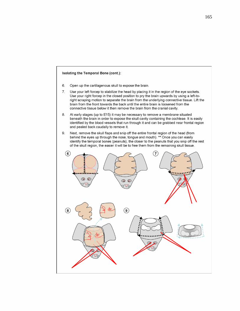
165

166

167

168

169

170

171

172
References
Abdouh, A., G. Despres, et al. (1993). "Hair cell overproduction in the developing
mammalian cochlea in culture." Neuroreport 5(1): 33-6.
Abdouh, A., G. Despres, et al. (1994). "Histochemical and scanning electron microscopic
studies of supernumerary hair cells in embryonic rat cochlea in vitro." Brain Res
660(2): 181-91.
Adam, J., A. Myat, et al. (1998). "Cell fate choices and the expression of Notch, Delta
and Serrate homologues in the chick inner ear: parallels with Drosophila sense-
organ development." Development 125(23): 4645-54.
Adamska, M., H. Herbrand, et al. (2001). "FGFs control the patterning of the inner ear
but are not able to induce the full ear program." Mech Dev 109(2): 303-13.
Bailey, A. P., S. Bhattacharyya, et al. (2006). "Lens specification is the ground state of all
sensory placodes, from which FGF promotes olfactory identity." Dev Cell 11(4):
505-17.
Baker, C. V. and M. Bronner-Fraser (2001). "Vertebrate cranial placodes I. Embryonic
induction." Dev Biol 232(1): 1-61.
173
Bermingham-McDonogh, O., E. C. Oesterle, et al. (2006). "Expression of Prox1 during
mouse cochlear development." J Comp Neurol 496(2): 172-86.
Bermingham-McDonogh, O. and E. W. Rubel (2003). "Hair cell regeneration: winging
our way towards a sound future." Curr Opin Neurobiol 13(1): 119-26.
Bermingham-McDonogh, O., J. S. Stone, et al. (2001). "FGFR3 expression during
development and regeneration of the chick inner ear sensory epithelia." Dev Biol
238(2): 247-59.
Bermingham, N. A., B. A. Hassan, et al. (1999). "Math1: an essential gene for the
generation of inner ear hair cells." Science 284(5421): 1837-41.
Bok, J., W. Chang, et al. (2007). "Patterning and morphogenesis of the vertebrate inner
ear." Int J Dev Biol 51(6-7): 521-33.
Brooker, R., K. Hozumi, et al. (2006). "Notch ligands with contrasting functions: Jagged1
and Delta1 in the mouse inner ear." Development 133(7): 1277-86.
Chao, M. V. and M. Bothwell (2002). "Neurotrophins: to cleave or not to cleave."
Neuron 33(1): 9-12.

174
Chen, L., R. Adar, et al. (1999). "Gly369Cys mutation in mouse FGFR3 causes
achondroplasia by affecting both chondrogenesis and osteogenesis." J Clin Invest
104(11): 1517-25.
Chen, P. and N. Segil (1999). "p27(Kip1) links cell proliferation to morphogenesis in the
developing organ of Corti." Development 126(8): 1581-90.
Cohen, G. M. and C. D. Fermin (1978). "The development of hair cells in the embryonic
chick's basilar papilla." Acta Otolaryngol 86(5-6): 342-58.
Coleman, B., J. B. Fallon, et al. (2007). "Auditory hair cell explant co-cultures promote
the differentiation of stem cells into bipolar neurons." Exp Cell Res 313(2): 232-
43.
Colvin, J. S., B. A. Bohne, et al. (1996). "Skeletal overgrowth and deafness in mice
lacking fibroblast growth factor receptor 3." Nat Genet 12(4): 390-7.
Corwin, J. T. and D. A. Cotanche (1988). "Regeneration of sensory hair cells after
acoustic trauma." Science 240(4860): 1772-4.
Cotanche, D. A. (1987). "Regeneration of hair cell stereociliary bundles in the chick
cochlea following severe acoustic trauma." Hear Res 30(2-3): 181-95.

175
Cotanche, D. A. (1997). "Hair cell regeneration in the avian cochlea." Ann Otol Rhinol
Laryngol Suppl 168: 9-15.
Cotanche, D. A., K. H. Lee, et al. (1994). "Hair cell regeneration in the bird cochlea
following noise damage or ototoxic drug damage." Anat Embryol (Berl) 189(1):
1-18.
Cruz, R. M., P. R. Lambert, et al. (1987). "Light microscopic evidence of hair cell
regeneration after gentamicin toxicity in chick cochlea." Arch Otolaryngol Head
Neck Surg 113(10): 1058-62.
Dallos, P., X. Wu, et al. (2008). "Prestin-based outer hair cell motility is necessary for
mammalian cochlear amplification." Neuron 58(3): 333-9.
Daudet, N., L. Ariza-McNaughton, et al. (2007). "Notch signalling is needed to maintain,
but not to initiate, the formation of prosensory patches in the chick inner ear."
Development 134(12): 2369-78.
Daudet, N. and J. Lewis (2005). "Two contrasting roles for Notch activity in chick inner
ear development: specification of prosensory patches and lateral inhibition of
hair-cell differentiation." Development 132(3): 541-51.
176
Demanez, J. P. and L. Demanez (2003). "Anatomophysiology of the central auditory
nervous system: basic concepts." Acta Otorhinolaryngol Belg 57(4): 227-36.
Driver, E. C., S. P. Pryor, et al. (2008). "Hedgehog signaling regulates sensory cell
formation and auditory function in mice and humans." J Neurosci 28(29): 7350-8.
Duester, G. (2007). "Retinoic acid regulation of the somitogenesis clock." Birth Defects
Res C Embryo Today 81(2): 84-92.
Duncan, L. J., D. A. Mangiardi, et al. (2006). "Differential expression of unconventional
myosins in apoptotic and regenerating chick hair cells confirms two regeneration
mechanisms." J Comp Neurol 499(5): 691-701.
Echteler, S. M., R. R. Fay, et al. (1994). Structure of the mammalian cochlea.
Comparative Hearing: Mammals (Springer Handbook of Auditory Research). F.
RR and P. AN. Verlag, New York, Springer. 4: 260.
Fay, R. R. and A. N. Popper (2000). "Evolution of hearing in vertebrates: the inner ears
and processing." Hear Res 149(1-2): 1-10.
Fekete, D. M., S. Muthukumar, et al. (1998). "Hair cells and supporting cells share a
common progenitor in the avian inner ear." J Neurosci 18(19): 7811-21.

177
Friedman, L. M., A. A. Dror, et al. (2007). "Mouse models to study inner ear
development and hereditary hearing loss." Int J Dev Biol 51(6-7): 609-31.
Fukuchi-Shimogori, T. and E. A. Grove (2003). "Emx2 patterns the neocortex by
regulating FGF positional signaling." Nat Neurosci 6(8): 825-31.
Funato, N., K. Ohtani, et al. (2001). "Common regulation of growth arrest and
differentiation of osteoblasts by helix-loop-helix factors." Mol Cell Biol 21(21):
7416-28.
Furthauer, M., W. Lin, et al. (2002). "Sef is a feedback-induced antagonist of
Ras/MAPK-mediated FGF signalling." Nat Cell Biol 4(2): 170-4.
Gestwa, G., B. Wiechers, et al. (1999). "Differential expression of trkB.T1 and trkB.T2,
truncated trkC, and p75(NGFR) in the cochlea prior to hearing function." J Comp
Neurol 414(1): 33-49.
Govindarajan, V. and P. A. Overbeek (2001). "Secreted FGFR3, but not FGFR1, inhibits
lens fiber differentiation." Development 128(9): 1617-27.
Gross, J., A. Machulik, et al. (2005). "Expression of prestin mRNA in the organotypic
culture of rat cochlea." Hear Res 204(1-2): 183-90.

178
Hanafusa, H., S. Torii, et al. (2002). "Sprouty1 and Sprouty2 provide a control
mechanism for the Ras/MAPK signalling pathway." Nat Cell Biol 4(11): 850-8.
Hasson, T., P. G. Gillespie, et al. (1997). "Unconventional myosins in inner-ear sensory
epithelia." J Cell Biol 137(6): 1287-307.
Hayashi, T., D. Cunningham, et al. (2007). "Loss of Fgfr3 leads to excess hair cell
development in the mouse organ of Corti." Dev Dyn 236(2): 525-33.
Hayashi, T., H. Kokubo, et al. (2008). "Hesr1 and Hesr2 may act as early effectors of
Notch signaling in the developing cochlea." Dev Biol 316(1): 87-99.
Hayashi, T., C. A. Ray, et al. (2008). "Fgf20 is required for sensory epithelial
specification in the developing cochlea." J Neurosci 28(23): 5991-9.
Hebert, J. M. and S. K. McConnell (2000). "Targeting of cre to the Foxg1 (BF-1) locus
mediates loxP recombination in the telencephalon and other developing head
structures." Dev Biol 222(2): 296-306.
Ito, M., S. S. Spicer, et al. (1995). "Cytological changes related to maturation of the organ
of Corti and opening of Corti's tunnel." Hear Res 88(1-2): 107-23.

179
Izumikawa, M., R. Minoda, et al. (2005). "Auditory hair cell replacement and hearing
improvement by Atoh1 gene therapy in deaf mammals." Nat Med 11(3): 271-6.
Jacques, B. E., M. E. Montcouquiol, et al. (2007). "Fgf8 induces pillar cell fate and
regulates cellular patterning in the mammalian cochlea." Development 134(16):
3021-9.
Jones, J. M., M. Montcouquiol, et al. (2006). "Inhibitors of differentiation and DNA
binding (Ids) regulate Math1 and hair cell formation during the development of
the organ of Corti." J Neurosci 26(2): 550-8.
Jorgensen, J. M. and N. A. Locket (1995). "The inner ear of the echidna Tachyglossus
aculeatus: the vestibular sensory organs." Proc Biol Sci 260(1358): 183-9.
Kachar, B., M. Parakkal, et al. (2000). "High-resolution structure of hair-cell tip links."
Proc Natl Acad Sci U S A 97(24): 13336-41.
Kaiser, C. L., B. J. Chapman, et al. (2008). "Comparison of activated caspase detection
methods in the gentamicin-treated chick cochlea." Hear Res 240(1-2): 1-11.
Kaltenbach, J. A. and P. R. Falzarano (1997). "The development of the reticular lamina in
the hamster: an examination of transitory features and their functional roles." Int J
Dev Neurosci 15(4-5): 509-21.

180
Katayama, A. and J. T. Corwin (1989). "Cell production in the chicken cochlea." J Comp
Neurol 281(1): 129-35.
Kelley, M. W. (2007). "Cellular commitment and differentiation in the organ of Corti."
Int J Dev Biol 51(6-7): 571-83.
Kelley, M. W., X. M. Xu, et al. (1993). "The developing organ of Corti contains retinoic
acid and forms supernumerary hair cells in response to exogenous retinoic acid in
culture." Development 119(4): 1041-53.
Kiernan, A. E., R. Cordes, et al. (2005). "The Notch ligands DLL1 and JAG2 act
synergistically to regulate hair cell development in the mammalian inner ear."
Development 132(19): 4353-62.
Koppl, C., G. A. Manley, et al. (2000). "Auditory processing in birds." Curr Opin
Neurobiol 10(4): 474-81.
Ladhams, A. and J. O. Pickles (1996). "Morphology of the monotreme organ of Corti and
macula lagena." J Comp Neurol 366(2): 335-47.
Lanford, P. J., Y. Lan, et al. (1999). "Notch signalling pathway mediates hair cell
development in mammalian cochlea." Nat Genet 21(3): 289-92.

181
Lavigne-Rebillard, M. and R. Pujol (1986). "Development of the auditory hair cell
surface in human fetuses. A scanning electron microscopy study." Anat Embryol
(Berl) 174(3): 369-77.
Lavigne-Rebillard, M. and R. Pujol (1987). "Surface aspects of the developing human
organ of Corti." Acta Otolaryngol Suppl 436: 43-50.
Levic, S., L. Nie, et al. (2007). "Development and regeneration of hair cells share
common functional features." Proc Natl Acad Sci U S A 104(48): 19108-13.
Lim, D. J. (1986). "Functional structure of the organ of Corti: a review." Hear Res 22:
117-46.
Luo, L., H. Koutnouyan, et al. (1993). "Acidic and basic FGF mRNA expression in the
adult and developing rat cochlea." Hear Res 69(1-2): 182-93.
Ma, E. Y., E. W. Rubel, et al. (2008). "Notch signaling regulates the extent of hair cell
regeneration in the zebrafish lateral line." J Neurosci 28(9): 2261-73.
MacArthur, C. A., A. Lawshe, et al. (1995). "FGF-8 isoforms activate receptor splice
forms that are expressed in mesenchymal regions of mouse development."
Development 121(11): 3603-13.

182
Manley, G. A. and C. Koppl (1998). "Phylogenetic development of the cochlea and its
innervation." Curr Opin Neurobiol 8(4): 468-74.
Maroon, H., J. Walshe, et al. (2002). "Fgf3 and Fgf8 are required together for formation
of the otic placode and vesicle." Development 129(9): 2099-108.
Massague, J., J. Seoane, et al. (2005). "Smad transcription factors." Genes Dev 19(23):
2783-810.
Matsui, J. I., J. M. Ogilvie, et al. (2002). "Inhibition of caspases prevents ototoxic and
ongoing hair cell death." J Neurosci 22(4): 1218-27.
Matsui, J. I., M. A. Parker, et al. (2005). "Regeneration and replacement in the vertebrate
inner ear." Drug Discov Today 10(19): 1307-12.
McKenzie, E., A. Krupin, et al. (2004). "Cellular growth and rearrangement during the
development of the mammalian organ of Corti." Dev Dyn 229(4): 802-12.
Meyers, E. N., M. Lewandoski, et al. (1998). "An Fgf8 mutant allelic series generated by
Cre- and Flp-mediated recombination." Nat Genet 18(2): 136-41.

183
Miki, T., D. P. Bottaro, et al. (1992). "Determination of ligand-binding specificity by
alternative splicing: two distinct growth factor receptors encoded by a single
gene." Proc Natl Acad Sci U S A 89(1): 246-50.
Mohammadi, M., G. McMahon, et al. (1997). "Structures of the tyrosine kinase domain
of fibroblast growth factor receptor in complex with inhibitors." Science
276(5314): 955-60.
Mohammadi, M., S. K. Olsen, et al. (2005). "Structural basis for fibroblast growth factor
receptor activation." Cytokine Growth Factor Rev 16(2): 107-37.
Montcouquiol, M. and J. T. Corwin (2001). "Brief treatments with forskolin enhance s-
phase entry in balance epithelia from the ears of rats." J Neurosci 21(3): 974-82.
Montcouquiol, M. and J. T. Corwin (2001). "Intracellular signals that control cell
proliferation in mammalian balance epithelia: key roles for phosphatidylinositol-3
kinase, mammalian target of rapamycin, and S6 kinases in preference to calcium,
protein kinase C, and mitogen-activated protein kinase." J Neurosci 21(2): 570-
80.
Montcouquiol, M. and M. W. Kelley (2003). "Planar and vertical signals control cellular
differentiation and patterning in the mammalian cochlea." J Neurosci 23(28):
9469-78.

184
Montcouquiol, M., N. Sans, et al. (2006). "Asymmetric localization of Vangl2 and Fz3
indicate novel mechanisms for planar cell polarity in mammals." J Neurosci
26(19): 5265-75.
Mueller, K. L., B. E. Jacques, et al. (2002). "Fibroblast growth factor signaling regulates
pillar cell development in the organ of corti." J Neurosci 22(21): 9368-77.
Neves, J., A. Kamaid, et al. (2007). "Differential expression of Sox2 and Sox3 in
neuronal and sensory progenitors of the developing inner ear of the chick." J
Comp Neurol 503(4): 487-500.
Nilsen, K. E. and I. J. Russell (1999). "Timing of cochlear feedback: spatial and temporal
representation of a tone across the basilar membrane." Nat Neurosci 2(7): 642-8.
O'Brien, S. J. (2008). "The platypus genome unraveled." Cell 133(6): 953-5.
Ohyama, T., A. K. Groves, et al. (2007). "The first steps towards hearing: mechanisms of
otic placode induction." Int J Dev Biol 51(6-7): 463-72.
Olsen, S. K., J. Y. Li, et al. (2006). "Structural basis by which alternative splicing
modulates the organizer activity of FGF8 in the brain." Genes Dev 20(2): 185-98.

185
Ornitz, D. M. (2000). "FGFs, heparan sulfate and FGFRs: complex interactions essential
for development." Bioessays 22(2): 108-12.
Ornitz, D. M. and N. Itoh (2001). "Fibroblast growth factors." Genome Biol 2(3):
REVIEWS3005.
Ornitz, D. M., J. Xu, et al. (1996). "Receptor specificity of the fibroblast growth factor
family." J Biol Chem 271(25): 15292-7.
Pauley, S., T. J. Wright, et al. (2003). "Expression and function of FGF10 in mammalian
inner ear development." Dev Dyn 227(2): 203-15.
Peters, K., D. Ornitz, et al. (1993). "Unique expression pattern of the FGF receptor 3
gene during mouse organogenesis." Dev Biol 155(2): 423-30.
Phillips, B. T., E. M. Storch, et al. (2004). "A direct role for Fgf but not Wnt in otic
placode induction." Development 131(4): 923-31.
Pickles, J. O. (2001). "The expression of fibroblast growth factors and their receptors in
the embryonic and neonatal mouse inner ear." Hear Res 155(1-2): 54-62.

186
Pickles, J. O., S. D. Comis, et al. (1984). "Cross-links between stereocilia in the guinea
pig organ of Corti, and their possible relation to sensory transduction." Hear Res
15(2): 103-12.
Pirvola, U., Y. Cao, et al. (1995). "The site of action of neuronal acidic fibroblast growth
factor is the organ of Corti of the rat cochlea." Proc Natl Acad Sci U S A 92(20):
9269-73.
Pirvola, U., J. Ylikoski, et al. (2002). "FGFR1 is required for the development of the
auditory sensory epithelium." Neuron 35(4): 671-80.
Puligilla, C., F. Feng, et al. (2007). "Disruption of fibroblast growth factor receptor 3
signaling results in defects in cellular differentiation, neuronal patterning, and
hearing impairment." Dev Dyn 236(7): 1905-17.
Raphael, Y. and R. A. Altschuler (2003). "Structure and innervation of the cochlea."
Brain Res Bull 60(5-6): 397-422.
Rau, A., P. K. Legan, et al. (1999). "Tectorin mRNA expression is spatially and
temporally restricted during mouse inner ear development." J Comp Neurol
405(2): 271-80.

187
Raz, Y. and M. W. Kelley (1999). "Retinoic acid signaling is necessary for the
development of the organ of Corti." Dev Biol 213(1): 180-93.
Roberson, D. W., J. A. Alosi, et al. (2004). "Direct transdifferentiation gives rise to the
earliest new hair cells in regenerating avian auditory epithelium." J Neurosci Res
78(4): 461-71.
Romand, R., P. Dolle, et al. (2006). "Retinoid signaling in inner ear development." J
Neurobiol 66(7): 687-704.
Sahly, I., A. El-Amraoui, et al. (1997). "Expression of myosin VIIA during mouse
embryogenesis." Anat Embryol (Berl) 196(2): 159-70.
Sanchez-Calderon, H., G. Martin-Partido, et al. (2002). "Differential expression of Otx2,
Gbx2, Pax2, and Fgf8 in the developing vestibular and auditory sensory organs."
Brain Res Bull 57(3-4): 321-3.
Sanchez-Calderon, H., G. Martin-Partido, et al. (2004). "Otx2, Gbx2, and Fgf8
expression patterns in the chick developing inner ear and their possible roles in
otic specification and early innervation." Gene Expr Patterns 4(6): 659-69.
188
Sanchez-Calderon, H., M. Milo, et al. (2007). "A network of growth and transcription
factors controls neuronal differentation and survival in the developing ear." Int J
Dev Biol 51(6-7): 557-70.
Sano, H., J. Mukai, et al. (2001). "Expression of p75NTR and its associated protein
NADE in the rat cochlea." Laryngoscope 111(3): 535-8.
Schimmang, T. (2007). "Expression and functions of FGF ligands during early otic
development." Int J Dev Biol 51(6-7): 473-81.
Shim, K., G. Minowada, et al. (2005). "Sprouty2, a mouse deafness gene, regulates cell
fate decisions in the auditory sensory epithelium by antagonizing FGF signaling."
Dev Cell 8(4): 553-64.
Shin, J. W., M. Min, et al. (2006). "Prox1 promotes lineage-specific expression of
fibroblast growth factor (FGF) receptor-3 in lymphatic endothelium: a role for
FGF signaling in lymphangiogenesis." Mol Biol Cell 17(2): 576-84.
Si, F., H. Brodie, et al. (2003). "Developmental assembly of transduction apparatus in
chick basilar papilla." J Neurosci 23(34): 10815-26.

189
Slepecky, N. B. and Y. Ogata (1996). "[Immunohistochemical labeling of inner ear
tissues embedded in polyethylene glycol 4000--comparative study with araldite
and unicryl embedded sections]." Nippon Jibiinkoka Gakkai Kaiho 99(3): 361-9.
Smith, C. A. and T. Takasaka (1971). "Auditory receptor organs of reptiles, birds, and
mammals." Contrib Sens Physiol 5: 129-78.
Sobin, A. and M. Anniko (1984). "Early development of cochlear hair cell stereociliary
surface morphology." Arch Otorhinolaryngol 241(1): 55-64.
Stone, J. S. and D. A. Cotanche (1991). "Hair cell differentiation in the developing chick
cochlea and in embryonic cochlear organ culture." J Comp Neurol 314(3): 614-
25.
Stone, J. S. and D. A. Cotanche (2007). "Hair cell regeneration in the avian auditory
epithelium." Int J Dev Biol 51(6-7): 633-47.
Stone, J. S. and E. W. Rubel (2000). "Temporal, spatial, and morphologic features of hair
cell regeneration in the avian basilar papilla." J Comp Neurol 417(1): 1-16.
Stone, J. S., J. L. Shang, et al. (2004). "cProx1 immunoreactivity distinguishes progenitor
cells and predicts hair cell fate during avian hair cell regeneration." Dev Dyn
230(4): 597-614.

190
Storm, E. E., J. L. Rubenstein, et al. (2003). "Dosage of Fgf8 determines whether cell
survival is positively or negatively regulated in the developing forebrain." Proc
Natl Acad Sci U S A 100(4): 1757-62.
Tefft, J. D., M. Lee, et al. (1999). "Conserved function of mSpry-2, a murine homolog of
Drosophila sprouty, which negatively modulates respiratory organogenesis." Curr
Biol 9(4): 219-22.
Thisse, B. and C. Thisse (2005). "Functions and regulations of fibroblast growth factor
signaling during embryonic development." Dev Biol 287(2): 390-402.
Tsang, M., R. Friesel, et al. (2002). "Identification of Sef, a novel modulator of FGF
signalling." Nat Cell Biol 4(2): 165-9.
Tucker, J. B., J. B. Mackie, et al. (1999). "Cytoskeletal integration in a highly ordered
sensory epithelium in the organ of Corti: reponse to loss of cell partners in the
Bronx waltzer mouse." J Neurocytol 28(12): 1017-34.
Uchikawa, M., Y. Kamachi, et al. (1999). "Two distinct subgroups of Group B Sox genes
for transcriptional activators and repressors: their expression during embryonic
organogenesis of the chicken." Mech Dev 84(1-2): 103-20.

191
von Bartheld, C. S., S. L. Patterson, et al. (1991). "Expression of nerve growth factor
(NGF) receptors in the developing inner ear of chick and rat." Development
113(2): 455-70.
Wakioka, T., A. Sasaki, et al. (2001). "Spred is a Sprouty-related suppressor of Ras
signalling." Nature 412(6847): 647-51.
Warchol, M. E. (2001). "Lectin from Griffonia simplicifolia identifies an immature-
appearing subpopulation of sensory hair cells in the avian utricle." J Neurocytol
30(3): 253-64.
Werthessen, N. T. (1949). "A technique of organ culture for protracted metabolism
studies." Endocrinology 44(2): 109-26.
White, P. M., A. Doetzlhofer, et al. (2006). "Mammalian cochlear supporting cells can
divide and trans-differentiate into hair cells." Nature 441(7096): 984-7.
Whitlon, D. S. (1993). "E-cadherin in the mature and developing organ of Corti of the
mouse." J Neurocytol 22(12): 1030-8.
Woods, C., M. Montcouquiol, et al. (2004). "Math1 regulates development of the sensory
epithelium in the mammalian cochlea." Nat Neurosci 7(12): 1310-8.

192
Wu, D. K. and S. H. Oh (1996). "Sensory organ generation in the chick inner ear." J
Neurosci 16(20): 6454-62.
Zelarayan, L. C., V. Vendrell, et al. (2007). "Differential requirements for FGF3, FGF8
and FGF10 during inner ear development." Dev Biol 308(2): 379-91.
Zheng, J. L. and W. Q. Gao (2000). "Overexpression of Math1 induces robust production
of extra hair cells in postnatal rat inner ears." Nat Neurosci 3(6): 580-6.
Related Documents