IMMO5Decreased CD11b and CXCR2 Expression in β-thalassemia/HbE Leukocytes Sirikwan Siriworadetkun* Chayada Thiengtavorn** Dr.Kittiphong Paiboonsukwong*** Suthat Fucharoen**** Dr.Kovit Pattanapanyasat***** Dr.Pornthip Chaichompoo****** Dr.Saovaros Svasti******* ABSTRACT Infection is the major complication and the first cause of death in β-thalassemia/HbE patients. Immunological abnormalities in thalassemia have been documented with related to the clinical feature such as splenectomy, frequent blood transfusion and iron overload. However, the relationship of immune cells between innate and adaptive immunity is not emphasized yet. In this study, the multi -color flow cytometry was developed and analyzed number of immune cells and cellular markers that related to phagocytosis (CD16 and CD11b) and migration (CXCR2) of neutrophils and monocytes. The low expression of cellular markers on neutrophils and monocytes in patients were found when compared to normal subjects . There was correlation between CD11b expression on monocyte and number of both CD4 and CD8 T lymphocytes . This implies that monocytes could be a key immune cells that cause of abnormal immunity in thalassemia. Keywords: Leukocyte, β-Thalassemia, Flow cytometry * Student, Master of Science Program in Molecular Genetics and Genetic Engineering, Institute of Molecular Biosciences, Mahidol University ** Student, Master of Science Program in Pathobiology, Faculty of Science, Mahidol University *** Lecturer, MD., Thalassemia Research Center, Institute of Molecular Biosciences, Mahidol University **** Professor, MD., Thalassemia Research Center, Institute of Molecular Biosciences, Mahidol University ***** Professor, Office for Research and Development, Faculty of Medicine Siriraj Hospital, Mahidol University ****** Lecturer, Department of Pathobiology, Faculty of Science, Mahidol University ******* Associate Professor, Thalassemia Research Center, Institute of Molecular Biosciences, Mahidol University - 274 -

Welcome message from author
This document is posted to help you gain knowledge. Please leave a comment to let me know what you think about it! Share it to your friends and learn new things together.
Transcript

IMMO5-1
Decreased CD11b and CXCR2 Expression in β-thalassemia/HbE Leukocytes
Sirikwan Siriworadetkun* Chayada Thiengtavorn** Dr.Kittiphong Paiboonsukwong*** Suthat Fucharoen**** Dr.Kovit Pattanapanyasat***** Dr.Pornthip Chaichompoo****** Dr.Saovaros Svasti*******
ABSTRACT
Infection is the major complication and the first cause of death in β-thalassemia/HbE patients. Immunological abnormalities in thalassemia have been documented with related to the clinical feature such as splenectomy, frequent blood transfusion and iron overload. However, the relationship of immune cells between innate and adaptive immunity is not emphasized yet. In this study, the multi-color flow cytometry was developed and analyzed number of immune cells and cellular markers that related to phagocytosis (CD16 and CD11b) and migration (CXCR2) of neutrophils and monocytes. The low expression of cellular markers on neutrophils and monocytes in patients were found when compared to normal subjects. There was correlation between CD11b expression on monocyte and number of both CD4 and CD8 T lymphocytes. This implies that monocytes could be a key immune cells that cause of abnormal immunity in thalassemia.
Keywords: Leukocyte, β-Thalassemia, Flow cytometry
* Student, Master of Science Program in Molecular Genetics and Genetic Engineering, Institute of Molecular Biosciences, Mahidol University ** Student, Master of Science Program in Pathobiology, Faculty of Science, Mahidol University *** Lecturer, MD., Thalassemia Research Center, Institute of Molecular Biosciences, Mahidol University **** Professor, MD., Thalassemia Research Center, Institute of Molecular Biosciences, Mahidol University ***** Professor, Office for Research and Development, Faculty of Medicine Siriraj Hospital, Mahidol University ****** Lecturer, Department of Pathobiology, Faculty of Science, Mahidol University ******* Associate Professor, Thalassemia Research Center, Institute of Molecular Biosciences, Mahidol University
- 274 -
IMMO5-2
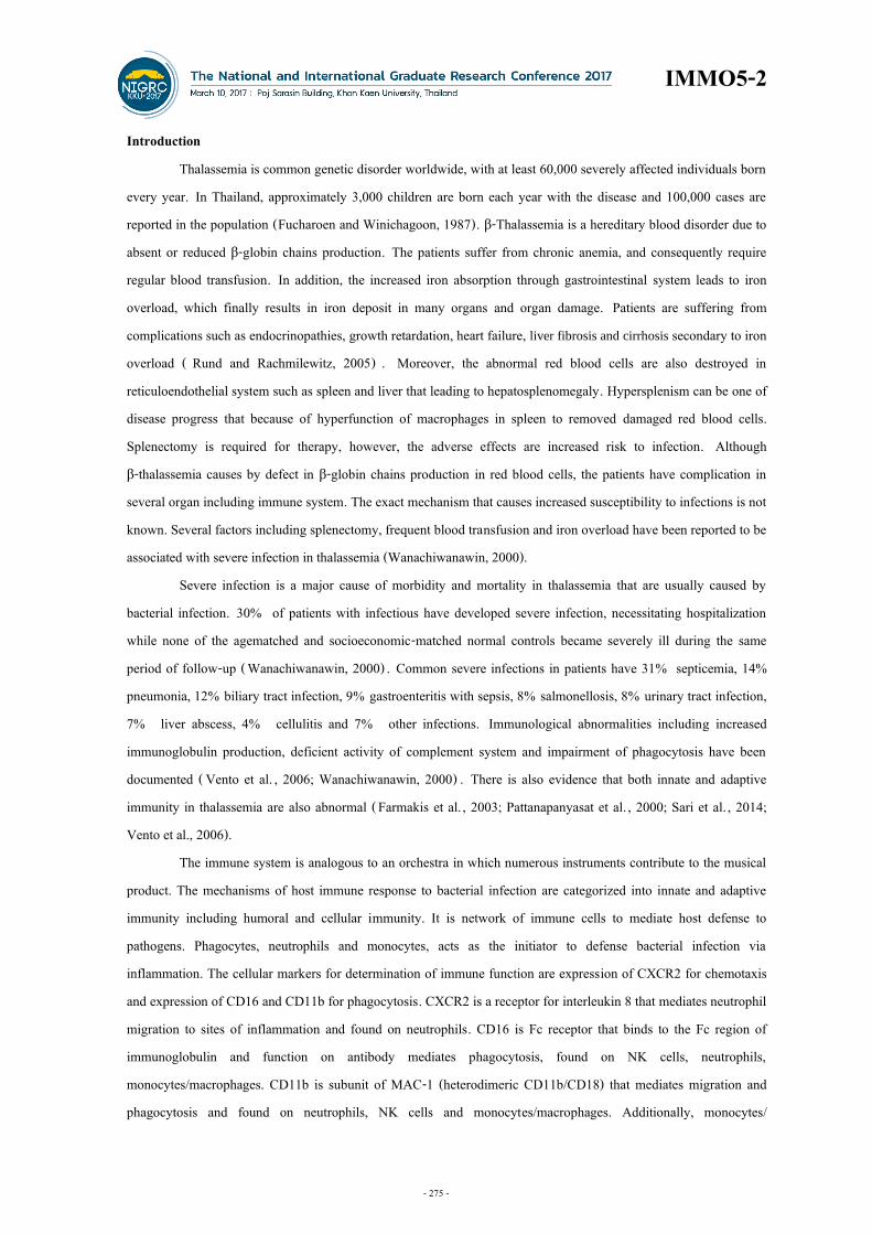
Introduction Thalassemia is common genetic disorder worldwide, with at least 60,000 severely affected individuals born
every year. In Thailand, approximately 3,000 children are born each year with the disease and 100,000 cases are reported in the population (Fucharoen and Winichagoon, 1987). β-Thalassemia is a hereditary blood disorder due to absent or reduced β-globin chains production. The patients suffer from chronic anemia, and consequently require regular blood transfusion. In addition, the increased iron absorption through gastrointestinal system leads to iron overload, which finally results in iron deposit in many organs and organ damage. Patients are suffering from complications such as endocrinopathies, growth retardation, heart failure, liver fibrosis and cirrhosis secondary to iron overload ( Rund and Rachmilewitz, 2005) . Moreover, the abnormal red blood cells are also destroyed in reticuloendothelial system such as spleen and liver that leading to hepatosplenomegaly. Hypersplenism can be one of disease progress that because of hyperfunction of macrophages in spleen to removed damaged red blood cells. Splenectomy is required for therapy, however, the adverse effects are increased risk to infection. Although β-thalassemia causes by defect in β-globin chains production in red blood cells, the patients have complication in several organ including immune system. The exact mechanism that causes increased susceptibility to infections is not known. Several factors including splenectomy, frequent blood transfusion and iron overload have been reported to be associated with severe infection in thalassemia (Wanachiwanawin, 2000).
Severe infection is a major cause of morbidity and mortality in thalassemia that are usually caused by bacterial infection. 30% of patients with infectious have developed severe infection, necessitating hospitalization while none of the agematched and socioeconomic-matched normal controls became severely ill during the same period of follow-up ( Wanachiwanawin, 2000) . Common severe infections in patients have 31% septicemia, 14% pneumonia, 12% biliary tract infection, 9% gastroenteritis with sepsis, 8% salmonellosis, 8% urinary tract infection, 7% liver abscess, 4% cellulitis and 7% other infections. Immunological abnormalities including increased immunoglobulin production, deficient activity of complement system and impairment of phagocytosis have been documented ( Vento et al. , 2006; Wanachiwanawin, 2000) . There is also evidence that both innate and adaptive immunity in thalassemia are also abnormal ( Farmakis et al. , 2003; Pattanapanyasat et al. , 2000; Sari et al. , 2014; Vento et al., 2006).
The immune system is analogous to an orchestra in which numerous instruments contribute to the musical product. The mechanisms of host immune response to bacterial infection are categorized into innate and adaptive immunity including humoral and cellular immunity. It is network of immune cells to mediate host defense to pathogens. Phagocytes, neutrophils and monocytes, acts as the initiator to defense bacterial infection via inflammation. The cellular markers for determination of immune function are expression of CXCR2 for chemotaxis and expression of CD16 and CD11b for phagocytosis. CXCR2 is a receptor for interleukin 8 that mediates neutrophil migration to sites of inflammation and found on neutrophils. CD16 is Fc receptor that binds to the Fc region of immunoglobulin and function on antibody mediates phagocytosis, found on NK cells, neutrophils, monocytes/macrophages. CD11b is subunit of MAC-1 (heterodimeric CD11b/CD18) that mediates migration and phagocytosis and found on neutrophils, NK cells and monocytes/macrophages. Additionally, monocytes/
- 275 -
IMMO5-3
macrophages are also can destroy microbe and generate the peptides of microbe and presented peptides to T cell via major histocompatibility complex and initiate adaptive immune response. The CD4 and CD8 T-lymphocytes are main function in cellular immunity. The CD4 T-lymphocytes or T helper cells play an important role to B lymphocyte antibody class switching, activation and growth of CD8 T lymphocytes and activation of phagocytes . The CD8 T lymphocytes or cytotoxic T cells release the cytotoxins act to trigger apoptosis of infected cells.
In fact, the previous studies had mainly investigating on the individual part of immune system associated with the clinical feature in thalassemia (splenectomy, frequent blood transfusion and iron overload) that were correlated to susceptibility to infection in thalassemia (Wanachiwanawin, 2000). However, the relationship of immune cells in term of the innate immune system plays a crucial role to initiation and subsequent direction of adaptive immune system is not emphasized yet. In this study, the relationship of immune cells and their cellular markers as funtionl analysis in splecentomized and non-splenectomized β-thalassemia/HbE patients were emphasized. Objective of the study
To study the cellular markers (CD16 and CD11b as phagocytosis and CXCR2 as migration) as functional analysis on neutrophils and monocytes and the number and ratio of CD4+/CD8+ T cell lymphocytes in splecentomized and non-splenectomized β-thalassemia/HbE patients.
Methodology
Subjects Ten of β-thalassemia/HbE patients (5 spenectomy and 5 non-splenectomy) and 5 normal subjects were
recruited in this study. The study protocol was approved by the Mahidol University Institutional Review Board (approval number 2015/052.0104). Written informed consent was obtained from all individual participants included in the study. All subjects had no hydroxyurea, prednisolone, blood transfusion and iron chelator before blood sample collection for at least 4 weeks. All blood samples were collected and processed at room temperature (RT) within 2-3 h. Hematological parameters were described in Table 1.
Flow cytometric analysis Fifty microliters of whole blood in CPDA1 anticoagulant were lyzed red blood cells with 1×FACS lysing
solution (BD Biosciences) for 10 min at RT, after incabation and wash, samples were incubated 30 min at 4°C in dark with specific cocktail monoclonal antibodies as following; For lymphocyte subset analysis, fluorochrome conjugated monoclonal antibodies specific to CD4, CD8, CD45 and glycophorin A (GPA). For expression of migration and phagocytotic markers on neutrophil and monocyte analysis, fluorochrome conjugated monoclonal antibodies specific to CD11b, CD16, CD45, CXCR2 and GPA. Isotype control and unstainined cells were used as negative control. All monoclonal antibodies were purchased from BioLegend. Samples were staining according to manufacturer’s recommended. Data acquisition at 10,000 events of leukocytes by BD LSRII flow cytometer (BD Biosciences). The
- 276 -

IMMO5-4
percentages and the mean fluorescent intensity (MFI) of specific markers were analyzed by BD Diva software (BD Biosciences) as shown in Fig.1 & 2.
Statistic analysis All descriptive analysis (mean, SD, coefficient of variation and ranges) was performed using GraphPad
PRISM 5.0 (GraphPad Software, Inc.). Comparisons between parameters were analyzed with non-parametric Mann-Whitney U test. Simple linear regression and Spearman’s correlation coefficient (rs) were calculated. The threshold for statistical significance for all comparisons was choose as P < 0.05. Table 1 Hematological parameters
Description Normal subjects β-Thalassemia/HbE patients
Splenectomy Non-splenectomy Number 5 5 5 Hb typing A2A EA or EFA EA or EFA RBC (x106 cells/μL) 4.7 ± 0.4 3.1 ± 0.5 ** 4.1 ± 0.7 Hb (g/dL) 12.8 ± 1.3 6.5 ± 0.5 *** 7.4 ± 0.9 *** HCT (%) 39.3 ± 2.8 23.1 ± 2.3 *** 25.2 ± 2.9 *** MCV (fL) 84.4 ± 9.5 74.8 ± 7.2 61.9 ± 5.5 ** MCH (pg) 27.5 ± 3.9 21.2 ± 2.2 * 18.1 ± 2 *** MCHC (g/dL) 32.5 ± 1.1 28.4 ± 0.9 *** 29.3 ± 0.9 *** RDW (%) 14.3 ± 1.4 23.6 ± 1.1 *** 23.9 ± 0.9 *** Corrected WBC (x 103 cells/μL) 7.0 ± 2.2 14.3 ± 2.8 *** 5.1 ± 1.9 WBC differnetial count (%)
Band form neutrophil 0 ± 0 0 ± 0 0 ± 0 Neutrophils 53.4 ± 3.6 43.8 ± 6.9 57.2 ± 12.0 Eosinophils 2.0 ± 1.6 2.2 ± 2.2 1.2 ± 1.1 Basophils 0 ± 0 0 ± 0 0 ± 0 Monocytes 6.0 ± 2.1 5.4 ± 0.9 7.6 ± 2.7 Lymphocytes 38.6 ± 4.8 48.6 ± 8.7 34 ± 11.3
NRBCs (cells/100WBC) 0 ± 0 385.6 ± 121.6 *** 11 ± 7.9 Platelet (x 103 cells/μL) 233 ± 23.6 665.8 ± 93.7 *** 187.6 ± 5.0 Reticulocyte count (%) 1.34 ± 0.4 12.3 ± 2.5 *** 2.7 ± 0.8 Absolute reticulocyte count (x 109 cells/L) 68.8 ± 23.1 378.9 ± 80.1 *** 116.16 ± 47.9 RBC = red blood cells, Hb = hemoglobin, Hct = hematocrits, MCV = mean corpuscular volume, MCH = mean corpuscular hemoglobin, MCHC = mean corpuscular hemoglobin concentration, WBC = white blood cells, NRBCs = nucleated red blood cells. Significant different at * P < 0.05, ** P < 0.01, *** P < 0.001 when compared to normal subjects.
- 277 -
IMMO5-5
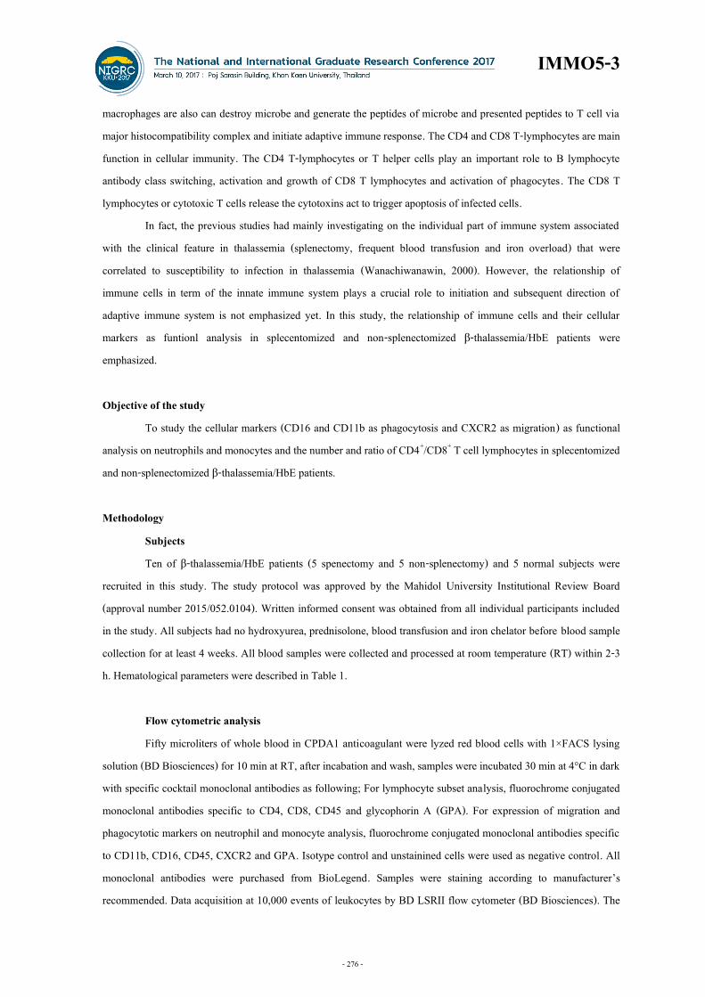
Figure 1 Lymphocyte subset analysis. (A) Leukocytes were gated as glycophorin A (GPA) negative in R1 region to
(B) analysis of lymphocytes (CD45+/SSC-A) in R2 region. (C) The percentages of CD4+ (R3 region) and CD8+lymphocytes (R4 region) were examined.
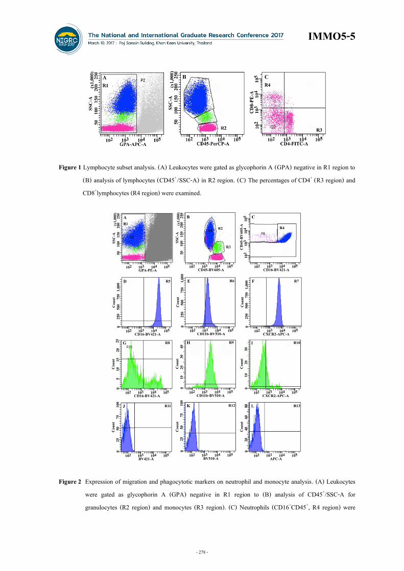
Figure 2 Expression of migration and phagocytotic markers on neutrophil and monocyte analysis. (A) Leukocytes
were gated as glycophorin A (GPA) negative in R1 region to (B) analysis of CD45+/SSC-A for granulocytes (R2 region) and monocytes (R3 region). (C) Neutrophils (CD16+CD45+, R4 region) were
- 278 -
IMMO5-6
gated from R2 region. The percentages and mean fluorescent intensity of CD16, CD11b and CXCR2 on neutrophils (D-F, respectively) and monocytes (G-I, respectively). Negative controls shown in J-L.
Results
Lymphocyte subsets in β-thalassemia/HbE patients β-Thalassemia/HbE patients had microcytic hypochromic anemia as shown in Table 1. Differential white
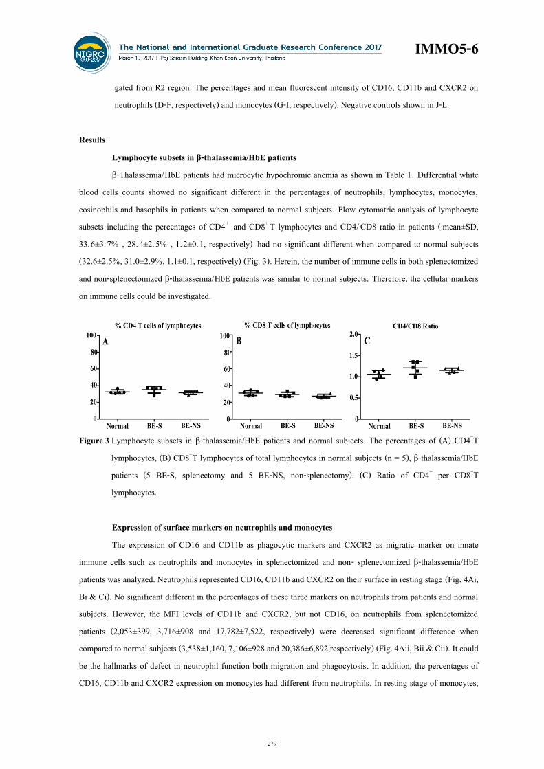
blood cells counts showed no significant different in the percentages of neutrophils, lymphocytes, monocytes, eosinophils and basophils in patients when compared to normal subjects. Flow cytomatric analysis of lymphocyte subsets including the percentages of CD4+ and CD8+ T lymphocytes and CD4/ CD8 ratio in patients ( mean±SD, 33.6±3. 7% , 28. 4±2. 5% , 1. 2±0. 1, respectively) had no significant different when compared to normal subjects (32.6±2.5%, 31.0±2.9%, 1.1±0.1, respectively) (Fig. 3). Herein, the number of immune cells in both splenectomized and non-splenectomized β-thalassemia/HbE patients was similar to normal subjects. Therefore, the cellular markers on immune cells could be investigated.
Figure 3 Lymphocyte subsets in β-thalassemia/HbE patients and normal subjects. The percentages of (A) CD4+T
lymphocytes, (B) CD8+T lymphocytes of total lymphocytes in normal subjects (n = 5), β-thalassemia/HbE patients (5 BE-S, splenectomy and 5 BE-NS, non-splenectomy). (C) Ratio of CD4+ per CD8+T lymphocytes. Expression of surface markers on neutrophils and monocytes The expression of CD16 and CD11b as phagocytic markers and CXCR2 as migratic marker on innate
immune cells such as neutrophils and monocytes in splenectomized and non- splenectomized β-thalassemia/HbE patients was analyzed. Neutrophils represented CD16, CD11b and CXCR2 on their surface in resting stage (Fig. 4Ai, Bi & Ci). No significant different in the percentages of these three markers on neutrophils from patients and normal subjects. However, the MFI levels of CD11b and CXCR2, but not CD16, on neutrophils from splenectomized patients (2,053±399, 3,716±908 and 17,782±7,522, respectively) were decreased significant difference when compared to normal subjects (3,538±1,160, 7,106±928 and 20,386±6,892,respectively) (Fig. 4Aii, Bii & Cii). It could be the hallmarks of defect in neutrophil function both migration and phagocytosis. In addition, the percentages of CD16, CD11b and CXCR2 expression on monocytes had different from neutrophils. In resting stage of monocytes,
- 279 -

IMMO5-7
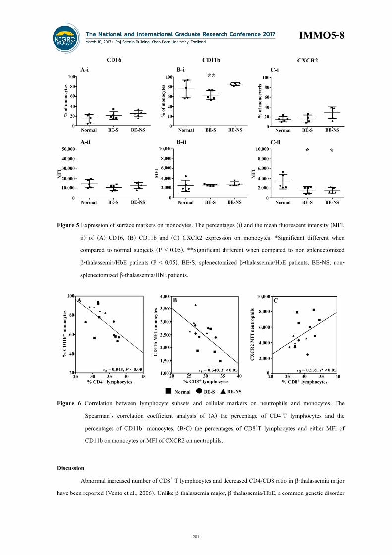
there were 14.6±9%CD16+, 75.6±18%CD11b+ and 15.7±5%CXCR2+monocyes obtained from normal subjects (Fig. 5). Thalassemic patients had no significant different of both the percentages and MFI of CD16 and CD11b on monocytes when compared to normal subjects. However, the MFI levels of CXCR2 on monocytes obtained from patients were decreased significant difference when compared to normal subjects as found in thalassemic neutrophils.
CD11b on monocytes related to lymphocyte subsets To address the relationship between innate immunity and adaptive immunity in thalassemia. Data from all
subjects in this cohort was analyzed the correlation between parameters. The percentages of CD4+T lymphocytes had significant correlated with the percentages of CD11b+monocytes (rs = 0.543, P < 0.05) (Fig. 6A). While the percentages of CD8+T lymphocytes had significant correlated with the MFI of CD11b+monocytes (rs = 0.548, P < 0.05) (Fig. 6B) and with the MFI of CXCR2 on neutrophils (rs = 0.535, P < 0.05) (Fig. 6C).
Figure 4 Expression of surface markers on neutrophils. The percentages (i) and the mean fluorescent intensity (MFI,
ii) of (A) CD16, (B) CD11b and (C) CXCR2 expression on neutrophils. *Significant different when compared to normal subjects (P < 0.05). BE-S; splenectomized β-thalassemia/HbE patients, BE-NS; non-splenectomized β-thalassemia/ HbE patients.
- 280 -
IMMO5-8
Figure 5 Expression of surface markers on monocytes. The percentages (i) and the mean fluorescent intensity (MFI,
ii) of (A) CD16, (B) CD11b and (C) CXCR2 expression on monocytes. *Significant different when compared to normal subjects (P < 0.05). **Significant different when compared to non-splenectomized β-thalassemia/HbE patients (P < 0.05). BE-S; splenectomized β-thalassemia/HbE patients, BE-NS; non-splenectomized β-thalassemia/HbE patients.
Figure 6 Correlation between lymphocyte subsets and cellular markers on neutrophils and monocytes. The
Spearman’s correlation coefficient analysis of (A) the percentage of CD4+T lymphocytes and the percentages of CD11b+ monocytes, (B-C) the percentages of CD8+T lymphocytes and either MFI of CD11b on monocytes or MFI of CXCR2 on neutrophils.
Discussion
Abnormal increased number of CD8+ T lymphocytes and decreased CD4/CD8 ratio in β-thalassemia major have been reported (Vento et al., 2006). Unlike β-thalassemia major, β-thalassemia/HbE, a common genetic disorder
- 281 -

IMMO5-9
in Southern Asia, had no different number of CD4+ , CD8+ T lymphocytes and CD4/CD8 ratio when compared to normal subjects. In addition, these results were similar to our previous reported (Pattanapanyasat et al., 2000). These phenomenon might be caused of different genetic background, the combination of HbE and β-thalassemia spans thalassemia phenotypes from a condition indistinguishable from thalassemia major to a mild form of thalassemia intermedia ( Habib et al. , 2008) . Patients in this cohort were non-blood transfusion dependent thalassemia, while β-thalassemia major patients are transfusion dependent.
Peripheral blood analysis for thalassemia by flow cytometry was developed for excluded nucleated red blood cells (RBCs) and intact RBCs as contamination. Splenectomized β-thalassemia/HbE patients had increased number of nucleated RBCs that can cause of contamination in gating of lymphocyte population by FSC /SSC. Anti-GPA antibody was used in the developed method to excluded contamination from debris/intact RBC and nucleated RBCs. Multi-color staining and multi-channel detection of flow cytometry are simple, practical and low cost for measurement of several cellular markers to monitoring and diagnosis of immune cell function.
β-Thalassemia/HbE neutrophils and monocytes had 2-fold decreased CXCR2 expression that could be defects to migrated property of them and inflammatory response. In fact that monocytes could be a key of abnormal immunity in β-thalassemia/HbE patients. Low percentages and MFI of CD11b as phagocytic markers of thalassemic monocytes that could be focus. During phagocytosis, the CD11b on monocytes could be increased to initial T lymphocyte proliferation and increased neutrophil migration to infection site. In this cohort, we found that there was no significant difference of number of immune cells in peripheral blood from splenectomized and non- splenectomized β-thalassemia/HbE patients when compared to normal subjects. No significant difference between splenectomize and non-splenectomized β-thalassemia/HbE patients on the expression of CD16, CD11b and CXCR2 on neutrophils and monocytes in our cohort. Additionally, we found that splenectomized patients had low percentages of CD11b+monocytes but had high percentages of CD4+T lymphocytes. By contrast, some patients who had highly CD11b MFI expression on monocytes but had low percentages of CD8+T lymphocytes. In fact the high CD11b expression on monocytes could implied active monocytic function on phagocytosis and could activate both CD4 and CD8 T lymphocytes. Thus, the number of both CD4 and CD8 T lymphocytes could be increasing related to CD11b expression on monocytes. However, it is still unclear the mechanism of immune cell net-work in β-thalassemia/HbE patients that could be further study. Conclusion
This study demonstrated the multi-color immunophenotyping method utilize to assessment of immune cell function in peripheral blood samples including thalassemia. β-Thalassemia/HbE patients had defects of neutrophil and monocyte properties that could be effect to susceptibility to infection.
- 282 -
IMMO5-10
Acknowledgements This study was supported by Thailand Research Fund (TRF) (MRG5980043, DPG5980001, IRG5780009
and IRG5780011), Office of the Higher Education Commission and Mahidol University under the National Research University Initiative, Research Chair Grant, National Science and Technology Development Agency, Thailand and Faculty of Science and Faculty of Medicine Ramathibodi Hospital, Mahidol University.
References Farmakis, D., Giakoumis, A., Polymeropoulos, E., and Aessopos, A. Pathogenetic aspects of immune deficiency
associated with beta-thalassemia. Med Sci Monit.2003: 9; RA19-22. Fucharoen, S., and Winichagoon, P. Hemoglobinopathies in Southeast Asia. Hemoglobin.1987: 11; 65-88. Habib, A., Kunzelmann, C., Shamseddeen, W., Zobairi, F., Freyssinet, J.M., and Taher, A. Elevated levels of
circulating procoagulant microparticles in patients with beta-thalassemia intermedia. Haematologica.2008: 93; 941-942.
Pattanapanyasat, K., Thepthai, C., Lamchiagdhase, P., Lerdwana, S., Tachavanich, K., Thanomsuk, P., Wanachiwanawin, W., Fucharoen, S., and Darden, J.M. Lymphocyte subsets and specific T-cell immune response in thalassemia. Cytometry. 2000: 42; 11-17.
Rund, D., and Rachmilewitz, E. Beta-thalassemia. N Engl J Med.2005: 353;1135-1146. Sari, T.T., Gatot, D., Akib, A.A., Bardosono, S., Hadinegoro, S.R., Harahap, A.R., and Idjradinata, P.S. Immune
response of thalassemia major patients in Indonesia with and without splenectomy. Acta Med Indones.2014: 46; 217-225.
Vento, S., Cainelli, F., and Cesario, F. Infections and thalassaemia. Lancet Infect Dis. 2006: 6; 226-233. Wanachiwanawin, W. Infections in E-beta thalassemia. J Pediatr Hematol Oncol.2000: 22; 581-587.
- 283 -
Related Documents