Abstract Our client desires a device to detect the electrical activity of the bladder during the voiding process. The device will be used in a urodynamics lab in conjunction with diagnostic tools. The final design incorporates external and internal electrodes, an EMG circuit, and a digital oscilloscope.

Abstract Our client desires a device to detect the electrical activity of the bladder during the voiding process. The device will be used in a urodynamics.
Jan 03, 2016
Welcome message from author
This document is posted to help you gain knowledge. Please leave a comment to let me know what you think about it! Share it to your friends and learn new things together.
Transcript
Abstract
Our client desires a device to detect the electrical activity of the bladder during the voiding process. The device will be used in a urodynamics lab in conjunction with diagnostic tools. The final design incorporates external and internal electrodes, an EMG circuit, and a digital oscilloscope.

Motivation Overactive Bladder (OAB): A sudden urge to
urinate immediately followed by a bladder contraction, resulting in involuntary micturition• Affects 33 million Americans
• May cause urinary incontinence
Limited treatment options• Disposable pads
• Medication
• Catheters

Problem Statement Bladder EMG has never been
consistently detected Frequency and magnitude of electrical
signal are not well established Pelvic bone and abdominal muscles
distort/interfere with signal detection
Bladder Composition Epithelium Lamina propria Detrusor muscle
• Provides force required to void
• Three layers of smooth muscle
Perivesical soft tissue
LALA
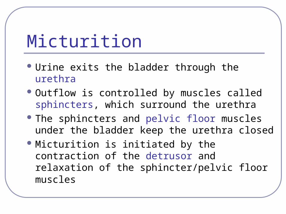
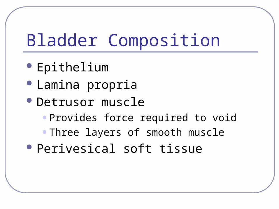
Micturition Urine exits the bladder through the urethra Outflow is controlled by muscles called
sphincters, which surround the urethra The sphincters and pelvic floor muscles
under the bladder keep the urethra closed Micturition is initiated by the contraction of
the detrusor and relaxation of the sphincter/pelvic floor muscles
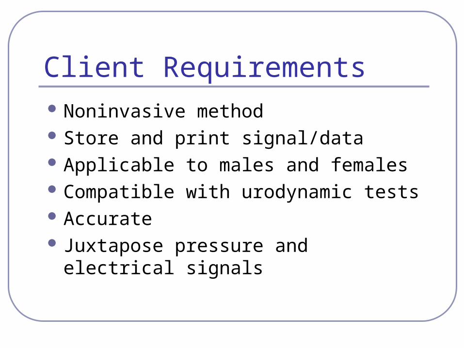
Client Requirements Noninvasive method Store and print signal/data Applicable to males and females Compatible with urodynamic tests Accurate Juxtapose pressure and electrical
signals
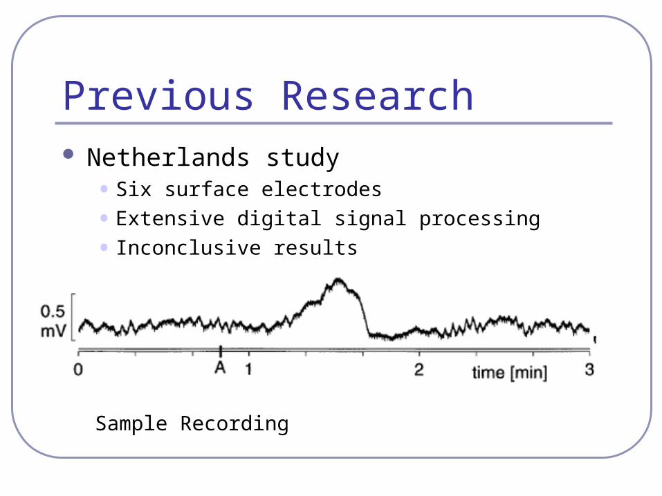
Previous Research Netherlands study
• Six surface electrodes
• Extensive digital signal processing
• Inconclusive results
Sample Recording

Design Alternatives Electrode Design
• Memory Alloy
• Constellation
• Suction
• Needle Electrode Placement
• Vaginal
• Rectal
• Urethral
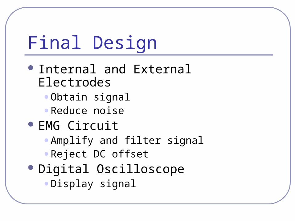
Final Design Internal and External Electrodes
• Obtain signal
• Reduce noise EMG Circuit
• Amplify and filter signal
• Reject DC offset Digital Oscilloscope
• Display signal

Motion Artifact Fact: Netherland study recorded a 0.5 mV
change during micturition Problem: Surface electrodes can cause skin
motion artifact greater than 0.5 mV Question: Is the 0.5 mV signal from the
bladder or a result of skin motion artifact? Solution: Abrade the skin to eliminate skin
motion artifact
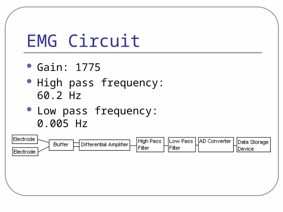
EMG Circuit Gain: 1775 High pass frequency: 60.2 Hz Low pass frequency: 0.005 Hz CMRR: 105.54 dB

EMG Circuit Diagram
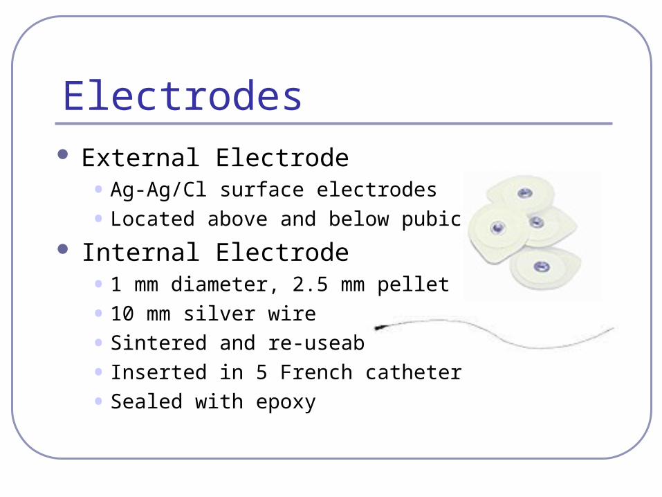
Electrodes External Electrode
• Ag-Ag/Cl surface electrodes
• Located above and below pubic bone
Internal Electrode• 1 mm diameter, 2.5 mm pellet
• 10 mm silver wire
• Sintered and re-useable
• Inserted in 5 French catheter
• Sealed with epoxy
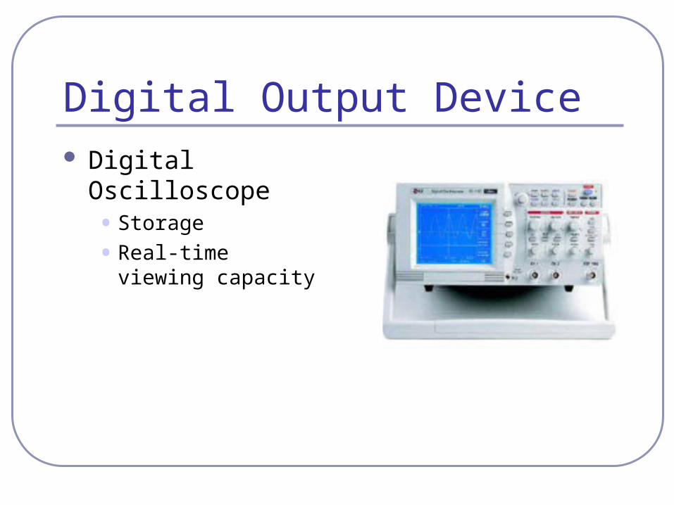
Digital Output Device Digital Oscilloscope
• Storage
• Real-time viewing capacity
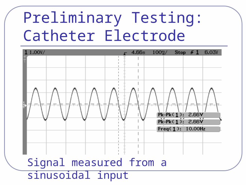
Preliminary Testing: Catheter Electrode
Signal measured from a sinusoidal input
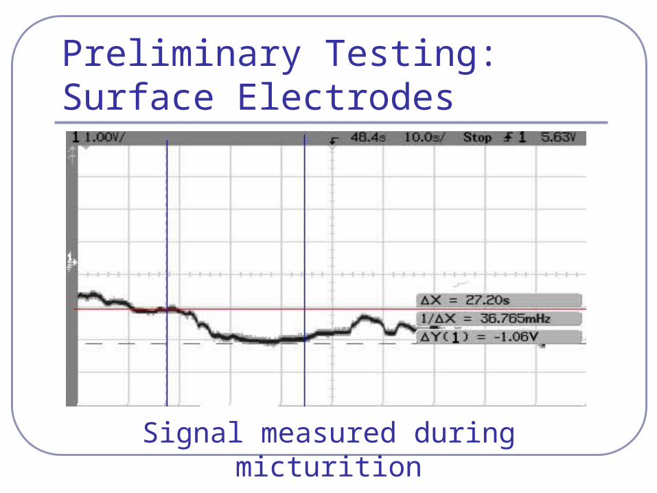
Preliminary Testing: Surface Electrodes
Signal measured during micturition
Future Work Create protocol Fine tune the circuit Obtain a clear signal Develop computer software Test extensively
• Clinical Setting
• Statistical Analysis data

References Ballaro A, Mundy AR, Fry CH, and Craggs MD. Bladder electrical activity: the
elusive electromyogram. BJU International, 2003. 92: 78-84. “Catheters and Transducers.” Medtronic. http://www.medtronic.com/neuro/mfd/consumables/acc_cat_2k1_trans.pdf
September 25, 2003. Kinder MV, van Waalwijk ESC, Gommer ED, and Janknegt RA. A non-invasive
method for bladder electromyography in humans. Archives of Physiology and Biochemistry, 1998. 106: 2-11.
“Pelvic Soft Tissue Structures.” Barts and the London, Queen Mary’s school of Dentistry and Medicine.
http://www.mds.qmw.ac.uk/biomed/kb/grossanatomy/basic_anat/pelvic_soft.htm September 25, 2003.
Kinder MV, van Waalwijk ESC, Gommer ED, and Janknegt RA. A non-invasive method for bladder electromyography in humans. Archives of Physiology and Biochemistry, 1998. 106: 2-11.
“TECA NCS Disposable Surface Electrodes.” Oxford Instruments. http://www.oxford-instruments.com/MDCPDP346.htm September 25, 2003.
Paul Victorey, Biomedical Engineering Department, UW-Madison
Related Documents