West Indian Med J DOI: 10.7727/wimj.2015.559 Mediastinal Thymolipoma: An Evaluation of the Clinical and Radiological Features and the Surgical Outcomes in 11 Cases Ö Oruç 1 , GH Ekinci 1 , H Yilmaz 2 , A Ersev 3 , O Hacıomeroglu 1 , C Atinkaya 2 , T Morali 1 , A Yilmaz 1 ABSTRACT Background: Thymolipoma is a rare benign tumor of the mediastinum, accounting for 2-9 % of all thymic tumors. Although many case reports have been published in the literature, few studies have analyzed the clinical and radiological features and the surgical outcomes of this tumor. Aim: To evaluate the clinical and radiological features and the surgical outcomes of the patients with thymolipoma. Methods: We reviewed the records of pathology department from the beginning of 2005 to the end of 2013. Results: We identified 11 patients with thymolipoma. There were eight male and three female patients. Their ages ranged from 27 to 72 years, with mean age of 40 years. All patients described pulmonary or extrapulmonary symptoms. Two patients (18.2 %) had myasthenia gravis. Chest X-ray was normal in four patients. Computed tomography of the thorax revealed a mass located in the anterior mediastinum in all patients. It showed fat attenuation in four of 11 patients (36.4 %). Thymectomy was performed in all patients. The surgical approach was thoracotomy in five, sternotomy in four, and video-assisted thoracic surgery in two. Thymolipomas ranged in size from 4 to 33 cm. One patient died two years after surgery. None of remaining patients had evidence of recurrence on follow-up. Conclusions: Thymolipoma is a rare tumor of the thymus. It may be associated with myasthenia gravis. Surgical resection is the treatment of choice in the patients with thymolipoma. Complete surgical resection is the cure in most patients. Keywords: Mediastinal tumor, myasthenia gravis, surgery, thymolipoma From: Süreyyapaşa Chest Diseases and Thoracic Surgery Training and Research Hospital, Istanbul- Turkey, 1 Department of Pulmonology, 2 Department of Thoracic Suergery, 3 Department of Pathology Correspondence: Dr GH Ekinci, Süreyyapaşa Göğüs Hastalıkları ve Göğüs, Cerrahisi Eğitim ve Araştırma Hastanesi Maltepe, İstanbul/Turkey. E-mail: [email protected]

Welcome message from author
This document is posted to help you gain knowledge. Please leave a comment to let me know what you think about it! Share it to your friends and learn new things together.
Transcript

West Indian Med J DOI: 10.7727/wimj.2015.559
Mediastinal Thymolipoma: An Evaluation of the Clinical and Radiological Features and the
Surgical Outcomes in 11 Cases
Ö Oruç1, GH Ekinci1, H Yilmaz2, A Ersev3, O Hacıomeroglu1, C Atinkaya2, T Morali1, A Yilmaz1
ABSTRACT
Background: Thymolipoma is a rare benign tumor of the mediastinum, accounting for 2-9 % of all
thymic tumors. Although many case reports have been published in the literature, few studies have
analyzed the clinical and radiological features and the surgical outcomes of this tumor.
Aim: To evaluate the clinical and radiological features and the surgical outcomes of the patients with
thymolipoma.
Methods: We reviewed the records of pathology department from the beginning of 2005 to the end of
2013.
Results: We identified 11 patients with thymolipoma. There were eight male and three female
patients. Their ages ranged from 27 to 72 years, with mean age of 40 years. All patients described
pulmonary or extrapulmonary symptoms. Two patients (18.2 %) had myasthenia gravis. Chest X-ray
was normal in four patients. Computed tomography of the thorax revealed a mass located in the
anterior mediastinum in all patients. It showed fat attenuation in four of 11 patients (36.4 %).
Thymectomy was performed in all patients. The surgical approach was thoracotomy in five,
sternotomy in four, and video-assisted thoracic surgery in two. Thymolipomas ranged in size from 4 to
33 cm. One patient died two years after surgery. None of remaining patients had evidence of
recurrence on follow-up.
Conclusions: Thymolipoma is a rare tumor of the thymus. It may be associated with myasthenia
gravis. Surgical resection is the treatment of choice in the patients with thymolipoma. Complete
surgical resection is the cure in most patients.
Keywords: Mediastinal tumor, myasthenia gravis, surgery, thymolipoma
From: Süreyyapaşa Chest Diseases and Thoracic Surgery Training and Research Hospital, Istanbul-
Turkey, 1Department of Pulmonology, 2Department of Thoracic Suergery, 3Department of Pathology
Correspondence: Dr GH Ekinci, Süreyyapaşa Göğüs Hastalıkları ve Göğüs, Cerrahisi Eğitim ve
Araştırma Hastanesi Maltepe, İstanbul/Turkey. E-mail: [email protected]
Mediastinal Thymolipoma
2
INTRODUCTION
Thymolipoma is an uncommon benign tumor of the thymus consisting of mature adipose
tissue and thymic tissue in variable portions (1). The term thymolipoma was first introduced
in 1948 by Hall to describe this tumor (2). Up to 2008, less than 200 cases of thymolipoma
had been reported in the world literature (3). This tumor occurs more frequently in young
adults (4). Many patients are asymptomatic. Others may present with nonspecific symptoms
such as chest pain and dyspnea. The tumor is sometimes associated with parathymic disorders
such as myasthenia gravis, Graves’ disease, hypogammaglobulinemia, aplastic anemia, red
cell aplasia, and Hodgkin’s disease (1, 3-6). Chest X-ray may be normal or it may show an
abnormal radiological appearance such as cardiomegaly, a mediastinal mass or mediastinal
widening (3, 6, 7). Surgical resection is the treatment of choice in the patients with
thymolipoma (5). Few studies have analyzed the clinical and radiological features and the
surgical outcomes of this tumor (4, 5, 7, 8). We present an evaluation of the clinical and
radiological features and the surgical outcomes in 11 cases of thymolipoma.
MATERIALS AND METHODS
This retrospective study was conducted at Sureyyapasa Chest Diseases and Thoracic Surgery
Training and Research Hospital in Istanbul, Turkey. The study was approved by the scientific
committee of our institute. We reviewed pathology laboratory records from the beginning of
2005 to the end of 2013. We identified 11 cases of thymolipoma during this period. The
histology of all resected tumor specimens was reexamined by a pathologist experienced in
thoracic pathology. The diagnosis of thymolipoma was based on the presence of a mixture of
benign thymic tissue and mature adipose tissue in the tumor at microscopic examination.

Oruc et al
3
The clinical files of the patients were analyzed retrospectively. For each patients, the
following information was noted: age, sex, smoking habit, symptoms, radiological features,
diagnostic investigations, pathological findings, treatment, and outcomes. All patients
underwent routine laboratory studies and electrocardiography. Chest X-ray and computed
tomography (CT) of the thorax were evaluated in all patients. Two patients were additionally
evaluated with magnetic resonance imaging (MRI). 18-Fluoro-deoxyglucose positron
emission tomography-computed tomography (FDG PET-CT) scan was performed in three
patients. The patients’ follow-up was completed as of December 2014. The hospital electronic
database was reviewed to obtain follow-up information. Patients were asked about their
symptoms and chest-x ray were performed, and if necessary CT of the thorax. The modified
Osserman’s classification was used to assess the severity of myasthenia gravis throughout the
patients’ preoperative care and postoperative follow-up (4).
RESULTS
There were eight male and three female patients, indicating a male to female ratio of 2.7:1.
Their ages ranged from 27 to 72 years, with mean age of 40 years. Seven patents (63.6 %)
were younger than 40 years. All patients described pulmonary and/or extrapulmonary
symptoms. The most frequent symptom was chest pain. Two patients (18.2 %) had
myasthenia gravis. The severity score of the myasthenia gravis was IIa for one patient. She
received high dose corticosteroid therapy and pyridostigmine bromide. She had also Graves’
disease. The severity score was I for the other patient. He received only pyridostigmine
bromide. Table 1 shows the clinical data of the patients.
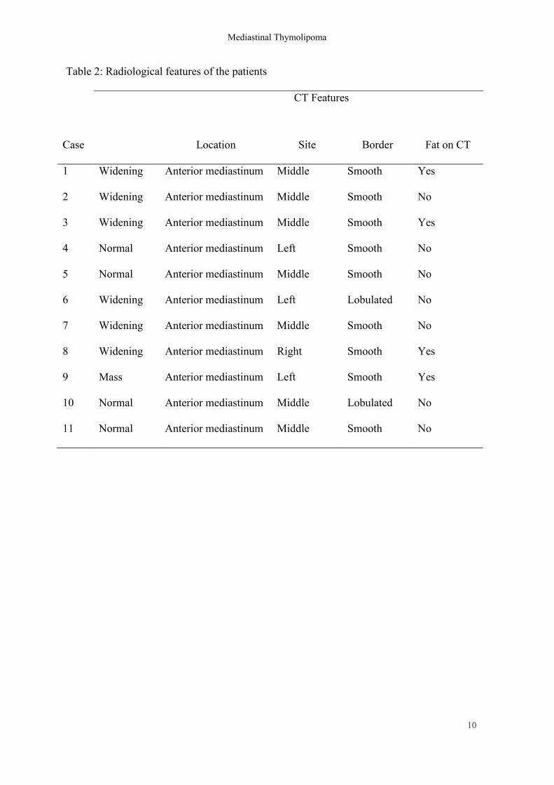
Radiological features are summarized in table 2. Chest X-ray was normal in four
patients (36.4 %). Computed tomography of the thorax revealed a mass located in the anterior
Mediastinal Thymolipoma
4
mediastinum in all patients. It showed fat attenuation within four of 11 masses (36.4 %).
Magnetic resonance imaging scans, obtained in two patients, demonstrated high signal
intensity intermixed with areas of intermediate intensity. FDG PET-CT scans obtained in
three patients. The maximum standardized uptake values (SUVmax) of the lesions on FDG
PET-CT were 2.9, 3.2, and 3.8.
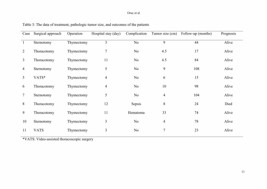
The data of treatment, pathologic tumor size, and outcomes of the patients are given
in table 3. All patients underwent a surgical procedure. The surgical approach was
thoracotomy in five patients, sternotomy in four patients, and video-assisted thoracoscopic
surgery in two patients. The tumor was located in the anterior mediastinum in all cases. Only
the thymus was removed in all patients. On macroscopic examination, all masses were
encapsuled and soft. The tumors varied in size from 4 cm to 33 cm in greatest dimension, with
mean size of 9 cm. Microscopic examination demonstrated a mixture of benign thymic tissue
and mature adipose tissue in all tumors. No histological evidence of malignancy was seen in
any of the tumors (figure 1). The hospital stay ranged from 3 days to 12 days, on average 6.1
days. Hematoma was developed in one patient on the 1st postoperative day following the
thoracotomy and repeated thoracotomy was necessary. Other patient developed sepsis on the
second postoperative day following the thoracotomy. He was treated with early aggressive
antibiotic therapy and mechanical ventilatory support in an intensive care unit. There were no
complications as a result of the resection of the thymolipoma in any other patients. One
patient died 2 years after operation due to cardiovascular disease. None of the remaining 10
patients had evidence of recurrence or residual tumor on follow-up. The patient with severity
score I experienced a nonimprovement in myasthenia gravis symptoms after thymectomy.
The patient with severity score IIa experienced an improvement in myasthenia gravis
symptoms after resection of thymolipoma.
Oruc et al
5
DISCUSSION
Thymolipoma is an uncommon benign tumor of the mediastinum. This tumor accounts for 2
to 9 % of all thymus tumors and 1.1 % of all solid mediastinal tumors (6, 9). Its incidence is
approximately 0.12 cases per 100.000 inhabitants per year (10). Up to 2008, less than 200
cases of thymolipoma had been reported in the world literature (3). The pathogenesis of this
tumor is unclear, but several theories have been proposed. The most widely accepted theory
proposes that diffuse thymic hyperplasia is replaced by adipose tissue, the same process that
occurs in the normal thymus (9, 11).
Thymolipoma can occur at any age, but is more common in young adults. The
majority of thymolipomas are diagnosed in the first four decades of life (7, 8, 12). There is no
sex predilection (7, 9, 12). Many patients are symptom-free at the time of diagnosis and the
tumors are identified incidentally by chest x-ray or computed tomography of the thorax during
a diagnostic workup for other medical problems. Others present with nonspecific symptoms
such as chest pain, cough, dyspnea, respiratory tract infections, hemoptysis, dysphagia,
hoarseness, tachypnea, cyanosis, and weight loss. Most symptoms are related to the local
growth of the tumor and mechanical compression of adjacent mediastinal organs (1, 5, 7-9,
11, 13). Thymolipomas often become large and they can reach a huge size (3, 5, 6, 8, 9).
According to our review of the literature, the largest size of thymolipomas that have been
reported in the literature is 36 cm and their weight ranges from 31 g to 16 kg (5, 8, 14). These
tumors are sometimes associated with parathymic disorders such as myasthenia gravis,
Graves’ disease, hypogammaglobulinemia, aplastic anemia, red cell aplasia, and Hodgkin’s
disease (1, 3-6). The incidence of myasthenia gravis associated with thymolipoma is between
2.8 % and 50 % (5). In the present series, all patients were symptomatic. Two patients had
myasthenia gravis. One of them had also Graves’ disease.
Mediastinal Thymolipoma
6
Radiographic features are variable. Chest X-ray may reveal an enlarged cardiac
silhouette mimicking cardiomegaly. It may show a mediastinal mass or widening. The tumor
may present with normal chest X-ray, as happened in our four cases (6,7,11,12). CT and MRI
may helpful in the diagnosis of thymolipoma. CT reveals a mass in the anterior mediastinum.
It may demonstrate fat tissue within the tumor, as showed in our four patients (3,7,11).
Although the diagnosis of thymolipoma is strongly suggested by CT and MRI, histological
examination requires to make an definitive diagnosis or even to differentiate benign from
malignant tumors (3,5,11). The differential diagnosis of this tumor includes lipoma,
mediastinal lipomatosis, mediastinal cysts, thymoma, thymic hyperplasia, teratoma, aneurysm
of aorta, liposarcoma, lymphoma, and thymic carcinoma (9).
The treatment of choice is surgical resection of the tumor, which can be
accomplished via sternotomy, thoracotomy or thoracoscopic approach (3, 5, 7, 11, 15). The
surgical approach is chosen according to the location and extent of the tumor (3, 5). Complete
surgical excision is generally curative (3, 7, 11). The tumor does not recur after surgical
resection (5,7). Surgical resection of the tumor may be followed by an improvement of
myasthenic symptoms in patients with myasthenia gravis (7). However, myasthenic
symptoms may persist in some patients (5). In the present series, the patients showed no
evidence of recurrence or residual tumor during the follow-up. While one patient experienced
an improvement in myasthenia gravis symptoms after a resection of thymolipoma, myasthenic
symptoms persisted in the other patient.
In conclusion, thymolipoma is a rare tumor of the thymus. It may be associated with
myasthenia gravis. Surgical resection is the treatment of choice in the patients with
thymolipoma. Complete surgical resection is curative in most patients.

Oruc et al
7
REFERENCES
1. Aghajanzadeh M, Alavi A, Pourrasouli Z, Aghajanzadeh G, Massahnia S. Giant
mediastinal thymolipoma in 35-year-old women. J Cardiovasc Thorac Res 2011; 3:
67–70.
2. Hall GFM. A case of thymolipoma with observations on a possible relationship to
intrathoracic lipomata. Br J Surg 1948; 36: 321–4.
3. Mourad OMA, de Andrade FM, Abrahao P, Monnerat A, Judice LF. Asymptomatic
giant mediastinal mass: a rare case of thymolipoma. J Bras Pneumol 2009; 35: 1049–
52.
4. Huang CS, Li WY, Lee PC, Kao KP, Chou TY, Wu MH et al. Analysis of outcomes
following surgical treatment of thymolipomatous myasthenia gravis: comparison with
thymomatous and non-thymomatous myasthenia gravis. Interact Cardiovasc Thorac
Surg 2014; 18: 475–81.
5. Rieker RJ, Schirmacher P, Schnabel PA, Moser K, Hoffmann H, Dienemann H et al.
Thymolipoma. A report of nine cases, with emphasis on its association with
myasthenia gravis. Surg Today 2010; 40: 132–6.
6. Tsukioka T, Inoue K, Iwata T, Mizuguchi S, Morita R, Suehiro S. Thymolipoma
associated with myasthenia gravis. General Thorac Cardiovasc Surg 2007; 55: 26–8.
7. Damadoglu E, Salturk C, Takir HB, Ertugrul M, Yilmaz A, Atasalihi A et al.
Mediastinal thymolipoma: An analysis of 10 cases. Respirology 2007; 12: 924–7.
8. Moran CA, Rosado-de-Christenson M, Suster S. Thymolipoma: clinicopathologic
review of 33 cases. Mod Pathol 1995; 8: 741–4.
9. Ghanem MA, Habambo G, Bahous J, Chouairy C, Khalil BA. Large thymolipoma
causing primary respiratory distress. J Med Liban 2012; 60: 106–9.
Mediastinal Thymolipoma
8
10. Gamondes JP, Balawi A, Greenland T. Seventeen years of surgical treatment of
thymoma: factors influencing survival. Eur J Cardiothorac Surg 1991; 5: 124–31.
11. Roque C, Rodriguez P, Quintero C, Santana N, Hussein M, Freixinet J. Giant
thymolipoma. Arch Bronchoneumol 2005; 41: 402–3.
12. Rosado-de-Christerson ML, Pugatch RD, Moran CA, Galobardes J. Thymolipoma:
analysis of 27 cases. Radiology 1994; 193: 121–6.
13. Hayashi A, Takamori S, Tayama K, Mitsuoka M, Ohtsuka S, Aoyama Y et al.
Thymolipoma: clinical and pathological features –report of three cases and review of
literature. Kurume Med J 1997; 44: 141–6.
14. Kilic D, Giray S, Bolat FA, Tercan F, Bilen A, Hatipoglu A. A rare combination of
thymic tumor: radiologically invisible thymolipoma associated with myasthenia
gravis. Neurol India 2006; 54: 322–3.
15. Tan TW, Kim DS, Wallach MT, Mangray S, Luks FI. Thoracoscopic resection of a
giant thymolipoma in a 4-year-old girl. J Laparoendosc Advanced Surgical Techniques
2008; 18: 903–5.

Oruc et al
9
Table 1: The clinical data of the patients
Case Age
(years)
Gender Smoking
Symptoms M.gravis Osserman
score
1 27 Female Yes Cough No
2 34 Male No Cough, chest pain No
3 43 Male Yes Chest pain No
4 35 Female No Dyspnea, weakness, Graves’ disease Yes IIa
5 30 Female No Dyspnea No
6 71 Male No Chest pain No
7 20 Male No Chest pain No
8 72 Male No Dyspnea No
9 23 Male No Cough, chest pain, dyspnea No
10 29 Male No Chest pain No
11 56 Male Yes Ocular findings Yes I
Mediastinal Thymolipoma
10
Table 2: Radiological features of the patients
CT Features
Case Location Site Border Fat on CT
1 Widening Anterior mediastinum Middle Smooth Yes
2 Widening Anterior mediastinum Middle Smooth No
3 Widening Anterior mediastinum Middle Smooth Yes
4 Normal Anterior mediastinum Left Smooth No
5 Normal Anterior mediastinum Middle Smooth No
6 Widening Anterior mediastinum Left Lobulated No
7 Widening Anterior mediastinum Middle Smooth No
8 Widening Anterior mediastinum Right Smooth Yes
9 Mass Anterior mediastinum Left Smooth Yes
10 Normal Anterior mediastinum Middle Lobulated No
11 Normal Anterior mediastinum Middle Smooth No
Oruc et al
11
Table 3: The data of treatment, pathologic tumor size, and outcomes of the patients
Case Surgical approach Operation Hospital stay (day) Complication Tumor size (cm) Follow-up (months) Prognosis
1 Sternotomy Thymectomy 3 No 9 44 Alive
2 Thoracotomy Thymectomy 7 No 4.5 17 Alive
3 Thoracotomy Thymectomy 11 No 4.5 84 Alive
4 Sternotomy Thymectomy 5 No 9 108 Alive
5 VATS* Thymectomy 4 No 6 15 Alive
6 Thoracotomy Thymectomy 4 No 10 98 Alive
7 Sternotomy Thymectomy 5 No 4 104 Alive
8 Thoracotomy Thymectomy 12 Sepsis 8 24 Died
9 Thoracotomy Thymectomy 11 Hematoma 33 74 Alive
10 Sternotomy Thymectomy 3 No 4 78 Alive
11 VATS Thymectomy 3 No 7 23 Alive
*VATS: Video-assisted thoracoscopic surgery
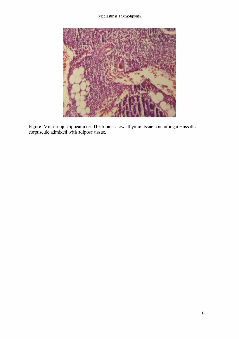
Mediastinal Thymolipoma
12
Figure: Microscopic appearance. The tumor shows thymic tissue containing a Hassall's
corpuscule admixed with adipose tissue.
Related Documents


