eCM Meeting Abstracts 2016, Collection 4; eCM XVII (page 1) www.ecmconferences.org Wnt signaling in bone development and regeneration Christine Hartmann Institute of Experimental Musculoskeletal Medicine, Dept. of Bone and Skeletal Research, Medical Faculty, University of Münster, Germany. INTRODUCTION & DISCUSSION: The Wnt family comprises 19 different members in vertebrates encoding secreted ligands that activate and signal through diverse pathways distinguishable by their intracellular components. The best-studied pathway is mediated by -catenin encoded by the Ctnnb1 gene. Signaling through a LRP5(6)/frizzled receptor complex results in stabilization of cytoplasmic -catenin and its nuclear translocation where i acts in combination with members of the Tcf/Lef family as a transcriptional co-activator [1]. Over the past years multiple roles for Wnt-signaling in bone development have been reported: its involvement in lineage-decision of skeletal precursor cells, differentiation of skeletal cells such as chondrocytes, osteoblasts and osteoclasts, in bone homeostasis and regeneration. Chondrocytic differentiation appears to be the default pathway of skeletal precursor cells. In embryogenesis -catenin is required to suppress the chondrogenic potential of osteoblast- and synovial joint lineage precursors, which are not yet committed [2-4]. Stabilization of -catenin in osteoblast precursors during embryogensis promotes the expansion of precursors but at the same time blocks their maturation [4]. In addition, Wnt/-catenin signaling in chondrocytes regulates the expression of the pro-osteoblastogenic factor Indian hedgehog [5]. During postnatal develop- ment genetic ablation of Ctnnb1 in osteoblastic precursor cells results in a cell fate shift towards adipocytes [6], while inactivation in more differentiated osteoblasts results in increased osteoclastogenesis due to downregulation of the anti-osteoclastic factor osteoprotegerin (opg) and increased Rankl expression [7,8]. In addition, - catenin plays a -autonomous role in osteoclasts [9-11]. Last but not least -catenin activity in hypertrophic chondrocytes locally regulates osteoclastogenesis at the chondro- osseous front primarily via Rankl suppression [12] (Houben A and Hartmann C, unpublished). Collagen 10a1-expressing, hypertrophic chondro- cytes have been identified as a source for trabecular and endosteal osteoblasts [13,14]. Differentation these chondrocyte-derived osteo- blasts requires -catenin activity in hypertrophic chondrocytes (Houben A and Hartmann C, unpublished). The Wnt/-catenin pathway influences the in vitro differentiation potential of mesenchymal stem cells (MSCs) in a differentiation stage-dependent manner: pathway activation in uncommitted MSCs inhibits osteoblastic differentiation [15-18], while in osteoblastic committed MSCs its activation promotes differentiation but interferes with final maturation [19-21]. Bone regeneration during fracture healing recapitulates the key steps of embryonic bone development, but involves in addition an early inflammatory response [22]. Here again the Wnt/- catenin pathway plays varying roles at the different stages of fracture healing [23-27].

Welcome message from author
This document is posted to help you gain knowledge. Please leave a comment to let me know what you think about it! Share it to your friends and learn new things together.
Transcript

eCM Meeting Abstracts 2016, Collection 4; eCM XVII (page 1)
www.ecmconferences.org
Wnt signaling in bone development and regeneration
Christine Hartmann
Institute of Experimental Musculoskeletal Medicine, Dept. of Bone and Skeletal Research, Medical
Faculty, University of Münster, Germany.
INTRODUCTION & DISCUSSION: The Wnt
family comprises 19 different members in
vertebrates encoding secreted ligands that activate
and signal through diverse pathways
distinguishable by their intracellular components.
The best-studied pathway is mediated by -catenin
encoded by the Ctnnb1 gene. Signaling through a
LRP5(6)/frizzled receptor complex results in
stabilization of cytoplasmic -catenin and its
nuclear translocation where i acts in combination
with members of the Tcf/Lef family as a
transcriptional co-activator [1]. Over the past years
multiple roles for Wnt-signaling in bone
development have been reported: its involvement
in lineage-decision of skeletal precursor cells,
differentiation of skeletal cells such as
chondrocytes, osteoblasts and osteoclasts, in bone
homeostasis and regeneration.
Chondrocytic differentiation appears to be the
default pathway of skeletal precursor cells. In
embryogenesis -catenin is required to suppress
the chondrogenic potential of osteoblast- and
synovial joint lineage precursors, which are not yet
committed [2-4]. Stabilization of -catenin in
osteoblast precursors during embryogensis
promotes the expansion of precursors but at the
same time blocks their maturation [4]. In addition,
Wnt/-catenin signaling in chondrocytes regulates
the expression of the pro-osteoblastogenic factor
Indian hedgehog [5]. During postnatal develop-
ment genetic ablation of Ctnnb1 in osteoblastic
precursor cells results in a cell fate shift towards
adipocytes [6], while inactivation in more
differentiated osteoblasts results in increased
osteoclastogenesis due to downregulation of the
anti-osteoclastic factor osteoprotegerin (opg) and
increased Rankl expression [7,8]. In addition, -
catenin plays a -autonomous role in
osteoclasts [9-11]. Last but not least -catenin
activity in hypertrophic chondrocytes locally
regulates osteoclastogenesis at the chondro-
osseous front primarily via Rankl suppression [12]
(Houben A and Hartmann C, unpublished).
Collagen 10a1-expressing, hypertrophic chondro-
cytes have been identified as a source for
trabecular and endosteal osteoblasts [13,14].
Differentation these chondrocyte-derived osteo-
blasts requires -catenin activity in hypertrophic
chondrocytes (Houben A and Hartmann C,
unpublished).
The Wnt/-catenin pathway influences the in vitro
differentiation potential of mesenchymal stem cells
(MSCs) in a differentiation stage-dependent
manner: pathway activation in uncommitted MSCs
inhibits osteoblastic differentiation [15-18], while
in osteoblastic committed MSCs its activation
promotes differentiation but interferes with final
maturation [19-21].
Bone regeneration during fracture healing
recapitulates the key steps of embryonic bone
development, but involves in addition an early
inflammatory response [22]. Here again the Wnt/-
catenin pathway plays varying roles at the different
stages of fracture healing [23-27].

eCM Meeting Abstracts 2016, Collection 4; eCM XVII (page 2)
www.ecmconferences.org
Evolution of the mineralized animal skeletons: Formation of bone
hydroxyapatite via amorphous Ca-carbonate and Ca-phosphate
WEG Müller and XH Wang
ERC Advanced Investigator Grant Research Group at the Institute for Physiological Chemistry,
University Medical Center of the Johannes Gutenberg University Mainz, Duesbergweg 6, D-55128
Mainz, GERMANY ([email protected])
INTRODUCTION: By learning from nature, our
group introduced novel biomaterials which have
the potential to be suitable for bone tissue
engineering. Building on the established facts that
all metazoan organisms evolved from a common
ancestor, the sponges (phylum: Porifera), as well
as the necessity that all organisms larger than 2 cm
need to be stabilized by a skeleton we investigated
the strategies of mineralization, used by basal
metazoans, for the fabrication of bone implants in
human. The evolutionary steps: The
phylogenetically oldest sponge taxa are the
siliceous sponges, followed by the calcareous
sponges; later, the corals, echinoderms, also having
calcareous skeletons, evolved, and finally, the
vertebrates appeared with their calcium
phosphate/HA skeletons.
Fig. 1: Evolution of the skeletal mineral from the
siliceous sponges, via the calcareous sponges to
the Ca-phosphate containing skeletal animals and
final to the hydroxyapatite-formed vertebrates.
THE EVOLUTIONARY STEPS TO
VERTEBRATE BONE: Stage 1: Siliceous
scaffold. Biosilica, a biocompatible, natural
inorganic polymer that is formed in siliceous
sponges to build up their inorganic skeleton, has
been shown to be a morphogenetically active
mineral and to induce mineralization in vitro and in
vivo. Stage 2: Amorphous Ca-carbonate (ACC)
scaffold. In human bone, amorphous calcium
carbonate (ACC) is enzymatically formed as a
precursor of the crystalline carbonated
apatite/hydroxyapatite (HA). We describe that the
metastable ACC phase can be stabilized by
inorganic polyphosphate (polyP). Both in vitro and
in vivo data revealed that ACC functions as a
morphogenetically-active mineral (bio-seed).
Stage 3: Amorphous Ca-phosphate (ACP)
scaffold. PolyP allowed the synthesis of
amorphous Ca-polyP hybrid particles with a size of
50 nm. Those Ca-polyP particles cause a strong
upregulation of the expression of the genes,
involved in bone formation and provide the ortho-
phosphate substrate for bone mineralization.
Fig. 2: Schematic presentation of the
endochondral ossification and the proposed phases
of bone mineral (hydroxyapatite/HA) deposition.
After (Phase I) enzymatic formation of ACC
(amorphous Ca-carbonate) and subsequent
tocarbonate-phosphate exchange ACP
(amorphous Ca-phosphate) (Phase II) the ACP is
transformed from the amorphous (Phase III/a) to
the crystalline phase, the bone HA (Phase III/b).

eCM Meeting Abstracts 2016, Collection 4; eCM XVII (page 3)
www.ecmconferences.org
The biology of heterotopic endochondral ossification and approaches to therapy
M Pacifici
The Children’s Hospital of Philadelphia, Translational Research Program in Pediatric
Orthopaedics, Philadelphia, Pennsylvania, USA
INTRODUCTION: Heterotopic ossification (HO)
consists of formation and accumulation of
endochondral bone at extraskeletal sites, causing
major health problems and even premature death1.
Fybrodysplasia Ossificans Progressiva (FOP) is a
congenital and severe condition involving
extensive and pervasive HO. FOP is caused by
activating mutations in ACVR1, and HO is usually
preceded –and likely promoted- by local flare-ups
and inflammation. Trauma, invasive surgery, deep
burns or protracted immobilization can induce non-
congenital forms of HO. Anti-inflammatory drugs
are often used as preventive HO treatments, but are
not very effective2. Surgery is often used in non-
congenital HO, but it can be dangerous and may
actually trigger another HO cycle. Thus, there is
urgent need for new and effective therapies. Recent
studies from our research groups have identified
synthetic retinoid agonists as novel, effective and
seemingly safe treatments for both forms of HO.
METHODS: FOP was modelled in transgenic
mice expressing ACVR1 R206H or Q207D
mutants. Trauma models consisted of subcutaneous
or intramuscular implantation of a scaffold
containing the pro-chondrogenic protein rhBMP2.
Drugs were given systemically by gavage, and
extent of HO was assessed by CT, histochemistry
and histomorphometry.
RESULTS: By being an endochondral process,
congenital or acquired HO initiates with
recruitment of progenitor cells to the inflamed or
injured site. The cells undergo chondrogenesis and
lay down cartilage tissue that undergoes maturation
and hypertrophy and is eventually replaced by
endochondral bone. Thus, we reasoned that
retinoid agonists could represent effective anti-HO
agents because they have long been known to be
anti-chondrogenic3, thus blocking the initial phase
of the HO process. Mice in which HO had been
induced by injury or transgene expression were
given synthetic retinoid agonists selective for the
nuclear retinoic acid receptor alpha (RAR) or
RAR by daily gavage. Control companions were
given vehicle. By 2 to 4 weeks post-HO induction,
control mice had developed extensive HO at the
affected sites. However, mice treated with RAR
agonists displayed much reduced HO levels, but
RAR agonists were moderately effective4. In
congenital models, HO was often extensive and
hampered skeletal growth and limb mobility.
These defects also were greatly ameliorated by
drug treatment 5.
Because anti-inflammatory drugs are often used as
prophylactic agents, we asked whether they would
help or hinder the anti-HO action of retinoids.
Thus, we tested a combination therapy in the
subdermal mouse model of HO and found that
prednisone enhanced the anti-HO action of retinoid
agonists (Fig. 1), though it had some side effects.
Fig. 1: Representative CT images of subdermal
HO in control mice or those treated with retinoid
agonist alone or in combination with prednisone.
Doses are in mg/kg/day over a 12 day treatment.
DISCUSSION & CONCLUSIONS: The data
clearly show that retinoid agonists can strongly
inhibit congenital and acquired forms of HO. The
effectiveness of the drugs likely reflects the fact
that they block chondrogenesis and canonical BMP
Smad1/5/8 signaling, and may even dampen
recruitment of inflammatory cells at the HO site.
Their potency is moderately enhanced by co-
treatment with anti-inflammatory drugs, thus
expanding their therapeutic range.
ACKNOWLEDGEMENTS: Data presented
here are from original studies and reports
with colleagues at CHOP, the University
of Pennsylvania and Regeneron. Financial
support was received from the NIH and the US
Department of Defense.
! ! ! Con RARgago 4.0 Pred 10 + RARgago 4.0

eCM Meeting Abstracts 2016, Collection 4; eCM XVII (page 4)
www.ecmconferences.org
Endochondral ossification in regenerative medicine
Eric Farrell, Department of Oral and Maxillofacial Surgery, Orthodontics and Special Dental Care, Erasmus MC, University Medical Centre, Rotterdam, The
Netherlands
INTRODUCTION: Repair of critical sized bone
defects that will not heal spontaneously is a costly
endeavour for patient and society alike. Treatment
of large bone defects still requires the surgical
harvesting of bone from another anatomical
location of the patient causing increased pain,
multiple surgeries, longer hospital stays and as a
result high associated costs. Clearly a less invasive
approach would be desirable to treat such injuries.
While the use of BMPs in certain circumstances is
successful, complications resulting from off label
use and generally high doses have necessitated the
search for alternatives. Within the field of
regenerative medicine there are many such options
being researched, including various combinations
of cells, materials and bioactives to induce defect
repair or even de novo bone formation. We and
others have demonstrated the ability of different
cell types, in our case the mesenchymal stem cell,
to initiate the process of endochondral ossification
in vivo following various in vitro cell priming
regimes. This is a very promising approach, since a
relatively simple in vitro priming initiates an
extremely complex series of cell processes in vivo,
ultimately resulting in the formation of marrow
containing bone both ectopically and
orthotopically. However there are still several
hurdles to overcome in order to bring such an
approach to the clinic; scale up, reproducibility and
reduced cost to name but a few. At present there is
still much we do not understand about how the
process of endochondral ossification occurs,
particularly with regard to this regenerative
medicine based approach using adult marrow
stromal cells. I will present the approaches we are
taking to better understand how such bone
formation occurs and how we might scale-up this
approach to generate larger quantities of bone in
shorter amounts of time thereby reducing cost. Our
research focuses on understanding the role of the
host/recipient in the formation of new bone in
order to properly engage the various body systems
(vascular, immune etc) in the generation of new
bone tissue. Advancing our knowledge of how new
bone is formed will allow us to develop new
therapies optimised to engage the patient’s own
biology to accelerate and enhance repair. This
should include considering the role of the immune
system and disease states in MSC mediated
endochondral ossification as well as specific
extracellular matrix components and secreted
factors critical for this process.al.
eCM Meeting Abstracts 2016, Collection 4; eCM XVII (page 5)
www.ecmconferences.org
Mesenchymal stem cell heterogeneity: Diversity in the endogenous synovial stem
cell compartment Kavitha Sivasubramaniyan
1, Wendy J. Koevoet
2, Eric J. Farrell
3, Maria Sande
1, Jan A.N. Verhaar
1,
Gerjo J.V.M. Osch1, 2
1Department of Orthopaedics,
2Department of Otorhinolaryngology,
3Department of Oral and
Maxillofacial Surgery, Special Dental Care and Orthodontics, Erasmus MC, Rotterdam, The
Netherlands
INTRODUCTION: Mesenchymal stem/stromal
cells (MSCs) render promise as cell-based
therapies for articular cartilage repair. Superiority
of synovium as a potential source of MSCs for
cartilage repair has been demonstrated, but the
cellular heterogeneity associated with endogenous
synovial MSCs is not yet clearly understood. In
our study, we define distinct endogenous human
synovial MSC subsets that differ in their
immunophenotype, function and anatomical
localization.
METHODS: Freshly isolated cells from synovium
of 9 patients undergoing total knee replacement
were stained with a panel of markers and analyzed
on a FACS canto II flow cytometer or sorted on a
FACS Jazz cell sorter. The sorted cells were
cultured, phenotypically characterized and
subjected to chondrogenic differentiation. The
anatomical localization of the different MSC
subsets in the synovium was verified by
immunohistochemistry.
RESULTS: Flow cytometric analysis and soring
demonstrated that a combination of CD45, CD31,
CD73 and CD90 can isolate two distinct MSC
subsets in the primary synovium. These MSC
subsets did not express CD45 or CD31 but
expressed CD73 and a sub-population of these
CD73+ cells expressed CD90. CD45-CD31-
CD73+CD90- MSCs were significantly more
chondrogenic than CD45-CD31-CD73+CD90+
MSCs. Interestingly, CD73+CD90- and
CD73+CD90+ MSCs had distinct anatomical
localization; CD73+CD90- cells were found in the
intimal layer lining the joint cavity whereas
CD73+CD90+ cells were located in the sub-
intimal layer, in the perivascular region. In
addition, primary bone marrow MSC specific
markers including CD271 and SUSD2 were
expressed only in the sub-intimal MSCs and not in
the intimal MSCs. Preliminary studies suggest that
the reduced chondrogenic ability of CD73+CD90+
cells could be reversed by the addition of BMP2,
showing discrete chondrogenic factor requirement
by distinct MSC subsets. This is linked to the
differential receptor expression in these MSC
subsets.
Fig. 1: A) Synovial cells were stained with CD45,
CD31, CD73 and CD90, gated on propidium
iodide negative live cells, followed by gating on
CD45-CD31- subset, then analyzed for expression
of CD73 and CD90. Sort windows were set as
shown in the CD73 vs CD90 plot and cells were
sorted with BD FACS Jazz. B &C) After FACS
sorting, cells were cultured and subjected to
chondrogenic differentiation. (Thionin staining).
D) Both CD73 and CD90 are expressed on
perivascular MSCs (marked by arrowhead). MSCs
in the lining intimal layer (marked by arrow)
expressed CD73 and are negative for CD90.
DISCUSSION & CONCLUSIONS: In summary,
we introduce markers which can isolate distinct
MSC subpopulations in synovium. CD73+CD90-
MSCs in the intimal layer are adjacent to the
cartilage in anatomical localization while
CD73+CD90+ MSCs are relatively away from the
cartilage. However, further studies are needed to
utilize these cells for therapeutic purposes, as little
information exists about their participation in
cartilage repair in vivo.
ACKNOWLEDGEMENTS: SmartStep, a
collaborative grant of the Medical Research
Council UK (MRC-MR-L022893) and the Dutch
Arthritis Foundation (CO-14-1-002)

eCM Meeting Abstracts 2016, Collection 4; eCM XVII (page 6)
www.ecmconferences.org
Fate of dental epithelial stem cells in vivo injected in the mouse incisor
G Orsini1, P Pagella
2, L Jimenez-Rojo
2, M Procaccini
1, A Putignano
1, TA Mitsiadis
2
1 Department of Clinical Sciences and Stomatology, Polytechnic University of Marche, Ancona,
Italy. 2 Orofacial Development and Regeneration, Institute of Oral Biology, University of Zurich,
Zurich, CH
INTRODUCTION: The continuously erupting
rodent incisor represents a unique organ system for
studying the cell biology of odontogenesis. The
posterior area of the incisor is characterized by the
presence of cervical loops (CLs) in which the
labial side contains dental epithelial stem cells
(DESCs), which are able to form all the dental
epithelial cell populations. Recently, we have
developed a useful method for the in vivo
administration of DESCs in the mouse CL area [l].
The aim of the present work is to test whether this
newly developed method can be a suitable model
to monitoring stem cells behaviour in vivo, by
following their fate at different time points after
administration.
METHODS: DESCs encoding Green Fluorescent
Protein (GFP) were administered in the incisor
CLs area of immunocompromised RAG1-/-mice at
8–12 weeks of age, using the “bone window
technique” [l]. The mice were sacrificed after 7,
12, 30 and 45 days and hemimandibles dissected,
decalcified and embedded in paraffin. The
specimens were further processed for
immunohistochemistry and immunofluorescence
analyses and observations were performed under
light microscopy and confocal electron
microscopy.
RESULTS: We have applied an experimental
model to locally administer DESCs encoding GFP.
The system consists of drilling a window in the
alveolar bone overlying the apex of the mouse
incisor, allowing local injection with relatively
large concentrations of DESCs into the apical
cervical loop. Immunofluorescence and
immunohistochemical results demonstrated that the
bone window technique allows the administration
of DESCs that can be traced not only in situ (bone
hole), but also at a certain distance from the site of
administration, within the dental epithelial
lineages. Indeed, GFP positivity was observed in
the different dental epithelial layers such as
ameloblasts and stratum intermedium, at all the
different time points.
.
Fig. 1: drilled hole overlying the mouse incisor
cervical loop (CL).
DISCUSSION & CONCLUSIONS:
This newly described approach has been
demonstrated to be useful to trace the in vivo fate
of DESCs after their administration. DESCs have
shown to have an integration capacity within the
epithelial dental lineages, giving rise to the
different epithelial layers of the incisor. These
findings contribute to the knowledge that epithelial
stem cells display remarkable differentiation
potential and can acquire epithelial lineage in vivo.
Current and future studies will be driven to further
explore whether dental epithelial stem cells of the
cervical loop are limited in giving rise to distinct
dental epithelial cell populations, or whether they
can multilineage differentiation potential. These
facts could contribute to establish innovative
treatment protocols after traumatic or pathological
injuries.
ACKNOWLEDGEMENTS: Institutional
funds from the University of Zurich, and funds
from the Polytechnic University of Marche.

eCM Meeting Abstracts 2016, Collection 4; eCM XVII (page 7)
www.ecmconferences.org
gene expression profiling shows the desregulation of multiple hormone signaling in
osteoporosis that could be therapeutically reversed by the treatment of strontium gluconate
J Li1,2
, YH Liang1, WJ Peng
1, TH Chen
1, ML Zhou
1, and GQ Zhou
1*
1 The Centre for Anti-ageing and Regenerative Medicine, Shenzhen University, Shenzhen, China, 2 Key Laboratory of Optoelectronic
Devices and Systems of Ministry of Education and Guangdong Province, Shenzhen University, Shenzhen, China
Introduction: Bone loss can be the result of various factors
and causes including the imbalance of multiple hormones
such as estrogen, glucocorticoid, parathyroid hormones,
Vitamine D and leptin[1]
. However, the interactive roles of
these hormones have been poorly understood on bone
tissues and cells. Stontium is well known to promote bone
formation by stimulating osteogenesis of osteoprogenitors
through influencing multiple cytoking and growth factor-
mediated signaling pathways. Its role in regulating hormone
signaling, however, has not been documented. To this end,
this study reports our recent findings in gene expression
profiling analysis of osteoporotic bone tissues induced by
the overdose of glucocorticoid with or without oral
administration of strontium gluconate (GluSr).
Subjects and Methods: A total of 16 sprague-dawley rats,
aged between 7-8 weeks, were induced for GIOP by
subcutaneous injection of glucocorticoid, 5.0mg/kg, 3
times/week. At the same time, half of these rats were
treated with GluSr for oral administration, 400mg/kg.d. An
additional 8 rats were left intact as control group. After
treating 12 weeks, the microarchitecture of the trabecular
bone from distal femur and proximal tibiae was analyzed
with micro-CT scanner. Compact bone of long bone was
cut into small pieces for RNA extraction. Total RNA
sample was quantified and Agilent Array platform was
employed for microarray analysis.
Results: Images of micro-CT show that trabecular bone
microstructure in the distal femur and proximal tibiae of
GIOP rats become less dense and more porous than GIOP
rats treated with GluSr and control rats (Fig.1A-C). The
BMD value of distal femur of GC, GC+GluSr, and CTRL
Fig. 1. micro-CT features and the bone mineral density of the tibia and femur of osteoporotic rats with or without GluSr treatment in
comparison with healthy controls. Ultrastructure of bone of GC (A),
GC+GluSr (B), and CTRL (C) were observed in coronal (A1-C1), transverse of distal femur (A2-C2) and proximal tibiae (A3-C3). (D) BMD
of proximal tibiae. (E) BMD of distal femur.
groups were 0.09±0.01, 0.44±0.04, 0.21±0.03 g/cm2, and
the BMD value of proximal tibiae were 0.33±0.02, 0.51±
0.04, 0.43 ± 0.04 g/cm2, respectively (Fig.1D).
Interestingly, after the GIOP rats treated with GluSr, the
BMD value striking increased and even significantly higher
than CTRL groups. The distinguishable gene expression
among samples of hormones-related signaling pathways
were listed as figure. 2A-B. A total of 30 genes were
changed with up or down regulation, which are highly
relevant to glucocorticoid receptor (GR), estrogen receptor
(ESR), parathyroid (PTH), leptin, Vitamin D receptor
(VDR), Ca2+ signaling pathways.
Fig. 2. A portion of differentially expressed genes in glucocoid-induced
rats (red) , treated with GluSr (green) and controls (blue), grouped in GR,
ESR, PTH, Leptin, VDR, and Ca2+ pathways.
Dissussion and Conclusions: Hormones play a critical role
in regulating mineral metabolism and bone mass. Our
study provides preliminary evidences that various hormone
signaling are interactively influenced in bone tissue and
cells, particularly osteoblasts. Furthermore, this is the first
study to show that hormone signaling pathways are
regulated or even reversed by the anti-osteoporosis Sr-
containing reagent, probably implicating its potential value
of Prophylactic intervention against chronic osteoporotic
conditions. Further work on revealing the function of
particular genes newly identified is undergoing.
Acknowledgements: This work was supported by the
Natural Science Foundation of China (NSFC#91029738).

eCM Meeting Abstracts 2016, Collection 4; eCM XVII (page 8)
www.ecmconferences.org
Mesenchymal stem cells: where is the stem..?
B. Péault
Centre for Regenerative Medicine and Cardiovascular Science Centre, University of Edinburgh,
UK and Orthopaedic Hospital Research Center and Broad Stem Cell Research Center, University
of California, Los Angeles, USA
Mesenchymal stem cells – MSCs – have been very
popular among cell therapists and tissue engineers,
as shown by the use of MSCs in over 500 clinical
trials. This success is justified by the diverse
positive contributions exerted by MSCs toward
organ repair as tissue progenitors, pro-angiogenic
and immunosuppressive cells and supportive niche
cells for lineage-committed stem cells.
Mesenchymal stem cells are also, importantly,
remarkably easy to derive and expand since MSC
extraction is a mere primary culture of unselected
dissociated cells. Moreover, MSCs can be grown
from virtually any vascularized organ, leaving a
choice of convenient, abundant and dispensable
sources of these cells such as adult adipose tissue
and fetal appendages at birth.
On the negative side, indirect selection by
adherence and proliferation in culture has long
obscured the biologic characteristics of innate
mesenchymal stem cells. MSCs being by essence
long-term cultured cells, the native embryonic
origin, identity, lineage affiliation, tissue
distribution, frequency and – importantly – actual
role of these cells in normal tissue homeostasis and
repair remained unknown decades after the initial
discovery of MSCs. In the past ten years, the very
identity of native mesenchymal stem cells has been
progressively uncovered, revealing a perivascular
origin for these elusive regenerative cells. We will
review and discuss experimental evidence
demonstrating that MSCs isolated from distinct
organs share blood vessel associated ancestors.
The prospective identification of innate MSCs now
opens the possibility of using highly purified –
and, in some instances, uncultured – precisely
characterized perivascular cells for cell therapies,
in place of their heterogeneous, culture selected
conventional progeny. We will also review the
medical use of customary, in vitro derived
mesenchymal stem cells, and put in perspective
recent attempts and future plans to achieve tissue
regeneration using their perivascular native
counterparts.
eCM Meeting Abstracts 2016, Collection 4; eCM XVII (page 9)
www.ecmconferences.org
Chondral cell differentiation
Brian Johnstone, PhD
Department of Orthopaedics and Rehabilitation, Oregon Health & Science University, OR, USA
The variation in chondrogenic capacity among adult human-derived stem/progenitor cell populations is an important consideration in tissue engineering. Beyond the obvious outcome that poorly chondrogenic cells make little extracellular matrix, our latest work suggests variation in intrinsic chondrogenicity will influence experimental results. For example, the baseline chondrogenic capacity influences a stem cell’s response to a physiologic low oxygen environment (physioxia) in 3D culture. Biologic replicate of human bone marrow-derived stem cells (MSCs) and articular cartilage-derived progenitor cells (ACPs) were categorized as high- or low-GAG based on a threshold defined by their total GAG production relative to that of healthy human articular chondrocytes in the same 3D pellet conditions at 20% oxygen (hyperoxia). While physioxic culture increased GAG production across all MSC preps and the majority of ACP clones, physioxia was of greater benefit to biologic replicates that exhibited very low GAG production at baseline in hyperoxia, driving a greater fold change than for clones that started with high GAG production and chondrogenic capacity in hyperoxia. However, even with this significantly higher fold-induction, the pellets of low-GAG cell preparations of both cell types were still poorly chondrogenic in comparison with matched high-GAG pellets. Furthermore, MSCs and ACPs of high chondrogenicity upregulate protein expression of the articular chondrocyte phenotype and downregulate the hypertrophic phenotype in physioxia; however, only ACPs consistently attenuate hypertrophic markers at the tissue level in the physioxia while MSCs retain high type X collagen protein regardless of oxygen tension. Thus, ACPs may overcome the historical challenges of MSC hypertrophy in tissue engineering applications.
We then developed conditions to create larger, scaffold-free cartilage 3D implants from various cell types, using pre-selected cell preparations of high chondrogenicity. Multiple biological replicates of bone marrow-derived MSCs, articular chondrocytes (ACs) and ACPs derived from
healthy human adult articular cartilage, were guided toward self-organization through cell condensation. Discoid tissue was produced from all three cell sources. Regardless of oxygen tension and consistent with pellet culture, MSCs produced neocartilage tissue of a hypertrophic phenotype. In comparison with culture in hyperoxia, AC neocartilage cultured at physioxia exhibited a significant increase in chondrogenic gene expression, proteoglycan production, and mechanical properties with a concomitant decrease in collagen content. ACP-derived neocartilage produced tissue with significantly enhanced mechanical properties and collagen content relative to HAC-derived neocartilage. Interestingly, they had much lower differential responses between physioxia and hyperoxia. Regardless of oxygen tension, neocartilage from ACPs exhibited anisotropic organization of native cartilage with respect to a pericellular matrix when compared with AC-derived neocartilage; however, only ACs produced abundant surface-localized lubricin. To date, few methods utilizing adult human cells in scaffold-free approaches to tissue engineering have been reported. Guiding human-derived cells toward condensation and subsequent culture in physioxia promoted the articular cartilage tissue phenotype for ACs and ACPs, but less so for MSCs. The advantage of ACPs over ACs is that they can be cloned and are highly expandable while retaining chondrogenicity. Ultimately, the ability to generate tissues of the articular cartilage phenotype utilizing a scaffold-free approach from a single cell origin may provide the functional properties and therapeutic level of neocartilage destined for autologous repair.

eCM Meeting Abstracts 2016, Collection 4; eCM XVII (page 10)
www.ecmconferences.org
Autogenic mesenchymal stromal cells (MSC) are superior to allogenic MSC in
regeneration of large bone defects
AE Rapp1, R Bindl
1, M Rojweski
2,3, J Kemmler
1, H Schrezenmeier
2,3, I Müller
4, A Ignatius
1
1Institute of Orthopaedic Research and Biomechanics, University of Ulm, Ulm, Germany; 2Institute of Clinical Transfusion Medicine
and Immunogenetics, German Red Cross Blood Transfusion Service, Baden Wuerttemberg-Hessen, Ulm, Germany; 3Institute of
Transfusion Medicine, University of Ulm, Germany; 4Clinic for Paediatric Haematology and Oncology, Bone Marrow
Transplantation Unit, University Medical Centre Hamburg-Eppendorf, Germany
INTRODUCTION: Mesenchymal stem cells
(MSC) are promising tools for the regeneration of
large bone defect. While the benefit of autologous
MSC on for bone regeneration is widely
acknowledged, the efficacy of allogeneic MSC has
been poorly investigated so far and available
studies report inhomogeneous results1-3
. As the use
of allogenic MSC would overcome the limited
availability of autogenic cells, further
investigations on the use of allogeneic SMC are
necessary. This study compared the potential of
allogeneic and autologous human MSC (hMSC) to
regenerate large bone defects in an animal model
that mimics the human immune system.
METHODS: In humanized NOD/scid-IL2Ryc-/-
mice, which had established a human immune
system after engraftment with human
hematopoietic stem cells, a 1 mm defect, stabilized
by an external fixator, was created in the right
femur. The defect was left untreated or filled with
either allogeneic or autogenic hMSC in a collagen
type-1 matrix. The animals were killed after 3, 10
or 35 days. The healing outcome was analysed by
µCT, histmorphometry and immunohistochemistry
for human β2-microglobulin, human CD8,
PECAM (CD31), Runx2 and Osteocalcin.
RESULTS: Staining for human β2-microglobulin
confirmed the presence of transplanted human cells
in the defect region. Newly formed bone in the
defect region in both, allogenic and autogenic
treated mice did not stain positive for the human
marker. µCT analysis after 35 days showed a
significantly higher bone volume in the defect
region of mice that received autologous MSC
compared to allogeneic MSC (+132%) or untreated
defects (+205%). Histomorphometry confirmed
this finding. Consequently, staining for osteogenic
markers on day 10 (Runx2) and 35 (osteocalcin)
was more intense in mice treated with autologous
MSC. To detect adverse immune reactions, we
stained for CD8+ T-cells. 3 days after surgery,
CD8+ cells were detected near the implant in mice
that received allogeneic MSC, while positive cells
were absent in mice with autologous treatment.
The same observation was made on day 10.
Staining for PECAM revealed newly formed
vessels in the surrounding of the collagen gels in
both treatment groups with no obvious differences.
On day 35 however, more stained structures were
found in mice treated with autologous MSC
compared to allogeneic MSC, indicating increased
angiogenesis. Furthermore the distribution of the
vessel-like structures was different, in autogenic
treated mice, the vessels were distribute throughout
the defect region, while they were at the margins of
the defect region in allogeneic treated mice.
DISCUSSION & CONCLUSIONS: Our results
indicate a higher efficiency of autogenic hMSC for
bone regeneration compared to allogeneic hMSC,
as treatment with autogenic hMSC led to a
significantly higher bone formation compared to
empty defects or defects treated with allogeneic
hMSC. We found no signs of a strong adverse
immune reaction in animals that received
allogeneic hMSC; albeit CD8+ cells were detected.
There are hints that T cells and interferon-gamma
might be associated with inhibition of bone
formation in allogeneic settings4; however this has
to be investigated further. It is still unclear, how
the implanted cells contribute to bone regeneration.
We found signs for enhanced angiogenesis and
osteogenesis after autologous treatment. Together
with the absence of bone stained positive for
human β2-microglobulin, this indicates an indirect
action of the implanted cells via trophic factors
rather than a direct contribution. In conclusion, our
results demonstrate a superior efficacy of
autogenic hMSC treatment compared to allogeneic
hMSC in supporting bone healing. 1
ACKNOWLEDGEMENTS: This study
was funded by the 7th Framework
Programme “Reborne” of the European
Commission.

eCM Meeting Abstracts 2016, Collection 4; eCM XVII (page 11)
www.ecmconferences.org
The osteogenic differentiation of mesenchymal stromal cells is enhanced by the
BMP2 variant L51P in the presence of intervertebral disc-derived cells
A Tekari1, R May
1, DA Frauchiger
1, HJ Sebald
2, LM Benneker
3, B Gantenbein
1
1 Tissue and Organ Mechanobiology, Institute for Surgical Technology & Biomechanics, University
of Bern, CH. 2 The Spine Center, Thun, CH.
3 Department of Orthopaedic Surgery &
Traumatology, University of Bern, Inselspital, CH.
INTRODUCTION: Discectomy and spinal fusion
represents the gold standard treatment for spinal
disorder to relieve pain. Fusion can be hindered,
however, for yet unknown reasons that lead to non-
union with pseudo-arthrosis. We previously
showed that intervertebral disc (IVD)-derived cells
hinder the ossification process of human bone
marrow-derived stromal cells (hMSC) [1]. Within
this study, we hypothesized that BMP-antagonists
secreted by IVD cells are the responsible factors
for such inhibition and that this can be reversed by
addition of L51P. L51P is an engineered BMP2
variant [2] that has been recently demonstrated to
be a generic antagonist of a variety of BMP-
inhibitors that controls osteoinduction of bone
[3,4].
METHODS: The experimental work was ethically
approved and written consent of patients was
obtained. hMSCs, primary nucleus pulposus (NPC)
and annulus fibrosus cells (AFC) were obtained
from patients undergoing spinal surgery, isolated
and expanded in monolayer cultures up to passage
3. IVD cells were seeded in 1.2% alginate beads
(4Mio/mL) and separated by culture inserts from
hMSCs in a co-culture (CC) set-up. The allogenic
CCs were paired in 11 repeated experiments.
MSCs were kept in 1: osteogenic medium (positive
control, ±alginate beads), 2: osteogenic
medium+NPC (±100ng/mL L51P), 3: osteogenic
medium+AFC (±100ng/mL L51P) and 4: basal
medium (negative control) for 21 days. Relative
gene expression of bone-related markers was
quantified with qPCR, and histological staining for
calcium deposition and Alkaline Phosphatase
(ALP) assay were performed. The endogenous
expression of three common BMP-antagonists in
IVD cells (passage 1) was evaluated by qPCR,
immunohistochemistry and flow cytometry.
RESULTS: Osteogenesis of hMSCs was hindered
as shown by reduced alizarin red staining in the
presence of NPC and AFC. However, L51P added
to CCs of hMSCs with either NPC or AFC induced
mineralization by blocking the activity of the IVD
cell’s secreted BMP-antagonists (Fig. 1).
Fig. 1: Osteogenic differentiation of hMSC is
inhibited in CC with NPC and AFC as shown
macroscopically (top row) and microscopically
(bottom row) at 10x magnification. L51P blocks
the inhibitory effect of IVD cells in CC of NPC or
AFC and restores the osteogenic differentiation.
It was noted that L51P caused a general reduction
in ALP activity in all experimental groups. ALP
activity was significantly up-regulated in positive
control, and in CCAFC+L51P relative to negative
control, suggesting osteogenesis in these groups.
Gene expression analysis confirmed these
observations. IVD cells expressed BMP-
antagonists, namely noggin, gremlin and chordin
as measured by transcript and protein levels.
DISCUSSION & CONCLUSIONS: The IVD
cells secrete BMP-antagonists, which are
responsible for bone non-union. The concept of
antagonizing endogenous BMP inhibitors with
L51P may represent a promising clinical option to
augment and accelerate bone regeneration during
4
ACKNOWLEDGEMENTS: This study
was supported by funds from the Lindenhof
Foundation “Funds Research &
Teaching” (project #15-05-F), by Hansjörg Wyss
Medical and the Swiss National Science
Foundation (project #310030_153411). Eva
Roth assisted in the biochemical assays.

eCM Meeting Abstracts 2016, Collection 4; eCM XVII (page 12)
www.ecmconferences.org
Growth environments and cues for engineering bone tissue in vitro
El Haj AJ
Institute of Science and Technology in Medicine, Guy Hilton Research Centre, Keele University UK
INTRODUCTION: Engineering bone tissue for
use in Orthopaedics poses multiple challenges.
Providing the appropriate growth environment
which will allow complex tissues such as bone to
grow is one of these challenges. There are multiple
design factors which must be considered in order
to generate in vitro a functional tissue for
replacement surgery in the clinic. Complex
bioreactors have been designed which allow for
different stress regimes such as compressive, shear
and rotational forces to be applied to 3D
engineered constructs but ultimately we need
simplified prototypes which can be standardized
for scale up. Combined with biological directional
cues, we aim to provide the right conditions to
grow bone tissue ex vivo as models or for
implantation for orthopaedic repair.
METHODS: Human bone marrow derived MSCs
are grown on multiple material substrates and
cultured either in vitro within a well plate or placed
within an ex vivo chick femur epiphyseal defect. A
hydrostatic stimulation regime has been developed
with a pressure range of 0-280 KPa at a frequency
of 1 Hz for 1 hour daily. Examples of other
bioreactors such as the magnetic force bioreactor
have been tested for comparison on different
configurations. Osteogenic differentiation in vitro
is identified by increased bone marker expression
and amplified mineralisation. Biological cues such
as Wnt proteins and growth factors have been
patterned to enable spatial differentiation cues
within the bioreactor environment.
Fig. 1: Monitoring hydrogels during mechanical stimulation
in the hydrostatic force bioreactor. Relative displacement
maps were generated by elastography algorithms. Colour
represents displacement.
RESULTS: Our results have demonstrated the
interplay between the biological cues and the
mechanical environment. Creating mechanical
environments which can be monitored 1
combined
with biological cues such as Wnt in spatial
orientations2
can provide new bone tissue
morphogenesis. Further examples of the different
models and growth environments will be
presented.
Fig 2 : hMSCs cultured on the active Wnt3A surfaces coated
with collagen gel were stained for DAPI to determine cell
number. Gels were imaged as z-stacks and the number of
cells in each layer was counted: lower (up to 72µm / 46%
gel), middle (up to 132µm, 85% gel) and upper layers (up to
179µm, 100% gel). Values represent average cell counts,
error bars represent SEM, * denotes p<0.05.
DISCUSSION & CONCLUSIONS: Mimicking
the biological niche conditions involved in tissue
growth, repair or development to regenerate tissues
requires complex engineering of biological cues in
spatial and time directed manner. Engineering
these niche environments in 3D requires novel
designs of simple growth bioreactors for
standardised production. We are aiming to define
protocols which can be used as biological
models or as repair strategies for regenerative
medicine.
ACKNOWLEDGEMENTS : BioDesign
EUFP7-NMP.20102.3-1;262948; MRC UK
Regenerative Medicine Programme- Niche and
Delivery Hubs
eCM Meeting Abstracts 2016, Collection 4; eCM XVII (page 13)
www.ecmconferences.org
Additive Biomanufacturing – The rationale to change the current paradigm by changing the question from “what can we do with this method?” to “how can we change this technology
platform to achieve what we need for Skeletal Tissue Engineering & Regenerative Medicine”.
Dietmar W Hutmacher
| QUT Chair in Regenerative Medicine| Institute of Health and Biomedical Innovation | Queensland University of Technology | 60 Musk Avenue, Kelvin Grove QLD 4059 |
Nature provides an outstanding blueprint for scientists, engineers and architects who seek to learn from the natural geometries and structures formed throughout millions of years of iterations. World-renowned Spanish architect Antoni Gaudi is among those prodigious innovators who pursued inspiration in the natural world and achieved an unprecedented biomimetic design approach, which revolutionized the way in which architecture was understood in his time. Nature uses fibre reinforcement to transform weak structures into outstandingly mechanically robust ones and hard and soft structural natural composites discovered in biology have spurred motivation for the design of advanced synthetic materials. Many examples of bio-inspired hard materials based on natures design of bone, dentine, seashell nacre can be found in the literature, however far less attention has been devoted to soft tissues such as articular cartilage, breast and heart valves as well as ocular tissues formed by stiff and strong collagen fibres intertwined within a weak hydrogel matrix of proteoglycans. The combination of a bioinspired & biomimetic strategy translating natures approach into soft network composites has remained largely unexplored in science, technology, engineering and mathematics (STEM) disciplines. By bringing this novel natural design perspective of fibre reinforcement into the field of biomaterials science & tissue
engineering (BS&TE) the talk will deliver fundamental and applied research concepts in cross-disciplinary areas of regenerative medicine, bioengineering, advanced manufacturing, materials science, biology and biomechanics; and delivering innovations in design & fabrication of soft and hard tissue replacement materials for tissue engineering applications with a focus on Skeletal Tissue Engineering & Regenerative Medicine

eCM Meeting Abstracts 2016, Collection 4; eCM XVII (page 14)
www.ecmconferences.org
Water-based polyurethane 3D printed scaffolds with controlled release function for
customized osteochondral tissue engineering
K.-C. Hung1, C.-S. Tseng
2, L.-G. Dai
3, S.-h. Hsu
1,4
1Institute of Polymer Science and Engineering, National Taiwan University, Taiwan, R.O.C.
2Department of Mechanical Engineering, National Central University, Taiwan, R.O.C.
3 Department of Orthopedics, Shuang Ho Hospital, Taipei Medical University, Taiwan, R.O.C.
4Center of Tissue Engineering and 3D Printing, National Taiwan University, Taiwan, R.O.C.
INTRODUCTION: Conventional three-
dimensional (3D) printing may not readily
incorporate bioactive ingredients for controlled
release because the process often involves the use
of heat, organic solvent, or crosslinkers that reduce
the bioactivity of the ingredients.[1]
Here we
develop customized scaffolds with cell aggregation
capacity and controlled release function based on
polyurethane (PU) elastomer and natural polymer.
We show that the waterborne process can retain the
bioactivity of encapsulated growth factor or drug.
Self-clustering of mesenchymal stem cells (MSCs)
within the 3D printed scaffolds is followed by the
tissue formation as the embedded bioactive
compound is timely released from the scaffolds
without giving any exogenous induction medium.
We further prove that scaffolds printed from the
ink are effective in regenerating rabbit cartilage
defect. The platform may be modified for bone
tissue engineering.
METHODS: The biodegradable PU elastomers
were synthesized from a water-based process. The
soft segment was poly(-polycaprolactone) diol
and polyethylene butylene adipate diol. The hard
segment was isophorone diisocyanate, 2,2-
bis(hydroxymethyl) propionic acid and
ethylenediamine. 3D scaffolds were printed from a
feed containing PU, hyaluronan (HA), and Y
compound. The expression levels of chondrogenic,
hypertrophic, and fibrotic marker genes for MSCs
grown in the scaffolds were analyzed by qRT-
PCR. The contents of glycosaminoglycan were
determined by dimethylmethylene blue assay. The
capacity for chondral regeneration of the scaffolds
was evaluated in a rabbit chondral defect model.
RESULTS: Water-based 3D printing of compliant
and bioactive tissue engineering scaffolds is
achieved by a growth factor-free process from PU
dispersion mixed with HA and Y compound. These
scaffolds promote the self-aggregation of MSCs
and, with timely release of the bioactive
ingredients, induce the chondrogenic differentia-
tion of MSCs and produce matrix for cartilage
repair. Moreover, the growth factor-free controlled
release design may prevent cartilage hypertrophy.
Rabbit knee implantation supports the potential of
the novel 3D printing scaffolds in cartilage
regeneration (Fig. 1).
Fig. 1: Flow chart for the fabrication of PU/HA/Y
scaffolds and histological examination of
regenerated cartilage.
DISCUSSION & CONCLUSIONS:
Compliant and bioactive scaffolds were
printed from the water-based ink containing
PU, natural polymer, and soluble factor. MSCs
seeded in the scaffolds were self-assembled
into MSC aggregates and underwent
chondrogenesis effectively. This unique platform
may have potential in customized tissue
engineering.
ACKNOWLEDGEMENTS: This work
was supported by grants from the Ministry of
Science and Technology, Taiwan, R.O.C.
eCM Meeting Abstracts 2016, Collection 4; eCM XVII (page 15)
www.ecmconferences.org
Introduction of a new biodegradable composite implant for bone repair:
poly(trimethylene carbonate)-hydroxyapatite made by stereolithography
Guillaume O.1, Geven M.
2, Eberli U.
1, Zeiter S.
1, Grijpma D.
2, Alini M.
1 and Eglin D.
1
1-AO Research Institute, AO Foundation, Davos, CH.
2-Department of Biomaterials Science and Technology, University of Twente, Enschede, NL.
INTRODUCTION: Stereolithographic process
of scaffolds with controlled internal structure
and degradation, and with incorporation of
osteoinductive ceramic has seldom been
achieved. Poly(trimethylene carbonate)
(PTMC) based resin loaded with nano-
hydroxyapatite (nHA) were recently produced
to create implants using stereolithography
(SLA)[1]. In this study, films and scaffolds
were fabricated and assessed for their
osteopromotive effect in vitro and in vivo.
METHODS: PTMC-methacrylate resin mixed
with nHA at 0, 20 and 40% w/w were prepared
and films and scaffolds were produced using
SLA. Human bone marrow stromal cells
(hMSCs) were seeded on films and cultivated
for 4 weeks in osteogenic media and
differentiation was assessed by quantification
of alkaline phosphatase activity (ALP) and by
mineral deposition using alizarin red staining
(ARS). Subsequently, in vivo experiment was
conducted by creating 4 calvarial defects of 6
mm Ø on 8 rabbits (agreement 19A/2015).
After cleaning and washing, the defects were
either left empty (control group) or PTMC and
PTMC/nHA at 20 and 40% w/w scaffolds (Ø 6
mm x H 3.5 mm) were inserted in the cavities.
Following 6 weeks of implantation,
osseointegration was assessed by X-ray scan
and by histology (Giemsa-Eosin staining).
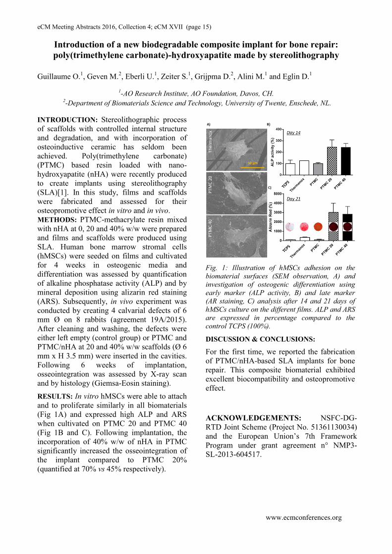
RESULTS: In vitro hMSCs were able to attach
and to proliferate similarly in all biomaterials
(Fig 1A) and expressed high ALP and ARS
when cultivated on PTMC 20 and PTMC 40
(Fig 1B and C). Following implantation, the
incorporation of 40% w/w of nHA in PTMC
significantly increased the osseointegration of
the implant compared to PTMC 20%
(quantified at 70% vs 45% respectively).
Fig. 1: Illustration of hMSCs adhesion on the
biomaterial surfaces (SEM observation, A) and
investigation of osteogenic differentiation using
early marker (ALP activity, B) and late marker
(AR staining, C) analysis after 14 and 21 days of
hMSCs culture on the different films. ALP and ARS
are expressed in percentage compared to the
control TCPS (100%).
DISCUSSION & CONCLUSIONS:
For the first time, we reported the fabrication
of PTMC/nHA-based SLA implants for bone
repair. This composite biomaterial exhibited
excellent biocompatibility and osteopromotive
effect.
ACKNOWLEDGEMENTS: NSFC-DG-
RTD Joint Scheme (Project No. 51361130034)
and the European Union’s 7th Framework
Program under grant agreement n° NMP3-
SL-2013-604517.

eCM Meeting Abstracts 2016, Collection 4; eCM XVII (page 16)
www.ecmconferences.org
Multi-scale mineralized collagen-polycaprolactone composites for craniofacial
bone tissue engineering
D.W. Weisgerber1, C. Flanagan
2, X. Ren
3, S.J. Hollister
2, J.C. Lee
3, M.B. Wheeler
1, B.A.C. Harley
1
1University of Illinois at Urbana-Champaign, Urbana, IL, USA.
2 University of Michigan, Ann
Arbor, MI, USA. 3 Greater Los Angeles VA Healthcare System, Los Angeles, CA, USA.
INTRODUCTION: Craniomaxillofacial (CMF)
defects present unique, unmet challenges for the
field of tissue engineering. Typically large in size
and marked by significant bone loss, these defects
are often treated via autogenous bone transplant.
Biomaterials for CMF repair must balance
considerations regarding mechanical competence
and load bearing, the need to fit complex 3D
defects unique to each patient, as well as
bioactivity and biotransport to support cells within
a large construct. We are developing a multi-scale
biomaterial that is mechanically competent for
large, load-bearing bone defects which also
supports cell bioactivity and tissue biosynthesis.
We have integrated mineralized collagen-GAG
scaffold with micron-scale porosity into a
mechanically-robust, polycaprolactone (PCL)
frame with mm-scale porosity. We report the
osteogenic nature of the collagen scaffold as well
as the regenerative capacity of the multi-scale
PCL-collagen composite via porcine mandible
defect and rabbit calvarial defect models.
METHODS: PCL frames were fabricated by
selective laser sintering of a powder precursor of
polycaprolactone and 4 wt% hydroxyapatite [1].
Mineralized collagen scaffolds (and non-
mineralized scaffold controls) were fabricated via
lyophilization from precursor suspensions of
collagen, GAG, and calcium phosphate [2].
Collagen-PCL composites were generated by
infiltrating the suspension into the PCL frame prior
to lyophilization. Scaffolds were seeded with
hMSCs and cultured in vitro, with osteogenic
differentiation evaluated via Western blot, RT-
PCR, ELISA, and histology. Alternatively,
collagen-PCL composites (vs. PCL or non-
mineralized scaffold controls) were implanted in
10 mm dia. mandible defects in 6mo Yorkshire
pigs (or rabbit calvarial defects [3]), with bone
infiltration assessed via microCT and histology.
RESULTS: Mineralized collagen scaffolds
instruct osteogenic MSC differentiation
independent of use of conventional osteogenic
media or supplemental BMP-2. The mineralized
scaffold constitutively activates canonical
(Smad1/5/8) and smad-independent (ERK1/2, Akt,
p38 MAPK) BMP receptor signaling paths in
hMSCs. PCL-collagen composites show a
significant (up to 6.8 ± 0.4 MPa) increases in
elastic modulus (6000-fold vs. scaffold only) and
ultimate stress (vs. PCL frame) which can be
specified by the PCL frame geometry.
Incorporation of the PCL frame does not reduce
the osteogenic capacity of the CGCaP scaffold. In
vivo, the composite (without MSCs, BMP2)
promotes porcine mandible repair (Fig. 1).
Fig. 1: Significantly increased radial bone infill (6
wks) in porcine mandible defect for collagen-PCL
composite vs. PCL (p) and scaffold (s) alone.
DISCUSSION & CONCLUSIONS: We report a
strategy to create a PCL-collagen composite that
combines a mineralized collagen scaffold that
promotes MSC osteogenesis in the absence of
osteogenic supplementation with a mechanically-
robust, patient-customizable PCL frame.
Mineralized scaffolds show enhanced bone repair
in a rabbit calvarial defect vs. non-mineralized
scaffolds [3]. Here we show collagen-PCL
composites show improved bone infill in a porcine
mandible defect. Ongoing efforts are evaluating
the efficacy of the composite in critically sized
(25mm dia.; 10mm thick) mandible defect.
ACKNOWLEDGEMENTS: Funding
provided by the AO Foundation (S-15-54H).

eCM Meeting Abstracts 2016, Collection 4; eCM XVII (page 17)
www.ecmconferences.org
Engineered bone for the repair of large bone defects:
The challenge of implanted MSC survival
Delphine Logeart-Avramoglou
Osteo-Articular Bioengineering and Bioimaging Laboratory, UMR7052 CNRS,
Paris Diderot University, Paris, France
INTRODUCTION: The repair of large bone defects due to trauma and
to pathological bone withdrawal/resorption still
represents a major challenge for orthopedic
surgeons. Stem-cell-based-bone tissue engineering
(TE) approaches are currently developed by
combining osteoprogenitors (such as multipotent
stromal cells derived from bone marrow (BMSC))
with a supporting substrate with the aim of
obtaining new bone tissue. The efficacy of bone
TE in experimental and clinical studies, although
promising, remains inferior to that of autologous
bone grafts. The underlying reasons for the limited
success of TE constructs are not yet fully
understood but, one of them may be the in vivo
massive death of transplanted cells observed after
engraftment into tissue constructs.
Delivered BMSC are believed to promote tissue
repair through direct participation after
differentiation and incorporation into new tissue
and/or through the paracrine activity of the cells
that modulates immune response, induces
angiogenesis, or promotes wound repair. Whatever
the mechanisms of action of BMSC in bone repair,
the poor viability of the cells after administration
remains a major impediment to their biological
functionality.
Among the possible factors responsible for such
massive cell death, the hostile environment that
BMSC faced upon implantation is considered as a
prime reason. When loaded on material constructs
devoid of pre-existing vascular network, BMSCs
encounter an ischemic environment with low
oxygen tension and deprivation of nutrients and,
consequently, a considerable bioenergetic
challenge.
To meet this challenge, various approaches have
been developed in order to sustain long term
viability of the TE constructs up to the host
vascular bed establishment. These strategies rely
on the development of scaffolds that favour the cell
viability or on approaches of BMSC
preconditioning to alleviate the ischemia-mediated
cell death.
eCM Meeting Abstracts 2016, Collection 4; eCM XVII (page 18)
www.ecmconferences.org
The Influence of Mechanical Environment in Bone Healing
Vaida Glatt
Institute of Health and Biomedical Innovation at Queensland University of Technology, Brisbane,
QLD, Australia
The management of bone defects and impaired
fracture healing remains one of the most
challenging clinical problems faced by
orthopedists today. Several treatments exist to aid
in the healing of large bone defects, including
recombinant human bone morphogenetic protein-2
(BMP-2). Although BMP-2 has shown preclinical
efficacy in animal models, the clinical
effectiveness has been disappointing. The current
practice of using extremely large amounts of BMP-
2 has major concerns about the many possible side
effects such as bone overgrowth in unwanted areas,
bone resorption, implant dislodgment, with even
cancer being reported1.
Regeneration of bone requires a coordinated
network of molecular signals, and is dependent
upon the local mechanical environment playing a
major role in the rate and success of healing. The
mechanical environment itself is determined by the
stiffness of the implant used to stabilize the
fracture and weight-bearing, and as a result, if
fixation is either too flexible or too rigid the
healing might fail. In our previous studies we
demonstrated that the local mechanical
environment influenced the healing of 5 mm large
bone defects in response to a standard dose of
BMP-2 using a rat model2. Based on our
preliminary experiments we hypothesized that the
healing of large-segmental bone defects and
fractures can be accelerated by the imposition of an
appropriate mechanical environment. This concept
arose based on evidence that flexible fixation
stimulates endochondral bone formation. However,
the same process would jeopardize bone
consolidation by disrupting the formation of blood
vessels of regenerating bone. Therefore, we
proposed the regimen we named Reverse
Dynamization (RD) where the defect is initially
stabilized using a fixator with low axial stiffness,
and subsequently increasing the fixator stiffness at
the first signs of radio-opacity. In fact, by imposing
RD, where the fixator stiffness was changed from
low to high after 2 weeks, a time when bone was
forming within the defect, this study demonstrated
accelerated healing and remodeling through the
modulation of the mechanical environment around
the defect site2.
Based on these observations, additional studies
were performed using Reverse Dynamization in a
rat model to extend it to a wider range of
stiffnesses and BMP-2 concentrations to learn
more about its scope and biology. The underlying
hypothesis was that by using the appropriate
stiffness parameters and timing, RD can enhance
the healing of large segmental defects thereby
minimizing the dose of BMP-2 required.
The results from this study showed that defect
healing was influenced by the dose of BMP-2
suggesting that a lower dose of (5.5 g) BMP-2 was
sufficient enough to enhance the healing of defects.
Although the healing was slightly delayed, the
quality of healed bone was equivalent compared to
a high dose (11 g) of BMP-2. It also demonstrated
that the mechanical environment plays a role when
using a lower dose, as was evident from the
presence of the radiopaque line at the end of
treatment, which is a consequence of prolonged
movements in the defect during the early stages of
healing when lower stiffness fixators are used3.
While further studies are essential, the results of
this study indicate that the fixation stability could
be used to maximise the regenerative capacity of
bone healing while minimising the dose of BMP-2
required clinically.
Similar studies are being performed to test the
effectiveness of Reverse Dynamization in a 1mm
osteotomy rat model. Initial results showed
superior healing outcomes when the RD regimen
was used, and this was time dependent. Although
additional studies will be required to confirm these
findings, this data suggest that fracture healing
could be accelerated through the manipulation of
fixation stability, and it also introduces a potential
clinical strategy to improve the healing outcome of
unstable fractures, particularly for delayed non-
unions through increased stabilization
ACKNOWLEDGEMENTS: U.S DoD-
W81XW H-10-1-0888), Vice-Chancellor’s
Research Fe-llowship, QUT, AU, PA Research
2015 Project Grant.

eCM Meeting Abstracts 2016, Collection 4; eCM XVII (page 19)
www.ecmconferences.org
Direct use of freshly-isolated adipose-derived cells for fracture augmentation in
a first-in-man phase I clinical trial
Franziska Saxer1 and Arnaud Scherberich
2, 3 Atanas Todorov
2, Patrick Studer
1, Sylvie Miot
2,
Simone Schreiner1, Sinan Güven
2, Laurent AH Tchang
3, Martin Haug
3, Michael Heberer
2, Dirk J
Schaefer3, Daniel Rikli
1, Marcel Jakob
1, Ivan Martin
2.
1Clinic of Traumatology,
2Department of Biomedicine and of Clinical Research,
3Clinics of Plastic,
Reconstructive and Aesthetic Surgery, University Hospital Basel, Basel, Switzerland
INTRODUCTION: Stromal Vascular Fraction
(SVF) cells, freshly isolated from adipose tissue,
are an abundant and easily accessible source of
mesenchymal/endothelial progenitors. Previous
studies have demonstrated their osteogenic and
vasculogenic properties. Given the
dysfunctionality of autologous bone in
osteoporosis, we aimed at investigating safety and
feasibility of a clinical implementation of the SVF
for fracture augmentation in the elderly. To
investigate the contribution of the implanted cells
to bone healing, a similar approach was evaluated
in a nude-rat femoral-defect model.
METHODS: Autologous human SVF-cells were
intra-operatively isolated using an automated
device (Celution®800CRS, Cytori, USA) and used
as cellular component of hydroxyapatite(HA)-
based composite grafts for the augmentation of
low-energy proximal humeral fractures after
locking-plate fixation in 8 elderly patients. The
grafts were assessed for cell characteristics,
viability and differentiation potential. Follow up
was performed for 6 month. In case of plate
revision or removal, a bone biopsy was taken from
the grafted area (n=6) and analysed using
microCT and histology. The safety of the approach
was defined as the absence adverse reactions (AR),
feasibility as the absence of protocol deviations.
Similar constructs were implanted in a segmental
femoral defect in immune-compromised rats after
locking-plate osteosynthesis (RatFix, RISystem,
CH), with cell-free grafts as control. Mechanical,
microCT and histological analysis was performed
after six weeks.
RESULTS: The intra-operative cell isolation from
272 ± 63 ml abdominal lipoaspirate yielded 121.4
± 72 million SVF-cells, manufacturing of the graft
was well feasible, the production standardized and
reproducible, the intervention was prolonged by
the manufacturing process with liposuction (≈ 60
min) and cell isolation (≈ 120 min). The procedure
was safe, without AR during the trial or in the
following up to 39 months. The duration of
hospitalization and the course of rehabilitation
were normal. MicroCT and histology of the repair
tissue from clinical biopsies demonstrated
formation of ossicles as early as 6 weeks
postoperative (fig 1.), structurally disconnected
and morphologically distinct from osteoconducted
bone, suggesting the osteogenic nature of
implanted SVF cells. In the animal model, only
SVF cell-treated defects healed mechanically
stable and displayed mature bone with osteocytes
and vascular structures of human origin.
Fig. 1:
Fig. 1: Hematoxylin/eosin staining from a biopsy of the
SVF-based graft after 6 weeks (A) and 6 months (B)
with nb marking new bone (opposed to gr.=granules,
ob=osteoconducted bone). Picture C shows the isolated
bone formation within the pores of the whitish HA
granules.
DISCUSSION & CONCLUSIONS: These trials
strongly suggest that the non-expanded SVF
without exogenous priming but within a fracture-
micro-environment, can safely promote de novo
generation of vascularized bone. The approach can
be streamlined to optimize the utilisation of
operating-room capacity. The efficacy of the
proposed approach should now be evaluated in
controlled trials with larger patient cohorts.
REGISTRATIONS/PERMITS:
ClinicalTrials.gov # NCT01532076, EKBB,
Ref.#348/10, BAG Ref.# Bk2010-nTx-Z046-N0-
V00, KVet Basel-Stadt, permission no. 2357
ACKNOWLEDGEMENTS: AO Start up grants
S-12-08S / S-09-112S and SNF Project Grant No.
310030-156291 partially supported this study.

eCM Meeting Abstracts 2016, Collection 4; eCM XVII (page 20)
The new regenerative frontiers in cranio-mandibulo-facial surgery in primates:
The pleiotropic inductive activities of the mammalian TGF-β3 isoform
U Ripamonti1, R Duarte
2, RM Klar
1,2, R Parak
1,3 , C Dickens
2, T Dix-Peek
2
1 Bone Research Laboratory,
2 Dept. Internal Medicine,
3 Dept. Oral Biological Sciences, School of
Oral Health Sciences and of Internal Medicine, Faculty of Health Sciences, the University of the
Witwatersrand, Johannesburg
INTRODUCTION: Contrary to results in rodents
and lagomorphs, heterotopic implantation of recombinant human transforming growth factor-β3
(hTGF-β3) in the rectus abdominis muscle of non-
human primates Papio ursinus results in the rapid
induction of bone formation. 1
Mechanistically, this is set into motion by a series of profiled bone
morphogenetic proteins (BMPs) and TGF-βs that
are expressed at different time points temporally and spatially regulating the induction of bone
formation. 1,2
METHODS: 27 Chacma baboons Papio ursinus
were implanted with doses of hTGF-β3 loaded
osteogenic devices in the rectus abdominis,
mandibular and calvarial sites. Harvested tissues at
15, 30, 60, 90 days and up to 14 months after
mandibular implantation were examined by
quantitative reverse- transcriptase polymerase
chain reaction (qRT-PCR) and compared to
morphological data obtained from sections
prepared using the EXAKT precision cutting and
grinding system on undecalcified specimen blocks.
RESULTS: Implantation of 125 µg hTGF-Β3 in
full thickness mandibular defects of P. ursinus
resulted in an unprecedented restitutio ad integrum
of the mandibular defects on day 30 with complete
healing after long term studies. hTGF-β3 failed
however to engineer regeneration of calvarial
defects which could be partially restored by adding
pericytic/perivascular/myoblastic stem cells from
morcellated fragments of rectus abdominis muscle.
The morphology of incomplete calvarial repair on
day 90 with bone formation pericranially and with
lack of bone induction endocranially above the
dura suggested a radius of activity set into motion
by inhibitory mechanisms originating from the
dura mater and/or the highly vascularized
leptomeninges below. Such a diffusion molecular
hypothesis was tested by surgically inserting a
nylon fold impermeable membrane below the
endocranium and above the dura and the
arachnoids, segregating the molecular and cellular
micro-environments of the calvarial defects from
the dura. Segregation restored the endocranial
induction of bone formation by hTGF-β3 (Fig. 1)
whilst segregated untreated defects showed
limited, if any, induction of bone formation
indicating the critical role of the dura mater in
calvarial tissue induction. hTGF-β3/treated
segregated constructs showed significant induction
of bone formation with OP-1, BMP-2,
Osteocalcin, RUNX-2, ID2 and ID3 up-regulated.
A
Fig. 1: (A) Segregated hTGF-β3 -treated calvarial
defect with a nylon fold membrane: induction of
bone formation across the defect.
DISCUSSION & CONCLUSIONS: In P.
ursinus, calvarial segregation restores the
induction of bone formation by hTGF-β3 by
blocking inhibitory signalling pathways from the
underlying dura and leptomeninges below, with
complete regeneration of large mandibular defects
up to 14m post-implantation. The overall tight
control of the induction of calvarial bone by
hTGF-β3 in P. ursinus is orchestrated by the dura
mater which is disrupted by relatively high doses
of the exogenously applied recombinant
morphogen. In primates and in primates only, the
TGF-β3 gene and gene product singly yet
synergistically and synchronously set into motion
the induction of bone formation that now demands
a paradigmatic shift in bone tissue engineering.
www.ecmconferences.org
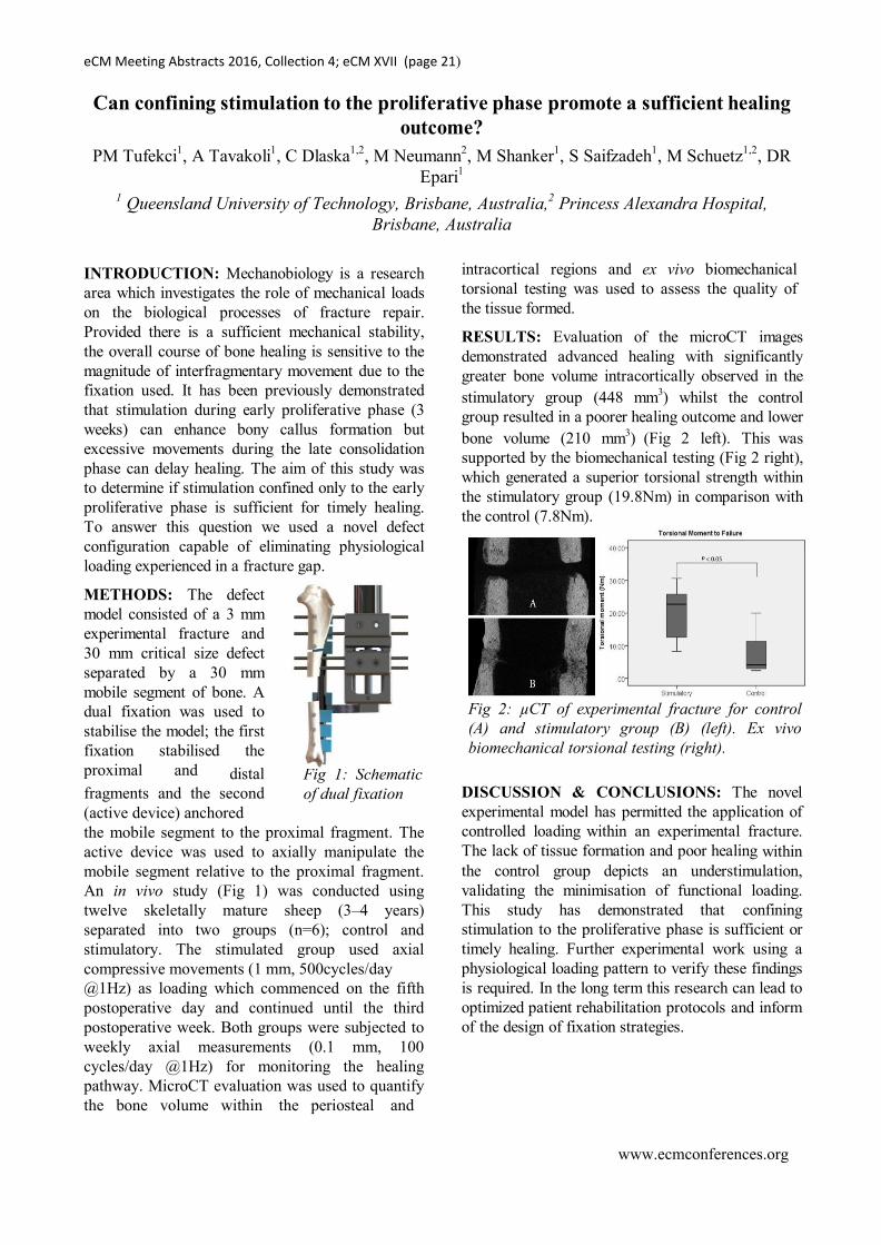
Fig 1: Schematic
of dual fixation
eCM Meeting Abstracts 2016, Collection 4; eCM XVII (page 21)
Can confining stimulation to the proliferative phase promote a sufficient healing
outcome?
PM Tufekci1, A Tavakoli
1, C Dlaska
1,2, M Neumann
2, M Shanker
1, S Saifzadeh
1, M Schuetz
1,2, DR
Epari1
1 Queensland University of Technology, Brisbane, Australia,
2 Princess Alexandra Hospital,
Brisbane, Australia
INTRODUCTION: Mechanobiology is a research
area which investigates the role of mechanical loads
on the biological processes of fracture repair.
Provided there is a sufficient mechanical stability,
the overall course of bone healing is sensitive to the
magnitude of interfragmentary movement due to the
fixation used. It has been previously demonstrated
that stimulation during early proliferative phase (3
weeks) can enhance bony callus formation but
excessive movements during the late consolidation
phase can delay healing. The aim of this study was
to determine if stimulation confined only to the early
proliferative phase is sufficient for timely healing.
To answer this question we used a novel defect
configuration capable of eliminating physiological
loading experienced in a fracture gap.
METHODS: The defect
model consisted of a 3 mm
experimental fracture and
30 mm critical size defect
separated by a 30 mm
mobile segment of bone. A
dual fixation was used to
stabilise the model; the first
fixation stabilised the
proximal and distal
fragments and the second
(active device) anchored
the mobile segment to the proximal fragment. The
active device was used to axially manipulate the
mobile segment relative to the proximal fragment.
An in vivo study (Fig 1) was conducted using
twelve skeletally mature sheep (3–4 years)
separated into two groups (n=6); control and
stimulatory. The stimulated group used axial
compressive movements (1 mm, 500cycles/day
@1Hz) as loading which commenced on the fifth
postoperative day and continued until the third
postoperative week. Both groups were subjected to
weekly axial measurements (0.1 mm, 100
cycles/day @1Hz) for monitoring the healing
pathway. MicroCT evaluation was used to quantify
the bone volume within the periosteal and
intracortical regions and ex vivo biomechanical
torsional testing was used to assess the quality of
the tissue formed.
RESULTS: Evaluation of the microCT images
demonstrated advanced healing with significantly
greater bone volume intracortically observed in the
stimulatory group (448 mm3) whilst the control
group resulted in a poorer healing outcome and lower
bone volume (210 mm3) (Fig 2 left). This was
supported by the biomechanical testing (Fig 2 right),
which generated a superior torsional strength within
the stimulatory group (19.8Nm) in comparison with
the control (7.8Nm).
Fig 2: µCT of experimental fracture for control
(A) and stimulatory group (B) (left). Ex vivo
biomechanical torsional testing (right).
DISCUSSION & CONCLUSIONS: The novel
experimental model has permitted the application of
controlled loading within an experimental fracture.
The lack of tissue formation and poor healing within
the control group depicts an understimulation,
validating the minimisation of functional loading.
This study has demonstrated that confining
stimulation to the proliferative phase is sufficient or
timely healing. Further experimental work using a
physiological loading pattern to verify these findings
is required. In the long term this research can lead to
optimized patient rehabilitation protocols and inform
of the design of fixation strategies.
www.ecmconferences.org

eCM Meeting Abstracts 2016, Collection 4; eCM XVII (page 22)
www.ecmconferences.org
Improved fracture healing by short-term G-CSF treatment
M Herrmann1, S Zeiter
1, U Eberli
1, M Hildebrand
1, K Camenisch
1, U Menzel
1, M Alini
1,
S Verrier1#
, V Stadelmann1
1 AO Research Institute Davos, CH,
#correspondence to [email protected]
INTRODUCTION: Stem- and progenitor cell
mobilization is a critical event in bone
regeneration. This includes mesenchymal stem
cells as a source of osteoprogenitor cells as well as
endothelial progenitor cells (EPCs), which may
promote neovascularization. In fact, impaired
vascularization is considered as one of the most
important causative factors for atrophic non-
unions. Granulocyte colony-stimulating factor (G-
CSF) mediated mobilization of CD34+ progenitor
cells into the circulation is widely applied for
peripheral stem cell donations and a positive effect
of G-CSF administration on bone healing has been
suggested. The aim of this study was to
characterize the different cell populations
mobilized by G-CSF and investigate the influence
of cell mobilization before and after surgery on the
healing of a critical size femoral defect in rats.
METHODS: First, the time course and cell
populations mobilized by G-CSF treatment were
determined. Rats were randomly assigned into four
experimental groups, including an untreated
control group and three groups receiving sub-
cutaneous injections of G-CSF for 5 consecutive
days, with blood analysis performed at day 1, day
5 and day 11 after the last injection. Measurements
included total leukocyte counts and flow cytometry
analysis. Second, bone healing was compared in a
saline-treated control group, a group receiving 5
consecutive daily G-CSF injections before surgery
and a group receiving 5 consecutive daily G-CSF
injections after surgery. An empty 4.5mm
critically-sized femoral defect was created in
female adult Fisher rats and fixed internally. Bone
healing was monitored by in vivo micro CT and
histology.
RESULTS: Leukocyte counts show a peak
increase at the first day after the last G-CSF
injection (Fig. 1A). Analysis of different cell
populations by flow cytometry revealed a cell-
specific mobilization kinetic. We found that
CD34+ progenitor cells were significantly enriched
at day 1, and further increased at day 5 and day 11
(Fig.1B). Micro CT measurements revealed
improved healing of the critically-sized bone
defect in both G-CSF treatment groups with an
increased bone volume in the fracture gap
compared to untreated control animals (Fig.2).
Although continuous bone formation was observed
in all rats, healing was incomplete at the end of the
observation period after 230 days, which was also
confirmed by histology.
Fig. 1: Blood cell values at different time points after 5
consecutive injections of G-CSF. Day 1 refers to the
first day after the last injection.
Fig. 2: In vivo micro CT monitoring of fracture healing.
A. Representative 3D renderings of segmented micro
CT scans taken 80 days after osteotomy. B. Bone
volume within femoral defects. Values are given as
mean ± SEM.
DISCUSSION & CONCLUSIONS: Our data
shows that different cell populations are
upregulated by G-CSF treatment in cell specific
patterns. Although in this study no bridging of the
critically-sized defect in the groups with G-CSF
injections was reached, an improved healing was
clearly shown. These results identify cell
mobilization by G-CSF as a potential treatment
option to facilitate the healing of large bone defects
in combination with other treatment strategies.
ACKNOWLEDGEMENTS: The authors thank
AOER/AO Foundation and AO Trauma for
funding. The FACS AriaIII was kindly donated by
the Innovationsstiftung Graubünden.

eCM Meeting Abstracts 2016, Collection 4; eCM XVII (page 23)
www.ecmconferences.org
Choosing the right animal model for bone-related questions
MJ Allen
Surgical Discovery Centre, Department of Veterinary Medicine, University of Cambridge,
Cambridge, United Kingdom
INTRODUCTION: Animal models continue to
represent a critical component of both
musculoskeletal discovery research and
orthopaedic product/device evaluation and
approval. This presentation will highlight the key
factors that must be considered when considering
an animal study in the context of bone-related
questions. The goal of this review will be to
challenge the audience to re-evaluate their current
thinking and to present best practices in model
selection, study design and the choice of clinically
relevant outcome measures and time points.
DISCUSSION: Model selection: No matter the
species being considered, every effort should be
made to limit the use of animals in scientific
research. There are tremendous potential
advantages (financial and, more important,
scientific) but unavoidable differences in body
size, skeletal biology, skeletal structure (including
skeletal size) and joint biomechanics may limit the
validity of small animal models as screening
systems for candidate new medical or surgical
therapies. Large animal models such as sheep, goat
and dog offer significant potential advantages in
this regard, with more clinically relevant bone
turnover rates, larger overall bone size and the
possibility of using functional implants (e.g. joint
replacements) that better simulate the intended
clinical application in humans. However, the use of
larger animals (especially companion animals) can
be viewed as ethically contentious and may be
outlawed in some jurisdictions. One of the key
questions that needs to be addressed at the start of
the pro cess is whether the study is intended as a
feasibility/proof of principle, or as a definitive
study for regulatory submission. Once this key step
has been addressed, the decision-making process
can move ahead.
Study design: Given the constraints of time, the
presentation will focus on three specific examples
in which animal models are used to address bone-
related research questions: fracture healing, bone
defect repair, and implant fixation. Each topic
requires separate consideration, but there are some
common themes that need to be addressed,
including: (1) Has the experimental construct been
validated (e.g. is the defect a critical size defect for
the given species and age of animal)? (2) Is there
an argument for/against doing multiple site
surgeries and/or bilateral procedures? This is a
very thorny issue with animal care and use
committees, and would need strong justification
and scientific support) (3) Are the biological
processes in the animal model relevant to those in
humans (e.g. if a pin is used as the sole
stabilisation for a long bone fracture in rats, is this
really mirroring the biology of fracture healing
following surgical stabilisation with the plates or
external fixators that are more typical in human
surgery)? (4) Appropriate controls? What is the
appropriate control for the experiment, is it ethical
and how will the data be interpreted? (5) Use of a
pilot study, to allow the research team to run
through the logistical and technical aspects of the
study, and to provide critical information that can
be used to perform an appropriate power/sample
size calculation.
Outcome measures and time points: One of the
most frustrating aspects of reviewing literature
relating to any aspect of bone research is the
apparent lack of consistency in the selection of
outcome measures and time points. To compound
this, even when a standardised set of procedures is
performed, it may not be reported adequately in the
resulting manuscript. The combination of
inconsistency and poor reporting contributes to the
problem of irreproducibility, which the NIH has
identified as a significant problem in animal
studies throughout science1. Recent initiatives in
terms of improved reporting will help in to
improve transparency2, but ongoing efforts are
needed to define the most appropriate outcome
measures for individual studies. With an extensive
range of outcome measures available within the
orthopaedic field, data collection can be
maximised through the creative use of a
combination of non-invasive, non-destructive
testing (e.g. imaging) with the standard destructive
outcome measures of mechanical testing and
histopathology.

eCM Meeting Abstracts 2016, Collection 4; eCM XVII (page 24)
www.ecmconferences.org
Reducing the use of animals by in vivo imaging of bone and blood vessels
R Müller1
1 Institute for Biomechanics, ETH Zurich, Zurich, CH.
INTRODUCTION: With recent advances in
skeletal tissue engineering and regenerative
medicine there is a strong need for quantitative
imaging of bone and blood vessels at the tissue and
even cellular level. A number of new in vivo
microstructural imaging modalities have been put
forward recently allowing quantification with high
precision and accuracy of these structures in a
time-lapsed fashion in live animals. Although
biomedical imaging technology is now readily
available, few attempts have been made to expand
the capabilities of these systems by adding
quantitative analysis tools to assess transient
structure-function relationships in a time-lapsed
fashion. In the spirit of 3R, such longitudinal
animal study designs not only allow to refine the
methods used to measure biological in vivo
function but also to directly reduce the number of
animals needed for these studies, avoiding
unnecessary cross-sectional studies. Using time-
lapsed vivo imaging, each animal can serve as its
own reference with respect to the changes
observed at each time point. The aim of this
contribution is to present recent developments in in
vivo computed tomography (CT) imaging of bone
and blood vessels in applications of tissue
engineering and regenerative medicine.
METHODS & RESULTS: X-ray-based CT is an
approach to image skeletal tissues and blood
vessels in a hierarchical fashion providing
multiscale imaging capabilities with isotropic
resolutions ranging from a few millimeters
(clinical CT), to a few micrometers (microCT)
down to one hundred nanometers (nanoCT). A
number of groups working in this field have
demonstrated over the last two decades that X-ray-
based tomographic imaging is a nondestructive and
noninvasive procedure that allows precise 3D
measurement of bone and blood vessels on all
levels of hierarchy. The technique has been used
predominantly in vitro but recently in vivo
applications have gained more and more interest
due the unprecedented resolutions in the order of
10 µm available in these in vivo systems. Due to
the time-lapsed nature of the images, not only
static but also dynamic morphometry can be
performed to assess tissue remodeling and
regeneration. With the recent introduction of
computational tools that allow calculation of the
mechanical microenvironment in these tissues,
links between mechanical cues acting on ensemble
of cells or even individual cells and the
corresponding tissue adaptation and/or repair can
now be monitored fully nondestructively in
individual animals in vivo at relatively moderate
cost and great ease of use. While studies in bone
(Fig. 1) are straightforward due to the natural
contrast of hard and soft tissue using X-rays,
monitoring blood vessels in vivo is more
challenging due to the required injection of
contrast agents that provide good contrast, no
extravasation and slow blood clearance (Fig. 2).
Fig. 1: Monitoring bone healing process in live
mice. Reconstructions of the defect site and
fracture callus for representative animals from
1.2mm defect group (A) and 2mm defect group (B).
DISCUSSION & CONCLUSIONS: Time-lapsed
in vivo imaging allows longitudinal quantification
of adaptation and regeneration of bone and blood
vessels, thereby reducing the number of animals
needed to show significant results. It is strongly
recommended that quantitative imaging is used
more often for in vivo animal studies in the area of
tissue engineering and regenerative medicine.
Fig. 2: Representation of blood vessels and bony
structures in a mouse limb (10 µm resolution).
ACKNOWLEDGEMENTS: Financial support
from the European Union (BIODESIGN FP7-
NMP-2012-262948) is gratefully acknowledged.

eCM Meeting Abstracts 2016, Collection 4; eCM XVII (page 25)
www.ecmconferences.org
injectable synthetic collagen beads hydrogel loaded with BMP2 promotes ectopic
bone formation
S. Fahmy-Garcia1,2
; D. Mumcuoglu 5; L. de Miguel
5; B.C.J van der Eerden
2; D. Eglin
6; S.G.J.M.
Kluijtman5; G.J.V.M van Osch
1,3 and E. Farrell
4
1Department of Orthopaedics,
2Department of Internal Medicine,
3Department of
Otorhinolaryngology, Head and Neck Surgery, 4Department of Oral and Maxillofacial Surgery,
Special Dental Care and Orthodontics, Erasmus MC, University Medical Center, Rotterdam, The
Netherlands. 5
Fujifilm Manufacturing Europe B.V, Tilburg, The Netherlands.6AO Research Institute Davos, Davos, Platz, Switzerland
INTRODUCTION: The goal of bone tissue
engineering is to use biomaterials, cells and
signaling molecules capable to induce bone
formation. Systems derived from polysaccharides
and natural proteins are ideal scaffolds for tissue
engineering since they mimic the extracellular
matrices. In this study, we used synthetic collagen
beads as developed by Fujifilm based on Cellnest
(recombinant peptide based on human collagen I)
loaded with BMP2, a well-known growth factor
involved in bone regeneration, and included them
in various hydrogels to generate an injectable
system. The used hydrogels were
thermoresponsive hyaluronic acid and two
alginates types with different physico-chemical
characteristics. The goal of this study was to
determine the most biocompatible injectable
collagen bead/hydrogel system in terms of host
reaction, vascularization and bone formation in a
cell free system.
METHODS: Twenty one male Sprague Dawley
rats at 10 weeks old were used in this study. One of
the alginate types or hyaluronic acid hydrogels
containing synthetic collagen microspheres loaded
with a constant concentration of rhBMP-2 were
subcutaneously injected (n=6 per condition). At the
end of 1-week, 4-week and 10-week period, six
animals respectively were euthanized using CO2.
All implants were harvested and scanned using
micro-CT to compare formation of mineralised
tissue. For histological analysis, implants were
paraffin embedded and processed for CD68, CD31,
CollII, TRAP and H&E staining.
RESULTS: Injectable alginate gel with collagen
beads containing BMP2 promotes ectopic bone
formation. At 1-week cellular infiltration was
visible, even though the hydrogel was not degraded
yet. On the micro-CT scans, at 4-week mineralised
tissue was observed in 4 of the
6 injected implants , especially at the edges or the
areas where the hydrogel started to break down. At
10 weeks 5 implants were retrieved and bone
formation was observed in all of them on CT. In
addition, bone marrow presence was verified via
histology. However, when thermoresponsive
hyaluronic acid was used as hydrogel, although
cellular infiltration was observed from the first
week on, bone was not found within 10 weeks.
DISCUSSION & CONCLUSIONS: This work
has shown that the use of collagen beads within
alginate hydrogels in combination with BMP2
promote ectopic bone formation. However, when
HA was used as hydrogel, most gel had
disappeared within the first weeks and a high cell
infiltration was observed, amongst which where
macrophages suggesting a high degradation rate.
Previous studies used hydrogel-beads combination
with the addition of osteoprogenitor cells to trigger
bone formation [1-2]. In our study, we obtained
bone formation in a very high percentage of the
constructs in a subcutaneous environment, which is
very challenging using a cell free approach.
A major limitation of the commonly used release
systems is the difficulty to modulate the release of
the growth factors or signaling molecules to
maintain their actions for a long time period. A
combination of the hydrogel and recombinant
collagen beads might help to solve this problem.
Our data demonstrate the importance of the choice
of the gel in this system. .
ACKNOWLEDGMENTS: BioInspire Marie
Curie Action (FP7/2007-2013)

eCM Meeting Abstracts 2016, Collection 4; eCM XVII (page 26)
www.ecmconferences.org
Identification and isolation of live mesenchymal stem cells based on
differentiation induced changes in mRNA expression
Bojun Li, Ursula Menzel, Claudia Loebel, Mauro Alini, Martin J. Stoddart
AO Research Institute, AO Foundation, Davos, CH.
INTRODUCTION: Osteogenesis of human bone
marrow derived mesenchymal stem cells
(hBMSCs) has been widely studied for bone tissue
engineering. Recent studies show that the
osteogenic differentiation of hBMSCs can be
assessed by quantifying the ratio of two important
transcription factors (Runx2/Sox9) 1. In previous
studies, these transcription factors were detected
via destructive methods, either intra-cellular
immunostaining or PCR. Here we demonstrate a
new technique to observe mRNA expression of
two genes in individual live cells using two
fluorescent probes specific for Runx2 or Sox9
mRNA. Functionally homogenous cells can then
be prospectively isolated based on the ratio of the
two markers.
METHODS: hBMSCs were isolated from bone
marrow aspirates by standard ficoll density
gradient, followed by plastic adherence. Expanded
cells (Passage 2-Passage 4) were plated at 10,000
/cm2 and induced towards osteogenesis for 6 days.
On the 6th day, media was replaced with fresh
growth medium (GM) or osteogenic medium (OM)
containing fluorescent Smartflare probes for
Runx2-Cy3 and Sox9-Cy5, and the cells were left
to incubate for 16 hours. The cells were then
harvested by trypsinisation and sorted based on
their relative Cy3:Cy5 fluorescence intensity
compared to unstimulated controls. Gene
expression analysis for a panel of osteogenic genes
was performed. Sorted cells were further cultured
to assess proliferation rate and osteogenic potential
by way of alizarin red staining.
RESULTS: Cell differentiation in live cells could
be monitored by investigating the relative ratio of
Runx2 and Sox9 using non-destructive
fluorescence based markers. Furthermore, cells
sorted on day 7 using this ratio exhibited changes
in osteogenic gene expression, proliferation rate
and calcification potential compared to the
unsorted population. One population (P1) appeared
to have a later stage osteoblast phenotype, with
low ALP, Runx2, Collagen I and the highest OC.
This population also proliferated slowest, a
characteristic of more differentiated cells, and
calcified to the greatest extent.
Fig. 1: Runx2 and Sox9 fluorescent probes can
detect mRNA expression of two genes in individual
live cells simultaneously and be used for cell
sorting. A clear shift in profile was observed
between cells in GM and OM. Cells in OM were
sored into 4 groups (P1-P4).
DISCUSSION & CONCLUSIONS: A new
method to observe mRNA expressions of two
biomarker genes in individual live cells
simultaneously, and isolate relatively
homogeneous stem cells based on mRNA
expression has been developed. Using this method
we could observe and quantify the mRNA
expression in live cells, and analyze cell
differentiation. The cells also can be sorted based
on mRNA expression for further studies. In
contrast to previous isolation methods which are
limited by available surface markers, isolation of
cells using mRNA specific probes provides the
opportunity to investigate intracellular markers,
such as transcription factors Runx2 or Sox9, and
isolate relatively homogeneous cell populations.
Adapting the transcription factors investigated will
allow this method to be utilized for other cell
phenotypes
1ACKNOWLEDGEMENTS: The
Smartflare probes were kindly provided by
Merck Millipore. This work was partly funded by
the EU FP7-NMP-2010_LARGE-4 project
BIODESIGN and part funded by the AO
Foundation.

eCM Meeting Abstracts 2016, Collection 4; eCM XVII (page 27)
www.ecmconferences.org
Hydrogels to control and recruit stem cells
SR Caliari, JL Holloway, SL Vega, JA Burdick
Department of Bioengineering, University of Pennsylvania, Philadelphia, PA, USA
INTRODUCTION: Hydrogels can be engineered
for controlled biochemical and biophysical
properties, to alter cellular behaviour (e.g.,
differentiation of mesenchymal stem cells
(MSCs)), as well as for the controlled release of
therapeutic agents (e.g., stromal cell derived
factor-1 (SDF)). Towards the repair of bone, we
have explored how gel properties alter the
osteogenesis of MSCs when cultured either atop or
within hydrogels across a range of mechanical and
degradative properties. Additionally, we designed
gels to release SDF local to a bone injury to recruit
CXCR4+ cells for repair. In both cases, gels were
engineered from hyaluronic acid (HA) and
included crosslinks susceptible to matrix
metalloproteinases (MMP) degradation.
METHODS: HA was modified with various
reactive groups (e.g., norbornenes, maleimides) to
form gels through either photoinitiated thiol-ene or
Michael addition reactions and containing pendant
RGD and MMP-sensitive crosslinkers (e.g..
VPMSMRGG).1 Gels were characterized for
mechanics (compression, rheological, AFM) and
degradation (uronic acid). Towards understanding
cellular interactions, MSCs were seeded atop or
encapsulated within gels across a range of
mechanics and with controlled degradation.
Outcomes such as cell traction, viability,
spreading, fate (e.g., ALP) and signalling (e.g.,
Yes-associated protein, YAP) were analyzed.2
Gels were used for the encapsulation of SDF and
bone morphogenetic protein-2 (BMP) and release
was monitored with ELISA. Gels were implanted
into rat cranial defects and characterized at 6 wks
with radiography, micro-CT, and histology.3
RESULTS: HA gels with variable stiffness (1-20
kPa) were fabricated and supported viable MSC
seeding and encapsulation (>90%). Higher
stiffness gels degraded more slowly than lower
stiffness gels. MSCs atop the gels increased their
spreading, ALP activity, YAP nuclear localization
and traction with increased mechanics, whereas
cells within the same gels saw inverse trends with
osteogenesis favoured in gels with lower
mechanics or that were not degradable due to
reduced spreading/traction (Fig. 1).
The release of SDF and BMP from the gels was
dependent on the crosslink density and enzyme
concentration and released molecules were active
in in vitro studies. When implanted into the rat
cranial defects, bone formation was greatest in the
group with SDF/BMP (Fig.2), which was similar
to BMP doses an order of magnitude higher
delivered without SDF.
Fig. 1: Nuclear YAP ratio (left) and comparison to
cell shape index (right) for MSCs within non-
degradable and degradable hydrogels.
Fig. 2: 3D reconstructions (left) and quantification
of bone volume (right) from micro-CT, 6 wks after
implantation in a critical-sized rat cranial defect.
DISCUSSION & CONCLUSIONS: Hydrogel
properties (e.g., degradation, mechanics)
influenced MSC fate and signalling differentially in
2D and 3D. When atop a hydrogel, increased
mechanics enhanced traction and osteogenesis,
whereas within a hydrogel, lower mechanics
permitted spreading and osteogenic differentiation.
Thus, hydrogel type and presentation is important
towards guiding cell behaviour. The controlled
release of SDF allowed for bone formation in the
presence of BMP doses much lower than when
BMP is delivered alone. These approaches
illustrate the ability of engineered hydrogels to be
used towards bone repair.
2 ACKNOWLEDGEMENTS: The authors
recognize funding from the National Institutes of
Health (R01EB008722, R01AR056624).
0
1
2
3
4
5
6
7
non-deg deg
Nu
cle
ar
YA
P R
atio
0
0.2
0.4
0.6
0.8
1
0 1 2 3 4
Ce
ll S
hap
e In
dex
Nuclear YAP Ratio
non-deg
deg
*

eCM Meeting Abstracts 2016, Collection 4; eCM XVII (page 28)
www.ecmconferences.org
Synthetic bone fillers: how important is the micro and macrostructure?
M Bohner1
1 RMS Foundation, Bettlach, CH.
INTRODUCTION: Synthetic bone fillers are
broadly used to repair bone defects. Numerous
studies have evidenced the importance of bone
fillers architecture for this process. A roughness
superior to 1 µm [1] and pores larger than 100 µm
“macropores”) [2] are considered to be ideal to
accelerate and enhance bone formation.
Additionally, a number of studies have underlined
the importance of pores smaller than 100 µm
(“micropores”), suggesting that multi-scale design
approaches should be applied [3]. Nevertheless,
past studies have delivered contradictory results,
perhaps because the responses to architecture and
composition are related [4]. The aim of my talk
will be to review the data collected in two sheep
studies [5-6], and reflect on the importance of the
link between structure and bone healing. A total of
12 different materials were tested in [5-6], 11 out
of β-tricalcium phosphate and one out of monetite
(= anhydrous dicalcium phosphate). The materials
had various micro and macrostructures, and
implantation times included 2, 4, 6, 8, 12 and 24
weeks. A particular focus of my talk will be set on
the nature of the tissues present within micropores
and on morphological changes occurring during
healing.

eCM Meeting Abstracts 2016, Collection 4; eCM XVII (page 29)
www.ecmconferences.org
Healing of a critical size long bone defect through a serum free
biomimetic cell-based approach J. Bolander
1,2, W. Ji
1,2, J. Leijten
1,2, V. Bloemen
2,3, L. Moreira Teixeira
1,2, D. Lambrechts
1,2 and F. P. Luyten
1,2.
1Tissue Engineering Laboratory, Skeletal Biology and Engineering Research Center, KU Leuven,
2 Prometheus,
Division of Skeletal Tissue Engineering, KU Leuven, 3 KU Leuven Campus Group T, Leuven.
INTRODUCTION: Cell-based constructs enable
delivery of the critical number of progenitor cells
required to heal complex large bone defects
lacking intrinsic regenerative properties. Yet, the
clinical success of such constructs has been
limited, likely due to the presence of serum in the
in vitro cell culture processes. Herein, we present a
serum free regime for in vitro priming of human
periosteum derived cells (hPDCs), crucial in
postnatal fracture healing1. As a biochemical
stimulatory factor, Bone morphogenetic protein -2
(BMP-2), was used due to its role in fracture
healing2, 3
, and its potent effect on hPDC-mediated
ossicle formation4.
RESULTS: A serum free chemically defined
media (CDM) was shown to maintain hPDC
viability without inducing proliferation, as
compared to standard serum conditions (GM).
After pre-conditioning in CDM, the MSC CD-
marker+ decreased, most prominent in CD105:
from 93% to 22%, whereas 73% became positive
for CD34, further confirmed by mRNA transcript
analysis, fig 1A. In addition, an increase in BMP
receptor expression was observed. After 6 days of
BMP-2 stimulation, elevated osteo-chondrogenic
differentiation was depicted by SOX9 and OSX
mRNA transcript analysis, fig 1B, as well
endogenous BMP-2-secretion. Dual positivity for
SOX9 and OSX in single cells were confirmed,
suggesting the onset of an intermediate osteo-
chondrogenic differentiation pathway. In short,
CDM pre-conditioning induced a more potent
progenitor phenotype than conventional media in
vitro. Upon assembly of the pre-conditioned cells
into microaggregates, mimicking cellular
condensations, the combined stimulation with
BMP-2 further induced cell specification, fig 1C.
Specifically, simultaneous aggregation and BMP-2
treatment led to osteo-chondrogenic differentiation
in vitro where increased aggregate size (50, 100 or
250 cells/aggregate) further induced osteochondral
cell specification. In vivo ectopic implantation
displayed the formation of a hypertrophic cartilage
intermediate after three weeks. When the in vitro
primed intermediate tissue were transplanted to a
critical size long bone defect model in nude mice,
the formation of a fracture callus were seen after
two weeks, which progressed to full bone bridging
after four weeks, fig 1D and E.
Fig. 1: CDM pre-conditioning led to elevated
expression of CD34 (A), and improved osteo-
chondrogenic differentiation followed BMP-2
stimulation (B). Simultaneous stimulation by BMP-
2 and aggregation led to further in vitro tissue
differentiation, where an increased aggregate size
further enhanced the effect (C). Endochondral
fracture healing in a critical long bone defect was
induced by the in vitro primed tissue as depicted by
in vivo x-ray analysis and Masson’s trichrome
staining (red arrows: cartilage, black arrows:
bone, yellow arrows: immature bone) (D and E).
Significance: *: to 3D- and
#: to 2D-control.
CONCLUSIONS: Taken together, a synergistic
stimulatory effect of serum free in vitro priming by
pre-conditioning, aggregation and BMP-2 was
seen. In vivo, transplantation of the in vitro primed
intermediate tissue led to the healing of a critical
size long bone fracture. Transplanted hPDCs
actively contributed to de novo endochondral tissue
formation. These findings provide an efficient and
robust serum free cell-based alternative for the
treatment of critical bone fractures.
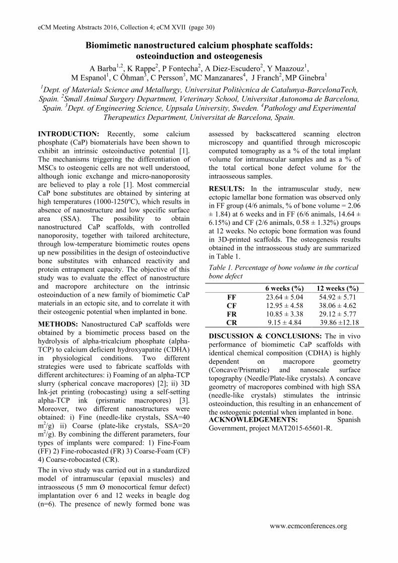
eCM Meeting Abstracts 2016, Collection 4; eCM XVII (page 30)
www.ecmconferences.org
Biomimetic nanostructured calcium phosphate scaffolds:
osteoinduction and osteogenesis
A Barba1,2
, K Rappe2, P Fontecha
2, A Diez-Escudero
2, Y Maazouz
1,
M Espanol1, C Öhman
3, C Persson
3, MC Manzanares
4, J Franch
2, MP Ginebra
1
1Dept. of Materials Science and Metallurgy, Universitat Politècnica de Catalunya-BarcelonaTech,
Spain. 2Small Animal Surgery Department, Veterinary School, Universitat Autonoma de Barcelona,
Spain. 3Dept. of Engineering Science, Uppsala University, Sweden.
4Pathology and Experimental
Therapeutics Department, Universitat de Barcelona, Spain.
INTRODUCTION: Recently, some calcium
phosphate (CaP) biomaterials have been shown to
exhibit an intrinsic osteoinductive potential [1].
The mechanisms triggering the differentiation of
MSCs to osteogenic cells are not well understood,
although ionic exchange and micro-nanoporosity
are believed to play a role [1]. Most commercial
CaP bone substitutes are obtained by sintering at
high temperatures (1000-1250ºC), which results in
absence of nanostructure and low specific surface
area (SSA). The possibility to obtain
nanostructured CaP scaffolds, with controlled
nanoporosity, together with tailored architecture,
through low-temperature biomimetic routes opens
up new possibilities in the design of osteoinductive
bone substitutes with enhanced reactivity and
protein entrapment capacity. The objective of this
study was to evaluate the effect of nanostructure
and macropore architecture on the intrinsic
osteoinduction of a new family of biomimetic CaP
materials in an ectopic site, and to correlate it with
their osteogenic potential when implanted in bone.
METHODS: Nanostructured CaP scaffolds were
obtained by a biomimetic process based on the
hydrolysis of alpha-tricalcium phosphate (alpha-
TCP) to calcium deficient hydroxyapatite (CDHA)
in physiological conditions. Two different
strategies were used to fabricate scaffolds with
different architectures: i) Foaming of an alpha-TCP
slurry (spherical concave macropores) [2]; ii) 3D
Ink-jet printing (robocasting) using a self-setting
alpha-TCP ink (prismatic macropores) [3].
Moreover, two different nanostructures were
obtained: i) Fine (needle-like crystals, SSA=40
m2/g) ii) Coarse (plate-like crystals, SSA=20
m2/g). By combining the different parameters, four
types of implants were compared: 1) Fine-Foam
(FF) 2) Fine-robocasted (FR) 3) Coarse-Foam (CF)
4) Coarse-robocasted (CR).
The in vivo study was carried out in a standardized
model of intramuscular (epaxial muscles) and
intraosseous (5 mm Ø monocortical femur defect)
implantation over 6 and 12 weeks in beagle dog
(n=6). The presence of newly formed bone was
assessed by backscattered scanning electron
microscopy and quantified through microscopic
computed tomography as a % of the total implant
volume for intramuscular samples and as a % of
the total cortical bone defect volume for the
intraosseous samples.
RESULTS: In the intramuscular study, new
ectopic lamellar bone formation was observed only
in FF group (4/6 animals, % of bone volume = 2.06
± 1.84) at 6 weeks and in FF (6/6 animals, 14.64 ±
6.15%) and CF (2/6 animals, 0.58 ± 1.32%) groups
at 12 weeks. No ectopic bone formation was found
in 3D-printed scaffolds. The osteogenesis results
obtained in the intraosseous study are summarized
in Table 1.
Table 1. Percentage of bone volume in the cortical
bone defect
6 weeks (%) 12 weeks (%)
FF 23.64 ± 5.04 54.92 ± 5.71
CF
FR
CR
12.95 ± 4.58
10.85 ± 3.38
9.15 ± 4.84
38.06 ± 4.62
29.12 ± 5.77
39.86 ±12.18
DISCUSSION & CONCLUSIONS: The in vivo
performance of biomimetic CaP scaffolds with
identical chemical composition (CDHA) is highly
dependent on macropore geometry
(Concave/Prismatic) and nanoscale surface
topography (Needle/Plate-like crystals). A concave
geometry of macropores combined with high SSA
(needle-like crystals) stimulates the intrinsic
osteoinduction, this resulting in an enhancement of
the osteogenic potential when implanted in bone. ACKNOWLEDGEMENTS: Spanish
Government, project MAT2015-65601-R.

eCM Meeting Abstracts 2016, Collection 4; eCM XVII (page 31)
www.ecmconferences.org
Why young companies will shape the MedTech World S Lauber Fürst
Inartis Network, CTI’s National Thematic Network (NTN) for the Life Sciences
WHY YOUNG COMPANIES: Breakthroughs in
MedTech will be resulting from rapid convergence
of the classical MedTech disciplines with Pharma
and IT/Internet of things/digitalisation, all
combined with the nanodimension of new
materials. Young companies embracing the speed
of the enormous growth of knowledge will be
particularly able to exceed all the usual dimensions
of transdisciplinary collaboration. Young MedTech
companies will therefore spearheading this
convergence, meaning the ongoing merger of life,
physical, digital and engineering sciences. Internal
critical preconditions will be the scientists’ ability
to engage in a true dialogue with other experts to
quickly learn from each other, identify new
opportunities and be effective from idea to market.
External critical preconditions will be the ability of
the regulatory bodies to define a solid and though
adaptable framework to ensure quality and safety
of those innovations.
THE ROLE OF INARTIS NETWORK: Inartis
Network is the Swiss Life Science Community and
a National Thematic Network (NTN) supported by
CTI, the Swiss federal agency for the promotion
and innovation. We are a motor and bridge builder
for transdisciplinary and cross-industry innovation
between business and academia, promoting
"Innovations Made in Switzerland” through vibrant
networking, initiating transdisciplinary and cross-
industry R&D projects and events.
Life Sciences, and in particular MedTech, belong
to the most dynamic and high-added value Swiss
industrial and academic sectors, with innovation as
driving force. Tomorrow’s breakthrough
innovations will not arise from within the
traditional Biotech, Medtech or Pharma, but from
transdisciplinary and cross-industry thinking and
working.
Inartis Network is federating the dialogue between
champions across the country and beyond. With
our Expert Network, we drive innovation focusing
on the areas of convergence, promoting
interactions, and securing Swiss-wide synergies
between the relevant disciplines and actors. We
aim at lowering all possible hurdles to innovation
and practice a no wrong door policy, which is the
only possible mind-set if you want to successfully
support young companies.
CONCLUSION: Young companies, able to
capture the benefits from the rapid convergence
will shape the MedTech world with disruptive
innovations. To succeed, however, those
companies need networks for quick and
unbureaucratic support and a regulatory framework
enabling and not blocking market access.
eCM Meeting Abstracts 2016, Collection 4; eCM XVII (page 32)
www.ecmconferences.org
Public support on the steep and thorny way from spinoff to market success
Gabor Szekely
Medical Image Analysis and Visualization
Computer Vision Laboratory
Swiss Federal Institute of Technology
ETH-Zentrum Zurich
INTRODUCTION: The talk will shortly
overview the numerous challenges and difficulties
whicha Medtech startup company faces from its
foundation to the successful introductionof its
product on the market. The different tools and
opportunities offered by public innovation support
will be presented and their role and significancein
this process will be analysed.

eCM Meeting Abstracts 2016, Collection 4; eCM XVII (page 33)
www.ecmconferences.org
A novel product and market entry challenges for a young medtech startup
Xiang Li & Elias BachmannZuriMED Technologies AG
INTRODUCTION: Although knee ligament
reconstruction is performed almost a million times
per year worldwide, a diverse range of techniques
is employed in the clinic – a situation that almost
always indicates that no technique is ideal. This
suspicion is confirmed by clinical evidence, with
highly variable long-term clinical outcomes after
knee ligament reconstruction. Many knee surgeons
are in favor of a bone-tendon-bone approach for
higher performance in terms of faster and stronger
healing, but at the cost of potentially severe pain at
the graft extraction site. The hamstring grafting
approach is widely used as well, which is less
painful, but with lower performance attributable to
slower healing with higher pullout risk.
We have developed a bone-tendon-bone
conversion kit, that has the potential to “convert” a
less painful “hamstring” graft into a higher
performance BTB-like graft. This device uses a
novel combination of advanced biomaterials that
could accelerate graft healing while minimizing
postoperative recovery time and pain. Our team
originated in and is currently based at the
Laboratory for Orthopedic Biomechanics at the
Uniklinik Balgrist. ZuriMED Technologies AG
was incorporated as a Balgrist and ETH Spin-off in
September 2015, and got financed to further
commercialize this device. Like most medtech
startups in the early stage, our biggest challenge
now is the regulatory pathway for market entry.
eCM Meeting Abstracts 2016, Collection 4; eCM XVII (page 34)
www.ecmconferences.org
A materials approach to bone regeneration: From idea to market Joost D. de Bruijn
Progentix Orthobiology BV, Bilthoven, The Netherlands; Institute of Bioengineering, Queen Mary University of London, UK; MIRA Institute, Twente
University, Enschede, the Netherlands
INTRODUCTION: The use of growth factors or
progenitor/stem cells for functional bone tissue
regeneration have received much attention as
potential alternatives to autologous bone grafting
in the past decades. Some of the hurdles to
overcome in these technologies include ensuring
cell survival with the cell therapy approach and
using potent but less supra-physiological
concentrations of growth factors to minimize
adverse reactions. To circumvent the necessity of
cells or growth factors in bone tissue regeneration,
we have developed a submicron structured calcium
phosphate ceramic that is capable of inducing bone
formation without the necessity of adding cells or
growth factors. These instructive ceramics have
shown excellent bone regeneration potential of
large, critical sized bone defects. In this talk, an
overview will be provided of the research
performed to develop this new class of bone graft
materials, including proof of efficacy and
regulatory studies.rnal.
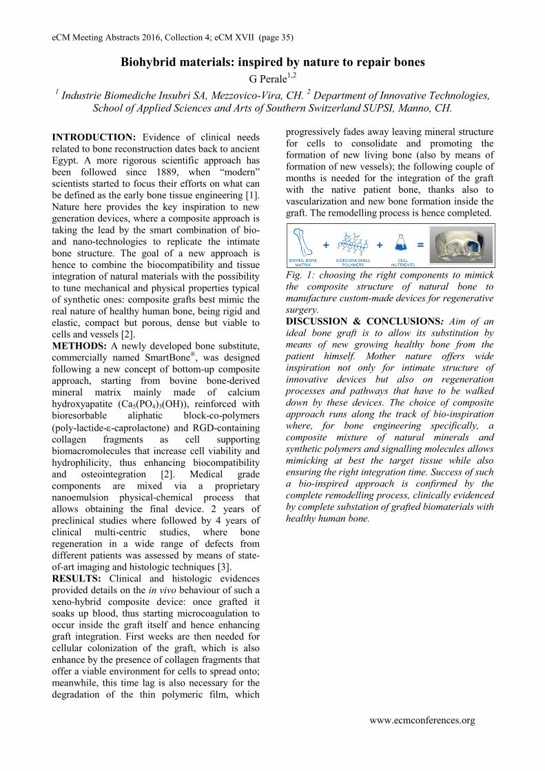
eCM Meeting Abstracts 2016, Collection 4; eCM XVII (page 35)
www.ecmconferences.org
Biohybrid materials: inspired by nature to repair bones
G Perale1,2
1Industrie Biomediche Insubri SA, Mezzovico-Vira, CH.
2 Department of Innovative Technologies,
School of Applied Sciences and Arts of Southern Switzerland SUPSI, Manno, CH.
INTRODUCTION: Evidence of clinical needs
related to bone reconstruction dates back to ancient
Egypt. A more rigorous scientific approach has
been followed since 1889, when “modern”
scientists started to focus their efforts on what can
be defined as the early bone tissue engineering [1].
Nature here provides the key inspiration to new
generation devices, where a composite approach is
taking the lead by the smart combination of bio-
and nano-technologies to replicate the intimate
bone structure. The goal of a new approach is
hence to combine the biocompatibility and tissue
integration of natural materials with the possibility
to tune mechanical and physical properties typical
of synthetic ones: composite grafts best mimic the
real nature of healthy human bone, being rigid and
elastic, compact but porous, dense but viable to
cells and vessels [2].
METHODS: A newly developed bone substitute,
commercially named SmartBone®, was designed
following a new concept of bottom-up composite
approach, starting from bovine bone-derived
mineral matrix mainly made of calcium
hydroxyapatite (Ca5(PO4)3(OH)), reinforced with
bioresorbable aliphatic block-co-polymers
(poly-lactide--caprolactone) and RGD-containing
collagen fragments as cell supporting
biomacromolecules that increase cell viability and
hydrophilicity, thus enhancing biocompatibility
and osteointegration [2]. Medical grade
components are mixed via a proprietary
nanoemulsion physical-chemical process that
allows obtaining the final device. 2 years of
preclinical studies where followed by 4 years of
clinical multi-centric studies, where bone
regeneration in a wide range of defects from
different patients was assessed by means of state-
of-art imaging and histologic techniques [3].
RESULTS: Clinical and histologic evidences
provided details on the in vivo behaviour of such a
xeno-hybrid composite device: once grafted it
soaks up blood, thus starting microcoagulation to
occur inside the graft itself and hence enhancing
graft integration. First weeks are then needed for
cellular colonization of the graft, which is also
enhance by the presence of collagen fragments that
offer a viable environment for cells to spread onto;
meanwhile, this time lag is also necessary for the
degradation of the thin polymeric film, which
progressively fades away leaving mineral structure
for cells to consolidate and promoting the
formation of new living bone (also by means of
formation of new vessels); the following couple of
months is needed for the integration of the graft
with the native patient bone, thanks also to
vascularization and new bone formation inside the
graft. The remodelling process is hence completed.
Fig. 1: choosing the right components to mimick
the composite structure of natural bone to
manufacture custom-made devices for regenerative
surgery.
DISCUSSION & CONCLUSIONS: Aim of an
ideal bone graft is to allow its substitution by
means of new growing healthy bone from the
patient himself. Mother nature offers wide
inspiration not only for intimate structure of
innovative devices but also on regeneration
processes and pathways that have to be walked
down by these devices. The choice of composite
approach runs along the track of bio-inspiration
where, for bone engineering specifically, a
composite mixture of natural minerals and
synthetic polymers and signalling molecules allows
mimicking at best the target tissue while also
ensuring the right integration time. Success of such
a bio-inspired approach is confirmed by the
complete remodelling process, clinically evidenced
by complete substation of grafted biomaterials with
healthy human bone.

eCM Meeting Abstracts 2016, Collection 4; eCM XVII (page 36)
www.ecmconferences.org
The adaptive immune system impacts bone healing processes
K Schmidt-Bleek1,2
, C Schlundt1,2
, C Bucher1,2
, A. Serra3, T ElKhassawna
4, S Wendler
1,2, H
Wagner1, HD Volk
2,5,GN Duda
1,2
1Julius Wolff Institut, Berlin, Germany,
2 Berlin-Brandenburg Center for Regenerative Therapies,
Berlin, Germany, 3 German Arthritis Research Center (DRFZ), Berlin, Germany,
4 Laboratory of
Experimental Trauma Surgery, Giessen, Germany, 5 Institute of Medical Immunology, Berlin,
Germany
INTRODUCTION: Delayed and disturbed bone
healing remains a relevant clinical problem and
will become even more relevant with the aging
population in developed countries. Apparently,
healing is challenged with an aged adaptive
immunity, characterized by a distinctly different
immune cell compositions in elderly compared to
young, less experienced individuals. Recently, the
influence of distinct immune cell subsets on the
regenerative healing capacity of bone has become
evident. The specific interplays of cellular
components of the adaptive immunity with those
of the bone system, however, have so far not been
fully understood.
METHODS: Fracture healing has been
investigated using either a fracture model (3 point
bending) with an internal nail fixation or a mouse
osteotomy model in which the left femur was
stabilized with an external fixator (RISystem) with
a 0.7 mm gap. Healing was observed for up to 28
days after surgery. The influence of the adaptive
immune system on the regenerative bone healing
process was investigated in WT Bl6 mice, in
RAG1-/-
mice lacking mature B and T cells, in JHT
mice lacking T cells, and in WT Bl6 mice depleted
of CD8+T cells using an antibody [anti-mCD8 AB
(YTS169.4), Bio-XCell] injection (200 mg of
mCD8 per injection for four consecutive days, with
the last day being the day of surgery) [1]. Immune
cell status, healing progress and healing success
were monitored using x-ray, biomechanical testing,
FACS, µCT, histology and immune histology.
RESULTS: Histological analyses of B and T cells
revealed the participation of these cells in all
fracture healing phases from early inflammation
until remodelling [2]. Animals lacking B and T
cells (RAG1-/-
) showed from early on a
significantly altered healing process [3]. Without B
and T cells organogenesis leads to stiffer bone
structure indicating the involvement of immune
cells in the organization of bone quality. To
analyse whether this effect in intact bones depends
on T or B cells, single knockout animals were
investigated. Animals lacking T cells showed
similar bone healing as RAG1-/-
animals while
animals lacking CD8+ T cells showed changes in
their osteocalcin positive cells. These cells are
responsible for the collagen I deposition, a major
extracellular matrix component in bone. In RAG1-/-
animals (lacking T and B cells) such changes in the
collagen deposition could be linked to changes in
the osteocalcin positive cell distribution and
biomechanical callus composition.
DISCUSSION & CONCLUSIONS: A distinct
interdependency of the skeletal and immune
system becomes apparent in bone healing but
recent findings also hint to an involvement of
immune cells (specifically CD8+ T cells) in bone
organogenesis. While the lack of T cells leads to
less mature collagen deposition, the positive or
detrimental effects of specific immune cell subsets
remains, however, still unclear: CD8+ effector
memory T cells have been proven to delay the
healing process [1] and regulatory T cells are
assumed to have a positive effect on the healing
[4]. Yet, the specific involvement of the immune
cells in the process of bone tissue formation and
mineralization remains still unclear. Our results so
far indicate, that a direct interaction of T cells and
osteocalcin positive cells could explain some of the
effects immune cell seem to have on the tissue
quality of bone during healing and organogenesis.
ACKNOWLEDGEMENTS: DFG
SCHE1594, SCHM2977, FG 2165, BCRT

eCM Meeting Abstracts 2016, Collection 4; eCM XVII (page 37)
www.ecmconferences.org
The role of angiogenesis in bone repair
Sabine FuchsExperimental Trauma Surgery, University Medical Center Schleswig Holstein, Kiel, Germany
INTRODUCTION: Vascularization is a critical
factor in bone healing, not only by ensuring the
supply with oxygen and nutrients. The current
understanding implies a central function of the
vascularization process in guiding bone repair at
the cellular and molecular level. These processes
are to a large extend controlled by the interaction
of endothelial cells and bone forming cells, as well
as circulating cells via paracrine factors,
extracellular matrix components and direct cell
communication mechanisms. The phenotype and
functional properties of circulating cells, such as
myeloid precursors, are determined by the
interaction with endothelial cells and osteogenic
cells mediating their differentiation towards
inflammatory cells or osteoclasts during bone
repair and remodeling. In addition, intermutual
interactions of endothelial cells and osteogenic
cells have a direct impact on the formation of new
blood vessels but also on the differentiation and
maturation of bone forming cells. Accordingly, a
better understanding of these cellular and
molecular processes might also result in new
therapeutical approaches to improve
vascularization. In this presentation we will
highlight how co-cultures might be used to study
the different mechanisms and pathways of cell
interaction which mediate vascularization, bone
repair or bone remodeling, respectively, but we
also briefly refer to biomaterial based approaches
creating vascularized tissue equivalents.
Such approaches might include the use of
autologous cells isolated from an adult patient. In
this context Outgrowth endothelial cells (OEC),
derived from cultures of mononuclear cells from
the peripheral blood, have been shown to form
functional blood vessels after co-culture or co-
implantation with osteogenic cells (Fuchs et al.,
2010; Fuchs et al., 2009a; Ghanaati et al., 2011).
Besides OEC, the initially diverse cell populations
from the peripheral blood also contain cells such as
early EPC contributing to the neovascularization in
vivo, although these cells don’t display the full
spectrum of mature endothelial markers. These
populations contain cells with a series of myeloid
and M2 macrophage characteristics and are able to
enhance the formation of vascular structures, when
added to co-cultures of OEC and MSCs (Shi et al.
2014). These observations are in accordance with
the current understanding of M2 macrophages
known to be actively involved in endothelial
repair. Nevertheless, myeloid precursor cells in
principle have the potential to transdifferentiate
into osteoclasts. In the co-culture approach the
gene expression of myeloid and osteoclast markers
seems to be modulated by endothelial as well as
bone forming cells (Shi et al 2016) thus
emphasizing the role these cellular interactions to
control cellular differentiation during bone repair.
eCM Meeting Abstracts 2016, Collection 4; eCM XVII (page 38)
www.ecmconferences.org
Small molecule Kartogenin promotes cartilage regeneration through activating
IL-6-based mesenchymal stem cell proloferation GQ Zhou, XLLi, T Liu, TH Chen, J Li, Y Zhu, YH Liang, ML Zhou
The Centre for Anti-ageing and Regenerative Medicine, Shenzhen University Medical School, Shenzhen, China; Shenzhen University, Shenzhen, China.
INTRODUCTION: Deregulation of the
endogenous stem cells in cartilage tissues is
considered to be a part of pathogenesis of
osteoarthritis. Kartogenin (KGN) has been
reported to exert stimulatory effects on
chondrogenesis of mesenchymal stem cells by
binding to filamin A that subsequently releases
and activates transcription factor CBFβ and by
up-regulate the Smad2/3 phosphosphorylation.
In this study, we report that KGN is capable of
stimulating the proliferation of primary
cartilage derived progenitor cells (CPCs).
METHODS: Cartilage derived progenitor
cells (CPC) were isolated from rat articular
cartilage tissues and KGN stimulated-CPC
proliferation was confirmed both in vitro and
in vivo. Slow-cycling cells within cartilage
were labeled and counted using RrdU and it
specific antibodies. Cell cycle was analyzed
using Flow Cytometry and RNA-seq was
performed on CPC co-incubated with KGN for
5 days. IL-6 and Stat3 phosporylation were
detected with ELISA kit and Western blotting.
Cartilage repair/regeneration by oral
administration and/or intra-articular injection
of KGN was observed in rat knee joint injury
models.
RESULTS: Our data shows that following 10
M KGN treatment for a week, the percentage
of G2-M phase cells in mitosis reached 9.6%,
nearly twice of the control group, which was
companied with the doubled total cell number.
In the meanwhile, even after 4 weeks
stimulation with KGN, Cells were proved to
remain the expression of mesenchymal stem
cell markers CD90(93%) and CD105(98%).
As a control, no significant number change
was observed in mature human T lymphocyte
treated with KGN in the similar manner.
Whole RNA-sequencing analysis of KGN-
stimulated MSCs showed that significant
expression changes of about 20 cell cycle-
related genes upon KGN treatment for 72
hours. Among a number of genes found to be
significantly changed by the KGN treatment
are IL-6 and its receptor Gp130, which reach
as much as 6 fold increase than the control.
We further confirmed that the IL-6 level was
significantly increased by KGN in both
cytoplasm and supernatant media of CPC
culture. We further demonstrated that the
phosphosphorylation of Stat-3 was up-
regulated at the same time. In-articular
injection of KGN were also found to increase
the number of BrdU-labeled slow-cycling
cells. In vivo experimental evidences of the
increased thickness in articular cartilage with
KGN treatment was further confirmed in the
rat model that were induced to have knee joint
injury. IHC staining of the KGN treatment
group showed up-regulate of Stat-3
phosphosphorylation in KGN-treated cartilage.
DISCUSSION & CONCLUSIONS: Based
on the our in vivo and in vitro data, we
propose that KGN may improve the number of
endogenous cartilage stem cells by promoting
their self-renewal in situ while inducing
chondrogenesis and its use in cartilage
regeneration and repair is definitely worth of
further explored.
ACKNOWLEDGEMENTS: This study was
supported by Natural Science Foundation of
China (NSFC 81472126).

eCM Meeting Abstracts 2016, Collection 4; eCM XVII (page 39)
www.ecmconferences.org
In vivo bone marrow and cytokine dynamics in two experimentally injured
tissues
L Leitão1,2,3
, CJ Alves1,2
, IS Alencastre1,2
, DM Sousa1,2
, E Neto1,2,4
, C Leitão1,5
, G Almeida-Porada6,
M Lamghari1,2,3
1Instituto de Investigação e Inovação em Saúde, Universidade do Porto (UP). Porto, PT.
2Instituto
de Engenharia Biomédica, UP, Porto, PT. 3Instituto Ciências Biomédicas Abel Salazar, UP, Porto,
PT. 4Faculdade de Medicina, UP, Porto, PT.
5Instituto de Biologia Molecular e Celular, UP, Porto,
PT. 6Wake Forest Institute for Regenerative Medicine, Wake Forest University School of Medicine,
Winston-Salem, USA.
INTRODUCTION: Bone marrow (BM) multiple
cell populations, namely hematopoietic cells
(HSCs), mesenchymal stem cells (MSCs) and
endothelial cells (EPCs), were reported to play a
key role in tissue repair and regeneration1.
Cytokine release was also described to be
associated to these processes, providing cells
signals to orchestrate in coordinated manner the
different phases of tissue regeneration, from
inflammation to ECM remodelling2. These
findings were described in different experimentally
injured tissues, including muscle, heart, kidney,
skin, bone, liver and brain1. However, little is
known whether different tissue insults, injury
incidence and regeneration trigger similar temporal
coordination of these BM multiple cell populations
as well as the associated cytokines. Here we
analysed the different subsets of BM cell
populations and serum cytokines in two injury
models: bone defect and skin/muscle laceration
wound at different stages of tissue regeneration.
METHODS: Adult C57/Bl6 mice were submitted
to skin/muscle laceration or to femoral bone defect.
Of note, in the bone defect group to access to
femoral bone and achieve bone injury, skin/muscle
cuts were also performed. Thus, injury incidence in
this group is higher when compared to skin/muscle
laceration group. Non-operated animals were used
as controls. Animals were sacrificed 1, 3 and 7
days post-injury. At each time point, the BM was
harvested from the femurs and its different cellular
populations were analysed by flow cytometry.
These included the mesenchymal stem cells
(Sca1+; CD105
+; CD140a
+; CD11b
-; CD45
-; CD34
-
), endothelial cells (CD105+; CD31
+, CD11b
-;
CD45-; CD34
-) and hematopoietic progenitor cells
(Sca1+; CD117
+, CD3
-; CD4
-; CD8
-; CD11b
-;
CD19-; TER119
-; Ly6G
-; F4/80
-). Immune cell
populations from the adaptive (CD8+ T cells, CD4
+
T cells, CD4-/CD8
- T cells and B cells) and the
innate immunity (CD11b+ cells, macrophages and
dendritic cells) were also analysed. Serum
cytokines and chemokines relative levels were
analysed at each time point using a commercially
available proteome profiler.
RESULTS AND DISCUSSION: Both
skin/muscle laceration and femoral bone defect
groups showed alterations in the BM populations
at the different time points upon injury. Each BM
subset undergoes level´s variation in a time
dependent manner. These alterations follow the
same pattern in both groups. Interestingly,
comparison of cell percentage of each BM subset
between the two injury groups revealed no
significant differences. Among several factors, pro
and anti-inflammatory cytokines, chemokines and
others have been described to play determinant
roles in inflammation, MSCs homing and ECM
remodelling. Cytokine analysis showed that G-
CSF, MCP-1, CXCL1, CXCL13 and TIMP-1 were
significantly triggered in both skin/muscle
laceration and bone femoral defect when compared
to non-operated animals. Surprisingly, in both
groups, the levels of SDF-1/CXCL12 remained
unchanged at the different time points after injury
and are comparable to those of the non-operated
animals. G-CSF, MCP-1, CXCL1, CXCL13 and
TIMP-1alterations follow a time course pattern but
with different magnitudes that are tissue injury
dependent. In addition, in bone defect animals,
cytokine levels are associated with long-
lasting changes. Overall, these results suggest a
reconfiguration of the BM cellular populations
upon skin/muscle and bone tissue injury and
during regeneration that seems to be tissue and
injury incidence-independent but accompanied
with differential systemic cytokine responses.
ACKNOWLEDGEMENTS: This work was
financed by FEDER through the PT2020, and by
Portuguese funds through FCT/MEC in the
framework of the project POCI-01-0145-FEDER-
007274.

eCM Meeting Abstracts 2016, Collection 4; eCM XVII (page 40)
www.ecmconferences.org
Effective vascularization and efficient bone formation in osteogenic grafts
requires VEGF dose control A Lunger
1,2, MG Burger
1,2, A Grosso
1, P Briguez
3, JA Hubbell
3 , D Schaefer
2, A Banfi
1,
N Di Maggio1
1 Cell and Gene Therapy, Department of Biomedicine, Basel University Hospital, Basel,
Switzerland; 2
Department of Plastic, Reconstructive, Aesthetic and Hand Surgery, Basel University
Hospital, Switzerland; 3Institute of Bioengineering, EPFL Lausanne, Switzerland
INTRODUCTION: Spontaneous vascularization
of clinically relevant, large-size bone grafts based
on bone marrow-derived mesenchymal stem cells
(BMSC) is insufficient and requires therapeutic
stimulation to ensure progenitor survival and bone
formation. Vascular endothelial growth factor-A
(VEGF) is the master regulator of angiogenesis.
However, we found that, while its sustained over-
expression by genetically modified human BMSC
effectively improved vascularization of osteogenic
grafts, it also impaired bone formation through
excessive osteoclast recruitment [l]. Recently we
found that delivery of VEGF for a limited duration
of 4 weeks in the form of recombinant protein
covalently bound to a fibrin hydrogel prevented
excessive bone resorption while ensuring increased
vascularization (Burger et al. unpublished results).
Here we sought to investigate the role of VEGF
dose on the coupling of angiogenesis and bone
formation, in order to define a VEGF therapeutic
window for vascularized tissue-engineered bone.
METHODS: Recombinant VEGF was engineered
with a transglutaminase substrate sequence (TG-
VEGF) to allow cross-linking into fibrin hydrogels
[2]. Osteogenic constructs were prepared with
primary human BMSC seeded on hydroxyapatite
granules in a fibrin hydrogel containing 4 different
TG-VEGF concentrations (0.1, 1, 10 and 100
µg/ml) and optimized to ensure the controlled
release of the factor over 4 weeks [3]. Control
grafts were generated with BMSC only or
retrovirally transduced BMSC that constitutively
express VEGF linked to the cell-surface marker
truncated CD8 (VICD8). Histological analysis 1, 4
and 8 weeks after ectopic subcutaneous
implantation in nude mice was used to determine
vascularization (CD31 immunostaining), bone
formation (H&E and Masson Trichrome) and
osteoclast recruitment (TRAP staining).
RESULTS: All VEGF doses effectively increased
vessel density up to 5-fold already after 1 week and
vascularization persisted at all later time-points.
After 4 and 8 weeks, bone tissue development was
enabled by 0.1 and 1 µg/ml of TG-VEGF as
efficiently as with naïve BMSC alone. However,
higher doses progressively impaired bone
formation and 100 µg/ml caused a similar
reduction as with VEGF-expressing genetically
modified BMSC. The loss of bone formation
correlated with increased osteoclast recruitment.
Fig. 1: Masson’s Trichrome staining shows that
increasing VEGF doses gradually impair bone
formation 8 weeks after in vivo implantation of
osteogenic grafts.
DISCUSSION & CONCLUSIONS: These data
suggest that VEGF effects on promoting
vascularization and bone resorption can be
uncoupled by short-term delivery of recombinant
VEGF protein. However, VEGF effects on bone
resorption are dose-dependent and a therapeutic
window exists that enables both rapid
vascularization and efficient bone formation. This
could provide a clinically applicable strategy with
several attractive features: 1) no genetic
modification; 2) homogeneous and tunable factor
doses; 3) limited and controllable duration of
factor delivery.
2 ACKNOWLEDGEMENTS: This work
was supported by a Marie Heim Vögtlin grant
(158312) by Swiss National Science Foundation
to N.D.M. and an intramural grant by the
Department of Plastic and Reconstructive
Surgery (Basel University Hospital)

eCM Meeting Abstracts 2016, Collection 4; eCM XVII (page 41)
www.ecmconferences.org
Adipose-derived stem cell seeded biominerizable nanocomposite for chest wall
repair: suppression of inflammatory response in a murine model
J Buschmann1, E Balli
1, SC Hess
2, WJ Stark
2, P Cinelli
3, S Märsmann
1,3, M Welti
1,4, W Weder
4,
W Jungraitmayr4
1Plastic Surgery, University Hospital Zürich, Zürich, CH.
2Institute for Chemical and
Bioengineering, ETH, Zürich, CH. 3Trauma Surgery, University Hospital Zürich, Zürich, CH.
4Thoracic Surgery, University Hospital Zürich, Zürich, CH.
INTRODUCTION: Defects to the chest wall can
occur after tumour resections or trauma caused by
accidents, and appropriate chest wall
reconstruction is therefore needed. Stability and
integrity of the repaired chest wall should reach
similarity to natural physiology. Addressing the
treatment of critical size full-thickness chest wall
defects, the ideal graft should be stable, fluid- and
air-tight, biocompatible inducing no inflammatory
reactions, biodegradable during the healing with
non- toxic degradation products as well as rapidly
integrating into the surrounding tissue. Here, we
present the implantation of a biocompatible,
biodegradable and easily vascularizable
nanocomposite seeded with adipose-derived stem
cells (ASCs) as a chest wall graft in a murine
model. The cellular response towards this graft is
compared to the cell-free graft.
METHODS: An electrospun poly(lactic-co-
glycolide)/amorphous calcium phosphate
(PLGA/aCaP) nanocomposite was seeded on both
sides with murine ASCs and cultivated for two
weeks before implantation as a chest wall graft. In
addition, a cell-free analogous PLGA/aCaP
scaffold was implanted on top of the cell-seeded
scaffold towards the skin in order to be able to
study not only direct cell-to-cell contact-based
effects, but also to address paracrine effects caused
by ASCs (control: cell-free scaffold alone).
Histomorphometric analysis was performed at 4
and 8 weeks post-operation, respectively, to assess
cell density of macrophages, lymphocytes and
foreign body giant cells (Figure 1).
RESULTS: Inflammatory response towards the
graft material was significantly reduced for
macrophages, lymphocytes and foreign body giant
cells in the presence of ASCs compared to cell-free
scaffolds. Moreover, this anti-inflammatory action
caused by ASCs was not only found on the side
where direct cell-to-cell contact between seeded
ASCs and local cell population was enabled and
studied, but also on the scaffold side where
predominantly diffusible factors secreted by ASCs
were active (paracrine function).
Fig. 1: F4/80 stained histological section at 8
weeks post-operation showing selected
macrophages (white arrows) and electrospun
PLGA/aCaP fibers of the degrading chest wall
graft.
DISCUSSION & CONCLUSIONS: In clinics,
the state of the art of repairing critical size chest
wall defects is to use inert materials such as
Goretex® which are not easily vascularizable and
not biodegradable. Here, we present a
biocompatible, biodegradable and well
vascularizable nanocomposite for chest wall repair.
In order to enhance integration of this graft
material and accelerate wound healing, ASCs were
seeded. A beneficial effect of these ASCs was that
the inflammatory response towards the implant
was significantly reduced. Therefore, such cell-
seeded nanocomposites may be applied as chest
wall grafts in clinics in the future.
ACKNOWLEDGEMENTS: We thank Gabriella
Meier Bürgisser for her help with analysis of
histological sections and Pia Fuchs for histological
staining.

eCM Meeting Abstracts 2016, Collection 4; eCM XVII (page 43)
www.ecmconferences.org
Clinical use of stem cells
Gun-Il Im 1 Donngguk University, Goyang, Koreas
Orthopaedic medicine has been traditionally
benefited from the innovation from other field of
science. Development of metal and chemical
engineering in the early 20th century contributed to
current orthopaedic practice as various implants
based on newly developed biomaterials were
devised and applied for treating patient. Recent
advancement in regenerative medicine has opened
a new horizon in the orthopedics and may shift the
paradigm in clinical practices in the future. The
diseases which are currently managed by surgical
treatment may be more effectively and more
economically treated by less invasive procedures
such as simple injection of cells.
Stem cell research has mostly inspired from the
need to explore the new therapeutic possibility for
intractable and lethal diseases. Although
musculoskeletal disorders are basically nonlethal,
the high prevalence of diseases and relative ease in
performing clinical trial has facilitated the clinical
application of stem cell in this field. On the other
hand, there is a relative paucity of reliable clinical
studies despite the plethora of in vitro and
preclinical studies in the area of stem cell research
for regenerative medicine in musculoskeletal
system.
The stem cell therapy can be locally applied for the
regeneration of bone, cartilage and tendon.
Candidate disease modalities in bone regeneration
includes large bone defect, nonunion of fracture,
osteonecrosis. Focal osteochondral defect and
osteoarthritis are current targets for cartilage
regeneration. For tendon regeneration, bone-tendon
junction problems such as rotator cuff tears are hot
topics in clinical research. In this talk, the current
status of stem cell application in clinical field is
introduced along with future perspective from the
author’s point.
ACKNOWLEDGEMENTS: This work was
supported by a grant from the National Research
Foundation (NRF) funded by the Korean
government (2015R1A2A1A09002793)

eCM Meeting Abstracts 2016, Collection 4; eCM XVII (page 44)
www.ecmconferences.org
BOOSTB4: a clinical study on pre- and/or postnatal stem cell transplantation
for treatment of osteogenesis imperfecta
C Götherström1,2
, C DeVile3, R Sakkers
4 O Semler
5, E Åström
6,7
on behalf of the BOOSTB4 consortium 1Department of Clinical Science Intervention and Technology and
2Center for Hematology and
Regenerative Medicine, Karolinska Institutet, Stockholm, Sweden. 3Great Ormond Street Hospital,
London, United Kingdom. 4University Medical Centrum Utrecht, Utrecht, the Netherlands,
5Uniklinik Köln, Köln, Germany.
6Astrid Lindgren´s Children Hospital and
7Department of Woman
and Children’s Health, Karolinska Institutet, Stockholm, Sweden.
INTRODUCTION: Osteogenesis imperfecta (OI)
is a heterogeneous inherited condition and severe
forms present already in utero. Persons with severe
OI is affected throughout their lifetime with
repeated, multiple fractures, short stature and
orthopaedic problems, considerable pain and
handicap. There is no curative or sufficiently
effective symptomatic treatment for OI.
Preliminary clinical experience in two cases
indicates that transplantation of fetal liver derived
mesenchymal stem cells (MSC) before and after
birth may ameliorate symptoms1,2
.
The main objective of the Boost Brittle Bones
Before Birth (BOOSTB4) phase I/II multicentre
study is to evaluate the safety and efficacy of pre-
and/or postnatal MSC transplantation in severe
vital forms of OI (type III and severe type IV).
METHODS: The study will include three groups:
1) Prenatal and postnatal transplantations in 15
patients, inclusion during pregnancy
2) Postnatal transplantation in 15 patients,
inclusion before one year of age
3) Historical and prospective controls, 30-60 cases
Over twenty months, the patients will receive four
postnatal infusions of same-donor MSC at 4-month
intervals.
The primary outcome is safety for the fetus, child
and pregnant woman. Secondary outcomes relate
to efficacy, including fracture frequency, growth,
bone mineral density over 20 months. Rapid
exome sequencing using a panel targeted for
skeletal disorders for definitive molecular
diagnosis of OI will be developed. Experience,
impact and perception of the therapy will be
evaluated in both treatment groups.
RESULTS: We have established a European
network centred around four clinical hubs in
Stockholm, Cologne, London and Utrecht/Leiden.
Early studies have shown that rapid diagnosis of
skeletal dysplasias using whole exome sequencing
is possible. Production of MSC is underway and
the clinical trial protocol for transplantation and
follow-up is being finalised. Recruitment to the
main treatment study will soon commence.
DISCUSSION & CONCLUSIONS: Prenatal
stem cell transplantation shows promise for the
treatment of inherited single gene disorders.
Demonstration that MSC transplantation improves
early outcome in patients with severe OI would
represent a major step forward in the management
of these patients. If successful, such treatment
could be relevant for the management of a range of
other inherited birth defects. The BOOSTB4
consortium welcomes clinical cases for diagnosis
of OI using rapid exome sequencing and, for the
first time, inclusion of European patients in the
clinical trial on treatment of OI with fetal MSC
postnatally.
Götherström more information:
pre- and/or Contact Cecilia
for
ACKNOWLEDGEMENTS: This project
has received funding from the European
Union’s Horizon 2020 research and innovation
programme under grant agreement No 681045
and from the Swedish Research Council
(E0720901).

eCM Meeting Abstracts 2016, Collection 4; eCM XVII (page 45)
www.ecmconferences.org
in situ poly(carboxybetaine) hydrogel for bone tissue application
Hsiu-Wen Chien1, Hsin-Yu Chen
1, Jiashing Yu*
1, Wei-Bor Tsai*
1
1 Biomedical and Tissue Engineering Lab, Department of Chemical Engineering, National Taiwan
University, Taipei, Taiwan
INTRODUCTION: We re-investigated the
cytotoxocity of CBMA, which was comparable to
other commonly used zwitterionic monomers, such
as 2-Methacryloyloxyethyl phosphorylcholine
(MPC) and sulfobetaine methacrylate (SBMA).
Biocompatibility and rheology of the
polymerization process was also investigated.
Next, the zwitterionic materials were directly
injected into a mouse subcutaneously to study the
tissue response. Finally, the hydrogels were
prepared with bio-functional molecules, such as
RGD peptides and hydroxyapatite (HAp) to
examine the concentration effects of adhesive
ligands for bone tissue engineering.
METHODS: Zwitterionic monomer solution was
prepared in PBS at a concentration of 20% (w/v).
Crosslinkers of NDMCC and PEGDMA and the
initiator of APS/TEMED were added to the
monomer solution at concentrations of 1 mol% of
monomer and 10 mM, respectively. After
polymerization, hydrogels were removed from the
casts and soaked in PBS for complete swelling.
The fully swollen hydrogel disks were compressed
to failure at a rate of 35.4 mm/min with a
mechanical tester (FGS-50V-H, NIDECSIMPO
Corporation, Japan) and a digital force gauge
(FGP-0.5, NIDECSIMPO Corporation, Japan). The
Young's moduli of poly(zwitterion) hydrogels were
calculated by applying 35 to 60% strain. Physical
properties and the gelation times of each hydrogel
were assessed with an AR2000 rheometer (TA In-
struments) and a parallel plate geometry. Primary
rat osteoblasts were isolated from calvariae bonds
of neonatal rat. Cells were mixed and suspended in
the solution to a final seeding den-sity of 5x10⁶
cells /mL and allowed for polymerization at 37°C
for 30 mins. For in vivo osteo-genic differentiation
in the injectable poly(CB) hydrogels, the three
experimental groups were designated as CB,
CB/RGD, and CB/RGD/HAp hydrogels. The
precursor solutions were in-jected into the
subcutaneous of a nude mouse (4-5 weeks old),
respectively.
RESULTS:
Fig. 1: Time dependence of storage modulus (G’,
solid) and loss modulus (G”, hollow) of
polymerized solutions gelled at pH 7.4 at 37oC.
Fig. 2: The images of the dissection of
MSC/scaffold constructs, stained by Alizarin red
staining. The scale bar represents 100 μm.
DISCUSSION & CONCLUSIONS:
This study demonstrated that three zwitterionic
monomers, CBMA, SBMA, and MPC, exhibited
low cytoviabilityies. We subsequently prepared the
CBMA precursor solution with Arg-Gly-Asp
(RGD) and hydroxyapatite (HAp) nanoparticles for
osteogenic tissue engineer-ing. Both in vitro and in
vivo studies demonstrated that HAp containing
poly(CB) hydrogels greatly enhanced the
mineralization on the deposited substrates. These
results indicated that poly(zwitterions) hydrogels
may be useful in generation of biocompatible,
implantable medical devices and tissue scaffolds.

eCM Meeting Abstracts 2016, Collection 4; eCM XVII (page 46)
www.ecmconferences.org
Novel approach to control drug release and adherence of PLA based implant
coatings
E Choinska1, W Swieszkowski
1
1Faculty of Materials Science and Engineering, Warsaw University of Technology, Warsaw, Poland
INTRODUCTION: The typical approach to
control drug release from polymeric implant
coatings is based on modifying composition and
molecular weight of carrier or geometric
parameters of the system [1]. In such DDS also the
proper adhesion of polymer to substrate have to be
provided. Usually it is done by increasing of
contact area (eg. surface development), or
chemical treatment of substrate (eg, silanization, or
phosphating) [2]. In this work novel approach,
based on changing of macromolecular structure of
PLA, is presented.
METHODS: Two linear (2b) and two 4-arms (4b)
polylactides were synthesized by ring opening
method (Tab.1). Model drug, gentamicin sulphate
(GS), and ion complex GS-AerosolOT (GS-AOT)
were incorporated into polymer carrier during
dissolving polymer in CHCl3, and then coatings
were prepare by dip-coating of silanized [3]
stainless steel substrate. The adherence of
polymers was measured by scratch test (F=1-10N,
2N/min, l=3mm) and critical loads (Lc) were
determined. Drug release test was perform in PBS,
at 37oC for 5 weeks and fitting of obtained data to
different mathematical models was done.
Table 1. Polymers used for implant coatings
preparation.
Polymer Mn
(kgmol-1
)
Mw
(kgmol-1
)
Tg
(ºC)
Tm
(ºC)
4bPdlLA 34.3 39.9 37.7 -
2bPdlLA 34.9 45.0 38.5 -
4bPlLA 40.7 46.0 60.6 171.0
2bPlLA 34.2 37.8 60.9 173.5
RESULTS: The kinetics of drug release is shown
in figure 1.
Fig. 1: Release of GS (a) and GS-AOT (b) from
linear and 4-armes PLAs.
Table 2. Results of scratch test.
Polymer 4bPdlLA 2bPdlLA 4bPlLA 2bPlLA
Lc (N) 3.6 2.8 3.5 2.8
DISCUSSION & CONCLUSIONS: Drug release
test showed that drug is faster release from
4bPLAs. GS diffuse according Higuchi model, but
for GS-AOT the best fitting was obtained for
Korsmeyer-Peppas model [4] . The analysis of
determined values of critical load and comparison
of area of damaged coating have shown that
adherence of polymers is increasing in following
order: 2bPlLA < 4bPlLA <2bPdlLA< 4bPdlLA.
It can be concluded, that changing of
macromolecular structure allow to control drug
release and adherence of PLA based implant
coatings.
ACKNOWLEDGEMENTS: This study
was financially supported by the National
Centre for
Research and Developments (MentorEye project
-STRATEGMED1/2333624/4/NCBR/2014).

eCM Meeting Abstracts 2016, Collection 4; eCM XVII (page 47)
www.ecmconferences.org
The efficacy of local bisphosphonate and BMP-2 delivery in improving bone
mass and mechanical implant stability
L Freitag¹*, C Günther¹*, L Kyllönen¹*, U Eberli¹, K Thompson¹, D Arens¹, S Zeiter¹, D Eglin¹, VA
Stadelmann¹ 1 AO Research Institute, AO Foundation, Davos, CH.
*Equivalent contributions
INTRODUCTION: Worldwide, osteoporosis
causes more than 8.9 million fractures, which
means one fracture every three seconds1. For
people older than 50 years, one out of three women
and one out of five men will have an osteoporotic
fracture 2 3 4
.
Implant fixation in osteoporotic bones can be quite
challenging due to low bone mass and reduced
mechanical properties.
This study aims to improve implant stability in
osteoporotic bone using a hyaluronan hydrogel for
local delivery of bisphosphonates (BP) and bone
morphogenetic protein 2(BMP-2).
We hypothetised, that BP would prevent early
resorption in response to interventional trauma and
BMP-2 support bone formation, which is impaired
in osteoporotic bone.
METHODS: 41 female wistar rats were divided
into 7 groups: Groups 1 and 2 were the healthy
controls, groups 3 to 7 were ovariectomized at 13
weeks. All animals received a BaSO4-PEEK
miniscrew in the proximal tibia at 25 weeks. In
groups 2 and 4, pure hydrogel was pipetted into the
drill hole before screw insertion. ZOL-BMP2
loaded hydrogel was given in group 5. Group 6
received zoledronate systemically, group 7
zoledronate systemically and BMP-2 locally.
Rats were euthanised 28 days post screw insertion.
Bone mineral density (at 12, 24 and 29 weeks) and
implant osseointegration (0, 3, 6, 9, 14, 20 and 28
days post-op) were monitored using in-vivo
microCT. Post mortem, samples underwent
histological examinations to determine
osseointegration on a cellular level.
RESULTS: Our preliminary data show, that the
pure hydrogel is bioinactive in terms of implant
fixation. ZOL-BMP2-hydrogel induces significant
increase of bone-implant contact and peri-implant
bone fraction, mostly through reduced resorption.
DISCUSSION & CONCLUSIONS: In
conclusion, combination of zoledronate and BMP-
2 might be able to improve implant stability in
osteoporotic bones significantly and local delivery
might be a potent alternative to systemic drug
administration at significantly lower
bisphosphonate doses.
ACKNOWLEDGEMENTS: This study
was funded by AO Trauma.

eCM Meeting Abstracts 2016, Collection 4; eCM XVII (page 48)
www.ecmconferences.org
Critical size bird bone defect healed with xenogeneic ostrich cancellous bone
grafts seeded with aviary bone cells – experimental bird model
D Harvanova1, T Spakova
1, J Plsikova
1, J Amrichova
1, S Hornak
2, J Rosocha
1
1 Associated Tissue Bank, Faculty of Medicine of P. J. Safarik University and L. Pasteur University
Hospital, Kosice, Slovakia, 2 Small animal’s clinic, University of veterinary medicine and pharmacy, Kosice, Slovakia
INTRODUCTION: Large bone defects do not
heal autonomously. Gold standard in treatment of
critical bone defects is autologous bone grafting.
Treatment of bone fractures in avian species has
many critical limitations and one of these is lack of
donor cancellous bone source for eventual bone
autografting. Lack of autologous bone grafts in
birds has prompted investigation of avian
xenografts for bone augmentation. The purpose of
this study was to evaluate model bone regeneration
of critical segmental bone defects in pigeons by
application of ostrich cancellous bone grafts
(OCBG) seeded with allogeneic donor bird bone
cells.
METHODS: The experiments were performed on
model animals - pigeons with the approval of the
University of Veterinary Medicine and Pharmacy
ethical committee. Critical segmental bone defects
(1 x 0,5 cm) were created on ulna and pigeons
were divided into 5 groups according to the bone
defects healing process. Bone defects were filled
with demineralized and non-demineralized OCBG
separately or in combination with aviary bone
cells. Control group was healed spontaneously
without treatment. Isolation and cultivation of
aviary bone cells and methods of their seeding on
OCBS was described previously [l] Bone
regeneration was evaluated by radiograph and
histology 1, 2, 3, 4, 5, 6, 9, 12 months after
implantation.
RESULTS: Bone regeneration was significantly
enhanced in the defects treated with demineralized
OCBG in combination with aviary cells when
compared with other experimental groups. Three
months after implantation, OCBG’s resorption was
observed in the defect. At 6 and 9 months after
implantation, complete bone regeneration without
residual bone substitute in the defect was obtained
(Fig.1). New bone tissue in the defect was
confirmed by histology. In the groups, where the
OCBG was implanted without aviary bone cells,
incomplete bone regeneration was observed. Nine
months after demineralized OCBG implantation,
smooth radiolucent line still remained on the
radiological images. Nevertheless, the new bone
formation was histologically confirmed in the
defect even in the groups without applied aviary
bone cells. No bone regeneration in the defect was
observed in the untreated group
Fig. 1: RTG evaluation of bone regeneration after
1,3,6,9 month’s implantation of demineralized
OCBG with aviary bone cells into bone defects on
pigeon’s ulna
DISCUSSION & CONCLUSIONS:
Ostrich bone grafts used for bone regeneration
were biocompatible but also have an appropriate
time of degradation. When the rate of substitute
degradation is too slow, the bone substitute itself
can prevent bone regeneration. We demonstrate
that OCBG was completely resorbed by 3 months
after implantation in all cases. More important fact
is that OCBG provided an effective biological
support for aviary bone cells. We conclude that
OCBG in combination with aviary bone cells had
osteoconductive and osteoinductive properties
which are necessary for the regeneration of bone
defects in birds.
ACKNOWLEDGEMENTS:
This research was supported by VEGA grant
No. 1/0772/13 and Slovak Research and
Development Agency (APVV 0684-12).

eCM Meeting Abstracts 2016, Collection 4; eCM XVII (page 49)
www.ecmconferences.org
Effect of zoledronate on periodontitis in a compromised rat model
L Huo1, LW Zheng
1
1 Discipline of Oral diagnosis & Polyclinics, Faculty of Dentistry, The University of Hong Kong,
Hong Kong SAR, China
INTRODUCTION: Bisphosphonate is a powerful
antiresorptive medication which has been widely
used to manage osteoporosis and bone
complications in cancer patients. Some studies
found that bisphosphonate may benefit the
treatment of periodontitis, however, other studies
implied that periodontitis is a precipitating factor
which may increase the risk of bisphosphonate
related necrosis of the jaw. The aim of this study
was to assess the risk and benefit of
bisphosphonate on treating periodontitis using a
compromised rat model.
METHODS: All 45 female SD rats received
bilateral ovariectomy and then were assigned
randomly to three groups, 15 in each: Group A
received zoledronic acid (ZA) at a high dose
(67μg/kg, thrice per week, i.p.), Group B received
ZA at a low dose (88μg/kg, once per month, i.p.),
and Group C was injected with saline[1]. Along
with ZA treatment, dexamethasone (DEX) was
administrated to all the animals to mimic a
compromised condition[2, 3]. At the same day of
the injection, ligature was placed around the left
mandibular first molar (L-M1) of each rat. 12
weeks later, all the rats were sacrificed and the
whole mandibles were collected for micro-CT
scanning. The distance between the cemento-
enamel junction and alveolar ridge crest
(attachment loss) of L-M1 was measured, the bone
mineral density (BMD) was also detected and
statistically analysed.
Fig. 1: The measurements of Bone loss (left) and
BMD (right)
RESULTS: Compared with saline treated group,
the attachment loss of the ligature site was
inhibited significantly in both low dose and high
dose ZA group (p<0.05), while no significant
different between low and high dose ZA was
noted. Compared with both saline treated and low
dose ZA treated groups, the BMD in the high dose
ZA treatment group was significantly improved.
Fig.2: Comparison of BMD (A) and Bone loss (B)
between different treatments
DISCUSSION & CONCLUSIONS: The
systemically administrated zoledronate could
benefit the treatment of periodontitis, either in high
dose or in low dose, by inhibiting alveolar bone
loss in the compromised rat model.
ACKNOWLEDGEMENTS: This template
was modified with kind permission from eCM
Journal.
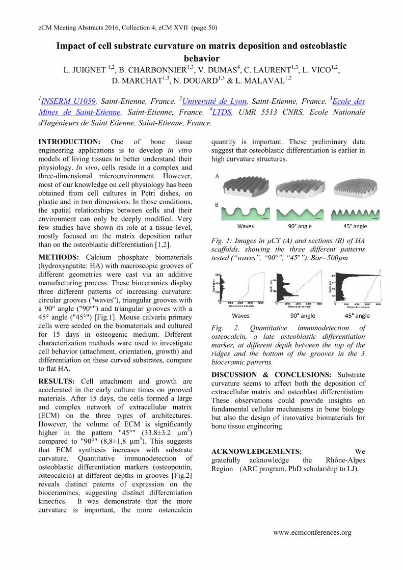
eCM Meeting Abstracts 2016, Collection 4; eCM XVII (page 50)
www.ecmconferences.org
Impact of cell substrate curvature on matrix deposition and osteoblastic
behavior
L. JUIGNET 1,2
, B. CHARBONNIER1,3
, V. DUMAS4, C. LAURENT
1,3, L. VICO
1,2,
D. MARCHAT1,3
, N. DOUARD1,3
& L. MALAVAL1,2
1INSERM U1059, Saint-Etienne, France.
2Université de Lyon, Saint-Etienne, France.
3Ecole des
Mines de Saint-Etienne, Saint-Etienne, France. 4LTDS, UMR 5513 CNRS, Ecole Nationale
d'Ingénieurs de Saint Etienne, Saint-Etienne, France.
INTRODUCTION: One of bone tissue
engineering applications is to develop in vitro
models of living tissues to better understand their
physiology. In vivo, cells reside in a complex and
three-dimensional microenvironment. However,
most of our knowledge on cell physiology has been
obtained from cell cultures in Petri dishes, on
plastic and in two dimensions. In those conditions,
the spatial relationships between cells and their
environment can only be deeply modified. Very
few studies have shown its role at a tissue level,
mostly focused on the matrix deposition rather
than on the osteoblastic differentiation [1,2].
METHODS: Calcium phosphate biomaterials
(hydroxyapatite: HA) with macroscopic grooves of
different geometries were cast via an additive
manufacturing process. These bioceramics display
three different patterns of increasing curvature:
circular grooves ("waves"), triangular grooves with
a 90° angle ("90°") and triangular grooves with a
45° angle ("45°") [Fig.1]. Mouse calvaria primary
cells were seeded on the biomaterials and cultured
for 15 days in osteogenic medium. Different
characterization methods ware used to investigate
cell behavior (attachment, orientation, growth) and
differentiation on these curved substrates, compare
to flat HA.
RESULTS: Cell attachment and growth are
accelerated in the early culture times on grooved
materials. After 15 days, the cells formed a large
and complex network of extracellular matrix
(ECM) on the three types of architectures.
However, the volume of ECM is significantly
higher in the pattern "45°" (33.8±3.2 µm3)
compared to "90°" (8,8±1,8 µm3). This suggests
that ECM synthesis increases with substrate
curvature. Quantitative immunodetection of
osteoblastic differentiation markers (osteopontin,
osteocalcin) at different depths in grooves [Fig.2]
reveals distinct paterns of expression on the
bioceramincs, suggesting distinct differentiation
kinectics. It was demonstrate that the more
curvature is important, the more osteocalcin
quantity is important. These preliminary data
suggest that osteoblastic differentiation is earlier in
high curvature structures.
Fig. 1: Images in µCT (A) and sections (B) of HA
scaffolds, showing the three different patterns
tested (“waves”, “90°”, “45°”). Bar=500µm
Fig. 2. Quantitative immunodetection of
osteocalcin, a late osteoblastic differentiation
marker, at different depth between the top of the
ridges and the bottom of the grooves in the 3
bioceramic patterns.
DISCUSSION & CONCLUSIONS: Substrate
curvature seems to affect both the deposition of
extracellular matrix and osteoblast differentiation.
These observations could provide insights on
fundamental cellular mechanisms in bone biology
but also the design of innovative biomaterials for
bone tissue engineering.
ACKNOWLEDGEMENTS: We
gratefully acknowledge the Rhône-Alpes
Region (ARC program, PhD scholarship to LJ).

eCM Meeting Abstracts 2016, Collection 4; eCM XVII (page 51)
www.ecmconferences.org
A new metaphyseal implant associated fracture healing model applied
in osteoporotic knockout mice
V Kauschke 1
, M Schneider1, A Jauch
1, M Kampschulte
2, M Schumacher
3, M Gelinsky
3, C
Heiss
1,4,
KS Lips1
1 Institute of Experimental Trauma Surgery, Justus-Liebig-University Giessen, Giessen, Germany
2 Department of Radiology, University Hospital of Giessen-Marburg, Giessen, Germany
3 Centre for Translational Bone, Joint and Soft Tissue Research, Medical Faculty and University Hospital,
Technische Universität Dresden, Dresden, Germany 4 Department of Trauma Surgery, University Hospital of Giessen-Marburg, Giessen, Germany
INTRODUCTION: Osteoporosis emerges when
an imbalance between osteoblastic bone formation
and osteoclastic bone resorption exists. It is a
systemic disease, characterized by alterations in
bone microarchitecture and reduced bone mineral
density, often resulting in fractures. There is an
urgent need for new bone substitution materials
adapted to properties of osteoporotic bone.
Calcium phosphate cements are known to support
mesenchymal stem cell differentiation into
osteoblasts and therefore considered as suitable
bone substitution materials [1]. However, in
osteoporosis osteoblast differentiation requires
additional stimulating agents. Analyses of fracture
gap tissue revealed that Brain-derived neurotrophic
factor (BDNF) was up-regulated during the bone
formation process, indicating that BDNF is
involved in fracture healing [2]. Recently it has
been shown that knockout (KO) of the muscarinic
acetylcholine receptor 3 (mAChR 3) resulted in an
osteoporotic phenotype in mice [3]. We therefore
conducted an osteoporotic murine metaphyseal
fracture healing model using muscarinic
acetylcholine receptor 3 knockout (M3 mAChR-
KO) mice for implantation of a calcium phosphate
cement paste containing BDNF-doped mesoporous
bioactive glass.
METHODS: Prior to in vivo experiments, BDNF
release from mesoporous bioactive glass
incorporated in calcium phosphate cement was
determined by Enzyme Linked Immunosorbent
Assay (ELISA) in vitro.
Surgeries were conducted on anaesthetized 16-
week-old female homozygous M3 mAChR-KO
mice and their corresponding wildtypes. A
metaphyseal titanium locking plate (AO
Foundation, RISystem, AO Research Institute
Davos, Switzerland) was placed on the
anterolateral side of the right femur as described by
T. Histing et al. [4]. Subsequently, a 1.2 mm
osteotomy was performed in the distal metaphyseal
region using a Piezosurgery osteotomy bone saw.
The fracture gap was filled with paste-like calcium
phosphate cement containing BDNF-doped
mesoporous bioactive glass. Mice were sacrificed
after 35 days and femurs obtained for micro-CT
and histological analyses.
RESULTS: ELISA confirmed that BDNF was
released from the mesoporous bioactive glass
embedded in the calcium phosphate cement.
Micro-CT images showed direct contact of newly
formed bone to the BDNF-doped mesoporous
bioactive glass modified calcium phosphate
cement.
DISCUSSION & CONCLUSIONS: Preliminary
results showed good biocompatibility of
mesoporous bioactive glass modified calcium
phosphate cement as well as osseointegration. To
our knowledge this is the first study introducing a
metaphyseal implant associated fracture healing
model in osteoporotic KO mice. The advantages of
using mice instead of rat or large animal models
are faster reproduction cycles and lower costs.
2
ACKNOWLEDGEMENTS: This study
was supported by the German Research
Foundation (SFB/TRR 79, projects B7, M2 and
Z3).
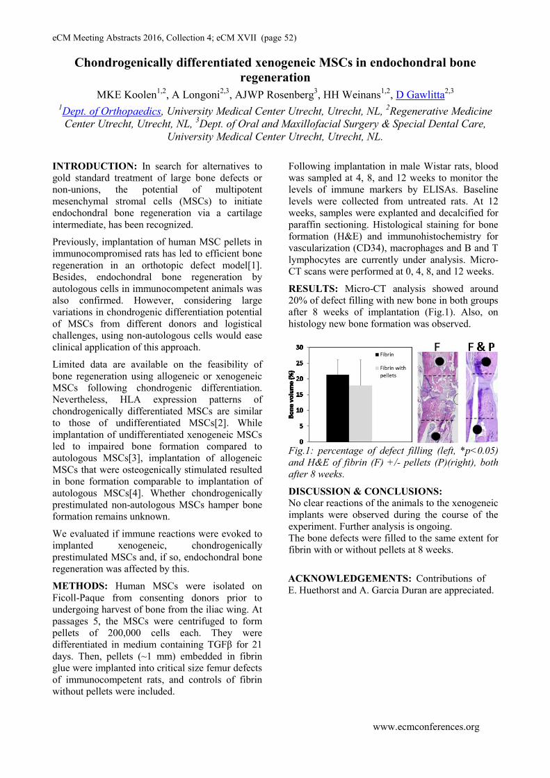
eCM Meeting Abstracts 2016, Collection 4; eCM XVII (page 52)
www.ecmconferences.org
Chondrogenically differentiated xenogeneic MSCs in endochondral bone
regeneration
MKE Koolen1,2
, A Longoni2,3
, AJWP Rosenberg3, HH Weinans
1,2, D Gawlitta
2,3
1Dept. of Orthopaedics, University Medical Center Utrecht, Utrecht, NL,
2Regenerative Medicine
Center Utrecht, Utrecht, NL, 3Dept. of Oral and Maxillofacial Surgery & Special Dental Care,
University Medical Center Utrecht, Utrecht, NL.
INTRODUCTION: In search for alternatives to
gold standard treatment of large bone defects or
non-unions, the potential of multipotent
mesenchymal stromal cells (MSCs) to initiate
endochondral bone regeneration via a cartilage
intermediate, has been recognized.
Previously, implantation of human MSC pellets in
immunocompromised rats has led to efficient bone
regeneration in an orthotopic defect model[1].
Besides, endochondral bone regeneration by
autologous cells in immunocompetent animals was
also confirmed. However, considering large
variations in chondrogenic differentiation potential
of MSCs from different donors and logistical
challenges, using non-autologous cells would ease
clinical application of this approach.
Limited data are available on the feasibility of
bone regeneration using allogeneic or xenogeneic
MSCs following chondrogenic differentiation.
Nevertheless, HLA expression patterns of
chondrogenically differentiated MSCs are similar
to those of undifferentiated MSCs[2]. While
implantation of undifferentiated xenogeneic MSCs
led to impaired bone formation compared to
autologous MSCs[3], implantation of allogeneic
MSCs that were osteogenically stimulated resulted
in bone formation comparable to implantation of
autologous MSCs[4]. Whether chondrogenically
prestimulated non-autologous MSCs hamper bone
formation remains unknown.
We evaluated if immune reactions were evoked to
implanted xenogeneic, chondrogenically
prestimulated MSCs and, if so, endochondral bone
regeneration was affected by this.
METHODS: Human MSCs were isolated on
Ficoll-Paque from consenting donors prior to
undergoing harvest of bone from the iliac wing. At
passages 5, the MSCs were centrifuged to form
pellets of 200,000 cells each. They were
differentiated in medium containing TGFβ for 21
days. Then, pellets (~1 mm) embedded in fibrin
glue were implanted into critical size femur defects
of immunocompetent rats, and controls of fibrin
without pellets were included.
Following implantation in male Wistar rats, blood
was sampled at 4, 8, and 12 weeks to monitor the
levels of immune markers by ELISAs. Baseline
levels were collected from untreated rats. At 12
weeks, samples were explanted and decalcified for
paraffin sectioning. Histological staining for bone
formation (H&E) and immunohistochemistry for
vascularization (CD34), macrophages and B and T
lymphocytes are currently under analysis. Micro-
CT scans were performed at 0, 4, 8, and 12 weeks.
RESULTS: Micro-CT analysis showed around
20% of defect filling with new bone in both groups
after 8 weeks of implantation (Fig.1). Also, on
histology new bone formation was observed.
Fig.1: percentage of defect filling (left, *p<0.05)
and H&E of fibrin (F) +/- pellets (P)(right), both
after 8 weeks.
DISCUSSION & CONCLUSIONS:
No clear reactions of the animals to the xenogeneic
implants were observed during the course of the
experiment. Further analysis is ongoing.
The bone defects were filled to the same extent for
fibrin with or without pellets at 8 weeks.
ACKNOWLEDGEMENTS: Contributions of
E. Huethorst and A. Garcia Duran are appreciated.

eCM Meeting Abstracts 2016, Collection 4; eCM XVII (page 53)
www.ecmconferences.org
human intra-articular stem cells isolated during arthroscopy surgery as a new cell source with
high potential for cartilage regeneration
J Li1, 2
, YH Liang1, ZY Zhou
1, WM Zhu
3, WJ Peng
1, and GQ Zhou
1*
1 The Centre for Anti-ageing and Regenerative Medicine, Shenzhen University, Shenzhen, China, 2 Key Laboratory of Optoelectronic
Devices and Systems of Ministry of Education and Guangdong Province, Shenzhen University, Shenzhen, China, 3 The First Affiliated
Hospital of Shenzhen University, Shenzhen, China
INTRODUCTION: Mesenchymal stem cells (MSCs) from
various adult tissues have emerged as a potential therapy for
osteoarthritic joint degeneration[1]. However, there remain
significant challenges in obtaining optimal cell source for
cartilage repair. Two primary criteria that are generally
considered are the regenerative properties of the cells and their
ease of access[2_ENREF_2]. Hereby, we will introduce a
new available and valuable way to get highly potential cell
source, human intra-articular-derived mesenchymal stem cells
(hIAMSCs) from arthroscopy surgery.
METHODS: Isolation of human intra-articular-derived
cells (hIA-derived cells). With the approval of university and
hospital institutional review board, 10 patients from 40 to 50
years old who had a diagnosis of knee osteoarthritis (based on
OARSI) were perfomanced with arthroscopic lavage and
debridement. We collected the lavage liquid of knee intra-
articular in the early surgery. After centrifuging, the deposit
with cells were resuspended in MEM with 20% FBS, and were
plated and cultured to form colonies. Non-adherent cells were
removed 2 days after initial plating. Characterizations of
hIAMSCs. Cell proliferation assay -- Passage 5 hIA-derived
cells were cultured in triplicate for designated time points and
were measured via CCK-8 assay. Induced differentiation
assays -- After incubating in adipogenic, osteogenic, and
chondrogenic media, the induced hIA-derived cells were
stained with 0.36% fresh oil red O, 0.1% alizarin red S, and
1% toluidine blue O solution, respectively. Flow cytometry
assay -- One million passage 5 hIA-derived cells were
incubated for 1 hour at 4℃ with conjugated antibodies to
CD34-FITC, CD45-FITC, CD73-APC, CD90-PerCP-Cy5.5,
or CD105-PE. Then these labelled cells were re-suspended
and subjected to flow cytometry analysis.
RESULTS: During primary culturing, hIA-derived cells
started to adhere after plating 24hrs and form obvious
distinctive colonies at 7 days (Fig. 1 A-D). During
subculturing the hIA-derived cells proliferated, with a typical
population doubling time of 23.8 hrs (Fig. 1E). The multi-
differentiation potential of hIA-derived cells was checked by
culturing them in adipogenic, osteogenic, and chondrogenic
media, respectively. After 14 days of culture in adipogenic
medium, lipid droplets were formed within hIA-derived cells
and observed with oil red O stainning (Fig. 2A). Meanwhile,
osteogenic and chondrogenic differentiation of these cells was
revealed by positive alizarin red-S and toluidine bule O
staining at 21 days (Fig. 2B-C). Fluorescently activated cell
sorting (FACS) analysis was utilised to label the hIA-derived
cells for a series of mesenchymal stem cell markers. We found
that these cells expressed in over 99% of the positive markers
of MSCs, including CD73, CD90, and CD105, whereas they
seldom expressed less than 1% in the negative markers such as
CD34, and CD45 (Fig. 3A-E).
DISCUSSION & CONCLUSIONS: Within arthroscopic
lavage liquid, we have successfully and efficiently isolated
hIA-derived cells which possess clonogenicity, self-renewing
Fig. 1. Colony formation and growth curve of hIA-derived cells. (A-C)
Cell colony formation at 1, 3, and 7 days, respectively. (D) Total
colonies stained with crystal violet at 7 days. (E) Cell growth curve. Scale bars, 200μm.
Fig. 2. Induced differentiation of hIA-derived cells. (A-C) Oil red O,
alizarin red S, and toluidine blue O staining for adipogenic, osteogenic, and chondrogenic differentiation, respectively. Scale bars,
200μm.
Fig. 3. Flow cytometry assay of hIA-derived cells. These hIA-derived
cells were identificated by labelling for of MSC surface markers CD34
(A), CD45 (B), CD73 (C), CD90 (D) and CD105 (E).
capability, multidifferentiation potential, and stem marker
expression, the common characteristics of MSC. Future
research will explore the cartilage repair capacity of hIAMSCs
in vivo animal and human, and compare the efficiency with
other commonly used MSCs. It may open a new avenues to
achieve highly potential cell source for cartilage repair and
renewal using cell therapy or tissue engineering approaches.
ACKNOWLEDGEMENTS: This study was supported
by NSFC (81472126) Shenzhen commission on innovation
and technology grant. (#JCYJ20150626090344603).

eCM Meeting Abstracts 2016, Collection 4; eCM XVII (page 54)
www.ecmconferences.org
Chondrogenic differentiation of bone marrow derived stem cells (MSCs) versus adipose derived stem cells (ASCs) at pro-inflammatory conditions
C. Neidlinger-Wilke1, G.Q. Teixeira
1-3, A. Rapp
1, D. Kletsas
4, A. Ignatius
1 1
1Institute of Orthopaedic Research and Biomechanics, Center for Musculoskeletal Research, University of Ulm, Germany, 2Institute
of Biomedical Engineering (INEB), Universidade do Porto, Portugal, 3Instituto de Ciências Biomédicas Abel Salazar (ICBAS),
Universidade do Porto, Portugal, 4Laboratory of Cell Proliferation & Ageing, Institute of Biology, NCSR Demokritos, Athens,
Greece.
INTRODUCTION: Stem cells are considered as
suitable cell source for regenerative therapies of
cartilaginous tissues and bone due to their potential
to differentiate towards osteogenic, adipogenic or
chondrogenic phenotypes [1]. Human adipose-
derived stem cells (ASCs) gained increasing
interest as an alternative cell source to bone
marrow-derived stem cells (MSCs) due to their
easy accessibility and lower donor site pain.
Regarding the treatment of degenerated
cartilaginous tissues it has to be considered that
degeneration is often associated with an increased
occurrence of inflammation factors such as IL-1β
and TNF-α [2]. These conditions are challenging
for regenerative cell therapy approaches as they
may influence cell physiology and differentiation.
For ASCs it has been shown that inflammatory
conditions influence their morphology and
proliferation but their osteogenic and adipogenic
differentiation capacity remains unaffected [3]. For
human MSCs an induction of osteogenic
differentiation by IL-1β has been reported [4].
Regarding chondrogenic differentiation at pro-
inflammatory conditions, a direct comparison of
both cell types is missing. Purpose of the present
study was to characterize human MSCs and ASCs
and to compare their chondrogenic differentiation
capacity at pro-inflammatory conditions with
regard to their suitability for cell therapy of
degenerated intervertebral discs or osteoarthritis.
METHODS: Human MSCs and ASCs from each
three donors (18-50 years old) that were pre-tested
regarding the expression of stem cell markers
(CD105, CD73, CD44, CD90 and SSEA4) were
cultured for four weeks in micromass-pellets of
each 200.000 cells in chondrogenic differentiation
medium with TGFβ3 as described [5]. For
simulation of pro-inflammatory conditions, the
chondrogenic medium of parallel pellet cultures
was supplemented by IL-1β during the whole
culture period. Cultures with standard medium
without TGFβ3- or IL-1β-supplementation served
as controls. At the end of the differentiation period
each three pellets/group were characterized with
regard to morphology and matrix formation
(Alcian blue staining) and expression of
chondrogenic target genes (Sox-9, Coll-II,
Aggrecan). Groups were compared by descriptive
statistics.
RESULTS: In cultures supplemented by medium
with TGFβ3, pellet formation occurred within the
first culture week with an increasing size and
optical density of the pellets during the four weeks
of culture time. Cultures supplemented with IL-1β
showed a 0.5-0.75-fold reduced pellet size.
Cultures with standard medium without TGFβ3
failed to form pellets. Differentiation of both
MSCs and ASCs could be confirmed by an up-
regulation of Aggrecan (up to 30-fold), Coll-2 (up
to 600-fold) and Sox-9 (up to 26-fold) expression
at presence of TGFβ3. This effect was strongly
decreased (0.6-0.2-fold) in IL-1β-supplemented
pellet cultures of both MSCs and ASCs with high
variability of the absolute gene expression levels
with cells from different donors. The impaired
differentiation capacity of ASCs and MSCs at
presence of IL-1β could also be confirmed by
reduced Alcian blue staining of the pellets. This
effect was similar with both ASCs and MSCs with
variability of staining intensity of pellets from
different donors.
DISCUSSION & CONCLUSIONS: Our findings
suggest that a pro-inflammatory stimulation with
IL-1β impairs chondrogenic differentiation
capacity of both MSCs and ASCs in pellet
cultures. Therefore, inflammatory conditions may
reduce matrix-formation if these cells are applied
for cell therapy, e.g. in osteoarthritis or in a
degenerated disc environment. As this effect could
be shown for ASCs and MSCs, cells from both
tissue sources appear to be similar sensitive
towards pro-inflammatory conditions. These
findings suggest that an anti-inflammatory
treatment previous to cell therapy might improve
the microenvironment for a successful cell therapy
of disc degeneration or osteoarthritis.

eCM Meeting Abstracts 2016, Collection 4; eCM XVII (page 55)
www.ecmconferences.org
THE EFFECTS OF HUMAN BONE GRAFTS ON OSTEOGENIC
DIFFERENTIATION OF ADIPOSE TISSUE DERIVED MESENCHYMAL
STROMAL CELLS IN VITRO.
J Plsikova1, D Harvanova
1, T Spakova
1, S Gromosova
1, M Lacko
2, T Kluka
3, J Sevc
4, J Rosocha
1
1Associated Tissue Bank, Faculty of Medicine, University of P.J. Safarik and L. Pasteur University
Hospital, Kosice. 2Department of Plastic, Reconstructive and Aesthetic Surgery,
Faculty of
Medicine, University of P.J. Safarik and L. Pasteur University Hospital, Kosice. 3Department of
Orthopaedics and Traumatology of Locomotory Apparatus, Faculty of Medicine, University of P.J.
Safarik and L. Pasteur University Hospital, Kosice. 4Institute of Biology and Ecology, Faculty of
Science, University of P. J. Safarik, Kosice.
INTRODUCTION: Human donor cancellous
bone, as originally allogeneic material is a well
utilized bone tissue engineering scaffold. Cell free
allograft bone has longer incorporation time than
autograft and cannot elicit the same osteoinductive
response as autograft bone. Processing of
allogeneic bone minimizes the risk of an
immunologic response of the recipient. Adipose
tissue derived human MSCs share many of the
characteristics of bone marrow derived MSCs
including extensive self-renewal capacity and
capacity to undergo differentiation into many
mesenchymal types with low immunogenicity in
recipient´s organism [1]. Many studies have
reported bone regeneration using MSCs from
adipose tissue [2]. It has been demonstrated, that
combination of the cancellous bone scaffolds with
MSCs accelerate and enhance bone formation
within osseous defects when compared with the
matrix alone [3]. Stem cell-seeded bone allografts
therefore have great potential for bone
regeneration.
METHODS: Adipose tissue derived human MSCs
(hADCs) were isolated from human subcutaneous
tissue with the enzyme Collagenase type I and
cultured in vitro up to 3rd passage.
Immunophenotype characterization (CD105,
CD90, CD73, CD29, CD45) of hADCs was
performed by flow cytometry and analyzed in a
BD FACSCalibur using CellQuest software.
Differentiation potential of hADCs was also
performed. Human cancellous bone scaffolds
(hCBS) were prepared according to standard
operating procedures used in Tissue Bank. ADCs
were labelled with PKH 26 dye and seeded on
hCBS. After 4 weeks of static in vitro cultivation,
cryosection of hCBS with cells were visualized on
fluorescence microscope. Proliferation of ADCs
alone and seeded on hCBS in standard and
osteogenic medium was analysed by MTS
colorimetric assay. Adherence, localization and
differentiation of hADCs on hCBS was observed
by HE staining, scanning electron microscopy
(SEM) and actin/vinculin double staining on
confocal microscopy.
RESULTS: Flow cytometric analysis showed that
ADCs were positive for CD90 (98.9%), CD105
(82.93%), CD73 (99.62%) and CD29 (98.27%)
antigens and negative for CD45 (0, 73%) antigen.
Mesenchymal character of ADCs was also
confirmed by in vitro differentiation test.
Attachment of PKH26 labeled cells on the surface
of hCBS was confirmed by fluorescence
microscopy. Proliferation of hADCs alone and
seeded on hCBS measured with MTS colorimetric
assay was comparable in both standard
(nonosteogenic) and osteogenic medium. The
effect of hCBS on osteogenic differentiation of
hADCs was observed with SEM. hADCs cultured
in osteogenic medium formed on the hCBS surface
sheets of cells with flat morphology like
osteoblasts. In comparison with hADCs cultured in
standard medium, hADCs did not formed layers,
they were observed like colonies of cells. The
adherence and localization of hADCs on hCBS
was confirmed also by HE staining and confocal
microscopy.
DISCUSSION & CONCLUSIONS: hADCs
seeded on hCBS adhere mainly on the graft
surface. hCBS had no significant effect on
differentiation of hADCs in comparison with
osteogenic medium.
ACKNOWLEDGEMENTS: This work
was supported by VEGA grant No. 1/0772/13
and Slovak Research and Development
Agency (APVV 0684-12)

eCM Meeting Abstracts 2016, Collection 4; eCM XVII (page 56)
www.ecmconferences.org
Characterization of a novel dynamizable external fixator for ovine tibial
segmental defects
Nicholas P. Quirk1, Andrew Thoreson
1, Rodolfo E. De la Vega, M.D.
1, Michael J. Coenen
1, Miguel
Trujillo, Ph.D1, Consuelo M. Lopez De Padilla, M.D.
1, Slobodon Tepic, D.Sc.
2, Christopher H. Evans,
Ph.D.1
1 Mayo Clinic, Rochester, MN,
2 Kyon AG, Zurich, Switzerland
INTRODUCTION: Large segmental defects in
long bones present a clinical challenge to surgeons.
There is much interest in the influence of the
mechanical environment on the healing of these
defects. Dynamization of the fracture gap has been
shown to promote the subsequent stages of healing
and maturation. This occurs through stiffness
modulation of the fixation construct, stabilizing
fracture during healing and has been successfully
evaluated in rodent models. Prior to large animal
study translation, a suitable, adjustable, well-
characterized, external fixator is required.
The ultimate goal of using this fixator is to modify
the defect mechanical environment in conjunction
with recombinant human BMP-2 to improve
healing in an ovine tibial segmental defect model.
METHODS: Fixators were characterized through
mechanical testing by sawbone and ovine cadaver
tibiae samples, and data was used to validate a
finite element (FE) model. A 30mm fracture defect
and 20 mm ‘bone-to-fixator’ offset was used on all
samples. Extensometers were attached across the
defect for inter-fragmentary movement (IFM) and
at fixator ends to characterize flexural deformation
of the fixator. Plastic and elastic axial compressive
testing, torsional testing and cyclic axial testing
were performed on the constructs. A FE model was
developed using ANSYS and utilized quadratic
tetrahedral elements (390052 elements and 624494
nodes), simplified geometry (no screw threads /
fixator ‘nuts & bolts’) and fixed (bone-screw,
fixator bodies) and frictional contact (screw-
fixator) conditions.
RESULTS: Plastic axial testing showed yielding
for low stiffness configuration at 520 N and 550 N
for high stiffness. Elastic axial testing showed
corroboration between sawbone and cadaveric
samples. Elastic axial testing and torsional testing
confirmed FE model predictions. IFM exhibited a
mean value of 1.526 mm and 0.901 mm for low
and high stiffness, respectively, for elastic axial
testing. Cyclic fatigue testing showed plateaued
deformation across 100,000 cycles for all groups.
Fig. 1: Methods of characterization for external
fixator (left to right); sawbone mechanical testing,
cadaveric sample mechanical testing, FE analysis.
DISCUSSION & CONCLUSIONS: Fixator
dynamization increased the construct stiffness by
approximately 2-fold. Based on prior results from
rat models, this is appropriate for enhanced bone
healing. Moreover, negligible IFM differences of
the fracture gap occurred during repeated load-
cycling to mimic the projected lifecycle of the
fixator while attached to the sheep. This shows
stability of fixator across its life span and efficacy
in a weight bearing animal. FE model results were
generally in agreement with bench testing in key
mechanical properties.
The successful design, manufacture and
characterization of this external fixator provides
the means to evaluate the efficacy of dynamization
in ovine models of bone healing. This fixator may
be useful in small animal veterinary practice and
could form the basis for a device suitable for use in
humans.
ACKNOWLEDGEMENTS: We would like
to thank Lawrence Berglund for assistance
with mechanical testing. This study was funded
as part of a Department of Defense research grant
(award number W81XWH-13-1-0324).

eCM Meeting Abstracts 2016, Collection 4; eCM XVII (page 57)
www.ecmconferences.org
One Year Follow Up on Hybrid System in Spinal Tuberculosis Surgery
Rahyussalim AJ1, D. Y. Pranatha
1, T Kurniawati
2
1Department of Orthopaedic and Traumatology Faculty of Medicein Universitas Indonesia –Ciptomangunkusumo Hospital 2Stem Cell Integrated Medical Service Faculty of Medicein Universitas Indonesia –Ciptomangunkusumo Hospital
INTRODUCTION: Tuberculous spondylitis
causes damage to the corpus and makes spinal
instability. Operating procedures have been
developed treating tuberculous spondilytis with
some combination of conventional techniques and
minimally invasive technique (Hybrid System)
which allows the incision is required at the level
of the vertebrae which will be stabilized.
METHODS: Patient who have tuberculous
spondylitis which had been had corpus damages
on vertebrae Th12-L1 and L4-L5, had been
treated with posterior spine stabilization with
Hybrid System and percutaneous abscess
drainage. Blood loss, duration of surgery, length
of stay, Visual Analogue Scale (VAS) and fusion
status were evaluated for one year. The
improvement of neurological was documented
and its functional outcome was assessed using
measurements Oswestry Disability Index (ODI)
RESULT: Intra operative blood loss was 150cc,
5 hours operating time and 5 days duration of
hospitalized. After one year of follow-up, we
found several data such as : a deflation in VAS
score from 7-8 into 0-1; an x-ray photograph of
anteroposterior thoracolumbal and lateral showed
appearance of bridging trabeculae at the anterior
and posterior column, there was no significant
changes on motoric function at lower limb and
improvement of neurological on L1 dermatome.
The functional scoring based ODI before surgery
was on 27 points (54% severe disability category)
and after one year of follow-up, it changed into 2
points (4% minimum disability category)
Fig. 1: Overview of Hybrid System procedure of
spinal tuberculosis surgery a, b: MRI showed
tuberculous spondylitis Th12-S1 with destruction
of intervertebral disc on T12-L1 and L4-L5.
Bilateral paravertebral abscess on Th7 into S2-S3
with involvement on bilateral m. Psoas, c: prone
position, d: pre-operative localization, e:
placement Illico MIS System, f: percutaneous
abscess drainage, g: result , e: result on C-arm.
Fig. 2: Patient of one year follow up. a:Pre
operative x-ray shows abscess and vertebral
damage, b:Multiple incission after one year,
c:Post operative x-ray shows implant on
thoracolumbar area.
DISCUSSION AND CONCLUSION:
Compared with conventional techniques, Hybrid
System procedures have similar effectiveness in
achieving spinal stability and abscess drainage.
This prosedur proved to be more effective in
reducing the amount of bleeding, tissue damage
and length of stay, although this procedure still
requires a longer operating time.
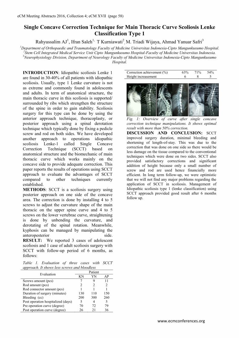
eCM Meeting Abstracts 2016, Collection 4; eCM XVII (page 58)
www.ecmconferences.org
Single Concave Correction Technique for Main Thoracic Curve Scoliosis Lenke
Classification Type 1
Rahyussalim AJ1, Ifran Saleh
1, T Kurniawati
2, M. Triadi Wijaya, Ahmad Yanuar Safri
3
1Department of Orthopaedic and Traumatology Faculty of Medicine Universitas Indonesia-Cipto Mangunkusumo Hospital.
2Stem Cell Integrated Medical Service Unit Cipto Mangunkusumo Hospital-Faculty of Medicine Universitas Indonesia.
3Neurophysiology Division, Department of Neurology Faculty of Medicine Universitas Indonesia-Cipto Mangunkusumo
Hospital.
INTRODUCTION: Idiopathic scoliosis Lenke 1
are found in 30-40% of all patients with idiopathic
scoliosis. Usually, type 1 Lenke curvature is not
as extreme and commonly found in adolescents
and adults. In term of anatomical structure, the
main thoracic curve in this scoliosis is supported/
surrounded by ribs which strengthen the structure
of the spine in order to gain stability. Scoliosis
surgery for this type can be done by using the
anterior approach technique, thoracoplasty, or
posterior approach using a spinal derotation
technique which typically done by fixing a pedicle
screw and rod on both sides. We have developed
another approach to operations idiopathic
scoliosis Lenke-1 called Single Concave
Correction Technique (SCCT) based on
anatomical structure and the biomechanic of main
thoracic curve which works mainly on the
concave side to provide adequate correction. This
paper reports the results of operations using SCCT
approach to evaluate the advantages of SCCT
compared to other techniques currently
established.
METHODS: SCCT is a scoliosis surgery using
posterior approach on one side of the concave
area. The correction is done by installing 4 to 5
screws to adjust the curvature shape of the main
thoracic on the upper spine curve and 4 to 5
screws on the lower vertebrae curve, straightening
is done by unbending the curvature, and
derotating of the spinal rotation. Meanwhile,
kyphosis can be managed by manipulating the
anteropesterior side.
RESULT: We reported 3 cases of adolescent
scoliosis and 1 case of adult scoliosis surgery with
SCCT with follow-up period of 6 months, as
follows:
Table 1. Evaluation of three cases with SCCT
approach. It shows less screws and bloodless
Evaluation Patient
KN YN AP
Screws amount (pcs) 7 9 11
Rod amount (pcs) 2 2 2
Rod connector amount (pcs) 1 1 1
Duration of surgery (minutes) 130 110 150
Bleeding (cc) 200 300 260
Post operation hospitalized (days) 5 4 5
Pre operation curve (degree) 70 72 79
Post operation curve (degree) 26 21 36
Correction achievement (%) 63% 71% 54%
Height increasement 6 8 5
Fig. 1: Overview of curve after single concave
correction technique manipulations. It shows optimal
result with more than 50% correction. DISCUSSION AND CONCLUSION: SCCT
improved surgery duration, minimal bleeding and
shortening of length-of-stay. This was due to the
correction that was done on one side so there would be
less damage on the tissue compared to the conventional
techniques which were done on two sides. SCCT also
provided satisfactory corrections and significant
addition of height because only a small number of
screw and rod are used hence financially more
efficient. In long term follow-up, we were optimistic
that we will not find any major problems regarding the
application of SCCT in scoliosis. Management of
Idiopathic scoliosis type 1 (lenke classification) using
SCCT approach provided good result after 6 months
follow up.

eCM Meeting Abstracts 2016, Collection 4; eCM XVII (page 59)
www.ecmconferences.org
Toxicity and Biocompatibility Profile of Scaffold developed by Universitas Indonesia
on Mesenchymal Stem Cells: A Preliminary Study
Rahyussalim AJ1, T Kurniawati
2, Aprilya D
1, R Anggraini
2, Yudan Whulanza
3, G Ramahdita
3
1Department of Orthopaedic and Traumatology Faculty of Medicine Universitas Indonesia-Cipto Mangunkusumo Hospital.
2Stem Cell Integrated Medical Service Unit Cipto Mangunkusumo Hospital-Faculty of Medicine Universitas Indonesia.
3Department of Metallurgy and Material Faculty of Engineering Universitas Indonesia
INTRODUCTION: Scaffold as a biomaterial must
fulfill some requirements to be safely implanted to
the human body. Toxicity and biocompatibility test
are needed to evaluate scaffold material in
mediating cell proliferation and differentiation,
secreting extracellular matrix and carrying bio
molecular signals for cell communication. This
study aims to evaluate the toxicity and
biocompatibility profile of various scaffolds with
different materials developed by Universitas
Indonesia by an in vitro study with mesenchymal
stem cells.
METHODS: Toxicity and biocompatibility test
were conducted on 24 scaffolds made of poly-L-
lactic acid (PLA), hydroxyapatite (HA), chitosan,
Alginates, dicalcium phosphate dihydrate (DCPC),
and polyvinyl alcohol (PVA) with direct contact test
and indirect contact test using MTT (3-(4,5-
dimethylthiazol-2-yl)-2,5-diphenyltetrazolium
bromide) tetrazolium reduction assay. To investigate
cell toxicity by direct contact test, scaffolds were
placed in the wells of mesenchymal stem cell (MSC)
culture plates and then the cell proliferation
inhibitions were determined. In MTT assay,
scaffolds and cultured cells are tested with
Vybrant® and the absorbance value was determined
by ELISA reader at 570 nm wave length. Cell
morphology, proliferation and cell adhesion to
plastic material were evaluated at day-2 and day-6.
RESULT: Table 1. Toxicity and biocompatibility of 24 scaffolds and
control
Scaffolds
Direct Indirect (MTT assay)
Material Inhibition
(%)
Inhibition
day-1 (%)
Inhibition
day-7 (%)
Primo 54.90 40.25 27.75 PLA
ALBAB 20 100.00 -43.50 -37.00 HA-Chitosan
ALBAB 30 100.00 -45.25 -41.25 HA-Chitosan
ALBAB 40 100.00 -48.5 -48.5 HA-Chitosan
ALBAB 80 100.00 -45.25 -48.75 HA-Chitosan
C2 100.00 -47.00 -48.50 HA-Chitosan
C3 100.00 -44.75 -53.00 HA-Chitosan
C4 100.00 -48.25 -46.75 HA-Chitosan
DM 0% 94.12 34.00 -45.25 HA-PVA
DM 25% 94.12 16.00 -24.25 HA-PVA
DM 40% 94.12 30.50 6.50 HA-PVA
DM 50% 96.08 33.50 -7.25 HA-PVA
N-20 74.51 -46.75 -10.00 HA-Chitosan
N-30 100.00 -45.00 -44.00 HA-Chitosan
N-40 100.00 52.50 -43.75 HA-Chitosan
N-80 98.04 -48.25 -45.75 HA-Chitosan
R-Alg 50 80.39 -43.75 -37.75 HA-Alginate
R-Alg 60 62.75 -48.25 -51.75 HA-Alginate
R-Alg 70 92.16 -44.25 -48.75 HA-Alginate
H-0% 100.00 -51.25 -51.75 DCPD-HA
H-10% 98.04 -47.00 -45.75 DCPD-HA
RA-1980 68.63 5.75 11.25 DCPD-HA
HC 100.00 -46.75 -46.25 HA-Chitosan
DH 100.00 -41.25 -44.5 DCPD-HA
Control 0.00 100.00 100.00 cells & medium
Fig. 1: Cell morphology and proliferation of various scaffolds were observed at day-6 1. Control; 2. Primo; 3. ALBAB 20; 4. ALBAB 30; 5. ALBAB 40; 6. ALBAB 80; 7. C2; 8. C3; 9. C4; 10.DM 0%; 11. DM 25%; 12. DM 40%; 13. DM 50%; 14. N-20; 15. N-30; 16. N-40; 17. N-80; 18. R-Alg 50; 19. R-Alg 60; 20. R-Alg 70; 21. H-0%; 22. H-10%; 23. RA-1980; 24. DC; 25. DH
DISCUSSION AND CONCLUSION: There were
cells-substrate adhesion impairment, morphological
changes, cell death and reduction in cell proliferation
seen at day-2 and day-6 in most tested scaffold except
Primo, DM 0%, DM 25%, DM 40%, N-20, R-Alg 50, R-
Alg 50, R-Alg 60, and RA 1980 scaffold. Cell count
result at day-6 showed proliferation inhibition of more
than 50% cell death (inhibition value > 50) in all tested
scaffold. In MTT assay, Primo, RA-1980, and DM 40%
were proven non-cytotoxic. Various scaffold materials
showed different cytotoxicity effect. Primo as a polymer
based scaffold showed the least cytotoxic effect, followed
by DM 40% and RA-1980.

eCM Meeting Abstracts 2016, Collection 4; eCM XVII (page 60)
www.ecmconferences.org
Implant Failure after Posterior Instrumentation on Extreme and Progressive
Congenital Scoliosis with Some Comorbidities: 4 Years Follow Up
Rahyussalim AJ1, Ifran Saleh
1, T Kurniawati
2, M. Triadi Wijaya, Ahmad Yanuar Safri
3
1Department of Orthopaedic and Traumatology Faculty of Medicine Universitas Indonesia-Cipto Mangunkusumo Hospital.
2Stem Cell Integrated Medical Service Unit Cipto Mangunkusumo Hospital-Faculty of Medicine Universitas Indonesia.
3Neurophysiology Division, Department of Neurology Faculty of Medicine Universitas Indonesia-Cipto Mangunkusumo
Hospital.
INTRODUCTION:
There are various complexities in congenital
scoliosis ranging from the characteristic of rapid
progressive, associated with fleeting cardiac and
pulmonary function deterioration, worsening
postural deformity predisposing limited range of
motion and presence of other congenital disorders
complicating the treatment. Until recently, the
management of complex congenital scoliosis has
not been satisfying as a consequence of poor
accurate diagnosis, associative congenital
comorbidities, and the possibility of crank shaft
phenomenon and short stature development.
Surgical techniques using posterior
instrumentation has been the treatment of choice
due to unsatisfactory conservative results. This
paper showed implant failure after posterior
instrumentation.
METHODS: It was a case report with 4 year
follow up and done observations of surgical
intervention, implant failure, progression and
achievement of correction.
Case illustration: Nine year old boy with corrected
anal and esophageal atresia which was diagnosed
with congenital scoliosis at 2 year age with 40
degree of Cobb angle. He had undergone
conservative and operative treatments since 5 year
old but no optimal outcome, delay or progressivity
and neither postural correction (figure 1).
RESULT: This case was performed two kinds of
surgical interventions. The first surgery was single
concave correction approach by using cervico-
thoracic junction rod. The rod was failure 7 month
after the first surgery. The second surgery was
done to change screw and rod with bigger one.
The second implants were also failure 35 month
after the second surgery. (figure 1).
Figure 1. Four years follow up: (a) immediately pre
operatif I; Cobb’s angle 120o (b) post operatif I;
Cobb’s angle 89o (c) 3 month follow up post operatif I;
Cobb’s angle 100o (d) 7 month follow up post operatif
I; Cobb’s angle 102o; implant failure (e) 9 month
follow up post operatif I; Cobb’s angle 113o; implant
failure (f) post operatif II; Cobb’s angle 76o (g) 21
month follow up post operatif II; Cobb’s angle 78o (h)
35 month follow up post operatif II; Cobb’s angle 95o;
implant failure.
Table 1. Evaluation of four years follow up on extreme
and progressive congenital scoliosis with some
comorbidities. It shows implant failure after posterior
instrumentation.
Scoring
Factor
Pre
Operatif
(2009)
Pre
Operatif I (Feb 2012)
Post
Operatif I (Apr 2012)
Follow
Up I (Jul 2012)
Follow
Up II (Nov 2012)
Follow
Up III (Jan 2013)
Post
Operatif II (Feb 2013)
Follow
Up IV (Nov 2014)
Follow
Up V (Jan 2016)
Cobbs
Angle 40
o120
o89
o100
o102
o113
o76
o78
o95o
Erect
Hide NA 94cm 105cm 100cm 98cm 94cm 108cm 105cm 100cm
Rod
Failure NA NA NO NO YES YES NO NO NO
Screw
Failure NA NA NO NO NO NO NO NO YES
DISCUSSION AND CONCLUSION:
Evaluation to the measures taken have been evaluated, following steps ahead and prediction of future outcome has been a continuous homework for our multidisciplinary team to improve his
quality of life.
Failure of implants in this case may be caused by
inappropriate harmony of biomechanical force
between growing bone and strength of implant
loading.

eCM Meeting Abstracts 2016, Collection 4; eCM XVII (page 61)
www.ecmconferences.org
Effect of in vitro differentiation of human mesenchymal stem cells on cartilage
repair in osteoarthritis of knee joint
T Spakova1, J Plsikova
1, D Harvanova, M Lacko
2, J Rosocha
1
1 Associated tissue bank and
2Department of orthopaedics and traumatology of locomotory
apparatus of Faculty of Medicine of P. J. Safarik University and University Hospital of L. Pasteur,
Trieda SNP 1, Kosice, Slovakia
INTRODUCTION: Osteoarthritis (OA) -
degenerative joint disease is characterised by
progressive degeneration of cartilage, subchondral
bone changes such as sclerosis, subchondral bone
cysts, osteophytes, and synovitis [1]. Enhanced
chondrogenesis can be achieved by kartogenin
(KGN) and mesenchymal stem cells (MSCs)
delivered on the cartilage surface. MSCs tend to
undergo terminal differentiation, which means that
cartilaginous tissue formed by MSCs is not stable
and cells become hyperthrophic. This process is
characterised by the production of hypertrophy-
related factors and mimics the embryonic process
of endochondral bone formation [2]. In our study
an osteoarthritis (OA) model was used to evaluate
the effect of KGN in vitro. We hypothesized that
KGN would promote chondrogenic differentiation
of MSCs without negative side effect on terminal
differentiation. Additionally we hypothesized that
local delivery of KGN with bone marrow derived
MSCs (BMSCs) to a cartilage defect would
improve the quality of the tissue formed and
induce immunomodulatory responses after
differentiation.
METHODS: Bone marrow and osteochondral
cylinders were obtained from OA patients
undergoing total knee joint replacement with full
ethical approval. The effects of prolonged
exposure of KGN on BMSCs in scaffold-free 2D
culture were monitored by xCelligence (RTCA)
system. BMSCs were expanded and seeded onto
osteochondral cylinders as previously described
[3]. Cylinders were embedded in agarose gel and
cultured in DMEM/F12 supplemented with 2%
ITS-A and 1% ATB. Culture media was changed
and collected two times a week. Secretome
analysis was done to control changes in response
of BMSCs to the cylinders before and after
induction with KGN. Analysis was performed
using the RayBio® Quantibody Human Array on
media collected during 21 days of co-culture. SEM
images were captured from both loaded (with
BMSCs and w/wo KGN) and non-loaded OA
cartilage surface to evaluate cell distribution and
collagen fibre orientation. Final tissue products of
3D cultures of OA cylinders with cells were
checked by histological and biochemical assays to
demonstrate chondrogenic differentiation and
evaluate terminal differentiation of human BMSCs.
All experiments were performed in scaffold-free
2D cultures, too.
RESULTS: BMSCs in monolayer were exposed to
medium containing 1, 10, 100 µM KGN and
viability and proliferation were monitored by
RTCA after 1, 3 and 7 days. An increase in
proliferation during 7 days was observed in
BMSCs cultured in 10 µM KGN. After three
weeks of co-culture anti-inflammatory factors were
produced at a higher level in loaded cylinders than
in non-loaded control. The retention and
differentiation of BMSCs at the fibrillated surface
of osteoarthritic articular cartilage was
demonstrated by SEM and histological assays.
KGN caused an upregulation in expression of Col
II, aggrecan and downregulation of osteocalcin,
MMP-13 and ALP activity. FACS analysis of
BMSCs phenotype after KGN exposure showed
higher expression of chondrogenic markers
(CD49e, CD26, CD54) compared to control.
DISCUSSION & CONCLUSIONS: In the
present study, we described an approach to the
assessment of effect of KGN in an in vitro OA
model. Preliminary results suggest that KGN may
be an effective accelerant for cartilage tissue
engineering by promoting chondrogenic
differentiation of MSCs but with no significant
effect on hypertrophic differentiation.
2
ACKNOWLEDGEMENTS: This work
was supported by the Slovak Research
and Development Agency under the contract
No. APVV-0684-12 and by VEGA grant
1/0217/16.

eCM Meeting Abstracts 2016, Collection 4; eCM XVII (page 62)
www.ecmconferences.org
Osteointegration in a CapFlex-PIP© finger implant: a histological case report.
CM Sprecher1, SF Schindele
2, S Hensler
3, S Milz
4
1 AO Research Institute, AO Foundation, Davos, CH.
2 Department of Hand Surgery, Schulthess
Klinik, Zurich, CH. 3 Department of Teaching, Research and Development, Schulthess Klinik,
Zurich, CH. 4 Department of Anatomy II, Ludwig-Maximilians-University of Munich, Munich, DE
INTRODUCTION: Primary press fit and
secondary osteointegration is a precondition for
component anchoring in articular surface
replacements of proximal interphalangeal (PIP)
joints. However, this outcome fails in many
existing prosthesis designs. In order to improve
osteointegration of the implant, the modular
prosthesis CapFlex-PIP© (KLS Martin Group,
Germany), a modern polyethylene-metal surface
replacement consisting of a proximal and distal
component, was developed1. We suspected that
long-term cementless fixation of the CapFlex-PIP©
could be achieved with initial press-fit technique
allowing secondary osteointegration at the bone
with the pure titanium pore backside of the
components. However, it is difficult to obtain
evidence of such osteointegration using standard
radiographs. The most detailed and accurate
statement can be made by histological analysis, but
this requires retrieval of the implant-bone
interface. We report such a rare case of an
explantation of a CapFlex-PIP© implant due to a
soft tissue complication.
CASE REPORT: We present the case of an 84-
year-old woman who had a traumatic rupture of the
radial collateral ligament 11 months after CapFlex-
PIP© replacement at the index finger. Fixed ulnar
deviation with functional limitations provided the
indication for revision surgery with removal of the
CapFlex-PIP© prosthesis and joint arthrodesis.
For histological analysis of osteointegration, the
removed implants and attached tissues were
immediately fixed in 70% methanol. After
dehydration, the blocks were cut in the transversal
plane and selected sections were stained with
Giemsa-eosin. The Bone-Implant-Contact (BIC)
rate, as a quantitative indicator for osteo-
integration, was measured on all stained sections
using a Zeiss Axioplan microscope (Fig. 1). The
average BIC value was 40.7% for the proximal and
46.5% for the distal implant component. The
observed values were within the BIC range
published for particular dental implants and higher
than those reported for the humeral parts of
resurfacing shoulder prostheses2,3
.
Fig. 1: Undecalcified sections of implants and
adherent tissues stained with Giemsa-eosin. The
red-stained bone is in direct contact with the
surface of the implant. a) Proximal component, b)
Distal component, c) Higher magnification of the
bone-implant interface showing intimate contact.
DISCUSSION & CONCLUSIONS: The present
case for the first time demonstrates a successful
osteointegration of an implant used for human
proximal interphalangeal joint replacement. The
histological result is in line with the radiographic
evaluation. The investigated CapFlex-PIP© implant
shows osteointegration of both components, a
result which is comparable to that of other load-
bearing and articulating implants at different
locations in the human body.
2
ACKNOWLEDGEMENTS: We would like
to thank Mrs Andrea Altwegg for organizing
the implant exchange and primary fixation of
the components after explantation.
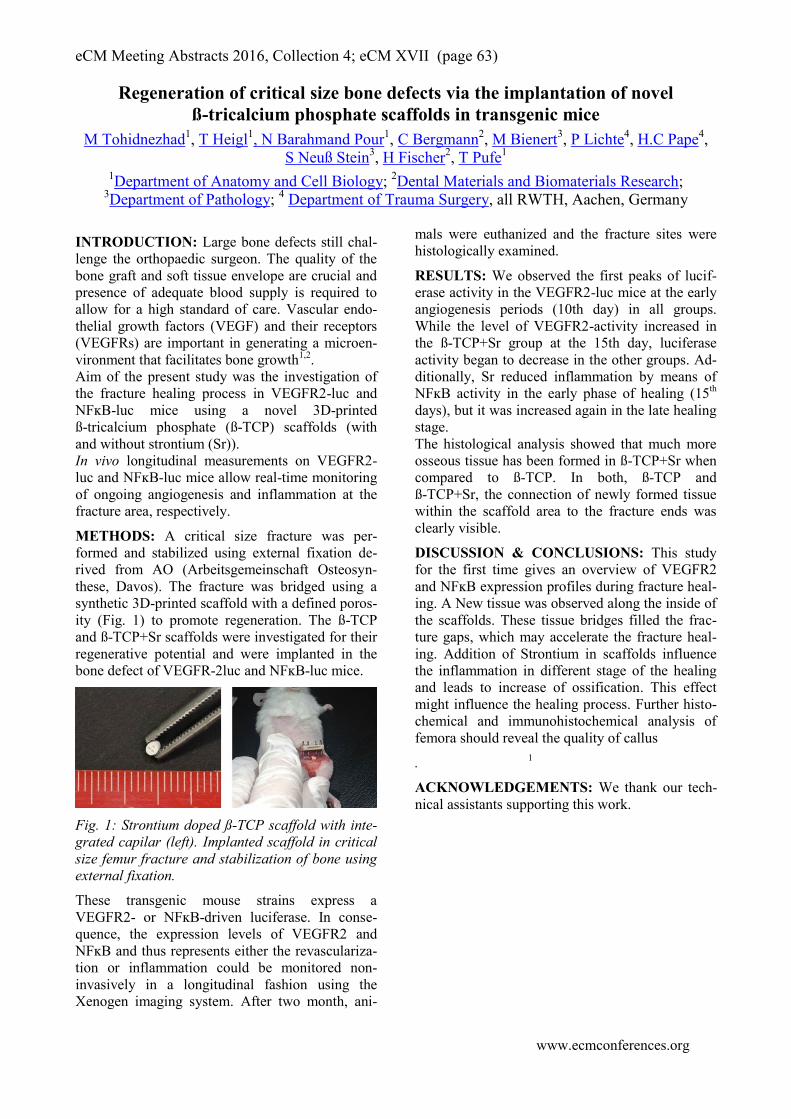
eCM Meeting Abstracts 2016, Collection 4; eCM XVII (page 63)
www.ecmconferences.org
Regeneration of critical size bone defects via the implantation of novel
ß-tricalcium phosphate scaffolds in transgenic mice
M Tohidnezhad1, T Heigl
1, N Barahmand Pour
1, C Bergmann
2, M Bienert
3, P Lichte
4, H.C Pape
4,
S Neuß Stein3, H Fischer
2, T Pufe
1
1Department of Anatomy and Cell Biology;
2Dental Materials and Biomaterials Research;
3Department of Pathology;
4 Department of Trauma Surgery, all RWTH, Aachen, Germany
INTRODUCTION: Large bone defects still chal-
lenge the orthopaedic surgeon. The quality of the
bone graft and soft tissue envelope are crucial and
presence of adequate blood supply is required to
allow for a high standard of care. Vascular endo-
thelial growth factors (VEGF) and their receptors
(VEGFRs) are important in generating a microen-
vironment that facilitates bone growth1,2
.
Aim of the present study was the investigation of
the fracture healing process in VEGFR2-luc and
NFĸB-luc mice using a novel 3D-printed
ß-tricalcium phosphate (ß-TCP) scaffolds (with
and without strontium (Sr)).
In vivo longitudinal measurements on VEGFR2-
luc and NFĸB-luc mice allow real-time monitoring
of ongoing angiogenesis and inflammation at the
fracture area, respectively.
METHODS: A critical size fracture was per-
formed and stabilized using external fixation de-
rived from AO (Arbeitsgemeinschaft Osteosyn-
these, Davos). The fracture was bridged using a
synthetic 3D-printed scaffold with a defined poros-
ity (Fig. 1) to promote regeneration. The ß-TCP
and ß-TCP+Sr scaffolds were investigated for their
regenerative potential and were implanted in the
bone defect of VEGFR-2luc and NFĸB-luc mice.
Fig. 1: Strontium doped ß-TCP scaffold with inte-
grated capilar (left). Implanted scaffold in critical
size femur fracture and stabilization of bone using
external fixation.
These transgenic mouse strains express a
VEGFR2- or NFĸB-driven luciferase. In conse-
quence, the expression levels of VEGFR2 and
NFĸB and thus represents either the revasculariza-
tion or inflammation could be monitored non-
invasively in a longitudinal fashion using the
Xenogen imaging system. After two month, ani-
mals were euthanized and the fracture sites were
histologically examined.
RESULTS: We observed the first peaks of lucif-
erase activity in the VEGFR2-luc mice at the early
angiogenesis periods (10th day) in all groups.
While the level of VEGFR2-activity increased in
the ß-TCP+Sr group at the 15th day, luciferase
activity began to decrease in the other groups. Ad-
ditionally, Sr reduced inflammation by means of
NFĸB activity in the early phase of healing (15th
days), but it was increased again in the late healing
stage.
The histological analysis showed that much more
osseous tissue has been formed in ß-TCP+Sr when
compared to ß-TCP. In both, ß-TCP and
ß-TCP+Sr, the connection of newly formed tissue
within the scaffold area to the fracture ends was
clearly visible.
DISCUSSION & CONCLUSIONS: This study
for the first time gives an overview of VEGFR2
and NFĸB expression profiles during fracture heal-
ing. A New tissue was observed along the inside of
the scaffolds. These tissue bridges filled the frac-
ture gaps, which may accelerate the fracture heal-
ing. Addition of Strontium in scaffolds influence
the inflammation in different stage of the healing
and leads to increase of ossification. This effect
might influence the healing process. Further histo-
chemical and immunohistochemical analysis of
femora should reveal the quality of callus
1 .
ACKNOWLEDGEMENTS: We thank our tech-
nical assistants supporting this work.

eCM Meeting Abstracts 2016, Collection 4; eCM XVII (page 64)
www.ecmconferences.org
Human dental pulp stem cells stabilize blood vessels by secreting vascular
basement membrane proteins collagen type IV and laminin in ovo
A Woloszyk1, TA Mitsiadis
1
1Institute of Oral Biology, Department of Orofacial Development & Regeneration, Center of Dental
Medicine, University of Zurich, Switzerland
INTRODUCTION: The requirement for tissue
and organ replacement is high and is expected to
increase in the future due to population aging,
especially in highly developed countries. Current
shortage of suitable donors might be overcome by
generating cell- and material-based biological
substitutes with the potential to regenerate and
replace lost tissues and organs. Teeth present an
easily accessible source of autologous multipotent
mesenchymal stem cells [1], which have been
previously shown to possess pro-angiogenic
properties [2]. As rapid vascularization and tissue
integration into the surrounding host tissues is a
prerequisite for the long-term survival of tissue
engineered implants, we investigated the effect of
human dental pulp stem cells (DPSCs) to improve
neovascularization of a 3D silk fibroin scaffold
using the chorioallantoic membrane (CAM) assay.
METHODS: Biocompatible silk fibroin scaffolds
(height: 3 mm; diameter: 5 mm; pore size: 200-
300 nm) were seeded with DPSCs and incubated
for 24 h in vitro before performing the CAM assay
for 7 days. Qualitative and quantitative blood
vessel analysis was performed on H&E-stained
paraffin sections. Cells of human origin were
identified immunohistochemically using an anti-
NuMA antibody, while anti-Collagen type IV and
anti-Laminin antibodies were applied to visualize
two of the main components of the vascular
basement membrane. Empty scaffolds served as
controls.
RESULTS: Cell-seeded scaffolds were found to
attract more vessels than empty scaffolds, while
creating their own extracellular matrix between the
scaffold fibers. Cells of human origin were
localized next to functional blood-perfused vessels,
where they contributed to vessel stabilization by
producing Laminin (Lam) (Fig. 1A) and Collagen
type IV (Coll-IV) (Fig. 1B) within as short as 7
days. Therefore, the presence of DPSCs improved
and accelerated vascular and tissue integration of
the implants.
Fig. 1: Expression of Lam and Coll-IV by hDPSCs.
(A, B) Human cell nuclei are stained in red (white
arrows), while Lam (A) and Coll-IV (B) appear in
purple. bv, blood vessel; sc, scaffold fiber. Scale
bar = 25 µm.
DISCUSSION & CONCLUSIONS: The study
has shown that a vascularized 3D tissue constructs
can be created by combining DPSCs with 3D silk
scaffolds. The cells have the potential to attract
vascularization through their natural pro-
angiogenic properties [2] without previous
induction. The production of Lam and Coll-IV
helps to stabilize ingrowing vessels, thereby
accelerating tissue integration, which is important
for clinical applications of cell-based tissue
regeneration strategies. Following the ‘3Rs’
principles of replacement, refinement, and
reduction of animal use in research, the CAM
assay provides a valuable intermediate platform for
initial assessments prior to pre-clinical studies in
mammals.
ACKNOWLEDGEMENTS: This work
was supported by the Swiss National
Foundation (SNSF) grant 31003A_135633 and by
institutional funds from the University of Zurich.
The authors thank Dr. Jolanda Baumgartner
(Institute of Biomechanics, ETH) for
manufacturing the silk scaffolds and Trudel
Silk Inc. for providing silk cocoons.
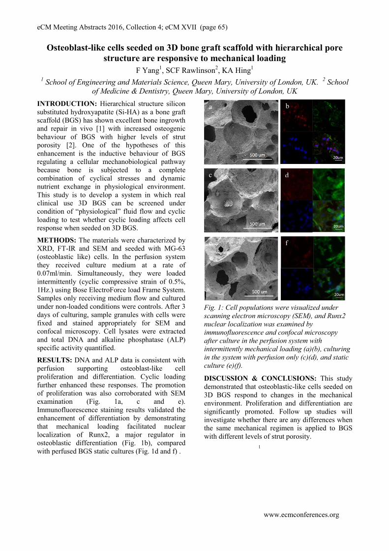
eCM Meeting Abstracts 2016, Collection 4; eCM XVII (page 65)
www.ecmconferences.org
Osteoblast-like cells seeded on 3D bone graft scaffold with hierarchical pore
structure are responsive to mechanical loading
F Yang1, SCF Rawlinson
2, KA Hing
1
1School of Engineering and Materials Science, Queen Mary, University of London, UK.
2 School
of Medicine & Dentistry, Queen Mary, University of London, UK
INTRODUCTION: Hierarchical structure silicon
substituted hydroxyapatite (Si-HA) as a bone graft
scaffold (BGS) has shown excellent bone ingrowth
and repair in vivo [1] with increased osteogenic
behaviour of BGS with higher levels of strut
porosity [2]. One of the hypotheses of this
enhancement is the inductive behaviour of BGS
regulating a cellular mechanobiological pathway
because bone is subjected to a complete
combination of cyclical stresses and dynamic
nutrient exchange in physiological environment.
This study is to develop a system in which real
clinical use 3D BGS can be screened under
condition of “physiological” fluid flow and cyclic
loading to test whether cyclic loading affects cell
response when seeded on 3D BGS.
METHODS: The materials were characterized by
XRD, FT-IR and SEM and seeded with MG-63
(osteoblastic like) cells. In the perfusion system
they received culture medium at a rate of
0.07ml/min. Simultaneously, they were loaded
intermittently (cyclic compressive strain of 0.5%,
1Hz.) using Bose ElectroForce load Frame System.
Samples only receiving medium flow and cultured
under non-loaded conditions were controls. After 3
days of culturing, sample granules with cells were
fixed and stained appropriately for SEM and
confocal microscopy. Cell lysates were extracted
and total DNA and alkaline phosphatase (ALP)
specific activity quantified.
RESULTS: DNA and ALP data is consistent with
perfusion supporting osteoblast-like cell
proliferation and differentiation. Cyclic loading
further enhanced these responses. The promotion
of proliferation was also corroborated with SEM
examination (Fig. 1a, c and e).
Immunofluorescence staining results validated the
enhancement of differentiation by demonstrating
that mechanical loading facilitated nuclear
localization of Runx2, a major regulator in
osteoblastic differentiation (Fig. 1b), compared
with perfused BGS static cultures (Fig. 1d and f) .
Fig. 1: Cell populations were visualized under
scanning electron microscopy (SEM), and Runx2
nuclear localization was examined by
immunofluorescence and confocal microscopy
after culture in the perfusion system with
intermittently mechanical loading (a)(b), culturing
in the system with perfusion only (c)(d), and static
culture (e)(f).
DISCUSSION & CONCLUSIONS: This study
demonstrated that osteoblastic-like cells seeded on
3D BGS respond to changes in the mechanical
environment. Proliferation and differentiation are
significantly promoted. Follow up studies will
investigate whether there are any differences when
the same mechanical regimen is applied to BGS
with different levels of strut porosity.
1
a b
c d
e f
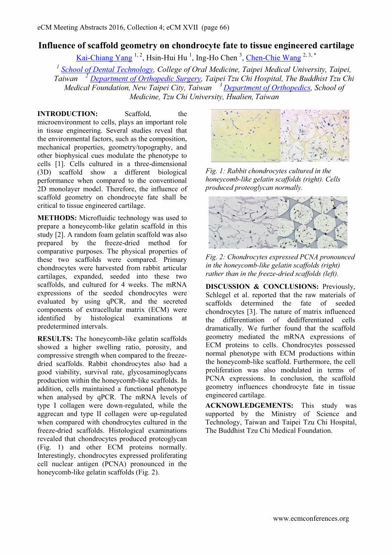
eCM Meeting Abstracts 2016, Collection 4; eCM XVII (page 66)
www.ecmconferences.org
Influence of scaffold geometry on chondrocyte fate to tissue engineered cartilage
Kai-Chiang Yang 1, 2
, Hsin-Hui Hu 1, Ing-Ho Chen
3, Chen-Chie Wang
2, 3, *
1 School of Dental Technology, College of Oral Medicine, Taipei Medical University, Taipei,
Taiwan 2 Department of Orthopedic Surgery, Taipei Tzu Chi Hospital, The Buddhist Tzu Chi
Medical Foundation, New Taipei City, Taiwan 3 Department of Orthopedics, School of
Medicine, Tzu Chi University, Hualien, Taiwan
INTRODUCTION: Scaffold, the
microenvironment to cells, plays an important role
in tissue engineering. Several studies reveal that
the environmental factors, such as the composition,
mechanical properties, geometry/topography, and
other biophysical cues modulate the phenotype to
cells [1]. Cells cultured in a three-dimensional
(3D) scaffold show a different biological
performance when compared to the conventional
2D monolayer model. Therefore, the influence of
scaffold geometry on chondrocyte fate shall be
critical to tissue engineered cartilage.
METHODS: Microfluidic technology was used to
prepare a honeycomb-like gelatin scaffold in this
study [2]. A random foam gelatin scaffold was also
prepared by the freeze-dried method for
comparative purposes. The physical properties of
these two scaffolds were compared. Primary
chondrocytes were harvested from rabbit articular
cartilages, expanded, seeded into these two
scaffolds, and cultured for 4 weeks. The mRNA
expressions of the seeded chondrocytes were
evaluated by using qPCR, and the secreted
components of extracellular matrix (ECM) were
identified by histological examinations at
predetermined intervals.
RESULTS: The honeycomb-like gelatin scaffolds
showed a higher swelling ratio, porosity, and
compressive strength when compared to the freeze-
dried scaffolds. Rabbit chondrocytes also had a
good viability, survival rate, glycosaminoglycans
production within the honeycomb-like scaffolds. In
addition, cells maintained a functional phenotype
when analysed by qPCR. The mRNA levels of
type I collagen were down-regulated, while the
aggrecan and type II collagen were up-regulated
when compared with chondrocytes cultured in the
freeze-dried scaffolds. Histological examinations
revealed that chondrocytes produced proteoglycan
(Fig. 1) and other ECM proteins normally.
Interestingly, chondrocytes expressed proliferating
cell nuclear antigen (PCNA) pronounced in the
honeycomb-like gelatin scaffolds (Fig. 2).
Fig. 1: Rabbit chondrocytes cultured in the
honeycomb-like gelatin scaffolds (right). Cells
produced proteoglycan normally.
Fig. 2: Chondrocytes expressed PCNA pronounced
in the honeycomb-like gelatin scaffolds (right)
rather than in the freeze-dried scaffolds (left).
DISCUSSION & CONCLUSIONS: Previously,
Schlegel et al. reported that the raw materials of
scaffolds determined the fate of seeded
chondrocytes [3]. The nature of matrix influenced
the differentiation of dedifferentiated cells
dramatically. We further found that the scaffold
geometry mediated the mRNA expressions of
ECM proteins to cells. Chondrocytes possessed
normal phenotype with ECM productions within
the honeycomb-like scaffold. Furthermore, the cell
proliferation was also modulated in terms of
PCNA expressions. In conclusion, the scaffold
geometry influences chondrocyte fate in tissue
engineered cartilage.
ACKNOWLEDGEMENTS: This study was
supported by the Ministry of Science and
Technology, Taiwan and Taipei Tzu Chi Hospital,
The Buddhist Tzu Chi Medical Foundation.

eCM Meeting Abstracts 2016, Collection 4; eCM XVII (page 67)
www.ecmconferences.org
Promotion of the osteogenic differentiation potential of human nasal inferior
turbinate-derived mesenchymal stem cells by mechanical memory
Byeong Gon Yun1, Sun Hwa Park
2, Se Hwan Hwang
1, Mi Hyun Lim
1,
Sang A Back1, Jung Ho Jeon
1, Sung Won Kim
1,2
1Department of Biomedical Science, the Catholic University of Korea, College of Medicine,
Seoul, Korea 2Department of Otolaryngology Head and Neck Surgery, the Catholic University of Korea,
College of Medicine, Seoul, Korea
INTRODUCTION: Mesenchymal stem cells
(MSCs) are used for cell-based therapy to prevent
degenerative bone defects. Therefore, since bone
regeneration is required, autologous tissues can be
used to generate implants in the appropriate
anatomical shape without risk of immunological
rejection. Human nasal inferior turbinate-derived
mesenchymal stem cells (hTMSCs) can be
potentially used as a source of adult stem cells for
therapeutic application due to their easy
availability, cultivation, and high proliferative
ability. We evaluated the capacity of hTMSCs for
osteogenic differentiation. We hypothesized that
different surfaces of the nano-pore surface plate
affect the morphology, proliferation, and
osteogenic differentiation of hTMSCs.
METHODS: We used human turbinate-derived
MSCs after the second passage. hTMSCs (4 x 10⁴
cells) were seeded in flat surface plates (20 mm ×
20 mm) and nano-pore surface plates (20 mm × 20
mm; diameter, 200 nm; depth, 500 nm, pore-to-
pore distance, 500 nm). The seeded hTMSCs were
assessed using a scanning electron microscope
(SEM) and focal actin staining to determine the
morphology. The proliferation of hTMSCs was
evaluated by measuring the cell metabolic activity
using a Cell CountingKit-8 (CCK-8). The media
used for osteo-differentiation was changed every
2–3 days. Osteogenic differentiation of hTMSCs
was identified using alkaline phosphatase (ALP),
alizarin red S staining, and vonkossa staining,
Real-time quantitation of mRNA of COL1A1,
osteocalcin, BMP-2, osterix, and bone sialoprotein
were also performed.
RESULTS: There was no significant difference in
the proliferation of seeded hTMSCs on a flat
surface plate and those on a nano-pore surface
plate. However, high osteogenic differentiation
was observed using alkaline phosphatase (ALP),
alizarin red S staining, von Kossa staining, and
real-time PCR of seeded hTMSCs on the surface of
a nano-pore plate.
Fig. 1: Human nasal inferior turbinate-derived
mesenchymal stem cells(hTMSCs) grown in the flat
surface plates and nano-pore surface plates of
through focal adhesion staining(FAK100). 21day
culture in osteogenic differentiation media.
DISCUSSION & CONCLUSIONS: These
findings suggest that hTMSCs are apparently
redirected toward the osteogenic phenotype in an
in vitro culture under specific conditions using
bone-formation stimulating factors. Further, the
osteogenic potentials observed were superior to
other tissues originating from MSCs.
ACKNOWLEDGEMENTS: The authors wish
to acknowledge the financial support of the
National Research Foundation of Korea (NRF),
funded by the Ministry of Science, ICT, and
Future Planning (2014R1A2A2A01004325), and
the Korea Health Industry Development
Institute, funded by the Ministry of Health and
Welfare (HI14C3228).
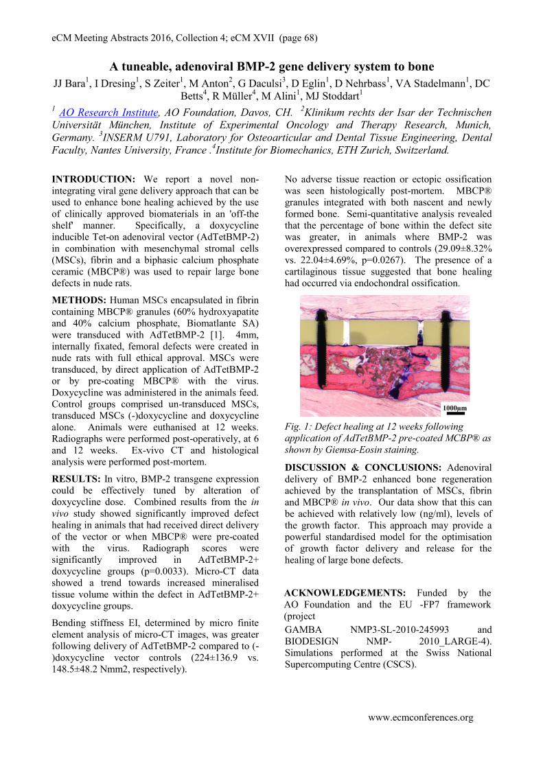
eCM Meeting Abstracts 2016, Collection 4; eCM XVII (page 68)
www.ecmconferences.org
A tuneable, adenoviral BMP-2 gene delivery system to bone
JJ Bara1, I Dresing
1, S Zeiter
1, M Anton
2, G Daculsi
3, D Eglin
1, D Nehrbass
1, VA Stadelmann
1, DC
Betts4, R Müller
4, M Alini
1, MJ Stoddart
1
1 AO Research Institute, AO Foundation, Davos, CH.
2Klinikum rechts der Isar der Technischen
Universität München, Institute of Experimental Oncology and Therapy Research, Munich,
Germany. 3INSERM U791, Laboratory for Osteoarticular and Dental Tissue Engineering, Dental
Faculty, Nantes University, France .4Institute for Biomechanics, ETH Zurich, Switzerland.
INTRODUCTION: We report a novel non-
integrating viral gene delivery approach that can be
used to enhance bone healing achieved by the use
of clinically approved biomaterials in an 'off-the
shelf' manner. Specifically, a doxycycline
inducible Tet-on adenoviral vector (AdTetBMP-2)
in combination with mesenchymal stromal cells
(MSCs), fibrin and a biphasic calcium phosphate
ceramic (MBCP®) was used to repair large bone
defects in nude rats.
METHODS: Human MSCs encapsulated in fibrin
containing MBCP® granules (60% hydroxyapatite
and 40% calcium phosphate, Biomatlante SA)
were transduced with AdTetBMP-2 [1]. 4mm,
internally fixated, femoral defects were created in
nude rats with full ethical approval. MSCs were
transduced, by direct application of AdTetBMP-2
or by pre-coating MBCP® with the virus.
Doxycycline was administered in the animals feed.
Control groups comprised un-transduced MSCs,
transduced MSCs (-)doxycycline and doxycycline
alone. Animals were euthanised at 12 weeks.
Radiographs were performed post-operatively, at 6
and 12 weeks. Ex-vivo CT and histological
analysis were performed post-mortem.
RESULTS: In vitro, BMP-2 transgene expression
could be effectively tuned by alteration of
doxycycline dose. Combined results from the in
vivo study showed significantly improved defect
healing in animals that had received direct delivery
of the vector or when MBCP® were pre-coated
with the virus. Radiograph scores were
significantly improved in AdTetBMP-2+
doxycycline groups (p=0.0033). Micro-CT data
showed a trend towards increased mineralised
tissue volume within the defect in AdTetBMP-2+
doxycycline groups.
Bending stiffness EI, determined by micro finite
element analysis of micro-CT images, was greater
following delivery of AdTetBMP-2 compared to (-
)doxycycline vector controls (224±136.9 vs.
148.5±48.2 Nmm2, respectively).
No adverse tissue reaction or ectopic ossification
was seen histologically post-mortem. MBCP®
granules integrated with both nascent and newly
formed bone. Semi-quantitative analysis revealed
that the percentage of bone within the defect site
was greater, in animals where BMP-2 was
overexpressed compared to controls (29.09±8.32%
vs. 22.04±4.69%, p=0.0267). The presence of a
cartilaginous tissue suggested that bone healing
had occurred via endochondral ossification.
Fig. 1: Defect healing at 12 weeks following
application of AdTetBMP-2 pre-coated MCBP® as
shown by Giemsa-Eosin staining.
DISCUSSION & CONCLUSIONS: Adenoviral
delivery of BMP-2 enhanced bone regeneration
achieved by the transplantation of MSCs, fibrin
and MBCP® in vivo. Our data show that this can
be achieved with relatively low (ng/ml), levels of
the growth factor. This approach may provide a
powerful standardised model for the optimisation
of growth factor delivery and release for the
healing of large bone defects.
ACKNOWLEDGEMENTS: Funded by the
AO Foundation and the EU -FP7 framework
(project
GAMBA NMP3-SL-2010-245993 and
BIODESIGN NMP- 2010_LARGE-4).
Simulations performed at the Swiss National
Supercomputing Centre (CSCS).

eCM Meeting Abstracts 2016, Collection 4; eCM XVII (page 69)
www.ecmconferences.org
Perfusion culture regulates the differentiation of mesenchymal stromal cells on
polylactic-glycolic acid in vitro
C Moser1,2
, K Bardsley3, C Wolfrum
2, AJ El Haj
3, M Alini
1, MJ Stoddart
1, JJ Bara
1
1AO Research Institute, AO Foundation, Davos, Switzerland.
2Institute for Translational Nutrition,
ETH Zürich, Switzerland. 3Institute for Science and Technology in Medicine, Keele University,
United Kingdom
INTRODUCTION: In vitro testing of biomaterial
suitability for bone repair is typically performed in
static culture. Yet mesenchymal stromal cells
(MSCs), are known to be responsive to shear
forces, which are present as a result of lacunar-
canalicular interstitial fluid flow. Here, we
investigate the behaviour of bone marrow-derived
MSCs on porous polylactic glycolic acid (PLGA)
scaffolds under dynamic culture conditions.
METHODS: PLGA (50:50 lactic: glycolic acid)
scaffolds were fabricated by solvent casting/salt
leaching as previously described.1 Primary human
MSCs from two donors were cultured on PLGA
scaffolds in a bidirectional perfusion culture
system in control vs. osteogenic induction media
(UCUP, CELLEC BIOTEK AG). MSCs cultured
on PLGA statically and on Thermanox™ served as
controls. DNA content, secreted nitric oxide (NO),
prostaglandin E2 (PGE2) and gene expression
were assessed throughout the experiment.
Histological analysis was performed at 3 weeks.
RESULTS: PLGA scaffolds degraded steadily
which corresponded with a gradual reduction in
DNA content. Gene expression analysis performed
at day 7 revealed up-regulation of Runx2 in MSCs
cultured in both control and osteogenic media on
PLGA. Following culture in osteogenic media,
Sox9 was down-regulated in Donor A and
remained unchanged in Donor B at day 7. Collagen
I and ALP were upregulated by MSCs cultured on
PLGA in osteogenic media at day 7. Osteocalcin
gene expression was higher in PLGA groups
compared to Thermanox™ controls and greatest
when MSCs had been cultured in osteogenic media
under perfusion (Fig 1.A). Neither NO nor PGE2
were detectable in culture supernatant. Donor A,
which exhibited a high Runx2/Sox9 ratio at day 7
following osteogenic induction in monolayer,
demonstrated significant mineralisation on PLGA
day 21 under perfusion (Fig 1.B) compared to
static culture (Fig 1.C). Conversely, donor B,
presented a comparatively lower Runx2/Sox9 ratio
at day 7 after osteogenic induction in monolayer –
and in this donor, collagen X was strongly
upregulated following culture on PLGA scaffolds
in osteogenic media – indicating hypertrophy. The
lack of mineral deposition at day 21 with Donor B
is in accordance with an immature hypertrophic
phenotype.
Fig. 1: (A) Osteocalcin gene expression at day
21.Mineral deposition as indicated by Von Kossa
staining at day 21 following (B) perfusion and (C)
static culture in osteogenic culture media.
DISCUSSION & CONCLUSIONS: Perfusion
culture enhanced the expression of osteocalcin in
osteogenically differentiated MSCs. The
Runx2/Sox9 ratio during early osteogenic
induction in monolayer may provide a reliable
predictor of hypertrophic vs. direct osteogenic
differentiation and mineralising potential of MSCs
cultured on PLGA in vitro.
1ACKNOWLEDGEMENTS: Funded by the
AO Foundation and the EU -FP7 framework
(project BIODESIGN NMP- 2010_LARGE-4).
B C
A
Related Documents