ABSTRACT ANATOMICAL STUDY OF THE ANTHURIUM PLANT, ANTHURIUM ANDREANUM, L., AND A COLOR BREAKDOWN DISORDER OF ITS FLOWER I. Anatomical study of the anthurium plant, Anthurium andreanum, L. II. Color breadkown in anthurium flowers, Anthurium andreanum, L. By Tadashi Higaki Section I The gross morphology and anatomy study on Anthurium andreanum, L., was by whole plant observation, using dissecting and light microscope, and scanning electron microscopy. Anthurium is a perennial-herbaceous monocoty- ledon in the family Araceae. It is low growing with chordate leaves and attractive chordate flowers. It has a juvenile phase when each leaf axil has a lateral vegeta- tive bud and a generative phase when each leaf axil has a flower and the lateral vegetative bud is located opposite the leaf attachment. The "commercial flower" consists of conspicious bract (spathe) and a protruding rachis (spadix Minute, botanically perfect flowers are borne spirally on the spadix. The flowers are protogynous as the stigma is

Welcome message from author
This document is posted to help you gain knowledge. Please leave a comment to let me know what you think about it! Share it to your friends and learn new things together.
Transcript
ABSTRACT
ANATOMICAL STUDY OF THE ANTHURIUM PLANT,ANTHURIUM ANDREANUM, L., AND A COLOR
BREAKDOWN DISORDER OF ITS FLOWER
I. Anatomical study of the anthurium plant,Anthurium andreanum, L.
II. Color breadkown in anthurium flowers,Anthurium andreanum, L.
ByTadashi Higaki
Section IThe gross morphology and anatomy study on Anthurium
andreanum, L., was by whole plant observation, using dissecting and light microscope, and scanning electron microscopy. Anthurium is a perennial-herbaceous monocotyledon in the family Araceae. It is low growing with chordate leaves and attractive chordate flowers. It has a juvenile phase when each leaf axil has a lateral vegetative bud and a generative phase when each leaf axil has a flower and the lateral vegetative bud is located opposite the leaf attachment. The "commercial flower" consists of conspicious bract (spathe) and a protruding rachis (spadix Minute, botanically perfect flowers are borne spirally on the spadix. The flowers are protogynous as the stigma is
Tadashi Higaki
receptive one week before shedding of pollen. Anatomically, the spathe has a one cell layered upper and lower epidermis, with 1 or 2 layers of hypodermis cells. Between the upper and lower hypodermis are 10-12 layers of spongy parenchyma cells. Vascular bundles are dispersed uniformly throughout the spathe. Anthocyanin pigments are localized in the hypodermal cells. The leaf blade is similar in structure to the spathe, except there is no hypodermis, but two layers of palisade parenchyma cells form the tissue immediately below the epidermis. Cholorplasts were dispersed throughout the mesophyll, but concentrated in the palisade cells. The pedicel, petiole and vegetative stem are typically monocot. Outer epidermal cells covered the cortex, a layer of sclerified parenchyma cells and the ground tissue. Vascular bundles were dispersed throughout the ground tissue. Roots were cylindrical, fleshy, epiphytic and adventitious. They were characterized by having multiple layers of epidermal cells called the velamen. Raphide and druse crystals were found scattered throughout the entire plant tissue. Above ground parts were covered with a thick waxy layer of cuticle.
Section IIA color breakdown disorder in the spathe of Anthurium
andreanum, L., was investigated. Ca deficiency in the lobe section of the spathe was found to cause the disorder. Elemental analysis of spathe and leaf tissue of color breakdown and normal plants revealed lower Ca in color breakdown plants (color breakdown - spathe: 0.372%, leaves: 0.363%;
Tadashi Higakinormal - spathe: 0.830%, leaves: 0.805%). Electron microprobe X-ray analysis revealed lower Ca in lobe than tip section of the spathe and higher Ca in epidermal than mesophyll tissue. Nutrient culture studies produced color breakdown symptoms in the spathe with No Ca treatment. Symptoms were tiny water-soaked lesions the size of pin heads on the upper epidermal surface of the spathe lobe.The dots multipled, increased in size and eventually coalesced to form large water-soaked lesions. The lesions dehydrated and turned brown. pH levels 3, 4, 5, 6, and 7 of complete nutrient solutions did not produce color breakdown in the spathe, although elemental chemical analysis of spathe and leaf tissues indicated lower Ca uptake of 3 and 4, higher at 6, and highest at pH 7. Lower Ca was found in the lobe than tip sections of both spathe and leaves, regardless of treatment. In a separate study, X-ray analysis produced similar results. In addition pH 9 gave lower uptake of Ca than 7. Microautoradiography study showed that calcium was deposited primarily in the cell wall. A field test with different Ca sources confirmed that the application of Ca significantly reduced the incidence of the disorder. A critical level of Ca within the tissue of the spathe is suggested with respect to the disorder. The critical level may be very narrow, but must be exceeded if tissue does not exhibit color breakdown. The critical level may be mediated by environmental conditions such as humidity and temperature. Like oedema disorder which is correlated
Tadashi Higaki
with environmental conditions which hinder the normal transpiration processes of the plant, anthurium color- breakdown may similarly be related to transpiration.Unlike oedema, however, the critical level of Ca in the lobe of the spathe is the major factor in the anthurium color breakdown disorder. Anatomical studies of color- breakdown and normal flowers showed collapsing mesophyll cells in affected spathe. Separation of cells, when present on the permanent slides, suggested breakdown of the middle lamella due to lack of Ca. No sign of hypertrophy or intumescence was found.
ANATOMICAL STUDY OF THE ANTHURIUM PLANT, ANTHURIUM ANDREANUM, L., AND A COLOR
BREAKDOWN DISORDER OF ITS FLOWER
ByTadashi Higaki
A DISSERTATION
Submitted to Michigan State University
in partial fulfillment of the requirements for the degree of
DOCTOR OF PHILOSOPHYDepartment of Horticulture
1976
ACKNOWLEDGEMENTS
I would like to express my sincere appreciation to all those who have helped me during the course of these studies, as well as throughout my graduate education.
To my major professor, Dr. H. Paul Rasmussen, goes my heartfelt thanks for his excellent counsel and encouragement .
To the members of my guidance committee: Drs. W. J. Carpenter, A. L. Kenworthy, W. H. Carlson, M. E. Miller, and Maxine S. Ferris, I am most grateful. Your help and advice have been most gratifying.
To members of the Michigan State and University of Hawaii Horticulture Departments, I would like to express my appreciation for providing an excellent environment conducive to such a meaningful graduate experience.
To the following individuals for their timely assistance and expert advice: Esther M. Higaki, Michiko Higaki, Drs. R.Bullock, Y. Sagawa, F. Laemmlen, Mr. V. Shull, and Mr. J. Kunisaki, my humble thanks.
Appreciation is also extended to Mr. Harold T. Tanouye of Hawaiian Anthuriums, Inc., for supplying anthurium flowers and shipping them to Michigan.
To the Farm Foundation Scholarship Program and the National Defense Act, I am most indebted for the funding of my fellowship.
My special appreciation to Mr. Gordon Shigeura for being a true friend especially when needed.
Finally, to my wife, Jean, and children Joanne, Connie, Pauline and Chad for their encouragement, inspiration and tolerance, my deepest heartfelt thanks.
A. A.A. A.A i . 3.
TABLE OF CONTENTS
LIST OF TABLES.................................. vLIST OF F I G U R E S ................................ viINTRODUCTION .................................... 1LITERATURE CITED ................................ 5
SECTION IANATOMICAL STUDY OF THE ANTHURIUM PLANT,
ANTHURIUM ANDREANUM, L.Literature Review .............................. 10Materials and Methods .......................... 12Results and Discussion .......................... 19Literature Cited ................................ 58
SECTION IICOLOR-BREAKDOWN IN ANTHURIUM FLOWERS,
ANTHURIUM ANDREANUM, L.Literature Review .............................. 61Materials and Methods .......................... 67Results and Discussion .......................... 82Literature Cited ................................ 98
APPENDIXCorrelation of the anatomical study and the spathe color-breakdown disorder OfAnthurium andreanum, L ........................... 103Literature Cited .................................... Ill
Page
iv
LIST OF TABLES
SECTION II1. Solution culture composition, concentration
and conditions used in the Ca experiment . . . . 772. Elemental analysis of anthurium spathe
and leaf from color-breakdown and normalplants........................................ 84
3. Ca X-ray analysis of a cross-section of the spathe of anthurium in normal andcolor-breakdown flowers........................ 88
4. Ca content of anthurium spathe and leaf tissues grown at various nutrientand pH l e v e l s ................................ 90
5. Field tests of the incidence of color- breakdown disorder of spathe inanthurium with different Ca sources ........... 95
Table Page
v
LIST OF FIGURES Figure Page
SECTION I1. An apparatus used for tissuemat
infiltration ................................ 162. A mature anthurium plant, Anthurium
andreanum, L., cv. Red Ozaki................ 213. Scanning electron micrograph of
rhombohedral crystals in the anthurium . . . . 254. A scanning electron micrograph of the
upper cuticle of anthurium l e a f ............ 275. The anatomy and morphology of the
anthurium flower ............................ 306. Photomicrograph of the stamen of anthurium . . 327. Photomicrograph of the gynoecium of
anthurium.................................. 358. The various spathe shapes of the
anthurium flower ............................ 389. Photomicrograph and SEM micrograph of
the spathe of the anthurium................ 4110. Photomicrographs and SEM micrographs
of the anthurium pedicel.................... 4311. SEM and light photomicrographs of the leaf
of the anthurium............................ 4612. Photomicrograph and SEM micrograph
of the anthurium r o o t ...................... 5013. Diagram of the vegetative stem of
the anthurium.............................. 5314. Photomicrograph of the vegetative
meristem of the anthurium.................. 56
vi
SECTION II1. Anthurium flowers showing various
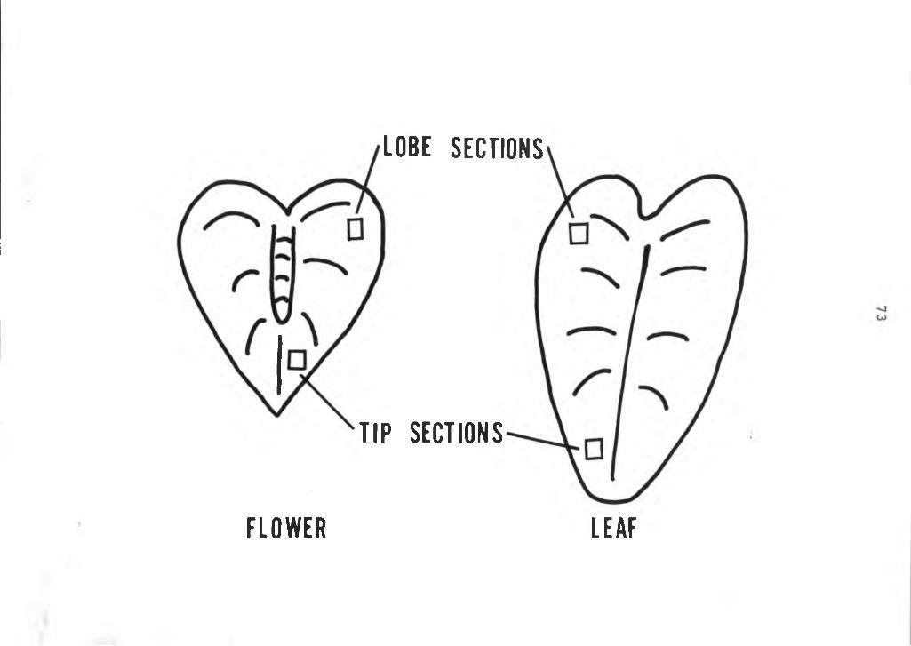
stages of the color breakdown disorder . . . . 632. Illustration of sampling of tissue from
lobe and tip sections of the anthuriumspathe............................ 73
3. Planting system used to grow anthuriumsin nutrient culture ........................ 76
4. Diagram of apparatus used forradioisotope study .......................... 80
5. Microprobe line profile X-ray analysisof Ca in the spathe of anthurium plants . . . 86
6. Energy dispersive X-ray analysis of anthurium flower sections of plantsgrown at various nutrient and pH levels . . . 92
APPENDIX1. A color breakdown and a normal anthurium
spathe tissue .................................. 1062. Color breakdown spathe showing cellular
separation........................ 109
LIST OF FIGURES— ContinuedFigure Page
vii
INTRODUCTION
The tropical anthurium, Anthurium andreanum, Lind., is a member of the family Araceae, which includes more than 100 genera and 1,500 species. In Hawaii they are grown in shade and high humidity. It is a perennial-herbaceous plant cultivated for its attractive, long lasting flowers.The flower is a complex of the colorful modified leaf (spathe) and hundreds of small true flowers on the pencillike protrusion (spadix) rising from the base of the spathe.
The anthurium plant produces flowers continuously throughout the year. One flower emerges from each leaf axil. The sequence of leaf, flower and new leaf is maintained throughout the life of the plant. The intervals between leaf emergence depend on the natural changes in environmental conditions. More flowers are produced during the summer months, when conditions are favorable for growth than during the winter months when temperatures are lower and light intensity is reduced (26).
The anthurium, a native of Central America, was brought to Hawaii from London in 1889 (27). Today, after 87 years of cultivation and hybridization in Hawaii, the Hawaiian anthurium flower is one of the State's principal ornamental exports to the mainland U.S., Canada, Australia, Japan,
1
Germany, Holland, Italy and many other countries. Anthurium culture has developed from a hobby or backyard operation 10 years ago, into a large-scale commercial enterprise, valued at approximately 2 million dollars at the farm level (8).The Hawaii State General Agricultural Plan (7) projects an industry valued at 16.2 million dollars by 1989. Its future for economic growth appears unlimited. The variety of colors and the exotic beauty of the flower have attracted many consumers and the demand for the Hawaiian anthurium is steadily increasing. The long shelf-life and ease in handling and packaging make it a durable product for shipping and mailing throughout the world. The Hawaii State legislature and the County government in Hawaii are increasingly supporting market promotion and development; farm loan programs; and research programs on production, post harvest, engineering and marketing problems of anthuriums. The future looks bright and growth seems inevitable.
Total acreage in anthurium production in the State is 320 acres, of which 95% is on the Island of Hawaii (8). The major acreage is confined to the windward portion of the island where the weather is ideal with cool temperatures (70-80°F day; 60-70°F night temperature) with abundant rainfall throughout the year (170-200 inches annually) with relative humidities of 75-100%.
This investigation examines the gross anatomy of the plant and a disorder of the spathe of Anthurium andreanum, Lind.
2
Information on the anatomy of Anthurium andreanum is limited. Yet, the need to know thoroughly the nature and function of the plant is the foundation for solving production and handling problems. Realistic interpretations and knowledge of the plant are imperative if Hawaii's anthurium research program is to develop properly and efficiently.This study examines extensively the external and internal anatomy and morphology of the anthurium plant. Established techniques in histochemistry and electron microscopy (31) were used to accomplish the objectives.
The second section of this dissertation deals with color and tissue breakdown in spathe of Anthurium andreanum, Lind.
Much of the limited research on Anthurium andreanum has been done in Hawaii including studies on keeping quality (1, 17, 32, 33, and 34), cost of production (6, 13, 14, and 15), cultural problems (2, 3, 5, 9, 10, 11, 12, 16, 18, 21, 25, 26, 28, 29, and 30), and breeding (4, 19, 20, 22, 23, and 24). Except for long range breeding, most research is problem-solving as it becomes acute in the industry. One problem facing the industry was a flower color and tissue disorder. The growers were faced with heavy losses due to a disorder that caused water-soaked lesions and color- breakdown on the lobes of the flower spathe. These water- soaked lesions, in time dessicated, turned dark brown and eventually dried. The flowers were unfit for sale. Field losses of up to 50% have been reported and losses after shipments to the overseas market of up to 20%. Many times
3
the visual symptoms were not evident during grading and packing, but were manifest after transit to the distant market. The following objectives were established for this study.
1. A well-balanced training in agricultural research, practical as well as basic.
2. A contribution of basic information on Anthurium andreanum, Lind., that could be utilized worldwide.
3. Solving of an acute problem on anthurium production for Hawaii's agriculture.
4
LITERATURE CITED
5
LITERATURE CITED
2.
3.
4.
5.
6 .
7.
8 .
9.
10.
11.
12.
13.
1 . Akamine, E. K. and T. Goo. 1972. Relationships between phsyical characteristics and vase life of anthuriums. Haw. Farm Sci. 21(2): 9-10.Aragaki, M. 1964. Maneb will control anthurium spadix rot. Haw. Farm Sci. 13(4): 16._______ and M. Ishii. 1960. A spadix rot of anthuriumin Hawaii. Plant Dis. Rep. 44(11): 865-867._______, H. Kamemoto and K. M. Maeda. 196 8.Anthracnose resistance in anthuriums. Haw. Agri. Expt. Sta. Prog. Report No. 169._______, M. Sherman and H. Kamemoto. 1957. Phytotoxicity of chemical sprays to anthuriums. Haw. Farm Sci. 5(3): 10-11.Barmettler, R. and J. Sheehan. 1963. A budgetary analysis for large-scale anthurium operation in Hawaii. Haw. Agri. Expt. Sta. Ag. Econ. Report No. 63.Department of Planning and Economic Development. 1970. Opportunities for Hawaiian agriculture. Ag. Dev. Plan. State of Hawaii, Honolulu, Hawaii.Hawaii Department of Agriculture. 1974. Statistics of Hawaiian agriculture. Hawaii Crop and Livestock Reporting Service, Honolulu, Hawaii.Hayward, A. C. 1972. A bacterial disease of anthurium in Hawaii. Plant. Dis. Rep. 56(10): 904-908.Higaki, T. 1973. Chemical weed control in anthurium. Haw. Agri. Expt. Sta. Res. Rep. No. 212._______ and D. P. Watson, 1967. Anthurium culture inHawaii. Haw. Coop. Ext. Ser. Cir. No. 420._______ and M. Aragaki. 1972. Benomyl for control ofanthurium anthracnose. Haw. Farm Sci. 21(3): 7-8.Hiroshige, H. H. and T. Shirakawa. 1964. Anthurium cost survey in Hawaii. U of H Farm Mangt. Series No. HI.
6
714. and T. Higaki. 1970. Anthurium cost of pro
duction_survey under Saran in Pahoa, Hilo, and MountainView areas. U of H Farm Mangt. Series No. H8.
15. _______, _______ and G. Aoki. 1966. Anthurium costsurvey under Saran in Hawaii. U of H Farm Mangt.Series No. H6.
16. Hunter, J. E., W. H. Ko, R. K. Kunimoto and T. Higaki. 1974. Foliar nematode disease of anthurium seedlings. Phytopath. 64(2): 267-268.
17. Kamemoto, H. 1962. Some factors affecting the keeping quality of anthurium flowers. Haw. Farm Sci.11(4): 2-5.
18. _______ and H. Y. Nakasone. 1953. Effects of media onproduction of anthuriums. Haw. Agri. Expt. Sta. Prog. Notes No. 94.
19. _______ and . 1955. Improving anthuriumsthrough breeding. Haw. Farm Sci. 3(3): 4-5.
20. _______ and . 1963. Evaluation and improvementof anthurium clones. Haw. Agri. Expt. Sta. Tech. Bui. No. 58.
21. _______ and . 1957. Wood shavings as a mediumfor anthuriums. Haw. Agri. Expt. Sta. Circ. No. 53.
22. _______ and _______ and M. Aragaki. 19 68. Improvementof anthuriums through breeding. Proc. Amer. Soc. Hort. Sci. Trop. Region. 12: 267-273.
23. _______, Mr. Aragaki, T. Higaki and J. T. Kunisaki.1969. Three new anthurium cultivars. Haw. Farm Sci.18: 1-3.
24. _______, _______, _______ and . 1971. A newanthracnose resistant white anthurium. Haw. Farm Sci.20 (3): 12.
25. Kunisaki, J. T. and Y. Sagawa. 1971. Intermittent mist to root anthurium cuttings. Haw. Farm Sci. 20(3): 4-5.
26. Nakashone, H. Y. and H. Kamemoto. 1962. Anthurium culture with emphasis on the effects of some induced environments on growth and flowering. Haw. Agri. Expt. Sta. Tech. Bui. No. 50.
27. Neal, Marie C. 1948. In gardens of Hawaii. Bernice P. Bishop Museum Special Publication No. 40. Honolulu, Hawaii.
828. Poole, R. T. and D. B. McConnell. 1971. Effects of
shade levels and fertilization on flowering of Anthurium andreanum "Nitta" and "Kaumana". Proc. Amer.Soc. Hort. Sci. Trop. Region 15: 189-195.
29. , R. Sakuoka and J. A. Silva. 1968. Nutritionof Anthurium andreanum. Proc. Amer. Soc. Hort. Sci. Trop. Region. 12: 284-287
30. Raabe, R. D. 1966. Control of anthracnose on theisland of Hawaii. Haw. Farm Sci. 15(2): 3.
31. Rasmussen, H. P. and B. D. Knezek. 1971. Electronmicroprobe: Techniques and uses in soil and plantanalysis. Mich. Agri. Expt. Sta. Journal Art. No. 5214. East Lansing, Michigan.
32. Shirakawa, T. and D. P. Watson. 1964. Improved methods of handling anthurium flowers. Haw. Farm Sci.13(2): 1-3.
33. and R. R. Dedolph and D. P. Watson. 1965.N-6 benzyladenine effects on chilling injury, respiration and keeping quality of Anthurium andreanum. Proc. Amer. Soc. Hort. Sci. 85: 642-646.
34. Watson, D. P. and T. Shirakawa. 1966. Gross morphology related to shelf life of anthurium flowers. Haw. Farm Sci. 15(2): 2-3.
SECTION IANATOMICAL STUDY OF THE ANTHURIUM PLANT,
ANTHURIUM ANDREANUM, L.
9
Section I: Anatomical Study of theAnthurium Plant,
Anthurium andreanum, L.
Literature Review
Christensen (2) has shown that Anthurium scherzerianum, and Anthurium andreanum, Lind., have a juvenile phase followed by a generative phase. The difference can be seen at the base of the petiole. Their observations were made by dissection or removal of the petioles or dissection of the shoot tips of 18 months to 2 year old seedling plants. The juvenile stage formed leaves with a short sheath and a vegetative bud at the axil; while the generative stage had a flower bud at the axil and no leaf sheath. Instead the stipules, which make up the sheath, cover the axil and the upper part of the petiole base and protect the flower bud.
Watson and Shirakawa (9) examined flowers of Anthurium andreanum, L., cv. Ozaki Red, at four stages of maturity to observe the gross morphology and arrangement of individual flowers. The study related the stages of development of the flowers to stages of maturity and its relationship to water loss and vase life. Greatest water loss occurred when the stigma was receptive, open and thus most vulnerable to water loss. Flowers, where the stigma was either enclosed within the tepals or the stigma no
10
longer receptive and had become dehydrated, were less vulnerable to loss of water from the spadix and, therefore, had better vase life. Spadixes dipped in paraffin at 70°C to prevent water loss were consistently turgid and fresher after 12 days than untreated flowers.
Sharma and Ehattacharyya (8) found the chromosome number of Anthurium andreanum, Lind., to be 2N=30.
Kamemoto and Nakasone (6) investigated the genetics of flowers in Anthurium andreanum, Lind, and they found Red, Rr, to be dominant to orange, R°; white, rr, breeds true to white. A red in heterozygous condition, RrR°, crossed to a white, rr, will give red, Rrr and coral pink, R°r, in equal porportions. Orange, R°R°, crossed to white, rr, results in only coral pink, R°r. Red or nonred spadix color is simply inherited and interacts to some extent with spathe color. Development of chlorophyll in the spathe is simply inherited, but the transmission of sucker productivity and doubleness of spathe is not clearly defined.
Akamine and Goo (1) found a correlation between physical characteristic and vase life of anthurium flowers. They posited that the physical characteristics of petiole length, diameter and flower weight of anthuriums were significantly correlated with vase life, that is, the greater the magnitude of the parameters, the shorter the vase life.
11
12Materials and Methods
Whole plant observation coupled with dissection, light microscope and scanning electron microscope observations were used to characterize the anthurium both morphologically and anatomically. Cv. Ozaki Red was used in the investigation.
1. Dissection of whole plants: Fresh matured anthurium plants were dissected systematically from root to stem, leaf and flower. The gross anatomy was studied and recorded. A Bausch- Lomb Stereo Zoom dissecting microscope and a Hasting Triplett 14X hand lens (Bausch and Lomb Optical Co., Rochester, N. Y.) aided in the study. Selected photographs were taken of the various plant parts with the aid of the dissecting microscope.
2. Light microscope: Microscopic histologicalprocedures as outlined by Jensen (5) were followed to prepare permanent slides of anthurium tissues. Fresh anthurium tissues, usually 5mm^, takenfrom various parts of the plant were killed and fixed in a formalin-acetic acid-alcohol (FAA) solution by placing them under partial vacuum for one hour to ensure thorough penetration of the FAA. FAA was prepared by mixing the following solutions:
90 ml of 50% ethyl alcohol 5 ml of glacial acetic acid 5 ml of commercial formalin (40%)
The solution was filtered once with Whatman #1 filter paper.
The tissues were left in the FAA for a minimum of 24 hours and then dehydrated through the
13
standard tert-butyl alcohol series. The serieiconsisted of the following solutions :
Solution Alcohol Content DistilledETOH TBANumber (ml) (ml) (ml) (ml)
1 50 40 10 502 70 50 20 303 85 50 35 154 95 45 55 ----
5 100 25 75 ----
6 TBA — 100 ----
The tissues were left in solutions #1 to #5 for a minimum of 4 hours and in the 100% TBA for a minimum of 12 hours, where it was repeated 3 times.
The tissues were then infiltrated with Tissuemat using the following procedure:
A. Tissue placed in 5 mm x 20 mm vials in TBA.Tissues were completely covered by the solution.
B. The content of the TBA was marked with a thinstrip of masking tape on the outside of the vial. This was done to monitor the exact level of the TBA as Tissuemat is slowly melted into the solution.
C. Whatman #1 filter paper, cut and shaped like a cone was suspended near the open end of each vial (Figure 1). Tiny pieces of Tissuematwere placed in the filter paper; the vial capped securely with a rubber stopper and the vials were placed in an oven at 60°F. The melted wax dripped through the filter paper, into the TBAand slowly infiltrated the tissue.
D. The Tissuemat was refilled at two hour intervalsuntil the content of melted Tissuemat and TBA doubled (using the marking placed earlier on the outside of the vial as a guide) or until enough Tissuemat was melted to assure the complete coverage of the tissues when the TBA was evaporated. This process took approximately 16 hours.
E. Once assured of the Tissuemat content in the vial, the stopper was removed and the TBA evaporated in the oven at 60°F for 12 hours or until no TBA could be detected.
The tissue was embedded in paper embedding boats and mounted on wooden blocks 15mm x 15mm x 25mm, sectioned at 8 micrometers with a Leitz Wetzlar rotary microtome and affixed to glass slides with Haupt's adhesive. The sections were spread with 4% formalin on a slide warmer at 46°C.The sections were positioned on the slide with a needle and the solution removed with a paper towel. The slides were left overnight on the slide warmer and subsequently stained
14
15
Figure 1 An apparatus used for Tissuemat infiltration.
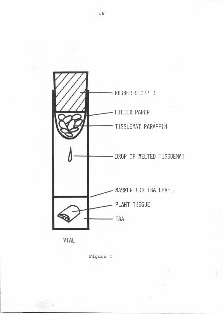
RUBBER STOPPER
F I L T E R PAPER
TISSUEMAT PARAFFIN
DROP OF MELTED TISSUEMAT
MARKER FOR TBA L E V E L
PLANT TISSUE
with safranin and counterstained with fast green. The staining procedure was as follows:
A. Removed the paraffin from the sections by placing the slides in xylene for five minutes and then in a 1:1 mixture of xylene and absolute alcohol for an additional five minutes.
B. Partially rehydrated the sections by passing them through a series of alcohols of decreasing concentration: absolute, 95%, 70%, and 50% (fiveminutes in each).
C. Stained in safranin for three hours (1% safranin in 95% alcohol and dilute with an equal amount of distilled water).
D. Washed in water, differentiated with acidified 70% alcohol and passed rapidly through 95% and absolute alcohol.
E. Counterstained with fast green (0.5% solution in50% clove oil, 50% alcohol) for 30 seconds.
F. Differentiated the fast green in a mixture of 50%clove oil, 25% absolute alcohol and 25% xylene.Two changes were made at 10 minutes each.
G. Placed them in xylene, making 3 changes of 15 minutes each.
Permanent mounts were made with 22 x 50 mm cover glass and Lipshaw Cover Glass Mounting Media. The tissues were studied with an Olympus compound microscope and pertinent data recorded. Photomicrographs were taken through a Zeiss photomicroscope.
17
3. Scanning electron microscope (SEM): Tissuepreparation for viewing with the SEM as outlinedby Rasmussen and Hooper (7) was followed. Fresh tissues about 5 mm were killed and fixed in FAA. They were dehydrated in a graded ethanol-water series for 30 minutes as follows:
25% ethyl alcohol 50% ethyl alcohol 75% ethyl alcohol 95% ethyl alcohol
100% ethyl alcohol The alcohol was then removed by an iso-amyl acetate series of:
25% iso-amyl acetate 50% iso-amyl acetate 75% iso-amyl acetate 100% iso-amyl acetate 2 times
The tissues were critical point dried in a Denton DCP-1 using liquid C02 at 1,650 pounds per square inch for 10 minutes. The tissues were mounted on 15 mm round coverglasses using a drop of Tube Koat (G. C. Electronics Company,Rockford, Illinois). Tube Koat, a carbon compound, was used as a glue to insure electrical conductivity. The tissues were coated with approximately 20 nm carbon followed by 20 to 40 nm of Au-Pd (60%-40%) by evaporation. The coated tissues were studied in the SEM (Advanced Metal
18
Research Model 900) at 21 kV accelerating potential. SEM photomicrographs were also taken.
Results and Discussions
Gross morphology: A typical Anthurium andreanum plant isshown in Figure 2. It is a relatively low growing perennial-herbaceous plant that thrives best in 60-80% shade with cool temperatures and high humidity. It has 3 to 6 leaves in clusters rising from the stem. The leaves are simple, chordate with smooth lamina margins and at maturity are usually 20 by 35 cm. The leaves are attached to a thin, long cylindrical petiole which forms a sheath or a pulvinus at its base which is fused around the stem. Stipules are borne at the sheath junction on mature leaves.
The usual short, thick stem has nodes approximately 1 cm apart. However, it may be a long thin stem with as much as 15 cm between nodes: this depends on the cultivarand the environment they grow in. For example, cv. Red Ozaki has short, stocky internodes, whereas, cv. Nitta Orange has long, thin internodes. Increased shade promotes elongation and long internodes, while high light intensities produce short internodes regardless of cultivar. The leaves have a spiral arrangement on the stem.
A vegetative lateral bud is formed at alternate nodes, opposite each leaf junction. The vegetative buds are not found in the leaf axils, but are spirally arranged opposite the leaves.
19
20
Figure 2. A mature anthurium plant, Anthruium andreanum, L., cv. Red Ozaki.
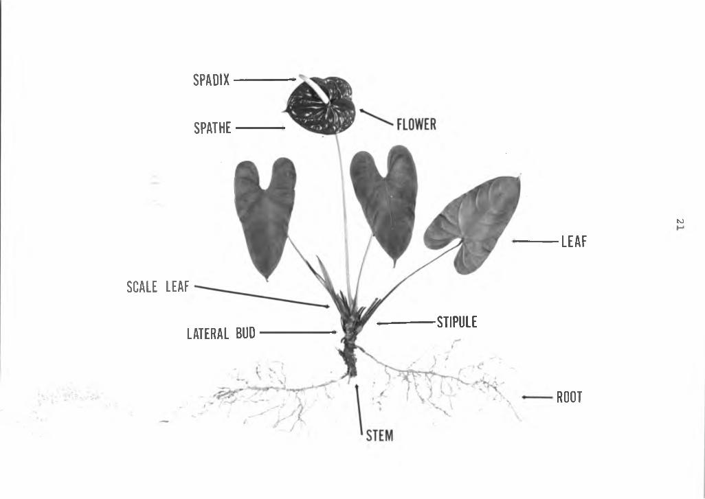
SPADIX
SPATHE
SCALE L E A F
LAT ERAL BUD■STIPULE
to
L E A F
ROOT
The flower buds are borne at each leaf axil. Mature flowers consist of a conspicuous bract (spathe) joined to the base of a cylindrical spike or rachis (spadix) and attached to a long pedicel. The botanical flowers are minute, perfect and densely cover the spadix. There are approximately 300 flowers spirally and closely arranged on the spadix. The fruits are small, globose, yellow berries closely arranged on the spadix. There are two seeds per berry which germinate approximately two weeks after sowing. The first flower is produced about 18 months after germination.
The roots are adventitious, all originating from the stem. They are cylindrical, thick and fleshy and emerge from the stem at each node, either below the vegetative bud, the leaf, or both. The primary roots branch to form secondary and tertiary roots. Root hairs are present 40- 50 mm from the meristematic root apex.
Two scale leaves or budscales are formed at each node. One between the flower pedicel and the stem, the other covering the lateral vegetative bud. The former is the remains of the budscale which originally covered the apical meristem of the plant.
Plant propagation is by seed and more often vegetatively by either suckers (lateral shoots), stem cuttings or terminal cuttings.
Christensen (2) reported that Anthurium andreanum when propagated by seed, goes through a juvenile phase, followed
22
by a generative phase. This study supports that finding. The difference in phases can be seen at the base of the leaf petiole. In seedlings the first leaves formed have short petiole sheaths with a vegetative bud at the axil.As the plant matures a flower bud forms at the axil of the leaf with no petiole sheath. Instead, the stipules form the sheath covering the axil and upper part of the petiole base, thus protecting the flower bud. In addition, the lateral vegetative bud develops opposite the leaf attachment.
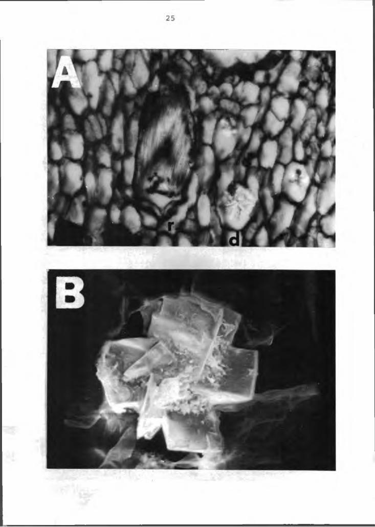
Crystals were found throughout the plant. The crystals were primarily the druse and raphide forms (Figure 3A). The druse crystal as viewed with the SEM is shown in Figure 3B. Energy dispersive X-ray analysis indicates it is high in Ca, probably as calcium oxylate. These crystals in the anthurium is in accord with most plants in the aroid family (4).
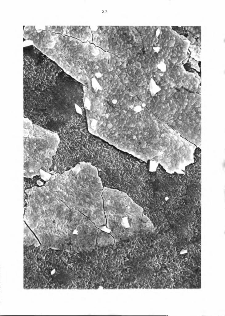
A most characteristic feature of the anthurium is the thick layer of cuticle on the epidermal cells of the aerial parts of the plant. The cuticle is of particular interest in anthurium research due to the problems of penetrability by chemicals applied as mineral nutrients, fungicides or herbicides (Figure 4). It also has a positive nature of protection.
Anatomy of the flower: The "commercial flower" ofAnthurium andreanum consists of three main parts: 1) thepedicel or the flower stem; 2) the bracteoles or the
23
24
Figure 3. Scanning electron micrograph of rhombohedral crystals in the anthurium. A left=raphide, right-druse (arrows), X30; B=SEM photomicrograph of the druse, X500. r=raphide crystal, d=druse crystal.
26
Figure 4 A scanning electron micrograph of the upper cuticle of an anthurium leaf. X2050.
27
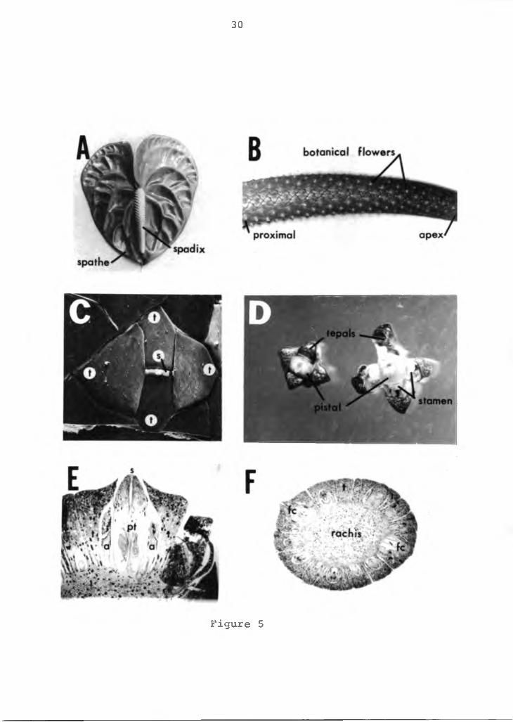
spathe; and 3) the spadix. All three parts are attached at a common junction. An anthurium flower is shown in Figure 5A. The botanical flower is perfect, small and borne on the spadix on a central rachis (Figure 5B). The proximal flowers on the spadix develop first progressing toward the apex. The surface anatomy and top view of the botanical flower is shown in Figures 5C and D. Four fleshy perianth segments (tepals) envelop four stamens and a fleshy pistal. Each stamen is located opposite a tepal and pressed firmly against the pistal. A cross section of a botanical flower and spadix is shown in Figures 5E andF. At the peripheral margin of the spadix the tepals form the floral cavity and are attached to the central rachis. The floral cavity contains the stamen and carpels. Anatomically the tepals have an outer epidermal cell layer over ground parenchyma cells. Vascular bundles are interspersed throughout the ground cells. Many tepal cells contain druse and raphide crystals, more than any other tissues in the anthurium plant. These crystals are found scattered throughout the plant, even in some epidermal cells.
The stamen has a flat filament with a single vascular bundle in the center, which traverse the entire filament and ends blindly in the connective tissue located between the two anthers. The anther is bilobed with each lobe containing two locules (Figures 6A and B). The outermost layer is a one celled epidermis. The subepidermal layer is the endothecium and the innermost layer the tapetum
28

29
Figure 5. The anatomy and morphology of the anthurium flower. A=anthurium flower with its spathe and spadix, X.75; B=spadix with its botanical flowers, Xl; C=surface morphology of the botanical flower, SEM photomicrograph, X30; D=botanical flower, XlO; E=cross section of the botanical flower, photomicrograph, X30.F=cross section of the spadix XlO; t=tepal, s=stigma, a=anther, pt=pistal, fc=floral cavity.
30
Figure 5
31
Figure 6. Photomicograph of the stamen of anthurium.A=longitudinal section of the stamen, X16; B=cross section of the anther, X32; C=pollen being shed on the spadix, X2; D=anther with its pollen, XlO. pg=pollen grain, a=anther, f=filament, e=epidermal layer, en=endothecium, lc=locule cavity, vb= vascular bundle.
32
(not shown in the photograph), is a nutritive tissue frequently composed of multinucleate cells. The tissue between the endothecium and tapetum is often crushed during development of the pollen sac. The developing sac is evident in the locules. Mature pollen can be detected as a white powdery mass on the spadix (Figures 6C and D).
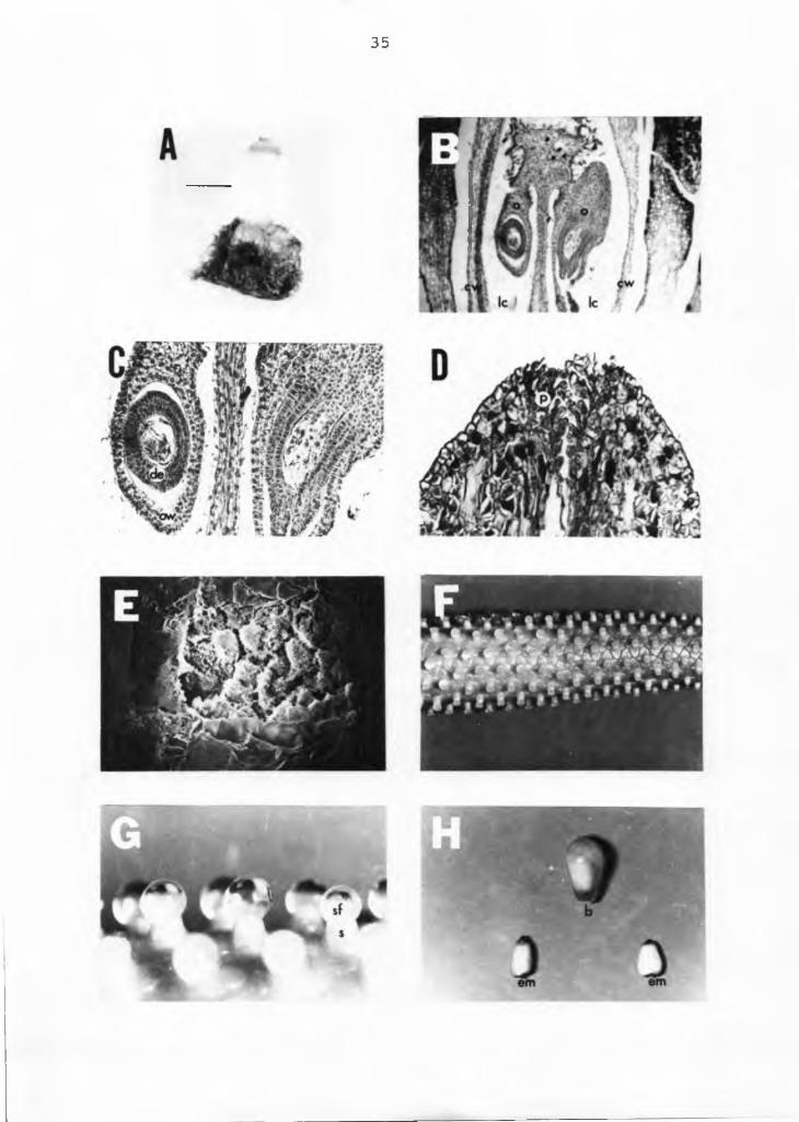
The anthurium pistal is shown in Figure 7. The ovary is hypogynous, as the tepals and androecium arise from the receptacle below the gynoecium (Figure 5E). It is a syncarpous gynoecium (more than one carpel) with two carpels fused together. Each carpel has a single locule with a single ovule. Therefore, each berry has two ovules or embryos (Figure 7B). Within the ovary the wall and the locule cavity can be identified (Figures 7B and C). Ovule placentation occurs in the center of the ovary where the carpellary margins meet (axile placentation). The single sessile style is an upward prolongation of the carpel with two stigmas that elongate into papillae, feather-like short hairs (Figure 7D). The stigma has glandular secreting tissue and the stigmatic fluid provides a suitable medium for pollen germination (3).
As the flower develops, the tip of the cylindric, subconic pistal protrudes through an opening at the tip junction of the tepals and the expanded style exposes the stigmatic surface (Figure 7E). The stigma is receptive to pollination when a sticky, translucent, stigmatic fluid is secreted at the tip of each flower on the spadix (Figures 7F and G). Pollination can be accomplished by collection
33
34
Figure 7. Photomicrograph of the gynoecium of anthurium.A=pistal (arrow), X5; B=longitudinal section of the pistal, X50; C=longitudinal section of the ovary, X125; D=longitudinal section of the stigma, X150; E=top view of the stigmatic area, X600; F=spadix with receptive stigma, XI; G=receptive stigma with its stigmatic fluid,X5; H=berry and two embryos, XI. o=ovule, cw=carpel wall, lc=locule cavity, de=developing embryo, ow=ovary wall, p=papillae hairs of the stigma, sf=stigmatic fluid, s=stigma, b=berry, em=embryo.
35
of pollen on a soft brush and applying it to a receptive stigma on the spadix. Natural pollination also takes place with insects.
Each fertilized ovary enlarges to form a single berry and each berry contains one or two embryos (Figure 7H).The seed develops and is ready for sowing in six months.The Anthurium andreanum flower is protogynyous. After the stigmas on the spadix protrude through the tepals they become receptive, dehydrate and shrink. The anthers grow around the pistal and shed their pollen above the tepals.The time lapse between the receptive stigma and the pollen maturity is about a week.
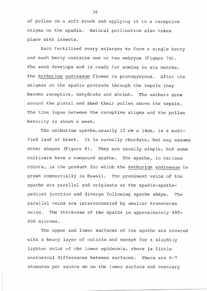
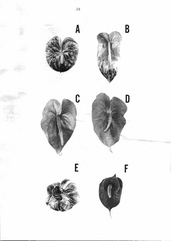
The anthurium spathe, usually 12 cm x 14cm, is a modified leaf or bract. It is normally chordate, but may assume other shapes (Figure 8). They are usually simple, but some cultivars have a compound spathe. The spathe, in various colors, is the product for which the Anthurium andreanum is grown commercially in Hawaii. The prominent veins of the spathe are parallel and originate at the spadix-spathe- pedicel junction and diverge following spathe shape. The parallel veins are interconnected by smaller transverse veins. The thickness of the spathe is approximately 400- 600 microns.
The upper and lower surfaces of the spathe are covered with a heavy layer of cuticle and except for a slightly lighter color of the lower epidermis, there is little anatomical differences between surfaces. There are 6-7 stomates per square mm on the lower surface and contrary
36
37
Figure 8. The various spathe shapes of the anthurium flower. A=cv. Ozaki Red B=UH 17 (pink obake); C=UH 16 (pink obake); D=cv. Anuenue;E=UH 100 (Red Double); F=cv. Red Elf.
38
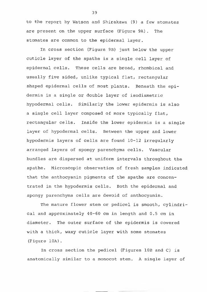
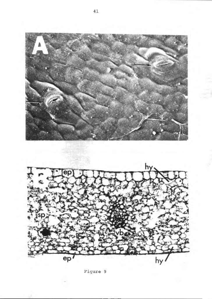
to the report by Watson and Shirakawa (9) a few stomates are present on the upper surface (Figure 9A). The stomates are common to the epidermal layer.
In cross section (Figure 9B) just below the upper cuticle layer of the spathe is a single cell layer of epidermal cells. These cells are broad, rhombical and usually five sided, unlike typical flat, rectangular shaped epidermal cells of most plants. Beneath the epidermis is a single or double layer of isodiametric hypodermal cells. Similarly the lower epidermis is also a single cell layer composed of more typically flat, rectangular cells. Inside the lower epidermis is a single layer of hypodermal cells. Between the upper and lower hypodermis layers of cells are found 10-12 irregularly arranged layers of spongy parenchyma cells. Vascular bundles are dispersed at uniform intervals throughout the spathe. Microscopic observation of fresh samples indicated that the anthocyanin pigments of the spathe are concentrated in the hypodermis cells. Both the epidermal and spongy parenchyma cells are devoid of anthocyanin.
The mature flower stem or pedicel is smooth, cylindrical and approximately 40-60 cm in length and 0.5 cm in diameter. The outer surface of the epidermis is covered with a thick, waxy cuticle layer with some stomates (Figure 10A) .
In cross section the pedicel (Figures 10B and C) is anatomically similar to a monocot stem. A single layer of
39
40
Figure 9. Photomicrograph and SEM micrograph of anthurium.A=stomata on the upper surface of the spathe (arrow), X2000; B=cross-section of the spathe, X16. ep=epidermis, hy=hypodermis, sp=spongy parenchyma tissue, vb=vascular bundle.
41
Figure 9
42
Figure 10. Photomicrographs and SEM micorgraph of theanthurium pedicle. A=stomata on the pedicel surface (arrow), X1600; B=cross section of the pedicel, X30; C=cross section of the pedicel, X60; D=cross section of the vascular bundle, X300. sl=sclerified parenchyma tissue, vb=vascular bundle, cx=cortex, ep=epidermis, ph=phloem, x=xylem, ss=sheath of sclerenchyma cells.
m
epidermal cells form the outer surface subtended by the cortex. A layer of subepidermal sclerified parenchyma cells separate the cortex and the ground parenchyma cells. The vascular bundles, consisting of the phloem and xylem are dispersed throughout the stem among the ground parenchyma cells (Figure 10D). These vascular bundles are enclosed in sheaths of sclerenchyma tissues. The pith is continuous and composed of ground parenchyma cells.
Anatomy of the leaf; The leaf blade is simple, green, chordate with smooth margins and is approximately 20 cm wide, 35 cm long and 400-500 mm thick. Leaf venation is parallel with the main veins emerging from the base of the leaf at the petiole-blade junction. The veins extend out to the leaf margins, converging or fusing at the leaf apex. The major veins are interconnected by smaller veins in a complex pattern. Stomatal pores are found only on the lower leaf surface (Figure 11A). They are located common to the epidermal cells with approximately 30-40 stomates per square mm with an apparently random distribution. No stipules, hairs or other appendages are evident on the leaf blade or the petiole.
The blade consists of an upper and lower one-celled epidermal layer (Figure 11B). The upper epidermal cells are thicker and isodiametric in shape, while the lower epidermal cells are much flatter. Between the epidermal layers is the mesophyll made up of one or two layers of compact palisade parenchyma cells. The palisade cells are
44
45
Figure 11. SEM and light photomicrograph of the leaf of the anthurium. A=lower leaf surface with stomata (arrows), X500; B=cross-section of the leaf, X35; C=petiole with stomata (arrow), X500; SEM photograph, X500; D=cross-section of petiole, X50; E=cross-section of vascular bundle, X60; F=logitudinal section of petiole, X200; sp=spongy parenchyma tissue, pp=palisade parenchyma tissue, vb=vascular bundle, ep= epidermis, sl=sclerified parenchyma cells, cx=cortex, ph=phloem, x=xylem, ss=sheath of sclerenchyma, cp=cortical parenchyma tissue.
46
Figure 11
found only beneath the upper epidermal layer of the leaf blade. The leaves are, therefore, dorsiventral or bifacial resulting in a dark green appearance on the upper surface and lighter green on the lower surface.
Vascular bundles of various sizes with adaxially located xylem and thick sclerenchyma fibers are dispersed throughout the mesophyll. Chloroplasts are present in all mesophyll cells, but are concentrated in the palisade cells.
The leaf petiole is cylindrical, smooth and usually 25-35 cm long. The base of the petiole forms a sheath around the stem. Stipules at the end of the sheath protect the flower bud in its early stages. The epidermal surface is smooth and covered with a thick waxy cuticle. A few stomates are dispersed irregularly on stem surface (Figure 11C) .
A cross section of the petiole is shown in Figures 11D and E. The anatomical structure of the petiole is similar to the pedicel described earlier. The epidermis is composed of a single layer of cells and subtending it is the cortex consisting of several layers of parenchyma cells. A layer of subepidermal sclerified parenchyma cells separate the cortex and the ground parenchyma cells. Vascular bundles are dispersed throughout the ground parenchyma tissue and are surrounded by a sclerenchymous sheath. The pith is continuous and composed of isodia- metric parenchyma cells.
47
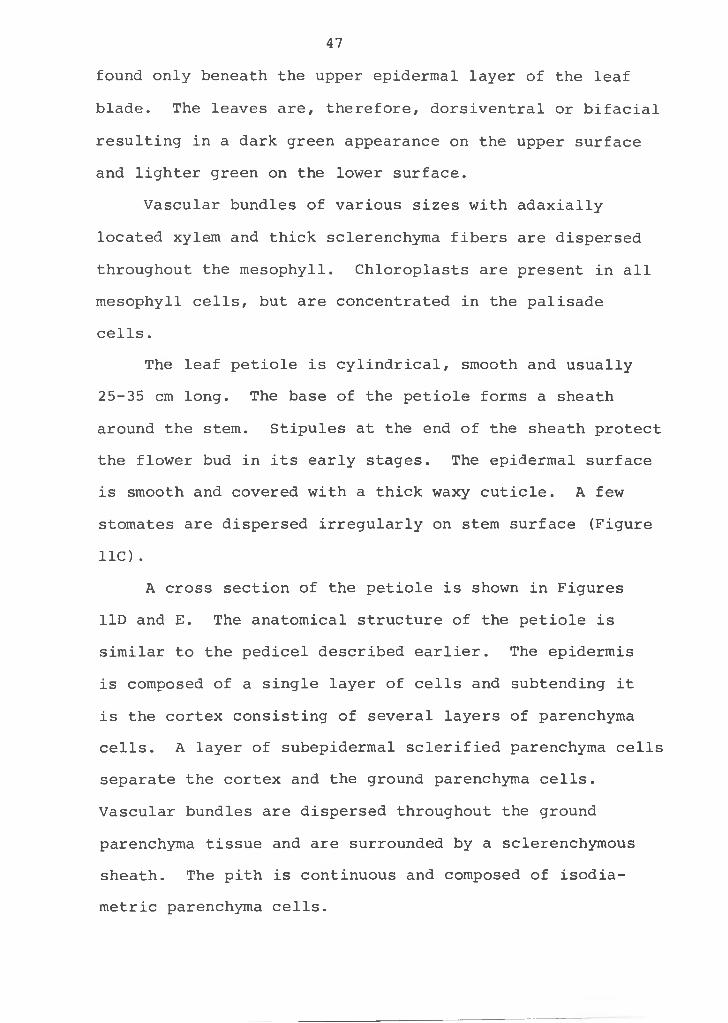
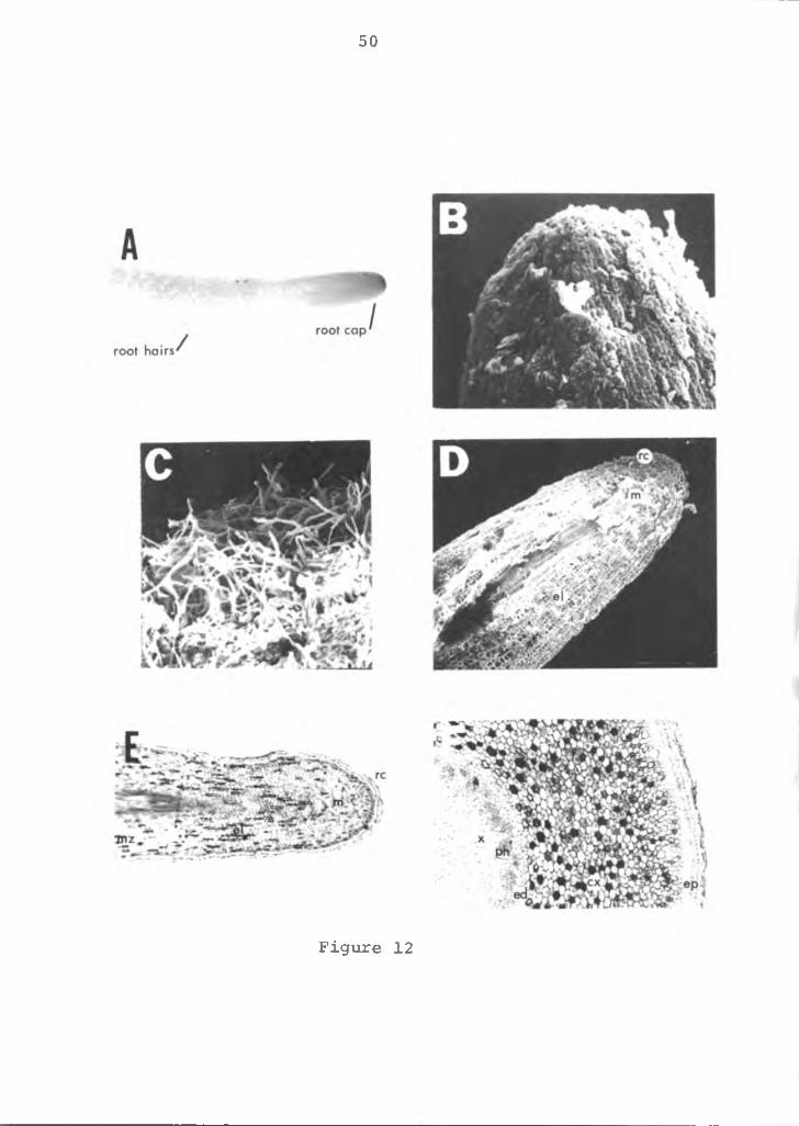
Anatomy of the root: The roots are adventitious arisingfrom the stem. Each root emerges just below an axillary bud or leaf at each node. Two roots normally emerge at each node. There is no main tap root, but roots branch readily into secondary, tertiary, etc., roots. The spongy, fleshy roots are cylindrical, 0.5-1.0 mm in diameter. They are epiphytic possessing some specialized contractile roots with the capability of attaching themselves to the surfaces, such as pots, tree bark, etc. Root hairs are apparent about 40-50 mm behind the root cap in the region of maturation (Figures 12A, B and C).
A longitudinal view of the root is shown in Figures 12D and E and as illustrated the outermost layer of cells form the root cap. The root cap protects the meristematic region located just behind the root cap. The cells are thin walled, cubical in shape with large nuclei and dense cytoplasm. Immediately adjacent is the region of elongation with no cell division, but where differentiation begins and maturation continues. These cells have large vacuoles, less dense cytoplasm and slightly thicker walls. In the root hair zone the cells are more differentiated and root hairs emerge from epidermal cells.
The root in cross section (Figure 12F) shows multiple layers of epidermal cells. The multiseriate tissue (multiple epidermis) called velamen is commonly found in the Araceae family. The tissue consists of compact nonliving cells, often with secondary wall thickening. The
48
49
Figure 12. Photomicrograph and SEM micrograph of theanthurium root. A=root tip, X4; B=root cap, X100; C=root hairs, X100; D=longitudinal section of the root tip, XlO; F=cross-section of root tip, X60. rc=root cap, m=meristematic region, el=region of elongation, mz=maturation zone, x=xylem, p=phloem, ed=endodermis, cx= cortex, ep=epidermis.
50
Figure 12
function of the velamen is absorption, but studies by Dycus and Knudson (3) on orchids indicated that its principle role is mechanical protection and water conservation. The cortex is located adjacent to the velamen with layers of large, thin walled parenchyma cells. The innermost cortical layer has smaller cells, which form the endo- dermis. The xylem, phloem and parenchyma cells compose the stele (Figure 12F).
Anatomy of the stem: The primary stem (Figure 13) iscylindrical, fibrous and at maturity is approximately 2 cm in diameter. Leaves are arranged spirally with a phyllo- taxis quotient of 2/7 and a divergence angle of 102.9°.The spiral pattern may develop either clockwise or counter clockwise. Factors affecting this pattern is unknown.The nodes appear as pairs as the internodes alternate between "short" and "long" internodes. One node gives rise to the lateral vegetative bud, while the other produces the leaf pediole. The short internode is about 0.5 cm long, while the long internode varies from 1 to 5 cm.The long internode responds to environmental conditions and its response varies with variety. In common usage, the short internode is referred to as the "node" and the long internode as the "internode." However, anatomically the stem is structured as previously discussed. Stem growth on a mature plant is about 5-10 cm per year, but as it is a perennial, stems up to 4 m long are common. A vegetative apical meristem develops at each lateral bud
51
52
Figure 13. Diagram of the vegetative stem of the anthurium.
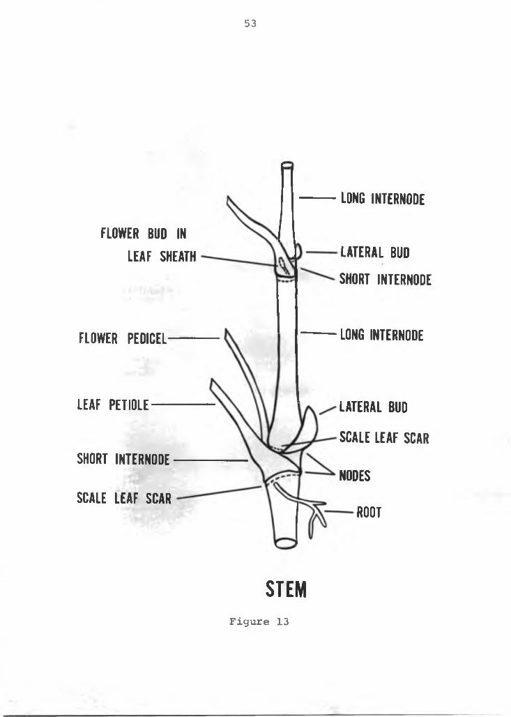
53
LONG INTERNODE
FLOWER PEDICEL LONG INTERNODE
LE A F PETIOLE —
SHORT INTERNODE
SCALE L E A F SCAR
LA T ER A L BUD
SCALE LEA F SCAR
NODES
ROOT
FLOWER BUD IN L E A F SHEATH LA T ER A L BUD
SHORT INTERNODE
S T E MFigure 13
and stem terminal. This results in a network of stem branching as the lateral shoots develop.
The anatomy of the stem is similar to the pedicel and petiole discussed earlier. The stem has one layer of cells forming the epidermis. The epidermis surrounds a cortex layer but is much thicker in width than the pedicle or petiole. A layer of sclerified parenchyma cells separate the cortex from the ground parenchyma cells. Vascular bundles enclosed in sheaths of sclerenchyma tissues are dispersed in the ground tissues. The pith is continuous and consists of thick walled parenchyma cells.
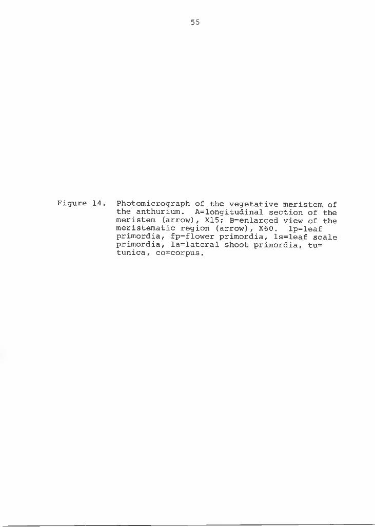
The vegetative apical meristem (Figure 14A) is offset from the terminal apex. This off-centered position is caused by the spiral characteristic of the stem in which the apical meristem is displaced terminally by the developing vegetative primordias. The primordias are shown at the periphery of the apex, possessing undifferentiated embryonic cells.
At the meristematic apex are two layers of tunica cells (Figure 14B), which divide perpendicular to the surface of the meristem (anticlinal division). Directly beneath the tunica is the corpus where cell division occurs in multiple planes (anticlinal and periclinal). The corpus adds bulk to the apical portion of the shoot by increasing in volune, whereas tunica maintains its continuity over the enlarging body by surface growth.
To summarize the section: a study of the morphology and anatomy of Anthurium andreanum, L., was conducted. It is a
54
55
Figure 14. Photomicrograph of the vegetative meristem of the anthurium. A=longitudinal section of the meristem (arrow), X15; B=enlarged view of the meristematic region (arrow), X60. lp=leaf primordia, fp=flower primordia, ls=leaf scale primordia, la=lateral shoot primordia, tu= tunica, co=corpus.
56
perennial-herbaceous monocotyledon in the family Araceae, therefore, possesses typical monocot anatomical structures. Of particular significance among its characteristics are:1 ) possesses a juvenile phase as seedlings and a generative phase at maturity. The juvenile phase has a lateral shoot at the leaf axil, whereas, the generative phase has a flower. The lateral shoot locates itself opposite the leaf attachment; 2) the "commercial anthurium flower" has chor- date spathe and a protruding spadix, whereas, the botanical flowers are minute, perfect and borne on the spadix; 3) the roots are covered with a multiple layer of epidermal cells called velamen; 4) raphide and druse crystals are dispersed throughout the entire plant tissue; and 5) the above ground parts of the plant is covered with a thick layer of waxy cuticle.
57
LITERATURE CITED
58
LITERATURE CITED
1. Akamine, E. K. and T. Goo. 1972. Relationships between physical characteristics and vase life of anthuriums. Haw. Farm Sci. 21(2): 9-10.
2. Christensen, V. 0. 1971. Morphological studies on the growth and flower formation of Anthurium scherzerianum, Schott and Anthurium andreanum, Lind. Statens Forsogsvirksomhed i Tidssdr Planteval 75(6): 793-798.
3. Dycus, A. M. and L. Knudson. 1957. The role of the velamen of the aerial roots of orchids. Bot. Gaz. 119:78-87.
4. Esau, Katherine. 1961. Anatomy of seed plants.John Wiley and Sons, Inc., N.Y.
5. Jensen, W. A. 1962. Botanical histochemistry.W. H. Freeman and Company, San Francisco, California.
6 . Kamemoto, H. and H. Y. Nakasone. 1955. Improving anthuriums through breeding. Haw. Farm Sci. 3(3): 4-5.
7. Rasmussen, H. P. and G. R. Hooper. 1974. Electronoptics: Principles, techniques and application inhorticulture. Hort. Science 9(5): 425-433.
8. Sharmg, A. K. and U. C. Bhattachryya. 1961. Structureand behavior of chromosomes in species of anthurium with special reference to the accessory chromosomes. Proc. Bio. Sci. 27:317-328.
9. Watson, D. P. and T. Shirakawa. 1966. Gross morphology related to shelf life of anthurium flowers. Haw. Farm Sci. 15(2): 2-3.
59
SECTION II COLOR BREAKDOWN IN ANTHURIUM FLOWERS,
ANTHURIUM ANDREANUM, L.
60
Section II: Color Breakdown in Anthurium Flowers,Anthurium andreanum, L.
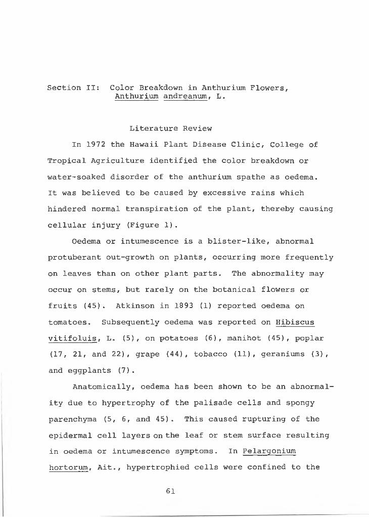
Literature Review In 1972 the Hawaii Plant Disease Clinic, College of
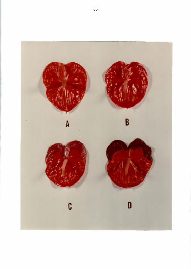
Tropical Agriculture identified the color breakdown or water-soaked disorder of the anthurium spathe as oedema.It was believed to be caused by excessive rains which hindered normal transpiration of the plant, thereby causing cellular injury (Figure 1).
Oedema or intumescence is a blister-like, abnormal protuberant out-growth on plants, occurring more frequently on leaves than on other plant parts. The abnormality may occur on stems, but rarely on the botanical flowers or fruits (45). Atkinson in 1893 (1) reported oedema on tomatoes. Subsequently oedema was reported on Hibiscus vitifoluis, L. (5), on potatoes (6), manihot (45), poplar (17, 21, and 22), grape (44), tobacco (11), geraniums (3), and eggplants (7).
Anatomically, oedema has been shown to be an abnormality due to hypertrophy of the palisade cells and spongy parenchyma (5, 6 , and 45). This caused rupturing of the epidermal cell layers on the leaf or stem surface resulting in oedema or intumescence symptoms. In Pelargonium hortorum, Ait., hypertrophied cells were confined to the
61
62
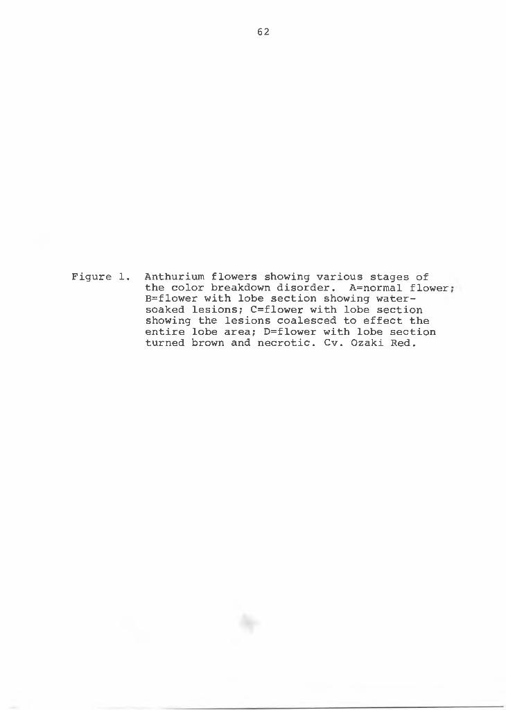
Figure 1. Anthurium flowers showing various stages ofthe color breakdown disorder. A=normal flower B=flower with lobe section showing water- soaked lesions; C=flower with lobe section showing the lesions coalesced to effect the entire lobe area; D=flower with lobe section turned brown and necrotic. Cv. Ozaki Red.
63
spongy parenchyma cells, filling the substomatal cavity and oedematic structures appeared only on the lower epidermis of the older leaves (3).
In all the reports cited, the abnormality was attributed to physiological rather than pathological conditions. It was generally concluded that hypertrophy of the palisade and spongy parenchyma cells resulted when water absorption exceeded transpiration, causing water to accumulate in the tissues. A continuous water supply to a warm, moist soil with cool night temperatures predisposed plants to oedema (3). Other environmental factors, e.g., light intensity, light quality and mineral concentration in the plant were studied, however, plant-water relationship was found to be the main factor causing oedema (27, 28, and 29).
According to Chupp and Sherf (4), oedema may be caused by any agent which stimulates groups of inner cells to grow abnormally. This condition was accentuated when moisture was high, soil temperature was high and air temperature was low. These conditions occur on a cool night after several warm rainy days when relative humidity is near 100%. Under these conditions the roots absorb water at a rapid rate exceeding that lost through transpiration. The result was an oversaturation of the cells with water, creating enough pressure to rupture the epidermis.
Similar physiological diseases due to moisture relationship have been reported: blossom-end rot and black
64
seed of tomatoes (8, 41, and 43), withertop of flax (18), bitter-pit of apples (38), blasted buds of lilies (37), crinkle leaf of cotton (31), internal browning of brussel sprouts (24), brownheart of escarole (25), internal tipburn of cabbage (26), tipburn of lettuce (42), brown rib of lettuce (15), blackheart of celery (10), and blossom-end rot of guava (39). Each disorder cited above was corrected or prevented by the application of Ca.
Sprague (40) has reported that Ca in plant cell walls forms a protective "sieve" for nutrient to seep through in passing into cells. It also acts as a cement (calcium pectate) between cell walls. Stackman (36) reported that Ca deficiency was invariably associated with the breakdown of cell walls. Rasmussen (33) reported that Ca deficiency weakened tobacco leaves, causing the constituent cells to separate under little stress. Microscopic studies showed that tobacco leaf tearing in tender leaf occurred in an irregular pattern between cells. When Ca was present in sufficient quantity, tearing occurred through cells. However, the total Ca content was approximately the same in normal and tender leaves. Further studies suggested that there may be equal quantities of Ca in the sum of pectate, crystal and soluable Ca in both normal and tender leaf tobacco, but there is less in the pectate of the cell walls of the tender leaf.
Lange (19) using beans, found that the lack of aeration for 12 hr caused as much as 49% decrease in Ca uptake,
65
but increased the P uptake as much as five times. Lange also found Ca uptake highest at pH 7.0. Ca accumulation by apple stems and leaves (46) was less at 5°C than at 20°C, 30°C and 40°C. The maximum Ca translocation was between 15°C to 30°C. In peas (13) decreasing the temperature from 20°C to 0°C during a 24 hr period decreased both water and Ca uptake. These studies indicate that aeration, pH and temperature affect Ca uptake.
Crowns of healthy strawberry plants (20) had a lower percentage of ash and a higher percentage of Ca than other parts of the plant. The roots of diseased plants had low Ca levels, while the leaf interior had higher Ca levels than the serrated edges. On the other hand, the serrated edges of the leaves of healthy plants were higher in Ca than the rest of the leaf. This study indicated that Ca content does vary in different parts of the plant and even within the same leaf. It may also vary between diseased and healthy plants.
Moser (30) growing soybeans and sorghum in sand culture showed that Ca concentration at the various pH levels (pH 3.8-4.2, 5.0-5.2, and 6 .0-6.5) was a more important factor on Ca uptake than pH levels. His results showed that increased supply of Ca influenced directly the concentration of Ca within the crop irrespective of pH.Lundegardh (23) found available Ca reduced from 25 mgms to 12.5 mgms per liter when the pH of the soil was changed from pH 7.0 to 5.0. He attributed this effect to chemical precipitation of phosphate and other minor elements with Ca.
6 6
Materials and MethodsThe problem of the color-breakdown in the spathe of
the anthurium flower was approached using the following methods:
1. Isolation and inoculation of microorganisms from diseased tissue.
2. Chemical elemental analysis of tissue from affected and normal healthy flowers.
Isolation and inoculation of microorganism from diseased tissue;
One 5 mm^ tissue section was taken from the water- soaked and color-breakdown area of ten anthurium spathes, cv. Ozaki Red. The tissue sections were surface sterilized with 0.8% sodium hypochloride for 3 minutes, rinsed 3 times with sterile distilled water and cultured on potato dextrose agar plates.
A fungus, identified as Colletochrichum gloeosporioides was isolated from the tissues. Spore suspensions of asci and conidia stages were prepared with distilled water and sprayed with a hand atomizer on the anthurium spathes of the cv. Ozaki Red. Treatments were as follows: 1. Tenflowers sprayed with asci suspension; 2. Ten flowers sprayed with conidia suspension; and 3. Ten flowers sprayed with distilled water. The flowers were placed in the greenhouse under a continuous fine mist for 48 hr in the shade at 80°F. Daily observations were made to determine development of incipient water-soaked or color- breakdown lesions.
67
68Chemical elemental analysis of anthurium tissues from affected and normal flowers:
Eighty, 75 percent mature flowers and 80 associated leaves on the same axil of the stem, were collected from the anthurium cv. Ozaki Red. Seventy-five percent mature flowers were distinguished by the degree of color change of the spadix, e.g., when the lower 3/4 of the spadix had changed from reddish-orange to light pink in cv. Ozaki Red. Forty of the flowers showed symptoms of the spathe color-breakdown, while forty appeared to be normal healthy flowers. The flowers and leaves were divided into four treatments: 1. Color-breakdown flowers; 2. Leaves ofcolor-breakdown flowers; 3. Normal flowers; and 4. Leaves of normal flowers. The 40 flowers and 40 leaves making up each treatment were further divided into 4 samples of 10 flowers and 10 leaves (4 reps).
The samples were rinsed with distilled water and air- dried. The spadix and pedicel were discarded and only the spathe analyzed; similarly, only the leaf blade was analyzed. Fresh and dry weights were taken and percent moisture determined. The samples were dried in a force air oven at 75°C for 48 hr, after which they were ground to a fine powder in a Spex Industries Ball Mill.
Levels for N, P, K, Ca, Mg, B, and SiC>2 were determined as follows:
Nitrogen: the standard Kjeldahl total nitrogen methodas outlined by Horwitz (12).
Phosphorus, potassium, calcium and magnesium: 5 gramsof the ground sample were ashed overnight in a muffle furnace at 500°C. The ash was "wet" down with distilled water and dissolved with 5 ml concentrated hydrochloric acid and left standing for one hr. The solution was transferred to a 50 ml volumetric flask, made up to volume with distilled water and left standing overnight. An aliquot was diluted 10 times and P was determined by the Molybdophosphoric blue color method (14) (Klett-Summerson Photoelectric Colorimeter, model 900.3). A second aliquot from the 50 ml solution was diluted 10 times with 0.5% Lanthanum solution and Ca and Mg were determined spectro- photometrically (Perkin-Elmer Atomic Absorption Spectrophotometer, Model 303). (The Lanthanum solution was made by wetting down 58.65 gm of La2<33 with distilled water and adding 250 ml of concentrated HC1 very slowly until the material was dissolved. The solution was diluted to 1000 ml with distilled water to give a 5% lanthanum solution in 25% (v/v) HC1. Aliquot was taken from this stock solution and diluted to a 0.5% Lanthanum solution. Lanthanum was added to prevent interference from P and Al.) A third aliquot from the 50 ml solution was diluted 10 times with distilled water and analyzed for K by flame photometry (Beckman DU Spectrophotometer).
Boron: Boron in ppm was determined by the Quinaliz-arin method (12). Three ml of saturated calcium hydroxide was added to 2 gm of the ground samples and left standing
69
for one hr. Samples were ashed overnight at 550°C. Ten ml of 0.36N H2SO4 were added to the samples and left standing for 2 hr. The solution was filtered with White Ribbon Filter No. 589 into a 40 ml soft glass test tube (non-pyrex) and shaken well. Two ml of the solution were pipetted into an absorption test tube, ten ml quinalizarin dye added (45 mg quinalizarin dye per liter of H2SO4), mixed thoroughly, cooled to room temperature and measured colorimetrically (Evelyn Colorimeter) at 595-660 micron.
Silicon dioxide: SiC>2 was determined by ashing 1 gmof ground sample at 500°C overnight, dissolving the ash in 5 ml concentrated hydrochloric acid and drying over low heat for three hr. The ash was again dissolved in 10 ml of 3N hydrochloric acid and filtered with White Ribbon Filter No. 589. The filter paper was ashed in a porcelain crucible for 3 hr at 700°C. The ash and crucible were cooled in a dessicator over calcium chloride and weighed. Subsequently, the crucible was cleaned with a stiff brush to remove the ash, re-ashed at 700°C for one hr, cooled in the dessicator and reweighed to get the weight of the ash. The ash weight was calculated as percent of original matter and reported as percent SiC>2 . A blank sample on the filter paper was similarly analyzed serving as a control for filter paper impurities.
After preliminary experiments were carried out and a hypothesis formulated, the following procedures were used to verify the hypothesis:
70
1. Electron microprobe X-ray analysis of elements in the tissues
2. Solution culture to induce the disorder symptoms3. Radioisotopes to study element distribution and
localization4. Field test to verify the results
Electron microprobe X-ray analysis;Fresh samples, about 5 x 15 mm from an anthurium
spathe showing symptoms of color-breakdown and from normal, healthy flowers were taken from cv. Ozaki Red for microprobe analysis. The tissues were taken from the lobe and tip sections of the spathe for analysis, since the symptoms of color-breakdown are localized in the lobe section of the spathe (Figure 2). The cyrostat tissue preparation method was used (34). The fresh tissue was immediately mounted and frozen in Optimum Cutting-Temperature Compound (OCT -15 to -30°C, Fisher Scientific Company). Sections 16 micrometers thick were cut at -16°C on the Model CTD- International-Harris-Cryostat and placed at room temperature on polished carbon disc 50 mm in diameter. The OCT served as the mounting medium. The samples were air-dried and placed in an electron microprobe X-ray analyzer (Applied Research Laboratory, model EMX-SM), operating at 21 kV accelerating voltage and 0.056 micro amperes sample current. No carbon conductive coating was used with sections of this thickness. Calcium X-ray emission counts as well as graphic line profile analysis of K(2000A),
71
72
Figure 2 Illustration of sampling of tissues from lobe and tip sections of the anthurium spathe.
L O B E S E C T I O N S
T I P S E C T I O N S
F L O W E R L E A F
74
P(200^), Na(200 ̂ ) and Ca(200?\) were taken and recorded. Solution culture:
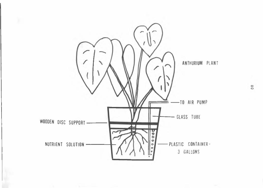
This method was used only after previous experiments indicated that calcium levels may cause color-breakdown in the spathe. Of prime interest was inducement of the color- breakdown disorder by eliminating calcium in the nutrient solution and/or by varying the pH levels of a complete nutrient solution to alter chemically the availability of calcium for uptake by the plants.
Twenty-eight mature anthurium plants of the cv. Ozaki Red were planted in coarse perlite media in 8" plastic pots. The pots were seated into a completely enclosed wooden box as illustrasted in Figure 3. The peat moss was kept moist at all times providing 1 00% relative humidity to the roots. A heating cable in the peat moss kept the temperature at the root zone at 80°F. The purpose of the box was to provide an environment of high humidity and high root temperature to increase metabolism, e.g., increase nutrient, water uptake and growth. The box also prevented drying of the roots as perlite is a poor water holding media. The plants were given a nutrient solution as described by Rasmussen, et al. (34) with modifications (Table I). Two hundred ml of each treatment solution wereadded three times a week. No additional watering wasnecessary. pH levels were 3, 4, 5, 6 , and 7 and were testedas well as concentrations of 0, 100, and 200 ppm. The solutions
75
Figure 3. Planting system used to grow anthuriums in nutrient culture.
AT 8 0 ° F W I T H W A T E R
E N C L O S E D WOODEN BOX
77
Table I. Solution Culture Composition, Concentrations and Conditions used in the Calcium Experiment_________
Nutrient Culture
Major elements g/liter*Ca (NO3 ) 2 • 4H20 1.18KNO3 0.51KH2PO4 0.14MgSC>4 • 7H20 0.49Iron sequestrene 0.0115
Minor elements g/literH3BO3 0.6MnCl2 • 4H20 0.4ZnSC>4 0.05CUSO4 • 5H20 0.05Treatments1. Complete, pH 62. 1 / 2 calcium, pH 63. No calcium, pH 64. Complete, pH 35. Complete, pH 46 . Complete, pH 57. Complete, PH 7
*In the 1/2 calcium and No calcium solutions, NH4NO3 at 0.20 g/1 and 0.40 g/1 were added respectively to compensate for the loss of N when Ca(NC>3 ) 2 * 4H20 was reduced or omitted.
were pre-mixed in 20 liter plastic carboys and pH readings taken weekly. When necessary, the pH was adjusted with 15% H2SO4 and/or 15% NaOH. Weekly production data and any observable nutritional deficiency symptoms were recorded. Calcium content of the spathe and the leaf associated with the flower was determined spectrophoto- metrically as described earlier. The spathe and leaf blade were divided into lobe and tip sections and analyzed. Radioisotopes:
Twelve mature anthurium plants cv. Ozaki Red, were grown in three gallon plastic containers in aerated solution culture (Figure 4). Plants were grown in a fiberglass greenhouse under 80% shade. Approximately 500 ft. candle of fluorescent light was given nightly from 5 p.m. to 5 a.m. to increase plant metabolism.Plants were grown in deionized water for 5 days and then
transferred to nutrient solution as given in Table I, with the exception that Ca (1̂ 0 3 ) 2 * 4H2O was omitted and CaCl2
added to make a solution at 5 X 1 0 molar. NH4NO3 was added at 0.40 gm/ 1 to compensate the loss of nitrogen by omission of Ca(N0 3 ) 2 * 4^0. The solutions were adjusted to pH 3, 5, 7, and 9. Three plants per treatment were grown for 7 days at the end of which, the solution was replaced, pH adjusted and 0.05 micro-curies per ml of ^5Ca added to each solution. The plants were allowed to take=up 4^Ca for an additional 14 days. pH was checked twice a day and adjusted for the duration of the experiment.
78
79
Figure 4. Diagram of apparatus used for radioisotope study.
WOODEN DISC S U P P O R T
N U T R I E N T S O L U T I O N
A N T H U R I U M P L A N T
T O A I R P U M P
- G L A S S T U B E
P L A S T I C C O N T A I N E R - 3 G A L L O N S
The plants were harvested at 14 days and prepared for microautoradiography and SEM studies. Fresh tissue 5 x 15 mm was taken from the flower spathe and leaf blade associated with the flower at the same stem axil. Tissues of both spathe and leaf were taken from the upper lobe area and from the tip section.
The tissues were fixed in a 3:1 95% alcohol-acetic acid solution (v/v) and carried through tert-butyl alcohol dehydration and tissuemat embedding (16). Sections were cut at 8 micrometers (American Optical Company, model 815 rotary microtome) and mounted on gelatin-chrome-alum coated glass slides. The paraffin was removed with xylene and microautoradiography procedures of Ficq (9) were followed. The sections were coated with Kodak Nuclear Track NTB2 emulsion and stored in the dark for proper exposure. After the microautoradiographs were developed, fixed, washed, air-dried and a cover glass mounted permanently with Pro- Texx mounting media, the sections were viewed with phase contrast or bright field microscopy to determine silver localization in the tissue.
For SEM energy dispersive X-ray analysis, the paraffin embedded tissue was cut at 10 micrometers, mounted on gelatin-chrome alum coated polished aluminum disc (17 mm in diameter) and the paraffin removed with xylene. The sections were air-dried, coated with 20 nm of carbon by evaporation and analyzed in a SEM (Cambridge Stereoscan S4 with Edax detector) (35).
81
Field test;To determine the effect of calcium on the color-
breakdown disorder, a randomized block design with three treatments and three replicates was used. Seventy-two mature anthurium plants, cv. Ozaki Red, were planted in 6 ' x 12' plots in black cinder media under 75% Saran shade. They were subjected to the following calcium treatments: 1. Calcium nitrate at the rate of 200#/A/yr;2. Calcium silicate at the rate of 1400#/A/yr; and 3. No calcium. Total calcium was equally divided into 3 parts by weight and given to the plants as a soil additive every 4 months.
Normal fertilizer program of Osmocote 14-14-14 was given 3 times a year at the rate of 300 # N-P-K/A/yr and standard pesticide practices were followed.
Data on normal, healthy flowers and flowers showing color-breakdown symptoms on the spathe were recorded.
Results and DiscussionAnthurium flowers sprayed with asci and conidia sus
pensions of Colletochrichum gloeosporioides showed no sign of the color-breakdown disorder in any of the treatments. The normal senescence symptoms, such as browning of the spadix and eventual abscission of the flower from the stem at the "stem-spathe-spadix junction" took place within 15 days after treatment. It was concluded that the fungus Colletochrichum gloeosporioides, earlier isolated from the color-breakdown spathe, was not the cause of that disorder
82

in anthuriums. Aragaki (2) writes:Colietotrichum gloeosporioides is a common fungus on tropical plants, especially on above ground parts. It is recovered in very high frequency from healthy-appearing tissues, so not surprisingly, the rate of recovery from necrotic tissue is even higher. Obviously, many of them are non- pathogenic, and this has been verified by innoculation tests. It is also a pathogenic species, however, and will cause many important diseases, including anthurium anthracnose. In the years (I) spent on screening anthuriums for anthracnose resistance, spathe lesions have been few and far between, and not reproducible.Approximately 50 isolates of C. gloeosporioides (including several from spathe) have been screened thus far.
Our results verify these results.Elemental analysis of anthurium spathe and leaf
tissue from color-breakdown and normal flowers is given in Table 2. Differences in Ca are evident. Color-breakdown spathe and leaves have a lower Ca content (0.372% and 0.363% respectively) than normal spathe and leaves (0.830% and0.805% respectively). No other elements varied between color-breakdown and normal plants. The data suggest that low Ca and not oedema may cause the anthurium color- breakdown disorder.
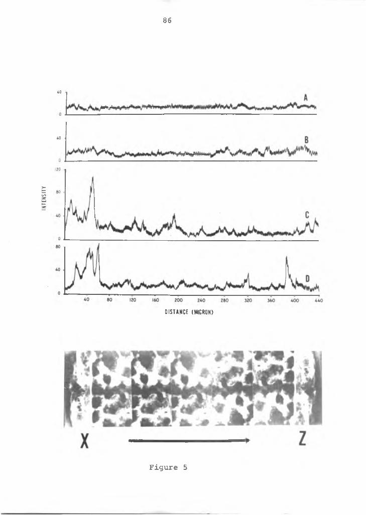
Electron microprobe X-ray analysis of a cross-section of spathe tissue in both color breakdown and normal tissue is given in Figure 5. A higher concentration of Ca is evident in the normal flowers as compared to color- breakdown flowers. Normal tissue had a higher concentration of Ca in the upper and lower epidermal tissue than in intermediate tissues. Ca distribution in color-breakdown tissue was uniformly low. Semiquantitative analysis for
83
84
Table 2. Elemental analysis of anthurium spathe and leaf from color-breakdown and normal plants.*_______
Spathe %N %P %K %Ca %Mg %Si02B
ppm%
moisture
Normal 1.55 .180 2.13 .830 .176 . 1 0 16.0 13.17
Color-break 1.76 . 2 0 1 2.44 .372 .150 . 1 2 14.0 13.78
LeavesNormal 2 . 1 2 .178 2 . 0 1 .805 .266 . 1 0 1 1 . 8 2 1 . 6 8
Color-break 2 . 20 .173 1.98 .363 .196 . 1 0 13.6 24.18
*Note: Each figure represents an average of 10 plants persample and analysis ran on 4 replicated samples.
85
Figure 5. Microprobe line profile X-ray analysis of Ca in the spathe of anthurium plants. A=color-break- down flower, lobe section; B-color-breakdown flower, tip section; C=normal flower, lobe section; and D=normal flower, tip section. The line scan proceeded from X, upper epidermis, to Z, lower epidermis.
INTE
NSI
TY
86
40
40
D I S T A N C E ( M I C R O N )
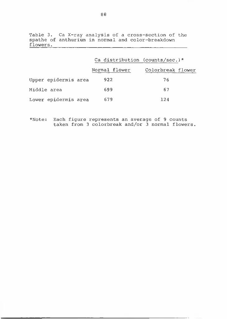
Figure 5
Ca in cross-sections of the upper, middle and lower sections of the spathe tissue produced similar results (Table 3). Lanning (20) found variation in Ca content of strawberry within the same plant part and even in the same cross- section. These data strengthen the hypothesis that low Ca is the cause of the color-breakdown disorder in anthuriums. Tissue exhibiting the color-breakdown had a lower Ca content than normal tissue.
Nutrient culture studies carried out in Hawaii produced color-breakdown symptoms on the spathe after 6 months of growth in nutrient solutions. Color-breakdown symptoms occurred consistently in the "No Ca" treatment. Other treatments such as 1/2 Ca and the various pH levels (pH 3, 4, 5, 6 , and 7) showed no symptoms of color breakdown after 12 months. However, the "No Ca" treatment plants which developed color breakdown symptoms produced normal flowers when Ca was added. The disorder symptoms were as follows: the spathe developed tiny water-soaked lesions ordiscolored areas the size of pin heads on the upper epidermis on the lobe section of the spathe. These lesions increased in number and size and eventually coalesced to form a water-soaked mass or large color-breakdown lesion.The lesions dehydrated, turned dark brown and died with an upward roll of the necrotic tissue. In extreme cases, the young leaves became chlorotic and were small and distorted. The margins were irregular and frequently contained spotted and necrotic areas. New leaves and flowers continue to
87
88
Table 3. Ca X-ray analysis of a cross-section of the spathe of anthurium in normal and color-breakdown flowers._____________________________________________
Ca distribution (counts/sec.)* Normal flower Colorbreak flower
Upper epidermis area 922 76Middle area 699 67Lower epidermis area 679 124
*Note: Each figure represents an average of 9 countstaken from 3 colorbreak and/or 3 normal flowers.
emerge, however, they die before they unfurl. The apical meristem was obviously affected since lateral shoots began to develop from the stem. The entire plant was stunted and eventually died.
Elemental analysis of plants grown at various nutrient and pH levels in the cultural solutions was made.The plant was separated into lobe and tip sections of both the spathe and leaf. The results are presented in Table 4. Again there was less Ca in those plants exhibiting color- breakdown (No Ca treatment), both in the spathe and in the leaves as compared to all other treatments. The results also indicate less Ca in the lobe than in the tip. This further suggests that low Ca is the cause of the disorder. The symptoms of this disorder are initiated and concentrated in the lobe section of the spathe. Calcium uptake increased with increase in pH levels from 3 to 7.
Tissue samples of the lobe and tip of the spathe and of plant leaves grown at various pH levels were analyzed by energy dispersive X-ray analysis (Figure 6). The uptake of Ca was lowest at pH 3, increased through pH 5, reached a maximum at pH 7 and decreased again at pH 9. These results are similar to other reports of the effect of pH on Ca uptake (19 and 23). This pH data sheds some light on the course of the color-breakdown disorder found in Hawaii's anthurium nurseries. The major occurrence of the color- breakdown disorder is in nurseries using volcanic black cinder as a growing medium. The medium is a light, porous, inert material which provides excellent drainage and
89
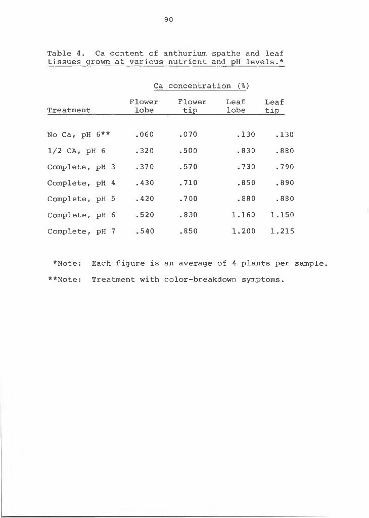
90
Table 4. Ca content of anthurium spathe and leaf tissues grown at various nutrient and pH levels.*
Ca concentration (%)
TreatmentFlowerlobe
Flowertip
Leaflobe
Leaftip
No Ca, pH 6** .060 .070 .130 .1301/2 CA, pH 6 . 320 .500 .830 .880Complete, pH 3 .370 .570 .730 .790Complete, PH 4 .430 .710 .850 .890Complete, PH 5 .420 .700 . 880 .880Complete, PH 6 .520 .830 1.160 1.150Complete, PH 7 .540 .850 1 . 2 0 0 1.215
*Note: Each figure is an average of 4 plants per sample.**Note: Treatment with color-breakdown symptoms.
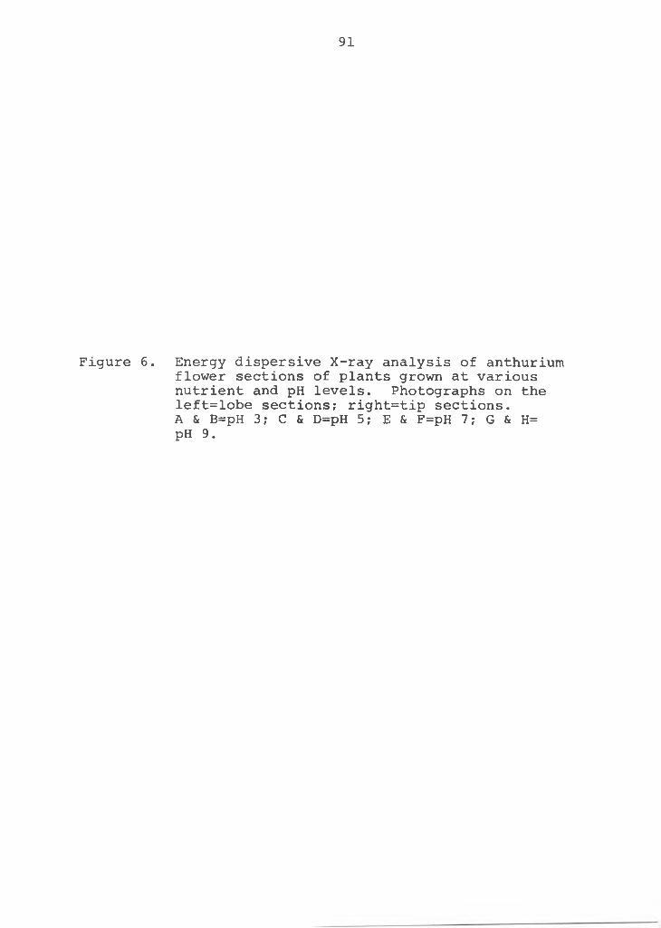
91
Figure 6 . Energy dispersive X-ray analysis of anthurium flower sections of plants grown at various nutrient and pH levels. Photographs on the left=lobe sections; right=tip sections.A & B=pH 3; C & D=pH 5; E & F=pH 7; G & H= pH 9.
93
aeration, is low-cost and readily available as it is a by-product of the active Hawaiian volcanoes. Its wide spread utilization was realized only after the development of slow-release fertilizers. Generally, the ordinary commercial inorganic fertilizers were not effective for anthuriums because they were readily leached with the heavy Hawaiian rains before the anthurium could utilize the nutrients. Unfertilized, fresh volcanic black cinders have a pH of 6.0 to 7.0. When used as a growth medium with organic fertilizers the pH drops to near 4.0. The acid pH, coupled with the fact that liming is not practiced in the nurseries, may account for the color-breakdown spathe disorder problem in Hawaii. On the other hand, pH studies with the nutrient solutions did not produce the color-breakdown disorder (page 87). The minimum Ca requirement was not established and even at pH 3.0 this may have been met in a nutrient system. Each solution treatment contained Ca (NO3) 2 ' 4H20 at 5 x 1 0--* molar. Similar results were reported by Noro (32) who found that olive trees fed "high" levels of Ca showed no deficiency symptoms even at pH 4.0-4.5 and Moser (30) concluded that the Ca concentration at various pH levels for soybeans and sorghum was a more important factor than pH levels on uptake of Ca.
Microautoradiography with Ca^5 showed that Ca was primarily deposited and concentrated in the cell wall.
The practical aspect of any scientific work is of utmost importance. Therefore, the hypothesis that Ca
94
deficiency was the cause of the disorder was tested in the field. The field test results (Table 5) confirm that the application of Ca will significantly reduce the incidence of the color-breakdown disorder of the anthurium spathe.Ca silicate gave slightly better results than Ca nitrate. However, both were superior to "No treatment".
Experimental results show that the lack of Ca in the anthurium is the major cause of the color-breakdown disorder of the spathe and not oedema as originally suspected. The lower Ca content in the lobe of the spathe as compared to the tip section explains why the symptoms of the color- breakdown disorder always occurs in the lobe of the spathe. The pH and Ca uptake studies showed highest Ca uptake at pH 7, lower at pH 5 and pH 9 and lowest at pH 3. This pH factor coupled with environmental conditions of high moisture and low temperatures could tend toward the disorder in the winter months even though it did not appear under experimental conditions. The benefit of applying Ca to correct the disorder is twofold: 1) Ca is provided tothe plants as a nutrient and 2) the pH level of the medium is raised to increase the chemical availability of Ca for the plants.
A critical level of Ca within the tissue of the spathe may be involved in the disorder. This critical level may be very narrow but must be exceeded if tissue does not show color-breakdown symptoms. This critical level may be mediated by environmental conditions such as high air and soil moisture, high soil and low air temperatures. The
95
Table 5. Field tests of the incidence of color-breakdown disorder of spathe in anthurium with different Ca sources,
Ca treatmentNo. of flowers No. of flowers % flowers w/o colorbreak w/colorbreak w/colorbreak
No treatment 2390 231 9.7CA Nitrate 2000#/A/yr.
Ca Silicate26402400
320
1.20.0
96
soil moisture, high soil and low air temperatures. Thehypothesis can be shown as:
Ca content in Environmental Expression ofregard to critical conditions that colorbreak symp-level in the favor color- toms on thetissue____________ breakdown______ anthurium spathe
1 . +
2 . + + —
3.4. - + +
The color-breakdown disorder will mainfest itself on the spathe only with the combination of weather and Ca as indicated in #4, where the Ca level in the tissue is below the critical level and the environmental conditions are favorable for the color breakdown disorder. This explains the higher incidence of the color-breakdown disorder as reported by growers during the colder, rainy winter months conditions favorable for color-breakdown as compared to the warmer, sunny months conditions not favorable for color- breakdown. The color-breakdown disorder has also been reported to occur and then disappear on the same spathe with variable weather conditions. This hypothesis would adequately allow for such a phenomena.
Like the oedema disorder which is correlated with environmental conditions which hinder the normal transpiration processes of the plant, the anthurium color-breakdown disorder may similarly be related to transpiration. Unlike oedema, however, the critical level of Ca in the lobe of
97
the spathe is the major factor in this anthurium color- breakdown disorder.
LITERATURE CITED
98
LITERATURE CITED
2.
3.
4.
5.
6.
7.
8 .
9 .
10 .
11.
1.
12.
Atkinson, G. J. 1893. Oedema of the tomato. Cornell Univ. Agr. Exp. Sta. Bui. 53: 77-108.Aragaki, M. 1973. Colletochrichum gloeosporioides on anthurium spathe. Personal communication.Balge, R. J., B. Esther Stuckmeyer and G. E. Beck.1969. Occurrence, severity and nature of oedema in Pelargonium hortorum, Ait. J. Amer. Soc. Hort. Sci. 94(2): 181-183.Chupp, C. and A. F. Sherf. 1960. Vegetable diseases and their control. The Ronald Press Co. N. Y. pg. 280-282.Dale, Elizabeth. 1901. Investigation on the abnormal outgrowth or intumescence on Hibiscus vitifoluis L. Phil. Trans. Roy. Soc. of London. 194: 163-182.Douglas, Gertude. 1907. The formation of intumes- cenes on potato plants. Bot. Gaz. XLII(4): 233-250.Eisa, H. M. and A. K. Dobrenz. 1971. Morphological and anatomical aspects of oedema in eggplants. J.Amer. Soc. Hort. Sci. 96(6) :766-769 .Estabrooks, D. N. and H. Tiessen. 1972. Blossom-end rot and black seeds of tomatoes. Can. J. Plant Sci. tw(6): 1076-1077.Ficq, A. 1959. Autoradiography, pp. 67-90. The cell, J. Bracket and A. E. Mirsky (ed.). Academic Press,N. Y.Geraldson, C. M. 1954. The control of blackheart of celery. Proc. Amer. Soc. Hort. Sci. 63: 353-358.Heggestad, H. E. 1945. Varietal variation and inheritance studies on natural water soaking in tobacco. Phytopathology. 35: 754-770.Horwitz, W. 1960. Official methods of analysis of the association of offical agricultural chemists.George Banta Company, Inc., Menaska, Wisconsin, pg. 16 and 109.
99
100
13. Hylmo, B. 1953. Transpiration and ion abosorption. Physiologia Plantorum. 6 : 333-405.
14. Jackson, M. L. 1974. Soil chemical analysis.Prentice-Hall, Inc., Englewood Cliffs, N. J. pg. 148- 151.
15. Jenkins, J., Jr. 1959. Brown rib of lettuce. Proc. Amer. Soc. Hort. Sci. 74: 587-591.
16. Jensen, W. A. 1962. Botanical histochemistry. W. H.Freeman and Company. S. F., Calif.
17. Kuster, E. 1903. Ueber experimentell erzeugteintumeszensen. Ber. Deutsch. Bot. Gesells. 21: 452- 458.
18. Laganiere, J. and W. E. Sackston. 1967. Effects on flax of various levels of boron and calcium. Phytoprotection. 48(1): 4-13.
19. Lange, A. H. 1959. Effects of environment on the uptake-transport of calcium and phosphorus by bean plants. Proc. Amer. Soc. Hort. Sci. 73: 349-354.
20. Lanning, F. C. and T. Garabedian. 1963. Distribution of ash, calcium, iron and silica in the tissues of strawberry plants. Proc. Amer. Soc. Hort. Sci.82: 287-291.
21. La Rue, C. D. 1933. Intumescences on poplar leaves.I. Structure and development. Amer. J. Bot. 20: 1-17.
22. _______. 19 33. Intumescences on poplar leaves. II.Physiological considerations. Amer. J. Bot. 20: 159- 175.
23. Lundegardh, H. 1935. The influence of the soil on the growth of plants. Soil Sci. 40:423-437.
24. Maynard, D. N. and A. V. Barker. 1972. Internalbrowning of brussels sprouts: a calcium deficiencydisorder. J. Amer. Soc. Hort. Sci. 97(6):789-792.
25. _______, B. Gersten and H. F. Vernell. 1962. Thecause and control of brownheart of escarole. J. Amer. Soc. Hort. Sci. 81: 371-375.
26. ,___ ___ and _____________. 1965. Thedistribution of calcium as related to internal tipburn, variety and calcium nutrition in cabbage. Proc. Amer. Soc. Hort. Sci. 86:392-396.
101
27. Metwally, A. W., B. Esther Struckmeyer and G. E. Beck.1970. Effects of three soil moisture regimes on the growth and anatomy of Pelargonium hortorum, Ait. J. Amer. Soc. Hort. Sci. 95(6): 808-813.
28. ________, G. E. Beck and B. Esther Struckmeyer. 1970.The role of water and cultural practices on oedema of Pelargonium hortorum, Ait. J. Amer. Soc. Hort. Sci. 95(6): 808-813.
29. ________, __________ and . 1971. Density andbehavior of stomata of Pelargonium hortorum, Ait.J. Amer. Soc. Hort. Sci. 961(1): 31-35.
30. Moser, F. 1942. Calcium nutrition of respective pH levels. Soil Sci. Soc. Amer. Proc. 7: 339-344.
31. Neal, D. C., W. J. Peevy and V. H. Young. 1950.Crinkle leaf of cotton in Louisiana and Arkansas in 1949. Phytopathology. 40(8): 790.
32. Noro, K. 1957. Effects of soil and nutrient on thegrowth of the olive. III. Special reference tocalcium and the growth of the olive tree. Tech. Bui.Agri. Kagawa Univ. 8(2): 148-156.
33. Rasmussen, H. P. 1967. Calcium and strength of leaves.I. Anatomy and histochemistry. Bot. Gaz. 128(3-4): 219-223.
34. _________ , V. E. Shull and H. T. Dryer. 1968. Determination_of element localization in plant tissue withthe microprobe. Dev. in Applied Spectroscopy.6 : 29-42.
35. Russ, J. C. 1972. Elemental x-ray analysis of materials, EXAM methods. EDAX International, Inc., Prairie View, Illinois.
36. Stakman, E. C. and Georgetta S. Harrar. 1957.Principles of Plant Pathology. The Ronald Press Co.N. Y. pg 324-325.
37. Seeley, J. G. 1950. Mineral nutrient deficiencies and leaf burn of Craft Easter lilies. Proc. Amer. Soc.Hort. Sci. 56: 439-445.
38. Shear, C. B. 1971. Symptoms of calcium deficiency on leaves and fruits of York Imperial apples. J. Amer Soc. Hort. Sci. 96(4): 415-416.
39. Shigeura, G. T. 1970. Blossom-end rot of guava. Personal communication.
102
40. Sprague, H. 1964. Hunger Signs in crops. Third ed. David McKay Co. N. Y. pg. 9.
41. Spurr, A. R. 1959. Anatomical aspects of blossom-end rot in the tomato with special reference to calcium nutrition. Hilgardia. 28(12):269-295.
42. Thibodeau, P. O. and P. L. Minotti. 1969. The influence of calcium on the development of lettuce tip burn. J. Amer. Soc. Hort. Sci. 94:373-376.
43. Van Goor, B. J. 1968. The role of calcium and cellpermeability in the disease blossom-end rot oftomatoes. Physiol. Plant. 21(5): 1110-1121.
44. Viala, P. and P. Pacottet. 1904. Surles verrues desfeuilles de la vigne. Compt. Rend. Acad. Sci. 138: 161-163.
45. Wolf, F. and F. E. Lloyd. 1912. Oedema in Manihot. Phytopathology. 2: 131-135.
46. Woodbridge, C. G. 1973. Effects of root temperature and humidity on the distribution of Ca in apple and sunflower seedlings. Z. Pflanzenernaehr Bodenkd.133: 64-69.
APPENDIX
CORRELATION OF THE ANATOMICAL STUDY AND THE SPATHE COLOR-BREAKDOWN DISORDER OF
ANTHURIUM ANDREANUM, L.
103
APPENDIX
Correlation of the Anatomical Study and the Spathe Color-Breakdown Disorder of Anthurium andreanum, L.
The following study was conducted to examine the effects of the spathe color-breakdown disorder on the internal anatomy of the anthurium flower.
Fresh anthurium tissues were taken of the spathe, spadix and pedicel of plants showing and not showing color-breakdown symptoms. The spathe tissues were separated into lobe and tip sections. The tissues were killed and fixed in FAA and histological procedures outlined by Jensen (1) and discussed under Materials and Methods, Section I was followed. The tissues were studied with an Olympus compound microscope and pertinent data recorded. Photomicrographs were taken with a Zeiss photomicroscope .
The cells of the color-breakdown spathe tissue were collapsed. The collapsing was more intense in the mesophyll than the epidermis (Figure 1). This is in accord with earlier findings where X-ray analysis showed concentration of Ca in the color-breakdown spathe tissue to be localized in the epidermal areas and not in the mesophyll. The color-breakdown tissues of the spathe were found to be
104
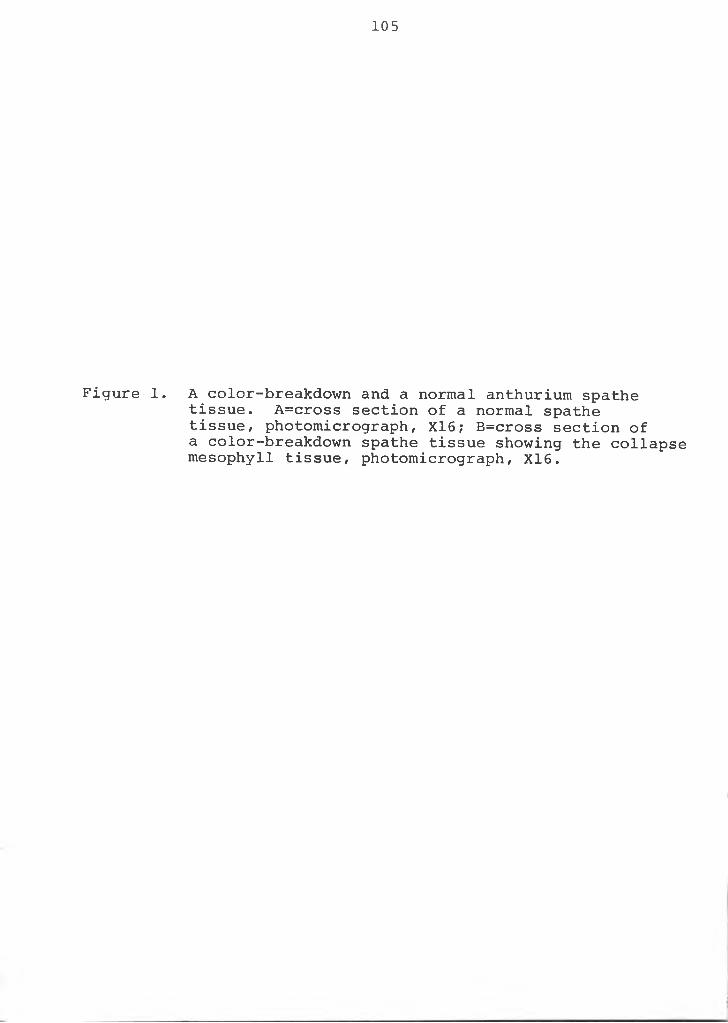
105
Figure 1. A color-breakdown and a normal anthurium spathe tissue. A=cross section of a normal spathe tissue, photomicrograph, X16; B=cross section of a color-breakdown spathe tissue showing the collapse mesophyll tissue, photomicrograph, X16.
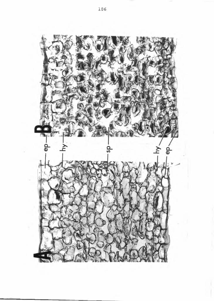
106
107
torn and extremely difficult to find perfect, untorn tissues. This was not true with normal tissues. The tearing of the cells occurred between cells (e.g., separation of the middle lamella) and rarely through the cells (Figure 2). This is similar to reports by Rasmussen (2) in his studies with tender tobacco leaves. While the tearing may have occurred during preparation procedures of the tissues and not in the intact spathe, it indicates lack of calcium pectate which results in the disintegration of the middle lamella and separation of the cell wall. Studies of the spadix and pedicel showed no sign of this primary cell wall disintegration or separation. No difference in anatomical structure between lobe and tip sections of color-breakdown spathe could be found.
Differences in cellular details were difficult to detect, as the protoplasmic constituents were not distinguishable with the histological procedures utilized.Cell outlines and total protoplasmic materials, however, were readily visible. Cells of the color-breakdown tissues were found to take up less stain than cells of the normal tissues. This was true in the spathe, spadix and pedicel. This phenomena indicates the disintegration of the cellular membrane (e.g., plasmalemma, tonoplast and the nuclear membrane) and with the resulting internal leakage, a lack of cytoplasmic materials.
No sign of cell hypertrophy or formation of intumes- cense as found oedema tissue could be detected nor difference
108
Figure 2. Color-breakdown anthurium spathe showing cellular separation. Photomicrograph, X16.
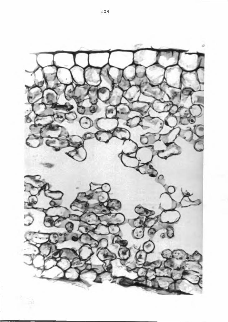
109
110
in Ca crystal content in affected and normal tissues.It is evident that more study on the cellular
details and the effect of low Ca must be pursued to completely comprehend the role of Ca in the color-breakdown disorder. Tissue damage of the magnitude of cell separation and primary cell wall disintegration to cause this disorder appears and disappears with changes in environmental conditions. Ca is essential for the maintenance and for the formation of cell-membrane systems on which the proper functional integrity of cell metabolism is dependent, and the lack of which has an effect on the internal chemistry and on color change seems a more logical explanation. The chemistry of the color change could be dependent on environmental factors such as moisture in the cell and manifest itself accordingly. Extreme cell damage leads to irreversible breakdown and eventual necrosis of the tissue. Ca effects on the primary cell wall are probably secondary to the above mentioned changes.
LITERATURE CITED
111
LITERATURE CITED
1. Jensen, W. A. 1962. Botanical histochemistry.W. H. Freemand and Company. S. F., California.
2. Rasmussen, H. P. 1967. Calcium and strength of leaves. I. Anatomy and histochemistry. Bot. Gaz. 128 (3-4) : 219-223.
112
Related Documents