See discussions, stats, and author profiles for this publication at: http://www.researchgate.net/publication/49741927 Absence of keratin 8 confers a paradoxical microflora-dependent resistance to apoptosis in the colon. ARTICLE in PROCEEDINGS OF THE NATIONAL ACADEMY OF SCIENCES · JANUARY 2011 Impact Factor: 9.81 · DOI: 10.1073/pnas.1010833108 · Source: PubMed CITATIONS 13 DOWNLOADS 83 VIEWS 172 7 AUTHORS, INCLUDING: Aida Habtezion Stanford University 38 PUBLICATIONS 1,181 CITATIONS SEE PROFILE Diana M Toivola Åbo Akademi University 63 PUBLICATIONS 2,304 CITATIONS SEE PROFILE Greg Kronmal Thermo Fisher Scientific 6 PUBLICATIONS 3,135 CITATIONS SEE PROFILE Eugene C Butcher Stanford University 402 PUBLICATIONS 49,868 CITATIONS SEE PROFILE Available from: Eugene C Butcher Retrieved on: 10 August 2015

Welcome message from author
This document is posted to help you gain knowledge. Please leave a comment to let me know what you think about it! Share it to your friends and learn new things together.
Transcript

Seediscussions,stats,andauthorprofilesforthispublicationat:http://www.researchgate.net/publication/49741927
Absenceofkeratin8confersaparadoxicalmicroflora-dependentresistancetoapoptosisinthecolon.
ARTICLEinPROCEEDINGSOFTHENATIONALACADEMYOFSCIENCES·JANUARY2011
ImpactFactor:9.81·DOI:10.1073/pnas.1010833108·Source:PubMed
CITATIONS
13
DOWNLOADS
83
VIEWS
172
7AUTHORS,INCLUDING:
AidaHabtezion
StanfordUniversity
38PUBLICATIONS1,181CITATIONS
SEEPROFILE
DianaMToivola
ÅboAkademiUniversity
63PUBLICATIONS2,304CITATIONS
SEEPROFILE
GregKronmal
ThermoFisherScientific
6PUBLICATIONS3,135CITATIONS
SEEPROFILE
EugeneCButcher
StanfordUniversity
402PUBLICATIONS49,868CITATIONS
SEEPROFILE
Availablefrom:EugeneCButcher
Retrievedon:10August2015

Absence of keratin 8 confers a paradoxicalmicroflora-dependent resistance to apoptosisin the colonAida Habteziona,b,1,2, Diana M. Toivolac,1, M. Nadeem Asgharc, Greg S. Kronmald, Jacqueline D. Brookse,Eugene C. Butcherb,f, and M. Bishr Omaryg,h
Departments of gMolecular and Integrative Physiology and hMedicine, University of Michigan Medical School, Ann Arbor, MI 48109; Departments ofaMedicine and fPathology, Palo Alto VA Medical Center, Palo Alto, CA 94304; bStanford University School of Medicine Digestive Disease Center, Stanford, CA94305; cDepartment of Biosciences, Åbo Akademi University, FIN-20520, Turku, Finland; dApplied Biosystems Inc., Foster City, CA 94404; and eGenomic Health,Inc., Redwood City, CA 94063
Edited by Sven Pettersson, Karolinska Institutet, Stockholm, Sweden, and accepted by the Editorial Board December 16, 2010 (received for review July23, 2010)
Keratin 8 (K8) is a major intermediate filament protein present inenterocytes and serves an antiapoptotic function in hepatocytes.K8-null mice develop colonic hyperplasia and colitis that arereversed after antibiotic treatment. To investigate the pathwaysthat underlie the mechanism of colonocyte hyperplasia and thenormalization of the colonic phenotype in response to antibiotics,we performed genome-wide microarray analysis. Functional anno-tation of genes that are differentially regulated in K8−/− and K8+/+
isolated colon crypts (colonocytes) identified apoptosis as a majoraltered pathway. Exposure of K8−/− colonocytes or colon organ(“organoid”) cultures, but not K8−/− small intestine organoid cul-tures, to apoptotic stimuli showed, surprisingly, that they are re-sistant to apoptosis compared with their wild-type counterparts.This resistance is not related to inflammation per se because T-cellreceptor α-null (TCR-α−/−) and wild-type colon cultures respondsimilarly upon induction of apoptosis. Following antibiotic treat-ment, K8−/− colonocytes and organ cultures become less resistantto apoptosis and respond similarly to the wild-type colonocytes.Antibiotics also normalize most differentially up-regulated genes,including survivin and β4-integrin. Treatment of K8−/− mice withanti–β4-integrin antibody up-regulated survivin, and induced phos-phorylation of focal adhesion kinase with decreased activation ofcaspases. Therefore, unlike the proapoptotic effect of K8 mutationor absence in hepatocytes, lack of K8 confers resistance to colono-cyte apoptosis in a microflora-dependent manner.
Keratins exist as obligate noncovalent heteropolymers of type I(K9–K28) and type II (K1–K8 and K71–K80) proteins and
make up the intermediate filament (IF) cytoskeleton of epithelialcells (1–3). In adult hepatocytes, the IF network consists of simpleepithelial K8 andK18, whereas in the intestine the network consistsof K7, K8, K18, K19, and K20, with K8 being the major type IIkeratin (2, 4, 5). Absence or mutation of K8 or K18 renders hep-atocytes markedly susceptible to apoptosis, and in humans andmice K8 and K18 mutations predispose their carriers to acute andchronic end-stage liver disease and liver disease progression (5–8).In addition to the effects of keratins in the liver,K8−/−mice developcolonic hyperplasia and chronic spontaneous colitis (9, 10) that isamenable to early treatment with broad-spectrum antibiotics (11).Although hepatocytes in K8−/− and K18−/− mice are highly
sensitive to apoptotic stimuli (5, 12, 13), K18−/− intestine appearsnormal (14), probably reflecting the functional redundancy of ad-ditional type I keratins in the intestine (4, 15). In humans, the as-sociation of K8 variants with inflammatory bowel disease (IBD) isunclear (16, 17). The differences between the liver- and intestine-proliferative phenotypes and the associations (or lack thereof) withhuman disease highlight the potential importance of the micro-environment and cell-specific modifiers.In contrast to the findings in K8−/− hepatocytes, we show in
this study that K8−/− colonocytes, but not K8−/− small intestineenterocytes or T-cell receptor α-null (TCRα−/−) colonocytes, arerelatively resistant to apoptosis, but this resistance can be reversed
in K8−/− mice by antibiotic treatment. In addition, we show thatK8−/− colonocytes up-regulate survivin and β4-integrin, with thelatter playing an important role in enterocyte anoikis (18, 19). Thekeratin IF network links to β4-integrin at the site of hemi-desmosomes via interaction with the cytoskeletal linker proteinplectin and BP180 (20, 21). Furthermore, we provide a mecha-nism for the altered susceptibility to apoptosis, because in vivotreatment of K8−/− mice with anti–β4-integrin antibody furtherup-regulates survivin, leads to the activation of down-streamphosphorylation of focal adhesion kinase (FAK), and decreasescaspase activation. The resistance to apoptosis observed at the tipof the colonic crypt coupled with colonocyte proliferation alongmost of the crypt likely contribute to the observed colonichyperproliferation in K8−/− mice.
ResultsExpression Profiling in K8−/− and K8+/+ Colonocytes Shows ApoptosisIs a Prominent Keratin-Regulated Biological Pathway. To understandbetter the effect of K8 absence on the colonic hyperplasia andcolitis phenotype and the normalization of this phenotype fol-lowing antibiotic treatment (11), we sought to identify colonocytegenes that are differentially expressed in response to the absenceof K8 or to suppression of luminal bacteria. Microarray analysisrevealed that several hundred genes were differentially regulatedin K8+/+ and K8−/− primary isolatedmouse colonocytes (Fig. 1A).The 20 genes most highly up-regulated in the K8−/− colon cryptsare listed in Table S1. Antibiotic pretreatment of the animals ledto near-complete normalization of the differentially altered genesin the K8−/− colon (Fig. 1B). The antibiotics used herein effec-tively suppressed the presence of luminal bacteria as determinedby stool bacterial DNA content (Table S2). Using real-time PCR,we validated several of the genes that manifested marked up- ordown-regulation in the microarray findings (Table S3). Analysisusing the Jubilant PathArt database package Physiology softwarerevealed that apoptosis was the most differentially regulated bi-ological pathway, with 46 of 160 apoptosis pathway genes showingsignificant differential expression (P = 3 × 10⁻⁵) in K8−/− andK8+/+ colonocytes (Table S4). The software identified growthand differentiation as the secondmost significant pathway, with 39
Author contributions: M.B.O. designed research; A.H., D.M.T., M.N.A., and M.B.O. per-formed research; G.S.K., J.D.B., E.C.B., and M.B.O. contributed new reagents/analytictools; A.H., D.M.T., and M.B.O. analyzed data; and A.H., D.M.T., and M.B.O. wrote thepaper.
The authors declare no conflict of interest.
This article is a PNAS Direct Submission. S.P. is a guest editor invited by the EditorialBoard.1A.H. and D.M.T. contributed equally to this work.2To whom correspondence should be addressed. E-mail: [email protected].
This article contains supporting information online at www.pnas.org/lookup/suppl/doi:10.1073/pnas.1010833108/-/DCSupplemental.
www.pnas.org/cgi/doi/10.1073/pnas.1010833108 PNAS | January 25, 2011 | vol. 108 | no. 4 | 1445–1450
CELL
BIOLO
GY

of 135 genes differentially expressed in K8−/− and K8+/+ colo-nocytes (P = 1 × 10⁻⁴).
K8−/− Colonocytes Overexpress Survivin and, Unlike K8−/− Cells fromthe Small Intestine and K8−/− Colonocytes from Antibiotic-TreatedMice, Are Resistant to Apoptosis. In the human small intestine, itis estimated that 1010 cells are shed per day; despite the largenumber of cells being shed, the epithelial barrier is maintained(22). Although the signals that initiate cell shedding are not known(22), enterocytes undergo a spontaneous form of apoptosis,termed “anoikis,” upon detachment from the cellular matrix (18).Because of the established predisposition of K8−/− (and K8, K18mutant) hepatocytes and livers to mechanical and nonmechanicalinjury and to apoptosis (23–28), we compared the apoptotic re-
sponse of K8−/− and K8+/+ small intestine and colons. K8−/− co-lonic crypts (here called “colonocytes”) were isolated in parallel,and cleavage/activation of caspases was assessed as an indicator ofapoptosis. Surprisingly, and unlike K8−/− hepatocytes, the K8−/−colonocytes are resistant to apoptosis based on the relative de-crease in cleavage of caspase-3, -7, and -9 (Fig. 2A). Activation ofapoptosis also was associated with K8 S80 phosphorylation inK8+/+ colonocytes (Fig. 2A), and ablation of S80A in hepatocytesby an S80A mutation leads to marked predisposition to apoptosisupon stimulation with Fas ligand (29). Similar findings were notedusing whole-organ fragments (here called “organoids”) of thecolon (Fig. 2B), but not of the small intestine (Fig. 2C), that werekept in culture for 1 h. Notably, small intestine organoids fromK8−/− mice appear to be more predisposed to apoptosis as com-pared to K8+/+ small intestine based on the enhanced cleavage ofcaspase-7 (Fig. 2C). Consistent with susceptibility to apoptosisafter 1 h culture, K18 fragmentation (K18 Asp237) (30) wasdetected in both colon and small intestine organoid K8+/+ cul-tures (Fig. 2 B and C). To ensure that caspase activation involvedcolonocytes rather than lamina propria or other nonepithelialcells, we analyzed cleaved caspase-7 in frozen colon pieces fromthe organ cultures by immunofluorescence staining (Fig. 2D). Asignificant increase in apoptotic cells was seen at the luminal orsurface end of the K8+/+ colonic crypts, compared with K8−/−colonic crypts, and the cells were identified as colonocytes by theircoexpression of K19 (Fig. 2D a and b).We then used known inducers of apoptosis such as staurosporine
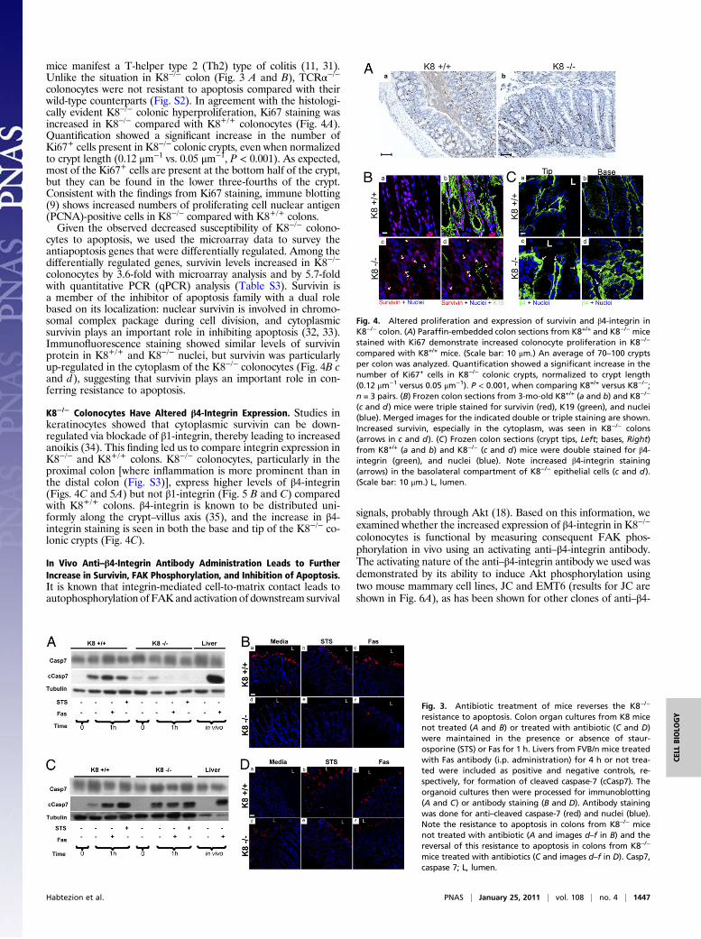
and Fas antibody to examine potential differential sensitivity toapoptosis of K8−/− and K8+/+ colonocytes. Similar to findingsnoted during colonocyte isolation (Fig. 2), K8−/− colons are moreresistant than K8+/+ colons to the induction of apoptosis by bothstaurosporine and Fas. This finding was confirmed biochemicallyby the limited formation of cleaved caspase-7 (Fig. 3A) and byimmunofluorescence staining (Fig. 3B). In addition, the resistanceof K8−/− colonocytes to apoptosis, compared with K8+/+ colono-cytes, was confirmed using TUNEL staining (Fig. S1). Of note, andconsistent with the “normalization” of the differentially regulatedgenes in K8−/− and K8+/+ colonocytes in antibiotic-treated mice,K8−/− colonocyte resistance to apoptosis was reversed with anti-biotic pretreatment (Fig. 3 C and D).It is difficult to study the relationship of microflora to the colonic
hyperplasia in the absence of inflammation, because treatmentwithantibiotics reverses both the inflammatory response and the colonichyperplasia inK8−/−mice (11). To assess whether inflammation perse or inflammation triggered by microbes contributes to the ob-served alteration in apoptosis, we examined the differences incolonocyte apoptosis in another chronic colitis model, namely theTCRα−/− model of chronic colitis (31). Both TCRα−/− and K8−/−
Fig. 1. Gene expression profiles of K8−/− and K8+/+ colonocytes show de-creased numbers of differentially regulated genes after antibiotic treatment.(A) Total RNA from freshly isolated K8−/− and K8+/+ colonocytes was used formicroarray analysis. The graph depicts results from GeneSpring analysis. Up-per and lower green lines represent genes threefold up- or down-regulated inK8+/+ compared with K8−/− colonocytes. (B) An analysis to that similar in Aexcept that the RNAwas isolated from colonocytes of antibiotic-treated K8−/−
and K8+/+ mice. Note the marked decrease in differential expression betweenK8−/− and K8+/+ colonocyte genes following antibiotic treatment. Two hun-dred eighty-eight genes were threefold up-regulated, and 460 genes werethreefold down-regulated in K8+/+ compared with K8−/− colonocytes, but af-ter antibiotic treatment only 189 genes and 290 genes remained threefold up-regulated and down-regulated, respectively.
Fig. 2. Primary colonocytes and colon but notsmall intestine organ cultures from K8−/− miceare less susceptible to apoptosis than parallelcultures from K8+/+ mice. (A) K8+/+ and K8−/−
colonocytes were cultured in medium for 1 h.Cell homogenates then were analyzed by im-munoblotting using antibodies to cleaved cas-pases (cCasp) or total caspases (Casp), K8 pS80,K8, and tubulin. (B) Colon and (C) small intestineorganoids were cultured and then analyzed asin A. (D) Frozen sections from K8+/+ (a and b)and K8−/− (c and d) 1-h colon primary organcultures were triple stained for anti–cleavedcaspase-7 red), anti-K19 (green), and nuclei(blue). Merged images are shown in b and d.Arrows point to apoptotic cells at the luminal (L)tips of K8+/+ crypts. Insets show magnified views.(Scale bar in c: 50 μm.)
1446 | www.pnas.org/cgi/doi/10.1073/pnas.1010833108 Habtezion et al.
mice manifest a T-helper type 2 (Th2) type of colitis (11, 31).Unlike the situation in K8−/− colon (Fig. 3 A and B), TCRα−/−colonocytes were not resistant to apoptosis compared with theirwild-type counterparts (Fig. S2). In agreement with the histologi-cally evident K8−/− colonic hyperproliferation, Ki67 staining wasincreased in K8−/− compared with K8+/+ colonocytes (Fig. 4A).Quantification showed a significant increase in the number ofKi67+ cells present in K8−/− colonic crypts, even when normalizedto crypt length (0.12 μm−1 vs. 0.05 μm−1, P < 0.001). As expected,most of the Ki67+ cells are present at the bottom half of the crypt,but they can be found in the lower three-fourths of the crypt.Consistent with the findings from Ki67 staining, immune blotting(9) shows increased numbers of proliferating cell nuclear antigen(PCNA)-positive cells in K8−/− compared with K8+/+ colons.Given the observed decreased susceptibility of K8−/− colono-
cytes to apoptosis, we used the microarray data to survey theantiapoptosis genes that were differentially regulated. Among thedifferentially regulated genes, survivin levels increased in K8−/−
colonocytes by 3.6-fold with microarray analysis and by 5.7-foldwith quantitative PCR (qPCR) analysis (Table S3). Survivin isa member of the inhibitor of apoptosis family with a dual rolebased on its localization: nuclear survivin is involved in chromo-somal complex package during cell division, and cytoplasmicsurvivin plays an important role in inhibiting apoptosis (32, 33).Immunofluorescence staining showed similar levels of survivinprotein in K8+/+ and K8−/− nuclei, but survivin was particularlyup-regulated in the cytoplasm of the K8−/− colonocytes (Fig. 4B cand d), suggesting that survivin plays an important role in con-ferring resistance to apoptosis.
K8−/− Colonocytes Have Altered β4-Integrin Expression. Studies inkeratinocytes showed that cytoplasmic survivin can be down-regulated via blockade of β1-integrin, thereby leading to increasedanoikis (34). This finding led us to compare integrin expression inK8−/− and K8+/+ colons. K8−/− colonocytes, particularly in theproximal colon [where inflammation is more prominent than inthe distal colon (Fig. S3)], express higher levels of β4-integrin(Figs. 4C and 5A) but not β1-integrin (Fig. 5 B and C) comparedwith K8+/+ colons. β4-integrin is known to be distributed uni-formly along the crypt–villus axis (35), and the increase in β4-integrin staining is seen in both the base and tip of the K8−/− co-lonic crypts (Fig. 4C).
In Vivo Anti–β4-Integrin Antibody Administration Leads to FurtherIncrease in Survivin, FAK Phosphorylation, and Inhibition of Apoptosis.It is known that integrin-mediated cell-to-matrix contact leads toautophosphorylation of FAKand activation of downstream survival
signals, probably through Akt (18). Based on this information, weexamined whether the increased expression of β4-integrin in K8−/−
colonocytes is functional by measuring consequent FAK phos-phorylation in vivo using an activating anti–β4-integrin antibody.The activating nature of the anti–β4-integrin antibody we used wasdemonstrated by its ability to induce Akt phosphorylation usingtwo mouse mammary cell lines, JC and EMT6 (results for JC areshown in Fig. 6A), as has been shown for other clones of anti–β4-
Fig. 3. Antibiotic treatment of mice reverses the K8−/−
resistance to apoptosis. Colon organ cultures from K8 micenot treated (A and B) or treated with antibiotic (C and D)were maintained in the presence or absence of staur-osporine (STS) or Fas for 1 h. Livers from FVB/n mice treatedwith Fas antibody (i.p. administration) for 4 h or not trea-ted were included as positive and negative controls, re-spectively, for formation of cleaved caspase-7 (cCasp7). Theorganoid cultures then were processed for immunoblotting(A and C) or antibody staining (B and D). Antibody stainingwas done for anti–cleaved caspase-7 (red) and nuclei (blue).Note the resistance to apoptosis in colons from K8−/− micenot treated with antibiotic (A and images d–f in B) and thereversal of this resistance to apoptosis in colons from K8−/−
mice treated with antibiotics (C and images d–f in D). Casp7,caspase 7; L, lumen.
Fig. 4. Altered proliferation and expression of survivin and β4-integrin inK8−/− colon. (A) Paraffin-embedded colon sections from K8+/+ and K8−/− micestained with Ki67 demonstrate increased colonocyte proliferation in K8−/−
compared with K8+/+ mice. (Scale bar: 10 μm.) An average of 70–100 cryptsper colon was analyzed. Quantification showed a significant increase in thenumber of Ki67+ cells in K8−/− colonic crypts, normalized to crypt length(0.12 μm−1 versus 0.05 μm−1). P < 0.001, when comparing K8+/+ versus K8−/−;n = 3 pairs. (B) Frozen colon sections from 3-mo-old K8+/+ (a and b) and K8−/−
(c and d) mice were triple stained for survivin (red), K19 (green), and nuclei(blue). Merged images for the indicated double or triple staining are shown.Increased survivin, especially in the cytoplasm, was seen in K8−/− colons(arrows in c and d). (C) Frozen colon sections (crypt tips, Left; bases, Right)from K8+/+ (a and b) and K8−/− (c and d) mice were double stained for β4-integrin (green), and nuclei (blue). Note increased β4-integrin staining(arrows) in the basolateral compartment of K8−/− epithelial cells (c and d).(Scale bar: 10 μm.) L, lumen.
Habtezion et al. PNAS | January 25, 2011 | vol. 108 | no. 4 | 1447
CELL
BIOLO
GY

integrin antibody using human breast and other cell lines (36, 37).We then tested the effect of β4-integrin antibody treatment onFAK phosphorylation and down-regulation of caspase activationin vivo. Notably, treatment of K8−/− mice with anti–β4-integrinantibody led to an increase in survivin mRNA (Fig. 6B) and pro-tein (Fig. 6C).The specificity of the anti–β4-integrin antibody is demonstrated
by the fact that FITC anti-rat IgG stained β4-integrin in colono-cytes of K8−/− mice treated with anti–β4-integrin antibody (Fig.6C b and d) but did not stain colonocytes from mice treated withthe isotype control antibody (Fig. 6C a and c). Furthermore,treatment with anti–β4-integrin antibody led to a robust increasein FAK pY397 in K8−/− colons (Fig. 6D). Although phosphory-lation of Akt is increased in K8−/− compared with K8+/+ colons,immune blotting did not show significant differences in phospho-Akt between K8−/− mice treated with isotype control and K8−/−
mice treated with anti–β4-integrin antibody (Fig. 6D). However,binding of β4-integrin leads not only to an increase in survivinexpression (Fig. 6 B and C) but also to inhibition of apoptosis, asshown by the decrease in cleavage of caspase-7 (Fig. 6D). In ad-dition, we assessed the activation of phosphatase and tensin ho-molog deleted on chromosome 10 (PTEN), because PTEN hasbeen shown to play an important inhibitory role in cell survival andinduction during apoptosis that is in part Akt dependent (38). Wedo not observe any evidence of PTEN activation (Fig. 6D). Col-lectively, these findings indicate that the up-regulation of β4-integrin in the K8−/− colon is likely to contribute to the observedinhibition of apoptosis via FAK activation.
DiscussionK8−/− and K18−/− (or keratin mutant) hepatocytes have similarlyincreased susceptibility to injury and apoptosis (5, 12, 13) becauseof the obligate heteropolymeric nature of K8/K18 and because,under basal conditions, K8 and K18 are the only keratins in adulthepatocytes (5). In the colon, K8 is the major type II keratin inmice (4) and humans (17) and is the major keratin that manifestsa phenotype when absent (9, 10, 39) probably because of thefunctional redundancy of the type I keratins in the colon and thelimited expression of K7 compared with K8 (5). Of the remainingkeratins (K7, K18, K19, K20) concurrently expressed in the colon,the absence of K18 (14) or K19 (40, 41) or the overexpression ofthe dominant negative K20 (4) do not manifest an overt phenotype(to our knowledge, K7−/− mice have not been reported). In theliver, K8 is antiapoptotic; in the colon, as shown here, its absenceconfers resistance to apoptosis but only under normal physiologicalconditions that include the presence of resident microflora. Theincreased susceptibility of keratin-lacking or keratin-mutant hep-atocytes to apoptosis, compared with their wild-type counterparts,is context dependent, in that differences are unmasked when ap-optosis is induced by Fas but not by TNF (26, 42). Similar context-dependent bacteria-modulated effects also may occur in the colon,but this possibility remains to be investigated. It also is plausiblethat absence of normal keratins induces resistance to apoptosis aspart of a normal stress response to luminal bacteria.The mechanism by which microflora provide an antiapoptotic
effect in the absence of colonocyte keratins is likely to be multi-factorial. A relevant mechanism demonstrated herein is the en-
Fig. 5. K8−/− colons have altered β4- but not β1-integrinexpression. (A) Lysates from proximal (PC) and distal colons(DC) shown from three independent age- and sex-matchedpairs of K8+/+ and K8−/− mice were blotted with antibodiesto β4-integrin and actin. Note the increase in β4-integrin(arrow) in proximal colons of K8−/− mice. (B) Lysates fromproximal (PC) and distal colons (DC) from K8+/+ and K8−/−
mice were blotted with antibodies to β1-integrin and actin. The blot from a representative pair of K8 mice is shown. (C) Frozen colon sections from K8+/+ (a)and K8−/− (b) mice were double stained for β1-integrin (red) and nuclei (blue). (Scale bar: 10 μm.) L, lumen.
Fig. 6. In K8−/− colon, in vivo treatment withanti–β4-integrin antibody increases the expres-sion of survivin and activation of FAK in concertwith decreased apoptosis. (A) Lysates from the JCmouse mammary cell line treated with control oranti–β4-integrin antibody were analyzed by im-munoblotting using antibodies to Akt, phospho-Akt, and tubulin. (B) RT-PCR analysis of survivin incolons of K8+/+ andK8−/−mice treatedwith eitherisotype control antibody (white bars) or anti–β4-integrin antibody (dark bars). Data are presentedas mean relative survivin expression ± SEM (n =3). *P < 0.05 when comparing control versusanti–β4-integrin antibody treatment. (C) Frozencolon sections from K8−/− mice treated with iso-type control (a and c) or anti–β4-integrin anti-body (b and d) were triple stained with anti-survivin (red), anti-rat IgG (green), and nuclei(blue). Higher-magnification images are shownin c and d. (Scale bars: 50 μm in a and b; 10 μm in cand d.) (D) Lysates from K8+/+ and K8−/− colons ofmice treated with control antibody or anti–β4-integrin antibody were analyzed by blotting us-ing antibodies to FAK, Akt, cleaved caspase-7(cCasp7), caspase-7 (Casp7), tubulin, and phos-pho-FAK/Akt/PTEN. (E) Schematic model sum-marizing the proposed effect of K8 absence oncolonocyte resistance to apoptosis through in-creased integrin signaling and increased cyto-plasmic levels of antiapoptotic survivin. These effects, in the presence of the luminalmicroflora and/or inflammation, prevent apoptosis and probably contributeto the observed hyperproliferative epithelium in K8−/− colon.
1448 | www.pnas.org/cgi/doi/10.1073/pnas.1010833108 Habtezion et al.
hanced integrin signaling in the K8−/− colon, which activates FAKand is mimicked and enhanced by administration of β4-integrinantibody. We also link K8 absence to increased levels of survivin,which is known to be involved, together with an activated FAK, inpromoting cell survival (43).Other potentialmicroflora effects thatrender keratin absence antiapoptotic may be related to quantita-tive or qualitative changes in the Toll-like receptors (TLRs), whichare required for the intestinal homeostasis and protection of in-testinal epithelial cells from injury (44, 45). For example, mis-targeting or altered expression of apical or basolateral receptors inK8-null enterocytes, as seen in the small intestine (39), colon (9),and here for β4-integrin in the colon (Fig. 4), alsomayoccur for oneor more TLRs. A survey of the microarray results of several TLRsshowed that TLR9 mRNA increased 2.5-fold, (particularly in theproximal colon; Fig. S4), a finding that was confirmed by immu-nohistochemistry and immunoblotting. Although the exact mech-anism is not clear, there appears to be a link between the absence ofK8 and up-regulation of β4-integrin expression in the colon. Thisnotion is supported further by the down-regulationof β4-integrin intransgenic mice overexpressing human K8 (Fig. S5).The importance of the microflora also is highlighted by the
difference in resistance to apoptosis observed in the K8−/− colonand small intestine (jejunum). The human colon harbors 1013–1014 bacteria, whereas the proximal small intestine is estimatedto have 103–104 bacteria (46). Factors other than microflora alsomay be involved in the differential resistance to apoptosis, be-cause the small intestine and colon epithelium differ in manyrespects, ranging from morphologic to functional states. It isunlikely that differences in the composition of the keratin net-work in K8−/− colon and small intestine account for the differ-ence in the two sites’ resistance to apoptosis, because previousstudies showed similar alterations at the two sites in theremaining keratins (K7, K18, K19, and K20) (10, 39).Colonic inflammation parallels the presence of luminal micro-
flora. It is possible that the observed resistance of K8−/− colono-cytes to apoptosis may be secondary to, or influenced by,inflammation. Inflammation is difficult to assess in the absence ofluminal microflora because inflammation and microflora go handin hand. Despite the different genetic or chemical mechanismsused to generate the colitis models, none of the current modelsdevelop colonic inflammation in a germ-free environment (47).Therefore we investigated and compared resistance to apoptosis inanother immune-based model of chronic colitis, TCRα−/− mice.We did not observe such resistance to apoptosis in the TCRα−/−model of colitis, which by definition harbors an inflamed epithe-lium (31). However, our studies do not control for possible con-tributions arising from strain differences or differences ininflammation andmicrobial environment in the two colitis models.The findings herein highlight the importance of differences (suchas microflora) in the microenvironments of hepatocytes andcolonocytes; these differences lead to a paradoxical effect in thecolon. If this effect also applies to hepatocytes, it might serve (e.g.,through microflora-mimetic stimulation) to protect them fromhepatotoxic injury. Taken together, these findings demonstratethat the absence of K8, coupled with the presence of microflora,renders colonocytes resistant to apoptosis. This resistance appearsto bemediated, at least in part, by the overexpression of β4-integrinthat can be linked to an increase in survivin and subsequent de-creased activation of caspases (Fig. 6E).The colonic hyperplasia that is observed in K8−/− mice (9, 10)
probably results from both decreased apoptosis (Figs. 2 and 3 andFig. S1) and increased proliferation (Fig. 4). We used two formsof stress to induce apoptosis: (i) colonocyte (colonic crypts) orwhole-colon tissue isolation (organoids) followed by ex vivo cul-ture, and (ii) application of known apoptosis-inducing agents exvivo. The former method is similar to the classically describedanoikis (a spontaneous form of apoptosis resulting from de-tachment from the cellular matrix), which occurs upon suppres-sion of β4-integrin signaling (18). Therefore, the endogenous up-regulation of β4-integrin signaling in situ in K8−/− colon (Fig. 6)provides one likely mechanism for the observed protection from
apoptosis. This protection occurs at the crypt surface, whereas theincrease in proliferation occurs below this level, mostly involvingthe crypt from its base to ≈75% of its upward length (based onthe Ki67 staining; Fig. 4A). These findings suggest that both re-sistance to apoptosis and the increase in proliferation probablyaccount for the colonic hyperplasia. The microarray data andpathway analysis also indicated that the growth and differentia-tion pathway is the second most significantly altered pathway(Table S4), providing further indirect evidence for the dual, andprobably regionally distinct, enhanced antiapoptosis and hyper-proliferation signals as a consequence of the combined absence ofK8 and presence of microflora. Other contributions to enhancedcell proliferation in the K8−/− colon probably are related to themistargeting and alteration of the stability of membrane proteinssuch as transporters that can modulate intracellular pH and en-hance cell proliferation (9).
Materials and MethodsMice and in Vivo Experiments. K8−/−mice (Jackson Laboratory); and their wild-type littermates, in an FVB/N background, were generated by interbreedingK8+/− mice as described (11). Age- and sex-matched mice (3–4 mo old) werestudied. For antibiotic treatment, K8+/+ and K8−/− mice were given vanco-mycin and imipenem in their drinking water (50 mg/kg body weight/d for8 wk) starting at postnatal d 18 or 19 (11). A group of K8+/+ and K8−/− micereceived 200 μg anti–β4–integrin antibody (clone 346–11A) or control isotypeantibody (rat IgG2a; BD Biosciences) i.p. every day for 3 d, and colons wereharvested on d 4. In separate experiments, FVB/Nmice were fasted overnight,and apoptosis was induced by i.p. injection of anti-Fas antibody (0.15 μg/g) (BDBiosciences) as described (42). After 4 h, the livers were harvested, and theirhomogenates were used as positive controls for caspase cleavage and apo-ptosis. TCRα+/+ and TCRα−/− mice on a C57BL/6 background were obtainedfrom the Jackson Laboratory. Mice overexpressing human K8were generatedand maintained as described previously (48). All animals were treatedaccording to National Institutes of Health guidelines and an approved animalinstitutional protocol.
Colonocyte Isolation and Intestine and Other Cell Culture. Colonocytes wereisolated as described (9). Isolated colonocytes were used for microarrayanalysis (see below) or were cultured in Dulbecco’s modified Eagle mediumsupplemented with 4.5 g/L glucose, 25 mM Hepes, 5 ng/mL recombinanthuman epidermal growth factor, 0.2 IU/mL insulin, 5% FBS, 50 μg/mL peni-cillin, and 50 μg/mL streptomycin at 37 °C for 1 h. For organoid whole-colonor small intestine cultures, organs were opened longitudinally and wererinsed gently and quickly in incubation medium (RPMI-1640 plus 10% heat-inactivated FCS and antibiotics). The intestines then were cut into 5- to 7-mmpieces and incubated in the culture medium described above for 1 h at 37 °C,5% CO2/95% O2. The incubations were carried out in the presence or ab-sence of the apoptosis-inducing agents staurosporine (4 μM) (CaymanChemical) and Fas antibody (500 ng/mL).
Activity of the anti–β4-integrin antibody or isotype control was tested incultured mouse mammary cell lines (JC and EMT6; American Type CultureCollection). The cells were serum starved overnight, trypsinized from thedishes, and washed with PBS. The cells (5 × 10⁶/mL) then were cultured at37 °C for 6–8 h in the presence of anti–β4-integrin or isotype control anti-body (6 μg/mL).
Real-Time PCR. Total colon RNA was isolated using an RNeasy midi kit andconverted into cDNA using a SuperScript II reverse transcriptase kit as rec-ommended by the supplier (Invitrogen). qPCR was performed with an ABIPrism 7900 Sequence Detection System as described (49). Target genes (TableS3) were amplified using specific primers and SYBR Green PCR Master Mix.Gene expression levels were normalized to the housekeeping gene GAPDH.Bacterial DNA was extracted from freshly collected stools using the QIAampDNA stool kit (Qiagen). Escherichia coli plasmid with a known amount ofDNA (a gift from David Relman, Stanford University, Stanford, CA) served asa positive control, and stool DNA qPCR was performed as described (50).
Histology. Freshly harvested colons or organ-culture colon pieces from K8+/+
and K8−/− mice were embedded in optimum cutting temperature (OCT)compound and frozen at –80 °C. Frozen 6-μm tissue sections were fixed inacetone and blocked with PBS containing 2% BSA and 2% goat serum (11,51). The tissue sections were incubated with antibodies directed to cleavedcaspase-7 (Cell Signaling Technology), K19 (Troma III; Developmental StudiesHybridoma Bank), survivin (Abcam), β4-integrin (BD Biosciences), or β1-
Habtezion et al. PNAS | January 25, 2011 | vol. 108 | no. 4 | 1449
CELL
BIOLO
GY

integrin (R&D Systems) followed by washing and then incubation with thesecondary antibodies, Texas Red or FITC goat anti-rabbit or anti-rat IgG(Jackson ImmunoResearch). Nuclei were stained after RNase treatment (51)using either Toto-3 or DAPI. Images were captured using a Zeiss LSM510confocal microscope. Ki67 and PCNA staining of paraffin-embedded colonsections was performed by Histo-Tec Laboratory.
TUNEL Staining. Organ-culture colon pieces from K8+/+ and K8−/− mice wereembedded in OCT compound and frozen at –80 °C. Frozen tissue sections(6 μm) were stained using ApopTag Red In Situ Apoptosis Detection Kit(Millipore).
Immunoblotting. Total liver and intestine homogenates were prepared insample buffer containing 3% SDS. Except where indicated, we used totalcolons, because colons are shorter in K8−/− mice than in K8+/+ mice. Isolatedcolonocyte lysates were prepared in the same buffer by shearing with a 1-mLsyringe mounted with a 21-G needle. Similarly, cell lysates were preparedfrom cultured JC and EMT6 cell lines by scraping the cells from the wellsand shearing. Proteins were separated using SDS/PAGE and then weretransferred to membranes. Membranes were blotted with the primary anti-bodies: anti-caspases, anti-cleaved caspases, anti-FAK, anti-pPTEN, anti–pAkt,
anti-Akt (Cell Signaling Technology); anti-pFAK (Upstate Biotechnology);anti-K8 (Troma I); anti-K8 pS79 (LJ4) (51); anti–β4-integrin (BD Biosciences);anti–β1-integrin (R&D Systems); anti-tubulin, or anti-actin (NeoMarkers).Proteins were visualized usingWestern LightningTMChemiluminescence Plus(Perkin-Elmer).
Statistical Analysis. Data are expressed as means ± SEM from at least threeindependent experiments. Significance of differences was determined usingthe two-tailed Student’s t test and ANOVA. P < 0.05 was considered statis-tically significant.
ACKNOWLEDGMENTS. We thank Robert Oshima and Helene Baribault forproviding the K8−/− mice; Evelyn Resurreccion for tissue sectioning and fluo-rescence staining; Jean Chen, Alexander Hanganu, and Kris Morrow forassisting with qPCR, colon homogenization, and figure preparation, respec-tively; and Elisabeth Bik and Daniel DiGiulio for guidance on microbial DNAqPCR. This work was supported by National Institutes of Health Grants R01DK47918 (to M.B.O.), K08 DK069385 (to A.H.), DK56339 (to Stanford Univer-sity), and DK34933 (to the University of Michigan), a European Union FP7International Reintegration Grant, and grants from the Academy of Finlandand the Juselius Foundation (to D.M.T.).
1. Coulombe PA, Omary MB (2002) ‘Hard’ and ‘soft’ principles defining the structure,functionand regulation ofkeratin intermediatefilaments.CurrOpinCell Biol 14:110–122.
2. Moll R, Franke WW, Schiller DL, Geiger B, Krepler R (1982) The catalog of humancytokeratins: Patterns of expression in normal epithelia, tumors and cultured cells.Cell 31:11–24.
3. Schweizer J, et al. (2006) New consensus nomenclature for mammalian keratins. J CellBiol 174:169–174.
4. Zhou Q, et al. (2003) Keratin 20 helps maintain intermediate filament organization inintestinal epithelia. Mol Biol Cell 14:2959–2971.
5. Omary MB, Ku NO, Strnad P, Hanada S (2009) Toward unraveling the complexity ofsimple epithelial keratins in human disease. J Clin Invest 119:1794–1805.
6. Ku NO, Gish R, Wright TL, Omary MB (2001) Keratin 8 mutations in patients withcryptogenic liver disease. N Engl J Med 344:1580–1587.
7. Strnad P, et al. (2006) Keratin variants associate with progression of fibrosis duringchronic hepatitis C infection. Hepatology 43:1354–1363.
8. Strnad P, et al.; Acute Liver Failure Study Group (2010) Keratin variants predisposeto acute liver failure and adverse outcome: Race and ethnic associations.Gastroenterology, 139:828–835, 835, e1–e3.
9. Toivola DM, Krishnan S, Binder HJ, Singh SK, Omary MB (2004) Keratins modulatecolonocyte electrolyte transport via protein mistargeting. J Cell Biol 164:911–921.
10. Baribault H, Penner J, Iozzo RV, Wilson-Heiner M (1994) Colorectal hyperplasia andinflammation in keratin 8-deficient FVB/N mice. Genes Dev 8:2964–2973.
11. Habtezion A, Toivola DM, Butcher EC, Omary MB (2005) Keratin-8-deficient micedevelop chronic spontaneous Th2 colitis amenable to antibiotic treatment. J Cell Sci118:1971–1980.
12. Leifeld L, et al. (2009) Keratin 18 provides resistance to Fas-mediated liver failure inmice. Eur J Clin Invest 39:481–488.
13. Marceau N, et al. (2007) Dual roles of intermediate filaments in apoptosis. Exp Cell Res313:2265–2281.
14. Magin TM, et al. (1998) Lessons from keratin 18 knockout mice: Formation of novelkeratin filaments, secondary loss of keratin 7 and accumulation of liver-specifickeratin 8-positive aggregates. J Cell Biol 140:1441–1451.
15. Hesse M, et al. (2007) A mutation of keratin 18 within the coil 1A consensus motifcauses widespread keratin aggregation but cell type-restricted lethality in mice. ExpCell Res 313:3127–3140.
16. Owens DW, et al. (2004) Human keratin 8 mutations that disturb filament assemblyobserved in inflammatory bowel disease patients. J Cell Sci 117:1989–1999.
17. Tao GZ, et al. (2007) Analysis of keratin polypeptides 8 and 19 variants ininflammatory bowel disease. Clin Gastroenterol Hepatol 5:857–864.
18. Frisch SM, Ruoslahti E (1997) Integrins and anoikis. Curr Opin Cell Biol 9:701–706.19. Frisch SM, Vuori K, Ruoslahti E, Chan-Hui PY (1996) Control of adhesion-dependent
cell survival by focal adhesion kinase. J Cell Biol 134:793–799.20. Jones JC, Hopkinson SB, Goldfinger LE (1998) Structure and assembly of
hemidesmosomes. Bioessays 20:488–494.21. Borradori L, Sonnenberg A (1999) Structure and function of hemidesmosomes: More
than simple adhesion complexes. J Invest Dermatol 112:411–418.22. Bullen TF, et al. (2006) Characterization of epithelial cell shedding from human small
intestine. Lab Invest 86:1052–1063.23. Ku NO, et al. (1996) Susceptibility to hepatotoxicity in transgenic mice that express
a dominant-negative human keratin 18 mutant. J Clin Invest 98:1034–1046.24. Loranger A, et al. (1997) Simple epithelium keratins are required for maintenance of
hepatocyte integrity. Am J Pathol 151:1673–1683.25. Toivola DM, et al. (1998) Protein phosphatase inhibition in normal and keratin 8/18
assembly-incompetent mouse strains supports a functional role of keratinintermediate filaments in preserving hepatocyte integrity. Hepatology 28:116–128.
26. Gilbert S, Loranger A, Daigle N, Marceau N (2001) Simple epithelium keratins 8 and 18provide resistance to Fas-mediated apoptosis. The protection occurs througha receptor-targeting modulation. J Cell Biol 154:763–773.
27. Caulin C, Ware CF, Magin TM, Oshima RG (2000) Keratin-dependent, epithelialresistance to tumor necrosis factor-induced apoptosis. J Cell Biol 149:17–22.
28. Zatloukal K, et al. (2000) Cytokeratin 8 protects from hepatotoxicity, and its ratio tocytokeratin 18 determines the ability of hepatocytes to form Mallory bodies. Am JPathol 156:1263–1274.
29. Ku NO, Omary MB (2006) A disease- and phosphorylation-related nonmechanicalfunction for keratin 8. J Cell Biol 174:115–125.
30. Tao GZ, et al. (2008) Monitoring of epithelial cell caspase activation via detection ofdurable keratin fragment formation. J Pathol 215:164–174.
31. Mombaerts P, et al. (1993) Spontaneous development of inflammatory bowel diseasein T cell receptor mutant mice. Cell 75:274–282.
32. Stauber RH, Mann W, Knauer SK (2007) Nuclear and cytoplasmic survivin: Molecularmechanism, prognostic, and therapeutic potential. Cancer Res 67:5999–6002.
33. Guha M, Altieri DC (2009) Survivin as a global target of intrinsic tumor suppressionnetworks. Cell Cycle 8:2708–2710.
34. Marconi A, et al. (2007) Survivin identifies keratinocyte stem cells and isdownregulated by anti-beta1 integrin during anoikis. Stem Cells 25:149–155.
35. Beaulieu JF (1999) Integrins and human intestinal cell functions. Front Biosci 4:D310–D321.
36. Tang K, Nie D, Cai Y, Honn KV (1999) The beta4 integrin subunit rescues A431 cellsfrom apoptosis through a PI3K/Akt kinase signaling pathway. Biochem Biophys ResCommun 264:127–132.
37. Gilcrease MZ, Zhou X, Welch K (2004) Adhesion-independent alpha6beta4 integrinclustering is mediated by phosphatidylinositol 3-kinase. Cancer Res 64:7395–7398.
38. Weng L, Brown J, Eng C (2001) PTEN induces apoptosis and cell cycle arrest throughphosphoinositol-3-kinase/Akt-dependent and -independent pathways. Hum MolGenet 10:237–242.
39. Ameen NA, Figueroa Y, Salas PJ (2001) Anomalous apical plasma membranephenotype in CK8-deficient mice indicates a novel role for intermediate filaments inthe polarization of simple epithelia. J Cell Sci 114:563–575.
40. Tamai Y, et al. (2000) Cytokeratins 8 and 19 in the mouse placental development.J Cell Biol 151:563–572.
41. Hesse M, Franz T, Tamai Y, Taketo MM, Magin TM (2000) Targeted deletion ofkeratins 18 and 19 leads to trophoblast fragility and early embryonic lethality. EMBO J19:5060–5070.
42. Ku NO, Soetikno RM, Omary MB (2003) Keratin mutation in transgenic micepredisposes to Fas but not TNF-induced apoptosis and massive liver injury. Hepatology37:1006–1014.
43. Altieri DC (2008) Survivin, cancer networks and pathway-directed drug discovery. NatRev Cancer 8:61–70.
44. Rakoff-Nahoum S, Paglino J, Eslami-Varzaneh F, Edberg S, Medzhitov R (2004)Recognition of commensal microflora by toll-like receptors is required for intestinalhomeostasis. Cell 118:229–241.
45. Gribar SC, Richardson WM, Sodhi CP, Hackam DJ (2008) No longer an innocentbystander: Epithelial toll-like receptor signaling in the development of mucosalinflammation. Mol Med 14:645–659.
46. Hao WL, Lee YK (2004) Microflora of the gastrointestinal tract: A review. MethodsMol Biol 268:491–502.
47. Elson CO, et al. (2005) Experimental models of inflammatory bowel disease revealinnate, adaptive, and regulatory mechanisms of host dialogue with the microbiota.Immunol Rev 206:260–276.
48. Nakamichi I, et al. (2005) Keratin 8 overexpression promotes mouse Mallory bodyformation. J Cell Biol 171:931–937.
49. Zhong B, et al. (2004) Organ-specific stress induces mouse pancreatic keratinoverexpression in association with NF-kappaB activation. J Cell Sci 117:1709–1719.
50. Palmer C, Bik EM, DiGiulio DB, Relman DA, Brown PO (2007) Development of thehuman infant intestinal microbiota. PLoS Biol 5:e177.
51. Ku NO, et al. (2004) Studying simple epithelial keratins in cells and tissues. MethodsCell Biol 78:489–517.
1450 | www.pnas.org/cgi/doi/10.1073/pnas.1010833108 Habtezion et al.
Related Documents