Structure, Vol. 13, 919–928, June, 2005, ©2005 Elsevier Ltd All rights reserved. DOI 10.1016/j.str.2005.03.017 AbrB-like Transcription Factors Assume a Swapped Hairpin Fold that Is Evolutionarily Related to Double-Psi Barrels Murray Coles, 1,3 Sergej Djuranovic, 1,3 so residues (AbrB-N) (Xu and Strauch, 2001). Despite Johannes Söding, 1 Tancred Frickey, 1 being known to regulate the expression of at least 60 Kristin Koretke, 2 Vincent Truffault, 1 different genes, its promotor recognition consensus se- Jörg Martin, 1 and Andrei N. Lupas 1, * quence and mode of interaction with DNA remain un- 1 Department of Protein Evolution clear (Strauch, 1995; Vaughn et al., 2000, 2001; Cava- Max-Planck-Institute for Developmental Biology nagh et al., 2002; Benson et al., 2002; Bobay et al., 72076 Tübingen 2004). Germany During a study of the substrate-recognition domain 2 Protein Bioinformatics Group of the AAA ATPase VAT, we found evidence for an evo- GlaxoSmithKline lutionary connection between double-psi β barrels and Collegeville, Pennsylvania 19426 AbrB-N (Coles et al., 1999). VAT is the Cdc48 homolog from the archaeon Thermoplasma acidophilum (Pam- nani et al., 1997), whose N-terminal, substrate-recogni- Summary tion domain (VatN) is formed of two subdomains, VatN-N and VatN-C. The former is a double-psi barrel, AbrB is a key transition-state regulator of Bacillus a complicated, six-stranded structure consisting of two subtilis. Based on the conservation of a struc- homologous halves, whose name derives from the fact tural unit, we proposed a barrel fold for its DNA that strands β1 and β2 of each half are connected by a binding domain, similar to, but topologically distinct loop that passes over the symmetry-related strand β2#, from, double-psi barrels. However, the NMR struc- resembling the Greek letter Ψ in top view (Castillo et ture revealed a novel fold, the “looped-hinge helix.” al., 1999). In an attempt to understand the evolu- To understand this discrepancy, we undertook a bio- tionary origin of such a topologically complex fold, we informatics study of AbrB and its homologs; these searched the sequence databases for possible precur- form a large superfamily, which includes SpoVT, PrlF, sor forms with a simpler topology, and we identified MraZ, addiction module antidotes (PemI, MazE), plas- AbrB as a distant homolog in the process. The similarity mid maintenance proteins (VagC, VapB), and archaeal between the two proteins hinged on the presence of a PhoU homologs. MazE and MraZ form swapped-hair- conserved sequence element, which in VatN-N formed pin barrels. We therefore reexamined the fold of two β strands flanking an α helix and enclosing an or- AbrB by NMR spectroscopy and found that it also thogonal turn with a conspicuous Gly-Asp motif (re- forms a swapped-hairpin barrel. The conservation of ferred to in the following as the GD box). This element the core element supports a common evolution- appeared to be elaborated by an additional N-terminal ary origin for swapped-hairpin and double-psi barrels, β strand in each of the two symmetry-related halves of which we group into a higher-order class, the cradle- VatN-N and by a C-terminal strand in AbrB-N, leading loop barrels, based on the peculiar shape of their li- us to propose that the two proteins were related by gand binding site. circular permutation (for an explanation of circular per- mutation in proteins, see Grishin, 2001). Correspond- Introduction ingly, we predicted AbrB-N to form a β barrel that re- sembled double-psi barrels but had a distinct topology In switching from exponential growth to stationary lacking the two psi loops (Coles et al., 1999), thus cor- phase, Bacillus subtilis may induce a number of func- responding to the simpler precursor fold we had been tions designed to insure survival in a more hostile envi- searching for. ronment. Among them are biofilm formation, antibiotic In 2000, Cavanagh and coworkers published the so- production, motility, development of competence for lution structure of AbrB-N (Vaughn et al., 2000), which DNA uptake, synthesis of extracellular enzymes, and differed substantially from the one predicted by com- sporulation (Strauch and Hoch, 1993; Phillips and parison to VatN-N. Instead of a barrel, AbrB-N ap- Strauch, 2002). The phase in which the cell decides on peared as a side-by-side dimer of two three-stranded its response to a degrading environment is called the β meanders, with two short helices placed equatorially transition state; it is dominated by the activity of a and connected to the meanders via long loops. The heterogeneous class of transcription factors, called authors called this novel fold the “looped-hinge helix.” transition-state regulators. One such key regulator in This fold was incompatible with the proposed evolu- B. subtilis is AbrB (antibiotic resistance protein B), a tionary scenario, since the structure of the core βαβ protein whose homologs appear to be broadly present element was not preserved. In order to understand this in bacterial species (Strauch et al., 1989; Klein and Mar- discrepancy, we undertook a theoretical and experi- ahiel, 2002; Shafikhani and Leighton, 2004; Hamon et mental reevaluation of AbrB-N. We found that AbrB is al., 2004). AbrB binds to DNA via its N-terminal 50 or a representative of a large superfamily of known and putative transcription factors and that its fold is a β bar- *Correspondence: [email protected] 3 These authors contributed equally to this work. rel consisting of two pairs of interleaved β hairpins, in

Welcome message from author
This document is posted to help you gain knowledge. Please leave a comment to let me know what you think about it! Share it to your friends and learn new things together.
Transcript
Structure, Vol. 13, 919–928, June, 2005, ©2005 Elsevier Ltd All rights reserved. DOI 10.1016/j.str.2005.03.017
AbrB-like Transcription Factors Assume a SwappedHairpin Fold that Is Evolutionarily Relatedto Double-Psi � Barrels
Murray Coles,1,3 Sergej Djuranovic,1,3
Johannes Söding,1 Tancred Frickey,1
Kristin Koretke,2 Vincent Truffault,1
Jörg Martin,1 and Andrei N. Lupas1,*1Department of Protein EvolutionMax-Planck-Institute for Developmental Biology72076 TübingenGermany2Protein Bioinformatics GroupGlaxoSmithKlineCollegeville, Pennsylvania 19426
Summary
AbrB is a key transition-state regulator of Bacillussubtilis. Based on the conservation of a ��� struc-tural unit, we proposed a � barrel fold for its DNAbinding domain, similar to, but topologically distinctfrom, double-psi � barrels. However, the NMR struc-ture revealed a novel fold, the “looped-hinge helix.”To understand this discrepancy, we undertook a bio-informatics study of AbrB and its homologs; theseform a large superfamily, which includes SpoVT, PrlF,MraZ, addiction module antidotes (PemI, MazE), plas-mid maintenance proteins (VagC, VapB), and archaealPhoU homologs. MazE and MraZ form swapped-hair-pin � barrels. We therefore reexamined the fold ofAbrB by NMR spectroscopy and found that it alsoforms a swapped-hairpin barrel. The conservation ofthe core ��� element supports a common evolution-ary origin for swapped-hairpin and double-psi barrels,which we group into a higher-order class, the cradle-loop barrels, based on the peculiar shape of their li-gand binding site.
Introduction
In switching from exponential growth to stationaryphase, Bacillus subtilis may induce a number of func-tions designed to insure survival in a more hostile envi-ronment. Among them are biofilm formation, antibioticproduction, motility, development of competence forDNA uptake, synthesis of extracellular enzymes, andsporulation (Strauch and Hoch, 1993; Phillips andStrauch, 2002). The phase in which the cell decides onits response to a degrading environment is called thetransition state; it is dominated by the activity of aheterogeneous class of transcription factors, calledtransition-state regulators. One such key regulator inB. subtilis is AbrB (antibiotic resistance protein B), aprotein whose homologs appear to be broadly presentin bacterial species (Strauch et al., 1989; Klein and Mar-ahiel, 2002; Shafikhani and Leighton, 2004; Hamon etal., 2004). AbrB binds to DNA via its N-terminal 50 or
*Correspondence: [email protected]
3 These authors contributed equally to this work.so residues (AbrB-N) (Xu and Strauch, 2001). Despitebeing known to regulate the expression of at least 60different genes, its promotor recognition consensus se-quence and mode of interaction with DNA remain un-clear (Strauch, 1995; Vaughn et al., 2000, 2001; Cava-nagh et al., 2002; Benson et al., 2002; Bobay et al.,2004).
During a study of the substrate-recognition domainof the AAA ATPase VAT, we found evidence for an evo-lutionary connection between double-psi β barrels andAbrB-N (Coles et al., 1999). VAT is the Cdc48 homologfrom the archaeon Thermoplasma acidophilum (Pam-nani et al., 1997), whose N-terminal, substrate-recogni-tion domain (VatN) is formed of two subdomains,VatN-N and VatN-C. The former is a double-psi barrel,a complicated, six-stranded structure consisting of twohomologous halves, whose name derives from the factthat strands β1 and β2 of each half are connected by aloop that passes over the symmetry-related strand β2#,resembling the Greek letter Ψ in top view (Castillo etal., 1999). In an attempt to understand the evolu-tionary origin of such a topologically complex fold, wesearched the sequence databases for possible precur-sor forms with a simpler topology, and we identifiedAbrB as a distant homolog in the process. The similaritybetween the two proteins hinged on the presence of aconserved sequence element, which in VatN-N formedtwo β strands flanking an α helix and enclosing an or-thogonal turn with a conspicuous Gly-Asp motif (re-ferred to in the following as the GD box). This elementappeared to be elaborated by an additional N-terminalβ strand in each of the two symmetry-related halves ofVatN-N and by a C-terminal strand in AbrB-N, leadingus to propose that the two proteins were related bycircular permutation (for an explanation of circular per-mutation in proteins, see Grishin, 2001). Correspond-ingly, we predicted AbrB-N to form a β barrel that re-sembled double-psi barrels but had a distinct topologylacking the two psi loops (Coles et al., 1999), thus cor-responding to the simpler precursor fold we had beensearching for.
In 2000, Cavanagh and coworkers published the so-lution structure of AbrB-N (Vaughn et al., 2000), whichdiffered substantially from the one predicted by com-parison to VatN-N. Instead of a barrel, AbrB-N ap-peared as a side-by-side dimer of two three-strandedβ meanders, with two short helices placed equatoriallyand connected to the meanders via long loops. Theauthors called this novel fold the “looped-hinge helix.”This fold was incompatible with the proposed evolu-tionary scenario, since the structure of the core βαβelement was not preserved. In order to understand thisdiscrepancy, we undertook a theoretical and experi-mental reevaluation of AbrB-N. We found that AbrB isa representative of a large superfamily of known andputative transcription factors and that its fold is a β bar-rel consisting of two pairs of interleaved β hairpins, in

Structure920
agreement with the crystal structures of two homologs amand in contradiction to the published AbrB structure.tCResultsmsA Superfamily of AbrB-like Transcription Factors
In a first step, we searched the current protein se- 2lquence database for homologs of AbrB, by using a new
software tool, HHsenser, which we obtained by com- fgbining our method for the comparison of profile Hidden
Markov Models (HHpred) (Soding, 2004) with a sensi-ftive search routine based on PSI-Blast (SENSER; see
Experimental Procedures) (Koretke et al., 2002). We wPidentified 724 sequences resembling AbrB-N in 580
unique proteins. A cluster analysis of these sequences Stby using CLANS (Frickey and Lupas, 2004) revealed
the existence of eight major groups (Figure 1), and ttsequence comparisons confirmed their similarity to
AbrB-N (Figure 2). The groups are as follows. gpAbrB Core Group
This group contains two well-defined subclusters, one bnformed by AbrB itself and its closest relatives, and the
other by SpoVT homologs. SpoVT is a late sporulation Eifactor that modulates forespore-specific, σG-depen-
dent transcription (Bagyan et al., 1996). AbrB and thSpoVT share 65% sequence identity in their N domains,
but they seem to have clearly distinct DNA binding 1
Figure 1. Cluster Analysis of the AbrB Superfamily
The eight main clusters are highlighted in different colors and explained in the text. The central AbrB core cluster is uncolored, except forthe three subclusters corresponding to AbrB, SpoVT, and PrlF, which are labeled. In the Vir cluster, the VagC subcluster is highlighted in adifferent color and labeled. Five sequences that were not assigned to any cluster are colored red and are explained in the text. The role ofincorrectly assigned start codons in generating apparent outliers (e.g., in the MraZ-N, but not the MraZ-C, cluster) is discussed in theExperimental Procedures.
ctivities, since AbrB-N cannot substitute for the ho-ologous domain in SpoVT (Dong et al., 2004). In con-
rast to AbrB, which has a short, largely unstructured-terminal region, SpoVT has a folded C-terminal do-ain of about 125 residues, which is essential for the
tructure and function of the N domain (Dong et al.,004). Using HHpred, we find that SpoVT-C is most
ikely a GAF or PAS domain and thus the effector siteor an as yet unidentified small molecule in the pro-ression of sporulation.AbrB and SpoVT are embedded into a large and dif-
use group of sequences from bacteria and archaea,hich also include PrlF homologs of proteobacteria.rlF, an enhancer of Lon protease activity (Snyder andilhavy, 1992), is also known as suppressor of HtrA pro-
ease (SohA), due to its ability to suppress the pheno-ype of temperature-sensitive HtrA (DegP) mutants andhus influence the stress response (Baird and Geor-opoulos, 1990) (its name was changed to “HtaR sup-ressor” in the annotation of Synechocystis PCC6803,ut we are not aware of an HtaR gene). In most ge-omes, PrlF is translationally coupled to a homolog of. coli YhaV, which shows distant but significant sim-
larity to RelE according to HHpred. RelE is part of aoxin-antitoxin system (together with the ribbon-helix-elix transcription factor RelB) (Gotfredsen and Gerdes,998); it is a global inhibitor of translation, which

Solution Structure of AbrB921
Figure 2. Multiple Alignment of AbrB-like Sequences
The sequences are grouped by cluster and are listed in the order in which they appear in the text. The consensus secondary structure foreach group is listed above the sequences (uppercase, observed; lowercase, predicted by Psipred; H/h, helix; S/s, strand). Residues formingthe hydrophobic core of the barrel are colored blue; the signature residues of the PxxxR motif and the GD box are colored red, as are thepositive charges in the β1–β2 hairpin, which are likely to contribute to DNA binding. The sequences are: AbrB core group: B. subtilis AbrB(gi113009), B. subtilis SpoVT (gi586883), E. coli PrlF (gi134687), Archaeoglobus fulgidus AF2359 (gi11499936), Sulfolobus solfataricus SSO5984(gi18272484); Vir group: Dichelobacter nodosus VapB (gi7388354), Shigella flexneri MvpT (gi32307073), Actinobacillus actinomycetemcomi-tans VppA (gi3786344), Salmonella dublin VagC (gi7388352); PemI group: E. coli PemI (gi41057026), E. coli MazE (gi126777), E. coli ChpBI(gi2144940), Neisseria meningitidis NMB0914 (gi15676809); archaeal PhoU group: A. fulgidus AF0472 (gi11498083), Pyrococcus furiosusPF0141 (gi18976513), Sulfolobus tokodaii ST1484 (gi15921777), Pyrobaculum aerophilum PAE2220 (gi18313189); MraZ-N and MraZC groups:Mycoplasma pneumoniae MraZ (gi2496334), E. coli MraZ (gi140163); YjiW group: E. coli YjiW (gi732102), Erwinia carotovora ECA2121(gi50121049) and ECA2120 (gi50121048); cyanobacterial ORFs: Crocosphaera watsonii Cwat03006552 (gi46118132), Synechocystis PCC6803sll0822 (gi16331736), Prochlorococcus marinus Pro0575 (gi33240026); ungrouped: Geobacter metallireducens RecG (gi48846167), Lactococ-cus lactis YkiI (permease) (gi15673060), Aeropyrum pernix IlvH (gi14602188), Pyrococcus abyssi PAB0691 (IlvH) (gi14521238), Thermoplasmaacidophilum VatN-N domain (gi11387127).
cleaves ribosome-associated transcripts and may berelated to eukaryotic nonsense-mediated RNA decay(Anantharaman and Aravind, 2003). Correspondingly,PrlF and YhaV may represent a toxin-antitoxin systeminvolved in the regulation of translation. Toxin-antitoxinoperons appear to be involved in the maintenance ofplasmids (“addiction modules”), as well as in the regu-lation of macromolecular synthesis during nutritionalstress and in the programmed cell death of prokaryotes(Engelberg-Kulka and Glaser, 1999; Gerdes, 2000). Thelabile antitoxin, which also serves as the transcriptionregulator for the operon, inhibits the stable toxinthrough formation of a complex. Upon degradation ofthe antitoxin (for example, after loss of the plasmid inaddiction systems), the toxin exerts its activity, fre-quently killing the cell.Maintenance Proteins of Virulence PlasmidsThis group contains several transcription factors (anti-toxins) of plasmid maintenance systems (postsegre-gational killing systems) and of pathogenicity islands.
Among them are VapB of Dichelobacter nodosus (Katzet al., 1992), MvpT of Shigella flexneri (Radnedge et al.,1997), VppA of Actinobacillus actinomycetemcomitans(Mayer et al., 1999), and VagC of Salmonella spp. (Pul-linger and Lax, 1992) (which forms a distinct subclusterwithin this group). The toxins of these systems are allrelated to the PilT-N terminal (PIN) domain and consistof a flavodoxin-like, doubly wound parallel β sheet withstrand order 2134 (PDB code 1O4W). They are mostlikely nucleases homologous to the 5# to 3# exo-nuclease domain of Taq polymerase and are groupedwith it in superfamily c.120.1. in the Structural Classifi-cation of Proteins (SCOP) database (Andreeva et al.,2004) (http://scop.mrc-lmb.cam.ac.uk/scop/index.html).Antidotes of Addiction ModulesThis group is also formed of transcription factors fromtoxin-antitoxin systems, but the toxins belong neitherto the RelE nor to the PIN class. Most of the addictionmodules, such as PemIK, MazEF (also known asChpRA or ChpAIK), kis/kid (ParD), and ChpBIK (all from

Structure922
E. coli) act through toxins with an SH3-like barrel fold HT(Kamada et al., 2003). Of these, PemK and MazF havefbeen shown to be endonucleases that cleave mRNAsEat specific sequences (Zhang et al., 2003, 2004). A mi-Mnority of the addiction modules in this group actrthrough a different toxin, called death on curing (DOC)o(Lehnherr et al., 1993). The structure of DOC domainsris unknown, but they have also been predicted to havepnuclease activity, based on their residue conservationtpatterns (Anantharaman and Aravind, 2003).cIdentification of this group was of particular impor-ltance to our study, since the crystal structure of MazEihas been determined, both alone (1MVF) (Loris et al.,p2003) and in complex with MazF (1UB4) (Kamada et al.,s2003). Its fold does not resemble that of AbrB in 1EKT,bdespite the clear homology of the two proteins. Rather,HMazE forms a β barrel with many of the properties weThad proposed by extrapolation from the double-psitbarrel of VatN (Figure 3A).lArchaeal PhoU HomologstThis group is formed entirely of proteins from archaea,
and its members share a conserved domain structureaconsisting of an N-terminal AbrB-like domain, a centralpdomain of unknown activity, and two C-terminal PhoUaelements, which come together into a single six-helicalfbundle with 2-fold pseudosymmetry, as seen in thes
crystal structures of PhoU from Thermotoga maritimao
(1SUM) and Aquifex aeolicus (1T72). The structure ofp
the central domain is predicted to be IF3 like (SCOP:q
d.68) by HHpred. Like PhoU homologs in proteobac- ateria (Wanner, 1993), the archaeal proteins are usually pfound in phosphate-specific transport (pst) system op- ferons. Since archaea lack the PhoRB two-component lsignal transduction system, which regulates the ex- apression of the pst operon in bacteria, it seems pos- Isible that transcriptional regulation has been integrated Ninto the archaeal PhoU protein via fusion to an AbrB- ilike domain. aThe N- and C-Terminal Halves of MraZ HomologsmraZ is the first gene in the division and cell wall (dcw) tcluster, which comprises 16 genes in E. coli and whose mgene order is highly conserved throughout bacteria (Vi- vcente et al., 1998). Although most of the genes have pbeen assigned a function, the role of mraZ remains un- uknown; our analysis suggests that it is a transcription ufactor involved in the expression of the dcw cluster. fStructurally, MraZ takes a special place among AbrB thomologs in that the two subunits needed to form the sfold are fused here into a single chain. Since the N and aC termini of the two subunits of AbrB are at opposite pends of the dimer, the distance must be bridged by a bconnecting sequence. The recently determined crystalstructure of MraZ from Mycoplasma pneumoniae S(1N0G) (Chen et al., 2004) shows that this is achieved Rvia a helical hairpin (Figure 3A). The helical hairpin ap- apears after each half of MraZ, and the two halves are (nearest neighbors in the cluster map, suggesting that ithe protein originated through duplication and fusion of san ancestral homodimeric MraZ, which already con- ptained the helical hairpin at its C terminus. The struc- tture of MraZ, claimed to represent a novel fold (Chen t
Let al., 2004), in fact closely resembles that of MazE.
ypothetical Proteins of Proteobacteriahis group is formed almost exclusively of proteinsrom proteobacteria, with the exception of EF2302 ofnterococcus faecalis, and is named for E. coli YjiW.any of the proteins appear in a conserved gene ar-
angement with a repressor resembling the Cro proteinf bacteriophage, a DnaG-like primase, and an integ-ase/recombinase, suggesting that they are part of arophage or mobile element. Particularly noteworthy ishe fact that in several such gene clusters of Erwiniaarotovora, the AbrB-like protein is preceeded by a re-
ated protein, which contains two AbrB-like sequencesn a single chain (like MraZ) and includes a helical hair-in between copies, but lacks the first β strand of theecond copy (Figure 2). This is the only group that com-ines homodimeric and single-chain versions of AbrB.ypothetical Proteins of Cyanobacteriahis last major group includes only hypothetical pro-eins of cyanobacteria and appears chromosomallyinked to cytochrome C subunits and riboflavin syn-hase.
In addition to these groups, the cluster map containsfew sequences that are not assignable and have a
eculiar domain structure (red dots in Figure 1). Thesere: (i) two RecG homologs from Geobacter spp., oneound close to PrlF, and the other, connected to it, out-ide the Vir cluster; (ii) two permeases of Lactobacilli,ne outside the PhoU cluster, and the other at the up-er edge of the map (two other homologs in the se-uence database were not captured by our search);nd (iii) a protein from the crenarchaeon Aeropyrumernix resembling acetolactate synthase small subunit,
ound between AbrB and MraZ-N (more distant homo-ogs with the same domain structure in Thermococcusnd Pyrococcus spp. were not captured by our search).
n all of these proteins, the AbrB-like domain is at theterminus (a full list of the proteins found in this study
s given in the Supplemental Data available with thisrticle online).The size and spectrum of the protein families iden-
ified in this study show that AbrB-like proteins form aajor group of prokaryotic transcription factors in-
olved in diverse processes. The definition of this su-erfamily permits functional inferences for families withnknown biological roles, such as a transcriptional reg-latory activity for MraZ. In addition, the study providesurther structural information on AbrB-N by revealinghe existence of two homologs with known crystaltructures. The concordance of these two structuresnd their difference to that published for AbrB-N (1EKT)rompted us to reinvestigate the structure of AbrB-Ny using NMR spectroscopy.
tructure Determination of AbrB-Nesonance assignments for AbrB could be transferredlmost completely from those previously publishedVaughn et al., 2000), with the exception of the termini,n which the sequences of the two constructs differlightly. Analysis of backbone chemical shifts with therogram TALOS (Cornilescu et al., 1999) indicated thathe AbrB monomer contains four β strands, in contrasto the three previously reported, and one α helix (I22–28). The additional strand (β2, G16–I20) showed sev-

Solution Structure of AbrB923
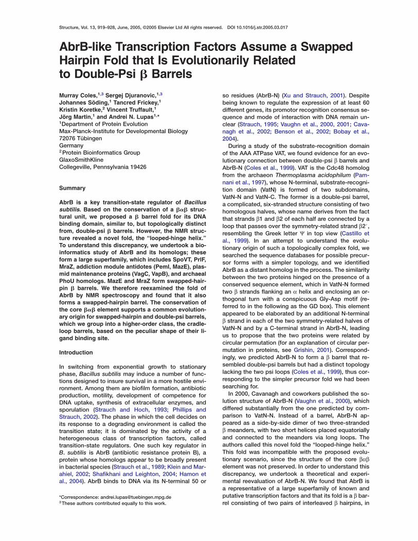
Figure 3. Structures of AbrB-N and Related β Barrels
(A) The double-psi barrel fold of VatN-N (1CZ4, top) and the swapped-hairpin barrels of MazE (1UB4, middle) and MraZ (1N0G, bottom). Thetop view (left) is related to the side view (right) by a 90° rotation around the horizontal axis. The secondary structure of one symmetrical halfof each domain is shown in bold color, with the β2 strand in red. The swapping of position of this strand and the two additional β strands inthe swapped-hairpin barrel are the main topological differences in the folds. VatN-N and MraZ are monomers, and MazE is a homodimer. Forreasons of topology, the two (pseudo) symmetrical halves of MraZ must be joined by a helical hairpin (the hairpin appears after each half,but the C-terminal hairpin has been omitted from the side view for clarity). The typical “horned” profile provided by the β1–β2 loop is a strikingfeature of all three domains.(B) Two structures for AbrB-N: the swapped-hairpin fold solved here (1YFB, top), and the looped-hinge helix fold previously reported byVaughn et al. (2000) (1EKT, bottom). The views and coloring are as for (A). The previously reported structure cannot explain the formation ofintermolecular β2–β2#contacts, while the β1–β3/β1#–β3# contacts are incorrectly assigned as intramolecular. The similarity of the structurepresented here to MazE and MraZ is apparent.(C) The conserved structural core of the swapped-hairpin and double-psi barrels. The left view shows a superposition of AbrB-N, MazE, andof both halves of MraZ and VatN-N. The right view shows the looped-hinge helix structure for AbrB-N, such that orientation of the helix andthe following GD box are the same as in the left panel. The superposition shows the structural equivalence of β1 of the double-psi barrel withβ4 of the swapped-hairpin barrel. A circular permutation cannot, however, be concluded, since the equivalence is to β4 of the symmetry-related subunit.
eral NOESY connectivities that were best explained byan intermolecular antiparallel β sheet contact with itssymmetry-related equivalent, β2#. This observation isincompatible with the fold originally reported for theprotein. The intermolecular contacts expected for thistopology, e.g., a strong Hα-Hα contact between R17 andV19, were confirmed in filtered/edited NOESY experi-ments on a sample containing asymmetrically 15N- and13C-labeled monomers (Figure 4). Intermolecular con-tacts were also observed between β1 (I8–V12) and β3#(D34–D41). This is also inconsistent with the originallyreported fold, in which the β1 strand made intramolecu-
lar contacts to β3. The contacts for β4 (K44–Y50), i.e.,the intramolecular β3–β4 and intermolecular β4–β4#contacts, were as previously reported. The overall foldwas thus consistent with that of the identified homo-logs MazE and MraZ and rationalized the sequenceconservation patterns of the AbrB superfamily (Figure 2).
Structural data for AbrB consisted of distance dataderived from a 3D 15N-HSQC-NOESY as well as several2D NOESY spectra, 3JHNHαcoupling constants derivedfrom an HNHA experiment, and chemical shift-derivedbackbone torsion angle restraints. An initial model wascreated by using all available data and was used as the

Structure924
Figure 4. Intermolecular Contacts Define theAbrB-N Topology
A section of the 14N,12C-filtered/13C-edited2D-NOESY spectrum containing primarilymethyl-methyl contacts between hydropho-bic residues is shown. Selected pairs of di-agonally related crosspeaks are labeled.They represent contacts between β2 and β2#(R17/V18–V18#/V19#), β1 and β3# (V9/V12–A35#/L36#), β4 and β4# (I45/L47–L47#), andbetween the α helix (L24) and the GD box(I30) and the hydrophobic core of the pro-tein. A total of 22 crosspeaks could be as-signed in this spectrum, contributing to thetotal of 55 intermolecular distance restraintsincluded in calculations.
starting point for iterative assignment of further NOEtconnectivities, resulting in the final set of experimental
restraints described in Table 1. The structure ensemble Mfis well defined (Figure 5), with an rmsd for the final set
of 20 dimeric structures (residues T6–Y50) of 0.20 Å for dcbackbone atoms and 0.70 Å for all heavy atoms.
The monomer structure (Figure 5) can be described aas a pair of β hairpins linked by the α helix and the GD sbox. The monomer does not contain an independent (hydrophobic core, and it thus requires dimerization to iform a viable fold. This dimerization takes place by in- Tterleaving the four β hairpin elements, such that each conly makes contacts to those of the dimeric partner. bThe interleaving is ensured by the extended nature of lthe connector between the α helix and the GD motif, in dwhich the two large hydrophobic residues L28 and I30 bare buried in the opening of the barrel, providing for the trigidity of the crossover connection. The result is an Meight-stranded, swapped-hairpin β barrel. The two heli- cces close the barrel at each end. The two GD boxes thave a key structural role, forming β turns that are an- schored into the core of the barrel by flanking hydropho- fbic residues. Hydrogen bonds between the turn and the iβ1 strand of the dimeric partner fix the positions of the sβ1–β2 loops. These loops project above the surface of Tthe barrel, giving the protein its characteristic horned iprofile (Figure 3) and forming a cleft rich in positive rcharge that, in MazE, has been implicated in interaction Twith DNA (Loris et al., 2003). s
The fold presented here is markedly different from tthat of Vaughn et al. (2000), in which β2, and therefore dthe β1–β2 hairpin, was absent and no interleaving ofmonomer elements occurred (Figure 3). In the resulting
Dstructure, each monomer was an independent foldingunit, and dimerization took place in a simple side-by-
Iside manner. The formal discrimination of inter- and in-atramolecular NOESY contacts in the current study withbdimer samples with asymmetrically labeled monomers
refutes this model. A
Comparison of the AbrB structure presented here tohose of the identified homologs, MraZ (1MVF) andazE (1N0G), shows the proteins to share a common
old (Figure 3) while revealing differences in structuraletails. Both MraZ and AbrB have short β1–β2 loops,omprised completely of a β turn (D13–G16 in AbrB)nd characterized by a web of interactions between theide chains of an aspartic acid and an arginine residueD13 and R17 in AbrB) and neighboring backbone am-des (the backbone rmsd of AbrB V12–R17 to 1N0G34–R40 is 0.4 Å). AbrB and MraZ lack formal β sheetontacts in the β1–β2 hairpin, meaning that they muste considered pseudobarrels. In contrast, the β1–β2
oop of MazE is somewhat longer, and a pair of hy-rogen bond contacts between β1 and β2 closes thearrel. AbrB shares with MazE the β turn structure overhe GD box residues typical of the fold family, while
raZ lacks both of these residues and the β turn. Aonserved PxxxR sequence within the α1 helix is alsoypical of the family (Figure 2). In AbrB and all othertructures in which this motif is present, the arginineorms a side chain H bond to the backbone carbonylmmediately preceding the proline, thus facilitating theharp change in chain direction between β2 and α1.his motif is only present in the first half of MraZ, while,
n the second half and in MazE, a large hydrophobicesidue replaces the arginine (I and M, respectively).hus, many of the conserved structural features do noteem to be determinants of the overall fold, explaininghe imperfect conservation pattern of the involved resi-ues in the AbrB superfamily.
iscussion
n the process of evaluating the discrepancy betweenn evolutionary scenario for the origin of double-psi βarrels and the experimentally determined fold forbrB-N, we defined a new superfamily of prokaryotic

Solution Structure of AbrB925
Table 1. Structural Statistics and Atomic Rms Deviations
Structural Statisticsa
SA <SA>r
Rmsd from distance restraints (Å)b
All (418) 0.027 ± 0.0010 0.026Intraresidue (145) 0.012 ± 0.0031 0.012Interresidue sequential (152) 0.026 ± 0.0016 0.024Medium range (37) 0.036 ± 0.0013 0.035Long range (29) 0.047 ± 0.0059 0.048Intermolecular (55) 0.033 ± 0.0013 0.032
Rmsd from dihedral restraints (106) 0.463 ± 0.073 0.427Rmsd from J coupling restraints (Hz) (32) 0.568 ± 0.012 0.586H bond restraint violations (Å/°)c (35) 2.10 ± 0.19/16.8 ± 8.6 2.11/14.1Deviations from ideal covalent geometry
Bonds (Å × 10−3) 1.67 ± 0.032 1.63Angles (°) 0.502 ± 0.003 0.497Impropers (°) 1.272 ± 0.051 1.200
Structure quality indicatorsd
Ramachandran map regions (%) 88.9/10.6/0.5/0.0 90.2/9.8/0./ 0.0Bad contacts per 100 residues 7.6 ± 2.4 7.5
Atomic Rms Differences (Å)e
SA versus <SA> SA versus <SA>r
Backbone All Backbone All
All residues 1.83 ± 0.78 2.31 ± 0.713 2.17 ± 1.262 2.82 ± 1.131Ordered residuesf (dimer) 0.20 ± 0.065 0.70 ± 0.070 0.27 ± 0.067 0.98 ± 0.121Ordered residues (monomer) 0.19 ± 0.064 0.70 ± 0.070 0.26 ± 0.066 0.98 ± 0.123
<SA> versus <SA>rg 0.18 0.69
a Structures are labeled as follows: SA, the set of 25 final simulated annealing structures; <SA>, the mean structure calculated by averagingthe coordinates of SA structures after fitting over secondary structure elements; <SA>r, the structure obtained by regularizing the meanstructure under experimental restraints.b Numbers in brackets indicate the number of restraints of each type per monomer.c H bonds were restrained by treating them as pseudocovalent bonds (see Experimental Procedures). Deviations are expressed as the averagedistance/average deviation from linearity for restrained H bonds.d Determined by using the program PROCHECK (Laskowski et al., 1993). Percentages are for residues in allowed/additionally allowed/generously allowed/disallowed regions of the Ramachandran map.e Based on heavy atoms superimpositions.f Defined as residues T6–Y50.g Rms difference for superimposition over ordered residues.
transcription factors, which includes transition-stateregulators, the antitoxins of several classes of postseg-regational killing systems, putative regulators of cellwall biosynthesis, regulators of phosphate uptake, anda large number of proteins of as yet unknown activity(Figures 1 and 2). This superfamily contained two mem-bers with known crystal structure that resembled eachother and differed from the solution structure reportedfor AbrB-N, prompting us to redetermine its structure.Our results are in agreement with the crystal structures(Figure 3); AbrB-N consists of four β strands arrangedinto two harpins, which are interleaved in the dimer(Figure 5), leading us to name this fold the swapped-hairpin barrel. In side view, the two N-terminal hairpinscurve upward, giving the barrel a characteristic hornedappearance and forming a deep binding cleft.
The similarities of the double-psi barrel to theswapped-hairpin barrel are striking (Figure 3). Despite adifferent overall fold, many structural features of VatN-Nremarkably resemble AbrB-N (Figure 3C). Particularlysimilar is the turn over the GD box and its hydro-gen bonding interaction with the symmetry-related β1strand. The arginine of the PxxxR motif also makes theexpected side chain-backbone hydrogen bond con-tacts (although the proline has been replaced by an
aspartic acid residue in both VatN-N repeats). The psiloops curve upward in the same way as the β1–β2 loopsof the swapped-hairpin barrel, giving the two folds asimilar profile, and forming a deep, positively chargedcleft. Indeed, this cleft has been implicated in substrateinteractions of VatN (Coles et al., 1999), and, further-more, expression of VatN-N alone leads to spontaneousdimerization and to a low but measurable DNA bindingactivity, as detected in band-shift assays with heterolo-gous DNA from E. coli (data not shown). All of theseobservations support a homologous origin for the twofolds, which we propose to denote as cradle-loop bar-rels, in view of their peculiar profile and cradle-shapedbinding surface. We have previously hypothesized thatfolded protein domains arose through the fusion andrecombination of a smaller number of subdomain-sizedpeptides (antecedent domain segments) (Lupas et al.,2001; Soding and Lupas, 2003), which themselvesemerged in the context of RNA-based replication andcatalysis (the “RNA world”). The strong similarity be-tween the swapped-hairpin and double-psi barrels inthe core βαβ region suggests that this element mightcorrespond to such an antecedent domain segment.
Nevertheless, the topological relationship betweenthe two folds is not obvious. The structural model we

Structure926
Figure 5. A Stereo View of the Ensemble ofthe 25 Lowest-Energy Structures for AbrB-N
The upper panel shows the AbrB-N mono-mer colored by secondary structure, whilethe lower panel shows the dimer colored bymonomer. The superimposition is over resi-dues T7–Y50 of each monomer, resulting inan rmsd of 0.20 Å for backbone atoms and0.70 Å for all heavy atoms.
moriginally proposed for AbrB (Coles et al., 1999) by ex-etrapolation from VatN-N is incorrect in that the barrel ismeight-, not six-stranded, and the two subunits formingi
the barrel are interleaved, not face-to-face. Although pthe position of β1 in each repeat of the double-psi bar- 1
arel is equivalent to that of β4 in the swapped-hairpintbarrel (Figure 3C), in accordance with our prediction,sthe relationship holds for β1 and β4# (i.e., β4 of the sym-I
metry-related half), introducing an unanticipated topo- alogical complexity and essentially ruling out a trans- a
tformation by circular permutation, as we originallysproposed. Thus, although the swapped-hairpin barrelbis topologically simpler than the double-psi barrel byh
virtue of lacking the knotted psi loops, it does not seem elikely to represent a precursor form. Rather, both barrel
qtypes appear to have evolved from a yet simpler thirdLtype. We are currently investigating the structures ofsseveral sequences with properties intermediate be-a
tween AbrB-N and VatN-N in order to identify this qthird type. w
aExperimental Procedures c
tSequence Searches and Cluster Analysis nFor sequence searches on the nonredundant protein sequence da- ttabase, we developed a method, HHsenser, by combining the ssearch strategy encoded in SENSER (Koretke et al., 2002) with a omethod for comparing profile Hidden Markov Models (HHsearch) i
T(Soding, 2004). HHsenser is thus an itermediate profile search
ethod that is based on HMM-HMM comparison. HHsenser firstmploys PSI-Blast (Altschul et al., 1997) to generate an initial align-ent of relatively close homologs of the query sequence (AbrB,
n this case). It then selects representative sequences (maximumairwise sequence identity of 40% and PSI-Blast E values of up to) in the vicinity of the query sequence as seeds for building newlignments with PSI-Blast. HMM-HMM comparison by HHsearch ishen employed to check if the new alignment is homologous to theuperalignment of all accepted homologs of the query sequence.f the alignment is accepted, it is included in the superalignment. Inddition, the representative sequences in the vicinity of the newlyccepted alignment are added to the list of seed sequences, andhe process is continued until all seeds have been processed. Aeries of heuristics ensures that run time is kept to a minimumy avoiding PSI-Blast searches that will probably not result in aomologous alignment. The AbrB superfamily alignment was gen-rated in 8.5 hr on a single 3 GHz 64-bit AMD CPU.After convergence of the sequence searches, the obtained se-
uences were clustered by using the program CLANS (Frickey andupas, 2004) at a P-value cutoff of e-4. The sequences corre-ponded in all cases to the parts identified as being similar to AbrBnd were thus almost always fragments of the complete se-uences. In some proteins, two fragments homologous to AbrBere recovered. The resulting map is shown in Figure 1. We reex-mined the individual clusters manually, as well as all proteins notlearly assigned to a cluster, but we did not detect any false posi-ives. We found, though, that the search routine had missed a smallumber of sequences in each cluster, which could be identified byaking that cluster as the starting point. We are not certain at pre-ent of the reason. We also observed that the incorrect assignmentf start codons in the database had shortened the AbrB domains
n some proteins, causing them to become outliers to their clusters.his is seen particularly clearly in the MraZ clusters, where the N

Solution Structure of AbrB927
domain cluster is much more irregular than the C domain cluster.The most extreme example of a miscalled start codon was in Geo-bacter RecG, where the annotated G. sulfurreducens homologlacks practically the entire AbrB domain and thus groups well awayfrom G. metalloreducens RecG, even though the two domains actu-ally share 50% sequence identity when the correct start codon isused.
Sample PreparationThe AbrB-N construct (encoding amino residues 1–53 of AbrB,gi113009) was amplified from B. subtilis PY79 chromosomal DNAby polymerase chain reaction (PCR) and was cloned into thepet30b vector (Novagen). The construct contained a His6-tag atthe amino terminus to facilitate purification. For expression inE. coli, cells were grown in LB medium at 37°C, induced at anOD600 of w0.6 with 1 mM IPTG, and was harvested after 4 hr. Uni-formly 15N- or 13C-labeled AbrB-N was made by growing bacteriain M9 minimal medium by using 15NH4Cl (0.7 g/l) and 13C6-glucose(2 g/l) as sole nitrogen or carbon sources. An asymmetrically 15N-and 13C-labeled AbrB-N sample was made by combining the sameamounts of harvested cells prior to lysis. Proteins were purified bya combination of immobilized metal affinity chromatography(IMAC), ion exchange, and gel sizing chromatography. Purificationunder denaturing conditions allowed for statistical mixing of thelabeled components, which was later confirmed by NMR. For NMRmeasurements, samples were concentrated to 8 mg/ml in buffercontaining 20 mM potassium phosphate, 50 mM KCl, 0.02% (w/v)NaN3 (pH 5.8).
NMR SpectroscopyAll spectra were recorded at 305 K on Bruker DMX600, DMX750,and DMX900 spectrometers. Resonance assignments were takenfrom Vaughn et al. (2000) and were confirmed either throughNOESY connectivities (for sequential assignments) or in a CCH-COSY experiment on the asymmetrically labeled sample (for sidechain assignments). This sample was additionally used to obtaincarbonyl assignments for 36 of 52 residues by using a HACACOexperiment. The stereospecific assignments of prochiral groupsand the resulting rotamer assignments were also checked.
Distance data were derived from a 3D-15N-HSQC-NOESY on a15N-labeled sample and a 2D-NOESY spectrum recorded on an un-labeled sample. Intermolecular NOESY contacts were identified inthe asymmetrically 15N- and 13C-labeled sample by using a14N,12C-filtered/13C-edited 2D-NOESY spectrum (see Figure 4). Re-sidual diagonal signals in this spectrum were effectively eliminatedby subtraction of an identical spectrum run with minimal NOESYmixing time. Intramolecular 14NH/13CH contacts were exclusivelyobservable in this spectrum and were thus discriminated from in-termolecular 15NH/13CH contacts. Contacts identified in the fil-tered/edited spectrum were quantified when possible in otherspectra.
NOESY crosspeaks in the 3D spectra were converted into dis-tance ranges after rescaling according to corresponding HSQC in-tensities. Crosspeaks were divided into four classes: strong, me-dium, weak, and very weak, which resulted in restraints on upperdistances of 2.7, 3.2, 4.0, and 5.0 Å, respectively. Lower distancerestraints were also included for very weak or absent sequentialHN-HN crosspeaks by using a minimum distance of 3.2 Å and me-dium intensity or weaker sequential and intraresidue HN-Hα cross-peaks by using a minimum distance of 2.7 Å. Allowances for theuse of pseudoatoms (using r−6 averaging) were added for methylgroups and nonstereospecifically assigned methylene groups. Di-hedral angle restraints were derived for backbone f and ψ anglesbased on Cα, Cβ, and Hα chemical shifts by using the programTALOS (Cornilescu et al., 1999). Restraints were applied for the 35high-confidence predictions found by the program by using thecalculated range ±10°. In addition, direct coupling constant re-straints were included for the backbone f angles of 32 residuesbased on 3JHNHα coupling constants measured from an HNHA ex-periment. Hydrogen bond restraints were applied for 26 residues insecondary structure with low water exchange rates, as judged bythe strength of water exchange crosspeaks in the 15N-HSQC-NOESY spectrum, and where donor-acceptor pairs were consis-
tently identified in preliminary calculations. This included 11 inter-molecular hydrogen bonds. The restraints were applied via inclu-sion of pseudocovalent bonds as described by Truffault et al.(2001).
Structures were calculated with XPLOR (NIH version 2.9.3) byusing standard protocols. Experimental restraints were appliedonly to one monomer, with noncrystallographic symmetry restraintsover the backbone of ordered residues (T7–Y50) used to ensure thesymmetry of the dimer. Sets of 50 structures were calculated, anda final set of 25 was chosen on the basis of lowest restraint viola-tions. An average structure was calculated and regularized to givea structure representative of the ensemble.
Supplemental Data
Supplemental Data including a full list of proteins used to constructthe cluster map shown in Figure 1 are available at http://www.structure.org/cgi/content/full/13/6/919/DC1.
Acknowledgments
The authors thank Prof. Horst Kessler and the staff of the BavarianNuclear Magnetic Resonance (NMR) Centre at the Technical Uni-versity, Munich for access to spectrometers and technical support.The bioinformatics study was conducted by A.N.L., J.S., T.F., andK.K., with support from S.D. The biochemistry is the work of S.D.,with support from J.M. The NMR structure was determined by M.C.and V.T. The authors declare that they have no competing finan-cial interests.
Received: February 25, 2005Revised: March 29, 2005Accepted: March 29, 2005Published: June 7, 2005
References
Altschul, S.F., Madden, T.L., Schaffer, A.A., Zhang, J., Zhang, Z.,Miller, W., and Lipman, D.J. (1997). Gapped BLAST and PSI-BLAST:a new generation of protein database search programs. NucleicAcids Res. 25, 3389–3402.
Anantharaman, V., and Aravind, L. (2003). New connections in theprokaryotic toxin-antitoxin network: relationship with the eukary-otic nonsense-mediated RNA decay system. Genome Biol. 4, R81.
Andreeva, A., Howorth, D., Brenner, S.E., Hubbard, T.J., Chothia,C., and Murzin, A.G. (2004). SCOP database in 2004: refinementsintegrate structure and sequence family data. Nucleic Acids Res.32, D226–D229.
Bagyan, I., Hobot, J., and Cutting, S. (1996). A compartmentalizedregulator of developmental gene expression in Bacillus subtilis. J.Bacteriol. 178, 4500–4507.
Baird, L., and Georgopoulos, C. (1990). Identification, cloning, andcharacterization of the Escherichia coli sohA gene, a suppressor ofthe htrA (degP) null phenotype. J. Bacteriol. 172, 1587–1594.
Benson, L.M., Vaughn, J.L., Strauch, M.A., Bobay, B.G., Thompson,R., Naylor, S., and Cavanagh, J. (2002). Macromolecular assemblyof the transition state regulator AbrB in its unbound and complexedstates probed by microelectrospray ionization mass spectrometry.Anal. Biochem. 306, 222–227.
Bobay, B.G., Benson, L., Naylor, S., Feeney, B., Clark, A.C., Goshe,M.B., Strauch, M.A., Thompson, R., and Cavanagh, J. (2004). Eval-uation of the DNA binding tendencies of the transition state regula-tor AbrB. Biochemistry 43, 16106–16118.
Castillo, R.M., Mizuguchi, K., Dhanaraj, V., Albert, A., Blundell, T.L.,and Murzin, A.G. (1999). A six-stranded double-psi beta barrel isshared by several protein superfamilies. Struct. Fold. Des. 7, 227–236.
Cavanagh, J., Thompson, R., Bobay, B., Benson, L.M., and Naylor,S. (2002). Stoichiometries of protein-protein/DNA binding and con-

Structure928
formational changes for the transition-state regulator AbrB mea- Psured by pseudo cell-size exclusion chromatography-mass spec- atrometry. Biochemistry 41, 7859–7865. 3
Chen, S., Jancrick, J., Yokota, H., Kim, R., and Kim, S.H. (2004). PCrystal structure of a protein associated with cell division from My- pcoplasma pneumoniae (GI: 13508053): a novel fold with a con- cserved sequence motif. Proteins 55, 785–791. RColes, M., Diercks, T., Liermann, J., Groger, A., Rockel, B., PBaumeister, W., Koretke, K.K., Lupas, A., Peters, J., and Kessler, H. S(1999). The solution structure of VAT-N reveals a ‘missing link’ in the Sevolution of complex enzymes from a simple betaalphabetabeta telement. Curr. Biol. 9, 1158–1168. 4Cornilescu, G., Delaglio, F., and Bax, A. (1999). Protein backbone Sangle restraints from searching a database for chemical shift and gsequence homology. J. Biomol. NMR 13, 289–302. BDong, T.C., Cutting, S.M., and Lewis, R.J. (2004). DNA-binding Sstudies on the Bacillus subtilis transcriptional regulator and AbrB phomologue, SpoVT. FEMS Microbiol. Lett. 233, 247–256.
SEngelberg-Kulka, H., and Glaser, G. (1999). Addiction modules and oprogrammed cell death and antideath in bacterial cultures. Annu.
SRev. Microbiol. 53, 43–70.c
Frickey, T., and Lupas, A. (2004). CLANS: a Java application for avisualizing protein families based on pairwise similarity. Bioinfor-
smatics 20, 3702–3704.
SGerdes, K. (2000). Toxin-antitoxin modules may regulate synthesis
sof macromolecules during nutritional stress. J. Bacteriol. 182,
M561–572.
SGotfredsen, M., and Gerdes, K. (1998). The Escherichia coli relBE
bgenes belong to a new toxin-antitoxin gene family. Mol. Microbiol.
r29, 1065–1076.
JGrishin, N.V. (2001). Fold change in evolution of protein structures.
TJ. Struct. Biol. 134, 167–185.L
Hamon, M.A., Stanley, N.R., Britton, R.A., Grossman, A.D., and La- tzazzera, B.A. (2004). Identification of AbrB-regulated genes in- 3volved in biofilm formation by Bacillus subtilis. Mol. Microbiol. 52,
V847–860.(
Kamada, K., Hanaoka, F., and Burley, S.K. (2003). Crystal structure Bof the MazE/MazF complex: molecular bases of antidote-toxin re- 1cognition. Mol. Cell 11, 875–884.
VKatz, M.E., Strugnell, R.A., and Rood, J.I. (1992). Molecular charac- Dterization of a genomic region associated with virulence in Dichelo- tbacter nodosus. Infect. Immun. 60, 4586–4592. tKlein, W., and Marahiel, M.A. (2002). Structure-function relationship
Vand regulation of two Bacillus subtilis DNA-binding proteins, HBsu
sand AbrB. J. Mol. Microbiol. Biotechnol. 4, 323–329.
CKoretke, K.K., Russell, R.B., and Lupas, A.N. (2002). Fold recogni-
Wtion without folds. Protein Sci. 11, 1575–1579.
tLaskowski, R.A., MacArthur, M.W., Moss, D.S., and Thornton, J.M.
X(1993). PROCHECK: a program to check the stereo chemical qual-
tity of protein structures. J. Appl. Crystallogr. 26, 283–291.1
Lehnherr, H., Maguin, E., Jafri, S., and Yarmolinsky, M.B. (1993).ZPlasmid addiction genes of bacteriophage P1: doc, which causesMcell death on curing of prophage, and phd, which prevents hostbdeath when prophage is retained. J. Mol. Biol. 233, 414–428.ZLoris, R., Marianovsky, I., Lah, J., Laeremans, T., Engelberg-Kulka,IH., Glaser, G., Muyldermans, S., and Wyns, L. (2003). Crystal struc-nture of the intrinsically flexible addiction antidote MazE. J. Biol.
Chem. 278, 28252–28257.A
Lupas, A.N., Ponting, C.P., and Russell, R.B. (2001). On the evolu-tion of protein folds: are similar motifs in different protein folds theresult of convergence, insertion, or relics of an ancient peptide Tworld? J. Struct. Biol. 134, 191–203. sMayer, M.P., Bueno, L.C., Hansen, E.J., and DiRienzo, J.M. (1999). cIdentification of a cytolethal distending toxin gene locus and fea-tures of a virulence-associated region in Actinobacillus actinomy- Ncetemcomitans. Infect. Immun. 67, 1227–1237.
DPamnani, V., Tamura, T., Lupas, A., Peters, J., Cejka, Z., Ashraf, W.,Pand Baumeister, W. (1997). Cloning, sequencing and expression ofMVAT, a CDC48/p97 ATPase homologue from the archaeon Thermo-
plasma acidophilum. FEBS Lett. 404, 263–268. m
hillips, Z.E., and Strauch, M.A. (2002). Bacillus subtilis sporulationnd stationary phase gene expression. Cell. Mol. Life Sci. 59,92–402.
ullinger, G.D., and Lax, A.J. (1992). A Salmonella dublin virulencelasmid locus that affects bacterial growth under nutrient-limitedonditions. Mol. Microbiol. 6, 1631–1643.
adnedge, L., Davis, M.A., Youngren, B., and Austin, S.J. (1997).lasmid maintenance functions of the large virulence plasmid ofhigella flexneri. J. Bacteriol. 179, 3670–3675.
hafikhani, S.H., and Leighton, T. (2004). AbrB and Spo0E controlhe proper timing of sporulation in Bacillus subtilis. Curr. Microbiol.8, 262–269.
nyder, W.B., and Silhavy, T.J. (1992). Enhanced export of beta-alactosidase fusion proteins in prlF mutants is Lon dependent. J.acteriol. 174, 5661–5668.
oding, J. (2004). Protein homology detection by HMM-HMM com-arison. Bioinformatics 21, 951–960.
oding, J., and Lupas, A.N. (2003). More than the sum of their parts:n the evolution of proteins from peptides. Bioessays 25, 837–846.
trauch, M.A. (1995). Delineation of AbrB-binding sites on the Ba-illus subtilis spo0H, kinB, ftsAZ, and pbpE promoters and use ofderived homology to identify a previously unsuspected binding
ite in the bsuB1 methylase promoter. J. Bacteriol. 177, 6999–7002.
trauch, M.A., and Hoch, J.A. (1993). Transition-state regulators:entinels of Bacillus subtilis post-exponential gene expression.ol. Microbiol. 7, 337–342.
trauch, M.A., Spiegelman, G.B., Perego, M., Johnson, W.C., Bur-ulys, D., and Hoch, J.A. (1989). The transition state transcriptionegulator abrB of Bacillus subtilis is a DNA binding protein. EMBO. 8, 1615–1621.
ruffault, V., Coles, M., Diercks, T., Abelmann, K., Eberhardt, S.,uttgen, H., Bacher, A., and Kessler, H. (2001). The solution struc-ure of the N-terminal domain of riboflavin synthase. J. Mol. Biol.09, 949–960.
aughn, J.L., Feher, V., Naylor, S., Strauch, M.A., and Cavanagh, J.2000). Novel DNA binding domain and genetic regulation model ofacillus subtilis transition state regulator abrB. Nat. Struct. Biol. 7,139–1146.
aughn, J.L., Feher, V.A., Bracken, C., and Cavanagh, J. (2001). TheNA-binding domain in the Bacillus subtilis transition-state regula-
or AbrB employs significant motion for promiscuous DNA recogni-ion. J. Mol. Biol. 305, 429–439.
icente, M., Gomez, M.J., and Ayala, J.A. (1998). Regulation of tran-cription of cell division genes in the Escherichia coli dcw cluster.ell. Mol. Life Sci. 54, 317–324.
anner, B.L. (1993). Gene regulation by phosphate in enteric bac-eria. J. Cell. Biochem. 51, 47–54.
u, K., and Strauch, M.A. (2001). DNA-binding activity of amino-erminal domains of the Bacillus subtilis AbrB protein. J. Bacteriol.83, 4094–4098.
hang, Y., Zhang, J., Hoeflich, K.P., Ikura, M., Qing, G., and Inouye,. (2003). MazF cleaves cellular mRNAs specifically at ACA to
lock protein synthesis in Escherichia coli. Mol. Cell 12, 913–923.
hang, J., Zhang, Y., Zhu, L., Suzuki, M., and Inouye, M. (2004).nterference of mRNA function by sequence-specific endoribo-uclease PemK. J. Biol. Chem. 279, 20678–20684.
ccession Numbers
he coordinates for the ensembles and the regularized averagetructure have been deposited in the Protein Data Bank (accessionodes 1YSF and 1YFB, respectively).
ote Added in Proof
uring processing of this manuscript, a corrigendum retracting theDB entry 1EKT has appeared in press (Vaughn et al. [2005], Nat.ol. Struct. Biol. 12, 380), and a revised PDB entry, 1Z0R, was sub-itted.
Related Documents