THE JOURNAL OF CELL BIOLOGY JCB: ARTICLE The Journal of Cell Biology, Vol. 170, No. 5, August 29, 2005 781–791 http://www.jcb.org/cgi/doi/10.1083/jcb.200502148 JCB 781 Abnormal nuclear shape and impaired mechanotransduction in emerin-deficient cells Jan Lammerding, 1 Janet Hsiao, 2 P. Christian Schulze, 1 Serguei Kozlov, 3 Colin L. Stewart, 3 and Richard T. Lee 1 1 Cardiovascular Division, Brigham and Women’s Hospital, Boston, MA 02115 2 HST Division, Massachusetts Institute of Technology, Cambridge, MA 02139 3 Cancer and Developmental Biology Lab, National Cancer Institute, Frederick, MD 21702 mery-Dreifuss muscular dystrophy can be caused by mutations in the nuclear envelope proteins lamin A/C and emerin. We recently demonstrated that A-type lamin-deficient cells have impaired nuclear me- chanics and altered mechanotransduction, suggesting two potential disease mechanisms (Lammerding, J., P.C. Schulze, T. Takahashi, S. Kozlov, T. Sullivan, R.D. Kamm, C.L. Stewart, and R.T. Lee. 2004. J. Clin. Invest. 113: 370–378). Here, we examined the function of emerin on nuclear mechanics and strain-induced signaling. Emerin- deficient mouse embryo fibroblasts have abnormal nu- clear shape, but in contrast to A-type lamin-deficient cells, exhibit nuclear deformations comparable to wild- E type cells in cellular strain experiments, and the integrity of emerin-deficient nuclear envelopes appeared normal in a nuclear microinjection assay. Interestingly, expres- sion of mechanosensitive genes in response to mechani- cal strain was impaired in emerin-deficient cells, and prolonged mechanical stimulation increased apoptosis in emerin-deficient cells. Thus, emerin-deficient mouse em- bryo fibroblasts have apparently normal nuclear me- chanics but impaired expression of mechanosensitive genes in response to strain, suggesting that emerin muta- tions may act through altered transcriptional regulation and not by increasing nuclear fragility. Introduction Emerin is a small (34 kD) nuclear envelope protein with a sin- gle transmembrane domain spanning the inner nuclear mem- brane and a large nucleoplasmic domain that can interact with other nuclear envelope proteins such as lamins (Bengtsson and Wilson, 2004). Emerin is not essential for cell viability, but it contributes to shared vital functions, including nuclear assem- bly and cell cycle progression. Emerin is encoded by the EMD gene (also known as STA) located on the X-chromosome (Bione et al., 1994). Mutations in this gene can cause the X-linked recessive form of Emery-Dreifuss muscular dystrophy (EDMD), a disease characterized by early onset in childhood with slowly progressive skeletal muscle wasting, contractures of the elbow, neck, and Achilles tendons, a rigid spine, abnor- mal heart rhythms, heart block, and cardiomyopathy leading to increased risk of cardiac arrest (Bione et al., 1994). The major- ity of the disease-causing mutations are nonsense mutations that produce soluble forms of the protein, leading to loss of emerin from the nuclear periphery and mislocalization of emerin to the cytoplasm (Ellis et al., 1998; Vaughan et al., 2001). Although X-linked EDMD is associated with a mutation in the EMD gene, the autosomal dominant form of EDMD is caused by missense mutations in the LMNA gene that encodes the nuclear A-type lamins (Bonne et al., 1999). These muta- tions often result in misfolding or failure of the A-type lamins to correctly assemble, leading to partial or complete loss of function (Burke and Stewart, 2002). Historically, X-linked EDMD was the first disorder to be recognized as a nuclear membrane disease and opened a new area of research on the role of the nuclear envelope in disease. Currently, more than 180 mutations have been identified in the LMNA and EMD genes that are causally linked to at least 10 distinct diseases, in- cluding EDMD, dilated cardiomyopathy, familial partial lipo- dystrophy, and Hutchinson Gilford progeria syndrome (Bonne et al., 1999; Fatkin et al., 1999; Cao and Hegele, 2000; Shackle- ton et al., 2000; Burke and Stewart, 2002; De Sandre-Giovan- noli et al., 2002, 2003; Eriksson et al., 2003). Although emerin and A-type lamins (predominantly lamins A and/or C) are expressed in most human tissues, EDMD predominantly affects skeletal and cardiac muscles and tendons. The reason for this tissue-specific phenotype is not yet Correspondence to Jan Lammerding: [email protected] Abbreviations used in this paper: BAF, barrier-to-autointegration factor; EDMD, Emery-Dreifuss muscular dystrophy; GAPDH, glyceraldehyde 3-phosphate dehy- drogenase; GCL, germ cell-less. The online version of this article contains supplemental material. on January 18, 2016 jcb.rupress.org Downloaded from Published August 22, 2005 http://jcb.rupress.org/content/suppl/2005/09/27/jcb.200502148.DC1.html Supplemental Material can be found at:

Welcome message from author
This document is posted to help you gain knowledge. Please leave a comment to let me know what you think about it! Share it to your friends and learn new things together.
Transcript

TH
EJ
OU
RN
AL
OF
CE
LL
BIO
LO
GY
JCB: ARTICLE
The Journal of Cell Biology, Vol. 170, No. 5, August 29, 2005 781–791http://www.jcb.org/cgi/doi/10.1083/jcb.200502148
JCB 781
Abnormal nuclear shape and impaired mechanotransduction in emerin-deficient cells
Jan Lammerding,
1
Janet Hsiao,
2
P. Christian Schulze,
1
Serguei Kozlov,
3
Colin L. Stewart,
3
and Richard T. Lee
1
1
Cardiovascular Division, Brigham and Women’s Hospital, Boston, MA 02115
2
HST Division, Massachusetts Institute of Technology, Cambridge, MA 02139
3
Cancer and Developmental Biology Lab, National Cancer Institute, Frederick, MD 21702
mery-Dreifuss muscular dystrophy can be causedby mutations in the nuclear envelope proteins laminA/C and emerin. We recently demonstrated that
A-type lamin-deficient cells have impaired nuclear me-chanics and altered mechanotransduction, suggestingtwo potential disease mechanisms (Lammerding, J., P.C.Schulze, T. Takahashi, S. Kozlov, T. Sullivan, R.D. Kamm,C.L. Stewart, and R.T. Lee. 2004.
J. Clin. Invest.
113:370–378). Here, we examined the function of emerin onnuclear mechanics and strain-induced signaling. Emerin-deficient mouse embryo fibroblasts have abnormal nu-clear shape, but in contrast to A-type lamin-deficientcells, exhibit nuclear deformations comparable to wild-
E
type cells in cellular strain experiments, and the integrityof emerin-deficient nuclear envelopes appeared normalin a nuclear microinjection assay. Interestingly, expres-sion of mechanosensitive genes in response to mechani-cal strain was impaired in emerin-deficient cells, andprolonged mechanical stimulation increased apoptosis inemerin-deficient cells. Thus, emerin-deficient mouse em-bryo fibroblasts have apparently normal nuclear me-chanics but impaired expression of mechanosensitivegenes in response to strain, suggesting that emerin muta-tions may act through altered transcriptional regulationand not by increasing nuclear fragility.
Introduction
Emerin is a small (34 kD) nuclear envelope protein with a sin-gle transmembrane domain spanning the inner nuclear mem-brane and a large nucleoplasmic domain that can interact withother nuclear envelope proteins such as lamins (Bengtsson andWilson, 2004). Emerin is not essential for cell viability, but itcontributes to shared vital functions, including nuclear assem-bly and cell cycle progression. Emerin is encoded by the
EMD
gene (also known as
STA
) located on the X-chromosome(Bione et al., 1994). Mutations in this gene can cause theX-linked recessive form of Emery-Dreifuss muscular dystrophy(EDMD), a disease characterized by early onset in childhoodwith slowly progressive skeletal muscle wasting, contracturesof the elbow, neck, and Achilles tendons, a rigid spine, abnor-mal heart rhythms, heart block, and cardiomyopathy leading toincreased risk of cardiac arrest (Bione et al., 1994). The major-ity of the disease-causing mutations are nonsense mutationsthat produce soluble forms of the protein, leading to loss of
emerin from the nuclear periphery and mislocalization ofemerin to the cytoplasm (Ellis et al., 1998; Vaughan et al.,2001). Although X-linked EDMD is associated with a mutationin the
EMD
gene, the autosomal dominant form of EDMD iscaused by missense mutations in the
LMNA
gene that encodesthe nuclear A-type lamins (Bonne et al., 1999). These muta-tions often result in misfolding or failure of the A-type laminsto correctly assemble, leading to partial or complete loss offunction (Burke and Stewart, 2002). Historically, X-linkedEDMD was the first disorder to be recognized as a nuclearmembrane disease and opened a new area of research on therole of the nuclear envelope in disease. Currently, more than180 mutations have been identified in the
LMNA
and
EMD
genes that are causally linked to at least 10 distinct diseases, in-cluding EDMD, dilated cardiomyopathy, familial partial lipo-dystrophy, and Hutchinson Gilford progeria syndrome (Bonneet al., 1999; Fatkin et al., 1999; Cao and Hegele, 2000; Shackle-ton et al., 2000; Burke and Stewart, 2002; De Sandre-Giovan-noli et al., 2002, 2003; Eriksson et al., 2003).
Although emerin and A-type lamins (predominantlylamins A and/or C) are expressed in most human tissues,EDMD predominantly affects skeletal and cardiac muscles andtendons. The reason for this tissue-specific phenotype is not yet
Correspondence to Jan Lammerding: [email protected] used in this paper: BAF, barrier-to-autointegration factor; EDMD,Emery-Dreifuss muscular dystrophy; GAPDH, glyceraldehyde 3-phosphate dehy-drogenase; GCL, germ cell-less.The online version of this article contains supplemental material.
on January 18, 2016jcb.rupress.org
Dow
nloaded from
Published August 22, 2005
http://jcb.rupress.org/content/suppl/2005/09/27/jcb.200502148.DC1.html Supplemental Material can be found at:

JCB • VOLUME 170 • NUMBER 5 • 2005782
clear, but two alternative hypotheses for the disease mechanismhave emerged. The “structural hypothesis” suggests that muta-tions in genes encoding emerin or A-type lamins lead to in-creased nuclear fragility and to eventual nuclear disruption inmechanically strained tissues, whereas the “gene regulation hy-pothesis” is based on the findings that lamin A/C and emerincan bind to a variety of transcriptional regulators that could ex-ert tissue-specific effects. Lamins A/C are a major componentof the nuclear lamina, and loss of A-type lamins leads to im-paired nuclear mechanics and increased nuclear fragility (Broerset al., 2004; Lammerding et al., 2004). Emerin binds to severalstructural proteins such as lamin A/C, lamin B, nesprin-1
�
/2,and nuclear actin, and has recently been demonstrated topromote actin polymerization in vitro (Mislow et al., 2002;Bengtsson and Wilson, 2004; Holaska et al., 2004; Zhang et al.,2005). Loss of emerin from the nuclear envelope could thus in-terfere with the normal function of these proteins and lead tonuclear structural abnormalities. At the same time, emerin canbind to the transcriptional repressors barrier-to-autointegrationfactor (BAF), germ cell-less (GCL), and Btf, and to the splicingfactor YT521-B, suggesting an important role in gene regula-tion (Nili et al., 2001; Holaska et al., 2003; Wilkinson et al.,2003; Bengtsson and Wilson, 2004; Haraguchi et al., 2004).The structural hypothesis and the gene regulation hypothesisare not mutually exclusive, and in fact A-type lamin-deficientcells have increased nuclear fragility and abnormal nuclear me-chanics as well as impaired signaling responses to mechanicalstrain or cytokine stimulation, indicating that tissue-specific ef-fects observed in laminopathies could arise from varied degreesof impaired nuclear mechanics and transcriptional activation(Lammerding et al., 2004).
Here, we report independent measures of the structuraland gene-regulatory functions of emerin-deficient, A-typelamin-deficient, and wild-type mouse embryo fibroblasts toexplore the specific function of emerin on nuclear mechanicsand gene regulation. We show that, in contrast to A-typelamin-deficient cells, emerin-deficient fibroblasts have appar-ently normal nuclear mechanics but display similar, althoughless profound, deficiencies in strain-induced gene regulation,leading to an increased rate of apoptosis in response to me-chanical strain. These data suggest that emerin-associatedlaminopathies are predominantly caused by an impaired sig-naling response and not through direct strain-induced injury tothe nuclear membrane.
Results
Emerin null fibroblasts
Emerin-deficient mouse embryo fibroblasts were derived frommale emerin hemizygous mice (
Emd
�
/y
) and have been shownto lack emerin expression (unpublished data). Emerin-deficientmice were derived by deletion of the entire coding region of the
Emd
gene, which resulted in the complete absence of emerinprotein as shown by Western and immunohistochemical analy-sis of muscle fibroblasts isolated from the null mice (Fig. 1).The
Emd
null mice (both
�
/y males and
�
/
�
females) showedno overt pathology in either their skeletal or cardiac muscle,
even in mice as old as 12–15 mo (unpublished data). Hereafterwe have focused our studies on analyzing
Emd
�
/y
fibroblastsderived from male embryos to avoid possible heterogeneitydue to X-chromosome–associated gene dosage compensationeffects in cell lines of female origin.
Nuclear shape
Fibroblasts derived from EDMD patients often have irregularlyshaped nuclei and show blebbing of the nuclear membrane(Fidzianska et al., 1998; Fidzianska and Hausmanowa-Petru-sewicz, 2003). Emerin-deficient mouse embryo fibroblastshad a significantly increased fraction of abnormally shapednuclei (Fig. 2, a and b) and nuclei with membrane andchromatin protrusions (nuclear blebs) when compared withwild-type fibroblasts. These nuclear shape abnormalities wereless pronounced compared with those of A-type lamin-deficientcells (Fig. 2 b). To assess the degree of irregular nuclear shapemore quantitatively, we measured nuclear cross-sectional areaand perimeter of Hoechst 33342 stained nuclei and computedthe nuclear contour ratio (4
�
�
area/perimeter
2
), which yieldsa quantitative measure of nuclear roundness. For a circularshape that maximizes the area-to-perimeter ratio the contourratio has a value of 1, whereas more convoluted outlines lead to
Figure 1. Characterization of emerin null fibroblasts. (a) Western analysisof homozygous (�/� and �/Y) and heterozygous (�/�) emerin null andwild-type (�/� and �/Y) adult muscle fibroblasts, showing a completelack of emerin in the homozygous cells. Protein expression of lamin A/Cand lamin B1 appeared normal in the emerin null homozygous and het-erozygous cells. (b) Female heterozygous (i.e., Emd�/�) muscle fibroblastsstained with anti-emerin antibody (left) and DAPI (right). Two cells (*)clearly lack emerin staining (as visualized by comparing with the DAPIstaining), reflecting the differential X-chromosome inactivation in the het-erozygous cell population.
on January 18, 2016jcb.rupress.org
Dow
nloaded from
Published August 22, 2005
EMERIN AND MECHANOTRANSDUCTION • LAMMERDING ET AL.
783
smaller values. We found that emerin-deficient cells had a sig-nificantly lower mean contour ratio compared with wild-typecells (Fig. 2 c), and the frequency distribution showed that theoverall distribution was shifted toward lower values of the con-tour ratio, indicating a higher prevalence of abnormally shapednuclei (Fig. 2 d). A-type lamin-deficient cells had an evenlower mean contour ratio, with very few cells reaching normalvalues (
�
0.90) and a much wider distribution overall, indicat-ing that both the frequency as well as the extent of irregular nu-clear shape is more severe in A-type lamin-deficient cells com-pared with wild-type and emerin-deficient cells (Fig. 2 d).These findings were confirmed when analyzing the nuclearlength/width ratio (unpublished data) that was close to unityfor wild-type cells and significantly lower for emerin and es-pecially for A-type lamin-deficient fibroblasts (unpublisheddata). Fourier shape analysis of nuclear cross-sectional outlines(Diaz et al., 1989) revealed that both emerin and A-type lamin-deficient cells had a significantly higher contribution of higherorder harmonics to the nuclear shape, again confirming thatthese cells have a more irregular nuclear shape compared with
wild-type cells (ratio of 1st order harmonics to higher orderharmonics was 11.9
�
0.08 for wild-type, 11.3
�
0.11 foremerin-deficient, and 7.4
�
0.07 for A-type lamin-deficientcells; P
�
0.001 for wild-type vs. emerin and A-type lamin-deficient cells).
Interestingly, we found that immortalized emerin andA-type lamin-deficient mouse embryo fibroblasts had signifi-cantly increased nuclear cross-sectional areas compared withimmortalized wild-type cells (Fig. 2, e and f), and DNA contentanalysis by flow cytometry revealed that the immortalizedA-type lamin and emerin-deficient cells had a significantlyhigher fraction of polyploidal nuclei compared with immortal-ized wild-type cells. This could be an indication of failure tocorrectly separate chromatin during mitosis due to abnormalnuclear envelope breakdown or subsequent reassembly. How-ever, DNA content distribution appeared normal in primary fi-broblasts, and thus it is possible that these problems in nuclearenvelope dynamics only become apparent in the more rapidlyproliferating immortalized cells or are an effect of the immor-talization process.
Figure 2. Emerin and A-type lamin-deficient fibroblastshave abnormal nuclear shape. (a) Fluorescently labelednuclei of emerin-deficient mouse embryo fibroblasts. As-terisk denotes nucleus with chromatin protruding from thenucleus (nuclear bleb). Arrows indicate nuclei that havemild deviations from the typical round shape. Bar, 20 �m.(b) The fraction of abnormally shaped nuclei and nuclearblebs was significantly increased in emerin and A-typelamin-deficient fibroblasts, with emerin-deficient cells dis-playing a milder phenotype (cell fractions with abnormalnuclear shape were 0.077 � 0.006 for wild-type, 0.163 �0.009 for emerin-deficient, and 0.378 � 0.011 forA-type lamin-deficient cells, P � 0.0001 for emerin-defi-cient and A-type lamin-deficient cells compared with wild-type cells; cell fractions with nuclear blebs were 0.012 �0.002, 0.042 � 0.005, and 0.093 � 0.006 for wild-type, emerin-deficient, and A-type lamin-deficient cells,respectively, P � 0.0001 for emerin-deficient andA-type lamin-deficient cells compared with wild-type cells;n �1,800 for each cell type). (c) Emerin-deficient cellshave a significantly decreased contour ratio comparedwith wild-type cells, but display a milder phenotype com-pared with A-type lamin-deficient cells (contour ratio 0.89 � 0.004 for wild-type, 0.86 � 0.006 for emerin-deficient, and 0.80 � 0.007 for A-type lamin-deficientcells, P � 0.001 for emerin-deficient and A-type lamin-deficient cells compared with wild-type; n �1500 for eachcell type in three independent experiments). (d) Relativefrequency distribution of the contour ratio for wild-type,emerin-deficient, and A-type lamin-deficient cells (medianvalues were 0.896, 0.869, and 0.817 for wild-type,emerin-deficient, and A-type lamin-deficient cells, respec-tively). The difference in the medians was statistically sig-nificant (P � 0.001 for emerin-deficient and A-typelamin-deficient vs. wild-type). (e) Emerin and A-typelamin-deficient cells have significantly increased nuclearcross-sectional areas compared with wild-type cells (Nu-clear cross-sectional area 178 � 4.5 �m2, 252 � 24.7�m2, and 259 � 25.6 �m2 for wild-type, emerin-deficient,and A-type lamin-deficient cells, respectively, P � 0.01 foremerin and A-type lamin-deficient compared with wild-typecells, n �1500 for each cell type in three independent ex-periments). (f) Frequency distribution of the cross-sectionalarea for wild-type, emerin-deficient, and A-type lamin-deficient cells.
on January 18, 2016jcb.rupress.org
Dow
nloaded from
Published August 22, 2005

JCB • VOLUME 170 • NUMBER 5 • 2005784
Figure 3. Emerin and A-type lamin-deficient cells have increased nuclear dynamics and decreased nuclear shape stability. (a) Time-lapse series of fibro-blasts over an 8 h, 20 min time period. Images shown were acquired at 5 min, 4 h, and 8 h. Wild-type nuclei (top row) appear very stable over time andhave only minor deformations, whereas A-type lamin-deficient nuclei (bottom row) show large nuclear deformations over time. Emerin-deficient nuclei(center row) display an intermediate phenotype, with some nuclei appearing very stable and other nuclei undergoing larger deformations. White crossesdenote initial positions of nucleoli, green crosses positions according to the least-square fit assuming linear affine transformations (see Materials and methods),and black crosses the actual nucleoli centroid positions. Deviations between the black and green positions indicate nuclear deformations independent oftranslation, rotation, or uniform changes in nuclear size. Plots on the right show the average deviation between the actual nucleoli positions and the least-square fit. Time-lapse videos are available online at http://www.jcb.org/cgi/content/full/jcb.200502148/DC1. (b) Time courses of the nuclear defor-mations for wild-type (top), emerin-deficient (center), and A-type lamin-deficient (bottom) fibroblasts, 25 nuclei each. (c) Emerin-deficient cells have sig-nificantly increased time-averaged nuclear deformations compared with wild-type cells, but to a much lesser extent than A-type lamin-deficient cells(time-averaged nuclear deformation 0.23 � 0.015 �m, 0.42 � 0.038 �m, and 1.11 � 0.104 �m for wild-type, emerin-deficient, and A-type lamin-deficient cells, respectively; P � 0.0001 for emerin and A-type lamin-deficient vs. wild-type cells). (d) A-type lamin-deficient cells have significantly in-creased time-averaged nuclear size changes compared with wild-type and emerin-deficient cells (time-averaged normalized size change 1.018 �0.006, 1.023 � 0.010, and 1.058 � 0.013 for wild-type, emerin-deficient, and A-type lamin-deficient cells, respectively; P � 0.01 for A-type lamin-deficient vs. wild-type cells).
on January 18, 2016jcb.rupress.org
Dow
nloaded from
Published August 22, 2005

EMERIN AND MECHANOTRANSDUCTION • LAMMERDING ET AL.
785
Nuclear dynamics
Liu et al. (2000) previously demonstrated that nuclei of lamin-deficient
Caenorhabditis elegans
cells display significantshape changes over time. To assess dynamic changes in nuclearshape in A-type lamin-deficient and emerin-deficient mouseembryo fibroblasts, we analyzed nuclear shape stability usingtime-lapse imaging. Phase-contrast images were acquired ev-ery 5 min over an 8 h, 20 min period of time for a total of 100frames. Nuclear motion and deformation were quantified bytracking individual nucleoli within a given nucleus over time(Fig. 3 a) and subsequently computing the translation, rotation,and deformation from these measurements. Cells that under-went mitosis during the observation period were excluded fromthe analysis. We defined the nuclear deformation as the aver-age deviation from a linear affine transformation, i.e., a changein geometry that can be reduced to a combination of transla-tion, rotation, and scaling in which relative positions to eachother are maintained.
In wild-type cells, 88
�
3.7% of the nuclei retained theirinitial shape throughout the observation period, resulting inonly small nuclear deformations over time (Fig. 3 b). In con-trast, only 4
�
3.7% of A-type lamin-deficient nuclei appearedstable while the others displayed large deformations over time,with the deviation from the initial shape increasing rapidly overtime (Fig. 3, a and b). Emerin-deficient cells displayed an inter-mediate phenotype, with 57
�
11.9% maintaining their shapeover time while some cells exhibited large nuclear deforma-tions over time (Fig. 3 b), but rarely to the same degree as theA-type lamin-deficient cells. Comparing the time-averaged nu-clear deformations, we found that emerin-deficient cells hadsignificantly increased deformations compared with wild-typecells, but to a much lesser extent than A-type lamin-deficientnuclei (Fig. 3 c). Interestingly, A-type lamin-deficient cellshad a significant increase in relative nuclear size comparedwith wild-type cells, whereas emerin-deficient nuclei had sizeincreases comparable to wild-type cells (Fig. 3 d).
Nuclear mechanics
The nuclear shape changes observed in the time-lapse experi-ments can be caused by intracellular forces exerted from the cy-toskeleton onto the nucleus, from intranuclear processes such asDNA synthesis and chromatin remodeling, or from nuclear en-velope dynamics (remodeling) over the 8 h 20 min observationtime. To explore the role of emerin on nuclear mechanics inde-pendently of intranuclear and cytoskeletal changes that occurover a relatively long time scale, cells were cultured on transpar-ent silicone membranes and subjected to
�
5% biaxial strain.The applied membrane strain is transmitted to the cytoskeletonthrough membrane receptors such as integrins, resulting in intra-cellular forces applied to the nucleus (Maniotis et al., 1997;Caille et al., 1998). The induced nuclear deformations were cal-culated by tracking distinct features in the fluorescently labeledchromatin and normalized to membrane strain to compensatefor small variations in the applied membrane strain. Thismethod of strain induction allows quantitative measurements ofnuclear stiffness compared with cytoskeletal stiffness in livingcells without having to isolate the nuclei (Caille et al., 1998;
Lammerding et al., 2004). In wild-type cells, the nucleus ismuch stiffer than the surrounding cytoskeleton and showed onlyminor deformations under strain (Fig. 4 a). Emerin-deficient nu-clei exhibited deformations comparable to those of wild-typecells, indicating apparently normal nuclear mechanics (Fig. 4 a).In contrast, A-type lamin-deficient nuclei had significantlylarger deformations compared with wild-type cells with in-creased normalized nuclear strain (Fig. 4 a). Experiments were
Figure 4. Emerin-deficient cells have apparently normal nuclear mechanics.(a) Emerin-deficient primary mouse embryo fibroblasts have normalizednuclear strain comparable to wild-type cells when subjected to biaxialstrain. In contrast, A-type lamin-deficient cells have significantly increasednormalized nuclear strain (normalized nuclear strain 0.27 � 0.044,0.23 � 0.066, and 0.57 � 0.059 for wild-type, emerin-deficient, andA-type lamin-deficient cells respectively; P � 0.001 for A-type lamin-defi-cient vs. wild-type cells). (b) Wild-type (left) and emerin-deficient (center)nuclei remain intact when microinjected with fluorescently labeled dex-tran, whereas A-type lamin-deficient nuclei (right) have more fragile nucleithat allow dextran to leak into the cytoplasm. Bars, 20 �m. (c) Emerin-defi-cient and wild-type cells have comparable frequency of nuclear rupturefor nuclear microinjection at 500 hPa, whereas A-type lamin-deficientcells have a significantly increased fraction of ruptured nuclei (percent-age of ruptured nuclei 69 � 4.9%, 64 � 5.5%, and 96 � 1.9% forwild-type, emerin-deficient, and A-type lamin-deficient cells, respectively).
on January 18, 2016jcb.rupress.org
Dow
nloaded from
Published August 22, 2005

JCB • VOLUME 170 • NUMBER 5 • 2005786
performed in primary and immortalized mouse embryo fibro-blasts, and we observed the same trend in all cell lines (datashown are for primary cells), with no significant difference be-tween emerin-deficient and wild-type cells and A-type lamin-deficient cells showing significantly larger deformations.
Strain-induced damage to a mechanically impaired nu-cleus could provide one explanation for the tissue-specific ef-fects of EDMD. To examine if the observed changes in nuclearstiffness correlate with increased nuclear envelope fragility, wemonitored the subcellular localization of fluorescently labeled70-kD dextran microinjected into emerin-deficient, A-typelamin-deficient, or wild-type fibroblasts. The high molecularweight dextran cannot cross the intact nuclear envelope and isthus excluded from the nucleus when injected into the cyto-plasm and retained in the intact nucleus in the case of nuclearinjection (Fig. 4 b). We recently demonstrated that the fractionof intact nuclei decreases with increasing injection pressure(Lammerding et al., 2004) and that nuclear microinjection canbe effectively used to measure nuclear rupture strength. In thisassay, emerin-deficient cells had a similar fraction of rupturednuclei compared with wild-type cells (Fig. 4 c), again indicat-ing apparently normal nuclear mechanics. In contrast, mi-croinjected A-type lamin-deficient nuclei displayed severelycompromised nuclear integrity, and fluorescently labeleddextran escaped into the cytoplasm in a significantly largerfraction of cells. At sufficiently high injection pressures, all nu-clei ruptured (unpublished data). Injection of the 70-kD dextraninto the cytoplasm showed that dextran was excluded from thenucleus in all three cell types, indicating that nuclear integrityunder resting conditions was not impaired in A-type lamin oremerin-deficient cells (unpublished data).
To evaluate the effect of emerin deficiency on cell sur-vival in response to prolonged mechanical stimulation, mouseembryo fibroblasts were subjected to 24 h cyclic biaxial strain(1 Hz; 3, 5, or 10% strain). Total cell death and apoptosis weresubsequently measured by propidium iodide uptake and DNAcontent analysis, respectively. Early apoptotic events can bedetected by a characteristic pattern of DNA strand breaksthrough endonucleolysis, resulting in increased amounts ofDNA fragments that are visible by flow cytometry as the sub-G1 phase in the DNA content distribution (Walker et al., 1993).Strain application at the two lowest settings (3 and 5% biaxialstrain) had no significant effect on cell death or apoptosis in ei-ther cell line, but at the highest strain rate (10% biaxial strain),A-type lamin-deficient fibroblasts had a significantly increasedfraction of dead cells compared with nonstretched controls andto strained wild-type cells (Fig. 5 a). Total cell death in emerin-deficient cells was not significantly increased compared withnonstretched controls and to wild-type cells. DNA contentanalysis of samples from the same experiments revealed a largeincrease in apoptotic cell fraction in the A-type lamin-deficientcells compared with nonstretched controls and strained wild-type cells. Interestingly, we also found a significantly increasedapoptotic cell fraction in the emerin-deficient fibroblasts (Fig.5 b). Baseline levels of apoptotic cells were comparable foremerin-deficient and wild-type cells, but significantly elevatedin the A-type lamin-deficient cells.
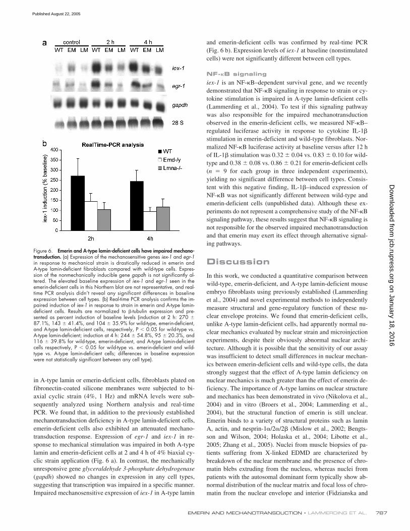
Impaired cellular signaling in response to mechanicalstimulation (mechanotransduction) can lead to an altered phys-iological response and to potentially increased apoptosis in me-chanically strained tissue. A-type lamin-deficient cells haveimpaired mechanotransduction signaling in vivo and in vitro(Lammerding et al., 2004; Nikolova et al., 2004), and insuffi-cient anti-apoptotic signaling could provide one explanationfor the increased apoptotic cell fractions in A-type lamin andemerin-deficient cells seen in the 24-h strain experiments. Inwild-type cells, expression of the mechanosensitive gene
egr-1
and the anti-apoptotic gene iex-1 is rapidly up-regulated in re-sponse to mechanical stimulation (Sadoshima et al., 1992;Morawietz et al., 1999; De Keulenaer et al., 2002). To evaluatewhether transcriptional activation for these genes was altered
Figure 5. Emerin and A-type lamin-deficient cells are more sensitive tomechanical strain. (a) A-type lamin-deficient cells have a significantly in-creased number of propidium iodide–positive cells after 24 h of cyclic,10% biaxial strain application. Emerin-deficient cells are not significantlydifferent from wild-type cells. (Percentage of propidium iodide–positivecells at rest vs. after 24 h strain application 0.13 � 0.03% vs. 0.38 �0.12%, 0.2 � 0.00% vs. 0.37 � 0.04%, and 0.68 � 0.18% vs. 2.94 �0.47% for wild-type, emerin-deficient, and A-type lamin-deficient cells re-spectively, P � 0.01 for wild-type vs. A-type lamin-deficient strained cells;P � 0.05 for wild-type vs. A-type lamin-deficient controls). (b) Emerin andA-type lamin-deficient cells have increased fractions of apoptotic cells after24 h of cyclic, 10% biaxial strain application (percentage of apoptoticcells at rest vs. after 24 h of strain application 0.37 � 0.05% vs. 0.30 �0.04% for wild-type, 0.43 � 0.02% vs. 0.70 � 0.06% for emerin-defi-cient, and 0.72 � 0.05% vs. 1.04 � 0.11% for A-type lamin-deficientcells; P � 0.001 for strained emerin and A-type lamin-deficient vs. wild-type cells).
on January 18, 2016jcb.rupress.org
Dow
nloaded from
Published August 22, 2005
EMERIN AND MECHANOTRANSDUCTION • LAMMERDING ET AL. 787
in A-type lamin or emerin-deficient cells, fibroblasts plated onfibronectin-coated silicone membranes were subjected to bi-axial cyclic strain (4%, 1 Hz) and mRNA levels were sub-sequently analyzed using Northern analysis and real-timePCR. We found that, in addition to the previously establishedmechanotransduction deficiency in A-type lamin-deficient cells,emerin-deficient cells also exhibited an attenuated mechano-transduction response. Expression of egr-1 and iex-1 in re-sponse to mechanical stimulation was impaired in both A-typelamin and emerin-deficient cells at 2 and 4 h of 4% biaxial cy-clic strain application (Fig. 6 a). In contrast, the mechanicallyunresponsive gene glyceraldehyde 3-phosphate dehydrogenase(gapdh) showed no changes in expression in any cell types,suggesting that transcription was impaired in a specific manner.Impaired mechanosensitive expression of iex-1 in A-type lamin
and emerin-deficient cells was confirmed by real-time PCR(Fig. 6 b). Expression levels of iex-1 at baseline (nonstimulatedcells) were not significantly different between cell types.
NF-�B signalingiex-1 is an NF-B–dependent survival gene, and we recentlydemonstrated that NF-B signaling in response to strain or cy-tokine stimulation is impaired in A-type lamin-deficient cells(Lammerding et al., 2004). To test if this signaling pathwaywas also responsible for the impaired mechanotransductionobserved in the emerin-deficient cells, we measured NF-B–regulated luciferase activity in response to cytokine IL-1�
stimulation in emerin-deficient and wild-type fibroblasts. Nor-malized NF-B luciferase activity at baseline versus after 12 hof IL-1� stimulation was 0.32 � 0.04 vs. 0.83 � 0.10 for wild-type and 0.38 � 0.08 vs. 0.86 � 0.21 for emerin-deficient cells(n 9 for each group in three independent experiments),yielding no significant difference between cell types. Consis-tent with this negative finding, IL-1�–induced expression ofNF-B was not significantly different between wild-type andemerin-deficient cells (unpublished data). Although these ex-periments do not represent a comprehensive study of the NF-Bsignaling pathway, these results suggest that NF-B signaling isnot responsible for the observed impaired mechanotransductionand that emerin may exert its effect through alternative signal-ing pathways.
DiscussionIn this work, we conducted a quantitative comparison betweenwild-type, emerin-deficient, and A-type lamin-deficient mouseembryo fibroblasts using previously established (Lammerdinget al., 2004) and novel experimental methods to independentlymeasure structural and gene-regulatory function of these nu-clear envelope proteins. We found that emerin-deficient cells,unlike A-type lamin-deficient cells, had apparently normal nu-clear mechanics evaluated by nuclear strain and microinjectionexperiments, despite their obviously abnormal nuclear archi-tecture. Although it is possible that the sensitivity of our assaywas insufficient to detect small differences in nuclear mechan-ics between emerin-deficient cells and wild-type cells, the datastrongly suggest that the effect of A-type lamin deficiency onnuclear mechanics is much greater than the effect of emerin de-ficiency. The importance of A-type lamins on nuclear structureand mechanics has been demonstrated in vivo (Nikolova et al.,2004) and in vitro (Broers et al., 2004; Lammerding et al.,2004), but the structural function of emerin is still unclear.Emerin binds to a variety of structural proteins such as laminA, actin, and nesprin-1�/2�/2� (Mislow et al., 2002; Bengts-son and Wilson, 2004; Holaska et al., 2004; Libotte et al.,2005; Zhang et al., 2005). Nuclei from muscle biopsies of pa-tients suffering from X-linked EDMD are characterized bybreakdown of the nuclear membrane and the presence of chro-matin blebs extruding from the nucleus, whereas nuclei frompatients with the autosomal dominant form typically show ab-normal distribution of the nuclear matrix and focal loss of chro-matin from the nuclear envelope and interior (Fidzianska and
Figure 6. Emerin and A-type lamin-deficient cells have impaired mechano-transduction. (a) Expression of the mechanosensitive genes iex-1 and egr-1in response to mechanical strain is drastically reduced in emerin andA-type lamin-deficient fibroblasts compared with wild-type cells. Expres-sion of the nonmechanically inducible gene gapdh is not significantly al-tered. The elevated baseline expression of iex-1 and egr-1 seen in theemerin-deficient cells in this Northern blot are not representative, and real-time PCR analysis didn’t reveal any significant differences in baselineexpression between cell types. (b) Real-time PCR analysis confirms the im-paired induction of iex-1 in response to strain in emerin and A-type lamin-deficient cells. Results are normalized to �-tubulin expression and pre-sented as percent induction of baseline levels (induction at 2 h: 270 �87.1%, 143 � 41.4%, and 104 � 35.9% for wild-type, emerin-deficient,and A-type lamin-deficient cells, respectively, P � 0.05 for wild-type vs.A-type lamin-deficient; induction at 4 h: 244 � 54.8%, 95 � 20.3%, and116 � 39.8% for wild-type, emerin-deficient, and A-type lamin-deficientcells respectively, P � 0.05 for wild-type vs. emerin-deficient and wild-type vs. A-type lamin-deficient cells; differences in baseline expressionwere not statistically significant between any cell type).
on January 18, 2016jcb.rupress.org
Dow
nloaded from
Published August 22, 2005

JCB • VOLUME 170 • NUMBER 5 • 2005788
Hausmanowa-Petrusewicz, 2003). Several nesprin isoforms(nesprin 1�, 2�, 2�) bind to emerin and are necessary to an-chor emerin at the inner nuclear membrane (Libotte et al.,2005; Zhang et al., 2005). Nesprin isoforms containing actin-binding domains are thought to physically connect the nucleusto the cytoskeleton and play an important role in anchoringmuscle nuclei at the neuromuscular junction (Starr and Han,2002, 2003; Padmakumar et al., 2004; Grady et al., 2005;Zhang et al., 2005). Furthermore, emerin can directly bind andstabilize the pointed ends of F-actin in vitro, suggesting thatemerin may stabilize actin polymers at the nuclear envelope(Holaska et al., 2004). At the same time, the nucleoplasmic do-main of emerin has dominant effects in Xenopus nuclear as-sembly extracts and overexpression of the full nucleoplasmicdomain of emerin extends the mammalian cell cycle by 7 h(Fairley et al., 2002), supporting emerin’s involvement in nu-clear assembly and cell cycle regulation. Our experimentalfindings suggest that loss of emerin does not directly affectlarge scale nuclear mechanics such as overall nuclear stiffnessand fragility. Instead, emerin could predominantly affect nu-clear envelope assembly and organization at a smaller lengthscale that is not apparent in global measurements of nuclearstrain and rupture. This dynamic and local effect is supportedby the more variable nuclear shape of emerin-deficient cellsobserved in our time-lapse sequences.
In contrast to the apparently normal nuclear mechanics,mechanosensitive gene regulation was deficient in emerin-deficient cells, resulting in a significantly increased rate ofapoptosis in response to mechanical stimulation. The absenceof changes in total cell death (propidium iodide uptake) inthese cells could be due to the fact that the DNA contents assaymeasures early apoptotic chromatin breakdown while loss ofplasma membrane integrity that is required for propidium io-dide uptake occurs at a much later stage in apoptosis. Consistentwith the apparently normal nuclear mechanics and the impairedactivation of anti-apoptotic genes in response to mechanicalstimulation, our findings indicate that emerin-deficient cells aremore susceptible to strain-induced apoptosis, whereas both ne-crosis and apoptosis contribute to mechanically induced celldeath in A-type lamin-deficient cells. Although the currentstudy was narrowed to two representative mechanosensitivegenes (egr-1 and iex-1) whose activation has previously beenreported to be impaired in A-type lamin-deficient cells (Lam-merding et al., 2004), we expect that the role of emerin (andA-type lamins) is not limited to these particular genes, and amore comprehensive gene array analysis of mechanically stim-ulated cells will provide more insight into the extent of lamin/emerin-dependent mechanotransduction.
The molecular mechanism that is responsible for the ob-served mechanotransduction deficiency in A-type lamin andemerin-deficient cells is not clear. We previously reported im-paired NF-B–regulated transcriptional activation in A-typelamin-deficient cells, but emerin-deficient cells had apparentlynormal NF-B signaling, indicating that alternative pathwaysare responsible for the observed mechanotransduction deficien-cies. Emerin directly binds to the transcriptional regulatorsBAF, Btf, GCL, and YT521-B (Nili et al., 2001; Holaska et al.,
2003; Wilkinson et al., 2003; Haraguchi et al., 2004). Btf is atranscription repressor that induces cell death when overex-pressed (Kasof et al., 1999). Emerin is cleaved during apoptosisin proliferating mouse myoblasts and differentiating myotubes,and emerin may have an anti-apoptotic effect by sequesteringBtf (Columbaro et al., 2001). This anti-apoptotic role of emerinis consistent with the increased rate of apoptosis observed inemerin-deficient cells after strain application. GCL is a repres-sor of E2F-DP–regulated genes. Emerin cannot bind BAF andGCL simultaneously, and presence of BAF at the nuclear enve-lope might inhibit GCL binding to emerin in vivo (Bengtssonand Wilson, 2004). YT521-B is involved in determining sitesfor alternate mRNA splicing, and emerin influences splice siteselection by YT521-B (Wilkinson et al., 2003). The role ofemerin as a modulator for transcriptional regulation is furthersupported by DNA microarray analysis of fibroblasts from pa-tients with the X-linked form of EDMD, which showed that atleast 28 genes are specifically up-regulated in the emerin mu-tant cells (Tsukahara et al., 2002). The affected genes includeboth structural (lamin A/C, �II-spectrin, and filamin) andsignal transduction–associated proteins such as protein phos-phatase 2A (Tsukahara et al., 2002).
In addition to these direct interactions between emerinand transcriptional regulators, we cannot exclude the possibil-ity that the impaired mechanotransduction response is causedby more indirect consequences of emerin deficiency. Abnormalnuclear shape and ultrastructure in emerin-deficient cells couldaffect force transmission from the cytoskeleton to the nucleus(Maniotis et al., 1997), and interaction of emerin with nuclearactin (Holaska et al., 2004) could affect both nuclear ultrastruc-ture as well as transcription itself, as nuclear actin is emergingas a critical component of polymerase II transcription (Hof-mann et al., 2004; Zhu et al., 2004).
In our experiments, emerin-deficient cells generally dis-played a milder phenotype compared with the A-type lamin-deficient cells, including fewer and less extensive nuclear shapeabnormalities, a less profound increase in apoptosis in responseto strain, and less severe mechanotransduction deficiency. Thisobservation is consistent with the milder phenotype found inemerin-deficient mice that don’t display overt muscular dystro-phy or cardiac problems but show signs of impaired muscle re-generation later on (unpublished data). Loss of function in laminA/C mutants often leads to mislocalization of emerin from thenuclear envelope to the ER (Sullivan et al., 1999; Östlund et al.,2001; Raharjo et al., 2001; Muchir et al., 2003) and to a subse-quent loss of normal emerin function at the nuclear envelope.Therefore, we expect that A-type lamin-deficient cells encom-pass phenotypes associated with emerin deficiency or loss offunction. Loss of emerin, on the other hand, does not appear togrossly affect A-type lamin function, providing a possible ex-planation for the more severe phenotype in the A-type lamin-deficient cells compared with the emerin-deficient cells.
In conclusion, we found that A-type lamin and emerindeficiencies share common features such as abnormal nuclearshape and impaired mechanotransduction, but also selec-tively interfere with other structural and gene-regulatoryfunctions. In the case of emerin deficiency, we found that
on January 18, 2016jcb.rupress.org
Dow
nloaded from
Published August 22, 2005

EMERIN AND MECHANOTRANSDUCTION • LAMMERDING ET AL. 789
emerin predominantly affects mechanosensitive gene regula-tion with only small effects on nuclear mechanics. By provid-ing independent tests for measuring structural and gene-regu-latory functions, our experiments can help clarify the effectsof specific mutations in nuclear envelope proteins. Elucidat-ing the molecular mechanisms will provide new insights intothe specific disease mechanisms of X-linked recessive andautosomal dominant EDMD, and might eventually lead tonew treatment courses.
Materials and methodsCellsEmerin-deficient (Emd�/� and Emd��y) mouse embryo and muscle fibro-blasts were derived from Emd null mice. Emd null mice were derived bydeleting the entire coding region for the Emd gene, using standard genetargeting procedures in mouse ES cells (Joyner, 1999). Lmna�/�, Lmna�/�,and Emd�/y mouse embryo fibroblasts were maintained in DME (Invitro-gen) containing 10% FCS (HyClone) and penicillin/streptomycin (Invitro-gen). Experiments were performed on primary mouse embryo fibroblastsand on cells immortalized by repeated passage of primary cells. Unlessotherwise specified, cells referred to in the text are immortalized mouseembryo fibroblasts.
Nuclear shape analysisTo quantify the overall fraction of irregularly shaped or blebbing nuclei,cells of various passages were incubated for 15 min with 1 �g/mlHoechst 33342 (Molecular Probes) and washed with HBSS (Invitrogen).For each passage, fluorescence images of 100–500 randomly selectednuclei were acquired at 20� on a microscope (model IX-70; Olympus) us-ing a CoolSNAP camera (Roper Scientific), and were stored for subse-quent image analysis. Nuclei of Lmna�/�, Lmna�/�, and Emd-/y cells werescored as normal, irregularly shaped (deviation from an oval or sphericalshape), or as nuclei with nuclear blebs. These determinations were madeusing an observer blinded for the genotype. To quantify the variation innuclear morphology, we measured nuclear area and perimeter in midsec-tions of the fluorescent nuclei using custom-written MATLAB software. Fromthese data, we computed the nuclear roundness or contour ratio (4� �area/perimeter2) (Goldman et al., 2004). The contour ratio reaches amaximum value of 1 for a circle and decreases with increasingly convo-luted nuclear shapes. Elliptic Fourier analysis of nuclear shape was per-formed as described previously (Diaz et al., 1989). Custom-written MAT-LAB software was used to automatically trace the outline of fluorescentlylabeled nuclei and to compute the first 20 elliptic harmonics. Each singleelliptic harmonic can be geometrically visualized as a pair of orthogonalsemiaxes, and we used the ratio of the sum of the first major and minorsemiaxes over the sum of the higher order semiaxes as indicator of irregu-lar nuclear shape, as the higher order harmonics represent deviationsfrom a purely elliptical shape.
Time-lapse experimentsCells were plated at subconfluent density in 35-mm polystyrene cell culturedishes (Corning). Cells were grown in DME � 10% FCS at 37 C for atleast 24 h before the start of the experiments. Subsequently, lidded culturedishes were sealed with parafilm M (American National Can) and placedat RT on the microscope stage (model IX-70; Olympus). After a brief equil-ibration period, images were automatically acquired every 5 min for aminimum of 8 h, 20 min (corresponding to 100 frames) using a digitalCCD camera (CoolSNAP HQ; Roper Scientific) and were stored on acomputer for subsequent analysis. Continued cell viability was confirmedby monitoring the cells for 24 h after the experiments. Nuclear rotation,translation, and shape changes were analyzed by tracking the centroidpositions of 3–6 nucleoli for each nucleus using custom-written MATLABsoftware. For each frame, the linear conformal image transformation wascomputed that best mapped the current centroid positions to the originalpositions minimizing the least-square error. The linear conformal trans-formations can account for a combination of translation, rotation, andscaling, and preserves the relative position of objects to each other. Thedeviation from the best fit, i.e., the error between the least-square fit trans-formation and the actual nucleoli positions, was used as a measure of nu-clear deformation as it describes the extent of nuclear deformation fromits initial shape independent of absolute nuclear movement or uniform
changes in size (see time-lapse videos, available at http://www.jcb.org/cgi/content/full/jcb.200502148/DC1). Nucleoli that fused over time orcells that underwent mitosis during or immediately after the observationperiod were excluded from the analysis. We analyzed 25 nuclei of eachcell type and computed for each nucleus the time-averaged deformation,the time-averaged and maximal change in size, translation, and rotation.
Nuclear strain experimentsExperiments were performed as previously described (Lammerding et al.,2004). In brief, cells were plated at subconfluent cell density on fibronec-tin-coated silicone membranes in DME supplemented with 10% FCS, fol-lowed by serum starvation for 48 h in DME containing ITS supplement(Sigma-Aldrich). Preceding the strain experiments, cells were incubatedwith Hoechst 33342 nuclear stain (final concentration 1 �g/ml; Molecu-lar Probes) in DME � ITS for 20 min. Membranes were placed on a cus-tom-made strain device and uniform biaxial strain was applied to the sili-cone membrane. Membrane and nuclear strains were computed based onbright field and fluorescence images acquired before, during, and afterstrain application using a custom-written image analysis algorithm. Nor-malized nuclear strain was defined as the ratio of nuclear strain to mem-brane strain to compensate for small variations in applied membranestrain (range 4.5–5.5%).
Microinjection experimentsCells were plated on fibronectin-coated glass dishes (WillCo Wells) and in-cubated overnight. Microinjections were performed using a microinjector(Eppendorf) with Femtotips (Eppendorf). In each dish, 30–50 cells were in-jected with Texas red–labeled 70-kD dextran (dissolved at 10 mg/ml inPBS; Molecular Probes) into the cytoplasm or into the nucleus (injection pres-sure 500 hPa; injection time 0.6 s). After the microinjection, cells werewashed with HBSS (Invitrogen) and intracellular localization of dextran–Texas red was recorded under a fluorescent microscope using a CCD cam-era (CoolSNAP HQ; Roper Scientific) on a microscope (model IX-70; Olym-pus). Cells were considered as having defective nuclear envelope integrityin the case of uniform nuclear and cytoplasmic staining, and the fraction ofdefective nuclei was expressed as the ratio of total fluorescent cells.
Image acquisition and manipulationPhase-contrast and fluorescence images were acquired as describedabove using a digital CCD-camera (CoolSNAP HQ; Roper Scientific)mounted on an inverted microscope (model IX-70; Olympus) using ImageProimage acquisition software (Media Cybernetics). Nuclear shape experi-ments, time-lapse studies, and microinjected cells were imaged using anOlympus LCPlanF 20� phase-contrast objective (NA 0.40), whereas nu-clear strain experiments were conducted using an Olympus UApo/34040� water immersion objective (NA 1.15). Cells were kept in HBSS�buffer during imaging, except for time-lapse experiments that were per-formed on cells in full media. All experiments were performed at RT. Ra-diographs from Northern analysis were digitized on a scanner (Perfection2450; Epson) using linear intensity settings. Digital images were pro-cessed using Adobe Photoshop (ver. 6.0) by adjusting the linear image in-tensity display range, and fluorescence grayscale images were colorizedin Adobe Photoshop by selecting a colorplane (RGB) appropriate for thechromophore (i.e., blue for Hoechst 33342, red for Texas red).
Mechanotransduction experimentsStrain stimulation was performed as described previously (Cheng et al.,1997; Lammerding et al., 2004). In brief, cells were plated on fibronectin-coated silicone membranes (�3,000 cells/cm2). After 72 h serum starva-tion, cells were subjected to bi-axial cyclic strain (4%, 1 Hz) for 2 or 4 h.For chemical stimulation, cells were incubated with IL-1� (10 ng/ml; R&DSystems) or PMA (200 ng/ml; Sigma-Aldrich) in DME � ITS. CellularmRNA of strained and unstrained control samples was isolated using theRNeasy Minikit (QIAGEN), and samples were subsequently analyzed byNorthern analysis and real-time PCR.
Cell viability and apoptosis assayCells were plated on fibronectin-coated silicone membranes and main-tained in full media for 48 h to provide sufficient cell attachment. After 24 hof cyclic biaxial strain application (1 Hz; 3, 5, or 10% strain), cells wereincubated with propidium iodide (PI, final concentration 1 �g/ml; Sigma-Aldrich). Cells and culture media were collected, washed once in PBS�and resuspend in HBSS�. Each sample was divided into two equal partsto independently measure cell death (PI uptake) and apoptosis (DNA con-tent analysis). One part was immediately analyzed for PI uptake using aCytomics FC 500 flow cytometer (Beckman Coulter), counting 10,000–
on January 18, 2016jcb.rupress.org
Dow
nloaded from
Published August 22, 2005
JCB • VOLUME 170 • NUMBER 5 • 2005790
30,000 events in each group. Thresholds for PI incorporation were deter-mined based on negative (no PI staining) and positive (cells permeabilizedby 50% ethanol) controls. The other cell fraction from the 24-h strain ex-periments was fixed in 80% ethanol and stored at �20 C. Samples werethen spun down, resuspended in PBS� and treated with Ribonuclease A(Sigma-Aldrich) for 1 h and subsequently stained with PI (final concentra-tion 100 �g/ml). DNA content was measured on a flow cytometer andthe apoptotic cell fraction was identified as cells with sub-G1 DNA con-tent, counting 30,000–50,000 events per sample as described previously(Walker et al., 1993).
Northern analysisFor Northern analysis of iex-1, egr-1, and GAPDH, mRNA from unstrainedcontrols and from cells subjected to 2 and 4 h of biaxial cyclic strain washarvested using RNAeasy Mini Kit kit (QIAGEN). 7–12 �g of each col-lected RNA sample were separated by gel electrophoresis at 110–130 Vand mRNA was transferred overnight to a transfer membrane (MAGNA,Nylon, 0.45 micron; Osmonics, Inc.). Expression of iex-,1egr-1, andGAPDH mRNA was assessed by Northern analysis as described previ-ously (De Keulenaer et al., 2002).
Real-time polymerase chain reactionFor further analysis of iex-1 expression, cells were prepared for strain ex-periments with 2 and 4 h biaxial strain. Cells were harvested using theQIAGEN RNAeasy kit. For each sample, 1 �l of collected RNA wasadded to RT Mixes of Stratagene Light Cycler kit with iex-1 and �-tubulinprimers from Integrated DNA Technologies, Inc. (De Keulenaer et al.,2002). The polymerase reaction was conducted in Roche Molecular Bio-chemicals Light Cycler Version 5.32 with 45 cycles. Results were normal-ized with �-tubulin expression and expressed as percent increase frombaseline (unstrained controls).
Luciferase experimentsCells were transfected with plasmids for NF-B–controlled luciferase ex-pression and SV40-regulated �-galactosidase (Promega) using FuGENE6(Roche). After transfection, cells were serum starved in DME � ITS mediumfor 48 h, followed by overnight stimulation with 200 ng/ml PMA or 10ng/ml IL-1�. Luciferase assays were quantified in a Victor2 MultilabelCounter (PerkinElmer). Results were normalized for �-galactosidase activityand expressed as percentage of wild-type control.
Statistical analysisAll experiments were performed at least three independent times. Dataare expressed as mean � SEM. Statistical analysis was performed usingthe PRISM 3.0 and INSTAT software (GraphPad). The data were ana-lyzed by unpaired t test (allowing different SD), one-way ANOVA (fol-lowed by Tukey’s multiple comparison test) or, in case of non-Gaussiandistribution, the Mann-Whitney or Kruskal Wallis tests (the latter whencomparing more than two groups, followed by Dunn’s multiple compari-son test). For nuclear shape analysis, two-way ANOVA was performedon datasets from three different passages to evaluate the source of varia-tion. In contrast to cell type, passage number was not a significantsource of variation, and thus datasets from different passages werepooled and analyzed using the Kruskal-Wallis test for non-Gaussian dis-tributions with Dunn’s post test. Real-time PCR data were analyzed usinga paired test and comparing induction of iex-1 (as percentage of base-line levels) at 2 and 4 h to wild-type induction. A two-tailed P-value of�0.05 was considered significant.
Online supplemental materialFull-length time-lapse sequences of A-type lamin-deficient, emerin-deficient,and wild-type mouse embryo fibroblasts corresponding to the still imagespresented in Fig. 3 can be found in the supplemental material. Each videoconsists of a sequence of images acquired every 5 min over an 8 h, 20min period for a total of 100 frames. Online supplemental material avail-able at http://www.jcb.org/cgi/content/full/jcb.200502148/DC1.
The authors would like to thank Julie Ji for her help in the nuclear morphologyanalysis. Microinjection was performed at the W.M. Keck Microscopy Facil-ity at the Whitehead Institute.
This work was supported by grants from National Heart, Lung, andBlood Institute (HL073809, HL64858) and a post-doctoral fellowship fromthe American Heart Association for J. Lammerding.
Submitted: 24 February 2005Accepted: 25 July 2005
ReferencesBengtsson, L., and K.L. Wilson. 2004. Multiple and surprising new functions for
emerin, a nuclear membrane protein. Curr. Opin. Cell Biol. 16:73–79.
Bione, S., E. Maestrini, S. Rivella, M. Mancini, S. Regis, G. Romeo, and D.Toniolo. 1994. Identification of a novel X-linked gene responsible forEmery-Dreifuss muscular dystrophy. Nat. Genet. 8:323–327.
Bonne, G., M.R. Di Barletta, S. Varnous, H.M. Becane, E.H. Hammouda, L.Merlini, F. Muntoni, C.R. Greenberg, F. Gary, J.A. Urtizberea, et al.1999. Mutations in the gene encoding lamin A/C cause autosomal domi-nant Emery-Dreifuss muscular dystrophy. Nat. Genet. 21:285–288.
Broers, J.L., E.A. Peeters, H.J. Kuijpers, J. Endert, C.V. Bouten, C.W. Oomens,F.P. Baaijens, and F.C. Ramaekers. 2004. Decreased mechanical stiff-ness in LMNA�/� cells is caused by defective nucleo-cytoskeletal in-tegrity: implications for the development of laminopathies. Hum. Mol.Genet. 13:2567–2580.
Burke, B., and C.L. Stewart. 2002. Life at the edge: the nuclear envelope andhuman disease. Nat. Rev. Mol. Cell Biol. 3:575–585.
Caille, N., Y. Tardy, and J.J. Meister. 1998. Assessment of strain field in endo-thelial cells subjected to uniaxial deformation of their substrate. Ann.Biomed. Eng. 26:409–416.
Cao, H., and R.A. Hegele. 2000. Nuclear lamin A/C R482Q mutation in Cana-dian kindreds with Dunnigan-type familial partial lipodystrophy. Hum.Mol. Genet. 9:109–112.
Cheng, G.C., W.H. Briggs, D.S. Gerson, P. Libby, A.J. Grodzinsky, M.L. Gray,and R.T. Lee. 1997. Mechanical strain tightly controls fibroblast growthfactor-2 release from cultured human vascular smooth muscle cells.Circ. Res. 80:28–36.
Columbaro, M., E. Mattioli, G. Lattanzi, C. Rutigliano, A. Ognibene, N.M. Ma-raldi, and S. Squarzoni. 2001. Staurosporine treatment and serum starva-tion promote the cleavage of emerin in cultured mouse myoblasts: in-volvement of a caspase-dependent mechanism. FEBS Lett. 509:423–429.
De Keulenaer, G.W., Y. Wang, Y. Feng, S. Muangman, K. Yamamoto, J.F.Thompson, T.G. Turi, K. Landschutz, and R.T. Lee. 2002. Identificationof IEX-1 as a biomechanically controlled nuclear factor-B target genethat inhibits cardiomyocyte hypertrophy. Circ. Res. 90:690–696.
De Sandre-Giovannoli, A., M. Chaouch, S. Kozlov, J.M. Vallat, M. Tazir, N.Kassouri, P. Szepetowski, T. Hammadouche, A. Vandenberghe, C.L.Stewart, et al. 2002. Homozygous defects in LMNA, encoding laminA/C nuclear-envelope proteins, cause autosomal recessive axonal neu-ropathy in human (Charcot-Marie-Tooth disorder type 2) and mouse.Am. J. Hum. Genet. 70:726–736.
De Sandre-Giovannoli, A., R. Bernard, P. Cau, C. Navarro, J. Amiel, I. Boc-caccio, S. Lyonnet, C.L. Stewart, A. Munnich, M. Le Merrer, and N.Levy. 2003. Lamin A truncation in Hutchinson-Gilford progeria. Science.300:2055.
Diaz, G., A. Zuccarelli, I. Pelligra, and A. Ghiani. 1989. Elliptic fourier analysisof cell and nuclear shapes. Comput. Biomed. Res. 22:405–414.
Ellis, J.A., M. Craxton, J.R. Yates, and J. Kendrick-Jones. 1998. Aberrant intra-cellular targeting and cell cycle-dependent phosphorylation of emerincontribute to the Emery-Dreifuss muscular dystrophy phenotype. J. CellSci. 111:781–792.
Eriksson, M., W.T. Brown, L.B. Gordon, M.W. Glynn, J. Singer, L. Scott,M.R. Erdos, C.M. Robbins, T.Y. Moses, P. Berglund, et al. 2003. Re-current de novo point mutations in lamin A cause Hutchinson-Gilfordprogeria syndrome. Nature. 423:293–298.
Fairley, E.A., A. Riddell, J.A. Ellis, and J. Kendrick-Jones. 2002. The cell cycledependent mislocalisation of emerin may contribute to the Emery-Dreifussmuscular dystrophy phenotype. J. Cell Sci. 115:341–354.
Fatkin, D., C. MacRae, T. Sasaki, M.R. Wolff, M. Porcu, M. Frenneaux, J.Atherton, H.J. Vidaillet Jr., S. Spudich, U. De Girolami, et al. 1999. Mis-sense mutations in the rod domain of the lamin A/C gene as causes of di-lated cardiomyopathy and conduction-system disease. N. Engl. J. Med.341:1715–1724.
Fidzianska, A., and I. Hausmanowa-Petrusewicz. 2003. Architectural abnormal-ities in muscle nuclei. Ultrastructural differences between X-linked andautosomal dominant forms of EDMD. J. Neurol. Sci. 210:47–51.
Fidzianska, A., D. Toniolo, and I. Hausmanowa-Petrusewicz. 1998. Ultrastruc-tural abnormality of sarcolemmal nuclei in Emery-Dreifuss musculardystrophy (EDMD). J. Neurol. Sci. 159:88–93.
Goldman, R.D., D.K. Shumaker, M.R. Erdos, M. Eriksson, A.E. Goldman, L.B.Gordon, Y. Gruenbaum, S. Khuon, M. Mendez, R. Varga, and F.S. Col-lins. 2004. Accumulation of mutant lamin A causes progressive changesin nuclear architecture in Hutchinson-Gilford progeria syndrome. Proc.Natl. Acad. Sci. USA. 101:8963–8968.
Grady, R.M., D.A. Starr, G.L. Ackerman, J.R. Sanes, and M. Han. 2005. Syneproteins anchor muscle nuclei at the neuromuscular junction. Proc. Natl.Acad. Sci. USA. 102:4359–4364.
on January 18, 2016jcb.rupress.org
Dow
nloaded from
Published August 22, 2005
EMERIN AND MECHANOTRANSDUCTION • LAMMERDING ET AL. 791
Haraguchi, T., J.M. Holaska, M. Yamane, T. Koujin, N. Hashiguchi, C. Mori,K.L. Wilson, and Y. Hiraoka. 2004. Emerin binding to Btf, a death-promoting transcriptional repressor, is disrupted by a missense muta-tion that causes Emery-Dreifuss muscular dystrophy. Eur. J. Biochem.271:1035–1045.
Hofmann, W.A., L. Stojiljkovic, B. Fuchsova, G.M. Vargas, E. Mavrommatis,V. Philimonenko, K. Kysela, J.A. Goodrich, J.L. Lessard, T.J. Hope, etal. 2004. Actin is part of pre-initiation complexes and is necessary fortranscription by RNA polymerase II. Nat. Cell Biol. 6:1094–1101.
Holaska, J.M., K.K. Lee, A.K. Kowalski, and K.L. Wilson. 2003. Transcrip-tional repressor germ cell-less (GCL) and barrier to autointegrationfactor (BAF) compete for binding to emerin in vitro. J. Biol. Chem.278:6969–6975.
Holaska, J.M., A.K. Kowalski, and K.L. Wilson. 2004. Emerin caps the pointedend of actin filaments: evidence for an actin cortical network at the nu-clear inner membrane. PLoS Biol. 2:E231.
Joyner, A. 1999. Gene Targeting: A Practical Approach. Oxford UniversityPress, Oxford, UK. 312 pp.
Kasof, G.M., L. Goyal, and E. White. 1999. Btf, a novel death-promoting tran-scriptional repressor that interacts with Bcl-2-related proteins. Mol. Cell.Biol. 19:4390–4404.
Lammerding, J., P.C. Schulze, T. Takahashi, S. Kozlov, T. Sullivan, R.D.Kamm, C.L. Stewart, and R.T. Lee. 2004. Lamin A/C deficiency causesdefective nuclear mechanics and mechanotransduction. J. Clin. Invest.113:370–378.
Libotte, T., H. Zaim, S. Abraham, V.C. Padmakumar, M. Schneider, W. Lu, M.Munck, C. Hutchison, M. Wehnert, B. Fahrenkrog, et al. 2005. LaminA/C dependent localization of nesprin-2, a giant scaffolder at the nuclearenvelope. Mol Biol Cell. 16:3411–3424
Liu, J., T.R. Ben-Shahar, D. Riemer, M. Treinin, P. Spann, K. Weber, A. Fire,and Y. Gruenbaum. 2000. Essential roles for Caenorhabditis eleganslamin gene in nuclear organization, cell cycle progression, and spatial or-ganization of nuclear pore complexes. Mol. Biol. Cell. 11:3937–3947.
Maniotis, A.J., C.S. Chen, and D.E. Ingber. 1997. Demonstration of mechanicalconnections between integrins, cytoskeletal filaments, and nucleoplasmthat stabilize nuclear structure. Proc. Natl. Acad. Sci. USA. 94:849–854.
Mislow, J.M., J.M. Holaska, M.S. Kim, K.K. Lee, M. Segura-Totten, K.L. Wil-son, and E.M. McNally. 2002. Nesprin-1� self-associates and binds di-rectly to emerin and lamin A in vitro. FEBS Lett. 525:135–140.
Morawietz, H., Y.H. Ma, F. Vives, E. Wilson, V.P. Sukhatme, J. Holtz, and H.E.Ives. 1999. Rapid induction and translocation of Egr-1 in response to me-chanical strain in vascular smooth muscle cells. Circ. Res. 84:678–687.
Muchir, A., B.G. van Engelen, M. Lammens, J.M. Mislow, E. McNally, K.Schwartz, and G. Bonne. 2003. Nuclear envelope alterations in fibroblastsfrom LGMD1B patients carrying nonsense Y259X heterozygous or ho-mozygous mutation in lamin A/C gene. Exp. Cell Res. 291:352–362.
Nikolova, V., C. Leimena, A.C. McMahon, J.C. Tan, S. Chandar, D. Jogia, S.H.Kesteven, J. Michalicek, R. Otway, F. Verheyen, et al. 2004. Defects innuclear structure and function promote dilated cardiomyopathy in laminA/C-deficient mice. J. Clin. Invest. 113:357–369.
Nili, E., G.S. Cojocaru, Y. Kalma, D. Ginsberg, N.G. Copeland, D.J. Gilbert,N.A. Jenkins, R. Berger, S. Shaklai, N. Amariglio, et al. 2001. Nuclearmembrane protein LAP2� mediates transcriptional repression aloneand together with its binding partner GCL (germ-cell-less). J. Cell Sci.114:3297–3307.
Östlund, C., G. Bonne, K. Schwartz, and H.J. Worman. 2001. Properties of laminA mutants found in Emery-Dreifuss muscular dystrophy, cardiomyopathyand Dunnigan-type partial lipodystrophy. J. Cell Sci. 114:4435–4445.
Padmakumar, V.C., S. Abraham, S. Braune, A.A. Noegel, B. Tunggal, I. Kar-akesisoglou, and E. Korenbaum. 2004. Enaptin, a giant actin-bindingprotein, is an element of the nuclear membrane and the actin cytoskeleton.Exp. Cell Res. 295:330–339.
Raharjo, W.H., P. Enarson, T. Sullivan, C.L. Stewart, and B. Burke. 2001. Nu-clear envelope defects associated with LMNA mutations cause dilatedcardiomyopathy and Emery-Dreifuss muscular dystrophy. J. Cell Sci.114:4447–4457.
Sadoshima, J., L. Jahn, T. Takahashi, T.J. Kulik, and S. Izumo. 1992. Molecularcharacterization of the stretch-induced adaptation of cultured cardiaccells. An in vitro model of load-induced cardiac hypertrophy. J. Biol.Chem. 267:10551–10560.
Shackleton, S., D.J. Lloyd, S.N. Jackson, R. Evans, M.F. Niermeijer, B.M.Singh, H. Schmidt, G. Brabant, S. Kumar, P.N. Durrington, et al. 2000.LMNA, encoding lamin A/C, is mutated in partial lipodystrophy. Nat.Genet. 24:153–156.
Starr, D.A., and M. Han. 2002. Role of ANC-1 in tethering nuclei to the actincytoskeleton. Science. 298:406–409.
Starr, D.A., and M. Han. 2003. ANChors away: an actin based mechanism ofnuclear positioning. J. Cell Sci. 116:211–216.
Sullivan, T., D. Escalante-Alcalde, H. Bhatt, M. Anver, N. Bhat, K. Nagashima,C.L. Stewart, and B. Burke. 1999. Loss of A-type lamin expression com-promises nuclear envelope integrity leading to muscular dystrophy. J. CellBiol. 147:913–920.
Tsukahara, T., S. Tsujino, and K. Arahata. 2002. CDNA microarray analysis ofgene expression in fibroblasts of patients with X-linked Emery-Dreifussmuscular dystrophy. Muscle Nerve. 25:898–901.
Vaughan, A., M. Alvarez-Reyes, J.M. Bridger, J.L. Broers, F.C. Ramaekers, M.Wehnert, G.E. Morris, W.G.F. Whitfield, and C.J. Hutchison. 2001.Both emerin and lamin C depend on lamin A for localization at the nu-clear envelope. J. Cell Sci. 114:2577–2590.
Walker, P.R., J. Kwast-Welfeld, H. Gourdeau, J. Leblanc, W. Neugebauer, and M.Sikorska. 1993. Relationship between apoptosis and the cell cycle in lym-phocytes: roles of protein kinase C, tyrosine phosphorylation, and AP1.Exp. Cell Res. 207:142–151.
Wilkinson, F.L., J.M. Holaska, Z. Zhang, A. Sharma, S. Manilal, I. Holt, S.Stamm, K.L. Wilson, and G.E. Morris. 2003. Emerin interacts in vitro withthe splicing-associated factor, YT521-B. Eur. J. Biochem. 270:2459–2466.
Zhang, Q., C.D. Ragnauth, J.N. Skepper, N.F. Worth, D.T. Warren, R.G. Rob-erts, P.L. Weissberg, J.A. Ellis, and C.M. Shanahan. 2005. Nesprin-2 isa multi-isomeric protein that binds lamin and emerin at the nuclear en-velope and forms a subcellular network in skeletal muscle. J. Cell Sci.118:673–687.
Zhu, X., X. Zeng, B. Huang, and S. Hao. 2004. Actin is closely associated withRNA polymerase II and involved in activation of gene transcription. Bio-chem. Biophys. Res. Commun. 321:623–630.
on January 18, 2016jcb.rupress.org
Dow
nloaded from
Published August 22, 2005
Related Documents