DOI: 10.1007/s11085-005-5715-0 Oxidation of Metals, Vol. 64, Nos. 1/2, August 2005 (© 2005) Abnormal High Growth Rates of Metastable Aluminas on FeCrAl Alloys H. El Kadiri, ∗ †‡ R. Molins, ∗ Y. Bienvenu, ∗ and M. F. Horstemeyer† Received July 21, 2004; revised March 22, 2005 Experimental evidence in high temperature oxidation of alumina-forming alloys has accumulated that the overall growth kinetics of the oxide scale are slower for 1000 ◦ C, where the stable α-Al 2 O 3 phase predominates, than for 900 ◦ C where metastable γ -Al 2 O 3 and/or θ -Al 2 O 3 polymorphs predomi- nate. This intriguing behaviour has been unanimously related to the substan- tial presence of twin boundaries and the cation vacancy network intrinsic to the metastable aluminas allowing faster diffusion than in the nearly close packed corundum structure. This paper shows that this abnormal growth rate accom- panying the presence of stable alumina polymorphs in platelets or needle-like morphology is rather due to the formation of a corundum-alumina-rich com- pact layer from an outer metastable layer by the concomitant sintering at the intersection vertices of the platelets and secondary recrystallization in these platelets. These phenomena are illustrated from oxidation tests performed on thin FeCrAl foils in both a conventional muffle furnace (designed by AET) and thermogravimetric analysis furnace (TGA) over the temperature range of 800– 1300 ◦ C using field emission scanning electron microscope (FEG-SEM), trans- mission electron microscope (TEM), electron probe microanalysis (EPMA), atomic force microscope (AFM), grazing incidence X-ray diffraction (GIXRD) and image analysis (IA) techniques. KEY WORDS: kinetics; platelet nucleation; metastable alumina; transformation; sintering; grain growth; platelet thickening. ∗ Ecole des Mines de Paris, Centre des Mat´ eriaux, CNRS UMR 7633, BP 87, 91003 Evry, France. † Center for Advanced Vehicular Systems, Mississippi State University, MS 39762-9627 USA. ‡ To whom correspondence should be sent. e-mail: [email protected] 63 0030-770X/05/0800–0063/0 © 2005 Springer Science+Business Media, Inc.

Welcome message from author
This document is posted to help you gain knowledge. Please leave a comment to let me know what you think about it! Share it to your friends and learn new things together.
Transcript
DOI: 10.1007/s11085-005-5715-0Oxidation of Metals, Vol. 64, Nos. 1/2, August 2005 (© 2005)
Abnormal High Growth Rates of Metastable Aluminason FeCrAl Alloys
H. El Kadiri,∗†‡ R. Molins,∗ Y. Bienvenu,∗ and M. F. Horstemeyer†
Received July 21, 2004; revised March 22, 2005
Experimental evidence in high temperature oxidation of alumina-formingalloys has accumulated that the overall growth kinetics of the oxide scaleare slower for 1000◦C, where the stable α-Al2O3 phase predominates, thanfor 900◦C where metastable γ -Al2O3 and/or θ -Al2O3 polymorphs predomi-nate. This intriguing behaviour has been unanimously related to the substan-tial presence of twin boundaries and the cation vacancy network intrinsic to themetastable aluminas allowing faster diffusion than in the nearly close packedcorundum structure. This paper shows that this abnormal growth rate accom-panying the presence of stable alumina polymorphs in platelets or needle-likemorphology is rather due to the formation of a corundum-alumina-rich com-pact layer from an outer metastable layer by the concomitant sintering at theintersection vertices of the platelets and secondary recrystallization in theseplatelets. These phenomena are illustrated from oxidation tests performed onthin FeCrAl foils in both a conventional muffle furnace (designed by AET) andthermogravimetric analysis furnace (TGA) over the temperature range of 800–1300◦C using field emission scanning electron microscope (FEG-SEM), trans-mission electron microscope (TEM), electron probe microanalysis (EPMA),atomic force microscope (AFM), grazing incidence X-ray diffraction(GIXRD) and image analysis (IA) techniques.
KEY WORDS: kinetics; platelet nucleation; metastable alumina; transformation; sintering;grain growth; platelet thickening.
∗Ecole des Mines de Paris, Centre des Materiaux, CNRS UMR 7633, BP 87, 91003 Evry,France.
†Center for Advanced Vehicular Systems, Mississippi State University, MS 39762-9627 USA.‡To whom correspondence should be sent. e-mail: [email protected]
63
0030-770X/05/0800–0063/0 © 2005 Springer Science+Business Media, Inc.
64 El Kadiri, Molins, Bienvenu, and Horstemeyer
INTRODUCTION
Alumina-forming alloys are now the base materials for many advancedhigh-temperature oxidation coatings in the aeronautical and power gen-eration sectors. For instance, automotive catalytic converters are increas-ingly produced with thin Fe20Cr5Al-RE foil that is an ideal substitutefor ceramics as a material for the carrier of the catalytically active wash-coat. The satisfactory oxidation resistance of FeCrAl-RE alloys in anexhaust gas environment relies on the formation of a continuous andslow growing α-alumina layer over the metal surface. However, it hasbeen widely reported that during the initial stages of oxidation, includ-ing heating up to the oxidation temperature, fast growing metastable poly-morphs of alumina, mainly γ -Al2O3, θ -Al2O3 and δ-Al2O3 with needlesor platelet-like morphology, are formed1 and transformed after furtherheating.2 This transformation is systematically accompanied with reduc-tion in oxide volume leading to tensile stresses and radial cracks det-rimental to the adherence of the final α-alumina layer. Our interest instudying the phase transformation was stimulated by the fact that theoxide morphology beneath the washcoat consists of unstable whiskers orplatelets obtained by an appropriate “preoxidation”. Moreover, in passen-ger cars, catalytic converters frequently operate at temperatures around900◦C.3 In this temperature range, several works showed a discontinu-ous increase in growth rate of two orders of magnitude with increasingtemperature even with materials beyond FeCrAl alloys.4,5 This reactivityanomaly was mostly attributed to the prolonged persistence of θ and γ
phases5 but also to an enhanced precipitation of chromium carbides thatmay penetrate in the alumina scale and react with oxygen.6 The nucle-ation, transformations, and growth kinetics of these transient aluminashave been extensively investigated by a wide range of techniques. Con-ventional transmission electron microscopy (CTEM) and high resolutionelectron microscopy (HREM) using planar or cross-section view specimensallow accurate structural analysis but do not provide enough informationabout the morphological evolutions of the scale that may be importantto understand the kinetic trends. Scanning electron microscopy (SEM) haspreviously been the main tool for morphological analysis of the aluminascales. However, due to the insulating properties of alumina, only limitedresolutions (<1/2 µm) have been reached and these are insufficient whencompared to the thickness of flat plates crystals (<100 nm). Therefore,a large diversity of structural properties of transient alumina has beenreported, but no formal relationship between their growth mechanisms andkinetics has yet been established. This paper investigates different transientoxidation stages at around 900◦C mainly by cross-sectional FEG-SEM
Abnormal High Growth Rates of Metastable Aluminas 65
techniques with an improvement in sample preparation that allows suit-ably high magnifications. A detailed analysis of the kinetics, morphologyand microstructure is addressed with a critical review of the scale growthmechanisms proposed in the literature.
EXPERIMENTAL PROCEDURES
The ArvinMeritor Company supplied a classical Fe20Cr5Al with a lowinterstitial level, “co-doped” with mishmetal (Ce + La), stabilized with Tiand Mn, cold rolled down to 95 µm, and annealed. Quantitative analysisof major and trace elements with EDS and WDS-EPMA performed oncross-sectional specimens polished down to 1/4 µm are presented in TableI in comparison with the chemical composition data provided by the steel-maker.7 The trace element contents are averaged over ten analyzed-windowsof 50×50µm2 squared of surface area.
The mass gain (�m/S) was measured both continuously for up to 100 hrand discontinuously up to 400 hr over the temperature range of 800–1200◦C.For thermogravimetric analyses, specimens in a specific polyhedral geometrywere ultrasonically cleaned in acetone and ethanol, dried, and introduced ina SETARAM thermobalance microstructural observations were carried outby optical microscopy (OM), FEG-SEM and TEM. For TEM observation,analysis and structural identification, oxidized cross-sectional coupons wereprepared from several oxidized strips mounted together with suitable resin.7
The nature of oxide phases was also identified from X-ray diffraction scansin Grazing incidence (1.5◦). Post-tensile tests on oxidized specimens wereuseful in increasing the resolution for FEG-SEM observations. Chemicalanalyses were performed using EPMA techniques.
RESULTS AND DISCUSSION
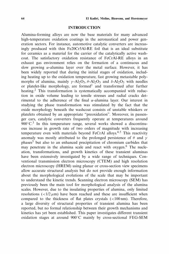
Typical mass gain results obtained in TGA and AET furnace areshown, respectively, in Fig. 1a,b. The general trend and the relativearrangement of these curves are in good agreement with those reportedpreviously in the literature.3,5 At extreme temperatures (800◦C or 1300◦C),parabolic kinetics are observed, and the oxidation process seems tobe thermally activated. However, for both furnaces, exposure at 900◦C
Table I. Chemical Composition in Weight %
Fe Cr Al C+N Si Mn Ti P S Ce La
Sandvik bal. 20.3 5.5 0.016 0.22 0.28 0.014 <0.0005 0.043EPMA 73.3 20.5 5.18 0.19 0.267 0.0070 0.015 0.0028 0.310 0.015
66 El Kadiri, Molins, Bienvenu, and Horstemeyer
0
0,2
0,4
0,6
0,8
1
1,2
1,4
0 20 40 60 80
Time (hr)
∆m/S(mg/cm2)
800°C
1000°C
950°C
900°C
1200°C
1300°C
100
950°C
0
0,4
0,8
1,2
1,6
2
0 100 200 300 400
Time (r)
∆m/S (mg.cm-2
)
1100°C
800°C
1000°C
900°C
1200°C1300°C
(a)
(b)
Fig. 1. Weight gain vs. time in (a) TGA apparatus and in (b) AET fur-nace using continuous measurement with several samples having the samedimensions.
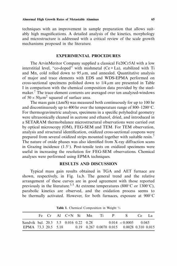
produces weight gain, which becomes permanently higher than thatproduced at 950 and 1000◦C after a critical value of a few hours (Fig. 2).Moreover, in synthetic air, the weight gain at 950◦C remains slightlygreater than that at 1000◦C when the oxidation duration exceeds the crit-ical value. For 1000 and 950◦C, short and fast transient oxidation occursat the beginning of the tests, and the mass change resulting from transientoxidation is larger than the mass change associated with the following par-abolic kinetics. The oxidation behavior at these two temperatures in both
Abnormal High Growth Rates of Metastable Aluminas 67
0
0.2
0.4
0.6
0.8
1
1.2
1.4
1.6
1.8
750 800 850 900 950 1000 1050 1100 1150 1200 1250 1300 1350
Temperature (C)
∆m/S (mg/cm-2)20hr60hr120hr
Weight gain pics
Fig. 2. Weight gain vs. temperature for different oxidation durations in a conven-tional muffle furnace. Discontinuous increase phenomenon at 900◦C persisting upto the steady stage of oxidation.
Fig. 3. Arrhenius diagram for a NiAl according to Brummand Grabke.
68 El Kadiri, Molins, Bienvenu, and Horstemeyer
conventional muffle furnace (AET) and TGA furnaces is discussed in adifferent paper.8
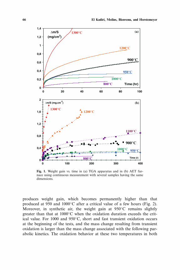
In order to derive values of activation energy for each growing alu-mina phase, most authors divided the mass weight temperature-curves intotwo or more stages.1,5,4,9,10 For each stage, an average parabolic rate con-stant K
averagep is calculated. The plot of the K
averagep values in an Ar-
rhenius diagram gives three separate straight lines that can be correlatedto three phases of alumina (Fig. 3). Fast growing γ -Al2O3 and θ -Al2O3are assigned respectively to K
averagep values calculated for temperatures
below 900◦C, and Kaveragep1 values for temperatures between about 950 and
1000◦C at the initial stages of oxidation. The final values of Kaveragep at
these temperatures give a common line together with those of high tem-peratures (>1100◦C) that are associated with the slow growing α-Al2O3.Nevertheless, the approximation of the K
averagep1 values depends on the cho-
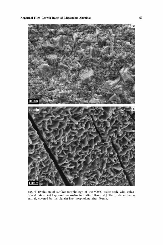
sen transition period leading to a significant scattering. To allow for theeffect of transient regimes, Monceau11 fitted kinetic data inside a time-window sliding over the entire test duration. But these numerical calcula-tions do not provide a real quantification of the transport properties ofunstable aluminas since transformation is continuously operating in thescale. A thermodynamic theory on Arrhenius diagram plots is beyondthe scope of this work, and will be discussed in a different paper. As such,the authors will not show the Arrhenius diagram plots. Instead, we focus onexplaining the discontinuous increase in the mass gain occurring at 900◦C,when the temperature decreases from 1000◦C and beyond, which must beillustrated by showing the oxidation kinetics over the 800–1200◦C range.
Early Stage of Oxidation at 900◦C
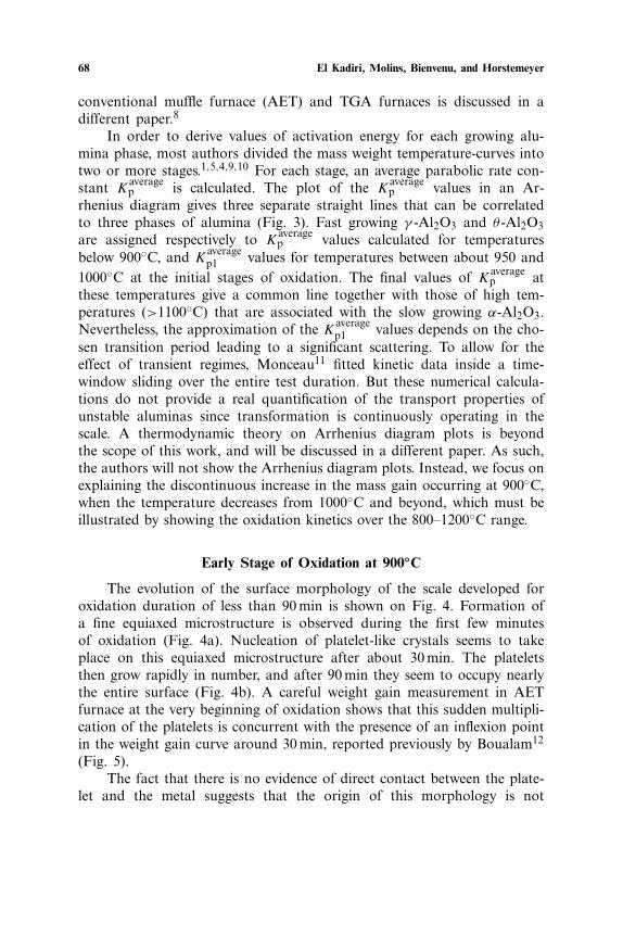
The evolution of the surface morphology of the scale developed foroxidation duration of less than 90 min is shown on Fig. 4. Formation ofa fine equiaxed microstructure is observed during the first few minutesof oxidation (Fig. 4a). Nucleation of platelet-like crystals seems to takeplace on this equiaxed microstructure after about 30 min. The plateletsthen grow rapidly in number, and after 90 min they seem to occupy nearlythe entire surface (Fig. 4b). A careful weight gain measurement in AETfurnace at the very beginning of oxidation shows that this sudden multipli-cation of the platelets is concurrent with the presence of an inflexion pointin the weight gain curve around 30 min, reported previously by Boualam12
(Fig. 5).The fact that there is no evidence of direct contact between the plate-
let and the metal suggests that the origin of this morphology is not
Abnormal High Growth Rates of Metastable Aluminas 69
Fig. 4. Evolution of surface morphology of the 900◦C oxide scale with oxida-tion duration. (a) Equiaxed microstructure after 30 min. (b) The oxide surface isentirely covered by the platelet-like morphology after 90 min.
70 El Kadiri, Molins, Bienvenu, and Horstemeyer
0
0,05
0,1
0,15
0,2
0 2 4
Time (r )
∆m/S
(mg/cm2)
Inflexion
1 3 5
Fig. 5. Weight gain in conventional muffle furnace (AET) at 900◦Cduring the beginning of oxidation.
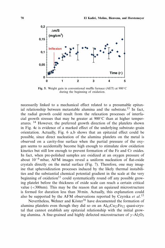
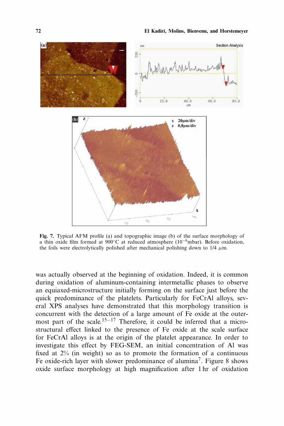
necessarily linked to a mechanical effect related to a presumable epitax-ial relationship between metastable alumina and the substrate.13 In fact,the radial growth could result from the relaxation processes of interfa-cial growth stresses that may be greater at 900◦C than at higher temper-atures. 14 However, the preferred growth direction of the platelets shownin Fig. 4c is evidence of a marked effect of the underlying substrate–grainorientation. Actually, Fig. 6 a,b shows that an epitaxial effect could bepossible, since direct nucleation of the alumina platelets on the metal isobserved on a cavity-free surface when the partial pressure of the oxy-gen seems to accidentally become high enough to stimulate slow oxidationkinetics but still low enough to prevent formation of the Fe and Cr oxides.In fact, when pre-polished samples are oxidized at an oxygen pressure ofabout 10−6 mbar, AFM images reveal a uniform nucleation of flat-oxidecrystals directly on the metal surface (Fig. 7). Therefore, one may imag-ine that spheroidization processes induced by the likely thermal instabili-ties and the substantial chemical potential gradient in the scale at the verybeginning of oxidation11 could systematically round off any possible grow-ing platelet before the thickness of oxide scale can reach a certain criticalvalue (<300nm). This may be the reason that an equiaxed microstructureis formed for duration less than 30 min. Actually, this explanation couldalso be supported by the AFM observations reported by Czyrska et al.15
Nevertheless, Wehner and Koster16 have documented the formation ofalumina platelets even though they did so on an Al63Cu25Fe12 quasi-crys-tal that cannot establish any epitaxial relationship with the initial grow-ing alumina. A fine-grained and highly defected microstructure of γ -Al2O3
Abnormal High Growth Rates of Metastable Aluminas 71
Fig. 6. Partial re-oxidation of cavity surfaces under oxide scale grown at 900◦Cafter 300 hr. (a) The platelets are sparse indicating the beginning of the re-oxida-tion. (b) The new-oxide scale is denser and some platelets developed typical polyg-onal geometry.
72 El Kadiri, Molins, Bienvenu, and Horstemeyer
Fig. 7. Typical AFM profile (a) and topographic image (b) of the surface morphology ofa thin oxide film formed at 900◦C at reduced atmosphere (10−6mbar). Before oxidation,the foils were electrolytically polished after mechanical polishing down to 1/4 µm.
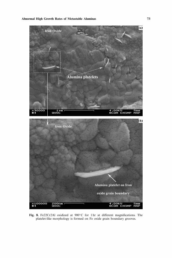
was actually observed at the beginning of oxidation. Indeed, it is commonduring oxidation of aluminum-containing intermetallic phases to observean equiaxed-microstructure initially forming on the surface just before thequick predominance of the platelets. Particularly for FeCrAl alloys, sev-eral XPS analyses have demonstrated that this morphology transition isconcurrent with the detection of a large amount of Fe oxide at the outer-most part of the scale.15−17 Therefore, it could be inferred that a micro-structural effect linked to the presence of Fe oxide at the scale surfacefor FeCrAl alloys is at the origin of the platelet appearance. In order toinvestigate this effect by FEG-SEM, an initial concentration of Al wasfixed at 2% (in weight) so as to promote the formation of a continuousFe oxide-rich layer with slower predominance of alumina7. Figure 8 showsoxide surface morphology at high magnification after 1 hr of oxidation
Abnormal High Growth Rates of Metastable Aluminas 73
Fig. 8. Fe22Cr2Al oxidized at 900◦C for 1 hr at different magnifications. Theplatelet-like morphology is formed on Fe oxide grain boundary grooves.
74 El Kadiri, Molins, Bienvenu, and Horstemeyer
of an 90 µm thin Fe22CrAl2Al foil at 900◦C. The platelet is clearly evi-dence of an outward aluminum-diffusion mechanism across the Fe-oxidegrain boundaries. Accordingly, the dissolution mechanisms of Fe oxidegrains put forward by Jedlinski et al.17 seem to begin by alumina forma-tion across their grain-boundaries, which leads systematically to platelet-like morphology.
Intermediate Stage of Oxidation at 900◦C
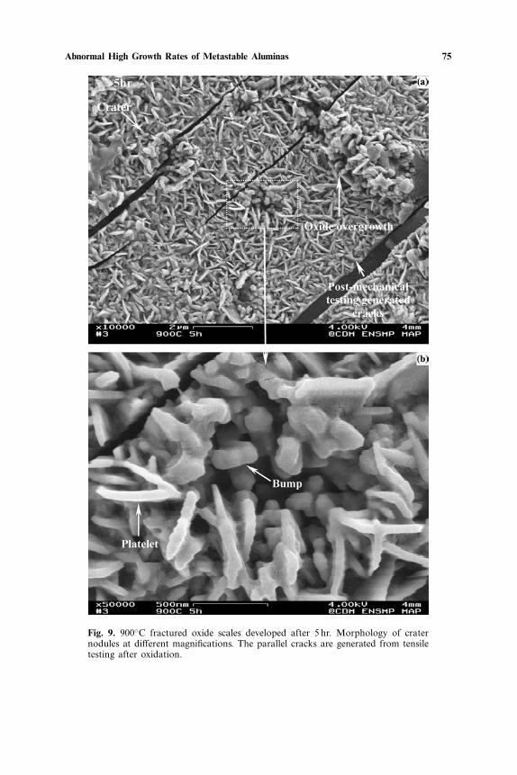
After several hours of oxidation (>3 hr), an enhanced formation offine oxide nodules aligned with polishing marks and accompanied by cra-ter formation is observed at the scale surface (Fig. 9a,b). At the boundaryregions of the oxide crater nodule, there is radial arrangement of the plate-lets, but they appear as bumps more and more towards the central region(Fig. 9b). This texture is evidence of contraction with local phase trans-formation by spheroidization phenomena.2
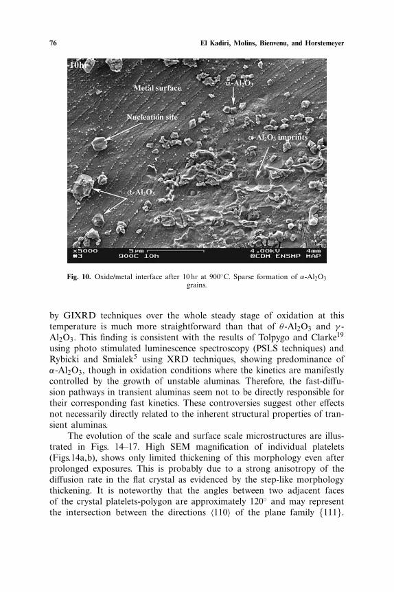
The onset of this transformation could be motivated by interfacialnucleation of α-Al2O3 (Fig. 10) that may destabilize the overlying plate-lets as suggested by Grabke.9 The formation of α-Al2O3 nuclei could bepromoted by the likely presence of the isostructural-hexagonal Cr2O3.17,18
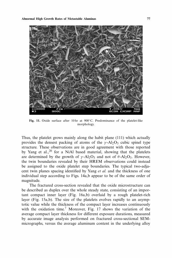
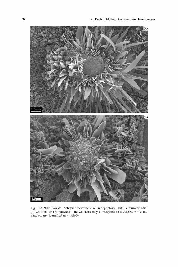
However, after about 10 hr of oxidation, the frequency of the plateletsincreases dramatically to the extent that they again cover the total sur-face (Fig. 11). This peculiar fast predominance of the platelets is difficultto explain without assuming considerable full cracking around the nod-ules, so that oxygen can penetrate across the crack and oxidize the result-ing exposed metal area by formation of oxide platelets. A self-consistentmicrograph of this phenomenon is an oxide “chrysanthemum”-like mor-phology (Fig. 12). Here, the circumferential whiskers (Fig. 12a) and/orplatelets (Fig. 12b) are reminiscent of circumferential cracks surroundingthe fully transformed nodule of alpha-alumina. This explanation is alsoconsistent with the fact that a slowing down of oxidation with substantialscattering in weight gain is concurrent with the nodule formation (Fig. 5).
Steady Stage of Oxidation at 900◦C
Oxide Bulk Microstructure
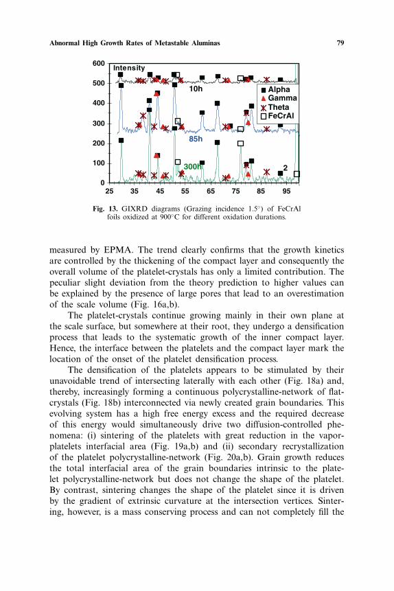
The substantial presence of twin boundaries and the cation vacancynetwork intrinsic to the metastable aluminas are commonly believedto account for rapid cation diffusion in these structures resulting in adiscontinuous increase in mass gain at temperatures around 900◦C. How-ever, as illustrated in Fig. 13, the identification of the corundum α-Al2O3
Abnormal High Growth Rates of Metastable Aluminas 75
Fig. 9. 900◦C fractured oxide scales developed after 5 hr. Morphology of craternodules at different magnifications. The parallel cracks are generated from tensiletesting after oxidation.
76 El Kadiri, Molins, Bienvenu, and Horstemeyer
Fig. 10. Oxide/metal interface after 10 hr at 900◦C. Sparse formation of α-Al2O3grains.
by GIXRD techniques over the whole steady stage of oxidation at thistemperature is much more straightforward than that of θ -Al2O3 and γ -Al2O3. This finding is consistent with the results of Tolpygo and Clarke19
using photo stimulated luminescence spectroscopy (PSLS techniques) andRybicki and Smialek5 using XRD techniques, showing predominance ofα-Al2O3, though in oxidation conditions where the kinetics are manifestlycontrolled by the growth of unstable aluminas. Therefore, the fast-diffu-sion pathways in transient aluminas seem not to be directly responsible fortheir corresponding fast kinetics. These controversies suggest other effectsnot necessarily directly related to the inherent structural properties of tran-sient aluminas.
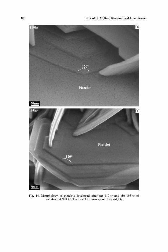
The evolution of the scale and surface scale microstructures are illus-trated in Figs. 14–17. High SEM magnification of individual platelets(Figs.14a,b), shows only limited thickening of this morphology even afterprolonged exposures. This is probably due to a strong anisotropy of thediffusion rate in the flat crystal as evidenced by the step-like morphologythickening. It is noteworthy that the angles between two adjacent facesof the crystal platelets-polygon are approximately 120◦ and may representthe intersection between the directions 〈110〉 of the plane family {111}.
Abnormal High Growth Rates of Metastable Aluminas 77
Fig. 11. Oxide surface after 10 hr at 900◦C. Predominance of the platelet-likemorphology.
Thus, the platelet grows mainly along the habit plane (111) which actuallyprovides the densest packing of atoms of the γ -Al2O3 cubic spinel typestructure. These observations are in good agreement with those reportedby Yang et al.,20 for a NiAl based material, showing that the plateletsare determined by the growth of γ -Al2O3 and not of θ -Al2O3. However,the twin boundaries revealed by their HREM observations could insteadbe assigned to the oxide platelet step boundaries. The typical two-adja-cent twin planes spacing identified by Yang et al. and the thickness of oneindividual step according to Figs. 14a,b appear to be of the same order ofmagnitude.
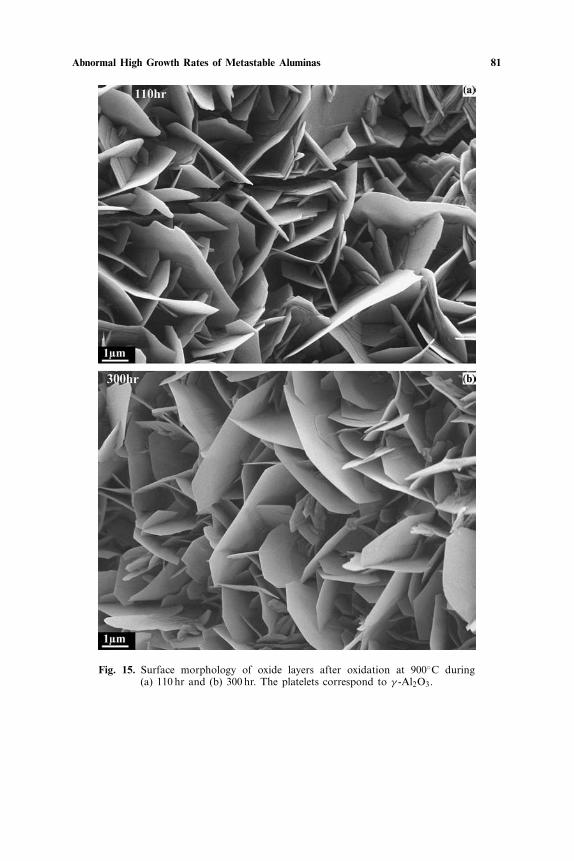
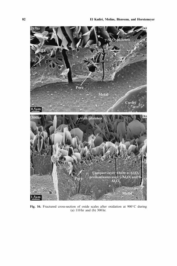
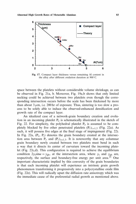
The fractured cross-section revealed that the oxide microstructure canbe described as duplex over the whole steady state, consisting of an impor-tant compact inner layer (Fig. 16a,b) overlaid by a rough platelet-richlayer (Fig. 15a,b). The size of the platelets evolves rapidly to an asymp-totic value while the thickness of the compact layer increases continuouslywith the oxidation time.7 Moreover, Fig. 17 shows the variation of theaverage compact layer thickness for different exposure durations, measuredby accurate image analysis performed on fractured cross-sectional SEM-micrographs, versus the average aluminum content in the underlying alloy
78 El Kadiri, Molins, Bienvenu, and Horstemeyer
Fig. 12. 900◦C-oxide “chrysanthemum”-like morphology with circumferential(a) whiskers or (b) platelets. The whiskers may correspond to θ -Al2O3, while theplatelets are identified as γ -Al2O3.
Abnormal High Growth Rates of Metastable Aluminas 79
0
100
200
300
400
500
600
25 35 45 55 65 75 85 95
2
Intensity
AlphaGammaThetaFeCrAl
85h
10h
300h
Fig. 13. GIXRD diagrams (Grazing incidence 1.5◦) of FeCrAlfoils oxidized at 900◦C for different oxidation durations.
measured by EPMA. The trend clearly confirms that the growth kineticsare controlled by the thickening of the compact layer and consequently theoverall volume of the platelet-crystals has only a limited contribution. Thepeculiar slight deviation from the theory prediction to higher values canbe explained by the presence of large pores that lead to an overestimationof the scale volume (Fig. 16a,b).
The platelet-crystals continue growing mainly in their own plane atthe scale surface, but somewhere at their root, they undergo a densificationprocess that leads to the systematic growth of the inner compact layer.Hence, the interface between the platelets and the compact layer mark thelocation of the onset of the platelet densification process.
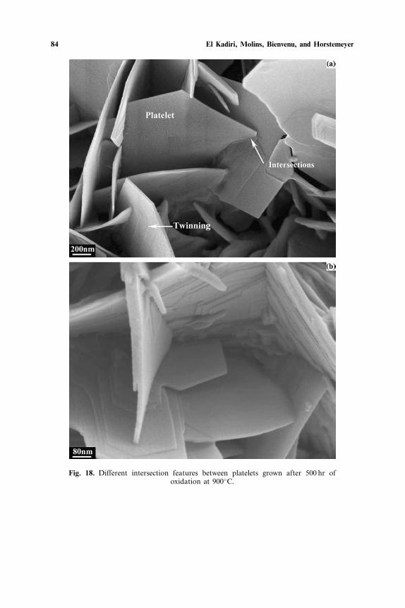
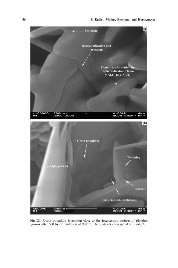
The densification of the platelets appears to be stimulated by theirunavoidable trend of intersecting laterally with each other (Fig. 18a) and,thereby, increasingly forming a continuous polycrystalline-network of flat-crystals (Fig. 18b) interconnected via newly created grain boundaries. Thisevolving system has a high free energy excess and the required decreaseof this energy would simultaneously drive two diffusion-controlled phe-nomena: (i) sintering of the platelets with great reduction in the vapor-platelets interfacial area (Fig. 19a,b) and (ii) secondary recrystallizationof the platelet polycrystalline-network (Fig. 20a,b). Grain growth reducesthe total interfacial area of the grain boundaries intrinsic to the plate-let polycrystalline-network but does not change the shape of the platelet.By contrast, sintering changes the shape of the platelet since it is drivenby the gradient of extrinsic curvature at the intersection vertices. Sinter-ing, however, is a mass conserving process and can not completely fill the
80 El Kadiri, Molins, Bienvenu, and Horstemeyer
Fig. 14. Morphology of platelets developed after (a) 110 hr and (b) 195 hr ofoxidation at 900◦C. The platelets correspond to γ -Al2O3.
Abnormal High Growth Rates of Metastable Aluminas 81
Fig. 15. Surface morphology of oxide layers after oxidation at 900◦C during(a) 110 hr and (b) 300 hr. The platelets correspond to γ -Al2O3.
82 El Kadiri, Molins, Bienvenu, and Horstemeyer
Fig. 16. Fractured cross-section of oxide scales after oxidation at 900◦C during(a) 110 hr and (b) 300 hr.
Abnormal High Growth Rates of Metastable Aluminas 83
0
1
2
3
4
5
6
2 4
Al(%m)
Thickness(µm)
Compact layer
Theory
1hr
200hr 144hr
60hr
6hr2hr
3 5 6
Fig. 17. Compact layer thickness versus remaining Al content inthe alloy after different oxidation duration at 900◦C.
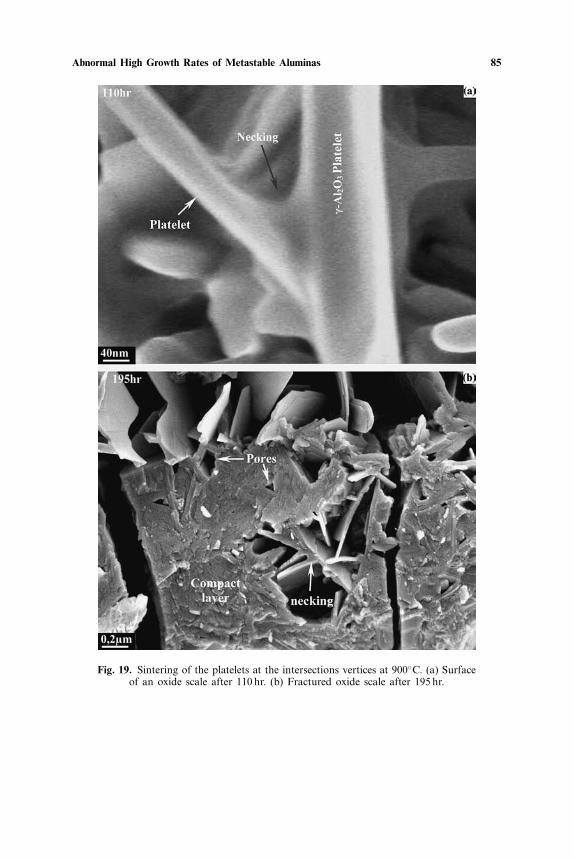
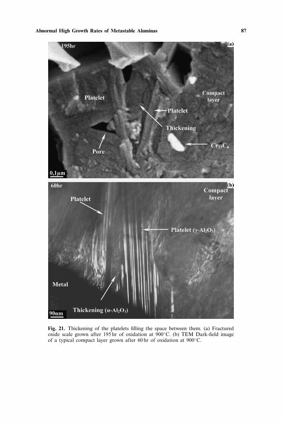
space between the platelets without considerable volume shrinkage, as canbe observed in Fig. 21a, b. Moreover, Fig. 19a,b shows that only limitednecking could be achieved between two platelets even though the corre-sponding intersection occurs before the scale has been thickened by morethan about 3 µm, i.e. 200 hr of exposure. Thus, sintering is too slow a pro-cess to be solely able to induce the observed-enhanced densification andgrowth rate of the compact layer.
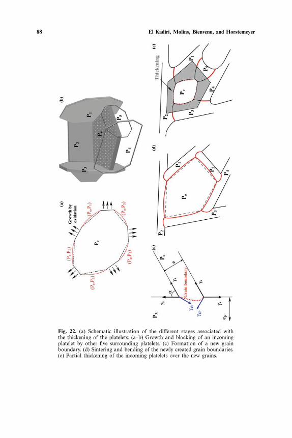
An idealized case of a network-grain boundary creation and evolu-tion in an incoming platelet Pe is schematically illustrated in the sketch ofFig. 22. For simplicity, the polyhedral platelet Pe is assumed to be com-pletely blocked by five other penetrated platelets (Pi )i=1,5 (Fig. 22a). Assuch, it will possess five edges at the final stage of impingement (Fig. 22).In Fig. 22a, (Pe, Pi) denotes the grain boundary created at the intersec-tion area between Pe and (Pi )i=1,5. It is noteworthy that any columnargrain boundary newly created between two platelets must bend in sucha way that it directs its center of curvature toward the incoming plate-let (Fig. 22c,d). This configuration is required to achieve the equilibriumcondition 2γssinα = γgb, at the intersection area, where γs and γgb are,respectively, the surface and boundary-free energy per unit area.21 Oneimportant characteristic implied by this convexity of the grain boundariesis that each incoming platelet will experience an intrinsic grain growthphenomenon transforming it progressively into a polycrystalline oxide film(Fig. 22e). This will radically upset the diffusion rate anisotropy which wasthe immediate cause of the preferential radial growth as mentioned above.
84 El Kadiri, Molins, Bienvenu, and Horstemeyer
Fig. 18. Different intersection features between platelets grown after 500 hr ofoxidation at 900◦C.
Abnormal High Growth Rates of Metastable Aluminas 85
Fig. 19. Sintering of the platelets at the intersections vertices at 900◦C. (a) Surfaceof an oxide scale after 110 hr. (b) Fractured oxide scale after 195 hr.
86 El Kadiri, Molins, Bienvenu, and Horstemeyer
Fig. 20. Grain boundary formation close to the intersection vertices of plateletsgrown after 500 hr of oxidation at 900◦C. The platelets correspond to γ -Al2O3.
Abnormal High Growth Rates of Metastable Aluminas 87
Fig. 21. Thickening of the platelets filling the space between them. (a) Fracturedoxide scale grown after 195 hr of oxidation at 900◦C. (b) TEM Dark-field imageof a typical compact layer grown after 60 hr of oxidation at 900◦C.
88 El Kadiri, Molins, Bienvenu, and Horstemeyer
Fig. 22. (a) Schematic illustration of the different stages associated withthe thickening of the platelets. (a–b) Growth and blocking of an incomingplatelet by other five surrounding platelets. (c) Formation of a new grainboundary. (d) Sintering and bending of the newly created grain boundaries.(e) Partial thickening of the incoming platelets over the new grains.
Abnormal High Growth Rates of Metastable Aluminas 89
Fig. 23. Scale surface after 500 hr of oxidation at 900◦C. (a) Creation of newgrain boundaries and (b) secondary recrystallization in incoming platelets.
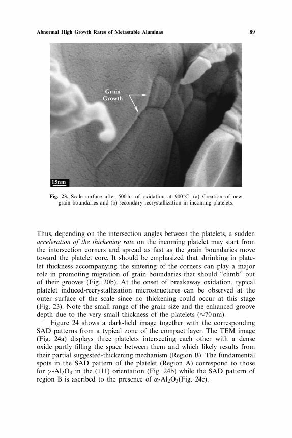
Thus, depending on the intersection angles between the platelets, a suddenacceleration of the thickening rate on the incoming platelet may start fromthe intersection corners and spread as fast as the grain boundaries movetoward the platelet core. It should be emphasized that shrinking in plate-let thickness accompanying the sintering of the corners can play a majorrole in promoting migration of grain boundaries that should “climb” outof their grooves (Fig. 20b). At the onset of breakaway oxidation, typicalplatelet induced-recrystallization microstructures can be observed at theouter surface of the scale since no thickening could occur at this stage(Fig. 23). Note the small range of the grain size and the enhanced groovedepth due to the very small thickness of the platelets (≈70 nm).
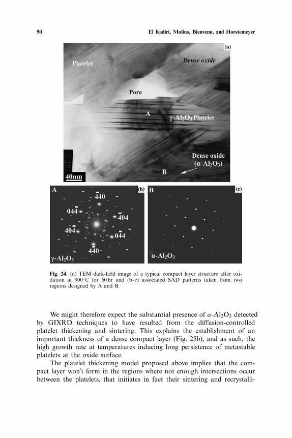
Figure 24 shows a dark-field image together with the correspondingSAD patterns from a typical zone of the compact layer. The TEM image(Fig. 24a) displays three platelets intersecting each other with a denseoxide partly filling the space between them and which likely results fromtheir partial suggested-thickening mechanism (Region B). The fundamentalspots in the SAD pattern of the platelet (Region A) correspond to thosefor γ -Al2O3 in the (111) orientation (Fig. 24b) while the SAD pattern ofregion B is ascribed to the presence of α-Al2O3(Fig. 24c).
90 El Kadiri, Molins, Bienvenu, and Horstemeyer
Fig. 24. (a) TEM dark-field image of a typical compact layer structure after oxi-dation at 900◦C for 60 hr and (b–c) associated SAD patterns taken from tworegions designed by A and B.
We might therefore expect the substantial presence of α-Al2O3 detectedby GIXRD techniques to have resulted from the diffusion-controlledplatelet thickening and sintering. This explains the establishment of animportant thickness of a dense compact layer (Fig. 25b), and as such, thehigh growth rate at temperatures inducing long persistence of metastableplatelets at the oxide surface.
The platelet thickening model proposed above implies that the com-pact layer won’t form in the regions where not enough intersections occurbetween the platelets, that initiates in fact their sintering and recrystalli-
Abnormal High Growth Rates of Metastable Aluminas 91
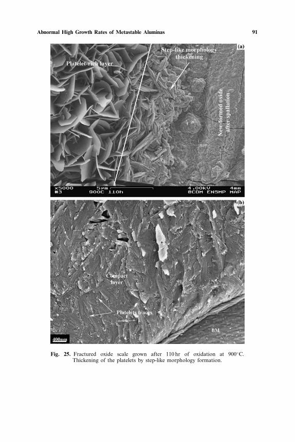
Fig. 25. Fractured oxide scale grown after 110 hr of oxidation at 900◦C.Thickening of the platelets by step-like morphology formation.
92 El Kadiri, Molins, Bienvenu, and Horstemeyer
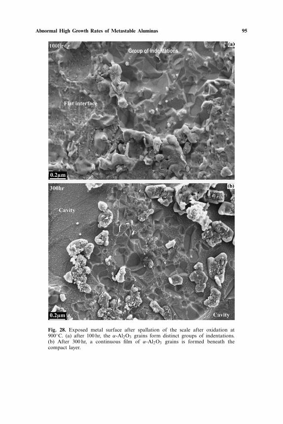
zation. Indeed, this corroborating phenomenon can be observed on oxidescales grown at the edges of the sample or on those adjacent to areasfrom which the oxide has spalled off during earlier stages of oxidation(Fig. 25a). An inner dense layer, however, is formed, but the densificationseems to be controlled by the slow step-like morphology thickening pro-cess that atypically leads to a smaller thickness compared to that of theouter platelet-rich layer.
Oxide Microstructure Close to the Oxide/Substrate Interface
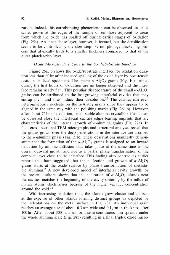
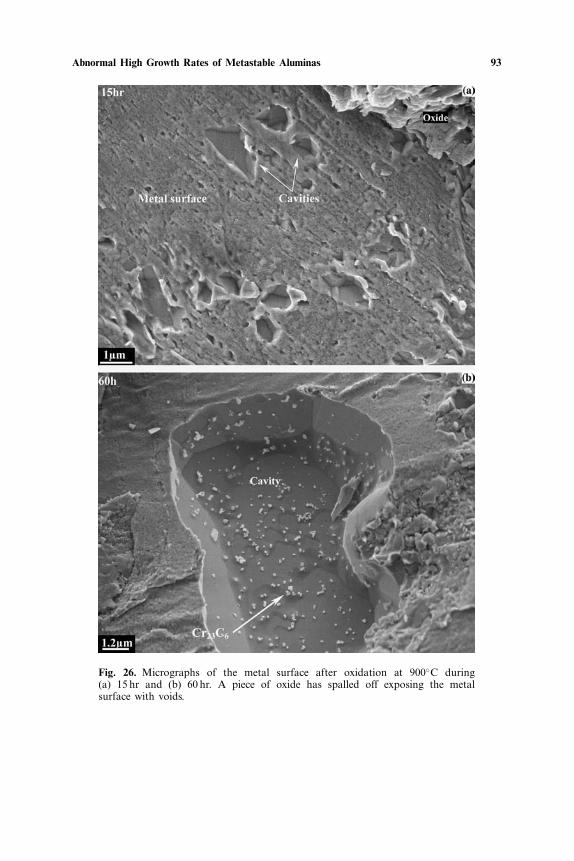
Figure 26a, b shows the oxide/substrate interface for oxidation dura-tion less than 60 hr after induced-spalling of the oxide layer by post-tensiletests on oxidized specimens. The sparse α-Al2O3 grains (Fig. 10) formedduring the first hours of oxidation are no longer observed and the inter-face remains nearly flat . This peculiar disappearance of the small α-Al2O3grains can be attributed to the fast-growing interfacial cavities that mayentrap them and thus induce their dissolution.22 The cavities can evenheterogeneously nucleate on the α-Al2O3 grains since they appear to bealigned in the same way with the polishing marks (Fig. 26a,b). However,after about 75 hr of oxidation, small stable alumina crystalline islands canbe observed close the interfacial cavities edges leaving imprints that arecharacteristics of the internal growth of α-alumina grains (Fig. 27a). Infact, cross- sectional TEM micrographs and structural analyses reveal thatthe grains grown over the deep penetrations in the interface are ascribedto the α-alumina phase (Fig. 27b). These observations manifestly demon-strate that the formation of the α-Al2O3 grains is assigned to an inwardoxidation by anionic diffusion that takes place at the same time as theoverall outward growth and not to a partial phase transformation of thecompact layer close to the interface. This finding also contradicts earlierreports that have suggested that the nucleation and growth of α-Al2O3grains starts at the oxide surface by phase transformation of metasta-ble aluminas.2 A new developed model of interfacial cavity growth, bythe present authors, shows that the nucleation of α-Al2O3 islands nearthe cavities matches the beginning of the cavity-sintering by the influx ofmatrix atoms which arises because of the higher vacancy concentrationaround the void.23
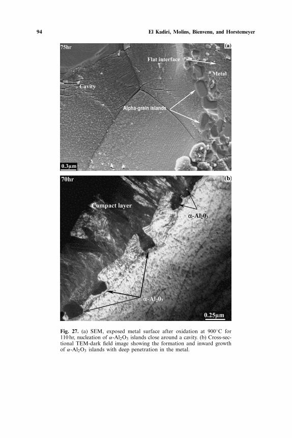
With increasing oxidation time, the islands grow, cluster and coarsenat the expense of other islands forming distinct groups as depicted bythe indentations on the metal surface in Fig. 28a. An individual grainreaches an average size of about 0.3 µm wide and 0.1 µm in thickness after100 hr. After about 300 hr, a uniform semi-continuous film spreads underthe whole alumina scale (Fig. 28b) resulting in a final triplex oxide micro-
Abnormal High Growth Rates of Metastable Aluminas 93
Fig. 26. Micrographs of the metal surface after oxidation at 900◦C during(a) 15 hr and (b) 60 hr. A piece of oxide has spalled off exposing the metalsurface with voids.
94 El Kadiri, Molins, Bienvenu, and Horstemeyer
Fig. 27. (a) SEM, exposed metal surface after oxidation at 900◦C for110 hr, nucleation of α-Al2O3 islands close around a cavity. (b) Cross-sec-tional TEM-dark field image showing the formation and inward growthof α-Al2O3 islands with deep penetration in the metal.
Abnormal High Growth Rates of Metastable Aluminas 95
Fig. 28. Exposed metal surface after spallation of the scale after oxidation at900◦C. (a) after 100 hr, the α-Al2O3 grains form distinct groups of indentations.(b) After 300 hr, a continuous film of α-Al2O3 grains is formed beneath thecompact layer.
96 El Kadiri, Molins, Bienvenu, and Horstemeyer
structure. The formation of this very thin underlying polycrystalline film ofα-Al2O3 grains seems not to have any noticeable effect on the subsequentoxidation kinetics.
CONCLUSION
Isothermal oxidation of FeCrAl-RE has been performed at tempera-tures near 900◦C and studied by AET, TGA, SEM, TEM, AFM, EPMAand GIXRD. At an early stage of oxidation, metastable platelet crystalscan nucleate either at Fe oxide grain boundary grooves or directly on themetal surface. The platelets rapidly cover the scale surface and their mul-tiplication is concurrent with a surge in weight gain after around 30 min.After several hours of oxidation, a competition between the multiplicationof the platelets and their transformation by nodule growth, accompaniedby crater formation, is observed at the scale surface. The phase transfor-mation may be correlated to an observed alpha-alumina nucleation at thesubscale. These events are also concurrent with a remarkable scattering inmass gain. The platelet-shaped crystals continue growing during furtherheating, most likely along the γ -Al2O3 (111) plane with a slight step-likemorphology thickening. At the outermost part of the scale, the plateletsincreasingly tend to intersect laterally with each other initiating the forma-tion of a complex polycrystalline network of interconnecting flat-crystals.A coupling phenomenon between sintering at the intersection vertices andgrain growth in the platelets was suggested as being responsible for the rapidthickening of the platelets and scale densification, leading to a fast growingcompact-layer of mixed α + γ + θ -Al2O3. These important morphologicaland structural evolutions may account for the “abnormal” high oxidationrate around 900◦C widely reported in the literature. Formation of an under-lying pure α-Al2O3 layer initiates after about 100 hr, as the nucleation ofsmall stable crystalline oxide islands close to the interfacial cavity edges.After 300 hr, a uniform continuous film spreads under the entire aluminascale.
REFERENCES1. W. C. Hagel, Corrosion 21, 316 (1965).2. J. K. Doychak, J. L. Smialek, and T. E. Mitchell, Metallurgical Transactions 20A, 499
(1989).3. A. Germidis, Doctoral Thesis, Ecole Nationale Superieure des Mines de Paris-CDM
(1996).4. M. W. Brumm and H. J. Grabke, Corrosion Science 33(11), 1677 (1992).5. G. C. Rybicki and J. L. Smialek, Oxidation of Metals 31 (3/4), 275 (1989).6. G. B. Abderrazik, G. Moulin, A. M. Huntz, and R. Berneron, Journal of Materials Sci-
ence 19, 3173 (1984).
Abnormal High Growth Rates of Metastable Aluminas 97
7. H. El Kadiri, Doctoral Thesis, Ecole Nationale Superieure des Mines de Paris-CDM(2004).
8. H. El Kadiri, R. Molins, Y. Bienvenu, and M. Horstemeyer, To be submitted.9. H. J. Grabke, Materials Science and Forum 251–254, 149–162 (1997).
10. E. Andrieu, A. Germidis, and R. Molins, Materials Science Forum 251–254, 357–364(1997).
11. D. Monceau and B. Pieraggi, Oxidation of Metals 50(5/6), 477 (1998).12. M. Boualam, Doctoral Thesis. Universite de Technologie de Compiegne (1992).13. B. Dionnet, Doctoral Thesis, Universite de Limoges (1993).14. Z. Suo, D. V. Kubair, A. G. Evans, D. Clarke, and V. Tolpygo, Acta Materialia 51, 959
(2003).15. A. Czyrska et al., Solid State Ionics 117, 13 (1999).16. B. I. Wehner, and U. Koster, Oxidation of Metals 54(5/6) 445 (2000).17. J. Jedlinski et al., Applied Surface Science 103, 205 (1996).18. M. Siegers, H. J. Grabke, and H. Viefhaus, Fresenius′ Journal of Analytical Chemistry 346
269 (1993).19. V. K. Tolpygo and D. R. Clarke, Materials at High Temperatures 17–1, 59 2000.20. J. C. Yang, E. Schumann, I. Levin, and M. Ruhle, Acta. Met. 46(6), 2195 (1998).21. W. W. Mullins, Acta. Met. 6 414 (1958).22. L. Rivoaland, V. Maurice, P. Josso, M.-P. Bacos, and P. Marcus, Oxidation of Metals
60(1/2), 137 2003.23. H. El Kadiri and M. Horstemeyer, To be submitted.
Related Documents