428 Am J Psychiatry 157:3, March 2000 Abnormal Angular Gyrus Asymmetry in Schizophrenia Margaret Niznikiewicz, Ph.D., Robert Donnino, A.B., Robert W. McCarley, M.D., Paul G. Nestor, Ph.D., Daniel V. Iosifescu, M.D., Brian O’Donnell, Ph.D., James Levitt, M.D., and Martha E. Shenton, Ph.D. Objective: Few studies have evaluated the parietal lobe in schizophrenia despite the fact that it has an important role in attention, memory, and language—all functions that have been reported to be abnormal in schizophrenia. The inferior parietal lobule, in particular, is of interest because it is not only part of the heteromodal association cortex but also is part of the semantic-lexical network, which also includes the planum temporale. Both the infe- rior parietal lobule, particularly the angular gyrus of the inferior parietal lobule, and the planum temporale are brain regions that play a critical role as biological substrates of lan- guage and thought. The authors compared volume and asymmetry measures of the indi- vidual gyri of the parietal lobe by means of magnetic resonance imaging (MRI) scans. Method: MRI scans with a 1.5-Tesla magnet were obtained from 15 male chronic schizo- phrenic and 15 comparison subjects matched for age, gender, and parental socioeconomic status. Results: Inferior parietal lobule volumes showed a leftward asymmetry (left 7.0% larger than right) in comparison subjects and a reversed asymmetry (left 6.3% smaller than right) in schizophrenic subjects. The angular gyrus accounted for this difference in asym- metry, with the left angular gyrus being significantly larger (18.7%) than the right in compar- ison subjects, a finding that was not observed in schizophrenic patients. A further test of angular gyrus asymmetry showed a reversal of the normal left-greater-than-right asymme- try in the schizophrenic patients. Conclusions: Patients with schizophrenia showed a re- versed asymmetry in the inferior parietal lobule that was localized to the angular gyrus, a structure belonging to the heteromodal association cortex as well as being part of the se- mantic-lexical network. This finding contributes to a more comprehensive understanding of the neural substrates of language and thought disorder in schizophrenia. (Am J Psychiatry 2000; 157:428–437) The parietal lobe has received little attention in schizophrenia despite its recognized importance in processes that are likely disturbed in schizophrenia, such as language (1), spatial working memory, and at- tention (2–4). This lack of attention to parietal regions of the brain is further highlighted by the fact that only nine magnetic resonance imaging (MRI) studies of the parietal lobe, in contrast to more than 35 for both tem- poral and frontal lobes, have been conducted in sub- jects with schizophrenia (5, 6). The parietal lobe itself consists of the postcentral gy- rus, superior parietal gyrus, and inferior parietal lobule, which is further subdivided into the supramarginal gy- rus (area 39) and angular gyrus (area 40). The inferior parietal lobule is part of the heteromodal association cortex, which has been proposed as the site of the key abnormality in schizophrenia (7). Further, both the su- pramarginal gyrus and the angular gyrus have been de- scribed as part of a semantic-lexical network that sup- ports “word meanings” represented by a “grid of connectivity” that constitutes a “final pathway for the chunking of words into thought” (1). The role of the in- ferior parietal lobule, and especially the angular gyrus, in language comprehension has been further confirmed Received Feb. 8, 1999; revisions received June 28, Aug. 16, and Sept. 20, 1999; accepted Sept. 20, 1999. From the Laboratory of Neuroscience, Clinical Neuroscience Division, Department of Psy- chiatry, VA Medical Center, Harvard Medical School; and the Surgi- cal Planning Laboratory, MRI Division, Department of Radiology, Brigham and Women’s Hospital, Harvard Medical School, Boston. Address reprint requests to Dr. Shenton, Department of Psychiatry- 116A, VA Medical Center, Harvard Medical School, 940 Belmont St., Brockton, MA 02301. Supported by NIMH grants MH-50740 and MH-01110 (to Dr. Shenton) and MH-40799 (to Dr. McCarley), and grants from the Department of Veterans Affairs Center for Clinical and Basic Neu- roscience Studies of Schizophrenia and a Department of Veterans Affairs Merit Application (Dr. McCarley). The authors thank Chris Dodd, B.A., Alaka Pellock, A.B., and Marie Fairbanks for their administrative assistance.

Welcome message from author
This document is posted to help you gain knowledge. Please leave a comment to let me know what you think about it! Share it to your friends and learn new things together.
Transcript

428 Am J Psychiatry 157:3, March 2000
Abnormal Angular Gyrus Asymmetry in Schizophrenia
Margaret Niznikiewicz, Ph.D., Robert Donnino, A.B., Robert W. McCarley, M.D., Paul G. Nestor, Ph.D., Daniel V. Iosifescu, M.D., Brian O’Donnell, Ph.D.,
James Levitt, M.D., and Martha E. Shenton, Ph.D.
Objective: Few studies have evaluated the parietal lobe in schizophrenia despite the factthat it has an important role in attention, memory, and language—all functions that havebeen reported to be abnormal in schizophrenia. The inferior parietal lobule, in particular, isof interest because it is not only part of the heteromodal association cortex but also is partof the semantic-lexical network, which also includes the planum temporale. Both the infe-rior parietal lobule, particularly the angular gyrus of the inferior parietal lobule, and theplanum temporale are brain regions that play a critical role as biological substrates of lan-guage and thought. The authors compared volume and asymmetry measures of the indi-vidual gyri of the parietal lobe by means of magnetic resonance imaging (MRI) scans.Method: MRI scans with a 1.5-Tesla magnet were obtained from 15 male chronic schizo-phrenic and 15 comparison subjects matched for age, gender, and parental socioeconomicstatus. Results: Inferior parietal lobule volumes showed a leftward asymmetry (left 7.0%larger than right) in comparison subjects and a reversed asymmetry (left 6.3% smaller thanright) in schizophrenic subjects. The angular gyrus accounted for this difference in asym-metry, with the left angular gyrus being significantly larger (18.7%) than the right in compar-ison subjects, a finding that was not observed in schizophrenic patients. A further test ofangular gyrus asymmetry showed a reversal of the normal left-greater-than-right asymme-try in the schizophrenic patients. Conclusions: Patients with schizophrenia showed a re-versed asymmetry in the inferior parietal lobule that was localized to the angular gyrus, astructure belonging to the heteromodal association cortex as well as being part of the se-mantic-lexical network. This finding contributes to a more comprehensive understanding ofthe neural substrates of language and thought disorder in schizophrenia.
(Am J Psychiatry 2000; 157:428–437)
The parietal lobe has received little attention inschizophrenia despite its recognized importance inprocesses that are likely disturbed in schizophrenia,such as language (1), spatial working memory, and at-tention (2–4). This lack of attention to parietal regions
of the brain is further highlighted by the fact that onlynine magnetic resonance imaging (MRI) studies of theparietal lobe, in contrast to more than 35 for both tem-poral and frontal lobes, have been conducted in sub-jects with schizophrenia (5, 6).
The parietal lobe itself consists of the postcentral gy-rus, superior parietal gyrus, and inferior parietal lobule,which is further subdivided into the supramarginal gy-rus (area 39) and angular gyrus (area 40). The inferiorparietal lobule is part of the heteromodal associationcortex, which has been proposed as the site of the keyabnormality in schizophrenia (7). Further, both the su-pramarginal gyrus and the angular gyrus have been de-scribed as part of a semantic-lexical network that sup-ports “word meanings” represented by a “grid ofconnectivity” that constitutes a “final pathway for thechunking of words into thought” (1). The role of the in-ferior parietal lobule, and especially the angular gyrus,in language comprehension has been further confirmed
Received Feb. 8, 1999; revisions received June 28, Aug. 16, andSept. 20, 1999; accepted Sept. 20, 1999. From the Laboratory ofNeuroscience, Clinical Neuroscience Division, Department of Psy-chiatry, VA Medical Center, Harvard Medical School; and the Surgi-cal Planning Laboratory, MRI Division, Department of Radiology,Brigham and Women’s Hospital, Harvard Medical School, Boston.Address reprint requests to Dr. Shenton, Department of Psychiatry-116A, VA Medical Center, Harvard Medical School, 940 Belmont St.,Brockton, MA 02301.
Supported by NIMH grants MH-50740 and MH-01110 (to Dr.Shenton) and MH-40799 (to Dr. McCarley), and grants from theDepartment of Veterans Affairs Center for Clinical and Basic Neu-roscience Studies of Schizophrenia and a Department of VeteransAffairs Merit Application (Dr. McCarley).
The authors thank Chris Dodd, B.A., Alaka Pellock, A.B., andMarie Fairbanks for their administrative assistance.
Am J Psychiatry 157:3, March 2000 429
NIZNIKIEWICZ, DONNINO, MCCARLEY, ET AL.
by functional MRI (fMRI) and positron emission to-mography studies (8–10). Moreover, the semantic-lexi-cal network proposed by Mesulam (1) includes both theinferior parietal lobule and the planum temporale, thelatter being located on the superioposterior surface ofsuperior temporal gyrus, which, in turn, has been corre-lated with symptoms of thought disorder (5, 6, 11).More anteriorly, the superior temporal gyrus has beencorrelated with auditory hallucinations (5, 6, 12).
Of note, the inferior parietal lobule and neighboringcortical regions often exhibit marked lateral asymmetry(13, 14) and belong to structures that support lan-guage. The presence of left-greater-than-right asymme-try also appears to be important for normal languagedevelopment. For example, the absence of normal later-alization in these regions has been reported in subjectswith autism (15, 16) as well as in other language disor-ders (17). In schizophrenia, a reversal of normal asym-metry in the superior temporal gyrus, including theplanum temporale, has been associated with thoughtdisorder (5, 6). Thus, a reversal in normal asymmetrymay be importantly related to schizophrenic pathology(18–20). Consistent with these findings, we hypothe-sized that a reversal of normal asymmetry in the supe-rior temporal gyrus would also be found in the inferiorparietal lobule. Furthermore, since cortical asymme-tries are present during fetal development (21), findingan absence or a reversal of normal asymmetry might in-dicate a disruption in neuronal development.
The relevance of the angular gyrus and supramar-ginal gyrus to schizophrenia stems not only from theirfunctions as part of the heteromodal association cortexbut also from their reciprocal neuroanatomical connec-tions to the prefrontal (22) and temporal lobes (23)—brain regions that have been shown to have disease-re-lated abnormalities. We examined these neuroanatomi-cal relationships by capitalizing on the opportunity ofhaving volumetric data on prefrontal and temporallobe regions in the same subjects (11, 24, 25). If the hy-pothesis that schizophrenia is a disorder characterizedprimarily by heteromodal association cortex abnormal-ities is correct, then higher correlations between the in-ferior parietal lobule and other areas interconnectedwith the heteromodal association cortex would be pre-dicted for schizophrenic patients but not for compari-son subjects. Also, in an exploratory analysis, we exam-ined the relationship between the heteromodal inferiorparietal lobule and formal thought disorder, as well asneuropsychological measures of attention and memory,all of which are likely to contribute to thinking andjudgment—functions commonly associated with the in-ferior parietal lobule.
METHOD
Subjects
Fifteen right-handed male subjects with chronic schizophreniawere recruited from the Veterans Affairs Medical Center in Brockton,Mass. (11). Diagnoses were determined by using DSM-III-R criteria
from the Schedule for Affective Disorders and Schizophrenia (26) andfrom hospital chart reviews. All patients were receiving neurolepticmedication equivalent to a mean of 881 mg of chlorpromazine perday. The patients had spent a mean of 7.1 years (SD=4.6) in the hos-pital, with a mean duration of illness of 15.8 years (SD=8.8).
Fifteen normal comparison subjects were matched to the patientgroup on gender (male), handedness (right), age (20–55 years,matched within 2 years), and parental socioeconomic status. Nocomparison subjects had a history of mental illness nor did theirfirst-degree relatives. The exclusion criteria for both groups in-cluded no major drug or alcohol abuse in the previous 5 years, nohistory of electroconvulsive therapy, no neurological illness, and nomedications known to affect brain MRI scans (e.g., steroids). Therewere no statistical differences between the comparison and schizo-phrenic subjects in height, weight, head circumference, or scores onthe WAIS-R information subscale (27). Written informed consentwas obtained from all 30 subjects after the procedures had beenfully explained.
Clinical and Neuropsychological Measures
Eleven patients were categorized as having mostly positive symp-toms according to the Scale for the Assessment of Positive Symptoms(28), none had mostly negative symptoms according to the Scale forthe Assessment of Negative Symptoms (29), and four were rated ashaving mixed negative and positive symptoms. The mean score onthe Thought Disorder Index (30) for the patients was 60.4 (SD=61.8), on which normal subjects score no greater than 5. Schizo-phrenic patients were evaluated by means of both standardized andexperimental neuropsychological procedures (31).
MRI Methods
Image acquisition. A detailed description of the parcellation rulesand anatomical definitions used in this study is included inappendix 1. The MRI images were acquired on a Signa 1.5-T system(GE Medical Systems, Milwaukee). A sagittal localizer was used toorient the images in the proper plane. Total brain volume was ob-tained by means of a double-echo, spin-echo acquisition with the fol-lowing parameters: TR=3000 msec, TE=30 and 80 msec, field ofview=24 cm, acquisition matrix=256 × 256 pixels, and voxel dimen-sions=0.9 mm × 0.9 mm × 3 mm, which resulted in 108 contiguousdouble-echo axial slices (54 levels).
For the inferior parietal lobule brain regions, a three-dimensionalFourier transform spoiled gradient recall acquisition was used withthe following parameters: TR=35 msec, TE=5 msec with one repeti-tion, flip angle=45°; field of view=24 cm; matrix=256 × 256 × 124(192 phase-encoding steps). The dimensions for each voxel were 0.9mm × 0.9 mm × 1.5 mm. The images were reformatted into 124 con-tiguous 1.5-mm coronal slices.
Image processing. Image processing was completed on worksta-tions (Sun Microsystems, Mountain View, Calif.) with several multi-step computer algorithms. A postprocessing filter was used to reducenoise and to enhance morphologic details (32), followed by a semi-automated segmentation algorithm used to separate gray matter,white matter, and CSF (33). Brain volumes for gray matter, whitematter, and CSF were then calculated by summing the voxels foreach of theses tissue classes across all brain slices (11, 24).
Reliability. Raters were blind to subject diagnosis for all measures.Both inter- and intrarater reliability were measured for each of theparietal regions by using an intraclass correlation coefficient. For in-terrater reliability, three judges (R.D., D.V.I., and J.L.) measuredeach of the parietal regions on 10 coronal slices (two sets of five con-tiguous slices) on three randomly selected brains, thus producing sixmeasures for each parietal region (i.e., a left and right measure foreach of three brains). For these six measures in each parietal region,interrater reliabilities were 0.96 for the inferior parietal lobule, 0.96for the superior parietal gyrus, and 0.97 for the postcentral gyrus.Intrarater reliabilities, computed by using all of the slices from onerandomly selected brain and measured by one rater (R.D.) at twoseparate times (approximately 1 year apart), were 0.97 for the infe-rior parietal lobule, 0.98 for the superior parietal gyrus, and 0.94 forthe postcentral gyrus.

430 Am J Psychiatry 157:3, March 2000
ANGULAR GYRUS ASYMMETRY IN SCHIZOPHRENIA
Statistical Analysis
All measures were corrected for total intracranial volume (unlessotherwise mentioned) in order to control for variations in head size.To test for group volume differences, t tests uncorrected for multiplecomparisons were conducted on the total volume of the parietallobe, superior parietal gyrus, and the postcentral gyrus. However, totest the main hypothesis of group volume differences within the infe-rior parietal lobule, a repeated measures analysis of variance(ANOVA) was used with diagnosis as a between-group factor (pa-tient versus normal comparison group) and two within-group fac-tors of laterality (left versus right) and region (angular gyrus and su-pramarginal gyrus). To follow up on the laterality-by-groupinteraction, asymmetry scores were also computed for each region ofinterest by using the following formula: (left–right/left+right) × 100.
Pearson correlations for both absolute and relative values wereused to examine the relationship between the volumes of parietallobe gray matter and anatomically connected regions of the prefron-tal (24, 25) and temporal gray matters (11). Because of the nonnor-mal distribution of clinical measures, an additional exploratoryanalysis was performed in which nonparametric Spearman rank or-der tests were used to test for significant correlations between the pa-rietal lobe gray matter volumes and clinical and neuropsychologicaldata. Again, we note that these correlations were exploratory in na-ture and not our major focus of interest.
RESULTS
MRI Volume Comparisons
For comparative purposes, table 1 provides volumesfor all parietal regions studied. As predicted, no groupvolume differences were found for the total parietallobe, superior parietal gyrus, or the postcentral gyrus.However, volume differences were noted between thetwo groups for the inferior parietal lobule, specificallythe angular gyrus.
A repeated measures ANOVA showed no overallgroup volume differences or an overall laterality effect(table 2). However, there was a significant laterality-by-group interaction that indicated a difference inasymmetry between the two groups. As shown infigure 1, the comparison subjects had a leftward asym-metry (left inferior parietal lobule volume 7.0% largerthan the right), and the schizophrenic patients showeda reversed asymmetry (left inferior parietal lobule vol-ume 6.3% smaller than the right).
The volumes of the angular gyrus and the supramar-ginal gyrus for the two groups were significantly differ-ent (table 2). There was also a significant region-by-group interaction. Post hoc tests indicated that schizo-phrenic patients had a larger right angular gyrus thanthe normal comparison group (table 1). In the compar-ison subjects, the left angular gyrus volume was con-siderably larger (18.4%) than the right (paired t=2.71,df=14, p=0.02), whereas in the patients, the left angu-lar gyrus was not significantly different (4.7% less vol-ume) from the right (t=0.7, df=14, p>0.05).
Left-Right MRI Volume Asymmetries
To evaluate further the aforementioned laterality-by-group interaction of the inferior parietal lobule as
TABLE 1. Parietal Lobe Region Relative Volumes in MalePatients With Schizophrenia and Healthy Male ComparisonSubjects
Region
Patients With Schizophrenia
(N=15)Comparison
Subjects (N=15)
Relative Volumea SD
Relative Volumea SD
Total parietal lobeLeft 337.4 37.9 334.6 30.9Right 332.8 32.5 315.9 33.6
Inferior parietal lobuleLeft 114.2 14.8 118.9 21.2Right 121.9 10.3 111.1 15.9
Angular gyrusLeft 50.6 10.3 50.8 11.4Rightb 53.1 13.6 42.9 5.7
Supramarginal gyrusLeft 63.6 9.3 68.1 12.2Right 68.8 12.1 68.1 12.5
Superior parietal gyrusLeft 132.7 21.9 120.9 18.7Right 131.1 18.1 122.4 17.1
Postcentral gyrusLeft 90.5 13.2 94.7 10.5Right 79.8 13.6 82.5 21.4
a Corrected for total intracranial volume to control for variations inhead size (regional volume/total intracranial capacity × 100).
b Significant difference between groups (t=−2.66, df=28, p<0.01).
TABLE 2. Inferior Parietal Lobule Volume Differences BetweenMale Patients With Schizophrenia and Healthy Male Compari-son Subjects
Effect/Interaction Fa p
Group (schizophrenia versus comparison) 0.26 0.61Laterality (left versus right) 0.00 0.98Region (angular gyrus and supramarginal gyrus) 114.04 <0.001Laterality by group 6.29 0.02Region by group 4.25 0.05Laterality by region 2.07 0.11Laterality by region by group 0.64 0.43a Repeated measures ANOVA (df=1, 28).
FIGURE 1. Differential Left-Versus-Right Asymmetry in InferiorParietal Lobule Volume in Male Patients With Schizophreniaand Healthy Male Comparison Subjects
0.0
0.2
0.4
0.6
0.8
1.0
1.2
1.4
Rel
ativ
e In
feri
or
Par
ieta
l Lo
bule
Vo
lum
e (%
)
Comparison
Subjects (N=15)
Left Right
Schizophrenic
Patients (N=15)
Am J Psychiatry 157:3, March 2000 431
NIZNIKIEWICZ, DONNINO, MCCARLEY, ET AL.
well as the laterality of the other parietal regions, wecomputed asymmetry coefficients by using the for-mula (left–right)/(left+right) × 100 (note that theasymmetry coefficient is dimensionless). A negativevalue indicates a larger right than left side volume.Student’s t tests were used to compare asymmetry co-efficients between the two groups for all of theregions.
There was a significant difference between the twogroups in the asymmetry coefficient for the total pari-etal lobe (table 3). The comparison group exhibited aleftward asymmetry, with the left parietal lobe 6.0%larger than right (paired t=3.18, df=14, p=0.007),while the schizophrenic group exhibited virtually nototal parietal asymmetry (t=–1.1, df=14, p>0.30).
Neither group exhibited significant asymmetry ofthe superior parietal gyrus, and both left and rightpostcentral gyrus volumes were similar in the schizo-phrenic and comparison groups. Both groups showeda significant leftward asymmetry, with the left postcen-tral gyrus being 14.8% larger than the right in thecomparison subjects (paired t=2.76, df=14, p=0.02),and 13.4% larger in the schizophrenic patients (pairedt=3.00, df=14, p=0.01).
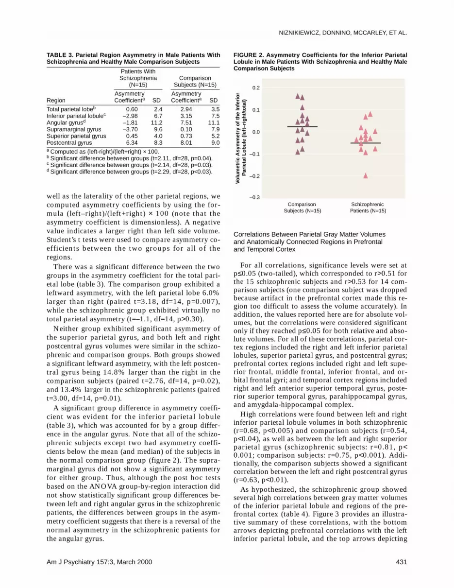
A significant group difference in asymmetry coeffi-cient was evident for the inferior parietal lobule(table 3), which was accounted for by a group differ-ence in the angular gyrus. Note that all of the schizo-phrenic subjects except two had asymmetry coeffi-cients below the mean (and median) of the subjects inthe normal comparison group (figure 2). The supra-marginal gyrus did not show a significant asymmetryfor either group. Thus, although the post hoc testsbased on the ANOVA group-by-region interaction didnot show statistically significant group differences be-tween left and right angular gyrus in the schizophrenicpatients, the differences between groups in the asym-metry coefficient suggests that there is a reversal of thenormal asymmetry in the schizophrenic patients forthe angular gyrus.
Correlations Between Parietal Gray Matter Volumes and Anatomically Connected Regions in Prefrontal and Temporal Cortex
For all correlations, significance levels were set atp≤0.05 (two-tailed), which corresponded to r>0.51 forthe 15 schizophrenic subjects and r>0.53 for 14 com-parison subjects (one comparison subject was droppedbecause artifact in the prefrontal cortex made this re-gion too difficult to assess the volume accurately). Inaddition, the values reported here are for absolute vol-umes, but the correlations were considered significantonly if they reached p≤0.05 for both relative and abso-lute volumes. For all of these correlations, parietal cor-tex regions included the right and left inferior parietallobules, superior parietal gyrus, and postcentral gyrus;prefrontal cortex regions included right and left supe-rior frontal, middle frontal, inferior frontal, and or-bital frontal gyri; and temporal cortex regions includedright and left anterior superior temporal gyrus, poste-rior superior temporal gyrus, parahippocampal gyrus,and amygdala-hippocampal complex.
High correlations were found between left and rightinferior parietal lobule volumes in both schizophrenic(r=0.68, p<0.005) and comparison subjects (r=0.54,p<0.04), as well as between the left and right superiorparietal gyrus (schizophrenic subjects: r=0.81, p<0.001; comparison subjects: r=0.75, p<0.001). Addi-tionally, the comparison subjects showed a significantcorrelation between the left and right postcentral gyrus(r=0.63, p<0.01).
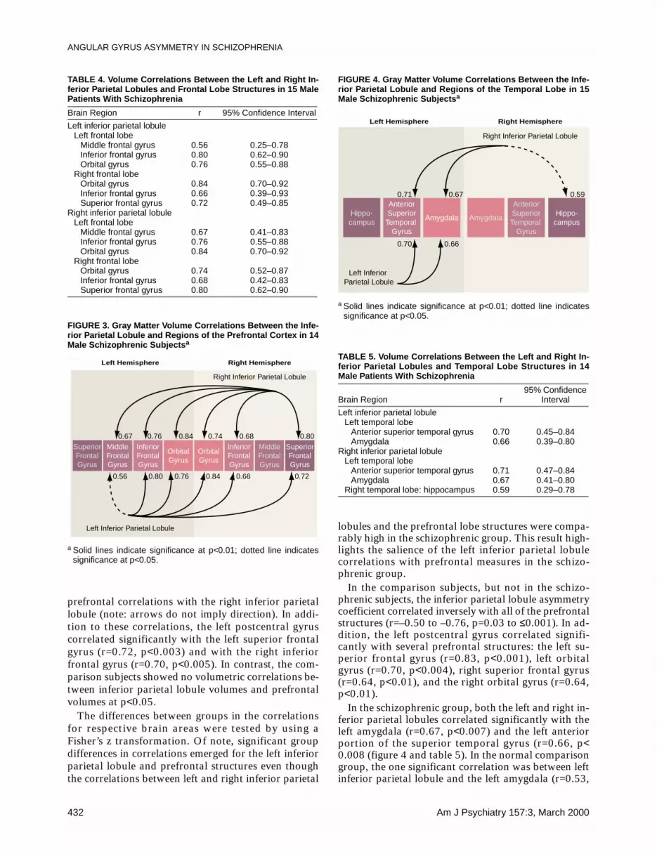
As hypothesized, the schizophrenic group showedseveral high correlations between gray matter volumesof the inferior parietal lobule and regions of the pre-frontal cortex (table 4). Figure 3 provides an illustra-tive summary of these correlations, with the bottomarrows depicting prefrontal correlations with the leftinferior parietal lobule, and the top arrows depicting
TABLE 3. Parietal Region Asymmetry in Male Patients WithSchizophrenia and Healthy Male Comparison Subjects
Region
Patients WithSchizophrenia
(N=15)Comparison
Subjects (N=15)
Asymmetry Coefficienta SD
Asymmetry Coefficienta SD
Total parietal lobeb 0.60 2.4 2.94 3.5Inferior parietal lobulec –2.98 6.7 3.15 7.5Angular gyrusd –1.81 11.2 7.51 11.1Supramarginal gyrus –3.70 9.6 0.10 7.9Superior parietal gyrus 0.45 4.0 0.73 5.2Postcentral gyrus 6.34 8.3 8.01 9.0a Computed as (left-right)/(left+right) × 100.b Significant difference between groups (t=2.11, df=28, p=0.04).c Significant difference between groups (t=2.14, df=28, p=0.03).d Significant difference between groups (t=2.29, df=28, p<0.03).
FIGURE 2. Asymmetry Coefficients for the Inferior ParietalLobule in Male Patients With Schizophrenia and Healthy MaleComparison Subjects
–0.3
–0.2
–0.1
0.0
0.1
0.2
Volu
met
ric
Asy
mm
etry
of
the
Infe
rio
r P
arie
tal L
obu
le (
left
–rig
ht/
tota
l)
Comparison Subjects (N=15)
Schizophrenic Patients (N=15)
432 Am J Psychiatry 157:3, March 2000
ANGULAR GYRUS ASYMMETRY IN SCHIZOPHRENIA
prefrontal correlations with the right inferior parietallobule (note: arrows do not imply direction). In addi-tion to these correlations, the left postcentral gyruscorrelated significantly with the left superior frontalgyrus (r=0.72, p<0.003) and with the right inferiorfrontal gyrus (r=0.70, p<0.005). In contrast, the com-parison subjects showed no volumetric correlations be-tween inferior parietal lobule volumes and prefrontalvolumes at p<0.05.
The differences between groups in the correlationsfor respective brain areas were tested by using aFisher’s z transformation. Of note, significant groupdifferences in correlations emerged for the left inferiorparietal lobule and prefrontal structures even thoughthe correlations between left and right inferior parietal
lobules and the prefrontal lobe structures were compa-rably high in the schizophrenic group. This result high-lights the salience of the left inferior parietal lobulecorrelations with prefrontal measures in the schizo-phrenic group.
In the comparison subjects, but not in the schizo-phrenic subjects, the inferior parietal lobule asymmetrycoefficient correlated inversely with all of the prefrontalstructures (r=–0.50 to –0.76, p=0.03 to ≤0.001). In ad-dition, the left postcentral gyrus correlated signifi-cantly with several prefrontal structures: the left su-perior frontal gyrus (r=0.83, p<0.001), left orbitalgyrus (r=0.70, p<0.004), right superior frontal gyrus(r=0.64, p<0.01), and the right orbital gyrus (r=0.64,p<0.01).
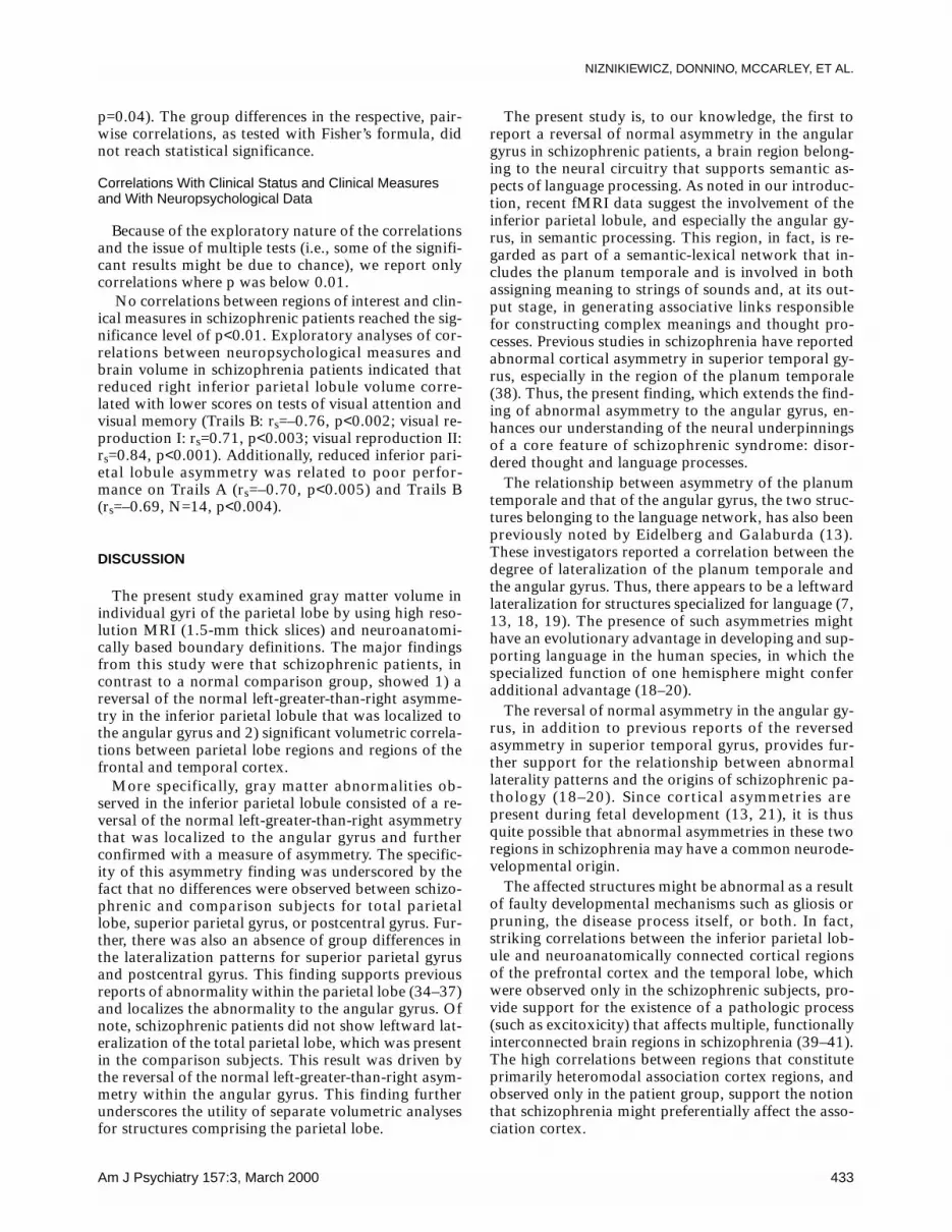
In the schizophrenic group, both the left and right in-ferior parietal lobules correlated significantly with theleft amygdala (r=0.67, p<0.007) and the left anteriorportion of the superior temporal gyrus (r=0.66, p<0.008 (figure 4 and table 5). In the normal comparisongroup, the one significant correlation was between leftinferior parietal lobule and the left amygdala (r=0.53,
TABLE 4. Volume Correlations Between the Left and Right In-ferior Parietal Lobules and Frontal Lobe Structures in 15 MalePatients With Schizophrenia
Brain Region r 95% Confidence Interval
Left inferior parietal lobuleLeft frontal lobe
Middle frontal gyrus 0.56 0.25–0.78Inferior frontal gyrus 0.80 0.62–0.90Orbital gyrus 0.76 0.55–0.88
Right frontal lobeOrbital gyrus 0.84 0.70–0.92Inferior frontal gyrus 0.66 0.39–0.93Superior frontal gyrus 0.72 0.49–0.85
Right inferior parietal lobuleLeft frontal lobe
Middle frontal gyrus 0.67 0.41–0.83Inferior frontal gyrus 0.76 0.55–0.88Orbital gyrus 0.84 0.70–0.92
Right frontal lobeOrbital gyrus 0.74 0.52–0.87Inferior frontal gyrus 0.68 0.42–0.83Superior frontal gyrus 0.80 0.62–0.90
FIGURE 3. Gray Matter Volume Correlations Between the Infe-rior Parietal Lobule and Regions of the Prefrontal Cortex in 14Male Schizophrenic Subjectsa
a Solid lines indicate significance at p<0.01; dotted line indicatessignificance at p<0.05.
0.67
0.56
0.76
0.80
0.84
0.76
0.74
0.84
0.68
0.66
0.80
0.72
Left Inferior Parietal Lobule
Right Inferior Parietal Lobule
Superior Frontal Gyrus
Middle
Frontal Gyrus
Inferior Frontal Gyrus
Orbital Gyrus
Superior Frontal Gyrus
Middle
Frontal Gyrus
Inferior Frontal Gyrus
Orbital Gyrus
Left Hemisphere Right Hemisphere
FIGURE 4. Gray Matter Volume Correlations Between the Infe-rior Parietal Lobule and Regions of the Temporal Lobe in 15Male Schizophrenic Subjectsa
a Solid lines indicate significance at p<0.01; dotted line indicatessignificance at p<0.05.
TABLE 5. Volume Correlations Between the Left and Right In-ferior Parietal Lobules and Temporal Lobe Structures in 14Male Patients With Schizophrenia
Brain Region r95% Confidence
Interval
Left inferior parietal lobuleLeft temporal lobe
Anterior superior temporal gyrus 0.70 0.45–0.84Amygdala 0.66 0.39–0.80
Right inferior parietal lobuleLeft temporal lobe
Anterior superior temporal gyrus 0.71 0.47–0.84Amygdala 0.67 0.41–0.80
Right temporal lobe: hippocampus 0.59 0.29–0.78
0.71
0.70
0.67
0.66
0.59
Left Inferior Parietal Lobule
Right Inferior Parietal Lobule
Hippo- campus
Hippo- campus
Anterior Superior
Temporal Gyrus
Anterior Superior
Temporal Gyrus
Amygdala Amygdala
Left Hemisphere Right Hemisphere
Am J Psychiatry 157:3, March 2000 433
NIZNIKIEWICZ, DONNINO, MCCARLEY, ET AL.
p=0.04). The group differences in the respective, pair-wise correlations, as tested with Fisher’s formula, didnot reach statistical significance.
Correlations With Clinical Status and Clinical Measures and With Neuropsychological Data
Because of the exploratory nature of the correlationsand the issue of multiple tests (i.e., some of the signifi-cant results might be due to chance), we report onlycorrelations where p was below 0.01.
No correlations between regions of interest and clin-ical measures in schizophrenic patients reached the sig-nificance level of p<0.01. Exploratory analyses of cor-relations between neuropsychological measures andbrain volume in schizophrenia patients indicated thatreduced right inferior parietal lobule volume corre-lated with lower scores on tests of visual attention andvisual memory (Trails B: rs=–0.76, p<0.002; visual re-production I: rs=0.71, p<0.003; visual reproduction II:rs=0.84, p<0.001). Additionally, reduced inferior pari-etal lobule asymmetry was related to poor perfor-mance on Trails A (rs=–0.70, p<0.005) and Trails B(rs=–0.69, N=14, p<0.004).
DISCUSSION
The present study examined gray matter volume inindividual gyri of the parietal lobe by using high reso-lution MRI (1.5-mm thick slices) and neuroanatomi-cally based boundary definitions. The major findingsfrom this study were that schizophrenic patients, incontrast to a normal comparison group, showed 1) areversal of the normal left-greater-than-right asymme-try in the inferior parietal lobule that was localized tothe angular gyrus and 2) significant volumetric correla-tions between parietal lobe regions and regions of thefrontal and temporal cortex.
More specifically, gray matter abnormalities ob-served in the inferior parietal lobule consisted of a re-versal of the normal left-greater-than-right asymmetrythat was localized to the angular gyrus and furtherconfirmed with a measure of asymmetry. The specific-ity of this asymmetry finding was underscored by thefact that no differences were observed between schizo-phrenic and comparison subjects for total parietallobe, superior parietal gyrus, or postcentral gyrus. Fur-ther, there was also an absence of group differences inthe lateralization patterns for superior parietal gyrusand postcentral gyrus. This finding supports previousreports of abnormality within the parietal lobe (34–37)and localizes the abnormality to the angular gyrus. Ofnote, schizophrenic patients did not show leftward lat-eralization of the total parietal lobe, which was presentin the comparison subjects. This result was driven bythe reversal of the normal left-greater-than-right asym-metry within the angular gyrus. This finding furtherunderscores the utility of separate volumetric analysesfor structures comprising the parietal lobe.
The present study is, to our knowledge, the first toreport a reversal of normal asymmetry in the angulargyrus in schizophrenic patients, a brain region belong-ing to the neural circuitry that supports semantic as-pects of language processing. As noted in our introduc-tion, recent fMRI data suggest the involvement of theinferior parietal lobule, and especially the angular gy-rus, in semantic processing. This region, in fact, is re-garded as part of a semantic-lexical network that in-cludes the planum temporale and is involved in bothassigning meaning to strings of sounds and, at its out-put stage, in generating associative links responsiblefor constructing complex meanings and thought pro-cesses. Previous studies in schizophrenia have reportedabnormal cortical asymmetry in superior temporal gy-rus, especially in the region of the planum temporale(38). Thus, the present finding, which extends the find-ing of abnormal asymmetry to the angular gyrus, en-hances our understanding of the neural underpinningsof a core feature of schizophrenic syndrome: disor-dered thought and language processes.
The relationship between asymmetry of the planumtemporale and that of the angular gyrus, the two struc-tures belonging to the language network, has also beenpreviously noted by Eidelberg and Galaburda (13).These investigators reported a correlation between thedegree of lateralization of the planum temporale andthe angular gyrus. Thus, there appears to be a leftwardlateralization for structures specialized for language (7,13, 18, 19). The presence of such asymmetries mighthave an evolutionary advantage in developing and sup-porting language in the human species, in which thespecialized function of one hemisphere might conferadditional advantage (18–20).
The reversal of normal asymmetry in the angular gy-rus, in addition to previous reports of the reversedasymmetry in superior temporal gyrus, provides fur-ther support for the relationship between abnormallaterality patterns and the origins of schizophrenic pa-thology (18–20). Since cortical asymmetries arepresent during fetal development (13, 21), it is thusquite possible that abnormal asymmetries in these tworegions in schizophrenia may have a common neurode-velopmental origin.
The affected structures might be abnormal as a resultof faulty developmental mechanisms such as gliosis orpruning, the disease process itself, or both. In fact,striking correlations between the inferior parietal lob-ule and neuroanatomically connected cortical regionsof the prefrontal cortex and the temporal lobe, whichwere observed only in the schizophrenic subjects, pro-vide support for the existence of a pathologic process(such as excitoxicity) that affects multiple, functionallyinterconnected brain regions in schizophrenia (39–41).The high correlations between regions that constituteprimarily heteromodal association cortex regions, andobserved only in the patient group, support the notionthat schizophrenia might preferentially affect the asso-ciation cortex.

434 Am J Psychiatry 157:3, March 2000
ANGULAR GYRUS ASYMMETRY IN SCHIZOPHRENIA
Also of interest is the finding that in comparison sub-jects, the asymmetry coefficient was inversely corre-lated with frontal lobe volumes, which indicates that anormal brain’s development entails leftward lateraliza-tion of language structures (13, 18, 19), and that thislateralization pattern is correlated in a healthy brainwith the frontal lobes, which are intimately involved inmediating processes in parietal language areas.
A limitation of this study is the use of multiple tests,which elevates the risk of type I errors. However, wefocused our attention on the inferior parietal lobule,where we had an a priori rationale for evaluating boththe angular and supramarginal gyri. Furthermore, weused more conservative criteria for the significancelevel (i.e., p≤0.01) of the exploratory analyses as acompromise for the multiple correlations performed.
In summary, this study, which employed improvedmethods of parcellation and measurement of the graymatter of the parietal lobe, provides evidence for theabsence of normal left-greater-than-right asymmetry inthe angular gyrus, a brain region categorized function-ally as part of the heteromodal association cortex and,importantly, linked to aspects of semantic processing.These findings, taken together, provide support for lo-calized volumetric changes in schizophrenia associatedwith selective cognitive impairments and a possibleneurodevelopmental component to schizophrenicpathogenesis. These findings also afford a more com-
prehensive understanding of schizophrenic pathologyby demonstrating similar pathologic processes that af-fect functionally related brain areas: the inferior pari-etal lobule region and the temporal and frontal areas.
APPENDIX 1. Delineating Regions Within the Parietal Lobe
For parcellating the parietal lobe we first used a reslice ed-iting algorithm to reformat images into the sagittal and axialplane. Specific markings were then made on the sagittal slicesin order to define specific neuroanatomical boundaries fordelineating the gyri. These markings were then recreated onthe original three-dimensional Fourier transform spoiled gra-dient recall acquisition coronal images, where the regions ofinterest were completed by using manually guided tracing.The final editing step was a surface-rendering algorithm thatwas used to create three-dimensional views of the relevantstructures (42). The three-dimensional images of the regionsof interest could then be viewed individually or within thecontext of the entire cortex, whereby images could be rotatedaround x, y, and z axes in order to achieve the best possiblevisualization of each region of interest. After examining thethree-dimensional images, any necessary corrections weremade on the original region of interest tracing. Once editingwas complete, volumetric measures were derived for each re-gion of interest, as was done for the whole brain volume, bysumming the voxels for each region of interest across all rel-evant slices.
Defining Regions of Interest
The boundaries for the regions of interest in this studywere determined with the assistance of an anatomical atlas(43). For all boundaries that involved cutting planes, we cor-
FIGURE 5. Midsagittal MRI Slice Depicting Measurement ofHead Rotation (Tilt) About the Bitemporal Axisa
a The reference line (in red), used to measure head tilt, was drawnfrom the most anterior point of the corpus callosum to the mostposterior point. This line was found to be virtually parallel to the bi-commissural line (i.e., the white line drawn between the anteriorcommissure and the posterior commissure).
FIGURE 6. Regions of the Parietal Lobea
a Image depicts a sagittal MRI slice approximately 5 mm lateral tothe midsagittal slice, with the superior parietal gyrus traced in yel-low and the postcentral gyrus in blue.
1
2
3
4C
B
A

Am J Psychiatry 157:3, March 2000 435
NIZNIKIEWICZ, DONNINO, MCCARLEY, ET AL.
rected for head rotation (tilt) around all three axes. Head ro-tation about the fronto-occipital axis was measured by a linedrawn perpendicular to the interhemispheric fissure on acoronal slice at the level of the parietal lobe. Head rotationabout the vertical (z) axis was measured by a line drawn per-pendicular to the interhemispheric fissure on an axial slice atthe level of the parietal lobe. Head rotation about the bitem-
poral axis was measured by a line drawn from the most an-terior point of the corpus callosum to the most posteriorpoint (illustrated in figure 5) on a midsagittal slice. This ref-erence line was more reliably determined than an anterior toposterior commissure line, which was verified as being virtu-ally parallel with the callosal line (mean difference angle lessthan 3°) for the 30 cases reported here. After correction for
FIGURE 7. Three-Dimensional Surface Rendering of the Cortexa
a Gyri of the parietal lobe are color-coded as follows: postcentral gyrus (blue), superior parietal gyrus (green), supramarginal gyrus (red),and angular gyrus (yellow). See text and subsequent figures for detailed descriptions of the numerically designated boundaries and thealphabetically labeled planes.
FIGURE 8. Boundaries Defining Plane B (from figure 7) of the Cortexa
a The left side of the figure shows a midsagittal MRI slice with the reference line from figure 5 in red. The white line was drawn from the mostanterior to the most posterior point of the corpus callosum, which formed a 9° angle with the reference line. The right side of the figuredepicts the second line that defined the boundary of plane B, which was drawn on a lateral (sagittal) MRI slice with the same x, y coordi-nates as the red reference line.
9

436 Am J Psychiatry 157:3, March 2000
ANGULAR GYRUS ASYMMETRY IN SCHIZOPHRENIA
rotation about the fronto-occipital and vertical axes, allbrain rotation about the bitemporal axis was corrected tomatch the brain with the least rotation (brain with the cal-losal line most nearly horizontal).
Parietal Lobe
Medial surface. The parietal lobe is bounded by the frontallobe and occipital lobe and by the cingulate gyrus (figure 6).We defined the frontoparietal border by the central sulcus(label 1) and the marginal ramus of the cingulate sulcus (la-bel 2) and, since the sulci do not intersect, by a vertical linethat extended from the most posterior portion of the centralsulcus to the cingulate sulcus (line A). This line was extendedlaterally in the coronal plane (perpendicular to the sagittalplane). The parieto-occipital fissure (label 4) was a clear an-atomical boundary that separated the parietal and occipitallobes. The parietal lobe and cingulate gyrus were boundedanteriorly by the subparietal sulcus (label 3). In the absenceof a clear anatomical division, we defined the posterior andventral parietocingulate border by a vertical line (line B) thatextended from the subparietal sulcus to the occipitoparietalfissure. This line was drawn on the coronal slice 13.5 mm(nine slices) posterior to the most posterior point of the cal-losum. This line was extended laterally in the coronal plane(perpendicular to the sagittal plane).
Lateral surface. The lateral boundaries between the pari-etal lobe and the frontal, temporal, and occipital lobes can beseen in the three-dimensional surface renderings of the cortexin figure 7.
Anteriorly, the central sulcus (label 1) is seen as the parieto-frontal lobe boundary. The Sylvian fissure (label 4) boundedthe parietal and temporal lobes anteriorly. More posteriorly,the ventral boundary of the parietal lobe was defined by thethree planes (labeled A, B, and C), which are all perpendicularto the sagittal plane. Plane A began at the dorsal level of theSylvian fissure on the most posterior coronal slice of the post-
central gyrus and continued posteriorly by using the samevertical (z) position for 15 mm (10 coronal slices).
Plane B of the ventral parietal boundary was defined bytwo parallel lines. The first line was drawn on a midsagittalslice from the most anterior point of the corpus callosum andextending posteriorly at a 9° angle to the callosal referenceline from figure 5 (figure 8, left image). The second line thatdefined this plane was drawn on a more lateral sagittal sliceand used the same coordinates as the first boundary line(figure 8, right image). The 9° angle between the referenceline and the boundary line was selected so as to include themaximum amount of parietal lobe gray matter without in-cluding any (or at least only minimal amounts of) temporalor occipital lobe tissue. The posterior boundary of the pari-etal lobe was the plane C in figure 7, defined by two parallellines. The first line was drawn through the parieto-occipitalfissure on a midsagittal slice (figure 9, left image). The sec-ond line was drawn on a more lateral sagittal slice by usingthe same coordinates as the first line (figure 9, right image).
Left and right parietal hemispheres were separated by theinterhemispheric fissure.
Parcellation of the Parietal Lobe
The regions of interest within the parietal lobe include thepostcentral gyrus, superior parietal gyrus, and inferior pari-etal lobule. The inferior parietal lobule consists of the angu-lar gyrus and the supramarginal gyrus. As can be seen infigure 7, the postcentral gyrus was separated from the supe-rior parietal gyrus and the inferior parietal lobule by thepostcentral sulcus (label 2). The inferior parietal lobule wasseparated from the superior parietal gyrus by the intrapari-etal sulcus (label 3). The inferior parietal lobule was furthersubdivided into the angular gyrus and the supramarginal gy-rus. In the absence of a clear, consistent anatomical boundarybetween the angular gyrus and the supramarginal gyrus, thebound between these two inferior parietal lobule structures
FIGURE 9. Boundaries Defining Plane C (from figure 7) of the Cortexa
a The plane was defined by two parallel lines, the first of which is depicted in the midsagittal MRI slice on the left as a straight line drawnthrough the parieto-occipital fissure. The second line was drawn with identical x, y coordinates as the first line and is shown on the morelateral sagittal MRI slice on the right.
Am J Psychiatry 157:3, March 2000 437
NIZNIKIEWICZ, DONNINO, MCCARLEY, ET AL.
was defined by the coronal slice midway between the mostposterior and most anterior coronal slices of the inferior pa-rietal lobule. For this reason it is possible that some volumet-ric data points categorized as belonging to the angular gyrusmight be a part of the supramarginal gyrus and conversely,some supramarginal gyrus volumetric data could have beencategorized as belonging to the angular gyrus. Subtle bias inthe final volumetric data is thus conceivable. Data fromother laboratories as well as data coming from this labora-tory from a different sample should help resolve this issue.
REFERENCES
1. Mesulam MM: Large-scale neurocognitive networks and dis-tributed processing for attention, language, and memory. AnnNeurol 1990; 28:597–613
2. Park S, Holzman PS: Schizophrenics show spatial workingmemory deficits. Arch Gen Psychiatry 1992; 49:975–982
3. Nuechterlein KH, Dawson ME: Information processing and at-tentional functioning in the developmental course of schizo-phrenic disorders. Schizophr Bull 1984; 10:160–203
4. Nestor PG, Faux SF, McCarley RW, Shenton ME, Sands SF:Measurement of visual sustained attention in schizophreniausing signal detection analysis and a newly developed com-puterized CPT task. Schizophr Res 1990; 3:329–332
5. Shenton ME, Wible CG, McCarley RW: A review of magneticresonance imaging studies of brain abnormalities in schizo-phrenia, in Brain Imaging in Clinical Psychiatry. Edited byKrishnan KRR, Doraiswamy PM. New York, Marcel Dekker,1997, pp 297–380
6. McCarley RW, Wible CG, Frumin M, Hirayasu Y, Levitt JJ, Fis-cher IA, Shenton ME: MRI anatomy of schizophrenia. BiolPsychiatry 1999; 45:1099–1119
7. Pearlson GD, Petty RG, Ross CA, Tien AY: Schizophrenia: adisease of heteromodal association cortex? Neuropsycho-pharmacology 1996; 14:1–17
8. Binder J: Functional magnetic resonance imaging: languagemapping. Neurosurg Clin N Am 1997; 8:383–392
9. Frackowiak RS: Functional mapping of verbal memory andlanguage. Trends Neurosci 1994; 17:109–115
10. Howard D, Patterson K, Wise R, Brown WD, Friston K, WeillerC, Frackowiak R: The cortical localization of the lexicons:positron emission tomography evidence. Brain 1992; 115:1769–1782
11. Shenton ME, Kikinis R, Jolesz FA, Pollak SD, LeMay M, WibleCG, Hokama H, Martin J, Metcalf D, Coleman M, McCarleyRW: Abnormalities of the left temporal lobe and thought disor-der in schizophrenia: a quantitative magnetic resonance im-aging study. N Engl J Med 1992; 327:604–612
12. Barta PE, Pearlson GD, Powers RE, Richards SS, Tune LE: Au-ditory hallucinations and smaller superior temporal gyral vol-ume in schizophrenia. Am J Psychiatry 1990; 147:1457–1462
13. Eidelberg D, Galaburda AM: Inferior parietal lobule: divergentarchitectonic asymmetries in the human brain. Arch Neurol1984; 41:843–852
14. Galaburda AM, Geschwind N: The human language areasand cerebral asymmetries. Rev Med Suisse Romande 1980;100:119–128
15. Hier D, LeMay M, Rosenberger P: Autism associated with re-versed cerebral asymmetry. Neurology 1978; 28:348–349
16. Hier DB, LeMay M, Rosenberger PB: Autism and unfavorableleft-right asymmetries of the brain. J Autism Dev Disord 1979;9:153–159
17. Rosenberger PB, Hier DB: Cerebral asymmetry and verbal in-tellectual deficits. Ann Neurol 1980; 8:300–304
18. Crow TJ: The continuum of psychosis and its genetic origins:the sixty-fifth Maudsley lecture. Br J Psychiatry 1990; 156:788–797
19. Crow TJ: Temporal lobe asymmetries as the key to the etiol-ogy of schizophrenia. Schizophr Bull 1990; 16:433–443
20. Crow TJ: The nature of psychotic illness. Psychiatr Neurol Jpn1992; 94:820–836
21. Chi JG, Dooling EC, Gilles FH: Gyral development of the hu-man brain. Ann Neurol 1977; 1:86–93
22. Cavada C, Goldman-Rakic PS: Posterior parietal cortex inrhesus monkey, I: parcellation of areas based on distinctivelimbic and sensory corticocortical connections. J Comp Neu-rol 1989; 287:393–421
23. Seltzer B, Pandya DN: Further observations on parieto-tem-poral connections in the rhesus monkey. Exp Brain Res 1984;55:301–312
24. Wible CG, Shenton ME, Hokama H, Kikinis R, Jolesz FA, Met-calf D, McCarley RW: Prefrontal cortex and schizophrenia: aquantitative magnetic resonance imaging study. Arch GenPsychiatry 1995; 52:279–288
25. Wible CG, Shenton ME, Fischer IA, Allard JE, Kikinis R, JoleszFA, Iosifescu DV, McCarley RW: Parcellation of the human pre-frontal cortex using MRI. Psychiatry Res 1997; 76:29–40
26. Spitzer RL, Endicott J: Schedule for Affective Disorders andSchizophrenia—Lifetime Version. New York, New York StatePsychiatric Institute, Biometrics Research, 1978
27. Wechsler D: Wechsler Memory Scale—Revised. New York,Harcourt Brace Jovanovich, 1981
28. Andreasen NC: Scale for the Assessment of Positive Symp-toms (SAPS). Iowa City, University of Iowa, 1984
29. Andreasen NC: Scale for the Assessment of Negative Symp-toms (SANS). Iowa City, University of Iowa, 1981
30. Johnston MH, Holzman PS: Assessing Schizophrenic Think-ing. San Francisco, Jossey-Bass, 1979
31. Nestor PG, Shenton ME, McCarley RW, Haimson J, SmithRS, O’Donnell B, Kimble M, Kikinis R, Jolesz FA: Neuropsy-chological correlates of MRI temporal lobe abnormalities inschizophrenia. Am J Psychiatry 1993; 150:1849–1855
32. Gerig G, Kikinis R, Kübler O: Significant Improvement of MR Im-age Data Quality Using Anisotropic Diffusion Filtering: TechnicalReport BIWI-TR-124. Zurich, Switzerland, Communication Tech-nology Laboratory, Image Science Division, ETH Zurich, 1990
33. Cline HE, Lorensen WE, Ludke S, Crawford CR, Teeter BC:Two algorithms for the three-dimensional reconstruction of to-mograms. Med Phys 1988; 15:320–327
34. McGilchrist I, Goldstein LH, Jadresic D, Fenwick P: Thalamo-frontal psychosis. Br J Psychiatry 1993; 163:113–115
35. Schlaepfer TE, Harris GJ, Tien AY, Peng LW, Lee S, Feder-man EB, Chase GA, Barta PE, Pearlson GD: Decreased re-gional cortical gray matter volume in schizophrenia. Am J Psy-chiatry 1994; 151:842–848
36. Andreasen NC, Flashman L, Flaum M, Arndt S, Swayze V II,O’Leary DS, Ehrhardt JC, Yuh WTC: Regional brain abnor-malities in schizophrenia measured with magnetic resonanceimaging. JAMA 1994; 272:1763–1769
37. Bilder RM, Wu H, Bogerts B, Degreef G, Ashtari M, Alvir JM,Snyder PJ, Lieberman JA: Absence of regional hemisphericvolume asymmetries in first-episode schizophrenia. Am JPsychiatry 1994; 151:1437–1447
38. Kwon JS, McCarley RW, Hirayasu Y, Anderson JE, Fischer IA,Kikinis R, Jolesz FA, Shenton ME: Left planum temporale vol-ume reduction in schizophrenia. Arch Gen Psychiatry 1999;56:142–148
39. McCarley RW, Shenton ME, O’Donnell BF, Nestor PG: UnitingKraepelin and Bleuler: the psychology of schizophrenia andthe biology of temporal lobe abnormalities. Harv Rev Psychi-atry 1993; 1:36–56
40. Grunze HC, Rainnie DG, Hasselmo ME, Barkai E, Hearn EF,McCarley RW, Greene RW: NMDA-dependent modulation ofCA1 local circuit inhibition. J Neurosci 1996; 16:2034–2043
41. Ikonomidou C, Bosch F, Miksa M, Bittigau P, Vockler J, Dikra-nian K, Tenkova TI, Stefovska V, Turski L, Olney JW: Blockadeof NMDA receptors and apoptotic neurodegeneration in thedeveloping brain. Science 1999; 283:70–74
42. Cline HE, Lorensen WE, Souza SP, Jolesz FA, Kikinis R, GerigG, Kennedy TE: 3D surface rendered MR images of the brainand its vasculature. J Comput Assist Tomogr 1991; 15:344–351
43. Duvernoy HM: The Human Brain: Surface, Three-DimensionalSectional Anatomy, and MRI. Vienna, Springer-Verlag, 1991
Related Documents