Ablation characteristics of quantum square pulse mode dental erbium laser Nejc Lukac ˇ Tomaž Suhovršnik Matjaž Lukac ˇ Matija Jezeršek Downloaded From: https://www.spiedigitallibrary.org/journals/Journal-of-Biomedical-Optics on 05 Jul 2022 Terms of Use: https://www.spiedigitallibrary.org/terms-of-use

Welcome message from author
This document is posted to help you gain knowledge. Please leave a comment to let me know what you think about it! Share it to your friends and learn new things together.
Transcript
Ablation characteristics of quantumsquare pulse mode dental erbiumlaser
Nejc LukacTomaž SuhovršnikMatjaž LukacMatija Jezeršek
Downloaded From: https://www.spiedigitallibrary.org/journals/Journal-of-Biomedical-Optics on 05 Jul 2022Terms of Use: https://www.spiedigitallibrary.org/terms-of-use
Ablation characteristics of quantum square pulsemode dental erbium laser
Nejc Lukac,a,* Tomaž Suhovršnik,b Matjaž Lukac,c and Matija Jezeršeka
aUniversity of Ljubljana, Faculty of Mechanical Engineering, Askerceva 6, Ljubljana 1000, SloveniabUniversity of Ljubljana, Faculty of Physics, Jadranska 39, Ljubljana 1000, SloveniacInstitute Josef Stefan, Jamova 39, Ljubljana 1000, Slovenia
Abstract. Erbium lasers are by now an accepted tool for performing ablative medical procedures, especiallywhen minimal invasiveness is desired. Ideally, a minimally invasive laser cutting procedure should be fast andprecise, and with minimal pain and thermal side effects. All these characteristics are significantly influenced bylaser pulse duration, albeit not in the same manner. For example, high cutting efficacy and low heat depositionare characteristics of short pulses, while vibrations and ejected debris screening are less pronounced at longerpulse durations. We report on a study of ablation characteristics on dental enamel and cementum, of a chopped-pulse Er:YAG [quantum square pulse (QSP)] mode, which was designed to reduce debris screening during anablation process. It is shown that in comparison to other studied standard Er:YAG and Er,Cr:YSGG laser pulseduration modes, the QSP mode exhibits the highest ablation drilling efficacy with lowest heat deposition andreduced vibrations, demonstrating that debris screening has a considerable influence on the ablation process.By measuring single-pulse ablation depths, we also show that tissue desiccation during the consecutive deliveryof laser pulses leads to a significant reduction of the intrinsic ablation efficacy that cannot be fully restored underclinical settings by rehydrating the tooth using an external water spray. © 2016 Society of Photo-Optical Instrumentation
Engineers (SPIE) [DOI: 10.1117/1.JBO.21.1.015012]
Keywords: laser ablation; hard dental tissue; erbium lasers; water cooling; triangulation; hydrokinetic effect.
Paper 150663R received Oct. 5, 2015; accepted for publication Dec. 18, 2015; published online Jan. 25, 2016.
1 IntroductionPulsed midinfrared erbium lasers, such as such Er:YAG (witha wavelength of λ ¼ 2.94 μm) or Er,Cr:YSGG (λ ¼ 2.78 μm),have been recognized as the lasers of choice when fast ablationof human tissues with minimal side effects is required.1–4 This isbecause erbium laser wavelengths approximately coincide withthe major absorption peak of the water molecule and are thusstrongly absorbed in virtually all biological tissues.4 Numerousstudies2–8 have established that thermal cutting is the basicmechanism by which tissue is removed with erbium lasers.The thermal cutting mechanism is based on heating theinterstitially trapped water within tissue to the evaporationtemperature.3 This leads to explosive subsurface expansion ofinterstitially trapped water inside the tissue and subsequently tomicroexplosive internal tearing of the tissue. Erbium lasers arebecause of their high ablation efficacy, used in various medicalfields including dentistry, dermatology, surgery, ophthalmology,and gynecology.2,9–12
In order to perform minimally invasive medicine, it is impor-tant to optimize erbium laser parameters such that laser cutting isfast, cuts are sharp and precise, the procedure is quiet and withminimal vibrations imposed on the treated tissue, and theamount of residual heat that remains in the tissue followingerbium laser irradiation is minimal. However, with standard,bell or quasisquare-shaped erbium pulse modes,13 these require-ments are to a certain extent contradictory, as parameters thatmay be optimal, e.g., achieving fastest ablation, may not beoptimal when minimal vibrations and noise are required.14,15
For example, it is well understood that ablation thresholdsdecrease toward shorter pulse durations,16–19 where short pulsesare pulses shorter than the thermal relaxation time of the ablatedtissue.18,19 At shorter times, the energy has little time to escapefrom the ablated volume, and so less heat is diffused into thesurrounding tissue.18 Shorter pulses, therefore, result in fasterablation and smaller amount of residual heat deposition.20 Onthe other hand, shorter laser pulses are expected to producehigher-frequency tissue vibrations21 and may therefore causemore discomfort to patients.22
Similarly, when undesirable interaction of the laser beamwith the debris cloud is considered,23 the effects of debrisscreening are less pronounced at longer pulse durations.18 Atshort pulse durations, the incoming laser beam gets stronglyabsorbed in the dense debris cloud, which forms above theirradiated area, resulting in a reduced ablation rate. Cuts arealso less precise, since the scattering effect caused by the cloudleads to spreading of the laser beam.24–26 Finally, the laser-reheated debris cloud is expected to contribute, as it falls backto the tissue surface, to additional heating of the tissue, andconsequently to additional residual heat deposition.25
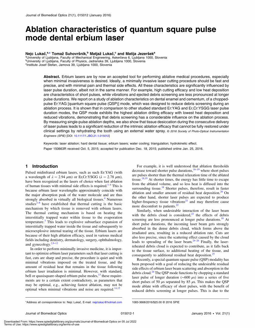
Recently, a special quantum square pulse (QSP) modality hasbeen proposed with a goal of reducing the undesirable residualside effects of erbium laser beam scattering and absorption in thedebris cloud.24 The QSP mode functions by chopping a standardlaser pulse of longer duration (∼600 μs) into a series of fiveshort pulses of 50 μs separated by 85 μs. This makes the QSPmode ablate with efficacy of short pulses, with the benefit ofreduced debris screening at longer pulses. This is due to the
*Address all correspondence to: Nejc Lukac, E-mail: [email protected] 1083-3668/2016/$25.00 © 2016 SPIE
Journal of Biomedical Optics 015012-1 January 2016 • Vol. 21(1)
Journal of Biomedical Optics 21(1), 015012 (January 2016)
Downloaded From: https://www.spiedigitallibrary.org/journals/Journal-of-Biomedical-Optics on 05 Jul 2022Terms of Use: https://www.spiedigitallibrary.org/terms-of-use
fact that the duration of each of the pulse quanta (∼50 μs) isshorter than the rise time of the debris cloud, while the separa-tion between the pulse quanta of ∼85 μs is longer than the decaytime of the debris cloud. Ablation measurements in dentalenamel have subsequently demonstrated that as a result ofreduced undesirable effects of laser–debris interaction, the useof the QSP mode results in sharper cuts and higher ablationefficacy in enamel.23,25,26
In this paper, we report on a systematic comparison of ablationcharacteristics of standard erbium laser pulse modes of differentpulse durations and wavelengths (Er:YAG and Er,Cr:YSGG) andof the new Er:YAG QSP chopped pulse profile mode. On anexample of ablation in dental enamel and cementum, we mea-sured ablation characteristics of the QSP mode with respect toablation drilling efficacy, vibrations, and residual heat deposition.
Typically, in order to improve the measurement accuracy,ablation cavities are made with a series of consecutive pulses,and the ablation efficacy expected from a single pulse is calcu-lated by dividing the measured volume or depth by the numberof delivered pulses.5,27 However, this approach may not bealways appropriate since, especially for tissues with relativelylow water content, the ablation efficacy has been observed toget progressively reduced upon consecutive delivery of laserpulses.28,29 When using such cumulative ablation methods,the measurement process influences the ablation process,which is being studied, and therefore, erroneous results and con-clusions may be reached. For example, it is well known that fordental enamel, the ablation process completely stalls after aconsecutive delivery of ∼10 laser pulses, unless the enamelis cooled by an external water spray.27,30–38 To explain thisobservation, an additional “hydrokinetic” ablative mechanismhas been proposed, which is based on the reception by waterspray particles of the laser energy and consequent “impartationof disruptive forces” by accelerated water particles to the tis-sue.32,33 The actual mechanism of laser ablation in the presenceof water spray is still controversial to a certain extent.27,34,35
In our study, in order to evaluate the influence of cumulativedesiccation on measured ablation efficacy, we carried out notonly cumulative but also individual pulse ablation measure-ments, using a sensitive triangulation measurement technique.We show that on enamel, the initial, “intrinsic” drilling efficacy(in mm3∕J) of the first pulse in a sequence of pulses is abouttwo-times higher compared to the cumulative efficacy of 10consecutively delivered pulses.
2 Materials and MethodsThe Er:YAG system used was a LightWalker AT (manufacturedby Fotona), and the Er,Cr:YSGG laser was a WaterLase iPlus(manufactured by Biolase). Both lasers were flashlamp pumpedand were fitted with the appropriate noncontact handpieces(Fotona H02 handpiece and Biolase Turbo handpiece withMX11 tip).
The lasers were operated in the following pulse modes anddurations: (a) Er:YAG laser in standard super short pulse (SSP,90 μs), medium short pulse (MSP, 130 μs), short pulse (SP,220 μs), and long pulse (LP, 360 μs) pulse modes, and in thenew QSP mode (600 μs) and (b) Er,Cr:YSGG laser in Hpulse (400 μs) duration mode. The temporal pulse shapes ofthe above modes have been reported in Refs. 17, 26, and 39,and are different for different modes and erbium laser types.For this reason, a more meaningful comparison of the measuredoutput laser pulse shapes can be made by integrating the
delivered laser energy over the duration of the laser pulse.39
The above pulse durations represent times during which 90%of the cumulative laser pulse energy (300 mJ for Er:YAG,and 170 mJ for Er,Cr:YSGG) were delivered to a target. Thetemporal structure of the chopped QSP mode and its relationto the development of ablation debris cloud is shown in Fig. 1.
Approximately 600-μs long QSP macropulse consists of fiveapproximately tQSP ≈ 50 μs long micropulses (pulse quanta)separated by a temporal spacing of ΔtQSP ≈ 85 μs. The durationof QSP micropulses is shorter than the rise time of the debriscloud, while the separation between the micropulses is longerthan the decay time of the debris cloud.24 It should be notedhere that the duration of the QSP micropulses is also shorterthan the thermal relaxation time (τ) for hard dental tissues ofapproximately τ ¼ 110 μs.18
The experiments were conducted on randomly chosenextracted premolar and molar teeth, which were stored in aphysiological saline solution immediately following extraction.Before each ablation experiment, the tooth was positioned tohave its surface perpendicular to the laser beam and to be atfocal distance of the laser beam. The diameters (d) of the laserbeam at the focal point were approximately d ¼ 0.9 mm for theEr:YAG laser and d ¼ 0.7 mm for the Er,Cr:YSGG laser, withexact diameters measured for each modality and laser energyusing a sensitive photographic paper.
Fig. 1 (a) Standard laser pulse; (b) QSP laser pulse. A 600-μs longmacropulse is chopped into five micropulses with micropulse durationof tQSP ¼ 50 μs, and temporal spacing between the micropulses ofΔtQSP ¼ 85 μs.
Journal of Biomedical Optics 015012-2 January 2016 • Vol. 21(1)
Lukac et al.: Ablation characteristics of quantum square pulse mode dental erbium laser
Downloaded From: https://www.spiedigitallibrary.org/journals/Journal-of-Biomedical-Optics on 05 Jul 2022Terms of Use: https://www.spiedigitallibrary.org/terms-of-use
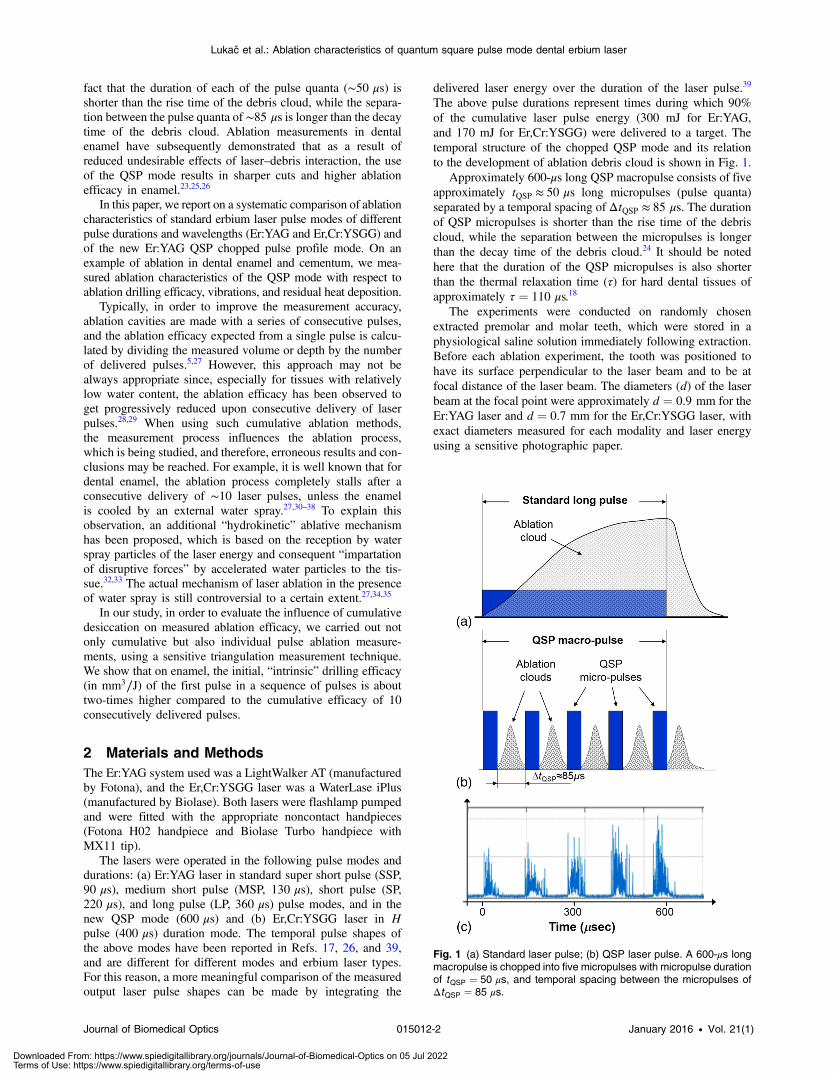
Measurements were made on enamel, which covers thecrown of the tooth, and on cementum, which covers the rootof the tooth. Enamel and cementum were chosen in order toevaluate the effects of Er:YAG laser radiation on hard tissuesat both extremes, i.e., in (a) enamel, which has a high contentof hydroxyapatite (96%) and a very low intrinsic water content(3%weight) and (b) in cementum, which consists of 45% ofhydroxyapatite, 33% of organic material mainly collagen andprotein polysaccharides and 22%weight water.
40 For comparison,dentin contains 12%weight of water, bone contains ∼22%weight ofwater, and soft tissue contains 70%weight of water.
Single-pulse laser energy, Eout (in J), was measured with anexternal energy meter at the handpiece output. Special care wastaken to ensure that with both laser systems, the ablation meas-urement pulse sequence was emitted after the laser output hadalready stabilized. The lasers were operated at a constantrepetition rate of 2 Hz and the repetition rate (f) of the pulsesdelivered to the tissue was controlled using an external shutter.Unless otherwise stated, the repetition rate was set tof ¼ 0.2 Hz. Since following an ablative Er:YAG laser pulsethe ablated tooth surface returns to the initial temperature in∼0.5 s,20 this slow repetition rate ensured that the tooth surfacecooled down between pulses, and therefore, each pulse startedthe ablation process from the same initial tooth temperature.
With the Er:YAG laser, 200 mJ of laser pulse energy wasused on cementum and 300 mJ on enamel. Different energieswere chosen in order to obtain cavities of similar depth inboth types of tissues. Similarly, with the smaller beam diameterEr,Cr:YSGG laser, lower pulse energies of 113 and 170 mJ wereused on cementum and enamel, respectively. The single-pulselaser fluence [F (in J∕mm2)], averaged over the irradiatedarea at the focal point, was calculated from F ¼ Eout∕ðπd2∕4Þ.
All laser pulses under consideration were multimode (trans-verse and longitudinal) pulses. The Er:YAG laser intensitydistributions in the irradiated area were approximately flat-top,concluding from the observed shapes of the shallow ablationcavities (h < 0.1 mm) made with a single-laser pulse. No effortwas made to measure exact intensity distributions as it wasdetermined in a separate study.41 The study showed that theshape of the ablated hole (with diameter smaller than 1 mm)depends not only on the spatial laser intensity distribution butalso on the depth of the ablated hole. Holes of medium depth(up to h ≈ 0.5 mm) are approximately conical while deepercavities obtain a more cylindrical shape.41
The irradiation fluences F, for QSP, SSP, SP, and H modes,as calculated from the measured laser pulse energies and beamdiameters, were correspondingly 38, 35, 40, and 44 J∕cm2 onenamel, and 26, 24, 27, and 29 J∕cm2 on cementum. These flu-ences were well above the published ablation threshold fluencesFth for enamel (with low water content) and dentin (with highwater content) of Fth ¼ 3.5 and 4 J∕cm2, respectively.18
The single-pulse ablation drilling efficacy [DF (in mm3∕J)],defined as ablation depth per laser fluence, was calculated fromDF ¼ L∕ðN × FÞ, where L is the depth of the ablated holefollowing N consecutively delivered laser pulses of the samepulse energy to the same spot on a target. Note that the drillingefficacy can be considered to represent also the ablation effi-cacy35 [AE (in mm3∕J)], defined as ablated volume per laserenergy, assuming a cylindrically shaped hole having a constantdiameter d over the depth L of the hole. In reality, the diameterof the hole gets smaller toward the bottom of the ablated
cavity,41 and therefore, the actual ablation efficacy is expectedto be smaller.
Experiments were carried out under three conditions:(a) “dry” conditions where after the tooth has been taken outof the saline solution, no external water was added to thetooth during the ablation experiment; (b) “hydration” experi-mental setup, which was designed to prevent, at least to a certainextent, the desiccation of the irradiated tooth surface. This wasaccomplished by rehydrating the ablation area following eachlaser pulse with an external water spray. This “hydration”phase lasted for T ¼ 1 s following each laser pulse. After rehy-dration, a special care was taken that the tooth surface was leftdevoid of any surface water. An air flow was directed to the abla-tion area for the duration of an “air-blowing” phase lasting 4 s, atthe end of which the tooth was again irradiated with a sub-sequent laser pulse. Note that this “hydration” experimentalsetup was similar to the “pores” condition used in Ref. 35;and (c) “spray” conditions where during the ablation experi-ment, a water/air spray was delivered continuously to the tooththrough the corresponding laser system handpieces. The spraywater flow rate (in ml/min) was determined by measuring thetime and water volume collected in an external container duringthat time. Unless otherwise stated, the error bars included inthe figures represent standard deviations of the sample data.
2.1 Optical Microscope Measurements
When optical microscope was used to measure the depth (h) ofablated holes, each ablation cavity was made with N ¼ 10, 20,or 30 consecutive laser pulses of the same pulse energy deliv-ered to the same spot. The depth of the ablated cavity followingthe ablation pulse sequence was then measured using a focusingoptical microscope (Leica, M205C). Each data point representsan average obtained from 10 different cavities, each made withN consecutive pulses.
2.2 Laser Triangulation Profilometry
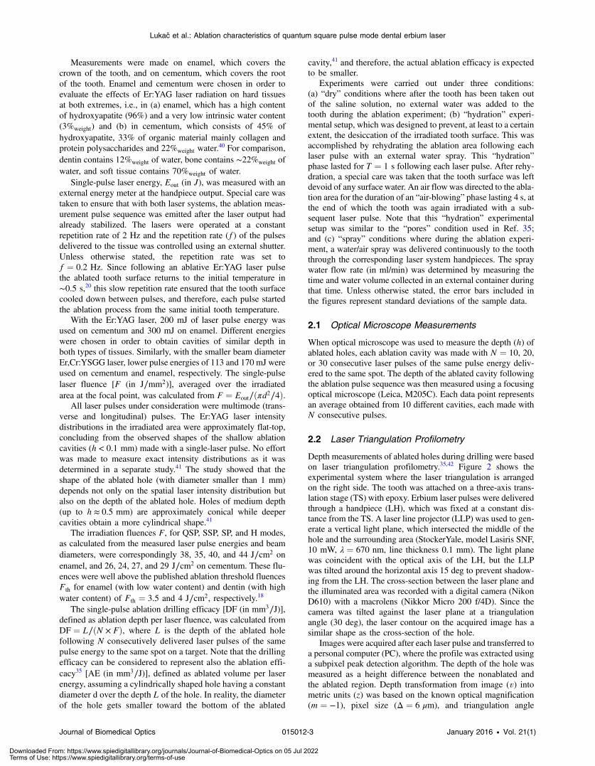
Depth measurements of ablated holes during drilling were basedon laser triangulation profilometry.35,42 Figure 2 shows theexperimental system where the laser triangulation is arrangedon the right side. The tooth was attached on a three-axis trans-lation stage (TS) with epoxy. Erbium laser pulses were deliveredthrough a handpiece (LH), which was fixed at a constant dis-tance from the TS. A laser line projector (LLP) was used to gen-erate a vertical light plane, which intersected the middle of thehole and the surrounding area (StockerYale, model Lasiris SNF,10 mW, λ ¼ 670 nm, line thickness 0.1 mm). The light planewas coincident with the optical axis of the LH, but the LLPwas tilted around the horizontal axis 15 deg to prevent shadow-ing from the LH. The cross-section between the laser plane andthe illuminated area was recorded with a digital camera (NikonD610) with a macrolens (Nikkor Micro 200 f/4D). Since thecamera was tilted against the laser plane at a triangulationangle (30 deg), the laser contour on the acquired image has asimilar shape as the cross-section of the hole.
Images were acquired after each laser pulse and transferred toa personal computer (PC), where the profile was extracted usinga subpixel peak detection algorithm. The depth of the hole wasmeasured as a height difference between the nonablated andthe ablated region. Depth transformation from image (v) intometric units (z) was based on the known optical magnification(m ¼ −1), pixel size (Δ ¼ 6 μm), and triangulation angle
Journal of Biomedical Optics 015012-3 January 2016 • Vol. 21(1)
Lukac et al.: Ablation characteristics of quantum square pulse mode dental erbium laser
Downloaded From: https://www.spiedigitallibrary.org/journals/Journal-of-Biomedical-Optics on 05 Jul 2022Terms of Use: https://www.spiedigitallibrary.org/terms-of-use
(α ¼ 30 deg) and was calculated from z ¼ Δv∕m sin α. Anexample of measured profiles after each of 20 consecutivelaser pulses is shown in Fig. 2 within the PC’s box.
2.3 Interferometric Vibration Measurements
An interferometric setup was implemented for measuring vibra-tion amplitudes of the tooth during the laser drilling. The inter-ferometer was based on the Fizeau configuration43 and isschematically shown on the left side of Fig. 2. A collimatedlaser beam from a HeNe laser (λ ¼ 633 nm, beam diameter ¼0.8 mm) was guided through a 50% beam splitter (BS) and awedge shaped reference plate (RP) and focused on the backsideof the tooth by an achromatic lens (AL) with a focal length of7.5 mm. Interference between the reference beam, reflectedfrom the RP, and the measuring beam, reflected from the toothsurface, was measured by a Si photodetector (PD1) (10-MHzbandwidth) positioned near the BS. In front of the PD1, a pin-hole with diameter of 0.5 mm was attached which transmittedonly the light from the central interference fringe. The second,InAs, photodetector (PD2) (60-MHz bandwidth) measured thereflected Er:YAG laser light and was used for triggering theoscilloscope (LeCroy, US, 600 MHz Wave Runner 64MXi-A).The amplitude of the first tooth oscillation after each laser pulse(which occurs at t0) was measured by counting the peaks andvalleys on the measured signal between t0 and the momentwhere the change in moving direction occurs. A typical exampleof such a change in moving direction is shown on oscilloscopein Fig. 2. The measured resonance frequency of the experimen-tal setup was ∼300 Hz. T∕2 in Fig. 2 represents half of the oscil-lation period, from the maximum displacement of the tooth inthe first direction to the maximum displacement of the tooth inthe opposite direction.
3 Results
3.1 Optical Microscope Technique Measurements
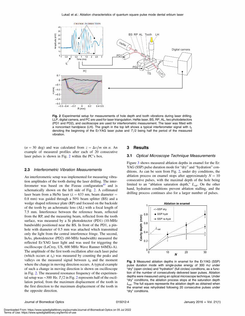
Figure 3 shows measured ablation depths in enamel for the Er:YAG (SSP) pulse duration mode for “dry” and “hydration” con-ditions. As can be seen from Fig. 2, under dry conditions, theablation process on enamel stops after approximately N ¼ 10consecutive pulses, with the maximal depth of the hole beinglimited to an “ablation saturation depth,” Lsat. On the otherhand, hydration conditions prevent ablation stalling, and thedrilling process continues also for a larger number of pulses.
Fig. 2 Experimental setup for measurements of hole depth and tooth vibrations during laser drilling.LLP, digital camera, and PC are used for laser triangulation. HeNe laser, BS, RP, AL, two photodetectors(PD1 and PD2), and oscilloscope are used for interferometric measurement. The laser was fitted witha noncontact handpiece (LH). The graph in the top left shows a typical interferometer signal with t0denoting the beginning of the Er:YAG laser pulse and T∕2 being half the period of the measuredvibration.
Fig. 3 Measured ablation depths in enamel for the Er:YAG (SSP)pulse duration mode with single-pulse energy of 300 mJ under“dry” (open circles) and “hydration” (full circles) conditions, as a func-tion of the number of consecutively delivered laser pulses. Ablationdepths were measured using an optical microscope technique. Under“dry” conditions, the ablation process stops at the saturation depthLsat. The full square represents the ablation depth as obtained whenthe enamel was rehydrated following 20 consecutive pulses under“dry” conditions.
Journal of Biomedical Optics 015012-4 January 2016 • Vol. 21(1)
Lukac et al.: Ablation characteristics of quantum square pulse mode dental erbium laser
Downloaded From: https://www.spiedigitallibrary.org/journals/Journal-of-Biomedical-Optics on 05 Jul 2022Terms of Use: https://www.spiedigitallibrary.org/terms-of-use

In a rehydration experiment represented in Fig. 3 by a fullsquare, the enamel was first irradiated with N ¼ 20 pulsesunder “dry” conditions and then rehydrated with a water sprayfor 30 min. After blowing off any remaining surface water, thesame ablation cavity was irradiated with additional 10 pulses,again under “dry” conditions. Rehydrating of the “dried out”enamel was observed to restart the ablation process.
Ablation stalling was observed for all measured laser modes,with the depth Lsat depending on the laser mode (see Fig. 4). Theablation saturation depth was measured to be largest for theQSP mode.
Figure 5 shows measured ablation depths in enamel for dif-ferent pulse modalities under hydrated conditions. The ablationdata were normalized to the same laser fluence of 35 J∕cm2 inorder to be able to compare the ablation depths of variousmodalities in enamel, as would be obtained using exactly thesame laser fluences. Here, it was assumed that within the smallrange of experimentally applied laser fluences (35 to 44 J∕cm2),which are significantly above ablation threshold, the depend-ence of the ablation depth on fluence for enamel is linear, inagreement with other published studies.5 As can be seen fromFig. 5, among all tested erbium modes under hydration condi-tions, the QSP mode exhibits largest drilling efficacy in enamel.
Measured ablation depths in cementum for the Er:YAG(SSP) pulse duration mode for “dry” and “hydration” conditionsare depicted in Fig. 6. Measurements demonstrated that on
cementum, there is no significant difference in observed cavitydepths under “dry” and “hydration” conditions. The same obser-vation was made not only for the SSP mode but also for all othererbium modes. Contrary to measurements on enamel, no signifi-cant ablation stalling is observed on cementum under “dry”conditions.
Figure 7 shows measured ablation depths in cementum fordifferent pulse modalities under “dry” conditions. As in Fig. 5for enamel, the ablation data were normalized to the same laserfluence of 35 J∕cm2 in order to be able to compare the ablationdepths of various modalities in cementum and enamel, as wouldbe obtained using exactly the same laser fluences. Again, thisis assuming that within the small range of considered laserfluences (26 to 35 J∕cm2), the dependence of the ablation depthon fluence in cementum is linear.5 Similarly to measurements onenamel, the QSP mode exhibits largest drilling efficacy incementum under “dry” and as well under “hydration” (notshown here) conditions.
3.2 Triangulation Technique Measurements
Figure 8 shows ablation depths in enamel under “dry” condi-tions for the QSP mode as a function of consecutively deliveredlaser pulses with pulse energy of 300 mJ, as measured with the
Fig. 4 Dependence of the ablation saturation depth (Lsat) in enamelon the erbium laser pulse mode.
Fig. 5 Measured ablation depths in enamel normalized to the samelaser fluence of F ¼ 0.35 J∕mm2, under “hydration” conditions fordifferent erbium laser modes. Ablation depths were measured usinga focusing optical microscope.
Fig. 6 Measured ablation depths in cementum for the Er:YAG (SSP)pulse duration mode with single pulse energy of 200 mJ under “dry”(open circles) and “hydration” (full circles) conditions, as a function ofthe number of consecutively delivered laser pulses. Ablation depthswere measured using a focusing optical microscope technique.
Fig. 7 Measured ablation depths in cementum normalized to thesame laser fluence of F ¼ 0.35 J∕mm2, under “dry” conditions fordifferent erbium laser modes. Ablation depths were measured usinga focusing optical microscope.
Journal of Biomedical Optics 015012-5 January 2016 • Vol. 21(1)
Lukac et al.: Ablation characteristics of quantum square pulse mode dental erbium laser
Downloaded From: https://www.spiedigitallibrary.org/journals/Journal-of-Biomedical-Optics on 05 Jul 2022Terms of Use: https://www.spiedigitallibrary.org/terms-of-use
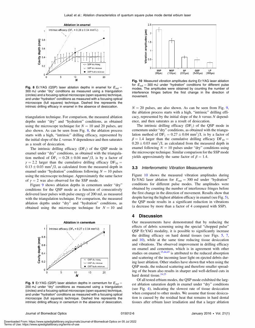
triangulation technique. For comparison, the measured ablationdepths under “dry” and “hydration” conditions, as obtainedusing the microscope technique for N ¼ 10 and 20 pulses, arealso shown. As can be seen from Fig. 8, the ablation processstarts with a high, “intrinsic” drilling efficacy, represented bythe initial slope of the L versus N dependence and then saturatesas a result of desiccation.
The intrinsic drilling efficacy (DF1) of the QSP mode inenamel under “dry” conditions, as obtained with the triangula-tion method of DF1 ¼ 0.28� 0.04 mm3∕J, is by a factor ofγ ¼ 2.2 larger than the cumulative drilling efficacy DF10 ¼0.13� 0.03 mm3∕J, as calculated from the measured depth inenamel under “hydration” conditions following N ¼ 10 pulsesusing the microscope technique. Approximately the same factorof γ ¼ 2 was also observed for the SSP mode.
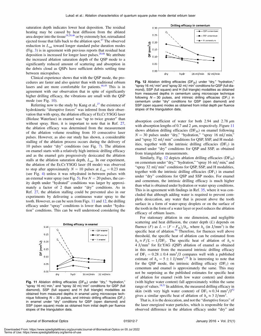
Figure 9 shows ablation depths in cementum under “dry”conditions for the QSP mode as a function of consecutivelydelivered laser pulses with pulse energy of 200 mJ, as measuredwith the triangulation technique. For comparison, the measuredablation depths under “dry” and “hydration” conditions, asobtained using the microscope technique for N ¼ 10 and
N ¼ 20 pulses, are also shown. As can be seen from Fig. 9,the ablation process starts with a high, “intrinsic” drilling effi-cacy, represented by the initial slope of the h versus N depend-ence, and then saturates as a result of desiccation.
The intrinsic drilling efficacy (DF1) of the QSP mode incementum under “dry” conditions, as obtained with the triangu-lation method of DF1 ¼ 0.27� 0.04 mm3∕J, is by a factor ofβ ¼ 1.4 larger than the cumulative drilling efficacy DF10 ¼0.20� 0.03 mm3∕J, as calculated from the measured depth inenamel following N ¼ 10 pulses under “dry” conditions usingthe microscope technique. Similar comparison for the SSP modeyields approximately the same factor of β ¼ 1.4.
3.3 Interferometric Vibration Measurements
Figure 10 shows the measured vibration amplitudes duringEr:YAG laser ablation for Eout ¼ 300 mJ under “hydration”conditions for different pulse modes. The amplitudes wereobtained by counting the number of interference fringes beforethe first change in the direction of movement. Results show thatdespite having the highest ablation efficacy in enamel (see Fig. 5),the QSP mode results in a significant reduction in vibrations(a decrease by more than a factor of 4 compared with SSP).
4 DiscussionOur measurements have demonstrated that by reducing theeffects of debris screening using the special “chopped pulse”QSP Er:YAG modality, it is possible to significantly increasethe drilling efficacy on hard dental tissues (see Figs. 5, 7,and 10), while at the same time reducing tissue desiccationand vibrations. The observed improvement in drilling efficacyon enamel and cementum, which is in agreement with otherstudies on enamel,25,44,45 is attributed to the reduced absorptionand scattering of the incoming laser light on ejected debris dur-ing laser ablation. Other studies have shown that when using theQSP mode, the reduced scattering and therefore smaller spread-ing of the beam also results in sharper and well-defined cuts inhard dental tissue.24,25
Of all tested erbium modes, the QSP mode exhibited the larg-est ablation saturation depth in enamel under “dry” conditions(see Fig. 4), indicating the slowest rate of tissue desiccationwhen compared to other modes. We assume that tissue desicca-tion is caused by the residual heat that remains in hard dentaltissues after erbium laser irradiation and that a larger ablation
Fig. 8 Er:YAG (QSP) laser ablation depths in enamel for Eout ¼300 mJ under “dry” conditions as measured using a triangulation(circles) and a focusing optical microscope (open squares) technique,and under “hydration” conditions as measured with a focusing opticalmicroscope (full squares) technique. Dashed line represents theintrinsic drilling efficacy in enamel in the absence of desiccation.
Fig. 9 Er:YAG (QSP) laser ablation depths in cementum for Eout ¼200 mJ under “dry” conditions as measured using a triangulation(circles) and a focusing optical microscope (open squares) technique,and under “hydration” conditions as measured with a focusing opticalmicroscope (full squares) technique. Dashed line represents theintrinsic drilling efficacy in cementum in the absence of desiccation.
Fig. 10 Measured vibration amplitudes during Er:YAG laser ablationfor Eout ¼ 300 mJ under “hydration” conditions for different pulsemodes. The amplitudes were obtained by counting the number ofinterference fringes before the first change in the direction ofmovement.
Journal of Biomedical Optics 015012-6 January 2016 • Vol. 21(1)
Lukac et al.: Ablation characteristics of quantum square pulse mode dental erbium laser
Downloaded From: https://www.spiedigitallibrary.org/journals/Journal-of-Biomedical-Optics on 05 Jul 2022Terms of Use: https://www.spiedigitallibrary.org/terms-of-use
saturation depth indicates lower heat deposition. The residualheating may be caused by heat diffusion from the ablatedarea deeper into the tissue18,20,46 or by extremely hot, reirradiatedejected tissue that falls back to the ablation spot.47 The observedreduction in Lsat toward longer standard pulse duration modes(Fig. 3) is in agreement with previous reports that residual heatdeposition is increased for longer laser pulses.20,46 We attributethe increased ablation saturation depth of the QSP mode to asignificantly reduced amount of scattering and absorption inthe debris cloud as QSPs have sufficient debris settling timebetween micropulses.
Clinical experience shows that with the QSP mode, the pro-cedures are faster and also quieter than with traditional erbiumlasers and are more comfortable for patients.48,49 This is inagreement with our observation that in spite of significantlyhigher drilling efficacy, the vibrations are small with the QSPmode (see Fig. 10).
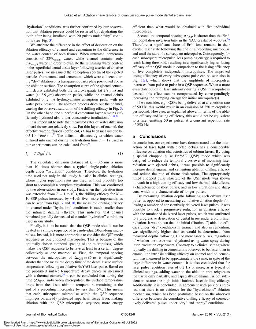
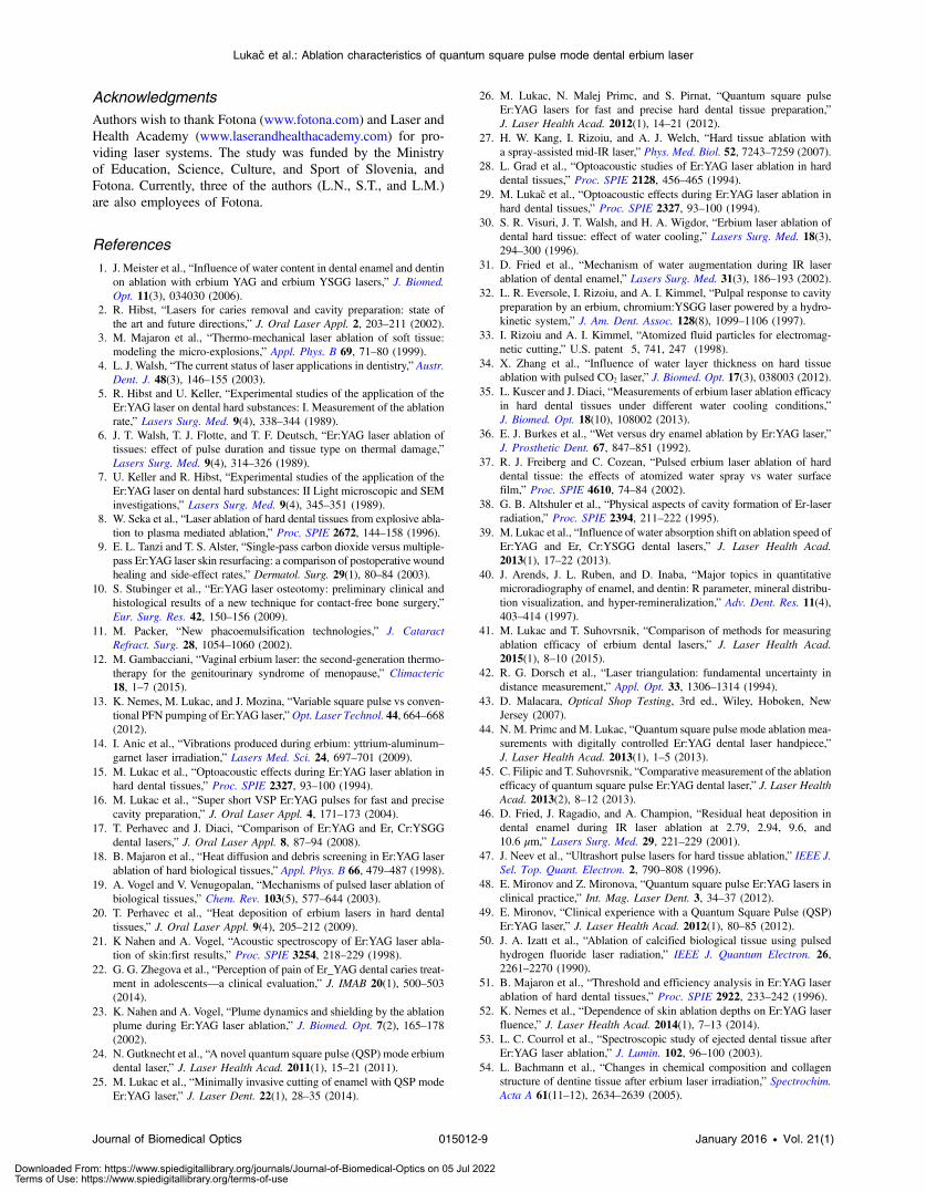
Referring now to the study by Kang et al.,27 the existence ofhydrokinetic “disruptive forces” was inferred from their obser-vation that with spray, the ablation efficacy of Er,Cr:YSGG laser(Biolase Waterlase) in enamel was “up to twice greater” thanwithout spray. Here, it is important to note that in Ref. 27,the ablation efficacy was determined from the measurementof the ablation volume resulting from 10 consecutive laserpulses. However, as also our study has shown, a considerablestalling of the ablation process occurs during the delivery of10 pulses under “dry” conditions (see Fig. 7). The ablationon enamel starts with a relatively high intrinsic drilling efficacyand as the enamel gets progressively desiccated the ablationstalls at the ablation saturation depth, Lsat. In our experiment,the ablation of the Er,Cr:YSGG laser (H mode) was observedto stop after approximately N ¼ 10 pulses at Lsat ¼ 0.23 mm(see Fig. 4) unless it was rehydrated in-between pulses withan external water spray (see Fig. 5). For N ¼ 20 pulses, the cav-ity depth under “hydrated” conditions was larger by approxi-mately a factor of 2 than under “dry” conditions. As inRef. 27, the ablation stalling could be prevented also in ourexperiments by delivering a continuous water spray to thetooth. However, as can be seen from Figs. 11 and 12, the drillingefficacy under “spray” conditions is lower than under “hydra-tion” conditions. This can be well understood considering the
absorption coefficient of water for both 2.94 and 2.78 μmwith absorption lengths of 0.7 and 2 μm, respectively. Figure 11shows ablation drilling efficacies (DF30) on enamel followingN ¼ 30 pulses under “dry,” “hydration,” “spray 16 ml∕min,”and “spray 32 ml∕min” conditions for QSP, SSP, and H modal-ities, together with the intrinsic drilling efficacies (DF1) inenamel under “dry” conditions for QSP and SSP, as obtainedfrom triangulation measurements.
Similarly, Fig. 12 depicts ablation drilling efficacies (DF30)on cementum under “dry,” “hydration,” “spray 16 ml∕min,” and“spray 32 ml∕min” conditions for QSP, SSP, and H modalities,together with the intrinsic drilling efficacies (DF1) in enamelunder “dry” conditions for QSP and SSP modes. For enameland cementum, the intrinsic drilling efficacy is much higherthan what is obtained under hydration or water spray conditions.This is in agreement with findings in Ref. 35, where it was con-cluded that although adding water is required to prevent com-plete desiccation, any water that is present above the toothsurface in a form of water-spray droplets or on the surface ofthe tooth in the form of a water layer or pool reduces the ablationefficacy of erbium lasers.
For stationary ablation in one dimension, and negligiblescattering and heat diffusion, the crater depth (L) depends onfluence (F) as L ¼ ðF − FthÞ∕ha, where ha (in J∕mm3) is thespecific heat of ablation.50 Therefore, for fluences well abovethreshold, the specific heat of ablation can be estimated fromha ≈ F∕L ¼ 1∕DF1. The specific heat of ablation of ha ≈4 J∕mm3 for Er:YAG (QSP) ablation of enamel as obtainedin this manner from the measured intrinsic drilling efficacyof DF1 ¼ 0.28� 0.4 mm3∕J compares well with a publishedestimate of ha ¼ 5� 1 J∕mm3.18 It is interesting to note thatfor the QSP mode, the intrinsic drilling efficacy (DF1) oncementum and enamel is approximately the same. This maynot be surprising as the published estimates for specific heatof ablation for enamel (with low water content) and dentin(with higher water content) fall approximately within the samerange of values.18,51 In addition, the measured drilling efficacy inskin (with very high water content) of DF1 ≈ 0.34 mm3∕J52gives a similar specific heat of ablation of ha ≈ 3 J∕mm3.
That is, it is the desiccation, and not the “disruptive forces” ofthe laser energized water particles, which is responsible for theobserved difference in the ablation efficacy under “dry” and
Fig. 11 Ablation drilling efficacies (DF30) under “dry,” “hydration,”“spray 16 ml∕min,” and “spray 32 ml∕min” conditions for QSP (fulldiamond), SSP (full square) and H (full triangle) modalities asobtained from measured depths in enamel using microscope tech-nique following N ¼ 30 pulses, and intrinsic drilling efficacies (DF1)in enamel under “dry” conditions for QSP (open diamond) andSSP (open square) mode as obtained from initial depth per fluenceslopes of the triangulation data.
Fig. 12 Ablation drilling efficacies (DF30) under “dry,” “hydration,”“spray 16 ml∕min” and “spray 32 ml∕min” conditions for QSP (full dia-mond), SSP (full square) and H (full triangle) modalities as obtainedfrom measured depths in cementum using microscope techniquefollowing N ¼ 30 pulses, and intrinsic drilling efficacies (DF1) incementum under “dry” conditions for QSP (open diamond) andSSP (open square) modes as obtained from initial depth per fluenceslopes of the triangulation data.
Journal of Biomedical Optics 015012-7 January 2016 • Vol. 21(1)
Lukac et al.: Ablation characteristics of quantum square pulse mode dental erbium laser
Downloaded From: https://www.spiedigitallibrary.org/journals/Journal-of-Biomedical-Optics on 05 Jul 2022Terms of Use: https://www.spiedigitallibrary.org/terms-of-use
“hydration” conditions, was further confirmed by our observa-tion that ablation process could be restarted by rehydrating thetooth after being irradiated with 20 pulses under “dry” condi-tions (see Fig. 3).
We attribute the difference in the effect of desiccation on theablation efficacy of enamel and cementum to the difference inthe water content of both tissues. When untreated, cementumconsists of 22%weight water, while enamel contains only3%weight water. In order to evaluate the remaining water contentin the superficial dental tissue layer following a series of ablativelaser pulses, we measured the absorption spectra of the ejectedparticles from enamel and cementum, which were collected dur-ing “dry” ablation on a transparent quartz plate positioned abovethe ablation surface. The absorption curve of the ejected cemen-tum debris exhibited both the hydroxyapatite (at 2.8 μm) andwater (at 2.9 μm) absorption peaks, while the enamel debrisexhibited only the hydroxyapatite absorption peak, with nowater peak present. The ablation process dries out the enamel,causing the observed saturation of the drilling efficacy in Fig. 3.On the other hand, the superficial cementum layer remains suf-ficiently hydrated also under consecutive irradiations.31,53,54
It is important to note that measured rates of water diffusionin hard tissues are relatively slow. For thin layers of enamel, theeffective water diffusion coefficient,Dh has been measured to be0.5 10−7 cm2 s−1.55 The diffusion distance ld to which waterdiffused into enamel during the hydration time T ¼ 1 s used inour experiments can be calculated from55
EQ-TARGET;temp:intralink-;e001;63;455ld ¼ T Dhπ2∕4: (1)
The calculated diffusion distance of ld ¼ 3.5 μm is morethan 10 times shorter than a typical single-pulse ablationdepth under “hydration” conditions. Therefore, the hydrationtime used not only in this study but also in clinical settings,where higher repetition rates are used, is most probably tooshort to accomplish a complete rehydration. This was confirmedby two observations in our study. First, when the hydration timewas extended from T ¼ 1 to 10 s, the drilling efficacy for N ¼10 SSP pulses increased by ∼10%. Even more importantly, ascan be seen from Figs. 7 and 10, the measured drilling efficacyon enamel under “hydration” conditions is much smaller thanthe intrinsic drilling efficacy. This indicates that enamelremained partially desiccated also under “hydration” conditionsused in our study.
Finally, it is to be noted that the QSP mode should not betreated as a simple sequence of five individual 50-μs-long micro-pulses. Instead, it is more appropriate to consider the QSP modesequence as one chopped macropulse. This is because of theoptimally chosen temporal spacing of the micropulses, whichmakes the QSP sequence to behave at least to a certain degreecollectively as one macropulse. First, the temporal spacingbetween the micropulses of ΔtQSP ≈ 85 μs is significantlyshorter than the measured decay time of the dental tissue surfacetemperature following an ablative Er:YAG laser pulse. Based onthe published surface temperature decay curves as measuredwith a thermal camera,20 it can be concluded that during thetime (ΔtQSP) in-between micropulses, the surface temperaturedrops from the tissue ablation temperature remaining at theend of a preceding micropulse by less than 5%. This meansthat each subsequent micropulse within the QSP sequenceimpinges on already preheated superficial tissue layer, makingablation with the QSP micropulse sequence more energy
efficient than what would be obtained with five individualmicropulses.
Second, the temporal spacing ΔtQSP is shorter than the Er3þion population inversion time in the YAG crystal of ∼300 μs.56
Therefore, a significant share of Er3þ ions remains in theirexcited laser state following the end of a preceding micropulseand until the start of a subsequent micropulse. Consequently, foreach subsequent micropulse, less pumping energy is required toreach lasing threshold, resulting in a significantly higher lasingefficacy of the QSP mode in comparison to the lasing efficiencyof five completely independent micropulses. The improvedlasing efficiency of every subsequent pulse can be seen also inFig. 1(c), which shows that the amplitude of micropulsesincreases from pulse to pulse in a QSP sequence. When a moreeven distribution of laser intensity during a QSP macropulse isdesired, this effect can be compensated by correspondinglyincreasing the pumping energy for initial micropulse(s).
If we consider, e.g., QSPs being delivered at a repetition rateof 50 Hz, this would result in an emission of 250 micropulsesper second. However, as explained above, in terms of the abla-tion efficacy and lasing efficiency, this would not be equivalentto a laser emitting 50 μs pulses at a constant repetition rateof 250 Hz.
5 ConclusionsIn conclusion, our experiments have demonstrated that the inter-action of laser light with ejected debris has a considerableinfluence on ablation characteristics of erbium lasers. By usinga special chopped pulse Er:YAG (QSP) mode which wasdesigned to reduce the temporal cross-over of incoming laserradiation with ejected debris, it was possible to significantlyincrease dental enamel and cementum ablation drilling efficacyand reduce the rate of tissue desiccation. The appropriatelytimed chopped pulse structure of the QSP mode was shownto result in a high cutting efficacy and low thermal side-effects,a characteristic of short pulses, and in low vibrations and sharpcuts, which is a characteristic of longer pulses.
By measuring ablation depths following each erbium laserpulse, as opposed to measuring cumulative ablation depths fol-lowing a number of consecutively delivered laser pulses, it waspossible to track a progressive reduction in ablation efficacywith the number of delivered laser pulses, which was attributedto a progressive desiccation of dental tissue under erbium laserradiation. It was shown that the initial (“intrinsic”) ablation effi-cacy under “dry” conditions in enamel, and also in cementum,was significantly higher than as would be determined frommeasured depths following 10 or more laser pulses, regardlessof whether the tissue was rehydrated using water spray duringlaser irradiation experiment. Contrary to a clinical setting wheretypically the drilling in cementum is observed to be faster than inenamel, the intrinsic drilling efficacy on enamel and on cemen-tum was measured to be approximately the same, in spite of thelarge difference in water content. It is also concluded that forlaser pulse repetition rates of 0.2 Hz or more, as is typical ofclinical settings, adding water to the ablation spot rehydratesthe tissue only partially, and especially in enamel, is not suffi-cient to restore the high initial intrinsic laser drilling efficacy.Additionally, it is concluded, in agreement with previous stud-ies, that there is no evidence for the “hydrokinetic” ablationmechanism, which has been postulated based on the observeddifference between the cumulative drilling efficacy of consecu-tively delivered pulses under “dry” and “spray” conditions.
Journal of Biomedical Optics 015012-8 January 2016 • Vol. 21(1)
Lukac et al.: Ablation characteristics of quantum square pulse mode dental erbium laser
Downloaded From: https://www.spiedigitallibrary.org/journals/Journal-of-Biomedical-Optics on 05 Jul 2022Terms of Use: https://www.spiedigitallibrary.org/terms-of-use
AcknowledgmentsAuthors wish to thank Fotona (www.fotona.com) and Laser andHealth Academy (www.laserandhealthacademy.com) for pro-viding laser systems. The study was funded by the Ministryof Education, Science, Culture, and Sport of Slovenia, andFotona. Currently, three of the authors (L.N., S.T., and L.M.)are also employees of Fotona.
References1. J. Meister et al., “Influence of water content in dental enamel and dentin
on ablation with erbium YAG and erbium YSGG lasers,” J. Biomed.Opt. 11(3), 034030 (2006).
2. R. Hibst, “Lasers for caries removal and cavity preparation: state ofthe art and future directions,” J. Oral Laser Appl. 2, 203–211 (2002).
3. M. Majaron et al., “Thermo-mechanical laser ablation of soft tissue:modeling the micro-explosions,” Appl. Phys. B 69, 71–80 (1999).
4. L. J. Walsh, “The current status of laser applications in dentistry,” Austr.Dent. J. 48(3), 146–155 (2003).
5. R. Hibst and U. Keller, “Experimental studies of the application of theEr:YAG laser on dental hard substances: I. Measurement of the ablationrate,” Lasers Surg. Med. 9(4), 338–344 (1989).
6. J. T. Walsh, T. J. Flotte, and T. F. Deutsch, “Er:YAG laser ablation oftissues: effect of pulse duration and tissue type on thermal damage,”Lasers Surg. Med. 9(4), 314–326 (1989).
7. U. Keller and R. Hibst, “Experimental studies of the application of theEr:YAG laser on dental hard substances: II Light microscopic and SEMinvestigations,” Lasers Surg. Med. 9(4), 345–351 (1989).
8. W. Seka et al., “Laser ablation of hard dental tissues from explosive abla-tion to plasma mediated ablation,” Proc. SPIE 2672, 144–158 (1996).
9. E. L. Tanzi and T. S. Alster, “Single-pass carbon dioxide versus multiple-pass Er:YAG laser skin resurfacing: a comparison of postoperative woundhealing and side-effect rates,” Dermatol. Surg. 29(1), 80–84 (2003).
10. S. Stubinger et al., “Er:YAG laser osteotomy: preliminary clinical andhistological results of a new technique for contact-free bone surgery,”Eur. Surg. Res. 42, 150–156 (2009).
11. M. Packer, “New phacoemulsification technologies,” J. CataractRefract. Surg. 28, 1054–1060 (2002).
12. M. Gambacciani, “Vaginal erbium laser: the second-generation thermo-therapy for the genitourinary syndrome of menopause,” Climacteric18, 1–7 (2015).
13. K. Nemes, M. Lukac, and J. Mozina, “Variable square pulse vs conven-tional PFN pumping of Er:YAG laser,”Opt. Laser Technol. 44, 664–668(2012).
14. I. Anic et al., “Vibrations produced during erbium: yttrium-aluminum–garnet laser irradiation,” Lasers Med. Sci. 24, 697–701 (2009).
15. M. Lukac et al., “Optoacoustic effects during Er:YAG laser ablation inhard dental tissues,” Proc. SPIE 2327, 93–100 (1994).
16. M. Lukac et al., “Super short VSP Er:YAG pulses for fast and precisecavity preparation,” J. Oral Laser Appl. 4, 171–173 (2004).
17. T. Perhavec and J. Diaci, “Comparison of Er:YAG and Er, Cr:YSGGdental lasers,” J. Oral Laser Appl. 8, 87–94 (2008).
18. B. Majaron et al., “Heat diffusion and debris screening in Er:YAG laserablation of hard biological tissues,” Appl. Phys. B 66, 479–487 (1998).
19. A. Vogel and V. Venugopalan, “Mechanisms of pulsed laser ablation ofbiological tissues,” Chem. Rev. 103(5), 577–644 (2003).
20. T. Perhavec et al., “Heat deposition of erbium lasers in hard dentaltissues,” J. Oral Laser Appl. 9(4), 205–212 (2009).
21. K Nahen and A. Vogel, “Acoustic spectroscopy of Er:YAG laser abla-tion of skin:first results,” Proc. SPIE 3254, 218–229 (1998).
22. G. G. Zhegova et al., “Perception of pain of Er_YAG dental caries treat-ment in adolescents—a clinical evaluation,” J. IMAB 20(1), 500–503(2014).
23. K. Nahen and A. Vogel, “Plume dynamics and shielding by the ablationplume during Er:YAG laser ablation,” J. Biomed. Opt. 7(2), 165–178(2002).
24. N. Gutknecht et al., “A novel quantum square pulse (QSP) mode erbiumdental laser,” J. Laser Health Acad. 2011(1), 15–21 (2011).
25. M. Lukac et al., “Minimally invasive cutting of enamel with QSP modeEr:YAG laser,” J. Laser Dent. 22(1), 28–35 (2014).
26. M. Lukac, N. Malej Primc, and S. Pirnat, “Quantum square pulseEr:YAG lasers for fast and precise hard dental tissue preparation,”J. Laser Health Acad. 2012(1), 14–21 (2012).
27. H. W. Kang, I. Rizoiu, and A. J. Welch, “Hard tissue ablation witha spray-assisted mid-IR laser,” Phys. Med. Biol. 52, 7243–7259 (2007).
28. L. Grad et al., “Optoacoustic studies of Er:YAG laser ablation in harddental tissues,” Proc. SPIE 2128, 456–465 (1994).
29. M. Lukac et al., “Optoacoustic effects during Er:YAG laser ablation inhard dental tissues,” Proc. SPIE 2327, 93–100 (1994).
30. S. R. Visuri, J. T. Walsh, and H. A. Wigdor, “Erbium laser ablation ofdental hard tissue: effect of water cooling,” Lasers Surg. Med. 18(3),294–300 (1996).
31. D. Fried et al., “Mechanism of water augmentation during IR laserablation of dental enamel,” Lasers Surg. Med. 31(3), 186–193 (2002).
32. L. R. Eversole, I. Rizoiu, and A. I. Kimmel, “Pulpal response to cavitypreparation by an erbium, chromium:YSGG laser powered by a hydro-kinetic system,” J. Am. Dent. Assoc. 128(8), 1099–1106 (1997).
33. I. Rizoiu and A. I. Kimmel, “Atomized fluid particles for electromag-netic cutting,” U.S. patent 5, 741, 247 (1998).
34. X. Zhang et al., “Influence of water layer thickness on hard tissueablation with pulsed CO2 laser,” J. Biomed. Opt. 17(3), 038003 (2012).
35. L. Kuscer and J. Diaci, “Measurements of erbium laser ablation efficacyin hard dental tissues under different water cooling conditions,”J. Biomed. Opt. 18(10), 108002 (2013).
36. E. J. Burkes et al., “Wet versus dry enamel ablation by Er:YAG laser,”J. Prosthetic Dent. 67, 847–851 (1992).
37. R. J. Freiberg and C. Cozean, “Pulsed erbium laser ablation of harddental tissue: the effects of atomized water spray vs water surfacefilm,” Proc. SPIE 4610, 74–84 (2002).
38. G. B. Altshuler et al., “Physical aspects of cavity formation of Er-laserradiation,” Proc. SPIE 2394, 211–222 (1995).
39. M. Lukac et al., “Influence of water absorption shift on ablation speed ofEr:YAG and Er, Cr:YSGG dental lasers,” J. Laser Health Acad.2013(1), 17–22 (2013).
40. J. Arends, J. L. Ruben, and D. Inaba, “Major topics in quantitativemicroradiography of enamel, and dentin: R parameter, mineral distribu-tion visualization, and hyper-remineralization,” Adv. Dent. Res. 11(4),403–414 (1997).
41. M. Lukac and T. Suhovrsnik, “Comparison of methods for measuringablation efficacy of erbium dental lasers,” J. Laser Health Acad.2015(1), 8–10 (2015).
42. R. G. Dorsch et al., “Laser triangulation: fundamental uncertainty indistance measurement,” Appl. Opt. 33, 1306–1314 (1994).
43. D. Malacara, Optical Shop Testing, 3rd ed., Wiley, Hoboken, NewJersey (2007).
44. N. M. Primc and M. Lukac, “Quantum square pulse mode ablation mea-surements with digitally controlled Er:YAG dental laser handpiece,”J. Laser Health Acad. 2013(1), 1–5 (2013).
45. C. Filipic and T. Suhovrsnik, “Comparative measurement of the ablationefficacy of quantum square pulse Er:YAG dental laser,” J. Laser HealthAcad. 2013(2), 8–12 (2013).
46. D. Fried, J. Ragadio, and A. Champion, “Residual heat deposition indental enamel during IR laser ablation at 2.79, 2.94, 9.6, and10.6 μm,” Lasers Surg. Med. 29, 221–229 (2001).
47. J. Neev et al., “Ultrashort pulse lasers for hard tissue ablation,” IEEE J.Sel. Top. Quant. Electron. 2, 790–808 (1996).
48. E. Mironov and Z. Mironova, “Quantum square pulse Er:YAG lasers inclinical practice,” Int. Mag. Laser Dent. 3, 34–37 (2012).
49. E. Mironov, “Clinical experience with a Quantum Square Pulse (QSP)Er:YAG laser,” J. Laser Health Acad. 2012(1), 80–85 (2012).
50. J. A. Izatt et al., “Ablation of calcified biological tissue using pulsedhydrogen fluoride laser radiation,” IEEE J. Quantum Electron. 26,2261–2270 (1990).
51. B. Majaron et al., “Threshold and efficiency analysis in Er:YAG laserablation of hard dental tissues,” Proc. SPIE 2922, 233–242 (1996).
52. K. Nemes et al., “Dependence of skin ablation depths on Er:YAG laserfluence,” J. Laser Health Acad. 2014(1), 7–13 (2014).
53. L. C. Courrol et al., “Spectroscopic study of ejected dental tissue afterEr:YAG laser ablation,” J. Lumin. 102, 96–100 (2003).
54. L. Bachmann et al., “Changes in chemical composition and collagenstructure of dentine tissue after erbium laser irradiation,” Spectrochim.Acta A 61(11–12), 2634–2639 (2005).
Journal of Biomedical Optics 015012-9 January 2016 • Vol. 21(1)
Lukac et al.: Ablation characteristics of quantum square pulse mode dental erbium laser
Downloaded From: https://www.spiedigitallibrary.org/journals/Journal-of-Biomedical-Optics on 05 Jul 2022Terms of Use: https://www.spiedigitallibrary.org/terms-of-use

55. G. H. Didbin, “The water in human dental enamel and its diffusionalexchange measured by clearance of tritiated water from enamel slabsof varying thickness,” Caries Res. 27(2), 81–86 (1993).
56. B. Majaron, T. Rupnik, and M. Lukac, “Temperature and gain dynamicsin flashlamp-pumped Er:YAG,” IEEE J. Quantum Electron. 32(9),1636–1996 (1996).
Nejc Lukac is a PhD student at the faculty of mechanical engineering,University of Ljubljana. His main topics of research are the use oflasers and laser pulse shaping in medical applications. He is currentlyan employee of Fotona, d.o.o.
Tomaž Suhovršnik received his MSc degree in physics from theUniversity of Ljubljana. His main topics of research are the use oflasers and dentistry. He is currently an employee of Fotona, d.o.o.
Matjaž Lukac received his MSc and PhD degrees in laser physics.Since 1986, he has been working as a research associate at the JosefStefan Institute. His main topics of research are the interaction of laserlight with biological tissues and physics of laser sources. Currently, heis also a managing director of Fotona, d.o.o.
Matija Jezeršek is an associate professor for laser applications atthe faculty of mechanical engineering, University of Ljubljana. He isworking at the chair of optodynamics and laser applications. Hereceived his PhD in mechanical engineering from the University ofLjubljana in 2004. His major research interests include three-dimen-sional optical metrology, laser processing, and research of optody-namic phenomena.
Journal of Biomedical Optics 015012-10 January 2016 • Vol. 21(1)
Lukac et al.: Ablation characteristics of quantum square pulse mode dental erbium laser
Downloaded From: https://www.spiedigitallibrary.org/journals/Journal-of-Biomedical-Optics on 05 Jul 2022Terms of Use: https://www.spiedigitallibrary.org/terms-of-use
Related Documents