Aberrant replication timing induces defective chromosome condensation in Drosophila ORC2 mutants Marie-Louise Loupart, Sue Ann Krause* and Margarete M.S. Heck Background: The accurate duplication and packaging of the genome is an absolute prerequisite to the segregation of chromosomes in mitosis. To understand the process of cell-cycle chromosome dynamics further, we have performed the first detailed characterization of a mutation affecting mitotic chromosome condensation in a metazoan. Our combined genetic and cytological approaches in Drosophila complement and extend existing work employing yeast genetics and Xenopus in vitro extract systems to characterize higher-order chromosome structure and function. Results: Two alleles of the ORC2 gene were found to cause death late in larval development, with defects in cell-cycle progression (delays in S-phase entry and metaphase exit) and chromosome condensation in mitosis. During S-phase progression in wild-type cells, euchromatin replicates early and heterochromatin replicates late. Both alleles disrupted the normal pattern of chromosomal replication, with some euchromatic regions replicating even later than heterochromatin. Mitotic chromosomes were irregularly condensed, with the abnormally late replicating regions of euchromatin exhibiting the greatest problems in mitotic condensation. Conclusions: The results not only reveal novel functions for ORC2 in chromosome architecture in metazoans, they also suggest that the correct timing of DNA replication may be essential for the assembly of chromatin that is fully competent to undergo mitotic condensation. Background The origin recognition complex (ORC) is composed of six subunits, ORC1–6 [1]. ORCs have been identified in yeast, flies, frogs, mice and humans [2], and several of the homologous subunits or even whole complexes are func- tionally interchangeable between species, suggesting a high degree of conservation of ORC function [3–5]. The Drosophila ORC complex was isolated from embryonic extracts by functional homology to the Saccharomyces cere- visiae complex, and all six Drosophila ORC genes (DmORC1–6) have now been cloned [4,6–8]. A fully func- tional Drosophila ORC has also been reconstituted from the six recombinant proteins [4]. In S. cerevisiae, ORC is bound throughout the cell cycle to specific DNA elements of replication origins [9–11]. Binding of Cdc6p to the origin is the initial step in the for- mation of a pre-replication complex (pre-RC) [12,13]. Sub- sequently, the minichromosome maintenance (MCM) proteins are loaded onto the origin, S-phase cyclin-depen- dent kinase is activated and Cdc45p associates with the pre-RC in preparation for S-phase entry [13,14]. Assembly of the pre-RC is likely to correspond to the ‘licensing’ of replication origins first described in the Xenopus cell-free system [15–19]. Although there are a number of differences in detail, a similar mechanism for the assembly of the pre- RC appears to exist in metazoans. Although ORC is principally involved in the initiation of DNA replication, additional roles in the establishment and maintenance of transcriptional silencing and heterochromatin have been suggested. In both Xenopus and Drosophila, ORC and HP-1 interact [7,20] and a single mutant copy of DmORC2 will suppress position-effect variegation [7]. Replication and transcriptional silencing have also been linked by ORC2 and ORC5 mutants in S. cerevisiae [21]. As transcriptional silencing at the yeast mating-type loci appears functionally similar to heterochromatin in higher eukaryotes, these experiments provide additional evi- dence for the involvement of ORC in the specification of transcription-rich euchromatin versus transcription-poor heterochromatin. As ORC is not required for the activation of origins in yeast and Xenopus once Cdc6 and MCM proteins have bound [22,23], ORC may only be necessary for the recruitment of these proteins to origins. ORC’s role in transcriptional reg- ulation suggests that it may also provide a ‘landing pad’ for other protein complexes important for chromosome dynamics [24]. Regulation of different complexes recruited Address: University of Edinburgh, Wellcome Trust Centre for Cell Biology, Institute of Cell and Molecular Biology, Michael Swann Building, King’s Buildings, Mayfield Road, Edinburgh EH9 3JR, UK. *Present address: University of Glasgow, Institute of Biological and Life Sciences, Division of Molecular Genetics, Robertson Building, 54 Dumbarton Road, Glasgow G11 6NU, UK. Correspondence: Margarete Heck E-mail: [email protected] Received: 10 August 2000 Revised: 18 October 2000 Accepted: 1 November 2000 Published: 22 November 2000 Current Biology 2000, 10:1547–1556 0960-9822/00/$ – see front matter © 2000 Elsevier Science Ltd. All rights reserved. Research Paper 1547

Welcome message from author
This document is posted to help you gain knowledge. Please leave a comment to let me know what you think about it! Share it to your friends and learn new things together.
Transcript
Aberrant replication timing induces defective chromosomecondensation in Drosophila ORC2 mutants Marie-Louise Loupart, Sue Ann Krause* and Margarete M.S. Heck
Background: The accurate duplication and packaging of the genome is anabsolute prerequisite to the segregation of chromosomes in mitosis. Tounderstand the process of cell-cycle chromosome dynamics further, we haveperformed the first detailed characterization of a mutation affecting mitoticchromosome condensation in a metazoan. Our combined genetic andcytological approaches in Drosophila complement and extend existing workemploying yeast genetics and Xenopus in vitro extract systems to characterizehigher-order chromosome structure and function.
Results: Two alleles of the ORC2 gene were found to cause death late in larvaldevelopment, with defects in cell-cycle progression (delays in S-phase entryand metaphase exit) and chromosome condensation in mitosis. During S-phaseprogression in wild-type cells, euchromatin replicates early and heterochromatinreplicates late. Both alleles disrupted the normal pattern of chromosomalreplication, with some euchromatic regions replicating even later thanheterochromatin. Mitotic chromosomes were irregularly condensed, with theabnormally late replicating regions of euchromatin exhibiting the greatestproblems in mitotic condensation.
Conclusions: The results not only reveal novel functions for ORC2 inchromosome architecture in metazoans, they also suggest that the correcttiming of DNA replication may be essential for the assembly of chromatin thatis fully competent to undergo mitotic condensation.
BackgroundThe origin recognition complex (ORC) is composed ofsix subunits, ORC1–6 [1]. ORCs have been identified inyeast, flies, frogs, mice and humans [2], and several of thehomologous subunits or even whole complexes are func-tionally interchangeable between species, suggesting ahigh degree of conservation of ORC function [3–5]. TheDrosophila ORC complex was isolated from embryonicextracts by functional homology to the Saccharomyces cere-visiae complex, and all six Drosophila ORC genes(DmORC1–6) have now been cloned [4,6–8]. A fully func-tional Drosophila ORC has also been reconstituted fromthe six recombinant proteins [4].
In S. cerevisiae, ORC is bound throughout the cell cycleto specific DNA elements of replication origins [9–11].Binding of Cdc6p to the origin is the initial step in the for-mation of a pre-replication complex (pre-RC) [12,13]. Sub-sequently, the minichromosome maintenance (MCM)proteins are loaded onto the origin, S-phase cyclin-depen-dent kinase is activated and Cdc45p associates with thepre-RC in preparation for S-phase entry [13,14]. Assemblyof the pre-RC is likely to correspond to the ‘licensing’ ofreplication origins first described in the Xenopus cell-freesystem [15–19]. Although there are a number of differences
in detail, a similar mechanism for the assembly of the pre-RC appears to exist in metazoans.
Although ORC is principally involved in the initiation ofDNA replication, additional roles in the establishment andmaintenance of transcriptional silencing and heterochromatinhave been suggested. In both Xenopus and Drosophila,ORC and HP-1 interact [7,20] and a single mutant copy ofDmORC2 will suppress position-effect variegation [7].Replication and transcriptional silencing have also beenlinked by ORC2 and ORC5 mutants in S. cerevisiae [21]. Astranscriptional silencing at the yeast mating-type lociappears functionally similar to heterochromatin in highereukaryotes, these experiments provide additional evi-dence for the involvement of ORC in the specification oftranscription-rich euchromatin versus transcription-poorheterochromatin.
As ORC is not required for the activation of origins in yeastand Xenopus once Cdc6 and MCM proteins have bound[22,23], ORC may only be necessary for the recruitment ofthese proteins to origins. ORC’s role in transcriptional reg-ulation suggests that it may also provide a ‘landing pad’ forother protein complexes important for chromosomedynamics [24]. Regulation of different complexes recruited
Address: University of Edinburgh, Wellcome TrustCentre for Cell Biology, Institute of Cell andMolecular Biology, Michael Swann Building, King’sBuildings, Mayfield Road, Edinburgh EH9 3JR, UK.
*Present address: University of Glasgow, Institute ofBiological and Life Sciences, Division of MolecularGenetics, Robertson Building, 54 Dumbarton Road,Glasgow G11 6NU, UK.
Correspondence: Margarete HeckE-mail: [email protected]
Received: 10 August 2000Revised: 18 October 2000Accepted: 1 November 2000
Published: 22 November 2000
Current Biology 2000, 10:1547–1556
0960-9822/00/$ – see front matter © 2000 Elsevier Science Ltd. All rights reserved.
Research Paper 1547
by ORC could be achieved through a conformational changein the complex, either as a result of the dissociation of oneor more subunits, or hydrolysis of bound ATP [25].
Although ORC is bound to the origin throughout the cellcycle in yeast, the protein levels of at least two ORC sub-units in Drosophila may fluctuate during the cell cycleand/or development. In the ovary, a developmentally regu-lated switch in the follicle cells from endoreduplication ofthe entire genome to specific amplification of the choriongenes causes a shift in the distribution of both ORC1 andORC2 within the nucleus to the foci of amplifying choriongenes [26,27]. ORC1 is an E2F-responsive gene and theprotein accumulates in late G1 and S phase in the eyeimaginal disc [26]. Thus, although all six subunits appearto be necessary for ORC’s function in replication initia-tion, it is not clear whether all subunits remain associatedwith the origin throughout the cell cycle in metazoans.
The Drosophila lethal mutant l(3)k43 [28,29] has a disc-lessphenotype and is generally defective in cell proliferationduring the larval stages of the Drosophila life cycle [30],exhibiting mitotic chromosome fragmentation and con-densation defects [31]. An additional female sterile alleleresults in a reduction of chorion gene amplification andDmORC2 was identified as the gene responsible for thesephenotypes [32]. To learn more about the role(s) ORC2may play in chromosome dynamics through the cell cycle,we have characterized in detail the cell-cycle progressionand striking chromosomal defects of two late larval lethalalleles of DmORC2.
ResultsDmORC21 is truncated by a premature stop codonThe DmORC2 protein is 618 amino acids in length [6] andthe lesions for the female sterile and the l(3)k43γ43 alleleshave been reported [32]. We sequenced the l(3)k431 alleleafter amplifying the gene from homozygous mutant larvalgenomic DNA and identified a single base substitution,which introduces a premature stop codon at residue 487(Figure 1). We also confirmed the frameshift near the startof the coding sequence of the l(3)k43γ4e allele [32]. There-fore, the evolutionarily conserved carboxy-terminal portionof the protein was missing from both DmORC21 andDmORC2γ4e (Figure 1). We also sequenced two DrosophilaORC2 expressed sequence tags (ESTs) and the gene froma wild-type stock, finding eight additional base changes inthe coding region of all sequences analyzed, comparedwith the published sequence [6]. Three cause amino-acidchanges in the predicted protein: T113A, R246A, andI274N; the other five base changes are silent (Figure 1).
Immunoblots of third instar larval protein extracts using twoindependently raised antibodies to DmORC2 detected aband of the expected size (82 kDa) in wild-type and het-erozygous larval extracts but not in either of the homozygous
mutant larval extracts (data not shown). We failed todetect any truncated ORC2 forms in either the l(3)k431 orl(3)k43γ4e homozygotes, suggesting that these forms maybe unstable or not produced.
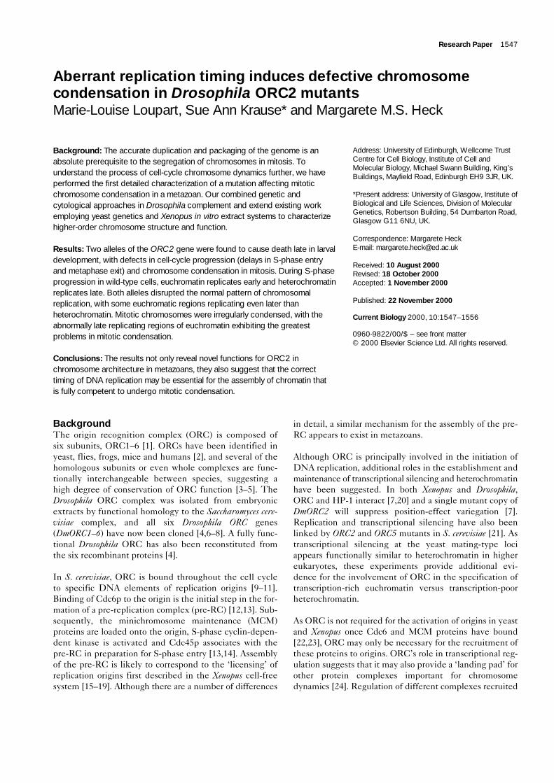
DmORC2 accumulates on chromosomes in lateanaphase/telophaseDuring interphase, ORC2 localization was strong in thenucleus, though also present at a low cytoplasmic level(data not shown). ORC2 was not detected on chromo-somes of mitotic wild-type neuroblasts from prophasethrough early anaphase (Figure 2a). In late anaphase, stain-ing of the segregating chromosomes became intense alongthe length of the chromatids, and persisted into telophase.This pattern of staining was also observed using affinity-purified ORC2 antibody and in three Drosophila cell linesfollowing either paraformaldehyde or methanol fixation(Figure 2b). ORC2 staining is remarkably similar to that ofXenopus ORC1, which is also absent from metaphase chro-mosomes but present on anaphase chromatids [33]. Itappears likely that as the chromatids segregate in anaphaseand reach the spindle poles, ORC is deposited on thereplication origins in preparation for the next cell cycle.
Irregular chromosome condensation in DmORC2 mutantmitotic neuroblastsExamination of DAPI-stained larval neuroblasts from bothl(3)k431 and l(3)k43γ4e alleles revealed many severe mitotic
1548 Current Biology Vol 10 No 24
Figure 1
DmORC2 is truncated in the l(3)k431 and l(3)k43γ4e alleles. Diagram ofthe l(3)k431 mutation (indicated by the black arrow on DmORC21), withthe wild-type and l(3)k43γ4e alleles shown for comparison. The mutationof the l(3)k43γ4e allele was published previously [32]. Within theDmORC2 coding sequence of all alleles sequenced, there were eightbase changes, resulting in three amino-acid substitutions (T113A, R246Aand I274N; filled arrows) and five silent base changes (open arrows).There were also base changes in intron 2 and in the 3′ untranslatedregion (UTR; open arrows), which would not affect the protein product.The adjusted sequence of the wild-type DmORC2 protein has beendeposited in GenBank under accession number AF246305.
Current Biology
1 574 575 1419 1420 1854
A337G
T113A
G737C
R24
6AT821A
I274N
T1555G
A85
8G
C3
48T
A8
82T
C1035T
C1131G
G121CTA41L
G1461AW487STOP
5′ UTR Exon1 Exon2 Exon3 3′ UTR
C2069–C2070AG2080TG2082TG2085C
DmORC2
DmORC2γ4e
DmORC21
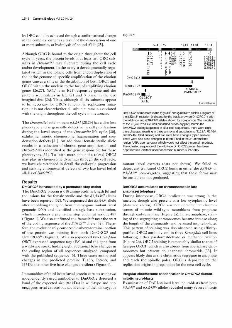
defects (Figure 3a). More than 90% of mitotic figures wereabnormal. Most frequently, mitotic chromosomes con-tained regions of undercondensed chromatin connectedby normally condensed or even highly condensed chro-matin, the same undercondensed region often visiblyaffected on both sister chromatids (Figure 3a, arrowheads).While the undercondensed chromatin could consist of avery fine though discernible thread, it was also clear thatchromosome breaks and rearrangements had occurred,
and that polyploidy was possible. In ~1% of mitoticfigures, the chromatin appeared to be ‘pulverized.’ Thesefigures were highly reminiscent of S-phase prematurelycondensed chromosomes (PCCs) [34], suggesting thesel(3)k43 PCC-like cells may have precociously exited Sphase and entered mitosis.
Although we have observed anaphase figures in DmORC2mutants (Figure 3b), the frequency of anaphase cells was
Research Paper DMORC2 in replication timing and chromosome condensation Loupart et al. 1549
Figure 2
DmORC2 accumulates on chromosomes inanaphase. Immunofluorescence of (a) wild-type neuroblasts (paraformaldehyde fixation)and (b) cultured Drosophila Dmel2 cells(methanol fixation) in metaphase, anaphaseand telophase of the cell cycle. In the mergedimages, DNA is red, and ORC2 is green.DAPI, 4,6-diamidino-2-phenylindole. Thecorresponding pre-immune serum producedfaint diffuse nuclear and cytoplasmic staining,but not the accumulation on late anaphase andtelophase chromosomes observed with theimmune serum. The scale bars represent 5 µm.
Current Biology
Anaphase B
DAPI MergedORC2Metaphase
Telophase
Anaphase A
MergedDAPI ORC2
Anaphase B
Metaphase
Telophase
Anaphase A
(a) Wild-type neuroblasts (b) Cultured DMel2 cells
Figure 3
The mitotic chromosomes of DmORC2mutant neuroblasts are irregularly condensedor fragmented whereas the polytenechromosomes appear normal. (a) Metaphaseand (b) anaphase figures of third instar wild-type, l(3)k431 and l(3)k43γ4e neuroblasts.The scale bars represent 5 µm. Chromosomesin which the same region on both sisterchromatids was affected are indicated byopen arrowheads. (c) Polytene chromosomesof third instar wild-type, l(3)k431 andl(3)k43γ4e salivary glands, each with a portionof the genome shown at higher magnification,demonstrating normal chromosome banding(inset). The scale bar represents 50 µm.
(a)
(c)
k431
k43γ4e
Wild type
k431
k431 k431 k431
k43γ4e k43γ4e
k431
k431
k43γ4e
k43γ4e
k43γ4e
k43γ4e(b)
Wild type
Wild type
Wild type
k43γ4eNear normal Irregularcondensation
Chromosomes
Current Biology
Pulverizedchromatin
greatly reduced compared with the wild type. Theanaphase figures we observed were all abnormal, showingeither condensation defects on sister chromatids (Figure 3b,arrowheads) or chromatid bridges. Therefore, even thoughapparently normal bipolar spindles had formed in theDmORC2 mutants (as judged by α- and γ-tubulin immuno-staining), cells appeared unable to complete mitosis.
Mutant salivary glands were of normal size and the poly-tene chromosomes of both alleles were unaffected withrespect to banding pattern and level of polyploidization(Figure 3c). Our data differ from a previous observationthat the l(3)k43γ4e polytene chromosomes are under-repli-cated with a poor, irregular banding pattern [20], but maybe explained by the use of a modified feeding regime thatincreases larval health. Either ORC2 is not needed for theendoreduplication cycles necessary for the formation ofsalivary gland polytene chromosomes or, more likely,maternally deposited ORC2 is sufficient to support replica-tion in the salivary glands (augmented by a lower turnoverof ORC in polyploid cells than in mitotically active cells).
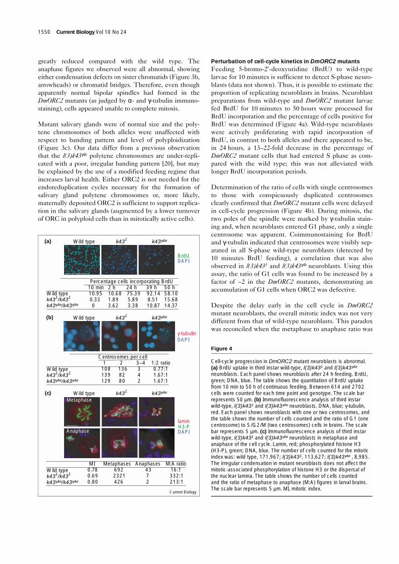
Perturbation of cell-cycle kinetics in DmORC2 mutantsFeeding 5-bromo-2′-deoxyuridine (BrdU) to wild-typelarvae for 10 minutes is sufficient to detect S-phase neuro-blasts (data not shown). Thus, it is possible to estimate theproportion of replicating neuroblasts in brains. Neuroblastpreparations from wild-type and DmORC2 mutant larvaefed BrdU for 10 minutes to 50 hours were processed forBrdU incorporation and the percentage of cells positive forBrdU was determined (Figure 4a). Wild-type neuroblastswere actively proliferating with rapid incorporation ofBrdU, in contrast to both alleles and there appeared to be,in 24 hours, a 13–22-fold decrease in the percentage ofDmORC2 mutant cells that had entered S phase as com-pared with the wild type; this was not alleviated withlonger BrdU incorporation periods.
Determination of the ratio of cells with single centrosomesto those with conspicuously duplicated centrosomesclearly confirmed that DmORC2 mutant cells were delayedin cell-cycle progression (Figure 4b). During mitosis, thetwo poles of the spindle were marked by γ-tubulin stain-ing and, when neuroblasts entered G1 phase, only a singlecentrosome was apparent. Coimmunostaining for BrdUand γ-tubulin indicated that centrosomes were visibly sep-arated in all S-phase wild-type neuroblasts (detected by10 minutes BrdU feeding), a correlation that was alsoobserved in l(3)k431 and l(3)k43γ4e neuroblasts. Using thisassay, the ratio of G1 cells was found to be increased by afactor of ~2 in the DmORC2 mutants, demonstrating anaccumulation of G1 cells when ORC2 was defective.
Despite the delay early in the cell cycle in DmORC2mutant neuroblasts, the overall mitotic index was not verydifferent from that of wild-type neuroblasts. This paradoxwas reconciled when the metaphase to anaphase ratio was
1550 Current Biology Vol 10 No 24
Current Biology
(b)
Wild typek431/k431
k43γ4e/k43γ4e
Wild typek431/k431
k43γ4e/k43γ4e
Wild typek431/k431
k43γ4e/k43γ4e
Percentage cells incorporating BrdU24 h75.395.893.38
10 min10.950.330
39 h92.148.5110.87
Wild type k431 k43γ4e
Wild type k431 k43γ4e
Wild type k431 k43γ4e
BrdUDAPI
2 h10.681.893.62
50 h58.1015.6814.37
(a)
γ-tubulinDAPI
Centrosomes per cell2
1368280
1108139129
3–4342
1:2 ratio0.77:11.67:11.67:1
(c)Metaphase
Anaphase
laminH3-PDAPI
Anaphases4372
Metaphases6922321426
M:A ratio 16:1332:1213:1
MI0.780.690.80
Figure 4
Cell-cycle progression in DmORC2 mutant neuroblasts is abnormal.(a) BrdU uptake in third instar wild-type, l(3)k431 and l(3)k43γ4e
neuroblasts. Each panel shows neuroblasts after 24 h feeding. BrdU,green; DNA, blue. The table shows the quantitation of BrdU uptakefrom 10 min to 50 h of continuous feeding. Between 614 and 2702cells were counted for each time point and genotype. The scale barrepresents 50 µm. (b) Immunofluorescence analysis of third instarwild-type, l(3)k431 and l(3)k43γ4e neuroblasts. DNA, blue; γ-tubulin,red. Each panel shows neuroblasts with one or two centrosomes, andthe table shows the number of cells counted and the ratio of G1 (onecentrosome) to S/G2/M (two centrosomes) cells in brains. The scalebar represents 5 µm. (c) Immunofluorescence analysis of third instarwild-type, l(3)k431 and l(3)k43γ4e neuroblasts in metaphase andanaphase of the cell cycle. Lamin, red; phosphorylated histone H3(H3-P), green; DNA, blue. The number of cells counted for the mitoticindex was: wild type, 171,967; l(3)k431, 113,627; l(3)k43γ4e , 8,985.The irregular condensation in mutant neuroblasts does not affect themitotic-associated phosphorylation of histone H3 or the dispersal ofthe nuclear lamina. The table shows the number of cells countedand the ratio of metaphase to anaphase (M:A) figures in larval brains.The scale bar represents 5 µm. MI, mitotic index.
determined after staining for the mitosis-specific phospho-rylated form of histone H3 [35]. In DmORC2 mutants, themetaphase to anaphase ratio was 13–21-fold higher than inwild-type cells (Figure 4c). Thus, we believe that thedelay in exiting mitosis compensates for the delay early inthe cell cycle to result in an overall mitotic index similar tothat in the wild type.
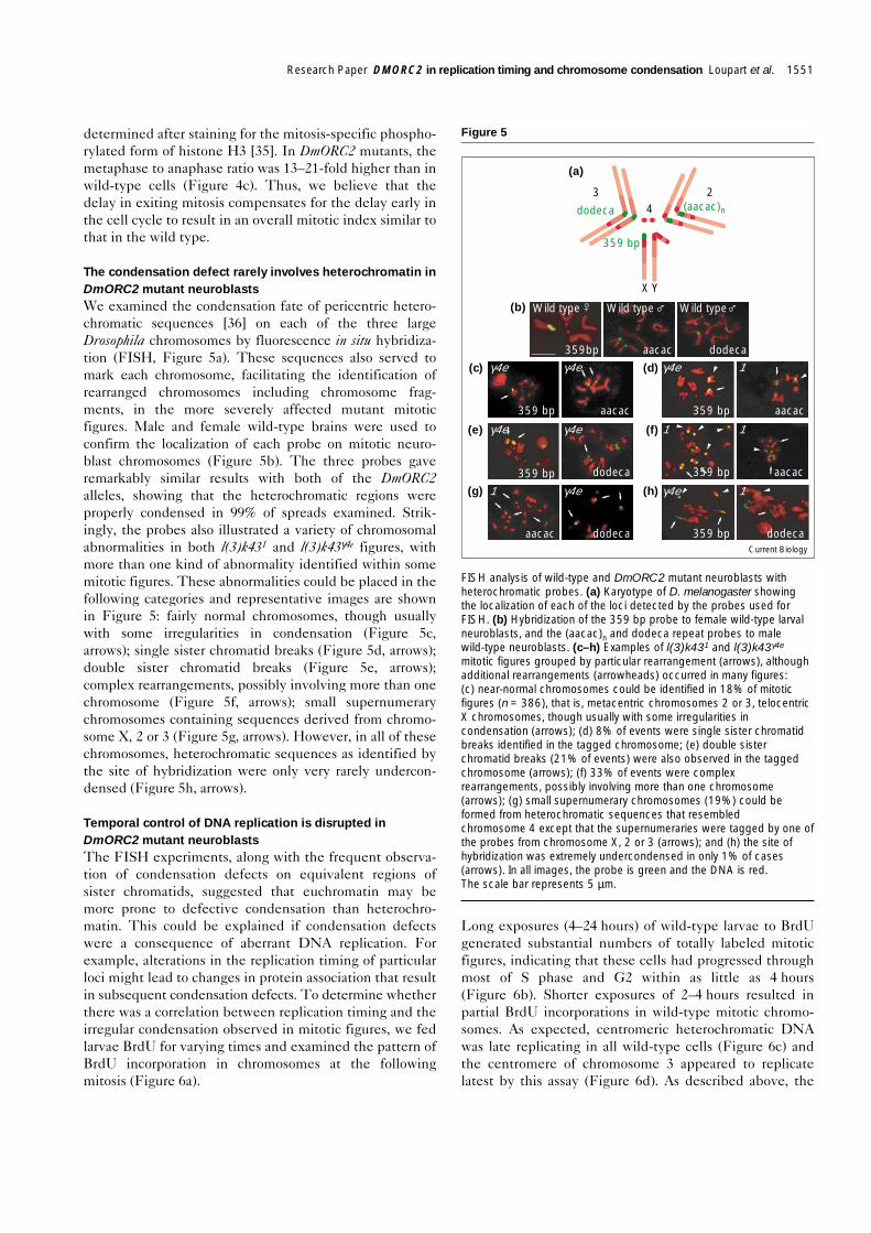
The condensation defect rarely involves heterochromatin inDmORC2 mutant neuroblastsWe examined the condensation fate of pericentric hetero-chromatic sequences [36] on each of the three largeDrosophila chromosomes by fluorescence in situ hybridiza-tion (FISH, Figure 5a). These sequences also served tomark each chromosome, facilitating the identification ofrearranged chromosomes including chromosome frag-ments, in the more severely affected mutant mitoticfigures. Male and female wild-type brains were used toconfirm the localization of each probe on mitotic neuro-blast chromosomes (Figure 5b). The three probes gaveremarkably similar results with both of the DmORC2alleles, showing that the heterochromatic regions wereproperly condensed in 99% of spreads examined. Strik-ingly, the probes also illustrated a variety of chromosomalabnormalities in both l(3)k431 and l(3)k43γ4e figures, withmore than one kind of abnormality identified within somemitotic figures. These abnormalities could be placed in thefollowing categories and representative images are shownin Figure 5: fairly normal chromosomes, though usuallywith some irregularities in condensation (Figure 5c,arrows); single sister chromatid breaks (Figure 5d, arrows);double sister chromatid breaks (Figure 5e, arrows);complex rearrangements, possibly involving more than onechromosome (Figure 5f, arrows); small supernumerarychromosomes containing sequences derived from chromo-some X, 2 or 3 (Figure 5g, arrows). However, in all of thesechromosomes, heterochromatic sequences as identified bythe site of hybridization were only very rarely undercon-densed (Figure 5h, arrows).
Temporal control of DNA replication is disrupted inDmORC2 mutant neuroblastsThe FISH experiments, along with the frequent observa-tion of condensation defects on equivalent regions ofsister chromatids, suggested that euchromatin may bemore prone to defective condensation than heterochro-matin. This could be explained if condensation defectswere a consequence of aberrant DNA replication. Forexample, alterations in the replication timing of particularloci might lead to changes in protein association that resultin subsequent condensation defects. To determine whetherthere was a correlation between replication timing and theirregular condensation observed in mitotic figures, we fedlarvae BrdU for varying times and examined the pattern ofBrdU incorporation in chromosomes at the followingmitosis (Figure 6a).
Long exposures (4–24 hours) of wild-type larvae to BrdUgenerated substantial numbers of totally labeled mitoticfigures, indicating that these cells had progressed throughmost of S phase and G2 within as little as 4 hours(Figure 6b). Shorter exposures of 2–4 hours resulted inpartial BrdU incorporations in wild-type mitotic chromo-somes. As expected, centromeric heterochromatic DNAwas late replicating in all wild-type cells (Figure 6c) andthe centromere of chromosome 3 appeared to replicatelatest by this assay (Figure 6d). As described above, the
Research Paper DMORC2 in replication timing and chromosome condensation Loupart et al. 1551
Figure 5
FISH analysis of wild-type and DmORC2 mutant neuroblasts withheterochromatic probes. (a) Karyotype of D. melanogaster showingthe localization of each of the loci detected by the probes used forFISH. (b) Hybridization of the 359 bp probe to female wild-type larvalneuroblasts, and the (aacac)n and dodeca repeat probes to malewild-type neuroblasts. (c–h) Examples of l(3)k431 and l(3)k43γ4e
mitotic figures grouped by particular rearrangement (arrows), althoughadditional rearrangements (arrowheads) occurred in many figures:(c) near-normal chromosomes could be identified in 18% of mitoticfigures (n = 386), that is, metacentric chromosomes 2 or 3, telocentricX chromosomes, though usually with some irregularities incondensation (arrows); (d) 8% of events were single sister chromatidbreaks identified in the tagged chromosome; (e) double sisterchromatid breaks (21% of events) were also observed in the taggedchromosome (arrows); (f) 33% of events were complexrearrangements, possibly involving more than one chromosome(arrows); (g) small supernumerary chromosomes (19%) could beformed from heterochromatic sequences that resembledchromosome 4 except that the supernumeraries were tagged by one ofthe probes from chromosome X, 2 or 3 (arrows); and (h) the site ofhybridization was extremely undercondensed in only 1% of cases(arrows). In all images, the probe is green and the DNA is red.The scale bar represents 5 µm.
Current Biology
(a)
43 2
X Y
dodeca (aacac)n
359 bp
(b)
γ4e γ4e γ4e 1
γ4e γ4e 1 1
1 γ4e γ4e 1
Wild type Wild type Wild type
359bp aacac dodeca
dodeca
aacac
aacac
359 bp
359 bp
359 bp359 bp
aacac
359 bp
dodeca
dodeca
aacac
(d)
(f)
(h)
(c)
(e)
(g)
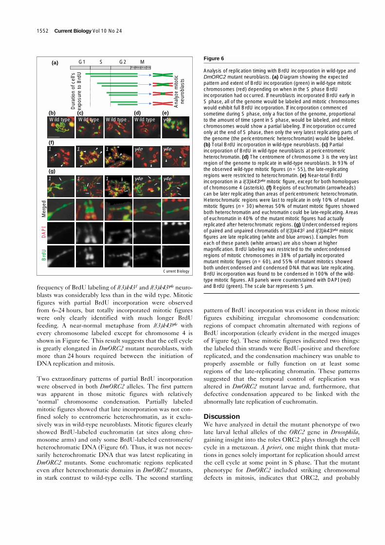
frequency of BrdU labeling of l(3)k431 and l(3)k43γ4e neuro-blasts was considerably less than in the wild type. Mitoticfigures with partial BrdU incorporation were observedfrom 6–24 hours, but totally incorporated mitotic figureswere only clearly identified with much longer BrdUfeeding. A near-normal metaphase from l(3)k43γ4e withevery chromosome labeled except for chromosome 4 isshown in Figure 6e. This result suggests that the cell cycleis greatly elongated in DmORC2 mutant neuroblasts, withmore than 24 hours required between the initiation ofDNA replication and mitosis.
Two extraordinary patterns of partial BrdU incorporationwere observed in both DmORC2 alleles. The first patternwas apparent in those mitotic figures with relatively‘normal’ chromosome condensation. Partially labeledmitotic figures showed that late incorporation was not con-fined solely to centromeric heterochromatin, as it exclu-sively was in wild-type neuroblasts. Mitotic figures clearlyshowed BrdU-labeled euchromatin (at sites along chro-mosome arms) and only some BrdU-labeled centromeric/heterochromatic DNA (Figure 6f). Thus, it was not neces-sarily heterochromatic DNA that was latest replicating inDmORC2 mutants. Some euchromatic regions replicatedeven after heterochromatic domains in DmORC2 mutants,in stark contrast to wild-type cells. The second startling
pattern of BrdU incorporation was evident in those mitoticfigures exhibiting irregular chromosome condensation:regions of compact chromatin alternated with regions ofBrdU incorporation (clearly evident in the merged imagesof Figure 6g). These mitotic figures indicated two things:the labeled thin strands were BrdU-positive and thereforereplicated, and the condensation machinery was unable toproperly assemble or fully function on at least someregions of the late-replicating chromatin. These patternssuggested that the temporal control of replication wasaltered in DmORC2 mutant larvae and, furthermore, thatdefective condensation appeared to be linked with theabnormally late replication of euchromatin.
DiscussionWe have analyzed in detail the mutant phenotype of twolate larval lethal alleles of the ORC2 gene in Drosophila,gaining insight into the roles ORC2 plays through the cellcycle in a metazoan. A priori, one might think that muta-tions in genes solely important for replication should arrestthe cell cycle at some point in S phase. That the mutantphenotype for DmORC2 included striking chromosomaldefects in mitosis, indicates that ORC2, and probably
1552 Current Biology Vol 10 No 24
Current Biology
(a) G1 MG2SPro Meta Ana Telo
Ana
lyze
mito
ticne
urob
last
s
Dur
atio
n of
cel
l’sex
posu
re to
Brd
U
Wild type Wild type
*
γ4e γ4e111
1 1 γ4eγ4e1
Mer
ged
DA
PI
Brd
U
(b)
(f)
(g)
(d) (e)(c)γ4eWild type Wild type
Figure 6
Analysis of replication timing with BrdU incorporation in wild-type andDmORC2 mutant neuroblasts. (a) Diagram showing the expectedpattern and extent of BrdU incorporation (green) in wild-type mitoticchromosomes (red) depending on when in the S phase BrdUincorporation had occurred. If neuroblasts incorporated BrdU early inS phase, all of the genome would be labeled and mitotic chromosomeswould exhibit full BrdU incorporation. If incorporation commencedsometime during S phase, only a fraction of the genome, proportionalto the amount of time spent in S phase, would be labeled, and mitoticchromosomes would show a partial labeling. If incorporation occurredonly at the end of S phase, then only the very latest replicating parts ofthe genome (the pericentromeric heterochromatin) would be labeled.(b) Total BrdU incorporation in wild-type neuroblasts. (c) Partialincorporation of BrdU in wild-type neuroblasts at pericentromericheterochromatin. (d) The centromere of chromosome 3 is the very lastregion of the genome to replicate in wild-type neuroblasts. In 93% ofthe observed wild-type mitotic figures (n = 55), the late-replicatingregions were restricted to heterochromatin. (e) Near-total BrdUincorporation in a l(3)k43γ4e mitotic figure, except for both homologuesof chromosome 4 (asterisk). (f) Regions of euchromatin (arrowheads)can be later replicating than areas of pericentromeric heterochromatin.Heterochromatic regions were last to replicate in only 10% of mutantmitotic figures (n = 30) whereas 50% of mutant mitotic figures showedboth heterochromatin and euchromatin could be late-replicating. Areasof euchromatin in 40% of the mutant mitotic figures had actuallyreplicated after heterochromatic regions. (g) Undercondensed regionsof paired and unpaired chromatids of l(3)k431 and l(3)k43γ4e mitoticfigures are late replicating (white and blue arrows). Examples fromeach of these panels (white arrows) are also shown at highermagnification. BrdU labeling was restricted to the undercondensedregions of mitotic chromosomes in 38% of partially incorporatedmutant mitotic figures (n = 60), and 55% of mutant mitotics showedboth undercondensed and condensed DNA that was late replicating.BrdU incorporation was found to be condensed in 100% of the wild-type mitotic figures. All panels were counterstained with DAPI (red)and BrdU (green). The scale bar represents 5 µm.
ORC, plays other roles in cell-cycle progression. AlthoughDmORC2 mutations had a dramatic effect on mitoticchromosome structure, a number of events associated withmitosis occurred normally. The depolymerization of thenuclear lamina occurred, as did the mitosis-specific phos-phorylation of histone H3. Bipolar spindles could beobserved, and the kinetochore attachment checkpointappeared also to be functional. However, more subtledefects were identified that demonstrated a crucial role forORC2 in determining proper replication timing.
The phenotype reported here is specific to mutations inORC2, and not observed generally for defects in otherreplication proteins. We have also analyzed mutations inDmRfc4, subunit 4 of replication factor C (important forloading proliferating cell nuclear antigen (PCNA) to allowprocessive replication). Although we observed mitoticdefects, they differ from the DmORC2 phenotype describedhere (resulting in either PCC-like figures or prematuresister chromatid separation), and are primarily due todefective checkpoint control (S.A.K., M-L.L., S. Vass,S. Harrison and M.M.S.H., unpublished work). The local-ization of Rfc4 during mitosis is also different from that ofORC2, as the protein does not appear to rebind chromatinduring anaphase. Thus, the DmORC2 phenotype is spe-cific to this protein, and not a general consequence ofinhibiting proteins essential for replication. Mitotic pheno-types may exist for other mutations in replication proteins.Proliferation defects and decreased BrdU incorporationhave been reported for Drosophila ORC3 [8], MCM2 [37]and MCM4 [38] mutants, but the phenotype of mitoticcells has not yet been described for any of these mutations.Neuroblasts of PCNA mutants do not exhibit mitotic abnor-malities (Daryl Henderson, personal communication).
Developmental control of chromosome architectureThe intense ORC2 accumulation on mitotic chromosomesin late anaphase and telophase is striking, and similar to theobserved localization of ORC1 in Xenopus cultured cells[33]. DmORC2 is strongly localized to the centromeres ofmetaphase and anaphase chromosomes of early syncytialDrosophila embryos and physically associates with the hete-rochromatin-binding protein, HP-1 [7]. We have observeda low, but detectable, concentration of ORC2 on pericen-tric regions of anaphase chromosomes in cultured cells(Figure 2b). These differences in ORC concentration oncentromeres could be attributable to the inherent biologi-cal differences of the two stages of Drosophila develop-ment: the rapid S/M cycling occurring in embryos versusthe slower cell cycles with Gap phases of neuroblasts. IfDmORC2 is retained more strongly at centromeric regionsduring embryogenesis, ORC may aid in the establishmentof heterochromatin, which occurs concomitant with cellu-larization in Drosophila embryos. Clearly, additional infor-mation will be gleaned from the analysis of diploid andpolyploid chromosomes from the DmORC3 mutant [8].
Delay in entering S phase is accompanied by temporaldisruption of replicationThe reduced frequency of BrdU incorporation andincreased occurrence of cells with a single centrosomeboth indicate an early cell-cycle delay in DmORC2 mutantneuroblasts. If the ORC complex is unstable when ORC2is mutated, then assembly of the pre-RC may be affectedand the time taken to enter S phase prolonged. However,at least some DmORC2 mutant neuroblasts did enterS phase, albeit with slow progression. Detailed analysis ofthe mitotic chromosomes following S phase in the pres-ence of BrdU highlighted significant changes to the repli-cation timing of at least some regions of euchromatin.Heterochromatin always replicated later than euchromatinin wild-type neuroblasts. In contrast, this temporal rela-tionship was perturbed in the DmORC2 mutants, such thatsome euchromatic regions became late replicating, evenlater than heterochromatin. This could be attributed todefects in RC formation and function in euchromatin.
This effect on euchromatin could be the result of a higheraffinity of ORC for heterochromatin. ORC2, at least,appears to be present on pericentric heterochromatin inearly anaphase, but not on euchromatic arms until later inanaphase. Perhaps the interaction of ORC with HP-1 atheterochromatin stabilizes the assembly of RCs, ensuringthe appropriate replication of these regions in the next cellcycle even when ORC2 function is compromised.
Elements of chromatin structure and function specific toeither heterochromatin or euchromatin exist. The RAD53kinase in S. cerevisiae is involved in distinguishing betweenearly and late replication origins and preventing lateorigins from firing prematurely [39] by delaying therecruitment of replication protein A (RPA) to these origins[40]. It is certainly possible in DmORC2 mutants that RPAhas not been properly recruited to the euchromatin thatreplicates very late. Other aspects of chromatin modelingsuch as histone acetylation may also be affected [41,42].The SAS2 acetyltransferase in S. cerevisiae, which is impor-tant for gene silencing [43], interacts genetically withORC2 and ORC5 [44], while human HBO1 acetyltrans-ferase interacts biochemically with ORC1 [45]. A defec-tive ORC subunit may result in decreased acetylation ofparticular chromatin regions which, in turn, may inducethat region to take on more heterochromatin-like (that is,late-replicating) characteristics.
Late-replicating euchromatin is more prone tocondensation defects than heterochromatinWe concluded from two lines of evidence that abnormallylate-replicating euchromatin was most affected in itsability to condense properly in DmORC2 mutant cells.First, FISH with probes for pericentric repeats on thethree large chromosomes showed that heterochromatinappeared to condense normally. Therefore, it is likely that
Research Paper DMORC2 in replication timing and chromosome condensation Loupart et al. 1553
euchromatin is the major source of inadequately con-densed chromatin. Second, analysis of BrdU incorporationin the subsequent mitosis demonstrated that late-replicat-ing euchromatin often exhibited condensation defects.DNA replication and the establishment of chromatidcohesion are intimately linked, elements of the latterprocess being laid down during S phase [46–50]. Perhapsaspects of the condensation machinery are also ‘tem-plated’ during S phase, and if replication is altered, thensubsequent assembly may be affected.
Intriguingly, the irregular condensation of mitotic chromo-somes occurred frequently in the same place on both sisterchromatids. The existence of distinct sister chromatidsstrongly suggested that these regions of the chromosomehad been replicated (in addition, undercondensed andcondensed regions could all be labeled with long BrdUincorporation periods). However, defects in replication-dependent chromatin assembly could be responsible for
the phenotypes that we observed. CAF-1 has been shownto interact biochemically with PCNA [51], and is also asso-ciated with a specific cycle of histone acetylation/deacetyl-ation during replication of heterochromatin [52]. Cohesionhas been genetically linked with PCNA-dependent repli-cation by Ctf7/Eco1 mutations in S. cerevisiae [49,50].Histone expression occurs during S phase, while CENP-A(a centromere-specific histone H3) must be expressedafter histones, in order to be properly targeted to cen-tromeres [53]. If CENP-A is expressed during S phase,centromere localization is abolished. Inappropriately latereplication timing is observed when the locus controlregion (LCR) is deleted from the β-globin locus in certainthalassemias, resulting in the failure to transcriptionallyactivate the cis-linked globin genes [54]. Therefore, theseexamples all point to the necessity of coupling DNA repli-cation temporally to the titration of specific chromatincomponents that may be important for chromosome struc-ture and gene expression.
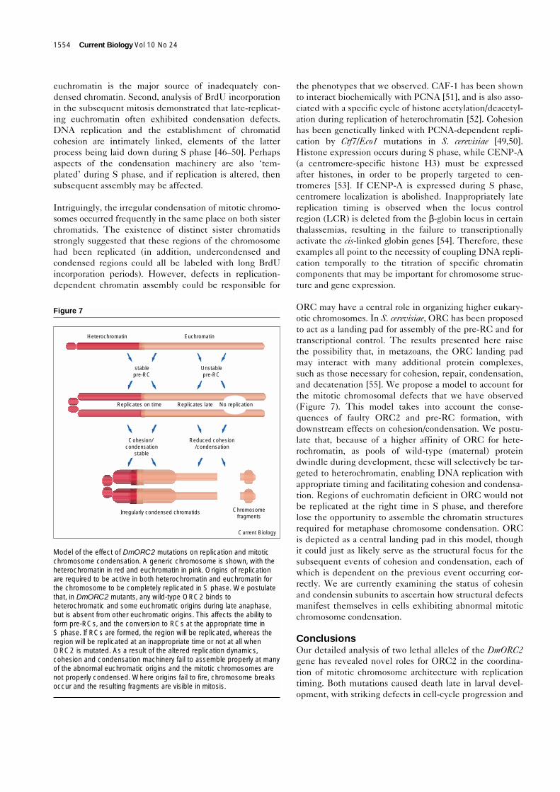
ORC may have a central role in organizing higher eukary-otic chromosomes. In S. cerevisiae, ORC has been proposedto act as a landing pad for assembly of the pre-RC and fortranscriptional control. The results presented here raisethe possibility that, in metazoans, the ORC landing padmay interact with many additional protein complexes,such as those necessary for cohesion, repair, condensation,and decatenation [55]. We propose a model to account forthe mitotic chromosomal defects that we have observed(Figure 7). This model takes into account the conse-quences of faulty ORC2 and pre-RC formation, withdownstream effects on cohesion/condensation. We postu-late that, because of a higher affinity of ORC for hete-rochromatin, as pools of wild-type (maternal) proteindwindle during development, these will selectively be tar-geted to heterochromatin, enabling DNA replication withappropriate timing and facilitating cohesion and condensa-tion. Regions of euchromatin deficient in ORC would notbe replicated at the right time in S phase, and thereforelose the opportunity to assemble the chromatin structuresrequired for metaphase chromosome condensation. ORCis depicted as a central landing pad in this model, thoughit could just as likely serve as the structural focus for thesubsequent events of cohesion and condensation, each ofwhich is dependent on the previous event occurring cor-rectly. We are currently examining the status of cohesinand condensin subunits to ascertain how structural defectsmanifest themselves in cells exhibiting abnormal mitoticchromosome condensation.
ConclusionsOur detailed analysis of two lethal alleles of the DmORC2gene has revealed novel roles for ORC2 in the coordina-tion of mitotic chromosome architecture with replicationtiming. Both mutations caused death late in larval devel-opment, with striking defects in cell-cycle progression and
1554 Current Biology Vol 10 No 24
Figure 7
Model of the effect of DmORC2 mutations on replication and mitoticchromosome condensation. A generic chromosome is shown, with theheterochromatin in red and euchromatin in pink. Origins of replicationare required to be active in both heterochromatin and euchromatin forthe chromosome to be completely replicated in S phase. We postulatethat, in DmORC2 mutants, any wild-type ORC2 binds toheterochromatic and some euchromatic origins during late anaphase,but is absent from other euchromatic origins. This affects the ability toform pre-RCs, and the conversion to RCs at the appropriate time inS phase. If RCs are formed, the region will be replicated, whereas theregion will be replicated at an inappropriate time or not at all whenORC2 is mutated. As a result of the altered replication dynamics,cohesion and condensation machinery fail to assemble properly at manyof the abnormal euchromatic origins and the mitotic chromosomes arenot properly condensed. Where origins fail to fire, chromosome breaksoccur and the resulting fragments are visible in mitosis.
Current Biology
Heterochromatin Euchromatin
stablepre-RC
Unstablepre-RC
Cohesion/ condensation
stable
Reduced cohesion/condensation
Irregularly condensed chromatids Chromosomefragments
Replicates on time Replicates late No replication
chromosome condensation in mitosis. These alleles alsodisrupted the normal pattern of chromosomal replication,with some euchromatic regions replicating even later thanheterochromatin. Mitotic chromosomes were irregularlycondensed, with the abnormally late replicating regions ofeuchromatin exhibiting the greatest problems in mitoticcondensation. Our results also suggest that the correcttiming of DNA replication may be essential for the assem-bly of chromatin that is fully competent to undergomitotic condensation.
Materials and methodsFly stocksCanton S and Df(3)redp52 (88A4-88B4,5) were obtained from theBloomington Stock Center; l(3)k431 was obtained from M. Gatti (Uni-versity of Rome) and l(3)k43γ4e from R. Kellum (University of Kentucky).The Df and both alleles were each maintained over the TM6B balancer,In(3LR)TM6B (markers: AntpHu e1 ryCB Tb1) for all the experimentsdescribed. Complementation tests confirmed that l(3)k431 andl(3)k43γ4e are allelic and that both are uncovered by Df(3R)redp52. Tran-sheterozygotes of the two alleles or with the deficiency Df(3R)redp52
showed the same overall mitotic defects, namely a range in severity ofirregularly condensed and broken chromosomes.
Immunofluorescence staining of larval neuroblastsThird instar larvae were rinsed and dissected in 1 × EBR (130 mMNaCl, 4.7 mM KCl, 1.9 mM CaCl2, 10 mM HEPES pH 6.9); the brains(without imaginal discs) were removed and hypotonically swollen for3 min in 0.25 × EBR. The brains were then fixed for 5 min inPFA/HOAc/TX-fix (4% paraformaldehyde, TAAB Laboratories, 5%acetic acid, 0.1% triton X-100, 0.1725 × EBR), during which time theywere flattened onto polylysine-treated slides under siliconized cover-slips. Preparations were frozen in liquid nitrogen, and the coverslipsflicked off with a razor blade. After 2 min hydration in PBS, the brainswere permeabilized for 3 × 10 min in PBS with 0.05% Triton X-100(PBTx) and blocked in 10% normal goat serum (Sigma) in PBS (PBS-NGS) or in 0.3% BSA in PBS (PBS-BSA) for 1 h at room temperature.The brains were washed for 5 min in PBTx and then incubated for 16 hat 4°C in the primary antibody (pre-immune or immune rabbit anti-ORC2 antiserum at 1:200 or affinity-purified) diluted in PBS-NGS or inPBS-BSA. Brains were then washed for 6 × 5 min in PBTx and incu-bated for 2 h at room temperature in the secondary antibody (Alexa594 goat anti-rabbit at 1:500, Molecular Probes), then washed again.Additional washes were performed as necessary before the brainswere stained with 75 ng/ml DAPI (Sigma). Coverslips were mountedonto the slides with Mowiol 4-88 (Calbiochem) and viewed using anOlympus Provis epifluorescence microscope equipped with a Photo-metrics Sensys CCD camera; images were captured using VysisSmartCapture software and processed with Adobe Photoshop.
BrdU incorporation into larval neuroblastsLarvae were fed for 10 min to 24 h with 50 mg/ml BrdU (BoehringerMannheim) in Drosophila instant food (Sigma). The food containedfood colouring to unambiguously discern which larvae had ingested thedrug. To visualize the BrdU, the larval brains were dissected, hypotoni-cally swollen, fixed and squashed onto polylysine-treated slides asdescribed above for analysis of neuroblast mitotic figures. Preparationswere then rinsed for 2 min in PBS, permeabilized for 5 min in PBTx,fixed again for 10 min with 4% PFA in PBS and washed for 5 min inPBTx. Brains were then treated for 30 min with freshly prepared 2NHCl and washed for 10 min in PBTx, blocked for 30 min in 0.3% BSAin PBS and washed for 5 min in PBTx. Brains were incubated with ratanti-BrdU antibody (1:2 dilution, Harlan Sera) for 16 h at 4°C. Theslides were washed 3 × 10 min at RT in PBTx and then incubated witheither FITC-conjugated goat anti-rat antibody (1:100, Harlan Sera) orAlexa 488-goat anti-rat conjugate (1:500, Molecular Probes) for 4 h at
4°C. The washes were repeated and the brains stained with 200 ng/mlDAPI during the second wash. Coverslips were mounted onto theslides with Mowiol and viewed as described above.
Supplementary materialSupplementary material including methodological detail of the cloningand sequencing of mutant DMORC2 alleles, immunofluorescence stain-ing of cultured Drosophila cells, DAPI staining of larval chromosomesand fluorescence in situ hybridization is available at http://current-biology.com/supmat/supmatin.htm.
AcknowledgementsWe thank Sharron Vass, Brian McHugh and Stefan Schoenfelder for assis-tance at different times in this project; Maurizio Gatti and Rebecca Kellumfor assorted fly stocks; Rick Austin, Steve Bell and Sue Cotterill for ORC2antibodies; Mar Carmena for the dodeca satellite plasmid; Tony Shermoenfor the 359 bp primers; and Bill Earnshaw and members of the Heck lab forstimulating discussions and critical reading of the manuscript. Research inthe Heck lab is funded by a Wellcome Trust Senior Research Fellowship inthe Basic Biomedical Sciences.
References1. Bell SP, Stillman B: ATP-dependent recognition of eukaryotic
origins of DNA replication by a multiprotein complex. Nature 1992,357:128-134.
2. Dutta A, Bell SP: Initiation of DNA replication in eukaryotic cells.Annu Rev Cell Dev Biol 1997, 13:293-332.
3. Carpenter PB, Mueller PR, Dunphy WG: Role for a XenopusOrc2-related protein in controlling DNA replication. Nature 1996,379:357-360.
4. Chesnokov I, Gossen M, Remus D, Botchan M: Assembly offunctionally active Drosophila origin recognition complex fromrecombinant proteins. Genes Dev 1999, 13:1289-1296.
5. Ehrenhofer-Murray AE, Gossen M, Pak DTS, Botchan MR, Rine J:Separation of origin recognition complex functions by cross-species complementation. Science 1995, 270:1671-1674.
6. Gossen M, Pak DTS, Hansen SK, Acharya JK, Botchan MR:A Drosophila homolog of the yeast origin recognition complex.Science 1995, 270:1674-1677.
7. Pak DTS, Pflumm M, Chesnokov I, Huang DW, Kellum R, Marr J, et al.:Association of the origin recognition complex with heterochromatinand HP1 in higher eukaryotes. Cell 1997, 91:311-323.
8. Pinto S, Quintanq DG, Smith P, Mihalek RM, Hou Z-H, Boynton S,et al.: latheo encodes a subunit of the origin recognition complexand disrupts neuronal proliferation and adult olfactory memorywhen mutant. Neuron 1999, 23:45-54.
9. Diffley JFX, Cocker JH: Protein-DNA interactions at a yeastreplication origin. Nature 1992, 357:169-172.
10. Diffley JFX, Cocker JH, Dowell SJ, Rowley A: Two steps in theassembly of complexes at yeast replication origins in vivo.Cell 1994, 78:303-316.
11. Austin RJ, Orr-Weaver TL, Bell SP: Drosophila ORC specificallybinds to ACE3, an origin of DNA replication control element.Genes Dev 1999, 13:2639-2649.
12. Liang C, Weinreich M, Stillman B: ORC and Cdc6p interact anddetermine the frequency of initiation of DNA replication in thegenome. Cell 1995, 81:667-676.
13. Aparicio OM, Weinstein DM, Bell SP: Components and dynamics ofDNA replication complexes in S. cerevisiae: redistribution of MCMproteins and Cdc45p during S phase. Cell 1997, 91:59-69.
14. Zou L, Stillman B: Formation of a preinitiation complex by S-phasecyclin CDK-dependent loading of cdc45p onto chromatin. Science1998, 280:593-596.
15. Chong JPJ, Thömmes P, Rowles A, Mahbubani HM, Blow JJ:Characterization of the Xenopus replication licensing system.Methods Enzymol 1997, 283:549-564.
16. Rowles A, Chong JPJ, Brown L, Howell M, Evan GI, Blow JJ:Interaction between the origin recognition complex and thereplication licensing system in Xenopus. Cell 1996, 87:287-296.
17. Rowles A, Tada S, Blow JJ: Changes in association of the Xenopusorigin recognition complex with chromatin on licensing ofreplication origins. J Cell Sci 1999, 112:2011-2018.
18. Kubota Y, Mimura S, Nishimoto S-i, Takisawa H, Nijima H:Identification of the yeast MCM3-related protein as a component ofXenopus DNA replication licensing factor. Cell 1995, 81:601-609.
Research Paper DMORC2 in replication timing and chromosome condensation Loupart et al. 1555
19. Thömmes P, Kubota Y, Takisawa H, Blow JJ: The RLF-M componentof the replication licensing system forms complexes containingall six MCM/P1 polypeptides. EMBO J 1997, 16:3312-3319.
20. Huang DW, Fanti L, Pak DTS, Botchan MR, Pimpinelli S, Kellum R:Distinct cytoplasmic and nuclear fractions of Drosophilaheterochromatin protein 1: their phosphorylation levels andassociations with origin recognition complex proteins. J Cell Biol1998, 142:307-318.
21. Loo S, Fox CA, Rine J, Koyayashi R, Stillman B, Bell S: The originrecognition complex in silencing, cell cycle progression, and DNAreplication. Mol Biol Cell 1995, 6:741-756.
22. Donovan S, Harwood J, Drury LS, Diffley JFX: Cdc6p-dependentloading of Mcm proteins onto pre-replicative chromatin inbudding yeast. Proc Natl Acad Sci USA 1997, 94:5611-5616.
23. Hua XH, Newport J: Identification of a preinitiation step in DNAreplication that is independent of origin recognition complex andcdc6, but dependent on cdk2. J Cell Biol 1998, 140:271-281.
24. Stillman B: Cell cycle control of DNA replication. Science 1996,274:1659-1664.
25. Klemm RD, Austin RJ, Bell SP: Coordinate binding of ATP andorigin DNA regulates the ATPase activity of the origin recognitioncomplex. Cell 1997, 88:493-502.
26. Asano M, Wharton RP: E2F mediates developmental and cell cycleregulation of ORC1 in Drosophila. EMBO J 1999, 18:2435-2448.
27. Royzman I, Austin RJ, Bosco G, Bell SP, Orr-Weaver TL: ORClocalization in Drosophila follicle cells and the effects ofmutations in dE2F and dDP. Genes Dev 1999, 13:827-840.
28. Shearn A, Rice T, Garen A, Gehring W: Imaginal disc abnormalitiesin lethal mutants of Drosophila. Proc Natl Acad Sci USA 1971,68:2594-2598.
29. Shearn A, Garen A: Genetic control of imaginal disc developmentin Drosophila. Proc Natl Acad Sci USA 1974, 71:1393-1397.
30. Szabad J, Bryant PJ: The mode of action of ‘Discless’ mutations inDrosophila melanogaster. Dev Biol 1982, 93:240-256.
31. Gatti M, Baker BS: Genes controlling essential cell-cycle functionsin Drosophila melanogaster. Genes Dev 1989, 3:438-453.
32. Landis G, Kelley R, Spradling AC, Tower J: The k43 gene, requiredfor chorion gene amplification and diploid cell chromosomereplication, encodes the Drosophila homolog of yeast originrecognition complex subunit 2. Proc Natl Acad Sci USA 1997,94:3888-3892.
33. Romanowski P, Madine MA, Rowles A, Blow JJ, Laskey RA:The Xenopus origin recognition complex is essential for DNAreplication and MCM binding to chromatin Curr Biol 1996,6:1416-1425.
34. Rao PN, Johnson RT: Mammalian cell fusion studies on theregulation of DNA synthesis and mitosis. Nature 1970,225:159-164.
35. Hendzel MJ, Wei Y, Mancini MA, Van Hooser A, Ranali T, Brinkley BR,et al.: Mitosis-specific phosphorylation of histone H3 initiatesprimarily within pericentromeric heterochromatin during G2 andspreads in an ordered fashion coincident with mitoticchromosome condensation. Chromosoma 1997, 106:348-360.
36. Lohe AR, Hilliker AJ, Roberts PA: Mapping simple repeated DNAsequences in heterochromatin of Drosophila melanogaster.Genetics 1993, 134:1149-1174.
37. Treisman JE, Follette PJ, O’Farrell PH, Rubin GM: Cell proliferationand DNA replication defects in a Drosophila MCM2 mutant. GenesDev 1995, 9:1709-1715.
38. Feger G, Vaessin H, Su TT, Wolff E, Jan LY, Jan YN: dpa, a memberof the MCM family, is required for mitotic DNA replication but notendoreplication in Drosophila. EMBO J 1995, 14:5387-5398.
39. Shirahige K, Hori Y, Shiraishi K, Yamashita M, Takahashi K, Obuse C,et al.: Regulation of DNA-replication origins during cell-cycleprogression. Nature 1998, 395:618-621.
40. Tanaka T, Nasmyth K: Association of RPA with chromosomalreplication origins requires an Mcm protein, and is regulated byRad53, and cyclin- and Dbf4-dependent kinases. EMBO J 1998,17:5182-5191.
41. Bradbury EM: Reversible histone modifications and thechromosome cell cycle. Bioessays 1992, 14:9-16.
42. Grunstein M: Histone acetylation in chromatin structure andtranscription. Nature 1997, 389:349-352.
43. Reifsnyder C, Lowell J, Clarke A, Pillus L: Yeast SAS silencing genesand human genes associated with AML and HIV-1 Tat interactionsare homologous with acetyltransferases. Nat Genet 1996,14:42-49.
44. Ehrenhofer-Murray AE, Rivier DH, Rine J: The role of Sas2, anacetyltransferase homologue of Saccharomyces cerevisiae, insilencing and ORC function. Genetics 1997, 145:93-934.
45. Iizuka M, Stillman B: Histone acetyltransferase HBO1 interacts withthe ORC1 subunit of the human initiator protein. J Biol Chem1999, 274:23027-23034.
46. Guacci V, Koshland D, Strunnikov A: A direct link between sisterchromatid cohesion and chromosome condensation revealedthrough the analysis of MCD1 in S. cerevisiae. Cell 1997,91:47-57.
47. Furuya K, Takahashi K, Yanagida M: Faithful anaphase is ensuredby Mis4, a sister chromatid cohesion molecule required inS phase and not destroyed in G1 phase. Genes Dev 1998,12:3408-3418.
48. Uhlmann F, Nasmyth K: Cohesion between sister chromatids mustbe established during DNA replication. Curr Biol 1998,8:1095-1101.
49. Skibbens RV, Corson LB, Koshland D, Hieter P: Ctf7p is essentialfor sister chromatid cohesion and links mitotic chromosomestructure to the DNA replication machinery. Genes Dev 1999,13:307-319.
50. Tóth A, Ciosk R, Uhlmann F, Galova M, Schleiffer A, Nasmyth K:Yeast cohesin complex requires a conserved protein, Eco1p(Ctf7), to establish cohesion between sister chromatids duringDNA replication. Genes Dev 1999, 13:320-333.
51. Shibahara K, Stillman B: Replication-dependent marking of DNA byPCNA facilitates CAF-1-coupled inheritance of chromatin. Cell1999, 96:575-585.
52. Taddei A, Roche D, Sibarita J-B, Turner BM, Almouzni G: Duplicationand maintenance of heterochromatin domains. J Cell Biol 1999,147:1153-1166.
53. Shelby RD, Vafa O, Sullivan KF: Assembly of CENP-A intocentromeric chromatin requires a cooperative array ofnucleosomal DNA contact sites. J Cell Biol 1997, 136:501-513.
54. Forrester WC, Epner E, Driscoll MC, Enver T, Brice M,Papayannopoulou T, Groudine M: A deletion of the human beta-globin locus activation region causes a major alteration inchromatin structure and replication across the entire beta-globinlocus. Genes Dev 1990, 4:1637-1649.
55. Cobbe N, Heck MMS: SMCs in the world of chromosome biology:from prokaryotes to eukaryotes. J Struct Biol 2000, 129:123-143.
1556 Current Biology Vol 10 No 24
Because Current Biology operates a ‘Continuous PublicationSystem’ for Research Papers, this paper has been publishedon the internet before being printed. The paper can beaccessed from http://biomednet.com/cbiology/cub
Related Documents