haematologica 2004; 89(2):February 2004 154 [haematologica] 2004;89:154-164 DAVIDE ROSSI DANIELA CAPELLO ANNUNZIATA GLOGHINI SILVIA FRANCESCHETTI MARCO P AULLI KISHOR BHATIA GIUSEPPE SAGLIO UMBERTO VITOLO STEFANO A. PILERI MANEL ESTELLER ANTONINO CARBONE GIANLUCA GAIDANO ADDITIONAL AUTHORS: CLARA DEAMBROGI EVA BERRA MICHAELA CERRI ANNARITA CONCONI CHIARA VENDRAMIN MARINA GUTIERREZ BARBARA BOTTO From the Hematology Unit, Dept. of Medical Sciences & IRCAD, Amedeo Avogadro University of Eastern Pied- mont, Novara, Italy (DR, DC, SF, GG, CD, EB, MC, ACO, CV); Division of Pathology, IRCCS, Centro di Riferimento Oncologico, Aviano, Italy (AG, ACA); Institute of Patholo- gy, IRCCS Policlinico San Matteo, University of Pavia, Pavia, Italy (MP); KFNCCC&R, King Faisal Specialist Hospital and Research Centre, Riyadh, Saudi Arabia (KB, MG); Division of Internal Medicine and Hematology, Department of Clinical and Biological Sciences, University of Turin, Orbassano, Italy (GS); Divi- sion of Hematology, A. O. San Gio- vanni Battista della Citta’ di Torino, Turin, Italy (UV, BB); Hematopatholo- gy Unit, L. & A. Seràgnoli Institute of Hematology and Oncology, Universi- ty of Bologna, Bologna, Italy (SAP); Cancer Epigenetics Laboratory, Spanish National Cancer Centre (CNIO), Madrid, Spain (ME). Correspondence: Gianluca Gaidano, M.D., Ph.D., Hematology Unit, Dept. of Medical Sciences & IRCAD, “Amedeo Avogadro” University of Eastern Piedmont, via Solaroli 17, 28100 Novara, Italy. E-mail: [email protected] ©2004, Ferrata Storti Foundation Aberrant promoter methylation of multiple genes throughout the clinico-pathologic spectrum of B-cell neoplasia Background and Objectives. Aberrant promoter methylation targets CpG islands caus- ing gene silencing. We explored aberrant promoter methylation of genes potentially involved in B-cell malignancies and encoding proteins implicated in DNA repair (O 6 -methylguanine- DNA methyltransferase, MGMT), detoxification of environmental xenobiotics (glutathione S-transferase P1, GSTP1), apoptosis regulation (death associated protein kinase, DAP-k and caspase 8, CASP8) and cell cycle control (p73). Design and Methods. Three hundred and seventeen B-cell malignancies were investigated by methylation-specific polymerase chain reaction (MSP) of MGMT, GSTP1, DAP-k, CASP8 and p73 genes. In selected cases, MSP results were matched to protein expression studies by immunohistochemistry or Western blotting. Results. DAP-k promoter methylation occurred at highest frequency in follicular lym- phoma (85.0%) and MALT-lymphoma (72.2%). MGMT methylation targeted both precur- sor B-cell neoplasia (23.8%) and mature B-cell tumors (27.6%). GSTP1 methylation was commonest in hairy cell leukemia (75.0%), follicular lymphoma (55.5%), Burkitt’s lym- phoma (52.0%), and MALT lymphoma (50.0%). Methylation of p73 and CASP8 was rare or absent. DAP-k and MGMT methylation caused absent protein expression. Interpretation and Conclusions. Methylation of MGMT, DAP-k and GSTP1 represents a major pathogenetic event in several B-cell malignancies. In follicular lymphoma and MALT lymphoma, frequent inactivation of the apoptosis extrinsic pathway through DAP-k methy- lation may reinforce the survival advantage already conferred by deregulation of the intrin- sic apoptotic pathway. Inactivation of GSTP1 in gastric MALT lymphoma represents an addi- tional mechanism favoring accumulation of reactive oxygen species and lymphomagene- sis. Finally, the frequency of GSTP1 aberrant methylation in diffuse large B-cell lymphoma prompts studies aimed at verifying the prognostic impact of this epigenetic lesion in these lymphomas. Key words: B-cell, lymphoma, leukemia, aberrant methylation, pathogenesis. G rowing evidence has implicated aber- rant promoter methylation in the molecular pathogenesis of several human cancers. 1-4 Aberrant promoter methy- lation is an epigenetic DNA modification tar- geting CpG islands located within the regu- latory regions of human genes. As a conse- quence, aberrant methylation of CpG islands causes repression of gene transcription and represents a mechanism for tumor suppres- sor gene inactivation alternative to muta- tions/deletions of the locus. 1-4 To date, the role of aberrant promoter methylation in B-cell lymphoid malignan- cies has not been investigated exhaustively, and extensive studies have been mainly restricted to the cyclin dependent kinase inhibitors p15 and p16. 5 Although other genes have been found to be targeted by aberrant methylation in lymphoid neoplasia, their analysis has been limited to specific types of B-cell malignancies or to lymphoid tumor cell lines. 6-9 These observations prompted our comprehensive analysis aimed at exploring the prevalence of aberrant pro- moter methylation in a selected panel of genes potentially involved in the pathogen- esis of B-cell malignancies and representa- tive of genes implicated in DNA repair (O 6 - methylguanine-DNA methyltransferase, MGMT), detoxification of environmental xenobiotics (glutathione S-transferase P1, GSTP1), apoptosis regulation (death associ- ated protein kinase, DAP-k and caspase-8, CASP8) and cell cycle control (p73). Previous studies have shown that promoter hyper- methylation of these genes represents the major mechanism of gene inactivation, whereas allelic loss or mutations are virtu- ally absent. 10-11 MGMT is a DNA repair gene that removes mutagenic and cytotoxic adducts introduced A B S T R A C T

Welcome message from author
This document is posted to help you gain knowledge. Please leave a comment to let me know what you think about it! Share it to your friends and learn new things together.
Transcript

haematologica 2004; 89(2):February 2004154
[haematologica]2004;89:154-164
DAVIDE ROSSI
DANIELA CAPELLO
ANNUNZIATA GLOGHINI
SILVIA FRANCESCHETTI
MARCO PAULLI
KISHOR BHATIA
GIUSEPPE SAGLIO
UMBERTO VITOLO
STEFANO A. PILERI
MANEL ESTELLER
ANTONINO CARBONE
GIANLUCA GAIDANO
ADDITIONAL AUTHORS:CLARA DEAMBROGI
EVA BERRA
MICHAELA CERRI
ANNARITA CONCONI
CHIARA VENDRAMIN
MARINA GUTIERREZ
BARBARA BOTTO
From the Hematology Unit, Dept. ofMedical Sciences & IRCAD, AmedeoAvogadro University of Eastern Pied-mont, Novara, Italy (DR, DC, SF, GG,CD, EB, MC, ACO, CV); Division ofPathology, IRCCS, Centro diRiferimento Oncologico, Aviano,Italy (AG, ACA); Institute of Patholo-gy, IRCCS Policlinico San Matteo,University of Pavia, Pavia, Italy (MP);KFNCCC&R, King Faisal SpecialistHospital and Research Centre,Riyadh, Saudi Arabia (KB, MG);Division of Internal Medicine andHematology, Department of Clinicaland Biological Sciences, Universityof Turin, Orbassano, Italy (GS); Divi-sion of Hematology, A. O. San Gio-vanni Battista della Citta’ di Torino,Turin, Italy (UV, BB); Hematopatholo-gy Unit, L. & A. Seràgnoli Institute ofHematology and Oncology, Universi-ty of Bologna, Bologna, Italy (SAP);Cancer Epigenetics Laboratory,Spanish National Cancer Centre(CNIO), Madrid, Spain (ME).
Correspondence: Gianluca Gaidano,M.D., Ph.D., Hematology Unit, Dept.of Medical Sciences & IRCAD,“Amedeo Avogadro” University ofEastern Piedmont, via Solaroli 17,28100 Novara, Italy.E-mail: [email protected]
©2004, Ferrata Storti Foundation
Aberrant promoter methylation of multiple genesthroughout the clinico-pathologic spectrum of B-cell neoplasia
Background and Objectives. Aberrant promoter methylation targets CpG islands caus-ing gene silencing. We explored aberrant promoter methylation of genes potentially involvedin B-cell malignancies and encoding proteins implicated in DNA repair (O6-methylguanine-DNA methyltransferase, MGMT), detoxification of environmental xenobiotics (glutathioneS-transferase P1, GSTP1), apoptosis regulation (death associated protein kinase, DAP-k andcaspase 8, CASP8) and cell cycle control (p73).
Design and Methods. Three hundred and seventeen B-cell malignancies were investigatedby methylation-specific polymerase chain reaction (MSP) of MGMT, GSTP1, DAP-k, CASP8and p73 genes. In selected cases, MSP results were matched to protein expression studiesby immunohistochemistry or Western blotting.
Results. DAP-k promoter methylation occurred at highest frequency in follicular lym-phoma (85.0%) and MALT-lymphoma (72.2%). MGMT methylation targeted both precur-sor B-cell neoplasia (23.8%) and mature B-cell tumors (27.6%). GSTP1 methylation wascommonest in hairy cell leukemia (75.0%), follicular lymphoma (55.5%), Burkitt’s lym-phoma (52.0%), and MALT lymphoma (50.0%). Methylation of p73 and CASP8 was rare orabsent. DAP-k and MGMT methylation caused absent protein expression.
Interpretation and Conclusions. Methylation of MGMT, DAP-k and GSTP1 represents amajor pathogenetic event in several B-cell malignancies. In follicular lymphoma and MALTlymphoma, frequent inactivation of the apoptosis extrinsic pathway through DAP-k methy-lation may reinforce the survival advantage already conferred by deregulation of the intrin-sic apoptotic pathway. Inactivation of GSTP1 in gastric MALT lymphoma represents an addi-tional mechanism favoring accumulation of reactive oxygen species and lymphomagene-sis. Finally, the frequency of GSTP1 aberrant methylation in diffuse large B-cell lymphomaprompts studies aimed at verifying the prognostic impact of this epigenetic lesion in theselymphomas.
Key words: B-cell, lymphoma, leukemia, aberrant methylation, pathogenesis.
Growing evidence has implicated aber-rant promoter methylation in themolecular pathogenesis of several
human cancers.1-4 Aberrant promoter methy-lation is an epigenetic DNA modification tar-geting CpG islands located within the regu-latory regions of human genes. As a conse-quence, aberrant methylation of CpG islandscauses repression of gene transcription andrepresents a mechanism for tumor suppres-sor gene inactivation alternative to muta-tions/deletions of the locus.1-4
To date, the role of aberrant promotermethylation in B-cell lymphoid malignan-cies has not been investigated exhaustively,and extensive studies have been mainlyrestricted to the cyclin dependent kinaseinhibitors p15 and p16.5 Although othergenes have been found to be targeted byaberrant methylation in lymphoid neoplasia,their analysis has been limited to specific
types of B-cell malignancies or to lymphoidtumor cell lines.6-9 These observationsprompted our comprehensive analysis aimedat exploring the prevalence of aberrant pro-moter methylation in a selected panel ofgenes potentially involved in the pathogen-esis of B-cell malignancies and representa-tive of genes implicated in DNA repair (O6-methylguanine-DNA methyltransferase,MGMT), detoxification of environmentalxenobiotics (glutathione S-transferase P1,GSTP1), apoptosis regulation (death associ-ated protein kinase, DAP-k and caspase-8,CASP8) and cell cycle control (p73). Previousstudies have shown that promoter hyper-methylation of these genes represents themajor mechanism of gene inactivation,whereas allelic loss or mutations are virtu-ally absent.10-11
MGMT is a DNA repair gene that removesmutagenic and cytotoxic adducts introduced
A B S T R A C TA B S T R A C T
into the DNA from environmental and therapeutic alky-lating agents.12 In particular, MGMT inactivationincreases cell sensitivity to the genotoxic effect of alky-lating agents both in vitro and in vivo.13,14 The potentialrole of MGMT in lymphoma stems from the fact thatMGMT inactivation favors lymphomagenesis in knock-out mice and represents a favorable prognostic markerfor diffuse large B-cell lymphoma (DLBCL) treated withregimens containing alkylating agents.8,15
Aberrant promoter methylation of DAP-k has beensuggested to have a potential role in lymphomagene-sis.7 DAP-k is a pro-apoptotic serine-threonine kinaseinvolved in the extrinsic pathway of apoptosis initiat-ed by interferon γ, tumor necrosis factor-α and FAS lig-and.7,16 In addition, DAP-k also participates in counter-acting c-MYC-induced transformation by activating thep53 checkpoint and favoring c-MYC-induced apopto-sis.17 Consequently, inactivation of DAP-k preventsapoptosis triggered by death receptors and weakens theapoptotic response secondary to c-MYC activation.
GSTP1 is an enzyme implicated in the detoxificationof environmental carcinogens and chemotherapeuticagents.18 Its loss of expression is a risk factor for thedevelopment of cancer in null mice.19 Although epige-netic alterations of GSTP1 are recurrent in a wide rangeof solid tumors, its methylation pattern in B-cell neo-plasia is not known.20 Caspase-8 (CASP8) and p73 arealso potential targets of aberrant promoter methyla-tion in lymphoma.6,21 Similarly to DAP-k, CASP8 isinvolved in the extrinsic pathway of programmed celldeath that transduces the apoptotic signal from a deathreceptor to the common pathway of apoptosis.21,22 Thep73 gene is a candidate tumor suppressor gene sharingstructural and functional similarities with p53 and isinvolved in cell cycle control and apoptosis.23
In this study we investigated aberrant promotermethylation of multiple genes in a large panel of B-cellneoplasms representative of the clinico-pathologicspectrum of the disease.
Design and Methods
Tumor samples and DNA preparationThis study was based on 317 tumor samples repre-
sentative of the clinico-pathologic spectrum of B-cellneoplasia recognized by the WHO classification.24 Tumorsamples were derived from lymph nodes, bone marrow,peripheral blood or other involved organs obtained dur-ing routine diagnostic procedures. In all instances, withthe exception of DLBCL transformed from a follicularphase, the specimens were collected at diagnosis beforespecific therapy. Diagnosis was based on morphologyand immunophenotypic analysis of cell surface mark-ers and was complemented by immunogenotypic analy-
sis of antigen receptor gene rearrangement and chro-mosomal translocations. In most cases, the fraction ofmalignant cells was > 70% and in all cases > 40%.According to the WHO classification,24 B-cell neoplasiaspecimens were classified as precursor B-cell acute lym-phoblastic leukemia (ALL; n = 21), B-cell chronic lym-phocytic leukemia (B-CLL; n = 30), lymphoplasmacyticlymphoma (LPL; n = 9), mantle cell lymphoma (MCL; n= 19), follicular lymphoma (FL; n = 21), mucosa-asso-ciated lymphoid tissue (MALT) lymphoma (n= 11), hairycell leukemia (HCL; n = 11), DLBCL (n = 140), mediasti-nal large B-cell lymphoma (MLBCL; n =10), Burkitt’slymphoma (BL; n = 29) and plasma cell myeloma (PCM;n = 16). The precursor B-cell ALL samples were repre-sentative of different molecular variants of the diseaseand included cases associated with hyperdiploidy (n =5), BCR/ABL rearrangement (n = 3), MLL rearrangement(n = 5), TEL/AML-1 rearrangement (n = 3), or not knowngenetic lesions (n = 5). MALT lymphomas originatedfrom the gastrointestinal tract (n = 9) or the thyroid (n= 2). DLBCL were subdivided into DLBCL arising de novowithout clinical evidence of previous lymphoma (denovo DLBCL; n = 129) and DLBCL transformed from aprevious follicular lymphoma (transformed DLBCL; n =11). The study was approved by the institutional reviewboard and written consent was obtained from thepatients.
Genomic DNA was purified by cell lysis followed bydigestion with proteinase K, salting out extraction, andprecipitation by ethanol.25
Bisulfite treatmentDNA from tumor specimens was subjected to chem-
ical treatment with sodium bisulfite as previouslyreported.26 Briefly, 1 µg of DNA was denatured by treat-ment with NaOH, and modified by sodium bisulfitetreatment for 16 h at 50°C. DNA samples were thenpurified using Wizard DNA purification resin (Promega),desulphonated by incubating in a final concentration of0.3 M NaOH, precipitated with ethanol, and resus-pended in water.
Methylation-specific polymerase chain reaction(MSP)
The modified DNA was used as a template for MSP, amolecular technique that allows the distinctionbetween methylated and unmethylated DNA.26 MSP wasperformed on 50 ng of bisulfite-treated DNA under thefollowing conditions: a denaturing step at 95°C for 7min is followed by 35 cycles at 95°C (30 sec per cycle),the annealing temperature being specific for each reac-tion (30 sec per cycle), and 72°C (30 sec per cycle), in aHybaid DNA thermal cycler. The PCR mixture contained10×Gold buffer (Perkin Elmer), 6.7 mM MgCl2, 10 µM ofeach primer, 1 mM dNTPs and 0.625 U of Taq Gold
haematologica 2004; 89(2):February 2004 155
Aberrant promoter methylation in B-cell neoplasia

(Perkin Elmer, 5 U/µL), in a final volume of 25 µL. Primers for MGMT were: 5’-TTT GTG TTT TGA TGT TTG
TAG GTT TTT GT-3’ (sense) and 5’-AAC TCC ACA CTC TTCCAA AAA CAA AAC A-3’ (antisense) for the unmethy-lated reaction; and 5’-TTT CGA CGT TCG TAG GTT TTCGC-3’ (sense) and 5’-GCA CTC TTC CGA AAA CGA AACG-3’ (antisense) for the methylated reaction.8 Theannealing temperature for both the unmethylated andthe methylated reactions was 59°C. Primers for DAP-kwere: 5’-GGA GGA TAG TTG GAT TGA GTT AAT GTT-3’(sense) and 5’-CAA ATC CCT CCC AAA CAC CAA-3’ (anti-sense) for the unmethylated reaction; and 5’-GGA TAGTCG GAT CGA GTT AAC GTC-3’ (sense) and 5’-CCC TCCCAA ACG CCG A-3’ (antisense) for the methylated reac-tion.7 The annealing temperature for both the unmethy-lated and the methylated reactions was 60°C. Primersfor p73 were: 5’-AGG GGA TGT AGT GAA ATT GGG GTTT-3’ (sense) and 5’-ATC ACA ACC CCA AAC ATC AACATC CA-3’ (antisense) for the unmethylated reaction;and 5’- GGA CGT AGC GAA ATC GGG GTT C -3’ (sense)and 5- ACC CCG AAC ATC GAC GTC CG -3’ (antisense)for the methylated reaction.6 The annealing temperaturefor both the unmethylated and the methylated reac-tions was 58°C. Primers for CASP8 were: 5’-TAG GGGATT TGG AGA TTG TGA-3’ (sense) and 5’-CAT ATA TCTACA TTC AAA ACA A-3’ (antisense) for the unmethylat-ed reaction; and 5’-TAG GGG ATT CGG AGA TTG CGA-3’ (sense) and 5-CGT ATA TCT ACA TTC GAA ACG A-3’(antisense) for the methylated reaction.21 The annealingtemperature was 54°C for the unmethylated reactionand 52°C for the methylated reaction. Primers for GSTP1were: 5’-GAT GTT TGG GGT GTA GTG GTT GTT -3’ (sense)and 5’-CCA CCC CAA TAC TAA ATC ACA ACA -3’ (anti-sense) for the unmethylated reaction; and 5’-TTC GGGGTG TAG CGG TCG TC-3’ (sense) and 5-GCC CCA ATACTA AAT CAC GAC G-3’ (antisense) for the methylatedreaction.20 The annealing temperature was 58°C for theunmethylated reaction and 52°C for the methylatedreaction.
All MSP analyses were performed with positive andnegative controls for both unmethylated and methy-lated alleles. Also, control experiments without DNAwere performed for each set of PCR. Ten microliters ofeach PCR were directly loaded onto 2.5% agarose gels,stained with ethidium bromide, and visualized underUV illumination.
Immunohistochemical staining for MGMTThe correlation between MGMT methylation status
and MGMT protein epression was assessed in a repre-sentative panel of 28 mature B-cell neoplasms. Paraf-fin-embedded tissue sections were deparaffinized withxylene, dehydrated by using a graded series of ethanoland treated for 30 minutes in TEC (Tris-EDTA-Citrate)solution (pH 7.8) in a microwave oven at 250 W.
Immunohistochemistry was performed using the ABCmethod (ABC-Elite kit, Vector, Burlingame, CA, USA)with diaminobenzidine as the chromogen. Commer-cially available mouse anti-MGMT monoclonal anti-body (clone MT3.1; Chemicon Intl., Temecula, CA, USA)at a 1:100 dilution was used.27 This antibody had beenpreviously demonstrated to be useful for immunohis-tochemistry and to correlate with MGMT activity.11,28
Nuclear staining was determined by two authors (A.G.and A.C.) who did not have knowledge of the molecu-lar analysis of the samples.
Protein preparation and Western blot analysis ofDAP-kinase
Ice cold lysis buffer was used to lyse 107 cells. Thecomposition of this lysis buffer was as follows: 10 mMphosphate buffer, pH 7.5, 100 mM NaCl, 1% Triton X-100, 0.5% sodium deoxycholate, 0.1% SDS, 5 mM EDTA,1 µg/mL phenylmethyl sulphonyl fluoride (PMSF), and1× complete protease inhibitor (Boehringer Mannheim).Subsequently, equal amounts of total protein (400 µg)from each sample were separated on 7.5% SDS-PAGE(Bio-Rad, Hercules, CA, USA). The proteins were trans-ferred onto a nitrocellulose membrane, which wasblocked with 5% BSA/TTBS for 2 hours and then incu-bated with an anti-DAP-k goat polyclonal antibody(1:1,000 dilution in 1% BSA/TTBS; Santa Cruz Biotech-nology, Santa Cruz, CA, USA) for 1-2 hours. After wash-ing, filters were reacted with horseradish peroxidase-coupled secondary antibody, and revealed with anenhanced chemiluminescence (ECL) detection system(Amersham, Arlington Heights, IL, USA) after exposureto film.
Results
DAP-k, MGMT, GSTP1, p73 and CASP8methylation in non-neoplastic samples
The sequences of MGMT, DAP-k, GSTP1, p73 andCASP8 analyzed in our study by MSP are localized inCpG islands spanning the 5’ non-coding region of thecorresponding genes. The aberrant methylation of theseregions has been reported to be associated with tran-scriptional silencing.6,7,11,20,21,28
To verify that aberrant methylation of these genes islimited to neoplastic B-cells, and to exclude the possi-bility of non-specific reactions, we tested a panel ofnon-neoplastic lymphoid tissues represented by normalperipheral blood leukocytes (n = 5), EBV-immortalizedB lymphocytes (n = 8), and samples of reactive poly-clonal B-cell hyperplasia (n = 12). None of these non-neoplastic samples displayed methylation of MGMT,DAP-k, GSTP1, p73 or CASP8 CpG islands (data notshown), confirming that aberrant methylation is not
haematologica 2004; 89(2):February 2004156
D. Rossi et al.

haematologica 2004; 89(2):February 2004 157
Aberrant promoter methylation in B-cell neoplasia
related to cell culture, viral transformation, cell activa-tion or proliferation.
Aberrant methylation of MGMT promoterin B-cell neoplasia
The results of MGMT methylation analysis in B-cellneoplasia are detailed in Table 1 and representativeexamples are shown in Figure 1. Overall, MGMT aber-rant methylation occurred in both precursor B-cell neo-plasia (5/21 ALL; 23.8%) and mature B-cell tumors79/286 (27.6%). MGMT aberrant methylation was het-erogeneously distributed throughout the entire clini-co-pathologic spectrum of B-cell neoplasia, and pref-erentially targeted MLBCL (7/10; 70.0%) and DLBCL(47/132; 38.0%) among the aggressive lymphomas, andHCL (4/10; 40.0%), MALT lymphoma (3/11; 27.2%) andFL (5/20; 25.0%) among the indolent lymphoid malig-nancies (Table 1).
In order to confirm the biological effect of MGMTpromoter methylation, we performed expression stud-ies by immunohistochemistry of the MGMT protein innormal germinal center B cells, which represent theputative normal counterpart of a large fraction ofmature B-cell lymphoid tumors, and in selected lym-phoma samples. Immunohistochemistry experimentsshowed that i) the MGMT protein is physiologicallyexpressed by normal germinal center B cells (Figure 2),which are physiologically unmethylated at the MGMTpromoter; and ii) MGMT promoter methylation corre-lated with absent MGMT protein expression in lym-phoma cells. In fact, all (n = 17) lymphoma samplescarrying MGMT aberrant methylation and tested byimmunohistochemistry failed to express the protein invirtually all tumor cells (Figure 3); conversely, all (n =11) lymphoma samples carrying unmethylated MGMT
alleles and tested by immunohistohemistry expressedthe MGMT protein (Figure 3).
Aberrant methylation of DAP-k promoterin B-cell neoplasia
The results of DAP-k promoter methylation in B-cellneoplasia are detailed in Table 1 and representativeexamples are shown in Figure 1. Overall, DAP-k pro-moter methylation was uncommon in ALL (3/22;14.2%), while it was frequently observed in mature B-cell neoplasia (132/281; 46.9%). Among mature B-cellneoplasms, aberrant methylation of DAP-k did not occurrandomly, but rather clustered with specific clinico-pathologic categories. In particular, FL and MALT-lym-phoma showed the highest prevalence of DAP-k aber-rant methylation, since the aberrant methylation wasdetected in 17/20 (85.0%) FL and 8/11 (72.2%) MALT-lymphomas. DAP-k aberrant methylation was also com-mon in aggressive NHL, occurring in DLBCL (71/126;56.3%), MLBCL (6/10; 60.0%) and BL (14/28 50.0%)(Table 1).
In order to confirm the biological effect of DAP-kaberrant methylation, we performed expression studiesby Western blot of the DAP-k protein in lymphoma celllines. This analysis showed that DAP-k protein wasexpressed by lymphoma cell lines unmethylated at theDAP-k promoter, while DAP-k protein expression wasabsent in lymphoma cell lines harboring DAP-k pro-moter methylation (Figure 4).
Aberrant methylation of GSTP1 promoterin B-cell neoplasia
GSTP1 promoter hypermethylation was observed inboth ALL (3/11; 27.7%) and mature B-cell neoplasia(78/207; 37.6%) (Table 1 and Figure 1). Among mature
Table 1. Aberrant methylation of DAP-k, MGMT, GSTP1, p73 and CASP8 in B-cell neoplasia.
Histology DAP-k MGMT GSTP1 p73 CASP8
Precursor B-cell neoplasmsAcute lymphoblastic leukemia 3/22 (14.2 %) 5/21 (23.8 %) 3/11 (27.7%) 3/22 (22.7%) 0/10
Peripheral B-cell neoplasmsB-cell chronic lymphocytic leukemia 8/30 (26.6 %) 3/30 (10.0 %) 1/10 (10.0%) 0/12 0/10Lymphoplasmacytic lymphoma 0/9 1/9 (11.1 %) 0/7 0/8 0/7Mantle cell lymphoma 5/19 (26.3 %) 1/18 (5.5 %) 2/8 (25.0%) 0/12 0/9Follicular lymphoma 17/20 (85.0 %) 5/20 (25.0 %) 10/18 (55.5%) 1/10 (10.0%) 0/9MALT lymphoma 8/11 (72.2 %) 3/11 (27.2 %) 5/10 (50.0%) 1/11 (9.1%) 0/9Hairy cell leukemia 3/11 (27.2 %) 4/10 (40.0 %) 6/8 (75.0%) 0/9 0/5Diffuse large B-cell lymphoma 71/126 (56.3 %) 47/132 (35.6 %) 38/99 (38.3%) 10/113 (8.8%) 0/100Mediastinal large B-cell lympnoma 6/10 (60.0%) 7/10 (70.0%) 3/10 (30.0%) 0/4 0/10Burkitt’s lymphoma 14/28 (50.0 %) 5/28 (17.8%) 13/25 (52.0%) 1/9 (9.1%) 0/9Primary effusion lymphoma 0/1 1/2 0/2 nda nda
Plasma cell myeloma 0/16 2/16 (12.5%) 0/10 0/11 0/10
and: not done.

haematologica 2004; 89(2):February 2004158
D. Rossi et al.
B-cell neoplasms, aberrant methylation of GSTP1 wasfrequently detected in HCL (6/8; 75.0%) and alsooccurred in a fraction of FL (10/18; 55.5%), BL (13/25;52.0%), MALT lymphoma (5/10; 50.0%) and DLBCL(38/99; 38.3%) (Table 1).
p73 promoter hypermethylation in B-cellneoplasia
Aberrant methylation of p73 occurred in a fractionof ALL (3/22; 22.7%), while it was uncommon in matureB-cell neoplasia (13/199; 6.5%) (Table 1). Notably,among BL cell lines, aberrant methylation of p73occurred at a remarkably higher frequency (5/11;45.4%) than among BL primary samples (1/9; 9.1%)(Table 1).
CASP8 promoter hypermethylation B-cellneoplasia
None of the B-cell neoplasms investigated in thisstudy showed promoter methylation of the CASP8 gene(Table 1).
Longitudinal follow-up of DAP-k, MGMTand GSTP1 aberrant methylation
In an attempt to clarify the timing of acquisition ofaberrant methylation of DAP-k, MGMT and GSTP1 intransformed cells, we studied 11 DLBCL transformedfrom a previous follicular phase before and after histo-logic progression (Table 2 and Figure 5). Aberrantmethylation of DAP-k occurred in 7/9 (77.7%) patientsin follicular phase and in 7/9 (77.7%) transformed sam-
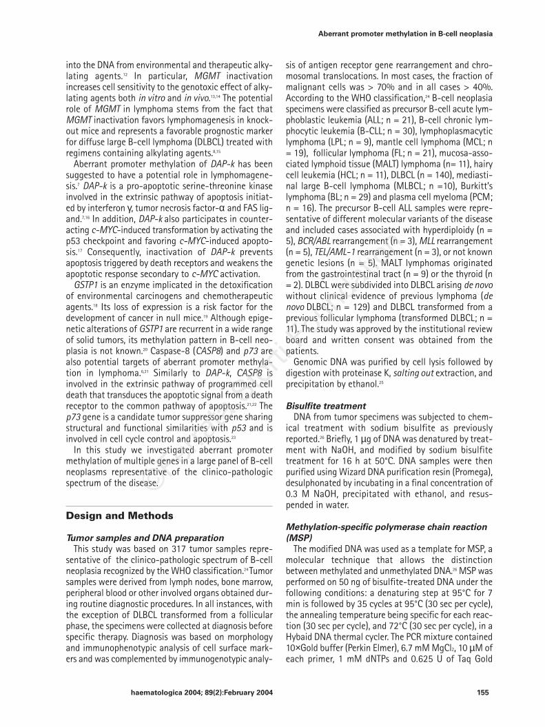
Figure 1. Methylation-specific PCR of the genes MGMT (panel A), DAP-k (panel B) and GSTP1 (panel C) in represen-tative cases of DLBCL. The presence of a visible PCR product in lane M indicates the presence of a methylated allele(MGMT: 81 bp; DAP-k: 98 bp; GSTP1: 91 bp), while the presence of a product in lane U indicates the presence ofunmethylated alleles (MGMT: 93 bp; DAP-k: 106 bp; GSTP1: 97 bp). In all experiments, the Hela cervical cancer cellline was used as the positive control for methylated alleles. Peripheral blood (PB) mononuclear cells were used asthe control for unmethylated alleles. MW, molecular weight markers.
A
B
C
93 bp81 bp
106 bp98 bp
97 bp91 bp
MW M U M U M U M U M U
MW M U M U M U M U M U
MW M U M U M U M U M U
PB HeLa #469 #499 #618
PB HeLa #469 #499 #618
PB HeLa #469 #499 #618

haematologica 2004; 89(2):February 2004 159
Aberrant promoter methylation in B-cell neoplasia
ples. All cases displaying DAP-k methylation in the fol-licular phase retained this alteration during the pro-gression to DLBCL. Aberrant methylation of MGMToccurred in 1/8 (12%) follicular phases and in 2/8 (25%)transformed samples. In particular, one patient dis-played aberrant methylation of MGMT in the trans-formed, but not in the follicular phase, suggesting thataberrant methylation had been acquired at the time ofhistologic progression. Aberrant methylation of GSTP1occurred in 5/8 (62.5%) patients in follicular phase andin 4/8 (50.0%) transformed cases. Two patients acquiredand 3 patients lost aberrant GSTP1 methylation at thetime of transformation.
Methylation status of B-cell neoplasiaWe performed a comparative analysis of the methy-
lation pattern of B-cell neoplasia. Overall, simultaneouspromoter hypermethylation in ≥ 3/5 genes occurred in
3/10 (30.0%) MALT lymphoma, 2/8 (25.0%) HCL, 11/102(10.7%) DLBCL, 1/18 (5.5%) FL and 1/24 (4.1%) BL.
Discussion
The aim of this study was a comprehensive investi-gation of aberrant promoter methylation of multiplegenes throughout the clinico-pathologic spectrum of B-cell neoplasia. We report that i) aberrant promotermethylation of MGMT, DAP-k and GSTP1 is involved inthe molecular pathogenesis of B-cell neoplasia; and ii)MGMT, DAP-k and GSTP1 promoter methylation is notrandomly distributed among B-cell neoplasms, but pref-erentially targets specific clinico-pathologic categoriesof the disease. Overall, these data have multiple impli-cations for the understanding of the molecular patho-genesis of B-cell neoplasia.
Figure 2. Panel A: MGMT protein expres-sion in normal B cells. MGMT protein isexpressed by normal mantle zone (MZ) andgerminal center (GC) B cells. Paraffin-embedded tissue sections; ABC method,hematoxylin counterstain. Original magni-fication ×¥250. Panel B: Methylation-specif-ic PCR of the MGMT gene in samples of nor-mal lymph nodes. MGMT promoter is phys-iologically unmethylated in normal lymphnodes. MW, molecular weight markers.
A
BU M U M U M MW
Lym
ph n
ode
1
Lym
ph n
ode
2
Lym
ph n
ode
3
93 bp →Æ

haematologica 2004; 89(2):February 2004160
D. Rossi et al.
DAP-k aberrant methylation is the commonest epi-genetic alteration identified to date in FL and MALTlymphoma, further confirming the role of apopotosisderegulation in the molecular pathogenesis of thesetypes of lymphoma. In fact, in lymphoma cells, DAP-kinactivation results in disruption of the extrinsic path-way of apoptosis initiated by interferon γ, tumor necro-sis factor α and FAS ligand.7,16,29 Because resistance toFAS-induced apoptosis is a common event in B-cell NHLpathogenesis and appears to occur independently ofFAS gene mutations or FAS protein expression,30 it ispossible that DAP-k methylation may represent a majordeterminant of the FAS-resistant phenotype in lym-phoma. Thus, inactivation of the extrinsic pathway of
apoptosis through DAP-k methylation may reinforceand possibly co-operate with the survival advantageconferred to lymphoma cells by BCL-2 deregulation inFL and NK-κB activation in MALT lymphoma.31 On thesebases, the concomitant disruption of both the intrinsicand the extrinsic pathways of apoptosis corroborate theview that FL and MALT lymphomas are diseases of celldeath regulation. DAP-k inactivation may also play asynergistic role with c-MYC deregulation. In fact, lossof DAP-k, by downregulating p53, may favor c-MYC-induced transformation.17 Remarkably, BL, which have c-MYC deregulation in all cases, also carry DAP-k inacti-vation in 50% of samples.7
Our results demonstrate that MGMT inactivation
A B
C D
Figure 3. MGMT protein expression in B-cell neoplasia. Panels A-B: Loss of MGMT protein expression in lymphomacells (panel A) is demonstrated in a representative case of DLBCL that displays MGMT promoter hypermethylation(panel B). The occurrence of an unmethylated signal in the methylation-specific PCR of the MGMT gene of this DLB-CL case (panel B) may be ascribed to the presence of contaminating non-neoplastic cells staining positive for MGMTexpression (panel A). Panel C-D: A case of primary effusion lymphoma (primary sample), unmethylated at the MGMTpromoter (panel D), shows expression of the MGMT protein (panel C). A) Paraffin-embedded tissue section, C) Cellblock; ABC method, hematoxylin counterstain. Original magnification ×¥ 400. MW, molecular weight markers.
93 bp81 bp
DLBCL
PEL
93 bp
U M MW
U M MW

haematologica 2004; 89(2):February 2004 161
Aberrant promoter methylation in B-cell neoplasia
through promoter methylation occurs with variable fre-quencies throughout the entire spectrum of B-cell neo-plasia. MGMT methylation is selectively restricted toneoplastic cells and is consistently absent in normallymphoid cells, pointing to a pathogenetic role of thisepigenetic lesion. Consistent with the DNA repair func-tion of MGMT against spontaneous G to A transitions,MGMT inactivation may cause genetic instability favor-ing lymphomagenesis through the acquisition of DNApoint mutations.32 The pathogenetic role of MGMT inac-tivation is further supported by the fact that MGMTknockout mice develop lymphoma at high frequency.15
Aberrant methylation of GSTP1 occurred frequently inseveral types of B-cell malignancies, including 50%MALT lymphomas. The potential pathogenetic role ofGSTP1 inactivation through promoter methylation islinked to its important role in scavenging reactive oxy-gen species and their metabolites and protecting cellsfrom DNA damage produced by these agents.18 Polymor-phisms in the GST gene family, to which GSTP1 belongs,may decrease enzyme activity and have been shown tobe a risk factor for the development of NHL.33-35 In par-ticular, the risk of gastric MALT lymphoma, which devel-ops in an inflammatory microenvironment rich in reac-
Figure 4.MSP analysis ofDAP-k promoter hyperme-thylation (panel A) andWestern Blot analysis ofDAP-k protein and βb−-actin(as an unrelated protein ref-erence) expression in vari-ous B-cell neoplasia celllines (panel B). The diffuselarge B-cell lymphoma cellline Ly1, unmethylated atthe DAP-k gene promoter,maintains DAP-k proteinexpression. In contrast theBurkitt’s lymphoma celllines Raji, Ca46, JD38 andWalker and the diffuse largeB-cell lymphoma cell linesLy8 and Val, which carryDAP-k promoter hyperme-thylation, lack DAP-k pro-tein expression. MW, molec-ular weight markers.
Table 2. Longitudinal follow-up of DAP-k, MGMT and GSTP1 aberrant methylation in follicular lymphoma in follicularphase and after transformation to diffuse large B-cell lymphoma (transformed phase).
Case Follicular phase Transformed phase
DAP-k MGMT GSTP1 DAP-k MGMT GSTP1
2368 M − − M − −2700 − M − − M −2701 M U U M U M2702 − U − − U −2703 M U M M U U2704 M U M M U M2705 M U U M M U2706 U U U U U M2718 U − M U − U2721 M U M M U M2723 M − M M − U
aU: unmethylated; M: methylated; −: not available.
160 kDa
43 kDa
βb-actin
DAP-k
A
BW
alker
Walk
er
Val
Val
Ly8
Ly8
Ca46
JD38
RajiLy1
Ca46
JD38
RajiLy1
MW M U M U M U M U M U M U M U
106 bp98 bp

haematologica 2004; 89(2):February 2004162
D. Rossi et al.
Figure 5. Longitudinal follow-up of DAP-k, MGMT and GSTP1 aberrant methylation by MSP analysis in three repre-sentative lymphoma cases tested in the follicular phase and after transformation to diffuse large B-cell lymphoma.Panel A: All cases displaying DAP-k methylation in the follicular phase retain this alteration during progression to dif-fuse large B-cell lymphoma. Panel B: Case 2705, unmethylated at the MGMT promoter in the follicular phase, acquiresMGMT methylation at the time of histologic progression to diffuse large B-cell lymphoma. Panel C: Case 2701 acquiresand case 2703 loses aberrant GSTP1 methylation at the time of transformation to diffuse large B-cell lymphoma. MW,molecular weight markers.
A
B
C
GSTP1transformed phase
GSTP1follicular phase
MGMTtransformed phase
MGMTfollicular phase
DAP-kfollicular phase
DAP-ktransformed phase
MW M U M U M U
MW M U M U M U
M U M U M U MW
M U M U M U MW
M U M U M U MW
M U M U M U MW
2701 2703 2705
2701 2703 2705
2701 2703 2705
2701 2703 2705
2701 2703 2705
2701 2703 2705
106 bp98 bp
106 bp98 bp
93 bp
81 bp
93 bp
97 bp
97 bp
91 bp
91 bp

haematologica 2004; 89(2):February 2004 163
Aberrant promoter methylation in B-cell neoplasia
tive oxygen species induced by Helicobacter pyloriinfection, is strongly influenced by polymorphismsaffecting the antioxidative capacity mediated by theGST enzyme family.36 In this respect, the frequent epi-genetic inactivation of GSTP1 in MALT lymphoma mayprovide an alternative mechanism favoring accumula-tion of reactive oxygen species and lymphomagenesisin the context of chronic gastric inflammation.
In a longitudinal follow-up, three follicular lymphomacases carrying GSTP1 promoter hypermethylation dur-ing the follicular phase lost GSTP1 methylation in thetransformed phase. Since aberrant promoter hyperme-thylation is generally an irreversible epigenetic modifi-cation of DNA,1 it is conceivable that loss of GSTP1methylation in these cases may have been caused bythe emergence of tumor subclones unmethylated at theGSTP1 promoter during transformation. Such subclonesmay have gained additional genetic lesions that ren-dered GSTP1 inactivation no longer necessary for neo-plastic cell survival.
Previous studies have reported a significant frequen-cy of p73 methylation in DLBCL and in BL.6,10,37,38 In thisreport, p73 aberrant methylation was restricted to afraction of ALL, while it was virtually absent in all oth-er primary tumor samples. Discrepancies may be relat-ed to the lower number of cases previously investigat-ed or to the predominance of cell line samples in pre-vious tumor panels. In fact, our results document thatthe prevalence of p73 hypermethylation is remarkablyhigher in BL cell lines than in BL primary samples, sug-gesting that p73 methylation may be selected for dur-ing in vitro establishment and/or growth of lymphomacell lines.
The frequent involvement of MGMT and GSTP1 inac-tivation in a fraction of B-cell neoplasms may also beof prognostic relevance. Indeed, MGMT hypermethyla-tion is a major determinant of alkylator refractorinessin human tumors and has been shown to predictimproved overall and progression-free survival in DLB-
CL patients treated with conventional cyclophos-phamide-containing regimens.8 Its prognostic impactin other B-cell malignancies has not been investigatedto date. GSTP1 is a phase 2 enzyme involved in detox-ification from chemotherapeutic agents, including dox-orubicin and alkylating agents.18 Polymorphisms in theGSTP1 gene may affect the enzyme’s function and havebeen associated with lower survival in patients withbreast cancer,39 while high GSTP1 protein expression,leading to increased detoxification of chemotherapeu-tic agents, correlates with a worse outcome in DLBCL.40
On these bases, the frequency of GSTP1 aberrantmethylation in DLBCL should prompt studies aimed atverifying the prognostic impact of this epigenetic lesionin these lymphomas.
Finally, the simultaneous inactivation of multiplegenes in a lymphoma sample may be of potential sig-nificance for demethylating therapeutic strategies.4Because at least a fraction of MALT lymphoma, HCLand DLBCL display aberrant hypermethylation of ≥3genes simultaneously, this study prompts future inves-tigations aimed at analyzing the methylation status oflarge number of genes in B-cell neoplasia.
DR and DC were primarily responsible for collecting and inter-preting the data of this work and DR prepared the first draft of themanuscript. SF and AG contributed to data collection and interpre-tation. AC, SAP and MP provided tumor samples and immunohisto-chemical data and participated in the study design. KB, GS and UVprovided tumor samples, revised clinical files and contributed to theconception of the study. ME provided expertise with methylationanalysis. GG was in charge of conceiving the study and revised thepaper. All authors critically read the manuscript and gave finalapproval of the version submitted to the journal. Other authors: CD,EB, MC, AC, CV, MG, and BB contributed to data collection and inter-pretation. The authors indicated no potential conflict of interest.
This work was supported by the Progetto Strategico Oncologia,CNR-MIUR, Rome, Italy; Cofin 2002, MIUR, Rome, Italy; Ricerca Final-izzata 2002, Ministero della Salute, Rome, Italy; “Fondazione CRT”,Torino, Italy; and Novara-AIL Onlus, Novara, Italy. EB is being sup-ported by a fellowship from the “Piera Pietro e Giovanni Ferrero”Foundation, Alba, Italy.
Received on August 19, 2003, accepted November 25, 2003.
References
1. Singal R, Ginder GD. DNA methylation.Blood 1999;93:4059-70.
2. Esteller M, Corn PG, Baylin SB, Herman JG.A gene hypermethylation profile of humancancer. Cancer Res 2001;61:3225-9.
3. Robertson KD. DNA methylation, methyl-transferases, and cancer. Oncogene 2001;20:3139-55.
4. Santini V, Kantarjian HM, Issa JP. Changesin DNA methylation in neoplasia: patho-physiology and therapeutic implications.Ann Intern Med 2001;134:573-86.
5. Baur AS, Shaw P, Burri N, Delacretaz F,Bosman FT, Chaubert P. Frequent methyla-tion silencing of p15(INK4b)(MTS2) andp16(INK4a) (MTS1) in B-cell and T-celllymphomas. Blood 1999;94:1773-81.
6. Corn PG, Kuerbitz SJ, van Noesel MM,
Esteller M, Compitello N, Baylin S, et al.Transcriptional silencing of the p73 genein acute lymphoblastic leukemia andBurkitt's lymphoma is associated with 5'CpG island methylation. Cancer Res 1999;59:3352-6.
7. Katzenellenbogen RA, Baylin SB, HermanJG. Hypermethylation of the DAP-kinaseCpG island is a common alteration in B-cell malignancies. Blood 1999;93:4347-53.
8. Esteller M, Gaidano G, Goodman SN,Zagonel V, Capello D, Botto B, et al. Hyper-methylation of the DNA repair gene O(6)-methylguanine DNA methyltransferaseand survival of patients with diffuse largeB-cell lymphoma. J Natl Cancer Inst 2002;94:26-32.
9. Rossi D, Gaidano G, Gloghini A, Deambro-gi C, Franceschetti E, Berra E, et al. Fre-quent aberrant promoter hypermethyla-
tion of O6-methylguanine-DNA-methyl-transferase and death associated proteinkinase genes in immunodeficiency-relat-ed lymphomas. Br J Haematol 2003; 123:475-8.
10. Kawano S, Miller CW, Gombart AF, Bar-tram CR, Matsuo Y, Asou H, et al. Loss ofp73 gene expression in leukemias/lym-phomas due to hypermethylation. Blood1999; 94: 1113-20.
11. Esteller M, Hamilton SR, Burger PC, BaylinSB, Herman JG. Inactivation of the DNArepair gene O6-methylguanine-DNA me-thyltransferase by promoter hypermethy-lation is a common event in primaryhuman neoplasia. Cancer Res 1999;59:793-7.
12. Pegg AE. Repair of O(6)-alkylguanine byalkyltransferases. Mutat Res 2000; 462:83-100.
13. Mattern J, Eichhorn U, Kaina B, Volm M.

haematologica 2004; 89(2):February 2004164
D. Rossi et al.
O6-methylguanine-DNA methyltransfe-rase activity and sensitivity to cyclophos-phamide and cisplatin in human lungtumor xenografts. Int J Cancer 1998;77:919-22.
14. Cai Y, Wu MH, Ludeman SM, Grdina DJ,Dolan ME. Role of O6-alkylguanine-DNAalkyltransferase in protecting againstcyclophosphamide-induced toxicity andmutagenicity. Cancer Res 1999;59: 3059-63.
15. Sakumi K, Shiraishi A, Shimizu S, TsuzukiT, Ishikawa T, Sekiguchi M. Methylnitro-sourea-induced tumorigenesis in MGMTgene knockout mice. Cancer Res 1997;57:2415-8.
16. Cohen O, Inbal B, Kissil JL, Raveh T, Beris-si H, Spivak-Kroizaman T, et al. DAP-kinase participates in TNF-α- and Fas-induced apoptosis and its functionrequires the death domain. J Cell Biol1999;146:141-8.
17. Raveh T, Droguett G, Horwitz MS, DePin-ho RA, Kimchi A. DAP kinase activates ap19ARF/p53-mediated apoptotic check-point to suppress oncogenic transforma-tion. Nat Cell Biol 2001; 3: 1-7.
18. Hayes JD, Strange RC. Glutathione S-transferase polymorphisms and their bio-logical consequences. Pharmacology2000; 61:154-66.
19. Henderson CJ, Smith AG, Ure J, Brown K,Bacon EJ, Wolf CR. Increased skin tumori-genesis in mice lacking pi class gluta-thione S-transferases. Proc Natl Acad SciUSA 1998;95:5275-80.
20. Esteller M, Corn PG, Urena JM, GabrielsonE, Baylin SB, Herman JG. Inactivation ofglutathione S-transferase P1 gene by pro-moter hypermethylation in human neo-plasia. Cancer Res 1998;58:4515-8.
21. Teitz T, Wei T, Valentine MB, Vanin EF,Grenet J, Valentine VA, et al. Caspase 8 isdeleted or silenced preferentially in child-hood neuroblastomas with amplificationof MYCN. Nat Med 2000;6:529-35.
22. Rossi D, Gaidano G. Messengers of celldeath: apoptotic signaling in health and
disease. Haematologica 2003;88:212-8.23. Jost CA, Marin MC, Kaelin WG Jr. p73 is a
simian p53-related protein that caninduce apoptosis. Nature 1997;389:191-4.
24. Jaffe ES, Harris NL, Stein H, Vardiman JW.World Health Organization Classificationof Tumours, Pathology and Genetics ofTumours of Haematopoietic and LymphoidTissues. Lyon: IARC Press. 2001.
25. Miller SA, Dykes DD, Polesky HF. A simplesalting out procedure for extracting DNAfrom human nucleated cells. Nucleic AcidsRes 1988;16:1215.
26. Herman JG, Graff JR, Myohanen S, NelkinBD, Baylin SB. Methylation-specific PCR:a novel PCR assay for methylation statusof CpG islands. Proc Natl Acad Sci USA1996;93:9821-6.
27. Brent TP, von Wronski M, Pegram CN,Bigner DD. Immunoaffinity purification ofhuman O6-alkylguanine-DNA alkyltrans-ferase using newly developed monoclon-al antibodies. Cancer Res 1990;50: 58-61.
28. Reese JS, Koc ON, Lee KM, Liu L, Allay JA,Phillips WP Jr, et al. Retroviral transduc-tion of a mutant methylguanine DNAmethyltransferase gene into human CD34cells confers resistance to O6-benzylgua-nine plus 1,3-bis(2-chloroethyl)-1-nitro-sourea. Proc Natl Acad Sci USA 1996;93:14088-93.
29. Ng MH. Death associated protein kinase:from regulation of apoptosis to tumorsuppressive functions and B cell malig-nancies. Apoptosis 2002;7:261-70.
30. Kitada S, Pedersen IM, Schimmer AD, ReedJC. Dysregulation of apoptosis genes inhematopoietic malignancies. Oncogene2002;21:3459-74.
31. Sanchez-Beato M, Sanchez-Aguilera A,Piris MA. Cell cycle deregulation in B-celllymphomas. Blood 2003;101:1220-35.
32. Esteller M, Risques RA, Toyota M, CapellaG, Moreno V, Peinado MA, et al. Promot-er hypermethylation of the DNA repairgene O(6)-methylguanine-DNA methyl-transferase is associated with the pres-
ence of G:C to A:T transition mutations inp53 in human colorectal tumorigenesis.Cancer Res 2001;61:4689-92.
33. Kerridge I, Lincz L, Scorgie F, Hickey D,Granter N, Spencer A. Associationbetween xenobiotic gene polymorphismsand non-Hodgkin's lymphoma risk. Br JHaematol 2002;118:477-81.
34. Dieckvoss BO, Stanulla M, Schrappe M,Beier R, Zimmermann M, Welte K, et al.Polymorphisms within glutathione S-transferase genes in pediatric non-Hodgkin's lymphoma. Haematologica2002;87:709-13.
35. Sarmanova J, Benesova K, Gut I, Nedel-cheva-Kristensen V, Tynkova L, Soucek P.Genetic polymorphisms of biotransforma-tion enzymes in patients with Hodgkin'sand non-Hodgkin's lymphomas. Hum MolGenet 2001;10:1265-73.
36. Rollinson S, Levene AP, Mensah FK, Rod-dam PL, Allan JM, Diss TC, et al. Gastricmarginal zone lymphoma is associatedwith polymorphisms in genes involved ininflammatory response and antioxidativecapacity. Blood 2003;102:1007-11.
37. Martinez-Delgado B, Melendez B, CuadrosM, Jose Garcia M, Nomdedeu J, et al. Fre-quent inactivation of the p73 gene byabnormal methylation or LOH in non-Hodgkin's lymphomas. Int J Cancer 2002;102:15-9.
38. Fung MK, Au WY, Liang R, Srivastava G,Kwong YL. Aberrant promoter methyla-tion in gastric lymphoma. Haematologica2003;88:231-2.
39. Ambrosone CB, Sweeney C, Coles BF,Thompson PA, McClure GY, Korourian S,et al. Polymorphisms in glutathione S-transferases (GSTM1 and GSTT1) and sur-vival after treatment for breast cancer.Cancer Res 2001;61:7130-5.
40. Ribrag V, Koscielny S, Carpiuc I, CebotaruC, Vande Walle H, Talbot M, et al. Prog-nostic value of GST-pi expression in dif-fuse large B-cell lymphomas. Leukemia2003;17:972-7.
Related Documents