Vaccine-related protection against PCV2 fetal infection in conventional gilts M. Daresta, DVM, V. Felice, DVM, S. Panarese, DBS, C. Bianco, DVM, M.L. Bacci, DVM, M. Dottori, DVM, P. Bonilauri, DBS, D. Lelli, DVM, G. Sarli, DVM, F. Ostanello, DVM PCV2 is involved in reproductive failure in swine that includes clinical and subclinical forms 2 . Clinical form is characterized by early termination of pregnancy, increased numbers of mummified foetuses, still born or weak-born piglets, microscopic lesions within foetal tissues (heart, liver or lymphoid tissues) and PCV2 antigen or PCV2 DNA within foetal tissues 2 . In contrast, subclinical PCV2 in utero infection is identified by the detection of PCV2 DNA or specific antibodies in foetal tissues, pre-suckling serum or foetal thoracic fluid without the presence of microscopic lesions or indication of reproductive failure 2 . For these reasons, it is important to collect data on the protective role of PCV2 vaccination against this two clinical entities. Four groups of conventional gilts: - Group VAI (n=6): vaccinated with a commercial inactivated PCV2 vaccine licensed for sows and piglets, 2mL, twice 4 weeks apart, IM; - Group VBI (n=6): vaccinated with a commercial vaccine based on a ORF2 capsid protein expressed in a baculovirus system and licensed for use in piglets, 1 mL, twice 4 weeks apart, IM; - Group NVI (n=6): non-vaccinated; - Group CTR (n=3): non-vaccinated and non-infected controls. Both types of vaccines were administered in gilts at 120 &150 days of life. After syncronization/superovulation protocol, VAI, VBI and NVI groups were inseminated with a double (24h apart) dose of PCV2-negative semen spiked with a PCV2b strain isolated in a PMWS outbreak in Italy. CTR gilts were fecundated with a double dose of PCV2-free semen. In the VAI group 4 out of 6 gilts were pregnant, in VBI 3 out of 6, in NVI 3 out of 6 and in CTR 2 out of 3, from which were collected 23, 19, 33 and 15 foetuses, respectively and the corresponding placenta membranes and amniotic fluid. No difference in dam antibody titre or vireamia was evidenced among groups. In the experimentally infected animals, compared to NVI, gilts of the VAI group showed a significantly (P<0.05) lower proportion of positivity in foetuses (VAI 21.74% vs NVI 48.48%) but not in amniotic fluid (VAI 17.39% vs NVI 33.33%) and placentas (VAI 73.91% vs NVI 72.73%), while in VBI group the percentages of samples positive to PCV2 genome were significantly higher (P<0.05) in amniotic fluid (VBI 68.42% vs NVI 33.33%), placentas (VBI 100% vs NVI 72.73%) but not in foetuses (VBI 36.84% vs NVI 48.48%). Comparing the two vaccinated groups, the difference between the proportion of samples positive to PCV2 genome in amniotic fluid (VAI 17.39% vs VBI 68.42%), placentas (VAI 73.91% vs VBI 100%) was found significant but not in foetuses (VAI 21.74% vs VBI 36.84%). Vaccination was found to be successful in reducing PCV2-associated reproductive failure and improving sow performance under field conditions 1 . However the effect of sow vaccination on foetal PCV2 infection in utero needs to be investigated because in PCV2-associated reproductive failure, as well as the clinical form, also the subclinical in utero foetal infection must be considered. For now, only data mainly addressed to demonstrate the role of vaccination on the clinical PCV2-associated reproductive failure are available. However, it is at least equally important to evaluate the protective effect of vaccination against the subclinical reproductive failures. In the present experimental conditions with a very severe challenge, vaccination with a commercial inactivated PCV2 vaccine licensed for sows and piglets (vaccine A) did not eliminate but significantly reduced the risk of foetus infection. Considering the most common way of foetus infection through the foetal membranes/fluids, the protective role of the two vaccines against the subclinical form of PCV2-associated reproductive failure does not seem to be similar. The lowest proportion of placentas and amniotic fluid infected in VAI compared to VBI groups may explain the lowest foetal positivity to PCV2 in this group of gilts compared to NVI animals. 1. Madson DM et al, . Effect of Porcine Circovirus Type 2 (PCV2) Vaccination of the Dam on PCV2 Replication In Utero. Clin and Vac Immunology, 2009; 16:830–834 2. Madson DM, Opriessnig T. Effect of porcine circovirus type 2 (PCV2) infection on reproduction: disease, vertical transmission, diagnostics and vaccination. Anim Health Res Rev 2011; 12:47-65. 3. Sarli G et al., Reproduction in porcine circovirus type 2 (PCV2) seropositive gilts inseminated with PCV2b spiked semen Acta Vet Scand 2012, 54:5. Days post-AI Control CTR PCV2 infected Samples (analysis) Vaccine A VAI Vaccine B VBI Non vaccinated NVI 0 artificial insemination PCV2 free semen 10 ml PCV2 spiked semen 10 ml 10 3.9 TCID 50 /ml Weekly blood samplings (serology , PCR) 29 Ultrasonography 55 Euthanasia of pregnant sows Fetuses: heart, liver, spleen ; placenta, amniotic fluid, (PCR) DISCUSSION AND CONCLUSION REFERENCES MATERIALS AND METHODS INTRODUCTION RESULTS Table 1. Experimental design Figure 1. PCR results. a,b,c: different letters mean statistical difference, p<0.05 0% 10% 20% 30% 40% 50% 60% 70% 80% 90% 100% Foetuses Amniotic fluid Placenta Percentage of PCR-positive samples CTR IVA IVB INV ab bc a c a a a a b b b c

Aasv2014 daresta et al. comparative pcv2 fetal protection-poster
Jul 18, 2015
Welcome message from author
This document is posted to help you gain knowledge. Please leave a comment to let me know what you think about it! Share it to your friends and learn new things together.
Transcript
Vaccine-related protection against PCV2 fetalinfection in conventional gilts
M. Daresta, DVM, V. Felice, DVM, S. Panarese, DBS, C. Bianco, DVM, M.L. Bacci, DVM, M. Dottori, DVM, P. Bonilauri, DBS, D. Lelli, DVM, G. Sarli, DVM, F. Ostanello, DVM
PCV2 is involved in reproductive failure in swine that includes clinical and subclinical forms2. Clinical form is characterized by early termination of pregnancy,increased numbers of mummified foetuses, still born or weak-born piglets, microscopic lesions within foetal tissues (heart, liver or lymphoid tissues) and PCV2antigen or PCV2 DNA within foetal tissues2. In contrast, subclinical PCV2 in utero infection is identified by the detection of PCV2 DNA or specific antibodies infoetal tissues, pre-suckling serum or foetal thoracic fluid without the presence of microscopic lesions or indication of reproductive failure2.
For these reasons, it is important to collect data on the protective role of PCV2 vaccination against this two clinical entities.
Four groups of conventional gilts:- Group VAI (n=6): vaccinated with a commercial inactivated PCV2
vaccine licensed for sows and piglets, 2mL, twice 4 weeks apart, IM;- Group VBI (n=6): vaccinated with a commercial vaccine based on a
ORF2 capsid protein expressed in a baculovirus system and licensedfor use in piglets, 1 mL, twice 4 weeks apart, IM;
- Group NVI (n=6): non-vaccinated;- Group CTR (n=3): non-vaccinated and non-infected controls.
Both types of vaccines were administered in gilts at 120 &150 days of life.
After syncronization/superovulation protocol, VAI, VBI and NVI groupswere inseminated with a double (24h apart) dose of PCV2-negativesemen spiked with a PCV2b strain isolated in a PMWS outbreak in Italy.CTR gilts were fecundated with a double dose of PCV2-free semen.
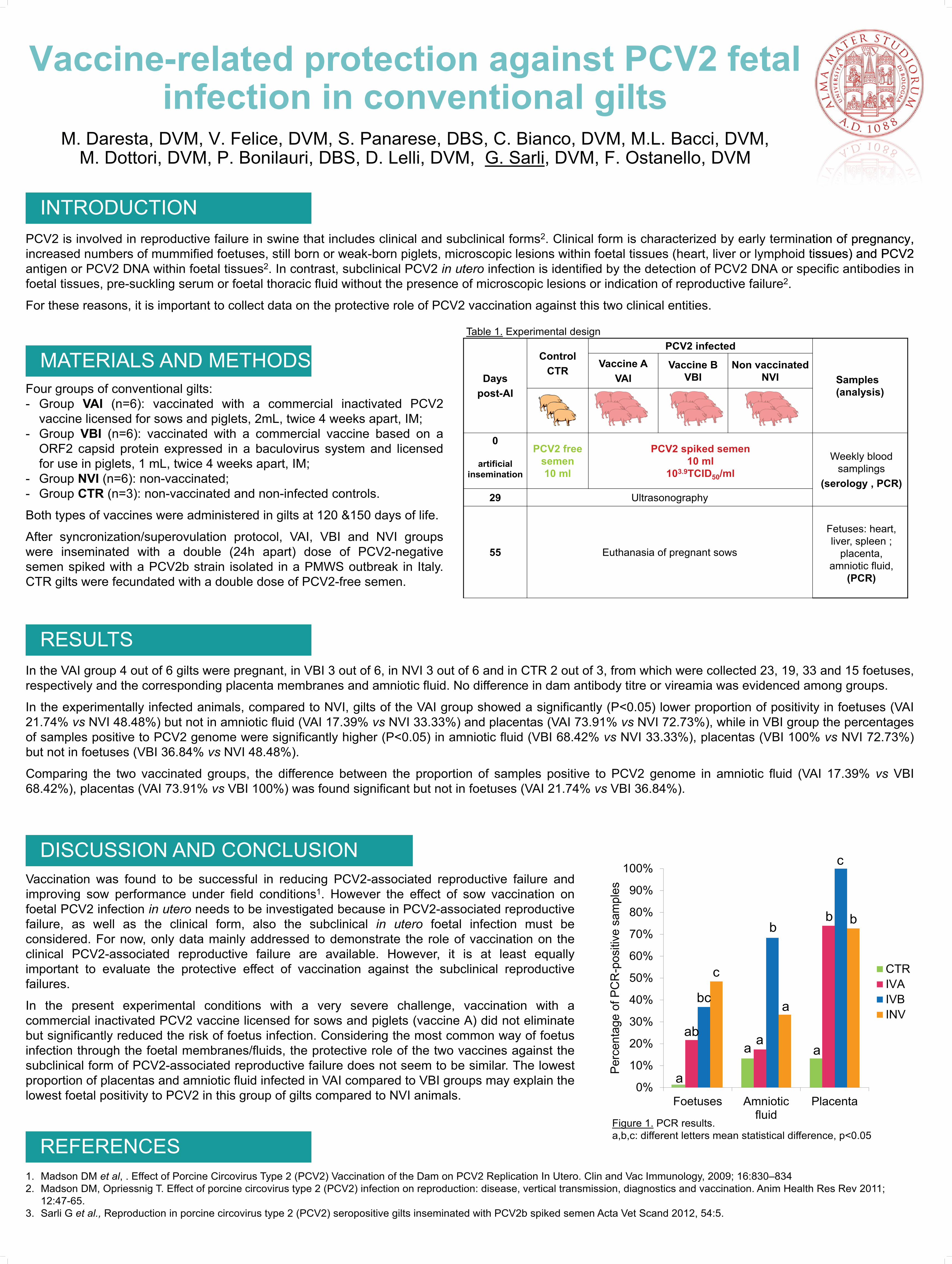
In the VAI group 4 out of 6 gilts were pregnant, in VBI 3 out of 6, in NVI 3 out of 6 and in CTR 2 out of 3, from which were collected 23, 19, 33 and 15 foetuses,respectively and the corresponding placenta membranes and amniotic fluid. No difference in dam antibody titre or vireamia was evidenced among groups.
In the experimentally infected animals, compared to NVI, gilts of the VAI group showed a significantly (P<0.05) lower proportion of positivity in foetuses (VAI21.74% vs NVI 48.48%) but not in amniotic fluid (VAI 17.39% vs NVI 33.33%) and placentas (VAI 73.91% vs NVI 72.73%), while in VBI group the percentagesof samples positive to PCV2 genome were significantly higher (P<0.05) in amniotic fluid (VBI 68.42% vs NVI 33.33%), placentas (VBI 100% vs NVI 72.73%)but not in foetuses (VBI 36.84% vs NVI 48.48%).
Comparing the two vaccinated groups, the difference between the proportion of samples positive to PCV2 genome in amniotic fluid (VAI 17.39% vs VBI68.42%), placentas (VAI 73.91% vs VBI 100%) was found significant but not in foetuses (VAI 21.74% vs VBI 36.84%).
Vaccination was found to be successful in reducing PCV2-associated reproductive failure andimproving sow performance under field conditions1. However the effect of sow vaccination onfoetal PCV2 infection in utero needs to be investigated because in PCV2-associated reproductivefailure, as well as the clinical form, also the subclinical in utero foetal infection must beconsidered. For now, only data mainly addressed to demonstrate the role of vaccination on theclinical PCV2-associated reproductive failure are available. However, it is at least equallyimportant to evaluate the protective effect of vaccination against the subclinical reproductivefailures.
In the present experimental conditions with a very severe challenge, vaccination with acommercial inactivated PCV2 vaccine licensed for sows and piglets (vaccine A) did not eliminatebut significantly reduced the risk of foetus infection. Considering the most common way of foetusinfection through the foetal membranes/fluids, the protective role of the two vaccines against thesubclinical form of PCV2-associated reproductive failure does not seem to be similar. The lowestproportion of placentas and amniotic fluid infected in VAI compared to VBI groups may explain thelowest foetal positivity to PCV2 in this group of gilts compared to NVI animals.
1. Madson DM et al, . Effect of Porcine Circovirus Type 2 (PCV2) Vaccination of the Dam on PCV2 Replication In Utero. Clin and Vac Immunology, 2009; 16:830–8342. Madson DM, Opriessnig T. Effect of porcine circovirus type 2 (PCV2) infection on reproduction: disease, vertical transmission, diagnostics and vaccination. Anim Health Res Rev 2011;
12:47-65.3. Sarli G et al., Reproduction in porcine circovirus type 2 (PCV2) seropositive gilts inseminated with PCV2b spiked semen Acta Vet Scand 2012, 54:5.
termination of pregnancy,tissues) and PCV2
Dayspost-AI
ControlCTR
PCV2 infected
Samples(analysis)
Vaccine AVAI
Vaccine BVBI
Non vaccinatedNVI
0
artificialinsemination
PCV2 free semen10 ml
PCV2 spiked semen10 ml
103.9TCID50/mlWeekly blood
samplings(serology , PCR)
29 Ultrasonography
55 Euthanasia of pregnant sows
Fetuses: heart, liver, spleen ;
placenta, amniotic fluid,
(PCR)
DISCUSSION AND CONCLUSION
REFERENCES
MATERIALS AND METHODS
INTRODUCTION
RESULTS
Table 1. Experimental design
Figure 1. PCR results.a,b,c: different letters mean statistical difference, p<0.05
0%
10%
20%
30%
40%
50%
60%
70%
80%
90%
100%
Foetuses Amnioticfluid
Placenta
Perc
enta
ge o
f PC
R-p
ositi
ve s
ampl
es
CTRIVAIVBINV
ab
bc
a
c
a aa
a
b bb
c
Related Documents