85 ABSTRACT Cells are able to respond to stress stimuli through the expression of proteins that compensate for or seek to return the cell to its functional status. Among the most studied proteins in relation to stress are heat shock proteins(HSPs) or chaperones; these proteins were originally discovered as induced proteins that facilitate proper folding of proteins with non-native conformations. HSPs are a set of highly conserved proteins that are present in all types of cells, from microbes to mammals. In bacteria, chaperones and proteases regulate cell activity under stress conditions. In echinoderms, aquatic invertebrates and fish stress proteins response mechanisms are associated with the expression of chaperones that protect them of temperature changes and oxidative stress. In mammalian cells, there is the expression of anti- stress mechanisms that are also associated with certain physiological conditions such as mitosis and maintaining the cell under homeostatic conditions. In plants, one of the conditions most studied is the response to osmotic stress, leading to the synthesis of membrane transporter systems that compensate the water flow and at the same time, over expressing intracellular mechanisms associated with enzymes that contribute to decrease the production of free oxygen radicals. In general, cells of all organisms are capable of initiating regulatory mechanisms that contribute a reduction in cell injury that can result in loss of homeostasis. When this mechanisms decline because aging or faulty metabolism, disease occur. This finding allows to use them as a therapeutic alternative for aging diseases as Alzheimer and cancer between others. KEYWORDS Cell stress, Homeostasis, Heat shock Proteins, Small heat Shock proteins, Aging-disease A world Under Stress, Molecular Response to Adverse Conditions Saúl Emmanuel Torres-García 1 , Eduardo Mauricio Martin-Álvarez Tostado 1 , Carlos Omar Rodríguez-Hernández 1 , Juan Armando Flores-de la Torre 2 , Kerry Mitchell Nigel 2 , Abraham Loera-Muro 1 , Flor Yazmín Ramírez-Castillo 1 , Luis Fernando López-Gutiérrez 3 , Carlos Olvera-Sandoval 2 , María Carolina De Luna- López 4 , Francisco Javier Avelar-González 2 , Magdalena Samantha Ramos-Gómez 2 and Alma Lilián Guerrero-Barrera 1 * 1 Departamento de Morfología, Centro de Ciencias Básicas, Universidad Autónoma de Aguascalientes, Aguascalientes, Ags., México, 20131 2 Departamento de Fisiología y Farmacología, Centro de Ciencias Básicas, Universidad Autónoma de Aguascalientes, Aguascalientes, Ags., México, 20131 3 Departamento de Química, Centro de Ciencias Básicas, Universidad Autónoma de Aguascalientes, Aguascalientes, Ags., México, 20131 4 Departamento de Clínica Veterianaria, Centro de Ciencias Agropecuarias, Universidad Autónoma de Aguascalientes, Aguascalientes, Ags., México, 20131 *Corresponding author: Laboratorio de BiologíaCelular y Tisular. Departamento de Morfología. Centro de Ciencias Básicas, Universidad Autónoma de Aguascalientes, Aguascalientes, Ags., México, 20131. Phone: (52) 4499107400 ext. 342. All authors participated in the same proportion. ISSN: 2347-3215 Volume 3 Number 1 (January-2015) pp. 85-100 www.ijcrar.com

Welcome message from author
This document is posted to help you gain knowledge. Please leave a comment to let me know what you think about it! Share it to your friends and learn new things together.
Transcript
85
A B S T R A C T
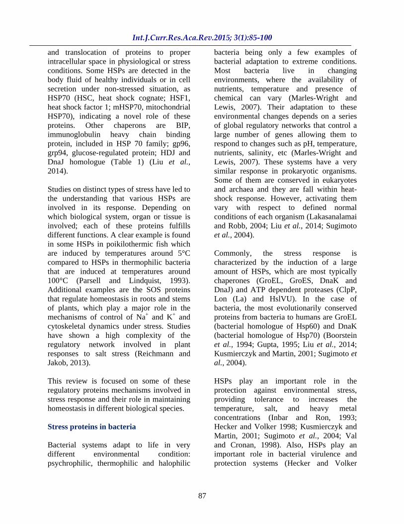
Cells are able to respond to stress stimuli through the expression of proteins that compensate for or seek to return the cell to its functional status. Among the most studied proteins in relation to stress are heat shock proteins(HSPs) or chaperones; these proteins were originally discovered as induced proteins that facilitate proper folding of proteins with non-native conformations. HSPs are a set of highly conserved proteins that are present in all types of cells, from microbes to mammals. In bacteria, chaperones and proteases regulate cell activity under stress conditions. In echinoderms, aquatic invertebrates and fish stress proteins response mechanisms are associated with the expression of chaperones that protect them of temperature changes and oxidative stress. In mammalian cells, there is the expression of anti-stress mechanisms that are also associated with certain physiological conditions such as mitosis and maintaining the cell under homeostatic conditions. In plants, one of the conditions most studied is the response to osmotic stress, leading to the synthesis of membrane transporter systems that compensate the water flow and at the same time, over expressing intracellular mechanisms associated with enzymes that contribute to decrease the production of free oxygen radicals. In general, cells of all organisms are capable of initiating regulatory mechanisms that contribute a reduction in cell injury that can result in loss of homeostasis. When this mechanisms decline because aging or faulty metabolism, disease occur. This finding allows to use them as a therapeutic alternative for aging diseases as Alzheimer and cancer between others.
KEYWORDS
Cell stress, Homeostasis, Heat shock Proteins, Small heat Shock proteins, Aging-disease
A world Under Stress, Molecular Response to Adverse Conditions
Saúl Emmanuel Torres-García1 , Eduardo Mauricio Martin-Álvarez Tostado1 , Carlos Omar Rodríguez-Hernández1 , Juan Armando Flores-de la Torre2 , Kerry Mitchell Nigel2 , Abraham Loera-Muro1 , Flor Yazmín Ramírez-Castillo1 , Luis Fernando López-Gutiérrez3 , Carlos Olvera-Sandoval2 , María Carolina De Luna-López4 , Francisco Javier Avelar-González2 , Magdalena Samantha Ramos-Gómez2
and Alma Lilián Guerrero-Barrera1 *
1Departamento de Morfología, Centro de Ciencias Básicas, Universidad Autónoma de Aguascalientes, Aguascalientes, Ags., México, 20131 2Departamento de Fisiología y Farmacología, Centro de Ciencias Básicas, Universidad Autónoma de Aguascalientes, Aguascalientes, Ags., México, 20131 3Departamento de Química, Centro de Ciencias Básicas, Universidad Autónoma de Aguascalientes, Aguascalientes, Ags., México, 20131 4Departamento de Clínica Veterianaria, Centro de Ciencias Agropecuarias, Universidad Autónoma de Aguascalientes, Aguascalientes, Ags., México, 20131 *Corresponding author: Laboratorio de BiologíaCelular y Tisular. Departamento de Morfología. Centro de Ciencias Básicas, Universidad Autónoma de Aguascalientes, Aguascalientes, Ags., México, 20131. Phone: (52) 4499107400 ext. 342. All authors participated in the same proportion.
ISSN: 2347-3215 Volume 3 Number 1 (January-2015) pp. 85-100
www.ijcrar.com

86
Introduction
At cellular level, living organisms constantly deal with unfavorable or stressful situations of diverse origin including oxidative stress (Oskala et al., 2014), resulting in the expression of specific genes that allows its variability and biological adaptation. When living cells or organisms are exposed to high temperatures or chronic and acute stress conditions, they respond by synthesizing HSPs (Srivastava and Lilly, 1992; Liu et al., 2014; Nakajima and Koizumi 2014).The response to heat stress was first observed by Ritossa at early 60´s while studying nucleic acid synthesis in the saliva glands cells of Drosophila melanogaster, where an increase in transcriptional activity when these cells were exposed to elevated temperatures was noticed (Ritossa, 1962 De Mario et al., 2010). This response has been identified in all cell types, since prokaryotes to eukaryotes (Craig, 1985; Hendstridge et al., 2014; Liu et al., 2014). Stress response is a protective homeostatic process that allows tolerance to increased temperatures and has been associated with other inducing factors that destabilize cell function and integrity (Hendstridge et al., 2014; Liu et al., 2014).
In addition to response to heat shock, stress response can be induced by anoxia (the 2,4 dinitrophenol, a substance that uncouples oxidative phosphorilation, and sodium azide, an inhibitor of cellular respiration) (Ritossa, 1962; Mahmood et al., 2014). There are also other inducers such as sodium arsenite, ethanol, thiol compounds, hydrogen peroxide, metals (Zn, Cd and HG), viral infections (Lindquist and Craig, 1988; Mahmood et al., 2014), hormones such as insulin (Ting et al., 1989), certain prostaglandins (Ohno et al., 1988), hypoxia (Benjamin et al., 1990; Mahmood et al., 2014), light, exercise and diet components (Locke et al., 1990; Liu et al., 2014).
HSPs are encoded by a set of genes, called heat shock genes, because they are the product of the induction of these genes in response to heat and other forms of stress, that have different cellular functions (Lindquist and Craig, 1988; Ritossa, 1962). Some of these proteins are known as chaperones, and are involved in functional folding of proteins and in the prevention of misfolding or nonfunctional aggregation of these proteins (Shiver and Ravid, 2014). This superfamily of proteins is widely conserved in different biological systems and is associated with transport functions, folding, and coupling and uncoupling of multi-structural units, and the degradation of misfolded or non-functionally aggregated proteins (Sørensen et al., 2003; Mahmood et al., 2014). Such tasks are performed under normal cellular conditions, and are further induced by stress factors that can cause cell damage.
The genes encoding these proteins are also highly conserved in different biological systems. This set of highly conserved proteins are present and can be induced in all types of cells in all species and are categorized into seven families on basis of their molecular weight: HSP10 (including FAM proteins of 130 144 amino acid), small HSPs (form 15 30KDa), HSP40, HSP60, HSP70, HSP90/HSP90B1 grp94 gp96 andHSP110 (Nakajima and Koizumi, 2014; Liu et al., 2014). Each family includes at least one member, but often more (Gething, 1997; Liu et al., 2014). HSP70 is one of the most conserved and inducible proteins known to date, with ~60% phylogenetic similarity between microbes and mammals. Intracellular HSPs are highly conservative and localized in different compartments in all type of cells in mammals. Most HSPs function as chaperones, involved in client protein assembly, stabilization, folding, refolding
87
and translocation of proteins to proper intracellular space in physiological or stress conditions. Some HSPs are detected in the body fluid of healthy individuals or in cell secretion under non-stressed situation, as HSP70 (HSC, heat shock cognate; HSF1, heat shock factor 1; mHSP70, mitochondrial HSP70), indicating a novel role of these proteins. Other chaperons are BIP, immunoglobulin heavy chain binding protein, included in HSP 70 family; gp96, grp94, glucose-regulated protein; HDJ and DnaJ homologue (Table 1) (Liu et al., 2014).
Studies on distinct types of stress have led to the understanding that various HSPs are involved in its response. Depending on which biological system, organ or tissue is involved; each of these proteins fulfills different functions. A clear example is found in some HSPs in poikilothermic fish which are induced by temperatures around 5°C compared to HSPs in thermophilic bacteria that are induced at temperatures around 100°C (Parsell and Lindquist, 1993). Additional examples are the SOS proteins that regulate homeostasis in roots and stems of plants, which play a major role in the mechanisms of control of Na+ and K+ and cytoskeletal dynamics under stress. Studies have shown a high complexity of the regulatory network involved in plant responses to salt stress (Reichmann and Jakob, 2013).
This review is focused on some of these regulatory proteins mechanisms involved in stress response and their role in maintaining homeostasis in different biological species.
Stress proteins in bacteria
Bacterial systems adapt to life in very different environmental condition: psychrophilic, thermophilic and halophilic
bacteria being only a few examples of bacterial adaptation to extreme conditions. Most bacteria live in changing environments, where the availability of nutrients, temperature and presence of chemical can vary (Marles-Wright and Lewis, 2007). Their adaptation to these environmental changes depends on a series of global regulatory networks that control a large number of genes allowing them to respond to changes such as pH, temperature, nutrients, salinity, etc (Marles-Wright and Lewis, 2007). These systems have a very similar response in prokaryotic organisms. Some of them are conserved in eukaryotes and archaea and they are fall within heat-shock response. However, activating them vary with respect to defined normal conditions of each organism (Lakasanalamai and Robb, 2004; Liu et al., 2014; Sugimoto et al., 2004).
Commonly, the stress response is characterized by the induction of a large amount of HSPs, which are most typically chaperones (GroEL, GroES, DnaK and DnaJ) and ATP dependent proteases (ClpP, Lon (La) and HslVU). In the case of bacteria, the most evolutionarily conserved proteins from bacteria to humans are GroEL (bacterial homologue of Hsp60) and DnaK (bacterial homologue of Hsp70) (Boorstein et al., 1994; Gupta, 1995; Liu et al., 2014; Kusmierczyk and Martin, 2001; Sugimoto et al., 2004).
HSPs play an important role in the protection against environmental stress, providing tolerance to increases the temperature, salt, and heavy metal concentrations (Inbar and Ron, 1993; Hecker and Volker 1998; Kusmierczyk and Martin, 2001; Sugimoto et al., 2004; Val and Cronan, 1998). Also, HSPs play an important role in bacterial virulence and protection systems (Hecker and Volker
88
1998; Val and Cronan, 1999; Liu et al., 2014). In Escherichia coli, small increases in temperature (28 30°C to 36 42°C) generate transient increases in the synthesis of HSPs, while significant increases (2830°C to 47 50°C) are lethal, resulting in a unique synthesis of HSPs and the inhibition of normal protein synthesis (VanBogelen et al., 1987). Another inducible HSP in E. coli is the protease encoded by lon gene, whose transcription rate increases multiple times with increases in temperature (28 30°C to 42°C) (Goff and Goldberg, 1985). It has also been found that the transcription of the lon gene increases upon exposure to ethanol 4% (Travers and Mace, 1982) and the induction in the synthesis of aberrant polypeptides (Goff et al., 1988).
Similarly, there are another HSP related proteins that respond to stress in bacteria, where the organism´s gene expression is modified to counteract the adverse effects of different types of biotic or abiotic stress. Upregulating of the transcription of genes that respond to stress is carried out by sigma factors and transcription factors that interact with RNA polymerase (Marles-Wright and Lewis, 2007). RNA polymerase selectivity for promoter sequences of genes is modulated primarily by the sigma factors and subsequently by transcription factors (Ishihama, 2010). Sigma factors are associated with resistance to various stresses, for example the overall stress sigma factor RpoS, strongly induced in E. coli under conditions of starvation, hyperosmolarity, or changes in pH optimum and growth temperature (Weber et al., 2005).
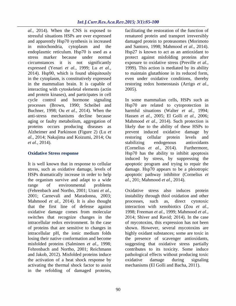
Alternatively, other proteins involved in stress response are the two component systems, consisting of a sensor protein (histidine kinase) and a regulatory response (Bourret, 2010) (Figure 1). The recognition of a specific signal from the sensor activates
a protein phosphorylation response regulator, which in turn activates or represses the transcription of a limited set of genes (Szurmant et al., 2007). For example, in E. coli the EnvZ/OmpR two component systems detect changes in osmolarity regulating the expression of the OmpF and OmpC porins in the outer membrane (Stock et al., 2000).
Stress protein in aquatic invertebrates, fish and echinoderms
Another type of stress response protein is known as metallothionein (MT). They are globular proteins of small molecular weight (7 kDa), found in all living cells. These proteins bind to minerals and ions (Cd+2, Cu+2, and Fe+2). They have high cysteine content (20 cysteine of about 60-62 amino acids) and are induced by toxic agents, which cause the loss of its specific biological function effectively blocking them. They are then accumulated and synthesized de novo in the target tissue, serving as marker proteins of cellular and molecular attack by environmental stress (Mahmood et al., 2014; Oskala et al., 2014). These proteins have been found in tissues of various aquatic species of cyanophyta algae, crustaceans, echinodermata, fishes, mollusca and nematode. In fish, thermal stress increased HSP levels that associated with lower protein and lipid oxidation, providing evidence that physiological increase in HSP levels can protect against oxidative stress. Other inducers of HSP response, including heavy metals or environmental toxins, hypoxia, handling, crowding, osmotic stress and cold shock induce oxidative stress in fish. The Hsp60 and Hsp70 families are the most prominent chaperone proteins and provide cytoprotection to various stress stimuli as HSP70, HSP60, HSP90, HSC70 and grp75 (Oskala et al., 2014; Mahmood et al., 2014).

89
Stress proteins in mammals
As previously mentioned, HSPs are constitutively expressed or expressed at the time that the cell perceives a stimulus that can be classified as stress. However, it has also been observed that the process of mitosis and processes involved in cell differentiation increase their production (Trautinger et al., 1995; Haslbeck and Buchner, 2002).
HSPs are present in all cell-cytosol compartments, mitochondria, nucleus and endoplasmic reticulum (Morimoto, 1998).In the case of normal cells (not under stress stimuli), the expression of HSPs is scarce, only appearing in the cellular environment after a certain change in the cellular environment or as a consequence of noxious stimuli (Jäättelä, 1999).With regard to development, HSPs are among the first proteins produced during embryonic development in mammals (Neuer et al., 1999). Their presence or absence influences in all aspects of development (Bergh and Arking, 1984) and are essential for normal growth conditions (Walsh et al., 1999). When this is not possible, misfolding proteins are discarded by proteasome. However, when this not happen, aggregation of misfolding proteins is produced, this is one of causes of diseases as Alzheimer and Parkinson, between others (Lu et al., 2014; Ou et al., 2014; Cornelius et al., 2014).
Stress proteins in the skeletal system
As observed in Table 1, the human Hsp27 belongs to the family of small HSPs. This protein is expressed constitutively at low levels in the cytosol of most human cells (Arrigo, 2000). During development, Hsp27 appears to play a crucial role in specific developmental stages associated with cell proliferation, differentiation and death
(Davidson and Morange, 2000; Shiver and Ravid, 2014). Although its expression has been more related to the differentiation stage of proliferation and cytoskeletal arrangement (Liu et al., 2014), it has also been involved in the balance between differentiation and apoptosis (Arrigo, 2000) and has been involved in the development of calcified tissue (Shakoori et al., 1992). Hsp27 has been detected in endochondral bone development in mouse and rat models (Tiffee et al., 2000). It is also expressed on a stage-specific pattern during the latter stages of odontogenesis (Ohshima et al., 2000). Additionally, it has been detected in rat molars and incisors, but in these cases, its expression is related to the reinforcement of the ameloblasts layer during enamel formation, as well as, the formation and maintenance of the ameloblasts. Another function that is attributed to Hsp27 is that has been identified as a protein associated with estrogen receptor p29, involved in osteoblast physiology (Cooper et al., 1994; Cornelius et al., 2014). Estrogen receptors that have been detected in long bones seem to be involved in the maturation and growth of the fetal skeleton (Ben-Hur et al., 1993). Moreover, it is possible that the resistance to apoptosis induced by estrogen osteoblasts is associated with increased Hsp27 expression (Cooper et al., 2000; Cornelius et al., 2014).
Stress Proteins in the Nervous System
Constitutive stress proteins whose function is directly involved in the protection of nerve cells and glia have been identified in the central nervous system (CNS) (Richter-Landsberg and Goldbaum, 2003; Nakajima and Koizumi, 2014). Within this group of HSPs, there are the smaller Hsp90, Hsp70, Hsp60 families and some smaller ones including -crystallin, ubiquitin and Hsp32, generally named based on their molecular weight (Sharp et al., 1999; Lu et
90
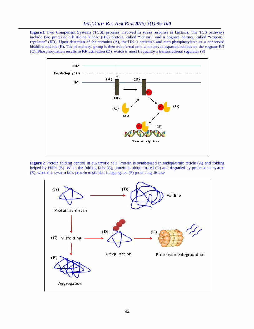
al., 2014). When the CNS is exposed to stressful situations HSPs are over expressed and apparently Hsp70 synthesis is increased in mitochondria, cytoplasm and the endoplasmic reticulum. Hsp70 is used as a stress marker because under normal circumstances it is not significantly expressed (Yenari et al., 1999; Lu et al., 2014). Hsp90, which is found ubiquitously in the cytoplasm, is constitutively expressed in the mammalian brain. It is capable of interacting with cytoskeletal elements (actin and protein kinases), and participates in cell cycle control and hormone signaling processes (Brown, 1990; Scheibel and Buchner, 1998; Ou et al., 2014). When the anti-stress mechanisms decline because aging or faulty metabolism, aggregation of proteins occurs producing diseases as Alzheimer and Parkinson (Figure 2) (Lu et al., 2014; Nakajima and Koizumi, 2014; Ou et al., 2014).
Oxidative Stress response
It is well known that in response to cellular stress, such as oxidative damage, levels of HSPs dramatically increase in order to help the organism survive and adapt to a wide range of environmental problems (Fehrenbach and Northo, 2001; Urani et al., 2001; Carnevali and Maradonna, 2003; Mahmood et al., 2014). It is also thought that the first line of defense against oxidative damage comes from molecular switches that recognize changes in the intracellular redox environment. In the case of proteins that are sensitive to changes in intracellular pH, the ionic medium folds losing their native conformation and become misfolded proteins (Salminen et al., 1998; Fehrenbach and Northo, 2001; Reichmann and Jakob, 2012). Misfolded proteins induce the activation of a heat shock response by activating the thermal shock factor to assist in the refolding of damaged proteins,
facilitating the restoration of the function of renatured protein and transport irreversibly damaged protein to proteasomes (Morimoto and Santoro, 1998; Mahmood et al., 2014). Hsp27 is known to act as an antioxidant to protect against misfolding proteins after exposure to oxidative stress (Preville et al., 1999). This action is mediated by its ability to maintain glutathione in its reduced form, even under oxidative conditions, thereby restoring redox homeostasis (Arrigo et al., 2005).
In some mammalian cells, HSPs such as Hsp70 are related to cytoprotection in harmful situations (Walter et al., 1994; Hassen et al., 2005; El Golli et al., 2006; Mahmood et al., 2014). Such protection is likely due to the ability of these HSPs to prevent induced oxidative damage by restoring cellular protein levels and stabilizing endogenous antioxidants (Cornelius et al., 2014). Furthermore, Hsp70 has the ability to inhibit apoptosis induced by stress, by suppressing the apoptotic program and trying to repair the damage. Hsp70 appears to be a pleiotropic apoptotic pathway inhibitor (Cornelius et al., 201; Mahmood et al., 2014).
Oxidative stress also induces protein instability through thiol oxidation and other processes, such as, direct cytotoxic interaction with xenobiotics (Zou et al., 1998; Freeman et al., 1999; Mahmood et al., 2014; Shiver and Ravid; 2014). In the case of mycotoxins, this expression has not been shown. However, several mycotoxins are highly oxidant substances; some are toxic in the presence of scavenger antioxidants, suggesting that oxidative stress partially contributes to its toxicity. Some induce pathological effects without producing toxic oxidative damage during signaling mechanisms (El Golli and Bacha, 2011).

91
Table.1 Stress protein families. The HSP superfamily consists of seven families whose members are grouped based on their molecular weights, which vary between 10 000 and 150 000 Da. They can be found in major cellular compartments (Liu et al., 2014 modified)
Family
Name Location Function
Small Hsp.
P20. Hsp22 ( B-
crystalline).
Hsp 25 (rats). Hsp 27
(human).
Cytoplasm. Cytoplasm and
Nucleus. Cytoplasm
Cytoplasm and Nucleus.
Vasodilation. Cytoskeletal stabilization.
Chaperones. Actin dynamics.
Chaperone/cytoskeletal stabilization
Hsp 40.
Hsp 40. Hsp 47.
Cytoplasm. Endoplasmic Ret.
Chaperones. Collagen synthesis control.
Hsp 60.
Hsp 58. Hsp 60.
Mitochondria. Mitochondria/
Cytoplasm
Chaperones. Chaperone activity in folding/ refolding/ assembly of multimeric protein structures
Hsp 70.
Hsp 70 (73).
Hsp 70 (72).
Hsp 75. Grp 78.
BIP
Cytoplasm. Cytoplasm/Nucleus.
Mitochondria. Endoplasmic
Reticulum Endoplasmic
Reticulum
Chaperones for nascent polypeptide chains Chaperones.
Folding/refolding, transport
Hsp 90.
Hsp 90
(86).
Hsp 90
(84). grp 94/grp
96
Cytoplasm.
Cytoplasm. Endoplasmic
Reticulum
Union of steroid hormone receptors. Chaperones.
Chaperoneactivity, proliferationand growth/bindstootherproteins/assistingthemaintenance of the HSF1 monomericstateunder normal conditions
Hsp 110.
Hsp 105. Hsp 110.
Cytoplasm. Cytoplasm/Nucleus.
Chaperones. Cytoprotective.
92
Figure.1 Two Component Systems (TCS), proteins involved in stress response in bacteria. The TCS pathways include two proteins: a histidine kinase (HK) protein, called sensor, and a cognate partner, called response regulator (RR). Upon detection of the stimulus (A), the HK is activated and auto-phosphorylates on a conserved histidine residue (B). The phosphoryl group is then transferred onto a conserved aspartate residue on the cognate RR (C). Phosphorylation results in RR activation (D), which is most frequently a transcriptional regulator (F)
Figure.2 Protein folding control in eukaryotic cell. Protein is synthesized in endoplasmic reticle (A) and folding helped by HSPs (B). When the folding fails (C), protein is ubiquitinated (D) and degraded by proteosome system (E), when this system fails protein misfolded is aggregated (F) producing disease
93
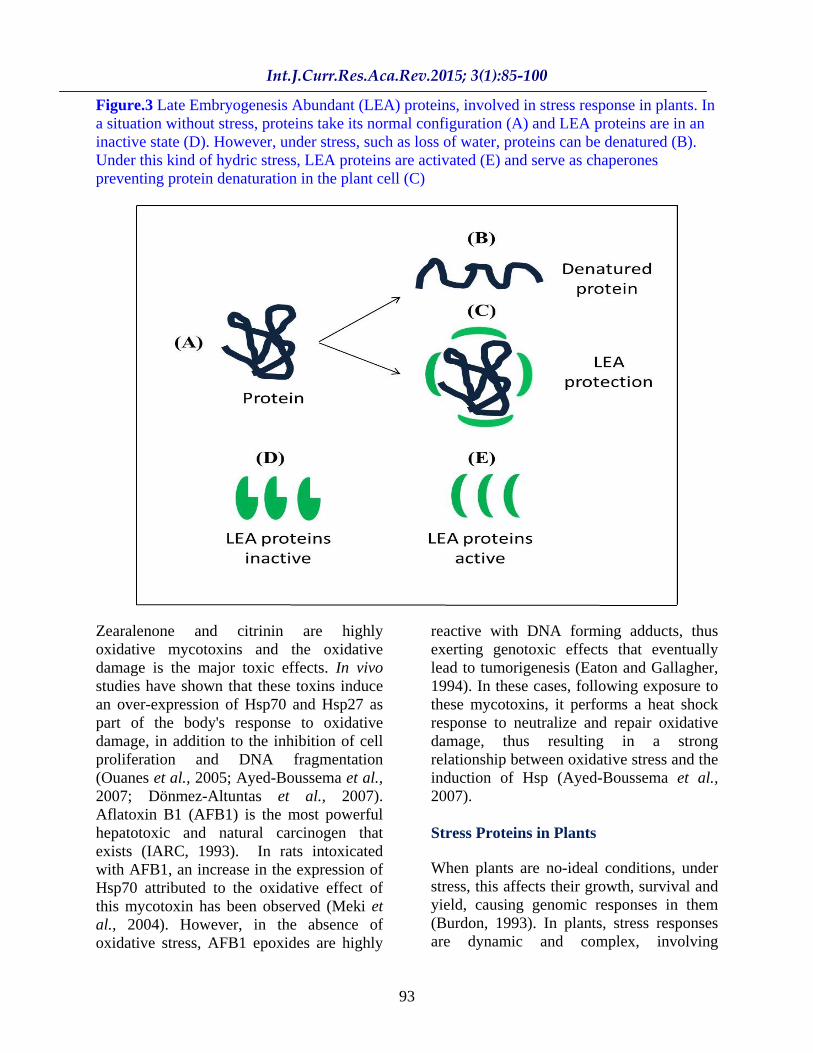
Figure.3 Late Embryogenesis Abundant (LEA) proteins, involved in stress response in plants. In a situation without stress, proteins take its normal configuration (A) and LEA proteins are in an inactive state (D). However, under stress, such as loss of water, proteins can be denatured (B). Under this kind of hydric stress, LEA proteins are activated (E) and serve as chaperones preventing protein denaturation in the plant cell (C)
Zearalenone and citrinin are highly oxidative mycotoxins and the oxidative damage is the major toxic effects. In vivo studies have shown that these toxins induce an over-expression of Hsp70 and Hsp27 as part of the body's response to oxidative damage, in addition to the inhibition of cell proliferation and DNA fragmentation (Ouanes et al., 2005; Ayed-Boussema et al., 2007; Dönmez-Altuntas et al., 2007). Aflatoxin B1 (AFB1) is the most powerful hepatotoxic and natural carcinogen that exists (IARC, 1993). In rats intoxicated with AFB1, an increase in the expression of Hsp70 attributed to the oxidative effect of this mycotoxin has been observed (Meki et al., 2004). However, in the absence of oxidative stress, AFB1 epoxides are highly
reactive with DNA forming adducts, thus exerting genotoxic effects that eventually lead to tumorigenesis (Eaton and Gallagher, 1994). In these cases, following exposure to these mycotoxins, it performs a heat shock response to neutralize and repair oxidative damage, thus resulting in a strong relationship between oxidative stress and the induction of Hsp (Ayed-Boussema et al., 2007).
Stress Proteins in Plants
When plants are no-ideal conditions, under stress, this affects their growth, survival and yield, causing genomic responses in them (Burdon, 1993). In plants, stress responses are dynamic and complex, involving

94
crosstalk between different levels of regulation, including the adjustment of metabolism and gene expression to physiological and morphological adaptation (Krasensky and Jonak, 2012). An example is the osmotic stress; this is one of the form of abiotic stress that can inhibit the growth and development of plants (Chen et al., 2012; Krasensky and Jonak, 2012). To cope with the osmotic stress, plants have developed various responses, such as the production of osmolytes for osmotic adjustment, synthesis of antiport transporters Na+/H+ to sequester ions, and many other mechanisms in order to minimize the accumulation of salts (Zang and Komatsu, 2007; Baek et al., 2011).
In the genus Arabidopsis (a glycophyte), salt overly sensitive (SOS) signaling pathway play an important role in tolerance to salinity. SOS signaling pathway includes several proteins with activity plasmatic membrane of Na+/H+ antiport (Zhang and Shi, 2013). SOS1 is one that plays a direct role in the tolerance of plants to salt conditions. SOS1 encodes for the Na+/H+
antiport transporter, which play an important role in the movement of Na+ from stems to roots. Under conditions of salinity, there is a sobreexpression of this protein and decrease the Na+ movement, increasing tolerance to saline conditions (Shi et al., 2003). The Na+
transporter AtHKT1 of the membrane is also a key determinant of salt tolerance in Arabidopsis a control of the absorption, transport and accumulation of Na+ (Shi et al., 2003; Zhang and Shi, 2013). It is believed that AtHKT1controls the accumulation of Na+ in plants by transporting from the xylem into the parenchyma in both, in roots and shoots, which ultimately reduce the transport of Na+
from the root xylem into the cells of the leaves (Sunarpi et al., 2005). On the other hand, the increase in gene expression for the vacuolar membrane antiport transporter Na+/H+AtNHX1 under osmotic stress has
been seen, representing another mechanism for the reduction of Na+ toxicity by sequestering Na+ in the vacuoles (Sunarpi et al., 2005; Zang et al., 2007; Zhang and Shi, 2013). The above permits that the turgor pressure of the cell can be maintained and decreased the lossof water in the plant cell under conditions of salt stress (Bassil et al., 2011). Likewise, three carriers membrane proteins: SOS1, AtHKT1 and AtNHX1, work together to reduce osmotic toxicity on a cellular and plant level (Zhang and Shi, 2013).
Plants can also suffer stress for reasons involving the lack of water (Shinde et al., 2012). Like all living organisms, they seek a mechanism to stabilize the process, and in doing so, they accumulate various proteins, sugars and compatible solutes used to replace water and stabilize the subcellular environment (Shinde et al., 2012). Some of these proteins are known as LAE (Late Embryogenesis Abundant), represent an important group of hydrophobic proteins that accumulate in high levels of water present in both, mitochondria and cytoplasm (Cuevas-Velazquez et al., 2014), generating an adoption process during stress due to lack of water, and as such providing important protection functions, like DNA protection, stabilization of cytoskeletal filaments, and acting as molecular chaperones (Waters, 2013). To perform the latter function, they act synergistically with sugars, such as trehalose, to prevent aggregation during the absence of water (Waters, 2013). Another response to the scarcity of water in plants is the hypo-production of ATP, causing the elevation of reactive oxigen species (ROS) (Schmidt and Schippers, 2014), to which plants respond with an increase in the production of multiple enzymes, such as, superoxide dismutase (SOD), catalase (CAT), ascorbate peroxidase (APX), mono-dehydroascorbate reductase (MDAR),

95
reductase dehydroascorbate (Dhar), glutathione peroxidase (GPX), and glutathione reductase (GR), found mostly in the cytoplasm (Schmidt and Schippers, 2014). Being the first line of defense of cellular stress, SOD belongs to the metalloprotease family of enzymes that catalyze the deprotonation of superoxide (O2) to molecular oxygen and hydrogen peroxide (H2O2)by a reduction of an electron, wrapping it in central copper atom (Schmidt and Schippers, 2014). SOD removes superoxide and therefore reduces the risk of the formation of hydroxyl radicals. SOD is classified into three groups: Mn-SOD, Fe-SOD and Cu/Zn-SOD (Schmidt and Schippers, 2014). Mn-SOD is predominantly found in mitochondria and peroxisomes, Fe-SOD is localized in chloroplasts and Cu/Zn-SOD found in chloroplasts, cytoplasm and possibly in the extracellular space (Schmidt and Schippers, 2014) (Fig. 3).
Therapeutic use of HSPs
As described before, oxidative stress increased proteotoxic and lipoperoxidative damage with protein clearance defects through the ubiquitin-proteasome system (Figure 2). When this system decline because aging aggregation of proteins occurs (Lu et al., 2014; Ou et al., 2014). The downregulation of small HSPs as FAM proteins identified in several nuclear proteins, including transcription factors, produces cancer (Nakajima and Koizumi, 2014), understand the regulation of the production of FAM proteins has been proposed as possible therapy against cancer (Nakajima and Koizumi, 2014). Protein misfolding and aggregation are associated with a number of human diseases (Lu et al., 2014; Ou et al., 2014). Misfolded proteins can aggregate and impair signal transduction pathways or cause cell toxicity (Henstridge
et al., 2014). HSPs participate in folding process of proteins and in targeting misfolded proteins to degradation through proteasome system (Shiver and Ravid, 2014). With aging this homeostatic process fail. Chemical chaperones or pharmaceutical chaperones are small molecules that stabilize the folding of proteins, provide improvements to protein folding and buffer abnormal protein aggregation (Henstridge et al., 2014). HSPs increased expression has been proposed as therapeutic alternative to treatment of several human diseases; however more studies are necessary in order to use these proteins as therapy.
Conclusion
When exposed to stressful conditions that cause morphological and physiological damage, different biological systems produce response mechanisms, such as, the expression of regulatory proteins, where HSPs being the most common. In the case of HSPs, their capacity of neutralize the damage and repair the cells is clear. Following exposure to stress, there is an expression and regulation of genes followed by an activation of signaling pathways, resulting in the induction of HSPs. Hsp70 and Hsps27 are the widely reported proteins that play an outstanding maintenance of homeostasis role. In addition to HSPs, there are other proteins in response to the stress conditions, such as, the chaperones GroEL and DnaK in bacterial systems, and metallothionein in aquatic species. In plants, the response mechanisms include the production of osmolytes and the expression of signaling pathways, such as SOS mechanism, which can work together to stabilize these vital functions. Finally, the particular mechanisms of action employed will depend on the biological system, the degree of stimulation and the particular factor that triggers the process. However,
96
the final goal of these protein systems is to neutralize and repair the damage caused by situations that affect homeostasis, regardless of the source.
References
Arrigo AP. (2000). sHsp as novel regulators of programmed cell death and tumorigenicity. Pathologie-biologie, 48(3): 280-288.
Arrigo AP, Virot S, Chaufour S, Firdaus W, Kretz-Remy C, Diaz-Latoud C. (2005). Hsp27 consolidates intracellular redox homeostasis by upholding glutathione in its reduced form and by decreasing iron intracellular levels. Antioxid Redox Signal, 7: 414-422.
Ayed-Boussema I, Ouanes Z, Bacha H, Abid A. (2007). Toxicities induced in cultured cells exposed to zearalenone: apoptosis or mutagenesis? J. Biochem Mol Toxicol, 639:85-92.
Baek, D., Jiang, J., Chung, J.-S., Wang, B., Chen, J., Xin, Z., Shi, H. (2011). Regulated AtHKT1 Gene Expression by a Distal Enhancer Element and DNA Methylation in the Promoter Plays an Important Role in Salt Tolerance. Plant Cell Physiol, 52(1): 149 161.
Bassil, E, Tajima H, Liang, YC, Ohto, M, Ushijima, K, Nakano R, Eusimi T, Coku A, Belmonte M, Blumwald, E. (2011). The Arabidopsis Na+/H+ Antiporters NHX1 and NHX2 Control Vacuolar pH and K+ Homeostasis to Regulate Growth, Flower Development, and Reproduction. Plant Cell, 23(9): 3482 3497. doi:10.1105/tpc.111.089581
Ben-Hur H, Mor G, Blickstein I, Likhman I, Kohen F, Dgani R,Ornoy A. (1993). Localization of estrogen receptors in long bones and vertebrae of human fetuses. Calcif Tissue Int, 53(2), 91 96.
Benjamin IJ, Kroger B, Williams RS. (1990). Activation of the heat shock transcription factor by hypoxia in mammalian cells.
Proc Natl Acad Sci USA, 87(16): 62636267.
Bergh S, Arking R. (1984). Developmental profile of the heat shock response in early embryos of Drosophila. J Exp Zool, 231(3), 379 391.
Boorstein WR, Ziegelhoffer T, Craig EA. (1994). Molecular evolution of the HSP70 multigene family. J Mol Evol, 38:1 17
Bourret RB. (2010). Receiver domain structure and function in response regulator proteins. Curr Opin Microbiol, 13(2): 142 9.
Brown IR. (1990). Induction of heat shock (stress) genes in the mammalian brain by hyperthermia and other traumatic events: a current perspective. J NeurosciRes, 27(3): 247 255.
Burdon RH. (1993). Stress proteins in plants. Bot J Scotland, 46(3), 463 475.
Carnevali O, Maradonna F. (2003). Exposure to xenobiotic compounds: looking for new biomarkers. Gen Comp Endocrinol, 131: 203 209.
Chen J, Cheng T, Wang P, Liu W, Xiao J, Yang Y, Hu X, Jiang Z, Zhang S, Shi, J. (2012). Salinity-induced changes in protein expression in the halophytic plant Nitrarias phaerocarpa. J Proteomics, 75(17): 5226 5243.
Cooper LF, Uoshima K. (1994). Differential estrogenic regulation of small M(r) heat shock protein expression in osteoblasts. J Biol Chem, 269(11): 7869 7873.
Cooper LF, Tiffee JC, Griffin JP, Hamano H, Guo Z. (2000). Estrogen-induced resistance to osteoblast apoptosis is associated with increased hsp27 expression. J Cell Physiol, 185(3): 401407.
Cornelius C, Koverech G, Crupi R , Di Paola R, Koverech A, Lodato F, Scuto M, Salinaro AT, Cuzzocrea S, Calabrese EJ, Calabrese V. (2014). Osteoporosis and Alzheimer pathology: role of celular stress response and hormetic redox signaling and aging and bonere modeling. Fronti Pharmacol, 5: 1-13.

97
Craig EA. (1985). The heat shock response.
CRC Crit Rev Biochem, 18: 239 280. Cuevas-Velazquez CL, Rendón-Luna DF,
Covarrubias AA. (2014). Dissecting the cryoprotection mechanisms for dehydrins. Front Plant Sci.
Davidson SM, Morange, M. (2000). Hsp25 and the p38 MAPK Pathway Are Involved in Differentiation of Cardiomyocytes. Dev Biol, 218(2): 146160.
De Mario A, Santoro MG, Tanguay RM, Hightower L E. (2012). Ferruccio Ritossa s scientific legacy 50 years after his discovery of the heat shock response: a new view of biology, a new society, and a new journal. Cell Stress Chaperones, 17(2): 139 143.
Dönmez-Altuntas H, Dumlupinar G, Imamoglu N, Hamurcu Z, Liman BC.(2007). Effects of the mycotoxin citrinin on micronucleus formation in a cytokinesisblockgenotoxicity assay in cultured human lymphocytes. J Appl Toxicol, 27(4): 337 341.
Eaton DL, Gallagher EP. (1994). Mechanisms of aflatoxin carcinogenesis. Annu Rev Pharmacol Toxicol, 34: 135 172.
El Golli E, Hassen W, Bouslimi A, Bouaziz C, Ladjimi MM, Bacha H. (2006). Induction of Hsp 70 in Vero cells in response to mycotoxins cytoprotection by sub-lethal heat shock and by Vitamin E. Toxicol Lett, 166:122 130.
El Golli E, Bacha H. (2011). Hsp70 expression as biomarkers of oxidative stress: mycotoxin s exploration. Toxicology, 287: 1 7.
Fehrenbach E, Northo H. (2001). Free radicals exercise apoptosis and heat shock proteins. Immunol Rev, 7:66 89.
Freeman ML, Borelli MJ, Meredith MJ, Lepock JR. (1999). On the path to the heat shock response: destabilisation and formation of partially folded protein intermediates, a consequence of protein thiol modification. Free Radic Biol Med, 26: 737 745.
Gething, MJ. (1997). Guidebook to Molecular Chaperones and Protein-Folding Catalysts. Oxford University Press.
Goff SA, Goldberg AL. (1985). Production of abnormal proteins in E. coli stimulates transcription of ion and other heat shock genes. Cell, 41: 587-595.
Gupta RS.(1995) Evolution of the chaperonin families (Hsp60, Hsp10 and Tcp-1) of proteins and the origin of eukaryotic cells. Mol Microbiol, 15: 1 11.
Haslbeck M, Buchner J. (2002). Chaperone Function of sHsps. In P. D. A.-P. Arrigo & W. E. G. Müller (Eds.), Small Stress Proteins (pp. 37 59). Springer Berlin Heidelberg. Retrieved from http://link.springer.com/chapter/10.1007/978-3-642-56348-5_3
Hassen W, El Golli E, Baudrimont I, Mobio TA, Ladjimi M, Creppy EE, Bacha H. (2005). Cytotoxicity and HSP70 induction in Hep G2 cells in response to zearalenone and cytoprotection by sub-lethal heat shock. Toxicology, 207:293301.
Hecker M, Volker U. (1998).Non-specific, general and multiple stress resistance of growth-restricted Bacillus subtilis cells by the expression of the sigma Bregulon. Mol Microbiol, 29: 1129 1136.
Hendstridge DC, Whitham M, Febraio MA. (2014). Chaperoning to metabolic party: The emerging therapeutic role of heat-shock proteins in obesity and type 2 diabetes.MolMetab, 3: 781-793.
IARC. (1993). IARC Monographs on the evaluation of carcinogenic risks to human. Vol. 56. Some naturally occurring susbstances: food items and constituent sm heterocyciclic aromatic amines and mycotoxins. International Agency for Research on Cancer, Lione.
Inbar O, Ron EZ. (1993). Induction of cadmium tolerance in Escherichia coli K-12. FEMS Lett, 113: 197 200
Ishihama, A. (2010). Prokaryotic genome regulation: multifactor promoters, multitarget regulators and hierarchic

98
networks. FEMS Microbiol Rev, 34(5): 628 45.
Jäättelä, M. (1999). Escaping Cell Death: Survival Proteins in Cancer. Exp Cell Res, 248(1): 30 43.
Krasensky J, Jonak C. (2012). Drought, salt, and temperature stress-induced metabolic rearrangements and regulatory networks. J Exp Bot, err460. doi:10.1093/jxb/err460
Kusmierczyk AR, Martin J. (2001). Chaperonins-keeping a lid on folding proteins. FEMS Lett, 505 (3): 343-347.
Lakasanalamai P, Robb FT. (2004). Small heat shock proteins from extremophiles: a review. Extremophiles, 8: 1 11. DOI 10.1007/s00792-003-0362-3
Lindquist S, Craig EA. (1988).The heat-shock proteins.Ann Rev Gen, 22: 631 677.
Liu H, Dicksved J , Lundh T, Lindberg JE. (2014) Heat Shock Proteins: Intestinal Gatekeepers that Are Influenced by Dietary Components and the Gut Microbiota. Pathology, 3: 187-210.
Locke M, Noble EG, Atkinson BG. (1990). Exercising mammals synthesize stress proteins. Am J Physiol, 258 (4 Pt 1): C723 729.
Lu RC, Tan MS, Wang H, XieAM, Yu JT, Tan L. (2014).Heat Shock Protein 70 in Alzheimer sDisease. Biomed Res Int, 2014: 435203. doi: 10.1155/2014/435203.
Mahmood K,Jadoon S,Mahmood Q,Irshad M,and Hussain J. (2014). Synergistic Effects of Toxic Elementson Heat Shock Proteins. Biomed Res Int, 2014: 564136. DOI: 10.1155/2014/564136
Marles-Wright J, Lewis RJ. (2007). Stress responses of bacteria. Curr Opin Struct Biol, 17(6): 755 760. doi:10.1016/j.sbi.2007.08.004
Meki AR, Esmail E-D, Hussein AA, Hassanein HM. (2004). Caspase-3 and heat shock protein-70 in rat liver treated with aflatoxin B1: effect of melatonin. Toxicon, 43(1):93 100.
Morimoto RI. (1998). Regulation of the heat shock transcriptional response: cross talk between a family of heat shock factors, molecular chaperones, and negative
regulators. Genes Dev, 12(24), 37883796. doi:10.1101/gad.12.24.3788
Morimoto RI, Santoro MG. (1998). Stressinducible responses and heat shock proteins: New pharmacologic targets for cytoprotection. Nat Biotechnol, 16(9): 833 838. doi:10.1038/nbt0998-833.
Nakajima H, Koizumi K. (2014). Family with sequence similarity 107: A family of stress responsive small proteins with diverse functions in cancer and the nervous system (Review). Biomedl Rep, 2: 321-325, 2014.
Neuer A, Spandorfer SD, Giraldo P, Jeremias J, Dieterle S, Korneeva I, Liu HC, Rosenwaks Z, Witkin, SS. (1999). Heat Shock Protein Expression During Gametogenesis and Embryogenesis. Infect Dis Obstet Gynecol, 7(1-2), 10 16. doi:10.1155/S1064744999000034
Ohno K, Fukushima M, Fujiwara M, Narumiya, S. (1988). Induction of 68,000-dalton heat shock proteins by cyclopentenone prostaglandins. Its association with prostaglandin-induced G1 block in cell cycle progression. J Biol Chem, 263(36): 19764 19770.
Ohshima H, Ajima H, Kawano Y, Nozawa-Inoue K, Wakisaka S, Maeda T. (2000). Transient Expression of Heat Shock Protein (Hsp) 25 in the Dental Pulp and Enamel Organ during Odontogenesis in the Rat Incisor.Arch HistolCytol, 63(4), 381 395.
Oksala NK, Ekmekçi FG, Ozsoy E, Kirankaya S, Kokkola T, Emecen G, Lappalainen J, Kaarniranta K, Atalay M. (2014) Natural thermal adaptation increases heat shock protein levels and decreases oxidative stress. Redox Biol, 3: 25-8.
Ou JR,Tan MS,Xie AM,Yu JT,and Tan L. (2014). Heat Shock Protein 90 in Alzheimer s Disease. BioMed Res Int, Volume 2014: 796869
Ouanes Z, Ayed-Boussema I, Baati T, Creppy EE, Bacha H. (2005). Zearalenone induces chromosome aberrations in mouse bone marrow: preventive effect of
99
17
estradiol, progesterone and Vitamin
E. Mutat Res, 565:139 149. Parsell, D. A., & Lindquist, S. (1993). The
Function of Heat-Shock Proteins in Stress Tolerance: Degradation and Reactivation of Damaged Proteins. Ann Rev Genetics, 27(1): 437 496. doi:10.1146/annurev.ge.27.120193.002253
Preville X, Salvemini F, Giraud S, Chaufour S, Paul C, Stepien G, Ursini MV, Arrigo AP. (1999). Mammalian small stress proteins protect against oxidative stress through their ability to increase glucose-6-phosphate dehydrogenase activity and by maintaining optimal cellular detoxifying machinery. Exp Cell Res, 247: 61 78.
Reichmann D, Jakob U. (2013). The roles in conditional disorder in redox proteins. Curr Opin Struct Biol, 23(3): 436 442.
Richter-Landsberg C, Goldbaum, O. (2003). Stress proteins in neural cells: functional roles in health and disease. Cell Mol Liffe Sci, 60(2): 337 349.
Ritossa F. (1962). A new puffing pattern induced by temperature shock and DNP in drosophila. Experientia, 18(12): 571 573.
Salminen WF, Voellmy R, Roberts SM. (1998). Effects of N-acetyl cysteine on heat shock protein induction by acetaminophen in mouse liver. J Pharmacol Exp Ther, 286: 519 524.
Scheibel T, Buchner J. (1998). The Hsp90 complex a super-chaperone machine as a novel drug target. Biochem Pharmacol, 56(6), 675 682.
Schmidt R, Schippers JH. (2014). ROS-mediated redox signaling during cell differentiation in plants. Biochim Bio phys Acta, pii: S0304-4165(14)00428-0.
Shakoori AR, OberdorfAM, Owen TA, Weber LA, Hickey E, Stein, JL, Lian JB, Stein, GS. (1992). Expression of heat shock genes during differentiation of mammalian osteoblasts and promyelocytic leukemia cells. J Cell Biochem, 48(3): 277 287.
Sharp FR, Massa SM, Swanson, R. A. (1999). Heat-shock protein protection. Trends Neurosci, 22(3): 97 99.
Shi H, Lee B, Wu SJ, Zhu JK. (2003). Overexpression of a plasma membrane Na+/H+ antiporter gene improves salt tolerance in Arabidopsis thaliana. Nat Biotechnol, 21(1), 81 85.
Shinde S, Nurul Islam M, Ng CK. (2012). Dehydratation stress-induced oscillations in LEA protein trenscrips involvesabscisic acid in the moss, Physcomitrella patens. New Phytol, 195(2): 321-328.
Shiver A, Ravid T. (2014). Chaperoning Proteins for Destruction: Diverse Roles of Hsp70 Chaperones and their Co-Chaperones in Targeting Misfolded Proteins to the Proteasome. Biomolecules, 4: 704-724.
Sørensen JG, Kristensen TN, Loeschcke V. (2003). The evolutionary and ecological role of heat shock proteins. Ecol Lett, 6(11): 1025 1037.
Srivastava, K. K., & Lilly, G. (1992). Stress proteins. Indian J Clin Biochem, 7(1): 1114.
Stock AM, Robinson VL, Goudreau, PN. (2000). Two-component signal transduction. Ann Rev Biochem, 69, 183215.
Sugimoto S, Saruwatari K, Higashi C, Matsumoto S, Nakayama J, Sonomoto K. (2008). In vivo and in vitro complementation study comparing the function of DnaK chaperone system from halophilic lactic acid bacterium Tetragenococcus halophilus and Escherichia coli. Biosci Biotechnol Biochem, 72 (3), 811-822.
Sunarpi, Horie T, Motoda J, Kubo M, Yang H, Yoda K, Horie R, Chan WY, Leung HY, Hattori K, Konomi M, Osumi M, Yamagami M, Schroeder JI, Uozumi, N. (2005). Enhanced salt tolerance mediated by AtHKT1 transporter-induced Na unloading from xylem vessels to xylem parenchyma cells. Plant J, 44(6): 928938. doi:10.1111/j.1365-313X.2005.02595.x

100
Szurmant H, White RA, Hoch JA. (2007).
Sensor complexes regulating two-component signal transduction. Curr Opin Struct Biol, 17(6): 706 15. doi:10.1016/j.sbi.2007.08.019
Tiffee JC, Griffin JP, Cooper LF. (2000). Immunolocalization of stress proteins and extracellular matrix proteins in the rat tibia. Tissue Cell, 32(2), 141 147. doi:10.1054/tice.2000.0097
Ting LP, Tu CL, Chou C.K. (1989). Insulin-induced expression of human heat-shock protein gene hsp70. J Biol Chem, 264(6): 3404 3408.
Trautinger F, Kindas-Mügge I, Dekrout B, Knobler RM, Metze, D. (1995). Expression of the 27-kDa heat shock protein in human epidermis and in epidermal neoplasms: an immunohistological study. Br J Dermatol, 133(2): 194
Travers AA, Mace, HAF. (1982). In Heat Shock from Bacteria to Man (M. J. Schlesinger, M. Ashburner and A. Tissieres, eds), pp. 127-130, Cold Spring Harbor Laboratory, Cold Spring Harbor, New York.
Urani C, Melchioretto P, Morazzoni F, Canevali C, Camatini M. (2001). Copper and zinc uptake and Hsp70 expression in HepG2 cells.Toxicol In Vitro, 15:497502.
Val DL, Cronan JE Jr. (1998). In vivo evidence that S-adenosylmethionine and fatty acid synthesis intermediates are the substrates for the LuxI family of autoinducer synthases. J Bacteriol 180: 2644 2651
Walsh, D., Grantham, J., Zhu, X. O., Wei Lin, J., van Oosterum, M., Taylor, R., & Edwards, M. (1999). The role of heat shock proteins in mammalian differentiation and development. EnvironMed, 43(2): 79 87.
Walter L, Rauh F, Gunther E. (1994). Comparative analysis of the three major histocompatibility complexed-linked heat shock protein 70 genes of the rat. Immunogenetics, 40: 325 330.
Waters ER. (2013). The evolution, function, structure and expression of the plant sHSPs. J Exp. Biol, 64(2): 391-403. Doi: 10.1093/jxb/ers355.
Weber H, Polen T, Heuveling J, Volker F, Hengge R, Wendisch, V. F. (2005). Genome-Wide Analysis of the General Stress Response Network in Escherichia coli : S -Dependent Genes , Promoters , and Sigma Factor Selectivity Genome-Wide Analysis of the General Stress Response Network in Escherichia coli : S -Dependent Genes , promoters, and sigma factor selectivity. J Bacteriol, 187(5): 1591-1603. doi:10.1128/JB.187.5.1591
Yenari MA, Giffard RG, Sapolsky RM, Steinberg GK. (1999). The neuroprotective potential of heat shock protein 70 (HSP70). Mol Med Today, 5(12), 525 531. doi:10.1016/S1357-4310(99)01599-3
Zang X, Komatsu S. (2007). A proteomics approach for identifying osmotic-stress-related proteins in rice. Phytochemistry, 68(4), 426 437.
Zhang JL, Shi H. (2013). Physiological and molecular mechanisms of plant salt tolerance. Photosynth Res, 115(1): 1 22. doi:10.1007/s11120-013-9813-6
Zou J, Salminen WF, Roberts SM, Voellmy R. (1998). Correlation between glutathione oxidation and trimerization of heat shock factor 1, an early step in stress induction of the Hsp response. Cell Stress Chaperones, 3: 130 141.
Related Documents