A Water Soluble CoQ 10 Formulation Improves Intracellular Distribution and Promotes Mitochondrial Respiration in Cultured Cells Christian Bergamini 1 , Noah Moruzzi 1 , Antonella Sblendido 2 , Giorgio Lenaz 1 , Romana Fato 1 * 1 Department of Biochemistry ‘‘G. Moruzzi’’, University of Bologna, Bologna, Italy, 2 Scharper Therapeutics, Medical Department, Sesto S. Giovanni, Milano, Italy Abstract Background: Mitochondria are both the cellular powerhouse and the major source of reactive oxygen species. Coenzyme Q 10 plays a key role in mitochondrial energy production and is recognized as a powerful antioxidant. For these reasons it can be argued that higher mitochondrial ubiquinone levels may enhance the energy state and protect from oxidative stress. Despite the large number of clinical studies on the effect of CoQ 10 supplementation, there are very few experimen- tal data about the mitochondrial ubiquinone content and the cellular bioenergetic state after supplementation. Controversial clinical and in vitro results are mainly due to the high hydrophobicity of this compound, which reduces its bioavailability. Principal Findings: We measured the cellular and mitochondrial ubiquinone content in two cell lines (T67 and H9c2) after supplementation with a hydrophilic CoQ 10 formulation (QterH) and native CoQ 10 . Our results show that the water soluble formulation is more efficient in increasing ubiquinone levels. We have evaluated the bioenergetics effect of ubiquinone treatment, demonstrating that intracellular CoQ 10 content after Qter supplementation positively correlates with an improved mitochondrial functionality (increased oxygen consumption rate, transmembrane potential, ATP synthesis) and resistance to oxidative stress. Conclusions: The improved cellular energy metabolism related to increased CoQ 10 content represents a strong rationale for the clinical use of coenzyme Q 10 and highlights the biological effects of QterH, that make it the eligible CoQ 10 formulation for the ubiquinone supplementation. Citation: Bergamini C, Moruzzi N, Sblendido A, Lenaz G, Fato R (2012) A Water Soluble CoQ 10 Formulation Improves Intracellular Distribution and Promotes Mitochondrial Respiration in Cultured Cells. PLoS ONE 7(3): e33712. doi:10.1371/journal.pone.0033712 Editor: Siyaram Pandey, University of Windsor, Canada Received October 13, 2011; Accepted February 15, 2012; Published March 14, 2012 Copyright: ß 2012 Bergamini et al. This is an open-access article distributed under the terms of the Creative Commons Attribution License, which permits unrestricted use, distribution, and reproduction in any medium, provided the original author and source are credited. Funding: The authors have no support or funding to report. Competing Interests: AS is currently employed in Scharper Therapeutics as a Medical Affairs Assistant and contributed to the data analysis and to the critical reading of the manuscript. This does not alter the authors’ adherence to all the PLoS ONE policies on sharing data and materials. * E-mail: [email protected] Introduction Coenzyme Q 10 (CoQ 10 ), also known as ubiquinone, is the predominant form of coenzyme Q in humans. It is a lipid-soluble molecule composed of a redox active quinone ring and a hydrophobic tail. In the mitochondrial respiratory chain it acts as a mobile electron transporter and is a cofactor of uncoupling proteins [1]. When reduced, it is a powerful antioxidant that prevents oxidative damage by free radicals, including oxidation of lipids within the mitochondrial membrane [2]. There is evidence that CoQ 10 affects the expression of hundreds of human genes involved in cell signaling, metabolism and nutrient transport [3] and it may have anti-inflammatory effects via gene expression modification [4]. Heart, kidney, brain and liver tissues show the highest concentration of CoQ 10 , which is endogenously synthe- sized and in small part assimilated from the diet [5]. The fundamental role of ubiquinone in mitochondrial function and cellular bioenergetics should make it the main dietary supplement in situations where its production is inadequate [6] or in pathological conditions where alterations of mitochondrial enzymes involved in CoQ 10 redox mechanisms occur [7] such as cardiovascular disease [8], metabolic diseases [9], oxidative stress and aging [10]. The rationale for CoQ 10 therapy is supported by the evidence of decreasing CoQ 10 levels with age in human and animal tissues, further suggesting a potential therapeutic role in age-related neurodegenerative disorders [11,12,13]. Despite these potential beneficial effects on disorders related to mitochondrial dysfunction, clinical studies showed controversial results. The use of CoQ 10 in neurodegenerative disorders failed to demonstrate any positive result in patients with Huntington’s [14] and Parkinson’s diseases [15] or amyotrophic lateral sclerosis [16]. Controversial results were observed in primary hypertension and statin induced myalgia [17] as well. Therapeutic applications of CoQ 10 are greatly limited by its poor bio-availability, due to its lack of solubility in aqueous media. A recent study demonstrated that, in rats, only 3% of orally administered CoQ 10 can be absorbed [18]. Several advancements have been made to enhance the bioavailability of CoQ 10 using various approaches like size reduction, solubility enhancement (by solid dispersion, prodrug, complexation, ionization) and use of novel drug carriers such as liposomes, microspheres, nanoparticles, PLoS ONE | www.plosone.org 1 March 2012 | Volume 7 | Issue 3 | e33712

Welcome message from author
This document is posted to help you gain knowledge. Please leave a comment to let me know what you think about it! Share it to your friends and learn new things together.
Transcript
A Water Soluble CoQ10 Formulation ImprovesIntracellular Distribution and Promotes MitochondrialRespiration in Cultured CellsChristian Bergamini1, Noah Moruzzi1, Antonella Sblendido2, Giorgio Lenaz1, Romana Fato1*
1 Department of Biochemistry ‘‘G. Moruzzi’’, University of Bologna, Bologna, Italy, 2 Scharper Therapeutics, Medical Department, Sesto S. Giovanni, Milano, Italy
Abstract
Background: Mitochondria are both the cellular powerhouse and the major source of reactive oxygen species. CoenzymeQ10 plays a key role in mitochondrial energy production and is recognized as a powerful antioxidant. For these reasons itcan be argued that higher mitochondrial ubiquinone levels may enhance the energy state and protect from oxidativestress. Despite the large number of clinical studies on the effect of CoQ10 supplementation, there are very few experimen-tal data about the mitochondrial ubiquinone content and the cellular bioenergetic state after supplementation.Controversial clinical and in vitro results are mainly due to the high hydrophobicity of this compound, which reduces itsbioavailability.
Principal Findings: We measured the cellular and mitochondrial ubiquinone content in two cell lines (T67 and H9c2) aftersupplementation with a hydrophilic CoQ10 formulation (QterH) and native CoQ10. Our results show that the water solubleformulation is more efficient in increasing ubiquinone levels. We have evaluated the bioenergetics effect of ubiquinonetreatment, demonstrating that intracellular CoQ10 content after Qter supplementation positively correlates with animproved mitochondrial functionality (increased oxygen consumption rate, transmembrane potential, ATP synthesis) andresistance to oxidative stress.
Conclusions: The improved cellular energy metabolism related to increased CoQ10 content represents a strong rationale forthe clinical use of coenzyme Q10 and highlights the biological effects of QterH, that make it the eligible CoQ10 formulationfor the ubiquinone supplementation.
Citation: Bergamini C, Moruzzi N, Sblendido A, Lenaz G, Fato R (2012) A Water Soluble CoQ10 Formulation Improves Intracellular Distribution and PromotesMitochondrial Respiration in Cultured Cells. PLoS ONE 7(3): e33712. doi:10.1371/journal.pone.0033712
Editor: Siyaram Pandey, University of Windsor, Canada
Received October 13, 2011; Accepted February 15, 2012; Published March 14, 2012
Copyright: � 2012 Bergamini et al. This is an open-access article distributed under the terms of the Creative Commons Attribution License, which permitsunrestricted use, distribution, and reproduction in any medium, provided the original author and source are credited.
Funding: The authors have no support or funding to report.
Competing Interests: AS is currently employed in Scharper Therapeutics as a Medical Affairs Assistant and contributed to the data analysis and to the criticalreading of the manuscript. This does not alter the authors’ adherence to all the PLoS ONE policies on sharing data and materials.
* E-mail: [email protected]
Introduction
Coenzyme Q10 (CoQ10), also known as ubiquinone, is the
predominant form of coenzyme Q in humans. It is a lipid-soluble
molecule composed of a redox active quinone ring and a
hydrophobic tail. In the mitochondrial respiratory chain it acts
as a mobile electron transporter and is a cofactor of uncoupling
proteins [1]. When reduced, it is a powerful antioxidant that
prevents oxidative damage by free radicals, including oxidation of
lipids within the mitochondrial membrane [2]. There is evidence
that CoQ10 affects the expression of hundreds of human genes
involved in cell signaling, metabolism and nutrient transport [3]
and it may have anti-inflammatory effects via gene expression
modification [4]. Heart, kidney, brain and liver tissues show the
highest concentration of CoQ10, which is endogenously synthe-
sized and in small part assimilated from the diet [5].
The fundamental role of ubiquinone in mitochondrial function
and cellular bioenergetics should make it the main dietary
supplement in situations where its production is inadequate [6]
or in pathological conditions where alterations of mitochondrial
enzymes involved in CoQ10 redox mechanisms occur [7] such as
cardiovascular disease [8], metabolic diseases [9], oxidative stress
and aging [10].
The rationale for CoQ10 therapy is supported by the evidence of
decreasing CoQ10 levels with age in human and animal tissues,
further suggesting a potential therapeutic role in age-related
neurodegenerative disorders [11,12,13].
Despite these potential beneficial effects on disorders related to
mitochondrial dysfunction, clinical studies showed controversial
results. The use of CoQ10 in neurodegenerative disorders failed to
demonstrate any positive result in patients with Huntington’s [14]
and Parkinson’s diseases [15] or amyotrophic lateral sclerosis [16].
Controversial results were observed in primary hypertension and
statin induced myalgia [17] as well.
Therapeutic applications of CoQ10 are greatly limited by its
poor bio-availability, due to its lack of solubility in aqueous media.
A recent study demonstrated that, in rats, only 3% of orally
administered CoQ10 can be absorbed [18]. Several advancements
have been made to enhance the bioavailability of CoQ10 using
various approaches like size reduction, solubility enhancement (by
solid dispersion, prodrug, complexation, ionization) and use of
novel drug carriers such as liposomes, microspheres, nanoparticles,
PLoS ONE | www.plosone.org 1 March 2012 | Volume 7 | Issue 3 | e33712
nanoemulsions and self-emulsifying systems [19,20]. For an
updated review see: Villalba et al. [21].
The goal of the present study was to increase the mitochondrial
content of CoQ10 in cultured cells (T67 and H9c2 cell lines), in
order to improve their bioenergetics parameters. For this purpose
we supplemented cultured cells with a water-soluble CoQ10
formulation QterH, obtained by terclatration of native CoQ10.
Mitochondrial respiration rate supported by different substrates
(glucose, glutamate/malate and succinate/glycerol 3-phosphate),
cellular ATP and protein content were analyzed to describe the
energy state of the cells. The antioxidant properties of CoQ10 and
QterH were assessed by means of fluorogenic probes (DCFDA and
MitoSOX red). Moreover, we wanted to highlight the importance
of a correct ubiquinone insertion into the mitochondrial
membrane that depends mainly on its bioavailability, rather than
on the administered amount.
Results
Titration of Coenzyme Q10 uptakePreliminary experiments were designed to establish the
adequate concentration of ubiquinone, and to evaluate the effects
of the pharmaceutically inactive matrix used to terclatrate CoQ10.
Treatment for 24 hours with CoQ10 concentrations ranging from
10 nM to 10 mM, or vehicle, did not significantly alter the cell
viability as confirmed by trypan blue exclusion method (data not
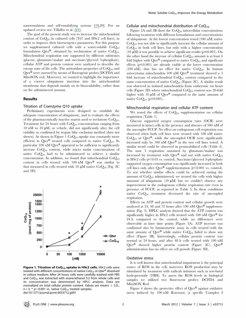
shown). As shown in Figure 1 CoQ10 uptake was constantly more
efficient in QterH treated cells compared to native CoQ10. In
particular 100 nM QterH appeared to be sufficient to significantly
increase CoQ10 content, while micro molar concentrations of
native CoQ10 had to be administered to achieve a similar
concentration. In addition, we found that mitochondrial CoQ10
content in cells treated with 100 nM QterH was similar to
that measured in cells treated with 10 mM native CoQ10 (Fig. 2C
and 2D).
Cellular and mitochondrial distribution of CoQ10
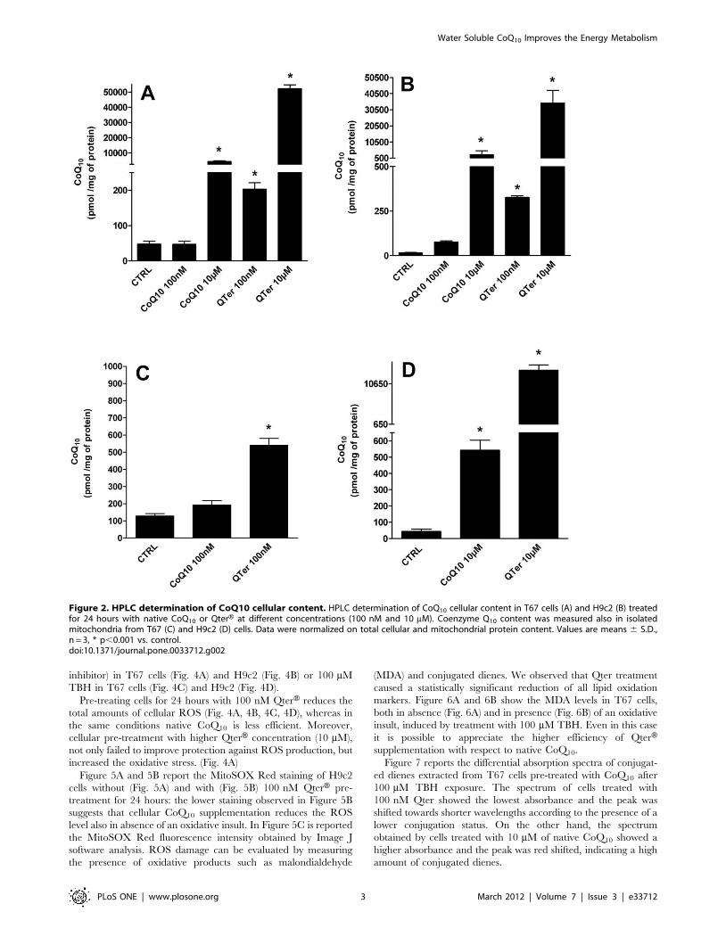
Figure 2A and 2B show the CoQ10 intracellular concentrations
following treatment with different formulations and concentrations
of ubiquinone. At the lowest concentration tested (100 nM) native
CoQ10 was not able to significantly increase the cellular amount of
CoQ10 in both cell lines, but only with a higher concentration
(10 mM) it was possible to achieve significant results (p#0.001). On
the other hand the increase of cellular CoQ10 amount is at least 4
fold higher with QterH compared to native CoQ10 and significant
effects (p#0.001) are already visible at the lower concentration
(100 nM), that has no effect for native CoQ10. In human
astrocytoma mitochondria 100 nM QterH treatment showed a 3
fold increase of mitochondrial CoQ10 content compared to the
same concentration of native CoQ10 (Figure 2C). A similar result
was observed in isolated mitochondria from embryonic rat heart
cells (Figure 2D) where mitochondrial CoQ10 content was 20-fold
higher with 10 mM of QterH compared to the same amount of
native CoQ10 (p#0.001).
Mitochondrial respiration and cellular ATP contentWe tested the effects of CoQ10 supplementation on cellular
respiration (Table 1).
Glucose supported oxygen consumption rates (OCR) were
measured in intact cells in the presence and absence of 500 nM of
the uncoupler FCCP. No effect on endogenous cell respiration was
observed when both cell lines were treated with 100 nM native
CoQ10 or QterH, while the uncoupled OCR were significantly
increased only by 100 nM QterH in the two cell lines tested. A
similar trend could be observed in permeabilized cells (Table 2).
The state 3 respiration sustained by glutamate/malate was
increased by treatment with QterH and not with native CoQ10
in H9c2 cells (p,0.05 vs. control). Succinate/glycerol 3-phosphate
supported oxygen consumption was significantly increased in both
cell lines only after QterH supplementation (p,0.001 vs. control).
To test whether similar effects could be achieved raising the
amount of CoQ10 administered, we treated the cells with higher
amounts of ubiquinone (10 mM) but we couldn’t observe any
improvement in the endogenous cellular respiration rate even in
presence of FCCP, as reported in Table 3. In these conditions
native CoQ10 treatment decreased the rate of uncoupled
respiration.
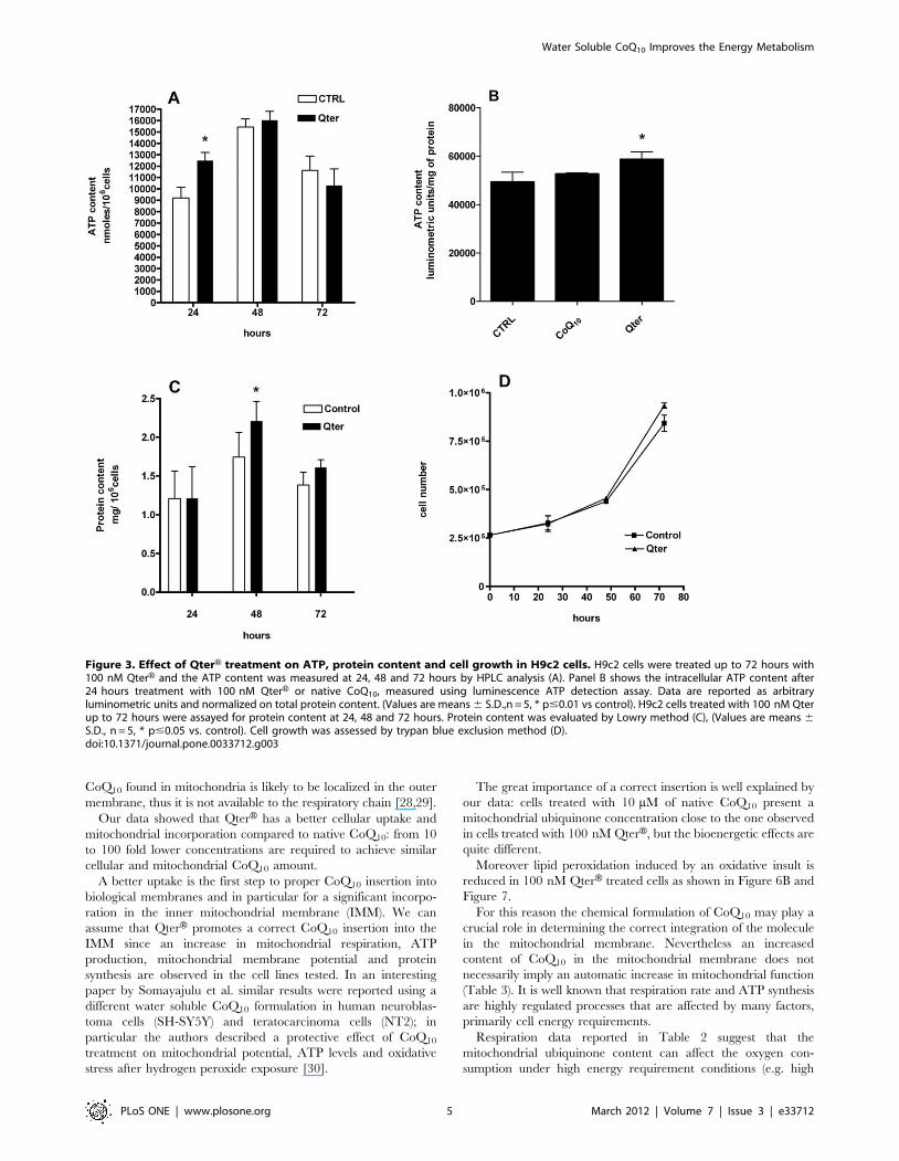
Effects on ATP and protein content and cellular growth were
analyzed at 24, 48 and 72 hours after 100 nM QterH supplemen-
tation (Fig. 3). HPLC analysis showed that the ATP content was
significantly higher in H9c2 cells treated with 100 nM QterH for
24 h compared to the control, while no differences were
observable at later time points (Figure 3A). ATP increase was
confirmed also by luminometric assay in cells treated with the
same amount of QterH while native CoQ10 failed to show any
effect (Figure 3B). Interestingly, cellular protein content was
normal at 24 hours, and after 48 h cells treated with 100 nM
QterH showed higher protein content (Figure 3C). QterHadministration has no effect on cell growth (Figure 3D).
Oxidative stressIt is well known that mitochondrial impairment is the principal
source of ROS in the cell, moreover ROS production may be
stimulated by treatment with radicals initiators such as tert-butyl
hydroperoxide (TBH). To assess the ROS levels in biological
samples we utilized two fluorescent probes: DCFDA and
MitoSOX Red.
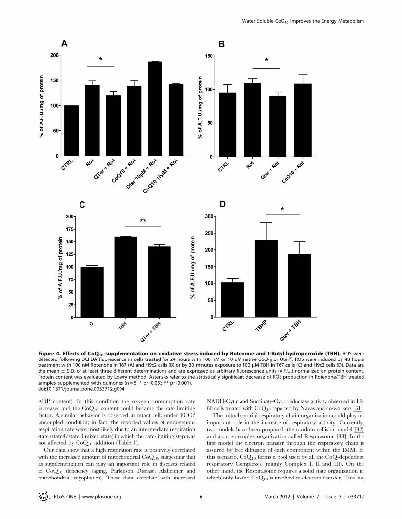
Figure 4 shows the protective effect of QterH against oxidative
stress induced by 100 nM Rotenone (a specific Complex I
Figure 1. Titration of CoQ10 uptake in H9c2 cells. H9c2 cells weretreated with different concentrations of native CoQ10 or QterH dissolvedin colture medium. After 24 hours cells were carefully washed with PBSand CoQ10 was extracted with exane/ethanol 5:2 from whole cells andits concentration was determined by HPLC analysis. Data arenormalized on total cellular protein content. Values are means 6 S.D.,n = 3, * p,0.001 vs. native CoQ10 treated samples.doi:10.1371/journal.pone.0033712.g001
Water Soluble CoQ10 Improves the Energy Metabolism
PLoS ONE | www.plosone.org 2 March 2012 | Volume 7 | Issue 3 | e33712
inhibitor) in T67 cells (Fig. 4A) and H9c2 (Fig. 4B) or 100 mM
TBH in T67 cells (Fig. 4C) and H9c2 (Fig. 4D).
Pre-treating cells for 24 hours with 100 nM QterH reduces the
total amounts of cellular ROS (Fig. 4A, 4B, 4C, 4D), whereas in
the same conditions native CoQ10 is less efficient. Moreover,
cellular pre-treatment with higher QterH concentration (10 mM),
not only failed to improve protection against ROS production, but
increased the oxidative stress. (Fig. 4A)
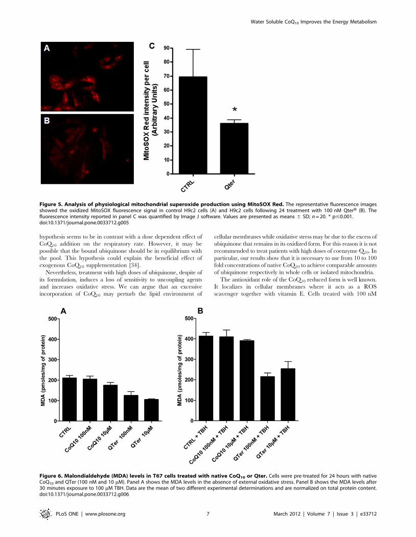
Figure 5A and 5B report the MitoSOX Red staining of H9c2
cells without (Fig. 5A) and with (Fig. 5B) 100 nM QterH pre-
treatment for 24 hours: the lower staining observed in Figure 5B
suggests that cellular CoQ10 supplementation reduces the ROS
level also in absence of an oxidative insult. In Figure 5C is reported
the MitoSOX Red fluorescence intensity obtained by Image J
software analysis. ROS damage can be evaluated by measuring
the presence of oxidative products such as malondialdehyde
(MDA) and conjugated dienes. We observed that Qter treatment
caused a statistically significant reduction of all lipid oxidation
markers. Figure 6A and 6B show the MDA levels in T67 cells,
both in absence (Fig. 6A) and in presence (Fig. 6B) of an oxidative
insult, induced by treatment with 100 mM TBH. Even in this case
it is possible to appreciate the higher efficiency of QterHsupplementation with respect to native CoQ10.
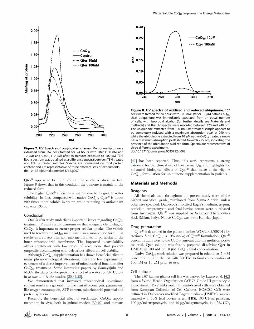
Figure 7 reports the differential absorption spectra of conjugat-
ed dienes extracted from T67 cells pre-treated with CoQ10 after
100 mM TBH exposure. The spectrum of cells treated with
100 nM Qter showed the lowest absorbance and the peak was
shifted towards shorter wavelengths according to the presence of a
lower conjugation status. On the other hand, the spectrum
obtained by cells treated with 10 mM of native CoQ10 showed a
higher absorbance and the peak was red shifted, indicating a high
amount of conjugated dienes.
Figure 2. HPLC determination of CoQ10 cellular content. HPLC determination of CoQ10 cellular content in T67 cells (A) and H9c2 (B) treatedfor 24 hours with native CoQ10 or QterH at different concentrations (100 nM and 10 mM). Coenzyme Q10 content was measured also in isolatedmitochondria from T67 (C) and H9c2 (D) cells. Data were normalized on total cellular and mitochondrial protein content. Values are means 6 S.D.,n = 3, * p,0.001 vs. control.doi:10.1371/journal.pone.0033712.g002
Water Soluble CoQ10 Improves the Energy Metabolism
PLoS ONE | www.plosone.org 3 March 2012 | Volume 7 | Issue 3 | e33712
The oxidative stress observed in cells treated with high amounts
of native CoQ10 can be due to an incomplete reduction of the
supplemented quinone. Figure 8 shows the absorption spectra of
CoQ10 extracted from T67 cells treated with 10 mM native CoQ10
or 100 nM Qter. The spectrum obtained from 100 nM Qter
treated cells showed a maximum close to 290 nm, indicating that
ubiquinone is mainly present in the reduced form. When cells were
treated with 10 mM of native CoQ10, the spectrum was broad and
the maximum was shifted towards 275 nm, indicating the
contemporary presence of oxidized and reduced quinone forms.
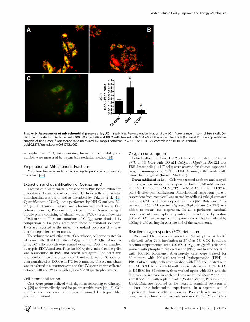
Mitochondrial membrane potentialTo assess whether QterH administration could alter mitochon-
drial membrane potential, JC-1 fluorescence assay was performed in
H9c2 cells. In control cells with normal mitochondrial membrane
potential, JC-1 accumulates in mitochondria as aggregates with a
red fluorescence emission while the monomeric form is prevalent in
the cytoplasm with a green fluorescence emission (Figure 9A).
In Qter treated cells, JC-1 probe was mainly in the aggregated
state resulting in a higher red/green fluorescence ratio, suggesting
a higher incorporation of the probe into mitochondria as a
consequence of a higher membrane potential(Figure 9B). Treat-
ment with 500 nM FCCP prevent JC-1 mitochondrial incorpo-
ration, resulting in a pronounced green fluorescence due to the
complete loss of mitochondrial membrane potential (Figure 9C).
The red/green fluorescence ratios are summarized in Figure 9D.
Discussion
Coenzyme Q10 is a lipid-soluble compound mainly found in
mitochondria. It is mostly endogenously produced within cells
though small amounts can be provided by food intake. Analysis of
CoQ10 subcellular distribution shows that a large portion of
CoQ10 (40–50%) is localized in the mitochondrial inner
membrane, with smaller amounts in the other organelles and in
the cytosol. The high concentration of CoQ10 in the mitochondria
reflects its important role in electron transport chain: age-related
decrease in mitochondrial CoQ10 content is responsible for oxygen
consumption decline).
From a physiological point of view, tissue CoQ10 content is
subject to regulation by several factors including oxidative stress
and aging [1,22].
Supplementation with CoQ10 has been thought to be beneficial,
especially for situations in which adequate CoQ10 production is
adversely affected [6]. A large number of clinical studies have
evaluated the effects of CoQ10 supplementation on oxidative
stress, both in physiological or pathological conditions.
The results obtained in vivo about CoQ10 tissue distribution are
quite controversial. Ibrahim et al. (Ibrahim et al., 2000) [23]
observed that CoQ10 oral administration did not alter the levels of
this compound in the heart. Furthermore there is no evidence so
far showing that dietary CoQ10, which is found to increase the
CoQ10 content in lipoproteins and in the liver, is taken up by other
tissues under normal conditions. These uncertain results could be
partially attributed to the poor water solubility of CoQ10 that
impairs its intestinal absorption, tissue distribution and mitochon-
drial incorporation.
The aim of the present study is to evaluate the in vitro efficacy of
CoQ10 supplementation in improving mitochondrial function and
protection against oxidative stress. Cells supplemented with CoQ10
do not often show any improvement in their bioenergetics status.
These negligible effects can be explained by several factors, first
of all its strong lipophilic nature that results in its accumulation in
extra-mitochondrial membranes [24,25] while only a small portion
(,11%) can reach the mitochondria [26,27]. The exogenous
Table 1. Respiratory rates of intact H9c2 and T67 cells treatedfor 24 hours with 100 nM native CoQ10 or QterH.
EndogenousRespirationnmoles O2 min21/106cells
UncoupledRespirationnmoles O2 min21/106cells
T67
Control 3.4960.42 4.2760.19
CoQ10 3.8460.19 4.2660.55
Qter 3.6060.28 4.9260.41*
H9c2
Control 6.7561.34 19.2362.60
CoQ10 7.4060.35 17.0560.36
Qter 7.3560.63 25.8563.28*
Respirometric analyses were performed under endogenous and uncoupledconditions. The maximal uncoupled respiration was measured in the presenceof 500 nM FCCP. Respiratory rates are expressed as nmoles O2 min21/106cells 6
S.D. from at least three independent experiments. *p,0.05 vs. control.doi:10.1371/journal.pone.0033712.t001
Table 2. Respiratory rates of permeabilized H9c2 and T67cells treated for 24 hours with 100 nM native CoQ10 or QterH.
Glutamate/Malatenmoles O2 min21/106cells
Succinate/Glycerol 3-phosphatenmoles O2 min21/106cells
T67
Control 3.9261.02 5.2661.31
CoQ10 3.2661.05 6.2560.92
QTer 4.361.14 7.8560.07 *
H9c2
Control 8.8160.16 12.6060.70
CoQ10 9.2060.14 13.7660.28
QTer 10.3260.66* 16.9560.21**
Respirometric analyses were performed in the presence of 5 mM Glutamate/Malate or 12,5 mM Succinate/Glycerol 3-phosphate. Respiratory rates areexpressed as nmoles O2 min21/106 cells 6 S.D. from at least three independentexperiments, *p,0.05 vs. control. **p,0.001 vs. control.doi:10.1371/journal.pone.0033712.t002
Table 3. Percentage of respiratory rates measured in T67 cellstreated for 24 hours with 10 mM native CoQ10 or QterH.
% of endogenous respiration rate
Cell treatment - FCCP +500 nM FCCP
No treatment 10067.2 131613
10 mM CoQ10 10068.6 8468,6
10 mM Qter 10061.1 102614
Respirometric analyses were performed under endogenous and uncoupledconditions (500 nM FCCP). Respiratory rates in the presence of FCCP areexpressed as percentage of oxygen consumption respect to the endogenousrespiration 6 S.D.doi:10.1371/journal.pone.0033712.t003
Water Soluble CoQ10 Improves the Energy Metabolism
PLoS ONE | www.plosone.org 4 March 2012 | Volume 7 | Issue 3 | e33712
CoQ10 found in mitochondria is likely to be localized in the outer
membrane, thus it is not available to the respiratory chain [28,29].
Our data showed that QterH has a better cellular uptake and
mitochondrial incorporation compared to native CoQ10: from 10
to 100 fold lower concentrations are required to achieve similar
cellular and mitochondrial CoQ10 amount.
A better uptake is the first step to proper CoQ10 insertion into
biological membranes and in particular for a significant incorpo-
ration in the inner mitochondrial membrane (IMM). We can
assume that QterH promotes a correct CoQ10 insertion into the
IMM since an increase in mitochondrial respiration, ATP
production, mitochondrial membrane potential and protein
synthesis are observed in the cell lines tested. In an interesting
paper by Somayajulu et al. similar results were reported using a
different water soluble CoQ10 formulation in human neuroblas-
toma cells (SH-SY5Y) and teratocarcinoma cells (NT2); in
particular the authors described a protective effect of CoQ10
treatment on mitochondrial potential, ATP levels and oxidative
stress after hydrogen peroxide exposure [30].
The great importance of a correct insertion is well explained by
our data: cells treated with 10 mM of native CoQ10 present a
mitochondrial ubiquinone concentration close to the one observed
in cells treated with 100 nM QterH, but the bioenergetic effects are
quite different.
Moreover lipid peroxidation induced by an oxidative insult is
reduced in 100 nM QterH treated cells as shown in Figure 6B and
Figure 7.
For this reason the chemical formulation of CoQ10 may play a
crucial role in determining the correct integration of the molecule
in the mitochondrial membrane. Nevertheless an increased
content of CoQ10 in the mitochondrial membrane does not
necessarily imply an automatic increase in mitochondrial function
(Table 3). It is well known that respiration rate and ATP synthesis
are highly regulated processes that are affected by many factors,
primarily cell energy requirements.
Respiration data reported in Table 2 suggest that the
mitochondrial ubiquinone content can affect the oxygen con-
sumption under high energy requirement conditions (e.g. high
Figure 3. Effect of QterH treatment on ATP, protein content and cell growth in H9c2 cells. H9c2 cells were treated up to 72 hours with100 nM QterH and the ATP content was measured at 24, 48 and 72 hours by HPLC analysis (A). Panel B shows the intracellular ATP content after24 hours treatment with 100 nM QterH or native CoQ10, measured using luminescence ATP detection assay. Data are reported as arbitraryluminometric units and normalized on total protein content. (Values are means 6 S.D.,n = 5, * p#0.01 vs control). H9c2 cells treated with 100 nM Qterup to 72 hours were assayed for protein content at 24, 48 and 72 hours. Protein content was evaluated by Lowry method (C), (Values are means 6S.D., n = 5, * p#0.05 vs. control). Cell growth was assessed by trypan blue exclusion method (D).doi:10.1371/journal.pone.0033712.g003
Water Soluble CoQ10 Improves the Energy Metabolism
PLoS ONE | www.plosone.org 5 March 2012 | Volume 7 | Issue 3 | e33712
ADP content). In this condition the oxygen consumption rate
increases and the CoQ10 content could became the rate limiting
factor. A similar behavior is observed in intact cells under FCCP
uncoupled condition; in fact, the reported values of endogenous
respiration rate were most likely due to an intermediate respiration
state (state4/state 3 mixed state) in which the rate-limiting step was
not affected by CoQ10 addition (Table 1).
Our data show that a high respiration rate is positively correlated
with the increased amount of mitochondrial CoQ10, suggesting that
its supplementation can play an important role in diseases related
to CoQ10 deficiency (aging, Parkinson Disease, Alzheimer and
mitochondrial myophaties). These data correlate with increased
NADH-Cyt.c and Succinate-Cyt.c reductase activity observed in Hl-
60 cells treated with CoQ10 reported by Navas and co-workers [31].
The mitochondrial respiratory chain organization could play an
important role in the increase of respiratory activity. Currently,
two models have been proposed: the random collision model [32]
and a supercomplex organization called Respirasome [33]. In the
first model the electron transfer through the respiratory chain is
assured by free diffusion of each component within the IMM. In
this scenario, CoQ10 forms a pool used by all the CoQ-dependent
respiratory Complexes (mainly Complex I, II and III). On the
other hand, the Respirasome requires a solid state organization in
which only bound CoQ10 is involved in electron transfer. This last
Figure 4. Effects of CoQ10 supplementation on oxidative stress induced by Rotenone and t-Butyl hydroperoxide (TBH). ROS weredetected following DCFDA fluorescence in cells treated for 24 hours with 100 nM or 10 uM native CoQ10 or QterH. ROS were induced by 48 hourstreatment with 100 nM Rotenone in T67 (A) and H9c2 cells (B) or by 30 minutes exposure to 100 mM TBH in T67 cells (C) and H9c2 cells (D). Data arethe mean 6 S.D. of at least three different determinations and are expressed as arbitrary fluorescence units (A.F.U.) normalized on protein content.Protein content was evaluated by Lowry method. Asterisks refer to the statistically significant decrease of ROS production in Rotenone/TBH treatedsamples supplemented with quinones (n = 5, * p#0.05); ** p#0.001).doi:10.1371/journal.pone.0033712.g004
Water Soluble CoQ10 Improves the Energy Metabolism
PLoS ONE | www.plosone.org 6 March 2012 | Volume 7 | Issue 3 | e33712
hypothesis seems to be in contrast with a dose dependent effect of
CoQ10 addition on the respiratory rate. However, it may be
possible that the bound ubiquinone should be in equilibrium with
the pool. This hypothesis could explain the beneficial effect of
exogenous CoQ10 supplementation [34].
Nevertheless, treatment with high doses of ubiquinone, despite of
its formulation, induces a loss of sensitivity to uncoupling agents
and increases oxidative stress. We can argue that an excessive
incorporation of CoQ10 may perturb the lipid environment of
cellular membranes while oxidative stress may be due to the excess of
ubiquinone that remains in its oxidized form. For this reason it is not
recommended to treat patients with high doses of coenzyme Q10. In
particular, our results show that it is necessary to use from 10 to 100
fold concentrations of native CoQ10 to achieve comparable amounts
of ubiquinone respectively in whole cells or isolated mitochondria.
The antioxidant role of the CoQ10 reduced form is well known.
It localizes in cellular membranes where it acts as a ROS
scavenger together with vitamin E. Cells treated with 100 nM
Figure 5. Analysis of physiological mitochondrial superoxide production using MitoSOX Red. The representative fluorescence imagesshowed the oxidized MitoSOX fluorescence signal in control H9c2 cells (A) and H9c2 cells following 24 treatment with 100 nM QterH (B). Thefluorescence intensity reported in panel C was quantified by Image J software. Values are presented as means 6 SD; n = 20. * p#0.001.doi:10.1371/journal.pone.0033712.g005
Figure 6. Malondialdehyde (MDA) levels in T67 cells treated with native CoQ10 or Qter. Cells were pre-treated for 24 hours with nativeCoQ10 and QTer (100 nM and 10 mM). Panel A shows the MDA levels in the absence of external oxidative stress. Panel B shows the MDA levels after30 minutes exposure to 100 mM TBH. Data are the mean of two different experimental determinations and are normalized on total protein content.doi:10.1371/journal.pone.0033712.g006
Water Soluble CoQ10 Improves the Energy Metabolism
PLoS ONE | www.plosone.org 7 March 2012 | Volume 7 | Issue 3 | e33712
QterH appear to be more resistant to oxidative stress; in fact,
Figure 8 shows that in this condition the quinone is mainly in the
reduced form
The higher QterH efficiency is mainly due to its greater water
solubility. In fact, compared with native CoQ10, QterH is about
200 times more soluble in water, while retaining its antioxidant
capacity [35,36].
ConclusionOur in vitro study underlines important issues regarding CoQ10
treatment. Present results demonstrate that adequate channeling of
CoQ10 is important to ensure proper cellular uptake. The vehicle
used to terclatrate CoQ10 maintains it in a monomeric form, that
results in a correct insertion into membranes, in particular in the
inner mitochondrial membrane. The improved bioavailability
allows treatments with low doses of ubiquinone that prevent
unspecific accumulation with deleterious effects on cell viability.
Although CoQ10 supplementation has shown beneficial effect in
many physiopathological alterations, there are few experimental
evidences of a direct improvement of mitochondrial functions after
CoQ10 treatment. Some interesting papers by Somayajulu and
McCarthy describe the protective effect of a water soluble CoQ10
in in vitro and in vivo studies [30,37,38].
We demonstrated that increased mitochondrial ubiquinone
content results in a general improvement of bioenergetic parameters,
like oxygen consumption, ATP content, mitochondrial potential and
protein synthesis.
Recently, the beneficial effect of terclatrated CoQ10 supple-
mentation in vivo, both in animal models [39,40] and humans
[41] has been reported. Thus, this work represents a strong
rationale for the clinical use of Coenzyme Q10 and highlights the
enhanced biological effects of QterH that make it the eligible
CoQ10 formulation for ubiquinone supplementation in patients.
Materials and Methods
ReagentsAll chemicals used throughout the present study were of the
highest analytical grade, purchased from Sigma-Aldrich, unless
otherwise specified. Dulbecco’s modified Eagle’s medium, trypsin,
penicillin, streptomycin and fetal bovine serum were purchased
from Invitrogen. QterH was supplied by Scharper Therapeutics
S.r.l. (Milan, Italy). Native CoQ10 was from Kaneka, Japan.
Drug preparationQterH is described in the patent number WO/2003/097012 by
Actimex S.r.l. CoQ10 is 10% (w/w) of QterH formulation. QterHconcentration refers to the CoQ10 amount into the multicomposite
material. Qter solution was freshly prepared dissolving Qter in
DMEM at 100 nM or 10 mM CoQ10 final concentration.
Native CoQ10 stock solution was prepared in ethanol at 5 mM
concentration and diluted with DMEM to final concentration of
100 nM or 10 mM prior to use.
Cell cultureThe T67 human glioma cell line was derived by Lauro et al. [42]
from a World Health Organization (WHO) Grade III gemistocytic
astrocytoma. H9c2 embryonal rat heart-derived cells were obtained
from European Collection of Cell Cultures, ECACC. Cells were
cultured in Dulbecco’s modified Eagle’s medium (DMEM), supple-
mented with 10% fetal bovine serum (FBS), 100 UI/ml penicillin,
100 mg/ml streptomycin, and 40 mg/ml gentamycin, in a 5% CO2
Figure 7. UV Spectra of conjugated dienes. Membrane lipids wereextracted from T67 cells treated for 24 hours with Qter (100 nM and10 mM) and CoQ10 (10 mM) after 30 minutes exposure to 100 mM TBH.Each spectrum was obtained as a difference spectra between TBH treatedand TBH untreated samples. Spectra are normalized on total proteincontent and are representative of three different sets of experiments.doi:10.1371/journal.pone.0033712.g007
Figure 8. UV spectra of oxidized and reduced ubiquinone. T67cells were treated for 24 hours with 100 nM Qter or 10 mM native CoQ10,then ubiquinone was immediately extracted, from an equal numberof cells, with isopropyl alcohol (for further details see Materials andmethods) and the UV spectra were recorded between 320 and 240 nm.The ubiquinone extracted from 100 nM Qter treated sample appears tobe completely reduced with a maximum absorption peak at 290 nm,while the ubiquinone extracted from 10 mM native CoQ10 treated samplehas a maximum absorption peak shifted towards 275 nm, indicating thepresence of the ubiquinone oxidized form. Spectra are representative ofthree different experiments.doi:10.1371/journal.pone.0033712.g008
Water Soluble CoQ10 Improves the Energy Metabolism
PLoS ONE | www.plosone.org 8 March 2012 | Volume 7 | Issue 3 | e33712
atmosphere at 37uC, with saturating humidity. Cell viability and
number were measured by trypan blue exclusion method [43].
Preparation of Mitochondria FractionsMitochondria were isolated according to procedures previously
described [44].
Extraction and quantification of Coenzyme QTreated cells were carefully washed with PBS before extraction
procedures. Extraction of coenzyme Q from cells and isolated
mitochondria was performed as described by Takada et al. [45].
Quantification of CoQ10 was performed by HPLC analysis. 50–
100 ml of ethanolic extract was chromatographed on a C18
column (Kinetex, Phenomenex, 2.6 mm, 10064.6 mm), using a
mobile phase consisting of ethanol: water (97:3, v/v) at a flow rate
of 0.6 ml/min. The concentrations of CoQ10 were obtained by
comparison of the peak areas with those of standard solutions.
Data are reported as the mean 6 standard deviation of at least
three independent experiments.
To evaluate the reduction state of ubiquinone, cells were treated for
24 hours with 10 mM of native CoQ10 or 100 nM Qter. After this
time, T67 adherent cells were washed twice with PBS, then detached
by trypsin-EDTA and centrifuged at 3006g for 3 min; then the pellet
was resuspended in PBS, and centrifuged again. The pellet was
resuspended in cold isopropyl alcohol and vortexed for 30 seconds,
then centrifuged at 15000 g at 4uC for 5 minutes. The organic phase
was transferred in a quartz cuvette and the UV spectrum was collected
between 240 and 320 nm with a Jasco V-550 spectrophotometer.
Cell permeabilizationCells were permeabilized with digitonin according to Chomyn
A. [39] and immediately used for polarographic assay [44,46]. Cell
number and permeabilization was measured by trypan blue
exclusion method.
Oxygen consumptionIntact cells. T67 and H9c2 cell lines were treated for 24 h at
37uC in 5% CO2 with 100 nM CoQ10 or QterH in DMEM plus
FBS. Intact cells (16106 cells) were assayed for glucose supported
oxygen consumption at 30uC in DMEM using a thermostatically
controlled oxygraph (Instech Mod.203).
Permeabilized cells. Cells were treated as above and assayed
for oxygen consumption in respiration buffer (250 mM sucrose,
20 mM HEPES, 10 mM MgCl2, 1 mM ADP, 2 mM KH2PO4,
pH 7.4) after permeabilization. Mitochondrial respiration (state 3
respiration) from complex I was started by adding 5 mM glutamate/
malate (G/M) and then stopped with 2.5 mM Rotenone. Sub-
sequently 12.5 mM succinate/glycerol-3-phosphate (S/G3P) was
added to restart the respiration. In all experiments maximal
respiration rate (uncoupled respiration) was achieved by adding
500 nM FCCP and oxygen consumption was completely inhibited by
adding 4 mM Antimycin A at the end of the experiments.
Reactive oxygen species (ROS) detectionH9c2 and T67 cells were seeded in 24-well plates at 46104
cells/well. After 24 h incubation at 37uC in 5% CO2 in culture
medium supplemented with 100 nM CoQ10 or QterH, cells were
washed with phosphate buffered saline (PBS) and treated for 48 h
with 100 nM Rotenone. Alternatively cells were treated for
30 minutes with 100 mM tert-butyl hydroperoxide (TBH) in
PBS. Subsequently, cells were washed with PBS and treated with
10 mM DCFDA (29,79-dichlorofluorescein diacetate, DCFH-DA)
in DMEM for 30 minutes, then washed again with PBS and the
fluorescence increase in each well was measured (lexc = 485 nm;
lem = 535 nm) with a plate reader (Wallac Victor, Perkin-Elmer,
USA). Data are reported as the mean 6 standard deviation of
at least three independent experiments. In a separate set of
experiments, basal oxidative stress in H9c2 cells was measured
using the mitochondrial superoxide indicator MitoSOX Red. Cells
Figure 9. Assessment of mitochondrial potential by JC-1 staining. Representative images show JC-1 fluorescence in control H9c2 cells (A),H9c2 cells treated for 24 hours with 100 nM QterH (B) and H9c2 cells treated with 500 nM of the uncoupler FCCP (C). Panel D shows quantitativeanalysis of Red/Green fluorescence ratio measured by ImageJ software. (n = 20, * p#0.001 vs. control; ?p#0.001 vs. control.).doi:10.1371/journal.pone.0033712.g009
Water Soluble CoQ10 Improves the Energy Metabolism
PLoS ONE | www.plosone.org 9 March 2012 | Volume 7 | Issue 3 | e33712
were treated with 100 nM QterH for 24 hours at 37uC in 5%
CO2, then 5 mM MitoSOX was added. After 20 minutes of
incubation, cells were washed twice with PBS and images were
obtained using an IX50 inverted fluorescence microscope
(Olympus, Tokyo) at 206 magnification. Fluorescence intensity
was quantified by Image J software (NIH).
ATP contentIntracellular ATP level was measured using luminescence ATP
detection assay (ATPlite, PerkinElmer, USA) according to
manufacturer’s instructions. Data were reported as arbitrary
luminometric units, measured with the microplate reader Wallac
Victor multilabel counter and normalized to total protein content,
determined by Lowry method [47]. Alternatively, intracellular
ATP was measured by HPLC method. ATP was extracted
essentially as described by Strehler et al. [48]. Distilled water
(180 ml) preheated to 100uC was added to cellular samples in
Eppendorf tubes and boiled for 5 min with occasional vortexing.
Tubes were transferred to ice until HPLC analysis. A mobile phase
containing 100 mM K2HPO4 (pH 5.75), 0.1% TBAF (tetrabuty-
lammonium fluoride) and 2% acetonitrile was pumped through a
Kinetex C18 (Phenomenex) column at ambient temperature at a
flow rate of 0,6 ml/min [49]. Absorbance at 254 nm was
monitored by a photodiode array detector (Waters 996). ATP
peak was identified by its retention time and by using co-
chromatography with standard.
Thiobarbituric acid assay of malondialdehyde and lipid-conjugated dienes assay
T67 cells were treated for 24 h at 37uC in 5% CO2 with native
CoQ10 (10 mM and 100 nM) or QterH (10 mM or 100 nM) in
DMEM plus FBS and washed with PBS before treating for
30 minutes with 100 mM tert-butyl hydroperoxide (TBH) in PBS.
After TBH treatment, adherent cells were washed twice with
PBS, then detached by trypsin-EDTA and centrifuged at 3006g
for 3 min; then the pellet was resuspended in PBS and centrifuged
again.
Quantification of thiobarbituric acid reactive substances
(TBARS) was carried out as described by Buege and Aust [50].
The formation of conjugated dienes, in T67 cells treated as
above was assayed according to Buege and Aust [50].
JC-1 stain for mitochondrial membrane potential (Dym)measurement
The fluorescent probe JC-1 (5, 59, 6, 69-tetrachloro-1, 19, 3, 39-
tetraethylbenzimidazol carbocyanine iodide) was used to measure
the mitochondrial membrane potential (Dy) of H9c2 cells. JC-1 is
a cationic dye that is accumulated in mitochondria following
membrane potential.
At low concentrations the probe is present in monomeric form,
with green fluorescence emission (525610 nm), but at higher
concentrations it forms J-aggregates after accumulation in the
mitochondrion, with red fluorescence emission (590610 nm).
After incubation with 10 mM JC-1 at 37uC for 10 min, the
culture medium containing JC-1 was removed. Cells were washed
twice with PBS, and analyzed by IX50 fluorescence microscope
(Olympus, Tokyo).) at 206 magnification. Fluorescence intensity
was quantified by Image J software (NIH). Mitochondrial
depolarization was achieved by treating cells with 500 nM FCCP,
indicated by a decrease in the red/green fluorescence intensity
ratio
Statistical analysisStatistical analysis of data was performed with Student’s t-test
using GraphPad Prism software.
Acknowledgments
The authors thank Dr. G. Manini and Dr. P. Arzuffi for critical reading of
the manuscript.
Author Contributions
Conceived and designed the experiments: RF CB GL. Performed the
experiments: CB NM. Analyzed the data: CB NM RF AS GL. Contributed
reagents/materials/analysis tools: RF. Wrote the paper: RF CB GL.
References
1. Spindler M, Beal MF, Henchcliffe C (2009) Coenzyme Q10 effects in
neurodegenerative disease. Neuropsychiatr Dis Treat 5: 597–610.
2. Geromel V, Rotig A, Munnich A, Rustin P (2002) Coenzyme Q10 depletion iscomparatively less detrimental to human cultured skin fibroblasts than
respiratory chain complex deficiencies. Free Radic Res 36: 375–379.
3. Groneberg DA, Kindermann B, Althammer M, Klapper M, Vormann J, et al.(2005) Coenzyme Q10 affects expression of genes involved in cell signalling,
metabolism and transport in human CaCo-2 cells. Int J Biochem Cell Biol 37:
1208–1218.
4. Schmelzer C, Lindner I, Rimbach G, Niklowitz P, Menke T, et al. (2008)
Functions of coenzyme Q10 in inflammation and gene expression. Biofactors 32:
179–183.
5. Zhang Y, Aberg F, Appelkvist EL, Dallner G, Ernster L (1995) Uptake of dietary
coenzyme Q supplement is limited in rats. J Nutr 125: 446–453.
6. Silver MA, Langsjoen PH, Szabo S, Patil H, Zelinger A (2004) Effect ofatorvastatin on left ventricular diastolic function and ability of coenzyme Q10 to
reverse that dysfunction. Am J Cardiol 94: 1306–1310.
7. Di Giovanni S, Mirabella M, Spinazzola A, Crociani P, Silvestri G, et al. (2001)Coenzyme Q10 reverses pathological phenotype and reduces apoptosis in
familial CoQ10 deficiency. Neurology 57: 515–518.
8. Kumar A, Kaur H, Devi P, Mohan V (2009) Role of coenzyme Q10 (CoQ10) incardiac disease, hypertension and Meniere-like syndrome. Pharmacol Ther 124:
259–268.
9. Hamilton SJ, Chew GT, Watts GF (2009) Coenzyme Q10 improves endothelial
dysfunction in statin-treated type 2 diabetic patients. Diabetes Care 32:810–812.
10. Henchcliffe C, Beal MF (2008) Mitochondrial biology and oxidative stress in
Parkinson disease pathogenesis. Nat Clin Pract Neurol 4: 600–609.
11. Sharma S, Kheradpezhou M, Shavali S, El Refaey H, Eken J, et al. (2004)
Neuroprotective actions of coenzyme Q10 in Parkinson’s disease. Methods
Enzymol 382: 488–509.
12. Shults CW, Haas R (2005) Clinical trials of coenzyme Q10 in neurological
disorders. Biofactors 25: 117–126.
13. Yang X, Dai G, Li G, Yang ES (2009) Coenzyme Q10 reduces beta-amyloidplaque in an APP/PS1 transgenic mouse model of Alzheimer’s disease. J Mol
Neurosci 41: 110–113.
14. Huntington Study Group (2001) A randomized, placebo-controlled trial ofcoenzyme Q10 and remacemide in Huntington’s disease. Neurology 57: 397–404.
15. Storch A, Jost WH, Vieregge P, Spiegel J, Greulich W, et al. (2007)
Randomized, double-blind, placebo-controlled trial on symptomatic effects ofcoenzyme Q(10) in Parkinson disease. Arch Neurol 64: 938–944.
16. Kaufmann P, Thompson JL, Levy G, Buchsbaum R, Shefner J, et al. (2009)Phase II trial of CoQ10 for ALS finds insufficient evidence to justify phase III.
Ann Neurol 66: 235–244.
17. Young JM, Florkowski CM, Molyneux SL, McEwan RG, Frampton CM, et al.(2007) Effect of coenzyme Q(10) supplementation on simvastatin-induced
myalgia. Am J Cardiol 100: 1400–1403.
18. Bhagavan HN, Chopra RK (2007) Plasma coenzyme Q10 response to oralingestion of coenzyme Q10 formulations. Mitochondrion 7 Suppl: S78–88.
19. Beg S, Javed S, Kohli K (2010) Bioavailability enhancement of coenzyme Q10:
an extensive review of patents. Recent Pat Drug Deliv Formul 4: 245–255.
20. Balakrishnan P, Lee BJ, Oh DH, Kim JO, Lee YI, et al. (2009) Enhanced oral
bioavailability of Coenzyme Q10 by self-emulsifying drug delivery systems.Int J Pharm 374: 66–72.
21. Villalba JM, Parrado C, Santos-Gonzalez M, Alcain FJ (2010) Therapeutic use
of coenzyme Q10 and coenzyme Q10-related compounds and formulations.Expert Opin Investig Drugs 19: 535–554.
22. Overvad K, Diamant B, Holm L, Holmer G, Mortensen SA, et al. (1999)
Coenzyme Q10 in health and disease. Eur J Clin Nutr 53: 764–770.
23. Ibrahim WH, Bhagavan HN, Chopra RK, Chow CK (2000) Dietary coenzyme
Q10 and vitamin E alter the status of these compounds in rat tissues andmitochondria. J Nutr 130: 2343–2348.
Water Soluble CoQ10 Improves the Energy Metabolism
PLoS ONE | www.plosone.org 10 March 2012 | Volume 7 | Issue 3 | e33712
24. Lenaz G, Samori B, Fato R, Battino M, Parenti Castelli G, et al. (1992)
Localization and preferred orientations of ubiquinone homologs in modelbilayers. Biochem Cell Biol 70: 504–514.
25. Cornell BA, Keniry MA, Post A, Robertson RN, Weir LE, et al. (1987) Location
and activity of ubiquinone 10 and ubiquinone analogues in model and biologicalmembranes. Biochemistry 26: 7702–7707.
26. Bentinger M, Dallner G, Chojnacki T, Swiezewska E (2003) Distribution andbreakdown of labeled coenzyme Q10 in rat. Free Radic Biol Med 34: 563–575.
27. Santos-Ocana C, Do TQ, Padilla S, Navas P, Clarke CF (2002) Uptake of
exogenous coenzyme Q and transport to mitochondria is required for bc1complex stability in yeast coq mutants. J Biol Chem 277: 10973–10981.
28. Geromel V, Darin N, Chretien D, Benit P, DeLonlay P, et al. (2002) CoenzymeQ(10) and idebenone in the therapy of respiratory chain diseases: rationale and
comparative benefits. Mol Genet Metab 77: 21–30.29. Lopez LC, Quinzii CM, Area E, Naini A, Rahman S, et al. (2010) Treatment of
CoQ(10) deficient fibroblasts with ubiquinone, CoQ analogs, and vitamin C:
time- and compound-dependent effects. PLoS One 5: e11897.30. Somayajulu M, McCarthy S, Hung M, Sikorska M, Borowy-Borowski H, et al.
(2005) Role of mitochondria in neuronal cell death induced by oxidative stress;neuroprotection by Coenzyme Q10. Neurobiol Dis 18: 618–627.
31. Fernandez-Ayala DJ, Lopez-Lluch G, Garcia-Valdes M, Arroyo A, Navas P
(2005) Specificity of coenzyme Q10 for a balanced function of respiratory chainand endogenous ubiquinone biosynthesis in human cells. Biochim Biophys Acta
1706: 174–183.32. Hackenbrock CR, Chazotte B, Gupte SS (1986) The random collision model
and a critical assessment of diffusion and collision in mitochondrial electrontransport. J Bioenerg Biomembr 18: 331–368.
33. Schagger H (2002) Respiratory chain supercomplexes of mitochondria and
bacteria. Biochim Biophys Acta 1555: 154–159.34. Lenaz G, Genova ML (2009) Mobility and function of coenzyme Q (ubiquinone)
in the mitochondrial respiratory chain. Biochim Biophys Acta 1787: 563–573.35. Carli F, Corvi Mora P, Canal T (2003) Co-grinding process for the preparation
of a ternary composition. European patent office. Wipo website. Available:
http://www.wipo.int/patentscope/search/en/WO2003097012. Accessed 2012Feb 23.
36. Corvi Mora P, Canal T, Ruzzier F (2008) Composition containing micronu-trients with improved anti-oxidant activity and the use thereof. Wipo website.
Available: http://www.wipo.int/patentscope/search/en/WO2007009997. Ac-cessed 2012 Feb 23.
37. Somayajulu-Nitu M, Sandhu JK, Cohen J, Sikorska M, Sridhar TS, et al. (2009)
Paraquat induces oxidative stress, neuronal loss in substantia nigra region and
parkinsonism in adult rats: neuroprotection and amelioration of symptoms by
water-soluble formulation of coenzyme Q10. BMC Neurosci 10: 88.
38. McCarthy S, Somayajulu M, Sikorska M, Borowy-Borowski H, Pandey S (2004)
Paraquat induces oxidative stress and neuronal cell death; neuroprotection by
water-soluble Coenzyme Q10. Toxicol Appl Pharmacol 201: 21–31.
39. Fetoni AR, Piacentini R, Fiorita A, Paludetti G, Troiani D (2009) Water-soluble
Coenzyme Q10 formulation (Q-ter) promotes outer hair cell survival in a guinea
pig model of noise induced hearing loss (NIHL). Brain Res 1257: 108–116.
40. Xu J, Seo AY, Vorobyeva DA, Carter CS, Anton SD, et al. (2010) Beneficial
effects of a Q-ter based nutritional mixture on functional performance,
mitochondrial function, and oxidative stress in rats. PLoS One 5: e10572.
41. Fumagalli S, Fattirolli F, Guarducci L, Cellai T, Baldasseroni S, et al. (2011)
Coenzyme Q10 terclatrate and creatine in chronic heart failure: a randomized,
placebo-controlled, double-blind study. Clin Cardiol 34: 211–217.
42. Lauro GM, Di Lorenzo N, Grossi M, Maleci A, Guidetti B (1986) Prostaglandin
E2 as an immunomodulating factor released in vitro by human glioma cells.
Acta Neuropathol 69: 278–282.
43. Gorman A, McCarthy J, Finucane D, Reville W (1996) Morphological
assessment of apoptosis. In Techniques in Apoptosis, A Users Guide
Cotter TG, Martin SJ, eds. pp 6–7.
44. Chomyn A (1996) Platelet-mediated transformation of human mitochondrial
DNA-less cells. Methods Enzymol 264: 334–339.
45. Takada M, Ikenoya S, Yuzuriha T, Katayama K (1984) Simultaneous
determination of reduced and oxidized ubiquinones. Methods Enzymol 105:
147–155.
46. Chomyn A (1996) In vivo labeling and analysis of human mitochondrial
translation products. Methods Enzymol 264: 197–211.
47. Lowry OH, Rosebrough NJ, Farr AL, Randall RJ (1951) Protein measurement
with the Folin phenol reagent. J Biol Chem 193: 265–275.
48. Strehler BL (1963) Adenosine-5V-triphosphate and creatine phosphate,
determination with luciferase. Methods of enzymatic analysis. pp 559–572.
49. Napolitano MJ, Shain DH (2005) Quantitating adenylate nucleotides in diverse
organisms. J Biochem Biophys Methods 63: 69–77.
50. Buege JA, Aust SD (1978) Microsomal lipid peroxidation. Methods Enzymol 52:
302–310.
Water Soluble CoQ10 Improves the Energy Metabolism
PLoS ONE | www.plosone.org 11 March 2012 | Volume 7 | Issue 3 | e33712
Related Documents


![Regulation of the intracellular Ca2+. Regulation of intracellular [H]:](https://static.cupdf.com/doc/110x72/5a4d1b717f8b9ab0599b56a5/regulation-of-the-intracellular-ca2-regulation-of-intracellular-h.jpg)


