BRAIN A JOURNAL OF NEUROLOGY A voxel-based morphometry study of grey matter loss in fragile X-associated tremor/ataxia syndrome Ryu-ichiro Hashimoto, 1 Alireza K. Javan, 1 Flora Tassone, 2,3 Randi J. Hagerman 2,4 and Susan M. Rivera 1,2,5 1 Center for Mind and Brain, University of California Davis, Davis, CA 95618, USA 2 M.I.N.D. Institute, University of California Davis, Sacramento, CA 95817, USA 3 Department of Biochemistry and Molecular Medicine, University of California, Davis, Sacramento, CA 95616, USA 4 Department of Paediatrics, University of California Davis Medical Center, Sacramento, CA 95817, USA 5 Department of Psychology, University of California Davis, Davis, CA 95618-5412, 95616, USA Correspondence to: Dr Susan M. Rivera, Center for Mind and Brain, University of California Davis, 267 Cousteau Place, Davis, CA 95618-5412, USA E-mail: [email protected] Fragile X-associated tremor/ataxia syndrome is a neurodegenerative disorder that primarily affects older male premutation carriers of the fragile X mental retardation gene. Although its core symptoms are mainly characterized by motor problems such as intention tremor and gait ataxia, cognitive decline and psychiatric problems are also commonly observed. Past radio- logical and histological approaches have focused on prominent neurodegenerative changes in specific brain structures including the cerebellum and limbic areas. However, quantitative investigations of the regional structural abnormalities have not been performed over the whole brain. In this study, we adopted the voxel-based morphometry method together with regions of interest analysis for the cerebellum to examine the pattern of regional grey matter change in the male premutation carriers with and without fragile X-associated tremor/ataxia syndrome. In a comparison with healthy controls, we found striking grey matter loss of the patients with fragile X-associated tremor/ataxia syndrome in multiple regions over the cortical and subcortical structures. In the cerebellum, the anterior lobe and the superior posterior lobe were profoundly reduced in both vermis and hemispheres. In the cerebral cortex, clusters of highly significant grey matter reduction were found in the extended areas in the medial surface of the brain, including the dorsomedial prefrontal cortex, anterior cingulate cortex and precuneus. The other prominent grey matter loss was found in the lateral prefrontal cortex, orbitofrontal cortex, amygdala and insula. Although the voxel-wise comparison between the asymptomatic premutation group and healthy controls did not reach significant difference, a regions of interest analysis revealed significant grey matter reduction in anterior subregions of the cerebellar vermis and hemisphere in the asymptomatic premutation group. Correlation analyses using behavioural scales of the premutation groups showed significant associations between grey matter loss in the left amygdala and increased levels of obsessive–compulsiveness and depression, and between decreased grey matter in the left inferior frontal cortex and anterior cingulate cortex and poor working memory performance. Furthermore, regression analyses revealed a significant negative effect of CGG repeat size on grey matter density in the dorsomedial frontal regions. A significant negative correlation with the clinical scale for the severity of fragile X-associated tremor/ataxia syndrome was found in a part of the vermis. These observations reveal the anatomical patterns of the neurodegenerative process that underlie the motor, cognitive and psychiatric problems of fragile X-associated tremor/ataxia syndrome, together with incipient structural abnormalities that may occur before the clinical onset of this disease. doi:10.1093/brain/awq368 Brain 2011: 134; 863–878 | 863 Received July 22, 2010. Revised October 16, 2010. Accepted November 3, 2010 ß The Author (2011). Published by Oxford University Press on behalf of the Guarantors of Brain. All rights reserved. For Permissions, please email: [email protected] at Serials RecordsSerials on July 29, 2011 brain.oxfordjournals.org Downloaded from

Welcome message from author
This document is posted to help you gain knowledge. Please leave a comment to let me know what you think about it! Share it to your friends and learn new things together.
Transcript
BRAINA JOURNAL OF NEUROLOGY
A voxel-based morphometry study of greymatter loss in fragile X-associated tremor/ataxiasyndromeRyu-ichiro Hashimoto,1 Alireza K. Javan,1 Flora Tassone,2,3 Randi J. Hagerman2,4 andSusan M. Rivera1,2,5
1 Center for Mind and Brain, University of California Davis, Davis, CA 95618, USA
2 M.I.N.D. Institute, University of California Davis, Sacramento, CA 95817, USA
3 Department of Biochemistry and Molecular Medicine, University of California, Davis, Sacramento, CA 95616, USA
4 Department of Paediatrics, University of California Davis Medical Center, Sacramento, CA 95817, USA
5 Department of Psychology, University of California Davis, Davis, CA 95618-5412, 95616, USA
Correspondence to: Dr Susan M. Rivera,
Center for Mind and Brain,
University of California Davis,
267 Cousteau Place,
Davis, CA 95618-5412, USA
E-mail: [email protected]
Fragile X-associated tremor/ataxia syndrome is a neurodegenerative disorder that primarily affects older male premutation
carriers of the fragile X mental retardation gene. Although its core symptoms are mainly characterized by motor problems
such as intention tremor and gait ataxia, cognitive decline and psychiatric problems are also commonly observed. Past radio-
logical and histological approaches have focused on prominent neurodegenerative changes in specific brain structures including
the cerebellum and limbic areas. However, quantitative investigations of the regional structural abnormalities have not been
performed over the whole brain. In this study, we adopted the voxel-based morphometry method together with regions of
interest analysis for the cerebellum to examine the pattern of regional grey matter change in the male premutation carriers with
and without fragile X-associated tremor/ataxia syndrome. In a comparison with healthy controls, we found striking grey matter
loss of the patients with fragile X-associated tremor/ataxia syndrome in multiple regions over the cortical and subcortical
structures. In the cerebellum, the anterior lobe and the superior posterior lobe were profoundly reduced in both vermis and
hemispheres. In the cerebral cortex, clusters of highly significant grey matter reduction were found in the extended areas in the
medial surface of the brain, including the dorsomedial prefrontal cortex, anterior cingulate cortex and precuneus. The other
prominent grey matter loss was found in the lateral prefrontal cortex, orbitofrontal cortex, amygdala and insula. Although the
voxel-wise comparison between the asymptomatic premutation group and healthy controls did not reach significant difference, a
regions of interest analysis revealed significant grey matter reduction in anterior subregions of the cerebellar vermis and
hemisphere in the asymptomatic premutation group. Correlation analyses using behavioural scales of the premutation groups
showed significant associations between grey matter loss in the left amygdala and increased levels of obsessive–compulsiveness
and depression, and between decreased grey matter in the left inferior frontal cortex and anterior cingulate cortex and poor
working memory performance. Furthermore, regression analyses revealed a significant negative effect of CGG repeat size on
grey matter density in the dorsomedial frontal regions. A significant negative correlation with the clinical scale for the severity
of fragile X-associated tremor/ataxia syndrome was found in a part of the vermis. These observations reveal the anatomical
patterns of the neurodegenerative process that underlie the motor, cognitive and psychiatric problems of fragile X-associated
tremor/ataxia syndrome, together with incipient structural abnormalities that may occur before the clinical onset of this disease.
doi:10.1093/brain/awq368 Brain 2011: 134; 863–878 | 863
Received July 22, 2010. Revised October 16, 2010. Accepted November 3, 2010
� The Author (2011). Published by Oxford University Press on behalf of the Guarantors of Brain. All rights reserved.
For Permissions, please email: [email protected]
at Serials R
ecordsSerials on July 29, 2011
brain.oxfordjournals.orgD
ownloaded from

Keywords: fragile X-associated tremor/ataxia syndrome; movement disorder; voxel based morphometry; cerebellum; atrophy
Abbreviations: FXTAS = fragile X-associated tremor/ataxia syndrome; FMR1 = fragile X mental retardation gene 1; PFX + = FMR1premutation carriers with FXTAS; PFX� = FMR1 premutation carriers without FXTAS; SCL-90-R = symptom Checklist-90 Revised
IntroductionAbnormalities of the fragile X mental retardation gene (FMR1) are
associated with a diverse range of behavioural and clinical pheno-
types depending on the type of mutation. Expansions of the CGG
trinucleotide repeats in the full mutation range (4200 CGG) are
the genetic cause of the FMR1 protein deficiency that underlies
the fragile X syndrome (Fu et al., 1991; Pieretti et al., 1991;
Verkerk et al., 1991). Smaller expansions of 55–200 repeats are
referred to as the premutation. Because the premutation does not
cause severe protein (FMR1 protein) deficiency as observed in the
full mutation, it was initially thought not to be associated with a
particular psychological or neurocognitive phenotype. However,
recent studies have accumulated evidence for several cognitive
and psychiatric problems in adult and child carriers of premutation
alleles (Franke et al., 1998; Johnston et al., 2001; Hagerman and
Hagerman, 2002; Moore et al., 2004; Cornish et al., 2005; Farzin
et al., 2006; Hessl et al., 2007).
Fragile X-associated tremor/ataxia syndrome (FXTAS) is prob-
ably the most clinically significant central nervous system
phenotype of the FMR1 premutation. FXTAS is a late-onset neu-
rodegenerative disorder primarily affecting older male premutation
carriers. Although it is principally characterized as a movement
disorder involving intention tremor and gait ataxia, cognitive
decline and psychiatric problems are also commonly observed
(Hagerman et al., 2001; Jacquemont et al., 2003; Bacalman
et al., 2006; Bourgeois et al., 2009). While its pathogenetic mech-
anism is still unclear, an RNA toxic ‘gain-of-function’ model has
been proposed based on several observations (Hagerman et al.,
2001; Hagerman and Hagerman, 2004), including: the presence of
elevated FMR1 messenger RNA among premutation carriers with-
out clear indications of abnormal FMR1 protein expression
(Tassone et al., 2000; Kenneson et al., 2001); the presence of
FMR1 messenger RNA within intranuclear inclusions (Greco
et al., 2006), one of the hallmarks of FXTAS (Tassone et al.,
2004); and premature neuronal cell death in culture combined
with dysregulation of several proteins secondary to elevated mes-
senger RNA (Chen et al., 2010, Garcia-Arocena and Hagerman,
2010).
Previous studies have revealed anatomical abnormalities in sev-
eral structures of the FXTAS brain. Particularly, abnormalities of
the cerebellum have been demonstrated in multiple methodolo-
gies. In a clinical MRI study, the middle cerebellar peduncle sign
was described as one of the most characteristic neuroradiological
features of FXTAS (Brunberg et al., 2002). Cerebellar abnormal-
ities were confirmed by post-mortem histological studies that iden-
tified several neuropathological features, including Purkinje cell
decreases and spongiform changes (Greco et al., 2002, 2006).
Significant loss in whole cerebellar volume has been revealed in
the male patients with FXTAS (Cohen et al., 2006), which
was further replicated in the female patients in a milder form
(Adams et al., 2007). However, there have been no studies that
attempted to identify the foci of neurodegeneration within the
cerebellum. This point is crucial given the fact that there are mul-
tiple functionally different subregions in the cerebellum (Stoodley
and Schmahmann, 2009) and therefore structural changes in dif-
ferent subregions may make distinct contributions to motor, cog-
nitive and psychiatric problems in FXTAS. Similarly, regionally
selective abnormalities in the cerebral cortex are entirely unclear,
although the aforementioned MRI volumetric study revealed sig-
nificant loss of the whole cerebral volume in the patients with
FXTAS (Cohen et al., 2006).
Previous behavioural studies revealed patterns of neurocognitive
and psychological deficits of FXTAS (Cornish et al., 2005, 2008;
Grigsby et al., 2006, 2007), which provide motivations for exam-
ining the possible regionally selective abnormalities in cortical and
subcortical structures in the FXTAS brain. One study applied an
extensive neuropsychological test battery to male premutation
carriers with and without FXTAS and reported that, whereas
language and visuospatial/attention functions were relatively
spared, patients with FXTAS displayed profound deficits of execu-
tive cognitive functions, working memory and declarative verbal
memory and learning (Grigsby et al., 2008). Several studies repli-
cated significant deficits of executive functions and working
memory not only in FXTAS but also in unaffected premutation
carriers (Cornish et al., 2008, 2009). For psychological symptoms,
it has been reported that major psychiatric features of FXTAS
include increased anxiety, depression, disinhibition and apathy
(Berry-Kravis et al., 2007b; Bourgeois et al., 2007, 2009). In a
large-scale study examining self-reported psychological symptoms
of patients with FXTAS and unaffected premutation carriers, the
level of the obsessive–compulsiveness was elevated even in
unaffected premutation carriers, whereas psychological symptoms
of FXTAS extended into other domains, including anxiety and
depression (Hessl et al., 2005). Although one recent region of
interest-based volumetric study reported a significant correlation
between the right hippocampal volume and the severity of
anxiety-related psychological symptoms among female patients
with FXTAS (Adams et al., 2010), there has been no study that
systematically investigated foci of structural abnormalities that may
underlie major neurocognitive and psychological problems in indi-
viduals with FXTAS and unaffected premutation carriers.
In this study, we adopted the voxel-based morphometry
method to examine the regional grey matter loss in the FMR1
premutation carriers with and without FXTAS. Voxel-based
morphometry is an automated analysis for assessment of the re-
gional volumetric change over the whole brain (Ashburner and
Friston, 2000). For a set of brain regions whose deficits can be
responsible for neurocognitive and psychological deficits of the
FMR1 premutation carriers, we performed region of interest ana-
lyses to examine associations between the grey matter abnormal-
ities in those regions and the severity of the behavioural problems
864 | Brain 2011: 134; 863–878 R.-i. Hashimoto et al.
at Serials R
ecordsSerials on July 29, 2011
brain.oxfordjournals.orgD
ownloaded from

(refer to ‘Materials and Methods’ section for selection of regions
of interest.) Simple voxel-based regression analyses using either
CGG repeat size or level of FMR1 messenger RNA of premutation
carriers were also performed to examine the effects of the genetic
molecular variables on the grey matter abnormality over the whole
brain. Lastly, the same simple regression analysis was applied using
a clinical scale for assessment of the FXTAS severity to identify
brain regions showing progressive neurodegeneration correlated
with the development of FXTAS.
Materials and methods
ParticipantsWe examined the brains of a total of 83 male participants between the
ages of 40 and 80 years, 28 healthy control participants, 31 partici-
pants with the premutation with FXTAS (PFX + ), and 24 participants
with the premutation without FXTAS (PFX�). In this study, the pre-
mutation range was defined as those with a CGG repeat size of be-
tween 55 and 200. CGG repeat size was 545 in all the healthy control
cases, so that there was no participant whose CGG repeat was within
the ‘grey zone’ (45–54 CGG repeats). Participant demographic infor-
mation is shown in Table 1. The group of PFX + was significantly older
than the other two groups [F(2, 80) = 5.19, P = 0.008]. Twenty-six
controls, 27 PFX + and 22 PFX� were assessed for full, verbal and
performance-scale IQ using the Wechsler Adult Intelligence Scale
(Third Edition). According to one-way analysis of variance (ANOVA),
a significant main effect of group was found for performance IQ [per-
formance IQ: F(2, 67) = 4.16, P = 0.019; full-scale IQ: F = 2.69,
P = 0.075; verbal IQ: F = 1.24, P = 0.30], with healthy control and
PFX� individuals having higher performance IQs than those with
PFX + (P5 0.05). Participants with the premutation were recruited
through pedigree analysis of families containing probands with fragile
X syndrome. Controls were recruited from the families and the local
community through the University of California Davis Medical Centre.
Neurological examinations on all healthy control participants were
normal, including absence of tremor and ataxia. A signed, written
informed consent was obtained according to the Declaration of
Helsinki. The protocol was approved by the institutional review
board at the University of California, Davis.
Molecular genetic dataGenomic DNA was isolated from peripheral blood lymphocytes using
standard methods (Puregene� Kit; Gentra Inc). For Southern blot
analysis, 5–10mg of isolated DNA was digested with EcoRI and NruI.
Hybridization was performed using the FMR1 genomic digoxygenin-
labelled StB12.3 probe. Genomic DNA was also amplified by polymer-
ase chain reaction using primers ‘c’ and ‘f’ (Fu et al., 1991).
Hybridization was performed with a digoxygenin-end-labelled oligo-
nucleotide probe (CGG)10. Analysis and calculation of the repeat size
for both Southern blot and polymerase chain reaction analysis were
carried out using an Alpha Innotech FluorChem 8800 Image Detection
System (Tassone et al., 2008).
Total cellular RNA was purified from 3–5 ml of peripheral blood
using standard methods (Purescript� kits, Gentra Inc.; Trizol�, BRL).
All quantification of FMR1 messenger RNA were performed using a
7900 Sequence detector (PE Biosystems) as previously described
(Tassone et al., 2000).
Assessment of clinical severity of fragileX-associated tremor/ataxia syndromeFor participants with CGG repeat count within the premutation range,
a trained physician (RJH) scored the severity of FXTAS on a scale
ranging from 0–6 as described by our previous studies (Bacalman
et al., 2006; Adams et al., 2007). This seven-point staging scale meas-
ures functional impairment as follows: 0 = normal functioning;
1 = subtle or questionable tremor or balance problems with no inter-
ference in activities of daily living; 2 = minor but clear tremor or bal-
ance problems producing minor interference with activities of daily
living; 3 = moderate tremor or balance problems with at least occa-
sional falls and significant interference in activities of daily living;
4 = severe tremor or balance problems requiring the use of a cane
or walker; 5 = use of a wheelchair on a daily basis and 6 = bedridden.
Premutation carriers with FXTAS scores of 0 or 1 were placed in the
PFX� group, while those with FXTAS scores of 2–5 were designated as
PFX + , meeting clinical criteria for the diagnosis of FXTAS established
initially by Jacquemont et al. (2003).
Clinical data acquisitionTo examine the relationship between grey matter loss in specific brain
regions and behavioural problems of the FMR1 premutation carriers,
PFX + and PFX� individuals were administered a series of examinations
for assessing their psychological and cognitive functioning.
Psychological assessment
The Symptom Checklist-90-Revised (SCL-90-R), a standardized self
report inventory of current psychological symptoms (Derogatis,
1994), was used for assessing the severity of psychological symptoms
Table 1 Statistics on participant demographic data
Healthy controls (n = 28) Premutation with FXTAS (PFX + ) (n = 31) Premutation without FXTAS (PFX�) (n = 24)
Mean (SD) Range Mean (SD) Range Mean (SD) Range
Age (years) 58.2 (11.1) 40–79 65.2 (7.45) 47–79 58.1 (10.0) 41–78
Full scale IQ 118.6 (16.7) 84–148 108.1 (14.7) 85–136 115.8 (16.5) 83–152
Performance IQ 116.5 (14.3) 89–144 104.9 (14.8) 79–128 115.4 (16.3) 91–155
Verbal IQ 117.0 (17.3) 76–148 109.5 (14.5) 87–135 113.8 (17.4) 78–142
FXTAS score NA 2.97 (0.98) 2–5 0.35 (0.48) 0–1
CGG repeat 28.6 (4.37) 17–34 93.8 (18.1) 59–130 94.9 (30.6) 55–166
FMR1 messenger RNA 1.29 (0.29) 0.63–1.85 3.32 (0.81) 1.75–5.25 3.15 (0.95) 1.86–5.14
NA = data not available; SD = standard deviation.
Grey matter abnormalities in FXTAS Brain 2011: 134; 863–878 | 865
at Serials R
ecordsSerials on July 29, 2011
brain.oxfordjournals.orgD
ownloaded from
in premutation participants. In this instrument, 90 items are clustered
into the symptom dimensions of somatization, obsessive–compulsive,
interpersonal sensitivity, depression, anxiety, hostility, phobic anxiety,
paranoid ideation and psychoticism. Among these dimensions, we se-
lected obsessive–compulsive, depression and anxiety as particularly
relevant dimensions to psychiatric problems of premutation carriers
based on our previous study using the SCL-90-R that revealed high
scores for obsessive–compulsiveness (Hessl et al., 2005), and our pre-
vious meta-analysis finding of the elevated level of anxiety and
depression among premutation carriers (Bourgeois et al., 2009).
We obtained scores in 26 PFX + cases and 17 PFX� cases as follows:
obsessive–compulsive = 64.71 � 11.27 (mean � standard deviation) in
PFX + and 55.76 � 7.88 in PFX�; depression = 63.21 � 13.16 in PFX +
and 54.79 � 9.46 in PFX�; anxiety = 58.38 � 12.10 in PFX + and
51.23 � 8.27 in PFX�. PFX + showed significantly higher scores in
the three dimensions than PFX� (obsessive–compulsive: t41 = 2.843,
P = 0.007; depression: t = 2.275, P = 0.028; anxiety: t = 2.128,
P = 0.039).
Cognitive assessment
We assessed the executive functions and working memory in premu-
tation carriers based on previous behavioural studies. We used the
‘Behavioural Dyscontrol Scale 2 as a measure of executive cognitive
functioning. The Behavioural Dyscontrol Scale consists of nine items
and measures the capacity for executive cognitive function that
addresses self-regulation over voluntary and goal-directed motor be-
haviours (Kaye et al., 1990). We used the sum of the nine sub-item
scores as a measure of executive function. As a measure of working
memory, we used the sum of working memory subscales (Working
Memory Score) of the Wechsler Adult Intelligence Scale. We obtained
Behavioural Dyscontrol Scale from 15 PFX + participants and 18 PFX�
participants, and the working memory score from 20 PFX + partici-
pants and 16 PFX� participants. Mean and standard deviations
were: Behavioural Dyscontrol Scale = 14.13 � 5.51 in PFX + and
20.05 � 4.09 in PFX�; working memory score = 32.0 � 7.36 in
PFX + and 34 � 8.0 in PFX�. PFX + showed significantly worse
Behavioural Dyscontrol Scale (t31 = 3.538, P = 0.001) whereas there
was no significant difference in working memory (t34 = 0.938,
P = 0.355).
Image acquisitionMRI data were acquired on a 1.5T GE Signa Horizon LX NV/I MRI
system package (GE Medical Systems, Milwaukee, WI, USA) using a
phased array whole-head coil. A high resolution T1-weighted spoiled
grass gradient (SPGR) 3D MRI sequence with 124 contiguous horizon-
tal slices (repetition time = 8.7 ms; echo time = 1.8 ms; in-plane reso-
lution = 0.86 � 0.86 mm; slice thickness = 1.3 mm; flip angle = 15�)
was administered. During the scan, a custom-built head holder was
used to prevent movement.
Voxel-based morphometry analysisMRI data were processed using Statistical Parametric Mapping soft-
ware (SPM5) (Wellcome Department of Cognitive Neurology, London,
UK) and its ‘VBM5’ toolbox (http://dbm.neuro.uni-jena.de/vbm/
vbm5-for-spm5) running on MATLAB version 7.4.0 (The Mathworks,
Inc., Natick, MA, USA). Image registration, tissue classification and
bias correction were performed under the ‘unified segmentation’
framework (Ashburner and Friston, 2005). In this framework, the
first 40 iterations of the initial segmentation estimation are followed
by 40 iterations of bias filed correction and finally 20 iterations are
made for warping the prior image to the data. This iterated scheme is
repeated until no significant changes occur. Standard International
Consortium for Brain Mapping grey matter/white matter templates
were used for normalization. All images were modulated by correcting
for non-linear warping effects and smoothed with a 12 mm full-width
at half-maximum smoothing kernel. Grey matter differences between
groups were assessed using the general linear model on a voxel-
by-voxel basis over the whole brain volume. Because of the significant
age difference between groups (Table 1), we included the age of
each participant as the covariate of non-interest. Statistical threshold
was set at family-wise error rate corrected P5 0.05 and a spatial
extent threshold (k) of 100 voxels was used for all the contrasts.
We first performed the contrast of healthy controls versus PFX + . For
the contrast of PFX� versus PFX + and for that of healthy controls
versus PFX�, we used an inclusive mask of healthy controls versus
PFX + (family-wise error-corrected P5 0.05, k = 100) to increase the
statistical power for detecting the intermediate changes that may
occur in PFX�.
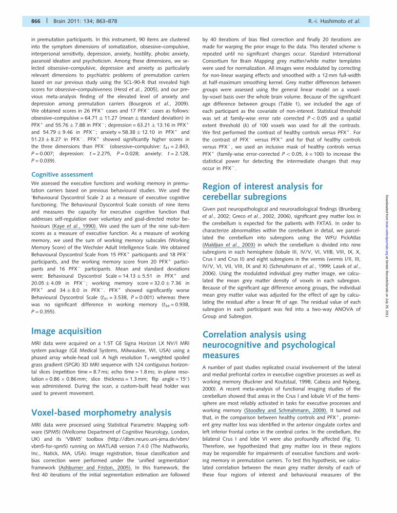
Region of interest analysis forcerebellar subregionsGiven past neuropathological and neuroradiological findings (Brunberg
et al., 2002; Greco et al., 2002, 2006), significant grey matter loss in
the cerebellum is expected for the patients with FXTAS. In order to
characterize abnormalities within the cerebellum in detail, we parcel-
lated the cerebellum into subregions using the WFU PickAtlas
(Maldjian et al., 2003) in which the cerebellum is divided into nine
subregions in each hemisphere (lobule III, IV/V, VI, VIIB, VIII, IX, X,
Crus I and Crus II) and eight subregions in the vermis (vermis I/II, III,
IV/V, VI, VII, VIII, IX and X) (Schmahmann et al., 1999; Lasek et al.,
2006). Using the modulated individual grey matter image, we calcu-
lated the mean grey matter density of voxels in each subregion.
Because of the significant age difference among groups, the individual
mean grey matter value was adjusted for the effect of age by calcu-
lating the residual after a linear fit of age. The residual value of each
subregion in each participant was fed into a two-way ANOVA of
Group and Subregion.
Correlation analysis usingneurocognitive and psychologicalmeasuresA number of past studies replicated crucial involvement of the lateral
and medial prefrontal cortex in executive cognitive processes as well as
working memory (Buckner and Koutstaal, 1998; Cabeza and Nyberg,
2000). A recent meta-analysis of functional imaging studies of the
cerebellum showed that areas in the Crus I and lobule VI of the hemi-
sphere are most reliably activated in tasks for executive processes and
working memory (Stoodley and Schmahmann, 2009). It turned out
that, in the comparison between healthy controls and PFX + , promin-
ent grey matter loss was identified in the anterior cingulate cortex and
left inferior frontal cortex in the cerebral cortex. In the cerebellum, the
bilateral Crus I and lobe VI were also profoundly affected (Fig. 1).
Therefore, we hypothesized that grey matter loss in these regions
may be responsible for impairments of executive functions and work-
ing memory in premutation carriers. To test this hypothesis, we calcu-
lated correlation between the mean grey matter density of each of
these four regions of interest and behavioural measures of the
866 | Brain 2011: 134; 863–878 R.-i. Hashimoto et al.
at Serials R
ecordsSerials on July 29, 2011
brain.oxfordjournals.orgD
ownloaded from

Behavioural Dyscontrol Scale and the working memory score. To focus
on parts of regions of interest where significant atrophy in PFX + was
identified, voxels in each region of interest was defined by the com-
bination of two binary masks: (i) the contrast map of healthy controls
versus PFX + (family-wise error-corrected P5 0.05, k = 100) and
(ii) the WFU PickAtlas (Maldjian et al., 2003). The mean grey
matter density of each region of interest was calculated from
all voxels in the individual modulated grey matter image that
satisfied both binary masks. The WFU PickAtlas mask of each
region of interest was generated as follows: left inferior frontal
cortex = ‘Frontal_Inf_Oper_L’ + ‘Frontal_Inf_Tri_L’; anterior cingulate
cortex = ‘Cingulum_Ant_L’ + ‘Cingulum_Ant_R’; left (right) Crus I
and lobule VI = ‘Cerebelum_Crus1_L(R)’ + ‘Cerebellum_6_L(R)’. The
individual mean grey matter value was then adjusted for the effect
of age by calculating the residual value after a linear fit of age. We
used the residual for the calculation of correlation with either
Behavioural Dyscontrol Scale or working memory score. To adjust
for the multiple statistical tests for each scale (four regions of inter-
est/statistical tests per scale), the Benjamini-Hochberg method was
implemented, with the false discovery rate set at 5% (Benjamini and
Hochberg, 1995).
There has been evidence that anxiety-related symptoms (e.g.
obsessive–compulsive disorder and general anxiety) involve abnormal-
ities in the amygdala, insula, anterior cingulate cortex and orbitofrontal
cortex (Paulus and Stein, 2006; Etkin and Wager, 2007; Chamberlain
et al., 2008). Past studies indicated that neural correlates of depression
involve the hippocampus, in addition to the amygdala and anterior
cingulate cortex (Soares and Mann, 1997; Sheline, 2000). In the cere-
bellum, the aforementioned review study indicated that vermal lobule
VII is involved in emotional processing by forming the cerebellar-limbic
circuitry (Stoodley and Schmahmann, 2009). Because the contrast of
healthy controls versus PFX + revealed prominent grey matter loss in
these regions except for the right amygdala and the right hippocam-
pus (Fig. 1), we performed correlation analyses between the mean
intensity of each of these regions of interest and subscales of the
SCL-90-R (obsessive–compulsive, depression and anxiety). We ex-
tracted the mean intensity of each region of interest using the contrast
map of healthy controls versus PFX + and the WFU PickAtlas in the
Figure 1 Significant grey matter reduction of FXTAS (PFX + ) identified by comparison either with healthy controls (HC) or with unaffected
premutation carriers (PFX�) in sagittal views. The contrast of healthy control versus PFX+ is shown in the yellow-red colour scale and the
one of PFX� versus PFX+ is shown in the deep-light blue scale. The numbers at the bottom left corner indicate the x-axis coordinates of
sagittal sections. The statistical threshold was set at P50.05 (family-wise error rate-corrected). The spatial extent threshold was set at
100 voxels.
Grey matter abnormalities in FXTAS Brain 2011: 134; 863–878 | 867
at Serials R
ecordsSerials on July 29, 2011
brain.oxfordjournals.orgD
ownloaded from

same way as was done in the correlation analysis of executive function
and working memory. The WFU PickAtlas mask of each region of
interest was generated as follows: left amygdala = ‘Amygadala_L’;
anterior cingulate cortex = ‘Cingulum_Ant_L’ + ‘Cingulum_Ant_R’;
left (right) insula = ‘Insula_L(R)’; left (right) orbitofrontal cortex =
‘Frontal_Mid_Orb_L(R)’ + ‘Frontal_Inf_Orb_L(R)’; the vermal lobule
VII = ‘Vermis_7’. Correlation with obsessive–compulsiveness and anx-
iety was tested for the left amygdala, anterior cingulate cortex, left
and right insula, left and right orbitofrontal cortex, and vermal lobule
VII. Correlation with depression was tested for the left amygdala, left
hippocampus, anterior cingulate cortex and the vermal lobule VII. The
Benjamini-Hochberg method was used for the adjustment of multiple
statistical tests for each subscale in the SCL-90-R (seven tests for
obsessive–compulsive and anxiety, and four tests for depression).
Regression analysis using FMR1molecular variables and fragileX-associated tremor/ataxia syndromeseverity scaleIn order to examine effects of FMR1 molecular variables on grey
matter, we performed a voxel-based simple regression analysis over
the whole brain using either the CGG repeat size or FMR1 messenger
RNA level. We included the age of each participant as the ‘nuisance’
covariate in the model. Because there is a systematic difference in the
distribution of both molecular variables between healthy controls and
the two premutation groups (Table 1), we included the data of the
two premutation groups only for this analysis in order to avoid con-
tamination of group (categorical) effects.We also performed the simple
regression analysis using the clinical scale for assessment of the FXTAS
severity to identify areas showing progressive grey matter reduction
caused by the development of FXTAS. We included age as the cov-
ariate of non-interest to isolate the effect of clinical severity from the
one of age. For this analysis, we used the data from premutation
participants whose FXTAS score was 51. We used the contrast of
healthy controls versus PFX + (family-wise error-corrected P5 0.05,
k = 100) as the inclusive mask in the three simple regression analyses.
Results
Group difference in the whole-brainanalysisFor the comparison between healthy controls and PFX+ , we found
clusters of significant grey matter reduction of PFX + in multiple
brain regions over the cerebrum, cerebellum and subcortical struc-
tures (Fig. 1 and Table 2). Particularly prominent grey matter loss
was observed in the cerebellum, dorsomedial frontal and parietal
regions, orbitofrontal regions, insula, medial temporal regions and
lateral prefrontal regions. Significant grey matter increase in FXTAS
was mainly found in the bilateral posterior superior/middle tem-
poral gyrus (Table 2). In the comparison between PFX + and PFX�,
we found significant grey matter reduction of PFX+ relative to
PFX� in parts of the cerebellum, dorsomedial prefrontal cortex,
and precuneus (Fig. 1 and Table 2). There were no significant
voxels showing PFX + 4PFX�. No significant voxels were
identified in the comparison between healthy controls and PFX�
in either direction.
Region of interest analyses forcerebellar subregionsA two-way ANOVA (Group � Subregion) for the 26 cerebellar
subregions revealed a significant main effect of Group [F(2,
80) = 9.427, P5 0.001] and interaction effect [F(50,
2000) = 2.918, P50.001]. Follow-up one-way ANOVA was per-
formed for each subregion using the Benjamini-Hochberg method
for the adjustment for the multiple tests. We did not observe
significant effects of Group in lobules VIII and IX in the vermis,
bilateral lobule VIII nor right lobule X in the hemisphere. All other
subregions showed a significant main effect of Group (P50.05;
Table 3). According to a post hoc test (Tukey’s Honestly
Significant Difference), there was a significant difference between
healthy controls and PFX+ in all of the regions of interest
(Table 3). Compared with healthy controls, PFX� showed signifi-
cant reduction in lobule I/II of the vermis and in lobule III in the
left hemisphere (P50.05; Fig. 2 and Table 3). Significant differ-
ences between PFX +and PFX� were found in lobules IV/V, VI and
VII in the vermis, and lobules IV/V, VI, Crus I and right Crus II in
the hemisphere (P50.05; Fig. 2 and Table 3).
Region of interest-based correlationanalysis using neurocognitive andpsychological measuresIn correlation analysis with Behavioural Dyscontrol Scale, none of
the four regions of interest for executive function reached signifi-
cance using the threshold corrected for multiple comparisons (left
inferior frontal cortex: r = 0.392, P = 0.0968; anterior cingulate
cortex: r = 0.300, P = 0.119; left Crus I/lobule VI: r = 0.265,
P = 0.1364; right Crus I/lobule VI: r = 0.373, P = 0.065), although
left inferior frontal cortex and the right Crus I/lobule VI showed
significant correlations at an uncorrected threshold (P = 0.024 and
P = 0.032, respectively). In correlation analysis using the working
memory score, we found significant correlations in the anterior
cingulate cortex (r = 0.498, P = 0.004) and the left inferior frontal
cortex (r = 0.518, P = 0.005) at the corrected threshold (Fig. 3).
The two cerebellar regions of interest, by contrast, showed no
significant correlation (left Crus I/lobule VI: r = 0.192, P = 0.263;
right Crus I/lobule VI: r = 0.289, P = 0.117) with Behavioural
Dyscontrol Scale.
In correlation analysis using the score of the obsessive–compulsive
symptom dimension in SCL-90-R, only the left amygdala reached
the significant level (P = 0.0126; Fig. 3). Although there were
several regions of interest for which a significant correlation was
observed at the uncorrected threshold (anterior cingulate cortex:
r = �0.358, P = 0.018; left insula: r = �0.316, P = 0.039; right
insula: r = �0.311, P = 0.042), these regions of interest did not
reach significant level after correction (anterior cingulate cortex:
P = 0.064; left insula: P = 0.091; right insula: P = 0.074). No other
regions of interest showed significant correlation (vermis lobule VII:
r = �0.205, P = 0.188; left orbitofrontal cortex: r = �0.272,
868 | Brain 2011: 134; 863–878 R.-i. Hashimoto et al.
at Serials R
ecordsSerials on July 29, 2011
brain.oxfordjournals.orgD
ownloaded from

P = 0.091; right orbitofrontal cortex: r = �0.277, P = 0.101).
In the correlation using the depression symptom dimension,
only the left amygdala was significantly correlated using the cor-
rected threshold (P = 0.019; Fig. 3). No other regions of interest
showed significant correlation after correction (anterior cingulate
cortex: r = �0.264, P = 0.116; left hippocampus: r = �0.301,
P = 0.100; vermis VII: r = �0.093, P = 0.554), although the left
hippocampus showed a marginally significant correlation at the
uncorrected threshold (P = 0.050). There was no region of inter-
est that showed a significant correlation with anxiety after
Table 2 Significant grey matter differences between groups
Region Cluster size x y z zmax
Healthy controls4 PFX +
Left and right cerebellar hemispheres, vermis, ventral medial temporal regions 53025 5 �77 �21 6.43
Cingulate cortex, dorsomedial prefrontal cortex, pre-SMA, SMA, precuneus 22729 �1 40 29 5.95
Left insula/frontal operculum 4663 �57 10 1 5.38
Left orbital frontal cortex 4228 �18 62 �19 6.78
Left and right cerebellar lobule IX 3558 �8 �45 �58 5.45
Right insula 2938 41 �6 3 5.75
Thalamus 2076 �2 �19 13 5.69
Left inferior frontal cortex 1876 �51 10 29 5.57
Right orbital frontal cortex 1656 21 65 �16 6.26
Left dorsolateral prefrontal cortex 1213 �33 54 23 4.98
Right superior frontal cortex 743 31 11 62 5.14
686 23 37 51 5.35
Bilateral gyrus rectus 559 3 29 �29 4.80
Left superior parietal cortex 553 �28 �77 47 5.22
Right superior parietal cortex 341 35 �69 53 4.77
Right premotor cortex 200 59 �5 39 5.08
Left angular gyrus 190 �55 �60 41 4.98
Right angular gyrus 173 62 �58 37 4.75
Left superior frontal cortex 159 �32 4 60 4.81
Left post-central gyrus 148 �61 �24 41 4.73
PFX + 4Healthy controls
Left superior/middle temporal gyrus 3674 �46 �50 0 5.92
275 �48 �9 �22 4.78
Right middle temporal gyrus 1560 52 �47 �12 5.40
239 49 �50 10 4.77
Left lateral occipital gyrus 204 �29 �85 �2 4.74
135 �33 �73 14 4.74
Left medial orbital gyrus 185 �20 26 �11 4.71
Right middle frontal gyrus 101 30 42 12 4.72
PFX�4 PFX +
Cerebellar lobules V/VI/VII 5072 5 �78 �22 5.27
Right cerebellar hemisphere VI 1613 30 �23 �36 5.10
Dorsal anterior cingulate/paracingulate 1471 6 30 40 4.65
Left cerebellar hemisphere VIIB/Crus I 841 �48 �53 �47 4.41
Thalamus 625 �4 �18 12 4.30
Right precuneus 355 6 �60 60 4.66
Right dorsomedial prefrontal cortex 257 16 51 42 4.54
Left cerebellar hemisphere Crus I 222 �22 �90 �27 4.20
Left frontal pole 191 �15 69 6 4.12
Right superior frontal cortex 174 33 47 33 4.51
Left premotor cortex 168 �52 6 40 4.34
Left superior frontal cortex 146 �34 4 59 4.61
Left posterior cingulate cortex 135 �5 �31 47 4.08
PFX + 4 PFX�
No significant clusters
Healthy controls4 PFX�
No significant clusters
PFX�4Healthy controls
No significant clusters
SMA = supplementary motor area.
Grey matter abnormalities in FXTAS Brain 2011: 134; 863–878 | 869
at Serials R
ecordsSerials on July 29, 2011
brain.oxfordjournals.orgD
ownloaded from

correction (left amygdala: r = �0.252, P = 0.717; anterior cingu-
late cortex: r = �0.241, P = 0.421; left insula: r = �0.217,
P = 0.284; right insula: r = �0.212, P = 0.242; left orbitofrontal
cortex: r = �0.222, P = 0.355; right orbitofrontal cortex:
r = �0.208, P = 0.210; vermis VII: r = �0.154, P = 0.324).
Whole brain regression analysesusing FMR1 molecular variables andclinical scale of fragile X-associatedtremor/ataxia syndromeWe performed regression analyses using CGG repeat size and
FMR1 messenger RNA. For the analysis involving CGG repeat
size, we found significant negative effects of CGG repeat size in
several clusters in the dorsal medial regions including the supple-
mentary motor area and the dorsomedial prefrontal cortex (Fig. 4
and Table 4). No voxels showed a significant positive effect of
CGG repeat size. We did not observe a significant correlation
with FMR1 messenger RNA in either direction. In the analysis
using the clinical scales of the FXTAS severity, we found significant
negative correlations in cerebellar lobule VI/VII and the orbitofron-
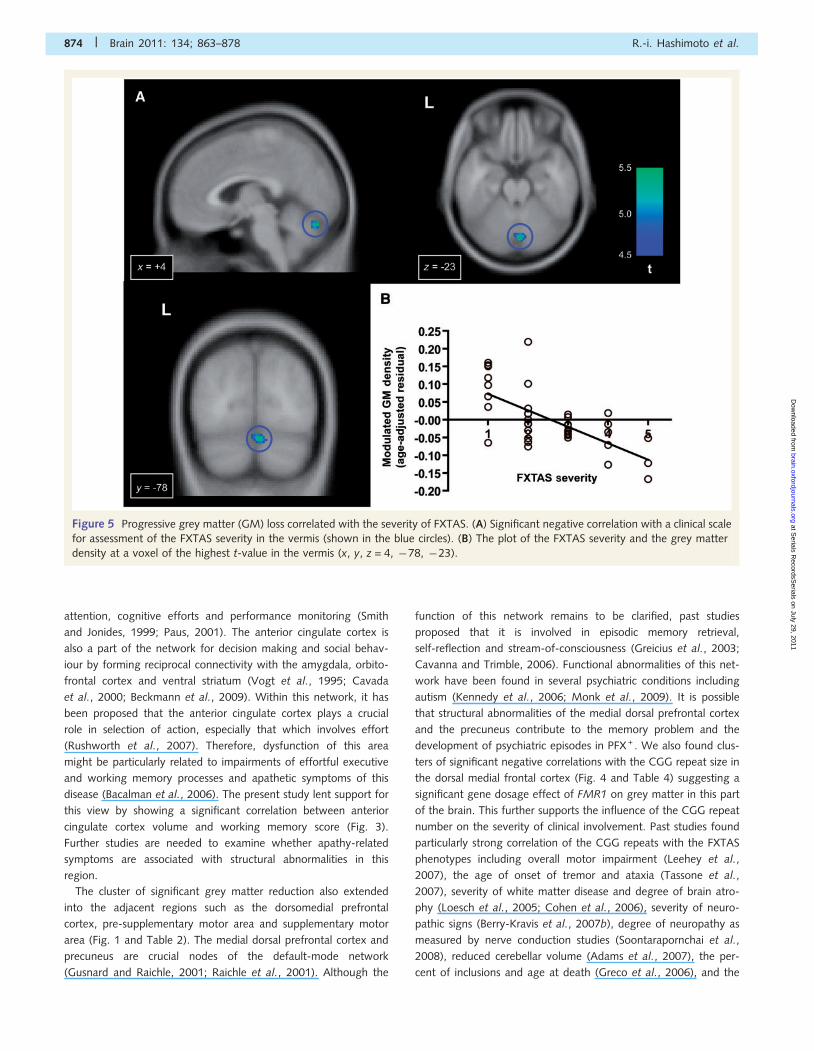
tal cortex (Fig. 5 and Table 4). No significant positive correlations
were observed.
DiscussionBrain abnormalities in PFX+ has been demonstrated in previous
MRI volumetric studies based on gross anatomical parcellation as
Table 3 Group comparisons of age-adjusted grey matter density in the cerebellar subregions
Region Healthy controls PFX + PFX� F-test
Vermis
Lobule I/II*,*** 3.48 � 1.10 �2.91 � 1.19 0.59 � 1.08 F = 8.42, P5 0.001
Lobule III* 3.32 � 1.04 �3.26 � 1.07 0.09 � 1.08 F = 10.09, P5 0.001
Lobule IV/V*,** 3.09 � 1.04 �3.76 � 1.09 0.87 � 0.91 F = 12.19, P5 0.001
Lobule VI*,** 4.93 � 1.13 �5.66 � 1.25 1.18 � 1.27 F = 20.66, P5 0.001
Lobule VII*,** 3.77 � 1.12 �4.93 � 1.31 1.45 � 1.14 F = 14.74, P5 0.001
Lobule VIII 2.16 � 1.16 �2.37 � 1.61 0.04 � 1.35 F = 2.72, P = 0.078
Lobule IX 2.71 � 1.41 �2.78 � 1.88 �0.35 � 1.63 F = 2.83, P = 0.077
Lobule X* 2.50 � 1.12 �2.31 � 1.34 �0.36 � 1.08 F = 4.22, P = 0.018
Hemisphere
Lobule III
L*,*** 3.54 � 1.04 �3.04 � 1.12 �0.46 � 1.13 F = 9.54, P5 0.001
R* 2.90 � 0.92 �2.87 � 0.98 0.01 � 0.84 F = 10.22, P5 0.001
Lobule IV/V
L*,** 3.60 � 0.95 �3.69 � 1.16 0.18 � 1.14 F = 11.86, P5 0.001
R*,** 2.83 � 1.03 �3.97 � 1.31 1.26 � 0.97 F = 10.28, P5 0.001
Lobule VI
L*,** 3.32 � 0.96 �3.90 � 1.33 0.66 � 1.18 F = 10.11, P5 0.001
R*,** 2.46 � 0.92 �3.95 � 1.27 1.55 � 0.95 F = 10.75, P5 0.001
Crus I
L*,** 3.15 � 0.81 �3.82 � 1.02 0.74 � 0.99 F = 14.76, P5 0.001
R*,** 2.64 � 0.84 �3.61 � 1.01 0.96 � 0.88 F = 12.98, P5 0.001
Crus II
L* 2.70 � 0.82 �3.09 � 1.22 0.30 � 1.15 F = 7.66, P5 0.001
R*,** 2.50 � 0.79 �2.98 � 0.95 0.66 � 0.91 F = 10.18, P5 0.001
Lobule VIIB
L* 2.22 � 0.94 �2.59 � 1.46 0.20 � 1.26 F = 3.91, P = 0.029
R* 2.23 � 0.96 �2.77 � 1.24 0.48 � 1.04 F = 5.61, P5 0.001
Lobule VIII
L 1.62 � 1.02 �1.61 � 1.68 �0.31 � 1.37 F = 1.40, P = 0.254
R 1.42 � 1.06 �1.91 � 1.44 0.26 � 1.05 F = 1.99, P = 0.148
Lobule IX
L* 3.04 � 1.28 �2.62 � 1.58 �0.65 � 1.46 F = 4.06, P = 0.027
R* 3.08 � 1.21 �2.68 � 1.65 �0.79 � 1.21 F = 4.51, P = 0.020
Lobule X
L* 1.19 � 0.52 �0.81 � 0.70 �0.38 � 0.70 F = 4.51, P = 0.021
R 0.99 � 0.63 �1.14 � 0.65 0.22 � 0.70 F = 2.79, P = 0.076
Age-adjusted grey matter density represents the residual from a linear regression of age over the modulated grey matter density extracted from each subregion. *Significantdifference between controls and PFX + by a post hoc test (Tukey HSD). **Significant difference between PFX� and PFX+ . ***Significant difference between healthycontrols and PFX�. L = left; R = right.
870 | Brain 2011: 134; 863–878 R.-i. Hashimoto et al.
at Serials R
ecordsSerials on July 29, 2011
brain.oxfordjournals.orgD
ownloaded from
well as clinical MRI investigations (Brunberg et al., 2002; Cohen
et al., 2006; Adams et al., 2007, 2010). However, systematic in-
vestigations of possible regionally selective abnormalities have not
been performed. To our knowledge, this study represents the first
demonstration of a spatial pattern of grey matter reduction in the
PFX + brain using voxel-based morphometry. We identified signifi-
cant grey matter reduction in multiple regions, particularly in the
cerebellum, the dorsomedial frontal-parietal regions, the medial
temporal regions and the insula. Correlation analysis using the
behavioural measurements of the premutation groups indicated
that psychological symptoms and working memory deficits of
FMR1 premutation carriers are associated with grey matter loss
in the left amygdala and in the left inferior frontal cortex and
anterior cingulate cortex, respectively. Regression analyses using
FMR1 molecular variables showed a significant contribution of
CGG repeat size to the grey matter reduction in the dorsomedial
frontal regions. Furthermore, progressive grey matter loss corre-
lated with the severity of FXTAS symptomatology was also re-
vealed in a part of the cerebellum and in the orbitofrontal
cortex. These findings identify the pattern of anatomical abnorm-
alities in FMR1 premutation carriers that might provide morpho-
logical bases for behavioural problems of this population.
Consistent with the past observations, both the whole-brain
voxel-based morphometry analysis and the region of interest
analyses revealed profound grey matter loss in the cerebellum of
PFX + . Significant grey matter reduction was widespread, affecting
almost the entire cerebellum except for some subregions in the
inferior posterior lobe such as the lobule VIII. In particular, grey
matter reductions in the anterior subregions of the vermis and
hemispheres were highly significant (Fig. 2). The anterior vermis
has been shown to be critical for the regulation of the postural
equilibrium while standing (Diener et al., 1989; Ouchi et al.,
1999). Severe atrophy in this region of the cerebellum therefore
may be directly responsible for gait ataxia, one of the core clinical
symptoms of PFX + . We suggest that analysis using behavioural
scales for the severity of ataxia, such as the International
Cooperative Ataxia Rating Scale, would be necessary to test this
hypothesis. It is important to note that significant grey matter
reduction was identified in several subregions in the anterior
vermis and hemisphere even among PFX� (Table 3). This obser-
vation raises the possibility that degeneration in this region may be
the initial pathological process before clinical signs of FXTAS.
Significant grey matter loss was also found in several subregions
of the posterior vermis. Previous neuropsychological studies
described cerebellar cognitive affect syndrome resulted from
damage to the posterior lobe (Schmahmann and Sherman,
1998). A recent meta-analysis of functional imaging studies indi-
cated that areas around the vermis lobule VII are recruited with
Figure 2 Region of interest analyses of the cerebellar subregions. (A) Sagittal views showing cerebellar subregions as determined by the
WFU PickAtlas (Maldjian et al., 2003). In the most medial section (x = 0), the eight subregions in the vermis are shown (lobules I/II, III, IV/
V, VI, VII, VIII, IX and X). The nine subregions in the hemisphere (lobules III, IV/V, VI, VIIB, VIII, IX, X, Crus I and Crus II) are shown in the
lateral sections (x = �10 and x = �20). (B) Comparisons of grey matter (GM) density among the three groups in selected cerebellar
subregions. Significant difference between two groups is shown by an asterisk. The y-axis represents the residual from a linear regression
of age over the mean modulated grey matter density extracted from each subregion.
Grey matter abnormalities in FXTAS Brain 2011: 134; 863–878 | 871
at Serials R
ecordsSerials on July 29, 2011
brain.oxfordjournals.orgD
ownloaded from

high probability in tasks of emotional processing (Stoodley and
Schmahmann, 2009). Although these findings suggest that signifi-
cant abnormality of this subregion may be related to psychiatric
problems in FXTAS, this possibility was not supported by our cor-
relation analysis that failed to show significant association between
the level of depression, anxiety or obsessive–compulsiveness in
SCL-90-R and grey matter reduction in this region.
In the cerebellar hemispheres, large grey matter reductions in
PFX + were observed in parts of the neocerebellum, including Crus
I, Crus II and lobule VI. According to the aforementioned
meta-analysis of cerebellar activation, these neocerebellar regions,
particularly the boundary between Crus I and lobule VI, are fre-
quently activated in executive function and working memory tasks
(Stoodley and Schmahmann, 2009), which suggests that abnorm-
alities in these cerebellar subregions may contribute to impaired
executive and working memory processes in PFX + . However, the
present correlation analysis using the Behavioural Dyscontrol Scale
and the working memory score did not support this possibility
except for the trend-level correlation between the right Crus I/
lobule VI volume and the Behavioural Dyscontrol Scale score.
Because executive functions comprise various cognitive processes,
it is necessary to examine possible relevance with other behaviour-
al tests of executive functions. Compared with these subregions,
grey matter reductions in several posterior subregions were less
pronounced. In particular, we did not find evidence of significant
abnormality of the hemisphere lobule VIII, a subregion that has
been shown to be most severely impaired in spinocerebellar ataxia
17 (SCA17), another neurodegenerative disorder caused by a
single gene mutation (Lasek et al., 2006). This dissociation is inter-
esting given the phenotypic overlap between this disease and
PFX + . Among the posterior subregions, the lobule IX showed
significant grey matter loss. This region has been suggested to
form the cerebellar node of the ‘default-mode’ network (Habas
et al., 2009). Grey matter loss in this region may thus be related
to highly significant abnormalities of cortical nodes of this net-
work, such as the dorsomedial prefrontal cortex and precuneus
(Fig. 1).
The comparison between the healthy controls and PFX + re-
vealed profound grey matter atrophy in several cortical and sub-
cortical regions outside of the cerebellum. In particular, a cluster of
significant reduction was identified over the extended areas in the
medial surface of the brain between the frontal and parietal re-
gions (Fig. 1). This huge cluster comprises multiple regions includ-
ing the dorsal anterior cingulate/paracingulate cortex, dorsomedial
prefrontal cortex, supplementary motor area, middle and posterior
paracingulate cortex and precuneus (Table 2). The most prominent
grey matter loss was centred in the dorsal anterior cingulate
cortex. Previous functional imaging and brain lesion studies have
shown that the anterior cingulate cortex is implicated in both cog-
nitive and emotional processing (Bush et al., 2000). In the cogni-
tive domain, activation of this area has been found under
executive cognitive and working memory tasks that require
Figure 3 Significant association of grey matter (GM) density in regions of interest with cognitive and psychological scales in premutation
carriers. (A and B) Relations between grey matter density in the left amygdala and self-reported psychological symptoms of obsessive–
compulsive (A) and depression (B) on the SCL-90-R. (C and D) Relations between the sum of the working memory subscales in Wechsler
Adult Intelligence Scale (Third Edition) and the grey matter density in the anterior cingulate cortex (C) and left inferior frontal cortex (D).
Correlation coefficient and P-value (uncorrected) are noted. The correlations were found to be significant after correction for multiple
comparisons. L = left; R = right.
872 | Brain 2011: 134; 863–878 R.-i. Hashimoto et al.
at Serials R
ecordsSerials on July 29, 2011
brain.oxfordjournals.orgD
ownloaded from

Figure 4 Significant negative effect of the CGG repeat size on the grey matter (GM) density in the FMR1 premutation carriers.
(A) Clusters of voxels in the dorsal medial regions showing significant negative correlation with the CGG repeat size. The blue circles
indicate the largest cluster in the supplementary motor area in the sagittal, coronal and axial views. (B) The plot of CGG repeat size and the
grey matter density at a voxel of the highest t-value in the supplementary motor area (x, y, z = 5, �6, 59). The y-axis represents the
residual from a linear regression of age over the modulated grey matter density at this coordinate.
Table 4 Regression analysis using CGG repeat size and FXTAS severity scale
Region Size x y z zmax
CGG repeat size
Supplementary motor area 283 5 �6 59 4.26
�3 �5 55 4.20
Mammillary body/anterior ventral hypothalamus 282 3 �5 �11 4.23
Right orbitofrontal cortex 165 16 69 �9 4.33
Left dorsomedial prefrontal cortex 144 �4 28 48 4.25
�4 49 30 4.11
�4 40 41 4.09
Right cerebellar hemisphere lobule IX 142 �13 �42 �54 4.35
Right anterior cingulate/paracingulate cortex 109 5 50 25 4.28
FXTAS severity scale
Right cerebellar vermis lobule VI/VII 639 4 �78 �23 4.66
Right orbitofrontal cortex 398 16 69 �11 4.63
Grey matter abnormalities in FXTAS Brain 2011: 134; 863–878 | 873
at Serials R
ecordsSerials on July 29, 2011
brain.oxfordjournals.orgD
ownloaded from
attention, cognitive efforts and performance monitoring (Smith
and Jonides, 1999; Paus, 2001). The anterior cingulate cortex is
also a part of the network for decision making and social behav-
iour by forming reciprocal connectivity with the amygdala, orbito-
frontal cortex and ventral striatum (Vogt et al., 1995; Cavada
et al., 2000; Beckmann et al., 2009). Within this network, it has
been proposed that the anterior cingulate cortex plays a crucial
role in selection of action, especially that which involves effort
(Rushworth et al., 2007). Therefore, dysfunction of this area
might be particularly related to impairments of effortful executive
and working memory processes and apathetic symptoms of this
disease (Bacalman et al., 2006). The present study lent support for
this view by showing a significant correlation between anterior
cingulate cortex volume and working memory score (Fig. 3).
Further studies are needed to examine whether apathy-related
symptoms are associated with structural abnormalities in this
region.
The cluster of significant grey matter reduction also extended
into the adjacent regions such as the dorsomedial prefrontal
cortex, pre-supplementary motor area and supplementary motor
area (Fig. 1 and Table 2). The medial dorsal prefrontal cortex and
precuneus are crucial nodes of the default-mode network
(Gusnard and Raichle, 2001; Raichle et al., 2001). Although the
function of this network remains to be clarified, past studies
proposed that it is involved in episodic memory retrieval,
self-reflection and stream-of-consciousness (Greicius et al., 2003;
Cavanna and Trimble, 2006). Functional abnormalities of this net-
work have been found in several psychiatric conditions including
autism (Kennedy et al., 2006; Monk et al., 2009). It is possible
that structural abnormalities of the medial dorsal prefrontal cortex
and the precuneus contribute to the memory problem and the
development of psychiatric episodes in PFX + . We also found clus-
ters of significant negative correlations with the CGG repeat size in
the dorsal medial frontal cortex (Fig. 4 and Table 4) suggesting a
significant gene dosage effect of FMR1 on grey matter in this part
of the brain. This further supports the influence of the CGG repeat
number on the severity of clinical involvement. Past studies found
particularly strong correlation of the CGG repeats with the FXTAS
phenotypes including overall motor impairment (Leehey et al.,
2007), the age of onset of tremor and ataxia (Tassone et al.,
2007), severity of white matter disease and degree of brain atro-
phy (Loesch et al., 2005; Cohen et al., 2006), severity of neuro-
pathic signs (Berry-Kravis et al., 2007b), degree of neuropathy as
measured by nerve conduction studies (Soontarapornchai et al.,
2008), reduced cerebellar volume (Adams et al., 2007), the per-
cent of inclusions and age at death (Greco et al., 2006), and the
Figure 5 Progressive grey matter (GM) loss correlated with the severity of FXTAS. (A) Significant negative correlation with a clinical scale
for assessment of the FXTAS severity in the vermis (shown in the blue circles). (B) The plot of the FXTAS severity and the grey matter
density at a voxel of the highest t-value in the vermis (x, y, z = 4, �78, �23).
874 | Brain 2011: 134; 863–878 R.-i. Hashimoto et al.
at Serials R
ecordsSerials on July 29, 2011
brain.oxfordjournals.orgD
ownloaded from

amplitude of electrophysiological response to word processing
(Olichney et al., 2010).
Significant grey matter loss was also observed in the bilateral
orbitofrontal cortex (Fig. 1 and Table 2). Past neuropsychological
studies have shown that patients with cortical damage to this area
display impulsive behaviours (Bechara et al., 2000). A recent func-
tional MRI study of patients with obsessive–compulsive disorder
and their unaffected close relatives proposed that abnormality
of the orbitofrontal cortex may be endophenotype of obsessive–
compulsive disorder (Chamberlain et al., 2008). Further, a recent
review proposed that this area is important for subjective pleas-
antness/hedonic processing (Kringelbach, 2005). Given these pro-
posals, it is tempting to associate grey matter reduction of this
structure with psychiatric symptoms of disinhibition, obsessive–
compulsiveness and impassivity/apathy of PFX+ (Hessl et al.,
2005; Bacalman et al., 2006; Bourgeois et al., 2009). However,
we did not find significant correlation of the grey matter volume
and the level of obsessive–compulsiveness. It remains to be tested
whether there is any association with the severity of disinhibition
and apathy. We note that, in the voxel-based morphometry ana-
lysis, this area is known to be particularly sensitive to misregistra-
tion and normalization error. Therefore, we suggest that the
structural abnormalities in the orbitofrontal cortex need to be
replicated using different methodologies such as manual tracing
(Nakamura et al., 2008) and cortical thickness measurements
(Kuperberg et al., 2003).
Grey matter loss in PFX + was clearly observed in the bilateral
insula. A classic review described multiple functional roles of insu-
lar cortex including the sensory, motor and cognitive domains
(Augustine, 1996). In our analysis, foci of grey matter loss were
located in the posterior part, although signs of the milder grey
matter loss in the anterior part were also observed when a more
liberal statistical threshold was used (false discovery rate corrected
P5 0.05, not reported). Recent functional anatomical studies of
both primates and humans highlighted the insula’s role in inter-
oception, the sense of physiological condition of the body includ-
ing the visceral, hunger, pain and thermal sensations (Craig,
2003a, b). It has been proposed that altered interoception and
anxiety are linked and that individuals prone to anxiety are asso-
ciated with exaggerated interoceptive prediction signals generated
in the anterior insula (Paulus and Stein, 2006). Because the pos-
terior insula provides direct inputs to its anterior part, structural
abnormalities of the posterior insula may adversely affect the func-
tion of the anterior insula, which contributes to anxiety-related
symptoms of PFX + and PFX�. In the present correlation analysis,
although the correlation between level of obsessive–compulsive-
ness and reduced insula volume was significant bilaterally at an
uncorrected threshold, it did not reach the significant level after
correction. More observations are needed to test the possible
association between the grey matter loss in the insula and
anxiety-related symptoms of the premutation carriers.
Significant grey matter loss of PFX + was also identified in the
medial temporal lobe structures including parts of the fusiform
gyrus and parahippocampus. Abnormalities were more extended
in the left hemisphere, involving the hippocampus and amygdala.
Although a number of studies indicated the pathological processes
of depression in the hippocampus (Soares and Mann, 1997;
Sheline, 2000), correlation between left hippocampal grey matter
density and the severity of depressive symptom was marginally
significant only when an uncorrected threshold was used.
Previous neuropsychological and functional imaging studies have
shown the critical role of the left hippocampus in verbal memory
(Milner, 1972; Strange et al., 1999). Structural abnormalities of
this area may therefore underlie impaired declarative verbal
memory in PFX+ (Grigsby et al., 2008), although this possibility
needs to be examined more directly by analysis using behavioural
measures of declarative memory. In contrast, we observed a clear
negative correlation between the left amygdala volume and ele-
vated levels of obsessive–compulsiveness and depression. Together
with our previous functional MRI study that found functional
abnormalities in the amygdala in PFX� (Hessl et al., 2007), this
finding further supports the view that abnormalities in the amyg-
dala play crucial roles in psychological symptoms of FMR1 premu-
tation carriers. It is interesting that the left amygdala underlies
both depression and an anxiety-related symptom, given a high
comorbidity between depression and anxiety disorders (Kessler
et al., 2003). Our result is consistent with previous findings of
pathological processes affecting structure and function of the
amygdala in patients with the obsessive–compulsive disorder
(Szeszko et al., 1999; Menzies et al., 2008) and in those with
depression (Sheline, 2000).
As predicted from neuropsychological observations, significant
clusters of grey matter loss of the patient group were found in
multiple areas for executive cognitive functions and working
memory, including the inferior frontal cortex, dorsolateral pre-
frontal cortex and superior parietal cortex (Figs 1 and 3;
Table 2). Significant correlations between left inferior frontal
cortex volume and working memory scores indicates that grey
matter loss in this region, together with that in the anterior cin-
gulate cortex, contributes to working memory deficits in PFX+ and
in PFX� (Grigsby et al., 2008; Cornish et al., 2009) (Fig. 3). A
previous behavioural study demonstrated significant impairment in
response inhibition in both affected and unaffected premutation
carriers (Cornish et al., 2008). Although past brain lesion and
functional imaging studies replicated the pivotal roles of the
right inferior frontal cortex in response inhibition (Konishi et al.,
1999; Aron et al., 2004), we did not observe significant voxels
with grey matter reduction of PFX+ in this region. It still remains
possible that abnormalities of this region may be more apparent in
the functional measures rather than the structural ones.
Furthermore, response inhibition is subserved by connectivity be-
tween the right inferior frontal cortex and other brain regions
including the striatum (Aron and Poldrack, 2006). It would be
interesting to examine in the future studies the structural and
functional connectivity between the right inferior frontal cortex
and other regions.
Although extensive grey matter atrophy was identified in PFX + ,
neuropsychological studies have widely reported cognitive func-
tions that remain preserved or only mildly impaired (Cornish
et al., 2008; Grigsby et al., 2008). In particular, language impair-
ments have not been reported for any of the 16 subscales for
‘language’ and ‘verbal reasoning and comprehension’ (Grigsby
et al., 2008). In the voxel-based analysis, however, a rather sig-
nificant increase of grey matter density for the patient group was
Grey matter abnormalities in FXTAS Brain 2011: 134; 863–878 | 875
at Serials R
ecordsSerials on July 29, 2011
brain.oxfordjournals.orgD
ownloaded from

identified in the bilateral posterior part of superior/middle tem-
poral gyrus. Although this observation has several possible explan-
ations, the structural increase may play compensatory roles for
maintaining language functions despite neurodegenerative pro-
cesses affecting other language-related regions including the left
inferior frontal cortex (Figs 1 and 3). Visuospatial functions and
visual attention were suggested to be only mildly impaired in
PFX + (Grigsby et al., 2008). The present voxel-based analysis re-
vealed significant grey matter reduction in some regions of the
visuospatial and dorsal visual systems, such as the right superior
parietal cortex, whereas the other regions of these systems, includ-
ing the right superior occipital gyrus, did not have voxels with
significant grey matter reduction. It is possible that there is a func-
tional change in intact regions that compensates for structural
degeneration affecting other parts of the systems.
In addition to atrophy affecting grey matter, importance of
pathological changes in the white matter has been increasingly
recognized in many neurological and psychiatric conditions
(Kanaan et al., 2005; Stebbins and Murphy, 2009). Indeed, hyper-
intensities in the periventricular and cerebellar zones have been
recognized as one of the hallmark neuroradiological features of
the FXTAS brain (Brunberg et al., 2002). Because the present ana-
lyses focused on alternations of grey matter, it remains un-
answered how the white matter abnormalities are related to the
FXTAS symptomatology and FMR1 molecular variables. The use of
the MRI sequences optimal for the white matter pathology, such
as the diffusion tensor imaging and the fluid-attenuated inversion
recovery sequence, would complement our present findings of the
grey matter abnormalities for better understanding of brain
abnormalities in PFX+ .
To conclude, the current voxel-based morphometry study re-
vealed the pattern of regional grey matter abnormalities of
PFX + over the whole brain that may underlie its behavioural prob-
lems in the motor, cognitive and psychiatric domains. Findings of
group comparisons together with regression analyses using FMR1
genetic molecular variables and a clinical severity scale provide
foundations at the systems-level for bridging the gap between
genetic molecular pathological processes and clinical behavioural
observations.
FundingThe National Institute of Health grants (UL1DE019583, DA024854
and HD036071); National Institute on Neurological Disorders and
Stroke grant (RL1NS062412); National Institute on Ageing grants
(RL1AG032119 and RL1AG032115); National Centre for Research
Resources (UL1 RR024146); Roche, Novartis, Seaside
Therapeutics, Forest, Johnson and Johnson, and Neuropharm,
treatment trials in fragile X or autism (to R.J.H.).
ReferencesAdams JS, Adams PE, Nguyen D, Brunberg JA, Tassone F, Zhang W,
et al. Volumetric brain changes in females with fragile X-associated
tremor/ataxia syndrome (FXTAS). Neurology 2007; 69: 851–9.
Adams PE, Adams JS, Nguyen DV, Hessl D, Brunberg JA, Tassone F,
et al. Psychological symptoms correlate with reduced hippocampal
volume in fragile X premutation carriers. Am J Med Genet B
Neuropsychiatr Genet 2010; 153B: 775–85.Aron AR, Monsell S, Sahakian BJ, Robbins TW. A componential analysis
of task-switching deficits associated with lesions of left and right
frontal cortex. Brain 2004; 127 (Pt 7): 1561–73.
Aron AR, Poldrack RA. Cortical and subcortical contributions to Stop
signal response inhibition: role of the subthalamic nucleus. J Neurosci
2006; 26: 2424–33.
Ashburner J, Friston KJ. Voxel-based morphometry—the methods.
Neuroimage 2000; 11 (6 Pt 1): 805–21.
Ashburner J, Friston KJ. Unified segmentation. Neuroimage 2005; 26:
839–51.
Augustine JR. Circuitry and functional aspects of the insular lobe in pri-
mates including humans. Brain Res Brain Res Rev 1996; 22: 229–44.
Bacalman S, Farzin F, Bourgeois JA, Cogswell J, Goodlin-Jones BL,
Gane LW, et al. Psychiatric phenotype of the fragile X-associated
tremor/ataxia syndrome (FXTAS) in males: newly described
fronto-subcortical dementia. J Clin Psychiatry 2006; 67: 87–94.
Bechara A, Damasio H, Damasio AR. Emotion, decision making and the
orbitofrontal cortex. Cereb Cortex 2000; 10: 295–307.
Beckmann M, Johansen-Berg H, Rushworth MF. Connectivity-based
parcellation of human cingulate cortex and its relation to functional
specialization. J Neurosci 2009; 29: 1175–90.
Benjamini Y, Hochberg Y. Controlling the false discovery rate: a practical
and powerful approach to multiple testing. J R Stat Soc 1995; 57:
289–300.Berry-Kravis E, Abrams L, Coffey SM, Hall DA, Greco C, Gane LW, et al.
Fragile X-associated tremor/ataxia syndrome: clinical features, genet-
ics, and testing guidelines. Mov Disord 2007a; 22: 2018–30.
Berry-Kravis E, Goetz CG, Leehey MA, Hagerman RJ, Zhang L, Li L, et al.
Neuropathic features in fragile X premutation carriers. Am J Med Gene
2007b; 143: 19–26.
Bourgeois JA, Coffey SM, Rivera SM, Hessl D, Gane LW, Tassone F,
et al. A review of fragile X premutation disorders: expanding the
psychiatric perspective. J Clin Psychiatry 2009; 70: 852–62.
Bourgeois JA, Cogswell JB, Hessl D, Zhang L, Ono MY, Tassone F, et al.
Cognitive, anxiety and mood disorders in the fragile X-associated
tremor/ataxia syndrome. Gen Hosp Psychiatry 2007; 29: 349–56.Brunberg JA, Jacquemont S, Hagerman RJ, Berry-Kravis EM, Grigsby J,
Leehey MA, et al. Fragile X premutation carriers: characteristic MR
imaging findings of adult male patients with progressive cerebellar
and cognitive dysfunction. AJNR Am J Neuroradiol 2002; 23:
1757–66.Buckner RL, Koutstaal W. Functional neuroimaging studies of encoding,
priming, and explicit memory retrieval. Proc Natl Acad Sci USA 1998;
95: 891–8.
Bush G, Luu P, Posner MI. Cognitive and emotional influences in anterior
cingulate cortex. Trends Cogn Sci 2000; 4: 215–22.
Cabeza R, Nyberg L. Imaging cognition II: an empirical review of 275
PET and fMRI studies. J Cogn Neurosci 2000; 12: 1–47.
Cavada C, Company T, Tejedor J, Cruz-Rizzolo RJ, Reinoso-Suarez F.
The anatomical connections of the macaque monkey orbitofrontal
cortex. A review. Cereb Cortex 2000; 10: 220–42.
Cavanna AE, Trimble MR. The precuneus: a review of its functional
anatomy and behavioural correlates. Brain 2006; 129 (Pt 3): 564–83.
Chamberlain SR, Menzies L, Hampshire A, Suckling J, Fineberg NA, del
Campo N, et al. Orbitofrontal dysfunction in patients with obsessive-
compulsive disorder and their unaffected relatives. Science 2008; 321:
421–2.
Chen Y, Tassone F, Berman RF, Hagerman PJ, Hagerman RJ,
Willemsen R, et al. Murine hippocampal neurons expressing Fmr1
gene premutations show early developmental deficits and late degen-
eration. Hum Mol Genet 2010; 19: 196–208.Cohen S, Masyn K, Adams J, Hessl D, Rivera S, Tassone F, et al.
Molecular and imaging correlates of the fragile X-associated tremor/
ataxia syndrome. Neurology 2006; 67: 1426–31.
876 | Brain 2011: 134; 863–878 R.-i. Hashimoto et al.
at Serials R
ecordsSerials on July 29, 2011
brain.oxfordjournals.orgD
ownloaded from

Cornish KM, Kogan CS, Li L, Turk J, Jacquemont S, Hagerman RJ.
Lifespan changes in working memory in fragile X premutation males.
Brain Cogn 2009; 69: 551–8.
Cornish KM, Li L, Kogan CS, Jacquemont S, Turk J, Dalton A, et al.
Age-dependent cognitive changes in carriers of the fragile X syn-
drome. Cortex 2008; 44: 628–36.
Cornish K, Kogan C, Turk J, Manly T, James N, Mills A, et al. The
emerging fragile X premutation phenotype: evidence from the
domain of social cognition. Brain Cogn 2005; 57: 53–60.Craig AD. Interoception: the sense of the physiological condition of the
body. Curr Opin Neurobiol 2003a; 13: 500–5.Craig AD. A new view of pain as a homeostatic emotion. Trends
Neurosci 2003b; 26: 303–7.Derogatis L. Symptom checklist-90-R: Adminstration, scoring, and pro-
cedures manual. 3rd edition, Minneapolis: National Compuer Systems,
Inc.; 1994.
Diener Healthy Control, Dichgans J, Guschlbauer B, Bacher M,
Langenbach P. Disturbances of motor preparation in basal ganglia
and cerebellar disorders. Prog Brain Res 1989; 80: 481–8.
Etkin A, Wager TD. Functional neuroimaging of anxiety: a meta-analysis
of emotional processing in PTSD, social anxiety disorder, and specific
phobia. Am J Psychiatry 2007; 164: 1476–88.
Farzin F, Perry H, Hessl D, Loesch D, Cohen J, Bacalman S, et al. Autism
spectrum disorders and attention-deficit/hyperactivity disorder in boys
with the fragile X premutation. J Dev Behav Pediatr 2006; 27
(2 Suppl.): S137–44.
Franke P, Leboyer M, Gansicke M, Weiffenbach O, Biancalana V,
Cornillet-Lefebre P, et al. Genotype-phenotype relationship in female
carriers of the premutation and full mutation of FMR-1. Psychiatry Res
1998; 80: 113–27.
Fu YH, Kuhl DP, Pizzuti A, Pieretti M, Sutcliffe JS, Richards S, et al.
Variation of the CGG repeat at the fragile X site results in genetic
instability: resolution of the Sherman paradox. Cell 1991; 67:
1047–58.
Garcia-Arocena D, Hagerman PJ. Advances in understanding the mo-
lecular basis of FXTAS. Hum Mol Genet 2010; 19: R83–9.
Greco CM, Berman RF, Martin RM, Tassone F, Schwartz PH, Chang A,
et al. Neuropathology of fragile X-associated tremor/ataxia syndrome
(FXTAS). Brain 2006; 129 (Pt 1): 243–55.
Greco CM, Hagerman RJ, Tassone F, Chudley AE, Del Bigio MR,
Jacquemont S, et al. Neuronal intranuclear inclusions in a new cere-
bellar tremor/ataxia syndrome among fragile X carriers. Brain 2002;
125 (Pt 8): 1760–71.Greicius MD, Krasnow B, Reiss AL, Menon V. Functional connectivity in
the resting brain: a network analysis of the default mode hypothesis.
Proc Natl Acad Sci USA 2003; 100: 253–8.
Grigsby J, Brega AG, Engle K, Leehey MA, Hagerman RJ, Tassone F,
et al. Cognitive profile of fragile X premutation carriers with and with-
out fragile X-associated tremor/ataxia syndrome. Neuropsychology
2008; 22: 48–60.
Grigsby J, Brega AG, Jacquemont S, Loesch DZ, Leehey MA,
Goodrich GK, et al. Impairment in the cognitive functioning of men
with fragile X-associated tremor/ataxia syndrome (FXTAS). J Neurol Sci
2006; 248: 227–33.
Grigsby J, Brega AG, Leehey MA, Goodrich GK, Jacquemont S,
Loesch DZ, et al. Impairment of executive cognitive functioning in
males with fragile X-associated tremor/ataxia syndrome. Mov Disord
2007; 22: 645–50.
Gusnard DA, Raichle ME. Searching for a baseline: functional imaging
and the resting human brain. Nat Rev Neurosci 2001; 2: 685–94.
Habas C, Kamdar N, Nguyen D, Prater K, Beckmann CF, Menon V, et al.
Distinct cerebellar contributions to intrinsic connectivity networks.
J Neurosci 2009; 29: 8586–94.Hagerman RJ, Hagerman PJ. The fragile X premutation: into the pheno-
typic fold. Curr Opin Genet Dev 2002; 12: 278–83.Hagerman PJ, Hagerman RJ. Fragile X-associated tremor/ataxia
syndrome (FXTAS). Ment Retard Dev Disabil Res Rev 2004; 10:
25–30.
Hagerman RJ, Leehey M, Heinrichs W, Tassone F, Wilson R, Hills J, et al.
Intention tremor, parkinsonism, and generalized brain atrophy in male
carriers of fragile X. Neurology 2001; 57: 127–30.
Hessl D, Rivera S, Koldewyn K, Cordeiro L, Adams J, Tassone F, et al.
Amygdala dysfunction in men with the fragile X premutation. Brain
2007; 130 (Pt 2): 404–16.
Hessl D, Tassone F, Loesch DZ, Berry-Kravis E, Leehey MA, Gane LW,
et al. Abnormal elevation of FMR1 mRNA is associated with psycho-
logical symptoms in individuals with the fragile X premutation.
Am J Med Genet B Neuropsychiatr Genet 2005; 139B: 115–21.Jacquemont S, Hagerman RJ, Leehey M, Grigsby J, Zhang L,
Brunberg JA, et al. Fragile X premutation tremor/ataxia syndrome:
molecular, clinical, and neuroimaging correlates. Am J Hum Genet
2003; 72: 869–78.
Johnston C, Eliez S, Dyer-Friedman J, Hessl D, Glaser B, Blasey C, et al.
Neurobehavioral phenotype in carriers of the fragile X premutation.
Am J Med Genet 2001; 103: 314–9.
Kanaan RA, Kim JS, Kaufmann WE, Pearlson GD, Barker GJ,
McGuire PK. Diffusion tensor imaging in schizophrenia. Biol
Psychiatry 2005; 58: 921–9.Kaye K, Grigsby J, Robbins LJ, Korzun B. Prediction of independent
functioning and behavior problems in geriatric patients. J Am Geriatr
Soc 1990; 38: 1304–10.Kennedy DP, Redcay E, Courchesne E. Failing to deactivate: resting func-
tional abnormalities in autism. Proc Natl Acad Sci USA 2006; 103:
8275–80.
Kenneson A, Zhang F, Hagedorn CH, Warren ST. Reduced FMR1 protein
and increased FMR1 transcription is proportionally associated with
CGG repeat number in intermediate-length and premutation carriers.
Hum Mol Genet 2001; 10: 1449–54.
Kessler RC, Berglund P, Demler O, Jin R, Koretz D, Merikangas KR, et al.
The epidemiology of major depressive disorder: results from the
National Comorbidity Survey Replication (NCS-R). Jama 2003; 289:
3095–105.Konishi S, Nakajima K, Uchida I, Kikyo H, Kameyama M, Miyashita Y.
Common inhibitory mechanism in human inferior prefrontal cortex
revealed by event-related functional MRI. Brain 1999; 122 (Pt 5):
981–91.
Kringelbach ML. The human orbitofrontal cortex: linking reward to
hedonic experience. Nat Rev Neurosci 2005; 6: 691–702.
Kuperberg GR, Broome MR, McGuire PK, David AS, Eddy M, Ozawa F,
et al. Regionally localized thinning of the cerebral cortex in schizophre-
nia. Arch Gen Psychiatry 2003; 60: 878–88.
Lasek K, Lencer R, Gaser C, Hagenah J, Walter U, Wolters A, et al.
Morphological basis for the spectrum of clinical deficits in spinocere-
bellar ataxia 17 (SCA17). Brain 2006; 129 (Pt 9): 2341–52.
Leehey MA, Berry-Kravis E, Min SJ, Hall DA, Rice CD, Zhang L, et al.
Progression of tremor and ataxia in male carriers of the FMR1
premutation. Mov Disord 2007; 22: 203–6.Loesch DZ, Churchyard A, Brotchie P, Marot M, Tassone F. Evidence
for, and a spectrum of, neurological involvement in carriers of the
fragile X pre-mutation: FXTAS and beyond. Clin Genet 2005; 67:
412–7.
Maldjian JA, Laurienti PJ, Kraft RA, Burdette JH. An automated method
for neuroanatomic and cytoarchitectonic atlas-based interrogation of
fMRI data sets. Neuroimage 2003; 19: 1233–9.
Menzies L, Chamberlain SR, Laird AR, Thelen SM, Sahakian BJ,
Bullmore ET. Integrating evidence from neuroimaging and neuropsy-
chological studies of obsessive-compulsive disorder: the orbitofronto-
striatal model revisited. Neurosci Biobehav Rev 2008; 32: 525–49.Milner B. Disorders of learning and memory after temporal lobe lesions in
man. Clin Neurosurg 1972; 19: 421–46.Monk CS, Peltier SJ, Wiggins JL, Weng SJ, Carrasco M, Risi S, et al.
Abnormalities of intrinsic functional connectivity in autism spectrum
disorders. Neuroimage 2009; 47: 764–72.Moore CJ, Daly EM, Tassone F, Tysoe C, Schmitz N, Ng V, et al. The
effect of pre-mutation of X chromosome CGG trinucleotide repeats on
brain anatomy. Brain 2004; 127 (Pt 12): 2672–81.
Grey matter abnormalities in FXTAS Brain 2011: 134; 863–878 | 877
at Serials R
ecordsSerials on July 29, 2011
brain.oxfordjournals.orgD
ownloaded from
Nakamura M, Nestor PG, Levitt JJ, Cohen AS, Kawashima T,Shenton ME, et al. Orbitofrontal volume deficit in schizophrenia and
thought disorder. Brain 2008; 131 (Pt 1): 180–95.
Olichney JM, Chan S, Wong LM, Schneider A, Seritan A, Niese A, et al.
Abnormal N400 word repetition effects in fragile X-associated tremor/ataxia syndrome. Brain 2010; 133 (Pt 5): 1438–50.
Ouchi Y, Okada H, Yoshikawa E, Nobezawa S, Futatsubashi M. Brain
activation during maintenance of standing postures in humans. Brain
1999; 122 (Pt 2): 329–38.Paulus MP, Stein MB. An insular view of anxiety. Biol Psychiatry 2006;
60: 383–7.
Paus T. Primate anterior cingulate cortex: where motor control, drive andcognition interface. Nat Rev Neurosci 2001; 2: 417–24.
Pieretti M, Zhang FP, Fu YH, Warren ST, Oostra BA, Caskey CT, et al.
Absence of expression of the FMR-1 gene in fragile X syndrome. Cell
1991; 66: 817–22.Raichle ME, MacLeod AM, Snyder AZ, Powers WJ, Gusnard DA,
Shulman GL. A default mode of brain function. Proc Natl Acad Sci
USA 2001; 98: 676–82.
Rushworth MF, Behrens TE, Rudebeck PH, Walton ME. Contrasting rolesfor cingulate and orbitofrontal cortex in decisions and social behaviour.
Trends Cogn Sci 2007; 11: 168–76.
Schmahmann JD, Doyon J, McDonald D, Holmes C, Lavoie K,
Hurwitz AS, et al. Three-dimensional MRI atlas of the human cerebel-lum in proportional stereotaxic space. Neuroimage 1999; 10 (3 Pt 1):
233–60.
Schmahmann JD, Sherman JC. The cerebellar cognitive affective syn-drome. Brain 1998; 121 (Pt 4): 561–79.
Sheline YI. 3D MRI studies of neuroanatomic changes in unipolar major
depression: the role of stress and medical comorbidity. Biol Psychiatry
2000; 48: 791–800.Smith EE, Jonides J. Storage and executive processes in the frontal lobes.
Science 1999; 283: 1657–61.
Soares JC, Mann JJ. The anatomy of mood disorders—
review of structural neuroimaging studies. Biol Psychiatry 1997; 41:86–106.
Soontarapornchai K, Maselli R, Fenton-Farrell G, Tassone F,Hagerman PJ, Hessl D, et al. Abnormal nerve conduction features in
fragile X premutation carriers. Arch Neurol 2008; 65: 495–8.
Stebbins GT, Murphy CM. Diffusion tensor imaging in Alzheimer’s dis-
ease and mild cognitive impairment. Behav Neurol 2009; 21: 39–49.Stoodley CJ, Schmahmann JD. Functional topography in the human cere-
bellum: a meta-analysis of neuroimaging studies. Neuroimage 2009;
44: 489–501.
Strange BA, Fletcher PC, Henson RN, Friston KJ, Dolan RJ. Segregatingthe functions of human hippocampus. Proc Natl Acad Sci USA 1999;
96: 4034–9.
Szeszko PR, Robinson D, Alvir JM, Bilder RM, Lencz T, Ashtari M, et al.Orbital frontal and amygdala volume reductions in obsessive-
compulsive disorder. Arch Gen Psychiatry 1999; 56: 913–9.
Tassone F, Adams J, Berry-Kravis EM, Cohen SS, Brusco A, Leehey MA,
et al. CGG repeat length correlates with age of onset of motor signs ofthe fragile X-associated tremor/ataxia syndrome (FXTAS). Am J Med
Genet B Neuropsychiatr Genet 2007; 144B: 566–9.
Tassone F, Pan R, Amiri K, Taylor AK, Hagerman PJ. A rapid polymerase
chain reaction-based screening method for identification of all ex-panded alleles of the fragile X (FMR1) gene in newborn and high-risk
populations. J Mol Diagn 2008; 10: 43–9.
Tassone F, Iwahashi C, Hagerman PJ. FMR1 RNA within the intranuclear
inclusions of fragile X-associated tremor/ataxia syndrome (FXTAS).RNA biology 2004; 1: 103–5.
Tassone F, Hagerman RJ, Taylor AK, Gane LW, Godfrey TE,
Hagerman PJ. Elevated levels of FMR1 mRNA in carrier males: anew mechanism of involvement in the fragile-X syndrome. Am J
Hum Genet 2000; 66: 6–15.
Verkerk AJ, Pieretti M, Sutcliffe JS, Fu YH, Kuhl DP, Pizzuti A, et al.
Identification of a gene (FMR-1) containing a CGG repeat coincidentwith a breakpoint cluster region exhibiting length variation in fragile X
syndrome. Cell 1991; 65: 905–14.
Vogt BA, Nimchinsky EA, Vogt LJ, Hof PR. Human cingulate cortex:
surface features, flat maps, and cytoarchitecture. J Comp Neurol1995; 359: 490–506.
878 | Brain 2011: 134; 863–878 R.-i. Hashimoto et al.
at Serials R
ecordsSerials on July 29, 2011
brain.oxfordjournals.orgD
ownloaded from
Related Documents