A vertebrate gene, ticrr, is an essential checkpoint and replication regulator Christopher L. Sansam, 1 Nelly M. Cruz, 1 Paul S. Danielian, Adam Amsterdam, Melissa L. Lau, Nancy Hopkins, and Jacqueline A. Lees 2 David H. Koch Institute for Integrative Cancer Research, Massachusetts Institute of Technology, Cambridge, Massachusetts 02139, USA Eukaryotes have numerous checkpoint pathways to protect genome fidelity during normal cell division and in response to DNA damage. Through a screen for G2/M checkpoint regulators in zebrafish, we identified ticrr (for TopBP1-interacting, checkpoint, and replication regulator), a previously uncharacterized gene that is required to prevent mitotic entry after treatment with ionizing radiation. Ticrr deficiency is embryonic-lethal in the absence of exogenous DNA damage because it is essential for normal cell cycle progression. Specifically, the loss of ticrr impairs DNA replication and disrupts the S/M checkpoint, leading to premature mitotic entry and mitotic catastrophe. We show that the human TICRR ortholog associates with TopBP1, a known checkpoint protein and a core component of the DNA replication preinitiation complex (pre-IC), and that the TICRR–TopBP1 interaction is stable without chromatin and requires BRCT motifs essential for TopBP1’s replication and checkpoint functions. Most importantly, we find that ticrr deficiency disrupts chromatin binding of pre-IC, but not prereplication complex, components. Taken together, our data show that TICRR acts in association with TopBP1 and plays an essential role in pre-IC formation. It remains to be determined whether Ticrr represents the vertebrate ortholog of the yeast pre-IC component Sld3, or a hitherto unknown metazoan replication and checkpoint regulator. [Keywords: DNA replication; S/M checkpoint; G2/M checkpoint; pre-RC; pre-IC; TopBP1; SLD3] Supplemental material is available at http://www.genesdev.org. Received September 2, 2009; revised version accepted November 25, 2009. Eukaryotic cells possess numerous mechanisms to en- sure the fidelity of the genome. In dividing cells, DNA replication is the primary potential source of errors. To ensure that DNA replication occurs at the appropriate cell cycle stage, and that the DNA is copied once and only once, this process is divided into two temporally distinct steps (Bell and Dutta 2002; Sclafani and Holzen 2007). During G1, the replicative helicase, the Mcm2–7 com- plex, is loaded onto origin DNA by the ORC (origin recognition complex), Cdc6, and Cdt1 proteins in a pro- cess called prereplication complex (pre-RC) formation. The loaded helicase is inactive, however, and is only activated upon entry into S phase during preinitiation complex (pre-IC) formation. This process requires CDK (cyclin-dependent kinase) and DDK (Dbf4-dependent ki- nase) activity, and involves recruitment of additional proteins to the Mcm2–7 complex, including TopBP1, Cdc45, and the GINS complex (Hashimoto and Takisawa 2003; Kubota et al. 2003; Aparicio et al. 2009). Once activated, the helicase and its associated proteins recruit the remaining DNA synthesis machinery leading to the formation of a pair of bidirectional replisomes. In addition to the core replication machinery, multiple checkpoint pathways exist to protect cells from DNA damage arising from replication errors and/or genotoxins (Bartek et al. 2004; Harper and Elledge 2007). Two of these pathways function during DNA replication: The intra- S-phase checkpoint stabilizes existing replication forks while inhibiting firing of late origins, and the S/M check- point prevents the cell from prematurely entering mitosis before it has fully replicated the genome (Bartek et al. 2004). Abrogation of the latter pathway allows cells to enter mitosis with incompletely replicated chromosomes, leading to chromosome fragmentation and segregation defects and, often, mitotic catastrophe (Canman 2001). In G2, a checkpoint blocks mitotic entry when DNA is damaged. This pathway is rapidly activated when cells are exposed to ultraviolet (UV) or ionizing radiation (IR), which primarily cause bulky DNA adducts or dsDNA breaks, respectively. Mutations that prevent cells from appropriately responding to DNA damage cause human developmental disorders, cancer, and aging (Harper and Elledge 2007). Importantly, nearly all cancer cells have 1 These authors contributed equally to this work. 2 Corresponding author. E-MAIL [email protected]; FAX (617) 253-9863. Article is online at http://www.genesdev.org/cgi/doi/10.1101/gad.1860310. GENES & DEVELOPMENT 24:183–194 Ó 2010 by Cold Spring Harbor Laboratory Press ISSN 0890-9369/10; www.genesdev.org 183 Cold Spring Harbor Laboratory Press on December 21, 2018 - Published by genesdev.cshlp.org Downloaded from

Welcome message from author
This document is posted to help you gain knowledge. Please leave a comment to let me know what you think about it! Share it to your friends and learn new things together.
Transcript

A vertebrate gene, ticrr, is an essentialcheckpoint and replication regulator
Christopher L. Sansam,1 Nelly M. Cruz,1 Paul S. Danielian, Adam Amsterdam, Melissa L. Lau,Nancy Hopkins, and Jacqueline A. Lees2
David H. Koch Institute for Integrative Cancer Research, Massachusetts Institute of Technology, Cambridge, Massachusetts02139, USA
Eukaryotes have numerous checkpoint pathways to protect genome fidelity during normal cell division and inresponse to DNA damage. Through a screen for G2/M checkpoint regulators in zebrafish, we identified ticrr(for TopBP1-interacting, checkpoint, and replication regulator), a previously uncharacterized gene that is requiredto prevent mitotic entry after treatment with ionizing radiation. Ticrr deficiency is embryonic-lethal in theabsence of exogenous DNA damage because it is essential for normal cell cycle progression. Specifically, the loss ofticrr impairs DNA replication and disrupts the S/M checkpoint, leading to premature mitotic entry andmitotic catastrophe. We show that the human TICRR ortholog associates with TopBP1, a known checkpointprotein and a core component of the DNA replication preinitiation complex (pre-IC), and that the TICRR–TopBP1interaction is stable without chromatin and requires BRCT motifs essential for TopBP1’s replication andcheckpoint functions. Most importantly, we find that ticrr deficiency disrupts chromatin binding of pre-IC, butnot prereplication complex, components. Taken together, our data show that TICRR acts in association withTopBP1 and plays an essential role in pre-IC formation. It remains to be determined whether Ticrr represents thevertebrate ortholog of the yeast pre-IC component Sld3, or a hitherto unknown metazoan replication andcheckpoint regulator.
[Keywords: DNA replication; S/M checkpoint; G2/M checkpoint; pre-RC; pre-IC; TopBP1; SLD3]
Supplemental material is available at http://www.genesdev.org.
Received September 2, 2009; revised version accepted November 25, 2009.
Eukaryotic cells possess numerous mechanisms to en-sure the fidelity of the genome. In dividing cells, DNAreplication is the primary potential source of errors. Toensure that DNA replication occurs at the appropriatecell cycle stage, and that the DNA is copied once and onlyonce, this process is divided into two temporally distinctsteps (Bell and Dutta 2002; Sclafani and Holzen 2007).During G1, the replicative helicase, the Mcm2–7 com-plex, is loaded onto origin DNA by the ORC (originrecognition complex), Cdc6, and Cdt1 proteins in a pro-cess called prereplication complex (pre-RC) formation.The loaded helicase is inactive, however, and is onlyactivated upon entry into S phase during preinitiationcomplex (pre-IC) formation. This process requires CDK(cyclin-dependent kinase) and DDK (Dbf4-dependent ki-nase) activity, and involves recruitment of additionalproteins to the Mcm2–7 complex, including TopBP1,Cdc45, and the GINS complex (Hashimoto and Takisawa2003; Kubota et al. 2003; Aparicio et al. 2009). Once
activated, the helicase and its associated proteins recruitthe remaining DNA synthesis machinery leading to theformation of a pair of bidirectional replisomes.
In addition to the core replication machinery, multiplecheckpoint pathways exist to protect cells from DNAdamage arising from replication errors and/or genotoxins(Bartek et al. 2004; Harper and Elledge 2007). Two of thesepathways function during DNA replication: The intra-S-phase checkpoint stabilizes existing replication forkswhile inhibiting firing of late origins, and the S/M check-point prevents the cell from prematurely entering mitosisbefore it has fully replicated the genome (Bartek et al.2004). Abrogation of the latter pathway allows cells toenter mitosis with incompletely replicated chromosomes,leading to chromosome fragmentation and segregationdefects and, often, mitotic catastrophe (Canman 2001). InG2, a checkpoint blocks mitotic entry when DNA isdamaged. This pathway is rapidly activated when cellsare exposed to ultraviolet (UV) or ionizing radiation (IR),which primarily cause bulky DNA adducts or dsDNAbreaks, respectively. Mutations that prevent cells fromappropriately responding to DNA damage cause humandevelopmental disorders, cancer, and aging (Harper andElledge 2007). Importantly, nearly all cancer cells have
1These authors contributed equally to this work.2Corresponding author.E-MAIL [email protected]; FAX (617) 253-9863.Article is online at http://www.genesdev.org/cgi/doi/10.1101/gad.1860310.
GENES & DEVELOPMENT 24:183–194 � 2010 by Cold Spring Harbor Laboratory Press ISSN 0890-9369/10; www.genesdev.org 183
Cold Spring Harbor Laboratory Press on December 21, 2018 - Published by genesdev.cshlp.orgDownloaded from

partially impaired checkpoints, and thus checkpointpathway components have emerged as important targetsfor anti-cancer drugs.
Considerable attention has focused on identifying theproteins that contribute to these checkpoint pathways.This has established the PIKK kinases, ATM and ATR, asplaying a central role in DNA damage response (Harrisonand Haber 2006). In vertebrates, ATM responds primarilyto double-stranded breaks, while ATR is more versatile,responding to a wide range of damage or replication stress(Brown and Baltimore 2003). ATR is a key player in theS/M checkpoint and is also required for the radiation-induced G2/M checkpoint (Nghiem et al. 2001). Onceactivated, ATR phosphorylates and activates the CHK1kinase. Importantly, activated CHK1 phosphorylates andinhibits the CDC25 phosphatases, thereby preventing theactivation of the Cyclin/CDK kinases and blocking cellcycle progression.
Numerous other proteins have been identified as sen-sors and/or mediators in checkpoint signaling pathways(Harrison and Haber 2006). Human TopBP1 and its ortho-logs Cut5 (in Xenopus), Mus101 (in Drosophila), Cut5/Rad4(in Saccharomyces pombe), and Dpb11 (in Saccharomy-ces cerevisiae) are particularly intriguing because theyhave been shown to be critical not only for checkpointresponse, but also DNA replication initiation in unper-turbed cells (Garcia et al. 2005). Studies in numerousorganisms establish TopBP1 as essential for the initiationof DNA replication. Consistent with these observations,TopBP1 associates with Cdc45, and the recruitment ofTopBP1, Cdc45, and GINS that is required for the pre-RC-to-pre-IC transition appears to be interdependent (VanHatten et al. 2002; Kubota et al. 2003; Takayama et al.2003; Schmidt et al. 2008). Sld3, a protein that is addi-tionally essential for pre-IC formation in yeast, also as-sociates with Dpb11TopBP1. However, to date, Sld3 ortho-logs have not been identified in higher eukaryotes.Importantly, TopBP1 has also been identified as beingessential for both the intra-S and S/M checkpoints innumerous organisms (Garcia et al. 2005). Although itseemed plausible that these checkpoint functions are anindirect consequence of TopBP1’s replication function,this is not the case: Studies with conditional and separa-tion of function mutants show that TopBP1’s checkpointfunction can be clearly separated from its action in pre-ICformation (McFarlane et al. 1997; Saka et al. 1997;Hashimoto et al. 2006; Yan et al. 2006). Finally, TopBP1is also required for activation of the G2/M checkpoint inresponse to DNA-damaging agents (Garcia et al. 2005).The widespread roles of TopBP1 are consistent with thebroad spectrum of phenotypes resulting from TopBP1deficiency. For example, mus101TopBP1 mutants displaydefects in chorion gene amplification, hypersensitivity toDNA-damaging agents, and mitotic chromosome insta-bility (Yamamoto et al. 2000).
The predominant feature of the TopBP1 protein is thatit contains multiple BRCT motif repeats (from fourin yeast to eight in humans) (Garcia et al. 2005). Thesedomains commonly mediate protein–protein interac-tions, and in some cases pairs of BRCT domains act as
phosphopeptide-binding motifs. Structure–function stud-ies have revealed a critical role for the N-terminal BRCTdomains in TopBP1 function (Garcia et al. 2005). TheBRCT domains in this part of the protein are conservedfrom yeast to humans. Accordingly, this N-terminal halfis both necessary and sufficient for DNA replication(Hashimoto et al. 2006). Moreover, BRCT motifs I and IIare required for binding to CDK-phosphorylated Sld3 andalso for TopBP1’s checkpoint function in vertebrates (Leeet al. 2007; Tanaka et al. 2007; Zegerman and Diffley2007; Yan and Michael 2009). Additional BRCT domainsin the C-terminal half of TopBP1 of higher eukaryotesare also involved in the response to DNA damage andreplication stress. Specifically, BRCTs VII and VIII areimportant for ATR-dependent phosphorylation of Chk1in response to replication stress in Xenopus extracts (Yanet al. 2006; Yan and Michael 2009), and BRCT domain V isrequired for TopBP1 to form nuclear foci in response todamage or stalled replication forks (Yamane et al. 2002).
In this study, we use zebrafish to conduct a screen forvertebrate DNA damage regulators. This screen identi-fied a novel gene, ticrr (for TopBP1-interacting, check-point, and replication regulator), which is required forboth the G2/M and S/M checkpoints and for normalDNA replication. This spectrum of defects is highly re-miniscent of those arising in TopBP1 mutants. Accord-ingly, we show that TICRR binds to TopBP1 in vivo and isessential for pre-IC formation in a similar manner toTopBP1.
Results
An insertional mutation in zebrafish that abrogatesIR-induced cell cycle arrest
Zebrafish is an excellent model in which to conductgenetic screens for vertebrate cell cycle and checkpointregulators. This is due primarily to its small size andfecundity, but also because maternal mRNA stores allowembryos to survive to developmental stages at whichdefects in cell-essential genes can be assayed. In addition,through a pilot genetic screen, we validated our abilityto identify novel cell cycle regulators using zebrafish(Sansam et al. 2006). In this prior study, we assayedmitotic index through whole-mount staining of zebrafishembryos for phosphorylated (Ser 10) histone H3 (pH3).Moreover, we established that the number of pH3-positive cells decreases rapidly when zebrafish embryosare exposed to 15 Gy IR, showing that the G2/M check-point is intact in these embryos.
Given this success, we now applied this screen to a largecollection of zebrafish mutants that carry stable viralinsertions within 335 different genes (Amsterdam et al.2004). These lines are fully viable as heterozygotes, butthe homozygous mutants display developmental defects24–72 h post-fertilization (hpf) that are typically lethal.For our cell cycle screen, we intercrossed the heterozy-gous mutants, treated 50 or more of the resulting embryosat 32 hpf with IR, and assayed pH3 staining 1 h later (Fig.1A). Lines were considered to have altered mitotic index if
Sansam et al.
184 GENES & DEVELOPMENT
Cold Spring Harbor Laboratory Press on December 21, 2018 - Published by genesdev.cshlp.orgDownloaded from
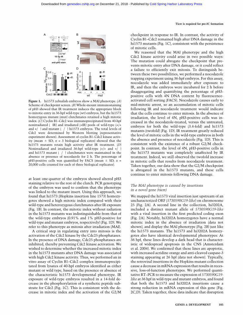
at least one-quarter of the embryos showed altered pH3staining relative to the rest of the clutch. PCR genotypingof the embryos was used to confirm that the phenotypewas linked to the mutant insert. Using this approach, wefound that hi1573 (Hopkins insertion line 1573) homozy-gotes showed a high mitotic index compared with theirwild-type and heterozygous clutchmates after IR exposure(Fig. 1B). In contrast, the mitotic index without radiationin the hi1573 mutants was indistinguishable from that ofthe wild-type embryos (0.91% and 1% pH3-positive forwild-type and mutant embryos, respectively) (Fig. 1D). Werefer to this phenotype as mitosis after irradiation (MAI).
A critical step in regulating entry into mitosis is theactivation of the Cdc2 kinase by the Cdc25 phosphatases.In the presence of DNA damage, Cdc25 phosphatases areinhibited, thereby preventing Cdc2 kinase activation. Wewished to determine whether the increased mitotic indexin the hi1573 mutants after DNA damage was associatedwith high Cdc2 kinase activity. Thus, we performed an invitro assay of Cyclin B1–Cdc2 complex immunoprecipi-tated from lysates of 40-hpf embryos identified as eithermutant or wild type, based on the presence or absence ofthe characteristic hi1573 developmental phenotype. IRexposure of wild-type embryos resulted in a 14-fold de-crease in the phosphorylation of a synthetic peptide sub-strate for Cdc2 (Fig. 1C). This is consistent with the de-crease in mitotic index and the activation of the G2/M
checkpoint in response to IR. In contrast, the activity ofCyclin B1–Cdc2 remained high after DNA damage in thehi1573 mutants (Fig. 1C), consistent with the persistenceof mitotic cells.
We reasoned that the MAI phenotype and the highCdc2 kinase activity could arise in two possible ways:The mutation could abrogate the checkpoint that pre-vents mitotic entry after DNA damage, or it could reflecta failure to efficiently exit mitosis. To distinguish be-tween these two possibilities, we performed a nocodazoletrapping experiment using 36-hpf embryos. For this assay,nocodazole was added immediately after exposure toIR, and then the embryos were incubated for 2 h beforedisaggregating and quantifying the percentage of pH3-positive cells with 4N DNA content by fluorescence-activated cell sorting (FACS). Nocodazole causes early tomid-mitotic arrest, so an accumulation of mitotic cellsfollowing IR and nocodazole treatment would indicatethat the cells continue to enter mitosis. In the absence ofirradiation, the level of 4N, pH3-positive cells was in-creased in the nocodazole-treated, versus the untreated,embryos for both the wild-type (3.4-fold) and hi1573mutants (twofold) (Fig. 1D). IR treatment greatly reducedthe level of mitotic cells in the wild-type embryos in boththe absence and presence of nocodazole (Fig. 1D). This isconsistent with the existence of a robust G2/M check-point. In contrast, the level of 4N, pH3-positive cells inthe hi1573 mutants was completely unaffected by IRtreatment. Indeed, we still observed the twofold increasein mitotic cells that results from nocodazole treatment.Taken together, our data show that the G2/M checkpointis abrogated in the hi1573 mutants, and these cellscontinue to enter mitosis following DNA damage.
The MAI phenotype is caused by insertionsin a novel gene (ticrr)
We mapped the hi1573 viral insertion just upstream of anuncharacterized ORF (5730590G19-like) on chromosome25 (Fig. 2A). A second line in the collection, hi3202A,included a distinct mutant allele of 5730590G19-likewith a viral insertion in the first predicted coding exon(Fig. 2A). Notably, hi3202A homozygotes have a normalmitotic index in the absence of irradiation (data notshown), and display the MAI phenotype (Fig. 2B) just likethe hi1573 mutants. The hi1573 and hi3202A homozy-gotes also have identical developmental phenotypes: At36 hpf, these lines develop a dark head that is character-istic of widespread apoptosis in the CNS (Amsterdamet al. 2004). We confirmed that these lines are apoptotic,with increased acridine orange and anti-cleaved caspase-3staining appearing at 26 hpf (data not shown). Typically,the retroviral insertions in the Hopkins mutant collectioncause a decrease in mRNA expression that results in reces-sive, loss-of-function phenotypes. We performed quanti-tative RT–PCR to measure the expression of 5730590G19-like at 36 hpf in wild-type and mutant embryos, and foundthat both the hi1573 and hi3202A insertions cause astrong reduction in mRNA expression of this gene (Fig.2C,D). Taken together, these data indicate that disruption
Figure 1. hi1573 zebrafish embryos show a MAI phenotype. (A)Scheme of checkpoint screen. (B) Whole-mount immunostainingof pH3 showed that IR treatment induces the appropriate blockto mitotic entry in 36-hpf wild-type (wt) embryos, but the hi1573homozygous mutant (mut) clutchmates retained a high mitoticindex. (C) Cyclin B1–Cdc2 was immunoprecipitated from 40-hpfnonirradiated (�IR) and irradiated (+IR) pools of wild-type (+/+and +/�) and mutant (�/�) hi1573 embryos. The total levels ofCdc2 were determined by Western blotting (representativeexperiment shown). Assessment of cyclin B1–Cdc2 kinase activ-ity (mean 6 SD; n = 3 biological replicates) showed that thehi1573 mutants retain high activity after IR treatment. (D)Nonirradiated and irradiated 36-hpf wild-type (+/+ and +/�)and hi1573 mutant (�/�) clutchmates were maintained in theabsence or presence of nocodazole for 2 h. The percentage ofpH3-positive cells was quantified by FACS (mean 6 SD; n =
20,000 cells counted for each of three biological replicates).
Ticrr is required for pre-IC formation
GENES & DEVELOPMENT 185
Cold Spring Harbor Laboratory Press on December 21, 2018 - Published by genesdev.cshlp.orgDownloaded from

of the novel gene 5730590G19-like causes embryoniclethality and the MAI phenotype. In the following sec-tions, we describe additional roles for this gene. In recog-nition of its broad spectrum of functions, we named thisgene ticrr.
Disruption of ticrr impairs S-phase progressionand causes premature chromatin condensation
DNA damage checkpoint genes are often required fornormal cell cycle control. For example, TopBP1 is requiredfor both checkpoint activation in response to DNA damageand for the initiation of DNA replication in undamagedcells (Garcia et al. 2005). Consistent with this precedent,our analysis of the hi1573 and hi3202A mutant zebrafishrevealed that ticrr is required for cell cycle control in theabsence of DNA damage. FACS analysis of DNA contentrevealed that 40-hpf ticrr mutant embryos had a higherpercentage of cells with between 2N and 4N DNA contentthan the wild-type control, suggesting an accumulation ofcells in S phase (Fig. 3A). To further explore this possibility,we compared the ability of wild-type and ticrr mutant40-hpf embryos to incorporate the nucleotide analogbromodeoxyuridine (BrdU) (Fig. 3B). The wild-type em-bryos had a high level of cells that had between 2N and4N DNA content and were BrdU-positive. This is in-dicative of the high rate of cell proliferation at this earlystage of zebrafish development. In contrast, the 40-hpfticrr mutant embryos incorporated very little BrdU, eventhough a significant fraction of the cells had between 2Nand 4N DNA content. This shows that the ticrr-deficientcells are impaired for DNA replication.
In addition to this replication defect, the 40-hpf ticrrmutants displayed defects in mitotic progression. First,
quantification of the percentage of cells in each phase ofmitosis by visual inspection of chromatin morphology ofpH3-positive cells showed that the distribution of thesepopulations differed between wild-type and ticrr mutantzebrafish (Fig. 3C). Specifically, the ticrr mutants hada much higher percentage of cells that appeared to be in
Figure 2. The checkpoint defect results from disruption ofa novel gene, ticrr. (A) Schematic of the zebrafish ticrr genedenoting the position of viral insertions in the hi1573 andhi3202A mutant lines. (B) Analysis of pH3 showed a low mitoticindex in wild-type embryos but not hi3202A mutant clutch-mates 1 h after IR exposure, showing that hi3202A also hasa checkpoint defect. (C,D) Quantification of total mRNA by RT–PCR showed that the levels of ticrr mRNA were greatly reducedin both the hi1573 (C) and hi3202A (D) homozygous mutants at36 hpf.
Figure 3. Ticrr is required for normal DNA replication and S/Mcheckpoint function. (A) Cell cycle profile from a representativepool of 40-hpf hi1573 wild-type (+/+ and +/�) and mutant (�/�)zebrafish embryos and quantification of cells with G1, S, or G2/MDNA content (mean 6 SD, n = 3 biological replicates) showedan increase in S-phase cells in the ticrr mutants. (B) BrdU andpropidium iodide (PI) FACS analysis of cells from pools of BrdUpulse-labeled wild-type and ticrr mutant embryos. Quantifica-tion of the BrdU+ population (mean 6 SD, n = 3 biologicalreplicates) showed a dramatic reduction in replicating DNA inticrr mutants. (C) Quantification of the proportion of pH3-positive cells in various mitotic phases (mean 6 SD; n = 100cells counted in each of four wild-type and mutant embryos)established a defect in mitotic progression and the presenceof abnormal anaphase cells. Representative cells with ana-phase bridges are shown. (D) Metaphase spreads of cells fromcolchicine-treated embryos showed mitotic cells with frag-mented chromosomes. A representative wild-type metaphasespread and examples of abnormal metaphase spreads frommutants are shown. (E) FACS measurement of anti-pH3 and PIstaining of cells from wild-type and mutant embryos showeda population of mutant pH3-positive cells with less than 4NDNA content (blue) in ticrr mutants, establishing entry intomitosis before completing DNA replication.
Sansam et al.
186 GENES & DEVELOPMENT
Cold Spring Harbor Laboratory Press on December 21, 2018 - Published by genesdev.cshlp.orgDownloaded from

prometaphase, as judged by the presence of condensedchromatin that is not aligned on the metaphase plate.Moreover, they had a reduced percentage of anaphasecells and, within this population, anaphase bridges wereprevalent (Fig. 3C). To explore this defect further, we ana-lyzed chromosome spreads from colchicine-arrested cells(Fig. 3D). This revealed a low incidence of cells withhighly fragmented, condensed chromosomes in the ticrrmutant, but never wild-type, zebrafish (Fig. 3D). Thislevel of chromosomal fragmentation is consistent withcells that have undergone premature chromatin conden-sation. Given their DNA replication problems, we hy-pothesized that the ticrr mutants have an impairedability to activate the S/M checkpoint and thus entermitosis with partially replicated DNA. To address thispossibility, we used FACS to determine the DNA contentof pH3-positive cells (Fig. 3E). It is well established thathistone H3 phosphorylation begins in late G2, peaksduring metaphase, and then declines through anaphase(Hendzel et al. 1997). Accordingly, in the wild-type em-bryos, histone H3 phosphorylation is restricted to thecells with 4N DNA content (Fig. 3E). In contrast, we findthat a high percentage of the pH3-positive cells in theticrr mutants have less than 4N DNA content. Takentogether, the cell cycle defects observed in 40-hpf ticrrmutants show that ticrr is required in the absence ofexogenous DNA damage for S-phase progression and alsofor activation of the S/M checkpoint. We propose that thefailure of these two processes causes cells to enter mitosiswith partially replicated genomes, resulting in the chro-mosomal abnormalities and loss of anaphase cells ob-served in the ticrr mutants.
If ticrr mutants are defective in DNA replication andthe S/M checkpoint, the DNA replication defect shouldbe the primary defect and the mitotic abnormalitiesa secondary consequence. To test this, we turned to anearlier developmental time point, when the ticrr defectswere just beginning to arise. Since we first detect apopto-sis in the hi1573 mutants at 26 hpf, we used 24-hpfembryos to screen for cell cycle defects. At this timepoint, the ticrr mutant embryos are morphologically in-distinguishable from the wild types. Thus, ticrr mutantswere identified by PCR-genotyping a small fraction ofcells dissociated from individual embryos. The remainingcells from 20 embryos of each genotype were then pooledfor cell cycle analysis. FACS showed that there is alreadyan increase in the percentage of cells with between 2Nand 4N DNA content in the 24-hpf ticrr mutants com-pared with the wild-type controls, suggesting that cellsare proceeding slowly through S phase (Fig. 4A). More-over, the ticrr mutants had a lower average level of BrdUincorporation in these S-phase cells than the wild types(Fig. 4B). Thus, the defect in DNA replication clearlyprecedes the initiation of apoptosis. Notably, analysis ofthe DNA content of pH3-positive cells showed that,unlike the 40-hpf embryos, pH3-positive cells with sub-4N DNA content are not observed in the 24-hpf ticrrmutants, indicating that there is no premature mitoticentry at this time point (Fig. 4C). These data show thatthe DNA replication defect is the earliest detectable
phenotype, and thus is not a consequence of the pre-mature mitotic entry.
TICRR binds TopBP1
The phenotypes of the ticrr mutant embryos are highlyreminiscent of those resulting from depletion of TopBP1/Dbp11/Cut5/Mus101. Specifically, these deficiencieshave been shown to disrupt DNA replication, the S/Mcheckpoint, and DNA damage checkpoints (Garcia et al.2005). This raised the possibility that Ticrr acts in theTopBP1 pathway. In particular, we wondered whetherTicrr might be the vertebrate ortholog of the buddingyeast protein Sld3, which interacts with Dpb11 and isrequired for DNA replication initiation (Kamimura et al.2001; Tanaka et al. 2007; Zegerman and Diffley 2007). Totest this hypothesis, we examined whether the Ticrrprotein shows any sequence homology with Sld3 or anyother protein that might yield insight into its biochem-ical function. The zebrafish ticrr gene is predicted toencode a 1824-residue protein (NM_001003887.1) withan anticipated molecular mass of 202 kDa. Genes encod-ing proteins with significant homology exist in othervertebrates, including humans, mice, and Xenopus (Table1; Supplemental Fig. 1). We also found putative Ticrr
Figure 4. A DNA replication defect but not premature mitoticentry occurs in 24-hpf ticrr mutants. (A) Cell cycle profiles from24-hpf hi1573 wild-type (+/+) and mutant (�/�) zebrafish em-bryos and quantitation of cells with G1, S, or G2/M DNA con-tent (mean 6 SD, n = 3 biological replicates), showed an increasein S-phase cells in mutants. (B) BrdU and PI FACS analysis ofcells from pools of BrdU pulse-labeled, 24-hpf wild-type andticrr mutant embryos. Quantification of the BrdU+ populationshowed no change in the percentage of cells replicating DNA(mean 6 SD, n = 20,000 cells in each of three biological repli-cates), but the level of BrdU incorporation per cell is significantlydecreased in the mutants (mean BrdU signal = Mean[BrdU+
signal]/Mean[BrdU� signal]; Student’s t-test P < 0.05). (C) FACSmeasurement of anti-pH3 and PI staining of cells from 24-hpfwild-type and mutant embryos showed no evidence of an S/Mdefect in the ticrr mutants at this time point, as judged by theabsence of pH3-positive cells with less than 4N DNA content.
Ticrr is required for pre-IC formation
GENES & DEVELOPMENT 187
Cold Spring Harbor Laboratory Press on December 21, 2018 - Published by genesdev.cshlp.orgDownloaded from
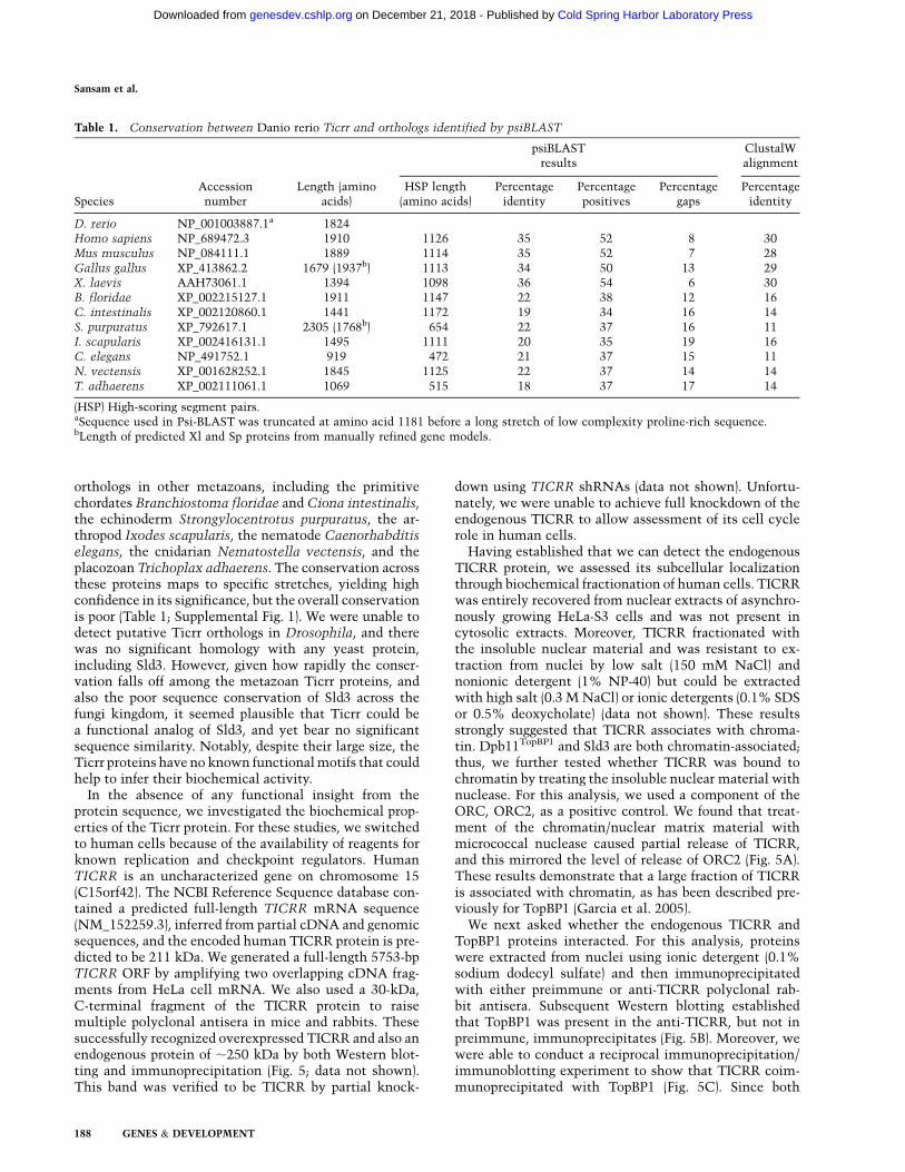
orthologs in other metazoans, including the primitivechordates Branchiostoma floridae and Ciona intestinalis,the echinoderm Strongylocentrotus purpuratus, the ar-thropod Ixodes scapularis, the nematode Caenorhabditiselegans, the cnidarian Nematostella vectensis, and theplacozoan Trichoplax adhaerens. The conservation acrossthese proteins maps to specific stretches, yielding highconfidence in its significance, but the overall conservationis poor (Table 1; Supplemental Fig. 1). We were unable todetect putative Ticrr orthologs in Drosophila, and therewas no significant homology with any yeast protein,including Sld3. However, given how rapidly the conser-vation falls off among the metazoan Ticrr proteins, andalso the poor sequence conservation of Sld3 across thefungi kingdom, it seemed plausible that Ticrr could bea functional analog of Sld3, and yet bear no significantsequence similarity. Notably, despite their large size, theTicrr proteins have no known functional motifs that couldhelp to infer their biochemical activity.
In the absence of any functional insight from theprotein sequence, we investigated the biochemical prop-erties of the Ticrr protein. For these studies, we switchedto human cells because of the availability of reagents forknown replication and checkpoint regulators. HumanTICRR is an uncharacterized gene on chromosome 15(C15orf42). The NCBI Reference Sequence database con-tained a predicted full-length TICRR mRNA sequence(NM_152259.3), inferred from partial cDNA and genomicsequences, and the encoded human TICRR protein is pre-dicted to be 211 kDa. We generated a full-length 5753-bpTICRR ORF by amplifying two overlapping cDNA frag-ments from HeLa cell mRNA. We also used a 30-kDa,C-terminal fragment of the TICRR protein to raisemultiple polyclonal antisera in mice and rabbits. Thesesuccessfully recognized overexpressed TICRR and also anendogenous protein of ;250 kDa by both Western blot-ting and immunoprecipitation (Fig. 5; data not shown).This band was verified to be TICRR by partial knock-
down using TICRR shRNAs (data not shown). Unfortu-nately, we were unable to achieve full knockdown of theendogenous TICRR to allow assessment of its cell cyclerole in human cells.
Having established that we can detect the endogenousTICRR protein, we assessed its subcellular localizationthrough biochemical fractionation of human cells. TICRRwas entirely recovered from nuclear extracts of asynchro-nously growing HeLa-S3 cells and was not present incytosolic extracts. Moreover, TICRR fractionated withthe insoluble nuclear material and was resistant to ex-traction from nuclei by low salt (150 mM NaCl) andnonionic detergent (1% NP-40) but could be extractedwith high salt (0.3 M NaCl) or ionic detergents (0.1% SDSor 0.5% deoxycholate) (data not shown). These resultsstrongly suggested that TICRR associates with chroma-tin. Dpb11TopBP1 and Sld3 are both chromatin-associated;thus, we further tested whether TICRR was bound tochromatin by treating the insoluble nuclear material withnuclease. For this analysis, we used a component of theORC, ORC2, as a positive control. We found that treat-ment of the chromatin/nuclear matrix material withmicrococcal nuclease caused partial release of TICRR,and this mirrored the level of release of ORC2 (Fig. 5A).These results demonstrate that a large fraction of TICRRis associated with chromatin, as has been described pre-viously for TopBP1 (Garcia et al. 2005).
We next asked whether the endogenous TICRR andTopBP1 proteins interacted. For this analysis, proteinswere extracted from nuclei using ionic detergent (0.1%sodium dodecyl sulfate) and then immunoprecipitatedwith either preimmune or anti-TICRR polyclonal rab-bit antisera. Subsequent Western blotting establishedthat TopBP1 was present in the anti-TICRR, but not inpreimmune, immunoprecipitates (Fig. 5B). Moreover, wewere able to conduct a reciprocal immunoprecipitation/immunoblotting experiment to show that TICRR coim-munoprecipitated with TopBP1 (Fig. 5C). Since both
Table 1. Conservation between Danio rerio Ticrr and orthologs identified by psiBLAST
SpeciesAccessionnumber
Length (aminoacids)
psiBLASTresults
ClustalWalignment
HSP length(amino acids)
Percentageidentity
Percentagepositives
Percentagegaps
Percentageidentity
D. rerio NP_001003887.1a 1824Homo sapiens NP_689472.3 1910 1126 35 52 8 30Mus musculus NP_084111.1 1889 1114 35 52 7 28Gallus gallus XP_413862.2 1679 (1937b) 1113 34 50 13 29X. laevis AAH73061.1 1394 1098 36 54 6 30B. floridae XP_002215127.1 1911 1147 22 38 12 16C. intestinalis XP_002120860.1 1441 1172 19 34 16 14S. purpuratus XP_792617.1 2305 (1768b) 654 22 37 16 11I. scapularis XP_002416131.1 1495 1111 20 35 19 16C. elegans NP_491752.1 919 472 21 37 15 11N. vectensis XP_001628252.1 1845 1125 22 37 14 14T. adhaerens XP_002111061.1 1069 515 18 37 17 14
(HSP) High-scoring segment pairs.aSequence used in Psi-BLAST was truncated at amino acid 1181 before a long stretch of low complexity proline-rich sequence.bLength of predicted Xl and Sp proteins from manually refined gene models.
Sansam et al.
188 GENES & DEVELOPMENT
Cold Spring Harbor Laboratory Press on December 21, 2018 - Published by genesdev.cshlp.orgDownloaded from
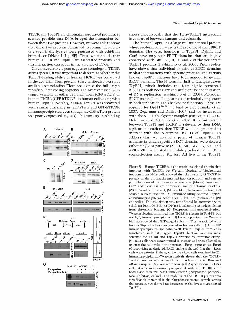
TICRR and TopBP1 are chromatin-associated proteins, itseemed possible that DNA bridged the interaction be-tween these two proteins. However, we were able to showthat these two proteins continued to coimmunoprecipi-tate even if the lysates were pretreated with ethidiumbromide or DNase I (Fig. 5B). Thus, we conclude thathuman TICRR and TopBP1 are associated proteins, andthis interaction can occur in the absence of DNA.
Given the relatively poor sequence homology of TICRRacross species, it was important to determine whether theTopBP1-binding ability of human TICRR was conservedin the zebrafish Ticrr protein. Since antibodies were notavailable for zebrafish Ticrr, we cloned the full-lengthzebrafish Ticrr coding sequence and overexpressed GFP-tagged versions of either zebrafish Ticrr (GFP-zTicrr) orhuman TICRR (GFP-hTICRR) in human cells along withhuman TopBP1. Notably, human TopBP1 was recoveredwith similar efficiency in GFP-zTicrr and GFP-hTICRRimmunoprecipitates, even though the GFP-zTicrr proteinwas poorly expressed (Fig. 5D). This cross-species binding
shows unequivocally that the Ticrr–TopBP1 interactionis conserved between humans and zebrafish.
The human TopBP1 is a large multifunctional proteinwhose predominant feature is the presence of eight BRCTdomains. The yeast homologs of TopBP1, Dpb11, andCut5 have only four BRCT domains that are highlyconserved with BRCTs I, II, IV, and V of the vertebrateTopBP1 proteins (Hashimoto et al. 2006). Prior studieshave shown that individual or pairs of BRCT domainsmediate interactions with specific proteins, and variousknown TopBP1 functions have been mapped to specificBRCT domains. The N-terminal half of Xenopus laevisTopBP1, which includes the four highly conservedBRCTs, is both necessary and sufficient for the initiationof DNA replication (Hashimoto et al. 2006). Moreover,BRCT motifs I and II appear to be particularly importantin both replication and checkpoint functions: These arerequired for Dpb11TopBP1 to bind to Sld3 (Tanaka et al.2007; Zegerman and Diffley 2007) and for interactionwith the 9–1–1 checkpoint complex (Furuya et al. 2004;Delacroix et al. 2007; Lee et al. 2007). If the interactionbetween TopBP1 and TICRR is relevant to their DNAreplication functions, then TICRR would be predicted tointeract with the N-terminal BRCTs of TopBP1. Toaddress this, we created a panel of human TopBP1mutants in which specific BRCT domains were deletedeither singly or pairwise (DI + II, DIII, DIV + V, DVI, andDVII + VIII), and tested their ability to bind to TICRR incotransfection assays (Fig. 5E). All five of the TopBP1
Figure 5. Human TICRR is a chromatin-associated protein thatinteracts with TopBP1. (A) Western blotting of biochemicalfractions from HeLa cells showed that the majority of TICRR ispresent in the chromatin-enriched fraction (chrom) and can bepartially released by micrococcal nuclease (Mnase) treatment.Orc2 and a-tubulin are chromatin and cytoplasmic markers.(WCE) Whole-cell extract; (S1) soluble cytoplasmic fraction; (S2)soluble nuclear fraction. (B) Immunoblotting showed TopBP1coimmunoprecipitates with TICRR but not preimmune (PI)antibodies. The association was not affected by treatment withethidium bromide (EtBr) or DNase I, indicating its independencefrom chromatin binding. (C) Reciprocal immunoprecipitation-Western blotting confirmed that TICRR is present in TopBP1, butnot IgG, immunoprecipitates. (D) Immunoprecipitation-Westernblotting showed that GFP-tagged zebrafish Ticrr associated withhuman TopBP1 when coexpressed in human cells. (E) Anti-GFPimmunoprecipitates and whole-cell lysates (input) from cellstransfected with GFP-tagged TopBP1 deletion mutants werescreened for TICRR and TopBP1 proteins by immunoblotting.(F) HeLa cells were synchronized in mitosis and then allowed tore-enter the cell cycle in the absence (�Rosc) or presence (+Rosc)of roscovitine as depicted. FACS analysis showed that the �Rosccells were entering S phase, while the +Rosc cells remained in G1.Immunoprecipitation-Western analysis shows that the TICRR–TopBP1 complex was recovered at similar levels in the �Rosc and+Rosc samples. (AS) Asynchronous. (G) Asynchronous HeLaS3cell extracts were immunoprecipitated with anti-TICRR anti-bodies and then incubated with either l phosphatase, phospha-tase inhibitors, or both. The mobility of the TICRR protein wassignificantly increased in the phosphatase-treated sample versusthe controls, but showed no difference in the levels of associatedTopBP1.
Ticrr is required for pre-IC formation
GENES & DEVELOPMENT 189
Cold Spring Harbor Laboratory Press on December 21, 2018 - Published by genesdev.cshlp.orgDownloaded from
mutants were expressed at similar levels to the wild-typeTopBP1. TICRR coimmunoprecipitated with four ofthese mutants (DIII, DIV + V, DVI, and DVII + VIII) andwith wild-type TopBP1. In contrast, no TICRR was re-covered in the immunoprecipitate of the DI + II deletionmutant, even though TICRR was present at high levels inthese cells. Taken together, our data show that TICRRassociates with TopBP1 in vivo, and this interactionrequires the two N-terminal BRCT domains that havebeen associated with TopBP1’s role in both checkpointsignaling and DNA replication.
We were intrigued to find that TICRR association mapsto the region of TopBP1 that is required for Sld3 binding.In yeast, formation of the Sld3–Dpb11 complex is knownto require CDK phosphorylation of Sld3, and this, to-gether with the phosphorylation of Sld2, accounts for theCDK dependence of DNA replication in this organism(Tanaka et al. 2007; Zegerman and Diffley 2007). CDK isalso essential for replication in human cells, but there issome doubt as to whether pre-IC components will be therelevant target (DeGregori et al. 1995). Given these ques-tions, we used two complementary approaches to exam-ine the role of CDK phosphorylation in the TICRR–TopBP1 interaction. First, we generated a population ofHeLa cells that were synchronously re-entering G1 frommitosis, and then we cultured them in the absence orpresence of the pan-CDK inhibitor roscovitine for 8 h (Fig.5F). Consistent with the known CDK dependence ofS-phase entry, FACS analysis showed that the untreatedcells (�ROSC) were beginning to enter S phase, while theroscovitine-treated cells (+ROSC) remained blocked inG1 (Fig. 5F). Notably, TopBP1 was recovered at compara-ble levels in the TICRR immunoprecipitates of boththe untreated and treated cells (Fig. 5F), suggesting thatbinding occurs in the presence of CDK inhibition. In thesecond approach, we incubated TICRR–TopBP1 immu-noprecipitates from asynchronous cell extracts with orwithout l phosphatase, and then assayed the associationof TICRR and TopBP1 by Western blotting. Notably,phosphatase treatment significantly increased the mobil-ity of the TICRR protein (Fig. 5G), indicating that TICRRis phosphorylated in vivo and that we successfully re-moved this modification. Despite this change, there wasno detectable difference in the levels of the TICRR–TopBP1 complex in phosphatase-treated versus untreatedcells (Fig. 5G). Importantly, this result was not altered byDNase treatment of the extracts prior to immunoprecip-itation (data not shown), indicating that this complexformation occurs in the absence of chromatin binding.Taken together, these synchronization and phosphataseexperiments strongly suggest that the interaction betweenTICRR and TopBP1 can occur in a CDK-independentmanner.
Ticrr deficiency inhibits pre-IC formation
The TopBP1 orthologs in X. laevis and budding and fissionyeast are known to be essential for the transition of thepre-RC into the pre-IC, an intermediate in the initiation ofDNA replication (Garcia et al. 2005). Having established
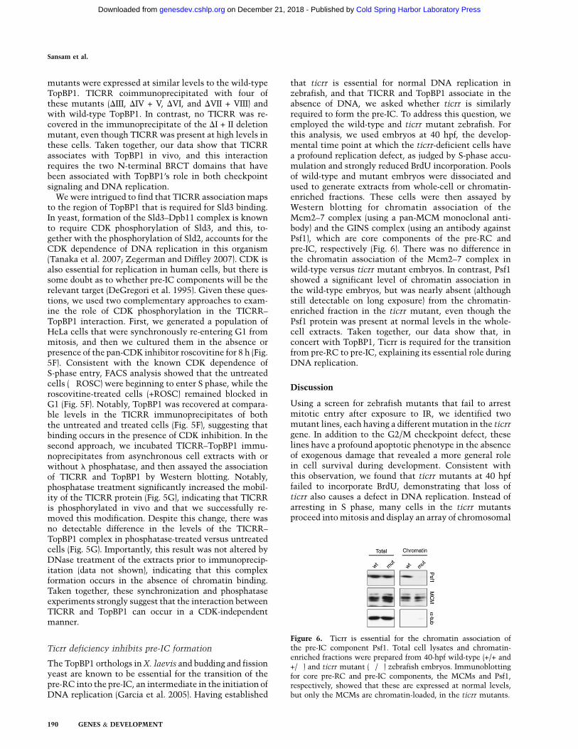
that ticrr is essential for normal DNA replication inzebrafish, and that TICRR and TopBP1 associate in theabsence of DNA, we asked whether ticrr is similarlyrequired to form the pre-IC. To address this question, weemployed the wild-type and ticrr mutant zebrafish. Forthis analysis, we used embryos at 40 hpf, the develop-mental time point at which the ticrr-deficient cells havea profound replication defect, as judged by S-phase accu-mulation and strongly reduced BrdU incorporation. Poolsof wild-type and mutant embryos were dissociated andused to generate extracts from whole-cell or chromatin-enriched fractions. These cells were then assayed byWestern blotting for chromatin association of theMcm2–7 complex (using a pan-MCM monoclonal anti-body) and the GINS complex (using an antibody againstPsf1), which are core components of the pre-RC andpre-IC, respectively (Fig. 6). There was no difference inthe chromatin association of the Mcm2–7 complex inwild-type versus ticrr mutant embryos. In contrast, Psf1showed a significant level of chromatin association inthe wild-type embryos, but was nearly absent (althoughstill detectable on long exposure) from the chromatin-enriched fraction in the ticrr mutant, even though thePsf1 protein was present at normal levels in the whole-cell extracts. Taken together, our data show that, inconcert with TopBP1, Ticrr is required for the transitionfrom pre-RC to pre-IC, explaining its essential role duringDNA replication.
Discussion
Using a screen for zebrafish mutants that fail to arrestmitotic entry after exposure to IR, we identified twomutant lines, each having a different mutation in the ticrrgene. In addition to the G2/M checkpoint defect, theselines have a profound apoptotic phenotype in the absenceof exogenous damage that revealed a more general rolein cell survival during development. Consistent withthis observation, we found that ticrr mutants at 40 hpffailed to incorporate BrdU, demonstrating that loss ofticrr also causes a defect in DNA replication. Instead ofarresting in S phase, many cells in the ticrr mutantsproceed into mitosis and display an array of chromosomal
Figure 6. Ticrr is essential for the chromatin association ofthe pre-IC component Psf1. Total cell lysates and chromatin-enriched fractions were prepared from 40-hpf wild-type (+/+ and+/�) and ticrr mutant (�/�) zebrafish embryos. Immunoblottingfor core pre-RC and pre-IC components, the MCMs and Psf1,respectively, showed that these are expressed at normal levels,but only the MCMs are chromatin-loaded, in the ticrr mutants.
Sansam et al.
190 GENES & DEVELOPMENT
Cold Spring Harbor Laboratory Press on December 21, 2018 - Published by genesdev.cshlp.orgDownloaded from

abnormalities—including fragmented chromosomes andanaphase bridges—that likely account for the loss ofanaphase cells through mitotic catastrophe. Thus, ticrris required to prevent mitotic entry both following ex-ogenous DNA damage and also when DNA replication isimpaired. These phenotypes are highly reminiscent ofthose arising when TopBP1 and its orthologs are inacti-vated. Consistent with this observation, we found thatthe human and zebrafish TICRR proteins both interactwith TopBP1 in human cells, and ticrr is required fortransformation of the pre-RC to the pre-IC in zebrafish.Taken together, these data suggest that TICRR functionswith TopBP1 in DNA replication and both the S/M andG2/M checkpoints.
TICRR in DNA replication
The replication machinery and its mechanisms of regula-tion have been studied in numerous organisms. Theseprocesses are generally conserved among eukaryotes,but a unifying model remains elusive because the orderof assembly of pre-IC proteins seems to differ betweenX. laevis and yeast, and known key components suchas Sld3 have not been identified in higher eukaryotes.The initiation of DNA replication is best understood inS. cerevisiae. Here, Dpb11TopBP1, Sld2, Sld3, GINS, andCdc45 are all required to transform the pre-RC into thepre-IC. Sld3 and Dpb11TopBP1 form a complex that isinduced by the CDK phosphorylation of Sld3 and requiresthe two N-terminal BRCT domains of Dpb11 (Tanakaet al. 2007; Zegerman and Diffley 2007). Although theformation of the Dpb11–Sld3 complex is critical forreplication initiation in S. cerevisiae, an analogous eventhas not been defined in higher eukaryotes.
Much of our understanding of vertebrate DNA replica-tion comes from studies in X. laevis (Bell and Dutta 2002;Sclafani and Holzen 2007). In this organism, Cut5TopBP1 isalso required for the recruitment of DNA polymerasesonto chromatin (Hashimoto and Takisawa 2003; Kubotaet al. 2003). Thus, the general function of Dpb11 in pre-ICformation seems to be conserved in vertebrate TopBP1.Our data show that TICRR displays some of the coreproperties of the Sld3 protein. First, Ticrr is essential forDNA replication in unperturbed cells, and it is specificallyrequired for formation of pre-ICs but not pre-RCs. Second,TICRR associates with TopBP1 through the N-terminalBRCT motifs I and II that are conserved with the Sld3-interacting BRCTs of Dpb11. However, our data do notaddress whether the interaction between TICRR andTopBP1 is direct, and other observations are less consis-tent with the idea that TICRR is a true ortholog of Sld3.First, we note that there is no detectable sequence sim-ilarity between these proteins. This is not a particularlytelling finding, since the Sld3 and TICRR proteins are bothpoorly conserved even within their own kingdoms, butit does raise questions about both the relationship andmechanism(s) of action of these proteins. The second, andmore striking, finding is the apparent discrepancy in therole of CDK phosphorylation in Sld3 versus TICRR re-gulation. Specifically, CDK phosphorylation of Sld3 is
required for it to bind to Dpb11, and this, together withthe phosphorylation of Sld2, accounts for the CDK de-pendence of DNA replication in yeast (Tanaka et al. 2007;Zegerman and Diffley 2007). In contrast, we find that theTICRR–TopBP1 interaction is completely unaffected byculturing in the presence of the pan-CDK inhibitorroscovitine or treatment with phosphatase. We note thatthere are potential limitations to these approaches: Theroscovitine-induced block to S-phase entry (Fig. 5F) couldreflect a reduction, but not full loss, of CDK activity,and/or the phosphatase may be unable to access thephosphorylated residue(s) mediating the TICRR–TopBP1interaction, even though it effectively targets other phos-phorylation sites (Fig. 5G). Despite these caveats, our dataare most consistent with the notion that TICRR associ-ates with TopBP1 in a CDK-independent manner. On firstconsideration, this finding seems to imply that TICRR isnot human Sld3. However, there is evidence to suggestthat the underlying basis for the CDK dependence of DNAreplication differs in human versus yeast cells. First,DeGregori et al. (1995) have shown that E2F1 expressioncompletely bypasses the CDK dependence of S phase inhuman cells. This argues that there is no absolute re-quirement for CDK phosphorylation of either pre-RC orpre-IC components in this organism. Consistent with thisconclusion, CDK phosphorylation is required for forma-tion of the yeast Sld2–Dpb11 complex but not the ver-tebrate counterpart RECQL4–TopBP1 (Matsuno et al.2006; Tanaka et al. 2007; Zegerman and Diffley 2007).Clearly, additional studies will be required to explore howsimilar or different Sld3 and TICRR are to one another inthe context of both DNA replication and, as describedbelow, checkpoint response.
TICRR in the S/M and G2/M checkpoints
The S/M checkpoint plays a vital role in ensuring that thegenome is fully replicated prior to mitotic entry. Our dataclearly show that Ticrr is essential for the integrity of thischeckpoint in vivo. Thus, Ticrr joins a short list of pro-teins that play a dual role in both replication regulationand S/M checkpoint response. Notably, the existing dualreplication/checkpoint proteins can be divided into twodifferent subclasses, based on their role in the S/M check-point. The Mcm2–7 complex, Cdt1, and Cdc45 are rep-resentative members of the first subclass. The analysis ofyeast conditional mutants shows that the loss of MCMs,Cdt1, or Cdc45 prior to the initiation of replication allowscells with unreplicated DNA to enter mitosis, but theloss of these proteins in replicating cells does not impairthe S/M checkpoint (Tercero et al. 2000; Labib et al.2001). These findings suggest that these pre-RC andpre-IC components are not involved directly in the S/Mcheckpoint, but they are required to create the replicationstructures that signal that S phase is ongoing and not yetcomplete. Studies using X. laevis extracts confirm thatreplication structures are a prerequisite for S-phase check-point signaling in vertebrates, and further suggest thatthe necessary feature is the RNA primer generated byDNA polymerase a (Michael et al. 2000).
Ticrr is required for pre-IC formation
GENES & DEVELOPMENT 191
Cold Spring Harbor Laboratory Press on December 21, 2018 - Published by genesdev.cshlp.orgDownloaded from

TopBP1 is an example of the second class of dual rep-lication/checkpoint protein. Studies with conditional anddeletion mutants show that Dpb11/TopBP1’s checkpointfunction can be separated from its role in pre-IC forma-tion (McFarlane et al. 1997; Saka et al. 1997; Hashimotoet al. 2006; Yan et al. 2006). Biochemical data show thatTopBP1 is recruited to the site of replication stress/DNAdamage by the 9–1–1 complex, dependent on the sametwo BRCT motifs that are required for TopBP1’s replica-tion function (Furuya et al. 2004; Delacroix et al. 2007;Lee et al. 2007). Once recruited, TopBP1 promotes ATRactivation (Kumagai et al. 2006). Thus, TopBP1 is a bonafide checkpoint protein.
Given the precedent of the existing dual replication/checkpoint proteins, it remains an open questionwhether Ticrr is directly involved in the S/M checkpointor whether this function simply reflects its role as anessential replication regulator. We favor the former hy-pothesis, based on the following observations. First, ourdata show that TICRR interacts with TopBP1, raisingthe possibility that it cooperates in TopBP1-dependentprocesses beyond DNA replication, such as checkpointsignaling. Second, our FACS data show that ticrr-deficient cells enter mitosis with partially replicatedDNA. This clearly differs from the yeast MCM, Cdt1,and Cdc45 mutants, which do not prematurely entermitosis once S phase has begun (Tercero et al. 2000; Labibet al. 2001). Third, we originally identified ticrr througha screen for G2/M checkpoint regulators. This showedthat ticrr-deficient cells fail to arrest in mitosis in re-sponse to treatment with IR. Importantly, our FACS anal-ysis shows that these mitotic cells have 4N DNA con-tent. Thus, we believe that this MAI phenotype reflectsa bona fide defect in the G2/M checkpoint and is notsimply an indirect consequence of the replication and/orS/M checkpoint defects. Consistent with this view, thereis no evidence in the literature that the G2/M checkpointis dependent on appropriate replication initiation. More-over, included in our zebrafish screen were a large num-ber of known replication gene mutants that did notdisplay the MAI phenotype. Finally, we again note thatTopBP1 has a well-documented role in the radiation-induced G2/M checkpoint (Garcia et al. 2005). Given allof these observations, we speculate that TICRR is a pre-viously unknown partner for TopBP1 in its myriad rolesas a core regulator of the DNA replication, S/M check-point, and the G2/M checkpoint machinery.
Materials and methods
Zebrafish maintenance, collection, genotyping,
and expression analysis
Zebrafish were maintained as described previously (Amsterdamet al. 2004). For the screen, heterozygous insertion carriers wereintercrossed and, at 24 hpf, the embryos were dechorionated and1-phenyl-2-thiourea (PTU, 0.003%) was added to suppress pig-mentation. At 32 hpf, 60 embryos from each clutch weresubjected to G2/M checkpoint analysis. Primers used for geno-typing and mRNA level analysis are described in the Supple-mental Material.
G2/M checkpoint assays
Embryos were exposed to 15 Gy IR from a 60Co source andanalyzed for pH3 1 h later as described (Sansam et al. 2006). Forthe nocodazole trapping experiment, embryos were exposed toIR, immediately placed in nocodazole (150 ng/mL + 1% DMSO),and incubated for 2 h at 28.5°C before analysis of pH3/DNAcontent by FACS.
In vitro kinase assay
Fifty embryos for each data point were treated with 15 Gy IR andincubated for 30 min at 28.5°C. Embryos were then dechorio-nated, deyolked, and homogenized in 400 mL of KLB (50 mM Trisat pH 7.4, 150 mM NaCl, 0.1% Triton X-100, 4 mM EDTA, 0.1%NP-40, 50 mM NaF, 0.2 mM Na3VO4, 100 mM leupeptin,5 mg/mL aprotonin, 1 mM PMSF). The homogenate was adjustedto 1 mg/mL prior to precipitation with anti-Cyclin B1. The beadswere washed twice with KLB and KAB (50 mM Tris at pH 8.0,10 mM MgCl2, 1 mM EGTA) and incubated for 15 min at 30°C in20 mL of kinase assay mix (KAB with 40 mM cold ATP, 100 mMpeptide [HATPPKKKRK], 1 mM DTT, 0.5mCi/ml g-32PATP[6000Ci/mmol]). Substrate phosphorylation was quantified byfilter binding and scintillation counting.
FACS analysis
For BrdU labeling in zebrafish, dechorionated embryos were in-cubated with 10 mM BrdU/15% DMSO for 15 min on ice, washed,and incubated for 15 min at 28.5°C. Mutant embryos were iden-tified by either developmental phenotype or PCR-genotyping ofa fraction of individual, fixed embryos. Cells were disaggregatedby triturating embryos in 0.25% Trypsin/1 mM EDTA usinga p200, and fixed in 70% ethanol overnight at�20°C. Suspensionsof cells from 20 wild-type or mutant embryos were pooled andprepared for pH3/propidium iodide (PI) or BrdU/PI FACS analysisas described (Pozarowski and Darzynkiewicz 2004). FACS analy-sis was conducted by FACScan (Becton-Dickinson). DNA contentwas quantified by ModFit LT (Verity Software), and pH3 and BrdUwas quantified by FlowJo (Tree Star, Inc.).
Plasmid construction
The construction of expression vectors for human TICRR, zebra-fish Ticrr, and human TopBP1 is described in the SupplementalMaterial. The TopBP1 BRCT domain deletion mutants carry thefollowing in-frame deletions within the 1522-amino-acid ORF:D98–306 (BRCTs I + II), D354–452 (BRCT III), D547–760 (BRCTsIV + V), D922–1011 (BRCT VI), D1267–1489 (BRCTs VII–VIII).Except for D922–1011, the deleted amino acids were replaced bytwo glycines.
Human cell culture and chromatin fractionation
HeLa, HeLa-S3, and 293FS cells were grown in DMEM with 10%FBS. For the synchronization experiments, HeLa cells werecultured in the presence of 100 ng/mL nocodazole (Calbiochem)for 18 h. Synchronized mitotic cells were recovered by shake-off,replated in DMEM containing 10% FBS for 2 h, and then in-cubated for a further 8 h in the presence or absence of 20 mMroscovitine (Calbiochem). The isolation of soluble fraction (S1),soluble nuclear fraction (S2), and chromatin-enriched fractionswere conducted as described (Mendez and Stillman 2000). Tosolubilize the chromatin-bound proteins, nuclei were treatedwith 50 U of micrococcal nuclease (Worthington) for 2 min at37°C. When indicated, lysates were treated with 2000 U/mL
Sansam et al.
192 GENES & DEVELOPMENT
Cold Spring Harbor Laboratory Press on December 21, 2018 - Published by genesdev.cshlp.orgDownloaded from

DNase I (30 min at 25°C; Roche) or 20 mg/mL ethidium bromide(30 min on ice) prior to TICRR immunoprecipitation. For phos-phatase treatment, precipitates were resuspended in l phospha-tase buffer (NEB) with or without 400 U of l phosphatase (NEB)and phosphatase inhibitors (50 mM NaF, 10 mM Na3VO4), andincubated for 30 min at 30°C. The immunoprecipitates werewashed four times with RIPA and were resuspended in 23
Laemmli Buffer.
Zebrafish chromatin preparation
Deyolked zebrafish embryos were triturated in 0.25% Trypsin/1 mMEDTA using a p200. Disaggregated cells were filtered througha 35-mm nylon mesh and washed once with PBS. The chromatin-enriched fraction was prepared essentially as described (Aparicioet al. 2009). Briefly, cells were lysed in 10 mM HEPES (pH 7.9),0.2 M KOAc, 0.1% Triton X-100, 0.34 M sucrose, 10% glycerol,1 mM 1,4-DTT, protease, and phosphatase inhibitors. Thechromatin-associated fraction was recovered by spinning at18,000g for 10 min and was washed twice in lysis buffer beforesuspension in Laemmli Buffer.
Antibodies
A 6-His-tagged N-terminal fragment of human TICRR(NP_689472.3 amino acids 1094–1348) was expressed in bacteria,purified over Ni2+ NTA-agarose resin (Qiagen), and used to im-munize BALB/c mice or New Zealand White rabbits (PoconoRabbit Farm). Other antibodies were phosho-H3 (sc-8656-R,Santa Cruz Biotechnologies), mouse anti-goldfish Cyclin-B1(B112) (Katsu et al. 1993), PSTAIRE-Cdc2 (sc-53, Santa CruzBiotechnologies), tubulin (T9026, Sigma), ORC2 (sc-13238, SantaCruz Biotechnologies), LaminA/C (2032, Cell Signaling), TopBP1(NB100-217, Novus Biologicals; and sc-32923, Santa Cruz Bio-technologies), rabbit polyclonal anti-human Psf1 (Aparicio et al.2009), mouse monoclonal anti-human Pan-MCM (Austin et al.1999), normal mouse IgG (sc-2025, Santa Cruz Biotechnologies),and GFP (11814460001, Roche).
Acknowledgments
Antibodies for this study were provided by Dr. MasakaneYamashita, Hokkaido University (anti-goldfish Cyclin-B1); Dr.Juan Mendez, Spanish National Cancer Research Center (anti-Psf1); and Dr. Stephen Bell, HHMI and MIT (Pan-MCM). We alsothank Stephen Bell, Sebastian Hoersch, and members of theHopkins and Lees laboratories for helpful discussions duringthis study and the preparation of this manuscript. This workwas supported by Ruth L. Kirschstein NRSAs to C.L.S. andN.C. J.A.L. is a Daniel K. Ludwig Scholar.
References
Amsterdam A, Nissen RM, Sun Z, Swindell EC, Farrington S,Hopkins N. 2004. Identification of 315 genes essential forearly zebrafish development. Proc Natl Acad Sci 101: 12792–12797.
Aparicio T, Guillou E, Coloma J, Montoya G, Mendez J. 2009.The human GINS complex associates with Cdc45 and MCMand is essential for DNA replication. Nucleic Acids Res 37:2087–2095.
Austin RJ, Orr-Weaver TL, Bell SP. 1999. Drosophila ORCspecifically binds to ACE3, an origin of DNA replicationcontrol element. Genes & Dev 13: 2639–2649.
Bartek J, Lukas C, Lukas J. 2004. Checking on DNA damage inS phase. Nat Rev Mol Cell Biol 5: 792–804.
Bell SP, Dutta A. 2002. DNA replication in eukaryotic cells.Annu Rev Biochem 71: 333–374.
Brown EJ, Baltimore D. 2003. Essential and dispensable roles ofATR in cell cycle arrest and genome maintenance. Genes &
Dev 17: 615–628.Canman CE. 2001. Replication checkpoint: Preventing mitotic
catastrophe. Curr Biol 11: R121–R124. doi: 10.1016/S0960-9822(01)00057-4.
DeGregori J, Leone G, Ohtani K, Miron A, Nevins JR. 1995. E2F-1accumulation bypasses a G1 arrest resulting from the in-hibition of G1 cyclin-dependent kinase activity. Genes &
Dev 9: 2873–2887.Delacroix S, Wagner JM, Kobayashi M, Yamamoto K, Karnitz
LM. 2007. The Rad9–Hus1–Rad1 (9–1–1) clamp activatescheckpoint signaling via TopBP1. Genes & Dev 21: 1472–1477.
Furuya K, Poitelea M, Guo L, Caspari T, Carr AM. 2004. Chk1activation requires Rad9 S/TQ-site phosphorylation topromote association with C-terminal BRCT domains ofRad4TOPBP1. Genes & Dev 18: 1154–1164.
Garcia V, Furuya K, Carr AM. 2005. Identification and func-tional analysis of TopBP1 and its homologs. DNA Repair
(Amst) 4: 1227–1239.Harper JW, Elledge SJ. 2007. The DNA damage response: Ten
years after. Mol Cell 28: 739–745.Harrison JC, Haber JE. 2006. Surviving the breakup: The DNA
damage checkpoint. Annu Rev Genet 40: 209–235.Hashimoto Y, Takisawa H. 2003. Xenopus Cut5 is essential for
a CDK-dependent process in the initiation of DNA replica-tion. EMBO J 22: 2526–2535.
Hashimoto Y, Tsujimura T, Sugino A, Takisawa H. 2006. Thephosphorylated C-terminal domain of Xenopus Cut5 directlymediates ATR-dependent activation of Chk1. Genes Cells
11: 993–1007.Hendzel MJ, Wei Y, Mancini MA, Van Hooser A, Ranalli T,
Brinkley BR, Bazett-Jones DP, Allis CD. 1997. Mitosis-specific phosphorylation of histone H3 initiates primarilywithin pericentromeric heterochromatin during G2 andspreads in an ordered fashion coincident with mitotic chro-mosome condensation. Chromosoma 106: 348–360.
Kamimura Y, Tak YS, Sugino A, Araki H. 2001. Sld3, whichinteracts with Cdc45 (Sld4), functions for chromosomalDNA replication in Saccharomyces cerevisiae. EMBO J 20:2097–2107.
Katsu Y, Yamashita M, Kajiura H, Nagahama Y. 1993. Behaviorof the components of maturation-promoting factor, cdc2kinase and cyclin B, during oocyte maturation of goldfish.Dev Biol 160: 99–107.
Kubota Y, Takase Y, Komori Y, Hashimoto Y, Arata T, KamimuraY, Araki H, Takisawa H. 2003. A novel ring-like complex ofXenopus proteins essential for the initiation of DNA replica-tion. Genes & Dev 17: 1141–1152.
Kumagai A, Lee J, Yoo HY, Dunphy WG. 2006. TopBP1 activatesthe ATR–ATRIP complex. Cell 124: 943–955.
Labib K, Kearsey SE, Diffley JF. 2001. MCM2-7 proteins areessential components of prereplicative complexes that accu-mulate cooperatively in the nucleus during G1-phase and arerequired to establish, but not maintain, the S-phase check-point. Mol Biol Cell 12: 3658–3667.
Lee J, Kumagai A, Dunphy WG. 2007. The Rad9–Hus1–Rad1checkpoint clamp regulates interaction of TopBP1 with ATR.J Biol Chem 282: 28036–28044.
Matsuno K, Kumano M, Kubota Y, Hashimoto Y, Takisawa H.2006. The N-terminal noncatalytic region of Xenopus RecQ4is required for chromatin binding of DNA polymerase a in theinitiation of DNA replication. Mol Cell Biol 26: 4843–4852.
Ticrr is required for pre-IC formation
GENES & DEVELOPMENT 193
Cold Spring Harbor Laboratory Press on December 21, 2018 - Published by genesdev.cshlp.orgDownloaded from
McFarlane RJ, Carr AM, Price C. 1997. Characterisation of theSchizosaccharomyces pombe rad4/cut5 mutant phenotypes:Dissection of DNA replication and G2 checkpoint controlfunction. Mol Gen Genet 255: 332–340.
Mendez J, Stillman B. 2000. Chromatin association of humanorigin recognition complex, cdc6, and minichromosomemaintenance proteins during the cell cycle: Assembly ofprereplication complexes in late mitosis. Mol Cell Biol 20:8602–8612.
Michael WM, Ott R, Fanning E, Newport J. 2000. Activation ofthe DNA replication checkpoint through RNA synthesis byprimase. Science 289: 2133–2137.
Nghiem P, Park PK, Kim Y, Vaziri C, Schreiber SL. 2001. ATRinhibition selectively sensitizes G1 checkpoint-deficientcells to lethal premature chromatin condensation. Proc Natl
Acad Sci 98: 9092–9097.Pozarowski P, Darzynkiewicz Z. 2004. Analysis of cell cycle by
flow cytometry. Methods Mol Biol 281: 301–311.Saka Y, Esashi F, Matsusaka T, Mochida S, Yanagida M. 1997.
Damage and replication checkpoint control in fission yeast isensured by interactions of Crb2, a protein with BRCT motif,with Cut5 and Chk1. Genes & Dev 11: 3387–3400.
Sansam CL, Shepard JL, Lai K, Ianari A, Danielian PS, AmsterdamA, Hopkins N, Lees JA. 2006. DTL/CDT2 is essential for bothCDT1 regulation and the early G2/M checkpoint. Genes &
Dev 20: 3117–3129.Schmidt U, Wollmann Y, Franke C, Grosse F, Saluz HP, Hanel F.
2008. Characterization of the interaction between the hu-man DNA topoisomerase IIb-binding protein 1 (TopBP1) andthe cell division cycle 45 (Cdc45) protein. Biochem J 409:169–177.
Sclafani RA, Holzen TM. 2007. Cell cycle regulation of DNAreplication. Annu Rev Genet 41: 237–280.
Takayama Y, Kamimura Y, Okawa M, Muramatsu S, Sugino A,Araki H. 2003. GINS, a novel multiprotein complex requiredfor chromosomal DNA replication in budding yeast. Genes &
Dev 17: 1153–1165.Tanaka S, Umemori T, Hirai K, Muramatsu S, Kamimura Y,
Araki H. 2007. CDK-dependent phosphorylation of Sld2 andSld3 initiates DNA replication in budding yeast. Nature 445:328–332.
Tercero JA, Labib K, Diffley JF. 2000. DNA synthesis at in-dividual replication forks requires the essential initiationfactor Cdc45p. EMBO J 19: 2082–2093.
Van Hatten RA, Tutter AV, Holway AH, Khederian AM, WalterJC, Michael WM. 2002. The Xenopus Xmus101 protein isrequired for the recruitment of Cdc45 to origins of DNAreplication. J Cell Biol 159: 541–547.
Yamamoto RR, Axton JM, Yamamoto Y, Saunders RD, GloverDM, Henderson DS. 2000. The Drosophila mus101 gene,which links DNA repair, replication and condensation ofheterochromatin in mitosis, encodes a protein with sevenBRCA1 C-terminus domains. Genetics 156: 711–721.
Yamane K, Wu X, Chen J. 2002. A DNA damage-regulatedBRCT-containing protein, TopBP1, is required for cell sur-vival. Mol Cell Biol 22: 555–566.
Yan S, Michael WM. 2009. TopBP1 and DNA polymerase-adirectly recruit the 9–1–1 complex to stalled DNA replica-tion forks. J Cell Biol 184: 793–804.
Yan S, Lindsay HD, Michael WM. 2006. Direct requirement forXmus101 in ATR-mediated phosphorylation of Claspinbound Chk1 during checkpoint signaling. J Cell Biol 173:181–186.
Zegerman P, Diffley JF. 2007. Phosphorylation of Sld2 and Sld3by cyclin-dependent kinases promotes DNA replication inbudding yeast. Nature 445: 281–285.
Sansam et al.
194 GENES & DEVELOPMENT
Cold Spring Harbor Laboratory Press on December 21, 2018 - Published by genesdev.cshlp.orgDownloaded from
10.1101/gad.1860310Access the most recent version at doi: 24:2010, Genes Dev.
Christopher L. Sansam, Nelly M. Cruz, Paul S. Danielian, et al. regulator
, is an essential checkpoint and replicationticrrA vertebrate gene,
Material
Supplemental
http://genesdev.cshlp.org/content/suppl/2009/12/30/24.2.183.DC1
References
http://genesdev.cshlp.org/content/24/2/183.full.html#ref-list-1
This article cites 41 articles, 24 of which can be accessed free at:
License
ServiceEmail Alerting
click here.right corner of the article or
Receive free email alerts when new articles cite this article - sign up in the box at the top
Copyright © 2010 by Cold Spring Harbor Laboratory Press
Cold Spring Harbor Laboratory Press on December 21, 2018 - Published by genesdev.cshlp.orgDownloaded from
Related Documents