REPORT A Truncating Mutation in SERPINB6 Is Associated with Autosomal-Recessive Nonsyndromic Sensorineural Hearing Loss Aslı Sırmacı, 1,2 Seyra Erbek, 3 Justin Price, 1,2 Mingqian Huang, 4 Duygu Duman, 5 F. Bas xak Cengiz, 5 Gu ¨ney Bademci, 1,2 Suna Tokgo ¨z-Yılmaz, 5 Burcu His xmi, 5 Hilal O ¨ zdag˘ , 6 Banu O ¨ ztu ¨rk, 7 Sevsen Kulaksızog˘ lu, 8 Erkan Yıldırım, 9 Haris Kokotas, 10 Maria Grigoriadou, 10 Michael B. Petersen, 10 Hashem Shahin, 11 Moien Kanaan, 11 Mary-Claire King, 12 Zheng-Yi Chen, 4 Susan H. Blanton, 1,2 Xue Z. Liu, 2,13 Stephan Zuchner, 1,2 Nejat Akar, 5 and Mustafa Tekin 1,2, * More than 270 million people worldwide have hearing loss that affects normal communication. Although astonishing progress has been made in the identification of more than 50 genes for deafness during the past decade, the majority of deafness genes are yet to be identified. In this study, we mapped a previously unknown autosomal-recessive nonsyndromic sensorineural hearing loss locus (DFNB91) to chromo- some 6p25 in a consanguineous Turkish family. The degree of hearing loss was moderate to severe in affected individuals. We subsequently identified a nonsense mutation (p.E245X) in SERPINB6, which is located within the linkage interval for DFNB91 and encodes for an intra- cellular protease inhibitor. The p.E245X mutation cosegregated in the family as a completely penetrant autosomal-recessive trait and was absent in 300 Turkish controls. The mRNA expression of SERPINB6 was reduced and production of protein was absent in the peripheral leukocytes of homozygotes, suggesting that the hearing loss is due to loss of function of SERPINB6. We also demonstrated that SERPINB6 was expressed primarily in the inner ear hair cells. We propose that SERPINB6 plays an important role in the inner ear in the protection against leakage of lysosomal content during stress and that loss of this protection results in cell death and sensorineural hearing loss. Genetic causes of hearing loss are estimated to account for 68% of cases in newborns and 55% of cases by the age of four. 1 Autosomal-recessive, dominant, and X-linked inheri- tance accounts for 77%, 22%, and 1% of the genetic deaf- ness, respectively. 2 Most cases of genetic deafness fall into the category of sensorineural hearing loss and are caused by pathologies of the inner ear or auditory nerves; these can be identified with audiological investigations. Hear- ing loss can be classified into syndromic (20%–30%) and nonsyndromic (70%–80%) forms based on the presence or absence of distinctive clinical or laboratory features. More than 50 dominant and/or recessive genes for nonsyndromic sensorineural hearing loss have been identified and most of the 35 genes for the autosomal-recessive form were initially mapped in consanguineous families or population isolates. During our studies on hereditary deafness, which were approved by Ankara University Ethics Committee (Turkey), by the IRB at the University of Miami (USA), and by the Ethics Committee of the Institute of Child Health, Athens (Greece), we ascertained a Turkish family, family 728, in which four children with sensorineural hearing loss were born to consanguineous parents. The father, whose parents were also first cousins, had sensori- neural hearing loss as well (Figure 1A). Diagnosis of senso- rineural hearing loss was established via standard audiom- etry. Audiograms of family 728 showed that the hearing loss was moderate to severe with some residual hearing in all affected individuals (Figure 1B). All affected members of the family had oral communication with partial help of lip reading. Individual 201, at 54 years old the eldest affected in the family, had the most severe hearing loss with more severe involvement of high frequencies. The youngest affected family member, 106, was 23 years old with hearing loss involving all frequencies. He self- reported that he started having more difficulties in hearing after age 20. A progressive nature of hearing loss was reported by affected individuals but has not been verified with audiograms. The age at the onset of hearing loss could not be precisely determined because previous audiograms were not available. The remainder of the examination was completely normal including normal anterior chamber and fundus of the eyes. Affected individuals did not have delays in gross motor development. Neither did they have balance problems, vertigo, dizziness, or spontaneous and positional nystagmus. Tandem walking was normal and Romberg test was negative. CT scans of the temporal 1 Dr. John T. Macdonald Department of Human Genetics, University of Miami, Miller School of Medicine, Miami, FL 33136, USA; 2 John P. Hussman Insti- tute for Human Genomics, University of Miami, Miller School of Medicine, Miami, FL 33136, USA; 3 Department of Otorhinolaryngology, Baskent Univer- sity School of Medicine, Ankara 06490, Turkey; 4 Eaton-Peabody Laboratory, Department of Otolaryngology, Massachusetts Eye and Ear Infirmary, Harvard Medical School, Boston 02114, USA; 5 Division of Genetics, Department of Pediatrics, Ankara University School of Medicine, Ankara 06100, Turkey; 6 Biotechnology Institute, Ankara University, Ankara 06100, Turkey; 7 Department of Ophthalmology, Selcuk University Meram School of Medicine, Konya 42080, Turkey; 8 Department of Biochemistry, Baskent University School of Medicine, Konya 42080, Turkey; 9 Department of Radiodiagnostics, Baskent University School of Medicine, Konya 42080, Turkey; 10 Department of Genetics, Institute of Child Health, ‘Aghia Sophia’ Children’s Hospital, Athens 11527, Greece; 11 Department of Life Sciences, Bethlehem University, Bethlehem, Palestinian Authority; 12 Division of Medical Genetics, Department of Medicine, University of Washington, Seattle, WA 98195, USA; 13 Department of Otorhinolaryngology, University of Miami, Miller School of Medicine, Miami, FL 33136, USA *Correspondence: [email protected] DOI 10.1016/j.ajhg.2010.04.004. ª2010 by The American Society of Human Genetics. All rights reserved. The American Journal of Human Genetics 86, 1–8, May 14, 2010 1 Please cite this article in press as: Sırmacı et al., A Truncating Mutation in SERPINB6 Is Associated with Autosomal-Recessive Nonsyndromic Sensorineural Hearing Loss, The American Journal of Human Genetics (2010), doi:10.1016/j.ajhg.2010.04.004

Welcome message from author
This document is posted to help you gain knowledge. Please leave a comment to let me know what you think about it! Share it to your friends and learn new things together.
Transcript

Please cite this article in press as: Sırmacı et al., A Truncating Mutation in SERPINB6 Is Associated with Autosomal-Recessive NonsyndromicSensorineural Hearing Loss, The American Journal of Human Genetics (2010), doi:10.1016/j.ajhg.2010.04.004
REPORT
A Truncating Mutation in SERPINB6 Is Associatedwith Autosomal-Recessive NonsyndromicSensorineural Hearing Loss
Aslı Sırmacı,1,2 Seyra Erbek,3 Justin Price,1,2 Mingqian Huang,4 Duygu Duman,5 F. Basxak Cengiz,5
Guney Bademci,1,2 Suna Tokgoz-Yılmaz,5 Burcu Hisxmi,5 Hilal Ozdag,6 Banu Ozturk,7
Sevsen Kulaksızoglu,8 Erkan Yıldırım,9 Haris Kokotas,10 Maria Grigoriadou,10 Michael B. Petersen,10
Hashem Shahin,11 Moien Kanaan,11 Mary-Claire King,12 Zheng-Yi Chen,4 Susan H. Blanton,1,2
Xue Z. Liu,2,13 Stephan Zuchner,1,2 Nejat Akar,5 and Mustafa Tekin1,2,*
More than 270 million people worldwide have hearing loss that affects normal communication. Although astonishing progress has been
made in the identification of more than 50 genes for deafness during the past decade, the majority of deafness genes are yet to be identified.
In this study, we mapped a previouslyunknown autosomal-recessive nonsyndromic sensorineural hearing loss locus (DFNB91) to chromo-
some 6p25 in a consanguineous Turkish family. The degree of hearing loss was moderate to severe in affected individuals. We subsequently
identified a nonsense mutation (p.E245X) in SERPINB6, which is located within the linkage interval for DFNB91 and encodes for an intra-
cellular protease inhibitor. The p.E245X mutation cosegregated in the family as a completely penetrant autosomal-recessive trait and was
absent in 300 Turkish controls. The mRNA expression of SERPINB6 was reduced and production of protein was absent in the peripheral
leukocytes of homozygotes, suggesting that the hearing loss is due to loss of function of SERPINB6. We also demonstrated that SERPINB6
was expressed primarily in the inner ear hair cells. We propose that SERPINB6 plays an important role in the inner ear in the protection
against leakage of lysosomal content during stress and that loss of this protection results in cell death and sensorineural hearing loss.
Genetic causes of hearing loss are estimated to account for
68% of cases in newborns and 55% of cases by the age of
four.1 Autosomal-recessive, dominant, and X-linked inheri-
tance accounts for 77%, 22%, and 1% of the genetic deaf-
ness, respectively.2 Most cases of genetic deafness fall into
the category of sensorineural hearing loss and are caused
by pathologies of the inner ear or auditory nerves; these
can be identified with audiological investigations. Hear-
ing loss can be classified into syndromic (20%–30%) and
nonsyndromic (70%–80%) forms based on the presence or
absence of distinctive clinical or laboratory features. More
than 50 dominant and/or recessive genes for nonsyndromic
sensorineural hearing loss have been identified and most of
the 35 genes for the autosomal-recessive form were initially
mapped in consanguineous families or population isolates.
During our studies on hereditary deafness, which
were approved by Ankara University Ethics Committee
(Turkey), by the IRB at the University of Miami (USA),
and by the Ethics Committee of the Institute of Child
Health, Athens (Greece), we ascertained a Turkish family,
family 728, in which four children with sensorineural
hearing loss were born to consanguineous parents. The
father, whose parents were also first cousins, had sensori-
1Dr. John T. Macdonald Department of Human Genetics, University of Miami,
tute for Human Genomics, University of Miami, Miller School of Medicine, Mi
sity School of Medicine, Ankara 06490, Turkey; 4Eaton-Peabody Laboratory, De
Medical School, Boston 02114, USA; 5Division of Genetics, Department of6Biotechnology Institute, Ankara University, Ankara 06100, Turkey; 7Departme
42080, Turkey; 8Department of Biochemistry, Baskent University School of M
University School of Medicine, Konya 42080, Turkey; 10Department of Gene
11527, Greece; 11Department of Life Sciences, Bethlehem University, Bethleh
Medicine, University of Washington, Seattle, WA 98195, USA; 13Department
Miami, FL 33136, USA
*Correspondence: [email protected]
DOI 10.1016/j.ajhg.2010.04.004. ª2010 by The American Society of Human
T
neural hearing loss as well (Figure 1A). Diagnosis of senso-
rineural hearing loss was established via standard audiom-
etry. Audiograms of family 728 showed that the hearing
loss was moderate to severe with some residual hearing
in all affected individuals (Figure 1B). All affected members
of the family had oral communication with partial help of
lip reading. Individual 201, at 54 years old the eldest
affected in the family, had the most severe hearing loss
with more severe involvement of high frequencies. The
youngest affected family member, 106, was 23 years
old with hearing loss involving all frequencies. He self-
reported that he started having more difficulties in hearing
after age 20. A progressive nature of hearing loss was
reported by affected individuals but has not been verified
with audiograms. The age at the onset of hearing loss could
not be precisely determined because previous audiograms
were not available. The remainder of the examination was
completely normal including normal anterior chamber
and fundus of the eyes. Affected individuals did not have
delays in gross motor development. Neither did they
have balance problems, vertigo, dizziness, or spontaneous
and positional nystagmus. Tandem walking was normal
and Romberg test was negative. CT scans of the temporal
Miller School of Medicine, Miami, FL 33136, USA; 2John P. Hussman Insti-
ami, FL 33136, USA; 3Department of Otorhinolaryngology, Baskent Univer-
partment of Otolaryngology, Massachusetts Eye and Ear Infirmary, Harvard
Pediatrics, Ankara University School of Medicine, Ankara 06100, Turkey;
nt of Ophthalmology, Selcuk University Meram School of Medicine, Konya
edicine, Konya 42080, Turkey; 9Department of Radiodiagnostics, Baskent
tics, Institute of Child Health, ‘Aghia Sophia’ Children’s Hospital, Athens
em, Palestinian Authority; 12Division of Medical Genetics, Department of
of Otorhinolaryngology, University of Miami, Miller School of Medicine,
Genetics. All rights reserved.
he American Journal of Human Genetics 86, 1–8, May 14, 2010 1
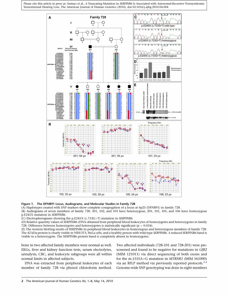
Figure 1. The DFNB91 Locus, Audiograms, and Molecular Studies in Family 728(A) Haplotypes created with SNP markers show complete cosegregation of a locus at 6p25 (DFNB91) in family 728.(B) Audiograms of seven members of family 728. 301, 102, and 103 have heterozygous, 201, 101, 105, and 106 have homozygousp.E245X mutation in SERPINB6.(C) Electropherograms showing the p.E245X (c.733G>T) mutation in SERPINB6.(D) Relative quantity values of SERPINB6 cDNA obtained from peripheral blood leukocytes of homozygotes and heterozygotes in family728. Difference between homozygotes and heterozygotes is statistically significant (p ¼ 0.034).(E) The western blotting results of SERPINB6 in peripheral blood leukocytes in homozygous and heterozygous members of family 728.The 42 kDa protein is clearly visible in NIH/3T3, HeLa cells, and a healthy person with wild-type SERPINB6. A reduced SERPINB6 band isvisible in a heterozygote. The SERPINB6 protein band is completely absent in homozygotes.
Please cite this article in press as: Sırmacı et al., A Truncating Mutation in SERPINB6 Is Associated with Autosomal-Recessive NonsyndromicSensorineural Hearing Loss, The American Journal of Human Genetics (2010), doi:10.1016/j.ajhg.2010.04.004
bone in two affected family members were normal as well.
EKGs, liver and kidney function tests, serum electrolytes,
urinalysis, CBC, and leukocyte subgroups were all within
normal limits in affected subjects.
DNA was extracted from peripheral leukocytes of each
member of family 728 via phenol chloroform method.
2 The American Journal of Human Genetics 86, 1–8, May 14, 2010
Two affected individuals (728-101 and 728-201) were pre-
screened and found to be negative for mutations in GJB2
(MIM 121011) via direct sequencing of both exons and
for the m.1555A>G mutation in MTRNR1 (MIM 561000)
via an RFLP method via previously reported protocols.3,4
Genome-wide SNP genotyping was done in eight members
Please cite this article in press as: Sırmacı et al., A Truncating Mutation in SERPINB6 Is Associated with Autosomal-Recessive NonsyndromicSensorineural Hearing Loss, The American Journal of Human Genetics (2010), doi:10.1016/j.ajhg.2010.04.004
of family 728 (201, 301, 101, 102, 103, 104, 105, and 106)
via Illumina 1M duo beadchips and assays (Illumina, CA).
The DNA samples were processed according to Illumina
procedures for processing of the Infinium II assay, the
BeadChips were scanned on the Illumina BeadArray
Reader, and data were extracted by the Illumina Beadstudio
software (Illumina). Before analysis, overall sample call
rate, gender consistency checks, relationship inference (via
the Graphical Representation of Relationships program),5
and the Mendelian inconsistency rates were used for
quality assessment. Genotypes were transferred into Excel
files and sorted according to genomic positions along with
all 35 previously identified autosomal-recessive nonsyn-
dromic deafness genes. The cosegregation of the flanking
genotypes for each gene was visually evaluated. None of
the known deafness loci cosegregated with the phenotype,
thereby excluding these as possible causes and suggesting
that a mutation in a previously unknown deafness gene
was responsible in this family. Copy number variants
(CNVs) were assessed by determining the relative loss or
gain of fluorescent signal intensity from SNP or CNV
probes on the array via PennCNV program6 and no segre-
gating CNVs were detected.
Regions of autozygosity from the SNP screen were first
sought with PLINK.7 All five affected members of family
728 were concordant for homozygous SNP marker blocks
that were larger than one megabase in five chromosomal
locations (Table S1 available online). Haplotypes were con-
structed for homozygous genomic segments and their
cosegregation with deafness was assessed visually. Only the
homozygous block between 289,878 bp (rs7762811) and
3,908,468 bp (rs13205752) (total length was 3,618,590
bp) at 6p25 cosegregated with hearing loss in the family
(Figure 1A) (NCBI Build 36.1; hg18). The other four large
shared homozygous streches in affected family members
did not cosegregate with the phenotype and were not
further analyzed. A genome-wide two-point linkage anal-
ysis was performed with SuperLink.8 Sixteen SNP markers
on chromosomes 6, 8, 16, and 21 were detected with
a LOD score of more than 3 (Table S2). Visual evaluation
of haplotypes in these regions confirmed that the largest
autozygous segment cosegregating with the phenotype
was the same 3,618,590 bp region at 6p25. There were
five other cosegregating segments on chromosomes 8,
16, and 21 with much smaller sizes ranging from 8,791
bp to 273,430 bp (Table S2). Before concentrating on the
largest autozygous region on chromosome 6, the other
five cosegregating regions were evaluated for their gene
content with the UCSC genome browser. There were no
known genes within the regions at chromosomes 16 and
21. The cosegregating segment on chromosome 8 was
14,852 bp long and partially included MSRA (MIM
601250). Sequencing of all six exons as well as intron-
exon boundaries of this gene in two affected members of
family 728 did not detect a mutation.
The only remaining autozygous segment that could
contain a previously unrecognized deafness gene in family
T
728 was at 6p25. Multipoint linkage analysis of this seg-
ment was conducted with Fastlink9 assuming a fully pene-
trant autosomal-recessive disease, with a disease allele fre-
quency of 0.0001. Both inbreeding loops were retained
in the analysis. SNPs spanning the homozygous region
were chosen for linkage analysis based on tagging and
heterozygosity in the parents. Allele frequencies were ob-
tained from the CEU HapMap. Linkage analysis detected
a maximum lod score of 5.0 between recombinant markers
rs7762811 and rs13205752 at 6p25 (Figure 1A). Examina-
tion of haplotypes between these two SNPs not included
in the multipoint analysis did not identify any recombina-
tions. This locus was designated as DFNB91 by the HUGO
Nomenclature Committee.
The linkage interval at 6p25 included 24 known protein
coding genes listed on the UCSC database (Figure S1).
None of these genes have been previously reported to
cause any forms of hearing loss in humans or in animals.
The coding exons and intron-exon boundaries of all
24 genes were sequenced in family 728 (Figure S1 and
Table S3). A homozygous nonsense mutation, p.E245X
(c.733G>T) (according to GenBank accession number
NM_004568.4), was identified in SERPINB6 (MIM 173321)
in all affected members of family 728 (Figure 1C). No other
DNA sequence change in this gene was detected. This
mutation removes 131 amino acids in the carboxy terminal
of the protein including the reactive center loop. It com-
pletely cosegregated with deafness in family 728. The iden-
tified mutation was not found in 300 Turkish controls
via an amplification refractory mutation detection system
(ARMS) protocol that was shown to be sensitive and specific
with DNA sequence analysis-proven homozygous, hetero-
zygous, and wild-type samples (Table S3). One previously
unreported intronic change and 21 previously reported
SNPs were detected in other sequenced genes. None of
these changes were predicted to have an effect on function
(Table S4).
A premature stop codon was likely to activate nonsense-
mediated mRNA decay response, thus leading to a
decreased SERPINB6 mRNA expression. SERPINB6 was
known to be expressed in white blood cells. Total RNA
was isolated from peripheral blood samples of seven
members of family 728 and cDNA was synthesized. The
sequencing of SERPINB6 in cDNA containing the point
mutation with exonic primers detected the p.E245X
(c.733G>T) mutation in both homozygotes and heterozy-
gotes, showing that the identified mutation was present at
the mRNA level. The quantitative expression analysis of
SERPINB6 was performed with TaqMan PCR assays in
quadriplicate for each individual on cDNA samples via
ABIPrism 7900 HT Sequence Detection system 2.3 (Applied
Biosystems Inc., CA) (for methods, see Table S3). The
mRNA expression was significantly reduced in homozy-
gotes compared to those in heterozygotes (Figure 1D)
(p ¼ 0.034; Mann-Whitney U test), indicating that the
premature stop codon destabilizes mRNA, leading to
mRNA decay.
he American Journal of Human Genetics 86, 1–8, May 14, 2010 3
Please cite this article in press as: Sırmacı et al., A Truncating Mutation in SERPINB6 Is Associated with Autosomal-Recessive NonsyndromicSensorineural Hearing Loss, The American Journal of Human Genetics (2010), doi:10.1016/j.ajhg.2010.04.004
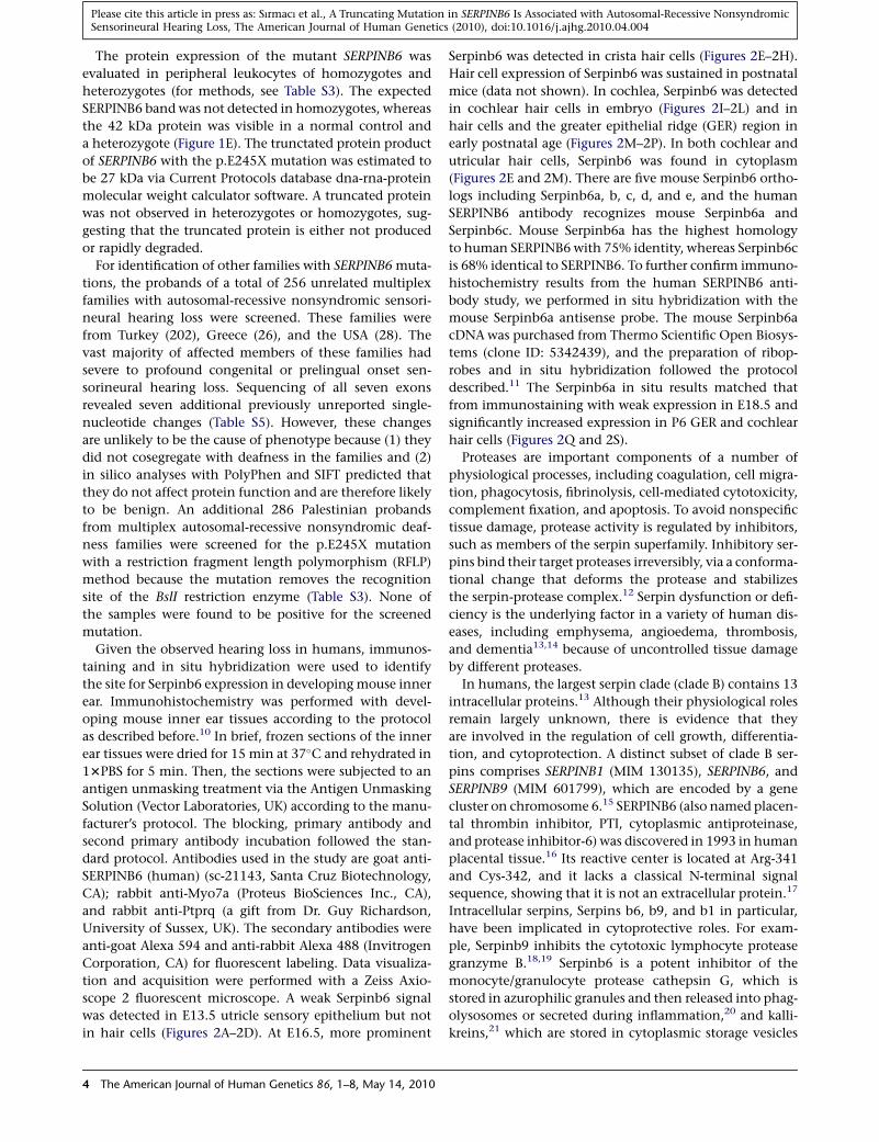
The protein expression of the mutant SERPINB6 was
evaluated in peripheral leukocytes of homozygotes and
heterozygotes (for methods, see Table S3). The expected
SERPINB6 band was not detected in homozygotes, whereas
the 42 kDa protein was visible in a normal control and
a heterozygote (Figure 1E). The trunctated protein product
of SERPINB6 with the p.E245X mutation was estimated to
be 27 kDa via Current Protocols database dna-rna-protein
molecular weight calculator software. A truncated protein
was not observed in heterozygotes or homozygotes, sug-
gesting that the truncated protein is either not produced
or rapidly degraded.
For identification of other families with SERPINB6 muta-
tions, the probands of a total of 256 unrelated multiplex
families with autosomal-recessive nonsyndromic sensori-
neural hearing loss were screened. These families were
from Turkey (202), Greece (26), and the USA (28). The
vast majority of affected members of these families had
severe to profound congenital or prelingual onset sen-
sorineural hearing loss. Sequencing of all seven exons
revealed seven additional previously unreported single-
nucleotide changes (Table S5). However, these changes
are unlikely to be the cause of phenotype because (1) they
did not cosegregate with deafness in the families and (2)
in silico analyses with PolyPhen and SIFT predicted that
they do not affect protein function and are therefore likely
to be benign. An additional 286 Palestinian probands
from multiplex autosomal-recessive nonsyndromic deaf-
ness families were screened for the p.E245X mutation
with a restriction fragment length polymorphism (RFLP)
method because the mutation removes the recognition
site of the BslI restriction enzyme (Table S3). None of
the samples were found to be positive for the screened
mutation.
Given the observed hearing loss in humans, immunos-
taining and in situ hybridization were used to identify
the site for Serpinb6 expression in developing mouse inner
ear. Immunohistochemistry was performed with devel-
oping mouse inner ear tissues according to the protocol
as described before.10 In brief, frozen sections of the inner
ear tissues were dried for 15 min at 37�C and rehydrated in
13PBS for 5 min. Then, the sections were subjected to an
antigen unmasking treatment via the Antigen Unmasking
Solution (Vector Laboratories, UK) according to the manu-
facturer’s protocol. The blocking, primary antibody and
second primary antibody incubation followed the stan-
dard protocol. Antibodies used in the study are goat anti-
SERPINB6 (human) (sc-21143, Santa Cruz Biotechnology,
CA); rabbit anti-Myo7a (Proteus BioSciences Inc., CA),
and rabbit anti-Ptprq (a gift from Dr. Guy Richardson,
University of Sussex, UK). The secondary antibodies were
anti-goat Alexa 594 and anti-rabbit Alexa 488 (Invitrogen
Corporation, CA) for fluorescent labeling. Data visualiza-
tion and acquisition were performed with a Zeiss Axio-
scope 2 fluorescent microscope. A weak Serpinb6 signal
was detected in E13.5 utricle sensory epithelium but not
in hair cells (Figures 2A–2D). At E16.5, more prominent
4 The American Journal of Human Genetics 86, 1–8, May 14, 2010
Serpinb6 was detected in crista hair cells (Figures 2E–2H).
Hair cell expression of Serpinb6 was sustained in postnatal
mice (data not shown). In cochlea, Serpinb6 was detected
in cochlear hair cells in embryo (Figures 2I–2L) and in
hair cells and the greater epithelial ridge (GER) region in
early postnatal age (Figures 2M–2P). In both cochlear and
utricular hair cells, Serpinb6 was found in cytoplasm
(Figures 2E and 2M). There are five mouse Serpinb6 ortho-
logs including Serpinb6a, b, c, d, and e, and the human
SERPINB6 antibody recognizes mouse Serpinb6a and
Serpinb6c. Mouse Serpinb6a has the highest homology
to human SERPINB6 with 75% identity, whereas Serpinb6c
is 68% identical to SERPINB6. To further confirm immuno-
histochemistry results from the human SERPINB6 anti-
body study, we performed in situ hybridization with the
mouse Serpinb6a antisense probe. The mouse Serpinb6a
cDNA was purchased from Thermo Scientific Open Biosys-
tems (clone ID: 5342439), and the preparation of ribop-
robes and in situ hybridization followed the protocol
described.11 The Serpinb6a in situ results matched that
from immunostaining with weak expression in E18.5 and
significantly increased expression in P6 GER and cochlear
hair cells (Figures 2Q and 2S).
Proteases are important components of a number of
physiological processes, including coagulation, cell migra-
tion, phagocytosis, fibrinolysis, cell-mediated cytotoxicity,
complement fixation, and apoptosis. To avoid nonspecific
tissue damage, protease activity is regulated by inhibitors,
such as members of the serpin superfamily. Inhibitory ser-
pins bind their target proteases irreversibly, via a conforma-
tional change that deforms the protease and stabilizes
the serpin-protease complex.12 Serpin dysfunction or defi-
ciency is the underlying factor in a variety of human dis-
eases, including emphysema, angioedema, thrombosis,
and dementia13,14 because of uncontrolled tissue damage
by different proteases.
In humans, the largest serpin clade (clade B) contains 13
intracellular proteins.13 Although their physiological roles
remain largely unknown, there is evidence that they
are involved in the regulation of cell growth, differentia-
tion, and cytoprotection. A distinct subset of clade B ser-
pins comprises SERPINB1 (MIM 130135), SERPINB6, and
SERPINB9 (MIM 601799), which are encoded by a gene
cluster on chromosome 6.15 SERPINB6 (also named placen-
tal thrombin inhibitor, PTI, cytoplasmic antiproteinase,
and protease inhibitor-6) was discovered in 1993 in human
placental tissue.16 Its reactive center is located at Arg-341
and Cys-342, and it lacks a classical N-terminal signal
sequence, showing that it is not an extracellular protein.17
Intracellular serpins, Serpins b6, b9, and b1 in particular,
have been implicated in cytoprotective roles. For exam-
ple, Serpinb9 inhibits the cytotoxic lymphocyte protease
granzyme B.18,19 Serpinb6 is a potent inhibitor of the
monocyte/granulocyte protease cathepsin G, which is
stored in azurophilic granules and then released into phag-
olysosomes or secreted during inflammation,20 and kalli-
kreins,21 which are stored in cytoplasmic storage vesicles
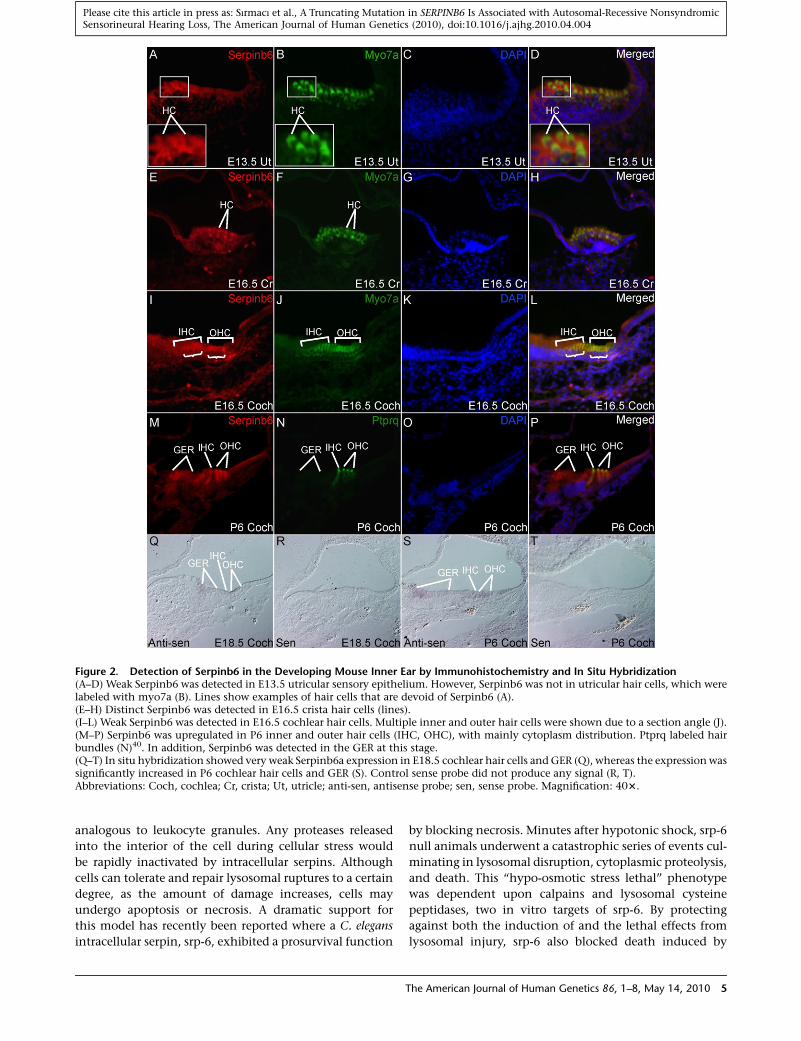
Figure 2. Detection of Serpinb6 in the Developing Mouse Inner Ear by Immunohistochemistry and In Situ Hybridization(A–D) Weak Serpinb6 was detected in E13.5 utricular sensory epithelium. However, Serpinb6 was not in utricular hair cells, which werelabeled with myo7a (B). Lines show examples of hair cells that are devoid of Serpinb6 (A).(E–H) Distinct Serpinb6 was detected in E16.5 crista hair cells (lines).(I–L) Weak Serpinb6 was detected in E16.5 cochlear hair cells. Multiple inner and outer hair cells were shown due to a section angle (J).(M–P) Serpinb6 was upregulated in P6 inner and outer hair cells (IHC, OHC), with mainly cytoplasm distribution. Ptprq labeled hairbundles (N)40. In addition, Serpinb6 was detected in the GER at this stage.(Q–T) In situ hybridization showed very weak Serpinb6a expression in E18.5 cochlear hair cells and GER (Q), whereas the expression wassignificantly increased in P6 cochlear hair cells and GER (S). Control sense probe did not produce any signal (R, T).Abbreviations: Coch, cochlea; Cr, crista; Ut, utricle; anti-sen, antisense probe; sen, sense probe. Magnification: 403.
Please cite this article in press as: Sırmacı et al., A Truncating Mutation in SERPINB6 Is Associated with Autosomal-Recessive NonsyndromicSensorineural Hearing Loss, The American Journal of Human Genetics (2010), doi:10.1016/j.ajhg.2010.04.004
analogous to leukocyte granules. Any proteases released
into the interior of the cell during cellular stress would
be rapidly inactivated by intracellular serpins. Although
cells can tolerate and repair lysosomal ruptures to a certain
degree, as the amount of damage increases, cells may
undergo apoptosis or necrosis. A dramatic support for
this model has recently been reported where a C. elegans
intracellular serpin, srp-6, exhibited a prosurvival function
T
by blocking necrosis. Minutes after hypotonic shock, srp-6
null animals underwent a catastrophic series of events cul-
minating in lysosomal disruption, cytoplasmic proteolysis,
and death. This ‘‘hypo-osmotic stress lethal’’ phenotype
was dependent upon calpains and lysosomal cysteine
peptidases, two in vitro targets of srp-6. By protecting
against both the induction of and the lethal effects from
lysosomal injury, srp-6 also blocked death induced by
he American Journal of Human Genetics 86, 1–8, May 14, 2010 5
Please cite this article in press as: Sırmacı et al., A Truncating Mutation in SERPINB6 Is Associated with Autosomal-Recessive NonsyndromicSensorineural Hearing Loss, The American Journal of Human Genetics (2010), doi:10.1016/j.ajhg.2010.04.004
heat shock, oxidative stress, hypoxia, and cation channel
hyperactivity.22
To assess the effects of overexpression of wild-type and
mutant SERPINB6 under osmotic stress, we studied tran-
siently transfected human cell lines and evaluated the
integrity of the lysosomal content. For the evaluation of
the lysosomal integrity, we used a pH-dependent marker
of lysosomes, LysoTracker-red, in cultured cells as reported
previously.23 The lysates of HeLa cells were probed for
SERPINB6 with monoclonal antibody (SA-622, Enzo Life-
Sciences, PA), which showed that HeLa cells expressed
SERPINB6 endogenously. Constructs encoding Serpinb6-
GFP fusion protein were subcloned from an untagged
Human cDNA clone (SC321547, OriGene Technologies,
MD) via PCR amplification and TOPO cloning via the
pcDNA3.1/NT-GFP-TOPO cloning kit (Invitrogen Corpora-
tion, CA). The primer sequences used for generating
the constructs are available in Table S3. In brief, the
SERPINB6-GFP-F primer was used in conjunction with
the SERPINB6-WT-R or the SERPINB6-MUT-GFP-R to
generate a PCR amplicon for either the wild-type or
mutant constructs, respectively. The proper sized amplicon
was selected for gel extraction and then cloned into
the pcDNA3.1/NT-GFP-TOPO. Colonies were selected and
screened via Sanger sequencing with Applied Biosystems
BigDye Terminator Cycle Sequencing Kit (Applied Biosys-
tems Inc., CA).
HeLa cells were transfected with the SERPINB6-WT-GFP,
the SERPINB6-MUT-GFP, and the pcDNA3.1-Control-GFP
expression vectors in 96-well BD Falcon plates (353948,
BD Bioscience, CA) with Lipofectamine 2000 and incubated
at 37�C with 5% CO2. Twenty-four hours after transfection,
cells were incubated in a 75 nM working concentration of
LysoTracker Red 99-DND (Invitrogen Corporation) and
then either continued to be cultured in PBS or introduced
to osmotic shock by replacing the culture media with
molecular biology grade water. After 5 min, plates were
measured on Cyntellect’s adherent cell cytometer Celigo
(Celigo Inc., CA). Three-channel, 8-bit stitched images were
generated covering whole wells to identify the surface area
and number of cells along with fluorescent intensities.
We measured three channels: bright field, to count and
locate cells; green, to measure number and intensity of
the expressed recombinant protein; and red, to measure
intensity of LysoTracker. Images were analyzed with the
Celigo software (Celigo Inc.). The intensity of the red
channel signal was averaged per each replicate in PBS and
water. A Student’s two-tailed t test was performed by com-
paring two histograms of intensity distributed over 35
bins from 0 to 255, representing the dynamic range at
which intensity can be measured across an 8-bit image.
HeLa cells transfected with wild-type SERPINB6 showed
less reduction in red florescence (lysosomal content
assessed through the maintenance of their lysosomal pH
gradient and acidic environment) than that of the trun-
cated mutant protein or the control GFP expression vector.
Differences for the average intensities of LysoTracker
6 The American Journal of Human Genetics 86, 1–8, May 14, 2010
between PBS and water incubation were 43.23%, 65.48%,
and 60.47% for SERPINB6-WT-GFP, SERPINB6-Mut-GFP,
and control-GFP, respectively (Figure 3), with all shifts
being found statistically significant with p values of 0.011,
0.00005, and 0.0001, respectively. The SERPINB6-Mut-GFP
and control-GFP showed 44.4% and 39.0% more intensity
decrease, respectively, in LysoTracker signal as compared to
the SERPINB6-WT-GFP protein when incubated in water;
p values are 0.003 and 0.024, respectively.
Further, we observed that cells incubated in PBS show
discernable red structures representing lysosomes. When
PBS was replaced with water, LysoTracker morphology
changed from bright punctuate spots to red spots with
a general pinkish hue in the cytosol (Figure 3). This shift
can also be seen in the histogram distributions (Figures
3A–3C), which indicates that lysosomes cannot maintain
their acidic environment, leading to leakage and disrup-
tion.23 Observed changes in red fluoresecence was roughly
equivalent between the Control-GFP and the SERPINB6
Mutant while disintegrity of lysosomes in cells from the
overexpressed SERPINB6-WT constructs was significantly
less (Figures 3A–3G). These results demonstrate that wild-
type SERPINB6 has a protective effect for lysosomal integ-
rity in osmotic stress in a human cell line, similar to the
results of a previous study for srp-6 shown in C. elegans.22
Loss of this protective effect, as shown in our experiments
with the truncated protein, is associated with increased
lysosomal disintegrity. We also showed that the truncated
protein is localized in the cytoplasm, similar to the wild-
type SERPINB6, and did not have an additional adverse
effect on lysosomal integrity. These results are consistent
with the theory that the phenotype associated with the
homozygous truncated SERPINB6 is due to loss of function
of the protein.
Given its increased expression in hair cells in the aging
inner ear, it is highly plausible to envision a role for serpins
in the inner ear cells, and particularly in hair cells, in the
protection against various noxious stimuli including noise,
ototoxic drugs, or trauma during aging process. These
stimuli as well as normal physiological hearing processes
may induce cellular stress, such as oxidative stress, that
results in death of hair cells as individuals get older.24
Leakage of lysosomal content resulting from various stress
conditions is likely to occur in hair cells. To reduce hair cell
damage and remain functional, SERPINB6 may be required
to counter the potentially cytotoxic components of leak-
ing lysosomal proteases.
In conclusion, several lines of evidence supported the
hypothesis that the identified SERPINB6 mutation was
responsible for hearing loss: (1) it completely cosegregated
with hearing loss in a large family; (2) the identified muta-
tion removed the functional domain of the SERPINB6
protein, SERPINB6 mRNA expression was reduced, and
protein expression was absent in homozygotes; (3) the
identified mutation was absent in 300 ethnically matched
controls; and (4) SERPINB6 was expressed in the inner
ear, in particular in hair cells. The phenotype associated
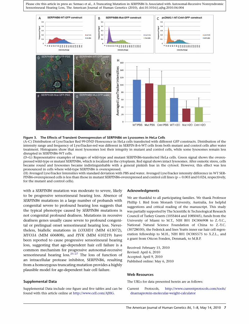
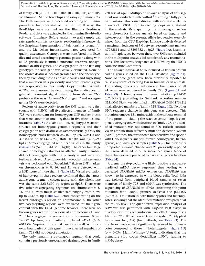
Figure 3. The Effects of Transient Overexpression of SERPINB6 on Lysosomes in HeLa Cells(A–C) Distribution of LysoTracker Red 99-DND Florescence in HeLa cells transfected with different GFP constructs. Distribution of theintensity range and frequency of LysoTracker-red was different in SERPIN-B-6-WT cells from both mutant and control cells after watertreatment. Histograms show that most lysosomes lost their integrity in mutant and control cells, while some lysosomes remain lessdisrupted in SERPINB6-WT cells.(D–G) Representative examples of images of wild-type and mutant SERPINB6-transfected HeLa cells. Green signal shows the overex-pressed wild-type or mutant SERPINB6, which is localized in the cytoplasm. Red signal shows intact lysosomes. After osmotic stress, cellsbecame round and lysosomes became indistinguishable with a general pinkish hue in the cytosol. However, this effect was lesspronounced in cells where wild-type SERPINB6 is overexpressed.(H) Averaged LysoTracker Intensities with standard deviation with PBS and water. Averaged LysoTracker intensity difference in WT SER-PINB6-overexpressed cells is less than those in mutant SERPINB6-overexpressed and control cell lines (p¼ 0.003 and 0.024, respectively,for the mutant and control cells).
Please cite this article in press as: Sırmacı et al., A Truncating Mutation in SERPINB6 Is Associated with Autosomal-Recessive NonsyndromicSensorineural Hearing Loss, The American Journal of Human Genetics (2010), doi:10.1016/j.ajhg.2010.04.004
with a SERPINB6 mutation was moderate to severe, likely
to be progressive sensorineural hearing loss. Absence of
SERPINB6 mutations in a large number of probands with
congenital severe to profound hearing loss suggests that
the typical phenotype caused by SERPINB6 mutations is
not congenital profound deafness. Mutations in recessive
deafness genes usually cause severe to profound congeni-
tal or prelingual onset sensorineural hearing loss. Never-
theless, biallelic mutations in LOXHD1 (MIM 613072),
MYO3A (MIM 606808), and PJVK (MIM 610219) have
been reported to cause progressive sensorineural hearing
loss, suggesting that age-dependent hair cell failure is a
common mechanism for progressive autosomal-recessive
sensorineural hearing loss.25–27 The loss of function of
an intracellular protease inhibitor, SERPINB6, resulting
from a homozygous truncating mutation provides a highly
plausible model for age-dependent hair cell failure.
Supplemental Data
Supplemental Data include one figure and five tables and can be
found with this article online at http://www.cell.com/AJHG.
T
Acknowledgments
We are thankful to all participating families. We thank Professor
Phillip I. Bird from Monash University, Australia, for helpful
suggestions and critical reading of the manuscript. This study
was partially supported by The Scientific & Technological Research
Council of Turkey Grants (105S464 and 108S045), funds from the
University of Miami to M.T., NIH R01 DC006908 to Z.-Y.C.,
National Natural Science Foundation of China to Z.-Y.C.
(30728030), the Federick and Ines Yeatts inner ear hair cell regen-
eration fellowship to M.H., NIH R01 DC005575 to X.Z.L., and
a grant from Oticon Fonden, Denmark, to M.B.P.
Received: February 15, 2010
Revised: April 6, 2010
Accepted: April 9, 2010
Published online: May 6, 2010
Web Resources
The URLs for data presented herein are as follows:
Current Protocols, http://www.currentprotocols.com/tools/
dnarnaprotein-molecular-weight-calculator
he American Journal of Human Genetics 86, 1–8, May 14, 2010 7
Please cite this article in press as: Sırmacı et al., A Truncating Mutation in SERPINB6 Is Associated with Autosomal-Recessive NonsyndromicSensorineural Hearing Loss, The American Journal of Human Genetics (2010), doi:10.1016/j.ajhg.2010.04.004
Hereditary Hearing Loss Homepage, http://webh01.ua.ac.be/hhh/
National Institutes of Health (NIH) HapMap-CEU, http://www.
ncbi.nih.gov/SNP/snp_viewTable.cgi?pop¼1409
Online Mendelian Inheritance in Man (OMIM), http://www.ncbi.
nlm.nih.gov/Omim/
Plink, http://pngu.mgh.harvard.edu/purcell/plink
PolyPhen, http://genetics.bwh.harvard.edu/pph/
SIFT, http://sift.jcvi.org/
UCSC Genome Browser, http://genome.ucsc.edu
References
1. Morton, C.C., and Nance, W.E. (2006). Newborn hearing
screening—A silent revolution.N. Engl. J.Med.354, 2151–2164.
2. Morton, N.E. (1991). Genetic epidemiology of hearing impair-
ment. Ann. N Y Acad. Sci. 630, 16–31.
3. Tekin, M., Duman, T., Bogoclu, G., Incesulu, A., Comak, E.,
Fitoz, S., Yilmaz, E., Ilhan, I., and Akar, N. (2003). Frequency
of mtDNA A1555G and A7445G mutations among children
with prelingual deafness in Turkey. Eur. J. Pediatr. 162,
154–158.
4. Tekin, M., and Arici, Z.S. (2007). Genetic epidemiological
studies of congenital/prelingual deafness in turkey: Population
structure and mating type are major determinants of mutation
identification. Am. J. Med. Genet. A. 143A, 1583–1591.
5. Abecasis, G.R., Cherny, S.S., Cookson, W.O., and Cardon, L.R.
(2001). GRR: Graphical representation of relationship errors.
Bioinformatics 17, 742–743.
6. Wang, K., Li, M., Hadley, D., Liu, R., Glessner, J., Grant, S.F.,
Hakonarson, H., and Bucan, M. (2007). PennCNV: An inte-
grated hidden markov model designed for high-resolution
copy number variation detection in whole-genome SNP geno-
typing data. Genome Res. 17, 1665–1674.
7. Purcell, S., Neale, B., Todd-Brown, K., Thomas, L., Ferreira,
M.A.R., Bender, D., Maller, J., Sklar, P., de Bakker, P.I.W.,
Daly, M.J., et al. (2007). PLINK: A toolset for whole-genome
association and population-based linkage analysis. Am. J.
Hum. Genet. 81, 559–575.
8. Fishelson, M., and Geiger, D. (2002). Exact genetic linkage
computations for general pedigrees. Bioinformatics 18 (Suppl 1),
S189–S198.
9. Schaffer, A.A. (1996). Faster linkage analysis computations for
pedigrees withloopsor unusedalleles.Hum.Hered.46, 226–235.
10. Sage, C., Huang, M., Vollrath, M.A., Brown, M.C., Hinds, P.W.,
Corey, D.P., Vetter, D.E., and Chen, Z.Y. (2006). Essential role
of retinoblastoma protein in mammalian hair cell develop-
ment and hearing. Proc. Natl. Acad. Sci. USA 103, 7345–7350.
11. Huang, M., Sage, C., Li, H., Xiang, M., Heller, S., and Chen,
Z.Y. (2008). Diverse expression patterns of LIM-homeodomain
transcription factors (LIM-HDs) in mammalian inner ear
development. Dev. Dyn. 237, 3305–3312.
12. Huntington, J.A., Read, R.A., and Carrell, R.W. (2000). Struc-
ture of a serpin-protease complex shows inhibition by defor-
mation. Nature 407, 923–926.
13. Silverman, G.A., Bird, P.I., Carrell, R.W., Church, F.C., Cough-
lin, P.B., Gettins, P.G.W., Irving, J.A., Lomas, D.A., Luke, C.J.,
Moyer, R.W., et al. (2001). The serpins are an expanding super-
family of structurally similar but functionally diverse proteins.
J. Biol. Chem. 276, 33293–33296.
14. Stein, P.E., and Carrell, R.W. (1995). What do dysfunctional
serpins tell us about molecular mobility and disease? Nat.
Struct. Biol. 2, 96–113.
8 The American Journal of Human Genetics 86, 1–8, May 14, 2010
15. Scott, F.L., Eyre, H.J., Lioumi, M., Ragoussis, J., Irving, J.A., Su-
therland, G.A., and Bird, P.I. (1999). Human ovalbumin serpin
evolution: phylogenic analysis, gene organization, and identi-
fication of new PI-8-related genes suggest that two interchro-
mosomal and several intrachromosomal duplications gener-
ated the gene clusters at 18q21-q23 and 6p25. Genomics 62,
490–499.
16. Coughlin, P.B., Tetaz, T., and Salem, H.H. (1993). Identifica-
tion and purification of a novel serine proteinase inhibitor.
J. Biol. Chem. 268, 9541–9547.
17. Scott, F.L., Coughlin, P.B., Bird, C., Cerruti, L., Hayman, J.A.,
and Bird, P. (1996). Proteinase inhibitor 6 cannot be secreted,
which suggests it is a new type of cellular serpin. J. Biol. Chem.
271, 1605–1612.
18. Bird, C.H., Sutton, V.R., Sun, J., Hirst, C.E., Novak, A., Trapani,
J.A., and Bird, P.I. (1998). Selective regulation of apoptosis: The
cytotoxic lymphocyte serpin proteinase inhibitor 9 protects
against granzyme B-mediated apoptosis without perturbing
the Fas cell death pathway. Mol. Cell. Biol. 18, 6387–6398.
19. Hirst, C.E., Buzza, M.S., Bird, C.H., Warren, H.S., Cameron,
P.U., Zhang, M., Ashton-Rickhardt, P.G., and Bird, P.I.
(2003). The intracellular granzyme B inhibitor, proteinase
inhibitor 9, is up-regulated during accessory cell maturation
and effector cell degranulation, and its overexpression
enhances CTL potency. J. Immunol. 170, 805–815.
20. Scott, F.L., Hirst, C.E., Sun, J., Bottomley, S.P., Bird, C.H., and
Bird, P.I. (1999). The intracellular serpin proteinase inhibitor
6 (PI-6) is expressed in monocytes and granulocytes and is
a potent inhibitor of the azurophilic granule proteinase,
cathepsin G. Blood 93, 2089–2097.
21. Scott, F.L., Sun, J., Whisstock, J.C., Kato, K., and Bird, P.I.
(2007). SerpinB6 is an inhibitor of kallikrein-8 in keratino-
cytes. J. Biochem. 142, 435–442.
22. Luke, C.J., Pak, S.C., Askew, Y.S., Naviglia, T.L., Askew, D.J.,
Nobar, S.M., Vetica, A.C., Long, O.S., Watkins, S.C., Stolz,
D.B., et al. (2007). An intracellular serpin regulates necrosis
by inhibiting the induction and sequelae of lysosomal injury.
Cell 130, 1108–1119.
23. Zheng, J., Yan, T., Feng, Y., and Zhai, Q. (2010). Involvement
of lysosomes in the early stages of axon degeneration. Neuro-
chem. Int. 56, 516–521.
24. Van De Water, T.R., Lallemend, F., Eshraghi, A.A., Ahsan, S.,
He, J., Guzman, J., Polak, M., Malgrange, B., Lefebvre, P.P.,
Staecker, H., et al. (2004). Caspases, the enemy within, and
their role in oxidative stress-induced apoptosis of inner ear
sensory cells. Otol. Neurotol. 25, 627–632.
25. Grillet, N., Schwander, M., Hildebrand, M.S., Sczaniecka, A.,
Kolatkar, A., Velasco, J., Webster, J.A., Kahrizi, K., Najmabadi,
H., Kimberling, W.J., et al. (2009). Mutations in LOXHD1, an
evolutionarily conserved stereociliary protein, disrupt hair
cell function in mice and cause progressive hearing loss in
humans. Am. J. Hum. Genet. 85, 328–337.
26. Walsh, T., Walsh, V., Vreugde, S., Hertzano, R., Shahin, H.,
Haika, S., Lee, M.K., Kanaan, M., King, M.C., and Avraham,
K.B. (2002). From flies’ eyes to our ears: mutations in a human
class III myosin cause progressive nonsyndromic hearing loss
DFNB30. Proc. Natl. Acad. Sci. USA 99, 7518–7523.
27. Delmaghani, S., del Castillo, F.J., Michel, V., Leibovici, M.,
Aghaie, A., Ron, U., Van Laer, L., Ben-Tal, N., Van Camp, G.,
Weil, D., et al. (2006). Mutations in the gene encoding pejvakin,
a newly identified protein of the afferent auditory pathway,
cause DFNB59 auditory neuropathy. Nat. Genet. 38, 770–778.
Related Documents