A transcriptional network in polycystic kidney disease Lionel Gresh 1,4 , Evelyne Fischer 1,4 , Andreas Reimann 1,4 , Myriam Tanguy 2 , Serge Garbay 1 , Xinli Shao 3 , Thomas Hiesberger 3 , Laurence Fiette 2 , Peter Igarashi 3 , Moshe Yaniv 1 and Marco Pontoglio 1, * 1 Unite ´ Expression Ge ´ne ´tique et Maladies/CNRS URA 1644, De ´partement de Biologie du De ´veloppement, Institut Pasteur, Paris, France, 2 Unite ´ de Recherche et d’Expertise en Histotechnologie et Pathologie, Institut Pasteur, Paris, France and 3 Division of Nephrology, Department of Internal Medicine, University of Texas Southwestern Medical Center, Dallas, TX, USA Mutations in cystic kidney disease genes represent a major genetic cause of end-stage renal disease. However, the molecular cascades controlling the expression of these genes are still poorly understood. Hepatocyte Nuclear Factor 1b (HNF1b) is a homeoprotein predominantly ex- pressed in renal, pancreatic and hepatic epithelia. We report here that mice with renal-specific inactivation of HNF1b develop polycystic kidney disease. We show that renal cyst formation is accompanied by a drastic defect in the transcriptional activation of Umod, Pkhd1 and Pkd2 genes, whose mutations are responsible for distinct cystic kidney syndromes. In vivo chromatin immunoprecipita- tion experiments demonstrated that HNF1b binds to sev- eral DNA elements in murine Umod, Pkhd1, Pkd2 and Tg737/Polaris genomic sequences. Our results uncover a direct transcriptional hierarchy between HNF1b and cystic disease genes. Interestingly, most of the identified HNF1b target gene products colocalize to the primary cilium, a crucial organelle that plays an important role in control- ling the proliferation of tubular cells. This may explain the increased proliferation of cystic cells in MODY5 patients carrying autosomal dominant mutations in HNF1b. The EMBO Journal (2004) 23, 1657–1668. doi:10.1038/ sj.emboj.7600160; Published online 18 March 2004 Subject Categories: molecular biology of disease Keywords: cilium; cysts; HNF1b; MODY5; proliferation Introduction The development of the kidney depends on reciprocal inter- actions between the ureteric bud and the metanephric me- senchyme. Factors secreted by the ureteric bud induce the aggregation of the mesenchyme and subsequent conversion into the epithelium. The induced mesenchyme in turn sends signals to the ureteric bud to promote its branching. This reciprocal induction proceeds in an ordered manner to give rise to the collecting duct system and simultaneously to the nephrons (see Vainio and Lin, 2002). Failure of the differentiation program during nephrogenesis can lead to diverse pathological conditions that can perturb renal structure and function. Mutations in cystic kidney disease genes constitute the most common genetic cause of chronic renal failure in both children and adults (see Igarashi and Somlo, 2002). Despite their genetic, clinical and histo- pathological heterogeneity, these renal diseases result in a similar outcome: dilation of tubules and cystogenesis. These observations have led to the hypothesis that the gene defects underlying these disorders might disrupt a common pathway. However, the mechanisms of cyst formation are still not completely elucidated. They are postulated to involve dys- functions in the control of cell proliferation, apoptosis, cell polarity, the localization of ion channels, fluid secretion and cell–cell adhesion or cell–matrix interactions (see Igarashi and Somlo, 2002). During the past decade, positional cloning strategies have identified several genes responsible for cystic kidney diseases. Genes mutated in autosomal dominant poly- cystic kidney disease (ADPKD) (PKD1 and PKD2) (Hughes et al, 1995; Mochizuki et al, 1996), autosomal recessive polycystic kidney disease (ARPKD) (PKHD1) (Ward et al, 2002), nephronophthisis (NPHP1 to 4) (Hildebrandt et al, 1997; Otto et al, 2002; Olbrich et al, 2003; Otto et al, 2003) and medullary cystic kidney disease (MCKD) type 2 (UMOD/THP) (Hart et al, 2002) have been identified. Recently, ADPKD, ARPKD and nephro- nophthisis gene products have been localized in the primary cilium, a solitary non-motile organelle that extends from the apical surface of renal epithelial cells (see Watnick and Germino, 2003). Pkd1- and Pkd2-encoded proteins may be part of a mechanosensitive Ca 2 þ -signaling pathway in renal cilia that inhibits renal tubular cell growth (Nauli et al, 2003). If ciliary structure or function is perturbed, tubular cell growth may continue, leading to cyst formation. The role of the proteins involved in ARPKD and nephronophthisis in the cilium is still not clear. Polaris, a protein involved in a mouse polycystic kidney disease model, is also localized in the cilium, and plays a role in the assembly of this organelle (Pazour et al, 2000). Mutations in the human UMOD gene are responsible for MCKD type 2, but the mechanisms underlying the cyst formation in this disease also remain elusive. Relatively little is known about the transcriptional net- works that control epithelial differentiation of renal tubules. Hepatocyte Nuclear Factor 1b (HNF1b) is a homeodomain- containing transcription factor that binds DNA and transacti- vates transcription as homodimer, or heterodimers with the closely related factor, HNF1a (see Cereghini, 1996). Both proteins were initially described as liver-enriched transcription factors, but their expression pattern is not restricted to hepatocytes (Blumenfeld et al, 1991; see Received: 11 November 2003; accepted: 13 February 2004; published online: 18 March 2004 *Corresponding author: Unite ´ Expression Ge ´ne ´tique et Maladies/CNRS URA 1644, De ´partement de Biologie du De ´veloppement, Institut Pasteur, 25, rue du Docteur Roux, 75724 Paris Cedex 15, France. Tel.: þ 33 1 45 68 85 14; Fax: þ 33 1 40 61 30 33; E-mail: [email protected] 4 These authors contributed equally to this work The EMBO Journal (2004) 23, 1657–1668 | & 2004 European Molecular Biology Organization | All Rights Reserved 0261-4189/04 www.embojournal.org & 2004 European Molecular Biology Organization The EMBO Journal VOL 23 | NO 7 | 2004 EMBO THE EMBO JOURNAL THE EMBO JOURNAL 1657

Welcome message from author
This document is posted to help you gain knowledge. Please leave a comment to let me know what you think about it! Share it to your friends and learn new things together.
Transcript

A transcriptional network in polycystic kidneydisease
Lionel Gresh1,4, Evelyne Fischer1,4, AndreasReimann1,4, Myriam Tanguy2, SergeGarbay1, Xinli Shao3, Thomas Hiesberger3,Laurence Fiette2, Peter Igarashi3, MosheYaniv1 and Marco Pontoglio1,*1Unite Expression Genetique et Maladies/CNRS URA 1644, Departementde Biologie du Developpement, Institut Pasteur, Paris, France, 2Unite deRecherche et d’Expertise en Histotechnologie et Pathologie, InstitutPasteur, Paris, France and 3Division of Nephrology, Department ofInternal Medicine, University of Texas Southwestern Medical Center,Dallas, TX, USA
Mutations in cystic kidney disease genes represent a major
genetic cause of end-stage renal disease. However, the
molecular cascades controlling the expression of these
genes are still poorly understood. Hepatocyte Nuclear
Factor 1b (HNF1b) is a homeoprotein predominantly ex-
pressed in renal, pancreatic and hepatic epithelia. We
report here that mice with renal-specific inactivation of
HNF1b develop polycystic kidney disease. We show that
renal cyst formation is accompanied by a drastic defect in
the transcriptional activation of Umod, Pkhd1 and Pkd2
genes, whose mutations are responsible for distinct cystic
kidney syndromes. In vivo chromatin immunoprecipita-
tion experiments demonstrated that HNF1b binds to sev-
eral DNA elements in murine Umod, Pkhd1, Pkd2 and
Tg737/Polaris genomic sequences. Our results uncover a
direct transcriptional hierarchy between HNF1b and cystic
disease genes. Interestingly, most of the identified HNF1btarget gene products colocalize to the primary cilium, a
crucial organelle that plays an important role in control-
ling the proliferation of tubular cells. This may explain the
increased proliferation of cystic cells in MODY5 patients
carrying autosomal dominant mutations in HNF1b.
The EMBO Journal (2004) 23, 1657–1668. doi:10.1038/
sj.emboj.7600160; Published online 18 March 2004
Subject Categories: molecular biology of disease
Keywords: cilium; cysts; HNF1b; MODY5; proliferation
Introduction
The development of the kidney depends on reciprocal inter-
actions between the ureteric bud and the metanephric me-
senchyme. Factors secreted by the ureteric bud induce the
aggregation of the mesenchyme and subsequent conversion
into the epithelium. The induced mesenchyme in turn sends
signals to the ureteric bud to promote its branching. This
reciprocal induction proceeds in an ordered manner to give
rise to the collecting duct system and simultaneously to the
nephrons (see Vainio and Lin, 2002).
Failure of the differentiation program during nephrogenesis
can lead to diverse pathological conditions that can perturb
renal structure and function. Mutations in cystic kidney
disease genes constitute the most common genetic cause of
chronic renal failure in both children and adults (see Igarashi
and Somlo, 2002). Despite their genetic, clinical and histo-
pathological heterogeneity, these renal diseases result in a
similar outcome: dilation of tubules and cystogenesis. These
observations have led to the hypothesis that the gene defects
underlying these disorders might disrupt a common pathway.
However, the mechanisms of cyst formation are still not
completely elucidated. They are postulated to involve dys-
functions in the control of cell proliferation, apoptosis, cell
polarity, the localization of ion channels, fluid secretion and
cell–cell adhesion or cell–matrix interactions (see Igarashi
and Somlo, 2002). During the past decade, positional cloning
strategies have identified several genes responsible for cystic
kidney diseases. Genes mutated in autosomal dominant poly-
cystic kidney disease (ADPKD) (PKD1 and PKD2) (Hughes
et al, 1995; Mochizuki et al, 1996), autosomal recessive
polycystic kidney disease (ARPKD) (PKHD1)
(Ward et al, 2002), nephronophthisis (NPHP1 to 4)
(Hildebrandt et al, 1997; Otto et al, 2002; Olbrich et al,
2003; Otto et al, 2003) and medullary cystic kidney disease
(MCKD) type 2 (UMOD/THP) (Hart et al, 2002) have
been identified. Recently, ADPKD, ARPKD and nephro-
nophthisis gene products have been localized in the primary
cilium, a solitary non-motile organelle that extends from
the apical surface of renal epithelial cells (see Watnick
and Germino, 2003). Pkd1- and Pkd2-encoded proteins
may be part of a mechanosensitive Ca2þ -signaling pathway
in renal cilia that inhibits renal tubular cell growth (Nauli et al,
2003). If ciliary structure or function is perturbed, tubular cell
growth may continue, leading to cyst formation. The role of
the proteins involved in ARPKD and nephronophthisis in the
cilium is still not clear. Polaris, a protein involved in a mouse
polycystic kidney disease model, is also localized in the
cilium, and plays a role in the assembly of this organelle
(Pazour et al, 2000). Mutations in the human UMOD gene are
responsible for MCKD type 2, but the mechanisms underlying
the cyst formation in this disease also remain elusive.
Relatively little is known about the transcriptional net-
works that control epithelial differentiation of renal tubules.
Hepatocyte Nuclear Factor 1b (HNF1b) is a homeodomain-
containing transcription factor that binds DNA and transacti-
vates transcription as homodimer, or heterodimers with
the closely related factor, HNF1a (see Cereghini, 1996).
Both proteins were initially described as liver-enriched
transcription factors, but their expression pattern is not
restricted to hepatocytes (Blumenfeld et al, 1991; seeReceived: 11 November 2003; accepted: 13 February 2004; publishedonline: 18 March 2004
*Corresponding author: Unite Expression Genetique et Maladies/CNRSURA 1644, Departement de Biologie du Developpement, Institut Pasteur,25, rue du Docteur Roux, 75724 Paris Cedex 15, France.Tel.: þ 33 1 45 68 85 14; Fax: þ 33 1 40 61 30 33;E-mail: [email protected] authors contributed equally to this work
The EMBO Journal (2004) 23, 1657–1668 | & 2004 European Molecular Biology Organization | All Rights Reserved 0261-4189/04
www.embojournal.org
&2004 European Molecular Biology Organization The EMBO Journal VOL 23 | NO 7 | 2004
EMBO
THE
EMBOJOURNAL
THE
EMBOJOURNAL
1657

Cereghini, 1996). In particular, these proteins are expressed
in the tubular epithelial cells of the kidney (Lazzaro et al,
1992; Pontoglio et al, 1996). HNF1b is expressed throughout
the entire nephron, from proximal tubules to collecting
ducts, whereas HNF1a expression is restricted to proximal
tubules. Interestingly, HNF1b is also highly expressed in
other tubular epithelia such as biliary and pancreatic ducts
(Coffinier et al, 1999a).
Autosomal dominant mutations in the HNF1b gene are
associated with a particular form of diabetes mellitus called
Maturity Onset Diabetes of the Young type 5 (MODY5)
(Horikawa et al, 1997). Affected patients also present diverse
forms of non-diabetic nephropathies and, with less penetrant
occurrence, genital malformations and liver dysfunction
(Nishigori et al, 1998; Lindner et al, 1999; Bingham et al,
2000). The most common and penetrant trait of these patients
is the presence of renal cysts and diabetes (RCAD). The renal
manifestations of the disease also include cystic renal dyspla-
sia, probably due to a defective nephrogenesis, oligomegane-
phronia and hypoplastic Glomerulocystic kidney disease
(GCKD) (Bingham et al, 2001). In zebrafish, mutations of
HNF1b affect pronephros development leading to cystic pro-
nephric tubules (Sun and Hopkins, 2001). The expression of
HNF1b mutants in Xenopus laevis affects the normal devel-
opment of pronephric structures (Bohn et al, 2003).
Furthermore, HNF1b plays an essential role during mouse
early embryogenesis. HNF1b-null mice die around embryonic
day 7 (E7) due to a defect in visceral endoderm differentiation
(Barbacci et al, 1999; Coffinier et al, 1999b). To assess the
roles of HNF1b during late embryogenesis, we made use of a
Cre-LoxP strategy. We demonstrate here that kidney-specific
inactivation of HNF1b leads to a polycystic phenotype. We
found that the expression of several genes involved in cystic
diseases is severely affected in the conditionally inactivated
mice. By chromatin immunoprecipitation, we show that three
of them (Umod, Pkhd1 and Pkd2) are directly controlled by
HNF1b. These results describe a direct transcriptional hier-
archy between HNF1b and these genes, and establish the role
of this transcription factor in regulating the terminal differ-
entiation of renal tubular cells.
Results
Kidney-specific inactivation of the HNF1b gene
The early lethality of HNF1b-deficient embryos prevented the
analysis of its function at later developmental stages. To
circumvent this problem, we inactivated the HNF1b gene
using the Cre-LoxP strategy. To restrict the inactivation to
renal cells, we made use of a KspCre transgenic mouse strain
that expresses the Cre recombinase under the control of the
Ksp-cadherin (Cadherin 16) promoter. This transgene drives
efficient Cre expression and Cre-mediated recombination in
collecting ducts, loops of Henle and only occasionally (less
than 5%) in proximal tubular cells (Shao et al, 2002).
Inactivation of the HNF1b gene was achieved by generating
mice carrying a homozygous floxed HNF1b gene (Coffinier
et al, 2002) along with the KspCre transgene (KspCre;
HNF1bflox/flox). In a parallel way, we also generated com-
pound heterozygous animals carrying a null HNF1b lacZ
knock-in (Coffinier et al, 1999b) and a floxed allele along
with the KspCre transgene (KspCre; HNF1bflox/LacZ). These
two genotypes involved a renal-specific inactivation of
HNF1b and gave rise to the same phenotype. For these
reasons, we described these animals as ‘mutants’.
Conversely, littermates carrying either (KspCre; HNF1bflox/þ )
or (HNF1bflox/flox) or (HNF1bflox/þ ) or (HNF1bflox/LacZ) were
phenotypically indistinguishable from the wild-type mice.
For this reason, we termed these animals collectively as
‘controls’.
Kidney-specific inactivation of HNF1b leads to postnatal
lethality and renal failure
Pups carrying all possible genotypes were born in normal
Mendelian ratios (N¼ 159, P40.05). No lethality was ob-
served up to postnatal day 8 (P8). Between days P10 and P21,
75% of mutants died (N¼ 8). Generally, mutant animals were
growth retarded by P8. At this stage, the average weight of
mutant pups was 11% lower than that of controls
(Ncontrol¼ 75, Nmutant¼ 22, Po0.05). Growth retardation in-
creased with age, and mutants that survived weaning exhib-
ited an approximately 25% decrease in body weight.
To investigate renal function, we determined seric urea and
creatinine concentrations. Measurements were performed at
different ages (P8 and on the few surviving animals at P17
and P30) (Figure 1). Both urea and creatinine levels were
increased in mutant mice at all time points. These data
indicated that mutants suffered, and died from, a severe
impairment of renal function. Most measurements required
euthanasia of the pups, therefore these data do not represent
an average time-course progression of renal disease.
Figure 1 Impaired renal function in mutant mice. Serum concen-trations of urea (A) and creatinine (B) at P8, P17 and P30.
HNF1b controls expression of cystic disease genesL Gresh et al
The EMBO Journal VOL 23 | NO 7 | 2004 &2004 European Molecular Biology Organization1658

Inactivation of HNF1b results in a renal polycystic
phenotype
We observed bilateral ureteral dilation, also involving the
renal pelvis, in most mutants at P8 (92%, N¼ 25) (Figure 2).
No signs of ureter obstruction were seen. As HNF1b is
expressed in the ureteral epithelium, and as KspCre had
driven recombination in the precursors of these cells (data
not shown), we concluded that this abnormality was a direct
consequence of HNF1b inactivation in ureter-epithelial cells.
The kidneys of mutants were much paler than the controls
but were apparently normally sized at P8 (Figure 2). Gross
abnormalities of mature nephrons suggested that terminal
morphogenesis did not occur normally at P1. In particular,
several tubules located in the medullary region of mutant
kidneys were dilated and contained more cells than in con-
trols (Figure 3A and B). Histological sections at P8 revealed
that the kidneys of all mutant mice exhibited numerous cysts
(Figure 3C and D). These cysts were mostly located in the
medulla, but a few could also be observed in the cortex. The
average size of cysts increased with age (Figure 3D and F). It
is noteworthy that dilation of the urinary space in some
glomeruli could be clearly observed at all stages (Figure 3F).
Renal cysts are lined with Cre recombined HNF1b�/�
cells
The KspCre transgene drives efficient recombination in the
renal medulla. Histological abnormalities in HNF1b-inacti-
vated mice mainly affected this region, suggesting that the
renal cysts were a direct consequence of HNF1b inactivation.
To confirm this hypothesis, we monitored the recombination
status of cystic cells using the ROSA26R reporter allele
(Soriano, 1999). We generated mice with kidney-specific
inactivated HNF1b combined with the ROSA26R reporter
allele (KspCre; HNF1bflox/flox; ROSA26R). This allele
contains a lacZ gene whose activity is induced by the Cre-
driven recombination (Soriano, 1999). These mice had the
same ureteral and cystic phenotype as KspCre; HNF1bflox/lacZ
animals (Figure 4B, 5F and data not shown). X-gal stained
kidney sections revealed that the renal cysts were lined
with b-galactosidase-positive cells (Figure 4B). As expected,
control mice (KspCre; HNF1bflox/þ ; ROSA26R) showed
Figure 2 HNF1b inactivation results in bilateral ureteral dilation. Macroscopic view of urinary tract in control (A) and mutant (B, C) mice atP8. Some mutants exhibit a mild ureter dilation (arrows) (compare B versus A), while others present a more severe phenotype (compare Cversus A). Mutant kidneys had a normal size, but were paler than controls. Scale bars: 0.25 cm.
Figure 3 Inactivation of HNF1b in kidney cells leads to polycystickidney disease. Hematoxylin–eosin-stained kidney sections fromcontrol and mutant mice at P1 (A, B), P8 (C, D) and P12 (E, F).Tubular dilations were visible in the medullary region of mutants atP1, with an increased number of nuclei per tubule section comparedto controls (B versus A). In mutants, the medulla was completelydisrupted by large cysts by P8 (D). The size of cysts increased withage (F). A few glomerular cysts were observed in the deep cortex (F,arrowheads). Scale bars: A, B: 100mm, C–F: 400 mm.
HNF1b controls expression of cystic disease genesL Gresh et al
&2004 European Molecular Biology Organization The EMBO Journal VOL 23 | NO 7 | 2004 1659

b-gal-positive cells lining most medullary segments of
the kidney tubules without any cyst formation (Figure 4A).
These data indicate that all cystic cells underwent
Cre-mediated recombination or were derived from recom-
bined progenitor cells.
To further prove that Cre activity had inactivated the
endogenous HNF1b gene, we performed immunofluorescent
detection using a novel polyclonal antibody (HNF1b-NC-AR)
(Figure 4C–H). This analysis revealed that the HNF1b protein
was indeed absent from cystic epithelium (Figure 4D and H),
Figure 4 Cystic cells underwent recombination and lack HNF1b expression. (A, B) X-gal staining on kidney sections of control KspCre;HNF1bflox/þ ; ROSA26R (A) and mutant KspCre; HNF1bflox/flox; ROSA26R (B) at P14. b-gal activity is an indicator of Cre-driven recombination onthe ROSA26R locus. (A) The medulla of control mice showed recombination in the tubular epithelium. (B) In mutants, all cysts were lined withrecombined cells. (C–H) Kidney sections of control HNF1bflox/lacZ and mutant KspCre; HNF1bflox/lacZ at P8. (C, D) HNF1b staining (fluorescein/green). (E, F) Nuclear staining of the same section (DAPI/red). (G, H) Merging. (G) In controls, HNF1b is expressed in tubular but not inmesenchymal cells (arrowhead). (H) Mutants showed no expression of HNF1b in cysts. cy: cyst. Scale bars: 75mm.
HNF1b controls expression of cystic disease genesL Gresh et al
The EMBO Journal VOL 23 | NO 7 | 2004 &2004 European Molecular Biology Organization1660

whereas its expression was conserved in proximal tubules
(data not shown). Thus, renal cysts were lined with recom-
bined HNF1b-null cells.
To establish the cellular origin of cysts, we analyzed
specific markers of collecting ducts and Henle’s loops. Most
cysts expressed Aquaporin2 (Aqp2) protein, a collecting duct
cell marker (Figure 7G). Some cysts expressed the sodium–
potassium-chloride cotransporter isoform 2 (Slc12a1), a spe-
cific marker of thick ascending limb of Henle’s loop (Figure
7C and K) (see Delpire et al, 1996). Taken together, these data
indicate that the cysts were mainly derived from two nephron
segments where KspCre is expressed.
Analysis of kidneys from P8 animals also showed that
mesenchymal cells were present in both control and mutant
mice, but they were more abundant in mutants (Figure 5A
and B). From P12 onwards, the presence of mesenchymal
cells was visible in mutants, but not in controls at the same age
(Figure 3E and F). We investigated whether these mesench-
ymal cells expressed HNF1b. We took advantage of the HNF1blacZ allele in heterozygous control mice (HNF1bflox/lacZ).
Mesenchymal cells were b-gal-negative, indicating that they
do not express HNF1b (Figure 5A). In addition, immuno-
fluorescence experiments showed that these cells do not
express the HNF1b protein (Figure 4G). Similarly, mesench-
ymal cells present in mutants (KspCre; HNF1bflox/lacZ) were
also b-gal-negative, indicating that these cells were not
programmed to express the endogenous HNF1b gene
(Figure 5B). In a parallel manner, we investigated whether
these mesenchymal cells had undergone KspCre-driven re-
combination using the KspCre; HNF1bflox/flox; ROSA26R mice.
X-gal staining of kidney sections from these animals showed
that all mesenchymal cells were b-gal-negative, whereas cells
lining cysts were b-gal-positive (Figure 5F). Thus, these
mesenchymal cells did not express KspCre and were not
Figure 5 HNF1b inactivation leads to multilayered tubular epithelia. (A, B) X-gal staining on kidney sections of control HNF1bflox/lacZ (A) andmutant KspCre; HNF1bflox/lacZ (B) at P8. b-gal activity is an indicator of the endogenous HNF1b promoter activity. (A) HNF1b was expressed inall tubular cells but not in mesenchyme (arrowhead). (B) Mesenchyme is b-gal negative (arrowhead), indicating that it was not programmed toexpress HNF1b. (C, D) Hematoxylin–eosin-stained sections of control (C) and mutant (D) at P8. Multilayered tubular epithelial cells wereobserved (arrows), as well as polyps growing into the cyst lumen (D and insert). (E, F) X-gal staining on kidney sections of control KspCre;HNF1bflox/þ ; ROSA26R (E) and mutant KspCre; HNF1bflox/flox; ROSA26R (F) at P14. b-gal activity is an indicator of Cre-driven recombination.(E) KspCre-driven recombination was seen in a large proportion of medullary tubular cells. (F) Several cysts lack the typical monolayeredepithelial structure (arrows). All epithelial cells are b-gal-positive, demonstrating that they underwent Cre recombination. The mesenchyme inmutants was not affected by recombination (arrowhead). Scale bars: 200mm.
HNF1b controls expression of cystic disease genesL Gresh et al
&2004 European Molecular Biology Organization The EMBO Journal VOL 23 | NO 7 | 2004 1661
derived from cells where the KspCre had been expressed.
Both experiments demonstrate that the increase in mesench-
ymal cells in mutant kidneys is a secondary consequence of
HNF1b inactivation.
Increased proliferation in mutant renal cysts is cell-
autonomous
Increased proliferation is one of the major mechanisms
underlying cyst formation in cystic kidney diseases.
Notwithstanding this propensity to proliferate, renal cystic
cells are usually organized in a monolayered epithelium (see
Igarashi and Somlo, 2002). This is actually the case for most
of the cysts that we found in mutants at P8. However, about
15% of cysts showed multilayered epithelia at this stage
(Figure 5D). Similar to what has been observed in human
polycystic kidney disease (see Bernstein et al, 1987), some
polyps developed into the cyst lumen (insert in Figure 5D).
Using the ROSA26R reporter allele, we have investigated the
lineage of these proliferating cells. Interestingly, in mutant
KspCre; HNF1bflox/flox; ROSA26R animals, all cells in multi-
layered structures were positively stained with X-gal, indicat-
ing that all of them had recombined (Figure 5F). Therefore,
the presence of multilayered cysts is probably due to a cell-
autonomous defect. This observation indicates that HNF1binactivation in tubular epithelial cells results in increased
proliferation.
Mutant mice show specific transcriptional defects
of several cystic disease genes
To unravel the molecular mechanisms underlying cyst for-
mation in our mouse model, we monitored the expression
levels of genes known to be involved in cystogenesis in mice
and humans. We performed quantitative RT–PCR experi-
ments using kidney RNA from mutant and control animals.
We found that the expression level of Umod (Uromodulin/
Tamm–Horsfall glycoprotein) was decreased 10-fold in mu-
tant kidney. This result could be due to a direct defective
transcriptional activation of the Umod gene by HNF1b.
Alternatively, this deficiency could be due to the absence of
cells that normally express this gene (Bachmann et al, 1990)
because of a developmental or differentiation defect. The
grossly normal expression of a marker of the thick ascending
limb of Henle’s loops (TAL), the sodium–potassium-chloride
cotransporter isoform 2 (Slc12a1/Nkcc2) (Figure 6), argued
against this possibility. In addition, when the expression of
Umod was normalized for the expression of Slc12a1, down-
regulation of Umod expression was still calculated to be 8.5-
fold (Figure 6). This normalization allowed us to demonstrate
that the Umod transcriptional defect is not linked to the
absence of TAL cells in mutant mice.
Interestingly, two other key genes of cyst formation, Pkhd1
and Pkd2, were also downregulated (3.4- and 1.5-fold, re-
spectively). In contrast, Pkd1 and Pkd2l2 (a Pkd2-related
gene) (Guo et al, 2000) expression levels were normal.
Among the genes involved in nephronophthisis, only
Nphp1 was significantly downregulated in mutant mice
(1.4-fold), whereas Invs/Nphp2, Nphp3 and Nphp4 were
normally expressed (Figure 6).
We also investigated the expression of two genes involved
in cystogenesis in mice, Tg737/Polaris (Moyer et al, 1994)
and Kif3a (Lin et al, 2003). The former was mildly, but
significantly, dowregulated (1.3-fold) in mutant mice,
whereas Kif3a was normally expressed (Figure 6).
We analyzed the expression of Aqp2 and arginine-vaso-
pressin-receptor type 2 (Avpr2). These two genes are ex-
pressed in collecting ducts, a structure that contributes to
cyst formation. Similar to what has been described in differ-
ent polycystic rodent models (Gattone et al, 2003), both of
these genes were upregulated in mutant kidneys (Figure 6).
Our results show that renal HNF1b inactivation results in
a reduced expression of several genes known to play crucial
roles in cystogenesis. In fact, at least five of these genes
(Umod, Pkhd1, Pkd2, Nphp1 and Tg737/Polaris) were speci-
fically affected by HNF1b inactivation.
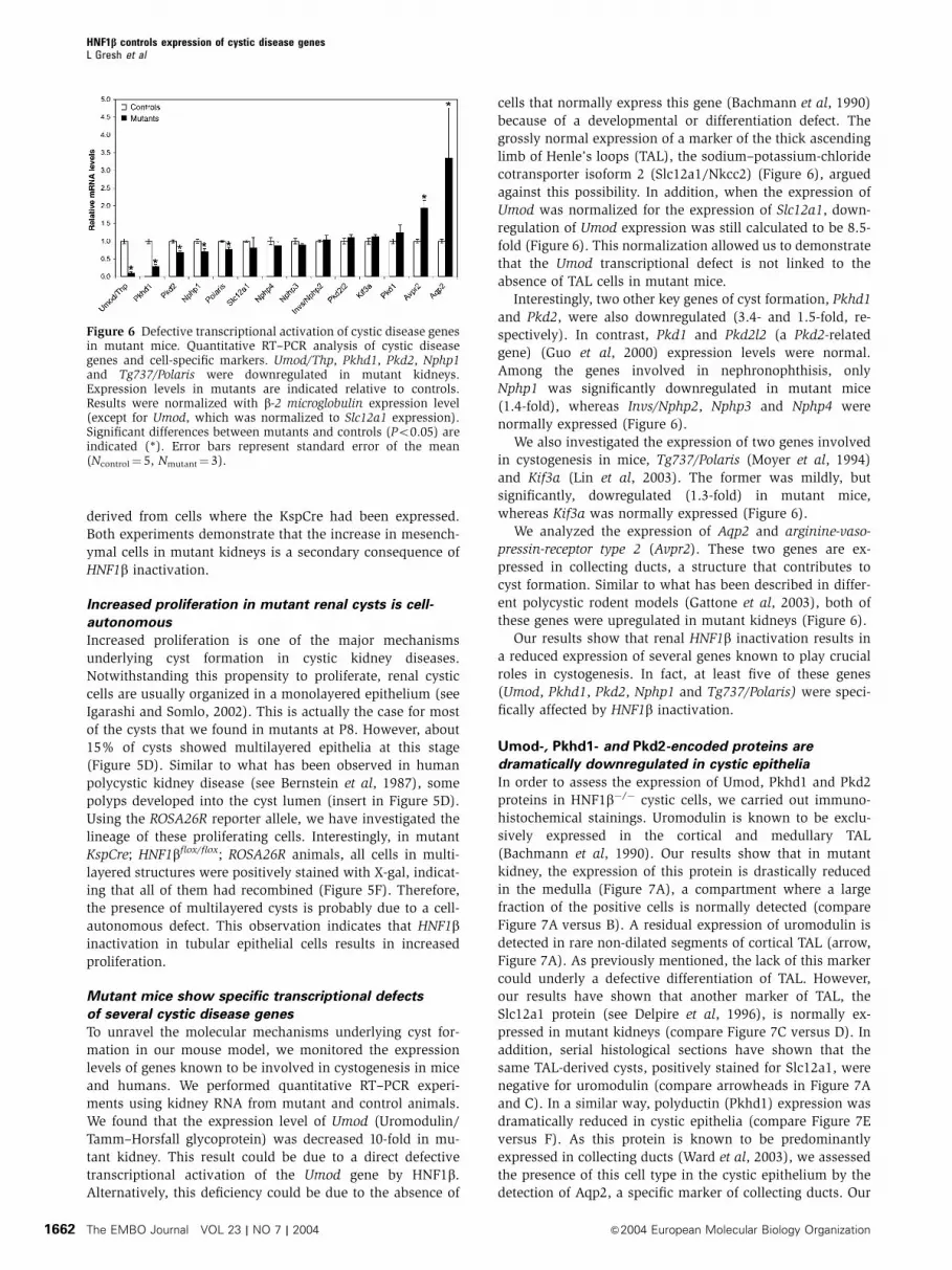
Umod-, Pkhd1- and Pkd2-encoded proteins are
dramatically downregulated in cystic epithelia
In order to assess the expression of Umod, Pkhd1 and Pkd2
proteins in HNF1b�/� cystic cells, we carried out immuno-
histochemical stainings. Uromodulin is known to be exclu-
sively expressed in the cortical and medullary TAL
(Bachmann et al, 1990). Our results show that in mutant
kidney, the expression of this protein is drastically reduced
in the medulla (Figure 7A), a compartment where a large
fraction of the positive cells is normally detected (compare
Figure 7A versus B). A residual expression of uromodulin is
detected in rare non-dilated segments of cortical TAL (arrow,
Figure 7A). As previously mentioned, the lack of this marker
could underly a defective differentiation of TAL. However,
our results have shown that another marker of TAL, the
Slc12a1 protein (see Delpire et al, 1996), is normally ex-
pressed in mutant kidneys (compare Figure 7C versus D). In
addition, serial histological sections have shown that the
same TAL-derived cysts, positively stained for Slc12a1, were
negative for uromodulin (compare arrowheads in Figure 7A
and C). In a similar way, polyductin (Pkhd1) expression was
dramatically reduced in cystic epithelia (compare Figure 7E
versus F). As this protein is known to be predominantly
expressed in collecting ducts (Ward et al, 2003), we assessed
the presence of this cell type in the cystic epithelium by the
detection of Aqp2, a specific marker of collecting ducts. Our
Figure 6 Defective transcriptional activation of cystic disease genesin mutant mice. Quantitative RT–PCR analysis of cystic diseasegenes and cell-specific markers. Umod/Thp, Pkhd1, Pkd2, Nphp1and Tg737/Polaris were downregulated in mutant kidneys.Expression levels in mutants are indicated relative to controls.Results were normalized with b-2 microglobulin expression level(except for Umod, which was normalized to Slc12a1 expression).Significant differences between mutants and controls (Po0.05) areindicated (*). Error bars represent standard error of the mean(Ncontrol¼ 5, Nmutant¼ 3).
HNF1b controls expression of cystic disease genesL Gresh et al
The EMBO Journal VOL 23 | NO 7 | 2004 &2004 European Molecular Biology Organization1662
results showed that this cell type was indeed present, as a
large proportion of cysts was positive for Aqp2 (compare
Figure 7G versus H). Notwithstanding a high background
signal detected in mesenchymal cells, we showed that the
expression of polycystin2 (Pkd2) was absent in cystic cells
(compare arrowed epithelia in Figure 7I versus 7J). Again,
since Pkd2 in the medulla is predominantly expressed in TAL
(Foggensteiner et al, 2000), we monitored the expression of
Slc12a1 in adjacent sections and showed that corresponding
cysts expressed this marker but not Pkd2 (see arrows in Figure
7I versus K). Remarkably, Pkd2 was normally expressed
throughout the whole cortex (Supplementary Figure 1). In
Figure 7 HNF1b inactivation leads to a strong decrease of Umod, Pkhd1 and Pkd2 protein levels in cystic cells. Immunostainings foruromodulin (A, B), Pkhd1 (E, F), Pkd2 (I, J), Slc12a1 (C, D, K, L), and Aqp2 (G, H) in mutant and control mice at P8. (A–D) Uromodulinexpression pattern. In mutants, no uromodulin staining was detected in the medullary cysts (compare A versus B). Some cysts were clearlyshown to be thick ascending limb (TAL)-derived (see the arrowheaded positive staining with a-Slc12a1 antibody in C). Arrowheads in Aindicate the position of the corresponding cysts in the adjacent section. In controls, both uromodulin and Slc12a1 proteins colocalized(compare adjacent sections B and D, respectively). In mutants, only few cortical nondilated TAL segments expressed uromodulin (see arrow inA). (E–H) Pkhd1 expression pattern. In mutants, Pkhd1 expression was strongly decreased in the vast majority of medullary cystic cells(compare E versus F). Pkhd1 downregulation is not due to the absence of collecting duct cells, as most cysts still expressed aquaporin2(compare G versus E). (I–L) Pkd2 expression pattern. Pkd2 expression is absent in cystic epithelia (compare I versus J). Again, some cysts wereclearly shown to be TAL-derived (see the arrowed positive staining with a-Slc12a1 antibody in K). Arrows in I indicate the position of thecorresponding cysts, in the adjacent section. In control animals, both Pkd2 and Slc12a1 proteins colocalized (compare arrows in serial sectionsB and D, respectively). Scale bars: 300 mm.
HNF1b controls expression of cystic disease genesL Gresh et al
&2004 European Molecular Biology Organization The EMBO Journal VOL 23 | NO 7 | 2004 1663
this region of the kidney, KspCre is not expressed and there-
fore HNF1b is not deleted. As a result of this residual cortical
expression, RT–PCR carried out on whole kidney RNAs might
strongly underestimate the actual downregulation of Pkd2
expression. Taken together, these results clearly indicate that
uromodulin, Pkhd1 and Pkd2 are drastically downregulated
in HNF1b-deficient cells.
An important fraction of Pkhd1 and Pkd2 proteins is
localized to the primary cilium of tubular cells (Pazour et al,
2002; Ward et al, 2003). To assess the morphological integrity
of this organelle, we performed immunostaining of acetylated
tubulin, a specific component of the primary cilium. We
found that mutant cells had normal cilia (Supplementary
Figure 2). Moreover, morphometric analysis showed that
the cilium length, specifically measured in collecting duct
cells (costained with a-Aqp2 antibodies), was normal.
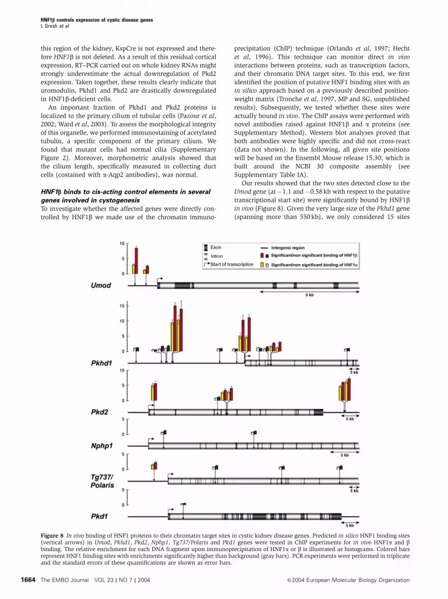
HNF1b binds to cis-acting control elements in several
genes involved in cystogenesis
To investigate whether the affected genes were directly con-
trolled by HNF1b we made use of the chromatin immuno-
precipitation (ChIP) technique (Orlando et al, 1997; Hecht
et al, 1996). This technique can monitor direct in vivo
interactions between proteins, such as transcription factors,
and their chromatin DNA target sites. To this end, we first
identified the position of putative HNF1 binding sites with an
in silico approach based on a previously described position-
weight matrix (Tronche et al, 1997, MP and SG, unpublished
results). Subsequently, we tested whether these sites were
actually bound in vivo. The ChIP assays were performed with
novel antibodies raised against HNF1b and a proteins (see
Supplementary Method). Western blot analyses proved that
both antibodies were highly specific and did not cross-react
(data not shown). In the following, all given site positions
will be based on the Ensembl Mouse release 15.30, which is
built around the NCBI 30 composite assembly (see
Supplementary Table IA).
Our results showed that the two sites detected close to the
Umod gene (at �1.1 and �0.58 kb with respect to the putative
transcriptional start site) were significantly bound by HNF1bin vivo (Figure 8). Given the very large size of the Pkhd1 gene
(spanning more than 550 kb), we only considered 15 sites
Figure 8 In vivo binding of HNF1 proteins to their chromatin target sites in cystic kidney disease genes. Predicted in silico HNF1 binding sites(vertical arrows) in Umod, Pkhd1, Pkd2, Nphp1, Tg737/Polaris and Pkd1 genes were tested in ChIP experiments for in vivo HNF1a and bbinding. The relative enrichment for each DNA fragment upon immunoprecipitation of HNF1a or b is illustrated as histograms. Colored barsrepresent HNF1 binding sites with enrichments significantly higher than background (gray bars). PCR experiments were performed in triplicateand the standard errors of these quantifications are shown as error bars.
HNF1b controls expression of cystic disease genesL Gresh et al
The EMBO Journal VOL 23 | NO 7 | 2004 &2004 European Molecular Biology Organization1664
identified in a window of 100 kb around the transcriptional
start site. Six of them showed a significant interaction with
HNF1b in vivo. Interestingly, strongly enriched sites were
located either in the proximal promoter (within B200 bp) or
much further upstream (�35 kb). All in vivo-enriched HNF1
sites in this gene occurred as doublets with a maximal inter-
distance of 400 bp. The in silico search identified four intronic
HNF1 sites in the Pkd2 gene and a doublet downstream of the
30 end of the gene (at around þ 51 kb). Among the four
intronic sites, only one was not enriched in vivo, whereas
the 30 doublet showed the highest enrichment (Bsix-fold).
The in silico search revealed four potential HNF1 binding
sites around the Tg737/Polaris gene. In this case only the
closest site (�6.64 kb) showed a weak, but significant, en-
richment for HNF1b. Nphp1 was the only candidate gene for
which we could not observe any in vivo interaction, notwith-
standing the in silico prediction of 2 sites. In order to assess
the specificity of our results, we also monitored the potential
binding of HNF1b to Pkd1, another cystic disease gene,
whose expression is not altered in our mice. We could predict
a single HNF1 binding site positioned at þ 4 kb, but this site
was not bound in vivo by HNF1b or HNF1a (Figure 8).
In vitro experiments have shown that HNF1a and HNF1bbind to their target sites with similar affinities, either as
homo- or heterodimers (De Simone et al, 1991; Rey-Campos
et al, 1991). Our results indicated that the enrichment
obtained with the a-HNF1a-specific antibody was system-
atically weaker than that of HNF1b. This observation could
be explained by the fact that HNF1b has a more widespread
expression pattern than HNF1a in the kidney. However, the
ratio of enrichment in the Umod gene was particularly strik-
ing. In this case, HNF1a binding was much lower, compared
to that of HNF1b. Interestingly, the Umod gene is the only
gene among the analyzed candidates that is not coexpressed
with HNF1a. This observation suggests that HNF1a is unable
to bind these sites efficiently in the proximal tubular cell, a
cell type that does not express the Umod gene.
In summary, our ChIP experiments demonstrate that
HNF1b clearly interacts with several HNF1 binding sites in
the Umod, Pkhd1 and Pkd2 genes. These cystic disease genes
can be considered as direct transcriptional targets of HNF1b.
Moreover, interaction with Tg737/Polaris could also be
shown, but to a weaker extent.
Discussion
Cystic kidney diseases represent a major cause of end-stage
renal failure. Several genes have recently been identified to be
involved in these disorders. However, little is known about
the transcriptional networks that regulate the expression of
these genes. HNF1b plays a crucial role in the development of
extraembryonic visceral endoderm and in the morphogenesis
of hepatic bile ducts. Here we show that mice lacking HNF1bspecifically in the kidney suffer from severe polycystic kidney
disease.
HNF1b and cystic kidney diseases
Our results show that the lack of HNF1b is directly respon-
sible for cyst formation via a cell-autonomous mechanism. In
humans, heterozygous mutations of HNF1b are associated
with the Renal Cysts and Diabetes (RCAD) syndrome. This
syndrome combines a form of type II diabetes called maturity
onset diabetes of the young type 5 (MODY5) with a non-
diabetic renal disease (Horikawa et al, 1997; Nishigori et al,
1998; Lindner et al, 1999; Bingham et al, 2000, 2001). The
manifestations of renal dysfunction are variable, ranging
from isolated cysts to multicystic kidneys. Little is known
about the molecular mechanisms underlying this phenotype.
Here we show that HNF1b directly controls the transcription
of several genes expressed in tubular epithelial cells, namely
Umod, Pkhd1 and Pkd2. In addition, HNF1b might contribute
to the transcriptional activation of Tg737/Polaris. All these
genes are known to be mutated in distinct cystic kidney
diseases: MCKD type 2 (UMOD) (Hart et al, 2002), ARPKD
(PKHD1) (Ward et al, 2002), ADPKD (PKD2) (Mochizuki et al,
1996) and a murine model of ARPKD (Tg737/Polaris) (Moyer
et al, 1994). Strikingly, although a defect in any of these genes
in humans is sufficient to elicit renal cyst formation, we
observed a concomitant downregulation of a specific subset
of cystic disease genes in mutant mice. The combined defects
in the expression of these genes could be the origin of the
massive cyst formation observed in our mouse model. These
results reveal a transcriptional hierarchy between HNF1b and
different cystic disease genes, and could explain why carriers
of autosomal dominant mutations in HNF1b develop renal
cysts.
Interestingly, MODY5 patients do not develop a strict
polycystic disease. They actually present a smaller number
of cysts in comparison to ADPKD or ARPKD patients. This
difference could be explained by the fact that the human
disease occurs in heterozygous carriers, whereas our model
involves homozygous HNF1b deletion. In order to understand
dominant inheritance in humans, it would be interesting to
test whether renal cysts in MODY5 patients develop upon loss
of the residual wild-type HNF1b allele. This ‘two-hit’ mechan-
ism has already been described for genes involved in ADPKD
(Pkd1 and Pkd2) (Watnick et al, 1998) and for tumor sup-
pressor genes.
Polyductin (Pkhd1) and polycystin2 (Pkd2) proteins are
preferentially localized in a specialized cellular organelle: the
primary cilium (Pazour et al, 2002; Ward et al, 2003). Several
studies suggest a link between a defect in cilium assembly, or
function, and renal cyst formation (see Watnick and
Germino, 2003). We show that HNF1b is not necessary for
cilium formation. However, our data demonstrate that the
genetic program driven by HNF1b involves the expression of
genes that play a crucial role in cilium function.
HNF1b inactivation leads to increased proliferation
of epithelial cells
The presence of renal cysts and the appearance of multi-
layered epithelia in mutant mice strongly suggest that the
inactivation of HNF1b leads to an increased rate of cellular
proliferation. Mice with liver-specific HNF1b inactivation
have dysplastic epithelia in both the gallbladder and in the
few remaining intrahepatic bile ducts. Multilayered epithelial
structures, reminiscent of those observed upon kidney-spe-
cific inactivation, are present in the biliary tract of these
animals (Coffinier et al, 2002). Recently, HNF1a was shown
to be involved in the control of cell proliferation. Indeed,
biallelic inactivation of HNF1a is frequently observed in liver
adenomas and in some hepatocellular carcinomas (Bluteau
et al, 2002). For this reason, HNF1a can be considered as a
tumor suppressor gene. We postulate that HNF1b could have
HNF1b controls expression of cystic disease genesL Gresh et al
&2004 European Molecular Biology Organization The EMBO Journal VOL 23 | NO 7 | 2004 1665

the same properties, and that the increased proliferation of
HNF1b�/� cells upon kidney- and liver-specific inactivation
of HNF1b is the result of defective regulation of cell cycle in
the absence of HNF1b. To validate this hypothesis, the
incidence of biallelic inactivation of HNF1b in renal cancers
and cholangiocarcinomas should be assessed. The antiproli-
ferative effects of HNF1b could be mediated by the biological
function of the Pkd1/Pkd2 complex, which has been recently
shown to control p21Cip1 transcription via the JAK/STAT
pathway (Bhunia et al, 2002). In addition, Pkd2 has been
shown to inhibit the Pkd1-dependent G protein activation
(Delmas et al, 2002). However, we cannot rule out that
defective expression of additional HNF1b target genes could
result in a nonfully differentiated state that, in turn, could
render growth arrest less efficient.
HNF1b chromatin targets
We show that the defective expression of the cystic disease
genes Umod, Pkhd1 and Pkd2 directly correlates with the
presence of several HNF1 binding sites that are bound by
HNF1b in vivo. This strongly suggests that HNF1b directly
controls the expression of these genes. Interestingly, the weak
interaction of HNF1b with its target site in Tg737/Polaris gene
is reflected by its marginally decreased transcription in the
mutant kidney.
The binding of HNF1b was not only restricted to proximal
promoter sites but also involved far upstream and down-
stream sites. In the case of Pkhd1, HNF1 binding sites were
identified at 35 kb upstream of the transcriptional start site.
These sites occur as doublet and are conserved in both
human and mouse genomes (data not shown).
Interestingly, this doublet is particularly enriched, suggesting
a strong interaction with HNF1b. Similarly, doublets of HNF1
binding sites in the Umod and Pkd2 genes were also strongly
bound, whereas isolated binding sites were weakly bound or
not bound at all. These data could suggest that HNF1boccupancy is enhanced by the presence of closely spaced
sites on chromatin. Alternatively, a higher relative enrich-
ment could also be explained by the increased probability for
a DNA fragment encompassing two HNF1 sites to be immu-
noprecipitated.
The liver-specific inactivation of HNF1b leads to defective
morphogenesis of intrahepatic bile ducts (IHBD) (ductal plate
malformation) (Coffinier et al, 2002). Interestingly, polycys-
tin2/Pkd2 and polyductin/Pkhd1 proteins are coexpressed
with HNF1b in biliary epithelial cells (Ong et al, 1999; Ward
et al, 2003). HNF1b could drive the expression of these genes
in both renal tubules and bile ducts. Notably, patients with
ARPKD/PKHD1 suffer from ductal plate malformations giving
rise to biliary dysgenesis, whereas patients with ADPKD/
PKD2 develop biliary cysts (see Igarashi and Somlo, 2002).
Although these patients do not strictly phenocopy the hepatic
HNF1b deficiency, concomitant transcriptional defects affect-
ing both Pkhd1 and Pkd2 genes could explain why IHBD fail
to differentiate in HNF1b�/� livers.
Our study provides novel insights into the molecular
mechanisms underlying HNF1b function in normal kidney
and in cystic disorders. Our results demonstrate that HNF1bis a critical regulator of a genetic cascade that is essential for
controlling the proliferation and differentiation of renal tub-
ular epithelial cells.
Materials and methods
AnimalsKidney-specific inactivation of HNF1b was obtained using aCre-LoxP strategy. KspCre (Shao et al), HNFsblacz/þ (Coffinieret al, 1999b) HNFsbflox/flox (Coffinier et al, 2002) and ROSA26R(Soriano) mice were previously described. All animals receivedhumane care, and the institutional review committee approvedthe study protocol.
Biochemical analysisSerum urea and creatinine levels were determined by the Centred’Explorations Fonctionnelles Integre (Faculte de Medecine X.Bichat).
Production of HNF1a and b antibodiesWe constructed GST fusion polypeptides of HNF1 a and b byexcluding regions of high homology between both proteins usinggene splicing by overlap extension PCR (SOE PCR) (Horton, 1995).All SOE PCRs were carried out with corresponding rat cDNAs. SeeSupplementary method 1 for more details.
Immunohistochemical proceduresKidney cryosections were fixed in 4% PFA and treated as previouslydescribed (Foggensteiner et al, 2000) with the following antibodies:rabbit a-HNF1b-NC-AR, 1:150; rabbit a-NKCC2 (a gift from MKnepper), 1:1000; rabbit a-Pkd2 (a gift from R Sandford), 1:200;a-THP (Biogenesis), 1:50. Aqp2 (rabbit a-Aqp2 antibody (a gift fromM Knepper), 1:400) and pkhd1 (mouse a-Pkhd1 antibody (a giftfrom C Ward), 1:200) were detected on paraffin sections accordingto Ward et al, (2003). X-gal stainings were performed as described(Pontoglio et al, 1996). For histological analysis, paraffin sectionswere stained with hematoxylin and eosin.
Quantitative RT–PCRcDNAs were generated from total kidney RNAs using the InvitrogenSuperscript II reagents. Quantitative RT–PCR was carried out on anABI PRISMs 7000 sequence detection system using SYBRs green.Primers were designed using PrimerExpress 2.0s software (AppliedBiosystems). b-2 microglobulin was used as the control gene fornormalization. All genes have been tested in P8 control (N¼ 5) andmutant (N¼ 3) mice. For primer sequences, see SupplementaryTable II.
Chromatin immunoprecipitation (ChIP)Nuclei were prepared from pooled kidneys (Cereghini et al, 1987).Chromatin preparation and ChIP were performed as described inSupplementary method 2. Three independent ChIP experimentswere performed with a-HNF1a-NC-AR or a-HNF1b-NC-AR anti-bodies, and corresponding eluates were pooled. Primers weredesigned using PrimerExpress 2.0s software. For primer sequences,see Supplementary Table IB. Quantification of precipitatedDNA fragments was carried out on an ABI PRISMs 7000sequence detection system using SYBRs green in triplicate.Relative fold in vivo enrichment of DNA fragments was calculatedusing the following formula: (ChIPtarget/ChIPnormalizer)/(inputtarget/inputnormalizer). As normalizer, we used a DNA fragment lacking anyHNF1 site, located in the first intron of aortic smooth muscle alpha-actin 2 gene.
Supplementary dataSupplementary data are available at The EMBO Journal Online.
Note added in proofSupporting our results, HNF1b has been shown to regulate theactivity of the pkhd1 promoter in cultured cells (Hiesberger T, Bai Y,ShaoX, McNally BT, Sinclair AM, Tian X, Somlo S, Igarashi P (2004)J Clin Invest (in press).
Acknowledgements
We are grateful to F Bourgade and A Doyen for technical assistanceand advice, to C Ward for the Pkhd1 antibody, to M Knepper for theSlc12a1 and Aqp2 antibodies and to R Sandford for the Pkd2antibody. We thank F Terzi for helpful discussion and J Weitzmanfor critical reading of the manuscript. LG was supported by a
HNF1b controls expression of cystic disease genesL Gresh et al
The EMBO Journal VOL 23 | NO 7 | 2004 &2004 European Molecular Biology Organization1666
fellowship from the French Ministry of Research and the Pierre etMarie Curie University. EF is a fellow of La Ligue Nationale Contrele Cancer. AR is supported by Boehringer Ingelheim Fonds. This
work was supported by HFSP RGP0024/2001-M, La Ligue contre leCancer, Genzyme Corp. and (NIH grant number R01 DK42921 andP50 DK57328) to PI.
References
Bachmann S, Metzger R, Bunnemann B (1990) Tamm-Horsfallprotein-mRNA synthesis is localized to the thick ascending limbof Henle’s loop in rat kidney. Histochemistry 94: 517–523
Barbacci E, Reber M, Ott MO, Breillat C, Huetz F, Cereghini S (1999)Variant hepatocyte nuclear factor 1 is required for visceralendoderm specification. Development 126: 4795–4805
Bernstein J, Evan AP, Gardner Jr. KD (1987) Epithelial hyperplasiain human polycystic kidney diseases. Its role in pathogenesis andrisk of neoplasia. Am J Pathol 129: 92–101
Bhunia AK, Piontek K, Boletta A, Liu L, Qian F, Xu PN, Germino FJ,Germino GG (2002) PKD1 induces p21(waf1) and regulation ofthe cell cycle via direct activation of the JAK-STAT signalingpathway in a process requiring PKD2. Cell 109: 157–168
Bingham C, Bulman MP, Ellard S, Allen LI, Lipkin GW, Hoff WG,Woolf AS, Rizzoni G, Novelli G, Nicholls AJ, Hattersley AT (2001)Mutations in the hepatocyte nuclear factor-1beta gene are asso-ciated with familial hypoplastic glomerulocystic kidney disease.Am J Hum Genet 68: 219–224
Bingham C, Ellard S, Allen L, Bulman M, Shepherd M, FraylingT,Berry PJ, Clark PM, Lindner T, Bell GI, Ryffel GU, Nicholls AJ,Hattersley AT (2000) Abnormal nephron development associatedwith a frameshift mutation in the transcription factor hepatocytenuclear factor-1 beta. Kidney Int 57: 898–907
Blumenfeld M, Maury M, Chouard T, Yaniv M, Condamine H (1991)Hepatic nuclear factor 1 (HNF1) shows a wider distribution thanproducts of its known target genes in developing mouse.Development 113: 589–599
Bluteau O, Jeannot E, Bioulac-Sage P, Marques JM, Blanc JF, Bui H,Beaudoin JC, Franco D, Balabaud C, Laurent-Puig P, Zucman-Rossi J (2002) Bi-allelic inactivation of TCF1 in hepatic adenomas.Nat Genet 32: 312–315
Bohn S, Thomas H, Turan G, Ellard S, Bingham C, Hattersley AT,Ryffel GU (2003) Distinct molecular and morphogenetic proper-ties of mutations in the human HNF1beta gene that lead todefective kidney development. J Am Soc Nephrol 14: 2033–2041
Cereghini S (1996) Liver-enriched transcription factors and hepato-cyte differentiation. FASEB J 10: 267–282
Cereghini S, Raymondjean M, Carranca AG, Herbomel P, Yaniv M(1987) Factors involved in control of tissue-specific expression ofalbumin gene. Cell 50: 627–638
Coffinier C, Barra J, Babinet C, Yaniv M (1999a) Expression of thevHNF1/HNF1beta homeoprotein gene during mouse organogen-esis. Mech Dev 89: 211–213
Coffinier C, Gresh L, Fiette L, Tronche F, Schutz G, Babinet C,Pontoglio M, Yaniv M, Barra J (2002) Bile system morphogenesisdefects and liver dysfunction upon targeted deletion of HNF1beta.Development 129: 1829–1838
Coffinier C, Thepot D, Babinet C, Yaniv M, Barra J (1999b) Essentialrole for the homeoprotein vHNF1/HNF1beta in visceral endo-derm differentiation. Development 126: 4785–4794
De Simone V, De Magistris L, Lazzaro D, Gerstner J, Monaci P,Nicosia A, Cortese R (1991) LFB3, a heterodimer-forming homeo-protein of the LFB1 family, is expressed in specialized epithelia.EMBO J 10: 1435–1443
Delmas P, Nomura H, Li X, Lakkis M, Luo Y, Segal Y, Fernandez-Fernandez JM, Harris P, Frischauf AM, Brown DA, Zhou J (2002)Constitutive activation of G-proteins by polycystin-1 is antago-nized by polycystin-2. J Biol Chem 277: 11276–11283
Delpire E, Kaplan MR, Plotkin MD, Hebert SC (1996) The Na-(K)-Clcotransporter family in the mammalian kidney: molecular identi-fication and function(s). Nephrol Dial Transplant 11: 1967–1973
Foggensteiner L, Bevan AP, Thomas R, Coleman N, Boulter C,Bradley J, Ibraghimov-Beskrovnaya O, Klinger K, Sandford R(2000) Cellular and subcellular distribution of polycystin-2,the protein product of the PKD2 gene. J Am Soc Nephrol 11:814–827
Gattone VH, Wang X, Harris PC, Torres VE (2003) Inhibition of renalcystic disease development and progression by a vasopressin V2receptor antagonist. Nat Med 9: 1323–1326
Guo L, Schreiber TH, Weremowicz S, Morton CC, Lee C, Zhou J(2000) Identification and characterization of a novel polycystinfamily member, polycystin-L2, in mouse and human: sequence,expression, alternative splicing, and chromosomal localization.Genomics 64: 241–251
Hart TC, Gorry MC, Hart PS, Woodard AS, Shihabi Z, Sandhu J,Shirts B, Xu L, Zhu H, Barmada MM, Bleyer AJ (2002) Mutationsof the UMOD gene are responsible for medullary cystic kidneydisease 2 and familial juvenile hyperuricaemic nephropathy.J Med Genet 39: 882–892
Hecht A, Strahl-Bolsinger S, Grunstein M (1996) Spreading oftranscriptional repressor SIR3 from telomeric heterochromatin.Nature 383: 92–96
Hildebrandt F, Otto E, Rensing C, Nothwang HG, Vollmer M,Adolphs J, Hanusch H, Brandis M (1997) A novel gene encodingan SH3 domain protein is mutated in nephronophthisis type 1.Nat Genet 17: 149–153
Horikawa Y, Iwasaki N, Hara M, Furuta H, Hinokio Y, Cockburn BN,Lindner T, Yamagata K, Ogata M, Tomonaga O, Kuroki H,Kasahara T, Iwamoto Y, Bell GI (1997) Mutation in hepatocytenuclear factor-1 beta gene (TCF2) associated with MODY. NatGenet 17: 384–385
Horton RM (1995) PCR-mediated recombination and muta-genesis. SOEing together tailor-made genes. Mol Biotechnol 3:93–99
Hughes J, Ward CJ, Peral B, Aspinwall R, Clark K, San Millan JL,Gamble V, Harris PC (1995) The polycystic kidney disease 1(PKD1) gene encodes a novel protein with multiple cell recogni-tion domains. Nat Genet 10: 151–160
Igarashi P, Somlo S (2002) Genetics and pathogenesis of polycystickidney disease. J Am Soc Nephrol 13: 2384–2398
Lazzaro D, De Simone V, De Magistris L, Lehtonen E, Cortese R(1992) LFB1 and LFB3 homeoproteins are sequentially expressedduring kidney development. Development 114: 469–479
Lin F, Hiesberger T, Cordes K, SinclairAM, Goldstein LS, Somlo S,Igarashi P (2003) Kidney-specific inactivation of the KIF3Asubunit of kinesin-II inhibits renal ciliogenesis andproduces polycystic kidney disease. Proc Natl Acad Sci USA 100:5286–5291
Lindner TH, Njolstad PR, Horikawa Y, Bostad L, Bell GI, Sovik O(1999) A novel syndrome of diabetes mellitus, renal dysfunctionand genital malformation associated with a partial deletion of thepseudo-POU domain of hepatocyte nuclear factor-1beta. HumMol Genet 8: 2001–2008
Mochizuki T, Wu G, Hayashi T, Xenophontos SL, Veldhuisen B,Saris JJ, Reynolds DM, Cai Y, Gabow PA, Pierides A, KimberlingWJ, Breuning MH, Deltas CC, Peters DJ, Somlo S (1996) PKD2, agene for polycystic kidney disease that encodes an integralmembrane protein. Science 272: 1339–1342
Moyer JH, Lee-Tischler MJ, Kwon HY, Schrick JJ, Avner ED,Sweeney WE, Godfrey VL, Cacheiro NL, Wilkinson JE, WoychikRP (1994) Candidate gene associated with a mutation causingrecessive polycystic kidney disease in mice. Science 264:1329–1333
Nauli SM, Alenghat FJ, LuoY, Williams E, Vassilev P, Li X, Elia AE,Lu W, Brown EM, Quinn SJ, Ingber DE, Zhou J (2003) Polycystins1 and 2 mediate mechanosensation in the primary cilium ofkidney cells. Nat Genet 33: 129–137
Nishigori H, Yamada S, Kohama T, Tomura H, Sho K, Horikawa Y,Bell GI, Takeuchi T, Takeda J (1998) Frameshift mutation,A263fsinsGG, in the hepatocyte nuclear factor-1beta geneassociated with diabetes and renal dysfunction. Diabetes 47:1354–1355
Olbrich H, Fliegauf M, Hoefele J, Kispert A, Otto E, Volz A, Wolf MT,Sasmaz G, Trauer U, Reinhardt R, Sudbrak R, Antignac C, GretzN, Walz G, Schermer B, Benzing T, Hildebrandt F, Omran H(2003) Mutations in a novel gene, NPHP3, cause adolescentnephronophthisis, tapeto-retinal degeneration and hepatic fibro-sis. Nat Genet 34: 455–459
HNF1b controls expression of cystic disease genesL Gresh et al
&2004 European Molecular Biology Organization The EMBO Journal VOL 23 | NO 7 | 2004 1667
Ong AC, Ward CJ, Butler RJ, Biddolph S, Bowker C, Torra R, Pei Y,Harris PC (1999) Coordinate expression of the autosomaldominant polycystic kidney disease proteins, polycystin-2and polycystin-1, in normal and cystic tissue. Am J Pathol 154:1721–1729
Orlando V, Strutt H, Paro R (1997) Analysis of chromatin structureby in vivo formaldehyde cross-linking. Methods 11: 205–214
Otto E, Hoefele J, Ruf R, Mueller AM, Hiller KS, Wolf MT,Schuermann MJ, Becker A, Birkenhager R, Sudbrak R, HenniesHC, Nurnberg P, Hildebrandt F (2002) A gene mutated in ne-phronophthisis and retinitis pigmentosa encodes a novelprotein, nephroretinin, conserved in evolution. Am J HumGenet 71: 1161–1167
Otto EA, Schermer B, Obara T, O’Toole JF, Hiller KS, Mueller AM,Ruf RG, Hoefele J, Beekmann F, Landau D, Foreman JW,Goodship JA, Strachan T, Kispert A, Wolf MT, Gagnadoux MF,Nivet H, Antignac C, Walz G, Drummond IA, Benzing T,Hildebrandt F (2003) Mutations in INVS encoding inversincause nephronophthisis type 2, linking renal cystic disease tothe function of primary cilia and left–right axis determination.Nat Genet 34: 413–420
Pazour GJ, Dickert BL, Vucica Y, Seeley ES, Rosenbaum JL,Witman GB, Cole DG (2000) Chlamydomonas IFT88 andits mouse homologue, polycystic kidney disease gene tg737,are required for assembly of cilia and flagella. J Cell Biol 151:709–718
Pazour GJ, San Agustin JT, Follit JA, Rosenbaum JL, Witman GB(2002) Polycystin-2 localizes to kidney cilia and the ciliary level iselevated in orpk mice with polycystic kidney disease. Curr Biol12: R378–380
Pontoglio M, Barra J, Hadchouel M, Doyen A, Kress C, Bach JP,Babinet C, Yaniv M (1996) Hepatocyte nuclear factor 1 inactiva-tion results in hepatic dysfunction, phenylketonuria, and renalFanconi syndrome. Cell 84: 575–585
Rey-Campos J, Chouard T, Yaniv M, Cereghini S (1991) vHNF1 is ahomeoprotein that activates transcription and forms heterodi-mers with HNF1. EMBO J 10: 1445–1457
Shao X, Somlo S, Igarashi P (2002) Epithelial-specific Cre/loxrecombination in the developing kidney and genitourinary tract.J Am Soc Nephrol 13: 1837–1846
Soriano P (1999) Generalized lacZ expression with the ROSA26 Crereporter strain. Nat Genet 21: 70–71
Sun Z, Hopkins N (2001) vhnf1, the MODY5 and familial GCKD-associated gene, regulates regional specification of the zebrafishgut, pronephros, and hindbrain. Genes Dev 15: 3217–3229
Tronche F, Ringeisen F, Blumenfeld M, Yaniv M, Pontoglio M (1997)Analysis of the distribution of binding sites for a tissue-specifictranscription factor in the vertebrate genome. J Mol Biol 266:231–245
Vainio S, Lin Y (2002) Coordinating early kidney development:lessons from gene targeting. Nat Rev Genet 3: 533–543
Ward CJ, Hogan MC, Rossetti S, Walker D, Sneddon T, Wang X,Kubly V, Cunningham JM, Bacallao R, Ishibashi M, Milliner DS,Torres VE, Harris PC (2002) The gene mutated in autosomalrecessive polycystic kidney disease encodes a large, receptor-likeprotein. Nat Genet 30: 259–269
Ward CJ, Yuan D, Masyuk TV, Wang X, Punyashthiti R, Whelan S,Bacallao R, Torra R, LaRusso NF, Torres VE, Harris PC (2003)Cellular and subcellular localization of the ARPKD protein;fibrocystin is expressed on primary cilia. Hum Mol Genet 12:2703–2710
Watnick T, Germino G (2003) From cilia to cyst. Nat Genet 34:355–356
Watnick TJ, Torres VE, Gandolph MA, Qian F, Onuchic LF, KlingerKW, Landes G, Germino GG (1998) Somatic mutation inindividual liver cysts supports a two-hit model of cystogenesisin autosomal dominant polycystic kidney disease. Mol Cell 2:247–251
HNF1b controls expression of cystic disease genesL Gresh et al
The EMBO Journal VOL 23 | NO 7 | 2004 &2004 European Molecular Biology Organization1668
Related Documents