Carcinogenesis vol.31 no.4 pp.687–694, 2010 doi:10.1093/carcin/bgp332 Advance Access publication January 22, 2010 A g-tocopherol-rich mixture of tocopherols inhibits chemically induced lung tumorigenesis in A/J mice and xenograft tumor growth Gang Lu 1 , Hang Xiao 1,2 , Guang-Xun Li 1 , Sonia C.Picinich 1,3 , Yu-Kuo Chen 1 , Anna Liu 1 , Mao-Jung Lee 1 , Shea Loy 1 and Chung S.Yang 1, 1 Susan Lehman Cullman Laboratory for Cancer Research, Department of Chemical Biology, Center for Cancer Prevention Research, Ernest Mario School of Pharmacy, Rutgers, The State University of New Jersey, 164 Frelinghuysen Road, Piscataway, NJ 08854, USA, 2 Department of Food Science, University of Massachusetts Amherst, 100 Holdsworth Way, MA 01003, USA and 3 Department of Pharmacology, Robert Wood Johnson Medical School, University of Medicine and Dentistry of New Jersey, Piscataway, NJ 08854, USA. To whom correspondence should be addressed. Tel: þ732 445 3400 ext. 244; Fax: þ732 445 0687; Email: [email protected] The present study investigated the effects of a preparation of a g-tocopherol-rich mixture of tocopherols (g-TmT) on chemically induced lung tumorigenesis in female A/J mice and the growth of H1299 human lung cancer cell xenograft tumors. In the A/J mouse model, the lung tumors were induced by either 4-(methyl- nitrosamino)-1-(3-pyridyl)-1-butanone (NNK; intraperitoneal in- jections with 100 and 75 mg/kg on Week 1 and 2, respectively) or NNK plus benzo[a]pyrene (B[a]P) (8 weekly gavages of 2 mmole each from Week 1 to 8). The NNK plus B[a]P treatment induced 21 tumors per lung on Week 19; dietary 0.3% g-TmT treatment during the entire experimental period significantly lowered tumor multiplicity, tumor volume and tumor burden (by 30, 50 and 55%, respectively; P < 0.05). For three groups of mice treated with NNK alone, the g-TmT diet was given during the initiation stage (Week 0 to 3), post-initiation stage (Week 3 to 19) or the entire experi- mental period, and the tumor multiplicity was reduced by 17.8, 19.7 or 29.3%, respectively (P < 0.05). g-TmT treatment during the tumor initiation stage or throughout the entire period of the experiment also significantly reduced tumor burden (by 36 or 43%, respectively). In the xenograft tumor model of human lung cancer H1299 cells in NCr-nu/nu mice, 0.3% dietary g-TmT treatment significantly reduced tumor volume and tumor weight by 56 and 47%, respectively (P < 0.05). In both the carcinogenesis and tumor growth models, the inhibitory action of g-TmT was associated with enhanced apoptosis and lowered levels of 8-hydroxydeoxyguanine, g-H2AX and nitrotyrosine in the tumors of the g-TmT-treated mice. In cell culture, the growth of H1299 cells was inhibited by tocopherols with their effectiveness follow- ing the order of d-T > g -TmT > g -T, whereas a-T was not effec- tive. These results demonstrate the inhibitory effect of g-TmT against lung tumorigenesis and the growth of xenograft tumors of human lung cancer cells. The inhibitory activity may be due mainly to the actions of d-T and g-T. Introduction Lung cancer is a major disease worldwide and is the leading cause of cancer death in the Western world (1). Prevention of lung cancer by dietary constituents and nutrients could be a safe, feasible and eco- nomical approach. It has been suggested that tocopherols (vitamin E) have cancer preventive activities (2–4). Tocopherols are a family of phenolic compounds, each containing a chromanol ring system and a 16-carbon tail (5). They function as chain-breaking antioxidants that prevent the propagation of free radical reactions (6). The major di- etary sources of tocopherols are vegetable oils, such as oils from soybean, corn, cottonseeds and nuts (7). There are four forms of tocopherols. c-Tocopherol (c-T) is the major form of tocopherol in our diet. a-Tocopherol (a-T) is the major tocopherol found in human blood and tissues, and it has superior activity over other tocopherols in the classic fertility-restoration assay (6). Therefore, a-T has previ- ously received the most attention, and its possible cancer-preventive activities have been studied extensively (8). Recent results from large- scale intervention studies with high doses of a-T, however, have been disappointing (9,10). As pointed out in several recent reviews, c-T has stronger anti- inflammatory activities than a-T, and it may be more effective in the prevention of cancer, as well as cardiovascular and neurodegen- erative diseases (3,4,11,12). Recent results from our collaborative group at Rutgers University have demonstrated the inhibition of co- lon, prostate and mammary carcinogenesis in animal models by a c-T- rich mixture of tocopherols (c-TmT) (2,13–16). For example, dietary c-TmT treatment (0.3 and 0.17% in AIN93M diet) significantly in- hibited inflammation and colon carcinogenesis in mice that had been previously treated with azoxymethane and dextran sulfate sodium (2). The effects of tocopherols on lung cancer chemoprevention have not been sufficiently studied. There have been four case–control stud- ies (17–20) and three cohort studies (21–23) on the relationship be- tween dietary or blood levels of tocopherols and risk of lung cancer. Three case–control studies (18–20) and two cohort studies (22,23) reported a significant inverse association between dietary intake of vitamin E and the risk of lung cancer. In the two cohort studies with the positive findings, the protective effects were found in current smok- ers, suggesting a preventive effect of dietary vitamin E against insult from cigarette smoking (22,23). In the a-Tocopherol, b-Carotene Cancer Prevention Study, daily supplement of 50 mg of all-racemic- a-tocopheryl acetate did not produce a significant effect on the incidence of lung cancer (24). One animal study reported that supple- mentation with a-T did not inhibit lung metastasis of intravenously injected murine colon adenocarcinoma cells in BALB/C mice (25). However, in our recent study, c-TmT inhibited the growth of CL-13 murine lung cancer cells injected subcutaneously in A/J mice (26). In the present study, we investigated the effects of c-TmT in chem- ically induced lung tumorigenesis in A/J mice and in H1299 human lung cancer cell xenograft tumors. The effects of c-TmT treatment on oxidative stress, nitrosative stress and apoptosis in the tumors were studied. We also characterized the inhibitory effects of different forms of pure tocopherols against human lung cancer H1299 cells. Materials and methods Animal treatment Female A/J mice (5 weeks old) were purchased from the Jackson Laboratory (Bar Harbor, ME). The animals were fed an AIN-93M diet and maintained at 20 ± 2°C with a relative humidity of 50 ± 10% and with an alternating 12 h light/dark cycle. They were acclimated in our animal facility for 1 week, and carcinogenesis was initiated by 4-(methylnitrosamino)-1-(3-pyridyl)-1- butanone (NNK) plus benzo[a]pyrene (B[a]P) (2 lmole each in saline or glycerol trioctanoate, respectively, for 8 weekly gavages from Week 1 to 8) or NNK (intraperitoneal injections with 100 and 75 mg/kg in saline on Week 1 and 2, respectively). The experiment was carried out under protocol 91-024 and designed as shown in Figure 1A. The mice treated with NNK plus B[a]P were maintained on either an AIN93M diet or AIN93M diet containing 0.3% c-TmT starting 1 week before the first carcinogen treatment (Week 0) to the end of the experiment (Week 19). The mice treated with NNK were given 0.3% c-TmT in the AIN93M diet during the initiation stage (Week 0 to Week 3), post-initiation stage (Week 3 to Week 19) or during the entire experimental Abbreviations: NNK, 4-(methylnitrosamino)-1-(3-pyridyl)-1-butanone; B[a]P, benzo[a]pyrene; T, tocopherol; c-TmT, c-tocopherol-rich mixture of tocopherols; PGE2, prostaglandin E2; LTB4, leukotriene B4; 8-OH-dG, 8-hydroxydeoxyguanine; EIA, enzyme immunoassay; MTT, 3-(4,5- dimethylthiazol-2-yl)-2,5-diphenyltetrazolium bromide; MVD, microvessel density. Ó The Author 2010. Published by Oxford University Press. All rights reserved. For Permissions, please email: [email protected] 687

Welcome message from author
This document is posted to help you gain knowledge. Please leave a comment to let me know what you think about it! Share it to your friends and learn new things together.
Transcript
Carcinogenesis vol.31 no.4 pp.687–694, 2010doi:10.1093/carcin/bgp332Advance Access publication January 22, 2010
A g-tocopherol-rich mixture of tocopherols inhibits chemically induced lungtumorigenesis in A/J mice and xenograft tumor growth
Gang Lu1, Hang Xiao1,2, Guang-Xun Li1, SoniaC.Picinich1,3, Yu-Kuo Chen1, Anna Liu1, Mao-Jung Lee1,Shea Loy1 and Chung S.Yang1,�
1Susan Lehman Cullman Laboratory for Cancer Research, Department ofChemical Biology, Center for Cancer Prevention Research, Ernest MarioSchool of Pharmacy, Rutgers, The State University of New Jersey, 164Frelinghuysen Road, Piscataway, NJ 08854, USA, 2Department of FoodScience, University of Massachusetts Amherst, 100 Holdsworth Way, MA01003, USA and 3Department of Pharmacology, Robert Wood JohnsonMedical School, University of Medicine and Dentistry of New Jersey,Piscataway, NJ 08854, USA.
�To whom correspondence should be addressed. Tel: þ732 445 3400 ext. 244;Fax: þ732 445 0687;Email: [email protected]
The present study investigated the effects of a preparation of ag-tocopherol-rich mixture of tocopherols (g-TmT) on chemicallyinduced lung tumorigenesis in female A/J mice and the growth ofH1299 human lung cancer cell xenograft tumors. In the A/Jmouse model, the lung tumors were induced by either 4-(methyl-nitrosamino)-1-(3-pyridyl)-1-butanone (NNK; intraperitoneal in-jections with 100 and 75 mg/kg on Week 1 and 2, respectively) orNNK plus benzo[a]pyrene (B[a]P) (8 weekly gavages of 2 mmoleeach from Week 1 to 8). The NNK plus B[a]P treatment induced21 tumors per lung on Week 19; dietary 0.3% g-TmT treatmentduring the entire experimental period significantly lowered tumormultiplicity, tumor volume and tumor burden (by 30, 50 and 55%,respectively; P < 0.05). For three groups of mice treated with NNKalone, the g-TmT diet was given during the initiation stage (Week0 to 3), post-initiation stage (Week 3 to 19) or the entire experi-mental period, and the tumor multiplicity was reduced by 17.8,19.7 or 29.3%, respectively (P < 0.05). g-TmT treatment duringthe tumor initiation stage or throughout the entire period of theexperiment also significantly reduced tumor burden (by 36 or43%, respectively). In the xenograft tumor model of human lungcancer H1299 cells in NCr-nu/nu mice, 0.3% dietary g-TmTtreatment significantly reduced tumor volume and tumor weightby 56 and 47%, respectively (P < 0.05). In both the carcinogenesisand tumor growth models, the inhibitory action of g-TmT wasassociated with enhanced apoptosis and lowered levels of8-hydroxydeoxyguanine, g-H2AX and nitrotyrosine in the tumorsof the g-TmT-treated mice. In cell culture, the growth of H1299cells was inhibited by tocopherols with their effectiveness follow-ing the order of d-T > g-TmT > g-T, whereas a-T was not effec-tive. These results demonstrate the inhibitory effect of g-TmTagainst lung tumorigenesis and the growth of xenograft tumorsof human lung cancer cells. The inhibitory activity may be duemainly to the actions of d-T and g-T.
Introduction
Lung cancer is a major disease worldwide and is the leading cause ofcancer death in the Western world (1). Prevention of lung cancer bydietary constituents and nutrients could be a safe, feasible and eco-nomical approach. It has been suggested that tocopherols (vitamin E)have cancer preventive activities (2–4). Tocopherols are a family of
phenolic compounds, each containing a chromanol ring system anda 16-carbon tail (5). They function as chain-breaking antioxidants thatprevent the propagation of free radical reactions (6). The major di-etary sources of tocopherols are vegetable oils, such as oils fromsoybean, corn, cottonseeds and nuts (7). There are four forms oftocopherols. c-Tocopherol (c-T) is the major form of tocopherol inour diet. a-Tocopherol (a-T) is the major tocopherol found in humanblood and tissues, and it has superior activity over other tocopherols inthe classic fertility-restoration assay (6). Therefore, a-T has previ-ously received the most attention, and its possible cancer-preventiveactivities have been studied extensively (8). Recent results from large-scale intervention studies with high doses of a-T, however, have beendisappointing (9,10).
As pointed out in several recent reviews, c-T has stronger anti-inflammatory activities than a-T, and it may be more effective inthe prevention of cancer, as well as cardiovascular and neurodegen-erative diseases (3,4,11,12). Recent results from our collaborativegroup at Rutgers University have demonstrated the inhibition of co-lon, prostate and mammary carcinogenesis in animal models by a c-T-rich mixture of tocopherols (c-TmT) (2,13–16). For example, dietaryc-TmT treatment (0.3 and 0.17% in AIN93M diet) significantly in-hibited inflammation and colon carcinogenesis in mice that had beenpreviously treated with azoxymethane and dextran sulfate sodium (2).
The effects of tocopherols on lung cancer chemoprevention havenot been sufficiently studied. There have been four case–control stud-ies (17–20) and three cohort studies (21–23) on the relationship be-tween dietary or blood levels of tocopherols and risk of lung cancer.Three case–control studies (18–20) and two cohort studies (22,23)reported a significant inverse association between dietary intake ofvitamin E and the risk of lung cancer. In the two cohort studies withthe positive findings, the protective effects were found in current smok-ers, suggesting a preventive effect of dietary vitamin E against insultfrom cigarette smoking (22,23). In the a-Tocopherol, b-CaroteneCancer Prevention Study, daily supplement of 50 mg of all-racemic-a-tocopheryl acetate did not produce a significant effect on theincidence of lung cancer (24). One animal study reported that supple-mentation with a-T did not inhibit lung metastasis of intravenouslyinjected murine colon adenocarcinoma cells in BALB/C mice (25).However, in our recent study, c-TmT inhibited the growth of CL-13murine lung cancer cells injected subcutaneously in A/J mice (26).
In the present study, we investigated the effects of c-TmT in chem-ically induced lung tumorigenesis in A/J mice and in H1299 humanlung cancer cell xenograft tumors. The effects of c-TmT treatment onoxidative stress, nitrosative stress and apoptosis in the tumors werestudied. We also characterized the inhibitory effects of different formsof pure tocopherols against human lung cancer H1299 cells.
Materials and methods
Animal treatment
Female A/J mice (5 weeks old) were purchased from the Jackson Laboratory(Bar Harbor, ME). The animals were fed an AIN-93M diet and maintained at20 ± 2�C with a relative humidity of 50 ± 10% and with an alternating 12 hlight/dark cycle. They were acclimated in our animal facility for 1 week, andcarcinogenesis was initiated by 4-(methylnitrosamino)-1-(3-pyridyl)-1-butanone (NNK) plus benzo[a]pyrene (B[a]P) (2 lmole each in saline orglycerol trioctanoate, respectively, for 8 weekly gavages from Week 1 to 8)or NNK (intraperitoneal injections with 100 and 75 mg/kg in saline on Week 1and 2, respectively). The experiment was carried out under protocol 91-024and designed as shown in Figure 1A. The mice treated with NNK plus B[a]Pwere maintained on either an AIN93M diet or AIN93M diet containing 0.3%c-TmT starting 1 week before the first carcinogen treatment (Week 0) to theend of the experiment (Week 19). The mice treated with NNK were given 0.3%c-TmT in the AIN93M diet during the initiation stage (Week 0 to Week 3),post-initiation stage (Week 3 to Week 19) or during the entire experimental
Abbreviations: NNK, 4-(methylnitrosamino)-1-(3-pyridyl)-1-butanone;B[a]P, benzo[a]pyrene; T, tocopherol; c-TmT, c-tocopherol-rich mixtureof tocopherols; PGE2, prostaglandin E2; LTB4, leukotriene B4; 8-OH-dG,8-hydroxydeoxyguanine; EIA, enzyme immunoassay; MTT, 3-(4,5-dimethylthiazol-2-yl)-2,5-diphenyltetrazolium bromide; MVD, microvesseldensity.
� The Author 2010. Published by Oxford University Press. All rights reserved. For Permissions, please email: [email protected] 687
period (Week 0 to Week 19). The c-TmT used was a mixture containing 57%c-T, 24% d-T, 13% a-T and 1.5% b-T (Cognis Corporation, Cincinnati, OH).Body weights, food and fluid consumption and general health status weremonitored weekly. The animals were killed by CO2 asphyxiation at Week19. The lungs of each animal were removed, inflated and fixed in 10% bufferedformalin. Blood was taken by cardiac puncture and centrifuged for 15 min at3000g for the preparation of plasma. The livers and the omentum fat pad werealso removed and weighed. Visible tumors (.0.1 mm in diameter) on thesurface of the lungs were counted, and their sizes were measured. For histo-pathological analysis, the formalin-fixed lungs were embedded in paraffin,dorsal sides facing down, so that most of the tumors were sectioned in 60 serial5 lm sections. Two sections (each covering all five lobes of the lung) weremounted on every glass slide. Three slides (taken from serial sections; numbers1, 15 and 30) from each sample were stained with hematoxylin and eosin forhistopathological analysis. NNK-induced lung tumors in A/J mice are alveolartype II cell-derived adenomas (27). The tumor diagnosis will be based on ourprevious criteria (28).
Xenograft experiment
Male NCr-nu/nu mice (5 weeks old) were purchased from Taconic Farm(Germantown, NY). Human lung cancer H1299 cells were obtained fromAmerican Type Cell Collection (Manassas, VA). All animals were housed fiveto a plastic cage with a filter top. Conditions of the animal room were the sameas the experiments with A/J mice described above. After 1 week of acclimation,animals were randomly distributed into control and experimental groups (10 pergroup) and H1299 cells (1 � 106 cells per site in 100 ll mixture of matrigel andculture medium at a 1:1 ratio) were injected subcutaneously to both flanks of themice. One day after the injection, animals were fed the AIN93M diet or thesame diet containing 0.3% c-TmT. Tumor size (length and width) was measuredby a caliper and calculated based on tumor volume 5 0.5 � width2 � length.Body weight and food consumption were monitored once a week, until thetermination of the study. All animals were killed by CO2 asphyxiation 7 weeksafter tumor injection, and tumors were harvested and weighed.
Immunohistochemistry
Immunohistochemistry was performed on lung tissue sections and xenografttumor sections using specific antibodies to detect the localization and to quantifythe levels of the positive stainings. In brief, antigens were unmasked in antigenunmasking solution (DAKO, Glostrup, Denmark). Endogenous peroxidase was
quenched using 3% H2O2 in distilled water. Sections were then blocked for 1 h atroom temperature in phosphate-buffered saline containing 3% serum and incu-bated with primary antibody overnight at 4�C. Biotin-conjugated secondary an-tibody (1:200) and avidin–biotin peroxidase complex were then applied to thesections. Diaminobenzidine (Sigma-Aldrich, St Louis, MO) was used as a chro-mogen. Negative controls were processed in the absence of the primary antibody.
Analysis of tocopherols by high-performance liquid chromatography
Our previous procedure was used for the determination of tocopherol levels(16). In brief, the fat-soluble materials in plasma or tumor samples wereextracted with hexane, redissolved in ethanol and then analyzed by high-performance liquid chromatography. The tocopherol concentrations in mouseplasma and tissues were determined by comparison with the peak heights of the‘standard plasma’ established in our laboratory (16).
Enzyme immunoassay
Enzyme immunoassays (EIA) for prostaglandin E2 (PGE2) and leukotriene B4
(LTB4) were conducted following the manufacturer’s instructions. In brief, theplasma samples were mixed with ethyl acetate, vortexed for 30 s and thencentrifuged at 10 000g for 20 min. The organic layer was collected and driedusing a Speed Vacuum Evaporator (VWR International, West Chester, PA).The dried samples were then reconstituted in EIA buffer (Cayman Chemical,Ann Arbor, MI), and levels of PGE2 and LTB4 were determined using EIA kits(Cayman Chemical).
Cell culture experiments
Human large-cell lung carcinoma H1299 cells were maintained in RPMI 1640media (Mediatech Inc, Herndon, VA) supplemented with 10% heat-inactivatedfetal bovine serum (Mediatech Inc), 100 U/ml of penicillin and 0.1 mg/ml ofstreptomycin (Sigma-Aldrich) at 37�C with 5% CO2 and 95% humidity. Cellswere kept subconfluent and media were changed every other day. Dimethylsulfoxide was used as the vehicle to deliver tocopherols, and the final concen-tration of dimethyl sulfoxide in all experiments was 0.2%. H1299 cells (1500cells/well or 5000 cells/well) were seeded in 96 well plates. After 24 h, cellswere treated with different concentrations of specific tocopherols (c-T, purifiedin our laboratory from c-TmT; d-T, Sigma-Aldrich; a-T, MP Biomedicals,Solon, CA) in 200 ll of serum complete media. At 48 and 72 h after treatment,cells were subject to 3-(4,5-dimethylthiazol-2-yl)-2,5-diphenyltetrazoliumbromide (MTT) assay. Media were replaced by 100 ll fresh media containing
Fig. 1. (A) Study design for evaluating the chemopreventive efficacy of c-TmT against NNK plus B[a]P- or NNK-induced lung tumorigenesis in the A/J mouse.In Experiment 1, the mice were treated with NNK plus B[a]P, and the c-TMT diet was started 1 week before the first carcinogen treatment (Week 0) to the end ofthe experiment (Week 19). In Experiment 2, the mice treated with NNK alone were given 0.3% c-TmT diet in the initiation stage (Week 0 to Week 3), post-initiation stage (Week 3 to Week 19) or during the entire experimental period (Week 0 to Week 19). (B) Effects of c-TmT on the tumor weight and tumor volume inH1299 cell xenograft tumors. c-TmT diet significantly reduced the tumor volume starting from Week 3. The tumor weight was measured at the end of theexperiment after the tumors were dissected. �P , 0.05 by Student’s t-test.
G.Lu et al.
688
0.5 mg/ml of MTT (Sigma-Aldrich). After 1–2 h incubation at 37�C, MTT-containing media was removed and the reduced formazan dye was solubilizedby adding 100 ll of dimethyl sulfoxide to each well. After gentle mixing, theabsorbance was monitored at 550 nm using a plate reader (TECAN, PhenixResearch Products, Candler, NC). The comparison between the treated andcontrol groups is expressed as the percentage of viable cells.
Statistical analysis
SPSS software was used to perform statistical analyses. For simple compar-isons between two groups, the two-tailed Student’s t-test was used. One-wayanalysis of variance was used for comparisons among multiple groups.
Results
Effects of dietary c-TmT treatment on general health and lung tumorformation in A/J mice
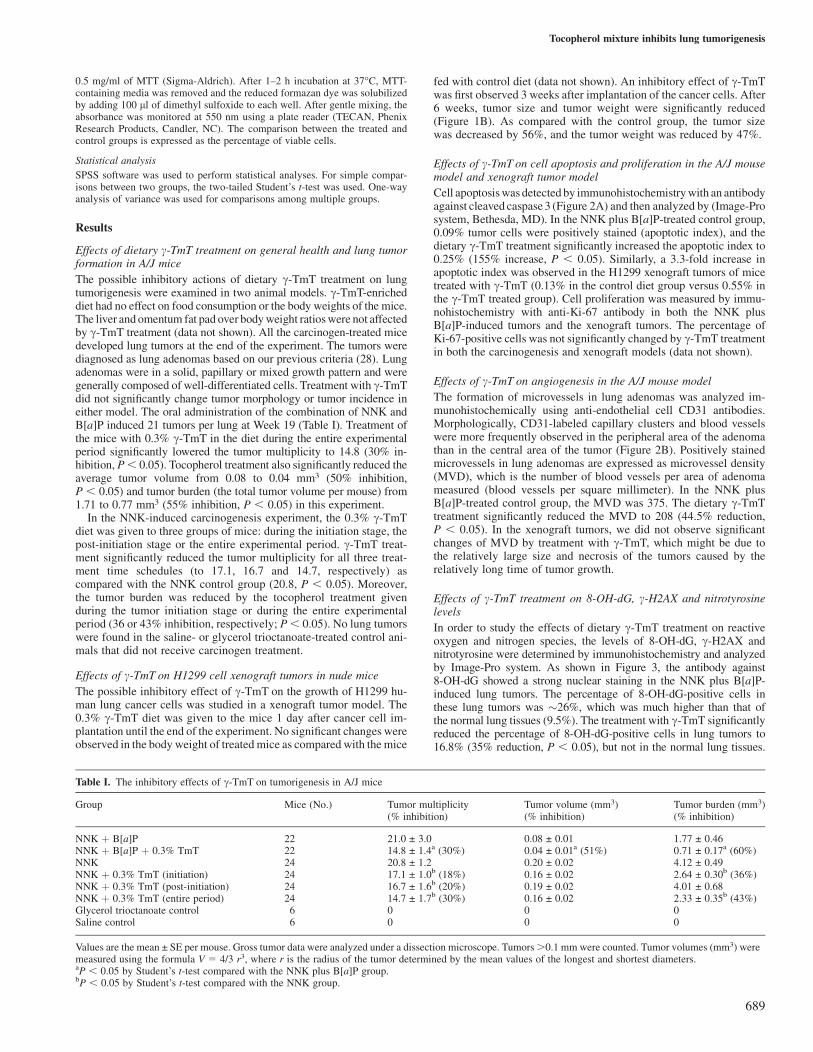
The possible inhibitory actions of dietary c-TmT treatment on lungtumorigenesis were examined in two animal models. c-TmT-enricheddiet had no effect on food consumption or the body weights of the mice.The liver and omentum fat pad over body weight ratios were not affectedby c-TmT treatment (data not shown). All the carcinogen-treated micedeveloped lung tumors at the end of the experiment. The tumors werediagnosed as lung adenomas based on our previous criteria (28). Lungadenomas were in a solid, papillary or mixed growth pattern and weregenerally composed of well-differentiated cells. Treatment with c-TmTdid not significantly change tumor morphology or tumor incidence ineither model. The oral administration of the combination of NNK andB[a]P induced 21 tumors per lung at Week 19 (Table I). Treatment ofthe mice with 0.3% c-TmT in the diet during the entire experimentalperiod significantly lowered the tumor multiplicity to 14.8 (30% in-hibition, P, 0.05). Tocopherol treatment also significantly reduced theaverage tumor volume from 0.08 to 0.04 mm3 (50% inhibition,P , 0.05) and tumor burden (the total tumor volume per mouse) from1.71 to 0.77 mm3 (55% inhibition, P , 0.05) in this experiment.
In the NNK-induced carcinogenesis experiment, the 0.3% c-TmTdiet was given to three groups of mice: during the initiation stage, thepost-initiation stage or the entire experimental period. c-TmT treat-ment significantly reduced the tumor multiplicity for all three treat-ment time schedules (to 17.1, 16.7 and 14.7, respectively) ascompared with the NNK control group (20.8, P , 0.05). Moreover,the tumor burden was reduced by the tocopherol treatment givenduring the tumor initiation stage or during the entire experimentalperiod (36 or 43% inhibition, respectively; P, 0.05). No lung tumorswere found in the saline- or glycerol trioctanoate-treated control ani-mals that did not receive carcinogen treatment.
Effects of c-TmT on H1299 cell xenograft tumors in nude mice
The possible inhibitory effect of c-TmT on the growth of H1299 hu-man lung cancer cells was studied in a xenograft tumor model. The0.3% c-TmT diet was given to the mice 1 day after cancer cell im-plantation until the end of the experiment. No significant changes wereobserved in the body weight of treated mice as compared with the mice
fed with control diet (data not shown). An inhibitory effect of c-TmTwas first observed 3 weeks after implantation of the cancer cells. After6 weeks, tumor size and tumor weight were significantly reduced(Figure 1B). As compared with the control group, the tumor sizewas decreased by 56%, and the tumor weight was reduced by 47%.
Effects of c-TmT on cell apoptosis and proliferation in the A/J mousemodel and xenograft tumor model
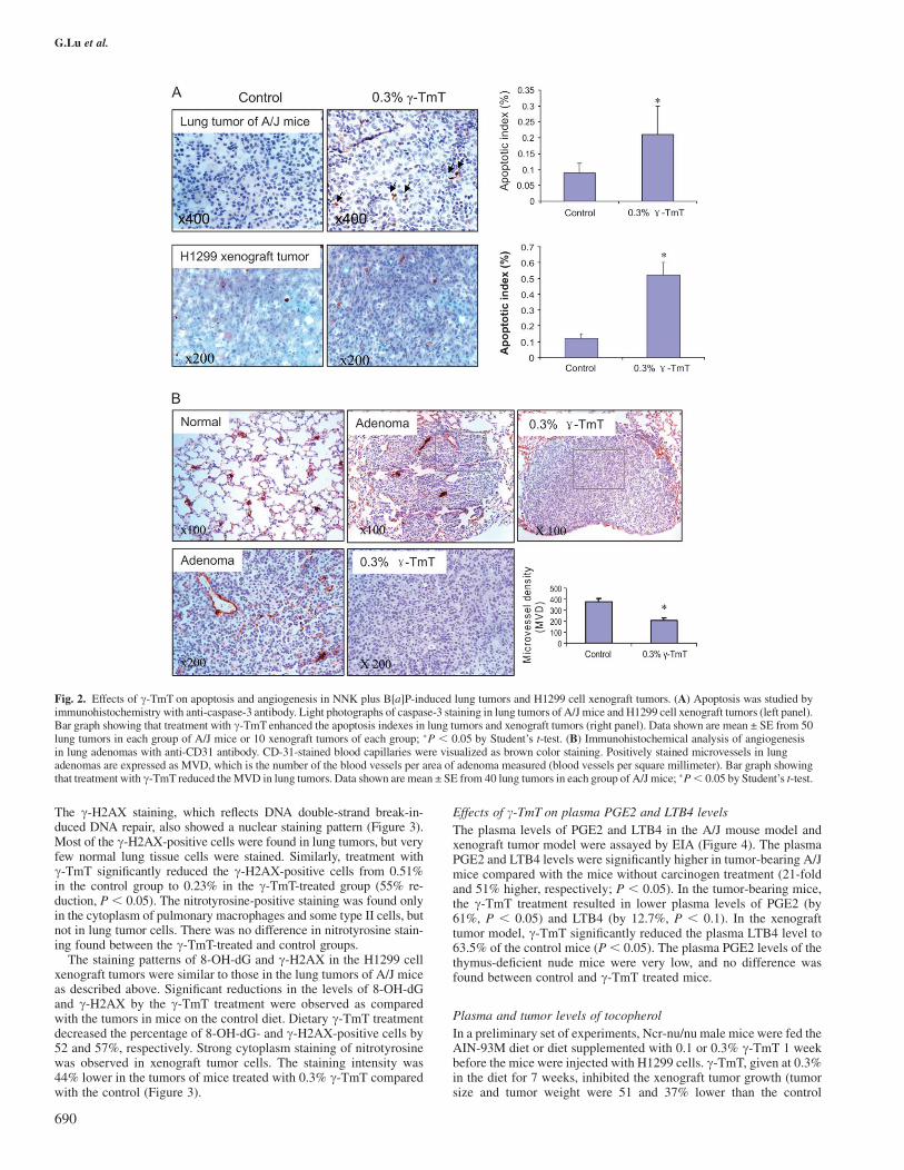
Cell apoptosis was detected by immunohistochemistry with an antibodyagainst cleaved caspase 3 (Figure 2A) and then analyzed by (Image-Prosystem, Bethesda, MD). In the NNK plus B[a]P-treated control group,0.09% tumor cells were positively stained (apoptotic index), and thedietary c-TmT treatment significantly increased the apoptotic index to0.25% (155% increase, P , 0.05). Similarly, a 3.3-fold increase inapoptotic index was observed in the H1299 xenograft tumors of micetreated with c-TmT (0.13% in the control diet group versus 0.55% inthe c-TmT treated group). Cell proliferation was measured by immu-nohistochemistry with anti-Ki-67 antibody in both the NNK plusB[a]P-induced tumors and the xenograft tumors. The percentage ofKi-67-positive cells was not significantly changed by c-TmT treatmentin both the carcinogenesis and xenograft models (data not shown).
Effects of c-TmT on angiogenesis in the A/J mouse model
The formation of microvessels in lung adenomas was analyzed im-munohistochemically using anti-endothelial cell CD31 antibodies.Morphologically, CD31-labeled capillary clusters and blood vesselswere more frequently observed in the peripheral area of the adenomathan in the central area of the tumor (Figure 2B). Positively stainedmicrovessels in lung adenomas are expressed as microvessel density(MVD), which is the number of blood vessels per area of adenomameasured (blood vessels per square millimeter). In the NNK plusB[a]P-treated control group, the MVD was 375. The dietary c-TmTtreatment significantly reduced the MVD to 208 (44.5% reduction,P , 0.05). In the xenograft tumors, we did not observe significantchanges of MVD by treatment with c-TmT, which might be due tothe relatively large size and necrosis of the tumors caused by therelatively long time of tumor growth.
Effects of c-TmT treatment on 8-OH-dG, c-H2AX and nitrotyrosinelevels
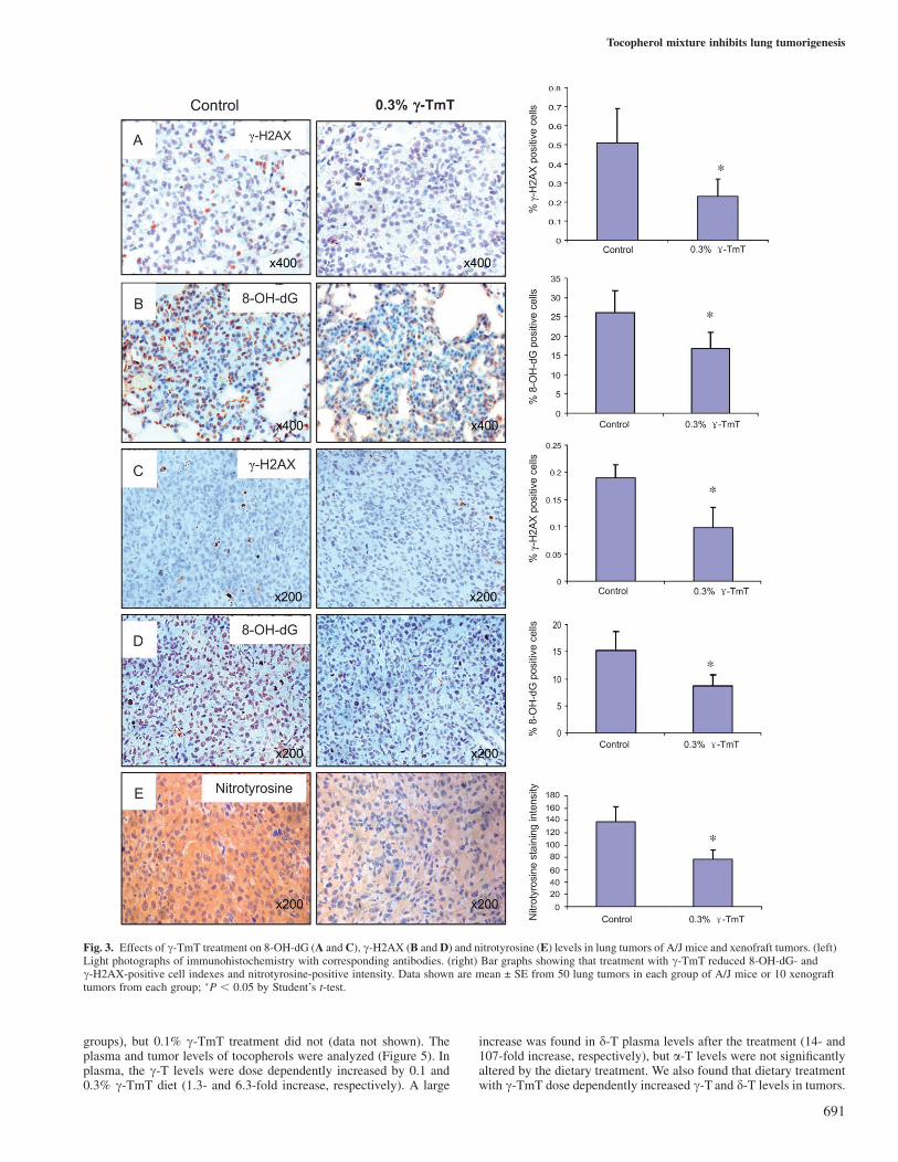
In order to study the effects of dietary c-TmT treatment on reactiveoxygen and nitrogen species, the levels of 8-OH-dG, c-H2AX andnitrotyrosine were determined by immunohistochemistry and analyzedby Image-Pro system. As shown in Figure 3, the antibody against8-OH-dG showed a strong nuclear staining in the NNK plus B[a]P-induced lung tumors. The percentage of 8-OH-dG-positive cells inthese lung tumors was �26%, which was much higher than that ofthe normal lung tissues (9.5%). The treatment with c-TmT significantlyreduced the percentage of 8-OH-dG-positive cells in lung tumors to16.8% (35% reduction, P , 0.05), but not in the normal lung tissues.
Table I. The inhibitory effects of c-TmT on tumorigenesis in A/J mice
Group Mice (No.) Tumor multiplicity(% inhibition)
Tumor volume (mm3)(% inhibition)
Tumor burden (mm3)(% inhibition)
NNK þ B[a]P 22 21.0 ± 3.0 0.08 ± 0.01 1.77 ± 0.46NNK þ B[a]P þ 0.3% TmT 22 14.8 ± 1.4a (30%) 0.04 ± 0.01a (51%) 0.71 ± 0.17a (60%)NNK 24 20.8 ± 1.2 0.20 ± 0.02 4.12 ± 0.49NNK þ 0.3% TmT (initiation) 24 17.1 ± 1.0b (18%) 0.16 ± 0.02 2.64 ± 0.30b (36%)NNK þ 0.3% TmT (post-initiation) 24 16.7 ± 1.6b (20%) 0.19 ± 0.02 4.01 ± 0.68NNK þ 0.3% TmT (entire period) 24 14.7 ± 1.7b (30%) 0.16 ± 0.02 2.33 ± 0.35b (43%)Glycerol trioctanoate control 6 0 0 0Saline control 6 0 0 0
Values are the mean ± SE per mouse. Gross tumor data were analyzed under a dissection microscope. Tumors .0.1 mm were counted. Tumor volumes (mm3) weremeasured using the formula V 5 4/3 r3, where r is the radius of the tumor determined by the mean values of the longest and shortest diameters.aP , 0.05 by Student’s t-test compared with the NNK plus B[a]P group.bP , 0.05 by Student’s t-test compared with the NNK group.
Tocopherol mixture inhibits lung tumorigenesis
689
The c-H2AX staining, which reflects DNA double-strand break-in-duced DNA repair, also showed a nuclear staining pattern (Figure 3).Most of the c-H2AX-positive cells were found in lung tumors, but veryfew normal lung tissue cells were stained. Similarly, treatment withc-TmT significantly reduced the c-H2AX-positive cells from 0.51%in the control group to 0.23% in the c-TmT-treated group (55% re-duction, P , 0.05). The nitrotyrosine-positive staining was found onlyin the cytoplasm of pulmonary macrophages and some type II cells, butnot in lung tumor cells. There was no difference in nitrotyrosine stain-ing found between the c-TmT-treated and control groups.
The staining patterns of 8-OH-dG and c-H2AX in the H1299 cellxenograft tumors were similar to those in the lung tumors of A/J miceas described above. Significant reductions in the levels of 8-OH-dGand c-H2AX by the c-TmT treatment were observed as comparedwith the tumors in mice on the control diet. Dietary c-TmT treatmentdecreased the percentage of 8-OH-dG- and c-H2AX-positive cells by52 and 57%, respectively. Strong cytoplasm staining of nitrotyrosinewas observed in xenograft tumor cells. The staining intensity was44% lower in the tumors of mice treated with 0.3% c-TmT comparedwith the control (Figure 3).
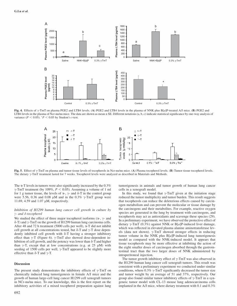
Effects of c-TmT on plasma PGE2 and LTB4 levels
The plasma levels of PGE2 and LTB4 in the A/J mouse model andxenograft tumor model were assayed by EIA (Figure 4). The plasmaPGE2 and LTB4 levels were significantly higher in tumor-bearing A/Jmice compared with the mice without carcinogen treatment (21-foldand 51% higher, respectively; P , 0.05). In the tumor-bearing mice,the c-TmT treatment resulted in lower plasma levels of PGE2 (by61%, P , 0.05) and LTB4 (by 12.7%, P , 0.1). In the xenografttumor model, c-TmT significantly reduced the plasma LTB4 level to63.5% of the control mice (P, 0.05). The plasma PGE2 levels of thethymus-deficient nude mice were very low, and no difference wasfound between control and c-TmT treated mice.
Plasma and tumor levels of tocopherol
In a preliminary set of experiments, Ncr-nu/nu male mice were fed theAIN-93M diet or diet supplemented with 0.1 or 0.3% c-TmT 1 weekbefore the mice were injected with H1299 cells. c-TmT, given at 0.3%in the diet for 7 weeks, inhibited the xenograft tumor growth (tumorsize and tumor weight were 51 and 37% lower than the control
Fig. 2. Effects of c-TmT on apoptosis and angiogenesis in NNK plus B[a]P-induced lung tumors and H1299 cell xenograft tumors. (A) Apoptosis was studied byimmunohistochemistry with anti-caspase-3 antibody. Light photographs of caspase-3 staining in lung tumors of A/J mice and H1299 cell xenograft tumors (left panel).Bar graph showing that treatment with c-TmT enhanced the apoptosis indexes in lung tumors and xenograft tumors (right panel). Data shown are mean ± SE from 50lung tumors in each group of A/J mice or 10 xenograft tumors of each group; �P , 0.05 by Student’s t-test. (B) Immunohistochemical analysis of angiogenesisin lung adenomas with anti-CD31 antibody. CD-31-stained blood capillaries were visualized as brown color staining. Positively stained microvessels in lungadenomas are expressed as MVD, which is the number of the blood vessels per area of adenoma measured (blood vessels per square millimeter). Bar graph showingthat treatment with c-TmT reduced the MVD in lung tumors. Data shown are mean ± SE from 40 lung tumors in each group of A/J mice; �P, 0.05 by Student’s t-test.
G.Lu et al.
690
groups), but 0.1% c-TmT treatment did not (data not shown). Theplasma and tumor levels of tocopherols were analyzed (Figure 5). Inplasma, the c-T levels were dose dependently increased by 0.1 and0.3% c-TmT diet (1.3- and 6.3-fold increase, respectively). A large
increase was found in d-T plasma levels after the treatment (14- and107-fold increase, respectively), but a-T levels were not significantlyaltered by the dietary treatment. We also found that dietary treatmentwith c-TmT dose dependently increased c-T and d-T levels in tumors.
Fig. 3. Effects of c-TmT treatment on 8-OH-dG (A and C), c-H2AX (B and D) and nitrotyrosine (E) levels in lung tumors of A/J mice and xenofraft tumors. (left)Light photographs of immunohistochemistry with corresponding antibodies. (right) Bar graphs showing that treatment with c-TmT reduced 8-OH-dG- andc-H2AX-positive cell indexes and nitrotyrosine-positive intensity. Data shown are mean ± SE from 50 lung tumors in each group of A/J mice or 10 xenografttumors from each group; �P , 0.05 by Student’s t-test.
Tocopherol mixture inhibits lung tumorigenesis
691
The a-T levels in tumors were also significantly increased by the 0.3%c-TmT treatment (by 109%, P , 0.05). Assuming a volume of 1 mlfor 1 g tumor tissue, the levels of a-, c- and d-T in the control groupwere 5.56, 0.36 and 0.08 lM and in the 0.3% c-TmT group were11.69, 4.59 and 1.07 lM, respectively.
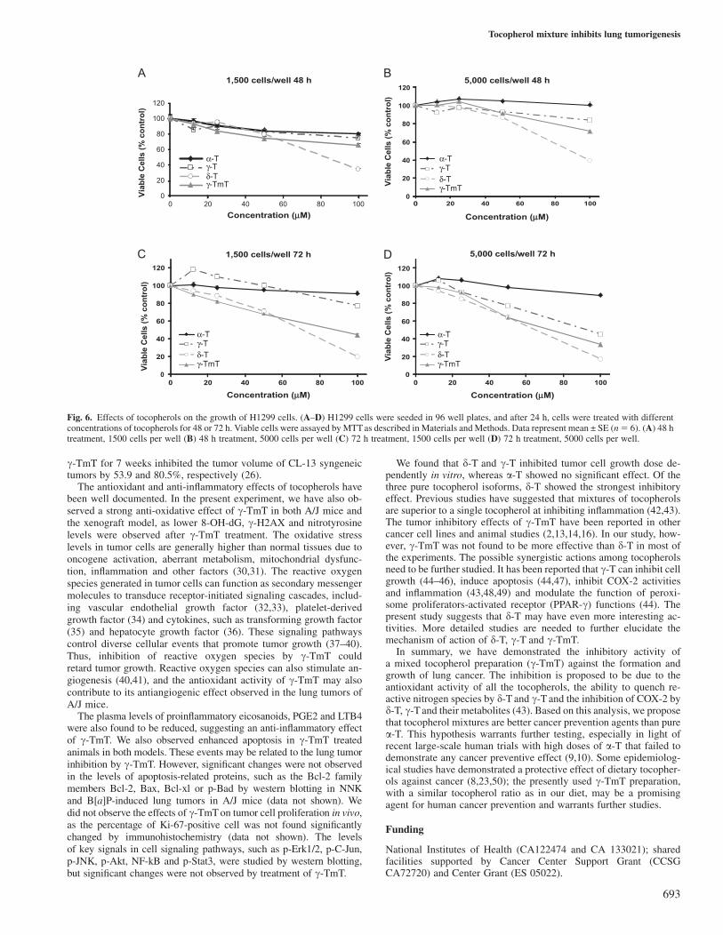
Inhibition of H1299 human lung cancer cell growth in culture byc- and d-tocopherolWe studied the effect of three major tocopherol isoforms (a-, c- andd-T) and c-TmT on the growth of H1299 human lung carcinoma cells.After 48 and 72 h treatment (5000 cells per well), a-T did not inhibitcell growth at all concentrations tested, but d-T and c-T dose depen-dently inhibited cell growth with d-T having a stronger inhibitoryeffect than c-T (Figure 6). c-TmT also showed dose-dependent in-hibition of cell growth, and the potency was lower than d-T and higherthan c-T, except that at low concentrations (e.g. at 25 lM) withseeding of 1500 cells per well, c-TmT appeared to be slightly moreeffective than d-T and c-T.
Discussion
The present study demonstrates the inhibitory effects of c-TmT onchemically induced lung tumorigenesis in female A/J mice and thegrowth of human large-cell lung cancer H1299 cell xenograft tumorsin NCr-nu/nu mice. To our knowledge, this is the first report on theinhibitory activities of a mixed tocopherol preparation against lung
tumorigenesis in animals and tumor growth of human lung cancercells in a xenograft model.
In this study, we found that c-TmT given at the initiation stagereduced the tumor multiplicity and tumor burden. This result suggeststhat tocopherols can reduce the deleterious effects caused by carcin-ogen metabolism and can prevent the molecular or tissue damage bythe carcinogens and their metabolites. For example, reactive oxygenspecies are generated in the lung by treatment with carcinogens, andtocopherols may act as antioxidants and scavenge these species (29).In a preliminary experiment, we have observed the protective effect ofdietary c-TmT (0.3%) against NNK or B[a]P-induced liver damage,which was reflected in elevated plasma alanine aminotransferase lev-els (data not shown). c-TmT showed stronger effects in reducingtumor volume in the NNK plus B[a]P-induced lung tumorigenesismodel as compared with the NNK-induced model. It appears thattissue tocopherols may be more effective at inhibiting the action ofthe eight smaller doses of carcinogen absorbed through the gastroin-testinal tract than the two larger doses of NNK administered byintraperitoneal injection.
The tumor growth inhibitory effect of c-TmT was also observed inthe H1299 human lung cancer cell xenograft tumors. This result wasconsistent with a preliminary experiment we conducted under similarconditions, where 0.3% c-TmT significantly decreased the tumor sizeand tumor weight by an average of 51 and 37%, respectively. Ourgroup also found similar tumor inhibitory effects of c-TmT in a syn-geneic tumor model with CL-13 mouse lung adenocarcinoma cellsimplanted in the A/J mice, where dietary treatment with 0.1 and 0.3%
Fig. 4. Effects of c-TmT on plasma PGE2 and LTB4 levels. (A) PGE2 and LTB4 levels in the plasma of NNK plus B[a]P-treated A/J mice. (B) PGE2 andLTB4 levels in the plasma of Ncr-nu/nu mice. The data are shown as mean ± SE. Different notations (a, b, c) indicate statistical significance by one-way analysis ofvariance (P , 0.05). �P , 0.05 by Student’s t-test.
Fig. 5. Effect of c-TmT on plasma and tumor tissue levels of tocopherols in Ncr-nu/nu mice. (A) Plasma tocopherol levels. (B) Tumor tissue tocopherol levels.The dietary c-TmT treatment lasted for 7 weeks. Tocopherol levels were analyzed as described in Materials and Methods.
G.Lu et al.
692
c-TmT for 7 weeks inhibited the tumor volume of CL-13 syngeneictumors by 53.9 and 80.5%, respectively (26).
The antioxidant and anti-inflammatory effects of tocopherols havebeen well documented. In the present experiment, we have also ob-served a strong anti-oxidative effect of c-TmT in both A/J mice andthe xenograft model, as lower 8-OH-dG, c-H2AX and nitrotyrosinelevels were observed after c-TmT treatment. The oxidative stresslevels in tumor cells are generally higher than normal tissues due tooncogene activation, aberrant metabolism, mitochondrial dysfunc-tion, inflammation and other factors (30,31). The reactive oxygenspecies generated in tumor cells can function as secondary messengermolecules to transduce receptor-initiated signaling cascades, includ-ing vascular endothelial growth factor (32,33), platelet-derivedgrowth factor (34) and cytokines, such as transforming growth factor(35) and hepatocyte growth factor (36). These signaling pathwayscontrol diverse cellular events that promote tumor growth (37–40).Thus, inhibition of reactive oxygen species by c-TmT couldretard tumor growth. Reactive oxygen species can also stimulate an-giogenesis (40,41), and the antioxidant activity of c-TmT may alsocontribute to its antiangiogenic effect observed in the lung tumors ofA/J mice.
The plasma levels of proinflammatory eicosanoids, PGE2 and LTB4were also found to be reduced, suggesting an anti-inflammatory effectof c-TmT. We also observed enhanced apoptosis in c-TmT treatedanimals in both models. These events may be related to the lung tumorinhibition by c-TmT. However, significant changes were not observedin the levels of apoptosis-related proteins, such as the Bcl-2 familymembers Bcl-2, Bax, Bcl-xl or p-Bad by western blotting in NNKand B[a]P-induced lung tumors in A/J mice (data not shown). Wedid not observe the effects of c-TmT on tumor cell proliferation in vivo,as the percentage of Ki-67-positive cell was not found significantlychanged by immunohistochemistry (data not shown). The levelsof key signals in cell signaling pathways, such as p-Erk1/2, p-C-Jun,p-JNK, p-Akt, NF-kB and p-Stat3, were studied by western blotting,but significant changes were not observed by treatment of c-TmT.
We found that d-T and c-T inhibited tumor cell growth dose de-pendently in vitro, whereas a-T showed no significant effect. Of thethree pure tocopherol isoforms, d-T showed the strongest inhibitoryeffect. Previous studies have suggested that mixtures of tocopherolsare superior to a single tocopherol at inhibiting inflammation (42,43).The tumor inhibitory effects of c-TmT have been reported in othercancer cell lines and animal studies (2,13,14,16). In our study, how-ever, c-TmT was not found to be more effective than d-T in most ofthe experiments. The possible synergistic actions among tocopherolsneed to be further studied. It has been reported that c-T can inhibit cellgrowth (44–46), induce apoptosis (44,47), inhibit COX-2 activitiesand inflammation (43,48,49) and modulate the function of peroxi-some proliferators-activated receptor (PPAR-c) functions (44). Thepresent study suggests that d-T may have even more interesting ac-tivities. More detailed studies are needed to further elucidate themechanism of action of d-T, c-T and c-TmT.
In summary, we have demonstrated the inhibitory activity ofa mixed tocopherol preparation (c-TmT) against the formation andgrowth of lung cancer. The inhibition is proposed to be due to theantioxidant activity of all the tocopherols, the ability to quench re-active nitrogen species by d-T and c-T and the inhibition of COX-2 byd-T, c-T and their metabolites (43). Based on this analysis, we proposethat tocopherol mixtures are better cancer prevention agents than purea-T. This hypothesis warrants further testing, especially in light ofrecent large-scale human trials with high doses of a-T that failed todemonstrate any cancer preventive effect (9,10). Some epidemiolog-ical studies have demonstrated a protective effect of dietary tocopher-ols against cancer (8,23,50); the presently used c-TmT preparation,with a similar tocopherol ratio as in our diet, may be a promisingagent for human cancer prevention and warrants further studies.
Funding
National Institutes of Health (CA122474 and CA 133021); sharedfacilities supported by Cancer Center Support Grant (CCSGCA72720) and Center Grant (ES 05022).
Fig. 6. Effects of tocopherols on the growth of H1299 cells. (A–D) H1299 cells were seeded in 96 well plates, and after 24 h, cells were treated with differentconcentrations of tocopherols for 48 or 72 h. Viable cells were assayed by MTT as described in Materials and Methods. Data represent mean ± SE (n5 6). (A) 48 htreatment, 1500 cells per well (B) 48 h treatment, 5000 cells per well (C) 72 h treatment, 1500 cells per well (D) 72 h treatment, 5000 cells per well.
Tocopherol mixture inhibits lung tumorigenesis
693
Acknowledgements
Conflict of Interest Statement: None declared.
References
1. Jemal,A. et al. (2008) Cancer statistics, 2008. CA Cancer J. Clin., 58, 71–96.
2. Ju,J. et al. (2009) A gamma-tocopherol-rich mixture of tocopherols inhibitscolon inflammation and carcinogenesis in azoxymethane and dextran sul-fate sodium-treated mice. Cancer Prev. Res. (Phila Pa), 2, 143–152.
3.Dietrich,M. et al. (2006) Does gamma-tocopherol play a role in the primaryprevention of heart disease and cancer? A review. J. Am. Coll. Nutr., 25,292–299.
4.Hensley,K. et al. (2004) New perspectives on vitamin E: gamma-tocopheroland carboxyelthylhydroxychroman metabolites in biology and medicine.Free Radic. Biol. Med., 36, 1–15.
5.Traber,M.G. (2006) Vitamin E. Modern Nutrition in Health and Disease,10th ed. Lippincott Williams and Wilkins Baltimore, MD, pp. 396–411.
6.Traber,M.G. et al. (2007) Vitamin E, antioxidant and nothing more. FreeRadic. Biol. Med., 43, 4–15.
7.Eitenmiller,T. et al. (2004) Vitamin E: Food Chemistry, Composition, andAnalysis. Marcel Dekker, Inc., New York, NY.
8. Ju,J. et al. (2009) Cancer preventive activities of tocopherols and tocotrie-nols. Carcinogenesis, Advanced access published September 11, 2009.
9.Lippman,S.M. et al. (2009) Effect of selenium and vitamin E on risk ofprostate cancer and other cancers: the Selenium and Vitamin E CancerPrevention Trial (SELECT). JAMA., 301, 39–51.
10.Gaziano,J.M. et al. (2009) Vitamins E and C in the prevention of prostateand total cancer in men: the Physicians’ Health Study II randomized con-trolled trial. JAMA., 301, 52–62.
11.Campbell,S. et al. (2003) Development of gamma (gamma)-tocopherol asa colorectal cancer chemopreventive agent. Crit. Rev. Oncol. Hematol., 47,249–259.
12.Reiter,E. et al. (2007) Anti-inflammatory properties of alpha- and gamma-tocopherol. Mol. Aspects Med., 28, 668–691.
13.Newmark,H.L. et al. (2006) Mixed tocopherols inhibit azoxymethane-induced aberrant crypt foci in rats. Nutr. Cancer, 56, 82–85.
14.Suh,N. et al. (2007) Mixed tocopherols inhibit N-methyl-N-nitrosourea-induced mammary tumor growth in rats. Nutr. Cancer, 59, 76–81.
15.Lee,H.J. et al. (2009) Mixed tocopherols prevent mammary tumorigene-sis by inhibiting estrogen action and activating PPAR-g. Clin. CancerRes, 15, 4242–4249.
16.Barve,A. et al. (2009) Gamma-tocopherol-enriched mixed tocopherol dietinhibits prostate carcinogenesis in TRAMP mice. Int. J. Cancer, 124, 1693–1699.
17.Comstock,G.W. et al. (1997) The risk of developing lung cancer associatedwith antioxidants in the blood: ascorbic acid, carotenoids, alpha-tocopherol,selenium, and total peroxyl radical absorbing capacity. Cancer Epidemiol.Biomarkers Prev., 6, 907–916.
18.Goodman,G.E. et al. (2003) The association between lung and prostatecancer risk, and serum micronutrients: results and lessons learned frombeta-carotene and retinol efficacy trial. Cancer Epidemiol. BiomarkersPrev., 12, 518–526.
19.Menkes,M.S. et al. (1986) Serum beta-carotene, vitamins A and E, sele-nium, and the risk of lung cancer. N. Engl. J. Med., 315, 1250–1254.
20.Ratnasinghe,D. et al. (2000) Serum tocopherols, selenium and lung cancerrisk among tin miners in China. Cancer Causes Control, 11, 129–135.
21.Shibata,A. et al. (1992) Intake of vegetables, fruits, beta-carotene, vitamin Cand vitamin supplements and cancer incidence among the elderly: a prospec-tive study. Br. J. Cancer, 66, 673–679.
22.Woodson,K. et al. (1999) Association between alcohol and lung cancer inthe alpha-tocopherol, beta-carotene cancer prevention study in Finland.Cancer Causes Control, 10, 219–226.
23.Yong,L.C. et al. (1997) Intake of vitamins E, C, and A and risk of lungcancer. The NHANES I epidemiologic followup study. First NationalHealth and Nutrition Examination Survey. Am. J. Epidemiol., 146, 231–243.
24.Albanes,D. et al. (1996) Alpha-Tocopherol and beta-carotene supplementsand lung cancer incidence in the alpha-tocopherol, beta-carotene cancerprevention study: effects of base-line characteristics and study compliance.J. Natl Cancer Inst., 88, 1560–1570.
25.Ogasawara,M. et al. (2007) Differential effects of antioxidants on thein vitro invasion, growth and lung metastasis of murine colon cancer cells.Biol. Pharm. Bull., 30, 200–204.
26.Lambert,J.D. et al. (2009) Inhibition of lung cancer growth in mice bydietary mixed tocopherols. Mol. Nutr. Food Res, 53, 1030–1035.
27.Belinsky,S.A. et al. (1992) Role of the alveolar type II cell in the developmentand progression of pulmonary tumors induced by 4-(methylnitrosamino)-1-(3-pyridyl)-1-butanone in the A/J mouse. Cancer Res., 52, 3164–3173.
28.Yang,G. et al. (1997) Characterization of early pulmonary hyperprolifera-tion and tumor progression and their inhibition by black tea in a 4-(methylnitrosamino)-1-(3-pyridyl)-1-butanone-induced lung tumorigenesismodel with A/J mice. Cancer Res., 57, 1889–1894.
29.Hecht,S.S. (1998) Biochemistry, biology, and carcinogenicity of tobacco-specific N-nitrosamines. Chem. Res. Toxicol., 11, 559–603.
30.Szatrowski,T.P. et al. (1991) Production of large amounts of hydrogenperoxide by human tumor cells. Cancer Res., 51, 794–798.
31.Kawanishi,S. et al. (2006) Oxidative and nitrative DNA damage in animalsand patients with inflammatory diseases in relation to inflammation-relatedcarcinogenesis. Biol. Chem., 387, 365–372.
32.Pani,G. et al. (2002) Determination of intracellular reactive oxygen speciesas function of cell density. Methods Enzymol., 352, 91–100.
33.Colavitti,R. et al. (2002) Reactive oxygen species as downstream mediatorsof angiogenic signaling by vascular endothelial growth factor receptor-2/KDR. J. Biol. Chem., 277, 3101–3108.
34.Choi,M.H. et al. (2005) Regulation of PDGF signalling and vascularremodelling by peroxiredoxin II. Nature, 435, 347–353.
35.Rhyu,D.Y. et al. (2005) Role of reactive oxygen species in TGF-beta1-induced mitogen-activated protein kinase activation and epithelial-mesen-chymal transition in renal tubular epithelial cells. J. Am. Soc. Nephrol., 16,667–675.
36.Arakaki,N. et al. (1999) Involvement of oxidative stress in tumor cytotoxicactivity of hepatocyte growth factor/scatter factor. J. Biol. Chem., 274,13541–13546.
37.Boonstra,J. et al. (2004) Molecular events associated with reactive oxygenspecies and cell cycle progression in mammalian cells. Gene, 337, 1–13.
38.Behrend,L. et al. (2003) Reactive oxygen species in oncogenic transforma-tion. Biochem. Soc. Trans., 31, (pt. 6),1441–1444.
39.Wu,W.S. (2006) The signaling mechanism of ROS in tumor progression.Cancer Metastasis Rev., 25, 695–705.
40.Ushio-Fukai,M. et al. (2004) Reactive oxygen species as mediators ofangiogenesis signaling: role of NAD(P)H oxidase. Mol. Cell Biochem.,264, 85–97.
41.Ushio-Fukai,M. et al. (2008) Reactive oxygen species and angiogenesis:NADPH oxidase as target for cancer therapy. Cancer Lett., 266, 37–52.
42.Devaraj,S. et al. (2008) Gamma-tocopherol supplementation alone and incombination with alpha-tocopherol alters biomarkers of oxidative stressand inflammation in subjects with metabolic syndrome. Free Radic. Biol.Med., 44, 1203–1208.
43. Jiang,Q. et al. (2000) gamma-tocopherol and its major metabolite, in con-trast to alpha-tocopherol, inhibit cyclooxygenase activity in macrophagesand epithelial cells. Proc. Natl Acad. Sci. USA, 97, 11494–11499.
44.Campbell,S.E. et al. (2003) Gamma (gamma) tocopherol upregulates per-oxisome proliferator activated receptor (PPAR) gamma (gamma) expres-sion in SW 480 human colon cancer cell lines. BMC Cancer, 3, 25.
45.Gysin,R. et al. (2002) Gamma-tocopherol inhibits human cancer cell cycleprogression and cell proliferation by down-regulation of cyclins. FASEB J.,16, 1952–1954.
46.Samandari,E. et al. (2006) The effect of gamma-tocopherol on prolifera-tion, integrin expression, adhesion, and migration of human glioma cells.Biochem. Biophys. Res. Commun., 342, 1329–1333.
47. Jiang,Q. et al. (2004) Gamma-tocopherol induces apoptosis in androgen-responsive LNCaP prostate cancer cells via caspase-dependent and inde-pendent mechanisms. Ann. N. Y. Acad. Sci., 1031, 399–400.
48. Jiang,Q. et al. (2003) Gamma-tocopherol, but not alpha-tocopherol, de-creases proinflammatory eicosanoids and inflammation damage in rats.FASEB J., 17, 816–822.
49.O’Leary,K.A. et al. (2004) Effect of flavonoids and vitamin E on cyclo-oxygenase-2 (COX-2) transcription. Mutat. Res., 551, 245–254.
50.Mahabir,S. et al. (2008) Dietary alpha-, beta-, gamma- and delta-tocopherolsin lung cancer risk. Int. J. Cancer, 123, 1173–1180.
Received October 23, 2009; revised December 14, 2009;accepted December 20, 2009
G.Lu et al.
694
Related Documents