134 Felice DRIVER, Richard J. MILNER and John W. H. TRUEMAN* CSIRO Entomology, GPO Box 1700, ACT 2601, Australia and *Research School of Biological Sciences, Australian National University, Canberra, ACT 2601, Australia. E-mail : richard.milner!ento.csiro.au Received 31 October 1997 ; accepted 17 July 1999. The taxonomy of Metarhizium has been reassessed using sequence data and RAPD patterns from 123 isolates recognised as M. anisopliae, M. flavoviride or M. album. A high level of genetic diversity was found which was best resolved at the species}variety level by sequence data from the ITS and 28S rDNA D3 regions. RAPD patterns correlated closely with the sequence data and revealed a much greater degree of diversity useful for distinguishing strains within a variety. Ten distinct clades were revealed by the cladogram based on the combined sequence data set. Several major evolutionary lines were revealed, but the taxonomic relationships at the base of the tree are poorly resolved. The data support the monopoly of the M. anisopliae group, and recognise four clades within it. Two correspond with M. anisopliae var. anisopliae and M. anisopliae var. majus. The other two are described as new varieties based on their distinctive ITS sequence data : M. anisopliae var. lepidiotum and M. anisopliae var. acridum vars nov. M. album, M. flavoviride var. flavoviride and M. flavoviride var. minus are recognised and redefined according to ITS sequence data. Three clades represent two new varieties, M. flavoviride var. novazealandicum and M. flavoviride var. pemphigum vars nov., based on their distinct ITS sequence data. The third, with two isolates, has not been named pending further data. INTRODUCTION Metarhizium is one of the best known genera of entomo- pathogenic fungi, commonly known as ‘ green muscardine fungus ’. M. anisopliae, described from Russia as a pathogen of the wheat cockchafer, Anisoplia austriaca (Coleoptera : Scara- baeidae), is used for insect control in many countries including the USA, Brazil, Australia and the Philippines. Its potential in Australia for control of locusts and grasshoppers was reviewed by Milner (1997). The life-cycle is simple : the infectious unit is an asexual, haploid conidiospore, formed in chains on phialides, which may or may not be swollen (Glare & Milner 1996). The conidia germinate on the cuticle of a susceptible insect, produce a germ tube which penetrates into the body cavity, where the fungus proliferates as hyphae, eventually killing the host. After death, if conditions are moist and humid, the fungus grows out through the cuticle and forms new green conidia aerially. It grows and sporulates well on simple media such as Sabouraud’s dextrose agar. No teleomorph is known from Metarhizium apart from a claim that Cordyceps taii is the teleomorph of M. taii (Liang et al. 1991). The robust conidia can be formulated as a mycoinsecticide for pest control. Registered products include BioBlast for termites in the USA and BioGreen for pasture cockchafers in Australia (Milner 1998). The current classification of Metarhizium is based on morphological characters and was reviewed by Tulloch (1976), who only accepted M. flavoviride and M. anisopliae, the latter with the short spored var. anisopliae and the long-spored var. majus. M. album, described from a leaf hopper in Sri Lanka by Petch (1931), was determined by Tulloch to be an immature specimen of M. anisopliae. Petch (1935) also described M. brunneum, from an homopteran host in the Philippines, but Latch (1965) examined authentic material of M. brunneum (IMI 14746) from wireworms (Coleoptera : Elateridae) and considered its colour to be similar to that of M. anisopliae from Costelytra zealandica which was dark brown on some media. Tulloch (1976) agreed with Latch (1965) that M. brunneum and M. anisopliae are synonymous, and speculated that M. brunneum might be a naturally occurring colour mutant. Rombach et al. (1986) reviewed the status of M. flavoviride, which is known from curculionid beetles and agricultural soil. They expanded the concept of the long- and short-spored forms of M. anisopliae described by Tulloch to accommodate Asian isolates of M. flavoviride var. minus, which were characterised by consistently producing smaller spores and having been collected from plant-hoppers and leaf- hoppers (Homoptera : Delphacidae) on rice in the Philippines and Solomon Islands. They also described M. flavoviride var. minus on a grasshopper (Orthoptera : Acrididae) from the Galapagos Islands (Evans & Samson 1982). Rombach et al. (1987) also described the morphological resemblance of M. Mycol. Res. 104 (2) : 134–150 (February 2000). Printed in the United Kingdom. A taxonomic revision of Metarhizium based on a phylogenetic analysis of rDNA sequence data

Welcome message from author
This document is posted to help you gain knowledge. Please leave a comment to let me know what you think about it! Share it to your friends and learn new things together.
Transcript
134
Felice DRIVER, Richard J. MILNER and John W. H. TRUEMAN*
CSIRO Entomology, GPO Box 1700, ACT 2601, Australia and *Research School of Biological Sciences, Australian National University, Canberra,
ACT 2601, Australia.
E-mail : richard.milner!ento.csiro.au
Received 31 October 1997 ; accepted 17 July 1999.
The taxonomy of Metarhizium has been reassessed using sequence data and RAPD patterns from 123 isolates recognised as M.
anisopliae, M. flavoviride or M. album. A high level of genetic diversity was found which was best resolved at the species}variety
level by sequence data from the ITS and 28S rDNA D3 regions. RAPD patterns correlated closely with the sequence data and
revealed a much greater degree of diversity useful for distinguishing strains within a variety. Ten distinct clades were revealed by
the cladogram based on the combined sequence data set. Several major evolutionary lines were revealed, but the taxonomic
relationships at the base of the tree are poorly resolved. The data support the monopoly of the M. anisopliae group, and recognise
four clades within it. Two correspond with M. anisopliae var. anisopliae and M. anisopliae var. majus. The other two are described as
new varieties based on their distinctive ITS sequence data : M. anisopliae var. lepidiotum and M. anisopliae var. acridum vars nov. M.
album, M. flavoviride var. flavoviride and M. flavoviride var. minus are recognised and redefined according to ITS sequence data.
Three clades represent two new varieties, M. flavoviride var. novazealandicum and M. flavoviride var. pemphigum vars nov., based on
their distinct ITS sequence data. The third, with two isolates, has not been named pending further data.
INTRODUCTION
Metarhizium is one of the best known genera of entomo-
pathogenic fungi, commonly known as ‘green muscardine
fungus ’. M. anisopliae, described from Russia as a pathogen of
the wheat cockchafer, Anisoplia austriaca (Coleoptera : Scara-
baeidae), is used for insect control in many countries including
the USA, Brazil, Australia and the Philippines. Its potential in
Australia for control of locusts and grasshoppers was reviewed
by Milner (1997). The life-cycle is simple : the infectious unit
is an asexual, haploid conidiospore, formed in chains on
phialides, which may or may not be swollen (Glare & Milner
1996). The conidia germinate on the cuticle of a susceptible
insect, produce a germ tube which penetrates into the body
cavity, where the fungus proliferates as hyphae, eventually
killing the host. After death, if conditions are moist and humid,
the fungus grows out through the cuticle and forms new green
conidia aerially. It grows and sporulates well on simple media
such as Sabouraud’s dextrose agar. No teleomorph is known
from Metarhizium apart from a claim that Cordyceps taii is the
teleomorph of M. taii (Liang et al. 1991). The robust conidia
can be formulated as a mycoinsecticide for pest control.
Registered products include BioBlast for termites in the USA
and BioGreen for pasture cockchafers in Australia (Milner
1998).
The current classification of Metarhizium is based on
morphological characters and was reviewed by Tulloch
(1976), who only accepted M. flavoviride and M. anisopliae, the
latter with the short spored var. anisopliae and the long-spored
var. majus. M. album, described from a leaf hopper in Sri Lanka
by Petch (1931), was determined by Tulloch to be an
immature specimen of M. anisopliae. Petch (1935) also
described M. brunneum, from an homopteran host in the
Philippines, but Latch (1965) examined authentic material of
M. brunneum (IMI 14746) from wireworms (Coleoptera :
Elateridae) and considered its colour to be similar to that of M.
anisopliae from Costelytra zealandica which was dark brown on
some media. Tulloch (1976) agreed with Latch (1965) that M.
brunneum and M. anisopliae are synonymous, and speculated
that M. brunneum might be a naturally occurring colour
mutant. Rombach et al. (1986) reviewed the status of M.
flavoviride, which is known from curculionid beetles and
agricultural soil. They expanded the concept of the long- and
short-spored forms of M. anisopliae described by Tulloch to
accommodate Asian isolates of M. flavoviride var. minus,
which were characterised by consistently producing smaller
spores and having been collected from plant-hoppers and leaf-
hoppers (Homoptera : Delphacidae) on rice in the Philippines
and Solomon Islands. They also described M. flavoviride var.
minus on a grasshopper (Orthoptera : Acrididae) from the
Galapagos Islands (Evans & Samson 1982). Rombach et al.
(1987) also described the morphological resemblance of M.
Mycol. Res. 104 (2) : 134–150 (February 2000). Printed in the United Kingdom.
A taxonomic revision of Metarhizium based on a phylogeneticanalysis of rDNA sequence data
F. Driver, R. J. Milner and J. W. H. Trueman 135
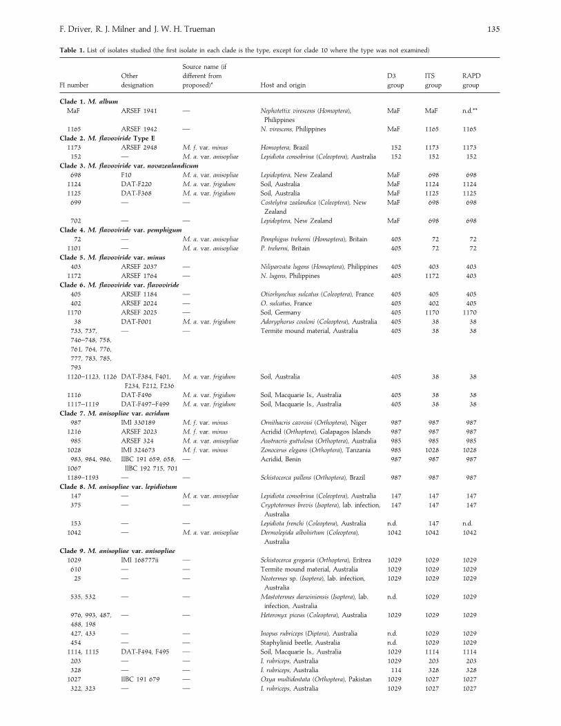
Table 1. List of isolates studied (the first isolate in each clade is the type, except for clade 10 where the type was not examined)
FI number
Other
designation
Source name (if
different from
proposed)* Host and origin
D3
group
ITS
group
RAPD
group
Clade 1. M. album
MaF ARSEF 1941 — Nephotettix virescens (Homoptera),
Philippines
MaF MaF n.d.**
1165 ARSEF 1942 — N. virescens, Philippines MaF 1165 1165
Clade 2. M. flavoviride Type E
1173 ARSEF 2948 M. f. var. minus Homoptera, Brazil 152 1173 1173
152 — M. a. var. anisopliae Lepidiota consobrina (Coleoptera), Australia 152 152 152
Clade 3. M. flavoviride var. novazealandicum
698 F10 M. a. var. anisopliae Lepidoptera, New Zealand MaF 698 698
1124 DAT-F220 M. a. var. frigidum Soil, Australia MaF 1124 1124
1125 DAT-F368 M. a. var. frigidum Soil, Australia MaF 1125 1125
699 — — Costelytra zealandica (Coleoptera), New
Zealand
MaF 698 698
702 — — Lepidoptera, New Zealand MaF 698 698
Clade 4. M. flavoviride var. pemphigum
72 — M. a. var. anisopliae Pemphigus treherni (Homoptera), Britain 405 72 72
1101 — M. a. var. anisopliae P. treherni, Britain 405 72 72
Clade 5. M. flavoviride var. minus
403 ARSEF 2037 — Niliparvata lugens (Homoptera), Philippines 405 403 403
1172 ARSEF 1764 — N. lugens, Philippines 405 1172 403
Clade 6. M. flavoviride var. flavoviride
405 ARSEF 1184 — Otiorhynchus sulcatus (Coleoptera), France 405 405 405
402 ARSEF 2024 — O. sulcatus, France 405 402 405
1170 ARSEF 2025 — Soil, Germany 405 1170 1170
38 DAT-F001 M. a. var. frigidum Adoryphorus couloni (Coleoptera), Australia 405 38 38
733, 737,
746–748, 758,
761, 764, 776,
777, 783, 785,
793
— — Termite mound material, Australia 405 38 38
1120–1123, 1126 DAT-F384, F401,
F234, F212, F236
M. a. var. frigidum Soil, Australia 405 38 38
1116 DAT-F496 M. a. var. frigidum Soil, Macquarie Is., Australia 405 38 38
1117–1119 DAT-F497–F499 M. a. var. frigidum Soil, Macquarie Is., Australia 405 38 38
Clade 7. M. anisopliae var. acridum
987 IMI 330189 M. f. var. minus Ornithacris cavroisi (Orthoptera), Niger 987 987 987
1216 ARSEF 2023 M. f. var. minus Acridid (Orthoptera), Galapagos Islands 987 987 987
985 ARSEF 324 M. a. var. anisopliae Austracris guttulosa (Orthoptera), Australia 985 985 985
1028 IMI 324673 M. f. var. minus Zonocerus elegans (Orthoptera), Tanzania 985 1028 1028
983, 984, 986,
1067
IIBC 191 659, 658,
IIBC 192 715, 701
— Acridid, Benin 987 987 987
1189–1193 — — Schistocerca pallens (Orthoptera), Brazil 987 987 987
Clade 8. M. anisopliae var. lepidiotum
147 — M. a. var. anisopliae Lepidiota consobrina (Coleoptera), Australia 147 147 147
375 — — Cryptotermes brevis (Isoptera), lab. infection,
Australia
147 147 147
153 — — Lepidiota frenchi (Coleoptera), Australia n.d. 147 n.d.
1042 — M. a. var. anisopliae Dermolepida albohirtum (Coleoptera),
Australia
1042 1042 1042
Clade 9. M. anisopliae var. anisopliae
1029 IMI 168777ii — Schistocerca gregaria (Orthoptera), Eritrea 1029 1029 1029
610 — — Termite mound material, Australia 1029 1029 1029
25 — — Neotermes sp. (Isoptera), lab. infection,
Australia
1029 1029 1029
535, 532 — — Mastotermes darwiniensis (Isoptera), lab.
infection, Australia
n.d. 1029 1029
976, 993, 487,
488, 198
— — Heteronyx piceus (Coleoptera), Australia 1029 1029 1029
427, 433 — — Inopus rubriceps (Diptera), Australia n.d. 1029 1029
454 — — Staphylinid beetle, Australia n.d. 1029 1029
1114, 1115 DAT-F494, F495 — Soil, Macquarie Is., Australia 1029 1114 1114
203 — — I. rubriceps, Australia 1029 203 203
328 — — I. rubriceps, Australia 114 328 328
1027 IIBC 191 679 — Oxya multidentata (Orthoptera), Pakistan 1029 1027 1027
322, 323 — — I. rubriceps, Australia 1029 1027 1027
Revision of Metarhizium 136
Table 1. (cont.)
FI number
Other
designation
Source name (if
different from
proposed)* Host and origin
D3
group
ITS
group
RAPD
group
Clade 9. (cont.)
379 — — I. rubriceps, Australia 114 379 379
1045 — — D. albohirtum, Australia 1029 1045 1045
330 — — I. rubriceps, Australia 114 1045 1045
1041 — — D. albohirtum, Australia n.d. 1045 1045
114 — — Antitrogus parvulus (Coleoptera), Australia 114 114 114
208 IIBC 191 625 — Phaulacridium vittatum (Orthoptera),
Australia
114 208 114
1033 IIBC 191 633 — Pseudosphingonotus savigni (Orthoptera),
Oman
n.d. 1033 1033
1031 — — Teleogryllus sp. (Orthoptera), Oman 1029 1031 1031
1091 ARSEF 437 M. a. var. anisopliae Teleogryllus commodus (Orthoptera),
Australia
1029 1091 1091
1030 IMI 351830 — T. commodus, Australia 1029 1091 1091
1037 ARSEF 448 — T. commodus, Australia n.d. 1091 1091
1090 ARSEF 436 — T. commodus, Australia 1029 1091 1091
1092–1100 ARSEF 438 — T. commodus, Australia n.d. 1091 1091
550 — — Termite mound material, Australia 1029 1091 1091
552 — — Termite mound material, Australia n.d. 1091 1091
161 — — I. rubriceps, Australia n.d. 1091 1091
207 — — P. vittatum, Australia 1029 1091 1091
1039 IMI 89–574 — Acrotylus sp. (Orthoptera), Pakistan 1029 1091 1091
1038 IMI 91–629 — Zonocerus variegatus (Orthoptera), Benin n.d. 1091 1091
691 — — Soil, Burma 1029 1091 1091
726 — — Soil, Burma 1029 1091 1091
1011 — — Anomola sp. (Coleoptera), Burma 1029 1091 1091
1034 IIBC 191–614 — Patanga succincta (Orthoptera), Thailand 1029 1034 1034
692 — — Soil, Burma 1029 1034 1034
694 — — Soil, Burma n.d. 1034 1034
522 — — H. piceus, Australia 1029 1034 1034
903 — — H. piceus, Australia n.d. 1034 1034
700 — — Costelytra zealandica (Coleoptera), New
Zealand
1029 700 n.d.
23 — — Anolamia albofasciata (Hemiptera), Mexico 1029 23 n.d.
1156 — — Immunocompromised patient, Australia 1029 1156 n.d.
327 — — I. rubriceps, Australia n.d. 1156 327
163 — — Kalotermes sp. (Isoptera), lab. infection,
Australia
1029 163 n.d.
911 — — H. piceus, Australia 1029 163 n.d.
1036 ARSEF 727 — Tettigonid, Brazil 1029 163 1036
358 — — Heteronychus arator (Coleoptera), Australia 1029 163 n.d.
Clade 10. M. anisopliae var. majus
388 ARSEF 1914 — Oryctes rhinoceros (Coleoptera), Philippines 1029 388 388
389 ARSEF 2151 — O. rhinoceros, Indonesia 1029 389 388
401 ARSEF 1946 — O. rhinoceros, Philippines 1029 389 401
Outgroups
297 — Beauveria bassiana Soil, Australia 297 297 n.d.
360 — Gliocladium sp. I. rubriceps, Australia 360 360 n.d.
442 — Gliocladium sp. I. rubriceps, Australia 360 442 n.d.
* M.a.¯M. anisopliae, M. f.¯M. flavoviride.
** n.d.¯ not done.
flavoviride var. minus to M. album. M. album, which had been
regarded as a synonym of M. anisopliae, was restored as a
separate species and described as a pathogen on plant- and
leaf-hoppers from rice (Rombach et al. 1987). They considered
the primary taxonomic criteria for delimiting species to be the
shapes of conidia and conidiogenous cells, presence or absence
of a subhymenial zone and whether or not conidia adhere
laterally to form prismatic columns. They gave only secondary
taxonomic value to the colour of the mycelium and conidia,
and suggested that conidial size is useful in delimiting species.
Metarhizium pingshaese, M. cylindrosporae (¯Nomuraea
cylindrospora, Tzean et al. 1993), M. guizhousense (Guo et al.
1986, Shimazu 1989), and M. taii and its teleomorph C. taii
(Liang et al. 1991) have been described from China and Japan,
but none is known to be deposited in culture collections and
so could not be included in this study.
We have previously used analysis of sequence data from the
ITS regions and the 5.8S gene to resolve evolutionary
relationships within Metarhizium (Curran et al. 1994). Par-
simony analysis and tail probability (T-PTP) testing supported
F. Driver, R. J. Milner and J. W. H. Trueman 137
Metarhizium as a monophyletic group and upheld the revision
of the genus by Rombach et al. (1987). Rakotonirainy et al.
(1994) used partial sequence from the 28S rDNA and isozyme
data for phylogenetic analysis of Metarhizium. All three data
sets confirmed the divergence of M. flavoviride and M.
anisopliae, as well as distinguishing var. majus and strains of
the ‘New Zealand ’ type, which were shown to be bio-
chemically different.
In this study we attempt to redefine the phylogenetic, and
by inference taxonomic, relationships of the major mor-
phological species clusters in Metarhizium. We correlated
RAPD-PCR banding patterns with sequence data from the ITS
and 5.8S rDNA. Phylogenetic estimates were made by
parsimony and distance using the ITS, the 5.8S and the D3
expansion region of the 28S rDNA (Michot et al. 1990, Nunn
et al. 1996).
MATERIALS AND METHODS
Isolates
Most of the 126 isolates (Table 1) used in this study are
deposited in the CSIRO Insect Pathogen Culture Collection
and have been isolated in Australia, but many important
isolates were kindly supplied by Dr C. Prior (IIBC, Silwood
Park, Ascot, UK), Dr P. D. Bridge (IMI, Bakeham Lane,
Surrey, UK) Dr R. Humber (USDA, Ithaca, NY), Dr T. R.
Glare (AgResearch, Lincoln, NZ) and Dr A. Rath (Biocare,
Sydney, Australia) prior to deposition in the CSIRO collection.
Isolates were maintained on Sabouraud’s dextrose agar with
1% yeast extract (SDYA).
Where possible, we have cross referenced the CSIRO codes
with the source codes, which have been used in other
published studies on the genetic variation and molecular
characterisation of Metarhizium. The first two isolates of each
clade as listed in Table 1 are critical to the development of our
understanding of the taxonomy of Metarhizium and allow a
direct comparison of the work described here with that of
other authors.
The isolates of M. album F1-MaF and FI-1165, from green
leafhoppers on rice, are derived from cultures examined by
Rombach et al. (1987) ; i.e. MROX and MLAG respectively.
They described such cultures from cicadellids on rice in Asia
as ‘producing chains of small, brown (or rarely white),
ellipsoid conidia on clavate phialides ’.
Rombach et al. (1986) described various northern European
cultures of M. flavoviride var. flavoviride from agricultural soil
(FI-1170) and beetles (FI-405 and FI-402). They distinguished
the small spored M. flavoviride var. minus as causing natural
epizootics of homopterans on rice in the Philippines and the
Solomon Islands. Material they examined included FI-403, the
ex-type isolate, and FI-1172. They also reported it on an
acridid host from the Galapagos Islands (FI-1216, collected by
Dr H. C. Evans in 1981). FI-1216 has been widely included in
many molecular and biochemical studies on the heterogeneity
of Metarhizium. We have also used other isolates from acridids
which are well known from the literature, such as FI-985,
which is variously described as M. anisopliae (St Leger et al.
1992b) and M. flavoviride (Bridge et al. 1993), FI-987 and FI-
1028, as well as other isolates from west Africa and a collection
of undescribed isolates from Brazil.
M. anisopliae DAT-F001 (Rath et al. 1995) has been
commercialised in Australia for the control of the coleopteran
pasture pest Adoryphorus couloni. This isolate has been cultured
from FI-38 (CISRO Insect Pathogen Culture Collection), and
is one of a number of ‘cold-active ’ isolates which we have
examined, with distinct carbohydrate utilisation profiles of
their strains 1 and 3 (Yip et al. 1992). Two other isolates are
worthy of note in this group, FI-1124 and FI-1125, cold-active
strain 2 isolates which are ‘distinct and distant from other
Metarhizium groups ’ but also classified as var. frigidum (Rath
et al. 1995).
The most commonly isolated species, M. anisopliae, is
known from an enormous variety of hosts. FI-1029 was used
by Veen (1968) for studies on infection in Schistocerca gregaria
and is the ex-type specimen examined by Tulloch (1976).
Isolates from Australian gryllids commonly used in other
studies are genetically very homogeneous (Leal et al. 1994,
Milner et al. 1996). Three isolates of M. anisopliae var. majus
and four of the previously named ‘B-type ’ variant (Curran et
al. 1994) of M. anisopliae complete the scope of isolates used.
DNA extraction
For DNA extraction, 50 ml of peptone}yeast extract broth in
a 250 ml flask was inoculated with conidia and incubated on
a shaker at 23 °C for 3 d. The mycelium was concentrated by
filtration through Mira Cloth (Calbiochem) and washed with
distilled water. The mycelial mat was squeezed dry in paper
towelling and stored at ®80 °C. The DNA was prepared by
the methods of Curran et al. (1994).
PCR amplification and sequencing of the ITS region
PCR (Saiki et al. 1988) was used to amplify the region of the
ribosomal repeat from the 3 prime end of the 16S rDNA to
the 5 prime end of the 28S rDNA, spanning ITS1, the 5.8S
rDNA and ITS2. Primer sequences and reaction conditions
were as described in Curran et al. (1994). PCR products were
purified and prepared for sequencing by electrophoresis in
0±8% TAE agarose gels containing 10 µg ml−" ethidium
bromide (Sambrook et al. 1989). Fragments were excised and
transferred to a microfuge tube. The agarose slices were
mashed with 30 µl sterile distilled water using a toothpick, and
incubated at 50 °C for 1 h. Samples were left at room
temperature overnight to allow the DNA to elute from the
gel. The samples were stored at ®20 °C until required.
Sequencing reactions were done in a total volume of 10 µl.
Each reaction contained 5 µl of the eluted PCR fragment
1±6 pM of either the TW81 or AB28 PCR primers, and 4 µl
Prism Ready Reaction DyeDeoxy Terminator Cycle
Sequencing mix from Applied Biosystems Inc (ABI), Australia.
Sequencing reactions were done in a thermocycler (Corbett
Research, Australia) using the following programme: cycle one
96 °C for 3 min, then 30 cycles at 96 °C for 30 s, 50 °C for
Revision of Metarhizium 138
15 s and 60 °C for 4 min. The sequencing reactions were
purified according to the manufacturer’s instructions and
loaded on to an ABI Model 373A Sequencer. Both strands of
each PCR fragment were sequenced. The accuracy and
repeatability of the sequence data were confirmed for selected
isolates by comparison with previously sequenced isolates
using both cloned and PCR-direct manual sequencing methods
(Curran et al. 1994).
PCR amplification and sequencing D3 expansion region
of 26S rDNA
Based on the results of ITS data, selected isolates were
sequenced for the divergent domain three of the 26S rDNA.
The D3 expansion region was amplified using the primers
of Nunn et al. (1996). Primer sequences were : D3A 5«-GACC CGTCTTGAAACACACGGA-3« and D3B 5«-TCGGAAGGAACCAGCTACTA-3«. Amplifications were
done in 50 µl reaction volumes containing 20 pM of each
primer, 200 µ each dATP, dTTP, dCTP and dGTP,
15 m MgCl#, 50–100 ng genomic DNA, 1¬ supplied buffer
and 2±5 U Taq polymerase (Bresatec, Australia) added as a
‘hot start ’. Reactions were covered by a drop of light mineral
oil and amplifications carried out in a thermocycler (Corbett
Research, Australia) using the following programme: denatu-
ration at 94 °C for 5 min followed by the addition of the Taq
polymerase, then 30 cycles of 94 °C for 60 s, 55 °C for 60 s
and 72 °C for 90 s. Final extension was carried out at 72 °Cfor 5 min. PCR products were prepared for sequencing and
completed as described above. Both strands of the PCR
fragments were sequenced using primers D3A and D3B.
RAPD-PCR amplifications
Amplifications were done in 25 µl reaction volumes containing
16±6 m (NH%)#SO
%, 67 m Tris HCl pH 8±8, 0±45% Triton
X100, 200 µg ml-1 gelatin, 3±5 m MgCl#, 200 µ each dATP,
dTTP, dCTP and dGTP, 30 pM primer, 10–100 ng DNA and
2±4 U Taq polymerase (Bresatec). The reactions were covered
by a drop of light mineral oil. Primers were those used by
Fegan et al. (1993) from Random Primer kits H (H01 and H02)
and F (F06, F07, F08 and F10) supplied by Operon
Technologies, Alameda, CA. Reactions were placed in a
thermocycler (Corbett Research, Australia) and the DNA
amplified using the following programme: one cycle at 94 °Cfor 5 min, 40 °C for 2 min and 72 °C for 3 min, then 39 cycles
at 94 °C for 60 s, 40 °C for 90 s and 72 °C for 2 min. PCR
products were separated by electrophoresis in 1±3% TBE
agarose gels containing 10 µg ml−" ethidium bromide
(Sambrook et al. 1989). Gels were run at 7 V cm−" for 2 h
and photographed on a UV transilluminator.
The presence or absence of RAPD-PCR band patterns was
scored visually to correlate any relationships with ITS sequence
data, without any attempt to produce an hierarchic ar-
rangement of clusters. RAPD groups were assigned on the
basis of perfectly identical, or nearly so, banding patterns.
Reproducibility of RAPD-PCR patterns was assured by
independent replicates using a range of DNA concentrations.
Table 2. Effect of temperature on growth of Metarhizium. Colony diam
(mm) after 3 wk incubation in the dark
10 °C 15 °C 25 °C 30 °C
FI-1165 M. album 0 30 63 33
FI-699 M. flavoviride var. novazealandicum 37 66 61 57
FI-1124 M. flavoviride var. novazealandicum 52 65 68 21
FI-72 M. flavoviride var. pemphigum 39 41 73 13
FI-1101 M. flavoviride var. pemphigum 40 86* 86 86
FI-38 M. flavoviride var. flavoviride 75 86 86 37
FI-1116 M. flavoviride var. flavoviride 70 85 86 26
FI-1117 M. flavoviride var. flavoviride 76 82 86 32
FI-985 M. anisopliae var. acridum 0 40 69 78
FI-1028 M. anisopliae var. acridum 0 51 80 80
FI-1114 M. anisopliae var. anisopliae 78 76 86 86
FI-1115 M. anisopliae var. anisopliae 52 75 80 63
* Colony covering the entire plate.
Cold-activity growth assays
Isolates listed in Table 2, chosen on the basis of their position
in the dendrogram, were tested for growth at low tempera-
tures. FI-1124 had been characterised by Yip et al. (1992) for
growth at low temperatures and its carbohydrate utilisation
patterns (Rath et al. 1995).
All cultures sporulated and grew on SDYA. Sterile 6 mm
filter paper discs were dipped in a suspension containing about
10) conidia ml−", drained and used to inoculate the centre of
each plate. Four replicates for each temperature assayed were
prepared for all isolates tested. The plates were sealed with
Parafilm, wrapped in aluminium foil and incubated in the dark.
The diameter of each colony was measured after 17 d.
Analysis of DNA sequences
ITS and 5.8S gene sequences were aligned using Clustal W
(Thompson et al. 1994) at default settings. The alignment was
checked visually and minor adjustments made manually. The
D3 sequences were aligned using Clustal W and the variable
regions were adjusted in accordance with a model of the
secondary structure (Michot et al. 1990). All unique ITS
sequences, listed as ITS groups are deposited in GenBank.
Alignments are lodged on the world wide web with the tree
project at Harvard (HYPERLINK http :}}herbaria.harvard.edu}treebase}http :}}herbaria.harvard.edu}treebase}, Accession
No. Tree BASE s386}No. M538). Analyses were done using
PAUP*4d52-59 (Swofford 1997). The partition homogeneity
test (Farris et al. 1995) was used to examine data for conflicting
hierarchic signals. Phylogenetic trees were estimated using
parsimony and distance methods. Branch support was assessed
using non-parametric bootstrap (Felsenstein 1985) and TPTP
(Faith 1991) tests. Tree comparisons were made using Kishino-
Hasegawa tests and Templeton tests (Templeton 1983, Kishino
& Hasegawa 1989) as encoded in PAUP*.
RESULTS
The partition homogeneity test indicated no incongruence in
phylogenetic signal amongst the ITS1, 5.8S, ITS2 and D3
regions. Reported results are based on the combined data. The
F. Driver, R. J. Milner and J. W. H. Trueman 139
FI-297
FI-380
FI-442
FI-MaF
FI-152
FI-1173
FI-1124
FI-1125
FI-698
FI-72
FI-1172
FI-403
FI-38
FI-402
FI-405
FI-1170
FI-987
FI-985
FI-1028
FI-147
FI-1042
FI-700
FI-1029
FI-1091
FI-1045
FI-1027
FI-1031
FI-1156
FI-163
FI-203
FI-23
FI-328
FI-379
FI-1033
FI-1034
FI-1114
FI-114
FI-208
FI-388
FI-389M. anisopliae var. majus (Clade 10)
M. anisopliae var. anisopliae (Clade 9)
M. anisopliae var. lepidiotum (Clade 8)
M. anisopliae var. acridum (Clade 7)
M. flavoviride var. flavoviride (Clade 6)
M. flavoviride var. minus (Clade 5)
M. flavoviride var. pemphigum (Clade 4)
M. flavoviride var. novazealandicum (Clade 3)
M. flavoviride Type E (Clade 2)
Metarhizium album (Clade 1)
Gliocladium sp.
Beauveria bassiana
100
100
98
10092
0.0174
0.04 83
89
76100
0.01
99
0.01
98
0.01
89
90
0.01
94
54
81
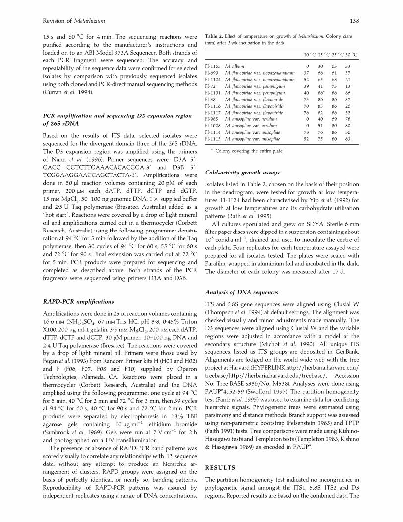
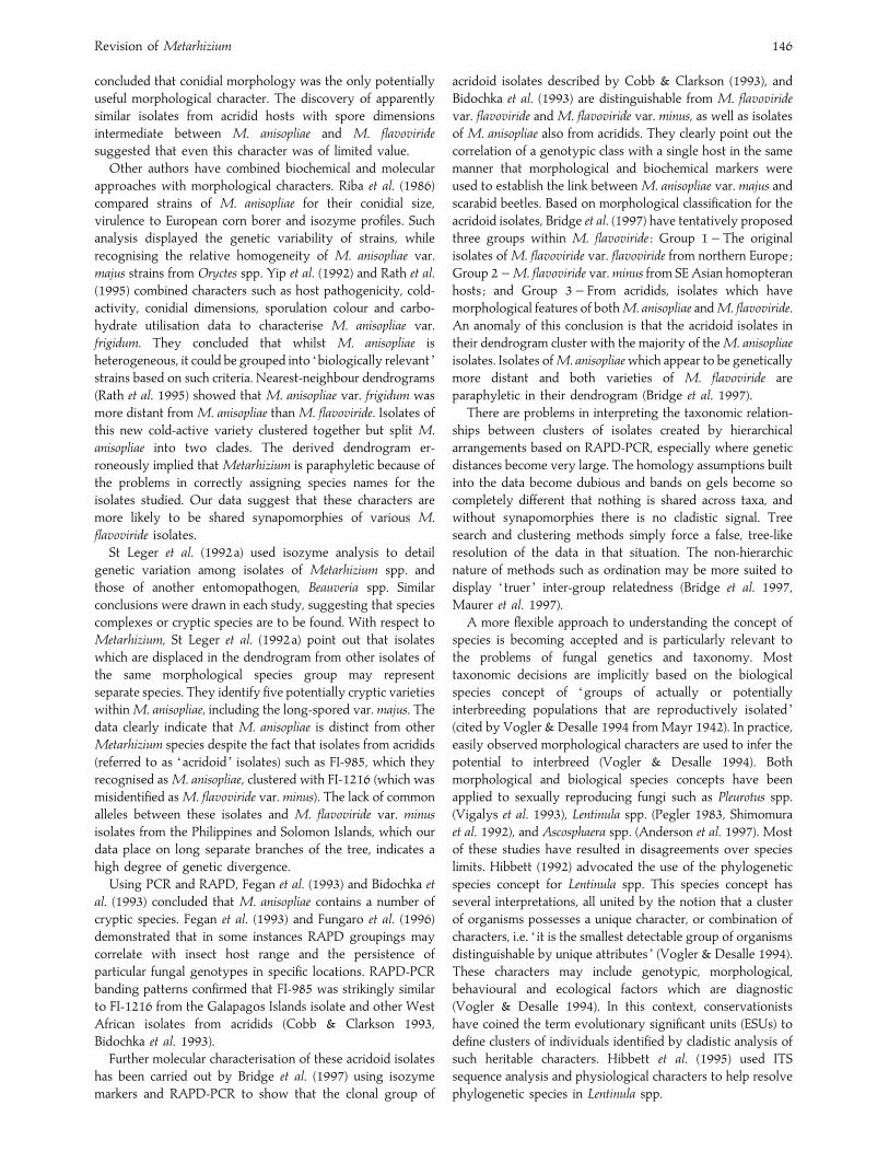
Fig. 1. The strict consensus most parsimonious tree of the whole ITS1 and ITS2 region data sets of Metarhizium spp. and outgroups.
ITS1 region contained 71 parsimony informative sites, the
5.8S region seven, the ITS2 region 80 and the D3 sequence a
further five.
With alignment gaps treated as missing data and with no
differential weighting of transversions against transitions,
parsimony analysis of either the whole data set or the ITS1
region identified in excess of 40000 most parsimonious (mp)
trees at length 415. Separate analysis of the ITS2 region
yielded a subset of those trees. The strict consensus of the
ITS1 and ITS2 whole data set mp trees is shown in Fig. 1.
As a check for possible sensitivity of these trees to
alignment assumptions, the sequences were realigned at each
of a wide range of gap penalties. The rDNA alignment was
found to vary slightly with gap parameters. As a check for
possible sensitivity to transversion : transition weighting the
original alignment was analysed by parsimony with weighting
2 :1. Under either manipulation the consensus of mp trees was
unchanged. To test for possible sensitivity to tree-estimation
procedure, neighbour-joining trees were constructed using the
Jukes–Cantor model, the Kimura 2-parameter model, and REV
general time reversible models, each being applied separately
to the ITS1 and ITS2 regions and to the whole data. So far as
relates to branches with bootstrap support" 50% the tree
topology was invariant to these manipulations.
To facilitate sampling across the large number of par-
simonious trees for each bootstrap, resampling of the data
matrix on the full taxon set the analysis was based on 100
bootstrap replicates each with 10 random-addition-sequence
starting trees, keeping% 50 trees at each replicate. Bootstrap
analysis of the reduced taxon set (Fig. 1) was based on 1000
bootstrap replicates each with an unlimited number of
bootstrap trees per replicate.
The data (Fig. 1) support : (i) the monophyly of M.
anisopliae (Clades 7–10) excluding FI-38 ; (ii) monophyly of a
Revision of Metarhizium 140
100
bp la
dder
FI-
1029
FI-
610
FI-
532
FI-
592
FI-
976
FI-
993
FI-
448
FI-
1034
FI-
692
FI-
694
FI-
522
FI-
903
FI-
921
FI-
775
FI-
774
FI-
788
FI-
792
FI-
114
FI-
208
FI-
1091
FI-
1011
FI-
161
FI-
207
FI-
552
FI-
1027
FI-
323
FI-
322
FI-
1114
FI-
1115
FI-
786
FI-
799
FI-
388
FI-
389
FI-
401
ITS group-1029 ITS group-1034 ITS group-775 ITS group-1091ITS
group-1027ITS
group-1114 Clade 10ITS
gro
up-1
14
ITS
gro
up-2
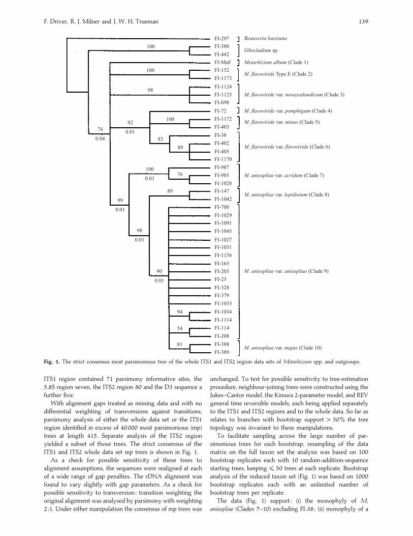
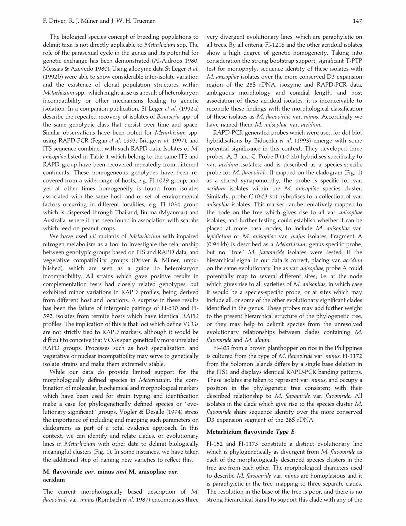
08Fig. 2. RAPD-PCR banding patterns, using primer OP-F08, for ITS groups of Metarhizium clade 9 (M. anisopliae var. anisopliae) and
clade 10 (M. anisopliae var. majus).
‘core ’ group of M. flavoviride including FI-38 (Clades 4–6) ;
(iii) monophyly of an acridoid group (Clade 7) ; and (iv) a sister
group relationship between the acridoid group (Clade 7) and
M. anisopliae (Clades 8–10) (excluding FI-38), rendering M.
flavoviride paraphyletic.
T-PTP tests confirmed the monophyly of M. anisopliae
(P¯ 0±01), as well as monophyly of the ‘core ’ M. flavoviride
group (P¯ 0±01), and the acridoid group (P¯ 0±01). The
sister relationship of the acridoid group and M. anisopliae was
examined by reducing the M. anisopliae clade to its basal node
and testing for monophyly of ‘anisopliaeacridoids ’. On an
a priori test the relationship was confirmed (P¯ 0±01).FI-38, commercially released under the name M. anisopliae,
clusters within the ‘core ’ M. flavoviride group and that
placement is supported by bootstrap scores" 90% and
T-PTP values of 0±01 on intervening branches. To confirm that
an alternative placement of FI-38 within M. anisopliae can be
rejected on these data, a T-PTP test for the non-monophyly
of ‘FI-38M. anisopliae ’ was conducted. The result, P¯ 0±01,confirms that FI-38 cannot group with any member of M.
anisopliae. Kishino–Hasegawa and Templeton tests comparing
the most-parsimonious trees with FI-38, including the M.
anisopliae against the most-parsimonious trees overall, also
confirm the incorrect identification of FI-38 as M. anisopliae
(K-H, length difference 37, .. 6±89, P! 0±0001 ; Templeton
z¯ 4±64, P! 0±0001). These tests strongly suggest that FI-38
belongs within M. flavoviride var. flavoviride.
Relationships within M. anisopliae var. anisopliae are almost
wholly unresolved due to the lack of informative sites. At the
base of the tree, relationships amongst the major clades are
also poorly resolved. The strict consensus of mp trees for the
entire taxon set shows a polytomy amongst FI-album,
FI-152FI-1173, FI-698FI-1125FI-1126 (NZ group),
‘core ’ M. flavoviride, and the ‘acridoid groupM. anisopliae ’.
The strict consensus for the reduced data set appears better
resolved but the additional resolution is without bootstrap
support. The branch immediately below the polytomy has
only 74% bootstrap support in the whole-taxon-set analysis.
A T-PTP test indicated that monophyly of the ‘ ingroup ’ taxa
(¯ all taxa except FI-297FI-360FI-442) is only weakly
supported (P¯ 0±04) on these data.
This may also be interpreted to indicate the level at which
we might delimit species. Nucleotide divergence for the ITS
region between morphologically defined species in Metarhi-
zium ranges between 14–18%, whilst divergence between
recognised varieties within species does not exceed 5%.
Levels of nucleotide divergence for clusters of isolates from
acridoid hosts and NZ blur the margins between species and
varietal limits, ranging from 6±9–17±2% in paired analyses
with morphologically recognised varieties. Cladistics requires
that species represent monophyletic groups (Wiley et al. 1991)
but there are no guide lines on taxonomic ranking (Seifert et
al. 1995).
RAPD-PCR groups
RAPD-group numbers listed in Table 1 are assigned on the
basis of their corresponding ITS-group. RAPD-PCR-groups
correlate with ITS sequence identity. Isolates which have
identical RAPD-PCR profiles, or exhibit only minor poly-
morphism with some primers, always belonged to the same
ITS-group. Results confirmed the findings of Bidochka et al.
(1993) and Fegan et al. (1993) of the greater variability of
RAPD-PCR banding patterns in M. anisopliae var. anisopliae
isolates. Small changes in ITS sequence, e.g. single point
mutations or deletions with respect to the ex-type strain of M.
anisopliae var. anisopliae are often accompanied by dramatically
different RAPD-PCR patterns with most primers. Conversely,
F. Driver, R. J. Milner and J. W. H. Trueman 141
100
bp la
dder
FI-
1216
FI-
1189
FI-
1190
FI-
1191
FI-
1192
FI-
1193
FI-
1067
FI-
983
FI-
984
FI-
986
FI-
987
FI-
1028
FI-
985
FI-
1155
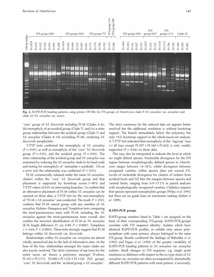
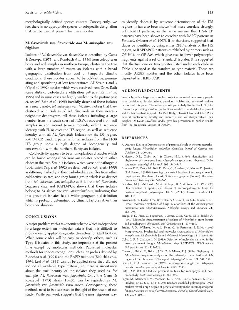
Fig. 3. RAPD-PCR banding patterns, using primer OP-F06, for
isolates of Metarhizium anisopliae var. acridum. For details of isolate
origin, see Table 1.
100
bp la
dder
FI-
1216
FI-
152
FI-
1173
FI-
403
FI-
1172
FI-
405
FI-
402
FI-
38
FI-
72F
I-11
01
FI-
698
FI-
699
FI-
1124
FI-
1165
FI-
1170
FI-
702
FI-
1126
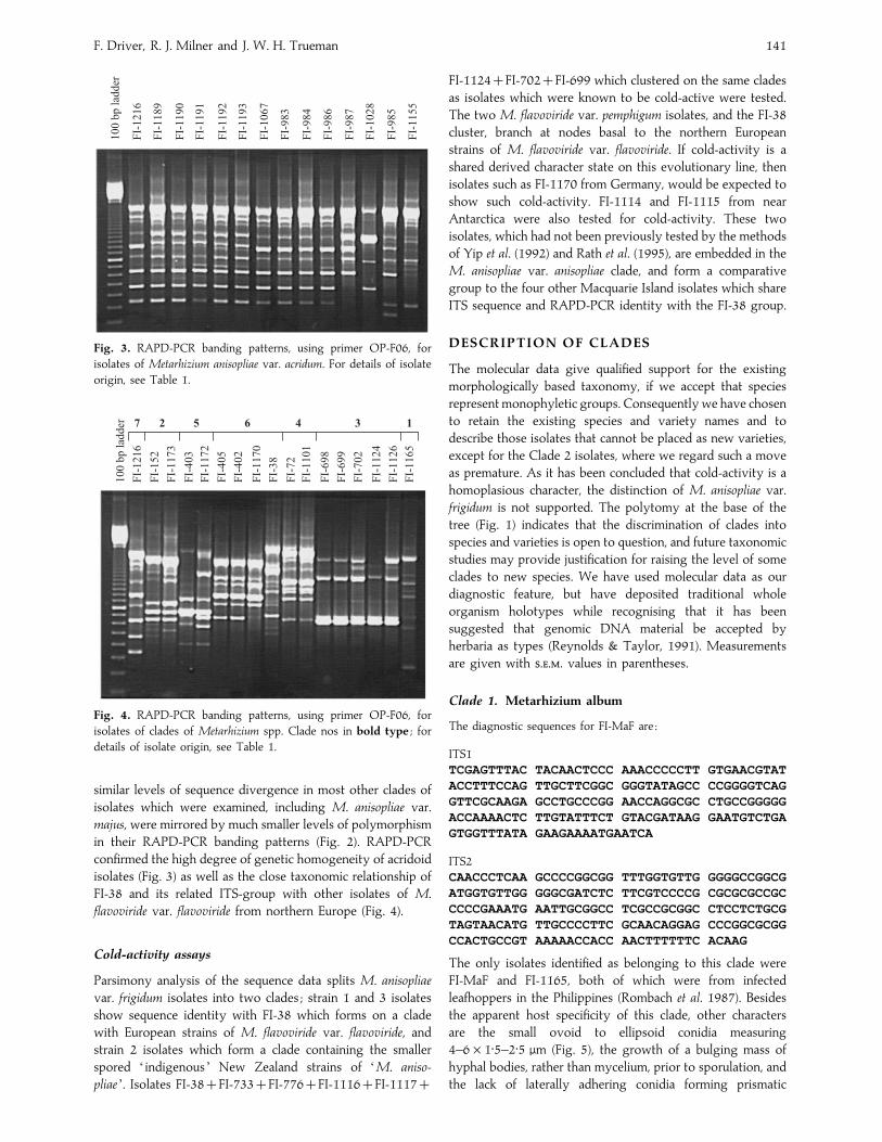
7 2 5 6 4 3 1
Fig. 4. RAPD-PCR banding patterns, using primer OP-F06, for
isolates of clades of Metarhizium spp. Clade nos in bold type ; for
details of isolate origin, see Table 1.
similar levels of sequence divergence in most other clades of
isolates which were examined, including M. anisopliae var.
majus, were mirrored by much smaller levels of polymorphism
in their RAPD-PCR banding patterns (Fig. 2). RAPD-PCR
confirmed the high degree of genetic homogeneity of acridoid
isolates (Fig. 3) as well as the close taxonomic relationship of
FI-38 and its related ITS-group with other isolates of M.
flavoviride var. flavoviride from northern Europe (Fig. 4).
Cold-activity assays
Parsimony analysis of the sequence data splits M. anisopliae
var. frigidum isolates into two clades ; strain 1 and 3 isolates
show sequence identity with FI-38 which forms on a clade
with European strains of M. flavoviride var. flavoviride, and
strain 2 isolates which form a clade containing the smaller
spored ‘ indigenous ’ New Zealand strains of ‘M. aniso-
pliae ’. Isolates FI-38FI-733FI-776FI-1116FI-1117
FI-1124FI-702FI-699 which clustered on the same clades
as isolates which were known to be cold-active were tested.
The two M. flavoviride var. pemphigum isolates, and the FI-38
cluster, branch at nodes basal to the northern European
strains of M. flavoviride var. flavoviride. If cold-activity is a
shared derived character state on this evolutionary line, then
isolates such as FI-1170 from Germany, would be expected to
show such cold-activity. FI-1114 and FI-1115 from near
Antarctica were also tested for cold-activity. These two
isolates, which had not been previously tested by the methods
of Yip et al. (1992) and Rath et al. (1995), are embedded in the
M. anisopliae var. anisopliae clade, and form a comparative
group to the four other Macquarie Island isolates which share
ITS sequence and RAPD-PCR identity with the FI-38 group.
DESCRIPTION OF CLADES
The molecular data give qualified support for the existing
morphologically based taxonomy, if we accept that species
represent monophyletic groups. Consequently we have chosen
to retain the existing species and variety names and to
describe those isolates that cannot be placed as new varieties,
except for the Clade 2 isolates, where we regard such a move
as premature. As it has been concluded that cold-activity is a
homoplasious character, the distinction of M. anisopliae var.
frigidum is not supported. The polytomy at the base of the
tree (Fig. 1) indicates that the discrimination of clades into
species and varieties is open to question, and future taxonomic
studies may provide justification for raising the level of some
clades to new species. We have used molecular data as our
diagnostic feature, but have deposited traditional whole
organism holotypes while recognising that it has been
suggested that genomic DNA material be accepted by
herbaria as types (Reynolds & Taylor, 1991). Measurements
are given with ... values in parentheses.
Clade 1. Metarhizium album
The diagnostic sequences for FI-MaF are :
ITS1
TCGAGTTTAC TACAACTCCC AAACCCCCTT GTGAACGTAT
ACCTTTCCAG TTGCTTCGGC GGGTATAGCC CCGGGGTCAG
GTTCGCAAGA GCCTGCCCGG AACCAGGCGC CTGCCGGGGG
ACCAAAACTC TTGTATTTCT GTACGATAAG GAATGTCTGA
GTGGTTTATA GAAGAAAATGAATCA
ITS2
CAACCCTCAA GCCCCGGCGG TTTGGTGTTG GGGGCCGGCG
ATGGTGTTGG GGGCGATCTC TTCGTCCCCG CGCGCGCCGC
CCCCGAAATG AATTGCGGCC TCGCCGCGGC CTCCTCTGCG
TAGTAACATG TTGCCCCTTC GCAACAGGAG CCCGGCGCGG
CCACTGCCGT AAAAACCACC AACTTTTTTC ACAAG
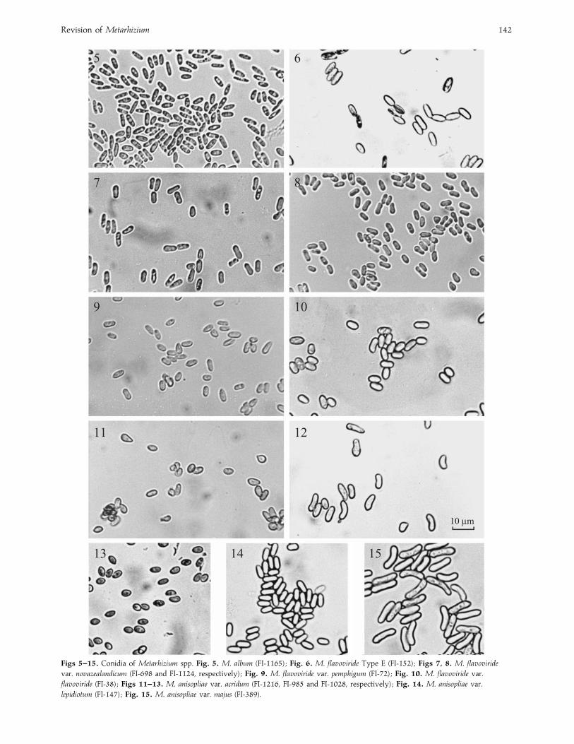
The only isolates identified as belonging to this clade were
FI-MaF and FI-1165, both of which were from infected
leafhoppers in the Philippines (Rombach et al. 1987). Besides
the apparent host specificity of this clade, other characters
are the small ovoid to ellipsoid conidia measuring
4–6¬1±5–2±5 µm (Fig. 5), the growth of a bulging mass of
hyphal bodies, rather than mycelium, prior to sporulation, and
the lack of laterally adhering conidia forming prismatic
Revision of Metarhizium 142
5 6
7 8
9 10
11 12
13 14 15
10 lm
Figs 5–15. Conidia of Metarhizium spp. Fig. 5. M. album (FI-1165) ; Fig. 6. M. flavoviride Type E (FI-152) ; Figs 7, 8. M. flavoviride
var. novazealandicum (FI-698 and FI-1124, respectively) ; Fig. 9. M. flavoviride var. pemphigum (FI-72) ; Fig. 10. M. flavoviride var.
flavoviride (FI-38) ; Figs 11–13. M. anisopliae var. acridum (FI-1216, FI-985 and FI-1028, respectively) ; Fig. 14. M. anisopliae var.
lepidiotum (FI-147) ; Fig. 15. M. anisopliae var. majus (FI-389).
F. Driver, R. J. Milner and J. W. H. Trueman 143
columns. The pale brown colour of the spores is also
characteristic, though of doubtful taxonomic significance.
Clade 2. Metarhizium flavoviride Type E
The diagnostic sequences for FI-152 are :
ITS1
CCGAGTTTAC AACTCCCAAA CCCCCAATGT GAACATATAC
CTTTACCGTT GCTTCGGCGG GCTCGGCCCC GGGAGCAGGC
TCGCCTGCCC CCCCGAGGCC TGCGCCCGCC GGGGGACTAA
AACAAACTCT TCTGTATCTT GTATAATAAG CCTGTCTGAG
TGGTATTTAA AATGAATCA
ITS2
CAACCCTCAA GCCCCGGCGG CTTGGTGTTG GGGACCGGCG
ACGGCGCTGC TCCGGCATGC GCGCGCCGCC CCCGAAATGA
ATTGGCGGTC TCGTCGCGGC CTCCCCTGCG TAGTACCACA
ACCTCGCAGC GGGAGCGCGG CGCGGCCACT GCCGTAAAAC
ACCCCAACTT CTCCAAGAG
This is an unusual clade containing just two, rather diverse,
isolates. FI-1173 from an homopteran fromBrazil was identified
as M. flavoviride var. minus by Humber (1992), but most other
isolates originally identified in this paper as M. flavoviride var.
minus were from leafhoppers and related insects in the
Philippines and Solomon Islands (Rombach et al. 1986) and
cluster in Clade 5. Morphologically they are all very similar
and come from similar hosts, so it is surprising that one isolate
should be genetically so distinct. Even more surprising is that
FI-152 (Fig. 6), from a scarab in Australia, and morphologically
resembling M. anisopliae var. anisopliae should also cluster into
this clade. FI-152 and FI-1173 show RAPD-PCR patterns
which clearly distinguish them from other isolates of M.
flavoviride var. minus. More work is needed to clarify this clade
and to determine if there are ecological and}or morphological
characteristics which are shared by these isolates, and to
collect other isolates from nature which might fit into this
clade.
Clade 3. Metarhizium flavoviride var. novazealandicum
Driver & Milner, var. nov.
Haec species, ob ordinem basium distinctam in regione ITS1 (FI-698)
CCGAGTTTAC AACTCCCAAA CCCCTGTGAA CTTATACCTT
TACTGTTGCT TCGGCGGGTC CGCCCCGGAA CAGGTTCGCG
AGAGCCGGCC CGGAACCAGG CGCCCGCCGG GGGACCAAAA
CTCTTGTATT TTTTACTTTT GCATGTCTGA GTGGAATCAT
AACAAATGAA TCA
et ITS2
CAACCCTCAA GCCCCAGCGG CTTGGTGTTG GGGACCGGCG
ACCGGCGCTG CTTCGGCAGG CCCGCGCCGC CCCCGAAATG
AATTGGCGGT CTCGTCGCGG CCTCCTCTGC GTAGTAGCAC
AACCTCGCAA CAGGAGCGCG GCGCGGCCAC TGCCGTAAAA
CGCCCAACTT TTTTTAGAG
diagnoscitur.
Holotypus : Dried culture of FI-698, originally from an
infected porina caterpillar (Lepidoptera : Hepialidae), deposited
as DAR 74293. Paratype : 74294.
Other cultures examined : FI-1124 and FI-1125 from soil,
Australia.
The diagnostic feature is the distinctive sequence data of the
ITS1 and ITS2 above. Conidia elongate, often waisted,
6 (³0±48)¬2±4 (³0±19) µm, borne in chains on clavate to
cylindrical phialides 7 (³1±5)¬1±8 (³0±3) µm (Glare et al.
1996). Hyphae broad measuring up to 0±45 µm wide. Colonies
slow-growing on SDYA attaining 5–6 mm diam. after 7 d at
25 °C. Only known to occur on soil insects and in soil in
Australia and New Zealand.
This clade contains quite a large number of isolates, many
from New Zealand, which have been recognised as distinct by
several workers (Riba et al. 1990, Curran et al. 1994, Glare &
Inwood 1997). Australian isolates described as Strain 2 by Yip
et al. (1992) have been included as part of a group of low
temperature isolates under the nomen nudum M. anisopliae var.
frigidum by Rath et al. (1995) who recognised that these were
as distinct from M. anisopliae var. anisopliae as they were from
M. flavoviride, and that they may represent a new species.
Isolates from this clade have cylindrical conidia (Figs 7 and 8)
and correspond to the short-spored form of M. anisopliae
described from New Zealand (Glare & Inwood 1997). All
isolates grow well at low temperatures (! 10 °C).
Clade 4. Metarhizium flavoviride var. flavoviride
The diagnostic sequences for FI-405 are :
ITS1
CCGAGTTTAC AAACTCCCAA ACCCCCTGTG AACTTATACC
TGTCTACCGT TGCTTCGGCG GGTTCGCCCG CCGAGGGACC
GACAAACAAA CTCTTGTATT TCTATCTATA GCATGTCTGA
GTGGAATCAT ACACAAATGA ATCA
ITS2
ACAACCCTCA AGCCCCCCCG GTGCGGGAAA CGGGCTTGGT
GTTGGGGACC GGCCAACTGG TGCCCTGCTG CTGCCGTAGC
AGGCGCCGGG CCGCCCCCGA AATGAATTGG CGGCCTCGTC
GCGGCCTCCC TCTGCGTAGT AGCACAAATC TCGCAGCTGG
AGCGCGGCGC GGCCACTGCC GTAAAACGCA CCAACTTTTT
TAAAG
This clade contains FI-405, derived from the type material
used for the original description of M. flavoviride (Gams &
Roszypal 1973). It also contains other isolates described by
Rombach et al. (1986) as M. flavoviride var. flavoviride. These
have large, somewhat swollen, conidia (Fig. 10) which form a
pale green conidial mat in culture and come from the soil or
from soil-inhabiting beetles, and grow well at low tempera-
tures. Rath et al. (1995) included FI-38 in M. anisopliae var.
frigidum and this isolate, along with a number of other soil-
derived isolates, has an ITS sequence largely identical with FI-
405 unequivocally identifying these isolates as M. flavoviride
var. flavoviride. RAPD-PCR banding patterns show a high
degree of homogeneity and conservation with the northern
European isolates, suggesting that this clade probably has a
very wide geographical distribution. Rath et al. (1995) provide
a nearest-neighbour dendrogram based on their carbohydrate
data which showed that FI-38 clustered nearer to M. flavoviride
than to M. anisopliae. Our data support this finding and it is
proposed, therefore, that the correct identity of this isolate
(and other isolates referred to as strains 1 and 3 in Rath et al.
1995) is M. flavoviride var. flavoviride.
Revision of Metarhizium 144
Clade 5. Metarhizium flavoviride var. minus
The diagnostic sequences for FI-403 are :
ITS1
TCGAGTTTAC TTTACAACTC CCAAACCCCC TGTGAACTTA
TACCTGTCTA CCGTTGCCTC GGCGGGCTCG CCCGCCGCGG
GACCGACAAA CAAAACTCTT GTATTTCTAT CTTTGGCATG
TCTGAGTGGA ATCACACATA AATGAATCA
ITS2
CAACCCTCAA GACCCCCCGG CGACGGGAAA CGGGCTTGGT
GTTGGGGACC GGCCAACCGG TGCCCTGCTG CTCCTGCGGC
AGGCGCCCGG CCGCCCCCGA AATGAATTGG CGGCCCCGTT
GCGGCCTCCC TCTGCGCAGT AGCACATGTC TCGCAGCTGG
AGCGCGGCGC GGCCACTGCC GTAAAACGCA CCAACTTTCT
TCTTTTAG
In our study only two isolates FI-403 (the ex-type isolate) and
FI-1172, both morphologically identified as Metarhizium
flavoviride var. minus, and obtained from infected leafhoppers
were included within this clade. These isolates fit the
description given by Rombach and were collected in the
Philippines and the Solomon Islands suggesting a narrow host
range and geographical distribution.
Clade 6. Metarhizium flavoviride var. pemphigum
Driver & Milner, var. nov.
Haec species, ob ordinem basium distinctam in regione ITS1 (FI-72)
CCGAGTTTAC AACTCCCAAA CCCAATGTGA ACTATACCTG
TCTACCGTTG CTTCGGCGGG TTCGCCCGCC GAGGGACCGA
CAAATAAACT CTTGTATTTC TATCTTTAGC ATGTCTGAGT
GGAATCATAA ACAAATGAAT CA
et ITS2
CAAGCCCTCA AGCCCCCCCG GCTTGGTGTT GGGGACCGGC
CACCGGTGCC CTGCTGCTTC GCGGCAGGCG CGCCCGGCCG
CCCCCGAAAT GAATTGGCGG CCCCGTCGCG GCCTCCCTCT
GCGTAGTAGC ACACATCTCG CAGCTGGAGC GCGGCGCGGC
CACTGCCGTA AAACGCACCA ACTTTTTTTT ACAG
diagnoscitur.
Holotypus : Laboratory infected Pemphigus bursarius dried over
silica gel and deposited as DAR 74295. Culture derived from
the type FI-72. Paratype : DAR 74296.
Other culture examined : FI-1101.
The diagnostic feature is the distinctive sequence data of the
ITS1 and ITS2 above. Conidia ovoid to elongate, 5±4(³0±47)¬2±4 (³0±43) µm, occasionally! 9 µm long, borne
in chains on cylindrical phialides. Spore mass light green
(Munsell greenish yellow 7±5Y 4}4). Colonies growing well
on SDYA, ca 20 mm after 7 d at 25 °C. Only known from the
U.K., where it occurs on root aphids (Pemphigus trehernei).
This small clade of two isolates with identical collection
data except the date. Both isolates were from infected root
aphids in Norfolk, England (Foster 1975) and they have
cylindrical green conidia morphologically resembling M.
anisopliae var. anisopliae (Fig. 9). They grow well at low
temperatures, however, and cluster with M. flavoviride so we
regard that as the correct specific name. More isolates
representing this clade are needed and it is possible that a
search for Metarhizium infection on other root aphids may
provide them.
Clade 7. Metarhizium anisopliae var. acridum Driver &
Milner, var. nov.
Haec species, ob ordinem basium distinctam in regione ITS (FI-987)
CCGAGTTTAC AAAACTCCCA AACCCCTGTG AACTATACCT
GTCACTGTTG CTTCGGCGGT ACCGACCCCC CGGGAAACCG
GGACCAGGCG CCCGCCGGGG ATCCTAGTAA CATCTTGAAT
CTTCTATATA ATATGGCATC TTCTGAGTGG TGGGAAAAAA
ATGAATCA
et ITS2
CGCCCCTCAA GCCCCCTGTG GGTTTGGTGT TGGGGATCGG
CGAAGCTTTT TTCAGCACGC GCCGTCCCTT AAATTTATTG
GCGGTCTCGC CGTGGCCTTT CCTCTGCGCA GTAGTAACTC
ACTCGCAACG GGAGCCCGGC GCGGTCCACT GCCGTAAAAC
CCCCAATCA CTTTGTTAC AG
diagnoscitur.
Holotypus : Locusta migratoria (Orthoptera : Acrididae) laboratory-
infected with FI-987 dried over silica gel. The original culture
was derived from a field-infected Ornithacris cavroisi collected
in Niger, West Africa. The holotype is deposited as DAR
74297. Paratypes : DAR 74298–74301.
Other cultures examined : FI-985, FI-1028 and FI-1216.
The diagnostic feature is the distinctive sequence data of the
ITS1 and ITS2 above. Conidia from the holotype are ovoid,
4±5 (³0±41)¬2±6 (³0±6) µm, borne on cylindrical to slightly
swollen phialides 7±3 (³1±9)¬2±5 (³0±4) µm. FI-985 is
unusual in having larger conidia, 7±6 (³0±78)¬2±9(³0±37) µm, borne on slightly larger phialides (Glare et al.
1996). Conidial mass dark yellow-green (Munsell 10Y 4}2).
Mycelium 2±5 µm wide. Colonies growing rapidly on SDYA,
30 mm diam. after 7 d at 25 °C. Only known from grass-
hoppers and locusts in Africa, Asia, South America and
Australia.
This clade contains all the acridoid isolates described as ‘M.
flavoviride Group 3 ’ (Bridge et al. 1997). Our data supports and
extends those of others (Bridge et al. 1993, 1997, Cobb &
Clarkson 1993, Bidochka et al. 1993) in showing that these
isolates are genetically quite uniform and are quite distinct
from the type material of M. flavoviride vars flavoviride and
minus. Bootstrapping and T-PTP tests give strong support for
the phylogenetic signal that unites these isolates to the major
evolutionary line that gives rise to M. anisopliae rather than
M. flavoviride.
Interestingly, other orthopteran hosts such as crickets are
not susceptible and in nature are attacked (exclusively) by
Clade 9 isolates. In our experience, grasshoppers are more
often infected in nature by Clade 9 isolates but these isolates
are less virulent than the rarer Clade 7 isolates (Bateman et al.
1996).
Most Clade 7 isolates produce small ovoid conidia which
would previously have identified them as M. flavoviride var.
minus. For example, FI-1216 (Figs 11, 13) was described under
that name by Rombach et al. (1985). Other isolates have larger
F. Driver, R. J. Milner and J. W. H. Trueman 145
spores which can be almost cylindrical (FI-985) (Fig. 12). All
isolates share some unusual characteristics such as the ability
to sporulate internally and to grow at 37 °C or higher (Welling
et al. 1994).
Clade 8. Metarhizium anisopliae var. lepidiotum Driver
& Milner, var. nov.
Haec species, ob ordinem basium distinctam in regione ITS1 (FI-147)
CCGAGTTCTG AAAAAACTCC CAACCCCTGT GAACTATACC
TGTAACTGTT GCTTCGGCGG GACTTGTGCC CGCCGGGGAC
CCAAACCTTC TGAATTTTTT TTAAGTATCT TCTGAGTGGT
AAAAAAAAAT GAATCA
et ITS2
CGCCCCTCAA GTCCCCTGCG GACTTGGTGT TGGGGATCGG
CGAGCGCTGT TTGCTCAGCA CGCCGTCCCC GAAATTCATT
GGCGGTCTCG CCGTGGCCCT CCTCTGCGCA GTAGTAAAAC
ACTCGCAACA GGAGCCCGGT GAGGTCCACT GCCGTAAAAC
CCCCGACTTT TTACAG
diagnoscitur.
Holotypus : Lepidiota consobrina laboratory-infected with FI-147
dried over silica gel. The original culture was derived from a
field-infected L. consobrina collected near Cairns, Queensland,
Australia. The holotype is deposited as DAR 74302.
Paratypes : DAR 74303–74306.
Other cultures examined : FI-1042.
The diagnostic feature is the distinctive sequence data of the
ITS1 and ITS2 region above. Conidia from the holotype
are parallel-sided, 7±3–10±6¬3–4±1 µm, borne on cylindrical
to slightly swollen phialides. Conidial mass dark yellow-green
(Munsell 7±5Y 4}2). Hyphae 2 µm wide. Colonies rapid
growing on SDYA, ca 70 mm after 14 d at 25 °C. Only known
from coleoptera in Australia, New Zealand and adjacent
Pacific Islands.
This is another clade with only a small number of known
isolates. All have cylindrical spores (Fig. 14) and produce a
profuse layer of green conidia in culture, and have been
isolated from scarab larvae. This cluster of isolates has
previously been described as ‘B-type ’ isolates of M. anisopliae
(Curran et al. 1994), and probably corresponds to those of
pathogenicity group 1 for Lepidiota spp. ; or RAPD group A
(Fegan et al. 1993). Similar isolates have been reported from
scarab larvae in Papua New Guinea and Fiji (Dr T. R. Glare,
pers. comm.). More needs to be known about this clade, but
the sequence data clearly separate it from M. anisopliae var.
acridum and M. anisopliae var. anisopliae.
Clade 9. Metarhizium anisopliae var. anisopliae
The diagnostic sequence data for FI-1029 are :
ITS1
CCGAGTTATC CAACTCCCAA CCCCTGTGAA TCATACCTTT
AATTGTTGCT TCGGCGGGAC TTCGCGCCCG CCGGGGACCC
AAACCTTCTG AATTTTTTAA TAAGTATCTT CTGAGTGGTT
AAAAAAAATG AATCA
ITS2
CGCCCCTCAA GTCCCCTGTG GACTTGGTGT TGGGGATCGG
CGAGGCTGGT TTTCCAGCAC AGCCGTCCCT TAAATTAATT
GGCGGTCTCG CCGTGGCCCT CCTCTGCGCA GTAGTAAAGC
ACTCGCAACA GGAGCCCGGC GCGGTCCACT GCCGTAAAAC
CCCCCAACTT TTTATAG
FI-1029, derived from the type material of Metarhizium
anisopliae var. anisopliae (Tulloch 1976), forms the basis for this
clade. It includes the majority of isolates found in nature and
is genetically highly diverse. St Leger et al. (1992b)
demonstrated the existence of clonal population structures in
M. anisopliae. Distinct groups can be identified within the
clade, e.g. a number of isolates from the black field cricket,
Teleogryllus commodus, in Australia, share identical ITS sequence
data and have very similar RAPD-PCR patterns (Milner et al.
1996). Isolates generally have green, cylindrical conidia,
5–7 µm long, which form in columns of chains. They normally
grow poorly outside the range 15–32 °C, though we have
found two cold temperature active isolates which came from
soil from Macquarie Island (Dr A. C. Rath, pers. comm.),
confirming that cold activity is a homoplasious character.
Clade 10. Metarhizium anisopliae var. majus
The diagnostic sequence data for FI-388 (not the type) are :
ITS1
CCGAGTTATC CAACTCCCAA CCCCTGTGAA TTATACCTTT
AATTGTTGCT TCGGCGGGAC TTCGCGCTCG CCGGGGACCC
AAACCTTCTG AATTTTTTAA TAAGGATCTT CTGAGTGGTT
AAAAAAAAAA TGAATCA
ITS2
CGCCCCTCAA GTCCCCTGTG GACTTGGTGT TGGGGATCGG
CGAGGCTGGT TTTCCAGCAC AGCCGTCCCT TAAATTGATT
GGCGGTCTCG CCGTGGCCCT CCTTTGCGCA GTAGTAAAAC
ACTCGCAACA GGAGCCCGGC GCGGTCCACT GCCGTAAAAC
ACCCCAACTT TTTATAG
A typical isolate from this clade is FI-388 which conforms to
the description by Tulloch (1976). Isolates are readily identified
on the basis of the very large conidia, usually" 10 µm long
(Fig. 15), rapidly growing colonies producing dark green
conidia, and are most frequently found attacking dynastine
beetles in tropical countries. Our results support those of
other workers (St Leger et al. 1992b, Leal et al. 1994), and
show that genetically this clade differs less from M. anisopliae
var. anisopliae Clade 9 isolates than do other isolates such as
those in Clade 8 (‘B-type ’), which are also currently described
as var. anisopliae.
DISCUSSION
This study has again shown the limitations of morphological
characters in distinguishing between species of Metarhizium.
Glare et al. (1996) tested the validity of phialide morphology,
as suggested by Rombach et al. (1986), as a useful taxonomic
character by examining 11 isolates representing M. anisopliae,
M. flavoviride, and M. album. They found that phialide
morphology of a single isolate could vary within the same
culture as well as between substrates. Furthermore they
Revision of Metarhizium 146
concluded that conidial morphology was the only potentially
useful morphological character. The discovery of apparently
similar isolates from acridid hosts with spore dimensions
intermediate between M. anisopliae and M. flavoviride
suggested that even this character was of limited value.
Other authors have combined biochemical and molecular
approaches with morphological characters. Riba et al. (1986)
compared strains of M. anisopliae for their conidial size,
virulence to European corn borer and isozyme profiles. Such
analysis displayed the genetic variability of strains, while
recognising the relative homogeneity of M. anisopliae var.
majus strains from Oryctes spp. Yip et al. (1992) and Rath et al.
(1995) combined characters such as host pathogenicity, cold-
activity, conidial dimensions, sporulation colour and carbo-
hydrate utilisation data to characterise M. anisopliae var.
frigidum. They concluded that whilst M. anisopliae is
heterogeneous, it could be grouped into ‘biologically relevant ’
strains based on such criteria. Nearest-neighbour dendrograms
(Rath et al. 1995) showed that M. anisopliae var. frigidum was
more distant from M. anisopliae than M. flavoviride. Isolates of
this new cold-active variety clustered together but split M.
anisopliae into two clades. The derived dendrogram er-
roneously implied that Metarhizium is paraphyletic because of
the problems in correctly assigning species names for the
isolates studied. Our data suggest that these characters are
more likely to be shared synapomorphies of various M.
flavoviride isolates.
St Leger et al. (1992a) used isozyme analysis to detail
genetic variation among isolates of Metarhizium spp. and
those of another entomopathogen, Beauveria spp. Similar
conclusions were drawn in each study, suggesting that species
complexes or cryptic species are to be found. With respect to
Metarhizium, St Leger et al. (1992a) point out that isolates
which are displaced in the dendrogram from other isolates of
the same morphological species group may represent
separate species. They identify five potentially cryptic varieties
within M. anisopliae, including the long-spored var. majus. The
data clearly indicate that M. anisopliae is distinct from other
Metarhizium species despite the fact that isolates from acridids
(referred to as ‘acridoid ’ isolates) such as FI-985, which they
recognised as M. anisopliae, clustered with FI-1216 (which was
misidentified as M. flavoviride var. minus). The lack of common
alleles between these isolates and M. flavoviride var. minus
isolates from the Philippines and Solomon Islands, which our
data place on long separate branches of the tree, indicates a
high degree of genetic divergence.
Using PCR and RAPD, Fegan et al. (1993) and Bidochka et
al. (1993) concluded that M. anisopliae contains a number of
cryptic species. Fegan et al. (1993) and Fungaro et al. (1996)
demonstrated that in some instances RAPD groupings may
correlate with insect host range and the persistence of
particular fungal genotypes in specific locations. RAPD-PCR
banding patterns confirmed that FI-985 was strikingly similar
to FI-1216 from the Galapagos Islands isolate and other West
African isolates from acridids (Cobb & Clarkson 1993,
Bidochka et al. 1993).
Further molecular characterisation of these acridoid isolates
has been carried out by Bridge et al. (1997) using isozyme
markers and RAPD-PCR to show that the clonal group of
acridoid isolates described by Cobb & Clarkson (1993), and
Bidochka et al. (1993) are distinguishable from M. flavoviride
var. flavoviride and M. flavoviride var. minus, as well as isolates
of M. anisopliae also from acridids. They clearly point out the
correlation of a genotypic class with a single host in the same
manner that morphological and biochemical markers were
used to establish the link between M. anisopliae var. majus and
scarabid beetles. Based on morphological classification for the
acridoid isolates, Bridge et al. (1997) have tentatively proposed
three groups within M. flavoviride : Group 1 – The original
isolates of M. flavoviride var. flavoviride from northern Europe ;
Group 2 – M. flavoviride var. minus from SE Asian homopteran
hosts ; and Group 3 – From acridids, isolates which have
morphological features of both M. anisopliae and M. flavoviride.
An anomaly of this conclusion is that the acridoid isolates in
their dendrogram cluster with the majority of the M. anisopliae
isolates. Isolates of M. anisopliae which appear to be genetically
more distant and both varieties of M. flavoviride are
paraphyletic in their dendrogram (Bridge et al. 1997).
There are problems in interpreting the taxonomic relation-
ships between clusters of isolates created by hierarchical
arrangements based on RAPD-PCR, especially where genetic
distances become very large. The homology assumptions built
into the data become dubious and bands on gels become so
completely different that nothing is shared across taxa, and
without synapomorphies there is no cladistic signal. Tree
search and clustering methods simply force a false, tree-like
resolution of the data in that situation. The non-hierarchic
nature of methods such as ordination may be more suited to
display ‘ truer ’ inter-group relatedness (Bridge et al. 1997,
Maurer et al. 1997).
A more flexible approach to understanding the concept of
species is becoming accepted and is particularly relevant to
the problems of fungal genetics and taxonomy. Most
taxonomic decisions are implicitly based on the biological
species concept of ‘groups of actually or potentially
interbreeding populations that are reproductively isolated ’
(cited by Vogler & Desalle 1994 from Mayr 1942). In practice,
easily observed morphological characters are used to infer the
potential to interbreed (Vogler & Desalle 1994). Both
morphological and biological species concepts have been
applied to sexually reproducing fungi such as Pleurotus spp.
(Vigalys et al. 1993), Lentinula spp. (Pegler 1983, Shimomura
et al. 1992), and Ascosphaera spp. (Anderson et al. 1997). Most
of these studies have resulted in disagreements over species
limits. Hibbett (1992) advocated the use of the phylogenetic
species concept for Lentinula spp. This species concept has
several interpretations, all united by the notion that a cluster
of organisms possesses a unique character, or combination of
characters, i.e. ‘ it is the smallest detectable group of organisms
distinguishable by unique attributes ’ (Vogler & Desalle 1994).
These characters may include genotypic, morphological,
behavioural and ecological factors which are diagnostic
(Vogler & Desalle 1994). In this context, conservationists
have coined the term evolutionary significant units (ESUs) to
define clusters of individuals identified by cladistic analysis of
such heritable characters. Hibbett et al. (1995) used ITS
sequence analysis and physiological characters to help resolve
phylogenetic species in Lentinula spp.
F. Driver, R. J. Milner and J. W. H. Trueman 147
The biological species concept of breeding populations to
delimit taxa is not directly applicable to Metarhizium spp. The
role of the parasexual cycle in the genus and its potential for
genetic exchange has been demonstrated (Al-Aidroos 1980,
Messias & Azevedo 1980). Using allozyme data St Leger et al.
(1992b) were able to show considerable inter-isolate variation
and the existence of clonal population structures within
Metarhizium spp., which might arise as a result of heterokaryon
incompatibility or other mechanisms leading to genetic
isolation. In a companion publication, St Leger et al. (1992a)
describe the repeated recovery of isolates of Beauveria spp. of
the same genotypic class that persist over time and space.
Similar observations have been noted for Metarhizium spp.
using RAPD-PCR (Fegan et al. 1993, Bridge et al. 1997), and
ITS sequence combined with such RAPD data. Isolates of M.
anisopliae listed in Table 1 which belong to the same ITS and
RAPD group have been recovered repeatedly from different
continents. These homogeneous genotypes have been re-
covered from a wide range of hosts, e.g. FI-1029 group, and
yet at other times homogeneity is found from isolates
associated with the same host, and or set of environmental
factors occurring in different localities, e.g. FI-1034 group
which is dispersed through Thailand, Burma (Myanmar) and
Australia, where it has been found in association with scarabs
which feed on peanut crops.
We have used nit mutants of Metarhizium with impaired
nitrogen metabolism as a tool to investigate the relationship
between genotypic groups based on ITS and RAPD data, and
vegetative compatibility groups (Driver & Milner, unpu-
blished), which are seen as a guide to heterokaryon
incompatibility. All strains which gave positive results in
complementation tests had closely related genotypes, but
exhibited minor variations in RAPD profiles, being derived
from different host and locations. A surprise in these results
has been the failure of intergenic pairings of FI-610 and FI-
592, isolates from termite hosts which have identical RAPD
profiles. The implication of this is that loci which define VCGs
are not strictly tied to RAPD markers, although it would be
difficult to conceive that VCGs span genetically more unrelated
RAPD groups. Processes such as host specialisation, and
vegetative or nuclear incompatibility may serve to genetically
isolate strains and make them extremely stable.
While our data do provide limited support for the
morphologically defined species in Metarhizium, the com-
bination of molecular, biochemical and morphological markers
which have been used for strain typing and identification
make a case for phylogenetically defined species or ‘evo-
lutionary significant ’ groups. Vogler & Desalle (1994) stress
the importance of including and mapping such parameters on
cladograms as part of a total evidence approach. In this
context, we can identify and relate clades, or evolutionary
lines in Metarhizium with other data to delimit biologically
meaningful clusters (Fig. 1). In some instances, we have taken
the additional step of naming new varieties to reflect this.
M. flavoviride var. minus and M. anisopliae var.
acridum
The current morphologically based description of M.
flavoviride var. minus (Rombach et al. 1987) encompasses three
very divergent evolutionary lines, which are paraphyletic on
all trees. By all criteria, FI-1216 and the other acridoid isolates
show a high degree of genetic homogeneity. Taking into
consideration the strong bootstrap support, significant T-PTP
test for monophyly, sequence identity of these isolates with
M. anisopliae isolates over the more conserved D3 expansion
region of the 28S rDNA, isozyme and RAPD-PCR data,
ambiguous morphology and conidial length, and host
association of these acridoid isolates, it is inconceivable to
reconcile these findings with the morphological classification
of these isolates as M. flavoviride var. minus. Accordingly we
have named them M. anisopliae var. acridum.
RAPD-PCR generated probes which were used for dot blot
hybridisations by Bidochka et al. (1993) emerge with some
potential significance in this context. They developed three
probes, A, B, and C. Probe B (1±6 kb) hybridises specifically to
var. acridum isolates, and is described as a species-specific
probe for M. flavoviride. If mapped on the cladogram (Fig. 1)
as a shared synapomorphy, the probe is specific for var.
acridum isolates within the M. anisopliae species cluster.
Similarly, probe C (0±63 kb) hybridises to a collection of var.
anisopliae isolates. This marker can be tentatively mapped to
the node on the tree which gives rise to all var. anisopliae
isolates, and further testing could establish whether it can be
placed at more basal nodes, to include M. anisopliae var.
lepidiotum or M. anisopliae var. majus isolates. Fragment A
(0±94 kb) is described as a Metarhizium genus-specific probe,
but no ‘ true ’ M. flavoviride isolates were tested. If the
hierarchical signal in our data is correct, placing var. acridum
on the same evolutionary line as var. anisopliae, probe A could
potentially map to several different sites ; i.e. at the node
which gives rise to all varieties of M. anisopliae, in which case
it would be a species-specific probe, or at sites which may
include all, or some of the other evolutionary significant clades
identified in the genus. These probes may add further weight
to the present hierarchical structure of the phylogenetic tree,
or they may help to delimit species from the unresolved
evolutionary relationships between clades containing M.
flavoviride and M. album.
FI-403 from a brown planthopper on rice in the Philippines
is cultured from the type of M. flavoviride var. minus. FI-1172
from the Solomon Islands differs by a single base deletion in
the ITS1 and displays identical RAPD-PCR banding patterns.
These isolates are taken to represent var. minus, and occupy a
position in the phylogenetic tree consistent with their
described relationship to M. flavoviride var. flavoviride. All
isolates in the clade which give rise to the species cluster M.
flavoviride share sequence identity over the more conserved
D3 expansion segment of the 28S rDNA.
Metarhizium flavoviride Type E
FI-152 and FI-1173 constitute a distinct evolutionary line
which is phylogenetically as divergent from M. flavoviride as
each of the morphologically described species clusters in the
tree are from each other. The morphological characters used
to describe M. flavoviride var. minus are homoplasious and it
is paraphyletic in the tree, mapping to three separate clades.
The resolution in the base of the tree is poor, and there is no
strong hierarchical signal to support this clade with any of the
Revision of Metarhizium 148
morphologically defined species clusters. Consequently, we
feel there is no appropriate species or subspecific designation
that can be used at present for these isolates.
M. flavoviride var. flavoviride and M. anisopliae var.
frigidum
Isolates of M. flavoviride var. flavoviride as described by Gams
& Roszypal (1973), and Rombach et al. (1986) from coleopteran
hosts and soil samples in northern Europe, cluster in the tree
with a large number of Australian isolates with a broad
geographic distribution from cool or temperate climatic
conditions. These isolates appear to be cold-active, germin-
ating and sporulating at low temperatures. All Strain 1 and 3
(Yip et al. 1992) isolates which were received from Dr A. Rath
share distinct carbohydrate utilisation patterns (Rath et al.
1995) and in some cases are highly virulent to the pasture pest
A. couloni. Rath et al. (1995) invalidly described these isolates
as a new variety, M. anisopliae var. frigidum, noting that they
clustered with isolates of M. flavoviride in their nearest-
neighbour dendrogram. All these isolates, including a large
number from the south coast of N.S.W. recovered from soil
samples in and around termite mounds, exhibit sequence
identity with FI-38 over the ITS region, as well as sequence
identity with all M. flavoviride isolates for the D3 region.
RAPD-PCR banding patterns for all isolates from the FI-38
ITS group show a high degree of homogeneity and
conservation with the northern European isolates.
Cold-activity appears to be a homoplasious character which
can be found amongst Metarhizium isolates placed in other
clades in the tree. Strain 2 isolates, which were not pathogenic
to A. couloni (Yip et al. 1992) are described by Rath et al. (1995)
as differing markedly in their carbohydrate profiles from other
cold-active isolates, and they form a group which is as distinct
from M. anisopliae var. anisopliae as it is from M. flavoviride.
Sequence data and RAPD-PCR shows that these isolates
belong to M. flavoviride var. novazealandicum, indicating that
this group of isolates has a wider geographic distribution
which is probably determined by climatic factors rather than
host specialisation.
CONCLUSIONS
A major problem with a taxonomic scheme which is dependent
to a large extent on molecular data is that it is difficult to
provide easily applied diagnostic characters for identification.
While some clades will be easy to identify, others, such as
Type E isolates in this study, are impossible at the present
time except by molecular methods. Published molecular
methods for species recognition such as the probes devised by
Bidochka et al. (1994) and the RAPD methods (Bidochka et al.
1994, Leal et al. 1994) cannot be applied since they did not
include all available type material, so there is uncertainty
about the true identity of the isolates they used as, for
example, M. flavoviride var. flavoviride. Only the Gams &
Roszypal (1973) strain, FI-405, can be regarded as M.
flavoviride var. flavoviride sensu stricto. Consequently, these
methods need to be reassessed in the light of the results of our
study. While our work suggests that the most rigorous way
to identify clades is by sequence determination of the ITS
regions, it has also been shown that these correlate strongly
with RAPD patterns, in the same manner that ITS-RFLP
patterns have been shown to correlate with RAPD patterns in
Beauveria (Maurer et al. 1997). It is, therefore, suggested that
clades be identified by using either RFLP analysis of the ITS
region, or RAPD-PCR patterns established by primers such as
OP-H01, or OP-A03 which give rise to fewer polymorphic
fragments against a set of ‘standard ’ isolates. It is suggested
that the first one or two isolates listed under each clade in
Table 1 be used as the standard or type material. These are
mostly ARSEF isolates and the other isolates have been
deposited in HERB-DAR.
ACKNOWLEDGEMENTS
Inevitably with a large and complex project as reported here, many people
have contributed to discussions, provided isolates and reviewed various
versions of this paper. The authors would particularly like to thank Dr John
Curran for providing most of the facilities needed to undertake the project
and for his constant support. Drs Paul Bridge, Travis Glare and Chris Prior
have all contributed, directly and indirectly, and we always valued their
insights. Dr David Swofford kindly gave his permission to publish results
from the pre-release version of PAUP*.
REFERENCES
Al Aidroos, K. (1980) Demonstration of parasexual cycle in the entomopatho-
genic fungus Metarhizium anisopliae. Canadian Journal of Genetics and
Cytology 22 : 309–314.
Anderson, D. L., Gibbs, A. J. & Gibson, N. L. (1997) Identification and
phylogeny of spore-cyst fungi (Ascosphaera spp.) using ribosomal DNA
sequences. Mycological Research 102 : 541–547.
Bateman, R. P., Carey, M., Batt, D., Prior, C., Abraham, Y., Moore, D., Jenkins,
N. & Fenlon, J. (1996) Screening for virulent isolates of entomopathogenic
fungi against the desert locust, Schistocerca gregaria (Forskal). Biocontrol
Science and Technology 6 : 549–560.
Bidochka, M. J., McDonald, M. A., St Leger, R. A. & Roberts, D. W. (1993)
Differentiation of species and strains of entomopathogenic fungi by
random amplified polymorphic DNA (RAPD). Current Genetics 21 :
107–113.
Bowman, B. H., Taylor, J. W., Brownlee, A. G., Lee, J., Lu, S.-D. & White, T. J.
(1992) Molecular evolution of fungi : relationships of the Basidiomycetes,
Ascomycetes and Chytridiomycetes. Molecular Biology and Evolution 92 :
285–296.
Bridge, P. D., Prior, C., Sogbohan, J., Lomer, C. M., Carey, M. & Buddie, A.
(1997) Molecular characterization of isolates of Metarhizium from locusts
and grasshoppers. Biodiversity and Conservation 5 : 177–189.
Bridge, P. D., Williams, M. A. J., Prior, C. & Patterson, R. R. M. (1993)
Morphological, biochemical and molecular characteristics of Metarhizium
anisopliae and M. flavoviride. Journal of General Microbiology 13 : 1163–1169.
Cobb, B. D. & Clarkson, J. M. (1993) Detection of molecular variation in the
insect pathogenic fungus Metarhizium using RAPD-PCR. FEMS Micro-
biological Letters 11 : 319–324.
Curran, J., Driver, F., Ballard, J. W. O. & Milner, R. J. (1994) Phylogeny of
Metarhizium : sequence analysis of the internally transcribed and 5.8s
region of the ribosomal DNA repeat. Mycological Research 9 : 547–552.
Evans, H. C. & Samson, R. A. (1982) Entomogenous fungi from Galapagos
Islands. Canadian Journal of Botany 6 : 2325–2333.
Faith, D. P. (1991) Cladistic permutation tests for monophyly and non-
monophyly. Systematic Zoology 4 : 366–375.
Fegan, M., Manners, J. M., MacLean, D. J., Irwin, J. A. G., Samuels, K. D. Z.,
Holdom, D. G. & Li, D. P. (1993) Random amplified polymorphic DNA
markers reveal a high degree of genetic diversity in the entomopathogenic
fungus Metarhizium anisopliae var. anisopliae. Journal of General Microbiology
13 : 2075–2081.
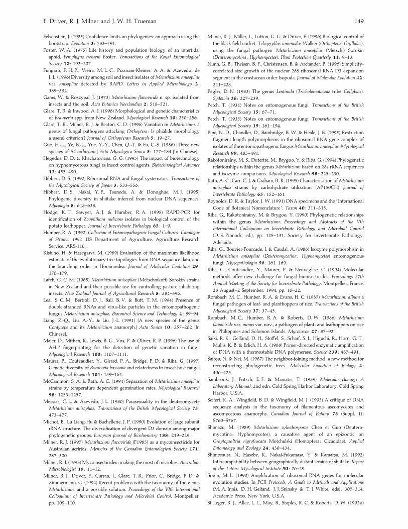
F. Driver, R. J. Milner and J. W. H. Trueman 149
Felsenstein, J. (1985) Confidence limits on phylogenies : an approach using the
bootstrap. Evolution 3 : 783–791.
Foster, W. A. (1975) Life history and population biology of an intertidal
aphid, Pemphigus treherni Foster. Transactions of the Royal Entomological
Society 12 : 192–207.
Fungaro, F. H. P., Vieira, M. L. C., Pizzirani-Kleiner, A. A. & Azevedo, de
J. L. (1996) Diversity among soil and insect isolates ofMetarhizium anisopliae
var. anisopliae detected by RAPD. Letters in Applied Microbiology 2 :
389–392.
Gams, W. & Roszypal, J. (1973) Metarhizium flavoviride n. sp. isolated from
insects and the soil. Acta Botanica Neerlandica 2 : 518–521.
Glare, T. R. & Inwood, A. J. (1998) Morphological and genetic characteristics
of Beauveria spp. from New Zealand. Mycological Research 10 : 250–256.
Glare, T. R., Milner, R. J. & Beaton, C. D. (1996) Variation in Metarhizium, a
genus of fungal pathogens attacking Orthoptera : Is phialide morphology
a useful criterion? Journal of Orthopteran Research 5 : 19–27.
Guo, H.-L., Ye, B.-L., Yue, Y.-Y., Chen, Q.-T. & Fu, C.-S. (1986) [Three new
species of Metarhizium.] Acta Mycologica Sinica 5 : 177–184 [In Chinese].
Hegedus, D. D. & Khachatorians, G. G. (1995) The impact of biotechnology
on hyphomycetous fungi as insect control agents. Biotechnological Advance
13 : 455–490.
Hibbert, D. S. (1992) Ribosomal RNA and fungal systematics. Transactions of
the Mycological Society of Japan 3 : 533–556.
Hibbert, D. S., Nakai, Y. F., Tsuneda, A. & Donoghue, M. J. (1995)
Phylogenic diversity in shiitake inferred from nuclear DNA sequences.
Mycologia 8 : 618–638.
Hodge, K. T., Sawyer, A. J. & Humber, R. A. (1995) RAPD-PCR for
identification of Zoophthora radicans isolates in biological control of the
potato leafhopper. Journal of Invertebrate Pathology 65 : 1–9.
Humber, R. A. (1992) Collection of Entomopathogenic Fungal Cultures : Catalogue
of Strains. 1992. US Department of Agriculture, Agriculture Research
Service, ARS-110.
Kishino, H. & Hasegawa, M. (1989) Evaluation of the maximum likelihood
estimate of the evolutionary tree topologies from DNA sequence data, and
the branching order in Hominoidea. Journal of Molecular Evolution 29 :
170–179.
Latch, G. C. M. (1965) Metarhizium anisopliae (Metschnikoff) Sorokin strains
in New Zealand and their possible use for controlling pasture inhabiting
insects. New Zealand Journal of Agricultural Research 8 : 384–396.
Leal, S. C. M., Bertioli, D. J., Ball, B. V. & Butt, T. M. (1994) Presence of
double-stranded RNAs and virus-like particles in the entomopathogenic
fungus Metarhizium anisopliae. Biocontrol Science and Technology 4 : 89–94.
Liang, Z.-Q., Liu, A.-Y., & Liu, J.-L. (1991) [A new species of the genus
Cordyceps and its Metarhizium anamorph.] Acta Sinica 10 : 257–262 [In
Chinese].
Majer, D., Mithen, R., Lewis, B. G., Vos, P. & Oliver, R. P. (1996) The use of
AFLP fingerprinting for the detection of genetic variation in fungi.
Mycological Research 100 : 1107–1111.
Maurer, P., Couteaudier, Y., Girard, P. A., Bridge, P. D. & Riba, G. (1997)
Genetic diversity of Beauveria bassiana and relatedness to insect host range.
Mycological Research 101 : 159–164.
McCammon, S. A. & Rath, A. C. (1994) Separation of Metarhizium anisopliae
strains by temperature dependent germination rates. Mycological Research
98 : 1253–1257.
Messias, C. L. & Azevedo, J. L. (1980) Parasexuality in the deuteromycete
Metarhizium anisopliae. Transactions of the British Mycological Society 75 :
473–477.
Michot, B., Lu Liang-Hu & Bachellerie, J. P. (1990) Evolution of large subunit
rRNA structure. The diversification of divergent D3 domain among major
phylogenetic groups. European Journal of Biochemistry 188 : 219–229.
Milner, R. J. (1997) Metarhizium flavoviride (F1985) as a mycoinsecticide for
Australian acririds. Memoirs of the Canadian Entomological Society 171 :
287–300.
Milner, R. J. (1998) Mycoinsecticides : making the most of microbes. Australian
Microbiologist 19 : 11–12.
Milner, R. J., Driver, F., Curran, J., Glare, T. R., Prior, C., Bridge, P. D. &
Zimmermann, G. (1994) Recent problems with the taxonomy of the genus
Metarhizium, and a possible solution. Proceedings of the VIth International
Colloquium of Invertebrate Pathology and Microbial Control, Montpellier,
pp. 109–110.
Milner, R. J., Miller, L., Lutton, G. G. & Driver, F. (1996) Biological control of
the black field cricket, Teleogryllus commodus Walker (Orthoptera : Gryllidae),
using the fungal pathogen Metarhizium anisopliae (Metsch.) Sorokin
(Deuteromycotina : Hyphomycetes). Plant Protection Quarterly 11 : 9–13.
Nunn, G. B., Theisen, B. F., Christensen, B. & Arctander, P. (1996) Simplicity-
correlated size growth of the nuclear 28S ribosomal RNA D3 expansion
segment in the crustacean order Isopoda. Journal of Molecular Evolution 42 :
211–223.
Pegler, D. N. (1983) The genus Lentinula (Tricholomataceae tribe Collybiae).
Sydowia 36 : 227–239.
Petch, T. (1931) Notes on entomogenous fungi. Transactions of the British
Mycological Society 15 : 67–71.
Petch, T. (1935) Notes on entomogenous fungi. Transactions of the British
Mycological Society 19 : 161–194.
Pipe, N. D., Chandler, D., Bainbridge, B. W. & Heale, J. B. (1995) Restriction
fragment length polymorphisms in the ribosomal RNA gene complex of
isolates of the entomopathogenic fungusMetarhizium anisopliae. Mycological
Research 99 : 485–491.
Rakotonirainy, M. S., Dutertre, M., Brygoo, Y. & Riba, G. (1994) Phylogenetic
relationships within the genus Metarhizium based on 28s rRNA sequences
and isozyme comparisons. Mycological Research 98 : 225–230.
Rath, A. C., Carr, C. J. & Graham, B. R. (1995) Characterisation of Metarhizium
anisopliae strains by carbohydrate utilization (AP150CH) Journal of
Invertebrate Pathology 65 : 152–161.
Reynolds, D. R. & Taylor, J. W. (1991) DNA specimens and the ‘ International
Code of Botanical Nomenclature ’. Taxon 40 : 311–315.
Riba, G., Rakotonirainy, M. & Brygoo, Y. (1990) Phylogenetic relationships
within the genus Metarhizium. Proceedings and Abstracts of the Vth
International Colloquium on Invertebrate Pathology and Microbial Control
(D. E. Pinnock, ed.), pp. 125–131, Society for Invertebrate Pathology,
Adelaide.
Riba, G., Bouvier-Fourcade, I. & Caudal, A. (1986) Isozyme polymorphism in
Metarhizium anisopliae (Deuteromycotina : Hyphomycetes) entomogenous
fungi. Mycopathologia 96 : 161–169.
Riba, G., Couteaudier, Y., Maurer, P. & Neuveglise, C. (1994) Molecular
methods offer new challenge for fungal bioinsecticides. Proceedings 27th
Annual Meeting of the Society for Invertebrate Pathology, Montpellier, France,
28 August–2 September, 1994, pp. 16–22.
Rombach, M. C., Humber, R. A. & Evans, H. C. (1987) Metarhizium album a
fungal pathogen of leaf- and planthoppers of rice. Transactions of the British
Mycological Society 37 : 37–45.
Rombach, M. C., Humber, R. A. & Roberts, D. W. (1986) Metarhizium
flavoviride var. minus var. nov., a pathogen of plant- and leafhoppers on rice
in Philippines and Solomon Islands. Mycotaxon 27 : 87–92.
Saiki, R. K., Gelfand, D. H., Stoffel, S., Scharf, S. J., Higuchi, R., Horn, G. T.,
Mullis, K. B. & Erlich, H. A. (1988) Primer-directed enzymatic amplification
of DNA with a thermostable DNA polymerase. Science 239 : 487–491.
Saitou, N. & Nei, M. (1987) The neighbor-joining method : a new method for
reconstructing phylogenetic trees. Molecular Evolution of Biology 4 :
406–425.
Sambrook, J., Fritsch, E. F. & Maniatis, T. (1989) Molecular cloning : A
Laboratory Manual, 2nd edn. Cold Spring Harbor Laboratory, Cold Spring
Harbor, U.S.A.
Seifert, K. A., Wingfield, B. D. & Wingfield, M. J. (1995) A critique of DNA
sequence analysis in the taxonomy of filamentous ascomycetes and
ascomycetous anamorphs. Canadian Journal of Botany 73 (Suppl. 1) :
S760–S767.
Shimazu, M. (1989) Metarhizium cylindrosporae Chen et Guo (Deutero-
mycotina : Hyphomycetes), a causative agent of an epizootic on
Graptopsaltria nigrofuscata Motchulski (Homoptera : Cicadidae). Applied
Entomology and Zoology 24 : 430–434.
Shimomura, N., Hasebe, K., Nakai-Fukumasa, Y. & Kamatsu, M. (1992)
Intercompatibility between geographically distant strains of shiitake. Report
of the Tottori Mycological Institute 30 : 26–29.
Sogin, M. L. (1990) Amplification of ribosomal RNA genes for molecular
evolution studies. In PCR Protocols. A Guide to Methods and Applications
(M. A. Innis, D. H. Gelfand, J. J. Sninsky & T. J. White, eds) : 307–314.
Academic Press, New York, U.S.A.
St Leger, R. J., Allee, L. L., May, B., Staples, R. C. & Roberts, D. W. (1992a)
Revision of Metarhizium 150
World-wide distribution of genetic variation among isolates of Beauveria
spp. Mycological Research 96 : 1007–1015.
St Leger, R. J., May, B., Allee, L. L., Frank, D. C., Staples, R. C. & Roberts,
D. W. (1992b) Genetic differences in allozymes and in formation of infection
structures among isolates of the entomopathogenic fungus Metarhizium
anisopliae. Journal of Invertebrate Pathology 60 : 89–101.
Swofford, D. L. (1997) PAUP*, Phylogenetic analysis using parsimony, beta
version s4d 52, 4D53-4d59. Computer program distributed by Sinauer,
Sunderland, MA, U.S.A.
Templeton, A. R. (1983) Phylogenetic inference from restriction endonuclease
cleavage site maps with particular reference to humans and apes. Evolution
37 : 221–244.
Thompson, J. D., Higgins, D. G. & Gibson, T. J. (1994) CLUSTAL W:
Improving the sensitivity of progressive multiple sequence alignment
through sequence weighting, position-specific gap penalties and weight
matrix choices. Nucleic Acids Research 22 : 4673–4680.
Tulloch, M. (1976) The genus Metarhizium. Transactions of the British
Mycological Society 66 : 407–411.
Tzean, S. S., Hsieh, L. S., Chen, J. L. & Wu, W. J. (1993) Nomuraea cylindrospora
comb. nov. Mycologia 85 : 514–519.
Veen, K. H. (1968) Recherches sur la maladie, due a Metarhizium anisopliae le
criquet pelerin. Mededingen Landbouwhogeschool Wageningen, Nederland 68 :
1–77.
Vilgalys, R., Smith, A. & Miller, O. K. (1993) Intersterility groups in Pleurotus
ostreatus complex from the continental United States and adjacent Canada.
Canadian Journal of Botany 71 : 113–128.
Vogler, A. P. & Desalle, R. (1994) Diagnosing units of conservation
management. Conservation Biology 8 : 354–363.
Welling, M., Nachtigall, G. & Zimmermann, G. (1994) Metarhizium spp.
isolates from Madagascar : morphology and effect of high temperature on
growth and infectivity to the migratory locust, Locusta migratoria.
Entomophaga 39 : 351–361.
Welsh, J. & McClelland, M. (1991) Genomic fingerprinting produced by PCR
with consensus tRNA gene primers. Nucleic Acids Research 19 : 861–866.
White, T. J., Bruns, T., Lee, S. & Taylor, J. (1990) Amplification and direct
sequencing of fungal ribosomal RNA genes for phylogenies. In PCR
Protocols. A Guide to Methods and Applications (M. A. Innis, D. H. Gelfand,
J. J. Sninsky & T. J. White, eds) : 315–322. Academic Press, New York,
U.S.A.
Wiley, E. O., Siegel-Causey, D., Brooks, D. R. & Funk, V. A. (1991) The
complete cladist. A primer of phylogenetic procedures. University of Kansas
Museum of Natural History Special Publication 19 : 1–158.
Yip, H., Rath, A. C. & Koen, T. B. (1992) Characterization of Metarhizium
anisopliae isolates from Tasmanian pasture soils and their pathogenicity to
redheaded cockchafer (Coleoptera : Scarabaeidae : Adoryphorus couloni).
Mycological Research 96 : 92–96.
Corresponding Editor : J. W. Taylor
Related Documents


![Saizen [somatropin (rDNA origin) for injection] … · Saizen® [somatropin (rDNA origin) for injection] cool.click ...](https://static.cupdf.com/doc/110x72/5b8977fc7f8b9abe1e8db089/saizen-somatropin-rdna-origin-for-injection-saizen-somatropin-rdna-origin.jpg)