A Systematic Review of Observational Studies Evaluating Implant Placement in the Maxillary Jaws of Medically Compromised Patients Georgios A. Kotsakis, DDS;* Andreas L. Ioannou, DDS; † James E. Hinrichs, DDS, MS; ‡ Georgios E. Romanos, DDS, PhD, Prof. Dr. med. dent. § ABSTRACT Background: Even though the efficacy of implant treatment and the excellent success rates that modern implant surfaces yield remain unchallenged, there is limited information available on implant success rates in medically compromised patients. Purpose: The aim of this systematic review was to evaluate the survival of implants placed in the maxillary jaws of medically compromised patients. Materials and Methods: Two reviewers using predefined selection criteria performed an electronic search complemented by a manual search, independently and in duplicate. Results: After the final selection, 11 studies reporting on four distinct medical conditions were included out of 405 potentially eligible titles. In detail, three studies reported on implants placed in diabetic patients, six on implants placed in patients with a history of oral cancer, one on implants in patients with a history of epilepsy, and one on implants in patients with autoimmune rheumatoid arthritis. Conclusions: Placement of maxillary implants in medically compromised patients seems to yield acceptable survival rates. Implant survival in well-controlled diabetic patients, patients diagnosed with rheumatoid arthritis, and patients treated for severe epilepsy is comparable to that in healthy patients. Implants placed in the maxillae of patients treated for oral cancer may attain osseointegration less predictably than in the mandible. KEY WORDS: implantology, implant survival, maxillary reconstruction, survival rate INTRODUCTION As access to health and quality of life are globally improving, the aging of the population challenges clini- cians to offer surgical implant treatment to medically compromised patients who demand high quality of life and longevity of treatment. 1 It is well established in the dental literature that implant rehabilitation is a success- ful treatment modality that significantly improves the oral health-related quality of life of patients of all ages. 2 Even though the efficacy of implant treatment and the excellent success rates that modern implant surfaces yield remain unchallenged, there is limited information available on implant success rates in medically compro- mised patients. 3 Owing to the rapid advancements in medicine during the last century, the life expectancy of patients with systemic diseases has increased. Moderate or serious systemic diseases are now well controlled with medication, and affected patients frequently seek implant treatment to increase their quality of life. 4 Indeed, the quality of life/risk from implant treatment ratio is currently considered as a decisive factor for the implant rehabilitation of medically compromised patients. 4 The current consensus is that although *Resident, Advanced Education Program in Periodontology, Univer- sity of Minnesota, Minneapolis, MN, USA; † Resident, Advanced Edu- cation Program in Periodontology, University of Minnesota, Minneapolis, MN, USA; ‡ Professor and Director, Advanced Educa- tion Program in Periodontology, University of Minnesota, Minne- apolis, MN, USA; § Professor, School of Dental Medicine, Stony Brook University, Stony Brook, NY, USA Corresponding Author: Prof. Georgios E. Romanos, School of Dental Medicine, Stony Brook University, 106 Rockland Hall, Stony Brook, NY 11794-8705, USA; e-mail: [email protected] © 2014 Wiley Periodicals, Inc. DOI 10.1111/cid.12240 1

Welcome message from author
This document is posted to help you gain knowledge. Please leave a comment to let me know what you think about it! Share it to your friends and learn new things together.
Transcript

A Systematic Review of Observational StudiesEvaluating Implant Placement in the MaxillaryJaws of Medically Compromised PatientsGeorgios A. Kotsakis, DDS;* Andreas L. Ioannou, DDS;† James E. Hinrichs, DDS, MS;‡
Georgios E. Romanos, DDS, PhD, Prof. Dr. med. dent.§
ABSTRACT
Background: Even though the efficacy of implant treatment and the excellent success rates that modern implant surfacesyield remain unchallenged, there is limited information available on implant success rates in medically compromisedpatients.
Purpose: The aim of this systematic review was to evaluate the survival of implants placed in the maxillary jaws of medicallycompromised patients.
Materials and Methods: Two reviewers using predefined selection criteria performed an electronic search complemented bya manual search, independently and in duplicate.
Results: After the final selection, 11 studies reporting on four distinct medical conditions were included out of 405potentially eligible titles. In detail, three studies reported on implants placed in diabetic patients, six on implants placed inpatients with a history of oral cancer, one on implants in patients with a history of epilepsy, and one on implants in patientswith autoimmune rheumatoid arthritis.
Conclusions: Placement of maxillary implants in medically compromised patients seems to yield acceptable survival rates.Implant survival in well-controlled diabetic patients, patients diagnosed with rheumatoid arthritis, and patients treated forsevere epilepsy is comparable to that in healthy patients. Implants placed in the maxillae of patients treated for oral cancermay attain osseointegration less predictably than in the mandible.
KEY WORDS: implantology, implant survival, maxillary reconstruction, survival rate
INTRODUCTION
As access to health and quality of life are globally
improving, the aging of the population challenges clini-
cians to offer surgical implant treatment to medically
compromised patients who demand high quality of life
and longevity of treatment.1 It is well established in the
dental literature that implant rehabilitation is a success-
ful treatment modality that significantly improves the
oral health-related quality of life of patients of all ages.2
Even though the efficacy of implant treatment and the
excellent success rates that modern implant surfaces
yield remain unchallenged, there is limited information
available on implant success rates in medically compro-
mised patients.3
Owing to the rapid advancements in medicine
during the last century, the life expectancy of patients
with systemic diseases has increased. Moderate or
serious systemic diseases are now well controlled with
medication, and affected patients frequently seek
implant treatment to increase their quality of life.4
Indeed, the quality of life/risk from implant treatment
ratio is currently considered as a decisive factor for
the implant rehabilitation of medically compromised
patients.4 The current consensus is that although
*Resident, Advanced Education Program in Periodontology, Univer-sity of Minnesota, Minneapolis, MN, USA; †Resident, Advanced Edu-cation Program in Periodontology, University of Minnesota,Minneapolis, MN, USA; ‡Professor and Director, Advanced Educa-tion Program in Periodontology, University of Minnesota, Minne-apolis, MN, USA; §Professor, School of Dental Medicine, Stony BrookUniversity, Stony Brook, NY, USA
Corresponding Author: Prof. Georgios E. Romanos, School of DentalMedicine, Stony Brook University, 106 Rockland Hall, Stony Brook,NY 11794-8705, USA; e-mail: [email protected]
© 2014 Wiley Periodicals, Inc.
DOI 10.1111/cid.12240
1
implant placement is acceptable in patients with sys-
temic health conditions, the level of evidence is still rela-
tively low, and the final decision is based on each
individual’s disease-control level.4
Yet, even when medical treatment ensures life
expectancy comparable to the general population, the
host’s response to wound healing and repair may deviate
from the physiological norm on a cellular level.5 The
potential for implant success to be imperiled by a com-
promised host’s response has long concerned the
implant community.6–9 The mechanisms that are impli-
cated in implant failure in case of metabolic diseases
have not been clearly elucidated. Currently available
information indicates that bone quality may be a key
determinant of implant success in compromised
patients.10,11
This finding is also true for the general population,
as results of longitudinal clinical studies have revealed a
distinctly lower survival rate for implants placed in the
maxillary jaw, which, as a general rule, has inferior bone
quality in comparison to the mandible.12–15
Based on this consideration, the question whether
maxillary implants are at a greater risk for failure when
placed in medically compromised patients arises. The
aim of this systematic review was to evaluate the survival
of implants placed in the maxillary jaws of medically
compromised patients.
MATERIALS AND METHODS
Search Strategy
For the identification of studies eligible for inclusion in
this systematic review, an initial search was performed
using two electronic databases: the PubMed database of
the US National Library of Medicine and the Cochrane
Central Register of Controlled Trials.16
The electronic databases were electronically
searched using the following combinations of keywords
and MeSH terms: “dental implants AND survival OR
success AND compromised patients” and “implants
AND medically compromised patients”. The search
included articles published from January 1990 up to and
including September 2013.
In addition to the electronic search, manual search-
ing of selected journal titles was performed, including
the Journal of Periodontology, Journal of Clinical
Periodontology, The International Journal of Oral and
Maxillofacial Implants, The International Journal of
Periodontics and Restorative Dentistry, Clinical Implant
Dentistry and Related Research, Clinical Oral Implants
Research, and Implant Dentistry, from January 1, 1990,
up to September 30, 2013.
Both the electronic and manual searches were
performed independently and in duplicate by two
reviewers.
Selection Criteria
As the population of interest in the present study was
humans with compromised medical history, the ran-
domization of subjects to undergo a specific treatment
protocol without taking the individual patient care
needs into account in treatment planning consider-
ations would be impractical, or even unethical for life-
threatening conditions such as oral cancer.17 Thus, the
present review aimed at the inclusion of observational
studies to answer the prespecified research question.18
The criteria for inclusion of studies for this review
were organized by the PICO (Population, Intervention,
Control, Outcome)19 approach as follows: Population –
subjects in the included trials must have been
humans with compromised medical history undergo-
ing implant treatment; Intervention – the intervention
of interest was implant placement in the maxilla, fol-
lowed by either immediate or conventional loading
with a fixed or removable prosthesis; Control – case–
control studies and cohort studies that reported on
implant survival at the study endpoint were included
in our search; Outcome – implant survival at least 12
months post-loading was set as the primary outcome
variable.
Studies were excluded if they reported on less than
10 patients or had less than 12 months of follow-up
post-loading, if inadequate data for the calculation of
implant survival were provided by the authors, if they
were animal or in vitro studies, and if they were pub-
lished in languages other than English.
At the first phase of selection, the titles and abstracts
of all articles found through the electronic and manual
searches were screened independently by two reviewers
(A.I., G.K.). When studies met the inclusion criteria or
when data from the abstracts were insufficient to deter-
mine eligibility, the full article was obtained.
Following the initial phase of selection, the full-
text articles of all relevant studies were scrutinized
for final inclusion by the two reviewers. Interre-
viewer agreement was assessed using Cohen’s kappa
2 Clinical Implant Dentistry and Related Research, Volume *, Number *, 2014

coefficient. In cases of missing data, an attempt was
made to contact the authors for further information
prior to the final decision for inclusion/exclusion of the
study. If there was disagreement between the two
reviewers, consensus was achieved by discussion with a
third reviewer (G.R.). The reason for exclusion of each
article at this phase was recorded. In case of duplicate
reports from the same study, the most recent publica-
tion was included.
Data Extraction
Data were independently extracted by the two reviewers
(G.K. and A.I.) using a specifically designed data collec-
tion form and were entered into two tables. One table
included description of characteristics for each of the
included studies, such as country of origin, study design,
randomization, masking, and information on the
loading protocol (immediate vs. conventional loading)
and study population characteristics. A second table was
used to extract data related to study outcomes. The
primary outcome variable assessed was maxillary
implant survival.
The quality assessment of all studies included in
this systematic review was performed based on the
Newcastle–Ottawa scale for assessment of selection,
comparability, exposure, and statistical bias of observa-
tional studies, as described by Chambrone and col-
leagues18,20 (Supporting Information Figure S1).
RESULTS
Study Selection
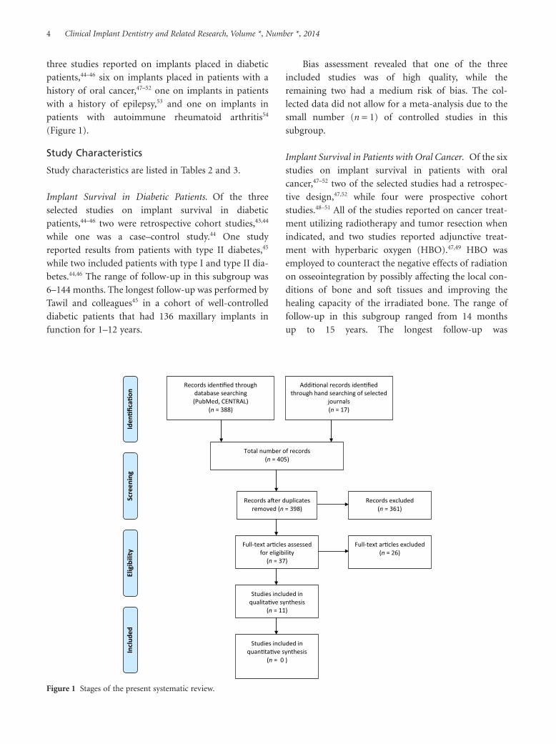
Initial screening of electronic databases yielded 388
potentially relevant publications; 385 were retrieved
from the search of PubMed, and 3 additional articles
were retrieved from the search of CENTRAL. Hand
searching of the selected journals added 17 publications
for a total of 405 articles. Removal of duplicate reports
from the same study population led to the inclusion of
398 studies in the first phase of screening. After screen-
ing of the titles and abstracts independently and in
duplicate by the two reviewers, the full-text versions of
37 studies were retrieved for assessment of eligibility for
inclusion in the systematic review. Interreviewer agree-
ment at the first phase of selection was very high
(k = 0.86).
During the second phase of selection, 26 studies
were excluded due to lack of adequate follow-up
(n = 3),21–23 lack of adequate sample size (n = 7),6,24–28
insufficient data on the medical conditions of the
patient cohort (n = 2),29,30 insufficient age of the patient
cohort (n = 1),31 report of mandibular implants only
(n = 5),32–36 or inadequate data to calculate maxillary
implant survival for the included patients (n = 8)11,37–43
(k = 0.92) (Table 1).
The final selection included 11 studies reporting on
four distinct medical conditions; thus, the included
studies were categorized into four subgroups. In detail,
TABLE 1 Studies Excluded in the Second Phase of Selection with Reasons for the Exclusion of Each Study
Reason for exclusion Studies
Less than 12 months of follow-up post-loading (n = 3) Dowell and colleagues,21 Memon and colleagues,22 Balshi and
Wolfinger23
Less than 10 subjects receiving maxillary implants (n = 7) Smith and colleagues,6 Eckert and colleagues,24 Friberg and
colleagues,25 Niimi and colleagues,26 Esser and Wagner,27
Turkyilmaz28
Unspecified medical conditions (n = 2) Jeffcoat,30 Grant and colleagues29
Implant placement in patients less than 18 years of age (n = 1) Bergendal and colleagues31
Only reported on mandibular implants (n = 5) Landes and Kovacs,32 Shernoff and colleagues,33 Stevenson and
colleagues,34 Oliveira and colleagues,35 Peled and colleagues36
Inadequate data for estimation of maxillary implant survival
(n = 6)
Becker and colleagues,11 Nelson and colleagues,37 Morris and
colleagues,38 Abdulwassie and Dhanrajani,39 Attard and
Zarb,40 Gu and Yu42
Inadequate data for estimation of maxillary implant survival
per medical condition (n = 2)
Moy and colleagues,41 Van Steenberghe and colleagues43
Maxillary Implants in Compromised Patients 3
three studies reported on implants placed in diabetic
patients,44–46 six on implants placed in patients with a
history of oral cancer,47–52 one on implants in patients
with a history of epilepsy,53 and one on implants in
patients with autoimmune rheumatoid arthritis54
(Figure 1).
Study Characteristics
Study characteristics are listed in Tables 2 and 3.
Implant Survival in Diabetic Patients. Of the three
selected studies on implant survival in diabetic
patients,44–46 two were retrospective cohort studies,43,44
while one was a case–control study.44 One study
reported results from patients with type II diabetes,45
while two included patients with type I and type II dia-
betes.44,46 The range of follow-up in this subgroup was
6–144 months. The longest follow-up was performed by
Tawil and colleagues45 in a cohort of well-controlled
diabetic patients that had 136 maxillary implants in
function for 1–12 years.
Bias assessment revealed that one of the three
included studies was of high quality, while the
remaining two had a medium risk of bias. The col-
lected data did not allow for a meta-analysis due to the
small number (n = 1) of controlled studies in this
subgroup.
Implant Survival in Patients with Oral Cancer. Of the six
studies on implant survival in patients with oral
cancer,47–52 two of the selected studies had a retrospec-
tive design,47,52 while four were prospective cohort
studies.48–51 All of the studies reported on cancer treat-
ment utilizing radiotherapy and tumor resection when
indicated, and two studies reported adjunctive treat-
ment with hyperbaric oxygen (HBO).47,49 HBO was
employed to counteract the negative effects of radiation
on osseointegration by possibly affecting the local con-
ditions of bone and soft tissues and improving the
healing capacity of the irradiated bone. The range of
follow-up in this subgroup ranged from 14 months
up to 15 years. The longest follow-up was
Records iden�fied throughdatabase searching(PubMed, CENTRAL)
(n = 388)
Screen
ing
Addi�onal records iden�fiedthrough hand searching of selected
journals(n = 17)
Total number of records(n = 405)
Records a�er duplicatesremoved (n = 398)
Records excluded(n = 361)
Full-text ar�cles assessedfor eligibility(n = 37)
Full-text ar�cles excluded(n = 26)
Studies included inqualita�ve synthesis
(n = 11)
Studies included inquan�ta�ve synthesis
(n = 0 )
Iden
�fica�o
nEligibility
Includ
ed
Figure 1 Stages of the present systematic review.
4 Clinical Implant Dentistry and Related Research, Volume *, Number *, 2014

TABLE 2 Main Characteristics of Studies Included After the Second Phase of Selection
Study Country Medical condition Funding Study design
Fiorellini and
colleagues44
USA Diabetes None reported Retrospective cohort
study
Farzad and colleagues46 Sweden Diabetes None reported Retrospective cohort
study
Tawil and colleagues45 Lebanon Diabetes None reported Case–control
Visch and colleagues51 Netherlands Oral cancer Institutional funding
(Rotterdam)
Prospective cohort study
Mericske-Stern and
colleagues47
Switzerland Oral cancer None reported Retrospective cohort
study
Barrowman and
colleagues49
Australia Oral cancer None reported Prospective cohort study
Linsen and colleagues50 Germany Oral cancer None reported Prospective cohort study
Heberer and colleagues48 Germany Oral cancer Partially funded by
Straumann AG, Basel,
Switzerland
Prospective cohort study
Buddula and
colleagues52
USA Oral cancer None reported Retrospective cohort
study
Cune and colleagues53 Netherlands Severe epilepsy None reported Retrospective cohort
study
Krennmair and
colleagues54
Austria Rheumatoid arthritis Self-funded/institutional
funding
Retrospective cohort
study
TABLE 3 Methodological Evaluation of Included Studies
Study Selection Comparability Exposure StatisticsMethodological
quality
Diabetes
Fiorellini and colleagues44 *** ** Low
Farzad and colleagues46 *** ** Low
Tawil and colleagues45 **** ** ** ** High
Oral cancer
Visch and colleagues51 **** *** ** High
Mericske-Stern and colleagues47 **** ** * Medium
Barrowman and colleagues49 *** ** Low
Linsen and colleagues50 **** *** * Medium
Heberer and colleagues48 **** ** * Medium
Buddula and colleagues52 *** *** * Medium
Severe epilepsy
Cune and colleagues53 ** *** * Medium
Rheumatoid arthritis
Krennmair and colleagues54 *** *** * Medium
One star was assigned for each methodological criterion that was fulfilled. Detailed description of the methodological criteria is presented in figure S1.Studies with less than 6 stars were considered to be of low methodological quality, medium methodological quality if having 6 to 8 stars while studies thathad 9 or more stars were considered to be of high methodological quality according to Chambrone et al.18.
Maxillary Implants in Compromised Patients 5

performed by Barrowman and colleagues,49 who
reported on 35 implants that were followed for 15 years.
Bias assessment revealed that one of the six included
studies was high-quality,51 one as low-quality,49 and the
remaining four studies as medium-quality.47,48,50,52 The
collected data did not allow for a meta-analysis due to
the lack of controlled studies in this subgroup.
Implant Survival in Patients with Epilepsy. One longitu-
dinal retrospective study was identified that followed 27
patients with 61 implants for 16 years.53 Bias assessment
revealed that the study had a medium risk of bias.
Implant Survival in Patients with Autoimmune Rheuma-
toid Arthritis. A single retrospective study evaluated 34
female patients with autoimmune rheumatoid arthri-
tis.54 Seventy-seven implants were placed in the maxillae
of patients for indications ranging from single-tooth
sites to complete edentulism. Patients were followed up
for a period ranging from 1 to 7 years. Bias assessment
revealed that the study had a medium risk of bias.
Results of Included Studies
Tables 4 and 5 summarize the outcomes of the included
studies.
Implant Survival in Diabetic Patients. Results showed
that implant survival rates in the maxillae of diabetic
patients ranged from 85.5% to 95.6% for a total of 323
implants.44–46 The highest survival rate was reported by
Tawil and colleagues.45 In this study the authors followed
28 type II diabetic patients for 1 to 12 years and reported
a 95.6% survival rate.45 In total, 136 implants were
placed: 116 were placed following a conventional
loading protocol and 20 with immediate loading. In the
control group, 31 nondiabetic control patients received
129 implants: 109 implants following a conventional
loading protocol and 20 with immediate loading. All
patients received antibiotic treatment perioperatively.
Results from comparison of the two groups showed that
well-controlled diabetic patients with a mean glycated
hemoglobin (HbA1c) of 7.2% in the perioperative period
had the same overall survival rate as control patients,
irrespective of the loading protocol.45 Implant survival
rate was also independent of age, gender, diabetes dura-
tion, and smoking in this well-controlled diabetic popu-
lation. Levels of HbA1c were the most important factor
affecting implant complication rate.45 One of the
remaining studies reported survival rates greater than
90% in type II diabetics.46 The lowest survival rate was
reported by Fiorellini and colleagues (85.5%).44 In this
retrospective analysis, 131 implants were placed in
patients with type I and type II diabetes at two clinical
centers. Chart review results showed that 19 failures
occurred for an overall success rate of 85.5% in the
maxilla.44 The mean time of functional loading was
4.1 1 2.6 years. When failed implants were studied, it
was found that most of the failures occurred within the
first year of functional loading. No difference in survival
rates was noted between the posterior or anterior
maxilla or between the two centers. No differences in the
responses of type I and type II diabetics were identified
in terms of implant survival.44
Not all of the studies discussed the loading protocol
utilized for the delivery of the implant-supported resto-
rations. Farzad and colleagues46 reported that the major-
ity of implants were placed using a two-stage placement
protocol in combination with conventional loading,
with only nine implants being immediately loaded in
selected cases. Tawil and colleagues45 performed both
conventional and immediate loading. In the remaining
study, mode of loading was not reported.45
The two included studies that presented the highest
success rates reported use of antibiotics in conjunction
with implant placement procedures.45,46 HbA1c percent-
ages were only reported by one study as a surrogate for
level of diabetes control.45 In the remaining two studies,
the authors reported that all patients had good glycemic
control, based on serum glucose levels.44,46
Implant Survival in Patients with Oral Cancer. Results
showed that implant survival rates in the maxilla for
patients with oral cancer ranged from 67.7% to 100%
for a total of 321 maxillary implants. Three studies
reported no failures in the respective study populations
for 100% implant survival rates.47–49 Mericske-Stern and
colleagues47 followed up 12 maxillary implants (ITI-
Straumann, Basel, Switzerland), and Heberer and col-
leagues48 followed-up 55 implants overall (28 modified
and 27 conventional sandblasted acid-etched implants).
Barrowman and colleagues49 utilized a two-stage
implant placement protocol to place 35 implants in the
maxilla with no failures during the 15-year observa-
tional period. The lowest survival rate in patients with a
history of oral cancer was reported by Buddula and
colleagues.52 In this retrospective chart review, 62
6 Clinical Implant Dentistry and Related Research, Volume *, Number *, 2014

TAB
LE4
Ou
tco
mes
Ass
essm
ent
of
the
Incl
ud
edSt
ud
ies
Stu
dy
Pati
ents
wit
hm
axill
ary
imp
lan
ts(n
)
Max
illar
yim
pla
nts
(n)
Faile
dm
axill
ary
imp
lan
ts(n
)
Imp
lan
tsu
rviv
al(%
)Im
pla
nt
site
sLo
adin
gp
roto
col
Imp
lan
tty
pe
Post
load
ing
follo
w-u
pp
erio
dA
dju
nct
ive
fact
ors
Dia
bete
s
Fior
ellin
ian
d
colle
agu
es44
NR
108
3369
.4A
nte
rior
and
post
erio
r
Con
ven
tion
alH
ydro
xyap
atit
e-
coat
ed
impl
ants
Up
to16
8m
onth
sA
llpa
tien
tsre
ceiv
edra
diot
her
apy
Farz
adan
dco
lleag
ues
46N
R12
010
0N
RN
RIT
I (Str
aum
ann
)
12–8
4m
onth
sSo
me
ofth
epa
tien
tsre
ceiv
ed
HB
Oan
d/or
radi
oth
erap
y
Taw
ilan
dco
lleag
ues
45N
R35
010
0N
RC
onve
nti
onal
Brå
nem
ark
(Nob
el
Bio
care
)
180
mon
ths
Som
eof
the
pati
ents
rece
ived
HB
O,r
adio
ther
apy,
and/
or
bon
egr
aft
Ora
lcan
cer
Vis
chan
dco
lleag
ues
51N
R55
010
0N
Rea
rly
Stra
um
ann
14.4
mon
ths
All
pati
ents
rece
ived
radi
oth
erap
y
and
clin
dam
ycin
Mer
icsk
e-St
ern
and
colle
agu
es47
NR
6220
67.7
An
teri
oran
d
post
erio
r
NR
NR
60m
onth
sA
llpa
tien
tsre
ceiv
edra
diot
her
apy
and
som
ere
ceiv
edbo
ne
graf
t
Bar
row
man
and
colle
agu
es49
NR
350
100
NR
Con
ven
tion
alB
rån
emar
k
(Nob
el
Bio
care
)
180
mon
ths
Som
eof
the
pati
ents
rece
ived
HB
O,r
adio
ther
apy,
and/
orbo
ne
graf
t
Lin
sen
and
colle
agu
es50
NR
131
1985
.5A
nte
rior
and
post
erio
r
NR
Brå
nem
ark,
Stra
um
ann
Up
to78
mon
ths
NR
Heb
erer
and
colle
agu
es48
NR
564
92.9
An
teri
oran
d
post
erio
r
Con
ven
tion
al
and
imm
edia
te
Brå
nem
ark
(Nob
el
Bio
care
)
12m
onth
sA
llpa
tien
tsre
ceiv
edpo
stop
erat
ive
anti
biot
ics
Bu
ddu
laan
dco
lleag
ues
5228
136
695
.6N
RC
onve
nti
onal
and
imm
edia
te
Brå
nem
ark
(Nob
el
Bio
care
)
12–1
44m
onth
sA
llpa
tien
tsre
ceiv
edch
lorh
exid
ine
and
anti
biot
ics,
and
som
e
pati
ents
rece
ived
sin
us
lift
and
bon
egr
aft
Seve
reep
ileps
y
Cu
ne
and
colle
agu
es53
2761
010
0A
nte
rior
and
post
erio
r
Con
ven
tion
alSt
rau
man
n19
2m
onth
sN
R
Rh
eum
atoi
dar
thri
tis
Kre
nn
mai
ran
d
colle
agu
es54
3477
692
.2A
nte
rior
and
post
erio
r
Con
ven
tion
alC
amlo
g12
–84
mon
ths
Som
epa
tien
tsre
ceiv
edN
SAID
s
and/
orgl
uco
cort
icoi
ds
NR
,not
repo
rted
;HB
O,h
yper
bari
cox
ygen
;NSA
IDS,
non
ster
oida
lan
ti-i
nfl
amm
ator
ydr
ugs
.
Maxillary Implants in Compromised Patients 7

implants were evaluated and 20 failures occurred, for an
overall success rate of 67.7% in the maxilla. The
implants were followed for a mean of 3.2 years and up to
16.9 years. No association was identified between sur-
vival and length, diameter, type of bone, or radiation
dose received, even though there was a tendency for
increased failure rates in patients receiving more than
50 Gy of radiation.52 The only predictor of implant
failure was the location of the implant, with a greater
tendency to fail for implants placed in the posterior
maxillary region compared with those placed in anterior
regions.52
When the adjunctive HBO was considered,
Barrowman and colleagues concluded that the high
retention rates seen in their patient cohort might have
been due to the HBO therapy rendered in their
study.49
Regarding loading protocols, most of the included
studies reported utilizing a conventional loading proto-
col, while reported use of adjunctive systemic antibiotics
was scarce.
Implant Survival in Patients with Epilepsy. The results of
Cune and colleagues showed no failures of maxillary
implants occurring among 27 patients, for an implant
success rate of 100%.53 The authors utilized a conven-
tional loading protocol in both anterior and posterior
areas, without any adjunctive treatment. Even though all
patients were inpatients and were judged as having inad-
equate oral hygiene due to multiple disabilities, only
mild inflammation of the peri-implant mucosa was
noted in some of the patients despite the frequent use of
antiepileptic medication, which has been shown to
induce gingival hyperplasia.
TABLE 5 Conclusions of the Included Studies
Study Conclusions
Diabetes
Fiorellini and
colleagues44
“Implant survival is significantly influenced by the location (maxilla versus mandible, 59% and
85%, respectively), by the incidence of bone-resection surgery in the jaw where the implant
was installed and by the irradiation dose at the implant site (<50 Gray or >50 Gray).”
Farzad and
colleagues46
“Treatment with implant-supported prostheses seemed to be advantageous for patients who
have undergone intraoral resections.”
Tawil and colleagues45 “Dental implants provide an important role in the oral rehabilitation of oral cancer patients.”
Oral cancer
Visch and colleagues51 “Implants with chemically modified and conventional SLA titanium surface show high success
rates in irradiated patients.”
Mericske-Stern and
colleagues47
“Dental implants placed in irradiated bone have a greater risk for failure. Survival is significantly
influenced by the location of the implant (maxilla or mandible, anterior or posterior).”
Barrowman and
colleagues49
“Dental implants provide an important role in the oral rehabilitation of oral cancer patients.”
Linsen and
colleagues50
“Survival rate of dental implants in controlled diabetic patients is lower than that documented
for the general population, but there is still a reasonable success rate.”
Heberer and
colleagues48
“Diabetics that undergo dental implant treatment do not encounter a higher failure rate than
the normal population, if their plasma glucose level is normal or close to normal.”
Buddula and
colleagues52
“Well- to fairly well-controlled diabetic patients with a mean HbA1c of 7.2% in the perioperative
period have the same overall survival rate as controls in conventional and advanced implant
therapy. Implant survival rate is independent from age, gender, diabetes duration, and
smoking in a well- to fairly well-controlled diabetic population.”
Severe epilepsy
Cune and colleagues53 “Dental implant treatment in a population of patients with severe epilepsy and additional
disabilities seems to be a viable treatment option.”
Rheumatoid arthritis
Krennmair and
colleagues54
“No atypical pattern of prosthodontic complications and maintenance efforts was observed for
implants and implant prosthodontics in RA patients.”
8 Clinical Implant Dentistry and Related Research, Volume *, Number *, 2014

Implant Survival in Patients with Autoimmune Rheuma-
toid Arthritis. Krennmair and colleagues retrospectively
evaluated 34 female patients with a mean age of 58.1
years.54 Seventy-seven Camlog implants (Camlog, Basel,
Switzerland) of different lengths and diameters were
placed in the maxillae of these patients. The implants
were placed in both anterior and posterior sites
following a conventional loading protocol. At the end of
the follow-up, a 100% survival rate was reported.
Patients included in this investigation had a diverse
medication schedule ranging from no pharmacological
therapy to medical treatment including nonsteroidal
anti-inflammatory drugs, treatment with glucocorti-
coids, or a combination.
DISCUSSION
Summary of Evidence
The results of the present review show that survival
rates of maxillary implants placed in medically com-
promised patients range from 65.5% to 100%. Survival
of implants placed in patients with a medical history
significant for oral cancer accounted for the lower end
of the range. It should be noted that except for two
studies that demonstrated less than 70% survival rates
for maxillary implants,51,52 the remaining studies
showed excellent implant survival, with three studies
showing no implant loss. Visch and colleagues51
reported a 69.4% implant survival after up to 14 years
of follow-up for 108 implants. In this study, almost half
of the implants were placed in the posterior maxilla,
and the vast majority of patients received more than
50 Gy of radiotherapy.51 Also, a large number of failed
implants were placed in sites treated with partial
maxillectomy. The authors attributed the low survival
rates to the unfavorable site-related conditions associ-
ated with surgical resection (partial maxillectomy)
such as prosthetic limitations due to bulky soft tissue
areas and lack of keratinized tissue as well as reduced
vascularization due to large doses of radiation exceed-
ing the 50-Gy threshold.51 A greater risk for implant
failure for the maxilla in comparison to the mandible
was also reported.51 Similarly, Buddula and colleagues52
attributed the low survival rates noted in their study to
a large percentage of implants being placed in the pos-
terior maxilla.
The findings of the present systematic review are in
accordance with the findings of a recent review by
Javed and colleagues,55 who concluded that irradia-
tion may have a negative impact on implant
osseointegration, yet implants placed in patients with a
history of oral cancer treatment can osseointegrate and
remain functional. Loading protocols for implants
placed in oral cancer patients were underreported in
general. In two studies where some of the implants were
immediately loaded, implant survival rates were greater
than 90%, supporting the delivery of immediate pros-
theses.48,52 Indeed, histological results of immediate
implant loading in the maxilla in a cancer patient who
was also a smoker revealed excellent bone-to-implant
contact that exceeded 50% in all implants, thus support-
ing the utilization of immediate loading in cancer
patients.10
When the survival of implant placement in dia-
betic patients was assessed, results were very promising
and ranged from 85.5% to 95.6%.44–46 The lower
rate was reported in a study that included type I and
type II diabetic patients, even though no association
was identified between type of diabetes and implant
failure.44 More recent studies showed excellent survival
rates that were comparable to the rates of implants
placed in healthy controls, even in complex cases
that required bone-grafting procedures.45,46 HbA1c
levels were underreported in the included studies,
but when utilized they were shown to be the only
appropriate indicators for risk of postoperative com-
plications, though not for implant survival.45 All
three included studies reported on well-controlled dia-
betic patients.44–46 Our results are in agreement with
the findings of a recent study that evaluated and
compared the microbiological and immunological
profiles of diabetic implant patients and compared
them with those of healthy controls.56 Results of this
study showed that the microbiological profiles and sali-
vary biomarkers in well-controlled diabetics and
healthy individuals do not differ significantly, thus
supporting the argument that implant placement is
a predictable treatment modality in well-controlled
diabetics.56
Two more categories of medical conditions were
discussed in the present review: epilepsy and rheuma-
toid arthritis.53,54 Despite the fact that only one study per
disease subgroup was available for review, the excellent
survival rates (100%) of maxillary implants placed in
both studies highlight the predictability of implant
treatment in these patient groups.53,54
Maxillary Implants in Compromised Patients 9

Limitations and Future Implications
The inclusion criteria utilized in the present review
aimed at the inclusion of only reasonably powered
observational studies. Therefore, except for the oral
cancer subgroup, a low number of studies were identi-
fied for the remaining medical conditions. Also, only
one study had a case–control study design,45 while all
remaining studies included only compromised patient
cohorts in their observations.
In the present systematic review, most of the
included cohort studies did not have internal compari-
son groups in the form of patients who were not medi-
cally compromised receiving maxillary implants. The
execution of good-quality individual cohort studies
including healthy and compromised patient cohorts
with similar exposure characteristics is needed in clinical
implant research to allow for outcome comparison
between the two groups.
Even when the most stringent quality criteria were
imposed, sources of bias could still be identified in all
studies. Reporting bias was frequently encountered in
the included studies, especially in the diabetes subgroup.
For example, glycated hemoglobin levels may currently
be the most accepted surrogate for diabetes control in
the medical literature but were only utilized in one
study.45 Also, in a study reporting on the survival of
implants placed in patients with epilepsy, the intake of
medication, which has been associated with gingival
overgrowth, was not explicitly reported, and there was
not a clear report of attempts to associate this intake
with peri-implant mucosal condition.53
The encouraging results for the survival of maxil-
lary implants in medically compromised patients that
were identified in the present literature search may
facilitate the overcoming of ethical dilemmas associated
with randomized controlled studies in medically com-
promised patient cohorts, which will provide a higher
level of evidence to support implant rehabilitation in the
maxilla for this sensitive patient pool.
CONCLUSIONS
Within the limitations of this review, we conclude that
although maxillary implants may have less favorable
prognosis compared with implants placed in the man-
dible, the placement of maxillary implants in medically
compromised patients seems to yield acceptable survival
rates.
Implant survival in well-controlled diabetic
patients, patients diagnosed with rheumatoid arthritis,
and patients treated for severe epilepsy is comparable to
that in healthy patients. Implants placed in the maxilla
of patients treated for oral cancer may attain
osseointegration less predictably than in the mandible.
Site-related factors (i.e., posterior sites and resected
sites), as well as dosage of radiotherapy exceeding 50 Gy,
should be considered as risk indicators of lower implant
survival.
ACKNOWLEDGMENTS
The authors wish to thank Dr. Nicolas Marcou for his
contribution in the literature search. The authors are
also grateful to Drs. R. Barrowman, S. Linsen, G. Tawil,
M. Cune, G. Krenmair, J. Katancik, and S. Memon for
sharing their original results for the conduct of the
present systematic review.
REFERENCES
1. Christensen K, Doblhammer G, Rau R, Vaupel JW. Ageing
populations: the challenges ahead. Lancet 2009; 374:1196–
1208.
2. Kuoppala R, Napankangas R, Raustia A. Quality of life of
patients treated with implant-supported mandibular
overdentures evaluated with the Oral Health Impact Profile
(OHIP-14): a survey of 58 patients. J Oral Maxillofac Res
2013; 4:e4.
3. Ferrigno N, Laureti M, Fanali S, Grippaudo G. A long-term
follow-up study of non-submerged ITI implants in the treat-
ment of totally edentulous jaws. Part I: ten-year life table
analysis of a prospective multicenter study with 1286
implants. Clin Oral Implants Res 2002; 13:260–273.
4. Diz P, Scully C, Sanz M. Dental implants in the medically
compromised patient. J Dent 2013; 41:195–206.
5. Tecilazich F, Dinh T, Pradhan-Nabzdyk L, et al. Role of
endothelial progenitor cells and inflammatory cytokines in
healing of diabetic foot ulcers. PLoS ONE 2013; 8:e83314.
6. Smith RA, Berger R, Dodson TB. Risk factors associated
with dental implants in healthy and medically compro-
mised patients. Int J Oral Maxillofac Implants 1992; 7:367–
372.
7. Wilson TG Jr, Higginbottom FL. Periodontal diseases and
dental implants in older adults. J Esthet Dent 1998; 10:265–
271.
8. Oikarinen K, Raustia AM, Hartikainen M. General and local
contraindications for endosseal implants – an epidemi-
ological panoramic radiograph study in 65-year-old
subjects. Community Dent Oral Epidemiol 1995; 23:114–
118.
10 Clinical Implant Dentistry and Related Research, Volume *, Number *, 2014

9. Bornstein MM, Cionca N, Mombelli A. Systemic conditions
and treatments as risks for implant therapy. Int J Oral
Maxillofac Implants 2009; 24(Suppl):12–27.
10. Romanos GE, Johansson CB. Immediate loading with com-
plete implant-supported restorations in an edentulous heavy
smoker: histologic and histomorphometric analyses. Int J
Oral Maxillofac Implants 2005; 20:282–290.
11. Becker W, Hujoel PP, Becker BE, Willingham H. Osteopo-
rosis and implant failure: an exploratory case-control study.
J Periodontol 2000; 71:625–631.
12. Lazzara R, Siddiqui AA, Binon P, et al. Retrospective multi-
center analysis of 3i endosseous dental implants placed over
a five-year period. Clin Oral Implants Res 1996; 7:73–
83.
13. Haas R, Mensdorff-Pouilly N, Mailath G, Watzek G. Survival
of 1,920 IMZ implants followed for up to 100 months. Int J
Oral Maxillofac Implants 1996; 11:581–588.
14. Carr AB. Implant location and radiotherapy are the only
factors linked to 2-year implant failure. J Evid Based Dent
Pract 2012; 12(3 Suppl):217–219.
15. Kim YJ, Henkin J. Micro-computed tomography assess-
ment of human alveolar bone: bone density and three-
dimensional micro-architecture. Clin Implant Dent Relat
Res 2013.
16. Shea BJ, Hamel C, Wells GA, et al. AMSTAR is a reliable and
valid measurement tool to assess the methodological quality
of systematic reviews. J Clin Epidemiol 2009; 62:1013–1020.
17. Hellman S, Hellman DS. Of mice but not men. Problems of
the randomized clinical trial. N Engl J Med 1991; 324:1585–
1589.
18. Chambrone L, Chambrone D, Lima LA, Chambrone LA.
Predictors of tooth loss during long-term periodontal main-
tenance: a systematic review of observational studies. J Clin
Periodontol 2010; 37:675–684.
19. Boudin F, Nie JY, Bartlett JC, et al. Combining classifiers for
robust PICO element detection. BMC Med Inform Decis
Mak 2010; 10:29.
20. Stang A. Critical evaluation of the Newcastle–Ottawa scale
for the assessment of the quality of nonrandomized studies
in meta-analyses. Eur J Epidemiol 2010; 25:603–605.
21. Dowell S, Oates TW, Robinson M. Implant success in people
with type 2 diabetes mellitus with varying glycemic control:
a pilot study. J Am Dent Assoc 2007; 138:355–361, quiz 97–8.
22. Memon S, Weltman RL, Katancik JA. Oral bisphosphonates:
early endosseous dental implant success and crestal bone
changes. A retrospective study. Int J Oral Maxillofac
Implants 2012; 27:1216–1222.
23. Balshi TJ, Wolfinger GJ. Dental implants in the diabetic
patient: a retrospective study. Implant Dent 1999; 8:355–
359.
24. Eckert SE, Desjardins RP, Keller EE, Tolman DE. Endosseous
implants in an irradiated tissue bed. J Prosthet Dent 1996;
76:45–49.
25. Friberg B, Ekestubbe A, Mellstrom D, Sennerby L.
Brånemark implants and osteoporosis: a clinical exploratory
study. Clin Implant Dent Relat Res 2001; 3:50–56.
26. Niimi A, Fujimoto T, Nosaka Y, Ueda M. A Japanese multi-
center study of osseointegrated implants placed in irradiated
tissues: a preliminary report. Int J Oral Maxillofac Implants
1997; 12:259–264.
27. Esser E, Wagner W. Dental implants following radical oral
cancer surgery and adjuvant radiotherapy. Int J Oral
Maxillofac Implants 1997; 12:552–557.
28. Turkyilmaz I. One-year clinical outcome of dental implants
placed in patients with type 2 diabetes mellitus: a case series.
Implant Dent 2010; 19:323–329.
29. Grant BT, Amenedo C, Freeman K, Kraut RA. Outcomes
of placing dental implants in patients taking oral
bisphosphonates: a review of 115 cases. J Oral Maxillofac
Surg 2008; 66:223–230.
30. Jeffcoat MK. Safety of oral bisphosphonates: controlled
studies on alveolar bone. Int J Oral Maxillofac Implants
2006; 21:349–353.
31. Bergendal B, Ekman A, Nilsson P. Implant failure in young
children with ectodermal dysplasia: a retrospective evalua-
tion of use and outcome of dental implant treatment in
children in Sweden. Int J Oral Maxillofac Implants 2008;
23:520–524.
32. Landes CA, Kovacs AF. Comparison of early telescope
loading of non-submerged ITI implants in irradiated and
non-irradiated oral cancer patients. Clin Oral Implants Res
2006; 17:367–374.
33. Shernoff AF, Colwell JA, Bingham SF. Implants for type II
diabetic patients: interim report. VA implants in diabetes
study group. Implant Dent 1994; 3:183–185.
34. Stevenson GC, Riano PC, Moretti AJ, et al. Short-term
success of osseointegrated dental implants in HIV-positive
individuals: a prospective study. J Contemp Dent Pract 2007;
8:1–10.
35. Oliveira MA, Gallottini M, Pallos D, et al. The success of
endosseous implants in human immunodeficiency virus-
positive patients receiving antiretroviral therapy: a pilot
study. J Am Dent Assoc 2011; 142:1010–1016.
36. Peled M, Ardekian L, Tagger-Green N, Gutmacher Z,
Machtei EE. Dental implants in patients with type 2 diabetes
mellitus: a clinical study. Implant Dent 2003; 12:116–
122.
37. Nelson K, Heberer S, Glatzer C. Survival analysis and clinical
evaluation of implant-retained prostheses in oral cancer
resection patients over a mean follow-up period of 10 years.
J Prosthet Dent 2007; 98:405–410.
38. Morris HF, Ochi S, Winkler S. Implant survival in patients
with type 2 diabetes: placement to 36 months. Ann
Periodontol 2000; 5:157–165.
39. Abdulwassie H, Dhanrajani PJ. Diabetes mellitus and dental
implants: a clinical study. Implant Dent 2002; 11:83–86.
Maxillary Implants in Compromised Patients 11

40. Attard NJ, Zarb GA. A study of dental implants in medically
treated hypothyroid patients. Clin Implant Dent Relat Res
2002; 4:220–231.
41. Moy PK, Medina D, Shetty V, Aghaloo TL. Dental implant
failure rates and associated risk factors. Int J Oral Maxillofac
Implants 2005; 20:569–577.
42. Gu L, Yu YC. Clinical outcome of dental implants placed in
liver transplant recipients after 3 years: a case series. Trans-
plant Proc 2011; 43:2678–2682.
43. van Steenberghe D, Jacobs R, Desnyder M, Maffei G,
Quirynen M. The relative impact of local and endogenous
patient-related factors on implant failure up to the abutment
stage. Clin Oral Implants Res 2002; 13:617–622.
44. Fiorellini JP, Chen PK, Nevins M, Nevins ML. A retrospec-
tive study of dental implants in diabetic patients. Int J
Periodontics Restorative Dent 2000; 20:366–373.
45. Tawil G, Younan R, Azar P, Sleilati G. Conventional and
advanced implant treatment in the type II diabetic patient:
surgical protocol and long-term clinical results. Int J Oral
Maxillofac Implants 2008; 23:744–752.
46. Farzad P, Andersson L, Nyberg J. Dental implant treatment
in diabetic patients. Implant Dent 2002; 11:262–267.
47. Mericske-Stern R, Perren R, Raveh J. Life table analysis and
clinical evaluation of oral implants supporting prostheses
after resection of malignant tumors. Int J Oral Maxillofac
Implants 1999; 14:673–680.
48. Heberer S, Kilic S, Hossamo J, Raguse JD, Nelson K. Reha-
bilitation of irradiated patients with modified and conven-
tional sandblasted acid-etched implants: preliminary results
of a split-mouth study. Clin Oral Implants Res 2011; 22:546–
551.
49. Barrowman RA, Wilson PR, Wiesenfeld D. Oral rehabilita-
tion with dental implants after cancer treatment. Aust Dent
J 2011; 56:160–165.
50. Linsen SS, Martini M, Stark H. Long-term results of endos-
teal implants following radical oral cancer surgery with and
without adjuvant radiation therapy. Clin Implant Dent Relat
Res 2012; 14:250–258.
51. Visch LL, van Waas MA, Schmitz PI, Levendag PC. A clinical
evaluation of implants in irradiated oral cancer patients.
J Dent Res 2002; 81:856–859.
52. Buddula A, Assad DA, Salinas TJ, et al. Survival of dental
implants in irradiated head and neck cancer patients: a ret-
rospective analysis. Clin Implant Dent Relat Res 2012;
14:716–722.
53. Cune MS, Strooker H, van der Reijden WA, et al. Dental
implants in persons with severe epilepsy and multiple
disabilities: a long-term retrospective study. Int J Oral
Maxillofac Implants 2009; 24:534–540.
54. Krennmair G, Seemann R, Piehslinger E. Dental implants in
patients with rheumatoid arthritis: clinical outcome and
peri-implant findings. J Clin Periodontol 2010; 37:928–936.
55. Javed F, Al-Hezaimi K, Al-Rasheed A, Almas K,
Romanos GE. Implant survival rate after oral cancer therapy:
a review. Oral Oncol 2010; 46:854–859.
56. Tatarakis N, Kinney JS, Inglehart M, et al. Clinical, micro-
biological, and salivary biomarker profiles of dental implant
patients with type 2 diabetes. Clin Oral Implants Res 2013:
DOI: 10.1111/clr.12139.
SUPPORTING INFORMATION
Additional Supporting Information may be found in the
online version of this article at the publisher’s web-site:
Figure S1. Quality assessment criteria selected from the
Newcastle–Ottawa scale.20
12 Clinical Implant Dentistry and Related Research, Volume *, Number *, 2014
Related Documents