Egypt, J. Plast. Reconstr. Surg., Vol. 31, No. 1, January: 87-96, 2007 A Suggested Algorithm for Post-Traumatic Lower Limb Soft Tissue Reconstruction YOUSSEF SALEH, M.D.*; BASEM WAHEEB, MS.c.*; MUSTAFA ALAA-ELDIN ABD-ELAZIZ, M.D.** and MAHMOUD EL-OTEIFY, M.D.* The Departments of Plastic Surgery* and General Surgery**, Faculty of Medicine, Assiut University. ABSTRACT The evolving technology in trauma management today permits salvage of many severe lower extremity injuries previously even considered to be lethal. The role of different reconstructive options in lower extremity injuries has been reviewed in 100 patients. The frequency of use of reconstruc- tive techniques, specific complications and benefits, effect of timing of wound closure, and rate of limb salvage were compared. The ages of the patients ranged between 3 and 66 years with a mean age of 19.76 years. 54 cases were with simple wounds with no exposed bone, tendons or neurovascular structures while 46 cases were with complex wounds with exposed bone, tendons or neurovascular structures. Initial coverage after significant lower extremity trauma in these 100 patients required 59 split thickness skin graft, 6 local muscle flaps, 28 local fascio-cutaneous flaps, 5 free flaps and 2 cases were managed by simple closure. These reconstructive techniques had been selected according to wound location, its severity, and flap availability. The traditional role of the gastrocnemius muscles for flap coverage of knee and proximal leg defects and the soleus muscle for the middle third of the leg was reaffirmed. The cross leg flap and local fasciocutaneous flaps were most valuable for defects of the lower leg or foot, otherwise, a free flap would have been necessary. INTRODUCTION Treatment of high energy lower extremity trau- ma with soft tissue and bone injury remains a formidable problem. Treatment requires a team approach with the orthopedic, vascular and plastic surgeon. The goal in treatment of open tibial frac- tures and lower extremity salvage is to preserve a limb that will be more functional than an amputa- tion. If the extremity cannot be salvaged, the goal is to maintain the maximum functional length [1]. There are many possible reconstructive options, which developed or modified for reconstruction of defects in the lower limb. These include; skin grafts, local flaps, distant flaps, and free flaps. However, each of these techniques has its own limitations [2]. The experience with the use of these reconstructive options is developing in various 87 centers. However, still the indications, the selection of a particular technique for the different cases are not well established and are rather a matter of personal judgment. Limb reconstruction is a long complicated process. Patients must be aware of the expected functional outcome. Patient selection is an important variable in evaluating the final outcome. Although normal function is not always achieved, most patients are grateful for their sal- vaged limbs [3]. The aim of the present thesis is to study the different reconstructive options for management of lower limb defects and to suggest an algorithm for soft tissue coverage in different parts of the lower limb. PATIENTS AND METHODS This study was carried upon one hundred pa- tients suffering from lower limb soft tissue injury who were admitted to Assuit University Hospital in the period from January 2005 to June 2006. All the patients received 1st aid resuscitative measures to minimize bleeding restore airway and correct shock. Detailed history was taken with special emphasis on the mechanism of trauma; the time elapsed since injury, history of previous sur- gical procedures. Then all the patients were sub- jected to full general and local clinical examination to assess the site and size of the defect, the presence or absence of exposed bone, tendons or neurovas- cular structures, the degree of wound contamina- tion, the condition of nearby skin (wounded, burned, infected or healthy) and full vascular and neurological examination with comparison to the other healthy limb when possible. Laboratory investigations necessary for surgical fitness were done. X-rays and Doppler studies were done when indicated.

A Suggested Algorithm for Post-Traumatic Lower Limb Soft Tissue Reconstruction
Oct 25, 2015
Welcome message from author
This document is posted to help you gain knowledge. Please leave a comment to let me know what you think about it! Share it to your friends and learn new things together.
Transcript

Egypt, J. Plast. Reconstr. Surg., Vol. 31, No. 1, January: 87-96, 2007
A Suggested Algorithm for Post-Traumatic Lower Limb SoftTissue Reconstruction
YOUSSEF SALEH, M.D.*; BASEM WAHEEB, MS.c.*; MUSTAFA ALAA-ELDIN ABD-ELAZIZ, M.D.** andMAHMOUD EL-OTEIFY, M.D.*
The Departments of Plastic Surgery* and General Surgery**, Faculty of Medicine, Assiut University.
ABSTRACT
The evolving technology in trauma management today
permits salvage of many severe lower extremity injuries
previously even considered to be lethal. The role of different
reconstructive options in lower extremity injuries has been
reviewed in 100 patients. The frequency of use of reconstruc-
tive techniques, specific complications and benefits, effect of
timing of wound closure, and rate of limb salvage were
compared. The ages of the patients ranged between 3 and 66
years with a mean age of 19.76 years. 54 cases were with
simple wounds with no exposed bone, tendons or neurovascular
structures while 46 cases were with complex wounds with
exposed bone, tendons or neurovascular structures. Initial
coverage after significant lower extremity trauma in these
100 patients required 59 split thickness skin graft, 6 local
muscle flaps, 28 local fascio-cutaneous flaps, 5 free flaps and
2 cases were managed by simple closure. These reconstructive
techniques had been selected according to wound location,
its severity, and flap availability. The traditional role of the
gastrocnemius muscles for flap coverage of knee and proximal
leg defects and the soleus muscle for the middle third of the
leg was reaffirmed. The cross leg flap and local fasciocutaneous
flaps were most valuable for defects of the lower leg or foot,
otherwise, a free flap would have been necessary.
INTRODUCTION
Treatment of high energy lower extremity trau-ma with soft tissue and bone injury remains aformidable problem. Treatment requires a teamapproach with the orthopedic, vascular and plasticsurgeon. The goal in treatment of open tibial frac-tures and lower extremity salvage is to preserve alimb that will be more functional than an amputa-tion. If the extremity cannot be salvaged, the goalis to maintain the maximum functional length [1].There are many possible reconstructive options,which developed or modified for reconstructionof defects in the lower limb. These include; skingrafts, local flaps, distant flaps, and free flaps.However, each of these techniques has its ownlimitations [2]. The experience with the use of thesereconstructive options is developing in various
87
centers. However, still the indications, the selectionof a particular technique for the different cases arenot well established and are rather a matter ofpersonal judgment. Limb reconstruction is a longcomplicated process. Patients must be aware ofthe expected functional outcome. Patient selectionis an important variable in evaluating the finaloutcome. Although normal function is not alwaysachieved, most patients are grateful for their sal-vaged limbs [3].
The aim of the present thesis is to study thedifferent reconstructive options for managementof lower limb defects and to suggest an algorithmfor soft tissue coverage in different parts of thelower limb.
PATIENTS AND METHODS
This study was carried upon one hundred pa-tients suffering from lower limb soft tissue injurywho were admitted to Assuit University Hospitalin the period from January 2005 to June 2006.
All the patients received 1st aid resuscitativemeasures to minimize bleeding restore airway andcorrect shock. Detailed history was taken withspecial emphasis on the mechanism of trauma; thetime elapsed since injury, history of previous sur-gical procedures. Then all the patients were sub-jected to full general and local clinical examinationto assess the site and size of the defect, the presenceor absence of exposed bone, tendons or neurovas-cular structures, the degree of wound contamina-tion, the condition of nearby skin (wounded,burned, infected or healthy) and full vascular andneurological examination with comparison to theother healthy limb when possible. Laboratoryinvestigations necessary for surgical fitness weredone. X-rays and Doppler studies were done whenindicated.

Skeletal stability and restoration of the circula-tion of the injured limb was achieved.
The sites of the lesion were grouped into the fol-lowing groups:
Thigh, knee, leg; which was further subdividedinto upper, middle and lower third and multipleregions.
Lastly the foot; which was further subdividedinto dorsum, sole, medial malleolus, lateral malle-olus, and anterior aspect of ankle, tendo-Achillesand multiple regions.
The appropriate reconstructive technique wasselected for every patient according to the recon-structive ladder putting into consideration the site,size and type of the defect (Simple or complex),the condition of local tissues, the vascular statusof the affected limb, previous surgical proceduresin the injured limb and patient’s general conditionespecially the neurological state.
Patients were classified into two groups accordingto the time of reconstruction:
Group I: Those who had immediate reconstruc-tion within the first 72 hours following trauma.
Group II: Those who had reconstruction lateron (delayed reconstruction) due to either:
1- The patient was haemodynamically unstabledue to the trauma.
2- Impaired neurological status of the patient dueto the trauma.
3- Suspected vascular injury of the limb.
4- Delayed transfer of the patient to the hospital.
5- Failed primary reconstructive procedure.
6- Uncertainty of the viability of the soft tissue,and the need for a second look debridement.
All the patients received postoperative careincluding proper antibiotic therapy, potent analge-sics in the post-operative period, elevation of thelimb to prevent edema and good monitoring of theflap color, temperature and capillary refill. Dressingof the skin graft was done on the 4th postoperativeday except in the surgically denuded raw area afterflap elevation in which dressing was done at theend of the 7 th postoperative day. Assistedambulation was allowed for the patients wheneverpossible at the end of the 2nd postoperative day toprevent deep venous thrombosis. Dependableweight bearing was allowed at the end of the 7th
postoperative day and this was according to thepresence of bone fractures and the method of bonefixation. Some patients received antithrombotic
agents as those who have been bed ridden for along period because of bone fractures, obese pa-tients and polytraumatized patients.
Evaluation parameters included viability andstability of the flap in cases managed by flaps, takeof the skin graft in cases managed by skin grafts,presence of pain and ulceration and hospital staying.
RESULTS
The ages of the patients ranged between 3 and66 years with a mean age of 19.76. Female to maleratio of 1:4.
The most common cause of injury was motorcar accident (81%). This was followed by crushinjury (6%), machine injury (4%), falling fromheight (2%), firearm injury (2%), gunshot injury(2%), cart accident (2%) and animal kick (1%).
The most common single site of injury was thefoot (43%). This was followed by the leg (36%),the thigh (7%) and the knee (6%). More than oneregion of the limb was involved in 8% of cases.
As regards the foot, there were 43 injuries. Themost common injured site in the foot was thedorsum (67.4% of foot injuries). This was followedby the sole (14%), the anterior aspect of the ankle(7%), the medial malleolus (4.7%), the lateralmalleolus (4.7%) and the tendo Achilles (2.3%).
There were 36 injuries in the leg. Most of thelesions involved more than one third of the leg(44.4% of leg injuries). The single most commoninjured site in the leg was the middle third (27.8%of leg injuries). This was followed by the lowerthird (22.2%), then the upper third (5.6%).
54 cases were with simple wounds with noexposed bone, tendons or neurovascular structures(54% of cases). 46 cases were with complexwounds with exposed bone, tendons or neurovas-cular structures (46% of cases).
60 cases had immediate reconstruction withinthe first 72 hours (60% of cases). 40 cases haddelayed reconstruction (40% of cases).
Hospital stay of the patients ranged from 3 daysto 178 days with a mean of 25.13 days. The mostimportant single factor which affected the time ofhospital stay of the patients was the time of recon-struction; weather immediate or delayed.
• In Group I (cases that had immediate reconstruc-tion): The mean hospital stay was 19.42 days.
• In Group II (cases that had delayed reconstruc-tion): The mean hospital stay was 34 days.
88 Vol. 31, No. 1 / Algorithm for Post-Traumatic Lower Limb Reconstruction

Different reconstructive procedures were usedaccording to the site of the defect and the presenceof exposed bone, tendons or neurovascular struc-tures (Table 1).
There were 7 cases presented with lesions inthe thigh. All the lesions were simple; consistedonly of skin loss with no bone or neurovascularstructures exposed. All the lesions were managedsuccessfully with split thickness skin graft. Therewere 6 cases presented with lesions in the kneeregion. 4 lesions were simple with no bone, tendonsor neurovascular structures. These were managedby split thickness skin graft except one lesion on
the popliteal fossa which was managed by a rota-tional flap. 2 lesions consisted of skin and softtissue loss with exposed bone (patella) and thesewere managed by gastrocnemius muscle flap (onewith medial head and one with both heads of thegastrocnemius).
There were 36 cases presented with lesions inthe leg. The reconstructive procedures used andtheir results are shown in Table (2).
There were 43 cases presented with lesions inthe foot. The reconstructive procedures used andtheir results are shown in Table (3).
Egypt, J. Plast. Reconstr. Surg., January 2007 89
Case (1-A): Preoperative post-traumaticdefect at the upper 1/3 ofthe leg.
Case (2-A): Preoperative open fractureof the tibia over the mid-dle 1/3 of the leg.
Clinical Cases
Case (1-B): Intraoperative medial headof gastr-cenmius musclewas transposed to cover thedefect.
Case (1-C): Late postoperative showa very nice healed splitthickness skin graft overthe muscle.
Case (2-B): A posterior calf fascio-cutaneous flap was trans-posed to cover the defect.
Case (2-C): Late postoperative withgood quality and appear-ance of the flap.
90 Vol. 31, No. 1 / Algorithm for Post-Traumatic Lower Limb Reconstruction
Case (3-A): Preoperative post-traumaticdefect on the lower 1/3 ofthe leg and ankle.
Case (4-A): Preoperative post-traumatic defect on the medialside of the foot with exposed bone.
Case (3-B): A reversed sural artery flapwas used to cover the de-fect.
Case (3-C): Late postoperative showexcellent coverage andnice flap appearance.
Case (4-B): Elevation of the free radial forearm flap.
Case (4-C): Late postoperative show good coverage of thedefect.
Case (4-D): Minimal donor site morbidity.
Egypt, J. Plast. Reconstr. Surg., January 2007 91
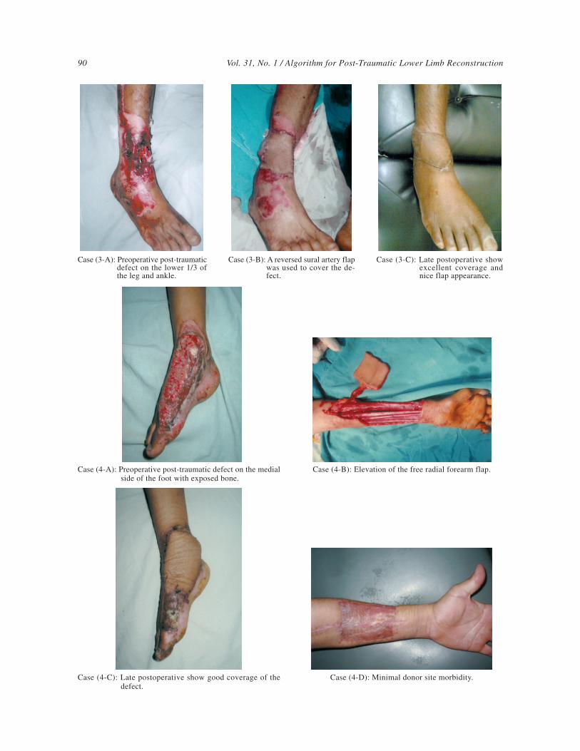
A suggested algorithm for post-traumatic lower limb soft tissue reconstruction
Simple wound (No exposed bone,tendons or neurovascular structures)
Complex wounds(exposed bone, tendons orneurovascular structures)
Split thickness skin graft except in weight bearing areas
Knee Leg Foot
• Gastrocnemiusmuscle flap
• Medial andlateral genicularartery flap
Upper third
• Gastrocnemiusmuscle flap
Middle third
• Medial hemisoleusmuscle flap
• Cross leg flap
• Free flap
Lower third
• Distally basedsural artery flap
• Free flap
Dorsum
• Cross legflap
• Free flap
Sole & heel
• Medial planterflap for the heel
• Distally basedsural artery flapfor the heel
• Local randomfasciocutaneousflaps for smalldefects of thesole
• Free flap
Ankle &maleoli
• Distally basedsural arteryflap
• Dorsalis pedisflap
• Abductorhallucisbrevis flap
• Free flap
Fig. (1): Show a suggested algorithm for post-traumatic lower limb soft tissue reconstruction
Table (1): Different reconstructive procedures used in the lower limb.
Thigh
Knee
Leg
Foot
Multiple
regions
Total
To
tal
7
6
36
43
8
100
Fre
e p
ara
scap
ula
rfl
ap
0
0
0
1
0
1
Fre
e l
ati
ssim
us
flap
0
0
0
2
0
2
Fre
e r
ad
ial
flap
0
0
0
2
0
2
Ab
du
cto
rh
all
ucis
fla
p
0
0
0
1
0
1
Bil
ate
ral
VY
fla
p
0
0
0
1
0
1
Tra
nsp
osi
tio
nfl
ap
0
1
3
1
0
5
Do
rsali
sp
ed
is f
lap
0
0
0
1
0
1
Hem
iso
leu
sm
usc
le f
lap
0
0
2
0
0
2
Gast
rocn
em
iou
sfl
ap
0
2
1
0
0
3
Dis
tall
y b
ase
dsu
ral
flap
0
0
3
6
0
9
Cro
ssle
g f
lap
0
0
3
9
0
12
Sim
ple
clo
sure
0
0
1
0
1
2
Sp
lit
thic
kn
ess
skin
gra
ft
7
3
23
19
7
59

92 Vol. 31, No. 1 / Algorithm for Post-Traumatic Lower Limb Reconstruction
Foot:Dorsum:
CountResult
Sole:CountResult
Medialmalleolus:
CountResult
Lateralmalleolus:
CountResult
Anterioraspectof ankle:
CountResult
Tendoachilles:
CountResult
Total count
Table (3): Different reconstructive procedures used in the foot and their results.
29
6
2
2
3
1
43
158 excellent
take.6 goodtake.
1 poortake
1Good take
0–
1Complete
loss
1Excellent
take
1Poor rake
19
Splitthicknessskin graft
Crossleg flap
9All
completelysurvivedonly one
haddehiscence
0–
0–
0–
0–
0–
9
Reversedsmallflap
1Completely
survived
3Completesurvival
0–
1Ischemiaof distal
1/3
1Sompletesurvival
0–
6
0–
0–
1Completesurvival
0–
0–
0–
1
Dorsalispedisflap
0–
0–
0–
0–
1Completesurvival
0–
1
Trans-position
flap
0–
1Completesurvival
0–
0–
0–
0–
1
V-Yflap
0–
0–
1Completesurvival
0–
0–
0–
1
Abduciorhallucisbrevis
muscle flap
Freeradial
forearmflap
2One
completelysurvivedthe other
wasischemic
0–
0–
0–
0–
0–
2
Freelatissmusdorsi flap
1Completely
survived
1Ischemic
0–
0–
0–
0–
2
Freeparas-
capularflap
1Completely
survived
0–
0–
0–
0–
0–
1
Leg:Upper third:
CountResult
Middle third:CountResult
Lower third:CountResult
Multiple regions:CountResult
Total count
Table (2): Reconstructive procedures used in the leg and their results.
Total
2
10
8
16
36
0–
1Completesurvival
2One survived,
the otherhad necrosisin distal 1/3
0–
3
Transpositionflap
0–
2One completely
survived,the other
completelylost
0–
0–
2
Hemisoleusmuscle flap
1The flap
completelysurvived
0–
0–
0–
1
Gastrocn-emious
flap
0–
0–
3Two of themcompletely
survived thethird wasischemic
0–
3
Reversedsmall flap
0–
3Completesurvival
0–
0–
3
Crossleg flap
0–
1Completehealing
0–
0–
1
Simpleclosure
1Good take of
the graft
3One excellent
take. Twogood take
3Good take
16One excellent
take.8 good. 6 poor.
one complete loss
23
Splitthicknessskin graft
DISCUSSION
The overall results showed that immediatewound reconstruction, whenever possible, is pre-ferred to delayed wound reconstruction in that itshortens the period of hospital stay significantly;in our thesis from a mean of 34 days in cases withdelayed reconstruction to 19.42 days in cases withimmediate reconstruction. This is associated withless pain with dressing changes, fewer operations,decreased infection rate and secondary necrosis ofexposed tissues. Thus, early consultation for softtissue reconstruction is advised and all attemptsshould be done to perform immediate reconstruc-tion. These results are in agreement with previousstudies [3,4,5].
In our thesis, 54 cases were managed with splitthickness skin graft. Split thickness skin graft wasan excellent choice and was considered the firstreconstructive option when only muscle or fasciawas exposed. However, in the weight bearing areasin the foot, they mostly can not withstand pressureand are subjected to repeated ulcerations. Also,around the knee joint, complications in the form ofcontractures and repeated ulcerations are common.These results are in agreement with others [6].
The gastrocnemius muscle flap was used in 3patients. Two flaps were applied on the knee andone flap on the upper third of the tibia. In twocases the medial belly was used and in the thirdcase both bellies of the muscle were used. Themuscle was covered immediately with split thick-ness skin graft. The flap survived completely inall patients. These results show that the gastrocne-mius muscle flap is indeed a highly reliable flapthat could be used safely to cover defects overdifferent aspects of the knee and the upper thirdof the leg. These results are in agreement withother studies [7-11].
Two medial hemisoleus muscle flap with splitthickness skin graft were used for coverage ofexposed tibial fractures at the middle third of theleg in two cases in this study. One case was prox-imally based and the other was distally based. Theproximally based flap completely survived withno complications but the distally based flap wascompletely ischemic. The proximally based flapis a reliable local option for soft-tissue coverageof a less extensive tibial wound in the middle thirdand the junction of the middle and distal thirds ofthe leg with good outcome and minimal morbidity.This is in agreement with [12,13]. The distally basedhemisoleus muscle flap is unreliable because thedissection in the proximal soleus muscle is difficult,
and the smaller inferior pedicles are less predictablein location and size. This is in agreement withMathes & Nahai [2]. However, this is not in agree-ment with others [13-16].
Abductor hallucis brevis muscle flap and splitthickness skin graft was used in our study to covera defect on the medial malleolus. The flap com-pletely survived and the graft readily took over themuscle. Abductor hallucis brevis muscle flap offersversatile and well-vascularized tissue to coverdefects at the medial aspect of the foot. This resultis consistent with [17] who used this flap also tocover defects on the calcaneus and the forefoot.
In our study, five cases were managed by localrandom pattern fasciocutaneous flaps; either trans-position or rotational flaps (one case in the poplitealfossa, 3 cases in the leg and one case in the anterioraspect of the ankle). All of these flaps were prox-imally based. Only one flap (20% of cases) hadtip necrosis and was left to heal with secondaryintention. These results show that local randompattern fasciocutaneous flaps are simple to raise,sensate, replace like with like tissue, ideal forsmaller defects and require no unusual surgicalskills. A disadvantage of fasciocutaneous flaps isthe unsightly donor site, and therefore their useshould be avoided in young females. They remainone of the useful methods of skin cover for lowerextremity defects. These results are in agreementwith other studies [18-22] who reported nearlysimilar complication rates, varying from 12 to 26%.
Chittoria & Mishra [23] Reported a much lowercomplication rate (one flap out of twenty had tipnecrosis). However, these results are not in agree-ment with others [24] who reported a 38% incidenceof tip necrosis in proximally based fasciocutaneousflaps.
Nine distally based sural artery flaps were usedin the leg and foot from which six flaps completelysurvived, two had necrosis of the distal third andone flap was completely lost. These results showedthat the distally based superficial sural artery flapcould be considered the main line of treatment insimilar defects in the lower leg, malleoli, anklejoint, weight bearing heel and even the dorsum ofthe foot. In addition to being one-stage operation,it is easy to perform with little expertise, quick toelevate, has a wide arc of rotation with reliableand constant vascular supply and there is no needto sacrifice any major artery or sensory nerve.These results in agreement with others [25,26,27]
who have been able to use this flap to cover defectsof the dorsum of the foot [28-32].
Egypt, J. Plast. Reconstr. Surg., January 2007 93

94 Vol. 31, No. 1 / Algorithm for Post-Traumatic Lower Limb Reconstruction
Twelve cross leg fasciocutaneous flaps wereapplied and the survival rate was 100%. This flapwas used successfully to cover large defects onthe leg and dorsum of the foot. These results showthat cross leg flap is a very reliable flap for legand foot defects if properly designed and transferredin selective cases where other flaps are expectedto be risky or not feasible. These results are inagreement with others [33,34]. However, theseresults are not in agreement with many other authorswho report that cross leg fasciocutaneous flapsshould not be considered frequently in currentmedical practice due to the availability of otheripsilateral flaps like fasciocutaneous flaps, muscleand musculocutaneous flaps and free flaps [35].
In our thesis, one dorsalis pedis flap was usedto reconstruct a defect on the lateral malleolus andit completely survived. Delayed donor site healingand donor site morbidity was the major drawbackof this flap; however these were accepted as com-pared with the benefits achieved. These results arein agreement with others [36,37,38]. However, Sam-son, et al. [39] reported that this flap should be usedwith caution as all patients had initially experienceddelayed donor-site healing.
The free latissimus dorsi flap was used for twocases in the present study. Unfortunately, one flapsurvived and the other one was ischemic.
Free fasciocutaneous flaps were used in threecases; two radial forearm and one parascapularflaps. Two of the three flaps completely survivedand the third one was completely ischemic. Thefailure in both cases could be attributed to thecondition of the recipient vessels (being in theproximity to the zone of injury).
The results of this thesis show that free tissuetransfer has several advantages. It offers a wellvascularized tissue to close cavities or defects ofdiminished vascularity. It can be carefully pickedfrom an area that has not been traumatized toomatch the existing functional and aesthetic lowerleg or foot defects. The donor morbidity can bekept to a minimum. They can be of large size.However, they are lengthy procedure; require theavailability of well trained micro-vascular teamand sophisticated equipment and high cost. A highdegree of success can be achieved only by extreme-ly careful patient selection. This is consistent with[40,41,42].
Conclusion:
As regards defects in regions of the lower limbat the thigh, skin grafting should be considered as
the first line of closure, followed by rotationalflaps. This is because the muscle flap of the thighacts as an envelope that serves as a source ofprotection for the underlying bone and neurovas-cular structures. It also provides a well vascularizedbed for split thickness skin graft application.
For complex defects around the knee regionand upper third of the leg, the gastrocnemius muscleflap is a reliable flap with a good range of move-ments allowing it to reach most aspects of the knee.
Regarding defects of the middle third of theleg, the proximally based hemisoleus muscle flapis the flap of choice, however, cross leg fascio-cutaqneous flap is another excellent choice in spiteof its drawbacks like inconvenience, discomfortand hospitalization for four weeks which wererelatively insignificant as compared with the ben-efits achieved. It should be avoided in cases withdiabetes, hypertension, previous history ofthrombosis and stiffness in either leg.
For the distal third of the leg, the distally basedsural island flap is an excellent choice. Localrandom fasciocutaneous flaps should also be con-sidered. Larger defects in the distal third are anindication for free flap transfer.
In cases of foot defects, each site has to beconsidered separately. Thus on the dorsum of thefoot, cross leg flap is the simplest solution. Other-wise, one has to use a free flap, especially thoseof thin skin as parascapular flap. However, distallybased sural artery flap can be used to cover defectson the dorsum of the foot.
Heel defects are best managed with medialplanter flap, however distally based sural arteryflap is another option. For smaller defects in thesole, one can use simple reconstructive options:Rotation, transposition or V-Y advancement flap.For larger defects, free flaps are optimal, providedthat they are well tailored.
For the ankle joint or malleolar defects, localflaps are ideal. The abductor hallucis brevis muscleflap can be used to cover the exposed medialmalleolus. Dorsalis pedis flap, distally based suralartery flap and local random pattern flaps can beused to cover such defects of the ankle or themalleoli.
We emphasize the importance of co-operationat the time of primary surgery between orthopedicand plastic surgeon to preserve access to potentialflaps. The technique of bony fixation of the tibiamay prevent the use of this flap, especially in the

presence of external fixation pins which may injureperforating vessels or tether the flap, restrictingits range of transposition.
Finally, the following algorithm may be pro-posed as a protocol for the management of lowerlimb defects.
REFERENCES
1- Armen K.K. and Nolan S.K.: Lower extremity recon-struction. Grabb and Smith’s plastic surgery, Lippincott-Raven, 5 ed.: 1031-1049, 1997.
2- Mathes S.J. and Nahai F.: Clinical applications for muscleand musculocutaneous flaps. St. Louis, CV Mosby, 1982.
3- Byrd H.S., Cierny G. III and Tebbetts J.B.: The manage-ment of open tibial fractures with associated soft-tissueloss: External pin fixation with early flap coverage. Plast.Reconstr. Surg., 68: 73, 1981.
4- Byrd H.S., Spicer T.E. and Cierney G.D.: Managementof open tibial fractures. Plast. Reconstr. Surg., 76: 719-730, 1985.
5- Godina M.: Early microsurgical reconstruction of complextrauma of the extremities. Plast. Reconstr. Surg., 78: 285-292, 1986.
6- McGregor I.A. and McGregor A.D.: Fundamental tech-niques of plastic surgery. Edited by Ian A., 9th edition,1998.
7- Morris A.M. and Buchan A.C.: The place of the cross legflap in reconstructive surgery of the lower leg and foot.A review of 165 cases; Plast. Reconstr., 31: 138, 1978.
8- Morris A.M. and Pribaz J.J.: Trans-tibial transposition ofgastrocnemius muscle and musculocutaneous flap. Br. J.Plast. Surg., 44 (8): 599-601, 1992.
9- Arnold P.G. and Mixter R.C.: Making the most of thegastrocnemius muscles. Plast. Reconstr. Surg. Jul., 72(1): 38-48, 1983.
10- Zaki M.S., Aly Y.A., El-Sharkawy A.G. and El-FaramawyA.A.: Anatomical grounds of the inferiorly based gastroc-nemius muscle flap. Egyptian Journal of Plastic andReconstructive Surgery, 15 (1): 8-12, 1991.
11- Cohen B.E. and Ciaravino M.E.: Gastrocnemius muscleand musculocutaneous flaps. Quoted from “Grabb’s en-cylopedia of flaps”, 2nd ed., 1998.
12- Fayman M.S., Orak F., Hugo B. and Berson S.D.: Thedistally based split soleus muscle flap. Br. J. Plast. Surg.,40: 20-26, 1987.
13- Lee L.Q.: Successful soft-tissue coverage of a tibial woundin the distal third of the leg with a medial hemisoleusmuscle flap. Plast. Reconstr. Surg., 115 (1): 245-251,2005.
14- Hughes L.A. and Mahoney J.L.: Anatomic basis of localmuscle flaps in the distal third of the leg. Plast. Reconstr.Surg., 92: 114, 1993.
15- Sadasivan K.K., Ogden J.T. and Albright J.A.: Anatomicvariations of the blood supply of the soleus muscle.Orthopedics, 14: 679, 1991.
16- Raveendrane S.S. and Kumaragama K.G.: Arterial supplyof the soleus muscle: Anatomical study of fifty lowerlimbs. Clin. Anat., 16: 248, 2003.
17- Anton H.S., Maziar S. and Ralfi G.: Versatility of theabductor hallucis muscle as a conjoined or distally-basedflap. The Journal of trauma injury, infection and CriticalCare Oct., 59 (4): 1007-1111, 2005.
18- Ponten B.: The fascio-cutaneous flap. Its use in soft tissuedefects of the lower leg. Br. J. Plast. Surg., 34: 215-220,1981.
19- Lamberty B.G.H.: Use of fasciocutaneous flaps in lowerextremity reconstruction. Plast. Reconstr. Surg., 4: 146-162, 1990.
20- Lagvankar S.P.: Distally-based random fascio-cutaneousflaps for one-stage reconstruction of defects in the uppertwo-thirds of the leg. Br. J. Plast. Surg., 43: 468-472,1990.
21- Hallock G.G.: Distal lower leg local random fasciocuta-neous flaps. Plast. Reconstr. Surg., 86: 30-411, 1990.
22- Georgiadis G.M., Behrens F.F., Joyce M.J., Earle A.S.and Simmons A.L.: Open tibial fractures with severe soft-tissue loss. J. Bone Joint Surg., 75-A: 143141, 1993.
23- Chittoria R. and Mishra S.M.: Fasciocutaneous flaps inreconstruction of lower extremity: Our experience. Kath-mandu Univ. Med. J. Oct-Dec., 2 (4): 344-8, 2004.
24- Small J.O. and Mollan R.A.B.: Management of the softtissues in open tibial fractures. Br. J. Plast. Surg., 45:571-7, 1992.
25- Masquelet A.C., Romana M.C. and Wolf G.: Skin islandflaps supplied by the vascular axis of the sensitive super-ficial nerves. Plast. Reconstr. Surg., 26: 202-213, 1992.
26- Hasegawa M., Torri S., Katoh H. and Esaki S. (1992):The distally based superficial sural artery flap. Plast.Reconstr. Surg., 93: 1012-1020, 1994.
27- Rajacis N., Darweesh M., Jayakrishnan K., Gang R.K.and Kojic S.: The distally based superficial sural flap forreconstruction of the lower leg and foot. Br. J. Plast.Surg., 49: 383-389, 1996.
28- Singh S. and Naasan A.: Use of distally based superficialsural island artery flaps in acute open fractures of thelower leg. Ann. Plast. Surg., 47 (5): 505-510, 2001.
29- Costa-Ferreira A., Reis J., Pinho C., Martins A. andAmarante J.: The distally based island superficial suralartery flap: Clinical experience with 36 flaps. Ann. Plast.Surg., 46: 308-313, 2001.
30- Almeida M.F., Da Costa P.R. and Okawa R.Y.: Reverse-flow island sural flap. Plast. Reconstr. Surg., 109: 583-591, 2002.
31- Sherine S., Raveendrane D., Perera T., Happuharachchiand Yoganathan V.: Superficial sural artery flap-a studyin 40 cases. Br. J. Plast. Surg., 57 (3): April 266-269,2004.
32- Hsieh C.H., Liang C.C., Kueh N.S., Tsai H.H. and JengS.F.: Distally based sural island flap for the reconstructionof a large soft tissue fracture with occluded anterior andposterior tibial arteries-a case report. Br. J. Plast. Surg.Jan., 58 (1): 112-115, 2005.
Egypt, J. Plast. Reconstr. Surg., January 2007 95

96 Vol. 31, No. 1 / Algorithm for Post-Traumatic Lower Limb Reconstruction
33- Aydan A.Y.: An unusual contracture of the foot caused byneglected burn wound salvaged by a cross leg flap. Plast.Reconstr. Surg., 110 (5): 1373, 2002.
34- Bhattacharya V. and Raveendra R.G.: Distal perforatorbased cross leg flaps for leg and foot defects. IndianJournal of Plastic Surgery, 38 (1): 18-21, 2005.
35- Barclay T.L., Cardos E. and Sharpe D.T.: Cross legfasciocutaneous flaps. Plast. Reconstr. Surg., 72: 847,1983.
36- Gould J.S.: The dorsalis pedis island pedicle flap for smalldefects of the foot and ankle. Plast. Reconstr. Surg. Jun.,9 (6): 867-71, 1986.
37- Simonka J.A. and Nacsai I.: Primary covering of tissuedefects using arteria dorsalis pedis flap (Article in Hun-garian). Magy Traumatol. Orthop. Helyreallito Sep., 34(1): 49-51, 1991.
38- Bos G.D. and Buehler M.J.J.: Am. Acad. Orthop. Surg.Nov., 2 (6): 342-351, 1994.
39- Samson M.C., Morris S.F. and Tweed A.E.: Dorsalis pedisflap donor site: Acceptable or not? Plast. Reconstr. Surg.,102 (5): 1549-54, 1998.
40- Sefarin D., Sabatier R.E. and Morris R.L.: Reconstructionof the lower extremity with vascularized composite tissue.Improved tissue survival and specific indications. Plast.Reconstr. Surg., 66: 230, 1980.
41- Morrison W.A., Grabb D. and O’Brien B.M.C., et al.: Theinstep of the foot as a fasciocutaneous island flap and asa free flap for heel defects. Plast. Reconstr. Surg., 72: 56-63, 1983.
42- Chen S.H.T., Wei F. and Chen H.: Emergency free flaptransfer for reconstruction of acute complex extremitywounds. Plast. Reconstr. Surg., 89: 882, 1992.
Related Documents


![Evaluation of Balance Disorders After Minor Head and Whiplash … · Lehmann et al. [2] suggested that patients who have experienced a traumatic brain injury without apparent orthopedic](https://static.cupdf.com/doc/110x72/5e445cc378a1511848332c16/evaluation-of-balance-disorders-after-minor-head-and-whiplash-lehmann-et-al-2.jpg)