A STUDY ON OUTCOME OF INTUBATION IN EMERGENCY DEPARTMENT, HOSPITAL UNIVERSITI SAINS MALAYSIA. By: Dr Rabiha Mohd Alip Dissertation Submitted In Partial Fulfillment Of The Requirement For The Degree Of Master of Medicine Program (Emergency Medicine) UNIVERSITI SAINS MALAYSIA MAY 2011

Welcome message from author
This document is posted to help you gain knowledge. Please leave a comment to let me know what you think about it! Share it to your friends and learn new things together.
Transcript
A STUDY ON
OUTCOME OF INTUBATION IN EMERGENCY
DEPARTMENT, HOSPITAL UNIVERSITI SAINS
MALAYSIA.
By:Dr Rabiha Mohd Alip
Dissertation Submitted InPartial Fulfillment Of The
Requirement For The Degree OfMaster of Medicine Program
(Emergency Medicine)
UNIVERSITI SAINS MALAYSIA
MAY 2011
i
Acknowledgement.
I wish to acknowledge my beloved family especially my husband and my three
children who tremendous patience and support through the years has made the
completion of dissertation possible.
To all my friends and fellow colleagues, thank you for their kind encouragement and
friendship.
Thanks also to the staff of Emergency Department, Hospital Universiti Sains
Malaysia for their kind assistance, patience, tolerance and hard work during my data
collection. Special thanks to the Associate Professor Dr. Nik Hisamuddin Nik Abdul
Rahman head of the Emergency Department for his support and encouragement
My special gratitude and thanks to Associate Professor Dr. Rashidi Ahmad who has
continuously and patiently guided me through the rough times during the preparation
of this dissertation.
Lastly , but most importantly, to Allah who has shown me the wisdom, inner
strength, courage and patience to understand myself and my role in life. Without His
divine guidance I would be nowhere.
ii
TABLE OF CONTENTS
Page
Acknowledgement i
List of tables iv
List of figures v
Abbreviation vii
Abstrak viii-ix
Abstract x-xi
1.0 Introduction 1-6
2.0 Literature Review 7-62
2.1 History of airway management 7 – 12
2.2 Anatomy and physiology of the upper airway 13-15
2.3 Emergency airway management 16-20
2.4 Basic technique 21-22
2.5 Endotracheal intubation 23-26
2.6 Rapid Sequence Intubation 27-40
2.7 Identification of the difficult airway. 41-44
2.8 The failed airway 45-46
2.9 Special Airway Considerations 46-57
2.10 Definition 58-62
iii
3.0 Objectives 63-65
3.1 General objectives. 64
3.2 Specific objectives. 64
3.3 Research hypothesis 65
3.4 Research questions. 65
4.0 Methadology 66-74
4.1 Study design 67
4.2 Study population and sample. 67
4.3 Research tool and data collection 68
4.4 Data entry and analysis 69-70
4.5 Conceptual framework. 71-74
5.0 Results and analysis 75-99
5.1 Demographic data of patient intubated in emergency
department in Hospital Universiti Sains Malaysia
76-81
5.2 Intubation process 82-99
6.0 Discussion 100-110
7.0 Conclusion 111-113
References 114-123
Appendices 124
iv
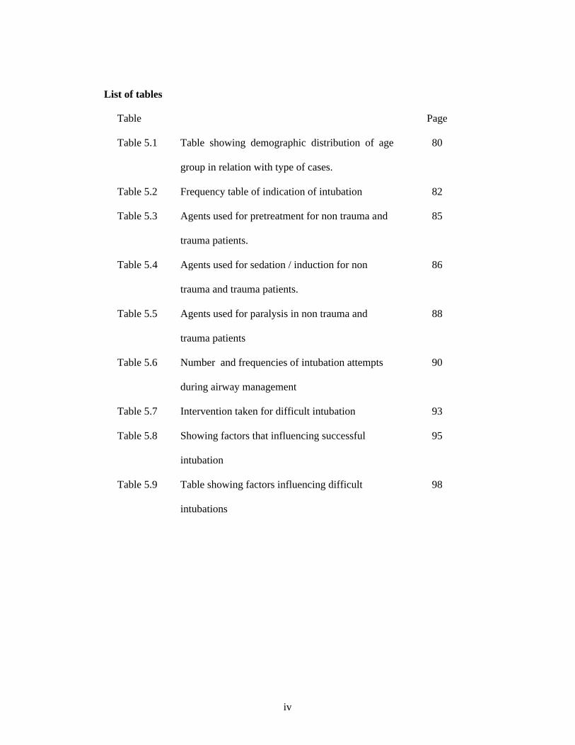
List of tables
Table Page
Table 5.1 Table showing demographic distribution of age
group in relation with type of cases.
80
Table 5.2 Frequency table of indication of intubation 82
Table 5.3 Agents used for pretreatment for non trauma and
trauma patients.
85
Table 5.4 Agents used for sedation / induction for non
trauma and trauma patients.
86
Table 5.5 Agents used for paralysis in non trauma and
trauma patients
88
Table 5.6 Number and frequencies of intubation attempts
during airway management
90
Table 5.7 Intervention taken for difficult intubation 93
Table 5.8 Showing factors that influencing successful
intubation
95
Table 5.9 Table showing factors influencing difficult
intubations
98
v
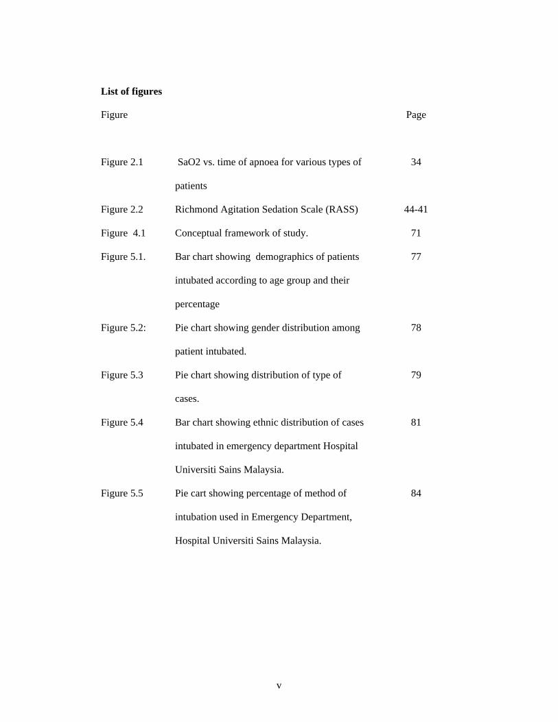
List of figures
Figure Page
Figure 2.1 SaO2 vs. time of apnoea for various types of
patients
34
Figure 2.2 Richmond Agitation Sedation Scale (RASS) 44-41
Figure 4.1 Conceptual framework of study. 71
Figure 5.1. Bar chart showing demographics of patients
intubated according to age group and their
percentage
77
Figure 5.2: Pie chart showing gender distribution among
patient intubated.
78
Figure 5.3 Pie chart showing distribution of type of
cases.
79
Figure 5.4 Bar chart showing ethnic distribution of cases
intubated in emergency department Hospital
Universiti Sains Malaysia.
81
Figure 5.5 Pie cart showing percentage of method of
intubation used in Emergency Department,
Hospital Universiti Sains Malaysia.
84
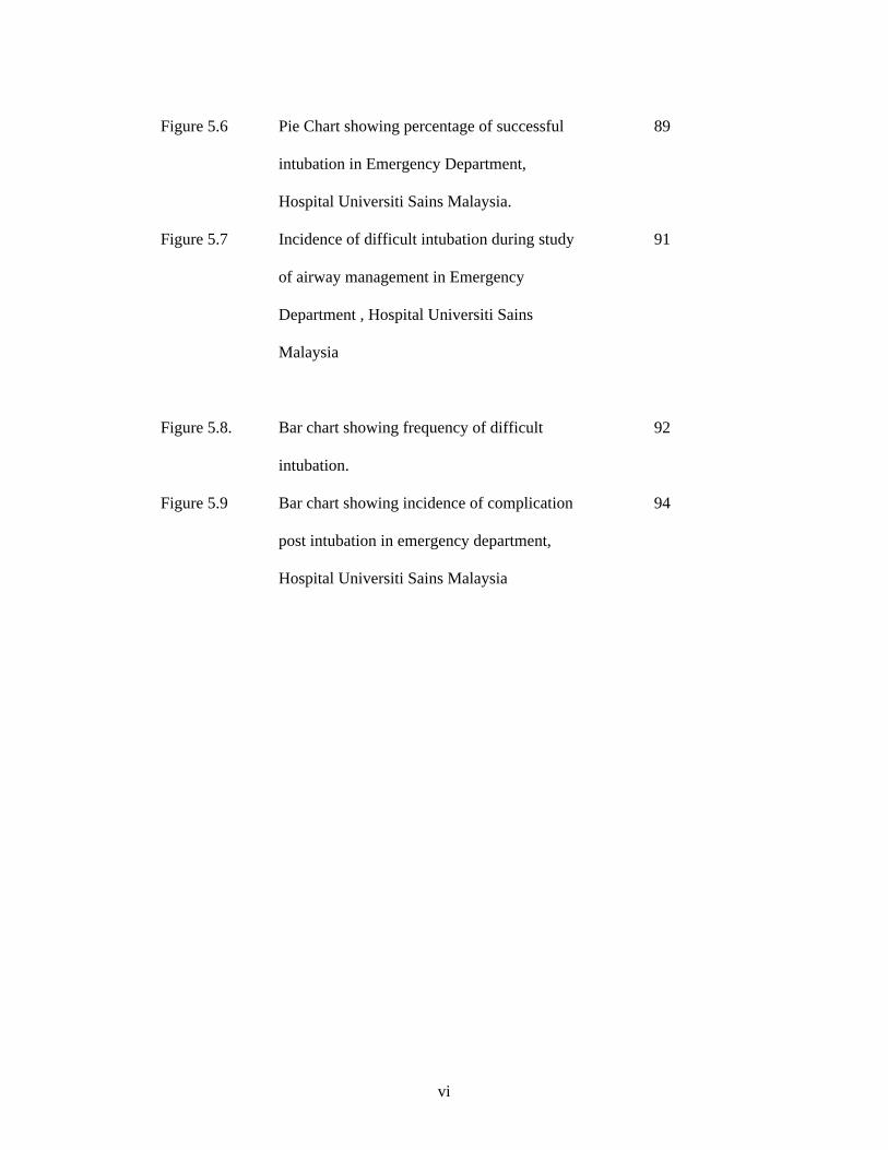
vi
Figure 5.6 Pie Chart showing percentage of successful
intubation in Emergency Department,
Hospital Universiti Sains Malaysia.
89
Figure 5.7 Incidence of difficult intubation during study
of airway management in Emergency
Department , Hospital Universiti Sains
Malaysia
91
Figure 5.8. Bar chart showing frequency of difficult
intubation.
92
Figure 5.9 Bar chart showing incidence of complication
post intubation in emergency department,
Hospital Universiti Sains Malaysia
94
vii
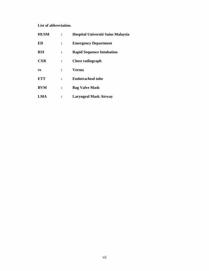
List of abbreviation.
HUSM : Hospital Universiti Sains Malaysia
ED : Emergency Department
RSI : Rapid Sequence Intubation
CXR : Chest radiograph
vs : Versus
ETT : Endotracheal tube
BVM : Bag Valve Mask
LMA : Laryngeal Mask Airway
viii
Abstrak:
KAJIAN TENTANG
KESUDAHAN PENGENDALIAN SALURAN PERNAFASAN
DI JABATAN KECEMASAN HOSPITAL UNIVERSITI SAINS MALAYSIA.
Pengenalan:
Bidang perawatan kecemasan di Malaysia merupakan satu bidang yang baru
diperkenalkan dan Universiti Sains Malaysia ialah universiti pertama di Malaysia
yang menawarkan kursus Pasca Ijazah Perubatan Kecemasan. Tujuan kajian ini
untuk mengenalpasti kesudahan pengendalian pengurusan saluran pernafasan di
jabatan kecemasan, Hospital Universiti Sains Malaysia (HUSM)..
Objektif:
Mengetahui kadar kejayaan intubasi saluran pernafasan, cara pengendalian sistem
saluran pernafasan sukar, mengenalpasti faktor yang mempengaruhi kesukaran
intubasi dan mengenalpasti komplikasi intubasi yang dilakukan oleh pengendali
saluran pernafasan di jabatan kecemasan, Hospital Universiti Sains Malaysia
(HUSM).
Kaedah.:
Ini adalah kajian pemerhatian yang dilakukan di jabatan kecemasan, Hospital
Universiti Sains Malaysia (HUSM) dalam jangka masa 6 bulan. Sampel adalah
sebanyak 128. Pesakit yang memenuhi kriteria dimasukkan didalam kajian. Semua
maklumat yang diisi di dalam borang yang telah disediakan.
ix
Keputusan:
Sebanyak 138 pesakit diintubasi . Min umur pesakit adalah 50.47 tahun. Kebanyakan
kes adalah lelaki, seramai 95 orang (68.84%). 94 (68.12%) adalah kes bukan trauma
dan 44 (31.88%) kes trauma. Indikasi utama intubasi adalah saluran pernafasan
berisiko (39%). “Rapid sequence intubation (RSI)” adalah cara pengendalian saluran
pernafasan yang paling kerap digunakan iaitu sebanyak 110 kes (79.71%). Fentanyl
adalah ubat pra rawatan yang selalu digunakan bagi 90 kes (65.2%) manakala untuk
sedasi, midazolam digunakan untuk 26.6% kes bukan trauma manakala bagi kes
trauman ialah propofol sebanyak 27.5% . Agen paralitik pula ialah succinylcholine
yang diberi kepada 100 pesakit (72.5%). 136 pesakit (98.55%) berjaya diintubasi
namun sebanyak 2 kes (1.45%) tidak berjaya. 111 kes berjaya diintubasi dengan
sekali cubaan (80.43%) . 6 kes cubaan lebih 3 kali (4.35%). 9 kes (6.52%) adalah
kes saluran pernafasan sukar dan penyebab utama pernafasan sukar adalah saluran
udara ‘anterior’ (44.4%). Cara pengendalian udara sukar adalah dengan
menggunakan bougie (44.4%). 57 (43.3%) mendapat komplikasi tekanan darah
rendah (49.1%), diikuti dengan renjatan jantung (33.3%). 2 faktor berkaitan dengan
kejayaan intubasi iaitu saluran pernafasan sukar dan percubaan berulang . Faktor yang
berkaitan dengan pernafasan sukar adalah percubaan berulang dan komplikasi.
Kesimpulan :
Jabatan kecemasan, Hospital Universiti Sains Malaysia (HUSM) mempunyai kadar
kejayaan yang tinggi dalam intubasi. Indikasi yang selalu digunakan adalah saluran
pernafasan berisiko. Cara pengendalian saluran pernafasan yang selalu digunakan
adalah “rapid sequence intubation (RSI)”. Kadar saluran pernafasan sukar adalah
rendah dan factor yang mempengaruhi kesukaran intubasi adalah kekerapan cubaan
intubasi dan komplikasi intubasi.

x
Abstract:
A STUDY ON
OUTCOME OF INTUBATION IN EMERGENCY DEPARTMENT,
HOSPITAL UNIVERSITI SAINS MALAYSIA.
Introduction.
In Malaysia, Emergency Medicine is a new specialty without any less
important role in health care delivery. Universiti Sains Malaysia is the first university
in Malaysia offering postgraduate study in Emergency Medicine. Aims of this study
were to observe the outcome of intubations, the success rate of intubations, method of
intubations , presence of difficult airway and complication after intubations in
emergency department, Hospital Universiti Sains Malaysia (HUSM).
Objectives:
To determine the success rate of intubations, method of airway management,
predictors of difficult intubation and complications of intubation performed by airway
personal in emergency department, Hospital Universiti Sains Malaysia (HUSM).
Methodology:
This was a cross sectional observational study done in emergency department,
Hospital Universiti Sains Malaysia (HUSM) in six months. Sample size were 128.
Patients fulfill the inclusion criteria were included in this study. Results were
documented in pre prepared data entry sheet.
Results:
138 patient intubated during this study. Mean age of patient is 50.47 years. Majority
were male , 95(68.84%). 94 case (68.12%) were due to non trauma case and 44
xi
(31.88%) were trauma case. Most frequent indication ware airway at risk with
percentage of 39% . Rapid sequence intubation (RSI) were the most frequent method
of airway management 110 case (79.71%). Fentanyl was the commonest
pretreatment agents used in 65.2% of case. For sedation, 26.6% of non trauma
patients were given midazolam while for trauma, propofol were used. Most common
paralytic agent given were succinylcholine, 72.5%. 98.55% were intubated
successfully and only 2 cases (1.45%) failed intubation. 111 case intubated in single
attempt (80.43%). 6 cases intubated with more then 3 attempts (4.35%). 9 cases
(6.52%) noted to have difficult intubation and most common cause of difficult
intubation were anterior cord position (44.4%). The most frequent intervention for
difficult airway was by using bougie in 4 cases (44.4%). 57 case (43.3%) develop
complication which were hypotension 49.1%, and cardiac arrest 33.3%. 2 factors
showed significant association with successful intubation which were difficult airway
and multiple attempts. Factor associated with difficult intubation were number of
attempts during intubation and the presence of complication.
Conclusion:
Emergency department, Hospital Universiti Sains Malaysia (HUSM) have a very
high success rate of intubation. The most common indications was airway at risk and
common method of intubation done was rapid sequence intubation (RSI). Agent
commonly use for pretreatment were fentanyl, while induction agent used were
propofol and midazolam. For paralytic agent succinylcholine were used in most
intubations. Complication rate were fairly high since due to most cases referred were
severely or critically ill. There were low incidence of difficult intubation. Predictors
of difficult intubations were multiple intubation attempts and development of
complications.

CHAPTER 1
INTRODUCTION

2
1.0 INTRODUCTION
In Malaysia, Emergency Medicine is a rather new specialty without any
less important role in health care delivery . Universiti Sains Malaysia (USM) is
the first university in Malaysia offering postgraduate study in Emergency
Medicine i.e. Master in Medicine (Emergency Medicine). It had been introduced
in 1997 and in 2003, the Emergency Unit, Hospital Universiti Sains Malaysia
(HUSM) was upgraded to a department status in 2003, in parallel with the
recognition of Emergency Medicine as a specialty on its own.
Emergency department, Hospital Universiti Sains Malaysia (HUSM)
received about 48,000 patients per year. It serves as an excellent place for
learning and gaining experience and plays an important role in training future
doctors and Emergency Physicians in dealing with critical and non critical cases
who arrived in Emergency Department.
Emergency department Hospital Universiti Sains Malaysia (HUSM) is a
primary care department that provides initial treatment to patient with a broad
spectrum of illnesses and injury. Some which may be life threatening and requires
medical attention.
One of the most important skills in treating life threatening condition for
the emergency resident is the airway management. Safe effective airway

3
management in critically ill or injured patients is the cornerstone of resuscitation
because failure to secure an adequate airway can quickly lead to death or
disability. (Butler, 2001)
Airway emergencies present in different fashion, the emergency resident
must be proficient in multiple techniques to protect and maintain the airway, and
must be prepared with all the necessary equipment to perform alternative
procedures should the initial plan fails.
Emergency department Hospital Universiti Saains Malaysia (HUSM) is
staffed by a group of emergency physicians, emergency medicine master student
and medical officers of varying training experiences. Training in airway
management was mandatory in the Emergency Medicine (M.Med) curriculum.
Experience in airway management is acquired through daily exposure to patients
requiring airway management and rotational posting for 6 months in
Anesthesiology. Emergency resident with the above experience is known in
this study as an ‘Airway Personal’.
When a critically ill patient presented at the emergency department, the
airway was first assessed by the emergency resident. If there is an indication for
intubations , the emergency resident will secure the airway using various method
of intubations. Study showed that the most common method used by emergency
resident is rapid sequence intubations (RSI), except for cases with respiratory or
4
cardiac arrest where crash intubations method were applied (Wong E, 2004).
When the attempt failed or if the airway was deemed to be difficult on initial
assessment, the anesthetist was then called.
One observational study was done in Hospital Selayang in 2001 regarding
airway management in General Hospital in Malaysia. It conclude that, most
patient who came to Emergency Department that need airway intervention was
intubated by emergency resident. The percentage was 98.47 % from 261 patient
intubated in their emergency department and the most common method of
intubations are rapid sequence induction which were about 84.7%. Even though
the success rate is high, the study did not mention the detail percentage of how
many attempts of intubations were attempted and what is the factor contributing
the multiple attempt of intubations. With the background of General Hospital,
most of the emergency resident are young doctors who did not have any
experience in airway management and during the study period, Selayang
Hospital was a very new hospital without any Emergency Physician and this may
contribute 1.53 % failure rate of intubations. Again the study did not comment
the intervention that were done due to failure of intubations (Ibrahim, 2002).
For being tertiary and referral centre, emergency department, Hospital
Universiti Sains Malaysia (HUSM) currently received about 48,000 patient in
2008 and from this total number of patient about 3600 of patient are categorized
as critically ill patient or triage as Red Zone case. Patient that requires early
5
airway intervention and intubated in emergency department are about 30 to 35
patient per month and roughly about 350 to 400 patients were intubated in 2008.
Even though the percentage of intubations for critically ill patient per year was
about 10% to 12%, it is considered high since some of the patient that came to
emergency department already has been intubated from the primary center.
As a tertiary centre, we need a study in emergency department Hospital
Universiti Sains Malaysia (HUSM) regarding the airway management and in
future, more study can be done to help improve emergency resident knowledge
and skills. It also will be a references to other general hospital in Malaysia in
practicing airway management in emergency department.
Since the study of airway management was first done in Hospital Selayang
(1st computerized government general hospital), the data only specifically to this
new hospital which have very limited emergency resident who have airway
management experience and limited used of anesthetic drugs in their emergency
department .
The aims of this study were to observe the outcome of intubations in
emergency department, Hospital Universiti Sains Malaysia (HUSM), specifically
on the success rate of intubations, method of intubations, presence of difficult
airway and factors related to it and complication after intubations .
6
The other data that will be recorded in this study are :-
Indication of airway intervention.
Type of cases.
Pharmacologic agent used.
Incidence of difficult intubation and factors effecting it.
Type of intervention applied after failed intubations.
Finally, the study will also give a basic database of intubations performed
in tertiary centers in Malaysia and will be a guidelines and reference future study
of emergency airway management in Malaysia.
7
CHAPTER 2
LITERITURE
REVIEW
8
2.0 Literature Review
2.1 History of airway management.
The origin of specific airway technique or tool used in airway
management are really impossible to be identify and the existence of airway
management were believe as early as 4000 years ago (Szmuk et al., 2008). The
earliest record of airway management came from an ancient Hindu book of
medicine, ‘Rig Veda’ which describe the healing of a throat incision around 2000
BC (GL, 1994). Then 500 years later, the Egyptian wrote the first documented
technique resembling tracheostomy to resolve upper respiratory obstruction (CG,
2005). It is also said that Alexander the Great in 356-323 BC, saved a soldier
from suffocation from aspirated bone by making a tracheal incision using the tip
of his dagger (Szmuk et al., 2008). In 160 AD, the Roman physician Galen
wrote “If you take a dead animal and blow air through its larynx (through a
reed), you will fill its bronchi and watch its lungs attain the greatest dimension”.
Sadly, the significance of that finding was not appreciated and research on
ventilation did not advance any further until a Muslim philosopher and physician
Avicenna in 980-1037 AD described intubation of the trachea using “a cannula of
gold or silver”(Szmuk et al., 2008) .
In the thirteenth century, tracheostomy is termed a “semi slaughter and a
scandal of surgery” (JJ Watkinson, 2000) and this description may explain the
demise of this procedure during this age. It was not until the Renaissance that
thracheostomy reappeared as a viable medical solution.

9
In 1546, Antonio Brasavola an Italian physician reintroduced
tracheostomy in humans by performing the first documented case of successful
tracheostomy in a patient with tonsillar obstruction (GL, 1994). In 1620, a book
of tracheostomy was publish by Parisian Nicholas Habicot (Szmuk et al., 2008).
In October 1667, Robert Hooke demonstrated tracheostomy during Royal Society
meeting on a dog and claims he preserved the canine’s life by breathing for it by
means of a bellows (JJ Watkinson, 2000). However, according to Sitting and
Pringnitz, only 50 life saving tracheostomies had been described in the entire
medical literature before year 1,800 (E Sitting, 2001). Only when great pioneers
such as Trousseau and Trendelenburg refined and popularized the operation and
refine the clinical use of the procedure when in 1833 Trousseau reported on his
experience with 200 diphtheria patients treated with tracheotomy. In 1871,
Trendelenburg performed a tracheotomy to prevent blood inhalation during upper
airway surgery (T Ezri, 2005).
During the time when many experience and experimental technique of
tracheostomy grew , other consideration of non invasive airway management also
were reported as early in 1754 when Benjamin Pugh, an English obstetrician
described an air pipe for neonatal resuscitation (Davison, 1965). In 1760, Buchan
described the first use of an opening in the patient windpipe during resuscitation.
In 1788, Charkes Kite first used endotracheal tube in resuscitation of drowned
persons and described their used by either the oral or nasal route (JA Lee, 1973).
In late 1700s, Chaussier a gynaecologist in maternity hospital in Paris perform

10
translaryngeal intubation with self made tube in neonates with obstructed airway
and also the first person to deliver oxygen to the newborn (WW Mushin, 1953).
Even with all the evidence and discoveries of advance airway management
in late 1700s, the implementation, decision and the practice of performing the
airway management among practitioner still erratic. Famous case, for example,
in December 1799, three physicians gathered around a dying man who keep
shifting his position and gasping for air. They tried to give him gargle but only
found out that this man nearly choke to death. They knew this man had severe
airway compromise. One physician aware of tracheostomy but was reluctant to
do it since that man is a famous person. As a result George Washington died due
to preventable suffocation from upper airway obstruction caused by severe
bacterial epiglottitis (E Sitting, 2001).
In 1827, Leroy demonstrated vigorous bellow ventilation will cause
emphysema and fatal pneumothorax in ventilated drown dog (Chinsky, 2001).
Positive pressure ventilation was then banned for more then hundred years
(Szmuk et al., 2008). Despite the setback tracheostomy and tracheal intubation
continued to be performed and their techniques improved through following
decades especially in acute airway management.
In 1880, in Scotland, William Mac Ewen described how to relieve airway
obstruction by passing an oral tube into the trachea. He practiced blind, digital
11
intubation using cadaver models and able to use this technique clinically
(Gillespie, 1948). A few years later, in USA, a paediatrician Joseph O’Dwyer,
developed a metal tube system that could be passed blindly to relieve airway
obstruction in children with diphtheria and needed surgery after witnessing
several mutilating effect of nasty tracheostomies. The only problem with
O’Dwyer intubation system is that they had to be placed blindly (Gillespie, 1948).
End of the nineteenth century, a German surgeon Franz Kuhn constructed
metal tube that he inserted oral with a digital blind technique (Gillespie, 1948). He
also described the used of curved tube introducer and publish the first paper of
nasal intubation (Gillespie, 1948). He also realize un blunted surgical stimulus
may lead to spasm of the larynx and believe that “cocainization” of the larynx was
a helpful adjunct for intubation (Gillespie, 1948). This was the first effort of
awake intubation under topical anaesthesia.
During World War I, Magill and Rawbotham performed several
endotracheal intubation with administration of endotracheal anesthesia and
realize reconstructive surgery in mutilated soldiers are more successful when the
airway are secured with endotracheal tubes. They also invented the Magill forcep
that is usefull for nasal intubation (Thomas, 1978, Condon and Gilchrist, 1986).
The next important development was development of direct laryngoscopy
which allowed visualization of the glottic structures. Manual Garcia (1805-1906),
12
perform autolaryngoscopy through the use of a dental mirror in combination with
a second, larger mirror used to direct sunlight into his mouth (Alberti, 1996). This
arrangement allowed him to see his larynx and trachea,
In 1940s, Miller and MacIntosh develop laryngoscopes and it use is
common clinical use today. In 1941, Robert Miller described his straight
laryngoscope blade, while in 1943 Robert MacIntosh described his curved blade
(Doyle, 2009). At the same time, in Montreal, Canada in 1942, Harold Griffith
introduced curare as a muscle relaxant with a view to facilitating abdominal
surgery and other procedures. As a result, tracheal intubation became routine in
major surgical procedures (Doyle, 2009).
Finally, any history of airway management would be incomplete without
mentioning supraglottic airway devices such as the Laryngeal Mask Airway
(LMA) by Dr. Archie Brain, the inventor of the LMA in 1937 (Doyle, 2009).
Landmarks in clinical airway management.
Biblical
Times
Death from airway obstruction recognized (trauma, strangulation,
leprosy, abscesses)
1842 Crawford Long discovers ether anesthesia
1854 Garcia, a professor of singing, develops indirect laryngoscopy
1878 Chloroform administered through tracheal tube (MacEwen)
1885 O’Dwyer popularizes intubation for diphtheria

13
1895 Kirstein develops direct laryngoscopy
1900 Kuhn develops a flexometallic tracheal tube
World
War I
Many casualties requiring head and neck surgery adds impetus to
widespread use of intubation in military
hospitals; Magill introduces tracheal tube with inflatable cuff
1920 Chevalier Jackson designs improved laryngoscope
1920s Magill develops blind nasal intubation
1942 Griffiths introduces curare into clinical practice
1946 Mendelson describes aspiration pneumonitis
1950s Popularization of the use of tracheal tubes for general anesthesia
1960s Advent of electronic patient monitoring
1962 Sellick maneuver and rapid-sequence induction developed
1940s-
1970s
Continuing improvements in laryngoscope and tube designs; use of
plastic
1970s Development of implant-tested low-irritation, low-cuff pressure
disposable tracheal tubes
1980s Popularization of fiberoptic intubation. Introduction of pulse oximetry
and capnography as non-invasive means of assessing oxygenation and
ventilation
1990 s Popularization of laryngeal mask airway, rigid fiberoptic laryngoscopes
(Bullard, Wu, etc.,) and ASA Practice Guidelines for Management of
the Difficult Airway. Increased awareness of the special challenges of
the «difficult extubation» patient.
14
1995 Founding of the Society for Airway Management (www.samhq.com)
2000s Introduction of video laryngoscopes (GlideScope, McGrath, etc.)
2.2 Anatomy and physiology of the upper airway.
Anatomically, the upper airway consists of the pharynx and nasal cavities.
However, functionally, the larynx and trachea may be included, and the oral
cavity provides an alternate entrance to the respiratory passages (Morris, 1988).
The nose is a pyramidal structure composed of bone and cartilage attached
to the facial skeleton, and is divided by a midline septum into the two nasal
cavities. Kiesslbach’s plexus (Little area) located at the anterior aspect of each
nostril and easily traumatized during the insertion of nasotracheal tube. This will
lead to severe epistaxis and known to be the most common complication during
insertion of nasotracheal intubation (Redden, 2000). In addition, softening the
endotracheal tube in warm water or saline have been demonstrated to reduce the
complication rate particularly epistaxis (Lu et al., 1998).
The paranasal sinuses drain into the nasal cavities and this nasal cavities
also continuous with the nasopharynx posteriorly (Ron M. Walls, 2008). The
adenoids are located just posteriorly and partially surround a depression in the
mucosal membrane. During insertion, the tube often enters into this depression
and resistance is encountered (Walls, 2008). Continued aggressive insertion can

15
cause the tube to penetrate the mucosa and may go deeper and cause
complication.
The nose functions as a heater and humidifier of inspired gas, a voice
resonator, and houses the olfactory receptors (Morris, 1988).
The mouth opens posteriorly into the oropharynx and forms the entrance
to the digestive tract as well as an alternate pathway for respiration. It is also
involved in phonation (Morris, 1988). Orotracheal intubation can be used as an
alternative to nasal intubation to achieve airway protection and ventilation when
necessary, however, variations in upper airway anatomy may make this technique
difficult. In supine unconscious persons, backward movement of the tongue and
lower jaw may cause airway obstruction.
The pharynx is a U-shaped fibromuscular tube extending from the base of
the skull to the cricoid cartilage at the entrance to the esophagus. Anteriorly it
opens into the nasal cavity, the mouth, and the larynx, which divide it into the
naso-, oro- and laryngopharynx, respectively. The pharynx thus forms a common
aerodigestive tract and is intimately involved with the act of swallowing.
The larynx consists of a framework of cartilages and fibroelastic
membranes covered by a sheet of muscles and lined with mucous membrane. It
evolved as a protective valve mechanism at the upper end of the lower airway
16
necessitated by an unusual crossover between the airway and alimentary canal. It
functions as an open valve in respiration, a partially closed valve in phonation,
and as a closed valve protecting against aspiration during swallowing. The larynx
extends from its oblique entrance formed by the aryepiglottic folds, the tip of the
epiglottis, and the posterior commissure to the lower border of the cricoid
cartilage and bulges posteriorly into the laryngopharynx. The larynx is the most
heavily innervated sensory structure in the body. Stimulation of the
unanesthetized larynx during intubation cause tremendous reflex sympathetic
activation and this may lead to elevation of intracranial pressure and may
aggravate myocardial ischemia (Ron M. Walls, 2008).
The trachea extends from the lower edge of the cricoid cartilage to the
carina where it divides into the mainstem bronchi. It is formed by U-shaped
cartilaginous rings anteriorly and is closed posteriorly by the trachealis muscle. A
properly placed endotracheal tube should have its tip at about midtracheal level.
Anyone who perform airway management need to familiarize with
anatomy of the airway and understood the physiology effect of laryngoscopy and
intubation of the upper airway. Attention of the anatomy in relation to technique
will often mean the difference between success and failure in managing airway,
particularly difficult airway (Walls and F.Murphy, 2008).
17
2.3 Emergency airway management.
Airway management is the single most important skill of the emergency
physician and emergency airway management is one of the defining domains of
the speciality of emergency medicine. Without a secure airway and adequate
oxygenation and ventilation, other resuscitative measures are doomed to failure
except of the immediate defibrillation of the cardiac arrest patient.
Emergence of new technology, such as various methods of video and
fiberoptic laryngoscopy is changing the fundamental approach to airway decision
making, particularly with respect to difficult intubation (Ron M. Walls, 2008).
Nevertheless, airway management still comprises a definable series of complex
actions and each need to be master by anybody who perform it. The sequence of
event include:
Rapid assessment of the need for intubation and the urgency of the situation.
Determination of the best method of airway management
Decide which pharmachological agent need to be use which depends on the
case and the indication, in what order and dosage.
The use of airway devices proficiently while minimizing the likelihood of
hypoxemia, hypercarbia and aspiration.
Recognize and planned alternative technique for airway management if initial
airway intervention failed.

18
Emergency physician responsible for airway management must be
proficient with rapid sequence intubation, which requires thorough knowledge of
the pharmachology and effects of neuromuscular blocking agent, sedative or
induction agents (Kuhn, 2004).
Indication For Intubation.
The decision to intubate should be base on three fundamental clinical
assessments (Walls RM, 2008d):
Is there a failure of airway maintenance or protection?
A patent airway is essential for adequate oxygenation and ventilation, and
also give airway protection against aspiration of gastric content. The ability of the
patient to phonate with a clear, unobstructed voice is a strong evidence of both
airway patency and protection. The patient’s ability to swallow spontaneously
and to handle normal oropharyngeal secretions is probably a better measure of the
patients ability to protect the airway.
The presence of a gag reflex has not been demonstrated to ensure the
presence of airway protection. In a study of 111 patients requiring neurological
observation in emergency department, Moulten et al (Moulton et al., 1991) found
no correlation between the Glasgow Coma Scale (GCS) and the presence or
19
absence of a gag reflex. The gag reflex was noted to be variably present across
the range of GCS from 6 to 15, independent of the patient’s perceived need for
intubation (Moulton et al., 1991).
The gag reflex is not involved in laryngeal closure or protection of the
airway. Bleach (Bleach, 1993) found an absent gag reflex in 27% of fully
conscious patient who had undergone speech therapy and videofluoroscopy to
assess for possible aspiration after neurological events. There is no correlation
between aspiration and the presence (or absence) of the gag reflex (Bleach, 1993).
Chan et al. (Chan et al., 1993) studied 414 patients with acute poisoning and
noted absence of the gag reflex to be only 70% sensitive in identifying patient
who required intubation. Absence of gag reflex was 100% specific in identifying
patients requiring intubation : the use of GCS score of 8 or less outperformed the
gag reflex, and evaluation of the gag reflex added nothing to the assessment of the
GCS score alone (Eizadi-Mood et al., 2009, Chan et al., 1993).
Is there a failure of ventilation or oxygenation?
If the patient is unable to ventilate adequately, or if adequate oxygenation
cannot be achieved despite the use of supplemental oxygen, then intubation is
indicated. Example in case of severe asthma or severe adult respiratory distress
syndrome which they can maintain the airway patency and oxygenation but due
to fatigability will lead to ventilatory failure resulting in hypoxemia and
respiratory arrest (Walls RM, 2008d).
20
What’s the anticipated clinical course.
These are the patients whose conditions, and airway, are predicted to
deteriorate, either because of dynamic and progressive changes related to the
presenting condition or because the work of breathing will become overwhelming
in the face of catastrophic illness or injury (Walls RM, 2008d).
This might include patients with oropharyngeal burns from a house fire,
facial trauma or facial abscesses. Airway compromise in these conditions is a real
possibility and airway management often becomes increasingly difficult as time
passes. If there is an anatomical distortion that will make intubation more
difficult as time goes on, it is the wise physician who will recognize the problem
and provide simple protection before the process progresses(Kuhn, 2004).
Considerations for early intubation in polytrauma patient with
hypotension and multiple severe injuries, including chest trauma. Patients shock
state causes inadequate tissue perfusion and increasing metabolic debt. This debt
significantly affect the muscles of respiration, and progressive respiratory fatigue.
Intubation improves tissue oxygenation during shock and help reduce the
increasing metabolic debt burden (Walls RM, 2008d).

21
Approach To The Airway In Emergency Department.
There are several questions the emergency physician must ask in
approaching airway of patient in emergency department. How much time do I
have? Is this a critical airway and a crash situation? Do I need to intubate now or
do I have a few minutes to prepare? If in a crash situation like a full
cardiopulmonary arrest, orotracheal intubation must be prepare without further
delay. If there is time to prepare, then the physician must predict which airway
intervention is best and most likely to succeed. Evaluation on whether it will be a
difficult airway to intubate and if it is a difficult airway and the first attempt at
intubation fails, can the patient be ventilated with a bag-valve-mask (Kuhn, 2004).
Walls in his text book on airway management in emergency department,
recommend the use of a “Universal Algorithm” for emergent airway management
along with several more specific algorithm for consideration, example: ‘Difficult
Airway Algorithm’, ‘Crash Airway Algorithm’, ‘Failed Airway Algorithm’
(Walls RM, 2008d). These guidelines represent a more appropriate application of
principles and constrains to airway management in the emergency department
setting (Ibrahim, 2002). Unfortunately, there are no systemic data supporting the
algorithm approach and the algorithm mainly result from careful review of the
American Society of Anesthesiology and composite knowledge an experience of
the writers as an expert panel in this.
22
While most situation requiring definitive airway control are relatively
easy to handle, the unusual difficult airway can turn into clinical disaster. The
most important aspect of advance airway management is being able to anticipate
the difficult airway in specific patients and having plan of action on how to
approach complicated deteriorating patients. Several steps can be taken to
minimize the potential for failure which is airway management should be
approach in control setting, proper positioning of the airway and proper use of
basic technique especially the usage of bag valve mask ventilation.
2.4 Basic Technique
Bag valve mask ventilation is the cornerstone of airway management.
This is the most important skill and the most difficult to perform correctly. In
fact, its appear importance as it buy time as one works through potential solution
in managing difficult or failed airway.
Successful bag valve mask ventilation depends on patent airway, an
adequate mask seal and proper ventilation. Technique in producing patent airway
include head extension, chin lift, and jaw thrust maneuvers. Adequate mask seal
requires understanding the design of the mask use and the anatomy of the face
itself. Appropriate volume, rate and appropriate force also must be given
correctly during ventilation
23
Opening the airway should be done before placing the mask on the face.
Two manoeuvres that commonly used are head tilt chin lift in non trauma case
and jaw thrust manoeuvres in trauma patient which both aim is to moves the
tongue anteriorly and relive the airway obstruction (Uzun et al., 2005, Guildner,
1976). Adjunct of the above technique is by inserting oropharyngeal and
nasopharyngeal airway.
Proper seal mask must be achieved by practicing multiple way of
handling the mask according to the type of the mask, patient facial anatomy and
also the size of the performer. The goal of effective ventilation is to deliver 1-12
reduced tidal volume breath (500cc) per minute (Davis et al., 1995, Wolcke et al.,
2000). The primary goal also is oxygenation without gastric inflation. This is
best accomplished by focusing on avoiding high airway pressure during bag mask
ventilation (Wolcke et al., 2000, Petito and Russell, 1988).
Proper application of cricoid pressure does appear to reduce the amount of
air entering the stomach when bag mask valve is performed with low to moderate
pressure (Petito and Russell, 1988). There is also literature demonstrated that
Sellick’s manoeuvre may not occlude the oesophagus (Smith et al., 2003) and
impair ventilation by partially obstructing the upper airway (Hartsilver and
Vanner, 2000). There also study that describe application of Sellick’s manoeuvre
may improve (Levitan et al., 2006) and worsen (Snider et al., 2005) during
introducing endotracheal tube during intubation.
24
2.5 Endotracheal intubation
Direct laryngoscopy is the centrepiece of endotracheal intubation. This
requires both dexterity and creativity to align oral, pharyngeal, and laryngeal axes
of the airway so that the person who perform the endotracheal intubation provided
the best view of the glottis. Best attempt of laryngoscopy has few component :
2.5.1 Well experience airway personal .
The decision to intubate implies that the airway personal has formulated a
primary airway and back up if initial plan failed. Before starting the intubation
attempts, the airway personal need to ensure all the equipment and drugs that
needed in each airway plan prepared. Intubation also need to have adequate
suction and trained assistance that is positioned on the right side of the patient
and be trained and prepared to pass equipment to airway manager, able to hold
the head in position, can perform Sellick’s maneuver application, laryngeal
manipulation and hold open the corner of the mouth during intubation and remain
in position until excused by the airway personal. One person also must be
designated for monitoring all vital sign and records the number of attempts and
required for each attempts.
Equipment also need to be checked before intubation attempts.
Endotracheal tube (ETT) be must check for balloon patency with 10cc syringe
and put it near to the patient for easy assess. Apply small amount of lubricant to
Related Documents


