Abstract The exterior coelomic septum (ECS) is a me- sentery-like structure that encloses the lantern of regular sea-urchins and connects it to the inner surface of the test. This paper describes the ultrastructure and microar- chitecture of the ECS in Stylocidaris affinis (Cidaridae, Echinoida) and provides information on its contractile and passive mechanical properties. The ECS forms five interambulacral pouches each of which has adthecal (test-facing) and adambulacral (ambulacrum-facing) walls. The ECS wall comprises two coelothelia separated by a layer of connective tissue. The outer coelothelium is a single layer of monociliated cuboidal peritoneocytes and basally located axon-like processes. The inner coelo- thelium is a single layer of squamous peritoneocytes overlying axon-like processes and, in the adthecal re- gions only, parallel arrays of elongated myocytes orien- tated obliquely or horizontally. The intraseptal connec- tive tissue consists mainly of collagen fibrils with sparsely distributed spherule cells and cells containing heterogeneous vesicles. In the adambulacral regions of the ECS hollow beaded microfibrils 20–23 nm in diame- ter form fibre-like aggregations. This layer also contains calcite spicules of variable size, shape, abundance and orientation. Isolated preparations of the ECS show con- centration-dependent contractile responses to K + ions and acetylcholine. The magnitude of the contractile force varies with the vertical position of the lantern (which de- termines the starting length of the ECS) in an unusual pattern. Cyclical loading-unloading tests indicate that, as the lantern is raised, the ECS shows low stiffness until the lantern reaches its normal resting position. It is con- cluded that the adthecal regions of the ECS help to set a limit to lantern retraction and that their contractility as- sists the protractor muscles in exerting a downward pull on the lantern. A. Introduction A mesentery is a thin sheet of tissue comprising two lay- ers of mesothelium (also referred to as ‘coelomic epithe- lium’ or ‘coelothelium’) whose basal laminae are sepa- rated by connective tissue. Despite their widespread oc- currence and their important role in tethering internal or- gans as well as enclosing nerves and circulatory vessels that supply these organs, the biology of mesenteries has been comparatively neglected. For example, even with regard to those of mammals, it is only within the last 20 years that much attention has been paid to their ultrastruc- ture (Gotloib et al. 1983), role in embryonic events (Nebot-Cegarra et al. 1999) and physiology (Parameswaran et al. 1998; Holzer-Petsche and Brodacz 1999), whilst there is still a dearth of information on their biomechanics (Hildebrandt et al. 1969). In sea-urchins, mesenteries connect the intestine to the inner surface of the test and contribute to the anatom- ical linkages between the masticatory apparatus (lantern) and the edge of the test (perignathic girdle) that borders the flexible body wall around the mouth. The mesentery associated with the lantern, which is usually known as the ‘exterior coelomic septum’ (ECS), separates two ma- jor body cavities, the perivisceral coelom (or somato- coel) and peripharyngeal coelom. The ECS is of great bi- ological interest because, whilst it forms conventional sheet-like structures that enclose the whole lantern com- plex, all skeletal, muscular and ligamentous components of this complex are derived from outpocketings of the inner (i.e. peripharyngeal) mesothelium of the ECS (Stauber 1993). In members of the Echinidae, which in- cludes some of the most intensively studied sea-urchins such as Echinus esculentus Linnaeus, 1758 and Paracen- trotus lividus (Lamarck, 1816), the ECS is very thin, I.C. Wilkie ( ✉ ) School of Biological and Biomedical Sciences, Glasgow Caledonian University, 70 Cowcaddens Road, Glasgow G4 0BA, Scotland e-mail: [email protected] Tel.: +44-141-3318515, Fax: +44-141-3313208 M.D. Candia Carnevali · F. Bonasoro Dipartimento di Biologia "Luigi Gorini", Università degli Studi di Milano, Via Celoria 26, 20133 Milano, Italy Zoomorphology (2000) 120:119–133 © Springer-Verlag 2000 ORIGINAL ARTICLE Iain C. Wilkie · M.D. Candia Carnevali · F. Bonasoro A spicule-reinforced contractile mesentery: organisation and mechanical behaviour of the exterior coelomic septum of Stylocidaris affinis (Echinodermata, Echinoida) Accepted: 24 August 2000

Welcome message from author
This document is posted to help you gain knowledge. Please leave a comment to let me know what you think about it! Share it to your friends and learn new things together.
Transcript

Abstract The exterior coelomic septum (ECS) is a me-sentery-like structure that encloses the lantern of regularsea-urchins and connects it to the inner surface of thetest. This paper describes the ultrastructure and microar-chitecture of the ECS in Stylocidaris affinis (Cidaridae,Echinoida) and provides information on its contractileand passive mechanical properties. The ECS forms fiveinterambulacral pouches each of which has adthecal(test-facing) and adambulacral (ambulacrum-facing)walls. The ECS wall comprises two coelothelia separatedby a layer of connective tissue. The outer coelothelium isa single layer of monociliated cuboidal peritoneocytesand basally located axon-like processes. The inner coelo-thelium is a single layer of squamous peritoneocytesoverlying axon-like processes and, in the adthecal re-gions only, parallel arrays of elongated myocytes orien-tated obliquely or horizontally. The intraseptal connec-tive tissue consists mainly of collagen fibrils withsparsely distributed spherule cells and cells containingheterogeneous vesicles. In the adambulacral regions ofthe ECS hollow beaded microfibrils 20–23 nm in diame-ter form fibre-like aggregations. This layer also containscalcite spicules of variable size, shape, abundance andorientation. Isolated preparations of the ECS show con-centration-dependent contractile responses to K+ ionsand acetylcholine. The magnitude of the contractile forcevaries with the vertical position of the lantern (which de-termines the starting length of the ECS) in an unusualpattern. Cyclical loading-unloading tests indicate that, asthe lantern is raised, the ECS shows low stiffness untilthe lantern reaches its normal resting position. It is con-
cluded that the adthecal regions of the ECS help to set alimit to lantern retraction and that their contractility as-sists the protractor muscles in exerting a downward pullon the lantern.
A. Introduction
A mesentery is a thin sheet of tissue comprising two lay-ers of mesothelium (also referred to as ‘coelomic epithe-lium’ or ‘coelothelium’) whose basal laminae are sepa-rated by connective tissue. Despite their widespread oc-currence and their important role in tethering internal or-gans as well as enclosing nerves and circulatory vesselsthat supply these organs, the biology of mesenteries hasbeen comparatively neglected. For example, even withregard to those of mammals, it is only within the last 20years that much attention has been paid to their ultrastruc-ture (Gotloib et al. 1983), role in embryonic events(Nebot-Cegarra et al. 1999) and physiology (Parameswaranet al. 1998; Holzer-Petsche and Brodacz 1999), whilstthere is still a dearth of information on their biomechanics(Hildebrandt et al. 1969).
In sea-urchins, mesenteries connect the intestine tothe inner surface of the test and contribute to the anatom-ical linkages between the masticatory apparatus (lantern)and the edge of the test (perignathic girdle) that bordersthe flexible body wall around the mouth. The mesenteryassociated with the lantern, which is usually known asthe ‘exterior coelomic septum’ (ECS), separates two ma-jor body cavities, the perivisceral coelom (or somato-coel) and peripharyngeal coelom. The ECS is of great bi-ological interest because, whilst it forms conventionalsheet-like structures that enclose the whole lantern com-plex, all skeletal, muscular and ligamentous componentsof this complex are derived from outpocketings of theinner (i.e. peripharyngeal) mesothelium of the ECS(Stauber 1993). In members of the Echinidae, which in-cludes some of the most intensively studied sea-urchinssuch as Echinus esculentus Linnaeus, 1758 and Paracen-trotus lividus (Lamarck, 1816), the ECS is very thin,
I.C. Wilkie (✉ )School of Biological and Biomedical Sciences,Glasgow Caledonian University, 70 Cowcaddens Road,Glasgow G4 0BA, Scotlande-mail: [email protected].: +44-141-3318515, Fax: +44-141-3313208
M.D. Candia Carnevali · F. BonasoroDipartimento di Biologia "Luigi Gorini",Università degli Studi di Milano, Via Celoria 26, 20133 Milano,Italy
Zoomorphology (2000) 120:119–133 © Springer-Verlag 2000
O R I G I N A L A RT I C L E
Iain C. Wilkie · M.D. Candia Carnevali · F. Bonasoro
A spicule-reinforced contractile mesentery: organisationand mechanical behaviour of the exterior coelomic septumof Stylocidaris affinis (Echinodermata, Echinoida)
Accepted: 24 August 2000

120
Fig. 1a, b Lantern of Stylocid-aris affinis preserved in 70%ethanol. a Oblique view of oneinterambulacral region to showadthecal exterior coelomic sep-tum (ECS; arrowheads point toadambulacral walls) whichcovers compass depressor liga-ments (cd), protractor muscles(edges indicated by fat arrows)and jaw (j). a Ampullae oftube-feet, ap apophysis, c com-pass ossicle, ce compass eleva-tor muscle, oe oesophagus(transected), s Stewart’s organs,thin arrow attachment to demi-epiphysis, 1, 2, 3 regions thatwere sectioned for ultrastruc-tural analysis. b Small area ofadthecal ECS wall near its at-tachment to apophysis, show-ing spicules (fat arrow) andspherule cells (thin arrow)

transparent and difficult to distinguish in fresh animals.By way of contrast, in members of the Cidaridae theECS is prominent due to its greater thickness and thepresence of calcite spicules (Fig. 1a, b; Birenheide 1992;Candia Carnevali et al. 1995).
During an investigation of the compass depressor lig-aments of the cidarid sea-urchin Stylocidaris affinis(Philippi, 1845) (Wilkie et al. 1998b) the authors weresurprised to discover that the ECS of this species cangenerate a considerable contractile force, thus raisingquestions about its possible role in the mechanical func-tioning of the lantern complex. This observation, and thepaucity of information on the morphology of the cidaridECS (Birenheide 1992; Candia Carnevali et al. 1995),prompted a detailed investigation of the ultrastructure ofthe ECS of S. affinis, the microarchitecture of its me-chanically significant components (i.e. extracellular fi-bres, myocytes and spicules) and its mechanical behav-iour.
B. Materials and methods
I. Animals
Specimens of S. affinis with a test diameter (TD) of 30–46 mmwere collected by scuba divers from depths of 60–70 m in theGulf of Taranto and Tyrrhenian Sea, Italy. They were transportedto the University of Milan and maintained in aquaria at 14–16°C.
II. Microscopy
1. Light microscopy
An intact lantern of S. affinis was fixed in 70% ethanol and photo-graphed with a Wild M3Z stereomicroscope. Part of the ECS wasexcised from this lantern, placed in 70% ethanol on a glass slideand flattened under a coverslip. This was examined and photo-graphed with an Olympus BH-2 microscope employing conven-tional brightfield optics, crossed polarisers and differential inter-ference contrast. This same specimen of ECS was then stained byMilligan’s trichrome method using aniline blue as a connective tis-sue stain (Humason 1979), dehydrated in an alcohol series, clearedin ‘Histolene’ and mounted in DPX under a coverslip. A secondspecimen of ECS from another lantern was also stained withMilligan’s trichrome and photographed with a Jenaval light micro-scope.
2. Transmission electron microscopy
Specimens were fixed in 2% glutaraldehyde in 0.1 M cacodylatebuffer at pH 7.2, postfixed in 1% osmium tetroxide in the samebuffer, dehydrated in an alcohol series and embedded in an Epon812–Araldite mixture. Ultrathin horizontal sections (50 nm) ofthree representative regions of the ECS (Fig. 1a) were stained withuranyl acetate and lead citrate and observed in a Jeol 100 SX elec-tron microscope.
III. Mechanical tests
1. Lantern preparation
The top two-thirds of the test were removed and the remainder,consisting of the lower one-third of the test and the intact lantern
complex, was anaesthetised in 0.1% propylene phenoxetol (NipaLaboratories) in seawater for at least 40 min. The peristomialmembrane was then excised and all protractor, retractor and com-pass elevator muscles transected. The compass depressor liga-ments were disabled by severing them near the compasses or bycutting through the compasses just above the ‘fork’. Because theseprocedures tended to damage the adambulacral walls of the ECS(see Fig. 1a), the lantern in the resulting preparations was connect-ed to the test by mainly the adthecal regions of the ECS. An alu-minium rod (diameter 1.5 mm) was inserted down the central axisof the lantern until its tip projected beyond the teeth by a few mil-limetres, and the tip was bent into a right angle. The whole prepa-ration was placed in a large volume of pure seawater for at least30 min to recover from the anaesthetic.
The test was clamped horizontally and the upper end of thealuminium rod was attached to an isometric force transducerwhich was fixed rigidly to a manipulator. Adjustment of the ma-nipulator enabled the vertical position of the transducer to be al-tered by known distances (Fig. 2a). With this arrangement only thevertical component of forces generated in or by the ECS was re-corded. All experiments were conducted with the preparationsheld in air at room temperature (23–26°C), but kept moist by fre-quent additions of seawater or test solution.
At the start of each experiment, the height of the lantern withrespect to the test was adjusted until it corresponded to the normal
121
Fig. 2a, b Experimental procedures. a Diagrammatic lateral viewof preparation and apparatus. ap Apophysis, c clamp, it isometrictransducer, la lantern, m manipulator, r aluminium rod, t sea-ur-chin test. b Experimental protocol for force-displacement tests,showing lantern displacement (lower graph) and recorded force(upper graph) against time. A Addition of 1 mM acetylcholine, Ccontractile force, S passive stretch resistance, W wash in pure sea-water

resting position in the living animal, i.e. where it is protracted byabout 1.5 mm below the level at which the peristomial membraneis horizontal (Andrietti et al. 1993). Hereafter, the location of thelantern is quantified as millimetres ‘retraction’ (displacementabove this resting position).
2. Dose-response experiments
Preparations were set up as in Fig. 2a and the lanterns were held at0.5 mm retraction to prestretch the ECS. Each preparation wasstimulated using either mixtures of 0.56 M potassium chloride so-lution and seawater giving a range of K+ concentrations (20–100 mM), or a series of dilutions of acetylcholine chloride(10 nM–10 mM) in seawater. For each concentration of K+ ions oracetylcholine the ECS was flooded with solution until the contrac-tile force stopped rising. The preparation was then drenched withseawater until relaxation was complete, and the next concentrationwas then applied.
3. Contractile force-displacement experiments
With the lantern 0.5 mm below the resting position, the prepara-tion was flooded with 1 mM acetylcholine until the contractileforce reached a maximum, at which point the preparation waswashed with seawater. Once relaxation was complete, the trans-ducer was elevated by 0.5 mm. Once the passive tension had sta-bilised, the preparation was stimulated and washed as before. Thissequence was repeated as the lantern was elevated by further0.5-mm increments up to a maximum of 4.0 mm above the restingposition (Fig. 2b).
The peak stretch resistance and peak contractile force (Fig. 2b)were measured from the oscillograph recordings and plottedagainst the vertical displacement of the lantern normalised as apercentage of the TD.
4. Passive force-displacement experiments
The stretch resistance of ECS preparations was recorded whilethey were subjected to cycles of loading and unloading. The trans-ducer was raised 0.5 mm every 30 s until the desired maximumlantern displacement was attained, and then lowered back to thestarting position in 0.5 mm/30 s steps. This protocol was appliedto two unanaesthetised preparations before and during treatmentwith seawater containing 1 mM acetylcholine, and after washingin pure seawater.
C. Results
I. Anatomical relations of the ECS
The ECS separates the whole lantern complex of S. affi-nis from the main body cavity, or perivisceral coelom.On the aboral (upper) surface of the lantern it is attacheddirectly to the compass elevator muscles and to the com-pass ossicles. In the ambulacral regions (radii) it ap-proaches closely to the lateral surface of the lantern. Ineach of the interambulacral regions (interradii) it is at-tached to the obliquely orientated aboral edges of the jawossicles known as demiepiphyses (see Candia Carnevaliet al. 1993), to the apophyses, which are upright projec-tions at the edge of the test, and to the inner surface ofthe peristomial membrane, which surrounds the mouth.In each interambulacrum the ECS thus forms a pouchwhich encloses the paired retractor muscles, protractor
muscles and compass depressor ligaments. The accountbelow refers only to the interambulacral pouches anddistinguishes between their adthecal wall, which facesthe inner surface of the test, and their adambulacral (i.e.ambulacrum-facing) walls (see Fig. 1a).
II. Ultrastructure of the ECS
The ECS consists of three layers: (1) an outer coelotheli-um, which is in contact with perivisceral coelomic fluid,(2) an inner coelothelium, which is in contact with peri-pharyngeal coelomic fluid, and (3) a middle layer of con-nective tissue.
1. Outer coelothelium
This consists of a single layer of roughly cuboidal,monociliated mesothelial cells (usually called ‘peritone-ocytes’; Rieger and Lombardi 1987) of height 4.0–7.4 µm (mean ± SD 5.1±1.0 µm, n=10) (Fig. 3a, b). Thesingle cilium is located in a pit surrounded by a ring ofmicrovilli linked to the cilium by granulofilamentous ex-tracellular material (Fig. 3c). The ciliary basal body hasa long striated rootlet (Fig. 3b). A low flange containingmaterial of medium electron density encircles the outeredge of each cell’s apical surface (Fig. 3a, f). Each cellcontains a Golgi apparatus, mitochondria, endoplasmicreticulum and many membrane-bounded vesicles of vari-able size and with heterogeneous contents (Fig. 3a, b, f).The peritoneocytes are connected to each other by junc-tional complexes comprising an outer zonula adherensand inner septate junction (Fig. 3f) and to the basal lami-na by attachment zones characterised by an electron-dense cytoplasmic plaque, a narrowing of the gap be-tween the plasmalemma and basal lamina, and the pres-ence of transverse filaments within the latter gap(Fig. 3e).
Located between the peritoneocytes or in grooves inthe basal side of these cells are bundles of axon-like,sometimes varicose, processes containing microtubulesalone or together with electron-dense granules (diameter130–230 nm), dense-core vesicles (diameter 250–290 nm) or vesicles with contents of low to mediumelectron density (diameter 100 nm) (Fig. 3a, b, e).
122
Fig. 3a–f Ultrastructure of outer coelothelium. a, b Peritoneocy-tes. ax Axon-like processes, b basal lamina, ci cilium, mi microvil-lus, pvc perivisceral coelom, fat arrow striated rootlet, thin arrowflange at apical edge of cell. c Cilium linked to microvilli by gran-ulofilamentous material (arrow). d Basal edge of peritoneocyte(pe) lying close to a decalcified spicule (sp) outlined by granularextracellular material (arrow) and vesicle-like cellular structures(v). co Collagen fibrils. e Axon-like processes between basal re-gions of two peritoneocytes and their basal lamina. Arrow Attach-ment zone. f Junctional complex (arrowhead) between adjacentperitoneocytes. Arrow Flange at edge of cell
▲

123
2. Inner coelothelium
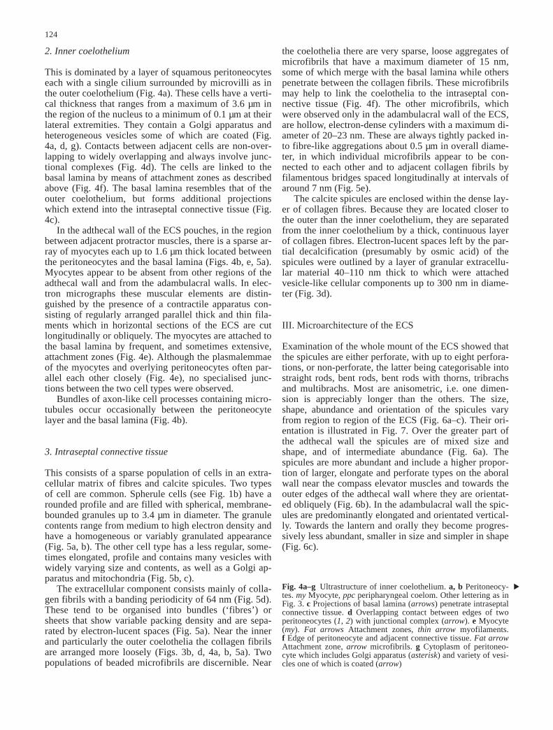
This is dominated by a layer of squamous peritoneocyteseach with a single cilium surrounded by microvilli as inthe outer coelothelium (Fig. 4a). These cells have a verti-cal thickness that ranges from a maximum of 3.6 µm inthe region of the nucleus to a minimum of 0.1 µm at theirlateral extremities. They contain a Golgi apparatus andheterogeneous vesicles some of which are coated (Fig.4a, d, g). Contacts between adjacent cells are non-over-lapping to widely overlapping and always involve junc-tional complexes (Fig. 4d). The cells are linked to thebasal lamina by means of attachment zones as describedabove (Fig. 4f). The basal lamina resembles that of theouter coelothelium, but forms additional projectionswhich extend into the intraseptal connective tissue (Fig.4c).
In the adthecal wall of the ECS pouches, in the regionbetween adjacent protractor muscles, there is a sparse ar-ray of myocytes each up to 1.6 µm thick located betweenthe peritoneocytes and the basal lamina (Figs. 4b, e, 5a).Myocytes appear to be absent from other regions of theadthecal wall and from the adambulacral walls. In elec-tron micrographs these muscular elements are distin-guished by the presence of a contractile apparatus con-sisting of regularly arranged parallel thick and thin fila-ments which in horizontal sections of the ECS are cutlongitudinally or obliquely. The myocytes are attached tothe basal lamina by frequent, and sometimes extensive,attachment zones (Fig. 4e). Although the plasmalemmaeof the myocytes and overlying peritoneocytes often par-allel each other closely (Fig. 4e), no specialised junc-tions between the two cell types were observed.
Bundles of axon-like cell processes containing micro-tubules occur occasionally between the peritoneocytelayer and the basal lamina (Fig. 4b).
3. Intraseptal connective tissue
This consists of a sparse population of cells in an extra-cellular matrix of fibres and calcite spicules. Two typesof cell are common. Spherule cells (see Fig. 1b) have arounded profile and are filled with spherical, membrane-bounded granules up to 3.4 µm in diameter. The granulecontents range from medium to high electron density andhave a homogeneous or variably granulated appearance(Fig. 5a, b). The other cell type has a less regular, some-times elongated, profile and contains many vesicles withwidely varying size and contents, as well as a Golgi ap-paratus and mitochondria (Fig. 5b, c).
The extracellular component consists mainly of colla-gen fibrils with a banding periodicity of 64 nm (Fig. 5d).These tend to be organised into bundles (‘fibres’) orsheets that show variable packing density and are sepa-rated by electron-lucent spaces (Fig. 5a). Near the innerand particularly the outer coelothelia the collagen fibrilsare arranged more loosely (Figs. 3b, d, 4a, b, 5a). Twopopulations of beaded microfibrils are discernible. Near
the coelothelia there are very sparse, loose aggregates ofmicrofibrils that have a maximum diameter of 15 nm,some of which merge with the basal lamina while otherspenetrate between the collagen fibrils. These microfibrilsmay help to link the coelothelia to the intraseptal con-nective tissue (Fig. 4f). The other microfibrils, whichwere observed only in the adambulacral wall of the ECS,are hollow, electron-dense cylinders with a maximum di-ameter of 20–23 nm. These are always tightly packed in-to fibre-like aggregations about 0.5 µm in overall diame-ter, in which individual microfibrils appear to be con-nected to each other and to adjacent collagen fibrils byfilamentous bridges spaced longitudinally at intervals ofaround 7 nm (Fig. 5e).
The calcite spicules are enclosed within the dense lay-er of collagen fibres. Because they are located closer tothe outer than the inner coelothelium, they are separatedfrom the inner coelothelium by a thick, continuous layerof collagen fibres. Electron-lucent spaces left by the par-tial decalcification (presumably by osmic acid) of thespicules were outlined by a layer of granular extracellu-lar material 40–110 nm thick to which were attachedvesicle-like cellular components up to 300 nm in diame-ter (Fig. 3d).
III. Microarchitecture of the ECS
Examination of the whole mount of the ECS showed thatthe spicules are either perforate, with up to eight perfora-tions, or non-perforate, the latter being categorisable intostraight rods, bent rods, bent rods with thorns, tribrachsand multibrachs. Most are anisometric, i.e. one dimen-sion is appreciably longer than the others. The size,shape, abundance and orientation of the spicules varyfrom region to region of the ECS (Fig. 6a–c). Their ori-entation is illustrated in Fig. 7. Over the greater part ofthe adthecal wall the spicules are of mixed size andshape, and of intermediate abundance (Fig. 6a). Thespicules are more abundant and include a higher propor-tion of larger, elongate and perforate types on the aboralwall near the compass elevator muscles and towards theouter edges of the adthecal wall where they are orientat-ed obliquely (Fig. 6b). In the adambulacral wall the spic-ules are predominantly elongated and orientated vertical-ly. Towards the lantern and orally they become progres-sively less abundant, smaller in size and simpler in shape(Fig. 6c).
124
Fig. 4a–g Ultrastructure of inner coelothelium. a, b Peritoneocy-tes. my Myocyte, ppc peripharyngeal coelom. Other lettering as inFig. 3. c Projections of basal lamina (arrows) penetrate intraseptalconnective tissue. d Overlapping contact between edges of twoperitoneocytes (1, 2) with junctional complex (arrow). e Myocyte(my). Fat arrows Attachment zones, thin arrow myofilaments.f Edge of peritoneocyte and adjacent connective tissue. Fat arrowAttachment zone, arrow microfibrils. g Cytoplasm of peritoneo-cyte which includes Golgi apparatus (asterisk) and variety of vesi-cles one of which is coated (arrow)
▲

125
Fig. 4a–g

126
Fig. 5a–e

127
Fig. 6a–j

In the adthecal wall connective tissue fibres ensheatheach spicule (Fig. 6d, e) and directly link together adja-cent spicules (Fig. 6g). Fibres in the continuous layer be-tween the spicules and the inner coelothelium form an ir-regular, oblique crossed-fibre lattice (Fig. 6h). In the out-er part of the adambulacral wall the connective tissue fi-bres form a more regular lattice with the fibres intersect-ing the vertical at a mean angle of 19° and with thesparse, elongated spicules aligned roughly parallel to thevertical bias (Fig. 6f). Towards the oral side of the adam-bulacral wall, tracts of vertical fibres become increasing-ly prominent.
In the whole mounts of ECS stained by Milligan’s tri-chrome method, the myocytes are identifiable as roughlyparallel arrays of very fine, strongly acidophilic fibres(Fig. 6i, j). Their orientation varies as shown in Fig. 7.Near the centre of the interprotractor region they areroughly horizontal, whereas near the protractor musclesthey slope upwards towards these muscles, and towardsthe oral apex two sets of sloping fibres form an obliquelattice. Thus, as a result of their mutual orientation, themyocytes cross the longitudinal axes of the spicules atangles generally within the range 45–90°.
IV. Mechanical behaviour of the ECS
Treatment with seawater containing acetylcholine or anelevated K+ concentration caused isolated ECS prepara-tions to generate a contractile force. Preparations devel-oped force more rapidly in response to acetylcholinethan to K+: the maximum force of contraction was
128
Fig. 5a–e Ultrastructure of intraseptal connective tissue. a Lowmagnification. co Bundles of collagen fibrils, my myocyte of innercoelothelium, sc spherule cell. b Spherule cell and cell containingheterogeneous vesicles (hv). c Cell containing heterogeneous vesi-cles. g Golgi apparatus, b basal lamina of inner coelothelium. dCollagen fibrils sectioned longitudinally and transversely. e Bun-dle of microfibrils which are hollow (arrow) and connected toeach other and to collagen fibrils by filamentous bridges (arrow-heads)Fig. 6a–j Microarchitecture of ECS. Light micrographs of wholeunsectioned preparations. Top of each photograph is aboral and itslateral margins are parallel to vertical anatomical axis. a–c Un-stained preparation observed by differential interference contrastshowing spicules at three locations: aboral region of adthecal ECSmidway between its attachments to demiepiphyses (a), lateral re-gion of adthecal ECS at lower level than previous, between pro-tractor muscle (p) and compass depressor (cd) (b), and adambulac-ral ECS, lantern towards right (c). d More highly magnified viewof spicule located near centre of a observed by differential inter-ference contrast. e Same field of view as in d after staining withMilligan’s trichrome, showing connective tissue fibres (arrows)that ensheath spicule. f Adambulacral region of unstained prepara-tion viewed between crossed polarisers to reveal sparse spicules(arrowheads) and regular crossed-fibre lattice of collagen fibres(arrow). g Adjacent spicules (stars) are interconnected by bundlesof connective tissue fibres (arrows). h Continuous layer of colla-gen fibres between spicules and inner epithelium. i Parallel arraysof myocytes (arrow) crossing spicules (stars) in upper region ofadthecal ECS near protractor muscle (located to left). j As for i butat lower level where myocytes are inclined more steeply
▲▲
Fig. 7 Orientation of spicules and myocytes in adthecal ECS. Dia-grammatic representation of one interambulacral region of lantern(based on Fig. 1a). Short thick lines indicate orientation of longaxes of anisometric spicules in right side only of ECS. Long thinlines indicate equivalent for myocytes in whole interprotractor ar-ea. Both sets of lines show dominant orientation of spicules ormyocytes at any particular location and convey no informationabout spicule or myocyte density. ap Apophysis, c compass ossi-cle, cd compass depressor ligament, ce compass elevator muscle, dattachment of ECS to demiepiphysis, p edge of protractor muscle
Fig. 8 Dose-response curves for acetylcholine (ACh; a) and K+
ions (K; b). Contractile force is expressed as percentage of maxi-mum developed by each preparation in each experiment. Eachpoint is mean of four values. Error bars are standard deviations

reached after exposure to acetylcholine for 9–47 s (mean± SD 25±10 s, n=16) and to elevated K+ concentrationsfor 59–218 s (131±52 s, n=14).
Figure 8 shows the results of the dose-response exper-iments. Whereas the curve for K+ was sigmoidal and in-dicated an optimum concentration of 80 mM, that foracetylcholine was exponential.
The relationship between the vertical position of thelantern and the passive stretch resistance and contractileforce developed by the ECS is illustrated in Fig. 9. Asthe lantern was progressively raised, thus graduallystretching the ECS, the stretch resistance increased con-tinuously in all five preparations (Fig. 9a), whereas infour preparations the contractile force dropped initiallythen increased to a peak which occurred at a retraction of4.0–7.6%TD (mean ± SD 6.2±1.3%TD). One prepara-tion did not show the initial drop in contractile force(Fig. 9b). Maximum contractile forces produced by ECSpreparations and by protractor muscle sets are comparedin Fig. 10. Despite the wide spread of both sets of datapoints, it appears that the vertical component of the con-tractile force generated by the ECS is close in magnitudeto that produced by the protractor muscles.
The two preparations that were subjected to loading-unloading cycles generated J-shaped loading curves, in-dicating that, as the ECS was progressively stretched, itsstiffness was at first low and then increased steeply, thetransition between low and high stiffness occurring at a
129
Fig. 9 Relationship between lantern position, passive stretch re-sistance (a) and contractile force (b) in five ECS preparations eachidentified by same symbols and line style as in a and b. Lanternretraction is normalised as a percentage of the test diameter (TD)
Fig. 10 Comparison of vertical force generated by preparationsconsisting of ECS alone or of intact protractor muscle sets alone(data on latter from Wilkie et al. 1998a) from animals of differentsizes
Fig. 11a, b Results of loading-unloading tests. a Two consecutivetests on one untreated preparation. b Three consecutive tests onone preparation: 1 untreated, 2 immediately after treatment with1 mM acetylcholine, 3 after subsequent wash with pure seawater

lantern retraction of –0.5–0 mm. When subjected to twoconsecutive cycles, the preparations were less stiff dur-ing the second cycle, except at maximum lantern dis-placement, and showed less hysteresis, i.e. energy lossdue to internal viscous mechanisms (represented by thearea between the loading and unloading curves; Fig.11a). Treatment with 1 mM acetylcholine caused an in-crease in the stiffness of the ECS over the whole lanterndisplacement range which was reversed partially bywashing in seawater (Fig. 11b).
D. Discussion
I. Ultrastructure
1. Coelothelia
Like other mesenteries (Gotloib et al. 1983) the ECS ofthe sea-urchin lantern consists of a layer of connectivetissue sandwiched between two coelothelia. In the ECSof S. affinis the peritoneocytes of the inner and outercoelothelia share morphological features with the peri-toneocytes of other Echinodermata and other Deuterosto-mia: (1) they have a single cilium surrounded by a ‘col-lar’ of microvilli, (2) adjacent cells are connected at api-cal junctional complexes, (3) they are attached directlyto the basal lamina and (4) they contain heterogeneousmembrane-bounded vesicles indicating endocytosis andpossibly autophagy of cytoplasmic organelles (Nørrevangand Wingstrand 1970; Candia Carnevali et al. 1995;Welsch 1995).
Although secretory activity has been noted before inthe ECS coelothelia (Candia Carnevali et al. 1995) and isa common feature of the peritoneocytes of Deuterosto-mia (Gotloib et al. 1983; Welsch 1995; Dobbie andAnderson 1996), no definite evidence for this was seenin our material.
It has been observed previously that the inner coelo-thelium of the ECS includes basally located myocytesand it has been proposed that the lantern muscles are de-rived from the proliferation of these cells within folds ofthe coelothelium (Stauber 1993; Candia Carnevali et al.1995). In the present investigation our ultrastructural ob-servations and our examination of the Milligan’s tri-chrome-stained whole mounts indicated that myocytesare restricted to that part of the adthecal ECS wall lyingbetween adjacent protractor muscles.
2. Intraseptal connective tissue
Two cell types are common in this layer: spherule cellsand cells containing heterogeneous vesicles. Spherulecells are often found in echinoderm connective tissue.They are involved in the encapsulisation of foreign par-ticles, possibly have a role in wound repair and containbactericidal agents (Smith 1981). The second cell typeis also ubiquitous in echinoderm connective tissue and
often appears to have a fibroclastic function (Wilkie1984). The rather undifferentiated appearance of the lat-ter cells, together with the absence of obvious fibro-blasts, suggests that they are likely to be functionallypluripotential.
Notably absent from this layer were axons and cellsor cell processes containing large electron-dense gran-ules, both of which have been found in most echinodermcollagenous structures studied to date. Although largegranule-containing cells may have more than one func-tion, at least some of them are likely to be the effectorcells that modify directly the stiffness of echinodermmutable collagenous tissues (Trotter and Koob 1995;Wilkie 1996; Koob et al. 1999). These cells do not occurin certain collagenous structures that, on the basis of ex-perimental findings, appear to lack mutability, such asthe compass-rotular ligament in the lantern of Paracen-trotus lividus (Wilkie et al. 1995; Wilkie and CandiaCarnevali unpublished observations), and their absencefrom the intraseptal connective tissue suggests that thepassive mechanical properties of this layer are also notunder physiological control.
The extracellular component of the intraseptal layerconsists mainly of bundles of collagen fibrils associatedwith rarer beaded microfibrils of which there are twopopulations, one forming a loose array of connectionswith the coelothelial basal lamina and the other fibre-likeaggregations in the adambulacral wall of the ECS. Com-parable populations of microfibrils are common in othercollagenous structures of Echinodermata, particularlythose of the sea-urchin lantern (Wilkie et al. 1992, 1994,1998b), though our investigation seems to be the first tohave detected a size difference between the two, whichmay denote a difference in their composition and me-chanical function. In most connective tissues of Echino-dermata, individual collagen fibres (i.e. fibril bundles)are surrounded by a loose network of microfibrils(Wilkie 1996), a feature that was not observed in theECS. These microfibrils resemble the elastin-associatedmicrofibrils of Mammalia in their morphology and, as-suming that those of holothurian dermis are representa-tive, in containing a glycoprotein of the fibrillin familyand forming assemblages that exhibit long-range elastici-ty (Thurmond and Trotter 1996; Thurmond et al. 1997).The restriction of the fibre-like microfibril bundles to theadambulacral walls of the ECS thus implies that this re-gion has a greater capacity for passive strain recoverythan the interambulacral wall.
II. Microarchitecture and mechanical behaviour
1. Adthecal regions
The mechanical behaviour of the ECS depends predomi-nantly on the calcite spicules and connective tissue fi-bres, which are the primary determinants of its passivetensile properties, and the myocytes, which confer con-tractility.
130

The influence of spicules on the tensile stiffness ofpliable connective tissues has been analysed thoroughlyby Koehl (1982). In general, spicules have a stiffeningeffect the magnitude of which is correlated positivelywith spicule volume fraction and surface area/volume ra-tio, correlated negatively with spicule size (for a givenvolume fraction), and is greater parallel to the long axisof anisometric spicules. It can thus be inferred that thepresence of spicules in the adthecal region of each ECSpouch increases its overall stretch resistance, and that,due to the predominantly horizontal to oblique orienta-tion of the spicules, this stiffening effect is least pro-nounced in the vertical direction. Although the lattercould be interpreted as an adaptation to permit the ECSto stretch vertically when the lantern is raised, two fac-tors suggest that this does not happen in vivo. First, thepassive force-displacement tests indicated that the transi-tion between the low and high stiffness phases occurswhen the lantern is near the resting position, which sug-gests that the ECS is adapted to resist extension beyondthis level. Second, previous work on S. affinis showedthat the lantern is very immobile in comparison with thatof Echinidae such as P. lividus. For example, because ofthe relative positions of their insertions on the lanternand perignathic girdle, the retractor muscles cannot ele-vate the lantern more than 1–2%TD above the restingposition (Andrietti et al. 1993), and the mechanical prop-erties of the compass depressor ligaments indicate thatthey resist lantern retraction above the resting position(Wilkie et al. 1998b). Thus, although the pattern ofspiculation in the adthecal ECS wall will result in tensileanisotropy, this is probably an incidental consequence ofa microarchitectural arrangement determined by otherfunctional demands, since it is unlikely that the ECS hasto stretch in vivo beyond its dimensions at the restingposition. It is perhaps more relevant that the spicules willalso cause compressive and flexural anisotropy whichmight facilitate folding of the ECS when it slackens dur-ing either: (1) lowering of the whole lantern below theresting position (although S. affinis can achieve a maxi-mum protraction of only 3%TD due to the resistance ofthe heavily calcified peristomial membrane (Wilkie et al.1996) or (2) lowering of only the compass ossicles bycontraction of the compass depressors. Indeed, when thecompasses of an intact lantern were pressed down artifi-cially to mimic the effect of compass depressor contrac-tion, those regions of the ECS between the attachmentsto the compasses and the demiepiphyses buckled in aconstant way with the folds parallel to the long axes ofthe spicules (personal observations).
Vertical force generation by the ECS must dependprimarily on the obliquely orientated myocytes, sincetheir contraction, but not that of the horizontal myo-cytes, will have a vertical component. The transversealignment of the myocytes with respect to the overlyingspicules ensures that when they contract they will tendto pull the spicules together laterally rather than longitu-dinally, i.e. in the direction of least resistance to com-pression. The myocytes responsible for ECS contractili-
ty are restricted to the interprotractor regions of the ad-thecal walls. Since the interprotractor regions are at-tached aborally to the upper edges of the lantern demi-epiphyses and orally to the apophyses, contraction ofthese regions exerts a direct downward pull on the lan-tern and thus assists the action of the protractor muscles.For this to be possible, there must be strong mechanicalcoupling between the myocytes and the intraseptal con-nective tissue through which the ECS is attached to thelantern and apophyses. The ultrastructural investigationrevealed that this is achieved by: (1) the extensive at-tachment zones between the myocytes and the coelothe-lial basal lamina and (2) the linkage of the basal laminato the intraseptal connective tissue by microfibrils anddeeply penetrating extensions of the basal lamina itself.It is notable that the attachment zones resemble myoten-dinous junctions in other Echinodermata (Wilkie andEmson 1987; Stauber and Märkel 1988) and in otherphyla (Trotter et al. 1983) where, as in the ECS, thejunction is subjected much more to shear (in-plane)stress than tensile stress.
The active force-lantern displacement curves pro-duced in these experiments are effectively force-startinglength curves. As such they are unusual in comparisonwith those of most other contractile structures includingthe protractor muscles and compass depressors of S. affi-nis (Wilkie et al. 1998a, b) in which, as the structure isprogressively stretched, the active force rises to a maxi-mum and then declines. The ECS pattern, in which theactive force initially decreases then increases to a rela-tively low maximum (at a lantern displacement that isprobably never attained in vivo) before decreasing again,may be due in part to the variable orientation of the myo-cytes which will result in different myocytes reachingtheir optimal length for force generation at different lan-tern positions. However, we cannot yet provide a full ex-planation for this behaviour or its functional signifi-cance.
Acetylcholine increases the stretch resistance of theECS. This might be due entirely to active force genera-tion by the myocytes. However, in view of the ubiquityof mutable collagenous tissues in Echinodermata (Wilkie1996), the possibility that it results partly from nervouslyinduced stiffening of the intraseptal connective tissue hasto be considered. Although it has already been noted thatthe absence of putative effector cells in the intraseptallayer makes it highly unlikely that the stiffness of the tis-sue is under physiological control, this needs to be con-firmed by further investigation, perhaps involving theaccurate measurement of passive and active stress(force/cross-sectional area), which was not possible inthe experiments described herein.
2. Adambulacral regions
In comparison with the adthecal regions the adambulac-ral walls are more sparsely spiculated, non-contractile(there being no myocytes) and probably more elastic
131
(due to the presence of microfibril bundles). We specu-late that the primary function of these regions is respira-tory rather than mechanical. Vertical movements of thecompasses of S. affinis are believed to aid the circulationof fluid within the peripharyngeal coelomic compart-ments (Wilkie et al. 1998b) and, via changes in hydro-static pressure, probably cause the adambulacral walls toexpand and contract passively. Such behaviour was ob-served when the compasses of an intact lantern were arti-ficially manipulated: compass depression was accompa-nied by bulging out of the adambulacral walls and thesewere ‘sucked’ back in again during compass elevation(personal observations). These movements would requirethe adambulacral walls to have low flexural stiffness,which may provide a functional explanation for the re-duced spiculation of these regions. Since the ‘billowing’of the adambulacral walls will be accompanied by fluidflow over their outer and inner surfaces, these regions ofthe ECS could facilitate the transfer of respiratory gasesbetween the perivisceral fluid and the peripharyngealfluid bathing the lantern muscles, a role that has alsobeen ascribed to the thin-walled lobes that project fromthe oral side of the Stewart’s organs (De Ridder 1988). Itmay be relevant that both the lower part of the adambul-acral ECS regions and the oral lobes of the Stewart’s or-gans lie close to tube-foot ampullae, since, amongst oth-er functions, the tube-feet act as external gills (Smith1978; Shick 1983). Passive movements of the adambul-acral ECS walls may thus promote the delivery to thelantern muscles of oxygen acquired initially by the tube-feet.
III. Conclusions
As a result of its passive mechanical properties the ECSsets a limit to lantern retraction, whilst its contractileproperties assist the action of the protractor muscles. Af-ter a biomechanical analysis of the lantern of S. affinis,Andrietti et al. (1993) came to the surprising conclusionthat the fundamental role of the protractor and retractormuscles is to stabilise the position of the lantern, not tomove it (although these muscles will contribute to masti-catory movements of the jaws). Thus it is likely that boththe passive and active properties of the adthecal ECScontribute to lantern stabilisation. It has also been dem-onstrated that the compass depressor ligaments and peri-stomial membrane of S. affinis restrict lantern mobility(Andrietti et al. 1993; Wilkie et al. 1996, 1998b). Thefact that its immobility is assured by such a large numberof features indicates that this must be a dominant aspectof the functioning of the lantern in Cidaridae, althoughhow it relates to the lantern’s role in food collection andhandling or in other physiological activities will not berevealed until more is known about the basic biology ofthese sea-urchins.
Acknowledgements The research described in this paper re-ceived financial support from the Consiglio Nazionale delle Ricer-che, Rome, the Royal Society, London, and Glasgow CaledonianUniversity.
References
Andrietti F, Candia Carnevali MD, Wilkie IC (1993) A biome-chanical comparison of the lantern of the cidarid sea-urchinStylocidaris affinis with the typical camarodont lantern. J ZoolLond 231:595–610
Birenheide R (1992) The sea urchin lantern coelom: a circulatorysystem. In: Scalera-Liaci L, Canicattì C (eds) Echinoderm re-search 1991. Balkema, Rotterdam, pp 67–72
Candia Carnevali MD, Wilkie IC, Lucca E, Andrietti F, Melone G(1993) The Aristotle’s lantern of the sea-urchin Stylocidarisaffinis (Echinoida, Cidaridae): functional morphology of themusculo-skeletal system. Zoomorphology 113:173–189
Candia Carnevali, MD, Bonasoro F, Wilkie IC (1995) Coelom and‘tinkering’ in echinoids: morpho-functional adaptations of thelantern coelom. In: Lanzavecchia G, Valvassori R, CandiaCarnevali MD (eds) Body cavities: function and phylogeny.Mucchi, Modena, pp 135–165
De Ridder C (1988) Could the Stewart’s organs of cidaroidechinoids be internal gills? In: Burke RD, Mladenov PV,Lambert P, Parsley RL (eds) Echinoderm biology. Balkema,Rotterdam, pp 675–681
Dobbie JW, Anderson JA (1996) Ultrastructure, distribution anddensity of lamellar bodies in human peritoneum. Perit Dial Int16:482–487
Gotloib L, Digenis GE, Rabinovich S, Medline A, Oreopoulos D(1983) Ultrastructure of normal rabbit mesentery. Nephron34:248–255
Hildebrandt J, Fukaya H, Martin CJ (1969) Stress-strain relationsof tissue sheets undergoing uniform two-dimensional stretch. JAppl Physiol 27:758–762
Holzer-Petsche U, Brodacz B (1999) Traction on the mesentery asa model of visceral nociception. Pain 80:319–328
Humason GL (1979) Animal tissue techniques, 4th edn. Freeman,San Francisco
Koehl MAR (1982) Mechanical design of spicule-reinforced con-nective tissue: stiffness. J Exp Biol 98:239–267
Koob TJ, Koob-Emunds MM, Trotter JA (1999) Cell-derived stiff-ening and plasticizing factors in sea cucumber (Cucumariafrondosa) dermis. J Exp Biol 202:2291–2301
Nebot-Cegarra J, Macarulla-Sanz E, Reina-de la Torre F (1999)Factors involved in the ‘rotation’ of the human embryonicstomach around its longitudinal axis: computer-assisted mor-phometric analysis. J Anat 194:61–69
Nørrevang A, Wingstrand KG (1970) On the occurrence and struc-ture of choanocyte-like cells in some echinoderms. Acta Zool51:249–270
Parameswaran S, Brown LV, Lai-Fook SJ (1998) Effect of flow onhydraulic conductivity and reflection coefficient of rabbit me-sentery. Microcirculation 5:265–274
Rieger RM, Lombardi J (1987) Ultrastructure of coelomic lining inechinoderm podia: significance for concepts in the evolution ofmuscle and peritoneal cells. Zoomorphology 107:191–208
Shick JM (1983) Respiratory gas exchange in echinoderms. In:Jangoux M, Lawrence JM (eds) Echinoderm studies, vol 1.Balkema, Rotterdam, pp 67–110
Smith AB (1978) A functional classification of the coronal poresof regular echinoids. Palaeontology 21:759–789
Smith VJ (1981) The echinoderms. In: Ratcliffe NA, Rowley AF(eds) Invertebrate blood cells, vol 2. Academic Press, London,pp 513–562
Stauber M (1993) The lantern of Aristotle: organization of its coe-lom and origin of its muscles (Echinodermata, Echinoida).Zoomorphology 113:137–151
Stauber M, Märkel K (1988) Comparative morphology of muscle-skeleton attachments in the Echinodermata. Zoomorphology108:137–148
Thurmond FA, Trotter JA (1996) Morphology and biomechanicsof the microfibrillar network of sea cucumber dermis. J ExpBiol 199:1817–1828
Thurmond FA, Koob TJ, Bowness JM, Trotter JA (1997) Partialbiochemical and immunologic characterization of microfibrilsfrom sea cucumber dermis. Connect Tissue Res 36:211–222
132

Trotter JA, Koob TJ (1995) Evidence that calcium-dependent cel-lular processes are involved in the stiffening response of ho-lothurian dermis and that dermal cells contain an organic stiff-ening factor. J Exp Biol 198:1951–1961
Trotter JA, Eberhard S, Samora A (1983) Structural connectionsof the muscle-tendon junction. Cell Motil 3:431–438
Welsch U (1995) Evolution of the body cavities in Deuterostomia.In: Lanzavecchia G, Valvassori R, Candia Carnevali MD (eds)Body cavities: function and phylogeny. Mucchi, Modena, pp111–134
Wilkie IC (1984) Variable tensility in echinoderm collagenous tis-sues: a review. Mar Behav Physiol 9:229–248
Wilkie IC (1996) Mutable collagenous tissues: extracellular matrixas mechano-effector. In: Jangoux M, Lawrence JM (eds) Echi-noderm studies, vol 5. Balkema, Rotterdam, pp 61–102
Wilkie IC, Emson RH (1987) The tendons of Ophiocomina nigraand their role in autotomy (Echinodermata, Ophiuroida). Zoo-morphology 107:33–44
Wilkie IC, Candia Carnevali MD, Bonasoro F (1992) The com-pass depressors of Paracentrotus lividus (Echinodermata,Echinoida): ultrastructural and mechanical aspects of theirvariable tensility and contractility. Zoomorphology 112:143–153
Wilkie IC, Candia Carnevali MD, Andrietti F (1994) Microarchi-tecture and mechanics of the sea-urchin peristomial mem-brane. Boll Zool 61:39–51
Wilkie IC, McKew M, Candia Carnevali MD (1995) Anomalousphysico-chemical properties of the compass-rotular ligamentsin two species of sea-urchins: preliminary observations. In:Emson RH, Smith AB, Campbell AC (eds) Echinoderm re-search 1995. Balkema, Rotterdam, pp 147–152
Wilkie IC, Candia Carnevali MD, Andrietti F (1996) Mechanicalproperties of the peristomial membrane of the cidaroid sea-ur-chin Stylocidaris affinis. J Zool Lond 238:557–569
Wilkie IC, Candia Carnevali MD, Andrietti F (1998a) Mechanicalproperties of sea-urchin lantern muscles: a comparative inves-tigation of intact muscle groups in Paracentrotus lividus(Lam.) and Stylocidaris affinis (Phil.) (Echinodermata,Echinoidea). J Comp Physiol B 168:204–212
Wilkie IC, Candia Carnevali MD, Bonasoro F (1998b) Organiza-tion and mechanical behaviour of myocyte-ligament compos-ites in a sea-urchin lantern: the compass depressors of Stylo-cidaris affinis (Echinodermata, Echinoida). Zoomorphology118:87–101
133
Related Documents