A SIX-MONTH-OLD INFANT WITH LIVER STEATOSIS Michael O. Stormon, MBBS,FRACP, Ernest Cutz, MD, FRCPC, Katryn Furuya, MD, FRCPC, Melanie Bedford, MD, FRCPC, Laura Yerkes, MS,MA, Dean R. Tolan, PHD, and Annette Feigenbaum, MB,CHB, FRCPC Division of Gastroenterology and Nutrition, Division of Metabolic Genetics, Division of Pathology and Department of Pediatric Laboratory Medicine, The Hospital for Sick Children, University of Toronto, Ontario, Canada; and the Department of Biology, Boston University, Boston, Massachusetts. A6-month-old male infant was sent to a quaternary referral hospital from a tertiary referral hospital with a possible diagnosis of a fatty acid oxidation disorder. HISTORY OF PRESENTING ILLNESS The infant was well until 4 weeks before admission at the quaternary referral hospital. He, along with the rest of his family, experienced an acute gastroenteritic illness with vomiting and watery diarrhea. Although his siblings and parents recovered, he did not, and because of concerns of a presumed secondary lactose intolerance he was changed from his usual Similac (Ross Products, Abbott Laboratories, Columbus, Oh) formula to a soy-based formula (Alsoy, Nestle Inc, Eau Claire, Wisc) about one week into his illness. He became more lethargic and hypotonic with persisting vomiting and diarrhea, and was admitted to a tertiary referral hospital. PAST HISTORY He was born to nonconsanguineous parents of Italian (father) and Hungarian-French (mother) descent. He had two female siblings both of whom had been well in the past, and there was no family history of notable diseases. During pregnancy his mother had a laparoscopic cholecystectomy at 20 weeks’ gestation, and he was born by Cesarean delivery at term because of fetal bradycardia. There were no other problems in the perinatal period. He had been well before this current illness, with appropriate growth parameters and development. He was fed a combination of breast milk and formula (Similac, Ross Pharmaceuticals). Cereals were introduced at 5 months along with other age-appropriate foods such as bananas and some vegetables. TERTIARY REFERRAL HOSPITAL Symptoms of diarrhea and poor feeding had persisted for approximately two weeks before admission to the tertiary referral hospital, where he was found to be weak, floppy, and lethargic but easily rousable. He had massive hepatomegaly, with the liver edge palpable in the right iliac fossa, which had not been previously noted on several abdominal examinations. There was no splenomegaly, ascites, or signs of chronic liver disease. He was not clinically jaundiced despite elevated conjugated bilirubin. An Escherichia coli urinary tract infection (UTI) was Copyright © 2004 Elsevier Inc. All rights reserved. Reprint requests: Annette Feigenbaum, MB, ChB, FRCPC, Division Metabolic Genetics, Hospital for Sick Children, University of Toronto, 555 University Ave, Toronto, Ontario M5G 1×8, Canada. [email protected]. . NIH Public Access Author Manuscript J Pediatr. Author manuscript; available in PMC 2010 October 14. Published in final edited form as: J Pediatr. 2004 February ; 144(2): 258–263. doi:10.1016/j.jpeds.2003.11.037. NIH-PA Author Manuscript NIH-PA Author Manuscript NIH-PA Author Manuscript

Welcome message from author
This document is posted to help you gain knowledge. Please leave a comment to let me know what you think about it! Share it to your friends and learn new things together.
Transcript

A SIX-MONTH-OLD INFANT WITH LIVER STEATOSIS
Michael O. Stormon, MBBS,FRACP, Ernest Cutz, MD, FRCPC, Katryn Furuya, MD, FRCPC,Melanie Bedford, MD, FRCPC, Laura Yerkes, MS,MA, Dean R. Tolan, PHD, and AnnetteFeigenbaum, MB,CHB, FRCPCDivision of Gastroenterology and Nutrition, Division of Metabolic Genetics, Division of Pathologyand Department of Pediatric Laboratory Medicine, The Hospital for Sick Children, University ofToronto, Ontario, Canada; and the Department of Biology, Boston University, Boston,Massachusetts.
A6-month-old male infant was sent to a quaternary referral hospital from a tertiary referralhospital with a possible diagnosis of a fatty acid oxidation disorder.
HISTORY OF PRESENTING ILLNESSThe infant was well until 4 weeks before admission at the quaternary referral hospital. He,along with the rest of his family, experienced an acute gastroenteritic illness with vomiting andwatery diarrhea. Although his siblings and parents recovered, he did not, and because ofconcerns of a presumed secondary lactose intolerance he was changed from his usual Similac(Ross Products, Abbott Laboratories, Columbus, Oh) formula to a soy-based formula (Alsoy,Nestle Inc, Eau Claire, Wisc) about one week into his illness. He became more lethargic andhypotonic with persisting vomiting and diarrhea, and was admitted to a tertiary referral hospital.
PAST HISTORYHe was born to nonconsanguineous parents of Italian (father) and Hungarian-French (mother)descent. He had two female siblings both of whom had been well in the past, and there was nofamily history of notable diseases. During pregnancy his mother had a laparoscopiccholecystectomy at 20 weeks’ gestation, and he was born by Cesarean delivery at term becauseof fetal bradycardia. There were no other problems in the perinatal period. He had been wellbefore this current illness, with appropriate growth parameters and development. He was feda combination of breast milk and formula (Similac, Ross Pharmaceuticals). Cereals wereintroduced at 5 months along with other age-appropriate foods such as bananas and somevegetables.
TERTIARY REFERRAL HOSPITALSymptoms of diarrhea and poor feeding had persisted for approximately two weeks beforeadmission to the tertiary referral hospital, where he was found to be weak, floppy, and lethargicbut easily rousable. He had massive hepatomegaly, with the liver edge palpable in the rightiliac fossa, which had not been previously noted on several abdominal examinations. Therewas no splenomegaly, ascites, or signs of chronic liver disease. He was not clinically jaundiceddespite elevated conjugated bilirubin. An Escherichia coli urinary tract infection (UTI) was
Copyright © 2004 Elsevier Inc. All rights reserved.Reprint requests: Annette Feigenbaum, MB, ChB, FRCPC, Division Metabolic Genetics, Hospital for Sick Children, University ofToronto, 555 University Ave, Toronto, Ontario M5G 1×8, Canada. [email protected]. .
NIH Public AccessAuthor ManuscriptJ Pediatr. Author manuscript; available in PMC 2010 October 14.
Published in final edited form as:J Pediatr. 2004 February ; 144(2): 258–263. doi:10.1016/j.jpeds.2003.11.037.
NIH
-PA Author Manuscript
NIH
-PA Author Manuscript
NIH
-PA Author Manuscript
diagnosed, although fever was not noted. A subsequent micturating cystourethrogram wasnormal.
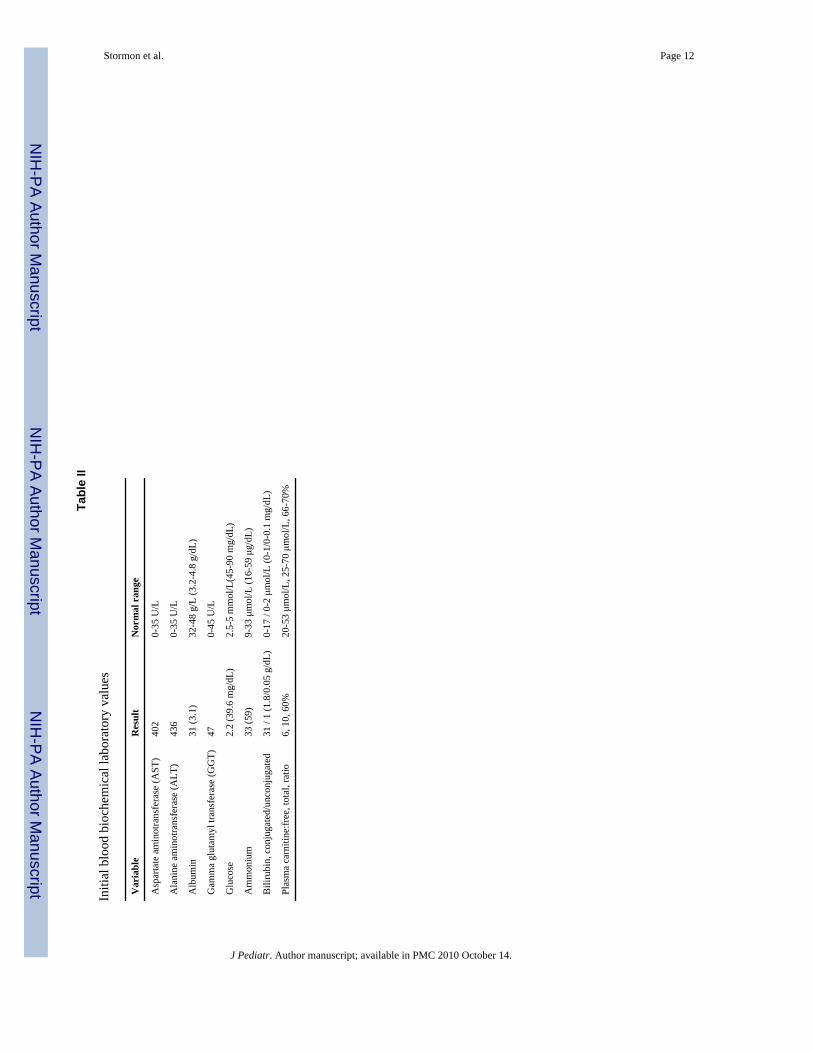
Initial hematology and biochemical results are shown in Tables I and II. Of note is the presenceof hypoglycemia, hypokalemia, hypophosphatemia, low plasma carnitine levels, hepaticdysfunction and coagulopathy. Other investigations that were normal included venous bloodgas, plasma lactate, amino acids, creatine kinase, galactosemia screen, and routine urinalysis(ketones negative). Gas chromatography mass spectroscopy analysis of urinary organic acidsshowed elevated dicarboxylic acids with hypoketosis at the time of hypoglycemia. Urinarysuccinylacetone was negative. Serology for hepatitis viruses A, B, and C was negative. Anechocardiogram demonstrated thickening of the interventricular septum, suggestive of apossible cardiomyopathy. An abdominal ultrasonogram showed an enlarged, heterogeneousliver with increased echogenicity and normal portal flow. A liver biopsy was performed andreported marked steatosis with minimal fibrosis. No specific diagnosis was made, although ametabolic disorder was suspected.
A provisional diagnosis of a fatty acid oxidation disorder (FAOD) was made. Skin fibroblastswere sent for analysis. Carnitine supplementation and feeds with Portagen (Mead JohnsonNutritionals, Indianapolis, Ind), a formula high in medium-chain triglycerides and low in long-chain triglycerides, were started. Referral to a quaternary referral hospital was arranged.
QUATERNARY REFERRAL HOSPITALThe patient had been unwell for nearly four weeks when he was admitted to our center. Hecontinued to have frequent episodes of vomiting and diarrhea on Portagen feeds. As had beennoted before, he was weak, floppy, and lethargic but responded appropriately to hisenvironment. A firm liver edge was palpable 8 cm below the right costal margin. He was stillnot clinically jaundiced and the conjugated hyperbilirubinemia was improving (Tables II andIII). There was no splenomegaly, ascites, or sign of chronic liver disease. Ophthalmologicexamination was normal. Clinical cardiac evaluation was also normal, as was a chestradiograph and electrocardiogram.
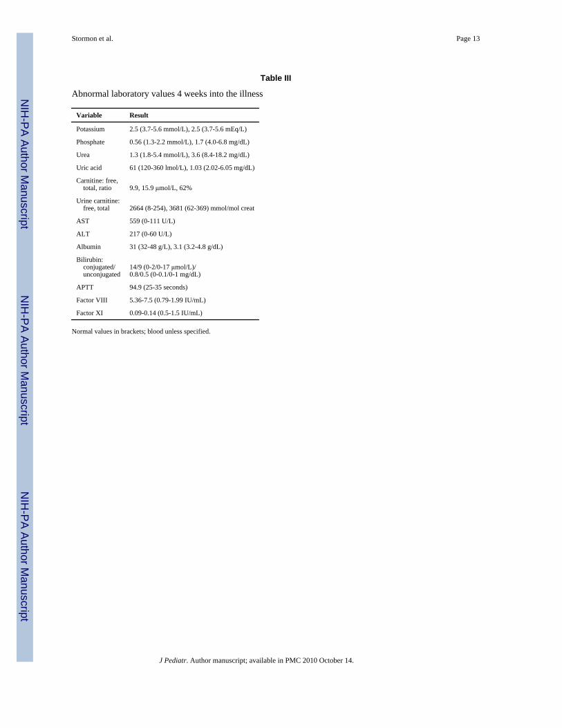
Abnormal laboratory tests done on admission are shown in Table III. Factors V and VII levelswere also low. A venous blood gas showed a compensated metabolic acidosis: pH 7.36, baseexcess −9, bicarbonate 15 mmol/L (15 mEq/L) and CO2 26 (40-50 mm Hg). Urinary sodiumwas 22 mmol/L, potassium 50 mmol/L, and uric acid:creatinine ratio was 2.16 (normal for age,0.92-1.04).1 These findings together with a generalized aminoaciduria and relative elevationof carnitine excretion suggested a proximal renal tubulopathy. Plasma lactate, fasting free fattyacid (0.1 mmol/L), and β-hydroxybutyrate (<0.024 mmol/L) levels and blood acylcarnitineanalysis were normal. Serum alkaline phosphatase and creatinine were normal. Sweat chloridewas normal. Leukocyte acid lipase activity for Wolman disease was also normal. Factor IXlevel and fibrinogen were normal. Factor XI inhibitor was negative. Parental Factor XI levelswere normal.
Abdominal ultrasonogram again showed an enlarged liver with increased echogenicity andnormal portal flow. Kidneys looked normal. A skeletal survey did not show any rachiticchanges, although reduced mineralization was reported “consistent with a chronic process.” Inview of his diarrhea, vomiting, hepatomegaly with liver dysfunction, hypoketotichypoglycemia, and low Factor XI levels, serum was sent for isoelectric focusing (IEF) oftransferrin to exclude carbohydrate-deficient glycoprotein syndrome (or, congenital disorderof glycosylation, CDG).
His clinical condition progressively worsened over the first week of this admission. Theinternational normalized ratio reached a maximum of 1.25 and the activated partial
Stormon et al. Page 2
J Pediatr. Author manuscript; available in PMC 2010 October 14.
NIH
-PA Author Manuscript
NIH
-PA Author Manuscript
NIH
-PA Author Manuscript

thromboplastin time, 101 seconds. His serum albumin fell to 22 g/L, and he developed moderateascites requiring albumin infusions with diuretic therapy. Despite supplementation withappropriate amounts of potassium, bicarbonate, and phosphate he continued to have acompensated metabolic acidosis with hypokalemia and hypophosphatemia. There were severalepisodes of hypoglycemia. He continued on the Portagen feeds with frequent vomiting, andhad as many as 11 loose nonbloody stools daily.
Two weeks later, due to his deteriorating condition and persistent vomiting and diarrhea, hisfeeds were stopped and total parenteral nutrition (TPN) was commenced. With TPN, hisvomiting and diarrhea resolved within several days, and his abnormal biochemical parametersslowly improved. After one week of TPN, feeds were reintroduced using Vivonex Pediatric(Novartis Nutrition Corp, Mississauga, Ontario, Canada) via nasogastric tube. This formulawas chosen in view of the possible FAOD, plus it did not contain sucrose (unlike Portagen),which was thought to be exacerbating his diarrhea. This formula was well tolerated, and a weeklater his electrolytes, liver function tests, and coagulation profile had normalized, and liver sizehad decreased. A second UTI caused by E coli was diagnosed and treatment was started withNovo-Trimel (Trimethoprim-Sulfamethoxazole, Novopharm Ltd, Toronto, Canada) oralsuspension. After one dose of Novo-Trimel the diarrhea recurred and persisted, but settledwhen the drug was stopped. At that time, the IEF transferrin was reported as abnormal,suggesting a possible diagnosis of CDG.
CASE DISCUSSIONThis 6-month-old child presented with massive hepatomegaly, raised liver transaminases,coagulopathy, hypoglycemia (with hypoketosis), and evidence of renal tubular dysfunction.These symptoms followed an episode of gastroenteritis and were associated with the two Ecoli UTIs. His health and development had been previously normal and there was neitherconsanguinuity nor a family history of significant illness.
Subsequent review of the liver biopsy pathology slides at our institution showed the findingsshown in Figure 1. There was also a sparse lymphocytic infiltrate. There was no evidence ofcholestasis. Electron microscopy is shown in Figure 2. There was also slight dilatation of roughendoplasmic reticulum. It showed the presence of peroxisomes, and dark staining mitochondriawithout any inclusions. There were no obvious ultrastructural abnormalities of themitochondrial cristae. Glycogen particles showed usual size and distribution.
Based on this pathology and the investigations, diseases such as galactosemia, glycogen storagediseases, tyrosinemia, urea cycle defects, organic acidemias, and Wolman disease wereexcluded.
One possible diagnosis was still an FAOD. Many features of this child’s illness could beexplained by a defect in mobilizing fatty acids to provide energy in the face of an intercurrentillness. Hepatomegaly with hepatic dysfunction (including coagulopathy), hypoketotichypoglycemia, possible cardiomyopathy, and low carnitine levels are consistent features of anFAOD. However, FAODs are most often associated with an elevated acylcarnitine fraction,fasting free fatty acid levels, and free fatty acid: β-hydroxybutyrate ratio which were not presentin this case. In one form of FAOD, carnitine palmitoyltransferase I (CPT I) deficiency,acylcarnitine concentrations are negligible or low,2 but carnitine levels and free fatty acid: β-hydroxybutyrate ratios are usually high. Thus our case’s profile did not fit this diagnosis. Inaddition, blood acylcarnitine analysis was normal. The urine organic acids showed onlydicarboxylic acids, which could be a feature of medium chain acyl-coenzyme A dehydrogenase(MCAD) deficiency or CPT I deficiency. However, his clinical picture was not that of MCAD,and the urinary dicarboxylic acids were thus considered to be a feature of his carnitine depletion
Stormon et al. Page 3
J Pediatr. Author manuscript; available in PMC 2010 October 14.
NIH
-PA Author Manuscript
NIH
-PA Author Manuscript
NIH
-PA Author Manuscript
due to renal wasting. Other aspects of this child’s illness not typical of an FAOD werepersistence of his diarrhea and vomiting, and worsening of his biochemical abnormalitiesdespite carnitine supplementation and feeding with Portagen (Mead Johnson Nutritionals).Subsequently, DNA analysis for the common MCAD and long-chain 3-hydroxyacyl-coenzyme A dehydrogenase mutations and 14C-propionate oxidation studies on fibroblastswere reported normal with no FAOD detected.
The finding of an abnormal IEF transferrin suggested the diagnosis of CDG, a group ofhereditary multisystem disorders characterized by hypoglycosylation of glycoproteins.3 Thedefect in CDG can result in a wide range of abnormalities because of effects on enzymes,hormones, transport proteins, coagulation factors, immunoglobulins, and complement. Themost common reported form, CDG type Ia, caused by phosphomannomutase deficiency, isassociated with severe neurologic disease, lipodystrophy, coagulation defects, and variablegastrointestinal and hepatic abnormalities.4 The phenotype of our patient was not consistentwith this form of CDG. Children with CDG type Ib have normal psychomotor development,with hepatic and gastrointestinal manifestations, because of a deficiency of phosphomannoseisomerase.5 The hepatic dysfunction, hypoglycemia, and diarrhea with possible protein losingenteropathy seen in our case could be consistent with CDG Ib.6 His major abnormality wasprolongation of the aPTT while the international normalized ratio was only ever slightlyelevated and he had a very low Factor XI, an unusual pattern for liver disease. The low FactorXI level could be explained by CDG, because of underglycosylation of coagulation factors,which has been previously reported.7 Furthermore, abnormal glycosylation may affectlymphocytes and other immune cells, resulting in an increased incidence of infection in thesepatients, which may explain his two UTIs.8 Hypoglycemia has been described in CDG Ib dueto varying mechanisms, including hyperinsulinism.9 More importantly, treatment with oralmannose supplementation appears to improve both the clinical and biochemical abnormalities.5,10 Treatment with mannose was fortunately not started in this child while we awaitedconfirmation of his diagnosis, because the potential conversion of mannose to fructose couldhave caused more complications. Features not consistent with CDG Ib included theimprovement in his clinical features without mannose supplementation. Furthermore,pathologic findings in CDG include significant hepatic fibrosis,5,6 sometimes with ductal platemalformation suggestive of congenital hepatic fibrosis or Caroli’s disease,11 whereas onlyminimal portal fibrosis was seen in our patient.
One further diagnosis that was entertained was that of hereditary fructose intolerance (HFI)(OMIM 22960).12 This is an autosomal recessive condition with uncertain prevalence in thepopulation. There are reports of an estimated frequency of 1 in 23,000 live births.13 Hereditaryfructose intolerance is caused by fructose-1-phosphate aldolase B (known as aldolase B)deficiency, which is expressed in the liver, small bowel, and kidney (E.C. 4.1.2.13). Undernormal conditions, dietary fructose is first converted to fructose-1-phosphate, which is thencatalyzed by aldolase B to form the trioses D-glyceraldehyde and dihydroxyacetone phosphate.These intermediate trioses are incorporated directly into either the glycolytic pathway(eventually forming lactate and pyruvate to act as substrate for the Krebs cycle) or thegluconeogenic pathway to form glucose or glycogen.14
In HFI, ingestion of fructose results in an exaggerated accumulation of fructose-1-phosphateand depletion of the intracellular pool of inorganic phosphate and guanosine triphosphate andhence adenosine triphosphate (ATP).14 The reduced phosphate concentration activatesadenosine deaminase,15,16 resulting in degradation of purine nucleotides to uric acid,17
producing the characteristic hyperuricemia of HFI. The hypoglycemia seen in HFI results fromdefects in both glycogenolysis and gluconeogenesis, which are thought to be secondary toinhibition by excessive fructose-1-phosphate.18,19 Impairment of gluconeogenesis combinedwith fructose-1-phosphate activation of pyruvate kinase14,20 results in accumulation of Krebs
Stormon et al. Page 4
J Pediatr. Author manuscript; available in PMC 2010 October 14.
NIH
-PA Author Manuscript
NIH
-PA Author Manuscript
NIH
-PA Author Manuscript

cycle precursors alanine, lactate, and pyruvate, which contribute to the metabolic acidosis seenin HFI. This is exacerbated by the proximal renal tubular acidosis (Fanconi syndrome) whereaminoaciduria, phosphaturia, and wasting of bicarbonate occurs, as seen in our case.21 Thecause of tissue damage to the liver is not well understood but has been attributed toaccumulation of osmotic substrates (such as fructose-1-phosphate) and depletion of ATP,which have been shown ultrastructurally in hepatocytes to result in membranous arrays, markedrarefaction of the hyaloplasm, formation of cytolysosomes, and damage to the endoplasmicreticulum.22-26
Our patient had many features of HFI. These included vomiting, diarrhea and lethargy, massivehepatomegaly, hypoglycemia, hepatic dysfunction, and renal tubular acidosis. All of thesefeatures resolved when fructose (in the form of sucrose in the Portagen formula) was removedfrom his diet. In addition, the Factor XI levels normalized. He was later inadvertently exposedto fructose when given an oral antibiotic suspension containing sorbitol (which is rapidlyconverted to fructose by sorbitol dehydrogenase in the liver), resulting in immediate vomitingand diarrhea and worsening of liver function. On re-review of the liver histopathology, thediagnosis of HFI would explain the diffuse steatosis, portal ductular proliferation, and mildpericholangitis, and also the distinctive electron microscopy findings of focal cytoplasmicnecrosis with dilatation of the rough endoplasmic reticulum. This reinforces the need for acomplete analysis of liver biopsies including electron microscopy in the diagnosis of geneticmetabolic disease, as well as review by an experienced liver pathologist in unusual cases. Theabnormal glycoprotein IEF has been reported in HFI,27 because fructose-1-phosphate has beenshown to inhibit phosphomannose isomerase28 resulting in the synthesis of abnormal plasmaglycoproteins, eg, transferrin isoforms and producing a pattern much like that seen in CDGtype I. It is important to differentiate between HFI and CDG. The gastrointestinal form of CDG,CDG type Ib, is treated with mannose. Mannose can be converted to fructose by hexokinaseand phosphomannoisomerase and this could have potentially worsened his symptoms, whichwe subsequently established were caused by HFI.
Several features in this case were not consistent with a diagnosis of HFI. The lack of lacticacidosis, a low uric acid level, and absence of hypermagnesemia are unusual in a patient withHFI and initially suggested alternative diagnoses. The low uric acid level in our patient couldpossibly be explained by excessive urinary losses of uric acid because the renal tubular acidosis.Another possibility is that phenotypic variability exists, although experts in this field maintainthat disease severity relates less to genotype than to the individual dietary exposure.14 Thesource of fructose, the cause of his deterioration, was from both the soya formula (Alsoy,Nestle) and particularly the Portagen (Mead Johnson Nutritionals) that he had been given, bothof which contain carbohydrate as sucrose, which is broken down to fructose and glucose in thegut. His prior diet also contained some fructose in fruit and vegetables although he was notsymptomatic, which probably relates to the amount of substrate he was exposed to and possiblyto his residual enzyme activity.
The gene for human aldolase B has been mapped to chromosome 9q22.329 and fullycharacterized.30 More than 23 different mutations causing HFI have been identified.31 Threeof these mutations, A149P, A174D, and N334K comprise about 80% of mutant alleles in theEuropean/North American population.31,32 Our patient has been found to have the commonA174D mutation on the maternal allele. The diagnosis of HFI was subsequently confirmed bythe finding of a second mutation L256P inherited from the father. This mutation has previouslybeen described in an Italian family where the proband was also a compound heterozygote forA149P/L256P.33 This T-C transition in exon 7 is in an alpha-helix, which is near one of thesubunit interfaces. The L256P-mutant protein has been shown to be thermally unstable.34 Italso affects aldolase B function by decreasing substrate affinity, maximal velocity, and/orenzyme stability.35 Thus, this genotype A174D/L256P is unusual and little is known about the
Stormon et al. Page 5
J Pediatr. Author manuscript; available in PMC 2010 October 14.
NIH
-PA Author Manuscript
NIH
-PA Author Manuscript
NIH
-PA Author Manuscript

genotype/phenotype correlation. In this case, the features of low uric acid and absence of lacticacidosis may be a reflection of this genotype. His family have declined further liver biopsy todemonstrate fructose-1-phosphate aldolase deficiency and the impact on function that thesemutations may have.
SUMMARYThis case highlights a rare condition, HFI, presenting in a 6-month-old child with vomiting,diarrhea, marked hepatomegaly with steatosis, hepatic dysfunction, renal tubular dysfunction,and hypoglycemia. The last century has seen the emergence of sucrose as both a commodityand a major constituent of Western diets, resulting in an apparent increased prevalence of HFI.This case was notable for probably being precipitated by infant formulas containing sucrosethat were prescribed for other perceived benefits. It also reinforces the need for closecollaboration between clinician and pathologist. Clinicians should remember to consider HFIin their differential diagnosis of children presenting with steatosis and hepatic dysfunction,especially if the pathologic findings are suggestive.
AcknowledgmentsWe gratefully acknowledge the assistance of Dr Jennifer Ramsay (Pathology, McMaster Health Sciences Center), DrsDon Whelan and Murray Potter (Paediatrics, McMaster Health Sciences Center), Dr Gordon Forstner, Division GINutrition, and Dr Joe Clarke, Division of Clinical and Metabolic Genetics, (The Hospital for Sick Children) and LorneSargeant (Laboratory Medicine, Manitoba Health Sciences Centre).
Glossary
ATP Adenosine triphosphate
HFI Hereditary fructose intolerance
CDG Congenital disorder of glycosylation
MCAD Medium-chain acyl-coenzyme a dehydrogenase
CPT I Carnitine palmitoyltransferase
TPN Total parenteral nutrition
FAOD Fatty acid oxidation disorder
UTI Urinary tract infection
IEF Isoelectric focusing
REFERENCES1. Kaufman JM, Greene ML, Seegmiller JE. Urine uric acid to creatinine ratio—a screening test for
inherited disorders of purine metabolism. Phosphoribosyltransferase (PRT) deficiency in X-linkedcerebral palsy and in a variant of gout. J Pediatr 1968;73:583–92. [PubMed: 5678000]
2. Olpin SE, Allen J, Bonham JR, Clark S, Clayton PT, Calvin J, et al. Features of carnitinepalmitoyltransferase type I deficiency. J Inherit Metab Dis 2001;24:35–42. [PubMed: 11286380]
3. Jaeken J, Carchon H. The carbohydrate-deficient glycoprotein syndromes: an overview. J Inherit MetabDis 1993;16:813–20. [PubMed: 8295395]
4. Jaeken J, Artigas J, Barone R, Fiumara A, de Koning TJ, Poll-The BT, et al. Phosphomannomutasedeficiency is the main cause of carbohydrate-deficient glycoprotein syndrome with type Iisoelectrofocusing pattern of serum sialotransferrins. J Inherit Metab Dis 1997;20:447–9. [PubMed:9266378]
Stormon et al. Page 6
J Pediatr. Author manuscript; available in PMC 2010 October 14.
NIH
-PA Author Manuscript
NIH
-PA Author Manuscript
NIH
-PA Author Manuscript
5. Jaeken J, Matthijs G, Saudubray JM, Dionisi-Vici C, Bertini E, de Lonlay P, et al. Phosphomannoseisomerase deficiency: a carbohydrate-deficient glycoprotein syndrome with hepatic-intestinalpresentation. Am J Hum Genet 1998;62:1535–9. [PubMed: 9585601]
6. Babovic-Vuksanovic D, Patterson MC, Schwenk WF, O’Brien JF, Vockley J, Freeze HH, et al. Severehypoglycemia as a presenting symptom of carbohydrate-deficient glycoprotein syndrome. J Pediatr1999;135:775–81. [PubMed: 10586187]
7. Young G, Driscoll MC. Coagulation abnormalities in the carbohydrate-deficient glycoproteinsyndrome: case report and review of the literature. Am J Hematol 1999;60:66–9. [PubMed: 9883808]
8. Bergmann M, Gross HJ, Abdelatty F, Moller P, Jaeken J, Schwartz-Albiez R. Abnormal surfaceexpression of sialoglycans on B lymphocyte cell lines from patients with carbohydrate deficientglycoprotein syndrome I A (CDGS I A). Glycobiology 1998;8:963–72. [PubMed: 9719677]
9. de Lonlay P, Cuer M, Vuillaumier-Barrot S, Beaune G, Castelnau P, Kretz M, et al. Hyperinsulinemichypoglycemia as a presenting sign in phosphomannose isomerase deficiency: a new manifestation ofcarbohydrate-deficient glycoprotein syndrome treatable with mannose. J Pediatr 1999;135:379–83.[PubMed: 10484808]
10. Niehues R, Hasilik M, Alton G, Korner C, Schiebe-Sukumar M, Koch HG, et al. Carbohydrate-deficient glycoprotein syndrome type Ib. Phosphomannose isomerase deficiency and mannosetherapy. J Clin Invest 1998;101:1414–20. [PubMed: 9525984]
11. De Koning TJ, Dorland L, Nikkels P, van Diggelen OP, Boonman AMC, de Jong GJ, et al.Phosphomannose isomerase deficiency with cyclic vomiting and congenital hepatic fibrosis. J InheritMetab Dis 1998;21:96.
12. Gitzelmann, R.; Steinmann, B.; Van den Berghe, G. Disorders of fructose metabolism. In: Scriver,CR.; Beaudet, AL.; Sly, WS., et al., editors. The metabolic and molecular bases of inherited disease.7th ed. McGraw-Hill, Inc; New York: 1995. p. 905-34.
13. James CL, Rellos P, Ali M, Heeley AF, Cox TM. Neonatal screening for hereditary fructoseintolerance: frequency of the most common mutant aldolase B allele (A149P) in the Britishpopulation. J Med Genet 1996;33:837–41. [PubMed: 8933337]
14. Ali M, Rellos P, Cox TM. Hereditary fructose intolerance. J Med Genet 1998;35:353–65. [PubMed:9610797]
15. van den Berghe G, Bronfman M, Vanneste R, Hers HG. The mechanism of adenosine triphosphatedepletion in the liver after a load of fructose. A kinetic study of liver adenylate deaminase. BiochemJ 1977;162:601–9. [PubMed: 869906]
16. Maenpaa PH, Raivio KO, Kekomaki MP. Liver adenine nucleotides: fructose-induced depletion andits effect on protein synthesis. Science 1968;161:1253–4. [PubMed: 5673437]
17. Bode JC, Zelder O, Rumpelt HJ, Wittkamp U. Depletion of liver adenosine phosphates and metaboliceffects of intravenous infusion of fructose or sorbitol in man and in the rat. Eur J Clin Invest1973;3:436–41. [PubMed: 4772339]
18. Kaufmann U, Froesch ER. Inhibition of phosphorylase-a by fructose-1-phosphate, alpha-glycerophosphate and fructose-1, 6-diphosphate: explanation for fructose-induced hypoglycaemiain hereditary fructose intolerance and fructose-1, 6-diphosphatase deficiency. Eur J Clin Invest1973;3:407–13. [PubMed: 4359036]
19. Thurston JH, Jones EM, Hauhart RE. Decrease and inhibition of liver glycogen phosphorylase afterfructose. An experimental model for the study of hereditary fructose intolerance. Diabetes1974;23:597–604. [PubMed: 4842327]
20. Eggleston LV, Woods HF. Activation of liver pyruvate kinase by fructose-1-phosphate. FEBS Lett1970;6:43–5. [PubMed: 11947332]
21. Morris RC Jr. An experimental renal acidification defect in patients with hereditary fructoseintolerance. II. Its distinction from classic renal tubular acidosis; its resemblance to the renalacidification defect associated with the Fanconi syndrome of children with cystinosis. J Clin Invest1968;47:1648–63. [PubMed: 5658593]
22. Jevon GP, Dimmick JE. Histopathologic approach to metabolic liver disease: part 2. Perspect PediatricPathol 1999;21:61–9.
23. Yu DT, Burch HB, Phillips MJ. Pathogenesis of fructose hepatotoxicity. Lab Invest 1974;30:85–92.[PubMed: 4812811]
Stormon et al. Page 7
J Pediatr. Author manuscript; available in PMC 2010 October 14.
NIH
-PA Author Manuscript
NIH
-PA Author Manuscript
NIH
-PA Author Manuscript
24. Phillips MJ, Hetenyi G Jr, Adachi F. Ultrastructural hepatocellular alterations induced by in vivofructose infusion. Lab Invest 1970;22:370–9. [PubMed: 5429537]
25. Phillips MJ, Little JA, Ptak TW. Subcellular pathology of hereditary fructose intolerance. Am J Med1968;44:910–21. [PubMed: 5656202]
26. Yu DT, Phillips MJ. Hepatic ultrastructural changes in acute fructose overload. J Ultrastructure Res1971;36:222–36.
27. Adamowicz M, Pronicka E. Carbohydrate deficient glycoprotein syndrome—like transferrinisoelectric focusing pattern in untreated fructosaemia. Eur J Pediatr 1996;155:347–8. [PubMed:8777936]
28. Jaeken J, Pirard M, Adamowicz M, Pronicka E, Van Schaftingen E. Inhibition of phosphomannoseisomerase by fructose 1-phosphate: an explanation for defective N-glycosylation in hereditaryfructose intolerance. Pediatr Res 1996;40:764–6. [PubMed: 8910943]
29. Lebo RV, Tolan DR, Bruce BD, Cheung MC, Kan YW. Spot-blot analysis of sorted chromosomesassigns a fructose intolerance disease locus to chromosome 9. Cytometry 1985;6:478–83. [PubMed:4042788]
30. Tolan DR, Penhoet EE. Characterization of the human aldolase B gene. Mol Biol Med 1986;3:245–64. [PubMed: 3016456]
31. Tolan DR. Molecular basis of hereditary fructose intolerance: mutations and polymorphisms in thehuman aldolase B gene. Hum Mutat 1995;6:210–8. [PubMed: 8535439]
32. Tolan DR, Brooks CC. Molecular analysis of common aldolase B alleles for hereditary fructoseintolerance in North Americans. Biochem Med Metabol Biol 1992;48:19–25.
33. Ali M, Sebastio G, Cox TM. Identification of a novel mutation (Leu 256→Pro) in the human aldolaseB gene associated with hereditary fructose intolerance [published erratum, Hum Mol Genet1994;3:684]. Hum Mol Genet 1994;3:203–4. [PubMed: 8162030]
34. Rellos P, Sygusch J, Cox TM. Expression, purification, and characterization of natural mutants ofhuman aldolase B. J Biological Chem 2000;275:1145–51.
35. Esposito G, Vitagliano L, Santamaria R, Viola A, Zagari A, Salvatore F. Structural and functionalanalysis of aldolase B mutants related to hereditary fructose intolerance. FEBS Lett 2002;531:152–6. [PubMed: 12417303]
Stormon et al. Page 8
J Pediatr. Author manuscript; available in PMC 2010 October 14.
NIH
-PA Author Manuscript
NIH
-PA Author Manuscript
NIH
-PA Author Manuscript
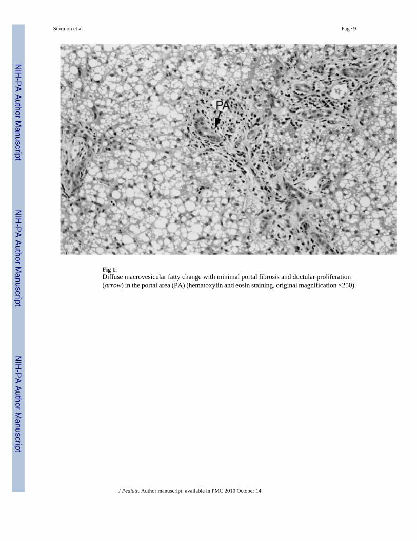
Fig 1.Diffuse macrovesicular fatty change with minimal portal fibrosis and ductular proliferation(arrow) in the portal area (PA) (hematoxylin and eosin staining, original magnification ×250).
Stormon et al. Page 9
J Pediatr. Author manuscript; available in PMC 2010 October 14.
NIH
-PA Author Manuscript
NIH
-PA Author Manuscript
NIH
-PA Author Manuscript

Fig 2.Electron micrograph of liver biopsy with microvesicular fat droplets (Fd) in hepatocyte, focalcytoplasmic necrosis (asterisk). Nucleus (Nu) and mitochondria (mi) show no specificabnormalities (TEM ×5000).
Stormon et al. Page 10
J Pediatr. Author manuscript; available in PMC 2010 October 14.
NIH
-PA Author Manuscript
NIH
-PA Author Manuscript
NIH
-PA Author Manuscript

NIH
-PA Author Manuscript
NIH
-PA Author Manuscript
NIH
-PA Author Manuscript
Stormon et al. Page 11
Table I
Initial hematologic laboratory values
Variable Result
Hemoglobin (100-140 g/L; 10-14 g/dL) 108 g/L (10.8 g/dL)
Mean corpuscular volume (f/L) 85.7
White cell count (5-15 × 109/L) 25 (lymph 14, neut 8)
Platelet count (150-400 × 109/L) 440
INR (0.9-1.1) 1.2
Partial thromboplastin time (25-35 sec) 103
J Pediatr. Author manuscript; available in PMC 2010 October 14.
NIH
-PA Author Manuscript
NIH
-PA Author Manuscript
NIH
-PA Author Manuscript
Stormon et al. Page 12
Tabl
e II
Initi
al b
lood
bio
chem
ical
labo
rato
ry v
alue
s
Var
iabl
eR
esul
tN
orm
al r
ange
Asp
arta
te a
min
otra
nsfe
rase
(AST
)40
20-
35 U
/L
Ala
nine
am
inot
rans
fera
se (A
LT)
436
0-35
U/L
Alb
umin
31 (3
.1)
32-4
8 g/
L (3
.2-4
.8 g
/dL)
Gam
ma
glut
amyl
tran
sfer
ase
(GG
T)47
0-45
U/L
Glu
cose
2.2
(39.
6 m
g/dL
)2.
5-5
mm
ol/L
(45-
90 m
g/dL
)
Am
mon
ium
33 (5
9)9-
33 μ
mol
/L (1
6-59
μg/
dL)
Bili
rubi
n, c
onju
gate
d/un
conj
ugat
ed31
/ 1
(1.8
/0.0
5 g/
dL)
0-17
/ 0-
2 μm
ol/L
(0-1
/0-0
.1 m
g/dL
)
Plas
ma
carn
itine
:free
, tot
al, r
atio
6, 1
0, 6
0%20
-53 μm
ol/L
, 25-
70 μ
mol
/L, 6
6-70
%
J Pediatr. Author manuscript; available in PMC 2010 October 14.
NIH
-PA Author Manuscript
NIH
-PA Author Manuscript
NIH
-PA Author Manuscript
Stormon et al. Page 13
Table III
Abnormal laboratory values 4 weeks into the illness
Variable Result
Potassium 2.5 (3.7-5.6 mmol/L), 2.5 (3.7-5.6 mEq/L)
Phosphate 0.56 (1.3-2.2 mmol/L), 1.7 (4.0-6.8 mg/dL)
Urea 1.3 (1.8-5.4 mmol/L), 3.6 (8.4-18.2 mg/dL)
Uric acid 61 (120-360 lmol/L), 1.03 (2.02-6.05 mg/dL)
Carnitine: free, total, ratio 9.9, 15.9 μmol/L, 62%
Urine carnitine: free, total 2664 (8-254), 3681 (62-369) mmol/mol creat
AST 559 (0-111 U/L)
ALT 217 (0-60 U/L)
Albumin 31 (32-48 g/L), 3.1 (3.2-4.8 g/dL)
Bilirubin: conjugated/ unconjugated
14/9 (0-2/0-17 μmol/L)/0.8/0.5 (0-0.1/0-1 mg/dL)
APTT 94.9 (25-35 seconds)
Factor VIII 5.36-7.5 (0.79-1.99 IU/mL)
Factor XI 0.09-0.14 (0.5-1.5 IU/mL)
Normal values in brackets; blood unless specified.
J Pediatr. Author manuscript; available in PMC 2010 October 14.
Related Documents


![LIVER 3 KISS 2018 [Kompatibilis üzemmód] · 2018. 3. 12. · 2018. 03. 12. 3 Liver alterations associated with pregnancy Acute steatosis in pregnancy –rare, from mild to severe](https://static.cupdf.com/doc/110x72/5ff28ad5a14fb268c76c9315/liver-3-kiss-2018-kompatibilis-zemmd-2018-3-12-2018-03-12-3-liver.jpg)


