The Rockefeller University Press $30.00 J. Exp. Med. 2015 www.jem.org/cgi/doi/10.1084/jem.20142182 Cite by DOI: 10.1084/jem.20142182 of Brief Definitive Report Immunoglobulin (IgE) is important for resis- tance to parasitic infections (Dombrowicz et al., 1996; Gould et al., 2003; Matsumoto et al., 2013) and protection against venom toxins (Arnold et al., 2007; Marichal et al., 2013; Palm et al., 2013).Yet IgE is also responsible for triggering allergic reactions, one of the most common chronic conditions worldwide (Dorrington and Bennich, 1978; Arnold et al., 2007; Pawankar et al., 2013; Plomp et al., 2014). These diseases include asthma and atopic dermatitis, as well as allergies to food, dust mites, insect venom, pol- len, and pet dander. Allergic reactions manifest as localized wheel and flare irritations, can have respiratory symptoms, including sneezing, rhi- nitis, and asthma, and in extreme cases can be life threatening in the form of anaphylaxis. Al- though IgE is the least abundant Ig in circulation, with a short serum half-life, it persists for weeks bound to the surface of mast cells by the high- affinity IgE receptor, FcRI, in tissues (Gould et al., 2003). Cross-linking of mast cell–bound IgE by allergens activates the cells and results in release of mediators that induce vasodilation, vas- cular permeability, and smooth muscle contrac- tility (Gould et al., 2003; Galli and Tsai, 2012). IgE is the most heavily glycosylated mono- meric Ig in mammals, with seven N-linked glycosylation consensus sequences (N-X-S/T) distributed across each heavy chain of human IgE (Arnold et al., 2007). The importance of glycosylation in Ig biology is increasingly ap- preciated. For example, the single glycan on IgG at N297 is essential for structural integrity of the constant fragment (Fc), and without it IgG cannot engage Fc receptors (Feige et al., 2009). However, the precise role of glycosyl- ation to IgE biology is less clear. Some studies concluded glycosylation of IgE is essential for CORRESPONDENCE Robert M. Anthony: [email protected] Abbreviations used: CD, circular dichroism; DNP, dinitrophenol; EndoF1, endoglycosidase F1; mBMMC, mouse BM-derived mast cell; MFI, mean fluor- escent intensity; PCA, passive cutaneous anaphylaxis; PNG, peptide-N-glycosidase F; poly- mIgE, polyclonal mouse IgE; TNP, trinitrophenol. A single glycan on IgE is indispensable for initiation of anaphylaxis Kai-Ting C. Shade, 1 Barbara Platzer, 2,3 Nathaniel Washburn, 4 Vinidhra Mani, 1 Yannic C. Bartsch, 1 Michelle Conroy, 1 Jose D. Pagan, 1 Carlos Bosques, 4 Thorsten R. Mempel, 1 Edda Fiebiger, 2,3 and Robert M. Anthony 1 1 Center for Immunology and Inflammatory Diseases, Division of Rheumatology, Allergy, and Immunology, Massachusetts General Hospital, Harvard Medical School, Charlestown, MA 02129 2 Division of Gastroenterology, Hepatology, and Nutrition, Boston Children’s Hospital and 3 Department of Pediatrics, Harvard Medical School, Boston, MA 02115 4 Momenta Pharmaceuticals, Cambridge, MA 02142 Immunoglobulin (IgE) antibodies are the primary mediators of allergic diseases, which affect more than in 0 individuals worldwide. IgE specific for innocuous environmental antigens, or allergens, binds and sensitizes tissue-resident mast cells expressing the high- affinity IgE receptor, FcRI. Subsequent allergen exposure cross-links mast cell–bound IgE, resulting in the release of inflammatory mediators and initiation of the allergic cascade. It is well established that precise glycosylation patterns exert profound effects on the biological activity of IgG. However, the contribution of glycosylation to IgE biology is less clear. Here, we demonstrate an absolute requirement for IgE glycosylation in allergic reactions. The obligatory glycan was mapped to a single N-linked oligomannose structure in the constant domain 3 (C3) of IgE, at asparagine-394 (N394) in human IgE and N384 in mouse. Genetic disruption of the site or enzymatic removal of the oligomannose glycan altered IgE secondary structure and abrogated IgE binding to FcRI, rendering IgE incapable of eliciting mast cell degranulation, thereby preventing anaphylaxis. These results underscore an unap- preciated and essential requirement of glycosylation in IgE biology. © 2015 Shade et al. This article is distributed under the terms of an Attribution– Noncommercial–Share Alike–No Mirror Sites license for the first six months after the publication date (see http://www.rupress.org/terms). After six months it is available under a Creative Commons License (Attribution–Noncommercial– Share Alike 3.0 Unported license, as described at http://creativecommons.org/ licenses/by-nc-sa/3.0/). The Journal of Experimental Medicine

Welcome message from author
This document is posted to help you gain knowledge. Please leave a comment to let me know what you think about it! Share it to your friends and learn new things together.
Transcript
The Rockefeller University Press $30.00J. Exp. Med. 2015www.jem.org/cgi/doi/10.1084/jem.20142182
Cite by DOI: 10.1084/jem.20142182 � of ��
Brief Definit ive Report
Immunoglobulin (IgE) is important for resis-tance to parasitic infections (Dombrowicz et al., 1996; Gould et al., 2003; Matsumoto et al., 2013) and protection against venom toxins (Arnold et al., 2007; Marichal et al., 2013; Palm et al., 2013). Yet IgE is also responsible for triggering allergic reactions, one of the most common chronic conditions worldwide (Dorrington and Bennich, 1978; Arnold et al., 2007; Pawankar et al., 2013; Plomp et al., 2014). These diseases include asthma and atopic dermatitis, as well as allergies to food, dust mites, insect venom, pol-len, and pet dander. Allergic reactions manifest as localized wheel and flare irritations, can have respiratory symptoms, including sneezing, rhi-nitis, and asthma, and in extreme cases can be life threatening in the form of anaphylaxis. Al-though IgE is the least abundant Ig in circulation, with a short serum half-life, it persists for weeks bound to the surface of mast cells by the high-affinity IgE receptor, FcRI, in tissues (Gould et al., 2003). Cross-linking of mast cell–bound
IgE by allergens activates the cells and results in release of mediators that induce vasodilation, vas-cular permeability, and smooth muscle contrac-tility (Gould et al., 2003; Galli and Tsai, 2012).
IgE is the most heavily glycosylated mono-meric Ig in mammals, with seven N-linked glycosylation consensus sequences (N-X-S/T) distributed across each heavy chain of human IgE (Arnold et al., 2007). The importance of glycosylation in Ig biology is increasingly ap-preciated. For example, the single glycan on IgG at N297 is essential for structural integrity of the constant fragment (Fc), and without it IgG cannot engage Fc receptors (Feige et al., 2009). However, the precise role of glycosyl-ation to IgE biology is less clear. Some studies concluded glycosylation of IgE is essential for
CORRESPONDENCE Robert M. Anthony: [email protected]
Abbreviations used: CD, circular dichroism; DNP, dinitrophenol; EndoF1, endoglycosidase F1; mBMMC, mouse BM-derived mast cell; MFI, mean fluor-escent intensity; PCA, passive cutaneous anaphylaxis; PNG, peptide-N-glycosidase F; poly-mIgE, polyclonal mouse IgE; TNP, trinitrophenol.
A single glycan on IgE is indispensable for initiation of anaphylaxis
Kai-Ting C. Shade,1 Barbara Platzer,2,3 Nathaniel Washburn,4 Vinidhra Mani,1 Yannic C. Bartsch,1 Michelle Conroy,1 Jose D. Pagan,1 Carlos Bosques,4 Thorsten R. Mempel,1 Edda Fiebiger,2,3 and Robert M. Anthony1
1Center for Immunology and Inflammatory Diseases, Division of Rheumatology, Allergy, and Immunology, Massachusetts General Hospital, Harvard Medical School, Charlestown, MA 021292Division of Gastroenterology, Hepatology, and Nutrition, Boston Children’s Hospital and 3Department of Pediatrics, Harvard Medical School, Boston, MA 021154Momenta Pharmaceuticals, Cambridge, MA 02142
Immunoglobulin (IgE) antibodies are the primary mediators of allergic diseases, which affect more than � in �0 individuals worldwide. IgE specific for innocuous environmental antigens, or allergens, binds and sensitizes tissue-resident mast cells expressing the high-affinity IgE receptor, FcRI. Subsequent allergen exposure cross-links mast cell–bound IgE, resulting in the release of inflammatory mediators and initiation of the allergic cascade. It is well established that precise glycosylation patterns exert profound effects on the biological activity of IgG. However, the contribution of glycosylation to IgE biology is less clear. Here, we demonstrate an absolute requirement for IgE glycosylation in allergic reactions. The obligatory glycan was mapped to a single N-linked oligomannose structure in the constant domain 3 (C3) of IgE, at asparagine-394 (N394) in human IgE and N384 in mouse. Genetic disruption of the site or enzymatic removal of the oligomannose glycan altered IgE secondary structure and abrogated IgE binding to FcRI, rendering IgE incapable of eliciting mast cell degranulation, thereby preventing anaphylaxis. These results underscore an unap-preciated and essential requirement of glycosylation in IgE biology.
© 2015 Shade et al. This article is distributed under the terms of an Attribution–Noncommercial–Share Alike–No Mirror Sites license for the first six months after the publication date (see http://www.rupress.org/terms). After six months it is available under a Creative Commons License (Attribution–Noncommercial– Share Alike 3.0 Unported license, as described at http://creativecommons.org/ licenses/by-nc-sa/3.0/).
The
Journ
al o
f Exp
erim
enta
l M
edic
ine
� of �� IgE glycosylation requirements for anaphylaxis | Shade et al.
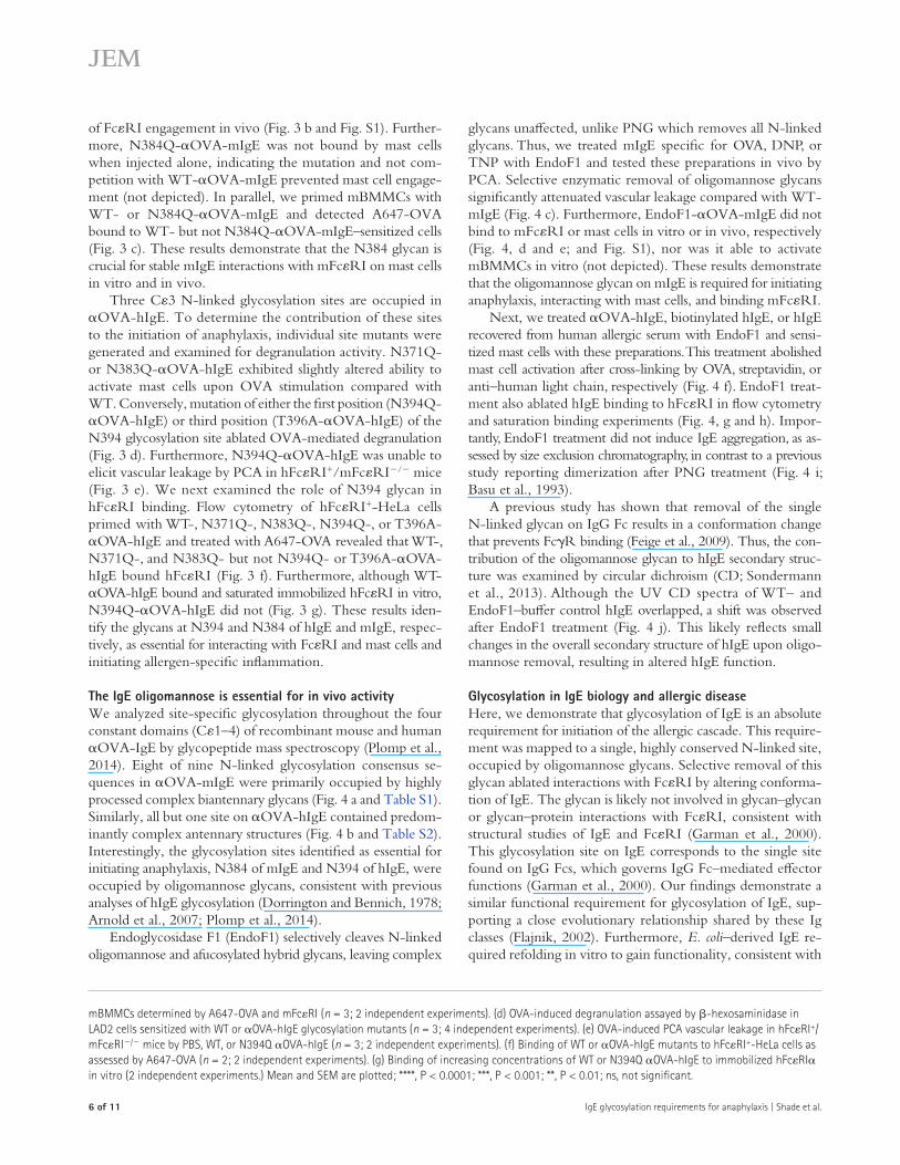
the IgE C3 domain to be essential for triggering anaphy-laxis. This site was occupied almost exclusively by oligoman-nose glycans, whereas complex antennary glycans were found at the other sites throughout mouse and human IgE. Selec-tive enzymatic removal of the oligomannose glycan altered secondary structure of IgE, prevented binding to FcRI on mast cells, and importantly, attenuated anaphylaxis in vivo. Together, the findings herein identify the IgE oligomannose glycan essential for in vivo activity and structural integrity of this Ig class.
FcRI binding and effector functions (Nettleton and Kochan, 1995; Sayers et al., 1998; Björklund et al., 1999; 2000; Hunt et al., 2005). However, these findings have been contradicted (Basu et al., 1993; Young et al., 1995), supported by studies using a functional aglycosylated IgE derived from Escherichia coli (Helm et al., 1988; Henry et al., 2000). Therefore, we sought to determine whether glycosylation was required for the in vivo activity of IgE.
We conducted a systematic analysis of all glycosylation sites on mouse and human IgE, which revealed a single glycan in
Figure �. N-linked glycosylation is essential for IgE-mediated allergic inflammation. (a and b) Quantified vascular leakage and representative ear images after PCA with PBS, WT, or PNG–poly-mIgE specific for OVA or peanut extracts (n = 4 ears; 2 independent experiments; a) or monoclonal mIgE specific for OVA, DNP, or TNP (n = 8; 2, 3, and 3 independent experiments for OVA, DNP, and TNP; b). Bar, 1 cm. (c) Histograms and mean fluorescent intensity (MFI) of WT or PNG-OVA-mIgE binding to mBMMCs determined by A647-OVA and mFcRI (n = 3; 2 independent experiments). (d) -Hexosaminidase activity after OVA stimulation of LAD2 cell sensitized with WT or PNG-treated OVA-hIgE (n = 3; 5 independent experiments). (e) Quantified OVA-induced PCA in mFcRI/ or hFcRI+mFcRI/ mice sensitized by WT or PNG-OVA-hIgE (n = 4; 2 independent experiments). (f) FACS of WT or PNG-OVA-hIgE to hFcRI+-HeLa cells assessed by A647-OVA and -hFcRI-PE (n = 2; 2 independent experiments). Means and SEM are plotted; ****, P < 0.0001; ***, P < 0.001; **, P < 0.01; *, P < 0.05; ns, not significant.
JEM 3 of ��
Br ief Definit ive Repor t
triggered robust vascular leakage in the ears of hFcRI+/mFcRI/ mice upon OVA challenge (Fig. 1 e). The re-sponse was significantly diminished in PNG-OVA-hIgE–treated ears (Fig. 1 e), confirming N-linked glycosylation is also essential for the in vivo activity of human IgE.
To determine whether hIgE glycosylation was important for hFcRI binding, HeLa cells engineered to express hFcRI (hFcRI+-HeLa) were sensitized with OVA-hIgE, incu-bated with A647-OVA, and analyzed by flow cytometry. Al-though OVA-hIgE bound to hFcRI+-HeLa cells was detected by A647-OVA, PNG-OVA-hIgE did not (Fig. 1 f ), despite recognizing OVA by ELISA (not depicted). Together, these re-sults indicate that IgE glycosylation is essential for the initiation of anaphylaxis and FcRI interactions.
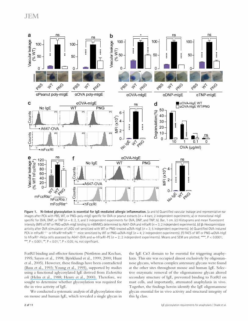
Mapping glycosylation requirements of IgEWe generated a panel of OVA-mIgE mutants selectively lacking all glycosylation sites on each constant domain (C1–4) by mutating asparagine (N) to glutamine (Q). After confirm-ing that all domain-specific mutants recognized OVA similarly (not depicted), these antibodies were tested in vivo by PCA. Although C2- or C4-OVA-mIgE domain mutants pro-moted robust vascular leakage after OVA challenge similar to WT (Fig. 2 a), C1-OVA-mIgE glycosylation mutants exhib-ited slightly enhanced vascular leakage. In contrast, anaphylaxis elicited by the C3-OVA-mIgE mutant was significantly attenuated. WT-, C1-, C2-, and C4-OVA-mIgE acti-vated mBMMCs upon OVA stimulation, but C3-OVA-mIgE did not (not depicted). OVA-hIgE domain–specific glycosylation mutants were also generated and tested for mast cell degranulating activity (Fig. 2 b). WT- and C2-OVA-hIgE domain mutants triggered robust degranulation upon OVA stimulation. Although degranulation was slightly re-duced in mast cells sensitized with C1-OVA-hIgE, mutation of C3 glycosylation sites completely abolished OVA-specific degranulation. Importantly, C1-OVA-hIgE mutants bound to FcRI+-HeLa cells, as determined by A647-OVA flow cytometry, but C3-OVA-hIgE did not (Fig. 2 c). Together, these results indicate that C1 glycosylation plays a minor role in modulating IgE functions, perhaps by controlling Fab arm flexibility (Arnold et al., 2007), but is not involved in FcRI binding. More importantly, C3 glycosylation is re-quired for both mouse and human IgE to bind FcRI and initiate anaphylaxis.
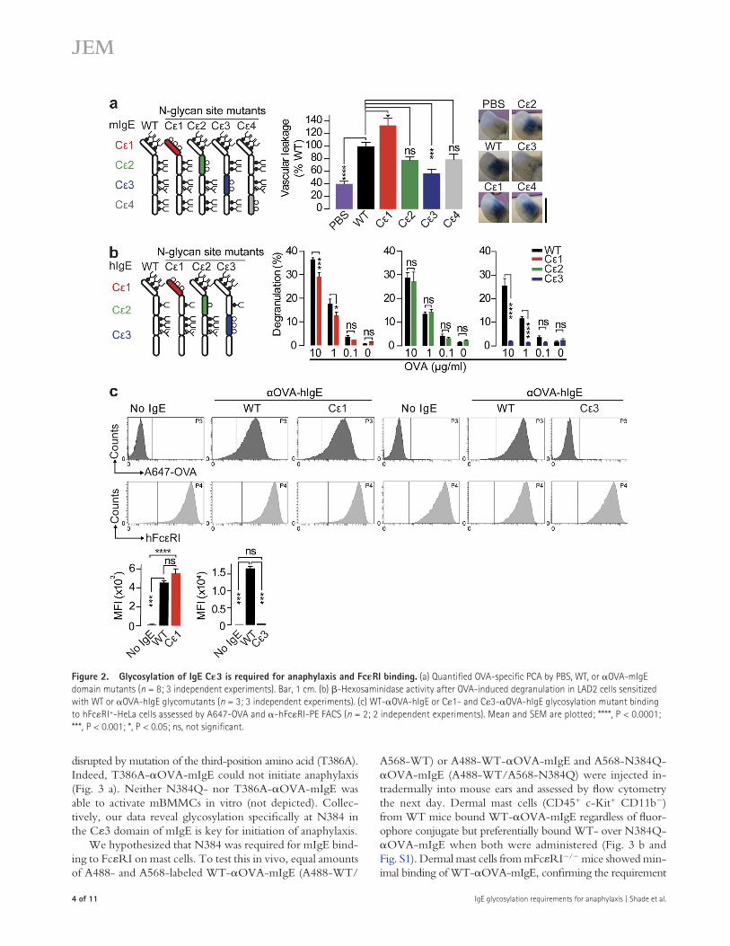
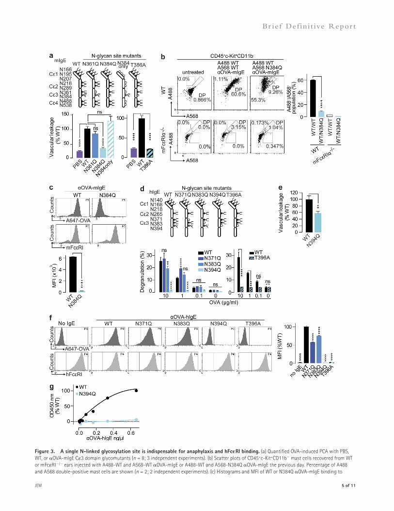
An obligate IgE C3 glycanTo dissect the importance of the two N-linked glycosylation sites in C3 of mIgE, individual glycosylation site mutants were generated by conversion of N361 or N384 to Q. Both WT- and N361Q-OVA-mIgE elicited a robust anaphylactic response in vivo, but N384Q-OVA-mIgE did not (Fig. 3 a). Moreover, a reciprocal mutant, in which all N-linked glyco-sylation sites were disrupted except N384 (N384only-OVA-mIgE), promoted a strong PCA upon OVA administration (Fig. 3 a). To confirm the importance of N384 glycosylation for mIgE function, the glycosylation consensus sequence was
RESULTS AND DISCUSSIONEnzymatic deglycosylation attenuates IgETo generate polyclonal mouse IgE (poly-mIgE), we immu-nized mice with OVA or extract from the common food al-lergen peanuts in alum. The mice were bled and IgG depleted from the serum. All N-linked glycans were removed from the poly-mIgE by treatment with the endoglycosidase peptide-N-glycosidase F (PNG). This treatment did not reduce rec-ognition of peanuts or OVA by poly-mIgE, as determined by ELISA (unpublished data). Next, we injected poly-mIgE in-tradermally into the ears of mice in a model of passive cutane-ous anaphylaxis (PCA), highly dependent on the interactions between IgE, FcRI+ mast cells, and allergens (Dombrowicz et al., 1993), and the next day intravenously challenged with appropriate allergens and Evan’s blue dye. Poly-mIgE elicited robust anaphylaxis to peanuts or OVA, as measured by vascu-lar leakage of the blue dye. However, after treatment with PNG, poly-mIgE induced significantly decreased anaphylaxis to either allergen (Fig. 1 a). Next, we treated three monoclonal mIgEs specific for model allergens OVA, dinitrophenol (DNP), or trinitrophenol (TNP) with PNG. Enzymatic deglycosyl-ation and antigen binding were verified by lectin blotting and ELISA assays, respectively (unpublished data). Although mIgE specific for OVA, DNP, or TNP triggered strong allergen-specific anaphylactic responses in vivo, PNG treatment sig-nificantly attenuated vascular leakage (Fig. 1 b).
Allergic reactions are highly dependent on IgE and FcRI interactions (Dombrowicz et al., 1993; Gould et al., 2003). To determine the contribution of glycosylation to inter-actions with mouse FcRI (mFcRI), mouse BM-derived mast cells (mBMMCs) were sensitized in vitro with native or de-glycosylated OVA-mIgE overnight. When ligand–receptor interactions were analyzed by flow cytometry using Alexa Fluor 647–OVA (A647-OVA), we found that although OVA-mIgE bound to the mast cells, PNG-OVA-mIgE did not (Fig. 1 c). Indeed, WT-mIgE specific for OVA, DNP, or TNP, but not PNG-mIgE, was able to activate mBMMCs upon antigen stimulation (not depicted). Together, these results demonstrate that mIgE glycosylation is necessary for mast cell binding in vitro and eliciting anaphylaxis to multiple antigens in vivo.
We next generated and enzymatically deglycosylated OVA-specific human IgE (OVA-hIgE, PNG-OVA-hIgE) and sensitized human LAD2 mast cells in vitro with these preparations. OVA stimulation resulted in dose-dependent de-granulation of OVA-hIgE–sensitized mast cells, as assessed by -hexosaminidase release. Consistent with our results, PNG-OVA-hIgE was incapable of instigating degranulation upon OVA stimulation (Fig. 1 d). In parallel, we administered OVA-hIgE or PNG-OVA-hIgE intradermally to transgenic mice expressing human FcRI while lacking mFcRI (hFcRI+/mFcRI/; Dombrowicz et al., 1996). These transgenic mice have a broad cellular distribution of hFcRI expres-sion, whereas mFcRI is restricted to mast cells and basophils in the WT mice. hIgE is unable to elicit PCA in mFcRI/ mice, which served as an injection control group. OVA-hIgE
4 of �� IgE glycosylation requirements for anaphylaxis | Shade et al.
A568-WT) or A488-WT-OVA-mIgE and A568-N384Q-OVA-mIgE (A488-WT/A568-N384Q) were injected in-tradermally into mouse ears and assessed by flow cytometry the next day. Dermal mast cells (CD45+ c-Kit+ CD11b) from WT mice bound WT-OVA-mIgE regardless of fluor-ophore conjugate but preferentially bound WT- over N384Q-OVA-mIgE when both were administered (Fig. 3 b and Fig. S1). Dermal mast cells from mFcRI/ mice showed min-imal binding of WT-OVA-mIgE, confirming the requirement
disrupted by mutation of the third-position amino acid (T386A). Indeed, T386A-OVA-mIgE could not initiate anaphylaxis (Fig. 3 a). Neither N384Q- nor T386A-OVA-mIgE was able to activate mBMMCs in vitro (not depicted). Collec-tively, our data reveal glycosylation specifically at N384 in the C3 domain of mIgE is key for initiation of anaphylaxis.
We hypothesized that N384 was required for mIgE bind-ing to FcRI on mast cells. To test this in vivo, equal amounts of A488- and A568-labeled WT-OVA-mIgE (A488-WT/
Figure �. Glycosylation of IgE C3 is required for anaphylaxis and FcRI binding. (a) Quantified OVA-specific PCA by PBS, WT, or OVA-mIgE domain mutants (n = 8; 3 independent experiments). Bar, 1 cm. (b) -Hexosaminidase activity after OVA-induced degranulation in LAD2 cells sensitized with WT or OVA-hIgE glycomutants (n = 3; 3 independent experiments). (c) WT-OVA-hIgE or C1- and C3-OVA-hIgE glycosylation mutant binding to hFcRI+-HeLa cells assessed by A647-OVA and -hFcRI-PE FACS (n = 2; 2 independent experiments). Mean and SEM are plotted; ****, P < 0.0001; ***, P < 0.001; *, P < 0.05; ns, not significant.
JEM � of ��
Br ief Definit ive Repor t
Figure 3. A single N-linked glycosylation site is indispensable for anaphylaxis and hFcRI binding. (a) Quantified OVA-induced PCA with PBS, WT, or OVA-mIgE C3 domain glycomutants (n = 8; 3 independent experiments). (b) Scatter plots of CD45+c-Kit+CD11b mast cells recovered from WT or mFcRI/ ears injected with A488-WT and A568-WT OVA-mIgE or A488-WT and A568-N384Q OVA-mIgE the previous day. Percentage of A488 and A568 double-positive mast cells are shown (n = 2; 2 independent experiments). (c) Histograms and MFI of WT or N384Q OVA-mIgE binding to
� of �� IgE glycosylation requirements for anaphylaxis | Shade et al.
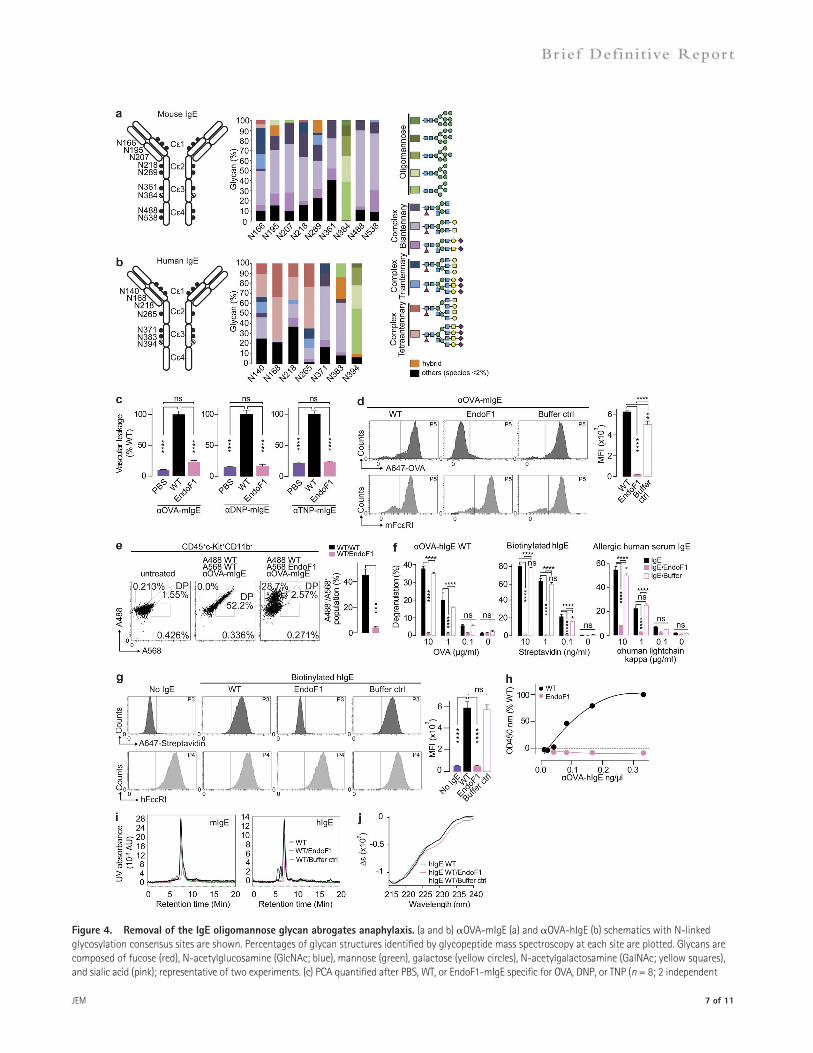
glycans unaffected, unlike PNG which removes all N-linked glycans. Thus, we treated mIgE specific for OVA, DNP, or TNP with EndoF1 and tested these preparations in vivo by PCA. Selective enzymatic removal of oligomannose glycans significantly attenuated vascular leakage compared with WT-mIgE (Fig. 4 c). Furthermore, EndoF1-OVA-mIgE did not bind to mFcRI or mast cells in vitro or in vivo, respectively (Fig. 4, d and e; and Fig. S1), nor was it able to activate mBMMCs in vitro (not depicted). These results demonstrate that the oligomannose glycan on mIgE is required for initiating anaphylaxis, interacting with mast cells, and binding mFcRI.
Next, we treated OVA-hIgE, biotinylated hIgE, or hIgE recovered from human allergic serum with EndoF1 and sensi-tized mast cells with these preparations. This treatment abolished mast cell activation after cross-linking by OVA, streptavidin, or anti–human light chain, respectively (Fig. 4 f). EndoF1 treat-ment also ablated hIgE binding to hFcRI in flow cytometry and saturation binding experiments (Fig. 4, g and h). Impor-tantly, EndoF1 treatment did not induce IgE aggregation, as as-sessed by size exclusion chromatography, in contrast to a previous study reporting dimerization after PNG treatment (Fig. 4 i; Basu et al., 1993).
A previous study has shown that removal of the single N-linked glycan on IgG Fc results in a conformation change that prevents FcR binding (Feige et al., 2009). Thus, the con-tribution of the oligomannose glycan to hIgE secondary struc-ture was examined by circular dichroism (CD; Sondermann et al., 2013). Although the UV CD spectra of WT– and EndoF1–buffer control hIgE overlapped, a shift was observed after EndoF1 treatment (Fig. 4 j). This likely reflects small changes in the overall secondary structure of hIgE upon oligo-mannose removal, resulting in altered hIgE function.
Glycosylation in IgE biology and allergic diseaseHere, we demonstrate that glycosylation of IgE is an absolute requirement for initiation of the allergic cascade. This require-ment was mapped to a single, highly conserved N-linked site, occupied by oligomannose glycans. Selective removal of this glycan ablated interactions with FcRI by altering conforma-tion of IgE. The glycan is likely not involved in glycan–glycan or glycan–protein interactions with FcRI, consistent with structural studies of IgE and FcRI (Garman et al., 2000). This glycosylation site on IgE corresponds to the single site found on IgG Fcs, which governs IgG Fc–mediated effector functions (Garman et al., 2000). Our findings demonstrate a similar functional requirement for glycosylation of IgE, sup-porting a close evolutionary relationship shared by these Ig classes (Flajnik, 2002). Furthermore, E. coli–derived IgE re-quired refolding in vitro to gain functionality, consistent with
of FcRI engagement in vivo (Fig. 3 b and Fig. S1). Further-more, N384Q-OVA-mIgE was not bound by mast cells when injected alone, indicating the mutation and not com-petition with WT-OVA-mIgE prevented mast cell engage-ment (not depicted). In parallel, we primed mBMMCs with WT- or N384Q-OVA-mIgE and detected A647-OVA bound to WT- but not N384Q-OVA-mIgE–sensitized cells (Fig. 3 c). These results demonstrate that the N384 glycan is crucial for stable mIgE interactions with mFcRI on mast cells in vitro and in vivo.
Three C3 N-linked glycosylation sites are occupied in OVA-hIgE. To determine the contribution of these sites to the initiation of anaphylaxis, individual site mutants were generated and examined for degranulation activity. N371Q- or N383Q-OVA-hIgE exhibited slightly altered ability to activate mast cells upon OVA stimulation compared with WT. Conversely, mutation of either the first position (N394Q-OVA-hIgE) or third position (T396A-OVA-hIgE) of the N394 glycosylation site ablated OVA-mediated degranulation (Fig. 3 d). Furthermore, N394Q-OVA-hIgE was unable to elicit vascular leakage by PCA in hFcRI+/mFcRI/ mice (Fig. 3 e). We next examined the role of N394 glycan in hFcRI binding. Flow cytometry of hFcRI+-HeLa cells primed with WT-, N371Q-, N383Q-, N394Q-, or T396A-OVA-hIgE and treated with A647-OVA revealed that WT-, N371Q-, and N383Q- but not N394Q- or T396A-OVA-hIgE bound hFcRI (Fig. 3 f). Furthermore, although WT-OVA-hIgE bound and saturated immobilized hFcRI in vitro, N394Q-OVA-hIgE did not (Fig. 3 g). These results iden-tify the glycans at N394 and N384 of hIgE and mIgE, respec-tively, as essential for interacting with FcRI and mast cells and initiating allergen-specific inflammation.
The IgE oligomannose is essential for in vivo activityWe analyzed site-specific glycosylation throughout the four constant domains (C1–4) of recombinant mouse and human OVA-IgE by glycopeptide mass spectroscopy (Plomp et al., 2014). Eight of nine N-linked glycosylation consensus se-quences in OVA-mIgE were primarily occupied by highly processed complex biantennary glycans (Fig. 4 a and Table S1). Similarly, all but one site on OVA-hIgE contained predom-inantly complex antennary structures (Fig. 4 b and Table S2). Interestingly, the glycosylation sites identified as essential for initiating anaphylaxis, N384 of mIgE and N394 of hIgE, were occupied by oligomannose glycans, consistent with previous analyses of hIgE glycosylation (Dorrington and Bennich, 1978; Arnold et al., 2007; Plomp et al., 2014).
Endoglycosidase F1 (EndoF1) selectively cleaves N-linked oligomannose and afucosylated hybrid glycans, leaving complex
mBMMCs determined by A647-OVA and mFcRI (n = 3; 2 independent experiments). (d) OVA-induced degranulation assayed by -hexosaminidase in LAD2 cells sensitized with WT or OVA-hIgE glycosylation mutants (n = 3; 4 independent experiments). (e) OVA-induced PCA vascular leakage in hFcRI+/mFcRI/ mice by PBS, WT, or N394Q OVA-hIgE (n = 3; 2 independent experiments). (f) Binding of WT or OVA-hIgE mutants to hFcRI+-HeLa cells as assessed by A647-OVA (n = 2; 2 independent experiments). (g) Binding of increasing concentrations of WT or N394Q OVA-hIgE to immobilized hFcRI in vitro (2 independent experiments.) Mean and SEM are plotted; ****, P < 0.0001; ***, P < 0.001; **, P < 0.01; ns, not significant.
JEM � of ��
Br ief Definit ive Repor t
Figure 4. Removal of the IgE oligomannose glycan abrogates anaphylaxis. (a and b) OVA-mIgE (a) and OVA-hIgE (b) schematics with N-linked glycosylation consensus sites are shown. Percentages of glycan structures identified by glycopeptide mass spectroscopy at each site are plotted. Glycans are composed of fucose (red), N-acetylglucosamine (GlcNAc; blue), mannose (green), galactose (yellow circles), N-acetylgalactosamine (GalNAc; yellow squares), and sialic acid (pink); representative of two experiments. (c) PCA quantified after PBS, WT, or EndoF1-mIgE specific for OVA, DNP, or TNP (n = 8; 2 independent
8 of �� IgE glycosylation requirements for anaphylaxis | Shade et al.
SalI and XbaI and the light chain was placed under EF1a promoter using restriction enzyme sites NotI and KpnI in pBUDCE4.1 expression vector (Invitrogen). A similar cloning strategy was used to construct OVA-hIgE vector. The N-linked glycosylation sequons of IgE were mutated using the QuikChange II XL Site-Directed Mutagenesis kit (Agilent Technologies), according to the manufacturer’s protocol. Recombinant antibodies were generated by transient transfection of the plasmids into HEK293T using Polyethylenimine “Max” (Polysciences, Inc.), followed by purification using N-hydroxysuccinimide–activated Sepharose beads (GE Healthcare) coupled to OVA (Sigma-Aldrich). Antibodies generated were verified by immuno-blots for size and quantified by IgE ELISA, and the specificity was confirmed by OVA ELISA (see below).
Polyclonal IgE specific for OVA or peanut extracts was prepared by in-jecting BALB/c mice with 10 µg OVA (Sigma-Aldrich) or peanut extracts (preparation) in aluminum hydroxide on days 0, 7, and 14. Mice were bled on days 10, 17, 19, and 21. The sera was separated from the blood by serum gel tubes (BD) and depleted of IgG by incubating with Protein G high-capacity agarose beads (Thermo Fisher Scientific).
Human IgE was purified from de-identified peanut-allergic sera using N-hydroxysuccinimide–activated Sepharose beads coupled to omalizumab (Xolair; Genentech) after IgG depletion by Protein G high-capacity agarose beads. Purified IgE was verified by Coomassie and immunoblots and quanti-fied by IgE ELISA. Patient sera were collected under protocols approved by the MGH Institutional Review Board.
Fluorescent labeling and glycan digestion. Human IgE (HE1 clone; Abcam) was biotinylated using the Biotin-XX Microscale Protein Labeling kit (Molecular Probes). WT- and N384Q-OVA-mIgE were conjugated to Alexa Fluor 488 or Alexa Fluor 567 (Molecular Probes) according to the manufacturer’s recommendations. IgE was digested with PNG (New Eng-land Biolabs, Inc.) or EndoF1 (Sigma-Aldrich) according to the manufacturer’s instructions under nondenaturing conditions at 37°C for 72 h. All digestions were verified by lectin blot (see below).
Immuno- and lectin blotting. Immuno- and lectin blotting were per-formed as described previously (Anthony et al., 2008). In brief, equal amounts of protein were resolved on 3–8% Tris-Acetate protein gels (Life Technolo-gies) in SDS-PAGE under nonreducing conditions. After transfer to polyvi-nylidene difluoride membranes, membranes were blocked with 5% dry milk in PBS containing 0.05% Tween 20 for immunoblotting or with Protein-Free Blocking Buffers (Thermo Fisher Scientific) for lectin blotting. Mouse or human IgE was probed by goat polyclonal anti–mouse or anti–human IgE-HRP (10 ng/ml; Bethyl Laboratories, Inc.), respectively. N-linked glycans were detected by biotinylated Lens culinaris agglutinin (LCA; 5 µg/ml; Vector Laboratories) and 1,3- and 1,6-linked high mannose structures were detected by biotinylated Galanthus nivalis lectin (GNA; 4 µg/ml; Vec-tor Laboratories).
ELISA. Mouse or human IgE was quantified by sandwich ELISA according to instructions from mouse or human IgE ELISA quantitation sets (Bethyl Laboratories, Inc.). Antibodies specific for OVA were verified by 96-well Nunc plates plate coated with 75 µg/ml OVA (Sigma-Aldrich), blocked with 2% BSA in PBS, and probed with goat polyclonal anti–mouse or anti–human IgE-HRP (2 ng/ml; Bethyl Laboratories, Inc.). A similar protocol
glycosylation playing a role in maintenance of the IgE C3 structure (Helm et al., 1988; Henry et al., 2000). Recently developed anti-IgE preventive therapies that neutralize circu-lating IgE or deplete IgE-producing cells have demonstrated some efficacy. However, clinical indications for these treatments have been limited to allergic asthma, chronic idiopathic urti-carial, and rhinitis (Galli and Tsai, 2012; Gauvreau et al., 2014; Saini et al., 2015). Thus, the IgE oligomannose may be a po-tential therapeutic target for both cell-bound and circulating IgE. Furthermore, it is possible that variations in the glycan composition at the conserved site may explain why not all in-dividuals with allergen-specific IgE suffer from allergies.
MATERIALS AND METHODSMice. 5–6-wk-old BALB/c female mice were purchased from The Jackson Laboratory. hFcRI+/mFcRI/ and mFcRI/ crossed at least 12 time to C57BL/6 mice (Dombrowicz et al., 1996) were maintained in the animal facility at Massachusetts General Hospital (MGH). Mice were all housed in specific pathogen–free conditions according to the National Institutes of Health (NIH), and all animal experiments were conducted under protocols approved by the Institutional Animal Care and Use Committee of MGH.
Peanut extract preparation. Unsalted dry-roasted peanuts (Blanched Jumbo Runner cultivar; Planters) were ground to a smooth paste, followed by washing with 20 vol cold acetone, filtered using Whatman paper, and dried. Protein was extracted by agitating the peanut flour overnight with PBS containing protease inhibitor cocktail without EDTA (Roche). The peanut protein extracts were collected as the supernatant after centrifugation at 24,000 g for 30 min.
PCA. Age- and sex-matched mice were randomized allocating to experi-mental group. 20 ng monoclonal mIgE specific for OVA, DNP (clone SPE-7; Sigma-Aldrich), or TNP (clone IgE-3; BD) or 5 ng polyclonal IgE specific for OVA or peanut extracts was injected intradermally in the BALB/c mice ears, and the next day mice were intravenously challenged with PBS con-taining 125 µg OVA (Sigma-Aldrich), peanut extract, DNP-HSA (Sigma-Aldrich), or TNP-BSA (conjugation ratio 13; Biosearch Technologies) and 2% Evans blue dye in PBS. 45 min after challenge, the mice were sacrificed and the ears were excised and minced before incubation in N,N-dimethyl-formamide (EMD Millipore) at 55°C for 3 h. The degree of blue dye in the ears was quantitated by the absorbance at 595 or 650 nm. For experiments of hIgE, 250 ng OVA-hIgE was injected intradermally in hFcRI+/mFcRI/ or mFcRI/ mice, followed by intravenous administra-tion of OVA and Evans blue dye 4 h later.
IgE antibodies. To generate recombinant IgE antibodies, the variable and constant regions of heavy and light chains were individually cloned from OVA-specific To hybridoma (provided by H. Oettgen, Boston Children’s Hospital, Harvard Medical School, Boston, MA), adapting from the proto-col previously described (Morrison, 2002). Once the variable and the con-stant regions of heavy and light chain were joined by overlapping PCR, the heavy chain was placed under CMV promoter using restriction enzyme sites
experiments). (d) Histograms and MFI of WT-, EndoF1-, and EndoF1 buffer only–OVA-mIgE bound to mBMMCs determined by A647-OVA and mFcRI FACS (n = 3; 2 independent experiments). (e) Scatter plots of dermal mast cells recovered from ears injected with A488-WT- and A568-WT- or A488-WT- and A568-EndoF1-OVA-mIgE (n = 2; 2 independent experiments). (f) -Hexosaminidase activity after stimulation by OVA, streptavidin, or anti–human kappa light chain in LAD2 cells sensitized with OVA-hIgE, biotinylated hIgE, or allergic human serum IgE or hIgE treated with EndoF1 or EndoF1 buffer only (n = 3; 2 independent experiments). (g) Binding of WT, EndoF1, or EndoF1 buffer only biotinylated hIgE to hFcRI-HeLa cells as assessed by FACS (n = 3; 2 indepen-dent experiments). (h) Quantitation of WT or EndoF1-treated OVA-hIgE bound to immobilized hFcRI (2 independent experiments). (i) Size exclusion chro-matography profile of OVA-mIgE or hIgE treated with EndoF1 or buffer only. (j) CD spectra of WT, EndoF1-treated, or EndoF1 buffer control hIgE (representative of two experiments). Mean and SEM are plotted; ****, P < 0.0001; ***, P < 0.001; **, P < 0.01; *, P < 0.05; ns, not significant.
JEM 9 of ��
Br ief Definit ive Repor t
abundant charge state for each of the peptides, except chymotrypsin was used for the analysis of N140 and N168 from OVA-hIgE and N166, N195, and N207 from OVA-mIgE. The isolated IgE was prepared for proteoly-sis by denaturing the protein in 6M guanidine HCl, followed by reduction with dithiothreitol and alkylation with iodoacetamide. The enzymatic digests were performed in 25 mM ammonium bicarbonate, pH 7.8, overnight (tryp-sin) or for 4 h (chymotrypsin). The digestion was quenched with formic acid added to 2% wt/wt. The separation was performed on an EasySpray C18 nLC column 0.75 µm × 50 cm (Thermo Fisher Scientific) using water and acetonitrile with 0.1% formic acid for mobile phase A and mobile phase B, respectively. A linear gradient from 1 to 35% mobile phase B was run 120 min. Mass spectra were recorded on a QExactive mass spectrometer (Thermo Fisher Scientific) operated in positive mode and using a top 12 data–dependent method. Glycopeptides were quantified in QualBrowser (Thermo Fisher Scientific) based on the extracted ion area for the most abundant charge state for each glycopeptide. The relative abundance was calculated for all identi-fied glycan species for each site using Excel (Microsoft).
Glycopeptides were quantified based on the extracted ion area for abun-dant charge state for each glycopeptide. The relative abundance was calcu-lated for all identified glycan species for each site without the use of internal standards making the relative abundances subject to the influence of ioniza-tion efficiency. The extracted ion chromatograms for the major and minor species from N394 are shown in Fig. S2 a. Identification of the glycopeptides was based on the presence of the oxonium ions 366.14 (Hex-HexNAc) and 204.08 (HexNAc) common to all glycosylated peptides, as well as the Y1 ion (peptide + HexNAc) which is unique to each site.
In cases where multiple chromatographic peaks were seen for a single mass, higher-energy collisionally activated dissociation (HCD) MS/MS was used for identifying differences between the chromatographically resolved isomers. Fig. S2 c shows the comparison of three neutral glycopeptides hav-ing the same apparent molecular weight. The MS2 spectrum for the first chromatographic peak shows evidence of a HexNAc-HexNAc structure with a mass of 407.16 D, supporting the assignment of this species as con-taining terminal N-acetylgalactosamine. The second peak shows evidence of a HexNAc-Hex-HexNAc structure at 569.20 not seen in the MS2 from the first chromatographic peak. Unique Y ions at 1804.81 and 1950.87 in this second spectrum support the assignment of isomer 2 as containing bisecting N-acetylglucosamine.
The MS2 was also used to differentiate between species of similar mass with different composition. The MS2 comparison in Fig. S2 d shows the frag-mentation pattern for a trifucosylated glycopeptide (top panel) from hIgE N371 with three sialylated/core-fucosylated glycopeptide (bottom three pan-els) from the same site. In Fig. S2 d, both oxonium (B) ions and Y ions were useful for elucidating the structure. The comparison of the MS2 spectra in the second and third panel from the top in Fig. S2 d suggests the major difference between these species is the location of the sialic acid. The oxonium ion at 495.17 from the MS2 spectrum in panel 2 of Fig. S2 d suggests a GalNAc-NeuAc linkage, whereas Gal-NeuAc is more likely the structure in the third panel based on the ion at 657.23. These spectra are representative of the data used to assign likely structures for each of the sites on both mIgE and hIgE.
CD. Untreated human IgE (HE1 clone; Abcam) or human IgE digested with EndoF1 or EndoF1 buffer was exchanged into 10 mM sodium phos-phate buffer, pH 7.0, and concentrated to 0.1 µg/µl. The CD spectra of IgE were acquired in a J-815 spectropolarimeter (Jasco) in a 1-mm quartz cell. Data were acquired in the far UV range (245–195 nm), and a total of four scans were averaged for each sample. Cells were maintained at 23°C using a bandwidth of 1 nm and a 4-s response time. Ellipticity is expressed in molar CD (), where = (Obs) × mean residue weight/10× solute concen-tration (C) × path length (l) × 3,298.
Flow cytometry. Untreated ears or ears that were intradermally injected with fluorescently labeled OVA-mIgE 16 h prior were separated into dor-sal and ventral halves and minced before digestion with Liberase (Roche) and subjected to disruption, to generate single cell suspensions, as previously
was used for verifying antibodies specific for DNP and TNP, except 5 µg/ml DNP-HSA (Sigma-Aldrich) and TNP-BSA (conjugation ratio 13; Biosearch Technologies) was used for coating. All reactions were detected by 3,3,5,5-tetramethylbenzidine (TMB; Thermo Fisher Scientific) and stopped by 2 M sulfuric acid, and the absorbance was measured at 450 nm.
LAD2 mast cell culture. Human LAD2 mast cell line (from D. Metcalfe, National Institute of Allergy and Infectious Diseases, NIH, Bethesda, MD) was cultured in StemPro-34 SFM medium (Life Technologies) supplemented with 2 mM l-glutamine, 100 U/ml penicillin, 100 µg/ml streptomycin, and 100 ng/ml recombinant human stem cell factor (PeproTech). The cells were hemi-depleted each week with fresh medium and maintained at 0.25–5 × 10⁵ cells/ml at 37°C and 5% CO2.
Human mast cell degranulation assay. Degranulation was measured as de-scribed previously (Kuehn et al., 2010). In brief, LAD2 cells were sensitized with 250 ng OVA-hIgE, 100 ng biotinylated hIgE, or 25 ng allergic human serum IgE overnight. Upon OVA or streptavidin (Sigma-Aldrich) activation, the level of mast cell degranulation was monitored by the release of -hexosaminidase in mast cell granules, quantified by the extent of its substrate p-nitrophenyl N-acetyl--d-glucosamide (PNAG) digested in a colorimetric assay.
IgE-FcRI binding assay and flow cytometry. mBMMCs were gener-ated by flushing BM precursors from tibias and femurs of C57BL/6 mice and culturing in RPMI supplemented with 10% fetal bovine serum, 2 mM l-glutamine, 100 U/ml penicillin, 100 µg/ml streptomycin, 10 ng/ml re-combinant mouse IL-3 (BioLegend), and 10 ng/ml mouse stem cell factor (PeproTech) for 4–6 wk at 37°C in 5% CO2. HeLa cells that express human FcRI- , -, and - chains (hFcRI+-HeLa cells) were generated by retro-viral transduction. BMMCs or hFcRI+-HeLa cells were incubated over-night with 100 ng of mouse or human IgE, respectively. Cells were washed to remove unbound IgE before incubation with A647-OVA (500 ng/ml; Molecular Probes) for OVA-specific IgE or Alexa Fluor 647–streptavidin (1:200 dilution; BioLegend) for biotinylated hIgE for 10 min at 37°C. FcRI expression of the IgE-sensitized cells was confirmed by staining with PE- labeled anti-hFcRI (CRA1-PE clone AER-37; eBioscience) and anti-mFcRI (MAR1-PE; BioLegend). Flow cytometric analysis was performed using the FACSCanto II and FACSDiva software (BD).
Saturation binding assays. The extracellular portion of the chain of hFcRI (shFcRI) was cloned from cDNA of human myeloid dendritic cells into HindIII and BamHI sites of p3x-FLAG-CMV-13 (Sigma-Aldrich) to generate shFcRI-flag. The plasmid was transiently transfected into HEK293T cells as described above, and shFcRI-flag protein was purified from culture supernatants using anti-flag M2 affinity gel (Sigma-Aldrich) per the manufacturer’s instructions. 96-well Nunc plates were coated with shFcRI-flag (10 ng/µl), blocked with 2% BSA in PBS, and incubated with increasing concentrations of WT- and N394Q-OVA-hIgE or hIgE (Abcam) and EndoF1-treated hIgE. After 30 min, the wells were washed, and shFcRI-flag–bound hIgE was probed by anti–Fab-HRP (Bethyl Lab-oratories, Inc.) and detected by TMB. The reactions were stopped by 2 M sulfuric acid, and the absorbance measured at 450 nm.
Size-exclusion chromatography. IgE monomers and aggregates were re-solved in 150 mM sodium phosphate, pH 7.0, containing 1.5 µM Bis-ANS (4,4-Dianilino-1,1-Binaphthyl-5,5-Disulfonic Acid, Dipotassium Salt) using an HPLC outfitted with a Sepax Zenix-C HP-SEC column 4.6 × 300 mm, 3 µm particle size, and detected by UV at 280 nm. The column was main-tained at 30°C and the flow rate at 350 µl/min.
Glycopeptide mass spectrometry analysis and data analysis. Site- specific glycosylation was quantified for both the recombinant OVA-mIgE and OVA-hIgE using nano LC-MS/MS after enzymatic digestion of the proteins, as previously described (Plomp et al., 2014). Most sites were quan-tified from the tryptic digest based on the extracted ion current for the most
�0 of �� IgE glycosylation requirements for anaphylaxis | Shade et al.
Dombrowicz, D., A.T. Brini, V. Flamand, E. Hicks, J.N. Snouwaert, J.P. Kinet, and B.H. Koller. 1996. Anaphylaxis mediated through a human-ized high affinity IgE receptor. J. Immunol. 157:1645–1651.
Dorrington, K.J., and H.H. Bennich. 1978. Structure-function relationships in human immunoglobulin E. Immunol. Rev. 41:3–25. http://dx.doi.org/ 10.1111/j.1600-065X.1978.tb01458.x
Feige, M.J., S. Nath, S.R. Catharino, D. Weinfurtner, S. Steinbacher, and J. Buchner. 2009. Structure of the murine unglycosylated IgG1 Fc frag-ment. J. Mol. Biol. 391:599–608. http://dx.doi.org/10.1016/j.jmb.2009 .06.048
Flajnik, M.F. 2002. Comparative analyses of immunoglobulin genes: sur-prises and portents. Nat. Rev. Immunol. 2:688–698. http://dx.doi.org/10 .1038/nri889
Galli, S.J., and M. Tsai. 2012. IgE and mast cells in allergic disease. Nat. Med. 18:693–704. http://dx.doi.org/10.1038/nm.2755
Garman, S.C., B.A. Wurzburg, S.S. Tarchevskaya, J.P. Kinet, and T.S. Jardetzky. 2000. Structure of the Fc fragment of human IgE bound to its high-affinity receptor FcRI. Nature. 406:259–266. http://dx.doi.org/10 .1038/35018500
Gauvreau, G.M., J.M. Harris, L.-P. Boulet, H. Scheerens, J.M. Fitzgerald, W.S. Putnam, D.W. Cockcroft, B.E. Davis, R. Leigh, Y. Zheng, et al. 2014. Targeting membrane-expressed IgE B cell receptor with an anti-body to the M1 prime epitope reduces IgE production. Sci. Transl. Med. 6:243ra85. http://dx.doi.org/10.1126/scitranslmed.3008961
Gould, H.J., B.J. Sutton, A.J. Beavil, R.L. Beavil, N. McCloskey, H.A. Coker, D. Fear, and L. Smurthwaite. 2003. The biology of IGE and the basis of allergic disease. Annu. Rev. Immunol. 21:579–628. http://dx.doi .org/10.1146/annurev.immunol.21.120601.141103
Helm, B., P. Marsh, D. Vercelli, E. Padlan, H. Gould, and R. Geha. 1988. The mast cell binding site on human immunoglobulin E. Nature. 331: 180–183. http://dx.doi.org/10.1038/331180a0
Henry, A.J.A., J.M.J. McDonnell, R. Ghirlando, B.J.B. Sutton, and H.J.H. Gould. 2000. Conformation of the isolated C3 domain of IgE and its complex with the high-affinity receptor, FcRI. Biochemistry. 39:7406–7413. http://dx.doi.org/10.1021/bi9928391
Hunt, J., R.L. Beavil, R.A. Calvert, H.J. Gould, B.J. Sutton, and A.J. Beavil. 2005. Disulfide linkage controls the affinity and stoichiometry of IgE Fc3–4 binding to FcRI. J. Biol. Chem. 280:16808–16814. http://dx.doi.org/10.1074/jbc.M500965200
Kuehn, H.S., M. Radinger, and A.M. Gilfillan. 2010. Measuring mast cell mediator release. Curr. Protoc. Immunol. Chapter 7:38.
Marichal, T., P. Starkl, L.L. Reber, J. Kalesnikoff, H.C. Oettgen, M. Tsai, M. Metz, and S.J. Galli. 2013. A beneficial role for immunoglobulin E in host defense against honeybee venom. Immunity. 39:963–975. http://dx.doi.org/10.1016/j.immuni.2013.10.005
Matsumoto, M., Y. Sasaki, K. Yasuda, T. Takai, M. Muramatsu, T. Yoshimoto, and K. Nakanishi. 2013. IgG and IgE collaboratively accelerate expul-sion of Strongyloides venezuelensis in a primary infection. Infect. Immun. 81:2518–2527. http://dx.doi.org/10.1128/IAI.00285-13
Morrison, S.L. 2002. Cloning, expression, and modification of antibody V regions. Curr. Protoc. Immunol. Chapter 2:12.
Nettleton, M.Y., and J.P. Kochan. 1995. Role of glycosylation sites in the IgE Fc molecule. Int. Arch. Allergy Immunol. 107:328–329. http://dx.doi.org/ 10.1159/000237017
Palm, N.W., R.K. Rosenstein, S. Yu, D.D. Schenten, E. Florsheim, and R. Medzhitov. 2013. Bee venom phospholipase A2 induces a primary type 2 response that is dependent on the receptor ST2 and confers pro-tective immunity. Immunity. 39:976–985. http://dx.doi.org/10.1016/j .immuni.2013.10.006
Pawankar, R., S.T. Holgate, G.W. Canonica, R.F. Lockey, and M.S. Blaiss. 2013. World Allergy Organization (WAO) White Book on Allergy: Update 2013. World Allergy Organization, Milwaukee, WI. 248 pp.
Plomp, R., P.J. Hensbergen, Y. Rombouts, G. Zauner, I. Dragan, C.A.M. Koeleman, A.M. Deelder, and M. Wuhrer. 2014. Site-specific N-glycosylation analysis of human immunoglobulin E. J. Proteome Res. 13:536–546. http://dx.doi.org/10.1021/pr400714w
Riol-Blanco, L., J. Ordovas-Montanes, M. Perro, E. Naval, A. Thiriot, D. Alvarez, S. Paust, J.N. Wood, and U.H. von Andrian. 2014. Nociceptive
described (Riol-Blanco et al., 2014). Suspension cells were resuspended in PBS and incubated with Zombie Yellow Fixable Viability kit (BioLegend) before incubation with anti–mouse CD16/CD32 (clone 93) in FACS buffer (2 mM EDTA and 0.5% BSA in PBS). Antibodies for surface antigen staining included Alexa Fluor 647 anti–mouse CD117 (c-Kit; clone 2B8; BioLegend), Pacific Blue anti–mouse CD45 (clone 30-F11; BioLegend), PE/Cy7 anti–mouse/human CD11b (clone M1/70; BioLegend), FITC anti–mouse CD8 (clone 53-6.7; BioLegend), PE anti–mouse CD8 (clone 53-6.7; BioLegend), APC anti–mouse CD8 (clone 53-6.7; BioLegend), and PE/Cy7 anti–mouse CD8 (clone 53-6.7; BioLegend). Cells were resuspended in FACS buffer after staining and acquired using an LSRII flow cytometer (BD). Data were ana-lyzed using FlowJo version 7.6 software (Tree Star).
Statistical analyses. All statistical analyses were performed using Prism 6 (GraphPad Software), and results are shown as means with SEM. An un-paired Student’s t test was used to compare two unmatched groups. For the comparison between three or more groups, one-way or two-way ANOVA with Bonferroni’s multiple comparisons test was used. Statistical power was not used to determine sample size.
Online supplemental material. Fig. S1 shows the gating strategy for identifying dermal mast cells. Fig. S2 shows glycopeptide mass spectrometry of recombinant mouse and human IgE. Tables S1 and S2, included as sepa-rate Excel files, show primary glycopeptide mass spectrometry data from mIgE and hIgE, respectively. Online supplemental material is available at http://www .jem.org/cgi/content/full/jem.20142182/DC1.
We are grateful to Andrew D. Luster, Fredrik Wermeling, and Kate L. Jeffrey for careful reading of the manuscript, Wayne Shreffler for providing de-identified patient samples, Bert Ruiter for providing peanut extract preparations, and Maya Kitaoka for excellent technical support.
This work was supported by National Institutes of Health grants U19AI095261 and 1K22AI091684 to R.M. Anthony, R01CA150975 to T.R. Mempel, K01DK093597 to B. Platzer, and RO1AI075037 to E. Fiebiger; Harvard Digestive Diseases Center grant P30DK034854 to E. Fiebiger; and a Mizutani Foundation for Glycoscience Research Grant to R.M. Anthony.
The authors declare no competing financial interests.
Submitted: 21 November 2014Accepted: 11 March 2015
REFERENCESAnthony, R.M., F. Nimmerjahn, D.J. Ashline, V.N. Reinhold, J.C. Paulson,
and J.V. Ravetch. 2008. Recapitulation of IVIG anti-inflammatory ac-tivity with a recombinant IgG Fc. Science. 320:373–376. http://dx.doi .org/10.1126/science.1154315
Arnold, J.N., M.R. Wormald, R.B. Sim, P.M. Rudd, and R.A. Dwek. 2007. The impact of glycosylation on the biological function and structure of human immunoglobulins. Annu. Rev. Immunol. 25:21–50. http://dx.doi .org/10.1146/annurev.immunol.25.022106.141702
Basu, M., J. Hakimi, E. Dharm, J.A.J. Kondas, W.H.W. Tsien, R.S.R. Pilson, P. Lin, A. Gilfillan, P. Haring, E.H.E. Braswell, et al. 1993. Purification and characterization of human recombinant IgE-Fc fragments that bind to the human high affinity IgE receptor. J. Biol. Chem. 268:13118–13127.
Björklund, J.E.M., T. Karlsson, and C.G.M. Magnusson. 1999. N-glycosylation influences epitope expression and receptor binding structures in human IgE. Mol. Immunol. 36:213–221. http://dx.doi.org/10.1016/S0161-5890 (99)00036-X
Björklund, J.E.M., M. Schmidt, and C.G.M. Magnusson. 2000. Characterisa-tion of recombinant human IgE-Fc fragments expressed in baculovi-rus-infected insect cells. Mol. Immunol. 37:169–177. http://dx.doi.org/10 .1016/S0161-5890(00)00028-6
Dombrowicz, D., V. Flamand, K.K. Brigman, B.H. Koller, and J.-P. Kinet. 1993. Abolition of anaphylaxis by targeted disruption of the high affinity immunoglobulin E receptor chain gene. Cell. 75:969–976. http://dx .doi.org/10.1016/0092-8674(93)90540-7
JEM �� of ��
Br ief Definit ive Repor t
sensory neurons drive interleukin-23-mediated psoriasiform skin inflammation. Nature. 510:157–161. http://dx.doi.org/10.1038/ nature13199
Saini, S.S., C. Bindslev-Jensen, M. Maurer, J.-J. Grob, E. Bülbül Baskan, M.S. Bradley, J. Canvin, A. Rahmaoui, P. Georgiou, O. Alpan, et al. 2015. Efficacy and safety of omalizumab in patients with chronic idiopathic/spontaneous urticaria who remain symptomatic on H1 antihistamines: a randomized, placebo-controlled study. J. Invest. Dermatol. 135:67–75. http://dx.doi.org/10.1038/jid.2014.306
Sayers, I., S.A. Cain, J.R. Swan, M.A. Pickett, P.J. Watt, S.T. Holgate, E.A. Padlan, P. Schuck, and B.A. Helm. 1998. Amino acid residues that influence
FcRI-mediated effector functions of human immunoglobulin E. Biochemistry. 37:16152–16164. http://dx.doi.org/10.1021/bi981456k
Sondermann, P., A. Pincetic, J. Maamary, K. Lammens, and J.V. Ravetch. 2013. General mechanism for modulating immunoglobulin effector function. Proc. Natl. Acad. Sci. USA. 110:9868–9872. http://dx.doi.org/10.1073/ pnas.1307864110
Young, R.J.R., R.J.R. Owens, G.A.G. Mackay, C.M.C. Chan, J. Shi, M. Hide, D.M.D. Francis, A.J.A. Henry, B.J.B. Sutton, and H.J.H. Gould. 1995. Secretion of recombinant human IgE-Fc by mammalian cells and bi-ological activity of glycosylation site mutants. Protein Eng. 8:193–199. http://dx.doi.org/10.1093/protein/8.2.193
Related Documents