A single betaproteobacterium dominates the microbial community of the crambescidine-containing sponge Crambe crambe Julie Croue ´ 1,2 , Nyree J. West 1,3 , Marie-Line Escande 1,4 , Laurent Intertaglia 1,3 , Philippe Lebaron 1,2 & Marcelino T. Suzuki 1,2 1 UPMC Univ. Paris 06, UMR 7621, LOMIC, UMR 7232, BIOM, UMS 2348 (Plate-forme Bio2Mar), Observatoire Oce ´anologique, F-66650 Banyuls-sur-Mer, France, 2 CNRS, UMR 7621, LOMIC, F-66650, Observatoire Oce ´anologique, Banyuls-sur-Mer, France, 3 CNRS, UMS 2348 (Plate-forme Bio2Mar), Observatoire Oce ´anologique, F-66650, Banyuls/Mer, France, 4 CNRS, UMR 7232, BIOM, Observatoire Oce ´anologique, F-66650, Banyuls/Mer, France. Crambe crambe is a marine sponge that produces high concentrations of the pharmacologically significant pentacyclic guanidine alkaloids (PGAs), Crambescines and Crambescidines. Although bio-mimetic chemical synthesis of PGAs suggests involvement of microorganisms in their biosynthesis, there are conflicting reports on whether bacteria are associated with this sponge or not. Using 16S rRNA gene pyrosequencing we show that the associated bacterial community of C. crambe is dominated by a single bacterial species affiliated to the Betaproteobacteria. Microscopy analysis of sponge tissue sections using a specific probe and in situ hybridization confirmed its dominance in the sponge mesohyl and a single microbial morphology was observed by transmission electron microscopy. If confirmed the presence of a simple bacteria community in C. crambe makes this association a very pertinent model to study sponge-bacteria interactions and should allow further research into the possible implication of bacteria in PGA biosynthesis. M arine sponges (phylum Porifera) host a diverse array of associated microorganisms including unicellular algae, Cyanobacteria, heterotrophic bacteria and Archaea 1–5 . Although these associations are ubiquitous, the degree of association with microorganisms varies among host species. In bacteriosponges 6 or high microbial abundance (HMA) sponges 2 bacteria can constitute up to 40% of their biomass and generally present a relatively high diversity while low microbial abundance (LMA) sponges harbor much smaller bacterial com- munities with a lower bacterial diversity 3,4,7–9 . LMA sponges are usually dominated by one or two phyla, usually belonging to the Proteobacteria or the Cyanobacteria 8,9 whereas HMA sponges can harbor more than 8 phyla 7,9 . Many of the bacteria associated with sponges fall into sponge-specific clusters that have been recovered from several different sponge species but not from the surrounding seawater 5,10 . However, an in depth pyrosequencing study on bacterial diversity in 32 sponges from 8 locations revealed that the majority of bacterial OTUs (opera- tional taxonomic units) were specific to a given sponge 4 . Sponges are also an important source of bioactive marine secondary metabolites making these organisms a target for research on compounds of pharmaceutical interest. The fact that many compounds found in sponges are complex polyketides or non-ribosomally synthesized peptides, whose biosynthesis is mostly associated with microorganisms suggests a bacterial origin for many sponge secondary metabolites 11–13 . However, this has only been unequivocally proven in a few cases. For example, a Salinospora strain isolated from the marine sponge Pseudoceratina clavata has been identified as a source of rifamycin antibiotics produced via polyketide biosynthesis 14 . In addition to polyketides and non-ribosomally synthesized peptides, other chemical classes such as penta- cyclic guanidine alkaloids (PGAs) that exhibit a wide range of activities 15,16 have been extracted from the tissues of marine sponges. PGAs are mostly present in sponges of the Order Poeciloscleridae [e.g. Crambe sp. 17 Monanchora sp. 18 Batzella sp. 19 Hemimycale sp. 15 ] but also reported in Halichondrida sponges [e.g. Ptilocaulis sp. 20 ]. This relatively wide phylogenetic distribution, as well as a hypothesized -albeit controversial (Olivier Thomas, personal OPEN SUBJECT AREAS: MOLECULAR ECOLOGY BIOOCEANOGRAPHY ENVIRONMENTAL MICROBIOLOGY MICROBIAL ECOLOGY Received 17 May 2013 Accepted 12 August 2013 Published 4 September 2013 Correspondence and requests for materials should be addressed to M.T.S. (suzuki@obs- banyuls.fr) SCIENTIFIC REPORTS | 3 : 2583 | DOI: 10.1038/srep02583 1

Welcome message from author
This document is posted to help you gain knowledge. Please leave a comment to let me know what you think about it! Share it to your friends and learn new things together.
Transcript
A single betaproteobacterium dominatesthe microbial community of thecrambescidine-containing spongeCrambe crambeJulie Croue1,2, Nyree J. West1,3, Marie-Line Escande1,4, Laurent Intertaglia1,3, Philippe Lebaron1,2
& Marcelino T. Suzuki1,2
1UPMC Univ. Paris 06, UMR 7621, LOMIC, UMR 7232, BIOM, UMS 2348 (Plate-forme Bio2Mar), Observatoire Oceanologique,F-66650 Banyuls-sur-Mer, France, 2CNRS, UMR 7621, LOMIC, F-66650, Observatoire Oceanologique, Banyuls-sur-Mer, France,3CNRS, UMS 2348 (Plate-forme Bio2Mar), Observatoire Oceanologique, F-66650, Banyuls/Mer, France, 4CNRS, UMR 7232,BIOM, Observatoire Oceanologique, F-66650, Banyuls/Mer, France.
Crambe crambe is a marine sponge that produces high concentrations of the pharmacologically significantpentacyclic guanidine alkaloids (PGAs), Crambescines and Crambescidines. Although bio-mimeticchemical synthesis of PGAs suggests involvement of microorganisms in their biosynthesis, there areconflicting reports on whether bacteria are associated with this sponge or not. Using 16S rRNA genepyrosequencing we show that the associated bacterial community of C. crambe is dominated by a singlebacterial species affiliated to the Betaproteobacteria. Microscopy analysis of sponge tissue sections using aspecific probe and in situ hybridization confirmed its dominance in the sponge mesohyl and a singlemicrobial morphology was observed by transmission electron microscopy. If confirmed the presence of asimple bacteria community in C. crambe makes this association a very pertinent model to studysponge-bacteria interactions and should allow further research into the possible implication of bacteria inPGA biosynthesis.
Marine sponges (phylum Porifera) host a diverse array of associated microorganisms including unicellularalgae, Cyanobacteria, heterotrophic bacteria and Archaea1–5. Although these associations are ubiquitous,the degree of association with microorganisms varies among host species. In bacteriosponges6 or high
microbial abundance (HMA) sponges2 bacteria can constitute up to 40% of their biomass and generally present arelatively high diversity while low microbial abundance (LMA) sponges harbor much smaller bacterial com-munities with a lower bacterial diversity3,4,7–9. LMA sponges are usually dominated by one or two phyla, usuallybelonging to the Proteobacteria or the Cyanobacteria8,9 whereas HMA sponges can harbor more than 8 phyla7,9.Many of the bacteria associated with sponges fall into sponge-specific clusters that have been recovered fromseveral different sponge species but not from the surrounding seawater5,10. However, an in depth pyrosequencingstudy on bacterial diversity in 32 sponges from 8 locations revealed that the majority of bacterial OTUs (opera-tional taxonomic units) were specific to a given sponge4.
Sponges are also an important source of bioactive marine secondary metabolites making these organisms atarget for research on compounds of pharmaceutical interest. The fact that many compounds found in spongesare complex polyketides or non-ribosomally synthesized peptides, whose biosynthesis is mostly associated withmicroorganisms suggests a bacterial origin for many sponge secondary metabolites11–13. However, this has onlybeen unequivocally proven in a few cases. For example, a Salinospora strain isolated from the marine spongePseudoceratina clavata has been identified as a source of rifamycin antibiotics produced via polyketidebiosynthesis14.
In addition to polyketides and non-ribosomally synthesized peptides, other chemical classes such as penta-cyclic guanidine alkaloids (PGAs) that exhibit a wide range of activities15,16 have been extracted from the tissues ofmarine sponges. PGAs are mostly present in sponges of the Order Poeciloscleridae [e.g. Crambe sp.17 Monanchorasp.18 Batzella sp.19 Hemimycale sp.15] but also reported in Halichondrida sponges [e.g. Ptilocaulis sp.20]. Thisrelatively wide phylogenetic distribution, as well as a hypothesized -albeit controversial (Olivier Thomas, personal
OPEN
SUBJECT AREAS:MOLECULAR ECOLOGY
BIOOCEANOGRAPHY
ENVIRONMENTALMICROBIOLOGY
MICROBIAL ECOLOGY
Received17 May 2013
Accepted12 August 2013
Published4 September 2013
Correspondence andrequests for materials
should be addressed toM.T.S. (suzuki@obs-
banyuls.fr)
SCIENTIFIC REPORTS | 3 : 2583 | DOI: 10.1038/srep02583 1
communication), biosynthetic pathway involving a polyketide-likeprecursor21,22, points to a possible involvement of microorganisms inPGA biosynthesis. Remarkably, very few studies have been con-ducted to examine the microbial community associated with spongesknown to produce these alkaloids, mostly using electron microscopyand focusing on the sponge Crambe crambe.
Crambe crambe (Schmidt, 1862) (Poecilosclerida) is a red incrust-ing marine sponge present in the Mediterranean Sea and reported toproduce diverse PGAs, namely crambescidins 800, 816, 830, 844, aswell as isocrambescidin 80017,22. The presence of bacteria associatedwith this sponge is somewhat controversial. Based on scanning elec-tron microscopy C. crambe has a surface devoid of epibionts23. Inaddition Uriz and colleagues reported that cells and mesohyl of thissponge were free of microsymbionts (bacteria or cyanobacteria)when examined by transmission electron microscopy24. This obser-vation contrasted to that of Sara25 who reported sporadic occurrenceof cyanobacteria in specimens from Italian shores. Although severalthousand sponge-derived 16S rRNA gene sequences are now avail-able5, the few sequences originating from C. crambe are from unpub-lished studies. Two such partial sequences clustered together within aBetaproteobacteria cluster5. However, since in that study the se-quences were added to a tree in a second parsimony step, their phy-logenetic relationship to the other sponge-derived sequences is notclear.
In order to address these contrasting reports, we examined thediversity of 16S rRNA genes amplified from extracts from the sponge
C. crambe, using 454-tag pyrosequencing and sequencing of 16SrRNA gene clone libraries for greater phylogenetic resolution. Todemonstrate the presence of bacteria in C. crambe, the sponge tissueof a different specimen was examined using transmission electronmicroscopy (TEM), and the dominant bacterium was localized usinga specific oligonucleotide probe combined with catalyzed reporteddeposition fluorescence hybridization (CARD-FISH).
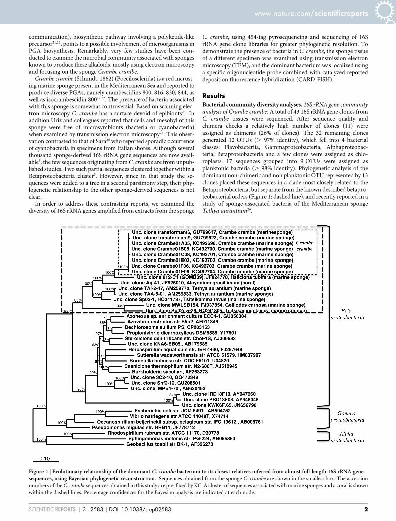
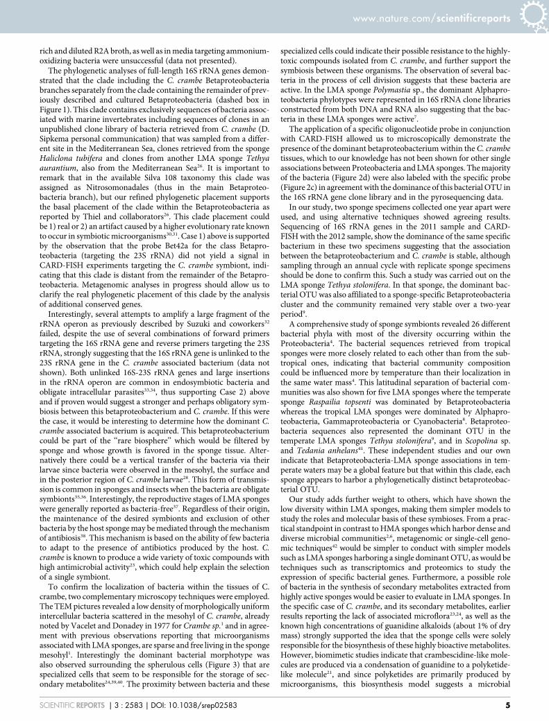
ResultsBacterial community diversity analyses. 16S rRNA gene communityanalysis of Crambe crambe. A total of 43 16S rRNA gene clones fromC. crambe tissues were sequenced. After sequence quality andchimera checks a relatively high number of clones (11) wereassigned as chimeras (26% of clones). The 32 remaining clonesgenerated 12 OTUs (. 97% identity), which fell into 4 bacterialclasses: Flavobacteriia, Gammaproteobacteria, Alphaproteobac-teria, Betaproteobacteria and a few clones were assigned as chlo-roplasts. 17 sequences grouped into 9 OTUs were assigned asplanktonic bacteria (. 98% identity). Phylogenetic analysis of thedominant non-chimeric and non planktonic OTU represented by 13clones placed these sequences in a clade most closely related to theBetaproteobacteria, but separate from the known described betapro-teobacterial orders (Figure 1; dashed line), and recently reported in astudy of sponge-associated bacteria of the Mediterranean spongeTethya aurantium26.
Figure 1 | Evolutionary relationship of the dominant C. crambe bacterium to its closest relatives inferred from almost full-length 16S rRNA genesequences, using Bayesian phylogenetic reconstruction. Sequences obtained from the sponge C. crambe are shown in the smallest box. The accession
numbers of the C. crambe sequences obtained in this study are pre-fixed by KC. A cluster of sequences associated with marine sponges and a coral is shown
within the dashed lines. Percentage confidences for the Bayesian analysis are indicated at each node.
www.nature.com/scientificreports
SCIENTIFIC REPORTS | 3 : 2583 | DOI: 10.1038/srep02583 2
454- 16S rRNA Pyrosequencing analyses. From an initial 5973 rawbacterial 16S rRNA tag sequences obtained from C. crambe, 4421sequences remained after denoising by AmpliconNoise. Subsequentstringent chimera detection removed a significant number of chi-meric sequences, many of which were singletons, which would haveotherwise significantly overinflated the total bacterial diversity (213chimeric sequences representing 70 OTUs). Rarefaction analysis ofnon-chimeric sequences shows a low level of diversity with rarefac-tion curve reaching a plateau at about 115 OTUs (Figure S1). Thislow diversity was also reflected by a Chao1 value of 107.7 and a smalltotal number of OTUs recovered (86 OTUs). Furthermore, amongthese OTUs, a single OTU, very similar (. 99% identity) in the V1-V3 region to the dominant clone in the 16S rRNA library, repre-sented 74% of the total sequences (3271 sequences). Among theremaining 85 OTUs, 42 (representing 22% of total sequences) wereclassified as planktonic. Therefore, considering only non-planktonicbacteria, likely transient in the sponge, this dominant betaproteobac-terium accounted for a remarkable 95% of the total sequences.
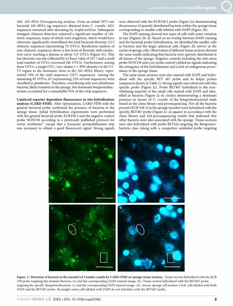
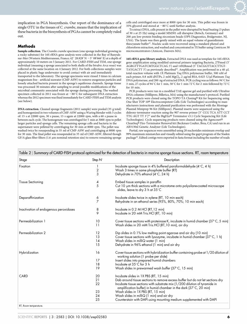
Catalyzed reporter deposition fluorescence in situ hybridizationanalyses (CARD-FISH). After optimization, CARD-FISH with thegeneral bacterial probe confirmed the presence of bacteria in thesponge tissue. Initial hybridization experiments were performedwith the general bacterial probe EUB338-I and the negative controlprobe NON338 according to a previously published protocol forworm symbionts27 except that a lysozyme permeabilization stepwas necessary to obtain a good fluorescent signal. Strong signals
were observed with the EUB338-I probe (Figure 2a) demonstratingthe presence of sparsely distributed bacteria within the sponge tissuecorresponding to smaller cells labeled with DAPI (Figure 2b).
The DAPI staining showed two types of cells with some variationin size (Figures 2b–d). Based on an overlap between DAPI stainingand the bacterial probe hybridization, we identified the smaller cellsas bacteria and the larger spherical cells (Figure 2b; arrow) as thenuclei of sponge cells. Observation of different tissue sections showedthe same results indicating that bacteria were sparsely distributed inall tissues of the sponge. Negative controls including the anti-senseprobe NON338 and a no-probe control yielded no signals indicatingthe stringency of the hybridization and a lack of endogenous perox-idases in the sponge tissue.
The same tissue sections were also stained with DAPI and hybri-dized with the specific BET 467 probe and its helper probes(sequences shown in Table 1). Strong signals were observed with thisspecific probe (Figure 2c). Probe BET467 hybridized to the over-whelming majority of the small cells stained with DAPI and iden-tified as bacteria (Figure 2c–d; circles) demonstrating a dominantpresence in tissues of C. crambe of the betaproteobacterial cladefound in the clone library and pyrosequencing. Not all the bacteriapresent (EUB 338-I) in the sponge mesohyl were hybridized with thespecific BET467 probe (Figure 2c–d; square) in accordance with theclone library and 454-pyrosequencing results that indicated thatother bacteria were also associated with the sponge. Tissue sectionswere also hybridized with probe BET42a targeting the Betaproteo-bacteria class (along with a competitor unlabeled probe targeting
Figure 2 | Detection of bacteria in the mesohyl of Crambe crambe by CARD-FISH on sponge-tissue sections. Tissue section hybridized with the EUB
338 probe targeting the domain Bacteria, (a) and the corresponding DAPI-stained image, (b). Tissue section hybridized with the BET467 probe
targeting the specific Betaproteobacteria, (c) and the corresponding DAPI-stained image, (d). Arrow: sponge cell nucleus. Oval: cells labeled with both
DAPI and the BET467 probe. Rectangle: some cells labeled with DAPI do not hybridize with the BET467 probe.
www.nature.com/scientificreports
SCIENTIFIC REPORTS | 3 : 2583 | DOI: 10.1038/srep02583 3
Gammaproteobacteria). Surprisingly no signal was detected. Finally,no signal was detected in hybridizations with the ARCH915 probesuggesting the absence of Archaea in C. crambe. Even though FISHtargeting eukaryotes was not performed, only sporadically red/orange fluorescence was observed under green excitation, thus it isunlikely that photosynthetic eukaryotes or cyanobacteria are import-ant members of the microbial community associated with C. crambe.
Although we did not use a confocal microscope, and thus did notaccurately quantify bacterial cells, an analysis of 20 different 20 mm2
areas showed an average of 6 bacterial cells/sponge cell (5Betaproteobacteria/sponge cell), resulting roughly in 3000 bacterialcells (2500 Betaproteobacteria cells) per cm2 of thin section. Sincethese micrographs were not randomly visualized, these numbers arelikely upper estimates.
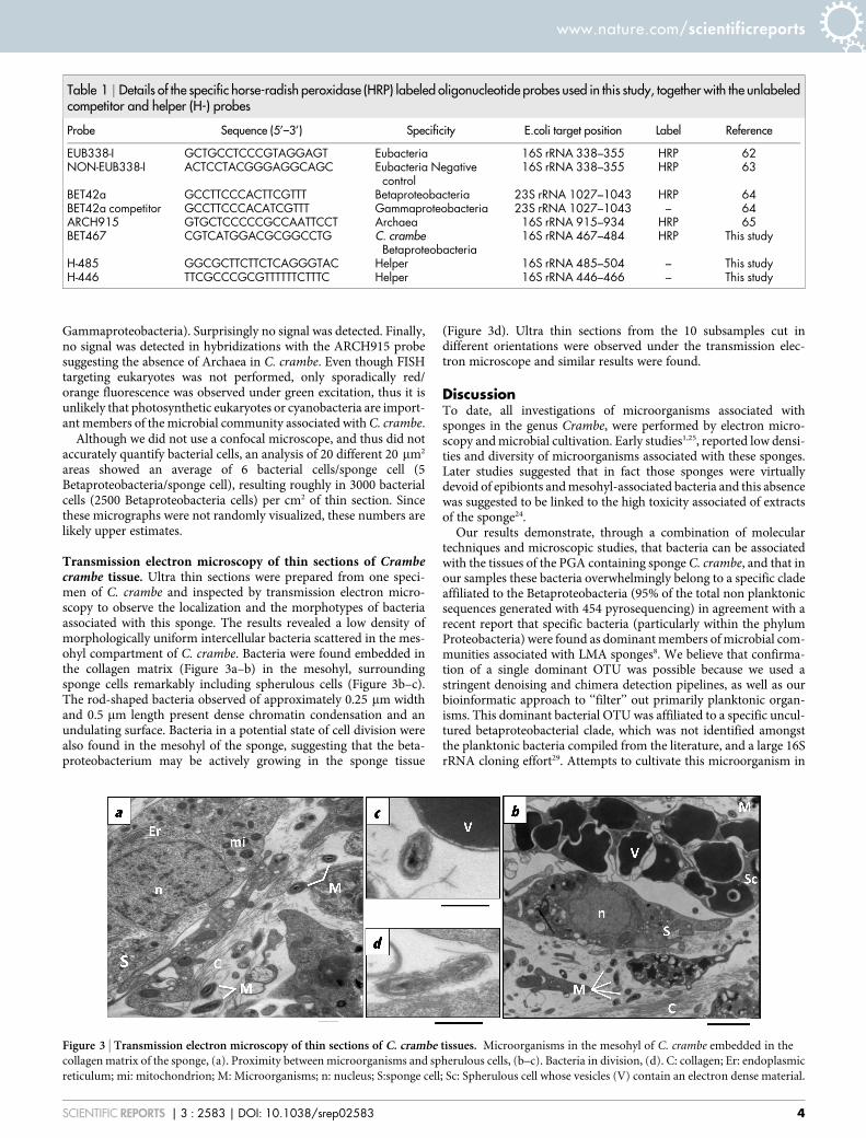
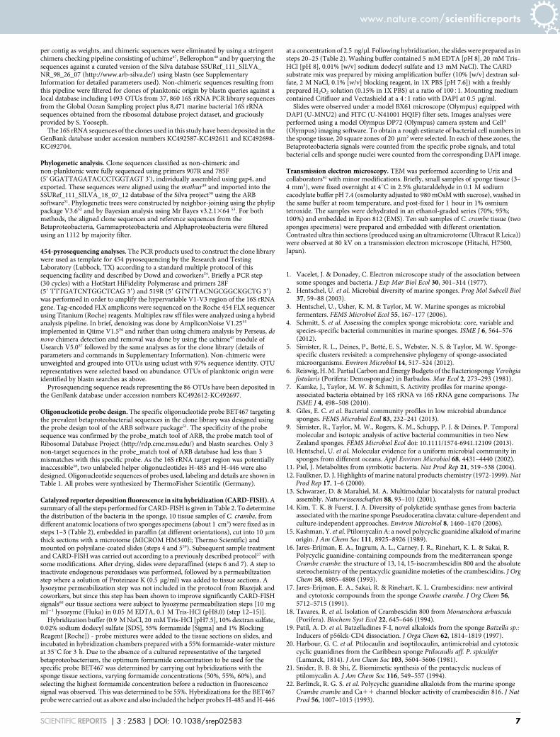
Transmission electron microscopy of thin sections of Crambecrambe tissue. Ultra thin sections were prepared from one speci-men of C. crambe and inspected by transmission electron micro-scopy to observe the localization and the morphotypes of bacteriaassociated with this sponge. The results revealed a low density ofmorphologically uniform intercellular bacteria scattered in the mes-ohyl compartment of C. crambe. Bacteria were found embedded inthe collagen matrix (Figure 3a–b) in the mesohyl, surroundingsponge cells remarkably including spherulous cells (Figure 3b–c).The rod-shaped bacteria observed of approximately 0.25 mm widthand 0.5 mm length present dense chromatin condensation and anundulating surface. Bacteria in a potential state of cell division werealso found in the mesohyl of the sponge, suggesting that the beta-proteobacterium may be actively growing in the sponge tissue
(Figure 3d). Ultra thin sections from the 10 subsamples cut indifferent orientations were observed under the transmission elec-tron microscope and similar results were found.
DiscussionTo date, all investigations of microorganisms associated withsponges in the genus Crambe, were performed by electron micro-scopy and microbial cultivation. Early studies1,25, reported low densi-ties and diversity of microorganisms associated with these sponges.Later studies suggested that in fact those sponges were virtuallydevoid of epibionts and mesohyl-associated bacteria and this absencewas suggested to be linked to the high toxicity associated of extractsof the sponge24.
Our results demonstrate, through a combination of moleculartechniques and microscopic studies, that bacteria can be associatedwith the tissues of the PGA containing sponge C. crambe, and that inour samples these bacteria overwhelmingly belong to a specific cladeaffiliated to the Betaproteobacteria (95% of the total non planktonicsequences generated with 454 pyrosequencing) in agreement with arecent report that specific bacteria (particularly within the phylumProteobacteria) were found as dominant members of microbial com-munities associated with LMA sponges8. We believe that confirma-tion of a single dominant OTU was possible because we used astringent denoising and chimera detection pipelines, as well as ourbioinformatic approach to ‘‘filter’’ out primarily planktonic organ-isms. This dominant bacterial OTU was affiliated to a specific uncul-tured betaproteobacterial clade, which was not identified amongstthe planktonic bacteria compiled from the literature, and a large 16SrRNA cloning effort29. Attempts to cultivate this microorganism in
Table 1 | Details of the specific horse-radish peroxidase (HRP) labeled oligonucleotide probes used in this study, together with the unlabeledcompetitor and helper (H-) probes
Probe Sequence (5’–3’) Specificity E.coli target position Label Reference
EUB338-I GCTGCCTCCCGTAGGAGT Eubacteria 16S rRNA 338–355 HRP 62NON-EUB338-I ACTCCTACGGGAGGCAGC Eubacteria Negative
control16S rRNA 338–355 HRP 63
BET42a GCCTTCCCACTTCGTTT Betaproteobacteria 23S rRNA 1027–1043 HRP 64BET42a competitor GCCTTCCCACATCGTTT Gammaproteobacteria 23S rRNA 1027–1043 – 64ARCH915 GTGCTCCCCCGCCAATTCCT Archaea 16S rRNA 915–934 HRP 65BET467 CGTCATGGACGCGGCCTG C. crambe
Betaproteobacteria16S rRNA 467–484 HRP This study
H-485 GGCGCTTCTTCTCAGGGTAC Helper 16S rRNA 485–504 – This studyH-446 TTCGCCCGCGTTTTTTCTTTC Helper 16S rRNA 446–466 – This study
Figure 3 | Transmission electron microscopy of thin sections of C. crambe tissues. Microorganisms in the mesohyl of C. crambe embedded in the
collagen matrix of the sponge, (a). Proximity between microorganisms and spherulous cells, (b–c). Bacteria in division, (d). C: collagen; Er: endoplasmic
reticulum; mi: mitochondrion; M: Microorganisms; n: nucleus; S:sponge cell; Sc: Spherulous cell whose vesicles (V) contain an electron dense material.
www.nature.com/scientificreports
SCIENTIFIC REPORTS | 3 : 2583 | DOI: 10.1038/srep02583 4
rich and diluted R2A broth, as well as in media targeting ammonium-oxidizing bacteria were unsuccessful (data not presented).
The phylogenetic analyses of full-length 16S rRNA genes demon-strated that the clade including the C. crambe Betaproteobacteriabranches separately from the clade containing the remainder of prev-iously described and cultured Betaproteobacteria (dashed box inFigure 1). This clade contains exclusively sequences of bacteria assoc-iated with marine invertebrates including sequences of clones in anunpublished clone library of bacteria retrieved from C. crambe (D.Sipkema personal communication) that was sampled from a differ-ent site in the Mediterranean Sea, clones retrieved from the spongeHaliclona tubifera and clones from another LMA sponge Tethyaaurantium, also from the Mediterranean Sea26. It is important toremark that in the available Silva 108 taxonomy this clade wasassigned as Nitrosomonadales (thus in the main Betaproteo-bacteria branch), but our refined phylogenetic placement supportsthe basal placement of the clade within the Betaproteobacteria asreported by Thiel and collaborators26. This clade placement couldbe 1) real or 2) an artifact caused by a higher evolutionary rate knownto occur in symbiotic microorganisms30,31. Case 1) above is supportedby the observation that the probe Bet42a for the class Betapro-teobacteria (targeting the 23S rRNA) did not yield a signal inCARD-FISH experiments targeting the C. crambe symbiont, indi-cating that this clade is distant from the remainder of the Betapro-teobacteria. Metagenomic analyses in progress should allow us toclarify the real phylogenetic placement of this clade by the analysisof additional conserved genes.
Interestingly, several attempts to amplify a large fragment of therRNA operon as previously described by Suzuki and coworkers32
failed, despite the use of several combinations of forward primerstargeting the 16S rRNA gene and reverse primers targeting the 23SrRNA, strongly suggesting that the 16S rRNA gene is unlinked to the23S rRNA gene in the C. crambe associated bacterium (data notshown). Both unlinked 16S-23S rRNA genes and large insertionsin the rRNA operon are common in endosymbiotic bacteria andobligate intracellular parasites33,34, thus supporting Case 2) aboveand if proven would suggest a stronger and perhaps obligatory sym-biosis between this betaproteobacterium and C. crambe. If this werethe case, it would be interesting to determine how the dominant C.crambe associated bacterium is acquired. This betaproteobacteriumcould be part of the ‘‘rare biosphere’’ which would be filtered bysponge and whose growth is favored in the sponge tissue. Alter-natively there could be a vertical transfer of the bacteria via theirlarvae since bacteria were observed in the mesohyl, the surface andin the posterior region of C. crambe larvae28. This form of transmis-sion is common in sponges and insects when the bacteria are obligatesymbionts35,36. Interestingly, the reproductive stages of LMA spongeswere generally reported as bacteria-free37. Regardless of their origin,the maintenance of the desired symbionts and exclusion of otherbacteria by the host sponge may be mediated through the mechanismof antibiosis38. This mechanism is based on the ability of few bacteriato adapt to the presence of antibiotics produced by the host. C.crambe is known to produce a wide variety of toxic compounds withhigh antimicrobial activity23, which could help explain the selectionof a single symbiont.
To confirm the localization of bacteria within the tissues of C.crambe, two complementary microscopy techniques were employed.The TEM pictures revealed a low density of morphologically uniformintercellular bacteria scattered in the mesohyl of C. crambe, alreadynoted by Vacelet and Donadey in 1977 for Crambe sp.1 and in agree-ment with previous observations reporting that microorganismsassociated with LMA sponges, are sparse and free living in the spongemesohyl1. Interestingly the dominant bacterial morphotype wasalso observed surrounding the spherulous cells (Figure 3) that arespecialized cells that seem to be responsible for the storage of sec-ondary metabolites24,39,40. The proximity between bacteria and these
specialized cells could indicate their possible resistance to the highly-toxic compounds isolated from C. crambe, and further support thesymbiosis between these organisms. The observation of several bac-teria in the process of cell division suggests that these bacteria areactive. In the LMA sponge Polymastia sp., the dominant Alphapro-teobacteria phylotypes were represented in 16S rRNA clone librariesconstructed from both DNA and RNA also suggesting that the bac-teria in these LMA sponges were active7.
The application of a specific oligonucleotide probe in conjunctionwith CARD-FISH allowed us to microscopically demonstrate thepresence of the dominant betaproteobacterium within the C. crambetissues, which to our knowledge has not been shown for other singleassociations between Proteobacteria and LMA sponges. The majorityof the bacteria (Figure 2d) were also labeled with the specific probe(Figure 2c) in agreement with the dominance of this bacterial OTU inthe 16S rRNA gene clone library and in the pyrosequencing data.
In our study, two sponge specimens collected one year apart wereused, and using alternative techniques showed agreeing results.Sequencing of 16S rRNA genes in the 2011 sample and CARD-FISH with the 2012 sample, show the dominance of the same specificbacterium in these two specimens suggesting that the associationbetween the betaproteobacterium and C. crambe is stable, althoughsampling through an annual cycle with replicate sponge specimensshould be done to confirm this. Such a study was carried out on theLMA sponge Tethya stolonifera. In that sponge, the dominant bac-terial OTU was also affiliated to a sponge-specific Betaproteobacteriacluster and the community remained very stable over a two-yearperiod9.
A comprehensive study of sponge symbionts revealed 26 differentbacterial phyla with most of the diversity occurring within theProteobacteria4. The bacterial sequences retrieved from tropicalsponges were more closely related to each other than from the sub-tropical ones, indicating that bacterial community compositioncould be influenced more by temperature than their localization inthe same water mass4. This latitudinal separation of bacterial com-munities was also shown for five LMA sponges where the temperatesponge Raspailia topsenti was dominated by Betaproteobacteriawhereas the tropical LMA sponges were dominated by Alphapro-teobacteria, Gammaproteobacteria or Cyanobacteria8. Betaproteo-bacteria sequences also represented the dominant OTU in thetemperate LMA sponges Tethya stolonifera9, and in Scopolina sp.and Tedania anhelans41. These independent studies and our ownindicate that Betaproteobacteria-LMA sponge associations in tem-perate waters may be a global feature but that within this clade, eachsponge appears to harbor a phylogenetically distinct betaproteobac-terial OTU.
Our study adds further weight to others, which have shown thelow diversity within LMA sponges, making them simpler models tostudy the roles and molecular basis of these symbioses. From a prac-tical standpoint in contrast to HMA sponges which harbor dense anddiverse microbial communities2,6, metagenomic or single-cell geno-mic techniques42 would be simpler to conduct with simpler modelssuch as LMA sponges harboring a single dominant OTU, as would betechniques such as transcriptomics and proteomics to study theexpression of specific bacterial genes. Furthermore, a possible roleof bacteria in the synthesis of secondary metabolites extracted fromhighly active sponges would be easier to evaluate in LMA sponges. Inthe specific case of C. crambe, and its secondary metabolites, earlierresults reporting the lack of associated microflora23,24, as well as theknown high concentrations of guanidine alkaloids (about 1% of drymass) strongly supported the idea that the sponge cells were solelyresponsible for the biosynthesis of these highly bioactive metabolites.However, biomimetic studies indicate that crambescidine-like mole-cules are produced via a condensation of guanidine to a polyketide-like molecule21, and since polyketides are primarily produced bymicroorganisms, this biosynthesis model suggests a microbial
www.nature.com/scientificreports
SCIENTIFIC REPORTS | 3 : 2583 | DOI: 10.1038/srep02583 5
implication in PGA biosynthesis. Our report of the dominance of asingle OTU in the tissues of C. crambe, means that the implication ofthese bacteria in the biosynthesis of PGAs cannot be completely ruledout.
MethodsSample collection. The Crambe crambe specimen (one sponge individual growing ina rocky substrate) for 16S rRNA gene analysis were collected in the Bay of Banyuls-sur-Mer (Western Mediterranean, France; 42u 28.828’ N 23u 08’666’ E) at a depth ofapproximately 10 meters on 5 January 2011. For CARD-FISH and TEM, one spongeindividual (meaning a sponge associated to both shells of the bivalve Arca noae) wascollected at the same location on 4 January 2012. For both collections samples wereplaced in plastic bags underwater to avoid contact with air and immediatelytransported to the laboratory. The sponge specimens were rinsed 3 times in calciummagnesium free - artificial seawater (CMF-ASW) to remove exogenous particles andloosely attached bacteria present in the sponge’s aquiferous channels. Sponge tissuewas processed 30 minutes after sampling to avoid possible modifications of themicrobial community associated with the sponge during processing. The washedspecimen collected in 2011 was frozen at 280uC for subsequent DNA extractionwhereas the 2012 specimen was fixed immediately for CARD-FISH and TEM analysis(see below).
DNA extraction. Cleaned sponge fragments (2011 sample) were pooled (35 g total)and homogenized in two volumes of CMF-ASW using a Waring blender with 3 cyclesof: 15 s at 22000 rpm, 30 s pause, 15 s again at 22000 rpm, with a 60 s pause inbetween each cycle. The homogenate was centrifuged for 5 min at 3000 rpm to pelletlarger particles and sponge cells. The remaining sponge cells and bacteria in thesupernatant were pelleted by centrifuging for 30 min at 8000 rpm. The pellet waswashed twice by resuspending in 35 ml of CMF-ASW and centrifuging at 8000 rpmfor 30 min. The final pellet was resuspended in 35 ml of CMF-ASW, filtered thoughGF/A glass fiber filters (1.6 mm nominal retention size) to remove remaining sponge
cells and centrifuged once more at 8000 rpm for 30 min. This pellet was frozen in35% glycerol and stored at 280uC until further analysis.
To extract DNA, cells present in the pellet were disrupted by bead beating (3 pulsesof 30 s at 25 Hz) using a model MM301 cell disruptor (Retsch, Germany) and200 mm low-protein binding zirconium beads (OPS Diagnostics, Bridgewater, NJ,USA). The lysate was then gently mixed with an equal volume of guanidiniumthiocyanate buffer43. Nucleic acids were recovered using a standard phenol andchloroform extraction, and washed and concentrated in TE buffer using Centricon 30microconcentrators (Amicon, Danvers MA).
16S rRNA gene library analysis. Extracted DNA was used as template for 16S rRNAgene amplification using modified universal primers targeting Bacteria, 27Fmod (5’AGRGTTTGATCMTGGCTCAG 3’) and 1492Rmod (5’ TACGGYTACCTTGT-TAYGACTT 3’) as previously described44. Amplification was performed in a 40 mLtotal-reaction volume with 1X Platinum Taq DNA polymerase buffer, 500 nM ofeach primer, 0.8 mM dNTPs, 2 mM MgCl2, 2 mg/ml BSA, 0.025 U/ml Platinum TaqDNA polymerase, and 200 ng of extracted DNA. PCR cycling was as follows: 94uC for3 min, 27 cycles of 94uC for 1 min, 50uC for 1 min 72uC for 2 min followed by 72uCfor 10 min.
PCR products were run in a modified TAE agarose gel and purified with UltrafreeDNA columns (Millipore, Billerica, MA) using the manufacturer’s protocol. PurifiedPCR products were cloned using the TOPO TA CloningH Kit for Sequencing usingOne Shot TOP 10H Electrocompetent Cells (Life Technologies) according to man-ufacturers instructions and plasmid purification was performed with the MontagePlasmid Miniprep 96 Kit (Millipore). Plasmid inserts were sequenced using thedideoxy-terminator reaction using the 907 reverse primer [59 CCG TCA ATT CCTTTG AGT TT 39]45 and the BigDyeH Terminator v3.1 Cycle Sequencing Kit (LifeTechnologies). Cycle sequencing products were cleaned using the AgencourtHCleanSeqH Dye-Terminator Removal kit (Beckman Coulter, Brea, CA) and run in anAB3130xl Genetic Analyzer (Life Technologies).
Partial, raw sequences were assembled using 20 nucleotides minimum overlap and99% maximum mismatches and visually edited using the gap4 program of the Stadenpackage46. Edited contigs were exported in fasta format including the number of reads
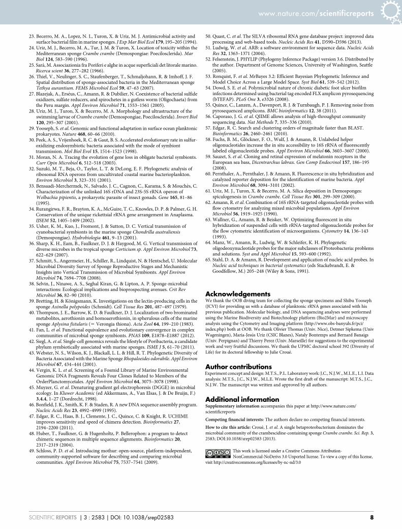
Table 2 | Summary of CARD-FISH protocol optimized for the detection of bacteria in marine sponge tissue sections. RT, room temperature
Stage Step Nu Description
Fixation 1 Incubate sponge tissue in 4% buffered paraformaldehyde (4uC, 4 h)2 Wash 3 times in same phosphate buffer (RT)3 Dehydrate in 70% ethanol (4uC, 24 h)
Tissue Sectioning 4 Embed tissue samples in paraffin5 Cut 10 mm thick sections with a microtome onto polysilane-coated microscope
slides, leave to dry 3 h at 35uC
Deparaffinization 6 Incubate slides twice in xylene (RT, 10 min each)7 Rehydrate in an ethanol series (95%, 80%, 70%; 10 min each)
Inactivation of endogenous peroxidases 8 Incubate in 0.2 M HCl (RT,12 min)9 Incubate in 20 mM Tris.HCl (RT, 10 min)
Permeabilization 1 10 Cover tissue sections with proteinase K, incubate in humid chamber (37uC, 5 min)11 Wash slides in 20 mM Tris.HCl (RT,10 min), air dry
Permeabilization 2 12 Dip slides in 0.1% low melting point agarose and air dry (10 min)13 Cover tissue sections with lysozyme, incubate in humid chamber (37uC, 1 h)14 Wash slides in milliQ water (1 min)15 Dehydrate in 96% ethanol (1 min) and air dry
Hybridization 16 Cover tissue sections with hybridization buffer containing probe at 1/20 dilution ofworking solution (1 probe per slide)
17 Insert slides into prepared humid chambers18 Incubate at 35uC for 3 h19 Wash slides in prewarmed wash buffer (37uC, 15 min)
CARD 20 Incubate slides in 1X PBS (RT, 15 min)21 Dab around tissue sections to remove excess buffer but do not let sections dry22 Incubate tissue sections with substrate mix (1/200 dilution of tyramide in
amplification buffer) in humid chamber in the dark (37uC, 20 min)23 Wash slides in 1X PBS (RT, 15 min)24 Wash slides in milliQ (1 min) and air dry25 Counterstain with DAPI using mounting medium supplemented with DAPI
RT, Room temperature.
www.nature.com/scientificreports
SCIENTIFIC REPORTS | 3 : 2583 | DOI: 10.1038/srep02583 6
per contig as weights, and chimeric sequences were eliminated by using a stringentchimera checking pipeline consisting of uchime47, Bellerophon48 and by querying thesequences against a curated version of the Silva database SSURef_111_SILVA_NR_98_26_07 (http://www.arb-silva.de/) using blastn (see SupplementaryInformation for detailed parameters used). Non-chimeric sequences resulting fromthis pipeline were filtered for clones of planktonic origin by blastn queries against alocal database including 1493 OTUs from 37, 860 16S rRNA PCR library sequencesfrom the Global Ocean Sampling project plus 8,471 marine bacterial 16S rRNAsequences obtained from the ribosomal database project dataset, and graciouslyprovided by S. Yooseph.
The 16S rRNA sequences of the clones used in this study have been deposited in theGenBank database under accession numbers KC492587-KC492611 and KC492698-KC492704.
Phylogenetic analysis. Clone sequences classified as non-chimeric andnon-planktonic were fully sequenced using primers 907R and 785F(5’ GGATTAGATACCCTGGTAGT 3’), individually assembled using gap4, andexported. These sequences were aligned using the mothur49 and imported into theSSURef_111_SILVA_18_07_12 database of the Silva project50 using the ARBsoftware51. Phylogenetic trees were constructed by neighbor-joining using the phylippackage V3.652 and by Bayesian analysis using Mr Bayes v3.2.1364 53. For bothmethods, the aligned clone sequences and reference sequences from theBetaproteobacteria, Gammaproteobacteria and Alphaproteobacteria were filteredusing an 1112 bp majority filter.
454-pyrosequencing analyses. The PCR products used to construct the clone librarywere used as template for 454 pyrosequencing by the Research and TestingLaboratory (Lubbock, TX) according to a standard multiple protocol of thissequencing facility and described by Dowd and coworkers54. Briefly a PCR step(30 cycles) with a HotStart HiFidelity Polymerase and primers 28F(59 TTTGATCNTGGCTCAG 39) and 519R (59 GTNTTACNGCGGCKGCTG 39)was performed in order to amplify the hypervariable V1-V3 region of the 16S rRNAgene. Tag-encoded FLX amplicons were sequenced on the Roche 454 FLX sequencerusing Titanium (Roche) reagents. Multiplex raw sff files were analyzed using a hybridanalysis pipeline. In brief, denoising was done by AmpliconNoise V1.2555
implemented in Qiime V1.556 and rather than using chimera analysis by Perseus, denovo chimera detection and removal was done by using the uchime47 module ofUsearch V5.057 followed by the same analyses as for the clone library (details ofparameters and commands in Supplementary Information). Non-chimeric wereunweighted and grouped into OTUs using uclust with 97% sequence identity. OTUrepresentatives were selected based on abundance. OTUs of planktonic origin wereidentified by blastn searches as above.
Pyrosequencing sequence reads representing the 86 OTUs have been deposited inthe GenBank database under accession numbers KC492612-KC492697.
Oligonucleotide probe design. The specific oligonucleotide probe BET467 targetingthe prevalent betaproteobacterial sequences in the clone library was designed usingthe probe design tool of the ARB software package51. The specificity of the probesequence was confirmed by the probe_match tool of ARB, the probe match tool ofRibosomal Database Project (http://rdp.cme.msu.edu/) and blastn searches. Only 3non-target sequences in the probe_match tool of ARB database had less than 3mismatches with this specific probe. As the 16S rRNA target region was potentiallyinaccessible58, two unlabeled helper oligonucleotides H-485 and H-446 were alsodesigned. Oligonucleotide sequences of probes used, labeling and details are shown inTable 1. All probes were synthesized by ThermoFisher Scientific (Germany).
Catalyzed reporter deposition fluorescence in situ hybridization (CARD-FISH). Asummary of all the steps performed for CARD-FISH is given in Table 2. To determinethe distribution of the bacteria in the sponge, 10 tissue samples of C. crambe, fromdifferent anatomic locations of two sponges specimens (about 1 cm3) were fixed as insteps 1–3 (Table 2), embedded in paraffin (at different orientations), cut into 10 mmthick sections with a microtome (MICROM HM340E; Thermo Scientific) andmounted on polysilane-coated slides (steps 4 and 559). Subsequent sample treatmentand CARD-FISH was carried out according to a previously described protocol27 withsome modifications. After drying, slides were deparaffined (steps 6 and 7). A step toinactivate endogenous peroxidases was performed, followed by a permeabilizationstep where a solution of Proteinase K (0.5 mg/ml) was added to tissue sections. Alysozyme permeabilization step was not included in the protocol from Blazejak andcoworkers, but since this step has been shown to improve significantly CARD-FISHsignals60 our tissue sections were subject to lysozyme permeabilization steps [10 mgml21 lysozyme (Fluka) in 0.05 M EDTA, 0.1 M Tris-HCl (pH8.0) (step 12–15)].
Hybridization buffer (0.9 M NaCl, 20 mM Tris-HCl [pH7.5], 10% dextran sulfate,0.02% sodium dodecyl sulfate [SDS], 55% formamide [Sigma] and 1% BlockingReagent [Roche]) - probe mixtures were added to the tissue sections on slides, andincubated in hybridization chambers prepared with a 55% formamide-water mixtureat 35uC for 3 h. Due to the absence of a cultured representative of the targetedbetaproteobacterium, the optimum formamide concentration to be used for thespecific probe BET467 was determined by carrying out hybridizations with thesponge tissue sections, varying formamide concentrations (50%, 55%, 60%), andselecting the highest formamide concentration before a reduction in fluorescencesignal was observed. This was determined to be 55%. Hybridizations for the BET467probe were carried out as above and also included the helper probes H-485 and H-446
at a concentration of 2.5 ng/ml. Following hybridization, the slides were prepared as insteps 20–25 (Table 2). Washing buffer contained 5 mM EDTA [pH 8], 20 mM Tris–HCl [pH 8], 0.01% [w/v] sodium dodecyl sulfate and 13 mM NaCl). The CARDsubstrate mix was prepared by mixing amplification buffer (10% [w/v] dextran sul-fate, 2 M NaCl, 0.1% [w/v] blocking reagent, in 1X PBS [pH 7.6]) with a freshlyprepared H2O2 solution (0.15% in 1X PBS) at a ratio of 10051. Mounting mediumcontained Citifluor and Vectashield at a 451 ratio with DAPI at 0.5 mg/ml.
Slides were observed under a model BX61 microscope (Olympus) equipped withDAPI (U-MNU2) and FITC (U-N41001 HQIF) filter sets. Images analyses wereperformed using a model Olympus DP72 (Olympus) camera system and CellA
(Olympus) imaging software. To obtain a rough estimate of bacterial cell numbers inthe sponge tissue, 20 square zones of 20 mm2 were selected. In each of these zones, theBetaproteobacteria signals were counted from the specific probe signals, and totalbacterial cells and sponge nuclei were counted from the corresponding DAPI image.
Transmission electron microscopy. TEM was performed according to Uriz andcollaborators61 with minor modifications. Briefly, small samples of sponge tissue (3–4 mm3), were fixed overnight at 4uC in 2.5% glutaraldehyde in 0.1 M sodiumcacodylate buffer pH 7.4 (osmolarity adjusted to 980 mOsM with sucrose), washed inthe same buffer at room temperature, and post-fixed for 1 hour in 1% osmiumtetroxide. The samples were dehydrated in an ethanol-graded series (70%; 95%;100%) and embedded in Epon 812 (EMS). Ten sub samples of C. crambe tissue (twosponges specimens) were prepared and embedded with different orientation.Contrasted ultra thin sections (produced using an ultramicrotome (Ultracut R Leica))were observed at 80 kV on a transmission electron microscope (Hitachi, H7500,Japan).
1. Vacelet, J. & Donadey, C. Electron microscope study of the association betweensome sponges and bacteria. J Exp Mar Biol Ecol 30, 301–314 (1977).
2. Hentschel, U. et al. Microbial diversity of marine sponges. Prog Mol Subcell Biol37, 59–88 (2003).
3. Hentschel, U., Usher, K. M. & Taylor, M. W. Marine sponges as microbialfermenters. FEMS Microbiol Ecol 55, 167–177 (2006).
4. Schmitt, S. et al. Assessing the complex sponge microbiota: core, variable andspecies-specific bacterial communities in marine sponges. ISME J 6, 564–576(2012).
5. Simister, R. L., Deines, P., Botte, E. S., Webster, N. S. & Taylor, M. W. Sponge-specific clusters revisited: a comprehensive phylogeny of sponge-associatedmicroorganisms. Environ Microbiol 14, 517–524 (2012).
6. Reiswig, H. M. Partial Carbon and Energy Budgets of the Bacteriosponge Verohgiafistularis (Porifera: Demospongiae) in Barbados. Mar Ecol 2, 273–293 (1981).
7. Kamke, J., Taylor, M. W. & Schmitt, S. Activity profiles for marine sponge-associated bacteria obtained by 16S rRNA vs 16S rRNA gene comparisons. TheISME J 4, 498–508 (2010).
8. Giles, E. C. et al. Bacterial community profiles in low microbial abundancesponges. FEMS Microbiol Ecol 83, 232–241 (2013).
9. Simister, R., Taylor, M. W., Rogers, K. M., Schupp, P. J. & Deines, P. Temporalmolecular and isotopic analysis of active bacterial communities in two NewZealand sponges. FEMS Microbiol Ecol doi: 10.1111/1574-6941.12109 (2013).
10. Hentschel, U. et al. Molecular evidence for a uniform microbial community insponges from different oceans. Appl Environ Microbiol 68, 4431–4440 (2002).
11. Piel, J. Metabolites from symbiotic bacteria. Nat Prod Rep 21, 519–538 (2004).12. Faulkner, D. J. Highlights of marine natural products chemistry (1972-1999). Nat
Prod Rep 17, 1–6 (2000).13. Schwarzer, D. & Marahiel, M. A. Multimodular biocatalysts for natural product
assembly. Naturwissenschaften 88, 93–101 (2001).14. Kim, T. K. & Fuerst, J. A. Diversity of polyketide synthase genes from bacteria
associated with the marine sponge Pseudoceratina clavata: culture-dependent andculture-independent approaches. Environ Microbiol 8, 1460–1470 (2006).
15. Kashman, Y. et al. Ptilomycalin A: a novel polycyclic guanidine alkaloid of marineorigin. J Am Chem Soc 111, 8925–8926 (1989).
16. Jares-Erijman, E. A., Ingrum, A. L., Carney, J. R., Rinehart, K. L. & Sakai, R.Polycyclic guanidine-containing compounds from the mediterranean spongeCrambe crambe: the structure of 13, 14, 15-isocrambescidin 800 and the absolutestereochemistry of the pentacyclic guanidine moieties of the crambescidins. J OrgChem 58, 4805–4808 (1993).
17. Jares-Erijman, E. A., Sakai, R. & Rinehart, K. L. Crambescidins: new antiviraland cytotoxic compounds from the sponge Crambe crambe. J Org Chem 56,5712–5715 (1991).
18. Tavares, R. et al. Isolation of Crambescidin 800 from Monanchora arbuscula(Porifera). Biochem Syst Ecol 22, 645–646 (1994).
19. Patil, A. D. et al. Batzelladines F-I, novel alkaloids from the sponge Batzella sp.:Inducers of p56lck-CD4 dissociation. J Orga Chem 62, 1814–1819 (1997).
20. Harbour, G. C. et al. Ptilocaulin and isoptilocaulin, antimicrobial and cytotoxiccyclic guanidines from the Caribbean sponge Ptilocaulis aff. P. spiculifer(Lamarck, 1814). J Am Chem Soc 103, 5604–5606 (1981).
21. Snider, B. B. & Shi, Z. Biomimetic synthesis of the pentacyclic nucleus ofptilomycalin A. J Am Chem Soc 116, 549–557 (1994).
22. Berlinck, R. G. S. et al. Polycyclic guanidine alkaloids from the marine spongeCrambe crambe and Ca11 channel blocker activity of crambescidin 816. J NatProd 56, 1007–1015 (1993).
www.nature.com/scientificreports
SCIENTIFIC REPORTS | 3 : 2583 | DOI: 10.1038/srep02583 7
23. Becerro, M. A., Lopez, N. I., Turon, X. & Uriz, M. J. Antimicrobial activity andsurface bacterial film in marine sponges. J Exp Mar Biol Ecol 179, 195–205 (1994).
24. Uriz, M. J., Becerro, M. A., Tur, J. M. & Turon, X. Location of toxicity within theMediterranean sponge Crambe crambe (Demospongiae: Poecilosclerida). MarBiol 124, 583–590 (1996).
25. Sara, M. Associazionia fra Poriferi e alghe in acque superficiali det litorale marino.Ricerca scient 36, 277–282 (1966).
26. Thiel, V., Neulinger, S. C., Staufenberger, T., Schmaljohann, R. & Imhoff, J. F.Spatial distribution of sponge-associated bacteria in the Mediterranean spongeTethya aurantium. FEMS Microbiol Ecol 59, 47–63 (2007).
27. Blazejak, A., Erseus, C., Amann, R. & Dubilier, N. Coexistence of bacterial sulfideoxidizers, sulfate reducers, and spirochetes in a gutless worm (Oligochaeta) fromthe Peru margin. Appl Environ Microbiol 71, 1553–1561 (2005).
28. Uriz, M. J., Turon, X. & Becerro, M. A. Morphology and ultrastructure of theswimming larvae of Crambe crambe (Demospongiae, Poecilosclerida). Invert Biol120, 295–307 (2001).
29. Yooseph, S. et al. Genomic and functional adaptation in surface ocean planktonicprokaryotes. Nature 468, 60–66 (2010).
30. Peek, A. S., Vrijenhoek, R. C. & Gaut, B. S. Accelerated evolutionary rate in sulfur-oxidizing endosymbiotic bacteria associated with the mode of symbionttransmission. Mol Biol Evol 15, 1514–1523 (1998).
31. Moran, N. A. Tracing the evolution of gene loss in obligate bacterial symbionts.Curr Opin Microbiol 6, 512–518 (2003).
32. Suzuki, M. T., Beja, O., Taylor, L. T. & DeLong, E. F. Phylogenetic analysis ofribosomal RNA operons from uncultivated coastal marine bacterioplankton.Environ Microbiol 3, 323–331 (2001).
33. Bensaadi-Merchermek, N., Salvado, J. C., Cagnon, C., Karama, S. & Mouches, C.Characterization of the unlinked 16S rDNA and 23S-5S rRNA operon ofWolbachia pipientis, a prokaryotic parasite of insect gonads. Gene 165, 81–86(1995).
34. Rurangirwa, F. R., Brayton, K. A., McGuire, T. C., Knowles, D. P. & Palmer, G. H.Conservation of the unique rickettsial rRNA gene arrangement in Anaplasma.IJSEM 52, 1405–1409 (2002).
35. Usher, K. M., Kuo, J., Fromont, J. & Sutton, D. C. Vertical transmission ofcyanobacterial symbionts in the marine sponge Chondrilla australiensis(Demospongiae). Hydrobiologia 461, 9–13 (2001).
36. Sharp, K. H., Eam, B., Faulkner, D. J. & Haygood, M. G. Vertical transmission ofdiverse microbes in the tropical sponge Corticium sp. Appl Environ Microbiol 73,622–629 (2007).
37. Schmitt, S., Angermeier, H., Schiller, R., Lindquist, N. & Hentschel, U. MolecularMicrobial Diversity Survey of Sponge Reproductive Stages and MechanisticInsights into Vertical Transmission of Microbial Symbionts. Appl EnvironMicrobiol 74, 7694–7708 (2008).
38. Selvin, J., Ninawe, A. S., Seghal Kiran, G. & Lipton, A. P. Sponge-microbialinteractions: Ecological implications and bioprospecting avenues. Crit RevMicrobiol 36, 82–90 (2010).
39. Bretting, H. & Konigsmann, K. Investigations on the lectin-producing cells in thesponge Axinella polypoides (Schmidt). Cell Tissue Res 201, 487–497 (1979).
40. Thompson, J. E., Barrow, K. D. & Faulkner, D. J. Localization of two brominatedmetabolites, aerothionin and homoaerothionin, in spherulous cells of the marinesponge Aplysina fistularis (5 Verongia thiona). Acta Zool 64, 199–210 (1983).
41. Fan, L. et al. Functional equivalence and evolutionary convergence in complexcommunities of microbial sponge symbionts. PNAS 109, E1878–E1887 (2012).
42. Siegl, A. et al. Single-cell genomics reveals the lifestyle of Poribacteria, a candidatephylum symbiotically associated with marine sponges. ISME J 5, 61–70 (2011).
43. Webster, N. S., Wilson, K. J., Blackall, L. L. & Hill, R. T. Phylogenetic Diversity ofBacteria Associated with the Marine Sponge Rhopaloeides odorabile. Appl EnvironMicrobiol 67, 434–444 (2001).
44. Vergin, K. L. et al. Screening of a Fosmid Library of Marine EnvironmentalGenomic DNA Fragments Reveals Four Clones Related to Members of theOrderPlanctomycetales. Appl Environ Microbiol 64, 3075–3078 (1998).
45. Muyzer, G. et al. Denaturing gradient gel electrophoresis (DGGE) in microbialecology. In Kluwer Academic (ed Akkermans, A., Van Elsas, J. & De Bruijn, F.)3.4.4, 1–27 (Dordrecht, 1998).
46. Bonfield, J. K., Smith, K. F. & Staden, R. A new DNA sequence assembly program.Nucleic Acids Res 23, 4992–4999 (1995).
47. Edgar, R. C., Haas, B. J., Clemente, J. C., Quince, C. & Knight, R. UCHIMEimproves sensitivity and speed of chimera detection. Bioinformatics 27,2194–2200 (2011).
48. Huber, T., Faulkner, G. & Hugenholtz, P. Bellerophon: a program to detectchimeric sequences in multiple sequence alignments. Bioinformatics 20,2317–2319 (2004).
49. Schloss, P. D. et al. Introducing mothur: open-source, platform-independent,community-supported software for describing and comparing microbialcommunities. Appl Environ Microbiol 75, 7537–7541 (2009).
50. Quast, C. et al. The SILVA ribosomal RNA gene database project: improved dataprocessing and web-based tools. Nucleic Acids Res 41, D590–D596 (2013).
51. Ludwig, W. et al. ARB: a software environment for sequence data. Nucleic AcidsRes 32, 1363–1371 (2004).
52. Felsenstein, J. PHYLIP (Phylogeny Inference Package) version 3.6. Distributed bythe author. Department of Genome Sciences, University of Washington, Seattle(2005).
53. Ronquist, F. et al. MrBayes 3.2: Efficient Bayesian Phylogenetic Inference andModel Choice Across a Large Model Space. Syst Biol 61, 539–542 (2012).
54. Dowd, S. E. et al. Polymicrobial nature of chronic diabetic foot ulcer biofilminfections determined using bacterial tag encoded FLX amplicon pyrosequencing(bTEFAP). PLoS One 3, e3326 (2008).
55. Quince, C., Lanzen, A., Davenport, R. J. & Turnbaugh, P. J. Removing noise frompyrosequenced amplicons. BMC bioinformatics 12, 38 (2011).
56. Caporaso, J. G. et al. QIIME allows analysis of high-throughput communitysequencing data. Nat Methods 7, 335–336 (2010).
57. Edgar, R. C. Search and clustering orders of magnitude faster than BLAST.Bioinformatics 26, 2460–2461 (2010).
58. Fuchs, B. M., Glockner, F. O., Wulf, J. & Amann, R. Unlabeled helperoligonucleotides increase the in situ accessibility to 16S rRNA of fluorescentlylabeled oligonucleotide probes. Appl Environ Microbiol 66, 3603–3607 (2000).
59. Sauzet, S. et al. Cloning and retinal expression of melatonin receptors in theEuropean sea bass, Dicentrarchus labrax. Gen Comp Endocrinol 157, 186–195(2008).
60. Pernthaler, A., Pernthaler, J. & Amann, R. Fluorescence in situ hybridization andcatalyzed reporter deposition for the identification of marine bacteria. ApplEnviron Microbiol 68, 3094–3101 (2002).
61. Uriz, M. J., Turon, X. & Becerro, M. A. Silica deposition in Demosponges:spiculogenesis in Crambe crambe. Cell Tissue Res 301, 299–309 (2000).
62. Amann, R. et al. Combination of 16S rRNA-targeted oligonucleotide probes withflow cytometry for analyzing mixed microbial populations. Appl EnvironMicrobiol 56, 1919–1925 (1990).
63. Wallner, G., Amann, R. & Beisker, W. Optimizing fluorescent in situhybridization of suspended cells with rRNA-targeted oligonucleotide probes forthe flow cytometric identification of microorganisms. Cytometry 14, 136–143(1993).
64. Manz, W., Amann, R., Ludwig, W. & Schleifer, K. H. Phylogeneticoligodeoxynucleotide probes for the major subclasses of Proteobacteria: problemsand solutions. Syst and Appl Microbiol 15, 593–600 (1992).
65. Stahl, D. A. & Amann, R. Development and application of nucleic acid probes. InNucleic acid techniques in bacterial systematics (eds Stackebrandt, E. &Goodfellow, M.) 205–248 (Wiley & Sons, 1991).
AcknowledgementsWe thank the OOB diving team for collecting the sponge specimens and Shibu Yooseph(JCVI) for providing us with a database of planktonic rRNA genes associated with hisprevious publication. Molecular biology, and DNA sequencing analyses were performedusing the Marine Biodiversity and Biotechnology platform (Bio2Mar) and microscopyanalysis using the Cytometry and Imaging platform (http://www.obs-banyuls.fr/pci/index.php) both at OOB. We thank Olivier Thomas (Univ. Nice), Detmer Sipkema (UnivWageningen), Maria-Jesus Uriz (CSIC Blanes), Nataly Bontemps and Bernard Banaigs(Univ. Perpignan) and Thierry Perez (Univ. Marseille) for suggestions to the experimentalwork and very fruitful discussions. We thank the UPMC doctoral school 392 (Diversity ofLife) for its doctoral fellowship to Julie Croue.
Author contributionsExperiment concept and design: M.T.S., P.L. Laboratory work: J.C., N.J.W., M.L.E., L.I. Dataanalysis: M.T.S., J.C., N.J.W., M.L.E. Wrote the first draft of the manuscript: M.T.S., J.C.,N.J.W. The manuscript was written and approved by all authors.
Additional informationSupplementary information accompanies this paper at http://www.nature.com/scientificreports
Competing financial interests: The authors declare no competing financial interests.
How to cite this article: Croue, J. et al. A single betaproteobacterium dominates themicrobial community of the crambescidine-containing sponge Crambe crambe. Sci. Rep. 3,2583; DOI:10.1038/srep02583 (2013).
This work is licensed under a Creative Commons Attribution-NonCommercial-NoDerivs 3.0 Unported license. To view a copy of this license,
visit http://creativecommons.org/licenses/by-nc-nd/3.0
www.nature.com/scientificreports
SCIENTIFIC REPORTS | 3 : 2583 | DOI: 10.1038/srep02583 8
Related Documents


