PAEDIATRIC A simple method for bone age assessment: the capitohamate planimetry Jung-Ah Choi 1 & Young Chul Kim 1 & Seon Jeong Min 1 & Eun Kyung Khil 1 Received: 28 September 2017 /Revised: 4 December 2017 /Accepted: 18 December 2017 # The Author(s) 2018. This article is an open access publication Abstract Objectives To determine if the capitohamate (CH) planimetry could be a reliable indicator of bone age, and to compare it with Greulich-Pyle (GP) method. Methods This retrospective study included 391 children (age, 1–180 months). Two reviewers manually measured the areas of the capitate and hamate on plain radiographs. CH planimetry was defined as the measurement of the sum of areas of the capitate and hamate. Two reviewers independently applied the CH planimetry and GP methods in 109 children whose heights were at the 50th percentile of the growth chart. Results There was a strong positive correlation between chronological age and CH planimetry measurement (right, r = 0.9702; left, r = 0.9709). There was no significant difference in accuracy between CH planimetry (84.39–84.46 %) and the GP method (85.15–87.66 %) (p ≥ 0.0867). The interobserver reproducibility of CH planimetry (precision, 4.42 %; 95 % limits of agreement [LOA], −10.5 to 13.4 months) was greater than that of the GP method (precision, 8.45 %; LOA, −29.5 to 21.1 months). Conclusions CH planimetry may be a reliable method for bone age assessment. Key Points • Bone age assessment is important in the work-up of paediatric endocrine disorders. • Radiography of the left hand is widely used to estimate bone age. • Capitatohamate planimetry is a reliable and reproducible method for assessing bone age. Keywords Hand . Radiography . Child . Bone . Growth Abbreviations CH Capitohamate GP Greulich-Pyle TW Tanner-White GR Gilsanz-Ratibin PACS Picture Archiving and Communication System ROI Region of interest Introduction Bone age assessment (BAA) is crucial in the evaluation of endocrine disorders and in the prediction of adult height when hormone therapy is the treatment [1]. Radiographic examination is an easy and cost-effective method for the assessment of bone age. BAAs are performed using various body parts such as the hand, elbow, knee, cer- vical vertebra or pelvis [2]. Hand radiography is certainly the most widely used examination [3]. Several methods have been proposed, including Greulich- Pyle (GP) atlas, Tanner-White (TW) score and Gilsanz- Ratibin (GR) atlas, which are qualitative [4]. The TW method is based on the level of maturity for 20 regions of interest (ROIs), including the epiphysis, metaphysis and diaphysis in the radius, ulna, first, third and fifth fingers and the carpal bones, so it is complex and time-consuming. The GR method is another technique that uses the new digital atlas that in- cludes reference images of the left hand taken at 6-month intervals from birth to age 6 years, and at 1-year intervals from age 7 to 17 years. However, this subjective method relies on matching of the hand radiograph of the subject with the GR atlas. In the GP method, bone age is determined by matching the left-hand radiograph of the subject with reference radio- graphs from the atlas. This explains why the GP method is generally easier and quicker to use, so it is more often selected * Young Chul Kim [email protected] 1 Department of Radiology, Hallym University Dongtan Sacred Heart Hospital, 7 Keunjaebong-gil, Hwaseong 18450, Gyeonggi-do, Republic of Korea European Radiology https://doi.org/10.1007/s00330-017-5255-4

Welcome message from author
This document is posted to help you gain knowledge. Please leave a comment to let me know what you think about it! Share it to your friends and learn new things together.
Transcript
PAEDIATRIC
A simplemethod for bone age assessment: the capitohamate planimetry
Jung-Ah Choi1 & Young Chul Kim1& Seon Jeong Min1
& Eun Kyung Khil1
Received: 28 September 2017 /Revised: 4 December 2017 /Accepted: 18 December 2017# The Author(s) 2018. This article is an open access publication
AbstractObjectives To determine if the capitohamate (CH) planimetry could be a reliable indicator of bone age, and to compare it withGreulich-Pyle (GP) method.Methods This retrospective study included 391 children (age, 1–180months). Two reviewers manuallymeasured the areas of thecapitate and hamate on plain radiographs. CH planimetry was defined as the measurement of the sum of areas of the capitate andhamate. Two reviewers independently applied the CH planimetry and GPmethods in 109 children whose heights were at the 50thpercentile of the growth chart.Results There was a strong positive correlation between chronological age and CH planimetry measurement (right, r = 0.9702;left, r = 0.9709). There was no significant difference in accuracy between CH planimetry (84.39–84.46 %) and the GP method(85.15–87.66 %) (p ≥ 0.0867). The interobserver reproducibility of CH planimetry (precision, 4.42 %; 95 % limits of agreement[LOA], −10.5 to 13.4 months) was greater than that of the GP method (precision, 8.45 %; LOA, −29.5 to 21.1 months).Conclusions CH planimetry may be a reliable method for bone age assessment.Key Points• Bone age assessment is important in the work-up of paediatric endocrine disorders.• Radiography of the left hand is widely used to estimate bone age.• Capitatohamate planimetry is a reliable and reproducible method for assessing bone age.
Keywords Hand . Radiography . Child . Bone . Growth
AbbreviationsCH CapitohamateGP Greulich-PyleTW Tanner-WhiteGR Gilsanz-RatibinPACS Picture Archiving and Communication SystemROI Region of interest
Introduction
Bone age assessment (BAA) is crucial in the evaluation ofendocrine disorders and in the prediction of adult height whenhormone therapy is the treatment [1].
Radiographic examination is an easy and cost-effectivemethod for the assessment of bone age. BAAs are performedusing various body parts such as the hand, elbow, knee, cer-vical vertebra or pelvis [2]. Hand radiography is certainly themost widely used examination [3].
Several methods have been proposed, including Greulich-Pyle (GP) atlas, Tanner-White (TW) score and Gilsanz-Ratibin (GR) atlas, which are qualitative [4]. The TWmethodis based on the level of maturity for 20 regions of interest(ROIs), including the epiphysis, metaphysis and diaphysis inthe radius, ulna, first, third and fifth fingers and the carpalbones, so it is complex and time-consuming. The GR methodis another technique that uses the new digital atlas that in-cludes reference images of the left hand taken at 6-monthintervals from birth to age 6 years, and at 1-year intervals fromage 7 to 17 years. However, this subjective method relies onmatching of the hand radiograph of the subject with the GRatlas. In the GP method, bone age is determined by matchingthe left-hand radiograph of the subject with reference radio-graphs from the atlas. This explains why the GP method isgenerally easier and quicker to use, so it is more often selected
* Young Chul [email protected]
1 Department of Radiology, Hallym University Dongtan Sacred HeartHospital, 7 Keunjaebong-gil, Hwaseong 18450, Gyeonggi-do,Republic of Korea
European Radiologyhttps://doi.org/10.1007/s00330-017-5255-4
over other methods [5–8]. One disadvantage of the GP meth-od is the characteristics inherent in subjective analysis, and theinterobserver reproducibility of BAAwith the GP method hasbeen controversial [5, 7].
A quantitative evaluation method with objective parame-ters is needed. BAA by using the capitate and hamate, whichare the first bones to develop embryologically, has been rec-ommended by a previous study but not performed as of yet[9]. In addition, validation of BAA across different ethnicitieswould be necessary for a more reliable method [10].Therefore, the aim of this study was to determine if the areaof the capitate and hamate bones measured on hand radiogra-phy could be a reliable indicator of bone age accuracy, preci-sion, and reproducibility between the planimetry and GPmethods.
Materials and methods
Populations
In this study, all data were collected from the electronic med-ical records of patients. From January 2014 to December2015, 423 patients (age 1–180 months) presented to the emer-gency department. The study population was selected accord-ing to the following inclusion criteria: PA radiography of bothhands, age younger than 15 years and belonging to a singleethnic group. The following patients were excluded: thosewith either only right-hand (n=42, 30 boys and 12 girls) oronly left-hand (n=33, 24 boys and nine girls) PA radiograph,one with acute leukaemia (one boy) one with idiopathic hy-pertrophic pyloric stenosis (one boy) and those with otherethnicities (one Chinese, one Japanese and one American)[1, 10]. Finally, a total of 391 consecutive patients (242 boysand 149 girls) were enrolled in this study.
In total, we analysed 782 hands of 391 patients (242 boysand 149 girls). The chronological age, sex and ethnicity of thepatients were recorded. The mean patient age was 5 years and8.52 months (age range 1–180 months). The patientscomplained of contusion (n=222), laceration (n=175) and/orfracture (finger, 40; humerus, 54). The absence or presence ofcapitate and/or hamate bones of the hand was registered in allpatients.
According to the growth chart published by theKorea Centers for Disease Control and Prevention(CDC) in 2007, among patients aged 1 month to 3years, those whose height was between the 50th percen-tile of a child who is 1 month younger and the 50thpercentile of a child who is 1 month older were selected[11]. Among patients aged 3–14 years, those whoseheight was between the 50th percentile of a child whois 6 months younger and the 50th percentile of a childwho is 6 months older were selected [12]. A total of
109 children (mean age 113.07 months; standard devia-tion 36.07) were included in the 50th percentile group.
Equipment and imaging protocol
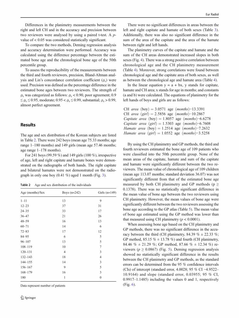
Hand radiography was performed using Innovion-SH (DKMedical Systems, Seoul, Korea). The patients were seatedalongside or facing a table, and examined in the PA position.The protocols used in this study for taking the PA radiographsof both hands are described in Table 1. Both hands were inneutral position with no flexion, extension or deviation, andwere placed palm down on the cassette with the fingers ex-tended [13].
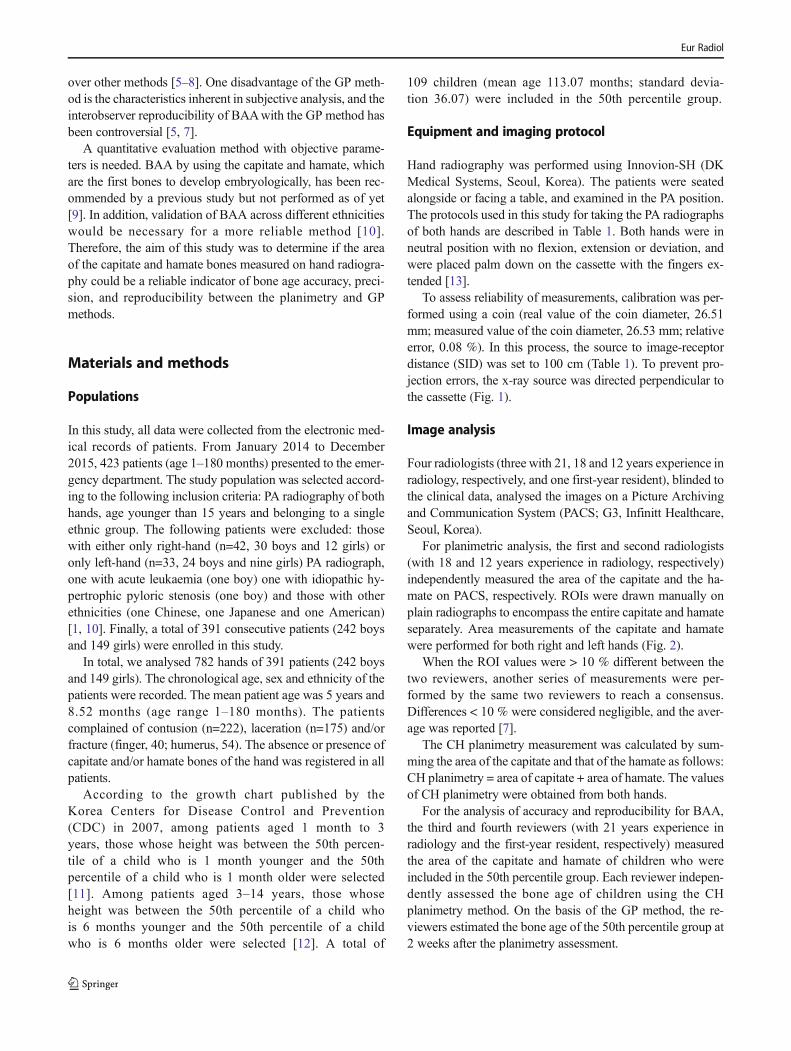
To assess reliability of measurements, calibration was per-formed using a coin (real value of the coin diameter, 26.51mm; measured value of the coin diameter, 26.53 mm; relativeerror, 0.08 %). In this process, the source to image-receptordistance (SID) was set to 100 cm (Table 1). To prevent pro-jection errors, the x-ray source was directed perpendicular tothe cassette (Fig. 1).
Image analysis
Four radiologists (three with 21, 18 and 12 years experience inradiology, respectively, and one first-year resident), blinded tothe clinical data, analysed the images on a Picture Archivingand Communication System (PACS; G3, Infinitt Healthcare,Seoul, Korea).
For planimetric analysis, the first and second radiologists(with 18 and 12 years experience in radiology, respectively)independently measured the area of the capitate and the ha-mate on PACS, respectively. ROIs were drawn manually onplain radiographs to encompass the entire capitate and hamateseparately. Area measurements of the capitate and hamatewere performed for both right and left hands (Fig. 2).
When the ROI values were > 10 % different between thetwo reviewers, another series of measurements were per-formed by the same two reviewers to reach a consensus.Differences < 10 % were considered negligible, and the aver-age was reported [7].
The CH planimetry measurement was calculated by sum-ming the area of the capitate and that of the hamate as follows:CH planimetry = area of capitate + area of hamate. The valuesof CH planimetry were obtained from both hands.
For the analysis of accuracy and reproducibility for BAA,the third and fourth reviewers (with 21 years experience inradiology and the first-year resident, respectively) measuredthe area of the capitate and hamate of children who wereincluded in the 50th percentile group. Each reviewer indepen-dently assessed the bone age of children using the CHplanimetry method. On the basis of the GP method, the re-viewers estimated the bone age of the 50th percentile group at2 weeks after the planimetry assessment.
Eur Radiol
Statistical analysis
All statistical analyses were conducted using SPSS (ver-sion 20; IBM Corporation). Linear regression analysiswas used to analyse the relationship between theplanimetry measurement of each hand and the chrono-logical age of each sex. Correlation coefficients of ≤ 0.3were considered to indicate a weak relationship; 0.30–
0.70, moderate relationship; and > 0.70, strong relation-ship [14]. A z-test was used to compare two correlationcoefficients.
Fig 2 75-month (6 years and 3 months)-old boy. (a and b) The areas ofthe bilateral capitate bones (solid line) and bilateral hamate bones (dottedline) were measured by drawing two separate regions of interest in theposterior-anterior (PA) radiograph of both hands
Fig. 1 (a) The diameter of a coin measured using a digital micrometrewas 26.51 mm. (b) The diameter of the coin on a digital radiograph wasmeasured as 26.53 mm on a Picture Archiving and CommunicationSystem (PACS)
Table 1 Imaging protocol of bothhands posterior-anterior (PA) x-ray
Projection Posterior-anterior bilateral projection
Detector size 24×30 cm
Orientation Landscape
Exposure 50–60Kvp/2–5mAs
Grid No
SID 100 cm
Central ray Between the two hands at the level of the metacarpophalangeal joints
Collimation To include one-third of the distal radius and ulna
to include soft tissues
SID source to image-receptor distance
Eur Radiol
Differences in the planimetry measurements between theright and left CH and in the accuracy and precision betweentwo reviewers were analysed by using a paired t-test. A p-value of < 0.05 was considered statistically significant.
To compare the two methods, Deming regression analysisand accuracy determination were performed. Accuracy wascalculated using the difference percentage between the esti-mated bone age and the chronological bone age of the 50thpercentile group.
To assess the reproducibility of the measurements betweenthe third and fourth reviewers, precision, Bland-Altman anal-ysis and Lin’s concordance correlation coefficient (ρc) wereused. Precision was defined as the percentage difference in theestimated bone ages between two reviewers. The strength ofρc was categorized as follows: ρc < 0.90, poor agreement; 0.9≤ ρc ≤ 0.95, moderate; 0.95 < ρc ≤ 0.99, substantial; ρc > 0.99,almost perfect agreement.
Results
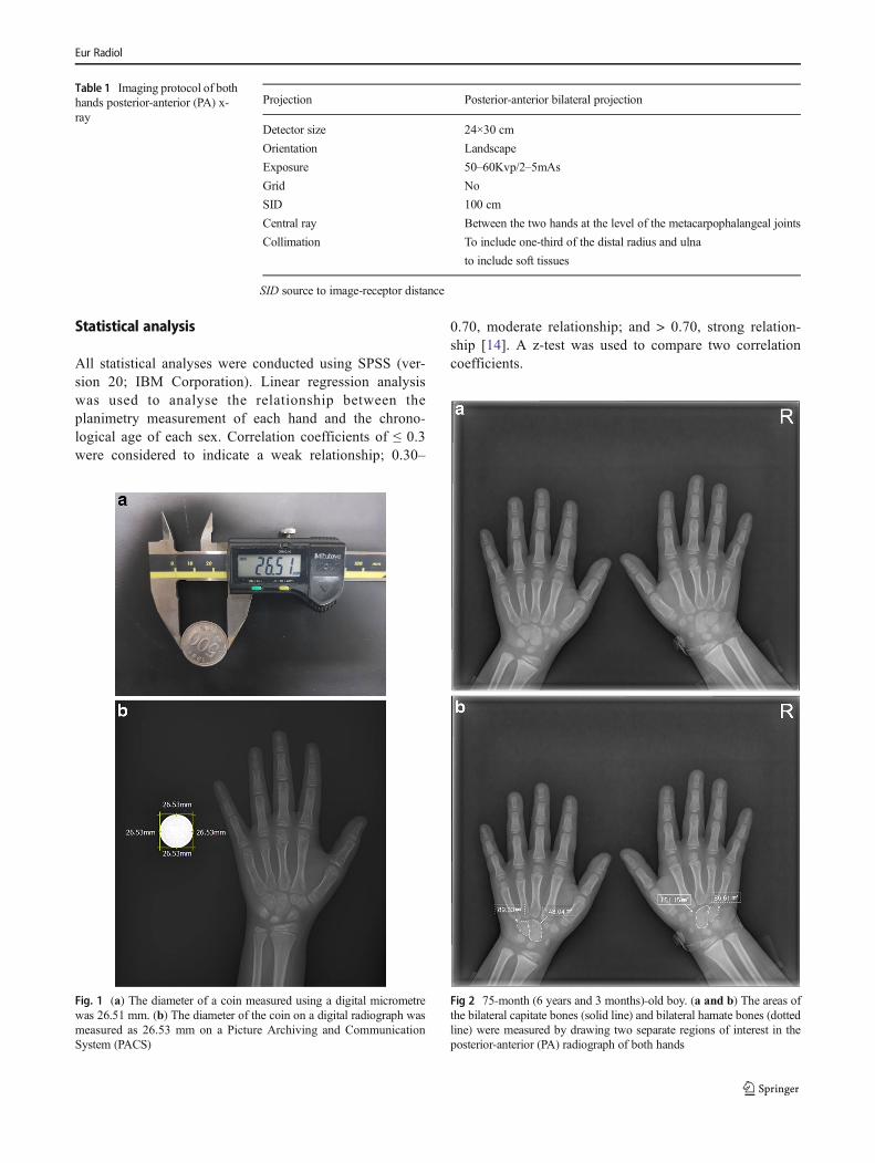
The age and sex distribution of the Korean subjects are listedin Table 2. There were 242 boys (mean age 75.33 months; agerange 1–180 months) and 149 girls (mean age 57.46 months;age range 1–178 months).
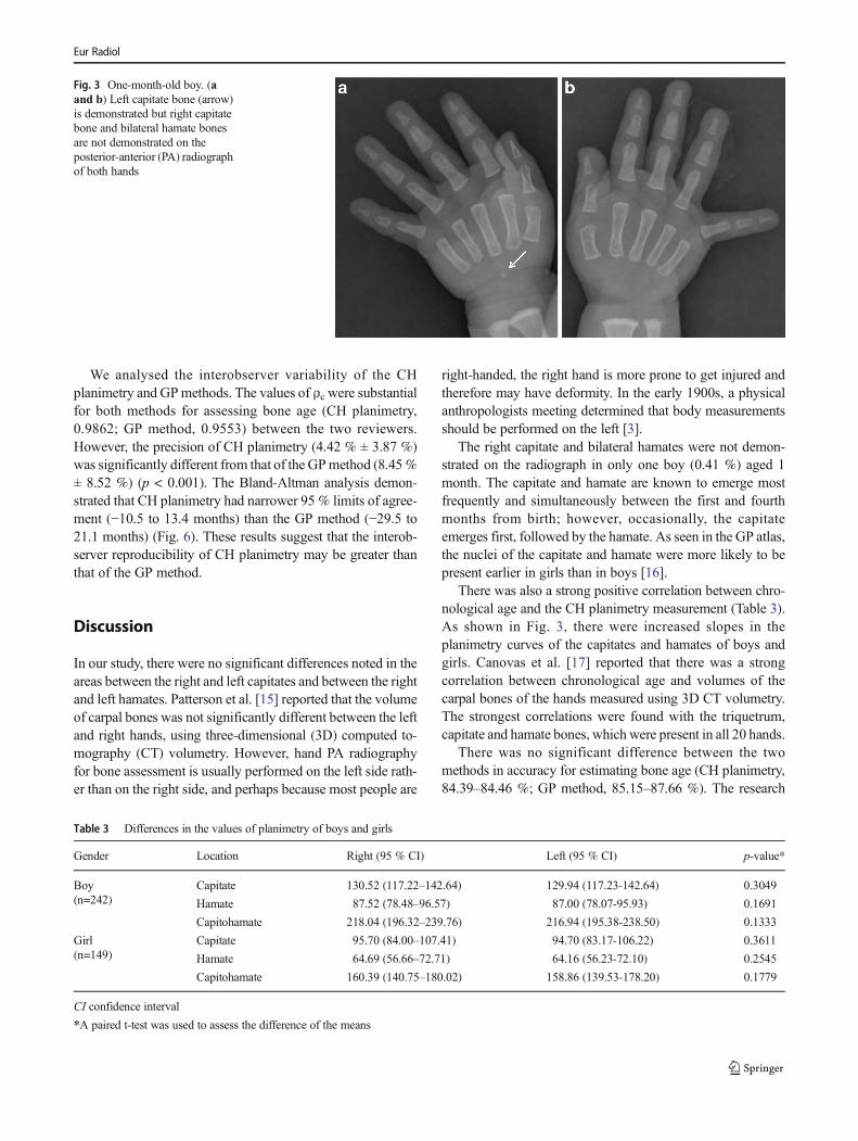
For 241 boys (99.59 %) and 149 girls (100 %), irrespectiveof age, left and right capitate and hamate bones were demon-strated on the radiographs of both hands. The right capitateand bilateral hamates were not demonstrated on the radio-graph in only one boy (0.41 %) aged 1 month (Fig. 3).
There were no significant differences in areas between theleft and right capitate and hamate of both sexes (Table 3).Additionally, there was also no significant difference in thesum of the area of the capitate and the area of the hamatebetween right and left hands.
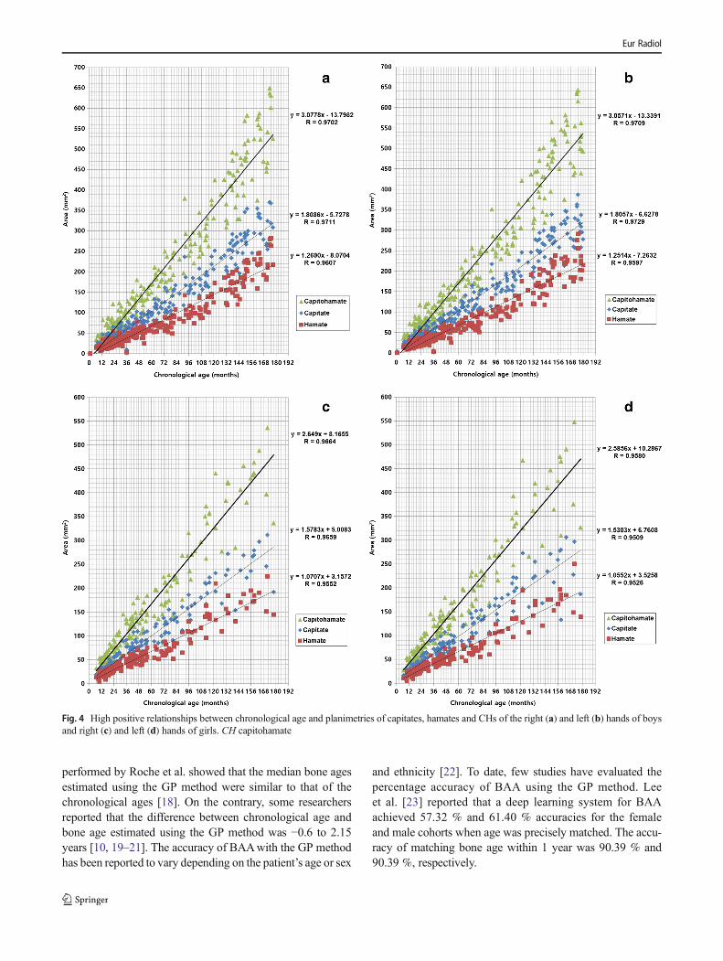
The planimetry curves of the capitate and hamate and thesum of the CH areas demonstrated increased slopes in bothsexes (Fig. 4). There was a strong positive correlation betweenchronological age and the CH planimetry measurement(Table 4). Moreover, strong correlations were found betweenchronological age and the capitate area of both sexes, as wellas between the chronological age and hamate area (Table 4).
In the linear equation y = a + bx, y stands for capitate,hamate and CH area; x stands for age in months; and constants(a and b) were calculated. The equations of planimetry for theleft hands of boys and girls are as follows:
CH area boyð Þ ¼ 3:0571 age monthsð Þ−13:3391CH area girlð Þ ¼ 2:5856 age monthsð Þ−10:2867Capitate area boyð Þ ¼ 1:8057 age monthsð Þ−6:6278Capitate area girlð Þ ¼ 1:5303 age monthsð Þ−6:7608Hamate area boyð Þ ¼ 1:2514 age monthsð Þ−7:2632Hamate area girlð Þ ¼ 1:0552 age monthsð Þ−3:5258
By using the CH planimetry and GP methods, the third andfourth reviewers estimated the bone age of 109 patients whowere classified into the 50th percentile group. None of themean areas of the capitate, hamate and sum of the capitateand hamate were significantly different between the two re-viewers. The mean value of chronological age of 109 children(mean age 113.07 months; standard deviation 36.07) was notsignificantly different from that of the estimated bone agemeasured by both CH planimetry and GP methods (p ≥0.1378). There was no statistically significant difference inthe mean value of bone age between the two reviewers usingCH planimetry. However, the mean values of bone age weresignificantly different between the two reviewers assessing thebone age according to the GP atlas (Table 5). The mean valueof bone age estimated using the GP method was lower thanthat measured using CH planimetry (p < 0.0001).
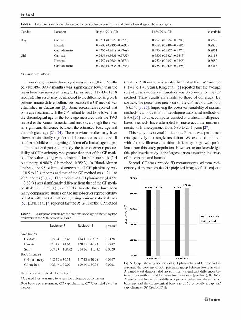
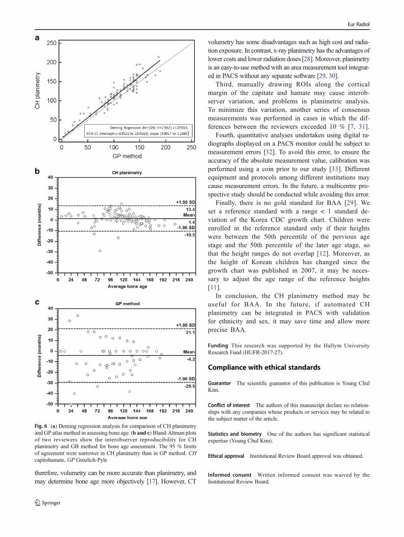
When assessing bone age based on the CH planimetry andGP methods, there was no significant difference in the accu-racy between the third (CH planimetry, 84.39 % ± 22.33 %;GP method, 85.15 % ± 13.78 %) and fourth (CH planimetry,84.46 % ± 21.29 %; GP method, 87.66 % ± 12.34 %) re-viewers (p ≥ 0.0867) (Fig. 5). Deming regression analysisshowed no statistically significant difference in the resultsbetween the CH planimetry and GP methods, as the standarderrors can be determined from the 95 % confidence intervals(CIs) of intercept (standard error, 4.0028; 95 % CI −4.9522–10.9164) and slope (standard error, 0.03955; 95 % CI,0.9917–1.1485) including the values 0 and 1, respectively(Fig. 6).
Table 2 Age and sex distribution of the individuals
Age (months)/Sex Boys (n=242) Girls (n=149)
1–11 13 9
12–23 37 31
24–35 33 17
36–47 21 26
48–59 16 13
60–71 14 6
72–83 17 12
84–95 6 5
96–107 13 5
108–119 10 7
120–131 4 3
132–143 18 4
144–155 14 3
156–167 9 5
168–179 16 3
180 1 0
Data represent number of patients
Eur Radiol
We analysed the interobserver variability of the CHplanimetry and GPmethods. The values of ρc were substantialfor both methods for assessing bone age (CH planimetry,0.9862; GP method, 0.9553) between the two reviewers.However, the precision of CH planimetry (4.42 % ± 3.87 %)was significantly different from that of the GPmethod (8.45%± 8.52 %) (p < 0.001). The Bland-Altman analysis demon-strated that CH planimetry had narrower 95 % limits of agree-ment (−10.5 to 13.4 months) than the GP method (−29.5 to21.1 months) (Fig. 6). These results suggest that the interob-server reproducibility of CH planimetry may be greater thanthat of the GP method.
Discussion
In our study, there were no significant differences noted in theareas between the right and left capitates and between the rightand left hamates. Patterson et al. [15] reported that the volumeof carpal bones was not significantly different between the leftand right hands, using three-dimensional (3D) computed to-mography (CT) volumetry. However, hand PA radiographyfor bone assessment is usually performed on the left side rath-er than on the right side, and perhaps because most people are
right-handed, the right hand is more prone to get injured andtherefore may have deformity. In the early 1900s, a physicalanthropologists meeting determined that body measurementsshould be performed on the left [3].
The right capitate and bilateral hamates were not demon-strated on the radiograph in only one boy (0.41 %) aged 1month. The capitate and hamate are known to emerge mostfrequently and simultaneously between the first and fourthmonths from birth; however, occasionally, the capitateemerges first, followed by the hamate. As seen in the GP atlas,the nuclei of the capitate and hamate were more likely to bepresent earlier in girls than in boys [16].
There was also a strong positive correlation between chro-nological age and the CH planimetry measurement (Table 3).As shown in Fig. 3, there were increased slopes in theplanimetry curves of the capitates and hamates of boys andgirls. Canovas et al. [17] reported that there was a strongcorrelation between chronological age and volumes of thecarpal bones of the hands measured using 3D CT volumetry.The strongest correlations were found with the triquetrum,capitate and hamate bones, which were present in all 20 hands.
There was no significant difference between the twomethods in accuracy for estimating bone age (CH planimetry,84.39–84.46 %; GP method, 85.15–87.66 %). The research
Table 3 Differences in the values of planimetry of boys and girls
Gender Location Right (95 % CI) Left (95 % CI) p-value*
Boy(n=242)
Capitate 130.52 (117.22–142.64) 129.94 (117.23-142.64) 0.3049
Hamate 87.52 (78.48–96.57) 87.00 (78.07-95.93) 0.1691
Capitohamate 218.04 (196.32–239.76) 216.94 (195.38-238.50) 0.1333
Girl(n=149)
Capitate 95.70 (84.00–107.41) 94.70 (83.17-106.22) 0.3611
Hamate 64.69 (56.66–72.71) 64.16 (56.23-72.10) 0.2545
Capitohamate 160.39 (140.75–180.02) 158.86 (139.53-178.20) 0.1779
CI confidence interval
*A paired t-test was used to assess the difference of the means
Fig. 3 One-month-old boy. (aand b) Left capitate bone (arrow)is demonstrated but right capitatebone and bilateral hamate bonesare not demonstrated on theposterior-anterior (PA) radiographof both hands
Eur Radiol
performed by Roche et al. showed that the median bone agesestimated using the GP method were similar to that of thechronological ages [18]. On the contrary, some researchersreported that the difference between chronological age andbone age estimated using the GP method was −0.6 to 2.15years [10, 19–21]. The accuracy of BAAwith the GP methodhas been reported to vary depending on the patient’s age or sex
and ethnicity [22]. To date, few studies have evaluated thepercentage accuracy of BAA using the GP method. Leeet al. [23] reported that a deep learning system for BAAachieved 57.32 % and 61.40 % accuracies for the femaleand male cohorts when age was precisely matched. The accu-racy of matching bone age within 1 year was 90.39 % and90.39 %, respectively.
Fig. 4 High positive relationships between chronological age and planimetries of capitates, hamates and CHs of the right (a) and left (b) hands of boysand right (c) and left (d) hands of girls. CH capitohamate
Eur Radiol
In our study, the mean bone agemeasured using the GPmeth-od (105.49–109.49 months) was significantly lower than themean bone age measured using CH planimetry (117.43–118.58months). This result may be attributed to the difference in growthpatterns among different ethnicities because the GP method wasestablished in Caucasians [3]. Some researchers reported thatbone age measured with the GP method tended to be lower thanthe chronological age or the bone age measured with the TW3method or the Korean bone standard method, although there wasno significant difference between the estimated bone age andchronological age [21, 24]. These previous studies may haveshown no statistically significant difference because of the smallnumber of children or targeting children of a limited age range.
In the second part of our study, the interobserver reproduc-ibility of CH planimetry was greater than that of the GP meth-od. The values of ρc were substantial for both methods (CHplanimetry, 0.9862; GP method, 0.9553). In Bland-Altmananalysis, the 95 % limit of agreement of CH planimetry was−10.5 to 13.4 months and that of the GP method was −21.1 to29.5 months (Fig. 6). The precision of CH planimetry (4.42 %± 3.87 %) was significantly different from that of the GPmeth-od (8.45 % ± 8.52 %) (p < 0.001). To date, there have beenmany comparative studies on the interobserver reproducibilityof BAAwith the GP method by using various statistical tests[5, 7]. Bull et al. [7] reported that the 95%CI of theGPmethod
(−2.46 to 2.18 years) was greater than that of the TW2 method(−1.48 to 1.43 years). King et al. [5] reported that the averagespread of intra-observer variation was 0.96 years for the GPmethod. These results are similar to those of our study. Bycontrast, the percentage precision of the GP method was 65.5−88.5 % [8, 25]. Improving the observer variability of manualmethods is a motivation for developing automated methods ofBAA [26]. To date, computer-assisted or artificial intelligence-based methods have attempted to make accurate measure-ments, with discrepancies from 0.39 to 2.41 years [27].
This study has several limitations. First, it was performedretrospectively at a single institution. We excluded childrenwith chronic illnesses, nutrition deficiency or growth prob-lems from this study population. However, to our knowledge,this planimetric study is the largest series assessing the areasof the capitate and hamate.
Second, CT scans provide 3D measurements, whereas radi-ography demonstrates the 2D projected images of 3D objects;
Fig. 5 Graph showing accuracy of CH planimetry and GP method inassessing the bone age of 50th percentile group between two reviewers.A paired t-test demonstrated no statistically significant differences be-tween two methods and between two reviewers (p-value ≥ 0.0867).Accuracy was defined as the difference percentage between the estimatedbone age and the chronological bone age of 50 percentile group. CHcapitohamate, GP Greulich-Pyle
Table 5 Descriptive statistics of the area and bone age estimated by tworeviewers in the 50th percentile group
Reviewer 3 Reviewer 4 p-value*
Area (mm2)
Capitate 185.94 ± 65.42 184.11 ± 67.97 0.1128
Hamate 121.45 ± 44.63 120.25 ± 46.23 0.2487
Sum 307.39 ± 108.92 304.36 ± 112.82 0.0729
BAA (months)
CH planimetry 118.58 ± 39.52 117.43 ± 40.96 0.0687
GP method 105.49 ± 39.00 109.49 ± 39.38 0.0003
Data are means ± standard deviation
*A paired t test was used to assess the difference of the means
BAA bone age assessment, CH capitohamate, GP Greulich-Pyle atlasmethod
Table 4 Differences in the correlation coefficients between planimetry and chronological age of boys and girls
Gender Location Right (95 % CI) Left (95 % CI) z-statistic
Boy Capitate 0.9711 (0.9629–0.9775) 0.9729 (0.9652–0.9789) 0.9729
Hamate 0.9607 (0.9496–0.9693) 0.9597 (0.9484–0.9686) 0.8886
Capitohamate 0.9702 (0.9618–0.9768) 0.9709 (0.9627–0.9774) 0.8951
Girl Capitate 0.9659 (0.9531–0.9752) 0.9509 (0.9327–0.9643) 0.1118
Hamate 0.9552 (0.9386–0.9674) 0.9526 (0.9351–0.9655) 0.8052
Capitohamate 0.9664 (0.9538–0.9756) 0.9580 (0.9424–0.9695) 0.3313
CI confidence interval
Eur Radiol
therefore, volumetry can be more accurate than planimetry, andmay determine bone age more objectively [17]. However, CT
volumetry has some disadvantages such as high cost and radia-tion exposure. In contrast, x-ray planimetry has the advantages oflower costs and lower radiation doses [28].Moreover, planimetryis an easy-to-use method with an area measurement tool integrat-ed in PACS without any separate software [29, 30].
Third, manually drawing ROIs along the corticalmargin of the capitate and hamate may cause interob-server variation, and problems in planimetric analysis.To minimize this variation, another series of consensusmeasurements was performed in cases in which the dif-ferences between the reviewers exceeded 10 % [7, 31].
Fourth, quantitative analyses undertaken using digital ra-diographs displayed on a PACS monitor could be subject tomeasurement errors [32]. To avoid this error, to ensure theaccuracy of the absolute measurement value, calibration wasperformed using a coin prior to our study [33]. Differentequipment and protocols among different institutions maycause measurement errors. In the future, a multicentre pro-spective study should be conducted while avoiding this error.
Finally, there is no gold standard for BAA [29]. Weset a reference standard with a range < 1 standard de-viation of the Korea CDC growth chart. Children wereenrolled in the reference standard only if their heightswere between the 50th percentile of the pervious agestage and the 50th percentile of the later age stage, sothat the height ranges do not overlap [12]. Moreover, asthe height of Korean children has changed since thegrowth chart was published in 2007, it may be neces-sary to adjust the age range of the reference heights[11].
In conclusion, the CH planimetry method may beuseful for BAA. In the future, if automated CHplanimetry can be integrated in PACS with validationfor ethnicity and sex, it may save time and allow moreprecise BAA.
Funding This research was supported by the Hallym UniversityResearch Fund (HUFR-2017-27).
Compliance with ethical standards
Guarantor The scientific guarantor of this publication is Young ChulKim.
Conflict of interest The authors of this manuscript declare no relation-ships with any companies whose products or services may be related tothe subject matter of the article.
Statistics and biometry One of the authors has significant statisticalexpertise (Young Chul Kim).
Ethical approval Institutional Review Board approval was obtained.
Informed consent Written informed consent was waived by theInstitutional Review Board.
Fig. 6 (a) Deming regression analysis for comparison of CH planimetryand GP atlas method in assessing bone age. (b and c) Bland-Altman plotsof two reviewers show the interobserver reproducibility for CHplanimetry and GB method for bone age assessment. The 95 % limitsof agreement were narrower in CH planimetry than in GP method. CHcapitohamate, GP Greulich-Pyle
Eur Radiol
Methodology Retrospective, observational, performed at oneinstitution.
Open Access This article is distributed under the terms of the CreativeCommons At t r ibut ion 4 .0 In te rna t ional License (h t tp : / /creativecommons.org/licenses/by/4.0/), which permits unrestricted use,distribution, and reproduction in any medium, provided you give appro-priate credit to the original author(s) and the source, provide a link to theCreative Commons license, and indicate if changes were made.
References
1. Sharma S, Mishra V, Kulshreshtha V (2014) Radiographical studyshowing asymmetry in the surface area of carpal bones in malnour-ished children. J Clin Diagn Res 8:AC08–AC10
2. Cameriere R, De Luca S, Biagi R, Cingolani M, Farronato G,Ferrante L (2012) Accuracy of three age estimation methods inchildren by measurements of developing teeth and carpals andepiphyses of the ulna and radius. J Forensic Sci 57:1263–1270
3. Satoh M (2015) Bone age: assessment methods and clinical appli-cations. Clin Pediatr Endocrinol 24:143–152
4. Pietka E, Gertych A, Pospiech S, Cao F, Huang HK, Gilsanz V(2001) Computer-assisted bone age assessment: image preprocess-ing and epiphyseal/metaphyseal ROI extraction. IEEE Trans MedImaging 20:715–729
5. King DG, Steventon DM, O'Sullivan MP et al (1994)Reproducibility of bone ages when performed by radiology regis-trars: an audit of Tanner and Whitehouse II versus Greulich andPyle methods. Br J Radiol 67:848–851
6. Manzoor Mughal A, Hassan N, Ahmed A (2014) Bone age assess-ment methods: a critical review. Pak J Med Sci 30:211–215
7. Bull RK, Edwards PD, Kemp PM, Fry S, Hughes IA (1999) Boneage assessment: a large scale comparison of the Greulich and Pyle,and Tanner and Whitehouse (TW2) methods. Arch Dis Child 81:172–173
8. Bilgili Y, Hizel S, Kara SA, Sanli C, Erdal HH, Altinok D (2003)Accuracy of skeletal age assessment in children from birth to 6years of age with the ultrasonographic version of the Greulich-Pyle atlas. J Ultrasound Med 22:683–690
9. Zhang A, Gertych A, Liu BJ (2007) Automatic bone age assess-ment for young children from newborn to 7-year-old using carpalbones. Comput Med Imaging Graph 31:299–310
10. Ontell FK, Ivanovic M, Ablin DS, Barlow TW (1996) Bone age inchildren of diverse ethnicity. AJR Am J Roentgenol 167:1395–1398
11. Ryoo NY, Shin HY, Kim JH, Moon JS, Lee CG (2015) Change inthe height of Korean children and adolescents: analysis from theKorea National Health and Nutrition Survey II and V. Korean JPediatr 58:336–340
12. Thodberg HH, Jenni OG, Ranke MB, Martin DD (2012)Standardization of the Tanner-Whitehouse bone age method inthe context of automated image analysis. Ann Hum Biol 39:68–75
13. Schreibman KL, Freeland A, Gilula LA, Yin Y (1997) Imaging ofthe hand and wrist. Orthop Clin North Am 28:537–582
14. Kim YC, Lim JS, Keum KC et al (2011) Comparison of diffusion-weighted MRI and MR volumetry in the evaluation of early treat-ment outcomes after preoperative chemoradiotherapy for locallyadvanced rectal cancer. J Magn Reson Imaging 34:570–576
15. Patterson RM, Elder KW, Viegas SF, Buford WL (1995) Carpalbone anatomy measured by computer analysis of three-dimensional reconstructions of computed tomography images. JHand Surg Am 20:923–929
16. Daneff M, Casalis C, Bruno CH, Bruno DA (2015) Bone age as-sessment with conventional ultrasonography in healthy infants from1 to 24 months of age. Pediatr Radiol 45:1007–1015
17. Canovas F, Banegas F, Cyteval C et al (2000) Carpal bone matura-tion assessment by image analysis from computed tomographyscans. Horm Res 54:6–13
18. Roche AF, Davila GH, Eyman SL (1971) A comparison betweenGreulich-Pyle and Tanner-Whitehouse assessments of skeletal ma-turity. Radiology 98:273–280
19. Andersen E (1971) Comparison of Tanner-Whitehouse andGreulich-Pyle methods in a large scale Danish Survey. Am JPhys Anthropol 35:373–376
20. Anderson M (1971) Use of the Greulich-Pyle "Atlas of SkeletalDevelopment of the Hand and Wrist" in a clinical context. Am JPhys Anthropol 35:347–352
21. Braude S, Henning L, Lambert M (2007) Accuracy of bone assess-ments for verifying age in adolescents-application in sport. SAJournal of Radiology 11:4–7
22. Chang HF, Wu KM, Chen KC (1990) A cross-sectional study onthe skeletal development of the hand and wrist from preadolescenceto early adulthood among Chinese in Taiwan. Zhonghua Ya Yi XueHui Za Zhi 9:1–11
23. Lee H, Tajmir S, Lee J et al (2017) Fully Automated Deep LearningSystem for Bone Age Assessment. J Digit Imaging 30:427–441
24. Kim JR, Lee YS, Yu J (2015) Assessment of bone age in prepuber-tal healthy Korean children: comparison among the Korean stan-dard bone age chart, Greulich-Pyle method, and Tanner-Whitehouse method. Korean J Radiol 16:201–205
25. Khan KM, Gonzalez-Bolanos MT, Holm T, Miller BS, SarafoglouK (2015) Use of Automated Bone Age for Critical GrowthAssessment. Clin Pediatr (Phila) 54:1038–1043
26. Mansourvar M, Ismail MA, Herawan T, Raj RG, Kareem SA,Nasaruddin FH (2013) Automated bone age assessment: motiva-tion, taxonomies, and challenges. Comput Math Methods Med2013:391626
27. Spampinato C, Palazzo S, Giordano D, Aldinucci M, Leonardi R(2017) Deep learning for automated skeletal bone age assessment inx-ray images. Med Image Anal 36:41–51
28. Bulut E, Sahin B, Muglali M, Bekcioglu B (2012) Comparison ofthe planimetry and point-counting methods for the assessment ofthe size of the mandible cysts on orthopantomograms. Med OralPatol Oral Cir Bucal 17:e442–e446
29. van Rijn RR, Thodberg HH (2013) Bone age assessment: automat-ed techniques coming of age? Acta Radiol 54:1024–1029
30. Bunch PM, Altes TA, McIlhenny J, Patrie J, Gaskin CM (2017)Skeletal development of the hand and wrist: digital bone agecompanion-a suitable alternative to the Greulich and Pyle atlas forbone age assessment? Skeletal Radiol 46:785–793
31. Pietka E, Pospiech-Kurkowska S, Gertych A, Cao F (2003)Integration of computer assisted bone age assessment with clinicalPACS. Comput Med Imaging Graph 27:217–228
32. Franken M, Grimm B, Heyligers I (2010) A comparison of foursystems for calibration when templating for total hip replacementwith digital radiography. J Bone Joint Surg Br 92:136–141
33. Wimsey S, Pickard R, Shaw G (2006) Accurate scaling of digitalradiographs of the pelvis. A prospective trial of two methods. JBone Joint Surg Br 88:1508–1512
Eur Radiol
Related Documents