A shear gradient–dependent platelet aggregation mechanism drives thrombus formation Warwick S Nesbitt 1,5 , Erik Westein 1,5 , Francisco Javier Tovar-Lopez 2 , Elham Tolouei 3 , Arnan Mitchell 2 , Jia Fu 1 , Josie Carberry 3 , Andreas Fouras 4 & Shaun P Jackson 1 Platelet aggregation at sites of vascular injury is essential for hemostasis and arterial thrombosis. It has long been assumed that platelet aggregation and thrombus growth are initiated by soluble agonists generated at sites of vascular injury. By using high- resolution intravital imaging techniques and hydrodynamic analyses, we show that platelet aggregation is primarily driven by changes in blood flow parameters (rheology), with soluble agonists having a secondary role, stabilizing formed aggregates. We find that in response to vascular injury, thrombi initially develop through the progressive stabilization of discoid platelet aggregates. Analysis of blood flow dynamics revealed that discoid platelets preferentially adhere in low-shear zones at the downstream face of forming thrombi, with stabilization of aggregates dependent on the dynamic restructuring of membrane tethers. These findings provide insight into the prothrombotic effects of disturbed blood flow parameters and suggest a fundamental reinterpretation of the mechanisms driving platelet aggregation and thrombus growth. Platelet aggregation at sites of vascular injury is central to the arrest of bleeding and for subsequent vascular repair; however, an exaggerated platelet aggregation response at sites of atherosclerotic plaque rupture can lead to the development of vascular occlusive thrombi, precipitat- ing diseases such as the acute coronary syndromes and ischemic stroke 1–3 . The key variables regulating thrombosis have been known for more than 150 years and include changes in the vessel wall, alterations in the thrombogenic potential of blood and changes in blood flow parameters (hemodynamics) 4 . Although the former two variables have a well defined role in arterial thrombosis, the mechan- ism by which alterations in blood flow affects the thrombotic process remains ill defined. Platelet aggregation under the influence of blood flow is dependent on the adhesive function of both the platelet glycoprotein (GP) Ib-V- IX receptor complex and integrin a IIb b 3 (also known as GP IIb-IIIa), wherein GPIb initiates reversible platelet recruitment, particularly at elevated shear rates, and integrin a IIb b 3 stabilizes forming aggregates 5–12 . A primary step regulating the transition from reversible to stable adhesion is activation of integrin a IIb b 3 , a process dependent on the generation of the soluble agonists adenosine diphosphate (ADP), thromboxane A 2 (TXA 2 ) and thrombin 2,5,6 . Soluble agonists stimulate a host of biochemical and functional responses, including cytosolic calcium flux, platelet shape change and granule release, that coincide with the rapid development of platelet aggregates 13 . However, the recent demonstration that platelets can form aggregates in vivo without a detectable increase in cytosolic calcium 14,15 , without under- going shape change 15,16 and without substantial a-granule secretion in the early phases of thrombus development 17 has raised the possibility that additional mechanisms may be involved 16,18 . One such mechanism may involve local changes in blood flow. Recent studies have identified that, in the range of physiological blood shear (strain) rates (1,000–10,000 s –1 ), small, transient discoid platelet aggregates can form through the development of membrane tethers 16 , whereas at pathological shear rates (410,000 s –1 ) large rolling aggregates can develop independently of integrin a IIb b 3 and platelet activation 18 . How important these rheologically driven aggregation mechanisms are, relative to soluble agonist-generated aggregates, in promoting thrombus development in vivo remains unclear. In this study we show that rapid changes in blood flow (shear microgradi- ents) represent a general feature of thrombus development, inducing stabilized aggregation of discoid platelets. This shear-regulated platelet aggregation mechanism involves a unique biomechanical sensing mechanism linked to the physical restructuring of membrane tethers. Our findings suggest that mechanosensory platelet activation mechan- isms have a major role in driving platelet aggregation and thrombus growth in vivo. RESULTS Shear microgradients promote discoid platelet aggregation It has long been recognized that platelet aggregation preferentially occurs at regions of flow disturbance after vascular injury 19,20 . To investigate the effects of rheological disturbance on platelet aggrega- tion dynamics in vivo, we employed intravital imaging to visualize the platelet aggregation process in mesenteric arterioles of mice. We Received 17 October 2008; accepted 27 March 2009; published online 24 May 2009; doi:10.1038/nm.1955 1 The Australian Centre for Blood Diseases, Monash University, Alfred Medical Research and Educational Precinct, Melbourne, Victoria, Australia. 2 Microelectronics and Materials Technology Centre, School of Electrical and Computer Engineering, RMIT University, Melbourne, Victoria, Australia. 3 Department of Mechanical Engineering and 4 Division of Biological Engineering, Monash University, Clayton, Victoria, Australia. 5 These authors contributed equally to this work. Correspondence should be addressed to W.S.N. ([email protected]) or S.P.J. ([email protected]). NATURE MEDICINE VOLUME 15 [ NUMBER 6 [ JUNE 2009 665 ARTICLES © 2009 Nature America, Inc. All rights reserved.

Welcome message from author
This document is posted to help you gain knowledge. Please leave a comment to let me know what you think about it! Share it to your friends and learn new things together.
Transcript
A shear gradient–dependent platelet aggregationmechanism drives thrombus formationWarwick S Nesbitt1,5, Erik Westein1,5, Francisco Javier Tovar-Lopez2, Elham Tolouei3, Arnan Mitchell2, Jia Fu1,Josie Carberry3, Andreas Fouras4 & Shaun P Jackson1
Platelet aggregation at sites of vascular injury is essential for hemostasis and arterial thrombosis. It has long been assumed that
platelet aggregation and thrombus growth are initiated by soluble agonists generated at sites of vascular injury. By using high-
resolution intravital imaging techniques and hydrodynamic analyses, we show that platelet aggregation is primarily driven by
changes in blood flow parameters (rheology), with soluble agonists having a secondary role, stabilizing formed aggregates. We find
that in response to vascular injury, thrombi initially develop through the progressive stabilization of discoid platelet aggregates.
Analysis of blood flow dynamics revealed that discoid platelets preferentially adhere in low-shear zones at the downstream face
of forming thrombi, with stabilization of aggregates dependent on the dynamic restructuring of membrane tethers. These findings
provide insight into the prothrombotic effects of disturbed blood flow parameters and suggest a fundamental reinterpretation of
the mechanisms driving platelet aggregation and thrombus growth.
Platelet aggregation at sites of vascular injury is central to the arrest ofbleeding and for subsequent vascular repair; however, an exaggeratedplatelet aggregation response at sites of atherosclerotic plaque rupturecan lead to the development of vascular occlusive thrombi, precipitat-ing diseases such as the acute coronary syndromes and ischemicstroke1–3. The key variables regulating thrombosis have been knownfor more than 150 years and include changes in the vessel wall,alterations in the thrombogenic potential of blood and changes inblood flow parameters (hemodynamics)4. Although the former twovariables have a well defined role in arterial thrombosis, the mechan-ism by which alterations in blood flow affects the thrombotic processremains ill defined.
Platelet aggregation under the influence of blood flow is dependenton the adhesive function of both the platelet glycoprotein (GP) Ib-V-IX receptor complex and integrin aIIbb3 (also known as GP IIb-IIIa),wherein GPIb initiates reversible platelet recruitment, particularlyat elevated shear rates, and integrin aIIbb3 stabilizes formingaggregates5–12. A primary step regulating the transition from reversibleto stable adhesion is activation of integrin aIIbb3, a process dependenton the generation of the soluble agonists adenosine diphosphate(ADP), thromboxane A2 (TXA2) and thrombin2,5,6. Soluble agonistsstimulate a host of biochemical and functional responses, includingcytosolic calcium flux, platelet shape change and granule release, thatcoincide with the rapid development of platelet aggregates13. However,the recent demonstration that platelets can form aggregates in vivowithout a detectable increase in cytosolic calcium14,15, without under-going shape change15,16 and without substantial a-granule secretion in
the early phases of thrombus development17 has raised the possibilitythat additional mechanisms may be involved16,18.
One such mechanism may involve local changes in blood flow.Recent studies have identified that, in the range of physiological bloodshear (strain) rates (1,000–10,000 s–1), small, transient discoid plateletaggregates can form through the development of membrane tethers16,whereas at pathological shear rates (410,000 s–1) large rollingaggregates can develop independently of integrin aIIbb3 and plateletactivation18. How important these rheologically driven aggregationmechanisms are, relative to soluble agonist-generated aggregates, inpromoting thrombus development in vivo remains unclear. In thisstudy we show that rapid changes in blood flow (shear microgradi-ents) represent a general feature of thrombus development, inducingstabilized aggregation of discoid platelets. This shear-regulated plateletaggregation mechanism involves a unique biomechanical sensingmechanism linked to the physical restructuring of membrane tethers.Our findings suggest that mechanosensory platelet activation mechan-isms have a major role in driving platelet aggregation and thrombusgrowth in vivo.
RESULTS
Shear microgradients promote discoid platelet aggregation
It has long been recognized that platelet aggregation preferentiallyoccurs at regions of flow disturbance after vascular injury19,20. Toinvestigate the effects of rheological disturbance on platelet aggrega-tion dynamics in vivo, we employed intravital imaging to visualize theplatelet aggregation process in mesenteric arterioles of mice. We
Received 17 October 2008; accepted 27 March 2009; published online 24 May 2009; doi:10.1038/nm.1955
1The Australian Centre for Blood Diseases, Monash University, Alfred Medical Research and Educational Precinct, Melbourne, Victoria, Australia. 2Microelectronics andMaterials Technology Centre, School of Electrical and Computer Engineering, RMIT University, Melbourne, Victoria, Australia. 3Department of Mechanical Engineeringand 4Division of Biological Engineering, Monash University, Clayton, Victoria, Australia. 5These authors contributed equally to this work. Correspondence should beaddressed to W.S.N. ([email protected]) or S.P.J. ([email protected]).
NATURE MEDICINE VOLUME 15 [ NUMBER 6 [ JUNE 2009 665
ART ICL ES©
2009
Nat
ure
Am
eric
a, In
c. A
ll ri
gh
ts r
eser
ved
.
initiated platelet adhesion and aggregation by mild crush injury of thevessel wall and monitored platelet thrombus formation via differentialinterference contrast (DIC) microscopy (Fig. 1a). We altered bloodflow by compressing the vessel side wall at the site of injury, using ablunted microinjection needle to progressively stenose the vessellumen (Fig. 1a). Localized stenosis markedly accelerated the rateand extent of platelet aggregation (Fig. 1a; 16.39 s), particularlywhen stenosis was greater than B90%. Notably, thrombi appearedto be composed of loosely packed platelets, suggesting that minimalplatelet activation had occurred (data not shown). Thrombus forma-tion was dynamic with cycles of aggregation and disaggregation cor-responding to serial increases and decreases in stenosis, with removalof the microinjector needle leading to disaggregation of the plateletmass (Fig. 1a; 24.13 s). Stenosis in the absence of vessel injury did notresult in platelet aggregation, indicating that flow changes per se wereinsufficient to induce platelet aggregation (data not shown).
To gain insight into the relationship between localized flow changesand platelet aggregation we performed computational fluid dynamic(CFD) modeling of the stenosed vessel geometry. Temporal analysis ofthrombus formation after vessel stenosis revealed that initial platelet
recruitment occurred specifically at the stenosis apex (Fig. 1a), withthe subsequent formation of discoid platelet aggregates occurring inthe downstream expansion zone (Fig. 1a). We observed plateletaggregation throughout the expansion zone, but aggregation rapidlyceased at the downstream margin of this zone (Fig. 1a), suggestingthat the stenosis had a spatially restricted effect on thrombus growth.CFD modeling of the in vivo vessel geometry predicted blood shearrates (g) Z 20,000 s–1 at the stenosis apex that rapidly transitioned tor862 s–1 within the post-stenosis expansion zone (Fig. 1b), suggest-ing that a zone of shear acceleration followed by a zone of deceleratingshear (shear microgradient) may be a requirement for the develop-ment of discoid platelet aggregates (Fig. 1b).
To examine the specific effect of localized shear microgradientson platelet aggregation dynamics, independent of potential non-hemodynamic effects on the vessel wall, we designed model micro-channels incorporating a fixed stenosis. These geometries were char-acterized by a backward-facing step of defined 90% stenosisimmediately followed by an expansion zone (Fig. 1c). This geometrygenerates a localized region of rapid flow acceleration followedby a zone of flow deceleration (Supplementary Fig. 1a online).
Figure 1 Shear microgradients promote platelet
aggregate formation in vivo. (a) DIC images
demonstrating the effect of localized vascular
stenosis on platelet aggregation after localized
crush injury of a mouse mesenteric arteriole
(n ¼ 3 experiments). Small platelet aggregates
are present at t ¼ 0 s (cyan shading). After
stenosis (11.87 s), a marked increase in platelet
aggregation was observed (blue arrow), which
specifically developed downstream from the site
of vascular injury (red arrow). Aggregate growthceased at the downstream stenosis margin
(yellow arrow; 16.39 s) where the vessel returned
to its native geometry. Subsequent removal of the
microneedle led to the rapid embolization of the
platelet aggregate (24.13 s). Scale bars, 10 mm.
(b) CFD simulation of blood flow dynamics after
localized vessel wall compression (left) and
schema illustrating the three principal
components defining a shear microgradient or
stenosis (right). The contraction demarcates the
region of shear acceleration. The throat or apex
demarcates the stenotic region at which peak
shear is experienced. The expansion demarcates
the region of shear deceleration. The CFD
simulation demonstrates that for a predicted
input shear of 1,800 s–1 (immediately upstream
from the stenosis), flow accelerates into the
stenosis throat, reaching a maximum shear ofZ20,000 s–1 at the apex. This elevated shear
rapidly decelerates in the downstream expansion
zone of the geometry, transitioning from 20,000
to 862 s–1 at the downstream margin of the
expansion (yellow arrows). (c) DIC image
sequence of blood perfusion through a
microchannel comprising a side-wall geometry
designed to induce a sharp phase of accelerating
shear from 1,800 s–1 coupled to an immediate
shear deceleration approaching 200 s–1. Red
arrow indicates the point of initial aggregation (t ¼ 12 s) and black arrow indicates the limit of thrombus growth in the expansion zone (representative of
n ¼ 3 experiments). (d) Representative aggregation traces showing the response of whole-blood perfusion through the microchannel in c. Control, hirudin-
anticoagulated whole-blood perfusion at an input (prestenosis) shear rate of 1,800 s–1 (representative of n ¼ 3 experiments); straight channel, hirudin-
anticoagulated whole-blood perfusion through a 100-mm straight (control) microchannel at a bulk shear rate of 20,000 s–1 (representative of n ¼ 3
experiments). (e) Relative thrombus size (% of maximum size) in wild-type mouse arterioles as a function of applied downstream vessel compression (gray
bars represent the periods of flow deceleration due to vessel compression) showing a direct correlation between reduced bulk flow rate and increased
thrombus size (DIC two-dimensional surface area in mm2; representative of n ¼ 4 experiments).
6,000 125
Shearmicrogradient
Expansion(deceleration)
Contraction(acceleration)
Shear deceleration
Shear acceleration
Needle Needle
0 s 11.87 s 16.39 s 24.13 s
Needle
γ (s–1)20,000
a
b
c
d e
15,200
10,500
5,720
954
Stenosis throat or apex (peak shear)
100
75
50
25
0100 125 1507550250
Control
Bottom
0 s 12 s 152 s 332 sTop
Straight channel(20,000 s–1 laminarflow)
Agg
rega
te s
ize
(µm
2 )
Rel
ativ
e th
rom
bus
size
(% o
f max
)
5,000
4,000
3,000
2,000
1,000
0
0 50 100 150 200Time (s) Time (s)
250 300 350
ART ICL ES
666 VOLUME 15 [ NUMBER 6 [ JUNE 2009 NATURE MEDICINE
©20
09 N
atu
re A
mer
ica,
Inc.
All
rig
hts
res
erve
d.
Whole-blood perfusion through this microchannel indicated thatdiscoid platelet aggregation was induced specifically by microscalechanges in shear (Fig. 1c,d). As was the case in vivo, initial plateletrecruitment occurred within the zone of peak shear, whereas largediscoid platelet aggregates occurred within the boundaries of thedeceleration zone (Fig. 1c). Platelet aggregation seemed to be influ-enced by the magnitude of the shear gradient, as reducing the rate ofshear deceleration, by altering the expansion angle, resulted in amarked decrease in platelet aggregate formation (data not shown).Furthermore, in the absence of shear microgradients platelet aggrega-tion did not initiate even under conditions of elevated shear (Fig. 1dand Supplementary Fig. 1b).
To directly examine the relationship between shear deceleration anddiscoid platelet aggregation in vivo, in the absence of a localizedstenosis, we conducted experiments in mouse mesenteric arterioles inwhich blood flow rates were progressively decelerated by compressionof the vessel B100 mm downstream from the injury site (Supple-mentary Fig. 1c,d). Progressive deceleration of blood flow had amarked effect on aggregate growth, amplifying both the rate and theextent of discoid platelet aggregation, such that serial acceleration anddeceleration of blood flow correlated directly with serial increases anddecreases in discoid platelet aggregation, respectively (Fig. 1e andSupplementary Video 1 online). Taken together, these studies demon-strate a major role for localized shear microgradients, and specificallythe shear deceleration phase, in enhancing discoid platelet aggregation.
Platelet adhesion receptors and shear microgradient aggregation
Platelet aggregation under physiological shear conditions requires thecooperative function of two major platelet adhesion receptors, GPIb
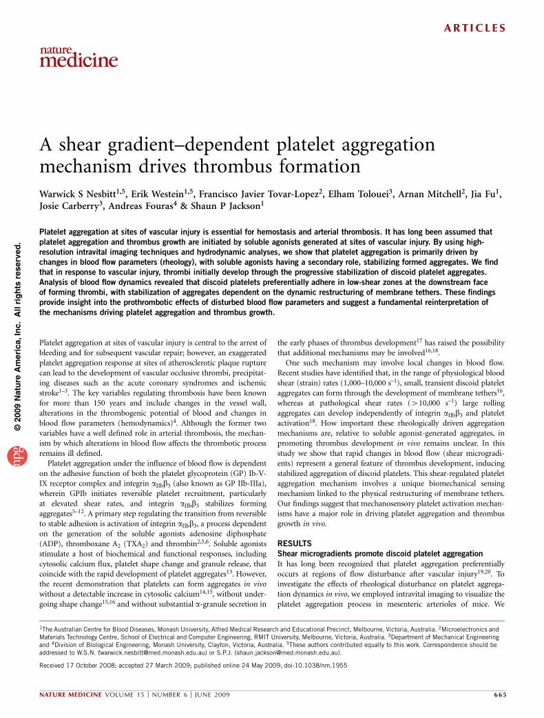
and integrin aIIbb3, with the relative contribution of each receptordependent on local hemodynamic conditions5. To investigate the roleof these receptors in promoting shear microgradient–dependentplatelet aggregation, we examined the effects of blocking GPIb orintegrin aIIbb3 function on discoid platelet aggregation in microchan-nel flow experiments. Inhibition of the ligand binding function ofGPIb revealed an absolute requirement for this receptor in initiatingplatelet recruitment to the peak shear zone in the microchannel(Fig. 2a). In contrast, blocking integrin aIIbb3 function had noinhibitory effect on initial platelet recruitment to the high-shear zone, but it markedly inhibited platelet aggregation in thedownstream deceleration zone (Fig. 2a), demonstrating distinctfunctions for these receptors.
Platelet activation at sites of vascular injury is stimulated by solubleagonists, principally ADP, TXA2 and thrombin. To investigate the roleof these agonists in promoting shear microgradient–dependent plate-let aggregation, we determined the effects of blocking TXA2 generation(with the cyclooxygenase inhibitor indomethacin), ADP (withapyrase, in combination with the P2Y1 and P2Y12 inhibitorsMRS2179 and 2-MeSAMP, respectively) and thrombin (with hirudin)on discoid platelet aggregation in microchannel flow experiments.Notably, eliminating the platelet-activating effects of ADP, TXA2 andthrombin had no effect on platelet aggregation dynamics induced bylocal shear microgradients (Fig. 2b and Supplementary Video 2online). Even in the complete absence of soluble agonists, stabilizeddiscoid platelet aggregates were able to persist for the full duration ofblood perfusion (up to 10 min); however, they remained reversibleand rapidly disaggregated after cessation of flow (data not shown).These studies show that shear microgradients can induce sustainedaggregation of discoid platelets independently of soluble agonists.
To investigate whether platelet aggregation induced by shear decel-eration in vivo is dependent on soluble agonists, we examined theeffects of blocking TXA2 generation (with aspirin), ADP (withclopidogrel and with the use of P2Y1–/– mice which lack the P2Y1
ADP receptor) and thrombin (with hirudin) on discoid platelet
6,000
a
c
d
f g
e
bControl
Control
Anti-αllbβ3Anti-GPlb
+ ADP and TxA2antagonists + hirudin5,000
4,000
Agg
rega
te s
ize
(µm
2 )
3,000
2,000
125
100
75
Rel
ativ
e th
rom
bus
size
(% o
f max
)
50
25
0
125
100
75
Rel
ativ
e th
rom
bus
size
(% o
f max
)
50
25
0
125
100
75
Rel
ativ
e th
rom
bus
size
(% o
f max
)
50
25
0125 150100
Time (s)7550250
125 150100Time (s)
7550250
125
100
75
Rel
ativ
e th
rom
bus
size
(% o
f max
)
50
25
125 150100Time (s)
75502500
100 200 300Time (s)
0
1,000
Normal flow
1 s 4 s 9 s 17 s
Normal flowFlow deceleration Flow deceleration
0
0 50 150 200Time (s)
250 300350100 0 50 150 200Time (s)
250 300350100
6,000
5,000
4,000
Agg
rega
te s
ize
(µm
2 )
3,000
2,000
1,000
0
Figure 2 Platelet aggregation induced shear microgradients occurs
independently of ADP, TXA2 and thrombin. (a) Aggregation traces showing
the response of whole-blood perfusion through the microchannel depicted in
Figure 1c. Control, hirudin-anticoagulated whole-blood perfusion at an input
(prestenosis) shear rate of 1,800 s–1 (representative of n ¼ 3 experiments);
anti-aIIbb3, hirudin-anticoagulated whole blood treated for 10 min with
30 mg ml–1 c7E3 Fab before blood perfusion (representative of n ¼ 3
experiments); anti-GPIb, hirudin-anticoagulated whole blood treated for
10 min with 50 mg ml–1 of the GPIb blocking IgG ALMA12 (representative
of n ¼ 3 experiments). (b) Microchannel aggregation as in a. Control,
hirudin-anticoagulated whole-blood perfusion at an input (prestenosis) shear
rate of 1,800 s–1; +ADP and TXA2 antagonists + hirudin, hirudin-
anticoagulated whole blood treated for 10 min with MRS2179 (100 mM),
2-MeSAMP (10 mM) and indomethacin (10 mM) (representative of n ¼ 3
experiments). (c) Representative DIC imaging showing the impact ofdownstream vessel compression and resultant flow deceleration on platelet
aggregate growth at a site of vessel crush injury (n ¼ 4 experiments). Dotted
marquee highlights the outer perimeter of the developing discoid platelet
aggregate. Solid line indicates the vessel wall. Black arrows highlight both
relative length and height increase. Scale bar, 10 mm. (d–g) Relative
thrombus size (% of maximum size) in mouse arterioles as a function of
applied downstream vessel compression (gray bars represent periods of flow
deceleration due to vessel compression) (DIC two-dimensional surface area
in mm2) in wild-type mice (d), wild-type mice pretreated orally with 10 mg
per kg body weight clopidogrel and 200 mg per kg body weight aspirin (e),
wild-type mice pretreated with 50 mg per kg body weight hirudin (f) or
P2Y1–/–mice pretreated with 50 mg per kg body weight clopidogrel and
200 mg per kg body weight aspirin and 50 mg per kg body weight
hirudin (g). In d–g, n ¼ 4 experiments in three mice per condition.
ART ICL ES
NATURE MEDICINE VOLUME 15 [ NUMBER 6 [ JUNE 2009 667
©20
09 N
atu
re A
mer
ica,
Inc.
All
rig
hts
res
erve
d.
aggregation in mesenteric arterioles as a function of flow deceleration(Fig. 2c,d and Supplementary Fig. 1c,d). Pretreatment of mice withcombined clopidogrel and aspirin or with hirudin had no inhibitoryeffect on discoid platelet aggregation induced by shear deceleration(Fig. 2e,f), such that serial deceleration and acceleration of blood flowinduced corresponding increases and decreases in aggregate size,respectively. Similarly, eliminating the platelet-activating effects ofADP, TXA2 and thrombin altogether by pretreating P2Y1–/– micewith high-dose clopidogrel, hirudin and aspirin also failed to inhibitplatelet aggregation induced by flow deceleration (Fig. 2g). Thesestudies show that flow deceleration can promote discoid plateletaggregation in vivo, independently of soluble agonists.
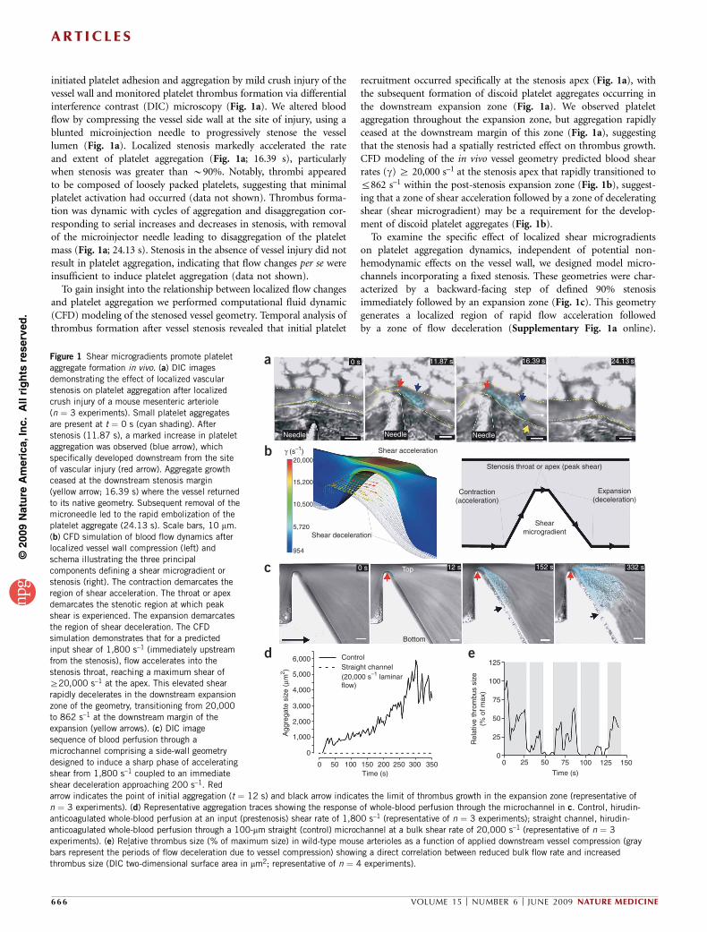
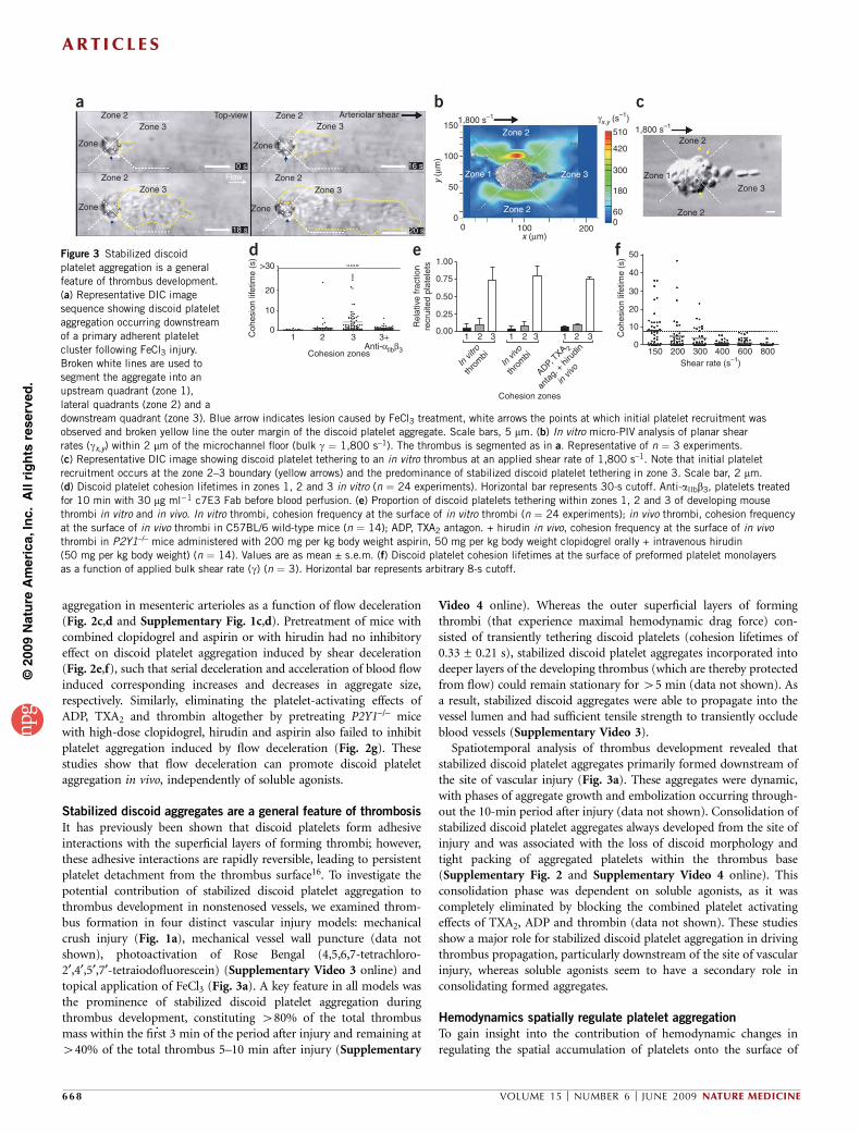
Stabilized discoid aggregates are a general feature of thrombosis
It has previously been shown that discoid platelets form adhesiveinteractions with the superficial layers of forming thrombi; however,these adhesive interactions are rapidly reversible, leading to persistentplatelet detachment from the thrombus surface16. To investigate thepotential contribution of stabilized discoid platelet aggregation tothrombus development in nonstenosed vessels, we examined throm-bus formation in four distinct vascular injury models: mechanicalcrush injury (Fig. 1a), mechanical vessel wall puncture (data notshown), photoactivation of Rose Bengal (4,5,6,7-tetrachloro-2¢,4¢,5¢,7¢-tetraiodofluorescein) (Supplementary Video 3 online) andtopical application of FeCl3 (Fig. 3a). A key feature in all models wasthe prominence of stabilized discoid platelet aggregation duringthrombus development, constituting 480% of the total thrombusmass within the first 3 min of the period after injury and remaining at440% of the total thrombus 5–10 min after injury (Supplementary
Video 4 online). Whereas the outer superficial layers of formingthrombi (that experience maximal hemodynamic drag force) con-sisted of transiently tethering discoid platelets (cohesion lifetimes of0.33 ± 0.21 s), stabilized discoid platelet aggregates incorporated intodeeper layers of the developing thrombus (which are thereby protectedfrom flow) could remain stationary for 45 min (data not shown). Asa result, stabilized discoid aggregates were able to propagate into thevessel lumen and had sufficient tensile strength to transiently occludeblood vessels (Supplementary Video 3).
Spatiotemporal analysis of thrombus development revealed thatstabilized discoid platelet aggregates primarily formed downstream ofthe site of vascular injury (Fig. 3a). These aggregates were dynamic,with phases of aggregate growth and embolization occurring through-out the 10-min period after injury (data not shown). Consolidation ofstabilized discoid platelet aggregates always developed from the site ofinjury and was associated with the loss of discoid morphology andtight packing of aggregated platelets within the thrombus base(Supplementary Fig. 2 and Supplementary Video 4 online). Thisconsolidation phase was dependent on soluble agonists, as it wascompletely eliminated by blocking the combined platelet activatingeffects of TXA2, ADP and thrombin (data not shown). These studiesshow a major role for stabilized discoid platelet aggregation in drivingthrombus propagation, particularly downstream of the site of vascularinjury, whereas soluble agonists seem to have a secondary role inconsolidating formed aggregates.
Hemodynamics spatially regulate platelet aggregation
To gain insight into the contribution of hemodynamic changes inregulating the spatial accumulation of platelets onto the surface of
Zone 1 Zone 1
Zone 2a b
d e f
cZone 3
Zone 1
Zone 2
Zone 1
Zone 2
Zone 2
Zone 3 Zone 3
1501,800 s–1
1,800 s–1γx,y (s
–1)
100
200
510
420
300
180
600
100x (µm)
y (µ
m)
50
>30 1.0050
150 200 300 400 600 800
40
30
20
10
0
0.50
Rel
ativ
e fr
actio
nre
crui
ted
plat
elet
s
Coh
esio
n lif
etim
e (s
)
0.25
In vi
tro
thro
mbi
In vi
vo
thro
mbi
ADP, TXA 2
anta
g. +
hirud
in
in viv
o
0.001 2 3 1 2
Cohesion zones
Shear rate (s–1)
3 1 2 3
0.7520
Coh
esio
n lif
etim
e (s
)
10
1 2 3
Cohesion zones
3+Anti-αllbβ3
0
00
Zone 3Arteriolar shear
Zone 1
Zone 2
Zone 2
Zone 3
Zone 1
Zone 2
Zone 2
Zone 3Flow0 s
18 s
16 s
20 s
Top-view
Figure 3 Stabilized discoid
platelet aggregation is a general
feature of thrombus development.
(a) Representative DIC image
sequence showing discoid platelet
aggregation occurring downstream
of a primary adherent platelet
cluster following FeCl3 injury.
Broken white lines are used to
segment the aggregate into an
upstream quadrant (zone 1),
lateral quadrants (zone 2) and a
downstream quadrant (zone 3). Blue arrow indicates lesion caused by FeCl3 treatment, white arrows the points at which initial platelet recruitment wasobserved and broken yellow line the outer margin of the discoid platelet aggregate. Scale bars, 5 mm. (b) In vitro micro-PIV analysis of planar shear
rates (gx,y) within 2 mm of the microchannel floor (bulk g ¼ 1,800 s–1). The thrombus is segmented as in a. Representative of n ¼ 3 experiments.
(c) Representative DIC image showing discoid platelet tethering to an in vitro thrombus at an applied shear rate of 1,800 s–1. Note that initial platelet
recruitment occurs at the zone 2–3 boundary (yellow arrows) and the predominance of stabilized discoid platelet tethering in zone 3. Scale bar, 2 mm.
(d) Discoid platelet cohesion lifetimes in zones 1, 2 and 3 in vitro (n ¼ 24 experiments). Horizontal bar represents 30-s cutoff. Anti-aIIbb3, platelets treated
for 10 min with 30 mg ml�1 c7E3 Fab before blood perfusion. (e) Proportion of discoid platelets tethering within zones 1, 2 and 3 of developing mouse
thrombi in vitro and in vivo. In vitro thrombi, cohesion frequency at the surface of in vitro thrombi (n ¼ 24 experiments); in vivo thrombi, cohesion frequency
at the surface of in vivo thrombi in C57BL/6 wild-type mice (n ¼ 14); ADP, TXA2 antagon. + hirudin in vivo, cohesion frequency at the surface of in vivo
thrombi in P2Y1–/– mice administered with 200 mg per kg body weight aspirin, 50 mg per kg body weight clopidogrel orally + intravenous hirudin
(50 mg per kg body weight) (n ¼ 14). Values are as mean ± s.e.m. (f) Discoid platelet cohesion lifetimes at the surface of preformed platelet monolayers
as a function of applied bulk shear rate (g) (n ¼ 3). Horizontal bar represents arbitrary 8-s cutoff.
ART ICL ES
668 VOLUME 15 [ NUMBER 6 [ JUNE 2009 NATURE MEDICINE
©20
09 N
atu
re A
mer
ica,
Inc.
All
rig
hts
res
erve
d.
thrombi, we developed a microscale particle imaging velocimetry(micro-PIV) approach to quantitatively map the planar shear (gx,y)environment around thrombi (Fig. 3b and Supplementary Fig. 3online). Examination of the planar (gx,y) shear rates around an in vitrothrombus at an applied bulk shear of 1,800 s–1 revealed the develop-ment of three spatially distinct shear zones at the thrombus surface. Atthe leading edge of thrombi (zone 1), shear (gx,y) was minimal,equivalent to free flow regions elsewhere in the flow channel(Fig. 3b and Supplementary Fig. 3). However, on the thrombussides (parallel to the direction of flow; zone 2) marked lateral shearacceleration (gx,y ¼ 240 s–1 to 4510 s–1) occurred (Fig. 3b). At thedownstream face of thrombi (zone 3), a distinct low-shear pocketdeveloped (gx,y ¼ 0–120 s–1; Fig. 3b). Notably, the spatial distributionof shear remained the same over a broad range of applied flow rates(Supplementary Fig. 3). These studies show that thrombi generatespatially discrete shear zones, with shear deceleration becomingprominent at the downstream thrombus face.
Analysis of platelet recruitment to thrombi revealed that initialrecruitment typically occurred in the shear acceleration zone on thesides of thrombi (zone 2; Fig. 3c and Supplementary Video 4). Theseinitial interactions were labile (cohesion lifetimes 0.18 ± 0.11 s),resulting in rapid platelet translocation into the low-shear zone atthe downstream face (zone 3) (Fig. 3d and Supplementary Video 5online). Once in zone 3, these tethered discoid platelets underwentstabilization, resulting in a 450-fold increase in cohesion lifetimes(from 0.18 s to Z30 s; (Fig. 3d)). As a consequence, 475% of stablediscoid platelet aggregation occurred within this low-shear pocket(Fig. 3e). This proportion did not change with inhibition of solubleagonists (data not shown); however, recruitment to the low-shear zonewas eliminated by integrin aIIbb3 blockade (Fig. 3d). We observed a
similar pattern of platelet recruitment for thrombus development in vivo(Fig. 3e and Supplementary Videos 4 and 5). These findings indicatethat local shear microgradients generated by developing thrombi, andmore specifically decelerating shear at the downstream face of thrombi,directly induce stabilization of discoid platelet aggregates.
To examine the relationship between shear and stabilization ofdiscoid platelet adhesion more directly, we performed platelet perfu-sion experiments on platelet monolayers over a laminar shear rangecovering those measured at the surface of thrombi (Fig. 3f). Thesestudies showed a direct inverse correlation between shear and adhe-sion lifetimes of discoid platelets, such that at 800 s–1 all discoidplatelet adhesive interactions were highly unstable (mean tetheringtime of o2 s), whereas we observed increased platelet adhesionlifetimes with progressive reductions in shear, similar to those occur-ring at the downstream face of forming thrombi (Fig. 3f). Overall,these studies support a model in which local changes in bloodhemodynamics around forming thrombi directly modify the stabilityof discoid platelet adhesive interactions.
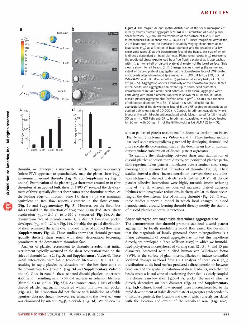
Shear microgradient magnitude determines aggregate size
The demonstration that thrombi promote stabilized discoid plateletaggregation by locally modulating blood flow raised the possibilitythat the magnitude of locally generated shear microgradients is amajor determinant of overall aggregate size. To test this hypothesisdirectly, we developed a ‘bead collision assay’, in which we immobi-lized polystyrene microspheres of varying sizes (2-, 5-, 9- and 15-mmdiameter), precoated with purified human von Willebrand factor(vWF), at the surface of glass microcapillaries to induce controlledlocalized changes in blood flow. CFD analysis of shear stress (tx,y)distributions at the bead surface predicted a direct correlation betweenbead size and the spatial distribution of shear gradients, such that thebeads create a lateral zone of accelerating shear that is closely coupledto a downstream low shear (r30.4 Pa) pocket, the size of which isdirectly dependent on bead diameter (Fig. 4a and SupplementaryFig. 4a,b online). Blood flow around these microspheres led to therapid development of stable discoid platelet aggregates (independentlyof soluble agonists), the location and size of which directly correlatedwith the location and extent of the low-shear zone (Fig. 4b,c).
�x,y (Pa)
Side view Top view
2 µm
5 µm
9 µm
Zone 3
15 µm
2 µm
5 µm
9 µm
15 µm
Zone 3
d
a b
125
100
75
50
25
0
Mea
n ag
greg
ate
size
(µm
2 )
400c
300
200
Mea
n ag
greg
ate
size
(µm
2 )
100
02 µm 5 µm 9 µm 15 µm
Contro
l
Anti-α llb
β 3
Anti-G
Plbα
137123
110968269554127140
z
y x
10,000 s–1
Figure 4 The magnitude and spatial distribution of the shear microgradient
directly affects platelet aggregate size. (a) CFD simulation of blood planar
shear stresses (tx,y) around microspheres at the surface of 0.2 � 2 mm
microcapillaries (bulk shear rate ¼ 10,000 s–1); inset, magnified view of the
2-mm bead case. Note the increase in spatially varying shear stress at the
bead sides (tx,y) as a function of bead diameter and the creation of a low
shear zone (zone 3) at the downstream face of the beads, the size of which
is directly dependent on bead diameter. Planar shear stress (tx,y) represents
the predicted stress experienced by a free flowing platelet as it approaches
within 1 mm (one-half of discoid platelet diameter) of the bead surface. Side
view is shown for all beads. (b) DIC image frames showing the nature and
extent of discoid platelet aggregation at the downstream face of vWF-coated
microbeads after whole-blood (pretreated with 100 mM MRS2179, 10 mM
2-MeSAMP and 10 mM indomethacin) perfusion at an applied g of 10,000
s–1 (n ¼ 5). Aggregation occurs exclusively at the downstream (zone 3) faceof the beads, and aggregates can extend up to seven bead diameters
downstream of initial platelet-bead adhesion, with overall aggregate width
correlating with bead diameter. Top view is shown for all beads. (c) Mean
discoid platelet aggregate size (surface area in mm2 ± s.e.m.) as a function
of microbead diameter (n ¼ 3). (d) Mean (± s.e.m.) discoid platelet
aggregate size at the downstream face of 5-mm vWF-coated microbeads at an
applied bulk shear rate of 10,000 s–1. Control, hirudin-anticoagulated whole
blood; anti-aIIbb3, hirudin-anticoagulated whole blood treated for 10 min with
30 mg ml–1 c7E3 Fab; anti-GPIb, hirudin-anticoagulated whole blood treated
for 10 min with 50 mg ml–1 of the GPIb-blocking IgG ALMA12 (n ¼ 3).
ART ICL ES
NATURE MEDICINE VOLUME 15 [ NUMBER 6 [ JUNE 2009 669
©20
09 N
atu
re A
mer
ica,
Inc.
All
rig
hts
res
erve
d.
Notably, the nature and extent of aggregation was equivalent to thatobserved during the early stages of thrombus development in vivo.Much as our earlier findings had indicated (Figs. 2a and 3d), GPIbwas essential for the initiation of platelet aggregation, whereas integrinaIIbb3 engagement stabilized discoid platelet aggregates (Fig. 4d).These studies show that the magnitude and spatial distribution ofshear microgradients directly influences platelet aggregate size.
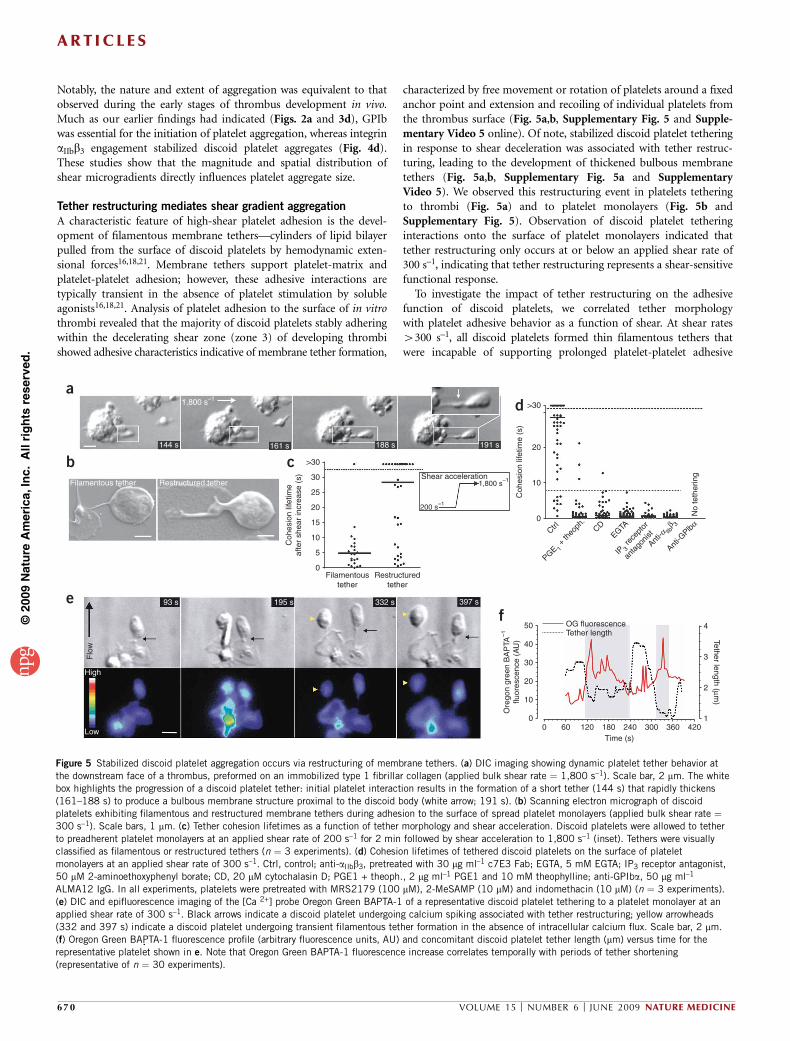
Tether restructuring mediates shear gradient aggregation
A characteristic feature of high-shear platelet adhesion is the devel-opment of filamentous membrane tethers—cylinders of lipid bilayerpulled from the surface of discoid platelets by hemodynamic exten-sional forces16,18,21. Membrane tethers support platelet-matrix andplatelet-platelet adhesion; however, these adhesive interactions aretypically transient in the absence of platelet stimulation by solubleagonists16,18,21. Analysis of platelet adhesion to the surface of in vitrothrombi revealed that the majority of discoid platelets stably adheringwithin the decelerating shear zone (zone 3) of developing thrombishowed adhesive characteristics indicative of membrane tether formation,
characterized by free movement or rotation of platelets around a fixedanchor point and extension and recoiling of individual platelets fromthe thrombus surface (Fig. 5a,b, Supplementary Fig. 5 and Supple-mentary Video 5 online). Of note, stabilized discoid platelet tetheringin response to shear deceleration was associated with tether restruc-turing, leading to the development of thickened bulbous membranetethers (Fig. 5a,b, Supplementary Fig. 5a and SupplementaryVideo 5). We observed this restructuring event in platelets tetheringto thrombi (Fig. 5a) and to platelet monolayers (Fig. 5b andSupplementary Fig. 5). Observation of discoid platelet tetheringinteractions onto the surface of platelet monolayers indicated thattether restructuring only occurs at or below an applied shear rate of300 s–1, indicating that tether restructuring represents a shear-sensitivefunctional response.
To investigate the impact of tether restructuring on the adhesivefunction of discoid platelets, we correlated tether morphologywith platelet adhesive behavior as a function of shear. At shear rates4300 s–1, all discoid platelets formed thin filamentous tethers thatwere incapable of supporting prolonged platelet-platelet adhesive
144 s
Filamentous tether
>30
a
b c
d
ef
30 Shear acceleration
200 s–1
1,800 s–1
25
20
Coh
esio
n lif
etim
eaf
ter
shea
r in
crea
se (
s)
15
10
5
0
50 4
3
2
1
OG fluorescenceTether length Tether length (µm
)
40
30
Ore
gon
gree
n B
AP
TA–1
fluor
esce
nce
(AU
)
20
10
0 60 120 180Time (s)
240 300 360 4200
>30
20
Coh
esio
n lif
etim
e (s
)
10
No
teth
erin
g
0
93 s
High
Flo
w
Low
195 s 332 s 397 s
Filamentoustether
Restructuredtether
Ctrl
PGE 1 +
theo
ph.
CDEGTA
IP 3 re
cept
or
anta
gonis
t
Anti-α llb
β 3
Anti-G
Plbα
Restructured tether
161 s 188 s 191 s
1,800 s–1
Figure 5 Stabilized discoid platelet aggregation occurs via restructuring of membrane tethers. (a) DIC imaging showing dynamic platelet tether behavior at
the downstream face of a thrombus, preformed on an immobilized type 1 fibrillar collagen (applied bulk shear rate ¼ 1,800 s–1). Scale bar, 2 mm. The white
box highlights the progression of a discoid platelet tether: initial platelet interaction results in the formation of a short tether (144 s) that rapidly thickens
(161–188 s) to produce a bulbous membrane structure proximal to the discoid body (white arrow; 191 s). (b) Scanning electron micrograph of discoidplatelets exhibiting filamentous and restructured membrane tethers during adhesion to the surface of spread platelet monolayers (applied bulk shear rate ¼300 s–1). Scale bars, 1 mm. (c) Tether cohesion lifetimes as a function of tether morphology and shear acceleration. Discoid platelets were allowed to tether
to preadherent platelet monolayers at an applied shear rate of 200 s–1 for 2 min followed by shear acceleration to 1,800 s–1 (inset). Tethers were visually
classified as filamentous or restructured tethers (n ¼ 3 experiments). (d) Cohesion lifetimes of tethered discoid platelets on the surface of platelet
monolayers at an applied shear rate of 300 s–1. Ctrl, control; anti-aIIbb3, pretreated with 30 mg ml–1 c7E3 Fab; EGTA, 5 mM EGTA; IP3 receptor antagonist,
50 mM 2-aminoethoxyphenyl borate; CD, 20 mM cytochalasin D; PGE1 + theoph., 2 mg ml–1 PGE1 and 10 mM theophylline; anti-GPIba, 50 mg ml–1
ALMA12 IgG. In all experiments, platelets were pretreated with MRS2179 (100 mM), 2-MeSAMP (10 mM) and indomethacin (10 mM) (n ¼ 3 experiments).
(e) DIC and epifluorescence imaging of the [Ca 2+] probe Oregon Green BAPTA-1 of a representative discoid platelet tethering to a platelet monolayer at an
applied shear rate of 300 s–1. Black arrows indicate a discoid platelet undergoing calcium spiking associated with tether restructuring; yellow arrowheads
(332 and 397 s) indicate a discoid platelet undergoing transient filamentous tether formation in the absence of intracellular calcium flux. Scale bar, 2 mm.
(f) Oregon Green BAPTA-1 fluorescence profile (arbitrary fluorescence units, AU) and concomitant discoid platelet tether length (mm) versus time for the
representative platelet shown in e. Note that Oregon Green BAPTA-1 fluorescence increase correlates temporally with periods of tether shortening
(representative of n ¼ 30 experiments).
ART ICL ES
670 VOLUME 15 [ NUMBER 6 [ JUNE 2009 NATURE MEDICINE
©20
09 N
atu
re A
mer
ica,
Inc.
All
rig
hts
res
erve
d.
interactions beyond 8 s. In contrast, at a shear of 300 s–1, 64 ± 14% ofplatelets showed overtly restructured tethers, resulting in an increasein cohesion lifetimes to a range of 8 s to 430 s (data not shown). Thisincrease in adhesion lifetime was associated with adhesion strengthen-ing, as discoid platelets with restructured tethers had a markedlyincreased ability to withstand the detaching effects of rapid shearincreases (Fig. 5c). Approximately 75% of platelets with restructuredtethers formed sustained adhesive interactions of 48-s duration aftershear acceleration, compared to o10% of discoid platelets with thinfilamentous tethers (Fig. 5c). These studies indicate that strengtheningof discoid platelet cohesive interactions involves the physical restruc-turing of membrane tethers.
Mechanotransduction underlies tether restructuring
To gain insight into the underlying mechanisms regulating tetherrestructuring, we treated platelets with global inhibitors of plateletactivation—prostaglandin E1 (PGE1) and the phosphodiesterase inhi-bitor theophylline (dimethylxanthine)—or with the actin polymeriza-tion inhibitor cytochalasin D. These inhibitors abolished tetherrestructuring and the associated adhesion strengthening, demonstrat-ing that this phenomenon is activation dependent (Fig. 5d). Adhesionstrengthening due to tether restructuring was calcium dependent, as itwas abolished by chelation of extracellular Ca2+ (with EGTA) and byinhibition of inositol triphosphate-mediated calcium release (Fig. 5d).Furthermore, tether restructuring occurred independently of TXA2,ADP and thrombin but required ligand binding to integrin aIIbb3
(Fig. 5d). Imaging of Ca2+ flux showed that initial filamentous tetherformation occurred without a detectable increase in Ca2+ concentra-tion (Fig. 5e,f; 93 s); however, subsequent tether restructuring wasassociated with short duration Ca2+ spikes that correlated with phasesof tether shortening or tensioning (Fig. 5c,e,f; 195 s; and Supplemen-tary Video 6 online). This transient Ca2+ flux was both IP3 and extra-cellular Ca2+ dependent (data not shown) and occurred in the absenceof global platelet shape change (Fig. 5e). These findings suggest that
tether restructuring involves a localized Ca2+-dependent signalingmechanism that occurs independently of global platelet activation.
To investigate the potential relevance of these calcium flux findingsto stabilized discoid platelet aggregation in vivo, we analyzed thedegree of platelet activation within discrete layers of forming thrombiby monitoring changes in cytosolic calcium flux (D[Ca2+]c) andsurface expression of the platelet a-granule protein, P-selectin (Sup-plementary Fig. 6a–e online). Discoid platelets undergoing transienttethering interactions with the surface of forming thrombi showedminimal D[Ca2+]c (Supplementary Fig. 6a) and had no detectableP-selectin expression (Supplementary Fig. 6c), whereas plateletswithin the stabilized discoid aggregate layers undergoing sustainedadhesion showed transient D[Ca2+]c but minimal P-selectin levels,similar to those observed in platelets undergoing tether restructuringin vitro (Supplementary Fig. 6a,d). Platelets stably incorporatedwithin the core of the thrombus showed sustained oscillatoryD[Ca2+]c and measurable amounts of P-selectin, consistent with therole of soluble agonists in promoting full platelet activation in thethrombus base (Supplementary Fig. 6a,e). Taken together, thesefindings show that stabilized discoid platelet aggregation is associatedwith a low level of transient cytosolic flux that is sufficient to sustainplatelet adhesive interactions independently of global platelet shapechange and degranulation.
DISCUSSION
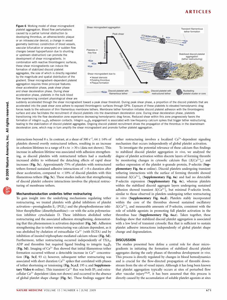
The studies presented here define a central role for shear micro-gradients in initiating the formation of stabilized discoid plateletaggregates during the early phases of thrombus development in vivo.This process is directly regulated by changes in blood hemodynamicsand is crucial for the flow-directed propagation of thrombi down-stream from the site of vessel injury. Although it has long been knownthat platelet aggregation typically occurs at sites of perturbed flowafter vascular injury19,20, it has been assumed that this process isdirectly caused by the accumulation of soluble platelet agonists at sites
Figure 6 Working model of shear microgradient
platelet aggregation. Blood flow perturbations
caused by a partial luminal obstruction (a
developing thrombus, an atherosclerotic plaque
or an intravascular device), a change in vessel
geometry (extrinsic constriction of blood vessels,
vascular bifurcation or aneurysm) or sudden flow
changes (vessel hypoperfusion due to shunting
or upstream obstruction) can promote the
development of shear microgradients. In
combination with reactive thrombogenic surfaces,
these shear microgradients can induce the
formation of stabilized discoid platelet
aggregates, the size of which is directly regulated
by the magnitude and spatial distribution of thegradient. Shear microgradient–dependent platelet
aggregation requires three principal features:
shear acceleration phase, peak shear phase
and shear deceleration phase. During shear
acceleration phase, platelets in the bulk blood
flow experiencing constant physiological shear are
suddenly accelerated through the shear microgradient toward a peak shear threshold. During peak shear phase, a proportion of the discoid platelets that are
accelerated into the peak shear zone adhere to exposed thrombogenic surfaces through GPIb. Exposure of these platelets to elevated hemodynamic drag
forces leads to the extrusion of thin filamentous membrane tethers. Membrane tether formation initiates discoid platelet adhesion with the thrombogenic
surface and also facilitates the recruitment of discoid platelets into the downstream deceleration zone. During shear deceleration phase, platelets
transitioning into the flow deceleration zone experience decreasing hemodynamic drag forces. Reduced shear within this zone progressively favors the
formation of integrin aIIbb3 adhesion contacts. Integrin aIIbb3 engagement is associated with low-frequency calcium spikes that trigger tether restructuring,
leading to the stabilization of discoid platelet aggregates. Ongoing discoid platelet recruitment drives the propagation of the thrombus in the downstream
deceleration zone, which may in turn amplify the shear microgradient and promote further platelet aggregation.
Shear microgradient aggregation
Peak shear2
13
12
3
Transient filamentoustether formation
Stabilized discoid plateletaggregation
(tether restructuring)
Shear deceleration
Platelets infree flow
Shear microgradient due to:
Vessel stenosisExisting thrombusPlaque formation
Discoid platelet withfilamentous tether
Ca2+ Discoid platelet withrestructured tether
Nucleatingplatelet cluster or core
She
ar a
ccel
erat
ion
ART ICL ES
NATURE MEDICINE VOLUME 15 [ NUMBER 6 [ JUNE 2009 671
©20
09 N
atu
re A
mer
ica,
Inc.
All
rig
hts
res
erve
d.
of flow disturbance. Our studies challenge this hypothesis and suggestthat platelets principally use a biomechanical platelet aggregationmechanism to promote the accumulation and stabilization of discoidplatelets at sites of vascular injury.
Our studies show that local shear microgradients, which occur withchanges in vessel geometry (stenosis) or as a consequence of thrombusformation itself, expose discoid platelets to rapidly changing hemo-dynamic conditions (specifically shear acceleration coupled to decel-eration), leading to the development of stabilized discoid plateletaggregates (Fig. 6). Mechanistically, platelet exposure to sudden accel-erations in shear promotes the development of membrane tethers,leading to the formation of transient discoid platelet aggregates16. Weshow that upon subsequent exposure to decelerating shear, thesetethers physically restructure, increasing the strength and stability ofdiscoid platelet aggregates, thereby promoting thrombus growth. Thismechanosensitive adhesion mechanism occurs independently of solu-ble agonists and therefore represents a mechanism of stabilizing plateletadhesion at sites of vascular injury. Overall, our findings indicate thateffective thrombus formation involves the cooperative interplaybetween two distinct, yet complementary, aggregation mechanisms,with stabilized discoid platelet aggregation representing the principalmechanism driving initial thrombus growth and soluble agonistgeneration primarily serving to stabilize formed aggregates.
Arterial thrombosis in the coronary or cerebrovascular circulationsis the principal pathological process underlying the acute coronarysyndromes and ischemic stroke, which together represent the leadingcause of morbidity and mortality in industrialized societies. Despiterecent improvements in antithrombotic therapies, the majority ofpatients receiving these drugs continue to die from acute thromboticevents3,22. Acute vascular occlusion represents the cumulative impactof atherosclerosis, superimposed thrombosis and vasoconstriction,with each of these processes having a major impact on vessel wallgeometry and blood hemodynamics. Although the importance ofperturbed hemodynamics in promoting thrombus formation has longbeen recognized4,23–28, the molecular mechanisms underpinning theseobservations have remained ill defined. Our findings demonstrate thatthe magnitude and spatial distribution of shear microgradients correlatedirectly with the extent of platelet aggregation and that the resultingstabilized aggregates of discoid platelets have sufficient tensile strengthto cause partial vascular occlusion, leading to a transient reduction inblood flow. The occlusive action of these aggregates, combined withtheir intrinsic instability and tendency to embolize to the distalcirculation, raises the possibility that this aggregation mechanism maycontribute to intermittent thromboembolic symptoms that are char-acteristic of the acute coronary syndromes and cerebrovascular disease.Furthermore, the demonstration that this aggregation process can occurindependent of soluble agonists may help explain the limited anti-thrombotic efficacy of aspirin, clopidogrel and thrombin inhibitors,particularly in individuals with severe vascular disease3.
Our findings help reconcile a number of fundamental controversiesin the field, providing a mechanistic explanation for the observationsthat discoid platelets form aggregates in vivo independently of sus-tained calcium signaling14,15 and that granule release and P-selectinexpression on the surface of platelets occurs relatively late in thethrombotic process17. These findings also provide insight into thedynamic, heterogeneous adhesive behavior of platelets during initialthrombus growth29. In this context, a greater understanding of thefundamental relationship between alterations in blood rheology,membrane-tether restructuring and platelet aggregation may lead tothe development of targeted antithrombotic therapies that are saferand more effective than existing approaches.
METHODS
Methods and any associated references are available in the onlineversion of the paper at http://www.nature.com/naturemedicine/.
Note: Supplementary information is available on the Nature Medicine website.
ACKNOWLEDGMENTSWe thank Z. Ruggeri, H.H. Salem, R. Andrews and B. Kile for helpful feedbackon the work; H. Blackburn and G. Rosengarten for advice and input on the CFDanalysis; C. Nguyen for technical assistance in the analysis of in vivo blood flowrates; SciTech Proprietary Ltd. and Andor Proprietary Ltd. for the generous loanof a DV897CS EMCCD camera; M. Hickey (Monash University Department ofMedicine, Monash, Australia) and A. Issekutz (Dalhousie University, Halifax,Canada) for providing the P-selectin–specific antibody; and C. Gachet (INSERM,Strasbourg, France) for P2Y1–/– mice. This work was supported by projectfunding from the National Health and Medical Research Council of Australiaand the Australian Research Council. E.W. was supported by the National HeartFoundation of Australia.
AUTHOR CONTRIBUTIONSW.S.N. designed the study and experiments, performed the in vitro platelet andmicro-PIV experiments, codesigned vascular mimetics, performed microchannelperfusion experiments, analyzed data, supervised the study and cowrote themanuscript. E.W. designed experiments, performed the in vivo and in vitroexperiments, analyzed data and assisted with manuscript preparation. F.J.T.-L.codesigned and fabricated vascular mimetics (microchannels), performedmicrochannel perfusion experiments and performed CFD simulations. E.T.performed and analyzed the micro-PIV experiments. J.F. performed in vivoexperiments. A.M. codesigned the vascular mimetics and supervised theirfabrication. J.C. supervised the micro-PIV analysis. A.F. designed and supervisedthe micro-PIV analysis and formulated and coded the micro-PIV analysissoftware. S.P.J. supervised the study and cowrote the manuscript.
Published online at http://www.nature.com/naturemedicine/
Reprints and permissions information is available online at http://npg.nature.com/
reprintsandpermissions/
1. Trip, M.D., Cats, V.M., van Capelle, F.J. & Vreeken, J. Platelet hyperreactivity andprognosis in survivors of myocardial infarction. N. Engl. J. Med. 322, 1549–1554(1990).
2. Ruggeri, Z.M. Platelets in atherothrombosis. Nat. Med. 8, 1227–1234 (2002).3. Bhatt, D.L. & Topol, E.J. Scientific and therapeutic advances in antiplatelet therapy.
Nat. Rev. Drug Discov. 2, 15–28 (2003).4. Virchow, R. Gesammelte abhandlungen zur wissenschaftlichen medicin. Medsinger
Sohn & Co. 219–732 (1856).5. Savage, B., Saldivar, E. & Ruggeri, Z.M. Initiation of platelet adhesion by arrest
onto fibrinogen or translocation on von Willebrand factor. Cell 84, 289–297 (1996).6. Savage, B., Almus-Jacobs, F. & Ruggeri, Z.M. Specific synergy of multiple substrate-
receptor interactions in platelet thrombus formation under flow. Cell 94, 657–666(1998).
7. Nesbitt, W.S. et al. Distinct glycoprotein Ib/V/IX and integrin aIIbb3-dependent calciumsignals cooperatively regulate platelet adhesion under flow. J. Biol. Chem. 277,2965–2972 (2002).
8. Nesbitt, W.S. et al. Intercellular calcium communication regulates platelet aggregationand thrombus growth. J. Cell Biol. 160, 1151–1161 (2003).
9. Giuliano, S., Nesbitt, W.S., Rooney, M. & Jackson, S.P. Bidirectional integrin aIIbb3
signalling regulating platelet adhesion under flow: contribution of protein kinase C.Biochem. J. 372, 163–172 (2003).
10. Goncalves, I., Nesbitt, W.S., Yuan, Y. & Jackson, S.P. Importance of temporal flowgradients and integrin aIIbb3 mechanotransduction for shear activation of platelets.J. Biol. Chem. 280, 15430–15437 (2005).
11. Ruggeri, Z.M. & Mendolicchio, G.L. Adhesion mechanisms in platelet function. Circ.Res. 100, 1673–1685 (2007).
12. Ruggeri, Z.M. The role of von Willebrand factor in thrombus formation. Thromb. Res.120 Suppl 1, S5–S9 (2007).
13. Jackson, S.P. The growing complexity of platelet aggregation. Blood 109, 5087–5095(2007).
14. Dubois, C., Panicot-Dubois, L., Gainor, J.F., Furie, B.C. & Furie, B. Thrombin-initiatedplatelet activation in vivo is vWF independent during thrombus formation in a laserinjury model. J. Clin. Invest. 117, 953–960 (2007).
15. van Gestel, M.A. et al. Real-time detection of activation patterns in individual plateletsduring thromboembolism in vivo: differences between thrombus growth and embolusformation. J. Vasc. Res. 39, 534–543 (2002).
16. Maxwell, M.J. et al. Identification of a 2-stage platelet aggregation process mediatingshear-dependent thrombus formation. Blood 109, 566–576 (2007).
17. Furie, B. & Furie, B.C. Thrombus formation in vivo. J. Clin. Invest. 115, 3355–3362(2005).
ART ICL ES
672 VOLUME 15 [ NUMBER 6 [ JUNE 2009 NATURE MEDICINE
©20
09 N
atu
re A
mer
ica,
Inc.
All
rig
hts
res
erve
d.
18. Ruggeri, Z.M., Orje, J.N., Habermann, R., Federici, A.B. & Reininger, A.J. Activation-independent platelet adhesion and aggregation under elevated shear stress. Blood 108,1903–1910 (2006).
19. Mustard, J.F., Jorgensen, L., Hovig, T., Glynn, M.F. & Rowsell, H.C. Role of platelets inthrombosis. Thromb. Diath. Haemorrh. Suppl. 21, 131–158 (1966).
20. Mustard, J.F., Murphy, E.A., Rowsell, H.C. & Downie, H.G. Factors influencingthrombus formation in vivo. Am. J. Med. 33, 621–647 (1962).
21. Dopheide, S.M., Maxwell, M.J. & Jackson, S.P. Shear-dependent tether formationduring platelet translocation on von Willebrand factor. Blood 99, 159–167(2002).
22. Antithrombotic Trialists’ Collaboration. Collaborative meta-analysis of randomisedtrials of antiplatelet therapy for prevention of death, myocardial infarction and strokein high risk patients. Br. Med. J. 324, 71–86 (2002).
23. Dintenfass, L. Rheologic approach to thrombosis and atherosclerosis. Angiology 15,333–343 (1964).
24. Stein, P.D. & Sabbah, H.N. Measured turbulence and its effect on thrombus formation.Circ. Res. 35, 608–614 (1974).
25. Yoganathan, A.P., Corcoran, W.H., Harrison, E.C. & Carl, J.R. The Bjork-Shiley aorticprosthesis: flow characteristics, thrombus formation and tissue overgrowth. Circulation58, 70–76 (1978).
26. Schoephoerster, R.T., Oynes, F., Nunez, G., Kapadvanjwala, M. & Dewanjee, M.K.Effects of local geometry and fluid dynamics on regional platelet deposition on artificialsurfaces. Arterioscler. Thromb. 13, 1806–1813 (1993).
27. Eckstein, E.C., Tilles, A.W. & Millero, F.J. III. Conditions for the occurrence of largenear-wall excesses of small particles during blood flow. Microvasc. Res. 36, 31–39(1988).
28. Wang, S.K. & Hwang, N.H. On transport of suspended particulates in tube flow.Biorheology 29, 353–377 (1992).
29. Kulkarni, S. et al. A revised model of platelet aggregation. J. Clin. Invest. 105,783–791 (2000).
ART ICL ES
NATURE MEDICINE VOLUME 15 [ NUMBER 6 [ JUNE 2009 673
©20
09 N
atu
re A
mer
ica,
Inc.
All
rig
hts
res
erve
d.
ONLINE METHODSMaterials. Lepirudin was from Pharmion; apyrase, hexamethyldisilazane
(HMDS), MRS2179, 2-MesAMP, indomethacin, EGTA, cytochalasin D (CD),
theophylline, 2-aminoethoxydiphenylborate (2-APB) and PGE1 were from
Sigma; Rose Bengal was from Acros Organics; c7E3 Fab was from Eli Lilly;
Oregon Green BAPTA-1 was from Molecular Probes; ALMA12 antibody was
provided by F. Lanza (Institut National de la Sante et de la Recherche Medicale
(INSERM) U.311, Strasbourg, France); and RMP-1Alexa488 was provided by
A. Issekutz (Dalhousie University, Halifax, Canada).
Platelet preparation. Washed platelets were isolated from anticoagulated whole
blood taken from consenting human donors according to a previously
described method30. Lepirudin (1 U ml–1) was included in platelet washing
buffer (4.3 mM K2HPO4, 4.3 mM Na2HPO4, 24.3 mM NaH2PO4, 113 mM
NaCl, 5.5 mM D-glucose and 10 mM theophylline, pH 6.5, with 0.02 U ml–1
apyrase) in Tyrode’s buffer (10 mM HEPES, 12 mM NaHCO3, 137 mM NaCl,
2.7 mM KCl and 5 mM glucose, pH 7.3). We obtained approval for these
studies from the Monash University Standing Committee on Ethics in Research
Involving Humans.
Intravital studies. Approval was obtained from the Alfred Medical Research
and Education Precinct Animal Ethics Committee for all experiments involving
animals. We performed intravital studies according to a modification of a
previously published method31,32. We used C57BL/6 wild-type mice weighing
18–20 g (Alfred Medical Research and Education Precinct Animal Services
(AMREP AS Pty Ltd)), and P2Y1–/– mice (previously described33,34). We
anesthetized mice (60 mg per kg body weight sodium pentabarbitone) and
injured their mesenteric arterioles (60–160 mm) via photoactivation (excitation
at 550 nm, 10–30 s) of systemically administered Rose Bengal (5 mg per kg
body weight)35, microinjection of 6% FeCl3 into the tissue adjacent to the
arteriole of interest36, mechanical puncture of the vessel wall with a micro-
injector needle (tip diameter 4–6 mm) or mild crush injury induced by ligation
of the vessel with a blunted microinjector needle (tip diameter 30–60 mm). For
the in vivo stenosis experiments, we administered P2Y1–/– mice, at time points
of –24 h and –2 h, 200 mg per kg body weight aspirin and either 10 mg per kg
body weight clopidogrel (causing an B50% reduction in ADP-induced platelet
aggregation) or 50 mg per kg body weight clopidogrel orally (causing complete
inhibition of ADP-induced platelet aggregation). In studies using hirudin, we
intravenously infused mice with high-dose hirudin (50 mg per kg body weight)
5 min before the onset of vascular injury. In control studies, we confirmed the
effectiveness of each of these antagonists by performing ex vivo platelet
functional assays or through coagulation testing37,38. We monitored P-selectin
expression after FeCl3-induced thrombus formation in the presence of systemi-
cally administered P-selectin–specific antibody coupled to Alexa488 fluoro-
phore (RMP-1Alexa488, obtained from M. Hickey; 1.3 mg per kg body weight)
and recorded with an ANDOR DV897DCS electron-multiplying charge-
coupled device (EM-CCD). We visualized platelet interactions by DIC micro-
scopy using a Leica DMIRB inverted microscope (objectives: 100� PL APO,
numerical aperture 1.40–0.7 and 63� HCX PL APO, numerical aperture 1.2)
and a DAGE MTI charge-coupled device camera.
Microchannel fabrication. We fabricated microchannels in polydimethyl-
siloxane (Sylgard) from a KMPR 1025 (polymethylglutarimide) photoresist
(MicroChem) mold on a 3-inch silicon wafer according to a modification of
a published method39. We used a high-resolution chrome mask to attain well
defined features to construct the mold. The overall channel depth was 80 mm.
Flow studies and analysis of tether restructuring. We performed flow assays
according to a modification of a published method32. We derivatized dried
microcapillaries with hexamethyldisilazane for 15 min at 21 1C and washed
them with double-distilled water. For monolayer experiments, we allowed
platelets in Tyrode’s buffer (6 � 108 per ml) supplemented with 2 mM EGTA
to spread for 15 min, and then incubated them with BSA (20 mg ml–1) for
5 min. We preincubated washed platelets for 10 min with MRS2179 (100 mM),
2-MesAMP (10 mM) and indomethacin (10 mM) in Tyrode’s buffer supple-
mented with 1 mM Ca2+ before reconstitution with washed red blood cells
(50% vol/vol) (containing 0.02 U ml–1 apyrase and 1 U ml–1 lepirudin). Before
reconstitution, we preincubated washed platelets with either of the following
inhibitors: EGTA (5 mM), c7E3 Fab (30 mg ml–1), cytochalasin D (20 mM),
2-aminoethoxydiphenylborate (2-APB) (50 mM), ALMA12 (50 mg ml–1) or
combined PGE1 (10 mg ml–1) and theophylline (10 mM). We performed
perfusion studies at wall shear rates of 300 s–1 (monolayer experiments). We
visualized adherent platelets by DIC microscopy (100� PL APO objective,
numerical aperture 1.40–0.7) on a Leica DMIRB microscope. We monitored
adhesion over a 300-s period and video-recorded it for offline analysis. Thirty
seconds after the start of the flow, we analyzed the tethering time of platelets
(cohesion lifetime) frame by frame (25 frames per second). We set the
maximum cohesion lifetime cutoff to 30 s. We included only platelets with a
discoid morphology in the analysis.
Microparticle image velocimetry. We conducted micro-PIV experiments
according to a modification of a published method40,41 (Supplementary
Methods online).
Bead collision assay. We performed bead collision assays according to a
modification of a published method42 (Supplementary Methods).
Analysis of platelet calcium flux. We monitored platelet calcium flux, with
minor modifications, as previously described43 (Supplementary Methods).
Computational fluid dynamics. We calculated estimates of shear rates after
solving the velocity field using the conservation of mass and momentum
equations for an incompressible fluid flow, using the CFD software package
FLUENT 6.0 (Fluent USA) based on a finite volume scheme44. We assumed the
validity of the continuum hypothesis and no-slip boundary condition. We
considered the flow to be three-dimensional, steady, laminar and incompressible.
We considered the fluid medium to have a constant density of 998.2 km m–3 and
viscosity of 0.00345 Pa s. Discretization scheme pressure was set as standard, and
the second-order upwind momentum option was enabled. For the case where
vessel stenosis was localized to the site of vessel injury (Fig. 1a), we modeled the
geometry of the blood vessel with SolidWorks v. 2004 (SolidWorks Corp.) using
a blood vessel diameter of 42 mm and an idealized geometry derived from video
footage of the in vivo stenosis (Fig. 1a). No elastic effects were considered,
keeping the perimeter of the transversal section of the blood vessel constant.
We transferred the geometry into a specialized preprocessing grid generation
(Fluent, Gambit) to generate a structured mesh of 76,800 elements. For
modeling bead collision in microchannels, the mesh consisted of two zones,
an unstructured region close to the bead and a structured mesh everywhere else.
We applied a symmetry condition at the center plane of the beads. The number
of cells in the structured mesh generated was directly dependent on bead size
ranging from 754,446 (2-mm beads) to 954,516 (15-mm beads).
Statistical analyses. Results are presented as mean ± s.e.m. for bar graphs and
median for dot plots. We assessed statistical significance by Student’s t-test. We
drew schematics with CorelDraw Graphics Suite (Corel). We digitized video
using Pinnacle Systems DV500 PLUS (Pinnacle Systems) and edited it with
Adobe Premiere Pro v1.5 (Adobe Systems).
30. Schoenwaelder, S.M. et al. Identification of a unique co-operative phosphoinositide3-kinase signaling mechanism regulating integrin a3 adhesive function in platelets.J. Biol. Chem. 282, 28648–28658 (2007).
31. Denis, C. et al. A mouse model of severe von Willebrand disease: defects in hemostasisand thrombosis. Proc. Natl. Acad. Sci. USA 95, 9524–9529 (1998).
32. Kulkarni, S. et al. Techniques to examine platelet adhesive interactions under flow.Methods Mol. Biol. 272, 165–186 (2004).
33. Leon, C. et al. Defective platelet aggregation and increased resistance to thrombosisin purinergic P2Y(1) receptor-null mice. J. Clin. Invest. 104, 1731–1737 (1999).
34. Mayadas, T.N., Johnson, R.C., Rayburn, H., Hynes, R.O. & Wagner, D.D. Leukocyterolling and extravasation are severely compromised in P selectin-deficient mice. Cell74, 541–554 (1993).
35. Rosen, E.D. et al. Laser-induced noninvasive vascular injury models in mice generateplatelet- and coagulation-dependent thrombi. Am. J. Pathol. 158, 1613–1622 (2001).
36. Kurz, K.D., Main, B.W. & Sandusky, G.E. Rat model of arterial thrombosis induced byferric chloride. Thromb. Res. 60, 269–280 (1990).
37. Sturgeon, S.A., Jones, C., Angus, J.A. & Wright, C.E. Advantages of a selectiveb-isoform phosphoinositide 3-kinase antagonist, an anti-thrombotic agent devoidof other cardiovascular actions in the rat. Eur. J. Pharmacol. 587, 209–215(2008).
38. Mangin, P. et al. Thrombin overcomes the thrombosis defect associated with plateletGPVI/FcRgamma deficiency. Blood 107, 4346–4353 (2006).
doi:10.1038/nm.1955 NATURE MEDICINE
©20
09 N
atu
re A
mer
ica,
Inc.
All
rig
hts
res
erve
d.
39. Weibel, D.B., Diluzio, W.R. & Whitesides, G.M. Microfabrication meets microbiology.Nat. Rev. Microbiol. 5, 209–218 (2007).
40. Fouras, A., Lo Jacono, D. & Hourigan, K. Target-free stereo PIV: a novel technique withinherent error estimation and improved accuracy. Exp. Fluids 44, 317–329 (2008).
41. Fouras, A.S.J. Accuaracy of out-of-plane vorticity measurements derived from in-planevelocity field data. Exp. Fluids 25, 409–430 (1998).
42. Edmondson, K.E., Denney, W.S. & Diamond, S.L. Neutrophil-bead collisionassay: pharmacologically induced changes in membrane mechanics regulate
the PSGL-1/P-selectin adhesion lifetime. Biophys. J. 89, 3603–3614(2005).
43. Yap, C.L. et al. Synergistic adhesive interactions and signaling mechanisms operatingbetween platelet glycoprotein Ib/IX and integrin alpha IIbbeta 3. Studies in humanplatelets and transfected Chinese hamster ovary cells. J. Biol. Chem. 275,41377–41388 (2000).
44. Versteeg, H. & Malalasekrera, W. An Introduction to Computational Fluid Dynamics:The Finite Volume Method (Longman Scientific & Technical, Harlow, UK, 1995).
NATURE MEDICINE doi:10.1038/nm.1955
©20
09 N
atu
re A
mer
ica,
Inc.
All
rig
hts
res
erve
d.
Related Documents