A seroepidemiologic survey of canine visceral leishmaniosis among apparently healthy dogs in Croatia T. Z ˇ ivic ˇnjak a, * , F. Martinkovic ´ a , A. Marinculic ´ a , V. Mrljak b , N. Kuc ˇer b , V. Matijatko b ,Z ˇ . Mihaljevic ´ c , R. Baric ´-Rafaj b a Department for Parasitology and Parasitic Diseases, Veterinary Faculty, University of Zagreb, Heinzelova 55, 10000 Zagreb, Croatia b Clinic for Internal Diseases with Chair for Cynology, Veterinary Faculty, University of Zagreb, Heinzelova 55, 10000 Zagreb, Croatia c Croatian Veterinary Institute, Savska 143, 10000 Zagreb, Croatia Received 12 November 2004; received in revised form 12 April 2005; accepted 22 April 2005 Abstract Cross-sectional investigation was done on seroprevalence of Leishmania sp. infection among apparently healthy dogs in an area where canine leishmaniosis is endemic. Survey included 68 dogs living in the coastal city of Split, and 238 dogs living in 12 villages scattered in the hinterland. Each dog was clinically examined for the presence of some discrete signs compatible with leishmaniosis and by dot-ELISA modification determined the presence of anti-Leishmania antibodies. The titre 1:600 and higher was regarded as positive in the study. The seroprevalence ranged from 0 to 42.85%, depending on the location. 54.34% of the seropositive dogs had moderately enlarged lymph nodes and/or some discrete changes on the skin. In our parasitological study, Leishmania sp. was isolated from several seropositive animals that had some clinical signs and from a few which did not have any. Data analysis revealed that serological positivity to Leishmania sp. was not associated with a dog’s outdoor lifestyle and utility, but was associated with the gender and age. # 2005 Elsevier B.V. All rights reserved. Keywords: Leishmania infantum; Dog; Epidemiology; Dot-ELISA; Croatia 1. Introduction Canine leishmaniosis, caused by Leishmania infantum, is an endemic in the Mediterranean basin and its seroprevalence ranges from 10 to 37% (Amela et al., 1995; Fisa et al., 1999; Sideris et al., 1999). Recent studies have demonstrated that in endemic areas, a high percentage of dogs (60–80%) have come into contact with the parasite without exhibiting any signs of the disease (Ferrer, 1999). Since, clinical manifestations including weight loss, elongated and deformed nails, mouth ulcers, skin lesions, hair loss, www.elsevier.com/locate/vetpar Veterinary Parasitology 131 (2005) 35–43 * Corresponding author. Tel.: +38 512 390 361; fax: +38 512 390 362. E-mail address: [email protected] (T. Z ˇ ivic ˇnjak). 0304-4017/$ – see front matter # 2005 Elsevier B.V. All rights reserved. doi:10.1016/j.vetpar.2005.04.036

Welcome message from author
This document is posted to help you gain knowledge. Please leave a comment to let me know what you think about it! Share it to your friends and learn new things together.
Transcript

www.elsevier.com/locate/vetpar
Veterinary Parasitology 131 (2005) 35–43
A seroepidemiologic survey of canine visceral leishmaniosis
among apparently healthy dogs in Croatia
T. Zivicnjak a,*, F. Martinkovic a, A. Marinculic a, V. Mrljak b, N. Kucer b,V. Matijatko b, Z. Mihaljevic c, R. Baric-Rafaj b
a Department for Parasitology and Parasitic Diseases, Veterinary Faculty, University of Zagreb, Heinzelova 55, 10000 Zagreb, Croatiab Clinic for Internal Diseases with Chair for Cynology, Veterinary Faculty, University of Zagreb,
Heinzelova 55, 10000 Zagreb, Croatiac Croatian Veterinary Institute, Savska 143, 10000 Zagreb, Croatia
Received 12 November 2004; received in revised form 12 April 2005; accepted 22 April 2005
Abstract
Cross-sectional investigation was done on seroprevalence of Leishmania sp. infection among apparently healthy dogs in an
area where canine leishmaniosis is endemic. Survey included 68 dogs living in the coastal city of Split, and 238 dogs living in 12
villages scattered in the hinterland. Each dog was clinically examined for the presence of some discrete signs compatible with
leishmaniosis and by dot-ELISA modification determined the presence of anti-Leishmania antibodies. The titre 1:600 and higher
was regarded as positive in the study. The seroprevalence ranged from 0 to 42.85%, depending on the location. 54.34% of the
seropositive dogs had moderately enlarged lymph nodes and/or some discrete changes on the skin. In our parasitological study,
Leishmania sp. was isolated from several seropositive animals that had some clinical signs and from a few which did not have
any.
Data analysis revealed that serological positivity to Leishmania sp. was not associated with a dog’s outdoor lifestyle and
utility, but was associated with the gender and age.
# 2005 Elsevier B.V. All rights reserved.
Keywords: Leishmania infantum; Dog; Epidemiology; Dot-ELISA; Croatia
1. Introduction
Canine leishmaniosis, caused by Leishmania
infantum, is an endemic in the Mediterranean basin
* Corresponding author. Tel.: +38 512 390 361;
fax: +38 512 390 362.
E-mail address: [email protected] (T. Zivicnjak).
0304-4017/$ – see front matter # 2005 Elsevier B.V. All rights reserved
doi:10.1016/j.vetpar.2005.04.036
and its seroprevalence ranges from 10 to 37% (Amela
et al., 1995; Fisa et al., 1999; Sideris et al., 1999).
Recent studies have demonstrated that in endemic
areas, a high percentage of dogs (60–80%) have come
into contact with the parasite without exhibiting any
signs of the disease (Ferrer, 1999). Since, clinical
manifestations including weight loss, elongated and
deformed nails, mouth ulcers, skin lesions, hair loss,
.

T. Zivicnjak et al. / Veterinary Parasitology 131 (2005) 35–4336
keratoconjuctivitis, dermatitis and lymphadenopathy
(Ferrer et al., 1988; Ciaramella et al., 1997; Koutinas
et al., 1999) are observed only in a low proportion of
the infected dogs, serodiagnosis has been considered
essential for evaluating the prevalence of the infection
(Gradoni et al., 1988; Ferrer, 1999). Seroepidemio-
logical studies of canine leishmaniosis have revealed a
large number of asymptomatic seropositive animals
(Portus et al., 1987; Sideris et al., 1999). The ability to
infect sand flies was similar in both asymptomatic
carriers and animals with different degrees of signs of
disease (Molina et al., 1994).
In the south littoral parts of Croatia, canine
leishmaniosis was recognized as a problem for the
first time in the first part of 20th century (Tartaglia,
1937). The medically relevant entomofauna of
Croatia has not yet been adequately investigated,
but some studies (Miscevic et al., 1998), clearly
indicate that Phlebotomus neglectus, P. perfiliewi
and P. tobii, which are proven vectors (Killick-
Kendrick, 1999) of protozoan parasite Leishmania
infantum, are to be found in the coastal region of
Dalmatia.
Since 1997, leishmaniosis in Croatia has been
proven parasitologically (demonstration of amasti-
gotes in stained smears of a lymph node aspirates), as
well as serologically (dot-ELISA, IIFAT) at the
Department for Parasitology and Parasitic Diseases
(Veterinary Faculty in Zagreb) in hundreds of
clinically ill dogs (Zivicnjak et al., 1998; Martinkovic
et al., 2001). Almost all the dogs lived in central and
south parts of Dalmatia (from the city of Trogir in the
west, Montenegro border in the east, Bosnia and
Herzegovina border in the north, the Adriatic sea in the
south, as well as on middle and south Dalmatian
islands). Although some dogs with proven leishma-
niosis lived in other parts of Croatia, they had, as a
rule, spent several summer weeks in the region.
Furthermore, the parasite has been successfully
isolated and cultivated in vitro. Electrophoresis of
four isolates from Dalmatia region was carried out in
starch gel according to previous protocols (Rioux
et al., 1990), using a panel of 13 enzymes (15
enzymatic loci) in WHO Collaborating Centre for
Leishmaniosis, Servicio de Parasitologia, Nacional de
Microbiologia, Instituto de Salud Carlos III; Madrid,
Spain by Carmen Chicharro Gonzalo. All of them
were Leishmania infantum zymodeme MON-1 (iso-
late no. 1: WHO code MCAN/HR/2003/LLM-1282;
no. 2: WHO code MCAN/HR/2003/LLM-1280; no. 3:
WHO code MCAN/HR/2003/LLM-1279; no. 4:
WHO code MCAN/HR/2003/LLM-1281.
Human visceral leishmaniosis has been sporadi-
cally reported in southern Croatia; however, as with
other diseases transmitted by arthropods, it is not a
major public health problem in Croatia (Mulic et al.,
2002).
The obligatory blood testing of all dogs exhibiting
evident clinical symptoms of leishmaniosis in Dalma-
tian counties is decreed by The Act on Veterinary
Care.
The aim of our study was to determine the
applicability of our dot-ELISA modification for
screening purposes on the seroprevalence of infection
among apparently healthy dogs which were not
included in the provisions of The Act on Veterinary
Care. The survey was performed in the endemic region
where a few hundred clinically sick dogs had been
passively detected (clinically, serologically and para-
sitologically confirmed) since 1997.
2. Materials and methods
2.1. Study area
The cross-sectional survey was carried out during
January and February 2003 in the central part of
Split-Dalmatia County in Croatia (Fig. 1). The county
is divided into three main parts: elevated hinterland
(Dalmatinska zagora), narrow coastal strip and the
islands. Parts of the Dinaric Alps, including Dinara
itself, form the border with Bosnia and Herzegovina,
while Kozjak, Mosor and Biokovo mountains separate
the coastal strip from the hinterland.
The county’s centre is Split (438300N, 168260E).
The population of the county is 463,676 (2001). Land
area is 4534 sq km. The climate and vegetation are
typically Mediterranean with temperate winters up to
600 m altitude. The mean annual air temperature
ranges from 13 to 17 8C. The yearly precipitation
pattern is maritime in character, with dry summers
and maximum precipitation during the cold months of
the year.
The survey included dogs from the coastal city
Split and twelve hinterland villages (latitudes 438340

T. Zivicnjak et al. / Veterinary Parasitology 131 (2005) 35–43 37
Fig. 1. A map of the study area. The numbers refer to the study sites, as numbered in Table 1. Squares (&) are indicating locations with at least
one positive dog. Dots (*) are indicating locations without positive dogs.
Table 1
Distribution of canine leishmaniosis in 13 locations in the Split-
Dalmatia County
Location Altitude
(m)
Positive/
studied
Frequency
(%)
1 Split 60 10/68 14.7
2 Kucine 130 1/11 9.09
3 Klis 241 8/29 27.58
4 Podgrape 132 3/14 21.42
5 Slime 457 0/10 0
6 Gornja Brela 538 1/14 7.14
7 Zagvozd 470 6/14 42.85
8 Zadvarje 231 6/58 10.34
9 Sestanovac 336 4/17 23.52
10 Zezevica 513 7/17 41.17
11 Lovrec 599 0/27 0
12 Studenci 699 0/12 0
13 Arzano 681 0/15 0
Total 46/306 15.03
The numbers refer to the study sites as numbered in Fig. 1.
and 438230N, longitude 168170 and 17830E) scattered
through the endemic region. The altitudes of the 12
villages included in this study varied from 130 to
699 m above sea level. The mean altitude of the city of
Split is 60 m above sea level (Table 1).
2.2. Animals
We focused on the dogs regarded as healthy by their
owners. The survey was carried out with 306 animals
aged six months or more (6–144 months; mean 35
months); 176 were males and 130 females; 68 were
from the city of Split and 238 dogs from the villages in
the region. The survey was carried out on hunting dogs
(N = 206) and guard dogs (N = 85) kept outdoors, and
a small number of pets (N = 15) kept indoors.
When the owners agreed with the protocol,
examination and peripheral blood collection were
carried out during the anti-rabies vaccination cam-
paign performed during January and February 2003
before the beginning of the phlebotominae season.
Information on age, sex, breed, and other character-

T. Zivicnjak et al. / Veterinary Parasitology 131 (2005) 35–4338
istics was gathered using a standardized questionnaire,
administered to the owners of each animal.
2.3. Clinical examination
Clinical status was evaluated according to criteria
suggested by Abranches et al. (1991) and modified by
Molina et al. (1994).
After clinical evaluation, dogs with obvious two or
more clinical signs of leishmaniosis (weight loss,
dermatitis, hair loss, mouth and skin ulcers, enlarged
lymph nodes, arthritis, and keratoconjuctivitis) were
excluded from the survey. In the survey were included
only those dogs without any signs (asymptomatic) and
those with moderately enlarged lymph nodes and/or
discrete skin changes, such as dull coat or exfoliative
dermatitis without hair loss.
2.4. Sample collection
Blood was collected by cephalic venipuncture; sera
were separated, transported to the laboratory and kept
at �20 8C until processing.
Popliteal lymph node aspirates were taken from the
dogs with enlarged lymph nodes and/or skin changes,
from the seropositive dogs and from 12 seronegative
asymptomatic dogs. Smears were prepared immedi-
ately after aspiration and fixed in methanol. In our
laboratory, we stained the smears with Giemsa and
examined under a light microscope for the presence of
amastigotes.
2.5. Parasite cultivation and antigen preparation
Leishmania infantum (WHO code MCAN/HR/
2003/LLM-1282) amastigotes isolated in 2002 from
popliteal lymph node of a seven-year-old, naturally
infected symptomatic German shepherd from Dalma-
tia were used to prepare antigen.
The amastigotes transformed in Evan’s modified
Tobie’s medium—EMTM (Evans, 1987) and promas-
tigotes for antigen production were cultivated in the
liquid medium as was previously described (Lımoncu
et al., 1997). Promastigotes were harvested during
stationary phase growth (5th day at 25 8C) and
characteristically contained >95% motile organisms.
These promastigotes were washed three times with
0.01 M phosphate-buffered saline solution (PBSS pH
7.2) followed by repeated centrifugation at 3000 rpm.
The sediment was resuspended in PBSS and adjusted
to a concentration of 20–30 promastigotes �107 ml�1. The suspension was three times frozen at
�20 8C and thawed at room temperature to disrupt the
cells. The protein concentration was adjusted (Brad-
ford, 1976) to 1 mg/ml. Aliquots of these proteins
were stored at �20 8C until required.
2.6. Dot-ELISA
In this survey, we used a rapid and economical dot-
ELISA to determine the presence of anti-Leishmania
antibodies, where 1 mg/ml of ‘‘crude’’ protozoan
antigen was ‘‘dotted’’ onto nitrocellulose strips
(3 mm � 25 mm); incubation took place in plastic
1.5 ml tubes (Eppendorff). Basically, the technique is
a combination of methods previously described
(Pappas et al., 1983; Mancianti et al., 1996, Vercam-
men et al., 1998) but modified and simplified.
Briefly, antigen was thawed at room temperature;
1 ml was dotted on each nitrocellulose membrane strip
and incubated for 30 min at 37 8C. The strips with
adsorbed antigen were placed in plastic 1.5 ml tubes
(Eppendorff) separately. Five hundred microliters of
blocking solution (5% BSA-PBSS) was added to
plastic tubes with antigen strips, and each strip was
incubated for 15 min at 37 8C.
Blocking solution was simply poured out, and
750 ml of diluted serum samples (1/600) was poured
into each tube. Incubation lasted for 30 min at 37 8C.
Diluted sera were poured out, and 1 ml 0.05% PBSS-
TWEEN 20 was added. After 10 min, the washing
solution was discarded; washing was repeated twice for
10 min. After the strips were rinsed three times, 1 ml of
a horseradish-labelled sheep anti-dog IgG conjugate
(Serotec, cat. no. AAI32P) diluted 1:5000 in PBSS was
added in each tube for 30 min with three washes in
succession. 1 ml of substrate solution (immediately
before the use 3 mg 4-chlor-1-naphtol dissolved in 1 ml
anhydrous methanol was mixed with 50 ml 9% H2O2,
than added to 25 ml PBSS) was added to each tube,
and incubated for 30 min at room temperature. The
chromogen solution was discarded and strips were
washed three times as described above. Colour
development was visually determined. The develop-
ment of a clear blue dot on strips when compared with
negative serum control was considered evidence of

T. Zivicnjak et al. / Veterinary Parasitology 131 (2005) 35–43 39
positivity. Those with indistinct or blurred stained area
were regarded negative in this study. Colour develop-
ment in negative controls was completely absent.
Two positive sera samples (titre � 1/1280) from
symptomatic dogs with parasitologically proven diag-
nosis and two negative control sera (titre � 1/40) from
healthy four-month-old puppies from endemic region
of Croatia (Dalmatia region) were included in every
assay. All sera were tested in duplicate and all were
retested at least once; at sera dilutions 1:600, the results
in each duplicate and retest were the same.
The sensitivity of the test was previously tested on
40 dogs with positive parasitological examination. All
40 sera samples from those dogs in serial dilutions
from 1:10 gave titres that were �1/1280. The
specificity was tested on 40 dogs from nonendemic
area (Slavonija region) and the highest positive
dilution was 1/160 in one dog, two dogs had positive
dilution 1/80, and 37 had �1/40 (unpublished data).
In order to separate clear positive and clear negative
results in the screening, we decided to test sera in
dilution 1/600. It was invariably a positive dilution in
dogs with positive parasitological examination, and an
invariably negative dilution in healthy control dogs
during sensitivity and specificity testing.
2.7. Data analysis
Epidemiological data on age, sex, dog utility and
location were recorded in a questionnaire filled out by
the owners and the participating veterinarians. Multi-
variate logistic regression models were used to
identify risk factors for Leishmania sp. seropositivity.
Each model was applied at the individual dog level
using epidemiological data as independent variables
and serological status as a dependent variable. For
assessing statistical significance of interaction, we
used likelihood ratio test (LRT). All statistical
analyses were performed using STATA 6.0 (Stata-
Corp. 2003. Stata Statistical Software: Release 6.
College Station, TX, USA).
Fig. 2. Age distribution. Lines are indicating sample size for each
age group, while bars are indicating prevalence of seropositive dogs
within each age group.
3. Results
A total of 46/306 samples analysed were positive
by dot-ELISA (titre 1/600) resulting with clear blue
dots on nitrocellulose strips.
The seropositive dogs were found in the city of
Split and in 8/12 villages surveyed. Mean seroposi-
tivity among the dog population in the city of Split
was 14.7%, and in the eight villages with canine
leishmaniosis it ranged from 7.1 to 42.8%. The
differences in prevalence among the different loca-
tions in which seropositive dogs were identified were
statistically significant ( p = 0.034). The highest odds
ratio (OR) for leismaniosis were noted with the dogs in
the villages Zagvozd (OR = 4.4; 95% confindence
interval/95% CI: 1.24–15.23), Zezevica (OR = 4.0;
95% CI: 1.25–13.16) and Klis (OR = 2.2; 95% CI:
0.77–5.04). The altitudes of the 12 villages included in
this study varied from 130 to 699 m above sea level.
The mean altitude of the city of Split is 60 m above sea
level. However, the differences in prevalence were not
associated to the altitude. Mean negative dogs’ age in
the survey was 34 months (range 6–144), while mean
seropositive dogs’ age was 43 months (range 12–120).
The significant differences ( p = 0.03) in the age of
seropositive dogs were found (Fig. 2). Leishmania
positive dogs were more likely to be aged 73–84 months
(OR = 7; 95% CI: 1.42–34.54) and 37–48 months
(OR = 5.9; 95% CI: 1.74–19.88). LRT test indicated
significant ( p < 0.01) interaction between location and
the dogs’ age. The highest OR was recorded in Zagvozd
(OR = 6.5; 95% CI: 1.65–25.43) and Zezevica
(OR = 5.8; 95% CI: 1.59–21.20), in the dogs aged
73–84 months (OR = 37.2; 95% CI: 4.14–331.1)
T. Zivicnjak et al. / Veterinary Parasitology 131 (2005) 35–4340
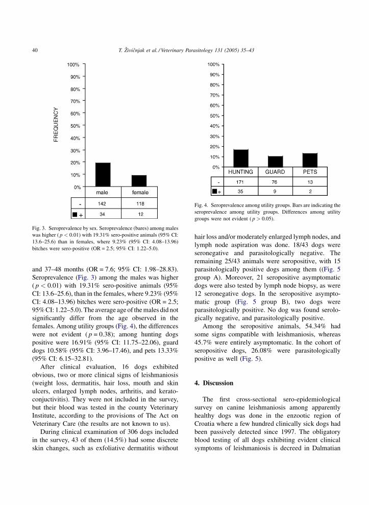
Fig. 3. Seroprevalence by sex. Seroprevalence (bares) among males
was higher ( p < 0.01) with 19.31% sero-positive animals (95% CI:
13.6–25.6) than in females, where 9.23% (95% CI: 4.08–13.96)
bitches were sero-positive (OR = 2.5; 95% CI: 1.22–5.0).
Fig. 4. Seroprevalence among utility groups. Bars are indicating the
seroprevalence among utility groups. Differences among utility
groups were not evident ( p > 0.05).
and 37–48 months (OR = 7.6; 95% CI: 1.98–28.83).
Seroprevalence (Fig. 3) among the males was higher
( p < 0.01) with 19.31% sero-positive animals (95%
CI: 13.6–25.6), than in the females, where 9.23% (95%
CI: 4.08–13.96) bitches were sero-positive (OR = 2.5;
95% CI: 1.22–5.0). The average age of the males did not
significantly differ from the age observed in the
females. Among utility groups (Fig. 4), the differences
were not evident ( p = 0.38); among hunting dogs
positive were 16.91% (95% CI: 11.75–22.06), guard
dogs 10.58% (95% CI: 3.96–17.46), and pets 13.33%
(95% CI: 6.15–32.81).
After clinical evaluation, 16 dogs exhibited
obvious, two or more clinical signs of leishmaniosis
(weight loss, dermatitis, hair loss, mouth and skin
ulcers, enlarged lymph nodes, arthritis, and kerato-
conjuctivitis). They were not included in the survey,
but their blood was tested in the county Veterinary
Institute, according to the provisions of The Act on
Veterinary Care (the results are not known to us).
During clinical examination of 306 dogs included
in the survey, 43 of them (14.5%) had some discrete
skin changes, such as exfoliative dermatitis without
hair loss and/or moderately enlarged lymph nodes, and
lymph node aspiration was done. 18/43 dogs were
seronegative and parasitologically negative. The
remaining 25/43 animals were seropositive, with 15
parasitologically positive dogs among them ((Fig. 5
group A). Moreover, 21 seropositive asymptomatic
dogs were also tested by lymph node biopsy, as were
12 seronegative dogs. In the seropositive asympto-
matic group (Fig. 5 group B), two dogs were
parasitologically positive. No dog was found serolo-
gically negative, and parasitologically positive.
Among the seropositive animals, 54.34% had
some signs compatible with leishmaniosis, whereas
45.7% were entirely asymptomatic. In the cohort of
seropositive dogs, 26.08% were parasitologically
positive as well (Fig. 5).
4. Discussion
The first cross-sectional sero-epidemiological
survey on canine leishmaniosis among apparently
healthy dogs was done in the enzootic region of
Croatia where a few hundred clinically sick dogs had
been passively detected since 1997. The obligatory
blood testing of all dogs exhibiting evident clinical
symptoms of leishmaniosis is decreed in Dalmatian

T. Zivicnjak et al. / Veterinary Parasitology 131 (2005) 35–43 41
Fig. 5. Clinical examination reliability. Group A: dogs with some
discrete skin changes, such as exfoliative dermatitis without hair loss
and/or moderately enlarged lymph nodes. Group B: asymptomatic
dogs, ser� par�: serologically and parasitologically negative; ser+ -
par+: serologically and parasitologically positive; ser+ par�: ser-
ologically positive and parasitologically negative.
counties by The Act on Veterinary Care. However,
following the usual pattern, the dog owners in this area
often euthanize the affected animals themseleves,
without ever consulting a veterinarian. Therefore, it is
impossible to estimate the number of ill dogs with any
precision or provide accurate figures. On the other
hand, the owners were unwilling to allow the testing
of dogs that did not appear ill for the ‘‘scientific
purposes’’; they often did not permit us to take their
blood. Consequently, we were able to obtain the
samples from only 306 asymptomatic dogs, out of
approximately 10,000 dogs living in the area. There-
fore, the dog samples should not be considered
representative of the whole dog population in the area
studied. We decided to use our dot-ELISA modifica-
tion as a simple and economical technique for field
screening of dog populations.
The survey was carried out on the dogs whose
owners regarded them healthy (not included in the
provisions of The Act on Veterinary Care) and agreed
with the protocol, but those dogs could not be regarded
as entirely asymptomatic. Although they worked well
and seemed well, and exhibited good appetite and
condition, some of them had some visible changes on
skin and/or moderate enlargement of lymph nodes.
Since, dogs considered healthy by their owners
(including asymptomatic infected dogs) will not be
taken to the veterinarian, these dogs will be a reservoir
for infection within the general population.
Similar to other countries in the Mediterranean
basin (Deplazes et al., 1998; Solano-Gallego et al.,
2001; Cardoso et al., 2004), the prevalence in the
endemic region of Croatia has been shown to widely
vary among the identified endemic areas. In the city of
Split, positive animals were scattered without any
grouping. Neither could we explain the differences in
prevalence among positive locations and high OR in
Zagvozd, Zezevica and Klis. These villages did not
differ significantly by the altitude. Three of four
villages without canine leishmaniosis are situated at
the altitude of 599 m or more. In Split-Dalmatia
County, 600 m altitude is the limit where the
Mediterranean climate switches to a cooler one,
associated with changes in ecological conditions. The
fourth village without positive dogs in the survey is
situated at an altitude of 457 m, in the close vicinity to
the three positive villages. According to Ashford
(2000), the distribution of leishmaniosis is determined
by its vector, reservoir host, and limiting factors, i.e.
specific environmental requirements that lead to focal
distribution of disease. In an entomological survey
performed in 1998 in the region (Miscevic et al.,
1998), the vectors were found in coastal cites of Split,
Omis and Makarska, but such survey was not
performed deeper in the hinterlands and at higher
altitudes. Further entomological investigations should
be done to investigate if the vector occurs at higher
altitudes. The distribution of seropositive dogs within
the canine population showed significant heterogene-
ity according to the age and sex; however, it was not
relevant whether the animal was kept mainly indoors
(pets) or outdoors (guard and hunting dogs). Our
prevalence results according to outdoors and indoors
lifestyles differ from the results published by
Zaffaroni et al., (1999) where serological positivity
to Leishmania sp. was significantly associated with a
dog’s outdoor lifestyle. The increase in the prevalence
of the seropositive dogs with age and the final decrease
in seropositivity in the older group of dogs (>84

T. Zivicnjak et al. / Veterinary Parasitology 131 (2005) 35–4342
months) is in agreement with other studies (Martinez
Cruz et al., 1990; Abranches et al., 1991; Fisa et al.,
1999). This could be related to the cumulative increase
of the time of exposure of dogs to phlebotomines and
the increase of the death rate in old animals.
As in some other Mediterranean foci (Martinez
Cruz et al., 1990; Fisa et al., 1999; Zaffaroni et al.,
1999), we found a higher prevalence among males
than in females. In our study, we could not find any
gender-associated differences in exposure to the
parasite; males and females were kept in the same
manner. One possible explanation could be an increase
in female mortality in which pregnancy and nursing
may play an important role (Fisa et al., 1999), but
gender-related differences in the host immune
response might play a role in the resistance and
susceptibility to infection. As it was shown in mice for
L. major (Mock and Nacy, 1988), hamsters for New
World leishmaniasis (Travi et al., 2002) and human
macrophages for L. donovani (Zhang et al., 2001)
possible testosterone immunomodulation in dogs
should be investigated.
Our dot-ELISA modification, used for screening
purposes, could detect subclinical and active leish-
maniosis at sera dilution 1/600, which was also
parasitologically proven in 26.08% cases. Lower titres
were not assayed in the screening to facilitate results
interpretation. Interpretation of results obtained by
detection of specific antibodies in dogs from endemic
areas is often difficult. When we deal with asympto-
matic animals in endemic areas, immunological
techniques do not discriminate between infected
and resistant animals (Cabral et al., 1998; Cardoso
et al., 1998).
We can conclude that we recorded a possible source
of infection in the area; we are also aware that there
might be a number of dogs with lower titre which were
not recorded in our cross-sectional study. There is a
need for the serological follow-up in a longitudinal
study, with serially diluted sera samples. Retests might
be helpful in distinguishing early phases of the disease
from resistant animals in those that have low antibody
titers.
Furthermore, the value of clinical examination
reliability that defines which dog has to be blood-
tested on leishmaniosis in the endemic area should be
re-evaluated. Although clinical examination is neces-
sary, our survey within asymptomatic dogs’ popula-
tion has shown how prevalence can be underestimated.
It is obvious that a diagnosis could be established only
after a combination of the clinical examination and
other diagnostic techniques.
References
Abranches, P., Silva-Pereira, M.C.D., Conceicao-Silva, F.M., Santos
Gomes, G.M., Janz, J.G., 1991. Canine leishmaniasis: patholo-
gical and ecological factors influencing transmission of infec-
tion. J. Parasitol. 77, 557–561.
Amela, C., Mendez, I., Torcal, J.M., Medina, G., Pachon, I.,
Canavate, C., Alvar, J., 1995. Epidemiology of canine leishma-
niasis in the Madrid region. Spain. Eur. J. Epidemiol. 11, 157–
161.
Ashford, R.W., 2000. The leishmaniases as emerging and reemer-
ging zoonoses. Int. J. Parasitol. 30, 1269–1281.
Bradford, M., 1976. A rapid and sensitive method for the quantifica-
tion of microgram quantities of protein utilizating the principle
of protein-dye binding. Anal. Biochem. 72, 248–254.
Cabral, M., O’Grady, J.E., Gomes, S.J., Sousa, C., Thompson, H.,
Alexander, J., 1998. The immunology of canine leishmaniasis:
strong evidence for a developing disease spectrum from asymp-
tomatic dogs. Vet. Parasitol. 76, 173–180.
Cardoso, L., Neto, F., Sousa, J.C., Rodrigues, M., Cabral, M., 1998.
Use of a leishmanin skin test in the detection of canine Leish-
mania-specific cellular immunity. Vet. Parasitol. 79, 213–220.
Cardoso, L., Rodrigues, M., Santos, H., Schoone, G.J., Carreta, P.,
Varejao, E., van Benthem, B., Afonso, M.O., Alves-Pires, C.,
Semiao-Santos, S.J., Rodrigues, J., Schallig, H.D.F.H., 2004.
Sero-epidemiological study of canine Leishmania spp. infection
in the municipality of Alijo (Alto Douro, Portugal) Vet. Para-
sitol. 121, 21–32.
Ciaramella, P., Oliva, G., Luna, R.D., Gradoni, L., Ambrosio, R.,
Cortese, L., Scalone, A., Persechino, A., 1997. A retrospective
clinical study of canine leishmaniasis in 150 dogs naturally
infected by Leishmania infantum. Vet. Rec. 141, 539–543.
Deplazes, P., Grimm, F., Papaprodromou, M., Cavaliero, T., Gra-
miccia, M., Christofi, G., Christofi, N., Economides, P., Eckert,
J., 1998. Canine leishmaniosis in Cyprus due to Leishmania
infantum MON 1. Acta Trop. 71, 169–178.
Evans, D.A., 1987. Leishmania. In: Taylor, A.E.R., Baker, H.R.
(Eds.), In Vitro Methods for Parasite Cultivation. Academic
Press, New York, pp. 52–75.
Ferrer, L., Rabanal, R., Fondevila, J., Ramos, A., Domingo, M.,
1988. Skin lesions in canine leishmaniasis. J. Small Anim. Pract.
29, 381–388.
Ferrer, L.M., 1999. Clinical aspects of canine leishmaniasis. In:
Killick-Kendrick, R. (Ed.), Canine Leishmaniasis: An Update.
Proceedings of a Canine Leishmaniasis Forum, Barcelona
(Sitges), 28–31 January, pp. 6–10.
Fisa, R., Gallego, M., Castillejo, S., Aisa, M.J., Serra, T., Riera, C.,
Carrio, J., Gallego, J., Portus, M., 1999. Epidemiology of canine
leishmaniosis in Catalonia (Spain). The example of the Priorat
focus. Vet. Parasitol. 83, 87–97.

T. Zivicnjak et al. / Veterinary Parasitology 131 (2005) 35–43 43
Gradoni, L., Gramiccia, M., Mancianti, F., Pieri, S., 1988. Studies on
canine leishmaniasis control. 2. Effectiveness of control mea-
sures against canine leishmaniasis in the isle of Elba. Italy Trans.
R. Soc. Trop. Med. Hyg. 82, 568–571.
Killick-Kendrick, R., 1999. The biology of phlebotomine sand flies.
Clin Dermatol. 17, 279–289.
Koutinas, A.F., Polizopoulou, Z.S., Saridomichelakis, M.N., Argyr-
iadis, D., Fytianou, A., Plevraki, K.G., 1999. Clinical considera-
tions on canine visceral leishmaniasis in Greece: a retrospective
study of 158 cases (1989–1996). J. Am. Anim. Hosp. Assoc. 35,
376–383.
Lımoncu, M.E., Balcioglu, C., Yerelı, K., Ozbel, Y., Ozbılgın, A.,
1997. A new experimental in vitro culture medium for cultiva-
tion of leishmania sp. J. Clin. Microbiol. 35, 430–431.
Mancianti, F., Pedonese, F., Poli, A., 1996. Evaluation of dot
enzyme-linked immunosorbent assay (dot-ELISA) for the ser-
odiagnosis of canine leishmaniosis as compared with indirect
immunofluorescence assay. Vet. Parasitol. 65, 1–9.
Martinez Cruz, M.S., Martinez Moreno, A., Martinez Moreno, F.J.,
Martinez Gomez, F., Hernandez Rodrıguez, S., 1990. Epide-
miologıa de la leishmaniosis canina en Cordoba. Rev. Iber.
Parasitol. 50, 1–7.
Martinkovic F., Lukas, D., Marinculic, A. Zivicnjak, T., Ramadan,
P., Dzakula, N., Stojcevic, D., 2001. Epidemiological Investiga-
tion of Leishmaniasis in Croatia. In: Abstract book. Croatian and
Slovenian Symposium on Microbiology and Infectious Dis-
eases: Zoonoses Today and Tomorrow. Plitvice, 2001.
Miscevic, Z., Milutinovic, M., Ivovic, V., 1998. Fauna and distribu-
tion of sandflies (Diptera, Phlebotomidae) in Yugoslavia, Croa-
tia Macedonia and their role in the transmission of parasitic and
viral diseases. Acta Vet. 48, 163–172.
Mock, B.A., Nacy, C.A., 1988. Hormonal modulation of sex differ-
ences in resistance to leishmania major systemic infections.
Infect. Immun. 56, 3316–3319.
Mulic, R., Ropac, B.D., Zoric, I., Bradaric, N., 2002. Epidemiologic
and ecologic characteristics of some diseases transmitted by
arthropods on the littoral of the Republic of Croatia. Mil. Med.
167, 321–325.
Molina, R., Amela, C., Nieto, J., San-Andres, M., Gonzalez, F.,
Castillo, J.A., Lucientes, J., Alvar, J., 1994. Infectivity of dogs
naturally infected with Leishmania infantum to colonized
Phlebotomus perniciosus. Trans. R. Soc. Trop. Med. Hyg. 88,
491–493.
Pappas, M.G., Hajkowski, R., Hockmeyer, W.T., 1983. Dot enzyme-
linked immunosorbent assay (dot-elisa): a micro technique for
the rapid diagnosis of visceral leishmaniasis. J. Immunol. Meth.
64, 205–214.
Portus, M., Fisa, R., Serra, T., Gallego, M., Mora, M., 1987. Estudios
seroepidemiologicos sobre la leishmaniasis canina en Catalunia.
Med. Vet. 4, 569–575.
Rioux, J.A., Lanotte, G., Serres, E., Pratlong, F., Bastien, P.,
Perieres, J., 1990. Taxonomy of leishmania use of isoenzymes.
Suggestions for a new classification. Ann. Parasitol. Hum.
Comp. 65, 111–125.
Sideris, V., Papadopoulou, G., Dotsika, E., Karagouni, E., 1999.
Asymptomatic canine leishmaniasis in Greater Athens area.
Greece Eur. J. Epidemiol. 15, 271–276.
Solano-Gallego, L., Morell, P., Arboix, M., Alberola, J., Ferrer, L.,
2001. Prevalence of Leishmania infantum infection in dogs
living in an area of canine leishmaniasis endemicity using
PCR on several tissues and serology. J. Clin. Microbiol. 39,
560–563.
Tartaglia, P., 1937. La leishmaniose canine a Split. Bull.de l’Office
Intern. d’Hyg. Publ. XXIV: 9 1927.
Travi, B.L., Osorio, Y., Melby, P.C., Chandrasekar, B., Arteaga, L.,
Saravia, N.G., 2002. Gender is a major determinant of the
clinical evolution and immune response in hamsters infected
with Leishmania spp. Infect. Immun. 70, 2288–2296.
Vercammen, F., Berkvens, D., Brandt, J., Vansteenkiste, W., 1998. A
sensitive and specific 30-min Dot-ELISA for the detection of anti-
leishmania antibodies in the dog. Vet. Parasitol. 79, 221–228.
Zaffaroni, E., Rubaudo, L., Lanfranchi, P., Mignone, W., 1999.
Epidemiological patterns of canine leishmaniosis in Western
Liguria (Italy). Vet. Parasitol. 81, 11–19.
Zhang, H., Zhao, J., Wang, P., Qiao, Z., 2001. Effect of testosterone
on Leishmania donovani infection of macrophages. Parasitol.
Res. 87, 674–676.
Zivicnjak T., Stojcevic D., Marinculic A., Ramadan P., Petrinovic T.,
Dzakula N, 1998. Epizootiological Survey for Canine Visceral
Leishmaniasis and the Isolation of the Protozoa Leishmania
infantum. In: Abstract Book from 1st Croatian Congress on
Infectious Diseases. October 1–3, Dubrovnik, Croatia, p. 106.
Related Documents


![[Medicina Veterinaria] Leishmaniosis Canina y Felina](https://static.cupdf.com/doc/110x72/55cf9b0e550346d033a48f8f/medicina-veterinaria-leishmaniosis-canina-y-felina.jpg)